cona and uea-i lectin histochemistry of parotid gland mucoepidermoid carcinoma
TRANSCRIPT

49
Abstract: Mucoepidermoid carcinoma (MEC)corresponds to 5-12% of all salivary gland tumours,and is classified as low, intermediate or high grade.Traditionally, immunohistochemistry was consideredas the complementary tool for diagnosis of salivarygland neoplasia. Lectin histochemistry has also beenincreasingly used in recent years. In this work, lectinswere used as histochemical markers for normal andtransformed parotid glands. Biopsy specimens of 15cases diagnosed as MEC (low, intermediate and highgrade) of the parotid gland were trypsin- and methanol-H2O2-treated and incubated with horseradishperoxidase (HRP)-conjugated lectins, ConcanavalinA (Con A-HRP) and Ulex europeus I (UEA-I-HRP). ConA stained the neoplasic cells of MEC (all grades). Inhigh and intermediate cases, ductal cells were weaklystained by Con A. UEA-I weakly stained normal cellsof the excretory duct and neoplasic cells in high grade.Neoplasic cells in intermediate grade were moderatelystained and in low grade, the cell membrane wasintensely stained with UEA-I. Stroma presented adirect relation between malignancy and stainingintensity for UEA-I. The results indicated that lectinhistochemistry distinguished the cell biology amonghistological grades of MEC. (J Oral Sci 52, 49-54, 2010)
Keywords: parotid gland; lectin histochemistry;mucoepidermoid carcinoma.
IntroductionCarbohydrates have an enormous potential for encoding
biological information. These combination molecules (inglycoproteins and glycolipids) dot the outer surface of allcells and serve as cellular identification tags to thesurrounding world (1). The extracellular matrix (ECM)provides a physical framework for cellular attachmentand facilitates normal physiological regulation of cellproliferation, migration and differentiation (2).
Lectins are structurally diverse carbohydrate-bindingproteins or glycoproteins of non-immune origin (3) thatagglutinate cells and recognize carbohydrates in oligo-saccharides and glycoconjugates (4). By virtue of theirbinding specifities, lectins have been used in the medicaland biological areas. In histochemistry, lectins with differentcarbohydrate specificity can provide a sensitive detectionsystem for changes in glycosylation and carbohydrateexpression that may occur during embryogenesis, growthand disease. Tumour lectinology has so far showncytochemical and histochemical differences betweennormal and transformed tissues such as mammary (5,6),brain (7), oral tissues (8), lung (9) and within a single classof tumour (10).
Quantitative and qualitative changes in glycoconjugatesof cell outer and inner membranes could be significant inthe development and progression of pathologies, including
Journal of Oral Science, Vol. 52, No. 1, 49-54, 2010
Correspondence to Dr. Ana Paula Veras Sobral, Rua MonteAlverne, 107/05, Hipódromo – Recife – PE 52041-610 – BrazilTel: +55-81-34581088Fax: +55-81-34586758E-mail: [email protected] & [email protected]
ConA and UEA-I lectin histochemistry of parotid glandmucoepidermoid carcinoma
Ana Paula V. Sobral1,2), Moacyr J. B. M. Rego1), Carmelita L. B. Cavalacanti1), Luiz B. Carvalho Jr1,3) and Eduardo I. C. Beltrão1,3)
1)Keizo Asami Immunopathology Laboratory (LIKA), Federal University of Pernambuco (UFPE), Recife, Brazil
2)Oral Pathology Laboratory, Dentistry College, Pernambuco University (UPE), Camaragibe, Brazil3)Biochemistry Department, Biological Science Center, Federal University of Pernambuco (UFPE),
Recife, Brazil
(Received 4 June and accepted 27 November 2009)
Original

50
neoplasias. Higher or weaker and even the absence ofstaining patterns between normal and transformed tissuessuggest derangement of secretory mechanisms. Theobservation that, in general, the more anaplastic the cellbecomes, more intense its staining, seems to indicate thatthe site and nature of cell-surface glycoconjugates arealtered. In addition, tissue factors may influence andinduce differentiation/dedifferentiation reflected in thedifferent lectin binding patterns (7).
Mucoepidermoid carcinoma (MEC) is the most commonmalignant salivary tumour, corresponding to 5-12% of allsalivary gland tumours, and is classified as low, intermediateor high grade. Salivary gland tumours are classifiedessentially based on morphology (11) and MEC ischaracterized by the presence of various cell typesresembling the excretory duct of salivary glands and itsconfiguration varies with the degree of malignancy (12).Regarding the pathology of salivary gland tumours,discussions related to morphology, pathogenesis, diagnosisand classification have taken place specially to characterizethe constituent tumour cells to establish differences amongtumour types.
This work aimed to evaluate the use of ConcanavalinA (Con A-HRP) and Ulex europeus I (UEA-I-HRP) inhistochemistry for the characterization of glucose and/ormannose and L-fucose profile in mucoepidemoid carcinomaof the major salivary gland.
Materials and MethodsSpecimens
Fifteen cases of mucoepidermoid carcinoma of theparotid gland (low grade, n = 5; intermediate grade, n =5 and high grade, n = 5) classified according to WHO (13),and 15 normal salivary glands were obtained from theTissue Archives of the Oral Pathology Department, Schoolof Dentistry, University of the State of Pernambuco (UPE).This work was approved by University of PernambucoEthical Committee.
Lectin histochemistry:Four-micrometer-thick sections of the specimens were
deparaffinized in xylene and dehydrated in graded seriesof alcohol (100-70%). Slices were treated with 0.1% (w/v)trypsin solution for 2 min at 37°C and with a 0.3% (v/v)methanol-H2O2 solution for 30 min at 25°C and thenincubated with HRP-conjugated lectins (Con A-HRP andUEA-I-HRP at 50 µg/ml). PBS (10 mM phosphate buffer,pH 7.2, 150 mM NaCl) was used to prepare all solutionsand as washing solution between each step. Peroxidase wasvisualized with a solution of diaminobenzidine-H2O2 for5-8 min. Haematoxylin was used for counter-staining.
Tissues were evaluated by light microscopy. Lectin-bindinginhibition assays were developed by incubating lectinwith its corresponding specific sugars, methyl-α-D-mannoside for Con A and L-fucose for UEA-I (100 to 500mM) prior to sample incubation.
ResultsIn normal parotid gland, myoepithelial cells and vascular
endothelium were weakly stained by Con A and UEA-I.Acinar and luminal cells of the excretory ducts were alsorecognized by UEA-I with a weak staining pattern.
Neoplasic cells in all cases of MEC were stained by ConA (glucose and mannose specific) with intense staining ofthe cytoplasm and/or membrane (Fig. 1). Intermediategrade MEC presented a moderate staining with Con A, andthis was the highest staining pattern among the threegrades (Fig. 1c). Neoplasic stroma was not stained withCon A in high grade MEC, while low and intermediategrades were weakly stained. Vascular endothelium and ductstructure were differentially stained with Con A (Table 1).
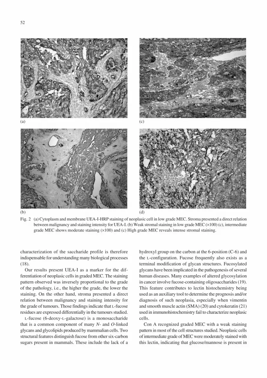
The UEA-I staining pattern varied from weak to intensefor neoplasic cells. The staining in low grade MEC wasintense in the cytoplasm and membrane (Fig. 2a). Stroma,connective tissue around the neoplasic cells, was notstained by UEA-I in low grade MEC (Fig. 2b), but wasweakly stained in the intermediate grade (Fig. 2c) andintensely stained in high grade (Fig. 2d). Detailed resultsare presented in Table 1.
Tissue staining was completely abolished in lectinbinding inhibition assays using methyl-α-D-mannosidefor Con A and L-fucose for UEA-I at 300 mM. Thisconcentration was the lowest, with the highest inhibitionas well as the lowest background staining.
DiscussionClinicopathologic investigations have described that
recurrence and overall survival in MEC of salivary glandsare linked to tumour grade and size, clinical stage ofdisease, perineural and vascular involvement, and lymphnode distant metastasis. MEC is a unique epithelialneoplasm composed of epidermoid, mucous andintermediate cells in variable proportions. The architectureof this neoplasm varies from predominantly cystic withabundant mucous cells to exclusively solid nests and sheetsof epidermoid cells with squamoid differentiation and apaucity of intermediate cells. Historically, this neoplasmpresents clinical behaviour dependent upon its his-topathologic appearance varying from a relatively benignneoplasm with a low grade morphology to an extremelyaggressive neoplasm which has a high mortality (12,14).
Glycoproteins are a family of complex proteins carrying

51
covalently linked oligosaccharide chains. In the oligo-saccharides, the monosaccharide moiety is hexosaminesin the acyl form, neutral sugar such as glucose, galactose,fucose, and mannose and various derivatives of sialic acid(N-acetyl neuraminic acid). These monosaccharide unitscan attach to one another at multiple points, formingbranches. Two identical monosaccharides can link to form11 different disaccharides (1,15). Neoplastic transformationis associated with altered cell surface carbohydratecomposition of the cell membrane and the changes in thesurface of tumour cells are relevant to their abnormalgrowth, metastasis and changes in cell adhesion (16).
Lectins decipher glycocodes, recognizing specific sugarsto carry out various functions such as cell attachment,migration or invasion. Glycocodes are different from eithercodes of the nucleotides in nucleic acids and the aminoacids in proteins, which can interconnect linearly. Among
all of the variety of adhesion molecules, cell-surfacecarbohydrate structures have been the focus of manyinvestigative efforts. Typically, lectins and their com-plimentary carbohydrate are located on the surfaces ofopposing cells, which can be of the same type or differenttypes. Their interactions are required for cell differentiation,development and pathological states (15). Cancer cells usecarbohydrate moieties to escape recognition by the immunecells as they migrate through the body (1).
In the last two decades, it has been possible to investigateother biologic features which reflect the identity of cells.In this context, lectin histochemistry is a useful tool sinceinvasiveness, adhesion, angiogenesis in tumour cellsurroundings and apoptotic susceptibility are functionallymaintained by a combination of defined molecules. Theseprocesses are affected, directly or indirectly, by N- and/orO-glycosylation of functional proteins or lipids (17). The
Fig. 1 (a) Con A-HRP staining in neoplasic cells of low grade MEC (×100), (b) intermediate grade MEC (×100) and (c) (×400)and high grade MEC (d).
(d)(b)
(a) (c)
52
characterization of the saccharide profile is thereforeindispensable for understanding many biological processes(18).
Our results present UEA-I as a marker for the dif-ferentiation of neoplasic cells in graded MEC. The stainingpattern observed was inversely proportional to the gradeof the pathology, i.e., the higher the grade, the lower thestaining. On the other hand, stroma presented a directrelation between malignancy and staining intensity forthe grade of tumours. Those findings indicate that L-fucoseresidues are expressed differentially in the tumours studied.
L-fucose (6-deoxy-L-galactose) is a monosaccharidethat is a common component of many N- and O-linkedglycans and glycolipids produced by mammalian cells. Twostructural features distinguish fucose from other six-carbonsugars present in mammals. These include the lack of a
hydroxyl group on the carbon at the 6-position (C-6) andthe L-configuration. Fucose frequently also exists as aterminal modification of glycan structures. Fucosylatedglycans have been implicated in the pathogenesis of severalhuman diseases. Many examples of altered glycosylationin cancer involve fucose-containing oligosaccharides (19).This feature contributes to lectin histochemistry beingused as an auxiliary tool to determine the prognosis and/ordiagnosis of such neoplasia, especially when vimentinand smooth muscle actin (SMA) (20) and cytokeratin (21)used in immunohistochemistry fail to characterize neoplasiccells.
Con A recognized graded MEC with a weak stainingpattern in most of the cell structures studied. Neoplasic cellsof intermediate grade of MEC were moderately stained withthis lectin, indicating that glucose/mannose is present in
Fig. 2 (a) Cytoplasm and membrane UEA-I-HRP staining of neoplasic cell in low grade MEC. Stroma presented a direct relationbetween malignancy and staining intensity for UEA-I. (b) Weak stromal staining in low grade MEC (×100) (c), intermediategrade MEC shows moderate staining (×100) and (c) High grade MEC reveals intense stromal staining.
(b) (d)
(a) (c)

53
a higher content in this grade. Since stroma was notrecognized by Con A in high grade MEC as well asvascular endothelium and desmoplasic cells, the absenceand/or non availability of Con A-specific sugars areindicative of biochemical alterations in the glycosylationor glycosyl transfer pathways.
An interesting finding was observed regarding connectivetissue around neoplasic cells; UEA-I strongly stainedthese cells in intermediate grade MEC but a weak stainingwas observed in the other two grades. Con A staining wasweak in high grade of MEC. Studies have demonstratedthat progression in grades of MEC is related to the decreasein desmoplasia. Such data suggest that there is no correlationbetween immunohistochemical markers (vimentin andSMA) and carbohydrate expression in connective tissuearound neoplasic cells using lectin histochemistry (20). Inthis context, the saccharide profile can be used as extrainformation for the characterization of stroma in the MEC.
As observed in other malignant tumours, lectin histo-chemistry is able to characterize cell biology with a
specificity as high as antibody-antigen recognition. Suchspecificity is corroborated by the abolishment of lectin-carbohydrate binding, which is as precise as that observedin immunohistochemistry. Data presented here indicate thatlectins give useful information in distinguishing and/orcharacterizing cell biology among histological grades ofMEC.
AcknowledgmentsThis work was financially supported by CNPq, CAPES
and FACEPE.
References1. Nangia-Makker P, Conklin J, Hogan V, Raz A (2002)
Carbohydrate-binding proteins in cancer, and theirligands as therapeutic agents. Trends Mol Med 8,187-192.
2. Suwiwat S, Ricciardelli C, Tammi R, Tammi M,Auvinen P, Kosma VM, LeBaron RG, RaymondWA, Tilley WD, Horsfall DJ (2004) Expression of
Table 1 Lectin histochemistry of mucoepidermoid carcinoma of salivary gland using ConA and UEA-I

54
extracellular matrix components versican,chondroitin sulfate, tenascin, and hyaluronan, andtheir association with disease outcome in node-negative breast cancer. Clin Cancer Res 10, 2491-2498.
3. Kennedy JF, Palva PMG, Corella MTS, CavalcantiMSM, Coelho LCBB (1995) Lectins, versatileproteins of recognition: a review. Carbohydr Polym26, 219-230.
4. Sharon N, Lis H (1993) Carbohydrates in cellrecognition. Sci Am 268, 82-89.
5. Campos LM, Cavalcanti CL, Lima-Filho JL,Carvalho LB, Beltrão EI (2006) Acridinium esterconjugated to lectin as chemiluminescenthistochemistry marker. Biomarkers 11, 480-484.
6. Korourian S, Siegel E, Kieber-Emmons T, Monzavi-Karbassi B (2008) Expression analysis ofcarbohydrate antigens in ductal carcinoma in situof the breast by lectin histochemistry. BMC cancer8, 136.
7. Beltrão EI, Medeiros PL, Rodrigues OG, Figueredo-Silva J, Valença MM, Coelho LC, Carvalho LB Jr(2003) Parkia pendula lectin histochemistry markerfor meningothelial tumour. Eur J Histochem 47,139-142.
8. Pillai KR, Remani P, Kannan S, Sujathan K, MatthewB, Vijayakumar T, Nair MK, Menon VP (1996)Lectin histochemistry of oral premalignant andmalignant lesions: correlation of JFL and PNAbinding pattern with tumour progression. Eur JCancer B Oral Oncol 32B, 32-37.
9. Thöm I, Schult-Kronefelda O, Burkholderb I, GoernM, Andritzky B, Blonski K, Kugler C, Edler L,Bokemeyer C, Schumacher U, Laack E (2007)Lectin histochemistry of metastatic adenocarcinomasof the lung. Lung Cancer 56, 391-397.
10. Gabius HJ (1998) Tumor lectinology: at theintersection of carbohydrate chemistry, biochemistry,cell biology and oncology. Angew Chem Int EdEng 27, 1267-1276.
11. Kokemueller H, Brueggemann N, Swennen G,Eckardt A (2005) Mucoepidermoid carcinoma of thesalivary glands – clinical review of 42 cases. OralOncol 41, 3-10.
12. Goode RK, El-Naggar AK (2005) Mucoepidermoidcarcinoma. In: World Health Organizationclassification of tumours: pathology and genetics ofhead and neck tumours, Barnes L, Eveson JW,Reichart P, Sidransky D eds, IARC Press, Lyon, 219-220.
13. Barnes L, Eveson JW, Reichart P, Sidransky D(2005) World Health Organization classification oftumours: pathology and genetics of head and necktumours. IARC Press, Lyon.
14. Hicks J, Flaitz C (2000) Mucoepidermoid carcinomaof salivary glands in children and adolescents:assessment of proliferation markers. Oral Oncol36, 454-460.
15. Sharon N, Lis H (2004) History of lectins: fromhemagglutinins to biological recognition molecules.Glycobiology 14, 53R-62R.
16. Manoharana S, Padmanabhana M, KolanjiappanaK, Ramachandranb CR, Suresha K (2004) Analysisof glycoconjugates in patients with oral squamouscell carcinoma. Clin Chim Acta 339, 91-96.
17. Ono M, Hakomori S (2004) Glycosylation definingcancer cell motility and invasiveness. Glycoconj J20, 71-78.
18. Shimma Y, Jigami Y (2004) Expression of humanglycosyltransferase genes in yeast as a toll forenzimatic synthesis of sugar chain. Glycoconj J 21,75-78.
19. Becker DJ, Lowe JB (2003) Fucose: biosynthesisand biological function in mammals. Glycobiology13, 41R-53R.
20. Sobral AP, Loducca SV, Nunes FD, de Araújo NS,Kowaski LP, Araújo VC (2004) Relationshipbetween major and minor sa l ivary glandmucoepidermoid carcinoma malignancy gradingand presence of s t romal myofibrobas t s :immunohistochemical study. J Oral Pathol Med 33,335-339.
21. Sobral AP, Loducca SV, Kowalski LP, Santos IR,Almeida OP, Araújo NS, Araújo VC (2002)Immunohistochemical distinction of high-grademucoepidermoid carcinoma and epidermoidcarcinoma of the parotid region. Oral Oncol 38,437-440.