comparison of the bacterial communities of wild and captive sponge clathria prolifera from the...
TRANSCRIPT
ORIGINAL ARTICLE
Comparison of the Bacterial Communities of Wildand Captive Sponge Clathria proliferafrom the Chesapeake Bay
LeLeng To Isaacs & Jinjun Kan & Linh Nguyen &
Patrick Videau & Matthew A. Anderson &
Toby L. Wright & Russell T. Hill
Received: 25 October 2008 /Accepted: 25 April 2009# Springer Science + Business Media, LLC 2009
Abstract The red-beard sponge Clathria prolifera, whichis widely distributed in the USA, has been widely used as amodel system in cell biology and has been proposed as asuitable teaching tool on biology and environmentalsciences. We undertook the first detailed microbiologicalstudy of this sponge on samples collected from theChesapeake Bay. A combination of culture-based studies,denaturing gradient gel electrophoresis, and bacterialcommunity characterization based on 16S rRNA genesequencing revealed that C. prolifera contains a diverseassemblage of bacteria that is different from that in thesurrounding water. C. prolifera individuals were success-fully maintained in a flow-through or recirculation aqua-culture system for over 6 months and shifts in the bacterialassemblages of sponges in aquaculture compared with wildsponges were examined. The proteobacteria, bacteroidetes,actinobacteria, and cyanobacteria represented over 90% ofthe species diversity present in the total bacterial commu-nity of the wild C. prolifera. Actinobacteria, cyanobacteria,and spirochetes were not represented in clones obtained
from C. prolifera maintained in the aquaculture systemalthough these three groups comprised ca. 20% of theclones from wild C. prolifera, showing a significant effectof aquaculture on the bacterial community composition.This is the first systematic characterization of the bacterialcommunity from a sponge found in the Chesapeake Bay.Changes in sponge bacterial composition were observed insponges maintained in aquaculture and demonstrate theimportance of monitoring microbial communities whencultivating sponges in aquaculture systems.
Keywords Clathria prolifera .Microciona prolifera .
Aquaculture . 16S rRNA gene
Introduction
Sponges form symbiotic relationships with a variety ofheterotrophic and autotrophic bacteria, protozoa, andmacroalgae (Lee et al. 2001; Hentschel et al. 2003; Hill2004b). In some cases, the complex microbial communitiescan represent more than 40% of the biomass of the sponge(Vacelet and Donadey 1977). Animals coevolved andcoexisted with diverse assemblages of microbes that maybe important for health and development and studyingspecies-specific associations of microbes with invertebratehosts may provide working models for studying vertebratehost–microbe communities (Ruby et al. 2004). Further-more, microbes play significant roles in aquatic ecosystemsand exert a crucial impact on environmental health andsustainability (Azam and Worden 2004). Therefore, infor-mation on normal microbiota of sponges is critical tounderstanding the significance of microbes in structuringhealthy aquatic ecosystems and serves as baseline data forstressed communities and in developing prognostic models.
Mar BiotechnolDOI 10.1007/s10126-009-9192-3
Contribution no. 07-118 from the Center of Marine Biotechnology.
L. T. Isaacs : L. Nguyen : P. Videau : T. L. WrightDepartment of Biology, Goucher College,Baltimore, MD 21204, USA
J. Kan :M. A. Anderson : T. L. Wright : R. T. Hill (*)Center of Marine Biotechnology,University of Maryland Biotechnology Institute,Columbus Center Suite 236, 701 E. Pratt Street,Baltimore, MD 21202, USAe-mail: [email protected]
Present Address:J. KanDepartment of Earth Sciences, University of Southern California,Los Angeles, CA, USA
Previous studies have shown that some sponges harbor highnumbers of bacteria and these symbiotic bacteria aremarkedly different with those in water column (Webster etal. 2001, 2004; Hentschel et al. 2003; Imhoff and Stohr2003; Taylor et al. 2004, 2007).
Sponge aquaculture is of potential interest from acommercial standpoint (Haygood et al. 1999; Munro et al.1999; Faulkner et al. 2000; Hart et al. 2000). Sponges areexcellent sources of novel bioactive compounds andrepresent the largest source of unusual metabolites andbioactive compounds isolated from aquatic organisms(Cragg et al. 2006; Newman and Hill 2006) although insome cases, sponge-derived compounds may be producedby symbiotic microbes rather than by the sponges them-selves (Haygood et al. 1999; Newman and Cragg 2004; Piel2006). They contain compounds known to exhibit antibac-terial, anticancer, antifungal, antiprotozoal, and antiviralactivities. Accumulating evidence supports the involvementof associated microorganisms as the true sources of manyof the bioactive compounds originally attributed to thesponge hosts (Kobayashi and Ishibashi 1993; Bewley et al.1996; Bewley and Faulkner 1998; Schmidt et al. 2000). Itmay be necessary to grow sponges in aquaculture toproduce some of these compounds and an understandingof the changes in the microbial communities of sponges ontransfer into captivity is likely to be important in thisregard. The roles that these compounds play in the biologyof sponges are of intense research interest and they arepresumed to have diverse functions including antifouling,deterring predators, protection from ultraviolet radiation,and maintaining symbiotic consortia (Proksch 1994; Hill2004a, b).
Aquaculture of sponges has been performed in opensystems (Müller et al. 1999; Munro et al. 1999). In the caseof the sponge Geodia cydonium, a molecular marker ofsponge health indicated that after 6 months, the health ofthe sponge had returned to that of wild sponges andproduction of bioactive compounds was retained. Detailedstudies on changes in bacterial communities in the spongesMycale laxissima and Ircinia strobilina on transfer to therecirculating aquaculture system also used in this studyshowed that the diversity of these communities increasedsignificantly in aquaculture (Mohamed et al. 2008a, b).
Clathria prolifera, previously Microciona prolifera,known commonly as the red-beard sponge (Hooper andVan Soest 2002), is widely distributed in North Americaand can be found from Nova Scotia to Florida and Texasand in the West from Washington to California. It belongsto the largest class of sponges, Demospongiae, and is abrilliant orange or red spreading encrustation with finger-like branches and miniscule pores. In the Chesapeake Bay,C. prolifera grows below the low-tide line on firm objectssuch as rocks and pilings and is generally found in the
southern part of the Bay where salinities are greater than10 ppt (Lippson and Lippson 1997).
Except for early studies indicating the presence of anantibacterial metabolite in C. prolifera (Der Marderosian1970), we are not aware of any published studies onantimicrobial agents from C. prolifera. However, C.prolifera has been widely used as a model system forstudying important biological processes such as recogni-tion and cell adhesion (Fernandez-Busquets et al. 1998;Guerardel et al. 2004) and apoptosis (Kaltenbach et al. 1999;Tepsuporn et al. 2003). The vivid orange color that aids infinding the sponge and its widespread geographic occur-rence in the USA make it suitable for use as a teaching toolin biology and environmental education (see, for example,http://serc.carleton.edu/microbelife/microbservatories/marinesponges/outreach.html and http://www.chesapeakebay.net/info/redbeard.cfm). Thus far, microbial communities ofC. prolifera have not been systematically characterized.Because of the use of C. prolifera as a model system in cellbiology, its potential as a teaching tool and as a proxy forother sponges that may be grown in aquaculture systems toproduce high-value bioactive compounds, we undertook tostudy the bacterial communities associated with C. proliferaand the changes in these communities on maintenance ofthe sponge in aquaculture systems.
We examined the bacterial community structure analysesof C. prolifera by using denaturing gradient gel electro-phoresis (DGGE) and compared 16S rRNA gene sequenceprofiles of the total bacterial community to those of themost common cultured bacterial isolates. We also analyzed16S rRNA gene sequences from clone libraries of wild andcaptive C. prolifera. This is the first systematic character-ization of the bacterial community from a sponge found inthe Chesapeake Bay.
Materials and Methods
Sponge Collection, Aquaculture, and Viability C. proliferasamples were collected from Hampton, VA in a water depthof less than 1 m. Three sponges were tagged and frozen at−80°C, weighed, freeze-dried, and used as material forDNA extractions. Five sponges were collected for transferto aquaculture. All eight sponges were collected at the sametime, were about the same size (ca. 8 cm in base and branchheight), and appeared visually similar in color and health,assessed by vibrant color and lack of necrotic tissue. Thefive individual sponges were acclimatized to aquaria tanksin plastic bags before being released to captivity in thetanks. After approximately 1 week in captivity, the spongesamples lost most of their vibrant red orange color. After2 months, sponges were transferred to a recirculationsystem. Aquaculture systems were maintained at 23°C with
Mar Biotechnol
actinic and 10,000 K lights set on a normal daily lightregime of 12 h light and 12 h dark, with phased turn onand turn off for replicate natural light conditions asclosely as possible. Full details on construction ofaquaculture systems are described in Mohamed et al.(2008a). The sponges were fed with a mixture of micro-algae comprising either a 1:1 mixture of Isochrysis andNanochloropsis, Isochrysis alone, or Nannochloropsisalone. Initially, the feeding regimen involved feeding threetimes a week. After several weeks, the feeding frequencywas reduced to about once a week to reduce fouling of thetanks by microalgae. Studies were done on sponge feedingrates by quantifying microalgae in sponge-containing andempty control tanks prior to adding food algae, immedi-ately after adding food algae, and 4 h postfeeding. Spongehealth was assessed by visual inspection and viability wasconfirmed by reaggregation studies of sponge cells disag-gregated by squeezing through a 50-μm Tyvek mesh(Bagby 1972). Reaggregation was followed over a 1-week period. Manually dispersed sponge tissue reaggre-gated spontaneously when the sponge cells were alive.The control consisted of disaggregated sponge cellstreated with 4% formaldehyde. Sponge viability wasassessed once a month. Attempts were made to alsoassess sponge viability by detection of water circulationindicating active filtration by the sponges through theaddition of colored vegetable dyes. Since C. prolifera doesnot have a primary osculum from which water is dischargedafter passage through the sponge, detecting viability by thismethod was inconclusive. Captive sponges were harvestedand sacrificed after ca. 24 weeks for microbiologicalanalyses.
Bacterial Cultivation One-centimeter cubes of spongesamples, including the outer surface and internal mesohylregion, were pulverized with a sterile mortar and pestle,suspended in 9 ml sterile 2% NaCl, serially diluted, and100 μl aliquots from serial dilutions plated on marine agar2216 (BD Biosciences, Franklin Lakes, NJ, USA), ISPMedium 2 agar (ISP2; BD Biosciences), and artificialseawater agar. Marine agar 2216 and artificial seawater agarwere used to grow representative heterotrophic bacteriawhereas ISP Medium 2 is selective for actinomycetes,previously shown to be present in many sponges (Websteret al. 2001; Taylor et al. 2007). ISP2 plates weresupplemented with nystatin (25 μg/ml), cycloheximide(10 μg/ml), and nalidixic acid (10 μg/ml) to select foractinobacteria by inhibiting the growth of fungi and rapidlygrowing Gram-negative bacteria. Approximately five to tencolonies representing the dominant morphotypes wereselected and purified by repeated streak plating. Puritywas assessed based on colony morphology, cell shape andarrangement, and Gram stain reaction.
Enumeration of Bacterial Cells Total bacterial counts weredetermined by epifluorescent microscopy counts of 4,6-diamidino-2-phenylindole (DAPI) stained cells as describedby Porter and Feig (1980). Briefly, 10 ml serial dilutions offormaldehyde-fixed and mechanically disrupted spongesamples were filtered on 0.2 μm black polycarbonate filterdiscs. The cells were stained with DAPI solution of 4 mg/mlfor 10 min in the dark. Bacterial cells were enumeratedunder UV excitation (280 nm) on a Zeiss Axioplanepifluorescence microscope. Counts were obtained for thedilution providing the most even distribution of cells and insufficient quantity to facilitate counting of ten grids of cells.At least 200 bacterial cells per sample were counted. Theconcentration of bacteria was calculated as cells/milliliter ofsponge tissue.
Extraction of Nucleic Acid DNAwas extracted from freeze-dried sponge tissue by a modified version of the techniqueused by Pitcher et al. (1989). The tissue was disrupted in aModel 1107900 bead beater (Biospec Products, Bartles-ville, OK, USA) with 0.1 and 1.0 mm zirconia/silica beadsand TE buffer (10 mM Tris and 1 mM ethylenediaminete-traacetic acid, pH8.0) using six pulses of 1 min each. DNAwas purified by guanidium thiocyanate/tris-saturated phenolfractionation and the resulting aqueous fraction wasextracted with 24:1 chloroform/isoamyl alcohol until theinterface between the two layers was clear. DNA wasprecipitated overnight at 4°C with 2× volume of 95% coldisoproposanol, pelleted at 13,000 rpm in a Beckmanrefrigerated centrifuge, washed with 70% EtOH, vacuumdried, resuspended in sterile TE, RNAse digested at 50°Cfor 1 h, and reprecipitated with ethanol. DNA wasquantified by UV spectrometry and 25–200 ng of DNAused for polymerase chain reaction (PCR) amplification asdescribed below. This optimization protocol allowed us todetermine 100 ng as the best DNA concentration for each50-μl PCR reaction. Bacterial DNA of purified colonieswas extracted with the UltraClean Bacterial DNA IsolationKit (MoBio Laboratories Inc, Carlsbad, CA, USA) follow-ing the manufacturer’s instructions.
PCR Amplification of 16S rRNA Genes PCR amplificationsfor isolated bacteria were performed in a 50-μl volumecontaining approximately 100 ng of template DNA, 1×PCR buffer, 1.5 mM MgCl2, 0.2 nM (each) of forward andreverse primers, 200 mM (each) deoxynucleotide, and2.5 U of Platinum Taq DNA polymerase (Invitrogen,Carlsbad, CA, USA). PCR cycling was performed with aPeltier Thermal Cycler PTC-200 (MJ Research, Waltham,MA, USA). For bacterial isolate characterization and clonelibrary construction, eubacterial PCR primers were 27F(AGAGTTTGATCMTGGCTCAG) and 1492R (TACGGYTACCTTGTTACGACTT; Lane 1991). The temperature
Mar Biotechnol

cycling conditions were as follows: 94°C for 5 min, a totalof 30 cycles were performed at 94°C for 0.5 min, 55°C for1 min, and 72°C for 3 min, followed by 5 min incubation at72°C. Standard PCR primers for eubacterial DGGE wereused: 1070F (ATGGCTGTCGTCAGCT) and 1392R-GC(CGCCCGCCGCGCCCCGCGCCCGGCCCGCCGCCCCCGCCCCACGGGCGGTGTGTAC; Ferris et al. 1996). Thetemperature cycling conditions were as follows: After apreincubation at 94°C for 5 min, a total of 27 cycles wereperformed at 94°C for 0.5 min, TA for 1 min, and 72°C for3 min. In the first 20 cycles, TA decreased by 1°C, stepwise,each two cycles, from 65°C in the first cycle to 56°C in the20th. In the last five cycles, TA was 55°C. Cycling wasfollowed by 5 min of incubation at 72°C. Agarose gelelectrophoresis was used to detect and estimate theconcentration of PCR amplicons. PCR products wereexcised and resolved by DGGE polyacrylamide gelelectrophoresis as described below.
DGGE DGGE was performed using a Dcode UniversalMutation Detection System (Bio-Rad). Similarly sized PCRproducts were separated on a 1.5-mm-thick vertical gelcontaining polyacrylamide (acrylamide-bisacrylamide,37.5:1) and a linear gradient of the denaturants urea andformamide, increasing from 40% at the top of the gel to65% at the bottom. Similar amount of PCR products wereloaded on the DGGE gel. Electrophoresis was performed at60°C in a 0.5× TAE buffer, and 75 V of electricity wereapplied to the submerged gel for 16 h. Nucleic acids werevisualized by staining with SYBR Gold and photographed.
Construction of 16S rRNA Gene Clone Libraries 16SrRNA gene clone libraries were constructed in the vectorTOPO XL (Invitrogen). 16S rRNA genes were amplifiedfrom purified DNA extracted from wild and captive C.prolifera using the amplification program and primersdescribed above except Platinum Taq DNA polymerasewas replaced with Elongase (Invitrogen) with the followingreaction components: a final MgCl2 concentration of1.5 mM, 5 μl each of TOPO XL buffers A and B, 100 ngof purified sponge DNA extract, 2.5 μl of 10 mM dNTPs(Invitrogen), 0.2 nM each of 27F and 1492R primers, andsterile deionized water to a final volume of 50µl. PCRproducts were resolved by electrophoresis in 0.8% agarosewith 160 ng/ml of crystal violet. PCR products wereexcised from the crystal violet-impregnated agarose gelwith a sterile razor blade. Sterile sodium iodide (6.6 M) wasadded to a volume 2.5 times the weight of the excisedagarose band and the sample incubated, with periodicshaking, at 45°C until the agarose was completely melted.After adding 1.5 volume of binding buffer, the excised PCRproducts were purified in S.N.A.P. (Invitrogen) columns asdescribed by the manufacturer. Gel-purified DNA samples
(4 μl) from wild and captive C. prolifera were added to 1 μlof TOPO XL vector and the reaction stopped with 1 μl ofTOPO stop solution after 5 min at room temperature. Theentire 6 μl of the ligation reaction was used to transformchemically competent Escherichia coli as described in thekit. Transformants were plated on Luria–Bertani (LB)plates containing 50 μg/ml kanamycin. Ninety-six trans-formants were selected from LB/kanamycin plates for wildand captive C. prolifera 16 S rRNA gene sequenceanalyses.
Sequencing and Phylogenetic Analysis PCR products frombacterial isolates were purified by Qiaquick PCR purifica-tion kit (Qiagen Inc., Valencia, CA, USA). Purified PCRproducts were sequenced with primers 27F, 907R, and1492R using Bigdye terminator chemistry by an ABIPRISM310 or 3100 Genetic Analyzer (Applied Biosystems,Foster City, CA, USA). For sequencing of DGGE bands,representative DNA bands were excised from DGGE gelswith sterile razor blades, reamplified, and analyzed withDGGE again. These procedures were repeated three timesto ensure purity of bands. The DGGE bands weresequenced using the primer 1070F.
All sequences were compared with the GenBankdatabase using BLAST, and the closest bacterial strainmatches were obtained. Phylogenetic trees were constructedusing MacVector 7.2 software package (GCG, Madison,WI, USA). Briefly, sequence alignment was performed withthe program CLUSTAL W. Evolutionary distances werecalculated by the Jukes–Cantor method (Jukes and Cantor1969) and a distance tree was constructed using theneighbor-joining algorithm (Saitou and Nei 1987). Boot-strap values were obtained from the analysis of 1,000resamplings of the data set.
Nucleotide Sequence Accession Numbers 16S rRNA genesequences from isolates were submitted to GenBank underaccession numbers EF414035 to EF414064. 16S rRNAgene sequences from clone libraries were submitted toGenBank under accession numbers EF414065 toEF414228. 16S rRNA gene fragment sequences obtainedfrom DGGE bands were submitted to GenBank underaccession numbers EF414028 to EF414034.
Results
Maintenance of Sponges in Aquaculture Sponges weresuccessfully maintained in the recirculating aquaculturesystem for ca. 24 weeks prior to microbiological analysis.During this period and at the time of harvesting, spongesremained viable as assessed by sponge cell reaggregationviability assays. Cells from captive C. prolifera reaggre-
Mar Biotechnol

gated in between 6 and 24 h whereas the formaldehyde-treated disaggregated cells did not reaggregate even after1 week. Reaggregation was a reliable indicator ofviability whereas viability of C. prolifera could not bedetermined by detection of water circulation with coloredvegetable dye indicating active pumping and filtration ofthe sponges.
Sponge Feeding Studies Comparison of microalgal countsfrom tanks containing sponges and empty tanks suggeststhat captive C. prolifera consumes Isochrysis added to thesurrounding tank water (data not shown). Initially, captivesponges were fed three times a week, then twice weekly,and then once every week. Captive sponges remainedviable for several months when fed every 7–9 days.Feeding more frequently than every 7–9 days resulted infouling of tanks with excess microalgae and developmentof a brown film comprised primarily of diatoms on C.prolifera individuals. Since diatoms and filamentous cya-nobacteria grew in the tanks, it is possible that C. proliferawere feeding on these diatoms or cyanobacteria in additionto the microalgae that were being added as food. Bright-field and epifluorescent microscopy showed the presence ofdiatoms and cyanobacteria within sponge tissues. Smallnematodes were also occasionally observed within spongetissue by microscopy. The aquaculture system for captiveC. prolifera may need further improvement for long-termmaintenance of sponges. Although sponges remainedviable, as assessed by the sponge cell reaggregation assayand by the lack of obvious tissue necrosis, C. prolifera lostits vibrant orange to red color soon after collection.
DGGE Analysis DGGE analysis of 16S rRNA gene frag-ments from C. prolifera samples collected at two Ches-apeake Bay sites and amplified with or without the 65–55°Ctouchdown amplification protocol is shown in Fig. 1. Thetouchdown method was more effective than conventionalPCR, yielding stronger-staining bands from spongeextracts. Nine bands were discernable and bands fromseven major 16S rRNA gene amplicons were successfullyexcised and sequenced (Fig. 1). The bacterial communityfingerprint from C. prolifera differed from that obtainedfrom the bacterial community present in Chesapeake Baywater samples. Sequence analyses of seven major bandsresolved by DGGE indicated that two bacteria (DGGEbands 1 and 5) are gammaproteobacteria closely related touncultured gammaproteobacteria AB054168 andAB054161, respectively. Bands 2 and 4 were most closelyrelated to uncultured environmental bacteria that were notfurther identified. Band 3 was most closely related to anuncultured bacterium from an Antarctic sponge Kirkpa-trickia varialosa (Webster et al. 2004), band 5 to anuncultured gammaproteobacterium from the marine sponge
Halichondia okadai (AB054161), band 6 to an unculturedbacterium from an unspecified Woods Hole sponge(AJ810798), and band 7 from the breadcrumb spongeHalichondria panicea (AY948359) (Wichels et al. 2006). Itis striking that four of the seven DGGE bands gavesequence most closely related to other sponge-associatedbacteria. This observation is consistent with the suggestionby Hentschel et al. (2002) of a uniform microbialcommunity in distantly related Demospongiae sponges.
Cultivated Bacteria from Wild and Captive Sponges Re-presentative isolates were selected on the basis of colony
Fig. 1 DGGE analysis of 16S rRNA genes amplified from a sampleof C. prolifera collected from the Chesapeake Bay. Lanes 1 and 4 are16S rRNA gene fragments amplified from bacteria concentrated fromChesapeake Bay water samples. Lanes 2 and 3 are 16S rRNA genefragments amplified from DNA extracted from samples of C. proliferawith a 65–55°C touchdown while lanes 5 and 6 are bands from anontouchdown PCR amplification. Bands labeled 1–7 were excisedfor sequence analysis and sequences are deposited in GenBank underaccession numbers EF414028 to EF414034, respectively
Mar Biotechnol
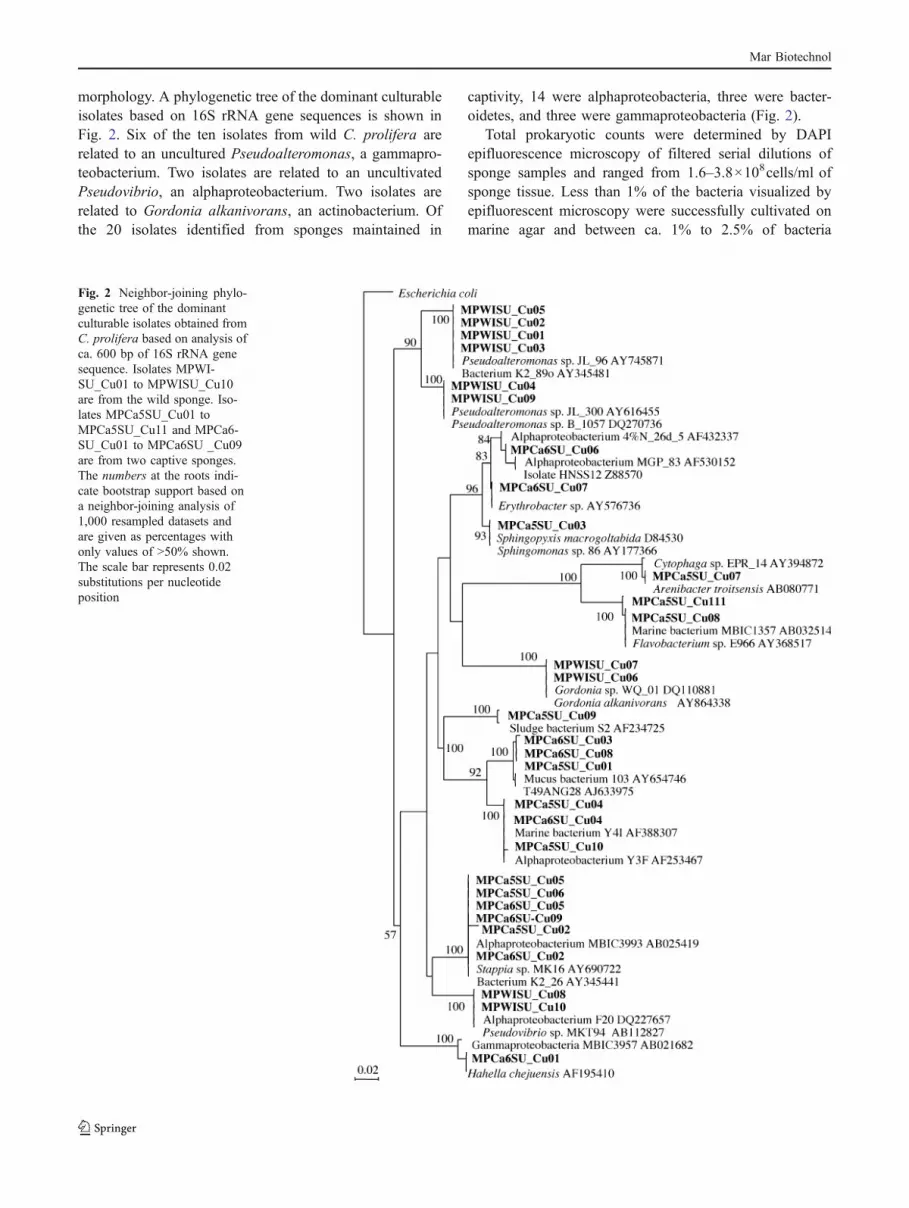
morphology. A phylogenetic tree of the dominant culturableisolates based on 16S rRNA gene sequences is shown inFig. 2. Six of the ten isolates from wild C. prolifera arerelated to an uncultured Pseudoalteromonas, a gammapro-teobacterium. Two isolates are related to an uncultivatedPseudovibrio, an alphaproteobacterium. Two isolates arerelated to Gordonia alkanivorans, an actinobacterium. Ofthe 20 isolates identified from sponges maintained in
captivity, 14 were alphaproteobacteria, three were bacter-oidetes, and three were gammaproteobacteria (Fig. 2).
Total prokaryotic counts were determined by DAPIepifluorescence microscopy of filtered serial dilutions ofsponge samples and ranged from 1.6–3.8×10⁸cells/ml ofsponge tissue. Less than 1% of the bacteria visualized byepifluorescent microscopy were successfully cultivated onmarine agar and between ca. 1% to 2.5% of bacteria
Fig. 2 Neighbor-joining phylo-genetic tree of the dominantculturable isolates obtained fromC. prolifera based on analysis ofca. 600 bp of 16S rRNA genesequence. Isolates MPWI-SU_Cu01 to MPWISU_Cu10are from the wild sponge. Iso-lates MPCa5SU_Cu01 toMPCa5SU_Cu11 and MPCa6-SU_Cu01 to MPCa6SU _Cu09are from two captive sponges.The numbers at the roots indi-cate bootstrap support based ona neighbor-joining analysis of1,000 resampled datasets andare given as percentages withonly values of >50% shown.The scale bar represents 0.02substitutions per nucleotideposition
Mar Biotechnol

from the surrounding waters were cultivated on marineagar.
Phylogenetic Analysis of Bacterial 16S rRNA Gene CloneLibraries Of the 192 clones from wild and captive C.prolifera, high-quality sequence was obtained for 163.Eighty-six of the 96 wild sponge clones were sequencedand these phylogenetic trees are shown in Figs. 3 and 4.Overall, there were 24% alphaproteobacteria (n=21), 7%were betaproteobacteria (n=6), 15% were gammaproteo-bacteria (n=13), 13% were deltaproteobacteria (n=11), 3%were planctomycetes (n=3), 15% were bacteroidetes (n=13), 1% were firmicutes (n=1), 2% were spirochetes (n=2),
7% were cyanobacteria (n=6), 1% was derived from thechloroplast of an eukaryotic alga (n=1), and 10% wereactinobacteria (n=9).
Seventy-seven clones were successfully sequenced from16S rRNA gene amplification products obtained fromcaptive C. prolifera and these phylogenetic trees are shownin Figs. 5 and 6. There were 31% alphaproteobacteria (n=27), 2% were betaproteobacteria (n=2), 5% were gammap-roteobacteria (n=4), 10% were deltaproteobacteria (n=9),2% were planctomycetes (n=2), 16% were bacteroidetes(n=14), 8% were firmicutes (n=7), 5%% were derivedfrom the chloroplasts of eukaryotic algae (n=4), and 9%were unclassified bacteria (n=8).
Fig. 3 Neighbor-joining phylo-genetic tree from analysis of ca.600 bp of 16S rRNA genesequences of proteobacterialclones in the library constructedfrom total DNA extracted fromwild C. prolifera. The numbersat the roots indicate bootstrapsupport based on a neighbor-joining analysis of 1,000resampled datasets and are givenas percentages with only valuesof >50% shown. The scale barrepresents 0.05 substitutions pernucleotide position
Mar Biotechnol

Discussion
Bacterial Community of Wild C. prolifera The bacterialcommunity associated with C. prolifera was clearlydifferent from that in the surrounding water based onthe DGGE analysis (Fig. 1). Six of the ten isolates fromwild C. prolifera were derived from bacteria in the genusPseudoalteromonas, a genus commonly isolated fromsponges. In contrast, the culturable community of the GreatBarrier Reef Rhopaloeides odorabile is dominated by analphaproteobacterium (Webster and Hill 2001). However,the genus Pseudoalteromonas is not a major component inthe total bacterial community associated with wild C.prolifera. Consistent with other studies of bacteria associ-ated with sponges, there is little similarity between thebacteria grown from sponge material and those in thetotal bacterial community associated with the spongerevealed by molecular approaches (Webster et al. 2001;
Hentschel et al. 2003), thus supporting the currentlimitations of using culture-based approaches in diversityanalyses. Laboratory culture approaches select for rapidgrowers in enriched media and impose selection pressuresthat are very different from those encountered in the naturalhabitat.
There is also little similarity between bacteria identifiedin the wild sample by DGGE and by the clone libraryanalyses. DGGE and clone libraries were prepared from thesame nucleic acid extracts and PCR amplifications in bothcases were with eubacterial primers. However, only a smallnumber of bands were successfully excised and sequencedfrom the DGGE gels whereas a far larger number of cloneswere identified by clone library analysis. This may explainthe lack of congruence between the two methods. Differ-ences in primer specificity between the standard primerpairs used for library construction designed to amplifyalmost full-length 16S rRNA genes and the DGGE primers
Fig. 4 Neighbor-joining phylo-genetic tree from analysis of ca.600 bp of 16S rRNA genesequences of nonproteobacterialclones in the library constructedfrom total DNA extracted fromwild C. prolifera. The numbersat the roots indicate bootstrapsupport based on a neighbor-joining analysis of 1,000resampled datasets and are givenas percentages with only valuesof >50% shown. The scale barrepresents 0.1 substitutions pernucleotide position
Mar Biotechnol

designed to amplify shorter fragments suitable for resolu-tion on DGGE gels may also be a factor in this lack ofcongruence. Nevertheless, DGGE analysis is a usefulmethod to monitor changes in the community compositionof individual sponges over time by rapidly and economi-cally analyzing multiple samples. In this study, DGGEclearly revealed marked similarities between the bacterialcommunities associated with two of our eight representativeC. prolifera sponges. These bacterial communities differed
greatly from the communities present in the surroundingseawater samples.
As there were multiple faint DGGE bands from whichsequence data were not obtained, the clone library providesmore information on the bacterial species richness of thetotal bacterial community present in the wild sponge. Theproteobacteria, bacteroidetes, actinobacteria, and cyanobac-teria represented over 90% of the species diversity presentin the total bacterial community of the wild C. prolifera. Of
Fig. 5 Neighbor-joining phylo-genetic tree from analysis of ca.600 bp of 16S rRNA genesequences of proteobacterialclones in the library constructedfrom total DNA extracted fromC. prolifera maintained in aqua-culture. The numbers at theroots indicate bootstrap supportbased on a neighbor-joininganalysis of 1,000 resampleddatasets and are given as per-centages with only values of>50% shown. The scale barrepresents 0.05 substitutions pernucleotide position
Mar Biotechnol

these groups, only the alphaproteobacterium Pseudovibrioand the actinobacterium Gordonia and isolates in the genusPseudoalteromonas were readily cultivable. The growth ofPseudoaltermonas, Pseudovibrio, and Gordonia on marineagar and ISP2 reflects the ability of these bacteria to growrapidly in enriched and selective media. Based on the clonelibrary, these two genera are not a major component of themicrobial community of the wild sponge and additionaleffort using novel culture-based methods would be neededto isolate representatives of all the groups present in thesponge.
Hentschel et al. (2002) documented molecular evidencefor a uniform microbial community common to Aplysinaaerophoba, Theonella swinhoei, and R. odorabile, distantlyrelated marine Demospongiae found in nonoverlappinggeographic region. These microbial communities were
distinct from those of surrounding seawater or the marinesediments. Poriferans are evolutionarily ancient metazoanswith origins dating back to the Precambrian. The commonbacterial components found in these sponges may reflect along evolutionary history in the symbiotic relationshipbetween bacteria and the sponge host. In contrast toHentschel et al. (2002) in which uniform bacterial commu-nities were found in different sponges, Taylor et al. (2004)reported substantial variability of microbial communitiesbetween different species of sponges although there waslittle variability of microbial communities within eachsponge species. Based on a previous culture-based studyof sponge-associated bacteria (Wilkinson et al. 1981),Taylor et al. (2004) designated as specialists, associates,or generalists those bacteria found respectively in specificsponge species, in different sponge species but not in
Fig. 6 Neighbor-joining phylo-genetic tree from analysis of ca.600 bp of 16S rRNA genesequences of nonproteobacterialclones in the library constructedfrom total DNA extracted fromC. prolifera maintained in aqua-culture. The numbers at theroots indicate bootstrap supportbased on a neighbor-joininganalysis of 1,000 resampleddatasets and are given as per-centages with only values of>50% shown. The scale barrepresents 0.05 substitutions pernucleotide position
Mar Biotechnol
seawater, and in different sponge species and sea water. Thebacterial community associated with C. prolifera differed inseveral respects from those generally described in othermarine sponges. Acidobacteria have been found to be well-represented in many sponge bacterial communities, includ-ing those associated with R. odorabile (Webster et al.2001), T. swinhoei and A. aerophoba (Hentschel et al.2002), and Xestospongia muta (Montalvo and Hill, unpub-lished) but were lacking in the C. prolifera clone libraries,except for two clones with homology to Holophaga 16SrRNA gene sequences in a C. prolifera sponge maintainedin the aquaculture system. Chloroflexi, a major componentof the bacterial communities in many marine sponges, werenotably absent from C. prolifera. The sponges A. aero-phoba, T. swinhoei, R. odorabile, and X. muta aredesignated high-microbial-abundance sponges or bacterio-sponges, defined as sponges in which the bacterialpopulation densities reach 108 to 1010 bacterial cells/ml ofsponge tissue. C. prolifera also falls into this category withbacterial counts ranging from 1.6–3.8×10⁸cells/ml ofsponge tissue sponges. These differences between thedominant bacteria represented in C. prolifera clone librariesand those generally found in other marine bacteria may be aconsequence of growth of the C. prolifera in our studybeing collected from an estuary with salinities of ca. 10 pptrather than in the open ocean and it would be interesting tocompare bacterial assemblages in marine-collected C.prolifera with our estuarine samples. In a study of thebiogeography of microbes found in the Australian spongeCymbastela concentrica, a sponge with a wide geographicdistribution, considerable geographic variability was ob-served in the microbial community from tropical versustemperate waters (Taylor et al. 2005) so it is certainlypossible that C. prolifera sponges growing in seawater at asalinity of 35 ppt may contain markedly different commu-nities from those collected in estuaries.
Comparison of Bacterial Communities in Wild and CaptiveC. prolifera The actinobacteria, cyanobacteria, and spiro-chetes, representing almost 20% of the bacterial speciesdiversity in wild C. prolifera, were not found in the spongesmaintained in the aquaculture system, showing a significanteffect of aquaculture on the bacterial community composi-tion. Symbiotic spirochetes are difficult to cultivate in thelaboratory and their loss in the captive sponge is thereforeperhaps not surprising. The loss of actinobacteria in thecaptive sponge is potentially a concern when consideringthis aquaculture procedure as a model system for futureproduction of bioactive compounds by sponges in aquacul-ture, as actinobacteria produce more than 65% of knownantimicrobials and may be the true “producer” symbiontsresponsible for some of the important bioactive compoundsfound in sponges (Hill 2004a, b). The numbers of clones
derived from beta- and gammaproteobacteria diminishedfrom over 22% in the wild sponges to less than 8% in thecaptive sponges suggesting that representatives of thesegroups may be less able to tolerate the environmentalconditions in aquaculture. Although the gammaproteobac-terial genus Oceanobacter was present in both wild andcaptive sponges, clones are derived from different speciesindicating that subtle shifts in species composition mayoccur in the sponges maintained in aquaculture. Previousstudies have suggested that the microbial communitiesassociated with sponges can be resistant to perturbationsduring starvation, treatment with antibiotics, or transplan-tation of sponges to different habitats (Imhoff and Stohr2003). This does not appear to be the case for themaintenance of estuarine C. prolifera in aquaculturesystems, at least not from the changes observed in clonesderived from actinobacteria, cyanobacteria, spirochetes, andsome members of the beta- and gammaproteobacteria.
Cyanobacteria present in the wild sponge includeuncultivated representatives of the genera Synechococcus,Pseudoanabena, and Phormidium. Our efforts to isolatethese cyanobacteria were unsuccessful. Autofluorescentcells, presumably cyanobacteria, were observed to dissociatefrom the sponge host soon after the sponge has been removedfrom its natural environment, concomitant with a rapid loss ofnormal sponge coloration. Whether the vibrant red or orangecolor of C. prolifera is due to cyanobacteria is not known.The vibrant red color in C. prolifera is likely attributable atleast in part to carotenoid pigments that have been describedfrom this sponge (Litchfield and Liaaen-Jensen 1980;Sliwka et al. 1987) although it is not clear whether thesepigments are of sponge or bacterial origin.
Additional work is needed to optimize the growth andmaintenance of sponges in aquaculture systems. Ouraquaculture system failed to maintain the C. prolifera incaptivity for longer than 9 months although sponges I.strobilina and M. laxissima were successfully maintained inthe same system for up to 2 years (Mohamed et al. 2008a, b).It is interesting to note that captive C. prolifera spongesappeared to have a reduced ability to prevent biofoulingwith some individuals becoming coated with a film ofdiatoms in captivity whereas wild samples were notgenerally observed to have the microalgal or cyanobacterialfilms seen on the captive samples. This may be indicativeof changes in the community of bacterial epibionts on thesurface of C. prolifera as bacterial epibionts can play animportant antifouling role (Krug 2006). We are unaware ofproduction of bioactive compounds by C. prolifera otherthan an early report of antibacterial activity in extracts of C.prolifera (Der Marderosian 1970). However, several otherrepresentatives of the genus Clathria have been found toproduce bioactive compounds, including the alkaloidmirabilin G and clathrins A–C (Capon et al. 2000, 2001).
Mar Biotechnol

This study provides information on the bacterial com-munity associated with estuarine C. prolifera and reinforcesthe importance of monitoring microbial communities whencultivating sponges in flow-through or recirculation aqua-culture systems.
Acknowledgments George Burbanck and Robert Jordan at Hamp-ton University are thanked for assistance in Chesapeake Baycollections. LTI was supported by Goucher College during asabbatical leave. Her research was supported by a Florence SeibertResearch Fellowship, a Goucher College faculty development grant,and the MacLane-To fund. TLW was funded by the NIGMS Bridgesto the Baccalaureate grant GM62005. LN and PV participated in theproject as part of a Directed Research course in Microbiology atGoucher College. Funding for this study was provided by the MicrobialObservatories Program, National Science Foundation (MCB-0238515and 0703467), and this support is gratefully acknowledged.
References
Azam F, Worden AZ (2004) Oceanography: microbes, molecules, andmarine ecosystems. Science 303:1622–1624
Bagby RM (1972) Formation and differentiation of the upperpinacoderm in reaggregation masses of the sponge Microcionaprolifera (Ellis and Solander). J Exp Zool 180:217–225
Bewley CA, Faulkner DJ (1998) Lithistid sponges: star performers orhosts to the stars. Angew Chem Int Ed 37:2162–2178
Bewley CA, Holland ND, Faulkner DJ (1996) Two classes ofmetabolites from Theonella swinhoei are localized in distinctpopulations of bacterial symbionts. Experientia 52:716–722
Capon RJ, Miller M, Rooney F (2000) Clathrins A–C: metabolitesfrom a Southern Australian marine sponge, Clathria species. JNat Prod 63:821–824
Capon RJ, Miller M, Rooney F (2001) Mirabilin G: a new alkaloidfrom a southern Australian marine sponge, Clathria species. JNat Prod 64:643–644
Cragg GM, Newman DJ, Yang SS (2006) Natural product extracts ofplant and marine origin having antileukemia potential. The NCIexperience. J Nat Prod 69:488–498
Der Marderosian AH (1970) Drugs from the sea—an overview. In:Youngken HW Jr (ed) Drugs from the sea proceedings 1969.Marine Technology Society, Washington, DC
Faulkner DJ, Harper MK, Haygood MG, Salomon CE, Schmidt EW(2000) Symbiotic bacteria in sponges: sources of bioactivesubstances. In: Fusetani N (ed) Drugs from the sea. Karger,Basel, pp 107–119
Fernandez-Busquets X, Gerosa D, Hess D, Burger MM (1998)Accumulation in marine sponge grafts of the mRNA encodingthe main proteins of the cell adhesion system. J Biol Chem273:29545–29553
Ferris MJ, Muyzer G, Ward DM (1996) Denaturing gradient gelelectrophoresis profiles of 16S rRNA-defined populations inhab-iting a hot spring microbial mat community. Appl EnvironMicrobiol 62:340–346
Guerardel Y, Czeszak X, Sumanovski LT, Karamanos Y, Popescu O,Strecker G, Misevic GN (2004) Molecular fingerprinting ofcarbohydrate structure phenotypes of three porifera proteoglycan-like glyconectins. J Biol Chem 279:15591–15603
Hart JB, Lill RE, Hichford SJH, Blunt JW, Munro MHG (2000) Thehalichondrins: chemistry, biology, supply and delivery. In:Fusetani N (ed) Drugs from the sea. Karger, Basel, pp 134–153
Haygood MG, Schmidt EW, Davidson SK, Faulkner DJ (1999)Microbial symbionts of marine invertebrates: opportunities formicrobial biotechnology. J Mol Microbiol Biotechnol 1:33–43
Hentschel U, Hopke J, Horn M, Friedrich AB, Wagner M, Hacker J,Moore BS (2002) Molecular evidence for a uniform microbialcommunity in sponges from different oceans. Appl EnvironMicrobiol 68:4431–4440
Hentschel U, Fieseler L, Wehrl M, Gernert C, Steinert M, Hacker J,Horn M (2003) Microbial diversity of marine sponges. In: MüllerWEG (ed) Sponges (Porifera). Springer, Berlin, pp 59–88
Hill RA (2004a) Marine natural products. Ann Rep Prog Chem Sec BOrg Chem 100:169–189
Hill RT (2004b) Microbes from marine sponges: a treasure trove ofbiodiversity for natural products discovery. In: Bull AT (ed) Microbialdiversity and bioprospecting. ASM, Washington, DC, pp 177–190
Hooper JNA, Van Soest RWM (eds) (2002) Systema Porifera: a guideto the classification of sponges. Kluwer Academic, New York
Imhoff JF, Stohr R (2003) Sponge-associated bacteria: generaloverview and special aspects of bacteria associated withHalichondria panicea. Prog Mol Subcell Biol 37:35–57
Jukes TH, Cantor CR (1969) Evolution of protein molecules. In:Munro HN (ed) Mammalian protein metabolism. Academic, NewYork, pp 21–132
Kaltenbach JC, Kuhns WJ, Simpson TL, Burger MM (1999) Intenseconcanavalin A staining and apoptosis of peripheral flagellatedcells in larvae of the marine sponge Microciona prolifera:significance in relation to morphogenesis. Biol Bull 197:271–273
Kobayashi J, Ishibashi M (1993) Bioactive metabolites of symbioticmarine microorganisms. Chem Rev 93:8305–8308
Krug PJ (2006) Defense of benthic invertebrates against surface coloniza-tion by larvae: a chemical arms race. Prog Mol Subcell Biol 42:1–53
Lane DJ (1991) 16S/23S rRNA sequencing. In: Stackebrandt E,Goodfellow M (eds) Nucleic acid techniques in bacterialsystematics. Wiley, New York, pp 115–175
Lee YK, Lee J-H, Lee HK (2001) Microbial symbiosis in marinesponges. J Microbiol 39:254–264
Lippson AJ, Lippson RL (1997) Life in the Chesapeake Bay. TheJohns Hopkins, Baltimore
Litchfield C, Liaaen-Jensen S (1980) Carotenoids of the marinespongeMicrociona prolifera. Comp Biochem Physiol B 66:359–366
Mohamed NM, Enticknap JJ, Lohr JE, McIntosh SM, Hill RT (2008a)Changes in bacterial communities of the marine sponge Mycalelaxissima on transfer into aquaculture. Appl Environ Microbiol74:1209–1222
MohamedNM,RaoV,HamannMT,KellyM,Hill RT (2008b)Monitoringbacterial diversity of the marine sponge Ircinia strobilina upontransfer into aquaculture. Appl Environ Microbiol 74:4133–4143
Müller WEG, Wimmer W, Schatton W, Böhm M, Batel R, Filic Z(1999) Initiation of an aquaculture of sponges for the sustainableproduction of bioactive metabolites in open systems: example,Geodia cydonium. Mar Biotechnol (NY) 1:569–579
Munro MH, Blunt JW, Dumdei EJ, Hickford SJ, Lill RE, Li S,Battershill CN, Duckworth AR (1999) The discovery anddevelopment of marine compounds with pharmaceutical poten-tial. J Biotechnol 70:15–25
Newman DJ, Cragg GM (2004) Marine natural products and relatedcompounds in clinical and advanced preclinical trials. J Nat Prod67:1216–1238
Newman DJ, Hill RT (2006) New drugs from marine microbes: thetide is turning. J Ind Microbiol Biotech 33:539–544
Piel J (2006) Bacterial symbionts: prospects for the sustainableproduction of invertebrate-derived pharmaceuticals. Curr MedChem 13:39–50
Pitcher DG, Saunders NA, Owen RJ (1989) Rapid extraction ofbacterial genomic DNA with guanidium thiocyanate. Lett ApplMicrobiol 8:151–156
Mar Biotechnol

Porter KG, Feig YS (1980) The use of DAPI for identifying andcounting aquatic microflora. Limnol Oceanogr 25:943–948
Proksch P (1994) Defensive roles for secondary metabolites from marinesponges and sponge-feeding nudibranchs. Toxicon 32:639–655
Ruby E, Henderson B, McFall-Ngai M (2004) Microbiology: we getby with a little help from our (Little) friends. Science 303:1305–1307
Saitou N, Nei M (1987) The neighbor-joining method: a new methodfor reconstructing phylogenetic trees. Mol Biol Evol 4:406–425
Schmidt EW, Obraztsova AY, Davidson SK, Faulkner DJ, HaygoodMG (2000) Identification of the antifungal peptide-containingsymbiont of the marine sponge Theonella swinhoei as a novel δ-proteobacterium, “Candidatus Entotheonella palauensis”. MarBiol 136:969–977
Sliwka HR, Nokleby OW, Liaaen-Jensen S (1987) Animal carote-noids. 31. Structure elucidation of a sponge metabolite viamesylate elimination. Acta Chem Scand B 41:245–252
Taylor MW, Schupp PJ, Dahllof I, Kjelleberg S, Steinberg PD (2004)Host specificity in marine sponge-associated bacteria, andpotential implications for marine microbial diversity. EnvironMicrobiol 6:121–130
Taylor MW, Schupp PJ, de Nys R, Kjelleberg S, Steinberg PD (2005)Biogeography of bacteria associated with the marine spongeCymbastela concentrica. Environ Microbiol 7:419–433
Taylor MW, Radax R, Steger D, Wagner M (2007) Sponge-associatedmicroorganisms: evolution, ecology, and biotechnological poten-tial. Microbiol Mol Biol Rev 71:295–347
Tepsuporn S, Kaltenbach JC, KuhnsWJ, BurgerMM, Fernandez-BusquetsX (2003) Apoptosis in Microciona prolifera allografts. Biol Bull205:199–201
Vacelet J, Donadey C (1977) Electron microscope study of theassociation between some sponges and bacteria. J Exp Mar BiolEcol 30:301–314
Webster NS, Hill RT (2001) The culturable microbial community ofthe Great Barrier Reef sponge Rhopaloeides odorabile isdominated by an α-Proteobacterium. Mar Biol 138:843–851
Webster NS, Wilson KJ, Blackall LL, Hill RT (2001) Phylogeneticdiversity of bacteria associated with the marine sponge Rhopa-loeides odorabile. Appl Environ Microbiol 67:434–444
Webster NS, Negri AP, Munro MM, Battershill CN (2004) Diversemicrobial communities inhabit Antarctic sponges. EnvironMicrobiol 6:288–300
Wichels A, Würtz S, Döpke H, Schütt C, Gerdts G (2006) Bacterialdiversity in the breadcrumb sponge Halichondria panicea(Pallas). FEMS Microb Ecol 56:102–118
Wilkinson CR, Nowak M, Austin B, Colwell RR (1981) Specificity ofbacterial symbionts in Mediterranean and Great Barrier Reefsponges. Microb Ecol 7:13–21
Mar Biotechnol