comparative analysis (hippotragini versus caprini, bovidae) of x-chromosome's constitutive...
TRANSCRIPT
doi: 10.1023/B:GENE.0000039859.33467.46
Comparative analysis (Hippotragini versus Caprini, Bovidae) of X-
chromosome’s constitutive heterochromatin by in situ restriction
endonuclease digestion: X-chromosome constitutive heterochromatin
evolution
Raquel Chaves, Sara Santos & Henrique Guedes-Pinto Department of Genetics and Biotechnology, Centre of Genetics and Biotechnology/ICETA-UTAD, University of
Tra´s-os-Montes and Alto Douro, P-5000-911 Vila Real, Portugal (Phone: +351-259-350-571; Fax: +351-259-350-
572; E-mail: [email protected], [email protected])
Key words: Caprini, constitutive heterochromatin, Hippotragini, in situ restriction endonuclease digestion, non-
Bovinae X-chromosome
Abstract The Bovidae X-chromosome shows a considerable variation, in contrast to the preservative autosomal
conservatism. The X-chromosome variation is mostly a consequence of the constitutive heterochromatin (CH)
variation; in what respect to its amount and position. This is especially common among the non-Bovinae
subfamilies and tribes. In order to characterize the X-chromosome CH in non-Bovinae species – Hippotragini and
Caprini tribes – we have used restriction endonuclease digestion on fixed chromosomes and sequential C-banding.
With these techniques we were able to distinguish between the two X-chromosome types (Hippotragini and
Caprini) CH, in what respect to its position and molecular nature. Moreover, we define at least, six subclasses of
CH in both X-chromosome types analyzed. Evolutionary considerations were draw based on the results obtained.
The technology used here for the analysis of the Bovidae X-chromosome CH showed to be more evolutionary
informative than the classical approaches.
Abbreviations: BAC – bacterial artificial chromosomes; CBP-banding – C-bands by barium hydroxide using propidium iodide; CH – constitutive heterochromatin; Chr – chromosome; DAPI – 4¢-6-diamidino-2-phenylindole;
DNA – deoxyribonucleic acid; FISH – fluorescent in situ hybridization; GTD-banding – G-bands by trypsin with
DAPI; MYBP – million years ago before present; NAA – number of autosomal arms; PBS – phosphate-buffered
saline; RE-banding – restriction endonuclease banding; RE-bands – restriction endonuclease bands; RE – restriction
endonuclease; REs – restriction endonucleases; SSC – saline sodium citrate.
1. Introduction
In general, the Bovidae are chromosomal relatively
conservative. Although the diploid chromosome
number ranges from 2n ¼ 30 to 2n ¼ 60, the number of autosomal arms (NAA) has remained constant at
56–58 for most species. Chromosomal change in the
Bovidae has occurred primarily through centric fusion
(Wurster & Benirschke, 1968; Effron et al., 1976;
Buckland & Evans, 1978a; Gallagher & Womack,
1992; Gallagher, Derr & Womack, 1994). Although
there is good evidence of tandem rearrangements in
some species (Gallagher & Womack, 1992) and
paracentric inversions in others (Hayes, Petit &
Dutrillaux,1991; Robinson et al., 1998), these seem to
be the exception rather than the rule. In contrast to the
preservative autosomal conservatism evident in the
Bovidae, the X-chromosome is reported to show
considerable variation between subfamilies, and often between tribes within subfamilies (Buckland & Evans,
1978b; Gallagher & Womack, 1992). The X-
chromosome variation in the bovids is due to
disruption of euchromatic regions, centromere
placement, heterochromatic variation, and autosomal
translocation (Robinson et al., 1998; Gallagher et al.,
1999; Iannuzzi et al., 2000). Several studies have been
done to delineate the structural rearrangements that
characterized the evolution of bovid X-chromosome
and to determine the primitive and ancestral condition
of Bovidae X-chromosome. These studies include G-
and C-banding comparisons, in situ hybridizations
with X paint probes and bacterial artificial
chromosomes (BAC) clones to localize the
homologous regions between the X-chromosomes of several species (Hayes, Petit & Dutrillaux, 1991;
Kaftanovskaya & Serov,1994; Robinson et al., 1997;
Piumi et al., 1998; Iannuzzi et al., 2000). A substantial
proportion of the higher eukaryote genome consists of
constitutive heterochromatin (CH). This genomic
fraction includes, among other repetitive sequences,
satellite deoxyribonucleic acid (DNAs) that usually
reside in the pericentromeric regions of chromosomes
(Chaves et al., 2000).
The C-banding technique is extremely useful to
identify CH, and its size differences. On the other

hand, other analytical techniques are necessary to
obtain better information on the nature and origin of
the CH. Among these technologies, the use of
restriction endonucleases (REs) (which cleave DNA in
specific sequences) and the base-specific
fluorochromes (which delimit chromosome regions
rich in different types of DNA bases) are techniques
that proven to be very useful in improving the
understanding of the mechanisms involved in the
evolution of CH in different genomes (Pieczarka et al.,
1998; for a revision see Gonzalez et al., 1997).
The use of RE techniques has also the ability to
demonstrate the C-heterochromatin heterogeneity
(Babu, 1988).
In this study, we used a panel of seven different
restriction enzymes and sequentially C-banding to
verify whether the X-chromosome CH of species from
the Hippotragini and Caprini tribes presents, or not, the same characteristics in terms of its base nature or
DNA composition. This technique proved to be more
informative in the study of Bovidae X CH than the
classical approach of single C-banding.
2. Materials and methods
Chromosome preparations
The material analyzed consists of chromosome
preparations made from short-term lymphocyte cultures of whole blood samples obtained from
animals of the Lisbon Zoo, Portugal: Addax
nasomaculatus (2n ¼ 58, female), Oryx dammah (2n
¼ 58, male), Oryx leucoryx (2n ¼ 58, male); and of
the University of Tras-os-Montes and Alto Douro,
Portugal: Ovis aries (2n ¼ 54, male).
Briefly, peripheral blood was cultured at 37 C using
standard protocols (Chaves et al., 2002). Cell
suspension swelling and fixation procedures used a 75
mM KCl hypotonic treatment at 37 C for 20 min, and
then chromosomes were fixed three times with methanol:acetic acid solution (3:1) and left overnight
at )20 C.
GTD-banding
Air dried slides were aged at 65 C for 5 h or
overnight and then were submitted to standard
procedures of G-banding with trypsin (Verma & Babu,
1995). Since the chromosomes preparations proceed
next to sequential C-banding techniques; they were not
stained at this stage. Instead, they were first fixed with
paraformaldehyde as described by Chaves et al.
(2002). Briefly, dry slides were placed in a 1 · PBS solution (2 · 5 min) before fixation in 4%
paraformaldehyde (Sigma)/1 · PBS (room
temperature). After fixation for 10 min, slides were
dehydrated for 2 min each in 70, 90 and 100% chilled
ethanol and air dried. At this step, slides were stained
with DAPI (instead of routine Giemsa) for a better
contrast (Chaves et al., 2002), and for the
identification of the respective Bovidae species
chromosomes. The inversion of the DAPI colour in
Adobe Photoshop revealed the chromosomes G-
banding (GTD-banding, G-bands by trypsin with
DAPI) for its identification. Karyotyping followed
standardization of the Domestic Bovids karyotypes
(ISCNDB, 2000), and Hippotragini karyotypes were
also according to Claro, Hayes and Cribiu (1996) and Kumamoto et al. (1999) descriptions.
In situ RE digestion
Air dried slides were aged at 65 C for 6 h and then
were submitted to in situ RE digestion. The restriction
enzymes used (Hinf I, Pst I, Dde I, Hae III, Rsa I, Mbo
I, and Apa I) were diluted in buffers indicated by the
manufacturer (Invitrogen, Life Technologies), and
final concentrations of 30 U were obtained per 100 ll.
The 100 ll of each one of these solutions were placed
on slides and covered with coverslips. These slides
were incubated in a moist humid chamber for 16 h at
37 C. Control slides were submitted to the same
treatment as described above but incubated only with
buffer. The slides were then washed in distillated
water and air dried. Since, the chromosomes
preparations proceed next to sequentially C-banding techniques; they were not stained at this stage. Instead
they were first fixed with paraformaldehyde as
described by Chaves et al. (2002), and briefly
described above (cf. GTD-banding). Finally, the slides
were stained with DAPI. The residual bands obtained
after the endonuclease digestion were adequate for
chromosome identification, namely the X-
chromosome. Karyotyping followed standardization of
the Domestic Bovids karyotypes (ISCNDB, 2000),
and Hippotragini karyotypes were also according to
Claro, Hayes and Cribiu (1996) and Kumamoto et al.
(1999) descriptions.
CBP-banding sequential to G-bands or RE-bands
The C-banding sequential to G-bands or to restriction
enzyme bands was performed by distaining the slides
and submitting them to C-banding technique. CBP-
banding (C-bands by barium hydroxide using
propidium iodide) was done following the standard
procedure of Sumner (1972) but with propidium
iodide as a counterstain. Briefly, the slides were
submitted to routine C-banding with classical treatment times reduced, approximately, to half of it:
hydrochloric acid (0.2 M) 10 min, barium hydroxide
(5% solution) 4.5 min and 2 · saline sodium citrate
(2SSC:0.3 mol/l NaCl, 0.03 mol/l sodium citrate) at 60
C only 20 min.
Chromosome observation
Chromosomes were observed with a Zeiss Axioplan
2 Imaging microscope coupled to Axiocam digital
camera and AxioVision software (version 2.0.5 –
Zeiss). Digitized photos were prepared for printing in
Adobe Photoshop (version 5.0); contrast and colour
optimization were the functions used and all affected
the whole of the image equally.
3. Results
Karyotypes description
The diploid number of the Hippotragini species –
Addax nasomaculatus, Oryx leucoryx, and Oryx
dammah – is 58. All chromosomes are acrocentric,
except for the first pair of autosomes, which is a submetacentric one. The Ovis aries (Caprini tribe)
diploid number is 54, in which all the autosomes are
acrocentric, except for the first three pairs that are
biarmed, according to ISCNDB (2000). The X-
chromosomes from the Hippotragini species are
morphological acrocentrics (Claro, Hayes & Cribiu,
1996). The X-chromosome from Ovis aries is a
submetacentric one, with the short arm very small
(ISCNDB, 2000).
In situ RE digestion and sequential C-banding
We used seven different restriction enzymes in
chromosome preparations of Hippotragini and Caprini species. The CH of the X-chromosomes was analyzed
with sequential C-banding techniques.
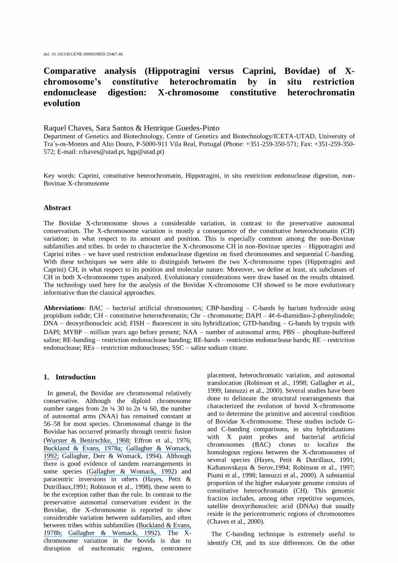
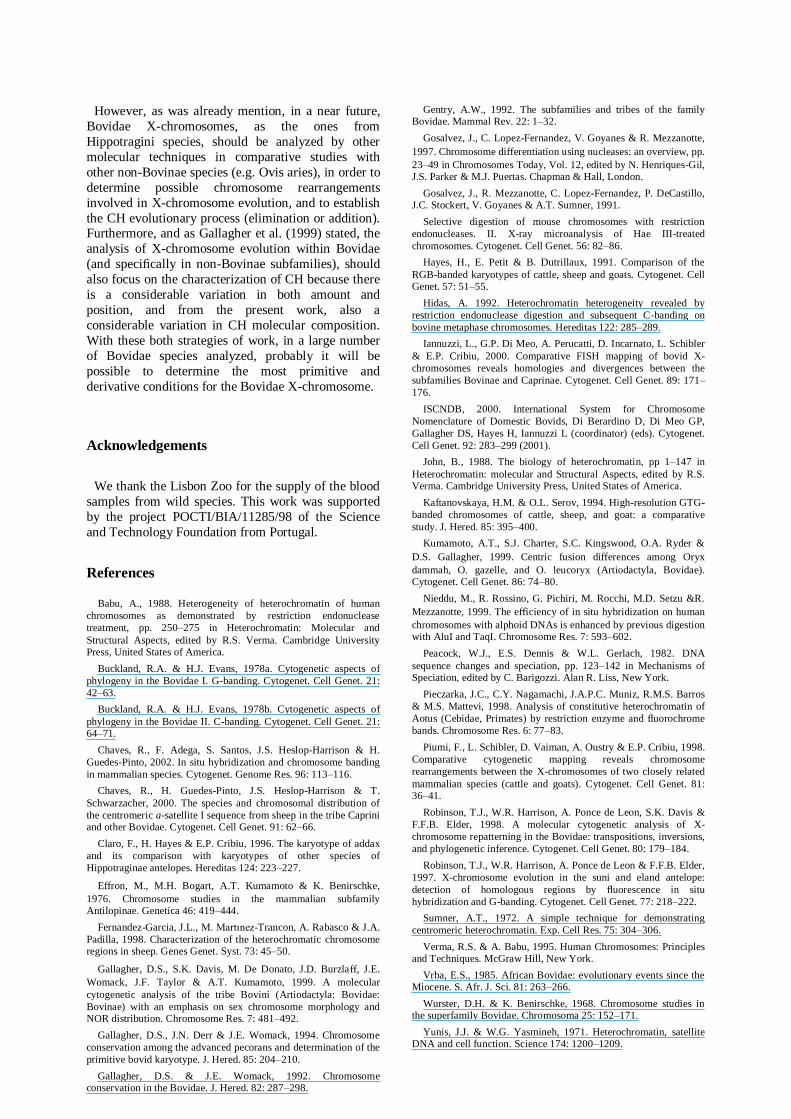
The Figure 1 presents demonstrative images of the
sequential procedures used in this work for the
identification of chromosomes (RE-banding with Dde
I, Figure 1a and b), and C-banding treatment (Figure
1c and d) in Oryx dammah. In Figure 1b is possible to
observe that the residual RE bands are appropriate for
the identification of most chromosomes, particularly
the X-chromosome. The G-banding technique was
only used for the identification of chromosomes in the
control experiment of C-banding (without previous treatment with in situ RE digestion).
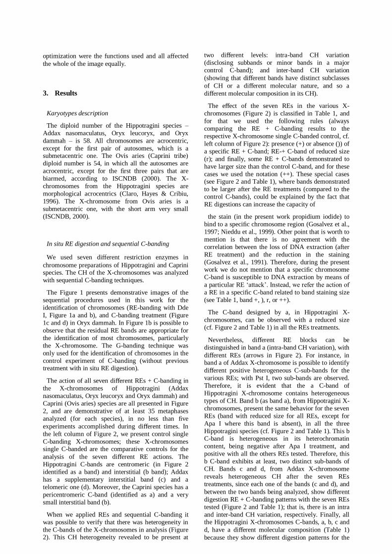
The action of all seven different REs + C-banding in
the X-chromosomes of Hippotragini (Addax
nasomaculatus, Oryx leucoryx and Oryx dammah) and
Caprini (Ovis aries) species are all presented in Figure
2, and are demonstrative of at least 35 metaphases
analyzed (for each species), in no less than five
experiments accomplished during different times. In
the left column of Figure 2, we present control single
C-banding X-chromosomes; these X-chromosomes single C-banded are the comparative controls for the
analysis of the seven different RE actions. The
Hippotragini C-bands are centromeric (in Figure 2
identified as a band) and interstitial (b band); Addax
has a supplementary interstitial band (c) and a
telomeric one (d). Moreover, the Caprini species has a
pericentromeric C-band (identified as a) and a very
small interstitial band (b).
When we applied REs and sequential C-banding it
was possible to verify that there was heterogeneity in
the C-bands of the X-chromosomes in analysis (Figure 2). This CH heterogeneity revealed to be present at
two different levels: intra-band CH variation
(disclosing subbands or minor bands in a major
control C-band); and inter-band CH variation
(showing that different bands have distinct subclasses
of CH or a different molecular nature, and so a
different molecular composition in its CH).
The effect of the seven REs in the various X-
chromosomes (Figure 2) is classified in Table 1, and
for that we used the following rules (always
comparing the RE + C-banding results to the
respective X-chromosome single C-banded control, cf.
left column of Figure 2): presence (+) or absence ()) of
a specific RE + C-band; RE-+ C-band of reduced size
(r); and finally, some RE + C-bands demonstrated to
have larger size than the control C-band, and for these
cases we used the notation (++). These special cases (see Figure 2 and Table 1), where bands demonstrated
to be larger after the RE treatments (compared to the
control C-bands), could be explained by the fact that
RE digestions can increase the capacity of
the stain (in the present work propidium iodide) to
bind to a specific chromosome region (Gosalvez et al.,
1997; Nieddu et al., 1999). Other point that is worth to
mention is that there is no agreement with the
correlation between the loss of DNA extraction (after
RE treatment) and the reduction in the staining
(Gosalvez et al., 1991). Therefore, during the present work we do not mention that a specific chromosome
C-band is susceptible to DNA extraction by means of
a particular RE ‘attack’. Instead, we refer the action of
a RE in a specific C-band related to band staining size
(see Table 1, band +, ), r, or ++).
The C-band designed by a, in Hippotragini X-
chromosomes, can be observed with a reduced size
(cf. Figure 2 and Table 1) in all the REs treatments.
Nevertheless, different RE blocks can be
distinguished in band a (intra-band CH variation), with
different REs (arrows in Figure 2). For instance, in band a of Addax X-chromosome is possible to identify
different positive heterogeneous C-sub-bands for the
various REs; with Pst I, two sub-bands are observed.
Therefore, it is evident that the a C-band of
Hippotragini X-chromosome contains heterogeneous
types of CH. Band b (as band a), from Hippotragini X-
chromosomes, present the same behavior for the seven
REs (band with reduced size for all REs, except for
Apa I where this band is absent), in all the three
Hippotragini species (cf. Figure 2 and Table 1). This b C-band is heterogeneous in its heterochromatin
content, being negative after Apa I treatment, and
positive with all the others REs tested. Therefore, this
b C-band exhibits at least, two distinct sub-bands of
CH. Bands c and d, from Addax X-chromosome
reveals heterogeneous CH after the seven REs
treatments, since each one of the bands (c and d), and
between the two bands being analyzed, show different
digestion RE + C-banding patterns with the seven REs
tested (Figure 2 and Table 1); that is, there is an intra
and inter-band CH variation, respectively. Finally, all the Hippotragini X-chromosomes C-bands, a, b, c and
d, have a different molecular composition (Table 1)
because they show different digestion patterns for the
same REs panel used in this work. However,
corresponding bands (by its chromosome localization)
exhibit similar reactions among the three species,
which is demonstrative of a common origin and
phylogenetic proximity.
In the Caprini X-chromosome, and after the REs
treatments, both a and b C-bands showed
heterogeneity of minor C-bands (cf. arrows in Figure 2
and Table 1), and reveal a different CH composition
between bands (inter-bands CH variation could be
observed in Ovis aries C-bands and with all
Hippotragini C-bands, see Figure 2 and Table 1).
Notably, one of the most aggressive RE (Apa I) for the
Hippotragini C-bands is the RE that in the Caprini C-
bands X-chromosome results, apparently, in more
pronounced C-bands. Specifically, Apa I + C-bands in
Caprini X-chromosome are positive. The b Apa I + C-
band seems even larger than the corresponding control
C-band. The use of Apa I (and Mbo I), lay in evidence a CH band, that by classical C-banding was hardly
seen.
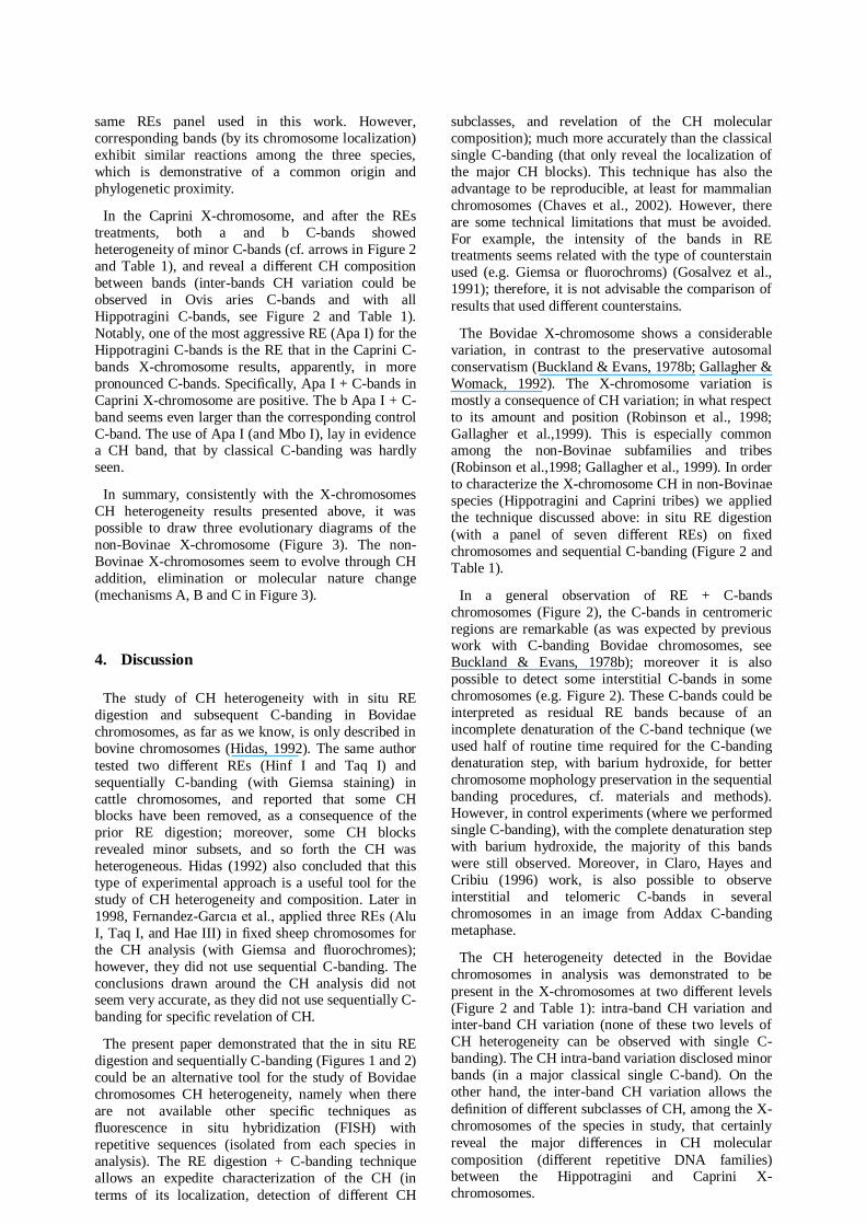
In summary, consistently with the X-chromosomes
CH heterogeneity results presented above, it was
possible to draw three evolutionary diagrams of the
non-Bovinae X-chromosome (Figure 3). The non-
Bovinae X-chromosomes seem to evolve through CH
addition, elimination or molecular nature change
(mechanisms A, B and C in Figure 3).
4. Discussion
The study of CH heterogeneity with in situ RE
digestion and subsequent C-banding in Bovidae
chromosomes, as far as we know, is only described in
bovine chromosomes (Hidas, 1992). The same author
tested two different REs (Hinf I and Taq I) and
sequentially C-banding (with Giemsa staining) in
cattle chromosomes, and reported that some CH blocks have been removed, as a consequence of the
prior RE digestion; moreover, some CH blocks
revealed minor subsets, and so forth the CH was
heterogeneous. Hidas (1992) also concluded that this
type of experimental approach is a useful tool for the
study of CH heterogeneity and composition. Later in
1998, Fernandez-Garcıa et al., applied three REs (Alu
I, Taq I, and Hae III) in fixed sheep chromosomes for
the CH analysis (with Giemsa and fluorochromes);
however, they did not use sequential C-banding. The
conclusions drawn around the CH analysis did not seem very accurate, as they did not use sequentially C-
banding for specific revelation of CH.
The present paper demonstrated that the in situ RE
digestion and sequentially C-banding (Figures 1 and 2)
could be an alternative tool for the study of Bovidae
chromosomes CH heterogeneity, namely when there
are not available other specific techniques as
fluorescence in situ hybridization (FISH) with
repetitive sequences (isolated from each species in
analysis). The RE digestion + C-banding technique
allows an expedite characterization of the CH (in
terms of its localization, detection of different CH
subclasses, and revelation of the CH molecular
composition); much more accurately than the classical
single C-banding (that only reveal the localization of
the major CH blocks). This technique has also the
advantage to be reproducible, at least for mammalian
chromosomes (Chaves et al., 2002). However, there
are some technical limitations that must be avoided.
For example, the intensity of the bands in RE treatments seems related with the type of counterstain
used (e.g. Giemsa or fluorochroms) (Gosalvez et al.,
1991); therefore, it is not advisable the comparison of
results that used different counterstains.
The Bovidae X-chromosome shows a considerable
variation, in contrast to the preservative autosomal
conservatism (Buckland & Evans, 1978b; Gallagher &
Womack, 1992). The X-chromosome variation is
mostly a consequence of CH variation; in what respect
to its amount and position (Robinson et al., 1998;
Gallagher et al.,1999). This is especially common among the non-Bovinae subfamilies and tribes
(Robinson et al.,1998; Gallagher et al., 1999). In order
to characterize the X-chromosome CH in non-Bovinae
species (Hippotragini and Caprini tribes) we applied
the technique discussed above: in situ RE digestion
(with a panel of seven different REs) on fixed
chromosomes and sequential C-banding (Figure 2 and
Table 1).
In a general observation of RE + C-bands
chromosomes (Figure 2), the C-bands in centromeric
regions are remarkable (as was expected by previous work with C-banding Bovidae chromosomes, see
Buckland & Evans, 1978b); moreover it is also
possible to detect some interstitial C-bands in some
chromosomes (e.g. Figure 2). These C-bands could be
interpreted as residual RE bands because of an
incomplete denaturation of the C-band technique (we
used half of routine time required for the C-banding
denaturation step, with barium hydroxide, for better
chromosome mophology preservation in the sequential
banding procedures, cf. materials and methods).
However, in control experiments (where we performed single C-banding), with the complete denaturation step
with barium hydroxide, the majority of this bands
were still observed. Moreover, in Claro, Hayes and
Cribiu (1996) work, is also possible to observe
interstitial and telomeric C-bands in several
chromosomes in an image from Addax C-banding
metaphase.
The CH heterogeneity detected in the Bovidae
chromosomes in analysis was demonstrated to be
present in the X-chromosomes at two different levels
(Figure 2 and Table 1): intra-band CH variation and inter-band CH variation (none of these two levels of
CH heterogeneity can be observed with single C-
banding). The CH intra-band variation disclosed minor
bands (in a major classical single C-band). On the
other hand, the inter-band CH variation allows the
definition of different subclasses of CH, among the X-
chromosomes of the species in study, that certainly
reveal the major differences in CH molecular
composition (different repetitive DNA families) between the Hippotragini and Caprini X-
chromosomes.
The inter-band CH variation is more useful for
evolutionary studies, since it allows specifically a
comparison between, or among, species. In Table 2 we
summarize the CH subclasses detected and identified
during this work, with the RE digestion and C-banding
technique, in Hippotragini and Caprini X-
chromosomes. In Hippotragini X-chromosome (cf.
Figure 2, Tables 1 and 2), it was possible to identify at least, four subclasses of CH, that are revealing of a
diff erent CH composition, demonstrated by a diff erent
RE digestion in the respective X-chromosomes bands
(Table 2): (a) centromeric (Addax and Oryx species),
(b) intersticial (Addax and Oryx species), (c)
intersticial (Addax species), and (d) telomeric (Addax
species). In the Caprini X-chromosome (cf. Figure 2,
Tables 1 and 2) we were able to demonstrate the
additional existence of at least, two types of CH (Table
2): (a¢) pericentromeric, and (b¢) intersticial. In
conclusion, the RE + C-bands from Hippotragini and
Caprini X-chromosomes demonstrated the existence of at least six CH subclasses (Table 2). This suggests that
the two types of X-chromosomes (Hippotragini and
Caprini), already diverged in what respect to its CH
content; besides, the diff erent morphological X-
chromosome types (submetacentric for Ovis aries, and
acrocentric for Hippotragini species) enable also that
conclusion, with most probable X-chromosome
rearrangements (centromeric transposition or
pericentric inversions). This observation leads to the
assumption that the CH heterogeneity found between
the Hippotragini and Caprini X-chromosomes is ancient, or it is a consequence of a rapid turnover of
repetitive DNA families, that deserve further
investigations at the molecular level. Nevertheless, and
for both suppositions, it seems reasonable that the
presence of CH facilitates the occurrence of
chromosome rearrangements, as it is in accordance
with several authors (Yunis & Yasmineh, 1971;
Peacock, Dennis & Gerlach, 1982; John, 1988).
Alternatively, similar RE reactions against all the
seven enzymes suggests a similar base composition,
and is probable that the CH of, for instance, X-chromosome band a from Addax and Oryx species
(Tables 1 and 2) have a common origin.
Hence, the primitive X-chromosome for Hippotragini
tribe could be one, of the two, hypothesis: an X-
chromosome type-Oryx or type-Addax. If the first
assumption is true, we have to assume CH addition
during Hippotragini X-chromosome evolution; quite
the opposite, for the second hypothesis the elimination
of CH seems the most probable event (cf. Figure 3). In
none of the two hypotheses, seems reasonable the CH
homogenization of DNA repetitive sequences among
different CH bands, because the CH base nature is diff erent for the four types of Hippotragini CH bands.
These results are in accordance with previous ones,
which described CH addition or elimination, during
the Bovidae X-chromosome rearrangements (e.g.
centromeric transpositions) (Piumi et al., 1998;
Robinson et al., 1998; Gallagher et al., 1999; Iannuzzi
et al., 2000). However, the data cited before was
obtained with the comparison between diff erent
morphological X-chromosomes (metacentric,
submetacentric and acrocentric) where X-chromosome
rearrangements are obvious, and some of that
rearrangements were consistently confirmed with
microdissected paints or BAC probes. Data related to
Bovidae species with different CH patterns, as is the
case of Hippotragini species studied here, are very
scarce. Consequently, as the X-chromosomes from Addax and Oryx are morphologically similar
(acrocentric ones), but with a different CH pattern
(representing at least four CH subclasses), as
demonstrated during the present work, it will be very
useful, in the future, the application of that tools
(namely BAC probes) in X-chromosomes like the ones
of Hippotragini tribe. Until then, it will be
unreasonable to deduce which chromosome
rearrangements occurred during the evolution of
Hippotragini X-chromosome, and which of the two
hypotheses is the most probable: CH addition or elimination. Nevertheless, these results highlight the
importance of analyzing similar morphological
Bovidae X-chromosomes, with different CH patterns,
and reveal once more the complexity of the
mechanisms in Bovidae X-chromosome evolution.
In summary, the characterization of CH in Bovidae
X-chromosome, namely of the Hippotragini and
Caprini tribes, with in situ RE digestion and sequential
C-banding, demonstrated the existence of an enormous
variation in its CH, not only in terms of its position,
but also as this work reveal, in its different CH molecular nature. In Figure 3 is possible to observe the
three possible mechanisms (A, B and C in Figure 3)
that resume the evolutionary considerations about the
most primitive X-chromosome condition with the
species studied in this work. First, it is important to
note, that the evolutionary step named by CH
molecular change (indicated by III in Figure 3) was
only possible to add to the diagram due to the
methodology used. Second, it seems evident that the
X-chromosome evolution among Hippotragini species
is an issue of CH addition or elimination, which is evident in all three possible mechanisms (A, B and C
in Figure 3). Third, the X-chromosome evolution
between Hippotragini and Caprini species is a process
that certainly involve CH molecular nature change
(most likely accompanied by centromeric
transpositions or pericentric inversions) (see Figure 3).
Finally, is unreasonable to certainly assume which is
the primitive X-chromosome type (Addax, Oryx or
Ovis, see Figure 3). However, it seems that the
mechanism C (Figure 3) is apparently the less
probable to happen. Several authors have proposed the presumably primitive condition for the Bovidae X-
chromosome, to be an acrocentric X-chromosome with
several CH bands which could facilitate intra-
chromosomal rearrangements during the X-
chromosome evolution (Robinson et al., 1998;
Gallager et al., 1999; Iannuzzi et al., 2000). From this
same point of view, the mechanism A (Figure 3)
seems to be the most plausible, and the X-
chromosome type-Addax would be the better
representative X-chromosome primitive condition of
the species analyzed in this work, with the X-chromosome type-Ovis being the most recent
derivative state.
However, as was already mention, in a near future,
Bovidae X-chromosomes, as the ones from
Hippotragini species, should be analyzed by other
molecular techniques in comparative studies with
other non-Bovinae species (e.g. Ovis aries), in order to
determine possible chromosome rearrangements
involved in X-chromosome evolution, and to establish
the CH evolutionary process (elimination or addition). Furthermore, and as Gallagher et al. (1999) stated, the
analysis of X-chromosome evolution within Bovidae
(and specifically in non-Bovinae subfamilies), should
also focus on the characterization of CH because there
is a considerable variation in both amount and
position, and from the present work, also a
considerable variation in CH molecular composition.
With these both strategies of work, in a large number
of Bovidae species analyzed, probably it will be
possible to determine the most primitive and
derivative conditions for the Bovidae X-chromosome.
Acknowledgements
We thank the Lisbon Zoo for the supply of the blood
samples from wild species. This work was supported
by the project POCTI/BIA/11285/98 of the Science
and Technology Foundation from Portugal.
References
Babu, A., 1988. Heterogeneity of heterochromatin of human
chromosomes as demonstrated by restriction endonuclease
treatment, pp. 250–275 in Heterochromatin: Molecular and
Structural Aspects, edited by R.S. Verma. Cambridge University Press, United States of America.
Buckland, R.A. & H.J. Evans, 1978a. Cytogenetic aspects of
phylogeny in the Bovidae I. G-banding. Cytogenet. Cell Genet. 21:
42–63.
Buckland, R.A. & H.J. Evans, 1978b. Cytogenetic aspects of
phylogeny in the Bovidae II. C-banding. Cytogenet. Cell Genet. 21: 64–71.
Chaves, R., F. Adega, S. Santos, J.S. Heslop-Harrison & H.
Guedes-Pinto, 2002. In situ hybridization and chromosome banding
in mammalian species. Cytogenet. Genome Res. 96: 113–116.
Chaves, R., H. Guedes-Pinto, J.S. Heslop-Harrison & T.
Schwarzacher, 2000. The species and chromosomal distribution of
the centromeric a-satellite I sequence from sheep in the tribe Caprini and other Bovidae. Cytogenet. Cell Genet. 91: 62–66.
Claro, F., H. Hayes & E.P. Cribiu, 1996. The karyotype of addax
and its comparison with karyotypes of other species of
Hippotraginae antelopes. Hereditas 124: 223–227.
Effron, M., M.H. Bogart, A.T. Kumamoto & K. Benirschke,
1976. Chromosome studies in the mammalian subfamily Antilopinae. Genetica 46: 419–444.
Fernandez-Garcia, J.L., M. Martınez-Trancon, A. Rabasco & J.A.
Padilla, 1998. Characterization of the heterochromatic chromosome regions in sheep. Genes Genet. Syst. 73: 45–50.
Gallagher, D.S., S.K. Davis, M. De Donato, J.D. Burzlaff, J.E.
Womack, J.F. Taylor & A.T. Kumamoto, 1999. A molecular
cytogenetic analysis of the tribe Bovini (Artiodactyla: Bovidae:
Bovinae) with an emphasis on sex chromosome morphology and NOR distribution. Chromosome Res. 7: 481–492.
Gallagher, D.S., J.N. Derr & J.E. Womack, 1994. Chromosome
conservation among the advanced pecorans and determination of the
primitive bovid karyotype. J. Hered. 85: 204–210.
Gallagher, D.S. & J.E. Womack, 1992. Chromosome conservation in the Bovidae. J. Hered. 82: 287–298.
Gentry, A.W., 1992. The subfamilies and tribes of the family Bovidae. Mammal Rev. 22: 1–32.
Gosalvez, J., C. Lopez-Fernandez, V. Goyanes & R. Mezzanotte,
1997. Chromosome differentiation using nucleases: an overview, pp.
23–49 in Chromosomes Today, Vol. 12, edited by N. Henriques-Gil, J.S. Parker & M.J. Puertas. Chapman & Hall, London.
Gosalvez, J., R. Mezzanotte, C. Lopez-Fernandez, P. DeCastillo, J.C. Stockert, V. Goyanes & A.T. Sumner, 1991.
Selective digestion of mouse chromosomes with restriction
endonucleases. II. X-ray microanalysis of Hae III-treated
chromosomes. Cytogenet. Cell Genet. 56: 82–86.
Hayes, H., E. Petit & B. Dutrillaux, 1991. Comparison of the
RGB-banded karyotypes of cattle, sheep and goats. Cytogenet. Cell Genet. 57: 51–55.
Hidas, A. 1992. Heterochromatin heterogeneity revealed by
restriction endonuclease digestion and subsequent C-banding on
bovine metaphase chromosomes. Hereditas 122: 285–289.
Iannuzzi, L., G.P. Di Meo, A. Perucatti, D. Incarnato, L. Schibler
& E.P. Cribiu, 2000. Comparative FISH mapping of bovid X-
chromosomes reveals homologies and divergences between the
subfamilies Bovinae and Caprinae. Cytogenet. Cell Genet. 89: 171–
176.
ISCNDB, 2000. International System for Chromosome
Nomenclature of Domestic Bovids, Di Berardino D, Di Meo GP,
Gallagher DS, Hayes H, Iannuzzi L (coordinator) (eds). Cytogenet.
Cell Genet. 92: 283–299 (2001).
John, B., 1988. The biology of heterochromatin, pp 1–147 in
Heterochromatin: molecular and Structural Aspects, edited by R.S. Verma. Cambridge University Press, United States of America.
Kaftanovskaya, H.M. & O.L. Serov, 1994. High-resolution GTG-
banded chromosomes of cattle, sheep, and goat: a comparative
study. J. Hered. 85: 395–400.
Kumamoto, A.T., S.J. Charter, S.C. Kingswood, O.A. Ryder &
D.S. Gallagher, 1999. Centric fusion differences among Oryx
dammah, O. gazelle, and O. leucoryx (Artiodactyla, Bovidae). Cytogenet. Cell Genet. 86: 74–80.
Nieddu, M., R. Rossino, G. Pichiri, M. Rocchi, M.D. Setzu &R.
Mezzanotte, 1999. The efficiency of in situ hybridization on human
chromosomes with alphoid DNAs is enhanced by previous digestion with AluI and TaqI. Chromosome Res. 7: 593–602.
Peacock, W.J., E.S. Dennis & W.L. Gerlach, 1982. DNA
sequence changes and speciation, pp. 123–142 in Mechanisms of Speciation, edited by C. Barigozzi. Alan R. Liss, New York.
Pieczarka, J.C., C.Y. Nagamachi, J.A.P.C. Muniz, R.M.S. Barros
& M.S. Mattevi, 1998. Analysis of constitutive heterochromatin of
Aotus (Cebidae, Primates) by restriction enzyme and fluorochrome
bands. Chromosome Res. 6: 77–83.
Piumi, F., L. Schibler, D. Vaiman, A. Oustry & E.P. Cribiu, 1998.
Comparative cytogenetic mapping reveals chromosome
rearrangements between the X-chromosomes of two closely related
mammalian species (cattle and goats). Cytogenet. Cell Genet. 81: 36–41.
Robinson, T.J., W.R. Harrison, A. Ponce de Leon, S.K. Davis &
F.F.B. Elder, 1998. A molecular cytogenetic analysis of X-
chromosome repatterning in the Bovidae: transpositions, inversions,
and phylogenetic inference. Cytogenet. Cell Genet. 80: 179–184.
Robinson, T.J., W.R. Harrison, A. Ponce de Leon & F.F.B. Elder,
1997. X-chromosome evolution in the suni and eland antelope:
detection of homologous regions by fluorescence in situ
hybridization and G-banding. Cytogenet. Cell Genet. 77: 218–222.
Sumner, A.T., 1972. A simple technique for demonstrating
centromeric heterochromatin. Exp. Cell Res. 75: 304–306.
Verma, R.S. & A. Babu, 1995. Human Chromosomes: Principles and Techniques. McGraw Hill, New York.
Vrba, E.S., 1985. African Bovidae: evolutionary events since the Miocene. S. Afr. J. Sci. 81: 263–266.
Wurster, D.H. & K. Benirschke, 1968. Chromosome studies in the superfamily Bovidae. Chromosoma 25: 152–171.
Yunis, J.J. & W.G. Yasmineh, 1971. Heterochromatin, satellite DNA and cell function. Science 174: 1200–1209.
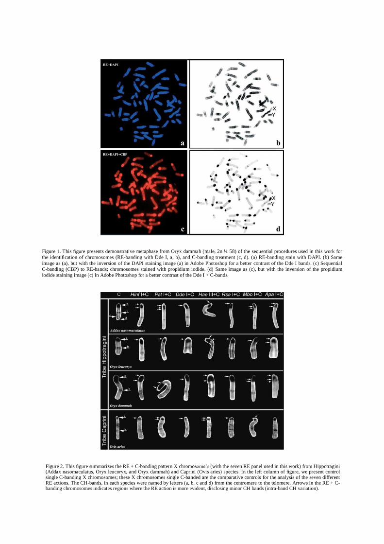
Figure 1. This figure presents demonstrative metaphase from Oryx dammah (male, 2n ¼ 58) of the sequential procedures used in this work for
the identification of chromosomes (RE-banding with Dde I, a, b), and C-banding treatment (c, d). (a) RE-banding stain with DAPI. (b) Same
image as (a), but with the inversion of the DAPI staining image (a) in Adobe Photoshop for a better contrast of the Dde I bands. (c) Sequential
C-banding (CBP) to RE-bands; chromosomes stained with propidium iodide. (d) Same image as (c), but with the inversion of the propidium
iodide staining image (c) in Adobe Photoshop for a better contrast of the Dde I + C-bands.
Figure 2. This figure summarizes the RE + C-banding pattern X chromosome’s (with the seven RE panel used in this work) from Hippotragini (Addax nasomaculatus, Oryx leucoryx, and Oryx dammah) and Caprini (Ovis aries) species. In the left column of figure, we present control single C-banding X chromosomes; these X chromosomes single C-banded are the comparative controls for the analysis of the seven different RE actions. The CH-bands, in each species were named by letters (a, b, c and d) from the centromere to the telomere. Arrows in the RE + C-banding chromosomes indicates regions where the RE action is more evident, disclosing minor CH bands (intra-band CH variation).
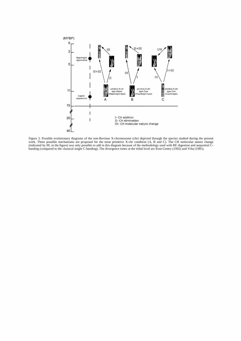
Figure 3. Possible evolutionary diagrams of the non-Bovinae X-chromosome (chr) depicted through the species studied during the present work. Three possible mechanisms are proposed for the most primitive X-chr condition (A, B and C). The CH molecular nature change (indicated by III, in the figure) was only possible to add in this diagram because of the methodology used with RE digestion and sequential C-banding (compared to the classical single C-banding). The divergence times at the tribal level are from Gentry (1992) and Vrba (1985).
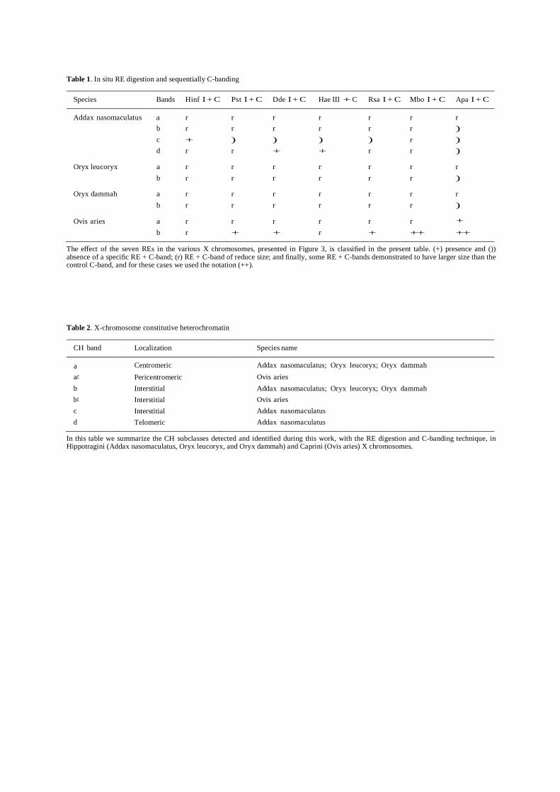
Table 1. In situ RE digestion and sequentially C-banding
Species Bands Hinf I + C Pst I + C Dde I + C Hae III + C Rsa I + C Mbo I + C Apa I + C
Addax nasomaculatus a r r r r r r r
b r r r r r r )
c + ) ) ) ) r )
d r r + + r r )
Oryx leucoryx a r r r r r r r
b r r r r r r )
Oryx dammah a r r r r r r r
b r r r r r r )
Ovis aries a r r r r r r +
b r + + r + ++ ++
The effect of the seven REs in the various X chromosomes, presented in Figure 3, is classified in the present table. (+) presence and ()) absence of a specific RE + C-band; (r) RE + C-band of reduce size; and finally, some RE + C-bands demonstrated to have larger size than the control C-band, and for these cases we used the notation (++).
Table 2. X-chromosome constitutive heterochromatin
CH band Localization Species name
a Centromeric Addax nasomaculatus; Oryx leucoryx; Oryx dammah a¢ Pericentromeric Ovis aries b Interstitial Addax nasomaculatus; Oryx leucoryx; Oryx dammah b¢ Interstitial Ovis aries c Interstitial Addax nasomaculatus d Telomeric Addax nasomaculatus
In this table we summarize the CH subclasses detected and identified during this work, with the RE digestion and C-banding technique, in Hippotragini (Addax nasomaculatus, Oryx leucoryx, and Oryx dammah) and Caprini (Ovis aries) X chromosomes.