combinatorial delivery of gallium (iii) nitrate and curcumin
TRANSCRIPT
Citation: Mohan Viswanathan, T.;
Krishnakumar, V.; Senthilkumar, D.;
Chitradevi, K.; Vijayabhaskar, R.;
Rajesh Kannan, V.; Senthil Kumar, N.;
Sundar, K.; Kunjiappan, S.;
Babkiewicz, E.; et al. Combinatorial
Delivery of Gallium (III) Nitrate and
Curcumin Complex-Loaded Hollow
Mesoporous Silica Nanoparticles for
Breast Cancer Treatment.
Nanomaterials 2022, 12, 1472.
https://doi.org/10.3390/
nano12091472
Academic Editors: Devika Chithrani,
Gopinath Packirisamy and Jose
L. Arias
Received: 19 March 2022
Accepted: 20 April 2022
Published: 26 April 2022
Publisher’s Note: MDPI stays neutral
with regard to jurisdictional claims in
published maps and institutional affil-
iations.
Copyright: © 2022 by the authors.
Licensee MDPI, Basel, Switzerland.
This article is an open access article
distributed under the terms and
conditions of the Creative Commons
Attribution (CC BY) license (https://
creativecommons.org/licenses/by/
4.0/).
nanomaterials
Article
Combinatorial Delivery of Gallium (III) Nitrate and CurcuminComplex-Loaded Hollow Mesoporous Silica Nanoparticles forBreast Cancer TreatmentThimma Mohan Viswanathan 1, Vaithilingam Krishnakumar 2, Dharmaraj Senthilkumar 1, Kaniraja Chitradevi 1,Ramakrishnan Vijayabhaskar 3, Velu Rajesh Kannan 2, Nachimuthu Senthil Kumar 4 , Krishnan Sundar 1,Selvaraj Kunjiappan 1 , Ewa Babkiewicz 5 , Piotr Maszczyk 5 and Thandavarayan Kathiresan 1,*
1 Department of Biotechnology, Kalasalingam Academy of Research and Education, Krishnankoil 626126, India;[email protected] (T.M.V.); [email protected] (D.S.); [email protected] (K.C.);[email protected] (K.S.); [email protected] (S.K.)
2 Department of Microbiology, Bharathidasan University, Tiruchirappalli 620024, India;[email protected] (V.K.); [email protected] (V.R.K.)
3 Department of Surgical Oncology, Meenakshi Mission Hospital and Research Centre, Madurai 625107, India;[email protected]
4 Department of Biotechnology, Mizoram University, Aizawl 796004, India; [email protected] Department of Hydrobiology, Faculty of Biology, University of Warsaw at Biology & Chemistry Research
Center, 02-089 Warsaw, Poland; [email protected] (E.B.); [email protected] (P.M.)* Correspondence: [email protected]; Tel.: +91-4563-289042; Fax: +91-4563-289322
Abstract: The main aims in the development of a novel drug delivery vehicle is to efficiently carrytherapeutic drugs in the body’s circulatory system and successfully deliver them to the targeted siteas needed to safely achieve the desired therapeutic effect. In the present study, a passive targetedfunctionalised nanocarrier was fabricated or wrapped the hollow mesoporous silica nanoparticleswith 3-aminopropyl triethoxysilane (APTES) to prepare APTES-coated hollow mesoporous silicananoparticles (HMSNAP). A nitrogen sorption analysis confirmed that the shape of hysteresisloops is altered, and subsequently the pore volume and pore diameters of GaC-HMSNAP wasreduced by around 56 and 37%, respectively, when compared with HMSNAP. The physico-chemicalcharacterisation studies of fabricated HMSNAP, Ga-HMSNAP and GaC-HMSNAP have confirmedtheir stability. The drug release capacity of the fabricated Ga-HMSNAP and GaC-HMSNAP fordelivery of gallium and curcumin was evaluated in the phosphate buffered saline (pH 3.0, 6.0 and 7.4).In an in silico molecular docking study of the gallium-curcumin complex in PDI, calnexin, HSP60,PDK, caspase 9, Akt1 and PTEN were found to be strong binding. In vitro antitumor activity of bothGa-HMSNAP and GaC-HMSNAP treated MCF-7 cells was investigated in a dose and time-dependentmanner. The IC50 values of GaC-HMSNAP (25 µM) were significantly reduced when compared withfree gallium concentration (40 µM). The mechanism of gallium-mediated apoptosis was analyzedthrough western blotting and GaC-HMSNAP has increased caspases 9, 6, cleaved caspase 6, PARP,and GSK 3β(S9) in MCF-7 cells. Similarly, GaC-HMSNAP is reduced mitochondrial proteins suchas prohibitin1, HSP60, and SOD1. The phosphorylation of oncogenic proteins such as Akt (S473),c-Raf (S249) PDK1 (S241) and induced cell death in MCF-7 cells. Furthermore, the findings revealedthat Ga-HMSNAP and GaC-HMSNAP provide a controlled release of loaded gallium, curcumin andtheir complex. Altogether, our results depicted that GaC-HMNSAP induced cell death through themitochondrial intrinsic cell death pathway, which could lead to novel therapeutic strategies for breastadenocarcinoma therapy.
Keywords: anticancer; cell viability; drug loading capacity; drug release; mitochondrial protein;nanomedicine
Nanomaterials 2022, 12, 1472. https://doi.org/10.3390/nano12091472 https://www.mdpi.com/journal/nanomaterials
Nanomaterials 2022, 12, 1472 2 of 23
1. Introduction
Hollow mesoporous silica nanoparticles (HMSNP) are highly versatile and favorablenanoplatforms for drug delivery systems (DDS) because of their assortment, accessibility,and biocompatibility. HMSNP is non-lethal and has enormous explicit surface territory,a tunable pore size, excellent physicochemical steadiness, and artificially modifiable sur-faces [1,2]. Enhancing the targeted drug delivery of HMSNPs to specific cells/tumor tissuesmight be adopted in two main strategies. One is the selected appropriate gatekeepers, whichare attached to pore openings of drug loaded HMSNPs by covalent linking [3]. The variouscompounds that are reported in silica nanoparticles are coated with polymers, such aspolyethylene glycol (PEG), polycaprolactone (PCL), dextran, chitosan, polyethyleneimine(PEI), and 3-aminopropyl triethoxysilane (APTES), to enhance its drug loading, reten-tion time, and release and prevention of aggregation. The other strategies are the drugmolecules linked to either the pore or surface of the HMSNPs through stimulus responsivelinkage [4,5]. The HMSNP is an efficient nanocarrier for enabling greater storage andrelease of chemical substances and chemotherapeutic drugs [6–8]. Silicon is naturally foundin bones and connective tissues of the human body [9]. The cytotoxicity and cellular uptakeof the MSNs depend on the size and the surface charges of the nanocarrier. The 15 nmdiameter nanocarriers induced more cytotoxicity than 100 nm nanocarriers in endothelialcells [10]. The mesoporous silica nanoparticles are loading large amounts of multiple drugs,which are double responsive to the synergistic therapy of breast cancers [11]. Subsequently,chemotherapeutic drugs, such as doxorubicin, are combined with proapoptotic peptide [12],curcumin [13], and Bcl-2 siRNA [14].
Until now, several chemotherapeutic drugs have been used against breast cancerthrough the alteration of histone deacetylase inhibitors and various cell surface receptors,like EGFR and HER2, to inhibit cancer survival pathways. An efficient cancer treatmentstrategy is the combination of multiple drugs which synergistically interact against cancercells but not in normal cells [15]. In this regard, curcumin and its subsidiaries express anti-cancer activity by stifling the multiplication of various tumour cell lines [16,17]. Specifically,curcumin deregulates GRP78, IRE1α, and CHOP, inducing cell death [13]. Gallium (Ga) isthe second metal compound after platinum [18] that is considered a high potential candidatefor anticancer therapy [19–21]. Gallium compounds bind with transferrin and are taken upby an endocytic process of cells through the transferrin receptor to form transferrin-gallium(Tf-Ga) complexes [22,23]. The Tf-Ga disturbs the normal uptaking of the transferrin-ironcomplex, which leads to iron deprivation in cancer cells [24]. Moreover, intracellulargallium compounds distort the three-dimensional structure of DNA and its replicationand protein synthesis, and inhibit ATPase, DNA polymerase and tyrosine-specific proteinphosphatase activity to induce cell death [25]. The gallium-based metallodrugs, such asgallium nitrate, gallium chloride, gallium maltolate, tris (8-quinolonato) gallium (III) orKP46, have been used as anticancer agents. Among them, gallium maltolate and KP46 havebeen evaluated in preclinical trials [26,27], and gallium III nitrate has gone up to clinicaltrial phase II [28]. Gallium compounds are also used in combination with other chemother-apeutic agents, such as paclitaxel [21], gemcitabine [29], vinorelbine [30], hydroxyurea [24],fludarabine [31], and interferon-γ [32], and are found to synergistically inhibit varioustypes of cancers. They are directed to assess wide ranges of antineoplastic movementand inhibition of tumor growths of Hodgkin’s and non-Hodgkin’s lymphoma, chroniclymphocytic leukemia, prostate, lung, ovarian, cervical, bladder, renal, breast, melanoma,and sarcoma [20].
The combination of gallium (III) nitrate and curcumin therapy has not yet beenattempted, and this is the first study to analyse their interaction as well as the mechanismbehind gallium-curcumin complex-induced cancer cell death. The present study focuses oncetyltrimethyl ammonium bromide (CTAB) as a template and tetraethyl ortho-orthosilicate(TEOS) as a silicon source for synthesised HMSNPs. The (3-aminopropyl) triethoxysilane(APTES) were hydrolysed, and NH2 was crippled on the surface of HMSNP to formHMSNAP, which facilitates binding through the amination reaction in the gallium and
Nanomaterials 2022, 12, 1472 3 of 23
hydroxyl group of enolic in curcumin. HMSNAP is used as a carrier for delivering gallium(III) nitrate alone (Ga-HMSNAP) and in combination with gallium (III) nitrate-curcumin(GaC-HMSNAP) complex against MCF-7 cells. The key questions asked in the presentstudy are: (1) whether HMSNAP is less toxic and has rapid drug loading capacity andsustained drug release in MCF-7 cells; (2) whether Ga-HMSNAP and GaC-HMSNAP caninduce cell death in MCF-7 cells at a low gallium concentration; and (3) which proteins areup-regulated or down-regulated due to GaC-HMSNAP treatment in MCF-7 cells. Overall,our studies portrayed an improvement in the therapeutic potential of the combinationof gallium and curcumin (GaC-HMSNAP), demonstrating that they could be a betteralternative for the current therapies against cancer.
2. Materials and Methods2.1. Materials and Reagents
The chemicals Gallium (III) nitrate (99.90% purity), curcumin (99.50% purity), tetraethylorthosilicate (TEOS) (99.00% purity), 3-Aminopropyl triethoxysilane (APTES) (99.00% pu-rity), cetyltrimethyl ammonium bromide (CTAB) (99.00% purity), dimethyl sulfoxide(DMSO) (99.70% purity), acridine orange (98.90% purity), ethidium bromide (99.50% pu-rity), and casein from bovine milk (99.00%) was procured from Sigma-Aldrich, (St. Louis,MO, USA). Dulbecco’s modified eagle medium (DMEM), Foetal bovine serum (FBS),and 0.25% trypsin EDTA (Gibco BRL, Waltham, MA, USA) and Tetrazolium salt (3-(4,5-dimethylthiazol-2yl)-2,5-diphenyltetrazolium bromide) (98.00% purity) was obtained from(Invitrogen, Carlsbad, CA, USA). BCA reagent was received from Bio-Rad (Hercules, CA,USA). All the experimental solutions were prepared using 18 MΩ milli-Q water 133 (Milli-pore system, Burlington, MA, USA). A few other analytical grade chemicals and reagentswere purchased from Thermo Fisher Scientific Ltd. Mumbai, India.
2.2. Synthesis of HMSNAP
The sol-gel method was adopted for the synthesis of hollow mesoporous silica nanopar-ticles (HMSNP) with slight modifications [33]. The formation of HMSNP was attained asammonia effectuate silica precursor underwent rapid hydrolysis with polycondensation ofan ethanol-water mixture by using surfactant CTAB as a structure-directing agent to assistin the formation of the mesostructure during the sol-gel process. A 40.5 mL mixture of anethanol-water containing 80 mg of CTAB and 500 µL of TEOS was added and mixed well,followed by 500 µL of liquid ammonia, and stirred for 3 h at 700 g. After 3 h, the resultantwhite precipitate products were washed with ethanol and dried. The dried HMSNP werecalcinated in the air at 800 C for 6 h. Functionalisation with amines, 50 mg HMSNP wereadded in 50 mL ethanol and 50 µL APTES and kept in a shaker for 24 h, then washed threetimes with ethanol and one time with distilled water to form HMSNAP.
2.3. Drug Loading and Release
For drug loading, 15 mg of synthesised HMSNAP were dispersed separately with30 to 50 mM of gallium alone, and a combination of 30 to 50 mM gallium with 30 mMcurcumin was dissolved in ethanol in an orbital shaker for 24 h. The unbound drug waswashed with ethanol and dried to obtain the nanoparticles, and various concentrationsof the drug-loaded nanocarrier were used in the cell viability assay to identify the IC50value of drugs and their combination. For the drug loading and release assay, 15 mg ofsynthesised HMSNP and HMSNAP were dispersed separately with 36 mM Gallium (III)nitrate alone and in the combination of 36 mM Gallium (III) nitrate with 30 mM curcuminadded in each 10 mL of ethanol in an orbital shaker for 48 h. The unbound free galliumand curcumin were estimated for each 6 h interval from 0 h to 48 h incubation. Theirabsorbance at 540 nm using a fluorescent microplate reader (Biotek, Winooski, VT, USA)was compared with control. The amount of gallium and curcumin-loaded nanostructurewas determined by the formula (Drug loading = O.D value at 0 h − O.D value at differenttime intervals/O.D value at 0 h) × 100.

Nanomaterials 2022, 12, 1472 4 of 23
The drug release experiments were accomplished with 2 mg mL−1 of 30 µM Ga-HMSNAP. A combination of 20 µM Gallium and 30 µM curcumin-loaded GaC-HMSNAPwas kept separately in a phosphate buffer saline (PBS) in three different pH: 3, 6 and 7.4.Approximately 200 µL of the sample was collected at every 6 h interval up to 72 h, and theabsorption of gallium release at 420 nm and the curcumin release at 540 nm was determinedusing a microplate reader.
2.4. Drug Release Kinetics
The gallium from HMSNAP and the gallium-curcumin complex from HMSNAPrelease data were separately fitted into five different kinetics models (zero, first, Higuchi,Korsmeyer-Peppas, and Hixson-Crowell) to describe the mechanism of the drug releasekinetics and diffusion. The release kinetic mechanisms were evaluated by using the aboveinvestigated in vitro release data through the DD solver 1.0 tool (Microsoft-Excel pluginmodel). The zero-order rate kinetics explains the rate of drug release and does not relateto their concentrations. The first-order rate kinetics explains the drug release from theformulation where the drug release rate related to their concentration is dependent. TheHiguchi model designated the drug release from the insoluble matrix as a square root of thetime-dependent process based on Fickian diffusion. The Korsmeyer-Peppas model explainsa simple mathematical relationship, describing the drug release from a formulation [34,35].
2.5. Characterisation of HMSNAP
The synthesised silica nanoparticles were powdered and analysed by SEM-EDAX,TEM, dynamic light scattering (DLS), zeta potential, X-ray diffraction (XRD), and Fouriertransform infrared spectroscopy (FTIR). To find out the morphological structure of HM-SNAP, Ga-HMSNAP and GaC-HMSNAP were loaded on carbon paper for SEM (Evo18Zeiss, Munich, Germany) and copper grid for TEM (HRTEM, T12 tecnai, Hillsboro, OR,USA) at HT650 ES1000W t 120 kV. The pore size of this HMSNAP was measured by us-ing the Image J software (National Institutes of Health, New York, NY, USA) of HRTEM.The nanostructures are dispersed in water and analysed for their size and surface chargethrough DLS and zeta potential using the Nanoparticle analyser SZ-100 (Horiba, Kyoto,Japan). XRD analysis was done in an X-ray tube 3 kW with a copper target, real-timemultiple strip solid-state detectors were used, K alpha was maintained at 0.001, and itwas scanned using the D8 Advance ECO XRD System (Bruker, Madison, WI, USA). FTIRmeasurements of the above nanoparticles were analysed using a Shimadzu spectrometer(Nishinokyo, Japan) in the ranges of 4000–400 cm−1 in the transmission mode.
The surface elemental composition of the nanoparticles was analysed with X-rayphotoelectron spectroscopy (VersaProbe III Scanning XPS Microprobe, Physical electronics,Chanhassen, MN, USA), applying an emission line of aluminium Kα x-ray source withhv of 1486.6 (eV), carried out with an emission current of 10 mA and an anode voltage of15 kV. The samples mounted on the gold layers sputtered onto the silicon substrate. Datawere analysed by using MultiPak Data Reduction Software (version 9.6.0) (2500 Hagisono,Chigasaki, Japan). Nitrogen adsorption-desorption isotherm analysis was carried out byQuantachrome® ASiQwin™ (Boynton Beach, FL, USA) at liquid nitrogen (−196 C). Beforemeasurement, the samples were degassed at 150 C under vacuum conditions for 24 h. Thespecific surface area (SBET) of nanocarriers was calculated from the relative pressure rangeof nitrogen adsorption from 0.05 to 0.2 using the Brunauer–Emmett–Teller (BET) equation.Pore size distributions (PSD) were determined from adsorption twigs of isotherms usingthe Barrett–Joyner–Halenda (BJH) method. The total pore volume (Vp) was estimated fromthe amount of nitrogen adsorbed at a relative pressure of 0.99. The drugs that bound withthe nanocarriers were evaluated by 1 H nuclear magnetic resonance (NMR). The spectrawere measured on a Bruker AV 500 MHz spectrometer with D2O as the solvents andtetramethylsilane (TMS) as the internal standard. The chemical structure of nanoparticleswith drug loading was determined through the use of TopSpin® 4.0 software.
Nanomaterials 2022, 12, 1472 5 of 23
2.6. Molecular Docking Analysis
Molecular docking was performed using AutoDock 4.2 software (The Scripps ResearchInstitute, San Diego, CA, USA). The basic and default settings were followed for protein andligand preparation. Docking of the gallium-curcumin complex ligand with proteins, suchas PDI, calnexin, HSP60, PDK caspase 9, Akt1, and PTEN, was performed. The bindingmodel for each protein was analysed using a Discovery Studio Visualizer v4.0 [36].
2.7. Cell Culture and Cell Viability Assay
The breast adenocarcinoma cell line MCF-7 was procured from NCCS (National CentreFor Cell Science, Pune, Maharashtra, India). The MCF-7 cells was grown in Dulbecco’smodified eagle medium (DMEM), which consisted of 10% fetal bovine serum (FBS) and 5%CO2 at 37 C. The MCF-7 cell viability was determined through MTT assay, as describedpreviously, with slight modifications [37]. The cells were trypsinised from T25 confluenceflasks and seeded in 96 well plates at 15,000 cells per well. The different concentrations(5 to 50 µg) of HMSNAP, Ga-HMSNAP, and GaC-HMSNAP were treated separately withMCF-7 cells and incubated at different time intervals for 24, 48 and 72 h with a serum (0.5%)deprivation medium. The cell viability of nanoparticles on MCF-7 cells was analysed usingtetrazolium salt. After respective time incubation, 10 µL of MTT solution was added toeach well and incubated for 4 h. The tetrazolium reacted with dehydrogenase reductase oflive cells to produce indissoluble formazan crystal. Then 100 µL DMSO was added to eachwell to dissolve the formazan crystal to form purple formazan, and the concentration wasread at 595 nm in a microtiter plate reader. The experiments were repeated three times withresults and were expressed as the percentage (%) of control cells. The percentage of cellviability and IC50 values of each drug were calculated using Prism-GraphPad version 5.
2.8. Measurement of Apoptosis by Acridine Orange/Ethidium Bromide (AO/EtBr)
The apoptosis assay was carried out with AO/EtBr double staining, in HMSNAP, Ga-HMSNAP and GaC-HMSNAP-treated MCF-7 cells. The drug-loaded nanoparticle-treatedcells were trypsinised and pelleted after 24, 48, and 72 h incubation periods. The MCF-7cells were dissolved in 1 mL of phosphate buffer saline containing 100 µL each of acridineorange (50 µg mL−1) and ethidium bromide (30 µg mL−1), and were examined under afluorescent microscope (Carl Zeiss, Oberkochen, Germany). Randomly selected variousfields were analysed and the percentages of live and dead cells were estimated.
2.9. Western Blot Analysis
The qualitative analyses of free gallium, Ga-HMSNAP, and GaC-HMSNAP inducedprotein alteration in MCF-7 cells were analysed using western blotting. The cells weregrown in 100 mm dishes and treated with 5µg HMSNAP, and an IC50 concentration of 40 µMfree gallium, 35µM Ga-HMSNAP, and combination of 25 µM gallium and 30 µM curcumin-loaded GaC-HSMNAP for 24 and 48 h incubation. After the respective incubation times,MCF-7 cells were harvested and lysed with 50 mM phosphate buffer (pH 7.4) containing25 mM sodium fluoride, 5 mM EDTA, 0.4% ASB-14, and protease and phosphatase cocktailinhibitors (Roche, Switzerland) on ice for 5 min. and the lysates were sonicated andfollowed by centrifugation at 12,000 rpm for 10 min. at 4 C. Next, the protein wasquantified with BCA reagent. Each 50 µg of proteins were loaded onto SDS-PAGE andproteins were run at 90 V for 2 h. The proteins were transferred to nitrocellulose membrane(Amersham Bioscience, Piscataway, NJ, USA). Anti-cleaved PARP, anti-caspase 12, anti-cleaved caspase 12, and 6, anti-caspases 9, 6, anti-calnexin, anti-PDI, anti-prohibitin, anti-HSP 60, anti-SOD1, anti-phospho-Akt (S473), anti-total Akt, anti-phospho-PTEN (S380),anti-phospho-c-Raf (S259), anti-phospho-PDK1 (S241), anti-phospho-GSK3β (S9), and anti-β-actin were obtained from Cell Signaling Technology (Danvers, MA, USA). The membranewas blocked with 5% casein for 1 h and incubated with primary antibodies for 1 h at roomtemperature followed by either anti-rabbit IgG or anti-mouse IgG HRP-linked secondaryantibodies (Santa Cruz, CA, USA) for 1 h. The presence of immunoreactive bands of
Nanomaterials 2022, 12, 1472 6 of 23
respective proteins was detected with the addition of lumiglo (Thermo scientific, Rockford,IL, USA). All of the immunoreactive and phosphorylation signals were quantified using thedensitometric scanner (Bio-Rad, Hercules, CA, USA). The blot was washed and reprobedwith anti-β-actin to ensure that an equal amount of proteins were loaded in each welland the data presented were representative of the two independent experiments withsimilar results.
2.10. Statistical Analysis
Each experiment was repeated three times with three replicates, and the standarderror of the mean (SEM) values were optimised for all experimental data as ±mean. Aone-way analysis of variance test was performed to assess the results between the controland all treated samples (p < 0.05).
3. Results and Discussion3.1. Synthesis and Characterisation of HMSNAP
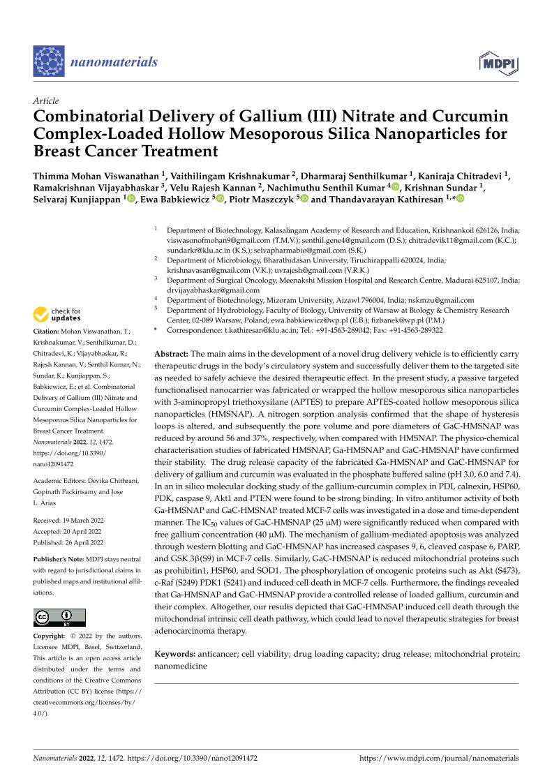
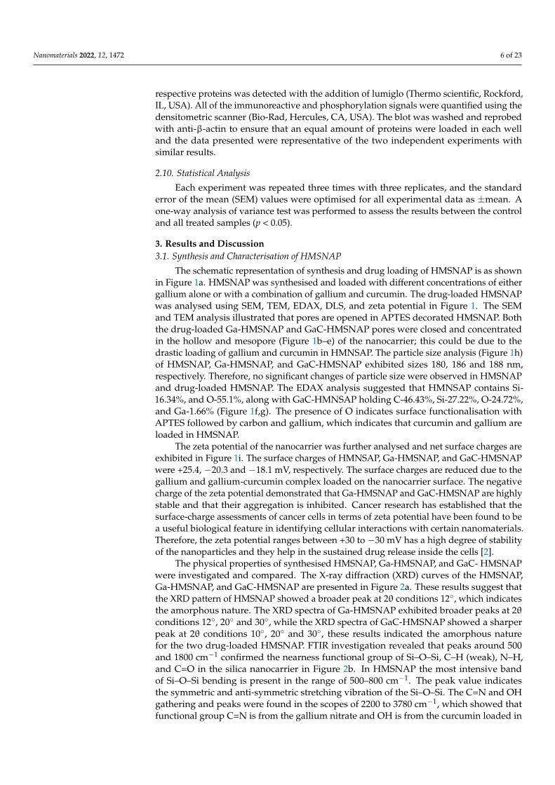
The schematic representation of synthesis and drug loading of HMSNAP is as shownin Figure 1a. HMSNAP was synthesised and loaded with different concentrations of eithergallium alone or with a combination of gallium and curcumin. The drug-loaded HMSNAPwas analysed using SEM, TEM, EDAX, DLS, and zeta potential in Figure 1. The SEMand TEM analysis illustrated that pores are opened in APTES decorated HMSNAP. Boththe drug-loaded Ga-HMSNAP and GaC-HMSNAP pores were closed and concentratedin the hollow and mesopore (Figure 1b–e) of the nanocarrier; this could be due to thedrastic loading of gallium and curcumin in HMNSAP. The particle size analysis (Figure 1h)of HMSNAP, Ga-HMSNAP, and GaC-HMSNAP exhibited sizes 180, 186 and 188 nm,respectively. Therefore, no significant changes of particle size were observed in HMSNAPand drug-loaded HMSNAP. The EDAX analysis suggested that HMNSAP contains Si-16.34%, and O-55.1%, along with GaC-HMNSAP holding C-46.43%, Si-27.22%, O-24.72%,and Ga-1.66% (Figure 1f,g). The presence of O indicates surface functionalisation withAPTES followed by carbon and gallium, which indicates that curcumin and gallium areloaded in HMSNAP.
The zeta potential of the nanocarrier was further analysed and net surface charges areexhibited in Figure 1i. The surface charges of HMNSAP, Ga-HMSNAP, and GaC-HMSNAPwere +25.4, −20.3 and −18.1 mV, respectively. The surface charges are reduced due to thegallium and gallium-curcumin complex loaded on the nanocarrier surface. The negativecharge of the zeta potential demonstrated that Ga-HMSNAP and GaC-HMSNAP are highlystable and that their aggregation is inhibited. Cancer research has established that thesurface-charge assessments of cancer cells in terms of zeta potential have been found to bea useful biological feature in identifying cellular interactions with certain nanomaterials.Therefore, the zeta potential ranges between +30 to −30 mV has a high degree of stabilityof the nanoparticles and they help in the sustained drug release inside the cells [2].
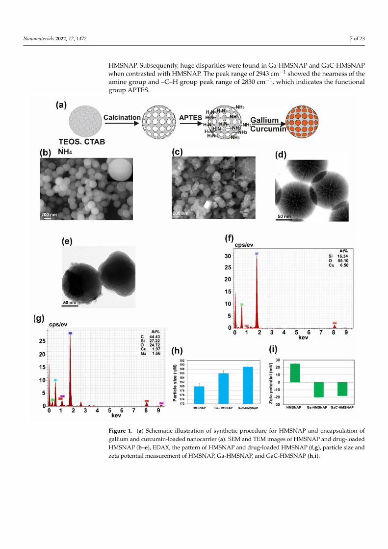
The physical properties of synthesised HMSNAP, Ga-HMSNAP, and GaC- HMSNAPwere investigated and compared. The X-ray diffraction (XRD) curves of the HMSNAP,Ga-HMSNAP, and GaC-HMSNAP are presented in Figure 2a. These results suggest thatthe XRD pattern of HMSNAP showed a broader peak at 2θ conditions 12, which indicatesthe amorphous nature. The XRD spectra of Ga-HMSNAP exhibited broader peaks at 2θconditions 12, 20 and 30, while the XRD spectra of GaC-HMSNAP showed a sharperpeak at 2θ conditions 10, 20 and 30, these results indicated the amorphous naturefor the two drug-loaded HMSNAP. FTIR investigation revealed that peaks around 500and 1800 cm−1 confirmed the nearness functional group of Si–O–Si, C–H (weak), N–H,and C=O in the silica nanocarrier in Figure 2b. In HMSNAP the most intensive bandof Si–O–Si bending is present in the range of 500–800 cm−1. The peak value indicatesthe symmetric and anti-symmetric stretching vibration of the Si–O–Si. The C=N and OHgathering and peaks were found in the scopes of 2200 to 3780 cm−1, which showed thatfunctional group C=N is from the gallium nitrate and OH is from the curcumin loaded in
Nanomaterials 2022, 12, 1472 7 of 23
HMSNAP. Subsequently, huge disparities were found in Ga-HMSNAP and GaC-HMSNAPwhen contrasted with HMSNAP. The peak range of 2943 cm−1 showed the nearness of theamine group and –C–H group peak range of 2830 cm−1, which indicates the functionalgroup APTES.
(g)
25
20
15
10
5
(a) Ca lei nation
•
TEOS. CTAB
NH4
(e)
cps/ev At%
C 44.43 Si 27.22 0 24.72 Cu 1.97 Ga 1.66
-1 2 3 4 5 6 7 8 9
kev
(h)
- 190 :E 188 - 186 18-4 ·en 1s2 a, 180
"ij 178 °f 176 :.. 174
172
(f)
30
25
20
15
10
5
cps/ev
tm At%
Si 16.34 0 55.10 Cu 6.50
o.,..,.=,,....-,...--r, 1
HMSNAP Ga-HMSNAP GaC-HMSNAP
2 3 4 5 6 7 8 kev
(i) 30
20
='= i 10 0
I 'g_a10
.fl ·20
N -30 HMSNAP Ga-HMSNAP GaC-HMSNAP
Figure 1. (a) Schematic illustration of synthetic procedure for HMSNAP and encapsulation ofgallium and curcumin-loaded nanocarrier (a). SEM and TEM images of HMSNAP and drug-loadedHMSNAP (b–e), EDAX, the pattern of HMSNAP and drug-loaded HMSNAP (f,g), particle size andzeta potential measurement of HMSNAP, Ga-HMSNAP, and GaC-HMSNAP (h,i).
Nanomaterials 2022, 12, 1472 8 of 23
Figure 2. (a) XRD and (b) FTIR pattern of HMSNAP and drug−loaded HMSNAP.
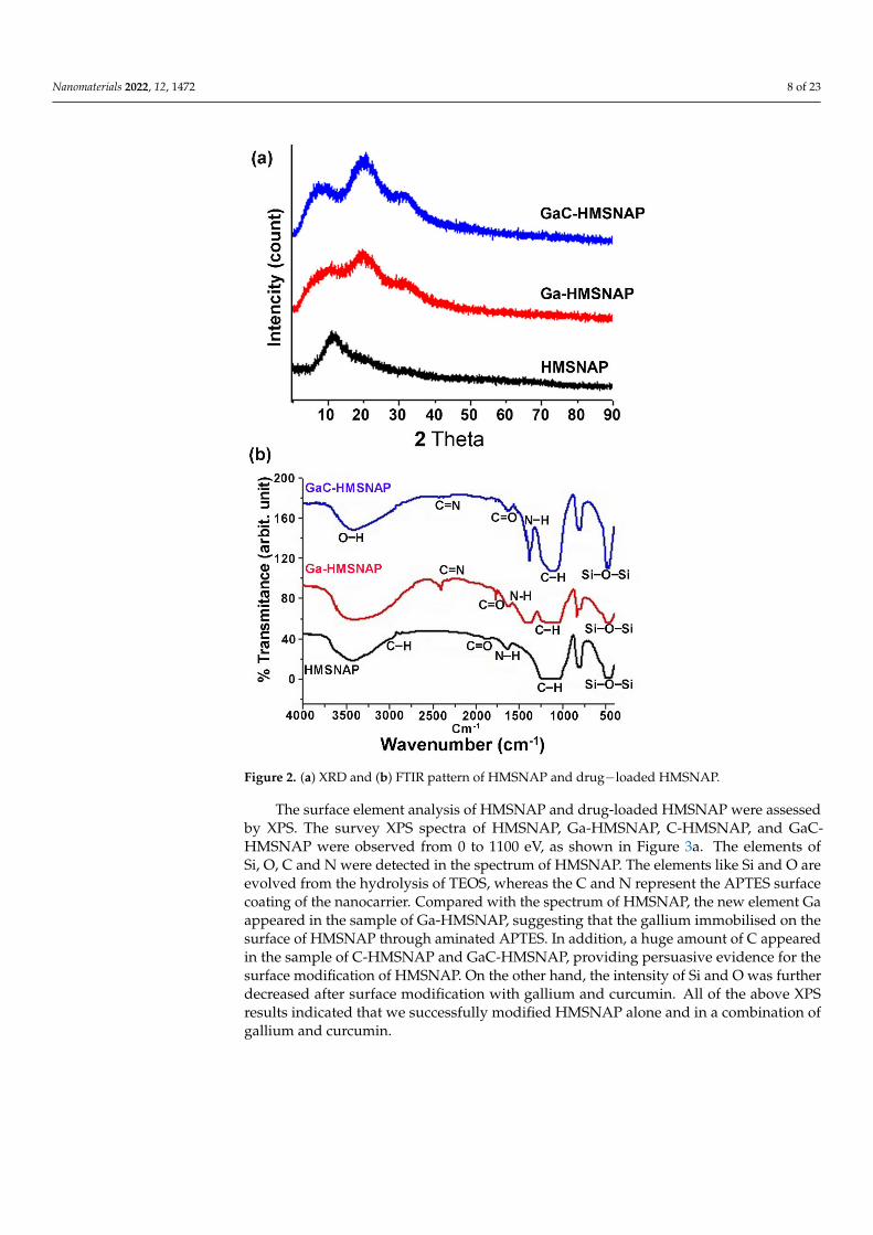
The surface element analysis of HMSNAP and drug-loaded HMSNAP were assessedby XPS. The survey XPS spectra of HMSNAP, Ga-HMSNAP, C-HMSNAP, and GaC-HMSNAP were observed from 0 to 1100 eV, as shown in Figure 3a. The elements ofSi, O, C and N were detected in the spectrum of HMSNAP. The elements like Si and O areevolved from the hydrolysis of TEOS, whereas the C and N represent the APTES surfacecoating of the nanocarrier. Compared with the spectrum of HMSNAP, the new element Gaappeared in the sample of Ga-HMSNAP, suggesting that the gallium immobilised on thesurface of HMSNAP through aminated APTES. In addition, a huge amount of C appearedin the sample of C-HMSNAP and GaC-HMSNAP, providing persuasive evidence for thesurface modification of HMSNAP. On the other hand, the intensity of Si and O was furtherdecreased after surface modification with gallium and curcumin. All of the above XPSresults indicated that we successfully modified HMSNAP alone and in a combination ofgallium and curcumin.
Nanomaterials 2022, 12, 1472 9 of 23
(a)
4000
2000
0
4000
2000
0
4000
2000
0
8000
6000
4000
2000
0
C-HMSNAP
.
•
-01, HMSNAP
-C1s -Si2s
-N1s I 1 l-s12p
1000 800 600 400 200
Binding Energy (eV)
280 275 270 265 Binding Energy (eV)
0
(b) 6000 Si2p
5000
.,4000
u3000
2000
1000
0 110 105 100 95
(d)
25,000 01
20,000
u,15,000 u
10,000
5000
Binding Energy (eV)
- HMSNAP c•o - Ga-HMSNAP
-Cur-HMSNAP -GaC-HMSNAP
540 535 530 525 520 Binding Energy (eV)
C
2600 N1s2400 2200
'B 2000
1800
415 410 405 400 395 390 Binding Energy (eV)
(e) - Ga-HMSNAP
500 - GaC-HMSNAP
400 u, u
300
200
.. Ga3d
Figure 3. The XPS spectra of survey (a) and high resolution of HMSNAP and drug-loaded HMSNAP(b–f). The region of Si2p (b), the region of N1s (c), the region of O1s (d), the region of Ga3d (e), theregion of C1s (f).
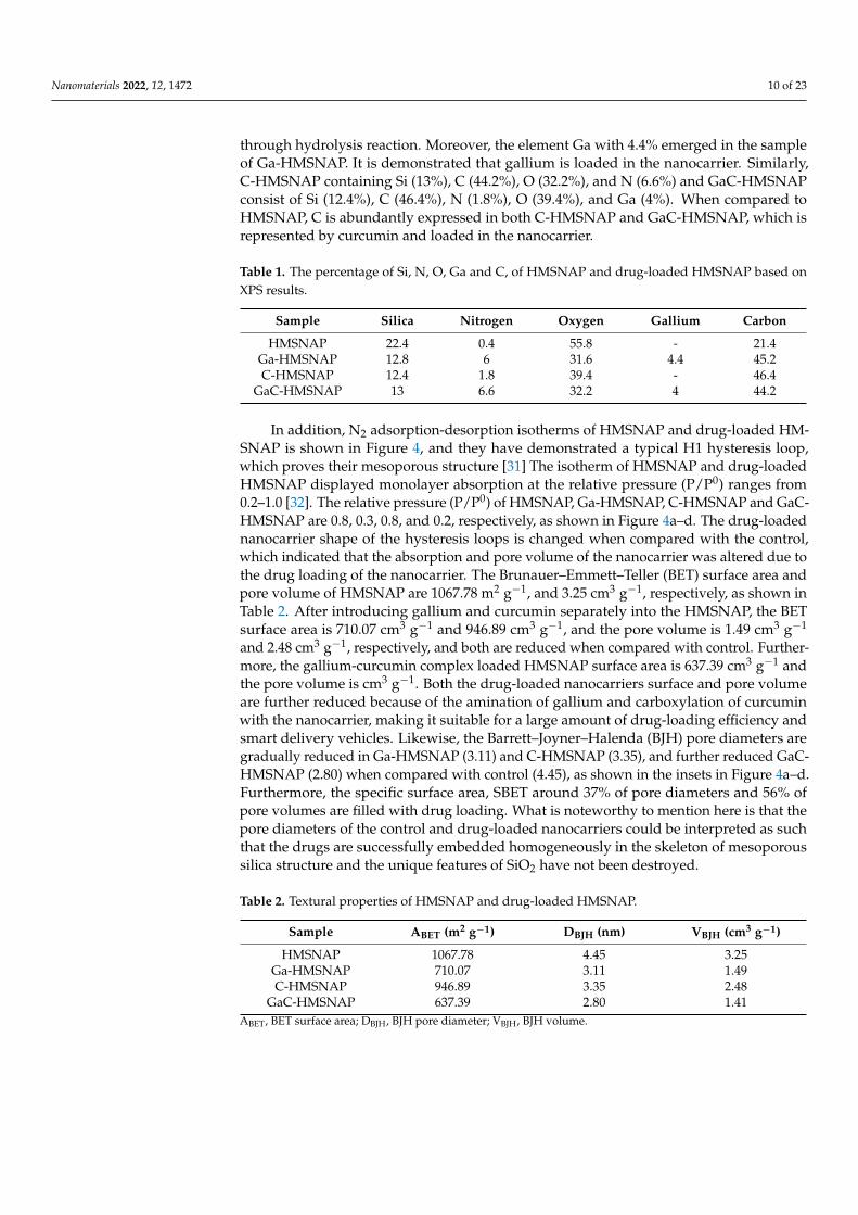
The high-resolution XPS spectra of Si2p, N1s, O1s, Ga3d, and C1s were shown inFigure 3b–f. More importantly, the peak intensities of Si2p in HMSNAP, Ga-HMSNAP,C-HMSNAP, and GaC-HMSNAP are shown in Figure 3b and the binding energy of Si2pwas located in between 101 to 103.8 eV. The intensity of drug-loaded HMSNAP of Si2p peakis around 42% reduced when compared with HMSNAP. The N1s spectra were displayedin Figure 3c and the ranges from 396.4 to 402.6. The intensity of N1s in Ga-HMSNAP, C-HMSNAP, and GaC-HMSNAP was decreased when compared with HMSNAP. In particular,the N1s spectrum in the sample HMSNAP shows a sharp peak and the others demonstrateblunt peaks. The intensity of O1 ranged from 531 to 533.6 eV, and high intensity was foundin HMSNAP and gradually decreased in C-HMSNAP, GaC-HMSNAP and Ga-HMSNAPin Figure 3d. Therefore, the O1 is masked by drugs loading on the nanocarrier. The high-resolution spectra of Ga3d are from 18.8 to 23.6 and two different peaks appeared in thespectra. The intensity of spectra is reduced in GaC-HMSNAP when compared with agallium-alone-loaded nanocarrier in Figure 3e. In the spectra of C1s, the peak position ofC1s in HMSNAP, Ga-HMSNAP, C-HMSNAP, and GaC-HMSNAP is shown in Figure 3fand the binding energy of C1s ranged from 270.8 to 273.8 eV. The percentages of elementsin HMSNAP, Ga-HMSNAP, C-HMSNAP, and GaC-HMSNAP were calculated via the XPSspectra, as shown in Table 1. The percentages of Si, N, O, and C in HMSNAP are 22.4, 0.4,55.8, and 21.4%, respectively. It is worth mentioning that the percentages of Si and O wereclose to the expected ratio of Si/O (1:2). Compared with HMSNAP, the percentages of Si, N,O, Ga, and C gain Ga-HMSNAP are changed to 12.8, 6.0, 31.6, 4.4, and 45.2%, respectively.The increase of N content suggests that APTES immobilised on the surface of HMSNAP
Nanomaterials 2022, 12, 1472 10 of 23
through hydrolysis reaction. Moreover, the element Ga with 4.4% emerged in the sampleof Ga-HMSNAP. It is demonstrated that gallium is loaded in the nanocarrier. Similarly,C-HMSNAP containing Si (13%), C (44.2%), O (32.2%), and N (6.6%) and GaC-HMSNAPconsist of Si (12.4%), C (46.4%), N (1.8%), O (39.4%), and Ga (4%). When compared toHMSNAP, C is abundantly expressed in both C-HMSNAP and GaC-HMSNAP, which isrepresented by curcumin and loaded in the nanocarrier.
Table 1. The percentage of Si, N, O, Ga and C, of HMSNAP and drug-loaded HMSNAP based onXPS results.
Sample Silica Nitrogen Oxygen Gallium Carbon
HMSNAP 22.4 0.4 55.8 - 21.4Ga-HMSNAP 12.8 6 31.6 4.4 45.2C-HMSNAP 12.4 1.8 39.4 - 46.4
GaC-HMSNAP 13 6.6 32.2 4 44.2
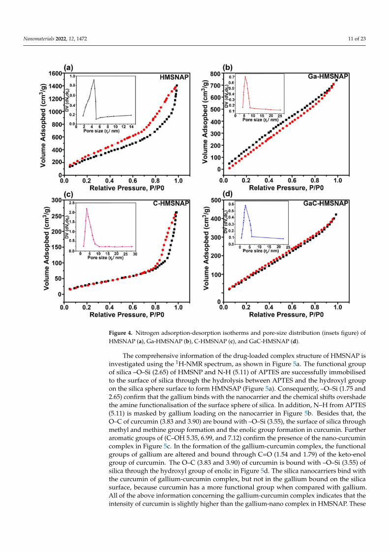
In addition, N2 adsorption-desorption isotherms of HMSNAP and drug-loaded HM-SNAP is shown in Figure 4, and they have demonstrated a typical H1 hysteresis loop,which proves their mesoporous structure [31] The isotherm of HMSNAP and drug-loadedHMSNAP displayed monolayer absorption at the relative pressure (P/P0) ranges from0.2–1.0 [32]. The relative pressure (P/P0) of HMSNAP, Ga-HMSNAP, C-HMSNAP and GaC-HMSNAP are 0.8, 0.3, 0.8, and 0.2, respectively, as shown in Figure 4a–d. The drug-loadednanocarrier shape of the hysteresis loops is changed when compared with the control,which indicated that the absorption and pore volume of the nanocarrier was altered due tothe drug loading of the nanocarrier. The Brunauer–Emmett–Teller (BET) surface area andpore volume of HMSNAP are 1067.78 m2 g−1, and 3.25 cm3 g−1, respectively, as shown inTable 2. After introducing gallium and curcumin separately into the HMSNAP, the BETsurface area is 710.07 cm3 g−1 and 946.89 cm3 g−1, and the pore volume is 1.49 cm3 g−1
and 2.48 cm3 g−1, respectively, and both are reduced when compared with control. Further-more, the gallium-curcumin complex loaded HMSNAP surface area is 637.39 cm3 g−1 andthe pore volume is cm3 g−1. Both the drug-loaded nanocarriers surface and pore volumeare further reduced because of the amination of gallium and carboxylation of curcuminwith the nanocarrier, making it suitable for a large amount of drug-loading efficiency andsmart delivery vehicles. Likewise, the Barrett–Joyner–Halenda (BJH) pore diameters aregradually reduced in Ga-HMSNAP (3.11) and C-HMSNAP (3.35), and further reduced GaC-HMSNAP (2.80) when compared with control (4.45), as shown in the insets in Figure 4a–d.Furthermore, the specific surface area, SBET around 37% of pore diameters and 56% ofpore volumes are filled with drug loading. What is noteworthy to mention here is that thepore diameters of the control and drug-loaded nanocarriers could be interpreted as suchthat the drugs are successfully embedded homogeneously in the skeleton of mesoporoussilica structure and the unique features of SiO2 have not been destroyed.
Table 2. Textural properties of HMSNAP and drug-loaded HMSNAP.
Sample ABET (m2 g−1) DBJH (nm) VBJH (cm3 g−1)
HMSNAP 1067.78 4.45 3.25Ga-HMSNAP 710.07 3.11 1.49C-HMSNAP 946.89 3.35 2.48
GaC-HMSNAP 637.39 2.80 1.41ABET, BET surface area; DBJH, BJH pore diameter; VBJH, BJH volume.
Nanomaterials 2022, 12, 1472 11 of 23
Figure 4. Nitrogen adsorption-desorption isotherms and pore-size distribution (insets figure) ofHMSNAP (a), Ga-HMSNAP (b), C-HMSNAP (c), and GaC-HMSNAP (d).
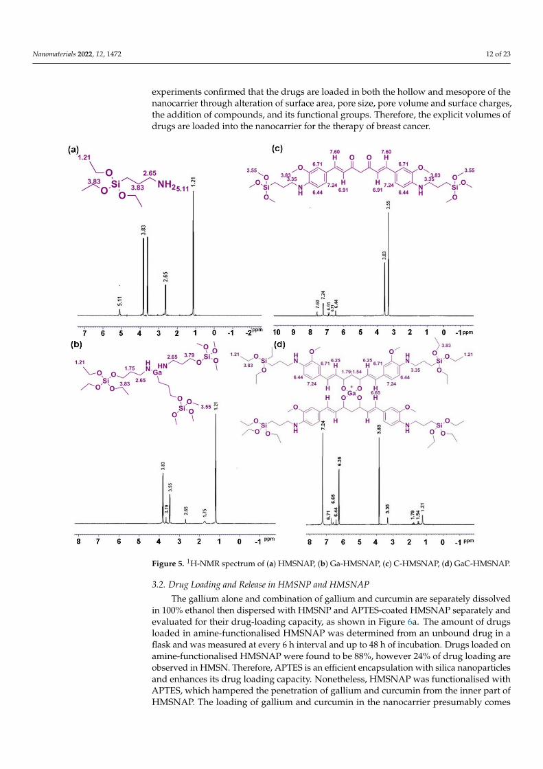
The comprehensive information of the drug-loaded complex structure of HMSNAP isinvestigated using the 1H-NMR spectrum, as shown in Figure 5a. The functional groupof silica –O–Si (2.65) of HMSNP and N-H (5.11) of APTES are successfully immobilisedto the surface of silica through the hydrolysis between APTES and the hydroxyl groupon the silica sphere surface to form HMNSAP (Figure 5a). Consequently, –O–Si (1.75 and2.65) confirm that the gallium binds with the nanocarrier and the chemical shifts overshadethe amine functionalisation of the surface sphere of silica. In addition, N–H from APTES(5.11) is masked by gallium loading on the nanocarrier in Figure 5b. Besides that, theO–C of curcumin (3.83 and 3.90) are bound with –O–Si (3.55), the surface of silica throughmethyl and methine group formation and the enolic group formation in curcumin. Furtheraromatic groups of (C–OH 5.35, 6.99, and 7.12) confirm the presence of the nano-curcumincomplex in Figure 5c. In the formation of the gallium-curcumin complex, the functionalgroups of gallium are altered and bound through C=O (1.54 and 1.79) of the keto-enolgroup of curcumin. The O–C (3.83 and 3.90) of curcumin is bound with –O–Si (3.55) ofsilica through the hydroxyl group of enolic in Figure 5d. The silica nanocarriers bind withthe curcumin of gallium-curcumin complex, but not in the gallium bound on the silicasurface, because curcumin has a more functional group when compared with gallium.All of the above information concerning the gallium-curcumin complex indicates that theintensity of curcumin is slightly higher than the gallium-nano complex in HMSNAP. These
Nanomaterials 2022, 12, 1472 12 of 23
experiments confirmed that the drugs are loaded in both the hollow and mesopore of thenanocarrier through alteration of surface area, pore size, pore volume and surface charges,the addition of compounds, and its functional groups. Therefore, the explicit volumes ofdrugs are loaded into the nanocarrier for the therapy of breast cancer.
Figure 5. 1H-NMR spectrum of (a) HMSNAP, (b) Ga-HMSNAP, (c) C-HMSNAP, (d) GaC-HMSNAP.
3.2. Drug Loading and Release in HMSNP and HMSNAP
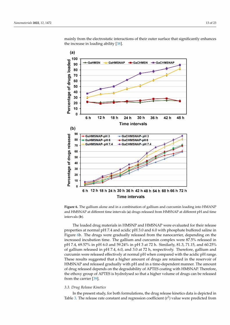
The gallium alone and combination of gallium and curcumin are separately dissolvedin 100% ethanol then dispersed with HMSNP and APTES-coated HMSNAP separately andevaluated for their drug-loading capacity, as shown in Figure 6a. The amount of drugsloaded in amine-functionalised HMSNAP was determined from an unbound drug in aflask and was measured at every 6 h interval and up to 48 h of incubation. Drugs loaded onamine-functionalised HMSNAP were found to be 88%, however 24% of drug loading areobserved in HMSN. Therefore, APTES is an efficient encapsulation with silica nanoparticlesand enhances its drug loading capacity. Nonetheless, HMSNAP was functionalised withAPTES, which hampered the penetration of gallium and curcumin from the inner part ofHMSNAP. The loading of gallium and curcumin in the nanocarrier presumably comes
Nanomaterials 2022, 12, 1472 13 of 23
mainly from the electrostatic interactions of their outer surface that significantly enhancesthe increase in loading ability [38].
Figure 6. The gallium alone and in a combination of gallium and curcumin loading into HMANPand HMSNAP at different time intervals (a) drugs released from HMSNAP at different pH and timeintervals (b).
The loaded drug materials in HMSNP and HMSNAP were evaluated for their releaseproperties at normal pH 7.4 and acidic pH 3.0 and 6.0 with phosphate buffered saline inFigure 6b. The drugs were gradually released from the nanocarrier, depending on theincreased incubation time. The gallium and curcumin complex were 87.5% released inpH 7.4, 69.57% in pH 6.0 and 59.24% in pH 3 at 72 h. Similarly, 81.3, 71.15, and 60.25%of gallium released in pH 7.4, 6.0, and 3.0 at 72 h, respectively. Therefore, gallium andcurcumin were released effectively at normal pH when compared with the acidic pH range.These results suggested that a higher amount of drugs are retained in the reservoir ofHMSNAP and released gradually with pH and in a time-dependent manner. The amountof drug released depends on the degradability of APTES coating with HMSNAP. Therefore,the ethoxy group of APTES is hydrolysed so that a higher volume of drugs can be releasedfrom the carrier [39].
3.3. Drug Release Kinetics
In the present study, for both formulations, the drug release kinetics data is depicted inTable 3. The release rate constant and regression coefficient (r2) value were predicted from
Nanomaterials 2022, 12, 1472 14 of 23
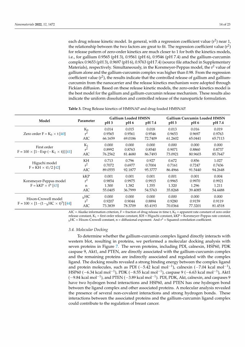
each drug release kinetic model. In general, with a regression coefficient value (r2) near 1,the relationship between the two factors are great to fit. The regression coefficient value (r2)for release pattern of zero-order kinetics are much closer to 1 for both the kinetics models,i.e., for gallium 0.9565 (pH 3), 0.9561 (pH 6), 0.9546 (pH 7.4) and the gallium-curcumincomplex 0.9653 (pH 3), 0.9697 (pH 6), 0.9763 (pH 7.4) (source file attached in SupplementaryMaterials), respectively. Simultaneously, in the Korsmeyer-Peppas model, the r2 value ofgallium alone and the gallium-curcumin complex was higher than 0.98. From the regressioncoefficient value (r2), the results indicate that the controlled release of gallium and gallium-curcumin from the nanocarrier and the release kinetics mechanism were adopted throughFickian diffusion. Based on these release kinetic models, the zero-order kinetics model isthe best model for the gallium and gallium-curcumin release mechanism. These results alsoindicate the uniform dissolution and controlled release of the nanoparticle formulation.
Table 3. Drug Release kinetics of HMSNAP and drug-loaded HMSNAP.
Model ParameterGallium Loaded HMSN Gallium Curcumin Loaded HMSN
pH 3 pH 6 pH 7.4 pH 3 pH 6 pH 7.4
Zero order F = K0 × t [40]K0 0.014 0.015 0.018 0.013 0.016 0.019r2 0.9565 0.9561 0.9546 0.9653 0.9697 0.9763
AIC 66.1659 69.0186 72.7409 61.2602 65.0441 65.7113
First orderF = 100 × [1−Exp (−K1 × t)] [41]
K1 0.000 0.000 0.000 0.000 0.000 0.000r2 0.8992 0.8763 0.8540 0.9071 0.8860 0.8737
AIC 76.2562 81.4680 86.7493 73.0921 80.9603 85.7647
Higuchi modelF = KH × t1/2 [42]
KH 0.713 0.796 0.927 0.672 0.856 1.027r2 0.7072 0.6977 0.7004 0.7161 0.7247 0.7436
AIC 89.0555 92.1877 95.3777 86.4966 91.5440 94.2648
Korsmeyer-Peppas modelF = kKP × tn [43]
kKP 0.001 0.001 0.001 0.001 0.001 0.004r2 0.9854 0.9975 0.9915 0.9965 0.9970 0.9921n 1.300 1.382 1.355 1.320 1.296 1.211
AIC 55.0405 36.7999 54.5763 35.8268 39.4085 54.4488
Hixon-Crowell modelF = 100 × [1−(1−kHC × t)3] [44]
kHC 0.000 0.000 0.000 0.000 0.000 0.000r2 0.9207 0.9044 0.8894 0.9280 0.9159 0.9119
AIC 73.3839 78.3709 83.4193 70.0364 77.3201 81.4518
AIC = Akaike information criterion, F = fraction of drug release in time t, K0 = apparent rate constant of zero orderrelease constant, K1 = first order release constant, KH = Higuchi constant, kKP = Korsmeyer–Peppas rate constant,kHC = Hixon–Crowell constant, n = diffusional exponent. And r2 = Squared correlation coefficient.
3.4. Molecular Docking
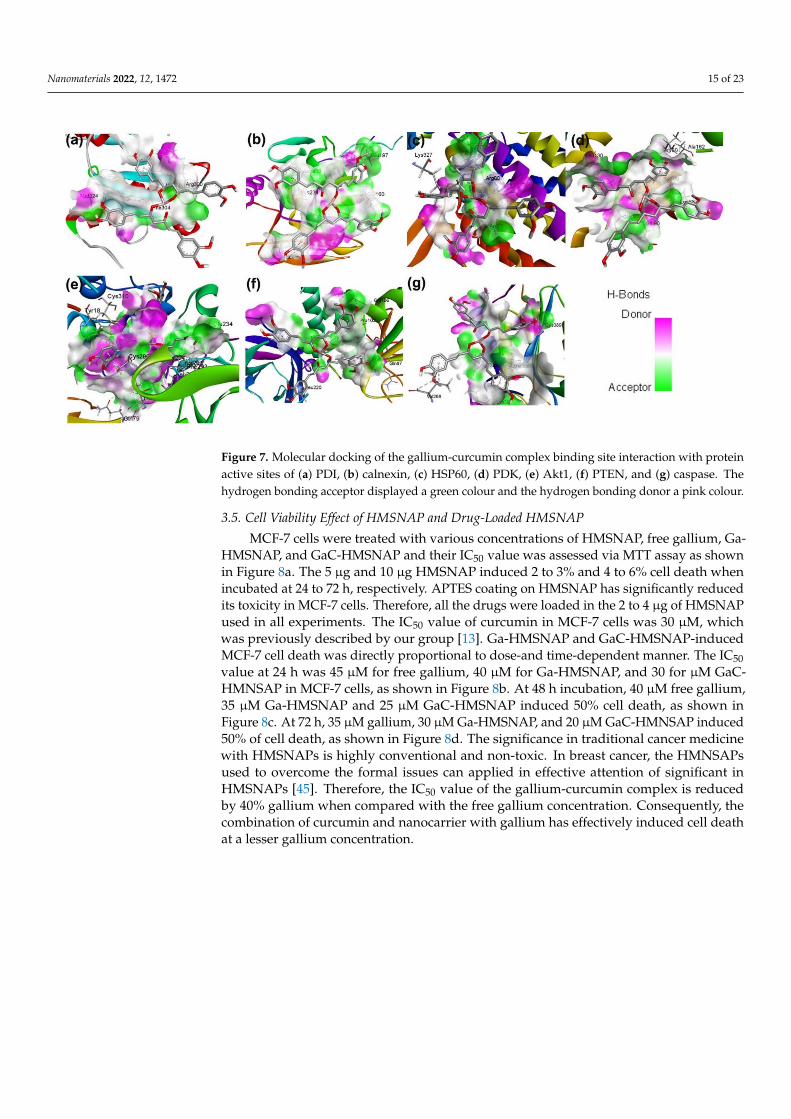
To determine whether the gallium-curcumin complex ligand directly interacts withwestern blot, resulting in proteins, we performed a molecular docking analysis withseven proteins in Figure 7. The seven proteins, including PDI, calnexin, HSP60, PDKcaspase 9, Akt1, and PTEN, are directly associated with the gallium-curcumin complexand the remaining proteins are indirectly associated and regulated with the complexligand. The docking results revealed a strong binding energy between the complex ligandand protein molecules, such as PDI (−5.42 kcal mol−1), calnexin (−7.04 kcal mol−1),HSP60 (−6.34 kcal mol−1), PDK (−8.55 kcal mol−1), caspase 9 (−6.63 kcal mol−1), Akt1(−9.84 kcal mol−1), and PTEN (−3.89 kcal mol−1). PDI, PDK, Akt, calnexin, and caspases 9have two hydrogen bond interactions and HSP60, and PTEN has one hydrogen bondbetween the ligand complex and other associated proteins. A molecular analysis revealedthe presence of several non-covalent interactions and strong hydrogen bonds. Theseinteractions between the associated proteins and the gallium-curcumin ligand complexcould contribute to the regulation of breast cancer.
Nanomaterials 2022, 12, 1472 15 of 23
Figure 7. Molecular docking of the gallium-curcumin complex binding site interaction with proteinactive sites of (a) PDI, (b) calnexin, (c) HSP60, (d) PDK, (e) Akt1, (f) PTEN, and (g) caspase. Thehydrogen bonding acceptor displayed a green colour and the hydrogen bonding donor a pink colour.
3.5. Cell Viability Effect of HMSNAP and Drug-Loaded HMSNAP
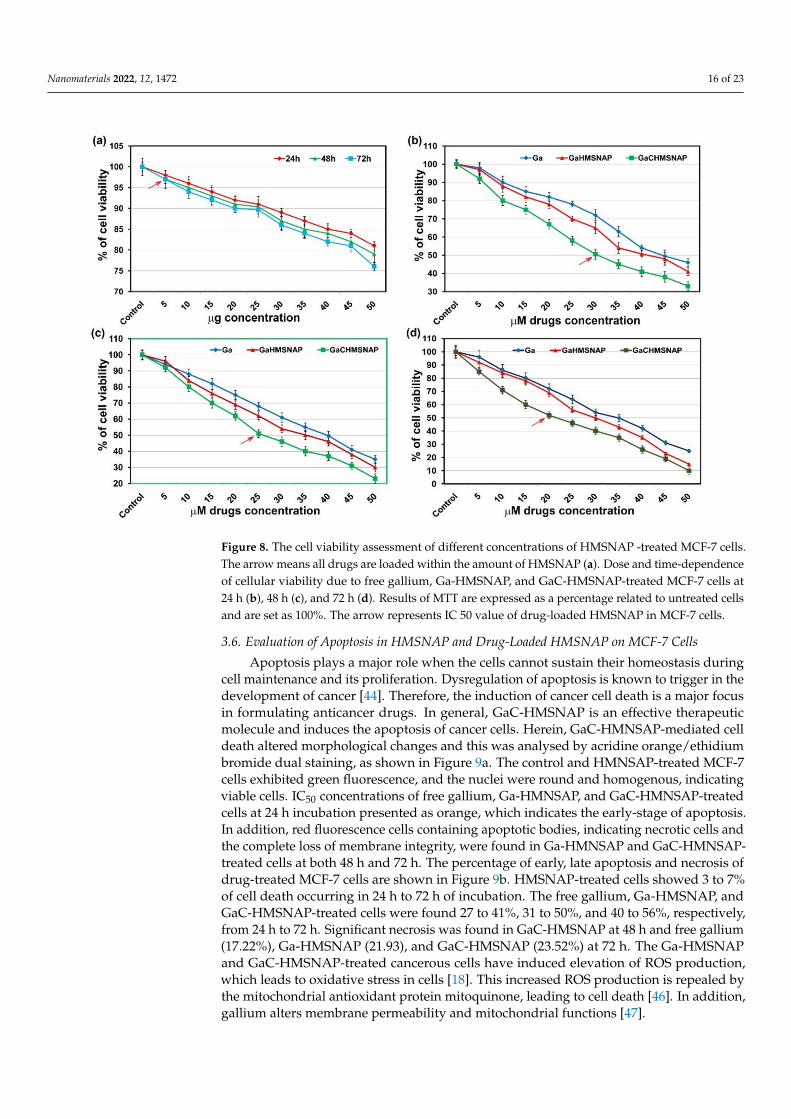
MCF-7 cells were treated with various concentrations of HMSNAP, free gallium, Ga-HMSNAP, and GaC-HMSNAP and their IC50 value was assessed via MTT assay as shownin Figure 8a. The 5 µg and 10 µg HMSNAP induced 2 to 3% and 4 to 6% cell death whenincubated at 24 to 72 h, respectively. APTES coating on HMSNAP has significantly reducedits toxicity in MCF-7 cells. Therefore, all the drugs were loaded in the 2 to 4 µg of HMSNAPused in all experiments. The IC50 value of curcumin in MCF-7 cells was 30 µM, whichwas previously described by our group [13]. Ga-HMSNAP and GaC-HMSNAP-inducedMCF-7 cell death was directly proportional to dose-and time-dependent manner. The IC50value at 24 h was 45 µM for free gallium, 40 µM for Ga-HMSNAP, and 30 for µM GaC-HMNSAP in MCF-7 cells, as shown in Figure 8b. At 48 h incubation, 40 µM free gallium,35 µM Ga-HMSNAP and 25 µM GaC-HMSNAP induced 50% cell death, as shown inFigure 8c. At 72 h, 35 µM gallium, 30 µM Ga-HMSNAP, and 20 µM GaC-HMNSAP induced50% of cell death, as shown in Figure 8d. The significance in traditional cancer medicinewith HMSNAPs is highly conventional and non-toxic. In breast cancer, the HMNSAPsused to overcome the formal issues can applied in effective attention of significant inHMSNAPs [45]. Therefore, the IC50 value of the gallium-curcumin complex is reducedby 40% gallium when compared with the free gallium concentration. Consequently, thecombination of curcumin and nanocarrier with gallium has effectively induced cell deathat a lesser gallium concentration.
Nanomaterials 2022, 12, 1472 16 of 23
Figure 8. The cell viability assessment of different concentrations of HMSNAP -treated MCF-7 cells.The arrow means all drugs are loaded within the amount of HMSNAP (a). Dose and time-dependenceof cellular viability due to free gallium, Ga-HMSNAP, and GaC-HMSNAP-treated MCF-7 cells at24 h (b), 48 h (c), and 72 h (d). Results of MTT are expressed as a percentage related to untreated cellsand are set as 100%. The arrow represents IC 50 value of drug-loaded HMSNAP in MCF-7 cells.
3.6. Evaluation of Apoptosis in HMSNAP and Drug-Loaded HMSNAP on MCF-7 Cells
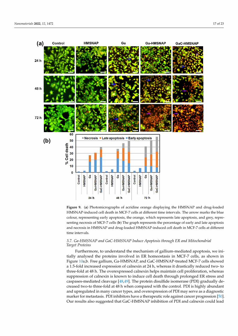
Apoptosis plays a major role when the cells cannot sustain their homeostasis duringcell maintenance and its proliferation. Dysregulation of apoptosis is known to trigger in thedevelopment of cancer [44]. Therefore, the induction of cancer cell death is a major focusin formulating anticancer drugs. In general, GaC-HMSNAP is an effective therapeuticmolecule and induces the apoptosis of cancer cells. Herein, GaC-HMNSAP-mediated celldeath altered morphological changes and this was analysed by acridine orange/ethidiumbromide dual staining, as shown in Figure 9a. The control and HMNSAP-treated MCF-7cells exhibited green fluorescence, and the nuclei were round and homogenous, indicatingviable cells. IC50 concentrations of free gallium, Ga-HMNSAP, and GaC-HMNSAP-treatedcells at 24 h incubation presented as orange, which indicates the early-stage of apoptosis.In addition, red fluorescence cells containing apoptotic bodies, indicating necrotic cells andthe complete loss of membrane integrity, were found in Ga-HMNSAP and GaC-HMNSAP-treated cells at both 48 h and 72 h. The percentage of early, late apoptosis and necrosis ofdrug-treated MCF-7 cells are shown in Figure 9b. HMSNAP-treated cells showed 3 to 7%of cell death occurring in 24 h to 72 h of incubation. The free gallium, Ga-HMSNAP, andGaC-HMSNAP-treated cells were found 27 to 41%, 31 to 50%, and 40 to 56%, respectively,from 24 h to 72 h. Significant necrosis was found in GaC-HMSNAP at 48 h and free gallium(17.22%), Ga-HMSNAP (21.93), and GaC-HMSNAP (23.52%) at 72 h. The Ga-HMSNAPand GaC-HMSNAP-treated cancerous cells have induced elevation of ROS production,which leads to oxidative stress in cells [18]. This increased ROS production is repealed bythe mitochondrial antioxidant protein mitoquinone, leading to cell death [46]. In addition,gallium alters membrane permeability and mitochondrial functions [47].
Nanomaterials 2022, 12, 1472 17 of 23
Figure 9. (a) Photomicrographs of acridine orange displaying the HMSNAP and drug-loadedHMSNAP-induced cell death in MCF-7 cells at different time intervals. The arrow marks the bluecolour, representing early apoptosis, the orange, which represents late apoptosis, and grey, repre-senting necrosis of MCF-7 cells (b) The graph represents the percentage of early and late apoptosisand necrosis in HMSNAP and drug-loaded HMSNAP-induced cell death in MCF-7 cells at differenttime intervals.
3.7. Ga-HMSNAP and GaC-HMSNAP Induce Apoptosis through ER and MitochondrialTarget Proteins
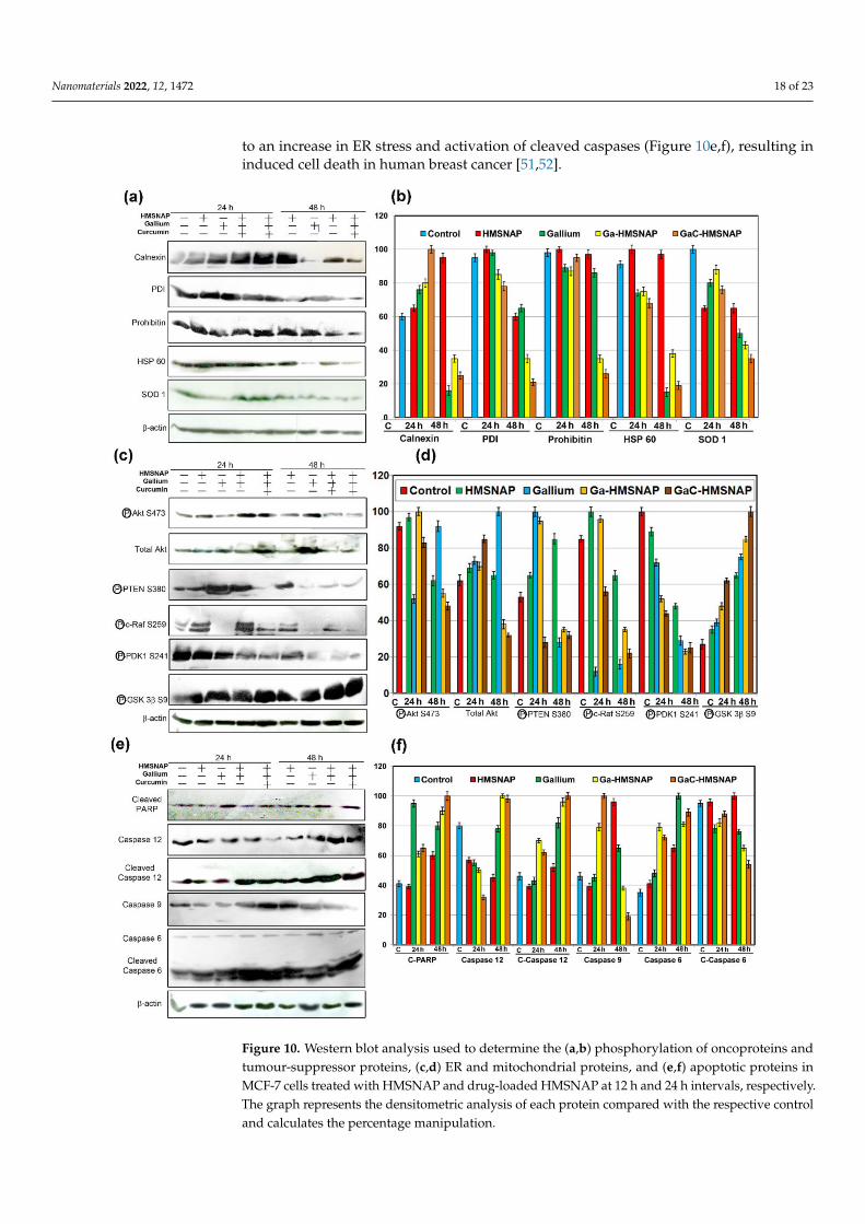
Furthermore, to understand the mechanism of gallium-mediated apoptosis, we ini-tially analysed the proteins involved in ER homeostasis in MCF-7 cells, as shown inFigure 10a,b. Free gallium, Ga-HMSNAP, and GaC-HMSNAP-treated MCF-7 cells showeda 1.5-fold increased expression of calnexin at 24 h, whereas it drastically reduced two- tothree-fold at 48 h. The overexpressed calnexin helps maintain cell proliferation, whereassuppression of calnexin is known to induce cell death through prolonged ER stress andcaspases-mediated cleavage [48,49]. The protein disulfide isomerase (PDI) gradually de-creased two-to three-fold at 48 h when compared with the control. PDI is highly abundantand upregulated in many cancer types, and overexpression of PDI may serve as a diagnosticmarker for metastasis. PDI inhibitors have a therapeutic role against cancer progression [50].Our results also suggested that GaC-HMSNAP inhibition of PDI and calnexin could lead
Nanomaterials 2022, 12, 1472 18 of 23
to an increase in ER stress and activation of cleaved caspases (Figure 10e,f), resulting ininduced cell death in human breast cancer [51,52].
Figure 10. Western blot analysis used to determine the (a,b) phosphorylation of oncoproteins andtumour-suppressor proteins, (c,d) ER and mitochondrial proteins, and (e,f) apoptotic proteins inMCF-7 cells treated with HMSNAP and drug-loaded HMSNAP at 12 h and 24 h intervals, respectively.The graph represents the densitometric analysis of each protein compared with the respective controland calculates the percentage manipulation.
Nanomaterials 2022, 12, 1472 19 of 23
The mitochondrial proteins prohibitin 1 and HSP 60 are three- to four-fold downreg-ulated at 48 h in Ga-HMSNAP and GaC-HMSNAP treated MCF-7 cells when comparedwith the control, as shown in Figure 10a,b. Prohibitin 1 is located in the mitochondria andplasma membrane and is predominantly involved in maintaining mitochondrial morphol-ogy and its functions. Similarly, the knockdown of prohibitin 1-induced the cell deathin the ovarian [53] and hepatocarcinoma cell lines [54]. The HSP 60 is highly expressedin human breast cancer when compared with normal tissues; this enhances cancer cellproliferation, and HSP 60 inhibitors downregulate HSP 60, thereby inducing the apoptosisof tumour cells [55]. Therefore, it can be concluded that gallium and curcumin complexdownregulates prohibitin 1 and HSP 60, and then enhances MCF-7 cell death throughan intrinsic apoptotic pathway. SOD1 is aberrantly expressed in cancerous tissues andpromotes cancer cell proliferation [56]. The Ga-HMSNAP and GaC-HMSNAP-treatedMCF-7 cells reduced SOD1 activity and induced cell death approximately three-fold. There-fore, gallium-mediated SOD1 inhibition could suppress the proliferation of cancer cellsthrough the downregulation of PTEN and Akt (Figure 10c,d) [57] and the upregulationof caspases 3 and 9 (Figure 10e,f) [58]. SOD1 inhibition induces apoptosis of cancer cellsthrough activation of the mitochondria-dependent apoptotic pathway.
3.8. Ga-HMSNAP and GaC-HMSNAP Inhibit Phosphorylation of Tumourigenic Proteins andInduce Phosphorylation of Tumour-Suppressor Proteins
Further mechanistic investigation of the gallium-curcumin complex-induced cell deathwas done by analysing the alteration of tumourigenic and tumour-suppressor proteinsin MCF-7 cells, as shown in Figure 10c,d. The free gallium and Ga-HMSNAP and GaC-HMSNAP-treated MCF-7 cells showed phosphorylation of Akt at S473, and the total Akt isdownregulated two-fold at 48 h incubation when compared with the respective control.Moreover, our results confirmed that gallium-curcumin complex-treated cells downregulateprohibitin 1 (Figure 10a,b), leading to the reduced phosphorylation of Akt and the inhibitionof cell proliferation [59]. The free gallium, Ga-HMSNAP, and GaC-HMSNAP-treated cellsdemonstrated 2.5-fold reduction of phosphorylation of PTEN at S380 compared to theuntreated cells (Figure 10c,d). The suppression of PTEN in MCF-7 cells inhibited cellproliferation and induced apoptosis by reducing Akt and induced the activation of ATPand caspases [60]. The immunoblot study reveals that c-Raf phosphorylation at S249 isfully inactivated by free gallium-treated MCF-7 cells at both 24 and 48 h. However, Ga-HMSNAP and GaC-HMSNAP-treated MCF-7 cells gradually reduced up to four-fold in48 h-treated cells when compared with the control. C-Raf is localised in the mitochondrialmembrane and plasma membrane and is phosphorylated at S259, inactivated by Akt andnegatively regulates the Raf activity, inducing cell death [61]. Prohibitin 1, along withAkt, is specifically involved in the Ras-Raf-MEK-ERK pathway. PDK1 promotes cancerthrough the activation of Akt and the downstream of PDK-1 may be a promising therapeutictarget of breast cancer [62]. Therefore, our results clearly suggest that downregulationof PHB1 leads to inactivation of phosphorylation of S471 of Akt, S259 of c-Raf, and S241of PDK1 and induces the intrinsic cell death pathway in MCF-7. In the western blotanalysis, Ga-HMSNAP and GaC-HMSNA-treated MCF-7 cells showed the activation ofphosphorylation of GSK3β at S9. GSK3β at S9 was gradually increased up to three- to four-fold in gallium, Ga-HMSNAP and GaC-HMSNAP-treated MCF-7 cells when comparedwith control. GSK 3β is involved in cell cycle regulation through phosphorylation of cyclinD1. The phosphorylation of S9 deactivates the GSK 3β. Therefore, our results showed thatphosphorylation of GSK 3β at S9 attenuates Akt and PKC, and then induces cell death [63].
3.9. GaC-HMSNAP Induces Mitochondrial and Other Apoptotic Proteins
PARP is a nuclear protein that is mainly involved in the repair of damaged DNA. It iscleaved by caspases to form cleaved PARP and is unable to repair DNA, thus triggeringapoptosis of the cells. Western blot analysis of the c-PARP, cleaved caspase 12, and caspase6 showed two- to 2.5-fold increases in Ga-HMSNAP and GaC-HMNSAP-treated MCF-7
Nanomaterials 2022, 12, 1472 20 of 23
cells when compared with the relevant control (Figure 10e,f. The PARP is cleaved bycaspases and is considered to be a hallmark of apoptosis [64–66]. Therefore, Ga-HMSNAPand GaC-HMSNAP induced cell death by promoting c-PARP. However, AO/ETBr stainedgallium and curcumin complex-treated MCF-7 cells showed ruptured plasma membranesdue to the increase of c-PARP. Caspase 12, cleaved caspase 12, caspase 6 and cleavedcaspase 6 expressions increased 1.5-fold upon Ga-HMSNAP and GaC-HMNSAP treatmentas compared to the control. No significant variations are observed in HMSNAP-treatedcells with untreated cells in all apoptotic proteins. The lesser concentration of HMSNAPis non-toxic. Therefore, our results clearly illustrate that HMSNAP does not induce anycell-death-regulated proteins.
4. Conclusions
In summary, the present study investigates the co-delivery efficacy of gallium (III)nitrate alone and in combination with gallium and curcumin-loaded hollow mesoporous sil-ica nanoparticles in MCF-7 cells. The drug-loaded nanomaterials are characterised throughFTIR, XPS, nitrogen sorption, 1 H-NMR, etc., confirming that both of the drugs are immo-bilised on the surface of HMSNAP during aminated APTES. In addition, an enormousamount of C was found in the drug-loaded nanocarrier, providing convincing proof for thesurface modification of HMSNAP. On the other hand, the intensity of Si and O was furtherdecreased after surface modification with gallium and curcumin. Around 37% of porediameters and 56% of pore volumes are occupied by the drug-loaded nanocarrier. All ofthe above XPS, nitrogen sorption, and 1H-NMR results indicated that we have successfullymodified HMSNAP alone and in combination with gallium and curcumin. The mechanismof drug delivery is triggered by the signalling proteins, which induce cell death in MCF-7cells. HMSNAP was non-toxic and no significant cell death occurred in MCF-7. The IC50values of the combination of gallium and curcumin (20 µM) are significantly reduced proto-oncoproteins that were downregulated in GaC-HMNSAP-treated cells. Therefore, it isreasonable to conclude that gallium and curcumin interact with these phosphoproteins andinduce cell death. The ER client proteins calnexin and PDI are drastically reduced in GaC-HMNSAP-treated MCF-7 cells, indicating that GaC-HMNSAP disturbs ER homeostasis.The western blot analysis clearly demonstrated that GaC-HMSNAP interacts with oncopro-teins and mitochondrial proteins, inducing an intrinsic cell death pathway. Further dockingstudies of gallium and curcumin complexes indicate the interaction of these molecules withthe apoptotic and mitochondrial proteins through hydrogen bonding formation and with ahigher binding energy that could lead to cell death. Altogether, our results clearly depictedthat GaC-HMNSAP altered tumour suppressor and mitochondrial proteins and inducedthe intrinsic cell death pathway.
Supplementary Materials: The drug release kinetics source file can be downloaded at: https://www.mdpi.com/article/10.3390/nano12091472/s1.
Author Contributions: T.M.V., T.K.; Methodology: T.M.V., V.K., D.S., K.C., R.V., V.R.K., N.S.K., S.K.,E.B., P.M., T.K.; Software: D.S., N.S.K., T.K.; Validation: T.K., N.S.K., P.M.; Formal analysis: T.M.V.,T.K., K.S., S.K.; Investigation: T.M.V., T.K., N.S.K.; Resources: T.K., N.S.K., K.C., R.V., V.R.K.; Datacuration: T.M.V., T.K., K.S., S.K.; Writing—Original draft: T.M.V., T.K., K.S., S.K.; Writing—Reviewingand editing: T.M.V., T.K., K.S., S.K., E.B., P.M.; Visualization: T.M.V., T.K.; Supervision: T.K.; Projectadministration: T.K., K.S.; Funding acquisition: T.K., S.K., K.S., N.S.K. The manuscript was writtenthrough contributions of all authors. All authors have read and agreed to the published version ofthe manuscript.
Funding: This study was supported in part by a Grant-in-Aid from the Department of Biotechnology(DBT), Govt. of India (BT/PR44695/NER/95/1880/2021) and (BT/PR36633/TRM/120/277/2020)to T.K., K.S. and S.K. N.S.K. is thankful to the Department of Biotechnology (DBT), Govt. of India,New Delhi for the Advanced Level State Biotech Hub (BT/NER/143/SP44475/2021) at MizoramUniversity. P.M. was funded by grant 2019/35/B/NZ8/04523 (National Science Centre, Poland).And the A.P.C. was funded by 2019/35/B/NZ8/04523 (National Science Centre, Poland).
Nanomaterials 2022, 12, 1472 21 of 23
Institutional Review Board Statement: Not applicable.
Informed Consent Statement: Not applicable.
Data Availability Statement: The data presented in this study are available in the article and Supple-mentary Materials.
Acknowledgments: The authors are grateful to the management of Kalasalingam Academy ofResearch and Education (Deemed to be University), Krishnankoil, India for the research fellowshipsand utilisation of the research facilities.
Conflicts of Interest: The authors declare no conflict of interest.
References1. Mout, R.; Moyano, D.F.; Rana, S.; Rotello, V.M. Surface functionalization of nanoparticles for nanomedicine. Chem. Soc. Rev. 2012,
41, 2539–2544. [CrossRef] [PubMed]2. Amoozgar, Z.; Yeo, Y. Recent advances in stealth coating of nanoparticle drug delivery systems. Wiley Interdiscip. Rev. Nanomed.
Nanobiotechnol. 2012, 4, 219–233. [CrossRef] [PubMed]3. Lai, C.-Y.; Trewyn, B.G.; Jeftinija, D.M.; Jeftinija, K.; Xu, S.; Jeftinija, S.; Lin, V.S.-Y. A mesoporous silica nanosphere-based carrier
system with chemically removable CdS nanoparticle caps for stimuli-responsive controlled release of neurotransmitters and drugmolecules. J. Am. Chem. Soc. 2003, 125, 4451–4459. [CrossRef] [PubMed]
4. Sabir, F.; Zeeshan, M.; Laraib, U.; Barani, M.; Rahdar, A.; Cucchiarini, M.; Pandey, S. DNA based and stimuli-responsive smartnanocarrier for diagnosis and treatment of cancer: Applications and challenges. Cancers 2021, 13, 3396. [CrossRef]
5. Shen, S.; Wu, Y.; Liu, Y.; Wu, D. High drug-loading nanomedicines: Progress, current status, and prospects. Int. J. Nanomed. 2017,12, 4085. [CrossRef]
6. Kong, M.; Tang, J.; Qiao, Q.; Wu, T.; Qi, Y.; Tan, S.; Gao, X.; Zhang, Z. Biodegradable hollow mesoporous silica nanoparticles forregulating tumor microenvironment and enhancing antitumor efficiency. Theranostics 2017, 7, 3276. [CrossRef]
7. Chen, F.; Hong, H.; Shi, S.; Goel, S.; Valdovinos, H.F.; Hernandez, R.; Theuer, C.P.; Barnhart, T.E.; Cai, W. Engineering of hollowmesoporous silica nanoparticles for remarkably enhanced tumor active targeting efficacy. Sci. Rep. 2014, 4, 1–10. [CrossRef]
8. Barani, M.; Hosseinikhah, S.M.; Rahdar, A.; Farhoudi, L.; Arshad, R.; Cucchiarini, M.; Pandey, S. Nanotechnology in bladdercancer: Diagnosis and treatment. Cancers 2021, 13, 2214. [CrossRef]
9. Xiao, D.; Jia, H.-Z.; Ma, N.; Zhuo, R.-X.; Zhang, X.-Z. A redox-responsive mesoporous silica nanoparticle capped with amphiphilicpeptides by self-assembly for cancer targeting drug delivery. Nanoscale 2015, 7, 10071–10077. [CrossRef]
10. Jugdaohsingh, R. Silicon and bone health. J. Nutr. Health Aging 2007, 11, 99.11. Napierska, D.; Thomassen, L.C.; Rabolli, V.; Lison, D.; Gonzalez, L.; Kirsch-Volders, M.; Martens, J.A.; Hoet, P.H. Size-dependent
cytotoxicity of monodisperse silica nanoparticles in human endothelial cells. Small 2009, 5, 846–853. [CrossRef] [PubMed]12. Niu, S.; Zhang, X.; Williams, G.R.; Wu, J.; Gao, F.; Fu, Z.; Chen, X.; Lu, S.; Zhu, L.-M. Hollow mesoporous silica nanoparticles
gated by chitosan-copper sulfide composites as theranostic agents for the treatment of breast cancer. Acta Biomater. 2021, 126,408–420. [CrossRef] [PubMed]
13. Zhang, P.; Tang, M.; Huang, Q.; Zhao, G.; Huang, N.; Zhang, X.; Tan, Y.; Cheng, Y. Combination of 3-methyladenine therapy andAsn-Gly-Arg (NGR)-modified mesoporous silica nanoparticles loaded with temozolomide for glioma therapy in vitro. Biochem.Biophys. Res. Commun. 2019, 509, 549–556. [CrossRef]
14. Harini, L.; Srivastava, S.; Gnanakumar, G.P.; Karthikeyan, B.; Ross, C.; Krishnakumar, V.; Kannan, V.R.; Sundar, K.; Kathiresan, T.An ingenious non-spherical mesoporous silica nanoparticle cargo with curcumin induces mitochondria-mediated apoptosis inbreast cancer (MCF-7) cells. Oncotarget 2019, 10, 1193. [CrossRef] [PubMed]
15. Zhou, X.; Chen, L.; Nie, W.; Wang, W.; Qin, M.; Mo, X.; Wang, H.; He, C. Dual-responsive mesoporous silica nanoparticlesmediated codelivery of doxorubicin and Bcl-2 SiRNA for targeted treatment of breast cancer. J. Phys. Chem. C 2016, 120,22375–22387. [CrossRef]
16. Chitambar, C.R. The therapeutic potential of iron-targeting gallium compounds in human disease: From basic research to clinicalapplication. Pharmacol. Res. 2017, 115, 56–64. [CrossRef] [PubMed]
17. Bandyopadhyay, D. Farmer to pharmacist: Curcumin as an anti-invasive and antimetastatic agent for the treatment of cancer1.Front. Chem. 2014, 2, 113. [CrossRef] [PubMed]
18. Harini, L.; Karthikeyan, B.; Srivastava, S.; Suresh, S.B.; Ross, C.; Gnanakumar, G.; Rajagopal, S.; Sundar, K.; Kathiresan, T.Polyethylenimine-modified curcumin-loaded mesoporus silica nanoparticle (MCM-41) induces cell death in MCF-7 cell line. IETNanobiotechnol. 2017, 11, 57–61. [CrossRef]
19. Foster, B.; Clagett-Carr, K.; Hoth, D.; Leyland-Jones, B. Gallium nitrate: The second metal with clinical activity. Cancer Treat. Rep.1986, 70, 1311–1319.
20. Qi, J.; Qian, K.; Tian, L.; Cheng, Z.; Wang, Y. Gallium (iii)–2-benzoylpyridine-thiosemicarbazone complexes promote apoptosisthrough Ca2+ signaling and ROS-mediated mitochondrial pathways. New J. Chem. 2018, 42, 10226–10233. [CrossRef]
Nanomaterials 2022, 12, 1472 22 of 23
21. Chitambar, C.R.; Purpi, D.P.; Woodliff, J.; Yang, M.; Wereley, J.P. Development of gallium compounds for treatment of lymphoma:Gallium maltolate, a novel hydroxypyrone gallium compound, induces apoptosis and circumvents lymphoma cell resistance togallium nitrate. J. Pharmacol. Exp. Ther. 2007, 322, 1228–1236. [CrossRef] [PubMed]
22. Hata, Y.; Sandler, A.; Loehrer, P.J.; Sledge, G.W.; Weber, G. Synergism of taxol and gallium nitrate in human breast carcinoma cells:Schedule dependency. Oncol. Res. Featur. Preclin. Clin. Cancer Ther. 1994, 6, 19–24.
23. Harris, W.R.; Pecoraro, V.L. Thermodynamic binding constants for gallium transferrin. Biochemistry 1983, 22, 292–299. [CrossRef][PubMed]
24. Chitambar, C.R.; Zivkovic, Z. Inhibition of hemoglobin production by transferrin-gallium. Blood 1987, 69, 144–149. [CrossRef]25. Chitambar, C.R.; Matthaeus, W.G.; Antholine, W.E.; Graff, K.; O’Brien, W.J. Inhibition of leukemic HL60 cell growth by transferrin-
gallium: Effects on ribonucleotide reductase and demonstration of drug synergy with hydroxyurea. Blood 1988, 72, 1930–1936.[CrossRef]
26. Perchellet, E.M.; Ladesich, J.B.; Collery, P.; Perchellet, J.-P. Microtubule-disrupting effects of gallium chloride in vitro. Anti-CancerDrugs 1999, 10, 477–488. [CrossRef]
27. Valiahdi, S.M.; Heffeter, P.; Jakupec, M.A.; Marculescu, R.; Berger, W.; Rappersberger, K.; Keppler, B.K. The gallium complex KP46exerts strong activity against primary explanted melanoma cells and induces apoptosis in melanoma cell lines. Melanoma Res.2009, 19, 283. [CrossRef]
28. Gogna, R.; Madan, E.; Keppler, B.; Pati, U. Gallium compound GaQ3-induced Ca2+ signalling triggers p53-dependent and-independent apoptosis in cancer cells. Br. J. Pharmacol. 2012, 166, 617–636. [CrossRef]
29. Chitambar, C.R. Gallium-containing anticancer compounds. Future Med. Chem. 2012, 4, 1257–1272. [CrossRef]30. Myette, M.S.; Elford, H.L.; Chitambar, C.R. Interaction of gallium nitrate with other inhibitors of ribonucleotide reductase: Effects
on the proliferation of human leukemic cells. Cancer Lett. 1998, 129, 199–204. [CrossRef]31. Collery, P.; Lechenault, F.; Juvin, E.; Khasanova, L.; Vernet, G.; Cazabat, A.; Lebargy, F. Synergistic effect between gallium chloride
and vinorelbine on U937 malignant cell lines. In Metal Ions in Biology and Medicine; John Libbey Eurotext: Arcueil, France, 1998;pp. 588–593.
32. Lundberg, J.H.; Chitambar, C.R. Interaction of gallium nitrate with fludarabine and iron chelators: Effects on the proliferation ofhuman leukemic HL60 cells. Cancer Res. 1990, 50, 6466–6470. [PubMed]
33. Chitambar, C.R.; Wereley, J.P. Synergistic inhibition of T-lymphoblastic leukemic CCRF-CEM cell growth by gallium andrecombinant human α-interferon through action on cellular iron uptake. Cancer Res. 1994, 54, 3224–3228. [PubMed]
34. Teng, Z.; Han, Y.; Li, J.; Yan, F.; Yang, W. Preparation of hollow mesoporous silica spheres by a sol–gel/emulsion approach.Microporous Mesoporous Mater. 2010, 127, 67–72. [CrossRef]
35. Kunjiappan, S.; Sankaranarayanan, M.; Kumar, B.K.; Pavadai, P.; Babkiewicz, E.; Maszczyk, P.; Glodkowska-Mrowka, E.;Arunachalam, S.; Pandian, S.R.K.; Ravishankar, V. Capsaicin-loaded solid lipid nanoparticles: Design, biodistribution, in silicomodeling and in vitro cytotoxicity evaluation. Nanotechnology 2020, 32, 095101. [CrossRef] [PubMed]
36. Baskararaj, S.; Panneerselvam, T.; Govindaraj, S.; Arunachalam, S.; Parasuraman, P.; Pandian, S.R.K.; Sankaranarayanan, M.;Mohan, U.P.; Palanisamy, P.; Ravishankar, V. Formulation and characterization of folate receptor-targeted PEGylated liposomeencapsulating bioactive compounds from Kappaphycus alvarezii for cancer therapy. 3 Biotech 2020, 10, 1–18. [CrossRef] [PubMed]
37. Morris, G.M.; Goodsell, D.S.; Halliday, R.S.; Huey, R.; Hart, W.E.; Belew, R.K.; Olson, A.J. Automated docking using a Lamarckiangenetic algorithm and an empirical binding free energy function. J. Comput. Chem. 1998, 19, 1639–1662. [CrossRef]
38. Zilla, M.K.; Nayak, D.; Vishwakarma, R.A.; Sharma, P.R.; Goswami, A.; Ali, A. A convergent synthesis of alkyne–azidecycloaddition derivatives of 4-α, β-2-propyne podophyllotoxin depicting potent cytotoxic activity. Eur. J. Med. Chem. 2014, 77,47–55. [CrossRef]
39. Ornelas-Soto, N.; Rubio-Govea, R.; Guerrero-Beltrán, C.E.; Vázquez-Garza, E.; Bernal-Ramírez, J.; García-García, A.; Oropeza-Almazán, Y.; García-Rivas, G.; Contreras-Torres, F.F. Enhancing internalization of silica particles in myocardial cells throughsurface modification. Mater. Sci. Eng. C 2017, 79, 831–840. [CrossRef]
40. Wang, Y.; Sun, Y.; Wang, J.; Yang, Y.; Li, Y.; Yuan, Y.; Liu, C. Charge-reversal APTES-modified mesoporous silica nanoparticleswith high drug loading and release controllability. ACS Appl. Mater. Interfaces 2016, 8, 17166–17175. [CrossRef]
41. Möckel, J.E.; Lippold, B.C. Zero-order drug release from hydrocolloid matrices. Pharm. Res. 1993, 10, 1066–1070. [CrossRef]42. Schwartz, J.B.; Simonelli, A.P.; Higuchi, W.I. Drug release from wax matrices I. Analysis of data with first-order kinetics and with
the diffusion-controlled model. J. Pharm. Sci. 1968, 57, 274–277. [CrossRef] [PubMed]43. Paul, D. Elaborations on the Higuchi model for drug delivery. Int. J. Pharm. 2011, 418, 13–17. [CrossRef] [PubMed]44. Wu, I.Y.; Bala, S.; Škalko-Basnet, N.; Di Cagno, M.P. Interpreting non-linear drug diffusion data: Utilizing Korsmeyer-Peppas
model to study drug release from liposomes. Eur. J. Pharm. Sci. 2019, 138, 105026. [CrossRef] [PubMed]45. Kalam, M.A.; Humayun, M.; Parvez, N.; Yadav, S.; Garg, A.; Amin, S.; Sultana, Y.; Ali, A. Release kinetics of modified
pharmaceutical dosage forms: A review. Cont. J. Pharm. Sci. 2007, 1, 30–35.46. Plati, J.; Bucur, O.; Khosravi-Far, R. Dysregulation of apoptotic signaling in cancer: Molecular mechanisms and therapeutic
opportunities. J. Cell. Biochem. 2008, 104, 1124–1149. [CrossRef]47. Kelso, G.F.; Porteous, C.M.; Coulter, C.V.; Hughes, G.; Porteous, W.K.; Ledgerwood, E.C.; Smith, R.A.; Murphy, M.P. Selective
targeting of a redox-active ubiquinone to mitochondria within cells: Antioxidant and antiapoptotic properties. J. Biol. Chem. 2001,276, 4588–4596. [CrossRef]
Nanomaterials 2022, 12, 1472 23 of 23
48. Chitambar, C.R. Gallium and its competing roles with iron in biological systems. Biochim. Biophys. Acta 2016, 1863, 2044–2053.[CrossRef]
49. Goplen, D.; Wang, J.; Enger, P.Ø.; Tysnes, B.B.; Terzis, A.; Laerum, O.D.; Bjerkvig, R. Protein disulfide isomerase expression isrelated to the invasive properties of malignant glioma. Cancer Res. 2006, 66, 9895–9902. [CrossRef]
50. Ryan, D.; Carberry, S.; Murphy, Á.C.; Lindner, A.U.; Fay, J.; Hector, S.; McCawley, N.; Bacon, O.; Concannon, C.G.; Kay, E.W.Calnexin, an ER-induced protein, is a prognostic marker and potential therapeutic target in colorectal cancer. J. Transl. Med. 2016,14, 1–10.
51. Xu, S.; Sankar, S.; Neamati, N. Protein disulfide isomerase: A promising target for cancer therapy. Drug Discov. Today 2014, 19,222–240. [CrossRef]
52. Lovat, P.E.; Corazzari, M.; Armstrong, J.L.; Martin, S.; Pagliarini, V.; Hill, D.; Brown, A.M.; Piacentini, M.; Birch-Machin, M.A.;Redfern, C.P. Increasing melanoma cell death using inhibitors of protein disulfide isomerases to abrogate survival responses toendoplasmic reticulum stress. Cancer Res. 2008, 68, 5363–5369. [CrossRef] [PubMed]
53. Hashida, T.; Kotake, Y.; Ohta, S. Protein disulfide isomerase knockdown-induced cell death is cell-line-dependent and involvesapoptosis in MCF-7 cells. J. Toxicol. Sci. 2011, 36, 1–7. [CrossRef] [PubMed]
54. Gregory-Bass, R.C.; Olatinwo, M.; Xu, W.; Matthews, R.; Stiles, J.K.; Thomas, K.; Liu, D.; Tsang, B.; Thompson, W.E. Prohibitinsilencing reverses stabilization of mitochondrial integrity and chemoresistance in ovarian cancer cells by increasing theirsensitivity to apoptosis. Int. J. Cancer 2008, 122, 1923–1930. [CrossRef] [PubMed]
55. Ko, K.S.; Tomasi, M.L.; Iglesias-Ara, A.; French, B.A.; French, S.W.; Ramani, K.; Lozano, J.J.; Oh, P.; He, L.; Stiles, B.L. Liver-specificdeletion of prohibitin 1 results in spontaneous liver injury, fibrosis, and hepatocellular carcinoma in mice. Hepatology 2010, 52,2096–2108. [CrossRef] [PubMed]
56. Ghosh, J.C.; Siegelin, M.D.; Dohi, T.; Altieri, D.C. Heat shock protein 60 regulation of the mitochondrial permeability transitionpore in tumor cells. Cancer Res. 2010, 70, 8988–8993. [CrossRef]
57. Liu, Y.; Fu, N.; Su, J.; Wang, X.; Li, X. Rapid enkephalin delivery using exosomes to promote neurons recovery in ischemic strokeby inhibiting neuronal p53/Caspase-3. BioMed Res. Int. 2019, 2019, 4273290. [CrossRef] [PubMed]
58. Li, S.; Fu, L.; Tian, T.; Deng, L.; Li, H.; Xia, W.; Gong, Q. Disrupting SOD1 activity inhibits cell growth and enhances lipidaccumulation in nasopharyngeal carcinoma. Cell Commun. Signal. 2018, 16, 1–13. [CrossRef] [PubMed]
59. Lin, S.; Ren, A.; Wang, L.; Huang, Y.; Wang, Y.; Wang, C.; Greene, N.D. Oxidative stress and apoptosis in benzo [a] pyrene-inducedneural tube defects. Free Radic. Biol. Med. 2018, 116, 149–158. [CrossRef]
60. Jiang, L.; Dong, P.; Zhang, Z.; Li, C.; Li, Y.; Liao, Y.; Li, X.; Wu, Z.; Guo, S.; Mai, S. Akt phosphorylates Prohibitin 1 to mediate itsmitochondrial localization and promote proliferation of bladder cancer cells. Cell Death Dis. 2015, 6, e1660. [CrossRef]
61. Yang, M.; Chitambar, C.R. Role of oxidative stress in the induction of metallothionein-2A and heme oxygenase-1 gene expressionby the antineoplastic agent gallium nitrate in human lymphoma cells. Free Radic. Biol. Med. 2008, 45, 763. [CrossRef]
62. Rajalingam, K.; Wunder, C.; Brinkmann, V.; Churin, Y.; Hekman, M.; Sievers, C.; Rapp, U.R.; Rudel, T. Prohibitin is required forRas-induced Raf–MEK–ERK activation and epithelial cell migration. Nat. Cell Biol. 2005, 7, 837–843. [CrossRef] [PubMed]
63. Lin, H.; Hsieh, F.; Song, H.; Lin, J. Elevated phosphorylation and activation of PDK-1/AKT pathway in human breast cancer. Br. J.Cancer 2005, 93, 1372–1381. [CrossRef] [PubMed]
64. Jacobs, K.M.; Bhave, S.R.; Ferraro, D.J.; Jaboin, J.J.; Hallahan, D.E.; Thotala, D. GSK-3β: A Bifunctional Role in Cell DeathPathways. Int. J. Cell Biol. 2012, 2012, 930710. [CrossRef]
65. Kaufmann, S.H.; Desnoyers, S.; Ottaviano, Y.; Davidson, N.E.; Poirier, G.G. Specific proteolytic cleavage of poly (ADP-ribose)polymerase: An early marker of chemotherapy-induced apoptosis. Cancer Res. 1993, 53, 3976–3985. [PubMed]
66. Chang, S.E.; Littlefield, J.W. Elevated dihydrofolate reductase messenger RNA levels in methotrexate-resistant BHK cells. Cell1976, 7, 391–396. [CrossRef]