curcumin modulates α-synuclein aggregation and toxicity
TRANSCRIPT

Curcumin Modulates α‑Synuclein Aggregation and ToxicityPradeep K. Singh,†,‡,§ Vasudha Kotia,†,‡,§ Dhiman Ghosh,†,‡ Ganesh M. Mohite,†,‡ Ashutosh Kumar,†,‡
and Samir K. Maji*,†,‡
†Department of Biosciences and Bioengineering and ‡Wadhwani Research Centre for Biosciences and Bioengineering, IndianInstitute of Technology Bombay, Mumbai, Maharashtra, India 400076
*S Supporting Information
ABSTRACT: In human beings, Parkinson’s disease (PD) is associated withthe oligomerization and amyloid formation of α-synuclein (α-Syn). Thepolyphenolic Asian food ingredient curcumin has proven to be effectiveagainst a wide range of human diseases including cancers and neurologicaldisorders. While curcumin has been shown to significantly reduce cell toxicityof α-Syn aggregates, its mechanism of action remains unexplored. Here, usinga series of biophysical techniques, we demonstrate that curcumin reducestoxicity by binding to preformed oligomers and fibrils and altering theirhydrophobic surface exposure. Further, our fluorescence and two-dimensionalnuclear magnetic resonance (2D-NMR) data indicate that curcumin does notbind to monomeric α-Syn but binds specifically to oligomeric intermediates. The degree of curcumin binding correlates with theextent of α-Syn oligomerization, suggesting that the ordered structure of protein is required for effective curcumin binding. Theacceleration of aggregation by curcumin may decrease the population of toxic oligomeric intermediates of α-Syn. Collectively;our results suggest that curcumin and related polyphenolic compounds can be pursued as candidate drug targets for treatment ofPD and other neurological diseases.
KEYWORDS: Curcumin, α-synuclein, amyloid, oligomers, toxicity, Parkinson’s disease
The major pathological hallmark of Parkinson’s disease(PD) is the presence of insoluble, fibrous aggregates,
composed of α-Syn in intraneuronal inclusions of Lewy bodies(LBs) and Lewy neuritis (LNs).1 The animal models thatoverexpress human α-Syn indicate the direct involvement of α-Syn aggregation in PD pathogenesis.2,3 Moreover, the discoveryof three disease-related substitution mutations (A30P, A53T,and E46K) in the SNCA gene that encodes α-Syn and theireffects on the aggregation kinetics in vitro further support thecentral role of α-Syn aggregation in PD pathogenesis.1,4,5 Themonomeric α-Syn is a natively unfolded protein, whichtransforms into cross-β-sheet rich amyloid by self-assembly atphysiological conditions via partially folded intermediates andsoluble oligomers.6 Recently, evidence have emerged from bothin vitro and in vivo studies that soluble, oligomeric forms of α-Syn have potent neurotoxic activities and may cause theneuronal injury and death in PD.7−11 The α-Syn mutants thatpreferentially formed oligomers when expressed in the rat brainshowed more neurotoxicity and cell death compared to themutants that mostly formed amyloid fibrils.9 Molecules thatinhibit the toxicity of oligomers and/or fibrils either byreducing their formation or by converting their toxic state tonontoxic state would be an immediate step for the developmentof effective therapeutics against PD.12−15 Guided by thisconcept, many investigators have either searched for existingsmall molecules or synthesized inhibitors against α-Synfibrillogenesis.15−23 Several small polyphenolic molecules suchas epigallocatechin gallate and curcumin have been shown tomodulate the assembly and/or toxicity of many amyloidogenic
protein/peptides such as Aβ, α-Syn, and prion.19,23−26 It hasbeen proposed that the antioxidative properties of thesepolyphenols along with their structural constraints might beresponsible for their efficacy in amyloid inhibition.24,27
Curcumin (diferuloylmethane) (Supporting InformationFigure S1) is a well-known polyphenolic in Asian foodingredient turmeric and has been shown to exhibit anti-inflammatory, antimicrobial, and anticarcinogenic activities.28
Due to its low cost, blood brain barrier crossing ability and itspharmacological safety as evident from preclinical studies havesuggested the potential therapeutic role of curcumin inneurological disorders including Alzheimer’s, Parkinson’s, andHuntington’s disease.28−33 For example, curcumin binds toamyloid β protein (Aβ) oligomers/fibrils, alters the Aβaggregation, and reduces the toxicity in AD.31,34
In PD, curcumin has been shown to inhibit the α-Synaggregation in vitro35−37 and attenuate the α-Syn oligomertoxicity in cells.17,38 However, the reduction of toxicity bycurcumin and its effect on the pathway of α-Syn aggregation inphysiological conditions is not clearly understood. Our workfocuses on studying the effect of curcumin on the morphologyand toxicity of oligomeric and fibrillar assemblies of α-Syn. Wesuggest that curcumin preferentially binds to the preformed α-Syn aggregates, modulates the morphology, and reduces their
Received: August 7, 2012Accepted: December 3, 2012Published: December 3, 2012
Research Article
pubs.acs.org/chemneuro
© 2012 American Chemical Society 393 dx.doi.org/10.1021/cn3001203 | ACS Chem. Neurosci. 2013, 4, 393−407
cellular toxicity by minimizing their hydrophobic surfaceexposure. In addition, the data reveals that curcumin acceleratesα-Syn aggregation in vitro and could reduce the population ofsoluble oligomers, which are cytotoxic. Thus, curcumin andrelated polyphenolic compounds could be used for thedevelopment of potential drugs against PD.
■ RESULTS AND DISCUSSION
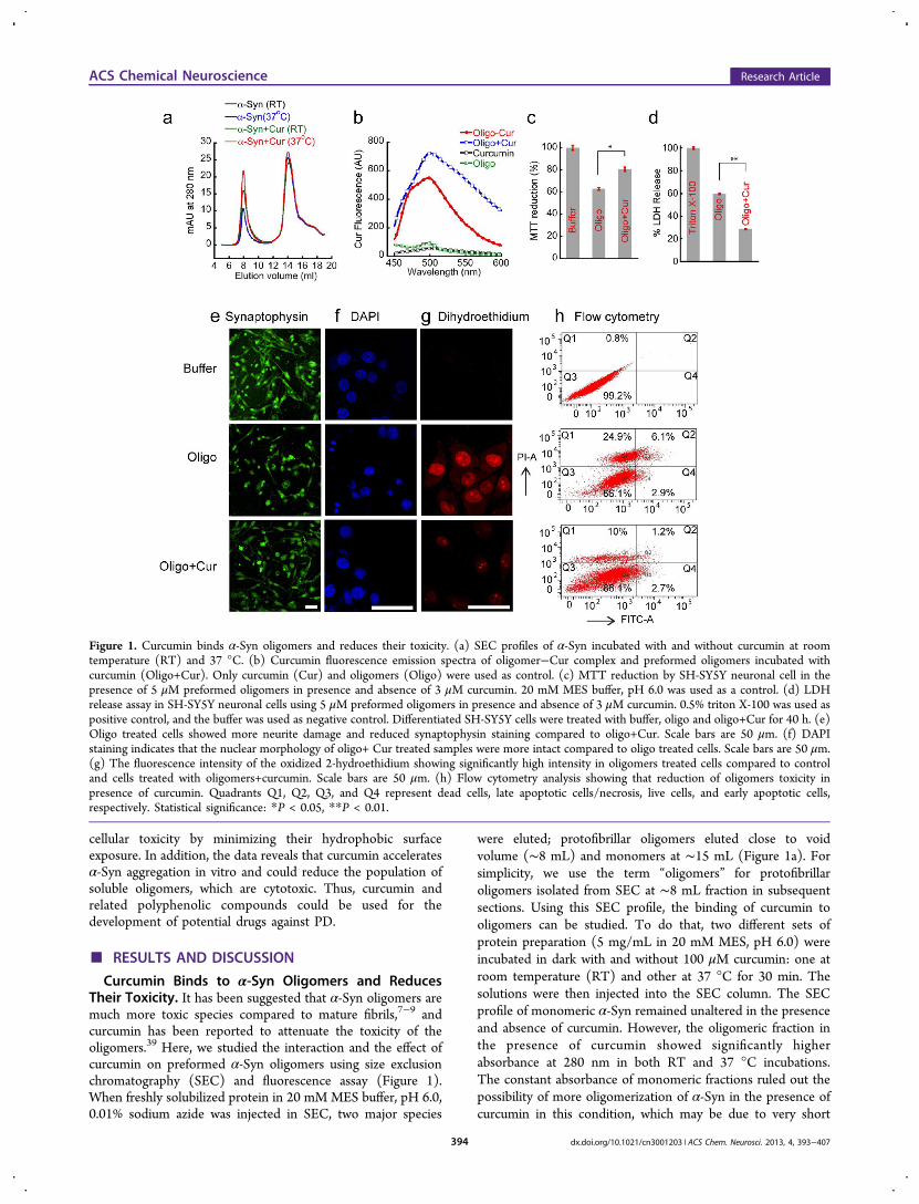
Curcumin Binds to α-Syn Oligomers and ReducesTheir Toxicity. It has been suggested that α-Syn oligomers aremuch more toxic species compared to mature fibrils,7−9 andcurcumin has been reported to attenuate the toxicity of theoligomers.39 Here, we studied the interaction and the effect ofcurcumin on preformed α-Syn oligomers using size exclusionchromatography (SEC) and fluorescence assay (Figure 1).When freshly solubilized protein in 20 mM MES buffer, pH 6.0,0.01% sodium azide was injected in SEC, two major species
were eluted; protofibrillar oligomers eluted close to voidvolume (∼8 mL) and monomers at ∼15 mL (Figure 1a). Forsimplicity, we use the term “oligomers” for protofibrillaroligomers isolated from SEC at ∼8 mL fraction in subsequentsections. Using this SEC profile, the binding of curcumin tooligomers can be studied. To do that, two different sets ofprotein preparation (5 mg/mL in 20 mM MES, pH 6.0) wereincubated in dark with and without 100 μM curcumin: one atroom temperature (RT) and other at 37 °C for 30 min. Thesolutions were then injected into the SEC column. The SECprofile of monomeric α-Syn remained unaltered in the presenceand absence of curcumin. However, the oligomeric fraction inthe presence of curcumin showed significantly higherabsorbance at 280 nm in both RT and 37 °C incubations.The constant absorbance of monomeric fractions ruled out thepossibility of more oligomerization of α-Syn in the presence ofcurcumin in this condition, which may be due to very short
Figure 1. Curcumin binds α-Syn oligomers and reduces their toxicity. (a) SEC profiles of α-Syn incubated with and without curcumin at roomtemperature (RT) and 37 °C. (b) Curcumin fluorescence emission spectra of oligomer−Cur complex and preformed oligomers incubated withcurcumin (Oligo+Cur). Only curcumin (Cur) and oligomers (Oligo) were used as control. (c) MTT reduction by SH-SY5Y neuronal cell in thepresence of 5 μM preformed oligomers in presence and absence of 3 μM curcumin. 20 mM MES buffer, pH 6.0 was used as a control. (d) LDHrelease assay in SH-SY5Y neuronal cells using 5 μM preformed oligomers in presence and absence of 3 μM curcumin. 0.5% triton X-100 was used aspositive control, and the buffer was used as negative control. Differentiated SH-SY5Y cells were treated with buffer, oligo and oligo+Cur for 40 h. (e)Oligo treated cells showed more neurite damage and reduced synaptophysin staining compared to oligo+Cur. Scale bars are 50 μm. (f) DAPIstaining indicates that the nuclear morphology of oligo+ Cur treated samples were more intact compared to oligo treated cells. Scale bars are 50 μm.(g) The fluorescence intensity of the oxidized 2-hydroethidium showing significantly high intensity in oligomers treated cells compared to controland cells treated with oligomers+curcumin. Scale bars are 50 μm. (h) Flow cytometry analysis showing that reduction of oligomers toxicity inpresence of curcumin. Quadrants Q1, Q2, Q3, and Q4 represent dead cells, late apoptotic cells/necrosis, live cells, and early apoptotic cells,respectively. Statistical significance: *P < 0.05, **P < 0.01.
ACS Chemical Neuroscience Research Article
dx.doi.org/10.1021/cn3001203 | ACS Chem. Neurosci. 2013, 4, 393−407394

incubation time and mild conditions (without agitation) usedfor the incubation. However, these observations raised thepossibility of interaction of curcumin to the preformedoligomeric protein, where curcumin might form anoligomer−curcumin complex, which possesses higher absorb-ance compared to oligomers alone. However, the probability ofincreased light scattering at 280 nm of this oligomeric assemblyin the presence of curcumin cannot be ruled out. The higherabsorbance of oligomers at 280 nm in the SEC profile of α-Synincubated with curcumin at 37 °C suggests increased binding ofcurcumin to the preformed oligomers at this temperature(Figure 1a). As the curcumin was dissolved in DMSO, theeffect of residual 0.05% DMSO on the size exclusion profile ofα-Syn was also monitored and found insignificant. The sizeexclusion of 100 μM curcumin did not show any significantabsorbance at 280 nm at ∼8 mL elution volume (data notshown).To further evaluate the interactions of curcumin with α-Syn
oligomers, we performed a curcumin fluorescence study.Curcumin is a weak fluorophore in water and produces lowintensity fluorescence at ∼540 nm when excited ∼426 nm.However, if it binds to the hydrophobic surface of a proteinsuch as amyloids, its fluorescence quantum yield increases witha blue shift in the fluorescence maximum (λmax).
40,41 Thecurcumin fluorescence of the oligomers−curcumin complexisolated from SEC (Oligo-Cur) showed high fluorescenceintensity with blue-shifted λmax ∼ 500 nm (Figure 1b). Thefluorescence intensity ∼500 nm was ∼50-fold higher comparedto fluorescence intensities of either oligomers or curcuminalone. Similarly, curcumin fluorescence was ∼70-fold moreintense when preformed oligomers were isolated from SEC andincubated with curcumin for 30 min at 37 °C prior tofluorescence study (Oligo+Cur) (Figure 1b). These observa-tions support the idea that curcumin strongly binds to thepreformed oligomeric α-Syn.The toxicity of the α-Syn oligomers was evaluated using
dopaminergic neuronal cell line SH-SY5Y. To evaluate thetoxicity of oligomers in presence and absence of curcumin,MTT assay42 was carried out (Figure 1c). In the presence of 5μM preformed α-Syn oligomers isolated from SEC, MTTreduction was decreased to 60% (Figure 1c). However, whenpreformed oligomers were incubated in the presence ofcurcumin for 30 min before adding to the cells, MTT reductionwas increased to 80%. The final curcumin concentration was 3μM, which alone did not show any toxicity. For furtherconfirmation of toxicity, LDH assay was performed. The LDHrelease assay is also widely used to determine the cytotoxicity ofchemicals or environmental toxic factors.43 LDH is a solublecytosolic enzyme that is released into the culture mediumfollowing the loss of membrane integrity.43 Figure 1d showsthat, in the presence of oligomers, the LDH release was 60%,whereas in the presence of oligomers pretreated with curcuminLDH release was decreased to 29%. Both MTT and LDHassays therefore suggest that curcumin is able to reduce thetoxicity of α-Syn oligomers.Further, we have analyzed whether curcumin based neuronal
cell protection involves changes in hallmarks of neuronal celldeath markers like nuclear morphology and syanptophysinstaining.44 The SH-SY5Y cells were differentiated using retinoicacid for 5 days (Supporting Information Figure S2a) andsubsequently treated with α-Syn oligomers in the presence andabsence of curcumin, and only buffer was used as a control.After 40 h of incubation, the attached cells in each culture dish
were fixed and immunostained with synaptophysin (Figure 1e).Aβ(25−35) fibril was used as a positive control (SupportingInformation Figure S2b). Similar experiments also were donefor observing the nuclear morphology using DAPI staining(Figure 1f). The synaptophysin expression was significantly lessin the cells treated with oligomers (Figure 1e). Moreover, morefragmented nuclei in cells treated with oligomers were observed(Figure 1f). Conversely, cells treated with buffer and oligomers+curcumin showed mostly normal synaptophysin staining andnuclear morphology (Figure 1e and f, respectively). Thisobservation clearly indicates that curcumin interacts and in turndetoxifies the α-Syn oligomers. Since curcumin is well-knownas an antioxidant,29 we studied whether curcumin may reducethe generation of reactive oxygen species (ROS) in SH-SY5Ycells in the presence of oligomers. To do that, 5 daysdifferentiated cells were treated with oligomers in presence andabsence of curcumin and with buffer only for 40 h. Afterincubation, 2-hydroethidium was used to measure the level ofsuperoxide anions generated in the cells. In the presence ofsuperoxide anions, the 2-hydroethidium gets oxidized into 2-hydroxyethidium that binds to DNA and becomes highlyfluorescent.45,46 By measuring the fluorescence intensity of 2-hydroxyethidium in cells using confocal microscopy, weanalyzed ROS generation in cells. The fluorescence microscopyimages (Figure 1g) show a high amount of superoxidegenerated in cells treated with α-Syn oligomers compared tocontrol (buffer) and cells treated with oligomers+curcumin.The fluorescence intensity quantification suggests that morethan 2-fold reduction in 2-hydroxyethidium fluorescenceintensity occurred when cells were treated with oligomers+curcumin (Supporting Information Figure S2c). The datafurther supports that curcumin can act as an antioxidant andreduces the cytotoxicity by minimizing generation and/orscavenging of reactive oxygen species. Further, the toxicity ofoligomers in the presence and absence of curcumin was studiedby Annexin V and PI binding with undifferentiated cells andsubsequently using flow cytometry analysis. It was suggestedthat Annexin V binds the phospholipid phosphatidylserine (PS)that is translocated from the inner to outer leaflet of plasmamembrane during the early apoptosis event in cells (Annexin V+).47,48 However, staining with Annexin V-FITC along with alive/dead dye propidium iodide (PI) allows identification ofcells undergoing late apoptosis and/or necrosis (Annexin V+,PI+). In contrast, completely viable cells would not bind eitherAnnexin V or PI (Annexin V−, PI−) and completely dead cellswould bind mostly with PI (Annexin V−, PI+).47,48 Our data(Figure 1h) suggest that 34% cells undergo apoptosis and deathin oligomer treated cells whereas curcumin treated oligomersshowed lesser amount of apoptosis and death (14%). Thecontrol of cells treated with buffer only showed only 1% celldeath (Figure 1h and Supporting Information Table 1). Thedata clearly suggest that curcumin reduces the toxicity ofoligomers.
Curcumin Alters Morphology of the α-Syn Oligomers.To evaluate the morphological changes of α-Syn oligomers inthe presence of curcumin, atomic force microscopy (AFM)experiments were performed. AFM images of oligomers(Oligo) and oligomers−curcumin complex (Oligo-Cur) thatwere directly isolated from SEC showed numerous globularoligomers (Figure 2a and Supporting Information Figure S3).The oligomers in absence of curcumin mostly were of ∼2−5nm in height. Whereas the Oligo-Cur complex showed largersized particles of ∼7−10 nm in height and most often
ACS Chemical Neuroscience Research Article
dx.doi.org/10.1021/cn3001203 | ACS Chem. Neurosci. 2013, 4, 393−407395

elongated protofibrils were also observed. When preformedoligomers were incubated in the presence of curcumin at 37 °C,large oligomers and elongated protofibrils were observed(Figure 2a and Supporting Information Figure S3, Oligo+Cur), where height of these particles were more than 12 nm.However, incubated oligomers in the absence of curcuminmostly showed round shaped particles with average height of 8nm. The AFM results demonstrated significant differences inthe morphology, distribution, and heights of oligomeric speciesin the presence and absence of curcumin. The average height ofthese oligomers increased remarkably in presence of curcuminindicates that curcumin interacts with oligomeric species andtransforms them to higher order oligomers with alteredmorphology. It has been shown previously that, dependingupon the extent of proteinase K digestion, the toxicity ofoligomers varies.46 Since curcumin alters the morphology andreduces the toxicity of oligomers, we tested the effect ofcurcumin in proteinase K digestion profile of oligomers (Figure2b). Our data suggest that the extent of proteinase K digestionswere similar when oligomers were treated in presence andabsence of curcumin for 30 min, indicating curcumin does notprotect oligomers structure from digestion.Curcumin Binds to α-Syn Fibrils and Reduces Its
Toxicity. To examine the effect of curcumin on α-Syn fibrils,the preformed fibrils were centrifuged and redissolved in 20mM MES buffer, pH 6.0, 0.01% sodium azide. The curcuminsolution was added to the α-Syn fibrils so that theconcentration of both α-Syn and curcumin was 100 μM.These samples were incubated for 20 h at RT and curcuminfluorescence was recorded (Figure 3). Significantly higherfluorescence intensity with prominent blue shift of λmax wasobserved when α-Syn fibrils were incubated with curcumin(Figure 3a). The results suggest the binding of curcumin to α-Syn fibrils. To study the effect of curcumin on the secondarystructure of α-Syn fibrils, circular dichroism (CD) study wasperformed. The CD spectra of fibrils showed mostly β-sheetstructure (with minima at ∼218 nm), and the β-sheet contentwas little decrease in presence of curcumin (Figure 3b). CongoRed (CR) binding assay49 was used to study the effect ofcurcumin on preformed amyloid fibrils. Thioflavin T (ThT) isnot suitable for this purpose as curcumin decreases thefluorescence quantum yield of ThT.40 CR binding wasmeasured by an increase of the dye’s molar absorptivity at
540 nm. In presence of curcumin, α-Syn amyloid showedslightly lesser binding to CR (Figure 3c). To further confirmthe results, CR fluorescence was also performed. Similar to CRabsorbance study, if CR binds to amyloid, it gives higherfluorescence at 595 nm when excited at 550 nm.50 The datashowed that, in the presence of curcumin, CR fluorescence offibrils decreased (Figure 3d), suggesting that curcumin maydecrease the amyloid content of preformed fibrils. The apparentconflicting data of CD and CR binding suggest that, in thepresence of curcumin, the β-sheet content of the amyloidsmight remain intact while altering their internal structure withlesser binding affinity to CR.Although recent studies suggest that α-Syn oligomers are
potential neurotoxic species, primarily responsible for PDpathogenesis,7−9 earlier studies showed that matured fibrils arealso cytotoxic.51−53 To study the toxicity of amyloid in thepresence and absence of curcumin, MTT reduction and LDHrelease assays were performed using the neuronal cell line SH-SY5Y. The incubation of 5 μM preformed fibrils to the cells for40 h decreased the MTT reduction by ∼15% (Figure 3e),whereas in fibrils in presence of 3 μM curcumin the MTTreduction decreased ∼6%. A concentration of 3 μM curcuminalone did not show any toxicity to the cells. To further confirmthe toxicity of amyloid in the presence and absence ofcurcumin, LDH assay was performed. Triton X-100 (0.5%) wasused as a positive control and considered to release ∼100% ofLDH (Figure 3f). The exposure of 5 μM fibrils released only∼18% of LDH. However, fibrils preincubated with curcuminreleases only ∼2% LDH (Figure 3f). The data suggest thattoxicity of fibrils could be decreased by curcumin. Ouroligomers (Figure 1) and fibrils toxicity (Figure 3) data furthersuggest that fibrils possess lesser toxicity compared tooligomers, which is consistent with the hypothesis thatoligomers are potential neurotoxin in PD pathogenesis.8,9
Curcumin Modifies the α-Syn Fibrils Morphologywithout Disintegrating Them to Monomers. To studythe effect of curcumin on the morphology of α-Syn fibrils,preformed fibrils (100 μM) were incubated in the presence andabsence of 100 μM curcumin for 20 h at RT. After incubation,the morphology of the samples was studied by AFM. The AFMdata showed numerous fibrillar structures (Figure 3g andSupporting Information Figure S4a) in α-Syn aggregates.However, AFM analysis of incubated samples in the presenceof curcumin showed significant population of fibrils along withclumped and amorphous aggregates (Figure 3g and SupportingInformation Figure S4b). The data suggest that curcumin mayconvert the fraction of amyloids into other kind of aggregates.To analyze whether curcumin can dissociate the preformedamyloids into monomeric protein, 100 μM of α-Syn fibrils wasincubated in presence and absence of 100, 200, and 300 μMcurcumin at 37 °C for 20 h. After incubation, samples werecentrifuged at 18 000g for 40 min. The resulting supernatantwas collected and analyzed by SDS-PAGE. The SDS-PAGEanalysis suggested that, in the absence and presence of varyingconcentrations of curcumin, similar amounts of solublefractions were observed (Supporting Information Figure S4c).This analysis suggests that curcumin did not significantlydissociate the preformed α-Syn fibrils into soluble protein up to20 h of incubation. To further investigate whether curcuminmodified the α-Syn fibrils, proteinase K digestion experimentswere performed. The undigested α-Syn fibrils showed fourdifferent bands in SDS-PAGE with a major band at ∼14 kDafor monomer (Figure 3h). In addition to monomer and core,
Figure 2. Curcumin modulates morphology of α-Syn oligomers. (a)AFM images of preformed oligomers isolated from SEC. Top leftpanel showing oligomers directly isolated from SEC. Top right panelshowing oligomers morphology of α-Syn−curcumin complex isolatedfrom SEC. Bottom panels showing 30 min incubated α-Syn oligomersin the presence (bottom right) and absence (bottom left) of curcumin.Scale bars are 500 nm. (b) Proteinase K digestion profile of oligomersin the presence and absence of curcumin showing similar extent ofproteinase K digestion.
ACS Chemical Neuroscience Research Article
dx.doi.org/10.1021/cn3001203 | ACS Chem. Neurosci. 2013, 4, 393−407396

SDS stable dimers and trimers were also observed, which areconsistent with previous reports.54−57 However, after 1 h ofproteinase K digestion, only one faint band was observed at ∼8kDa, representing protease resistant core of fibrils.58 The 1 h ofproteinase K digestion of fibrils pretreated with curcuminshowed a more intense band at ∼8 kDa and another faint bandabove ∼8 kDa, suggesting that curcumin may inhibit theprotease digestion and/or protect the core of α-Syn fibrils.Reduction of Solvent Exposed Hydrophobic Surface
of Oligomers and Fibrillar α-Syn by Curcumin. It has beensuggested that the extent of hydrophobic surface exposure mayplay a significant role in cellular toxicity of proteinaggregates.59,60 We hypothesize that, along with structuraland morphological changes, curcumin binding to the preformedα-Syn aggregates may also alter their exposed hydrophobicsurfaces and in turn reduce their toxicity. ANS is routinely usedas fluorescent dye to detect the solvent-exposed hydrophobicsurfaces of the protein.59 However, the ANS and bis-ANSfluorescence quantum yield decreases in presence of curcumin
as emission of ANS/bis-ANS falls in the range of excitation ofcurcumin and energy transfer from ANS/bis-ANS to curcuminmay occur (Supporting Information Figure S5). Therefore, wesuggest that ANS/bis-ANS is not a suitable fluorescent dye todetect exposed hydrophobic surfaces in presence of curcumin.We used Nile Red (NR), another sensitive dye for this purpose,which shows several fold increase in its fluorescence quantumyield with blue shift of λmax upon binding to the hydrophobicexposed surfaces of the protein.61,62 Moreover, the presence ofcurcumin did not affect the NR fluorescence significantly(Figure 4). For the analysis of exposed hydrophobic surfaces ofthe oligomers, we isolated the oligomers in 20 mM MES buffer,pH 6.0, 0.01% sodium azide using SEC. The oligomers wereincubated in the presence and absence of curcumin for 30 minat 37 °C, and NR fluorescence was performed. The datashowed a significantly high NR fluorescence intensity and blueshift of λmax after binding to α-Syn oligomers (Figure 4a)compared to control. However, NR fluorescence decreased toalmost half when similar experiments were performed in
Figure 3. Curcumin binds to α-Syn fibrils and reduces their toxicity. 100 μM preformed α-Syn fibrils were incubated with and without 100 μMcurcumin for 20 h, and these samples were used for all the assays. (a) Significant increase in curcumin fluorescence at ∼500 nm was observed afterbinding to fibrils when excited at 426 nm. (b) CD spectra of preformed α-Syn fibrils without (red) and with curcumin (blue). Both the spectrashowed mostly β-sheet conformation without any significant change. (c) CR absorbance at 540 nm after binding to α-Syn fibrils in the presence andabsence of curcumin. (d) CR fluorescence at 595 nm showing decreased in CR binding when preformed α-Syn fibrils were incubated in the presenceof curcumin. (e) MTT reduction assay using SH-SY5Y cell line by preformed α-Syn fibrils in the presence and absence of curcumin. (f) LDH releaseassay using SH-SY5Y cells showing less LDH release by fibrils incubated in the presence of curcumin. 0.5% Triton X-100 used as a positive controland showed 100% cell death. (g) AFM morphology of α-Syn fibrils in the presence and absence of curcumin. Left panel shows the distinct fibrillarmorphology of α-Syn amyloids, whereas the right panel shows clustered and clumped fibrillar aggregates along with some amorphous species whenα-Syn fibrils were incubated with curcumin. Scale bars are 500 nm. (h) Altered proteinase K digestion profile evident from SDS-PAGE analysisshowing α-Syn fibrils in the presence of curcumin is more resistant to proteolytic degradation. Statistical significance *P < 0.05; **P < 0.01; NS P >0.05.
ACS Chemical Neuroscience Research Article
dx.doi.org/10.1021/cn3001203 | ACS Chem. Neurosci. 2013, 4, 393−407397
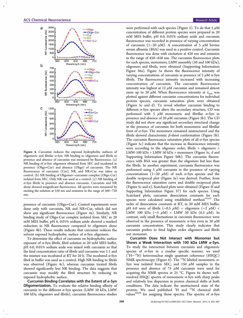
presence of curcumin (Oligo+Cur). Control experiments weredone only with curcumin, NR and NR+Cur, which did notshow any significant fluorescence (Figure 4a). Similarly, NRbinding study of Oligo-Cur complex isolated from SEC in 20mM MES buffer, pH 6, 0.01% sodium azide showed a markedreduction in NR fluorescence compared to oligomers alone(Figure 4b). These results indicate that curcumin reduces thesolvent exposed hydrophobic surface of α-Syn oligomers.To determine the effect of curcumin on hydrophobic surface
exposure of α-Syn fibrils, fibril solution in 20 mM MES buffer,pH 6.0, 0.01% sodium azide was mixed with curcumin so thatthe final concentration ratio of fibrils and curcumin was 1:1 andthe mixture was incubated at RT for 20 h. The incubated α-Synfibril in buffer was used as a control. High NR binding to fibrilswas observed (Figure 4c), whereas curcumin treated fibrilsshowed significantly less NR binding. The data suggests thatcurcumin may modify the fibril structure by reducing itsexposed hydrophobic surface.Curcumin Binding Correlates with the Extent of α-Syn
Oligomerization. To evaluate the relative binding affinity ofcurcumin to the different α-Syn species (LMW 50 kDa, LMW100 kDa, oligomers and fibrils), curcumin fluorescence studies
were performed with each species (Figure 5). To do that 5 μMconcentration of different protein species were prepared in 20mM MES buffer, pH 6.0, 0.01% sodium azide and curcuminfluorescence was recorded in presence of varying concentrationof curcumin (1−20 μM). A concentration of 5 μM bovineserum albumin (BSA) was used as a positive control. Curcuminfluorescence was done with excitation at 426 nm and emissionin the range of 450−650 nm. The curcumin fluorescence plotsfor each species, monomers, LMW assembly (50 and 100 kDa),oligomers and fibrils, were obtained (Supporting InformationFigure S6a). Figure 5a shows the fluorescence intensity ofvarying concentrations of curcumin in presence of 5 μM α-Synfibrils. The fluorescence intensity increased with increasingconcentration of curcumin. The curcumin fluorescenceintensity was highest at 12 μM curcumin and remained almostsame up to 20 μM. When fluorescence intensity at λmax wasplotted against different curcumin concentrations for individualprotein species, curcumin saturation plots were obtained(Figure 5c and d). To reveal whether curcumin binding todifferent α-Syn species alters the secondary structure, CD wasperformed with 5 μM monomeric and fibrillar α-Syn inpresence and absence of 20 μM curcumin (Figure 5b). The CDstudy did not show any significant secondary structural changein the presence of curcumin for both monomeric and fibrillarform of α-Syn. The monomers remained unstructured and thefibrils showed characteristic β-sheet conformation (Figure 5b).The curcumin fluorescence saturation plots of all α-Syn species(Figure 5c) indicate that the increase in fluorescence intensitywere according to the oligomer order; fibrils > oligomers >LMW-100 kDa > LMW 50 kDa > monomers (Figure 5c, d andSupporting Information Figure S6b). The curcumin fluores-cence with BSA was greater than the oligomers but less thanthe fibrils. In another experiment, curcumin fluorescence wasperformed using 5 μM curcumin in the presence of varyingconcentrations (1−20 μM) of each α-Syn species and thedouble reciprocal plot (Figure 5e) was derived. Further, fromthe fluorescence saturation curve and double reciprocal plots(Figure 5c and e), Scatchard plots were obtained (Figure 5f andSupporting Information Figure S7) for each species. UsingScatchard plots, curcumin dissociation constants for eachspecies were calculated using established method.41,63 Theorder of dissociation constants at RT, in 20 mM MES buffer,pH 6.0 were of fibrils (∼0.5 μM) < oligomers (∼2 μM) <LMW 100 kDa (∼5 μM) < LMW 50 kDa (6.5 μM). Incontrast, only small fluctuations in curcumin fluorescence wereobserved in the presence of monomeric α-Syn even in 20 μMcurcumin concentration. This study clearly indicates thatcurcumin prefers to bind higher order oligomers and fibrilsnot monomer.
Curcumin Does Not Interact with Monomer butShows a Weak Interaction with 100 kDa LMW α-Syn.To study the interaction between curcumin and oligomericspecies of α-Syn in a residue specific manner, we used(1H−15N) heteronuclear single quantum coherence (HSQC)NMR spectroscopy (Figure 6). The 15N labeled monomeric α-Syn was isolated from SEC, and 150 μM samples in thepresence and absence of 75 μM curcumin were used foracquiring the NMR spectra at 25 °C. Figure 6a shows well-resolved HSQC spectra of monomeric α-Syn with sharp peaksand relatively less dispersion in proton chemical shifts at bothconditions. The data indicate the unstructured state of theprotein. We used published 1H and 15N chemical shiftvalues44,64 for assigning those spectra. The spectra of α-Syn
Figure 4. Curcumin reduces the exposed hydrophobic surfaces ofoligomeric and fibrilar α-Syn. NR binding to oligomers and fibrils inpresence and absence of curcumin was measured by fluorescence. (a)NR binding of α-Syn oligomers obtained from SEC and incubated inpresence (Oligo+Cur) and absence (Oligo) of curcumin. The NRfluorescence of curcumin (Cur), NR, and NR+Cur was taken ascontrol. (b) NR binding of Oligomer−curcumin complex (Oligo-Cur)isolated from SEC. Only NR was used as a control. (c) NR binding ofα-Syn fibrils in presence and absence curcumin. Curcumin and NRalone showed insignificant fluorescence. All spectra were measured byexciting the solution at 550 nm and emission in the range of 560−720nm.
ACS Chemical Neuroscience Research Article
dx.doi.org/10.1021/cn3001203 | ACS Chem. Neurosci. 2013, 4, 393−407398

in the presence (Figure 6a, left panel, yellow spectrum) andabsence (Figure 6a, left panel, red spectrum) of curcumin wereoverlaid. Spectra of α-Syn in the presence of curcumin did notshow any significant chemical shift perturbation and retainedthe same resolution as of α-Syn in solution. We calculated thechange in chemical shifts and did not observe any significantperturbation in the presence and absence of curcumin (Figure6b, top panel). Further, the intensity ratios (I/I0) for allnonoverlapping peaks were calculated and values were found tobe ∼1.0 (data not shown). The data indicate that curcumindoes not interact with the monomeric α-Syn, which lacks anyordered structure.NMR experiments were also performed with 15N-labeled
LMW 100 kDa α-Syn in the presence and absence of curcumin,and both spectra were overlaid (Figure 6a, right panel). NMRpeaks in the 100 kDa LMW spectra both in the presence(Figure 6a, right panel, black spectrum) and absence (Figure6a, right panel, red spectrum) of curcumin appeared to bebroader than those of the monomeric protein and number ofpeaks in the regions of 8.3−8.5 and 120−125 ppm overlappedwith each other. Although most of the peaks had similarresonance frequencies in comparison to the monomerspectrum, ∼21 additional peaks were identified in the spectrumof LMW (arbitrary numbers were given to assign theseadditional peaks). These peaks were of comparatively lowintensity and did not have any corresponding peak in themonomer spectrum, suggesting that such resonances might bearising from oligomer population. We calculated the changes inthe resonance frequencies of LMW peaks in the presence andabsence of curcumin (Figure 6b, lower panel), which did notshow any definite pattern. However, chemical shift changeswere relatively larger compared to the monomer. The datasuggest that curcumin may transiently interact with LMW α-
Syn. To gain further insight, we performed intensity ratioanalysis for nonoverlapping peaks (Figure 6c and SupportingInformation Figure S8). No clear pattern emerged, suggestingthat oligomers do not have preferential binding sites forcurcumin. Interestingly, we noticed an increase in intensities foradditional peaks in the presence of curcumin shown in Figure6c and Supporting Information Figure S8, suggesting a possiblerole of curcumin in stabilizing higher order species of α-Syn.
Curcumin Accelerates α-Syn Aggregation to ProduceMorphologically Different Amyloid Fibrils in Vitro. α-Synis a natively unstructured protein, which gets converted to β-sheet-rich fibrils during incubation in vitro.6 To explore theeffect of curcumin on in vitro aggregation, 150 μM 100 kDaLMW α-Syn in 20 mM MES buffer, pH 6.0, 0.01% sodiumazide was incubated in the presence and absence of 75 μMcurcumin at 37 °C with slight rotation. It is important to notethat 100 kDa LMW α-Syn was used for this study as our NMRdata suggests that curcumin may weakly interact with thisspecies but not monomers. The curcumin was diluted fromDMSO stock solution such that the final DMSO concentrationwas 0.075% (v/v) in the solution. For control, 150 μM LMWα-Syn in the presence of 0.075% DMSO was also incubated.On each day of incubation, CD was performed to determinethe conformational transition of α-Syn in the presence andabsence of curcumin. When β-sheets were formed as measuredby CD, EM was used for morphological study (Figure 7).Immediately after addition of curcumin, no significant changesin CD spectra of α-Syn were observed. Both in the presenceand absence of curcumin, α-Syn showed mostly unstructuredconformation on day 0 (Figure 7a). However, CD study hasshowed that α-Syn was converted mostly to β-sheetconformation at day 1 in the presence of curcumin. On theother hand, α-Syn in the absence of curcumin was mostly
Figure 5. Relative binding of curcumin to different α-Syn species. (a) Curcumin fluorescence spectra of varying concentrations of curcumin (1−20μM) in the presence of 5 μM preformed α-Syn fibrils. (b) CD spectra of 5 μM α-Syn monomers and fibrils in the presence and absence of 20 μMcurcumin. (c) Curcumin fluorescence value at λmax (500 nm) in the presence of different α-Syn species with varying concentrations of curcuminshowing maximum curcumin binding for fibrils. (d) Comparative increase in curcumin fluorescence at 500 nm in the presence of 5 μM each of theLMW 50 kDa, LMW 100 kDa and monomer showing increase in curcumin binding according to the oligomer order. (e) Double-reciprocal plots ofvarious α-Syn species. (f) Scatchard plot of the α-Syn fibrils−curcumin complex. Curb and Curf indicate bound and free curcumin, respectively.
ACS Chemical Neuroscience Research Article
dx.doi.org/10.1021/cn3001203 | ACS Chem. Neurosci. 2013, 4, 393−407399

unstructured until day 1 and showed β-sheet structure only onday 2. The data suggests that curcumin accelerates theconformational transition from random coil (RC) to β-sheetduring amyloid formation (Figure 7a). The EM study after 3days of incubation suggested that α-Syn alone formed highlyordered amyloid fibrils composed of ∼2−3 individual filaments(Figure 7b). These fibrils were helically twisted to each otherwith varying degrees of lateral association. The diameter ofthese fibrils was ∼10−30 nm. In contrast, α-Syn in the presenceof curcumin showed narrow fibrils of ∼10−16 nm in diameterwith little discernible substructure. To evaluate whether fibrilsformed in the absence and presence of curcumin possessdifferent stability/internal structure, a proteinase K digestionexperiment was performed (Figure 7c). The SDS-PAGEanalysis showed five different bands from undigested fibrilsformed either in the presence or absence of curcumin. Thethree bands of more than 29 kDa were observed possibly due tothe presence of higher order SDS stable oligomers. The twobands below 18.4 kDa were observed due to monomers andfragments of α-Syn, which may be the protease stable α-Syncore. Further, the SDS-PAGE analysis of proteinase K digestedα-Syn fibrils formed in the absence of curcumin showedsignificant digestion, indicated by only one band at ∼8 kDa,
which may be the protease resistant core of α-Syn fibrils50
(Figure 7c). However, proteinase K digestion of fibrils formedin the presence of curcumin showed two bands below 18.4 kDa,in which one is monomeric and another could be the proteaseresistant core. The different proteinase K digestion profilesuggests that fibrils formed in the presence and absence ofcurcumin possess different stability against proteases.Further, to determine the detailed kinetics and confirm our
observation that curcumin accelerates the α-Syn aggregation,we performed time dependent AFM study during aggregation(Figure 7d). The data suggests that, immediately afterpreparation, LMW α-Syn in the presence and absence ofcurcumin formed small globular oligomers and duringincubation they gradually transformed to higher orderstructures. AFM imaging of 18 h incubated α-Syn in thepresence of curcumin showed some fibrils along with globularoligomers, which was converted to higher order fibrils at 22 h.In contrast, α-Syn in the absence of curcumin only showedsmall protofilament-like structures at 22 h without formation ofhigher order fibrils. The data clearly suggests that curcuminaccelerates the aggregation of α-Syn in vitro.The Kd determination and NMR studies showed that
curcumin binds to early oligomers and not to monomers.
Figure 6. Probing interaction of curcumin with monomer and LMW 100 kDa α-Syn by NMR spectroscopy. (a) Overlay of 1H−15N HSQC spectraof 150 μM monomeric α-Syn in the absence (red spectrum) and in the presence of 75 μM curcumin (yellow spectrum, top left). Top right panelshows the overlay of the 1H−15N HSQC spectra of 150 μM 100 kDa LMW α-Syn in the absence (black spectrum) and in the presence of 75 μMcurcumin (red spectrum, top right). In the HSQC spectrum of 100 kDa LMW, many additional peaks were observed compared to the HSQCspectrum of monomers and were numbered arbitrarily. (b) Difference in chemical shifts (Δδ) of individual amino acids in monomer (red) and 100kDa LMW (blue) of α-Syn in the presence and absence of curcumin were calculated. The Δδ (in ppm) was plotted against individual amino acidresidues. (c) Intensities of all extra peaks in HSQC spectrum of oligomers in presence (I) and absence (I0) of curcumin were calculated, and relativeintensities I/I0 were plotted against oligomer numbers (assigned arbitrary).
ACS Chemical Neuroscience Research Article
dx.doi.org/10.1021/cn3001203 | ACS Chem. Neurosci. 2013, 4, 393−407400

Moreover, we find that curcumin accelerates the structuralconversion and amyloid formation of LMW 100 kDa α-Syn,suggesting that curcumin may effectively reduce the populationof toxic oligomeric intermediates. In contrast, previous datasuggested that curcumin binds to monomeric α-Syn35 andinhibits its aggregation and amyloid formation.35−37 However,these previous studies were performed in the presence of either10% 2,2,2-trifluoroethanol (TFE) or 1 mM Fe3+. TFE and Fe3+
are known to modulate the protein aggregation65−67 andcomplex formation with curcumin,68 respectively. Moreover,previous studies have used ThT for monitoring the aggregationkinetics, which is not a suitable dye for monitoring amyloidaggregation in the presence of polyphenols such as curcumin.40
Interestingly, the modulation of curcumin mediated Aβaggregation in AD has also been reported to be conflicting,as both acceleration and inhibition of Aβ aggregation wereevident in the presence of curcumin.31,69,70 Cole and co-workers have suggested that curcumin inhibits Aβ aggregationand reduces toxicity.31 Further, it has been shown thatcurcumin reverses Alzheimer’s disease pathology and reducesthe plaque load in a mouse model of AD.34 Whereas recentstudies suggested that curcumin accelerates the aggregation ofAβ both in vitro and in vivo.70 Moreover, two previous reportsalso have indicated that curcumin might inhibit the conversionof Aβ monomers to oligomers but accelerates the conversion ofpreformed oligomers to fibrils with effective reduction of toxicoligomeric population.71,72 However, all these previous studieshave suggested a common theme that irrespective of whethercurcumin accelerates or delays the protein aggregation, itreduces the toxicity of protein aggregates in vitro and in vivo.In summary, our present data showed that curcumin binds to
preformed oligomers and fibrils and reduces their toxicity by
modifying their morphology and hydrophobic surface exposure.Further, curcumin binds to early oligomers and accelerates theirconversion to fibrils so that the effective population of toxicoligomeric intermediates could be reduced (Figure 8). Thisstudy suggests that curcumin or curcumin related compounds73
could be important small molecules that modulate and/or haltthe toxicity of α-Syn aggregates in PD.
■ METHODSChemicals and Reagent. Curcumin and other chemicals were
purchased from Sigma (St. Louis, MO) except Nile Red dye, whichwas purchased from Invitrogen (Carlsbad, CA). Water was doubledistilled and deionized using a Milli-Q system (Millipore Corp.,
Figure 7. Curcumin accelerates α-Syn aggregation. (a) Time dependent CD spectra of α-Syn in the presence and absence of curcumin. α-Syn in thepresence of curcumin showing accelerated conversion of random coil to β-sheet conformation during aggregation. (b) Electron micrographs of theaggregates after 3 days of incubation showing fibrillar morphology of α-Syn in the absence (left) and presence (right) of curcumin. Alteredmorphology of α-Syn aggregates were seen in the presence of curcumin. Scale bars are 500 nm. (c) SDS-PAGE analysis showing different proteinaseK digestion pattern by α-Syn fibrils formed in the presence and absence of curcumin. (d) Time dependent AFM analysis of α-Syn aggregation in thepresence and absence of curcumin showing accelerated conversion of oligomers to fibrils in the presence of curcumin. Scale bars are 700 nm.
Figure 8. Schematic representation showing the effect of curcumin onα-Syn aggregation and toxicity. Curcumin may interact with variousoligomeric intermediates of α-Syn and accelerate their conversion tofibrils. Curcumin also binds to preformed oligomers and fibrils andreduces their toxicity.
ACS Chemical Neuroscience Research Article
dx.doi.org/10.1021/cn3001203 | ACS Chem. Neurosci. 2013, 4, 393−407401

Bedford, MA). WT α-Syn plasmid (pRK172) was a kind gift of Prof.Roland Riek, ETH Zurich, Switzerland.Preparation of Curcumin Stock Solution. Curcumin stock
solution of 100 mM was made in DMSO. This stock solution wasfurther diluted in 20 mM MES buffer, pH 6.0, 0.01% sodium azide to57 mM. The working solution of curcumin was freshly prepared foreach use by dilution of 57 mM curcumin in same buffer.Protein Purification. α-Syn was expressed in Escherichia coli BL21
(DE3) strain according to the protocol described by Volles andLansbury.74 For 15N labeled α-Syn expression, minimal mediacontaining 15N-labeled ammonium chloride was used as a nitrogensource. Briefly, the IPTG induced bacterial cells were pelleted down bycentrifugation. The pellet was resuspended in buffer (50 mM Tris, pH8.0, 10 mM EDTA, 150 mM NaCl) with protease inhibitor cocktail(Roche), sonicated followed by heating in boiling water bath for 20min. Supernatant was collected after centrifugation (14 000g, 30 min).Streptomycin sulfate (10%; 136 μL/mL supernatant) and glacial aceticacid (228 μL/mL supernatant) were added to the supernatantfollowed by centrifugation (14 000g, 4 °C, 10 min). The resultingsupernatant was precipitated by equal volume of saturated ammoniumsulfate, prepared at 4 °C. Precipitated protein was washed with asolution of ammonium sulfate (saturated ammonium sulfate and water,1:1 v/v at 4 °C). The washed pellet was resuspended in 100 mMammonium acetate and stirred for 10 min. The α-Syn was precipitated,adding an equal volume of absolute ethanol. Ethanol precipitation wasrepeated twice at RT. Protein was again resuspended in 100 mMammonium acetate, lyophilized, and stored at −20 °C for further use.Preparation of Low Molecular Weight (LMW) α-Syn. For
binding and fibrillization studies, LMW (50 kDa and 100 kDa) of α-Syn was prepared from lyophilized protein. Briefly, protein wasdissolved in 20 mM MES buffer, pH 6.0, 0.01% sodium azide at aconcentration of 15 mg/mL. α-Syn is acidic in nature, the pH ofresulting solution was ∼5.0 and was sparingly soluble. To solubilize theprotein, few μL of 2 M NaOH solution was added until protein wasdissolved completely and clear solution was appeared. Then, the pHwas adjusted to 6.0 by adding few μL of 2 M HCl solutions. Theprotein solution was dialyzed against the same buffer overnight at 4 °Cusing a 10 kDa MWCO mini-dialysis unit (Millipore). This processwas carried out to remove salts and fragmented peptides. Further toremove any larger aggregates, the resulting solution was filtered bycentricon YM-50 MWCO or YM-100 MWCO filter (Millipore) toobtain LMW preparation of 50 kDa and 100 kDa, respectively. Thesupernatant was collected and used for the further study.Amyloid Fibril Formation. The assembly reaction was initiated
with 100 kDa LMW α-Syn at a concentration of ∼400 μM in 1.5 mLeppendorf tube in 20 mM MES buffer, pH 6.0, 0.01% sodium azide.The eppendorf tubes containing protein solutions were placed into anEchoTherm model RT11 rotating mixture (Torrey Pines Scientific)with a speed corresponding to 50 r.p.m. inside a 37 °C incubator. Thefibril formation was monitored by CD, ThT binding and confirmed byAFM/EM at the end of assembly reaction. For measuring α-Synaggregation kinetics in presence of curcumin, 100 kDa LMW α-Synand curcumin in 20 mMMES buffer, pH 6.0, 0.01% sodium azide weremixed such a way that the final concentrations of α-Syn and curcuminwas 150 μM and 75 μM, respectively. The DMSO concentration was0.075% (v/v). As a control, 150 μM of α-Syn was also incubated inpresence of 0.075% of DMSO. Three independent experiments wereperformed for each sample.Size Exclusion Chromatography (SEC). Purified α-Syn was
dissolved in 20 mM MES buffer, pH 6.0, 0.01% sodium azide asdescribed above. The protein solution was then centrifuged for 30 minat 14 000g using a benchtop microcentrifuge (HITACHI, himacCT15RE, Japan). The resulting solution was clear and free of anylarger aggregates. To study the curcumin interactions, protein solutionwas incubated with and without 100 μM curcumin at roomtemperature as well as at 37 °C for 30 min. Then 200 μL solutionswere loaded on a S200-Superdex gel filtration column attached to anAKTA purifier (GE Healthcare) and eluted isocratically at 4 °C in thesame buffer with a flow rate of 0.4 mL/min, and 200 μL fractions werecollected.
Circular Dichroism Spectroscopy (CD). Ten microliters ofprotein solution was diluted to 200 μL in 20 mM MES buffer, pH 6.0,0.01% sodium azide. The solution was placed into a 0.1 cm path lengthquartz cell (Hellma, Forest Hills, NY). Spectra were acquired using aJASCO-810 instrument. All measurements were done at 25 °C.Spectra were recorded over the wavelength range of 198−260 nm.Three independent experiments were performed with each sample.Raw data were processed by smoothing and subtraction of bufferspectra, according to the manufacturer’s instructions.
Curcumin Fluorescence. The curcumin fluorescence spectra wereacquired in the emission range of 450−650 nm by exciting the samplesat 426 nm. The experiments were performed using a HITACHIspectrofluorometer (model F-2500) with excitation and emission slitwidths of 2.5 and 5 nm, respectively. To study the interaction ofcurcumin with preformed α-Syn oligomers, the solid protein wasdissolved in 20 mM MES buffer, pH 6.0, 0.01% sodium azide at aconcentration of 5 mg/mL and incubated with 100 μM curcumin at 37°C. After 30 min of incubation, SEC was performed as describedearlier to obtain the preformed oligomers. The curcumin fluorescencewas performed with this preformed oligomer−curcumin complex. Thefluorescence of 100 μM curcumin in identical conditions was alsomeasured as a control. In another set of experiments, the preformed α-Syn oligomers from ∼8 mL fractions were collected from SEC byinjecting 5 mg/mL protein dissolved in 20 mM MES buffer, pH 6.0,0.01% sodium azide. The preformed oligomers with and without 40μM curcumin were incubated at 37 °C for 30 min and curcuminfluorescence recorded. Curcumin of 40 μM in identical conditions wasalso measured for control.
Congo Red Binding Assay. α-Syn preformed fibrils of 100 μMwere incubated with and without 100 μM curcumin for 20 h at RTwithout agitations. A 20 μL aliquot of sample was then mixed with 180μL of 20 mM MES buffer, pH 6.0, 0.01% sodium azide. Then 0.4 μL of10 mM CR solution (filtered through 0.2 μM filter) in 20 mM MESbuffer, pH 6.0, with 0.01% sodium azide was added such that the finalprotein concentration became 10 μM and CR 20 μM. After 10 min ofincubation at RT, absorbance was measured in the range of 400−600nm. For the measurement of the CR-only spectrum, 0.4 μL of CR wasmixed with 200 μL of 20 mM MES buffer, pH 6.0, with 0.01% sodiumazide and absorbance was measured as described. As a control, a 20 μLaliquot of fibrils with 180.4 μL of MES buffer was also measured.Three independent experiments were performed for each sample. ForCongo Red fluorescence studies, the samples were prepared in similarway as described for Congo Red absorbance. The Congo Redfluorescence spectra were acquired on a HITACHI fluorescencespectrophotometer (model F-2500). The samples were excited at awavelength of 550 nm, and emission was recorded in the wavelengthrange of 560−650 nm. The excitation and emission slit widths were2.5 and 5.0 nm, respectively. The fluorescence spectrum of 20 μMCongo Red in 20 mM MES buffer, pH 6.0, 0.01% sodium azide wasalso recorded as a control.
Determination of Kd by Curcumin Fluorescence. Dissociationconstants of curcumin to different species of α-Syn were determinedbased on curcumin fluorescence. For this study, different species of α-Syn were prepared as described earlier. For determining curcuminbinding to different species of α-Syn, 5 μM α-Syn species wereincubated in the presence and absence of varying concentrations ofcurcumin (1−20 μM) for 30 min at RT in the dark. BSA was used as acontrol for this study. Immediate after incubation curcuminfluorescence was measured with excitation at 426 nm and emissionin the range 450−650 nm. Experiments were performed usingShimadzu RF-5301PC spectrofluorometer with excitation andemission slit widths of 5 and 5 nm, respectively. The curcuminfluorescence saturation curves were obtained for each α-Syn species,and BSA by plotting curcumin fluorescence value at 500 nm againstdifferent concentration of curcumin.
In order to obtain a double reciprocal plot, 5 μM curcumin in 20mM MES buffer, pH 6.0, 0.01% sodium azide was incubated withvarying concentrations (0−20 μM) of each α-Syn species. The mixturewas incubated at RT for 30 min in the dark. After incubation,curcumin fluorescence was performed. Double reciprocal plots were
ACS Chemical Neuroscience Research Article
dx.doi.org/10.1021/cn3001203 | ACS Chem. Neurosci. 2013, 4, 393−407402

obtained for each α-Syn species by plotting 1/fluorescence intensity inthe Y-axis against 1/protein concentrations in the X-axis. From theintercept of the double reciprocal plot, maximum fluorescenceintensity was obtained when all curcumin was bound to protein.Using saturation and double reciprocal plots, concentrations of boundas well as free curcumin to each protein species were calculated. Thenratios of bound curcumin [curcuminb] to free curcumin [curcuminf]were plotted against curcuminb (Scatchard plot). This provides astraight line with a negative slope. The dissociation constants werecalculated from the formula Kd = −1/slope according to theestablished methods.41,63
Two Dimensional Nuclear Magnetic Resonance (2D NMR)spectroscopy. All NMR spectra were acquired on a Bruker Avance800 MHz spectrometer using a cryogenically cooled triple-resonanceprobe equipped with z-axis gradient coils. Data were acquired andprocessed using Topspin 2.1 version and analyzed with Sparky 3.114.DSS (4,4-dimethyl-4-silapentane-1-sulfonic acid) was used as aninternal reference for the calibration of proton chemical shifts,whereas nitrogen chemical shift was calibrated indirectly. Two-dimensional 1H−15N correlation heteronuclear single quantumcoherent (HSQC) experiments were performed on 15N-labeledprotein samples in phosphate buffer, pH 6.8 with a (90:10) H2O/D2O ratio, at 25 °C. Various sets of 1H−15N HSQC correlation spectrawere recorded on 150 μM α-Syn monomer and 150 μM 100 kDaLMW samples in the absence and presence of 75 μM curcumin. 2DHSQC spectra for monomer were recorded for 256 data points inindirect dimension with 16 scans per transient, whereas for 100 kDaLMW 512 data points were collected in the indirect dimension. Formapping the interaction sites between α-Syn and curcumin, wecalculated mean weighted chemical shift perturbations and change inintensity ratios (I/I0). Nonoverlapping
1H−15N HSQC amide cross-peaks were considered, and their intensities (I) with those of the samecross-peaks in the data set of free protein (I0) were calculated. The I/I0ratios of nonoverlapping cross-peaks were plotted as a function of theamino acid sequence to obtain the relative intensity variation acrossthe polypeptide chain. However, chemical shift perturbations werecalculated using ((5Δδ1H) 2 + (Δδ15N)2)1/2 to identify the significantperturbations upon interactions.Nile Red Binding Assay. The stock solution of 1 mM Nile Red
(NR) was prepared in DMSO and stored at −20 °C until further use.All the fluorescence studies were performed in 20 mM MES buffer, pH6.0 with 1 μM working concentration of Nile Red. 200 μL of 15 μMoligomeric α-Syn (isolated from SEC) was diluted to 400 μL in samebuffer. In 200 μL of 7.5 μM oligomers, curcumin was added such thatthe final curcumin concentration became 40 μM. The solution wasincubated at 37 °C for 30 min. The 200 μL of 7.5 μM oligomerswithout curcumin was used as a control. In another experiment, freshlyprepared 5 mg/mL α-Syn solution was incubated in the presence of100 μM curcumin at 37 °C for 30 min and injected for SEC. Theoligomers were collected from ∼8 mL peak fractions. Then 0.2 μL of 1mM NR was added to 200 μL each of the oligomers prepared in thepresence and absence of curcumin, where final NR was 1 μM. NRfluorescence was performed immediately by exiting at 550 nm andemission in the range of 560−720 nm. For NR binding study withpreformed fibrils, 200 μL of 75 μM α-Syn fibrils was incubated withand without 75 μM curcumin for 20 h. Then 20 μL of α-Syn fibrils wasdiluted to 200 μL in 20 mM MES buffer, pH 6.0, 0.01% sodium azide.Then 0.2 μL of 1 mM NR was added so that the final NRconcentration became 1 μM. The NR fluorescence was performed asdescribed; 1 μM NR in 200 μL of MES, pH 6.0 was used as a NRcontrol. To study whether curcumin can affect the NR fluorescence,fluorescence was measured for 1 μM NR, 7.5 μM curcumin, and themixture of NR and curcumin.Atomic Force Microscopy (AFM). For atomic force microscopy,
the oligomeric and fibrillar samples were diluted to ∼5 μM in distilledwater and diluted samples were spotted on a freshly cleaved mica sheetfollowed by washing with double distilled water. The mica was driedunder vacuum desiccators. AFM imaging was done in tapping modeunder a silicon nitride cantilever using a Veeco Nanoscope IV
multimode atomic force microscope. A minimum of five different areasof three independent samples were scanned with a scan rate of 1.5 Hz.
Electron Microscopy (EM). α-Syn fibrils formed in the presenceand absence of curcumin were diluted in distilled water to aconcentration of ∼40 μM, spotted on a glow-discharged, carbon-coated Formvar grid (Electron Microscopy Sciences, Fort Washington,PA), incubated for 5 min, washed with distilled water, and then stainedwith 1% (w/v) aqueous uranyl formate solution. Uranyl formatesolution was freshly prepared and filtered through 0.22 μm sterilesyringe filters (Milipore). EM analysis was performed using a FEITecnai G2 12 electron microscope at 120 kV with nominalmagnifications in the range of 26 000−60 000. Images were recordeddigitally by using the SIS Megaview III imaging system. At least twoindependent experiments were carried out for each sample.
MTT Metabolic Assay. The neuronal cell line of SH-SY5Y wascultured in Dulbecco’s modified Eagle's medium (DMEM) (Himedia,India) supplemented with 10% FBS (Invitrogen), 100 units/mLpenicillin, and 100 μg/mL streptomycin in a 5% CO2 humidifiedenvironment at 37 °C. Cells were seeded in 96-well plates in 100 μL ofmedia at a cell density of ∼10 000 cells/well. After 24 h of incubation,the old media was replaced with fresh media containing eitheroligomeric or fibrillar α-Syn preincubated with curcumin so that α-Synand curcumin concentration became 5 and 3 μM, respectively. Cellswere further incubated for 40 h at 37 °C. Only buffer and 3 μMcurcumin were also tested as a control. After 40 h, 10 μL of 5 mg/mLMTT prepared in PBS was added to each well and the incubation wascontinued for 4 h. Finally, 100 μL of a solution containing 50%dimethylformamide and 20% SDS (pH 4.7) was added and incubatedfor overnight. After incubation in a 5% CO2 humidified environmentat 37 °C, absorption values at 560 nm were determined with anautomatic micro titer plate reader (Thermo Fisher Scientific). Thebackground absorbance was also recorded at 690 nm and subtractedfrom the absorbance value of 560 nm.
Lactate Dehydrogenase (LDH) Release Assay. Lactatedehydrogenase release assay was performed to further evaluate theneuroprotective effect of curcumin over the toxic oligomeric andfibrillar α-Syn. Identical concentrations of preformed oligomers/fibrils(with and without curcumin) were used as mentioned for the MTTassay. SH-SY5Y neuronal cells were cultured and plated in 96-wellplates as described earlier. LDH assay was performed after 40 h ofincubation of neuronal cells with test samples (preformed oligomers/fibrils, with and without curcumin) using LDH toxicological kit (TOX-7, Sigma) according to the manufacturer's instructions.
SDS-PAGE Analysis. A concentration of 100 μM preformed α-Synfibrils was incubated with and without 100, 200, and 300 μM curcuminfor 20 h. After incubation, fibrils solutions were centrifuged at 18 000g,40 min. Supernatants were collected and analyzed via 12% Bis-TrisNUPAGE gel (Invitrogen) electrophoresis.
Proteinase K Digestion Assay. α-Syn (150 μM) aggregated inthe presence and absence of curcumin (75 μM) was subjected toproteinase K treatment. For proteinase K digestion assay, sampleswere half diluted in 20 mM MES, pH 6.0, 0.01% sodium azide and 1μL aliquot of 500 μg/mL proteinase K was added to 75 μL of α-Synfibrils formed in the presence and absence of curcumin. The finalconcentration of proteinase K was 6.5 μg. After 1 h of incubation at 37°C, the reaction was stopped by adding SDS sample buffer followed byheating to 100 °C for 10 min in a water bath. In another experiment,150 μM preformed α-Syn fibrils was incubated with and without 75μM curcumin for 1 h at room temperature and digested withproteinase K as described above. For proteinase K digestion ofoligomers, 30 μM oligomers (isolated from SEC) were incubated withand without 30 μM curcumin at 37 °C for 30 min. The samples weretreated with and without 0.1 μg proteinase K for 10 min at 37 °C andreaction was stopped as described. The samples were then used forSDS-PAGE using 12% Bis-Tris NuPAGE gel (Invitrogen) anddeveloped with silver stain (Bangalore Genei, India).
SH-SY5Y Cell Culture and Differentiation. The neuronal cellline of SH-SY5Y was cultured in Dulbecco’s modified Eagle's medium(DMEM) (Himedia, India) medium supplemented with 10% FBS(Invitrogen), 100 units/mL penicillin, and 100 μg/mL streptomycin in
ACS Chemical Neuroscience Research Article
dx.doi.org/10.1021/cn3001203 | ACS Chem. Neurosci. 2013, 4, 393−407403

a 5% CO2 humidified environment at 37 °C. For cell differentiation,50 μM all-trans retinoic acid (RA) (Sigma) was used.75 To do that,undifferentiated cells were seeded on cover glass in a 24-well plate for24 h, and after that cells were treated with 50 μM RA in media. Atevery alternate day, the old media was replaced with fresh mediacontaining RA. After 5 days of incubation, we analyzed the cellmorphology using phase contrast microscopy to observe neuriteprojections of differentiated cells.Immunocytochemistry. The 5 days differentiated cells were used
for synaptophysin staining. To do so, cell culture media was replacedwith fresh media containing oligomers, oligomers+curcumin, andbuffer; cells were further incubated for 40 h. The final oligomers andcurcumin concentrations were 6 and 3 μM, respectively. Afterincubation, media was discarded and the cells were fixed with 3.5%formaldehyde for 30 min at 37 °C. After fixing, the cells were washedwith PBS (1 mL × 2) and permeabilized with chilled methanol for 20min at −20 °C. Further, the cells were blocked with blocking solution(2% BSA in PBS) for 30 min at RT. Cells were incubated for 3 h at RTwith rabbit monoclonal anti-synaptophysin primary antibody diluted inblocking solution (1:350; ABCAM). After incubation, the solution wasdiscarded and cells were washed with 1 mL of PBS (3 × 5 min) withgentle shaking. The cells were then incubated with anti-rabbitAlexaFluor 488 (1:500, Invitrogen) secondary antibody prepared in2% BSA in PBS and incubated for 2 h in dark. After removing thesecondary antibody solution, the cells were mounted on a clean glassslide using mounting media. A positive control for synaptophysinspecificity was used where the differentiated SH-SY5Y cells weretreated with 6 μM Aβ(25−35) amyloid fibrils.76 Images were acquiredusing an Olympus IX 81 confocal microscope.To visualize the morphology of nucleus, the differentiated cells were
stained with 4,6- diamidino-2-phenylindole (DAPI; Sigma) (1 μg/mL)for 5 min at RT. After 5 min, the DAPI solution was removed and thecells were washed with PBS, mounted on a clean glass slide usingmounting media, and imaged under confocal microscope.Reactive Oxygen Species (ROS) Measurement. The differ-
entiated SH-SY5Y cells were treated with oligomers, oligomers+curcumin, and buffer only in 24-well plates for 40 h. After incubation,the old media was discarded and cells were very gently washed with 1×PBS buffer. A 3 mM stock of 2-hydroethidium (Sigma) was freshlyprepared in DMSO, and 2 μL of this solution in 1 mL of PBS wasadded to the cells and incubated for 5 min at RT in dark. The finalconcentration of 2-hydroethidium was 6 μM. The cells wereimmediately imaged under confocal microscopy with an excitation at543 nm and emission above 565 nm. The intensities of oxidizedhydroethidium (hydroxyethidium) fluorescence in cells treated withbuffer, oligomers, and oligomers+curcumin were quantified usingFV500 Tiempo (version 4.3) software.Flow Cytometry Analysis. For relative quantification of cell death
and apoptosis in oligomers and oligomers+curcumin treated samples,flow cytometry measurement was performed using an Annexin V-FITC apoptosis detection kit (Sigma). To do that, undifferentiatedcells were grown in T25 cell culture flasks (Nunc) until ∼70%confluency (∼106 cells). The cells were then treated with oligomers,oligomers+curcumin, and only buffer for 40 h. After incubation, thecells were trypsinized, centrifuged, and used for cell death assay usingthe Annexin V-FITC apoptosis detection kit (APOAF, Sigma). Thecell pellet was washed with 1× PBS and further resuspended in 1×binding buffer (Sigma). Cells were stained with Annexin V-FITC andpropidium iodide (PI) according to the manufacturer’s instructions.Unstained cells (without Annexin V-FITC and PI) were used as acontrol, and cells stained with either Annexin V-FITC or PI were usedas fluorescent compensation controls. Annexin V-FITC and PI stainingwere quantified in a flow cytometer (FACSAria, BDBiosciences, SanJose, CA) and analyzed using the BDFACS Diva software. For eachsample, 20 000 cells were analyzed. The upper left quadrant (Q1)represents the dead cell population (only PI positive), the upper rightquadrant (Q2) represents the population of cells in the late stage ofapoptosis (Annexin V-FITC positive and PI positive), the lower leftquadrant (Q3) represents the population of viable/live cells (AnnexinV-FITC negative and PI negative), and the lower right quadrant (Q4)
represents the population of cells in early stages of apoptosis (onlyAnnexin V-FITC positive). The X-axis represents the area of AnnexinV-FITC fluorescence, and the Y-axis represents PI fluorescenceintensities.
Statistical Analysis. Statistical significance was determined byusing one-way ANOVA followed by Newman-Keuls multiplecomparison post hoc test; *P < 0.05; **P < 0.01; NS P > 0.05.
■ ASSOCIATED CONTENT*S Supporting InformationTable 1 and Supplementary figures S1 to S8. This materials isavailable free of charge via the Internet http://pubs.acs.org
■ AUTHOR INFORMATIONCorresponding Author*Department of Biosciences and Bioengineering, IIT Bombay,Powai, Mumbai, India 400076. Telephone: +91-22-2576-7774.Fax: +91-22-2572 3480. E-mail: [email protected] Contributions§Equally contributing authors.Author ContributionsS.K.M. and P.K.S. conceived and designed the experiments.P.K.S., D.G., V.K., G.M.M., and A.K. performed the experi-ments: S.K.M., A.K., P.K.S., and D.G. analyzed the data. S.K.M.,P.K.S., and A.K. wrote the paper.FundingThe work was supported by Grants from CSIR (37(1404)/10/EMR-11), DST (SR/FR/LS-032/2009), and DBT (BT/PR14344Med/30/501/2010 and BT/PR13359/BRB/10/752/2009), Government of India.NotesThe authors declare no competing financial interest.
■ ACKNOWLEDGMENTSThe authors wish to acknowledge Central SPM Facility (IRCC,IIT Bombay) for AFM imaging and SAIF (IIT Bombay) forelectron microscopy. The authors also thank Dr. Shamik Sen,Reeba S. Jacob, Shruti Sahay, A. Anoop, and Subhadeep Das forcritically reading the manuscript and Prem Verma (Departmentof Physics, IIT Bombay) for the help during AFM imaging. Wewould also like to thank Ankit Rai, Sudarshan Kini, and SachinTawade for their valuable help in confocal microscopy and flowcytometry analysis.
■ ABBREVIATIONSα-Syn, α-synuclein; CD, circular dichroism; EM, electronmicroscopy; AFM, atomic force microscopy; AD, Alzheimer’sdisease; PD, Parkinson’s disease; NMR, nuclear magneticresonance; LMW, low molecular weight; CR, Congo Red; NR,Nile Red; SEC, size exclusion chromatography
■ REFERENCES(1) Cookson, M. R. (2005) The biochemistry of Parkinson’s disease.Annu. Rev. Biochem. 74, 29−52.(2) Lansbury, P. T., and Brice, A. (2002) Genetics of Parkinson’sdisease and biochemical studies of implicated gene products -Commentary. Curr. Opin Cell. Biol. 14, 653−660.(3) Feany, M. B. (2000) Studying human neurodegenerative diseasesin flies and worms. J. Neuropathol. Exp. Neurol. 59, 847−856.(4) Conway, K. A., Harper, J. D., and Lansbury, P. T. (1998)Accelerated in vitro fibril formation by a mutant α-synuclein linked toearly-onset Parkinson disease. Nat. Med. 4, 1318−1320.
ACS Chemical Neuroscience Research Article
dx.doi.org/10.1021/cn3001203 | ACS Chem. Neurosci. 2013, 4, 393−407404

(5) Conway, K. A., Lee, S. J., Rochet, J. C., Ding, T. T., Williamson,R. E., and Lansbury, P. T. (2000) Acceleration of oligomerization, notfibrillization, is a shared property of both a-synuclein mutations linkedto early-onset Parkinson’s disease: Implications for pathogenesis andtherapy. Proc. Natl. Acad. Sci. U.S.A. 97, 571−576.(6) Uversky, V. N., Lee, H. J., Li, J., Fink, A. L., and Lee, S. J. (2001)Stabilization of partially folded conformation during α-synucleinoligomerization in both purified and cytosolic preparations. J. Biol.Chem. 276, 43495−43498.(7) Goldberg, M. S., and Lansbury, P. T. (2000) Is there a cause-and-effect relationship between α-synuclein fibrillization and Parkinson’sdisease? Nat. Cell Biol. 2, E115−E119.(8) Karpinar, D. P., Balija, M. B., Kugler, S., Opazo, F., Rezaei-Ghaleh, N., Wender, N., Kim, H. Y., Taschenberger, G., Falkenburger,B. H., Heise, H., Kumar, A., Riedel, D., Fichtner, L., Voigt, A., Braus,G. H., Giller, K., Becker, S., Herzig, A., Baldus, M., Jackle, H., Eimer, S.,Schulz, J. B., Griesinger, C., and Zweckstetter, M. (2009) Pre-fibrillarα-synuclein variants with impaired β-structure increase neurotoxicityin Parkinson’s disease models. EMBO J. 28, 3256−3268.(9) Winner, B., Jappelli, R., Maji, S. K., Desplats, P. A., Boyer, L.,Aigner, S., Hetzer, C., Loher, T., Vilar, M., Campioni, S., Tzitzilonis,C., Soragni, A., Jessberger, S., Mira, H., Consiglio, A., Pham, E.,Masliah, E., Gage, F. H., and Riek, R. (2011) In vivo demonstrationthat α-synuclein oligomers are toxic. Proc. Natl. Acad. Sci. U.S.A. 108,4194−4199.(10) Martin, Z. S., Neugebauer, V., Dineley, K. T., Kayed, R., Zhang,W., Reese, L. C., and Taglialatela, G. (2012) α-Synuclein oligomersoppose long-term potentiation and impair memory through acalcineurin-dependent mechanism: relevance to human synucleopathicdiseases. J. Neurochem. 120, 440−452.(11) Danzer, K. M., Haasen, D., Karow, A. R., Moussaud, S., Habeck,M., Giese, A., Kretzschmar, H., Hengerer, B., and Kostka, M. (2007)Different species of α-synuclein oligomers induce calcium influx andseeding. J. Neurosci. 27, 9220−9232.(12) Lazo, N. D., Maji, S. K., Fradinger, E. A., Bitan, G., and Teplow,D. B. (2005) The Amyloid β Protein. In Amyloid proteins: The β-sheetconformation and disease (Sipe, J. D., Ed.), pp 384−491, Wiley-VCHPublishers, Weinheim, Germany.(13) Mason, J. M., Kokkoni, N., Stott, K., and Doig, A. J. (2003)Design strategies for anti-amyloid agents. Curr. Opin. Struct. Biol. 13,526−532.(14) Cohen, F. E., and Kelly, J. W. (2003) Therapeutic approaches toprotein-misfolding diseases. Nature 426, 905−909.(15) Hong, D. P., Fink, A. L., and Uversky, V. N. (2008) Structuralcharacteristics of α-synuclein oligomers stabilized by the flavonoidbaicalein. J. Mol. Biol. 383, 214−223.(16) El-Agnaf, O. M., Paleologou, K. E., Greer, B., Abogrein, A. M.,King, J. E., Salem, S. A., Fullwood, N. J., Benson, F. E., Hewitt, R.,Ford, K. J., Martin, F. L., Harriott, P., Cookson, M. R., and Allsop, D.(2004) A strategy for designing inhibitors of α-synuclein aggregationand toxicity as a novel treatment for Parkinson’s disease and relateddisorders. FASEB J. 18, 1315−1317.(17) Masuda, M., Suzuki, N., Taniguchi, S., Oikawa, T., Nonaka, T.,Iwatsubo, T., Hisanaga, S., Goedert, M., and Hasegawa, M. (2006)Small molecule inhibitors of α-synuclein filament assembly. Bio-chemistry 45, 6085−6094.(18) Lamberto, G. R., Binolfi, A., Orcellet, M. L., Bertoncini, C. W.,Zweckstetter, M., Griesinger, C., and Fernandez, C. O. (2009)Structural and mechanistic basis behind the inhibitory interaction ofPcTS on α-synuclein amyloid fibril formation. Proc. Natl. Acad. Sci.U.S.A. 106, 21057−21062.(19) Ehrnhoefer, D. E., Bieschke, J., Boeddrich, A., Herbst, M.,Masino, L., Lurz, R., Engemann, S., Pastore, A., and Wanker, E. E.(2008) EGCG redirects amyloidogenic polypeptides into unstruc-tured, off-pathway oligomers. Nat. Struct. Mol. Biol. 15, 558−566.(20) Masuda, M., Hasegawa, M., Nonaka, T., Oikawa, T., Yonetani,M., Yamaguchi, Y., Kato, K., Hisanaga, S., and Goedert, M. (2009)Inhibition of α-synuclein fibril assembly by small molecules: analysisusing epitope-specific antibodies. FEBS Lett. 583, 787−791.
(21) Zhu, M., Rajamani, S., Kaylor, J., Han, S., Zhou, F., and Fink, A.L. (2004) The flavonoid baicalein inhibits fibrillation of α-synucleinand disaggregates existing fibrils. J. Biol. Chem. 279, 26846−26857.(22) Rao, J. N., Dua, V., and Ulmer, T. S. (2008) Characterization ofα-synuclein interactions with selected aggregation-inhibiting smallmolecules. Biochemistry 47, 4651−4656.(23) Bieschke, J., Russ, J., Friedrich, R. P., Ehrnhoefer, D. E., Wobst,H., Neugebauer, K., and Wanker, E. E. (2010) EGCG remodelsmature α-synuclein and amyloid-beta fibrils and reduces cellulartoxicity. Proc. Natl. Acad. Sci. U.S.A. 107, 7710−7715.(24) Caruana, M., Hogen, T., Levin, J., Hillmer, A., Giese, A., andVassallo, N. (2011) Inhibition and disaggregation of α-synucleinoligomers by natural polyphenolic compounds. FEBS Lett. 585, 1113−1120.(25) Ferreira, N., Saraiva, M. J., and Almeida, M. R. (2011) Naturalpolyphenols inhibit different steps of the process of transthyretin(TTR) amyloid fibril formation. FEBS Lett. 585, 2424−2430.(26) Hafner-Bratkovic, I., Gaspersic, J., Smid, L. M., Bresjanac, M.,and Jerala, R. (2008) Curcumin binds to the α-helical intermediate andto the amyloid form of prion protein - a new mechanism for theinhibition of PrP(Sc) accumulation. J. Neurochem. 104, 1553−1564.(27) Porat, Y., Abramowitz, A., and Gazit, E. (2006) Inhibition ofamyloid fibril formation by polyphenols: structural similarity andaromatic interactions as a common inhibition mechanism. Chem. Biol.Drug. Des. 67, 27−37.(28) Goel, A., Kunnumakkara, A. B., and Aggarwal, B. B. (2008)Curcumin as ″Curecumin″: from kitchen to clinic. Biochem. Pharmacol.75, 787−809.(29) Cole, G. M., Teter, B., and Frautschy, S. A. (2007)Neuroprotective effects of curcumin. Adv. Exp. Med. Biol. 595, 197−212.(30) Lim, G. P., Chu, T., Yang, F. S., Beech, W., Frautschy, S. A., andCole, G. M. (2001) The curry spice curcumin reduces oxidativedamage and amyloid pathology in an Alzheimer transgenic mouse. J.Neurosci. 21, 8370−8377.(31) Yang, F., Lim, G. P., Begum, A. N., Ubeda, O. J., Simmons, M.R., Ambegaokar, S. S., Chen, P. P., Kayed, R., Glabe, C. G., Frautschy,S. A., and Cole, G. M. (2005) Curcumin inhibits formation of amyloidbeta oligomers and fibrils, binds plaques, and reduces amyloid in vivo.J. Biol. Chem. 280, 5892−5901.(32) Khuwaja, G., Khan, M. M., Ishrat, T., Ahmad, A., Raza, S. S.,Ashafaq, M., Javed, H., Khan, M. B., Khan, A., Vaibhav, K., Safhi, M.M., and Islam, F. (2011) Neuroprotective effects of curcumin on 6-hydroxydopamine-induced Parkinsonism in rats: behavioral, neuro-chemical and immunohistochemical studies. Brain Res. 1368, 254−263.(33) Hickey, M. A., Zhu, C., Medvedeva, V., Lerner, R. P., Patassini,S., Franich, N. R., Maiti, P., Frautschy, S. A., Zeitlin, S., Levine, M. S.,and Chesselet, M. F. (2012) Improvement of neuropathology andtranscriptional deficits in CAG 140 knock-in mice supports a beneficialeffect of dietary curcumin in Huntington’s disease. Mol. Neurodegener.7, 12.(34) Garcia-Alloza, M., Borrelli, L. A., Rozkalne, A., Hyman, B. T.,and Bacskai, B. J. (2007) Curcumin labels amyloid pathology in vivo,disrupts existing plaques, and partially restores distorted neurites in anAlzheimer mouse model. J. Neurochem. 102, 1095−1104.(35) Ahmad, B., and Lapidus, L. J. (2012) Curcumin preventsaggregation in α-synuclein by increasing reconfiguration rate. J. Biol.Chem. 287, 9193−9199.(36) Ono, K., and Yamada, M. (2006) Antioxidant compounds havepotent anti-fibrillogenic and fibril-destabilizing effects for α-synucleinfibrils in vitro. J. Neurochem. 97, 105−115.(37) Pandey, N., Strider, J., Nolan, W. C., Yan, S. X., and Galvin, J. E.(2008) Curcumin inhibits aggregation of α-synuclein. Acta Neuro-pathol. 115, 479−489.(38) Liu, Z., Yu, Y., Li, X., Ross, C. A., and Smith, W. W. (2011)Curcumin protects against A53T α-synuclein-induced toxicity in aPC12 inducible cell model for Parkinsonism. Pharmacol. Res. 63, 439−444.
ACS Chemical Neuroscience Research Article
dx.doi.org/10.1021/cn3001203 | ACS Chem. Neurosci. 2013, 4, 393−407405

(39) Wang, M. S., Boddapati, S., Emadi, S., and Sierks, M. R. (2010)Curcumin reduces α-synuclein induced cytotoxicity in Parkinson’sdisease cell model. BMC Neurosci. 11, 57.(40) Hudson, S. A., Ecroyd, H., Kee, T. W., and Carver, J. A. (2009)The thioflavin T fluorescence assay for amyloid fibril detection can bebiased by the presence of exogenous compounds. FEBS J. 276, 5960−5972.(41) Rai, D., Singh, J. K., Roy, N., and Panda, D. (2008) Curcumininhibits FtsZ assembly: an attractive mechanism for its antibacterialactivity. Biochem. J. 410, 147−155.(42) Mosmann, T. (1983) Rapid colorimetric assay for cellulargrowth and survival: application to proliferation and cytotoxicityassays. J. Immunol. Methods 65, 55−63.(43) Behl, C., Davis, J. B., Lesley, R., and Schubert, D. (1994)Hydrogen peroxide mediates amyloid beta protein toxicity. Cell 77,817−827.(44) Braga, C. A., Follmer, C., Palhano, F. L., Khattar, E., Freitas, M.S., Romao, L., Di Giovanni, S., Lashuel, H. A., Silva, J. L., and Foguel,D. (2011) The anti-Parkinsonian drug selegiline delays the nucleationphase of α-synuclein aggregation leading to the formation of nontoxicspecies. J. Mol. Biol. 405, 254−273.(45) Zhao, H., Joseph, J., Fales, H. M., Sokoloski, E. A., Levine, R. L.,Vasquez-Vivar, J., and Kalyanaraman, B. (2005) Detection andcharacterization of the product of hydroethidine and intracellularsuperoxide by HPLC and limitations of fluorescence. Proc. Natl. Acad.Sci. U.S.A. 102, 5727−5732.(46) Cremades, N., Cohen, S. I., Deas, E., Abramov, A. Y., Chen, A.Y., Orte, A., Sandal, M., Clarke, R. W., Dunne, P., Aprile, F. A.,Bertoncini, C. W., Wood, N. W., Knowles, T. P., Dobson, C. M., andKlenerman, D. (2012) Direct observation of the interconversion ofnormal and toxic forms of α-synuclein. Cell 149, 1048−1059.(47) Marimpietri, D., Nico, B., Vacca, A., Mangieri, D., Catarsi, P.,Ponzoni, M., and Ribatti, D. (2005) Synergistic inhibition of humanneuroblastoma-related angiogenesis by vinblastine and rapamycin.Oncogene 24, 6785−6795.(48) Vermes, I., Haanen, C., Steffens-Nakken, H., andReutelingsperger, C. (1995) A novel assay for apoptosis. Flowcytometric detection of phosphatidylserine expression on earlyapoptotic cells using fluorescein labelled Annexin V. J. Immunol.Methods 184, 39−51.(49) Klunk, W. E., Jacob, R. F., and Mason, R. P. (1999) Quantifyingamyloid β-peptide (Aβ) aggregation using the Congo Red-Aβ (CR-Aβ) spectrophotometric assay. Anal. Biochem. 266, 66−76.(50) Maji, S. K., Perrin, M. H., Sawaya, M. R., Jessberger, S.,Vadodaria, K., Rissman, R. A., Singru, P. S., Nilsson, K. P., Simon, R.,Schubert, D., Eisenberg, D., Rivier, J., Sawchenko, P., Vale, W., andRiek, R. (2009) Functional amyloids as natural storage of peptidehormones in pituitary secretory granules. Science 325, 328−332.(51) El-Agnaf, O. M. A., and Irvine, G. B. (2002) Aggregation andneurotoxicity of α-synuclein and related peptides. Biochem. Soc. Trans.30, 559−565.(52) Sidhu, A., Wersinger, C., Moussa, C. E., and Vernier, P. (2004)The role of α-synuclein in both neuroprotection and neuro-degeneration. Ann. N.Y. Acad. Sci. 1035, 250−270.(53) Grelle, G., Otto, A., Lorenz, M., Frank, R. F., Wanker, E. E., andBieschke, J. (2011) Black tea theaflavins inhibit formation of toxicamyloid-β and α-synuclein fibrils. Biochemistry 50, 10624−10636.(54) Langer, F., Eisele, Y. S., Fritschi, S. K., Staufenbiel, M., Walker,L. C., and Jucker, M. (2011) Soluble Abeta seeds are potent inducersof cerebral beta-amyloid deposition. J. Neurosci. 31, 14488−14495.(55) Podlisny, M. B., Ostaszewski, B. L., Squazzo, S. L., Koo, E. H.,Rydell, R. E., Teplow, D. B., and Selkoe, D. J. (1995) Aggregation ofsecreted amyloid beta-protein into sodium dodecyl sulfate-stableoligomers in cell culture. J. Biol. Chem. 270, 9564−9570.(56) Walsh, D. M., Tseng, B. P., Rydel, R. E., Podlisny, M. B., andSelkoe, D. J. (2000) The oligomerization of amyloid beta-proteinbegins intracellularly in cells derived from human brain. Biochemistry39, 10831−10839.
(57) Neumann, M., Kahle, P. J., Giasson, B. I., Ozmen, L., Borroni,E., Spooren, W., Muller, V., Odoy, S., Fujiwara, H., Hasegawa, M.,Iwatsubo, T., Trojanowski, J. Q., Kretzschmar, H. A., and Haass, C.(2002) Misfolded proteinase K-resistant hyperphosphorylated α-synuclein in aged transgenic mice with locomotor deterioration andin human α-synucleinopathies. J. Clin. Invest. 110, 1429−1439.(58) Vilar, M., Chou, H. T., Luhrs, T., Maji, S. K., Riek-Loher, D.,Verel, R., Manning, G., Stahlberg, H., and Riek, R. (2008) The fold ofα-synuclein fibrils. Proc. Natl. Acad. Sci. U.S.A. 105, 8637−8642.(59) Bolognesi, B., Kumita, J. R., Barros, T. P., Esbjorner, E. K.,Luheshi, L. M., Crowther, D. C., Wilson, M. R., Dobson, C. M., Favrin,G., and Yerbury, J. J. (2010) ANS binding reveals common features ofcytotoxic amyloid species. ACS Chem. Biol. 5, 735−740.(60) Campioni, S., Mannini, B., Zampagni, M., Pensalfini, A., Parrini,C., Evangelisti, E., Relini, A., Stefani, M., Dobson, C. M., Cecchi, C.,and Chiti, F. (2010) A causative link between the structure of aberrantprotein oligomers and their toxicity. Nat. Chem. Biol. 6, 140−147.(61) Daban, J. R., Samso, M., and Bartolome, S. (1991) Use of NileRed as a fluorescent probe for the study of the hydrophobic propertiesof protein-sodium dodecyl sulfate complexes in solution. Anal.Biochem. 199, 162−168.(62) Sackett, D. L., and Wolff, J. (1987) Nile Red as a polarity-sensitive fluorescent probe of hydrophobic protein surfaces. Anal.Biochem. 167, 228−234.(63) Fuchs, H., and Gessner, R. (2001) The result of equilibrium-constant calculations strongly depends on the evaluation method usedand on the type of experimental errors. Biochem. J. 359, 411−418.(64) Eliezer, D., Kutluay, E., Bussell, R., and Browne, G. (2001)Conformational properties of α-synuclein in its free and lipid-associated states. J. Mol. Biol. 307, 1061−1073.(65) Munishkina, L. A., Phelan, C., Uversky, V. N., and Fink, A. L.(2003) Conformational behavior and aggregation of α-synuclein inorganic solvents: Modeling the effects of membranes. Biochemistry 42,2720−2730.(66) Anderson, V. L., Ramlall, T. F., Rospigliosi, C. C., Webb, W. W.,and Eliezer, D. (2010) Identification of a helical intermediate intrifluoroethanol-induced α-synuclein aggregation. Proc. Natl. Acad. Sci.U.S.A. 107, 18850−18855.(67) Fezoui, Y., and Teplow, D. B. (2002) Kinetic studies of amyloidβ-protein fibril assembly - Differential effects of α-helix stabilization. J.Biol. Chem. 277, 36948−36954.(68) Dairam, A., Fogel, R., Daya, S., and Limson, J. L. (2008)Antioxidant and iron-binding properties of curcumin, capsaicin, and S-allylcysteine reduce oxidative stress in rat brain homogenate. J. Agric.Food Chem. 56, 3350−3356.(69) Hamaguchi, T., Ono, K., and Yamada, M. (2010) REVIEW:Curcumin and Alzheimer’s disease. CNS Neurosci. Ther. 16, 285−297.(70) Caesar, I., Jonson, M., Nilsson, K. P., Thor, S., andHammarstrom, P. (2012) Curcumin promotes Aβ fibrillation andreduces neurotoxicity in transgenic Drosophila. PLoS One 7, e31424.(71) Hamaguchi, T., Ono, K., Murase, A., and Yamada, M. (2009)Phenolic compounds prevent Alzheimer’s pathology through differenteffects on the amyloid-beta aggregation pathway. Am. J. Pathol. 175,2557−2565.(72) Necula, M., Kayed, R., Milton, S., and Glabe, C. G. (2007) Smallmolecule inhibitors of aggregation indicate that amyloid βoligomerization and fibrillization pathways are independent anddistinct. J. Biol. Chem. 282, 10311−10324.(73) Orlando, R. A., Gonzales, A. M., Royer, R. E., Deck, L. M., andVander Jagt, D. L. (2012) A chemical analog of curcumin as animproved inhibitor of amyloid β oligomerization. PLoS One 7, e31869.(74) Volles, M. J., and Lansbury, P. T., Jr. (2007) Relationshipsbetween the Sequence of α-Synuclein and its Membrane Affinity,Fibrillization Propensity, and Yeast Toxicity. J. Mol. Biol. 366, 1510−1522.(75) Lee, H. J., Khoshaghideh, F., Patel, S., and Lee, S. J. (2004)Clearance of α-synuclein oligomeric intermediates via the lysosomaldegradation pathway. J. Neurosci. 24, 1888−1896.
ACS Chemical Neuroscience Research Article
dx.doi.org/10.1021/cn3001203 | ACS Chem. Neurosci. 2013, 4, 393−407406

(76) Amijee, H., Bate, C., Williams, A., Virdee, J., Jeggo, R.,Spanswick, D., Scopes, D. I., Treherne, J. M., Mazzitelli, S., Chawner,R., Eyers, C. E., and Doig, A. J. (2012) The N-Methylated PeptideSEN304 Powerfully Inhibits Aβ(1−42) Toxicity by PerturbingOligomer Formation. Biochemistry 51, 8338−8352.
ACS Chemical Neuroscience Research Article
dx.doi.org/10.1021/cn3001203 | ACS Chem. Neurosci. 2013, 4, 393−407407