coding potential of transfected human placental lactogen genes
TRANSCRIPT

Nucleic Acids Research, Vol. 18, No. 16 4665
Coding potential of transfected human placental lactogengenes
Diana Resendez-Perez, Ramiro Ramirez-Solis, Alfredo Varela-Echavarria, Herminia G.Martinez-Rodriguez and Hugo A.Barrera-SaIdahna*Unidad de Laboratorios de Ingenieri'a y Expresion Genticas, Departamento de Bioquimica, Facultadde Medicina de la Universidad Autonoma de Nuevo Leon, Monterrey, NL, Mexico
Received June 28, 1990; Revised and Accepted July 16, 1990
ABSTRACT
We have joined the promoter-less sequences of thethree hPL genes (hPL-1, hPL-3 and hPL-4) to strongtranscriptional control elements. in vivo 35S-labeledproteins from the culture medium of cells transfectedwith the genes were resolved on SDS-polyacrylamidegels. The presence of characteristic labeled bands,visualized by autoradiography, determined that hPL-4and hPL-3, but not hPL-1, contribute to the productionof mature hPL. In these experiments hPL-3 expressedmore RNA and protein than hPL-4. By exchanging thefirst two exons among hPL and hGH genes, wedetermined that the abundance of chimeric proteinsdepended on the genetic origin of the first two exons.Finally, we found evidence indicating that the splicemutation (G-A) at the beginning of the second intronof hPL-1, is not the only cause of the apparent lack ofinactivity of this gene, since its reversion does notrestore expression.
INTRODUCTION
The human growth hormone-placental lactogen gene family isa multigene complex containing two human growth hormone(hGH) and three human placental lactogen (hPL, also known as
chorionic somatommamotropin; hCS) genes (1,2). The entiregene cluster is located on the long arm of human chromosome17 at bands q22 -24 (3). The genes display the following 5' to3' arrangement: hGH-N, hPL-1 (or hPL-L, L for like), hPL-4(also known as hCS-A or hCS-1), hGH-V and hPL-3 (also namedhCS-B or hCS-2). The best characterized protein products of thisfamily are the secreted hGH and hPL polypeptides containing191 amino acids. They are produced in the pituitary gland andplacenta, respectively.Recombinant DNA analysis has revealed a paradox in the
coding potential of the hGH and hPL genes. The hGH-N gene,both in vivo and in vitro, generates through differential splicingof its primary transcription product, 22 kDa (90%) and 20 kDa(10%) forms of hGH (4,5). The hGH-V gene, whose expression
has been demonstrated only in the placenta and in a single humanpituitary tumor (6), has recently been confirmed to also generatein vitro a 22 kDa form. However, no 20 kDa protein derivedfrom this gene has been detected (7). cDNA cloning and DNAsequencing also have revealed the existence in placenta of a
second type of hGH-V mRNA. By retaining an in-frame fourthintron, this new mRNA is predicted to encode a mature proteinof 26 kDa (8).A completely different situation is observed with the hPL genes.
The hPL-4 and hPL-3 genes have been found to be active in termplacenta. In addition, their cDNAs have been cloned andsequenced (9). Their mRNAs are slightly divergent in nucleotidesequence. The encoded pre-hormones of these two mRNAs differin a single amino acid position within the signal peptide (atposition -24, hPL-3 codes for alanine while hPL-4 codes forproline). Yet, the mature hormones are identical. The third gene(hPL-1), is presumably nonfunctional, since it contains a mutation(G- A) at the 5' or donor splice site of the second intron.Transcripts derived from it have not been detected (9, 10).Therefore, while two hGH genes generates at least four differenthormones, the sequences of the three hPL genes predict thesynthesis of a single form of mature hPL hormone.The sequence of both hPL-3 and hPL-4 genes and their cDNAs,
predict that they might contribute to the placental production ofhPL. However, since the mature proteins expected to be derivedfrom them are identical, it is impossible to distinguish theirgene(s) of origin. No evidence has been obtained demonstratingthat the expression of either gene actually specifies the maturehPL protein.
In this study we performed an analysis of the in vitro expressionproducts of all hPL genes. We specifically addressed the questionof whether or not the hPL-3 and hPL-4 genes, known to betranscriptionally active in term placenta, produce maturehormones. These two genes, at different expression levels, were
found capable of producing an intrinsic hPL protein. Experimentswere also designed to determine if the splice point mutation atthe beginning of the first intron of the hPL-1 gene, is the onlycause of its apparent lack of expression.
* To whom correspondence should be addressed
.-. 1990 Oxford University Press

4666 Nucleic Acids Research, Vol. 18, No. 16
MATERIALS AND METHODSRecombinant DNA constructions and preparation of plasmidDNARestriction and other enzymes were obtained from commercialsuppliers and used according to their manufacturers instructions.The isolation of hGH and hPL genes have been previouslyreported (11). pNUT, constructed by R. Palmiter et al. (12)which already carries the hGH-N structural gene in front of themetallothionein promoter, was a generous gift. The hPL genescloned in pSV2gpt (13) were kindly provided by G. Saunders.DNA restriction fragments were purified from preparativeagarose or polyacrylamide gels. This was performed byelectroelution, or by glass bead extraction (14) from the agarosegel slices. hPL and hGH promoter-less genes were subclonedinto pNUT. The large BamHI to EcoRI fragment ofpNUT, wasligated to DNA fragments carrying the genes of interest. Thegenes consisted of sequences from their naturally occurringBamHI site (except hPL-1; see Results) at nucleotide +2, to anatural or artificial (linkers) EcoRI site located several hundrednucleotides downstream of the polyadenylation signal. Toconstruct our negative control, pNUT(-), we took advantageof the presence, in pNUT, of two XmaI sites. They flank hGHcoding sequences: one artificial site is present at position -4,while the other is a natural site located four nucleotidesdownstream of the termination codon. By cutting with Xna I,diluting and ligating back, we obtained the derivative of pNUTlacking the hGH structural gene: pNUT(-).
Ligations, bacterial transformations and plasmid DNA isolationand characterization were carried out using standard protocols(15). Recombinant plasmids carrying all hGH, hPL or hybridgenes were characterized by digesting their DNAs with severaldiagnostic enzymes, by Southern blotting (16) or nucleotidesequencing (17).
Cell culture, DNA btansfection, isolation ofRNA and labelingof secreted proteinsCOS-7 cells (a gift from T. Kuo) were adapted to grow inDulbecco's modified Eagle's medium (Sigma chemical Co, StLouis MO.) containing 1% fetal calf serum (FCS), (HycloneLaboratories, Inc. Logan, Utah). They were maintained at 37°Cwith 5% CO2. By lowering the FCS concentration we couldprecipitate and analyze larger volumes of media. Plasmid DNA(7.5 yg/ 25 cm2 culture flask) was transfected by the calciumphosphate method (18). We evaluated transfection efficiencyperforming CAT assays or through radioactivity counting ofRNAhybridizated with the DHFR probe in slot blots. The CAT assayswere carried out on a fraction of cultured cells or the entire cultureco-transfected with both the test plasmid and pCMVCat (19).
Total RNA was recovered by the guanidinium thiocyanate-phenol-chloroform technique (20). Quantity and quality ofRNApreparations were determined spectrophotometrically andcorroborated by agarose gel electrophoresis (15).To label newly synthesized and secreted proteins, 48 h after
cells transfection the previously mentioned medium was replacedfor a methionine-free medium containing 1% dialyzed FCS and35S-methionine (Amersham Intl, Buckinghamshire, England). Invivo labeling of newly synthesized proteins was performed byextending the incubation period for an additional 4 h. We labeledwith 12.5 IACi of 35S-methionine per ml of medium. Theincubated medium was removed from culture flasks and stored.Since, the genes under study code for secreted proteins, werecovered their expressed products from 150 and 300 /l aliquots
of the media by precipitating twice with four volumes of coldacetone. Subsequently, we dissolved the recovered proteins inlayering buffer for SDS-polyacrylamide gel electrophoresis (21).
Southern blotting, Northern analysis, visualization of labeledproteins and radioimmunoanalysis32P-dCTP was purchased from Amersham Intl.(Buckinghamshire, England). Hybridization of DNA innitrocellulose membrane was carried out as described by Southern(16). RNA was denatured and resolved according to size byagarose gel (22) electrophoresis. Once the above was performed,they were transferred to nitrocellulose sheets and hybridized tothe probe (23). Both hybridization techniques used as probe, a550 bp HaeIl fragment of hPL cDNA (24) labeled with 32p-dCTP by the technique of random primers (25).
Protein samples dissolved in layering buffer were boiled for2 min and applied to 5-13% discontinuous polyacrylamide gels(21). Gels were placed on filter paper and dried under vacuumat 80°C. The dried gels were exposed to X-ray films at roomtemperature. Quantification of hGH was achieved using acommercially available hGH radioimmunoassay kit (Diagnosticproducts Co., Los Angeles, CA).
RESULTSA new set of high expression plasmids for hPL structuralgenespNUT, contains the SV40 enhancer and metallothionein promoterdirecting the transcription of the promoter-less hGH-N gene(figure IA). In addition, it efficiently expresses hGH in cellculture (12). We found, by radioimmunoassay, that COS-7 cellstransfected with pNUT by the calcium phosphate method (18),yielded extracellular hGH values averaging 700 ng per 25 cm2culture flask.We transferred the structural sequences (promoter-less) of all
the hPL members of the hGH-hPL multigene family into pNUT(see figure 1B). This was accomplished by simply replacing thehGH-N gene structural sequences present in pNUT, for thecorresponding sequences of the hPL genes. However, becausehPL-1 gene lacks the convenient BamHI site used for the transfer,we constructed a hybrid gene between hPL-l and hPL-3 genesto provide it with such a site. The hybrid consists of theBamHl-5' end flanked first exon, first intron, and part of thesecond exon of the hPL-3 gene. The rest consists of hPL-lsequences from the Pvull site, within the second exon, to theEcoRI site at the 3' end of the gene. Having constructed thishybrid allowed us to not only gain the useful BamHl site, butalso allowed us to retain intact the second exon/second intronboundary of hPL-1. This area includes the donor splice mutationof interest, previously identified as potentially being the causeof lack of hPL-1 gene expression. From here on, this hybrid genewill be used instead of the hPL-l wild-type gene. Therecombinant plasmids were characterized by digestion withrestriction enzymes (figure IC) and by Southern blot analysis(figure 1D).
Expression of transfected hPL genes at the protein levelThe figure 2 autoradiography reveals that cells transfected withthe plasmid carrying the hGH-N structural gene (pNUT), secretedcharacteristic 22 kDa and 20 kDa forms of hGH (lane: hGH-N). The lane containing media from pNUThPL-1 transfected cells(lane: hPL-l), does not exhibit bands of at least the same intensity

Nucleic Acids Research, Vol. 18, No. 16 4667
A B
e ~ P S P
E
, , , n~~~~~~~- hGH - N
a,SA9~~~ha'BnGH-V
EpNUTr~~~~~~~ B , _~~~ hPL-3
B P ShPL-1 iEI
C DM 2 3 4 5 M M 2 3 4 5 M
Kbp
5.0-4.0-3.0-2.0-1.6-1.0-
0 5-
4340
X
X . _
W._~ ~ ~ ~ M2:~~~~~2 K
.,_.___ _0_K
Kbp-3.1-2.4
2.
- 1.1-1.0
Figure 1. Construction of the hPL and hGH expression plasmids. To subclonehPL genes into pNUT(A), we replaced the hGH-N gene sequences in pNUTfor the corresponding ones of hPL genes (B). The drawings at the top illustratethe maps of the expression plasmids (A and B). Both, restriction enzyme (C)and DNA hybridization (D) analysis, confimed the identity of the new expressionplasmids. Lanes in C and D show gel and Southern autoradiography of plasmidscut with EcoRI plus BamHI and are as follows: 1, pNUT; 2, pNUThGH-V; 3,pNUThPL-1; 4, pNUThPL-3; and 5, pNUThPL-4. M corresponds to molecularweight (in kbp; at the left of C) DNA standards. Only the sizes of hybridizingbands are indicated (in kbp at the right of D). The hPL-l is really a hybrid ofhPL-3 and hPL-l (see Materials and Methods section for explanation). ThepNUThGH-V construct, presented here, was not analyzed. MTp=mousemetallothionein promoter; SVpe=SV40 early promoter; DHFR=structural genefor dihydrofolate reductase; Apr=B-lactamase gene; ORI=pBR322 origin ofreplication; B=BamHI; P=Pvull, S=SacI, E=EcoRI. Boxes represent exons.
and size of hGH. Media from cells transfected with pNUThPL4(lane: hPL-4) presents a less prominent band, but of slightlygreater size (close to 25 kDa) than that of hGH. Finally, the onlyhPL-3 form observed (lane: hPL-3), is of hPL4 size. However,its intensity is that of the 22 kDa form of hGH.
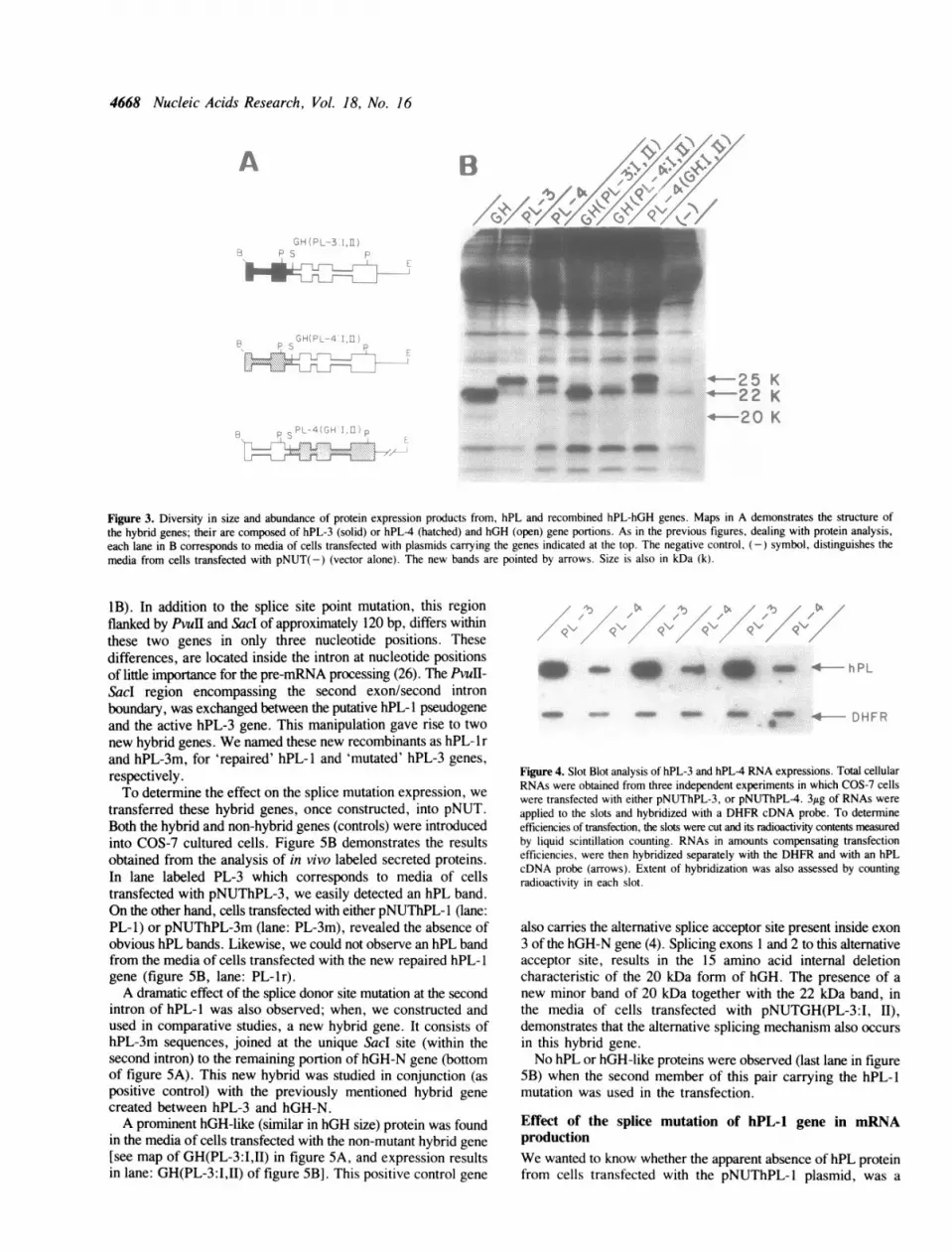
Differences in the expression of hPL proteinsAs noticed above (figure 2), while the hGH-N gene gives riseto a prominent band of approximately 22 kDa, hPL-3 and hPL4genes express proteins of slightly greater size (-25 kDa).Furthermore, and consistently throughout several independentexperiments, the hPL-4 band always appeared weaker than thehPL-3 band. We were interested in investigating the cause ofsuch heterogeneity in the expression levels of these genes.To approach this problem, we chose to study the cell culture
production of extracellular hPL-hGH chimeric proteins resultingfrom the transient expression of a new hybrid gene pair. Thesehybrids possess the first two exons from hPL-3 or hPL4 genes,and sequences of hGH-N gene that conform the remaining partof their structure. We named these hybrids GH(PL-3:I,ll) andGH(PL-4:I,l1) respectively (see map in figure 3A). By comparingboth hybrid genes, differences were observed in the expressionlevels of GH(PL-3:I,II) versus GH(PL-4:I,ll) chimeric proteins[figure 3B; compare lanes labelled GH(PL-3:I,II) and GH(PL-4:I,II)]. Same results, we might add, as with the proteins derivedfrom normal non-hybrid hPL-3 and hPL-4 genes (see lanes
Figure 2. In vitro production of secreted proteins by hGH-N and hPL genes.Media from COS-7 cells transfected with each of the plasmids and incubated inthe presence of 35S-methionine, was analyzed by discontinuous SDS-polyacrylamide gel (5% - 13%) electrophoresis and autoradiography. The genepresent in each plasmid used for transfection is indicated at the top. The (-)symbol identifies the media from cells transfected with the vector alone[pNUT(-)]. Sizes of characteristic hPL and hGH bands are indicated in kDa(K) at the left.
labeled hPL-3 and hPL-4 in figure 2 and labeled PL-3 and PL-4in figure 3B).
Consistently, we obtained less protein when sequences fromthe first two exons of hPL-4 gene were present. On the contrary,we detected more hGH-like protein when the first two exons werefrom hPL-3. Moreover, when the last three exons were fromhPL-4 gene, and the first two exons were from hGH-N (see mapof this chimeric at bottom of figure 3A), the band intensityresembled that of hGH-N protein [figure 3B, lane PL4(GH:I,Il)].Therefore, relative abundance of hPL protein products is afunction of the first two exons.To further investigate the different levels of in vitro expression
observed for these active hPL genes, we carried out estimationsof the relative abundance of their RNA transcripts. Using slotblot analysis, we found approximately -8-fold more RNAhybridizable to our probe from the total RNA isolated of cellstransfected with the hPL-3 structural gene sequences, ascompared to hPL-4 (figure 4). This same result was observedeven when only the two first exons of hPL-3 were contributingto a hybrid gene (D.R.-P. and H.A.B.-S., submitted. Therefore,the higher observed hPL-3 protein expression, seems to be aconsequence of having more RNA derived from the hPL-3 gene.
Dissecting the putative hPL-1 pseudogeneNext we, decided to test if the donor splice site point mutationat the second intron of the hPL-1 gene, was the only cause ofthe apparent inactivity of this gene. Comparing nucleotidesequences at the second exon/second intron border area, amongthe active hPL-3 gene and the putative hPL-l pseudogene,revealed that we could easily exchange this region between thesetwo genes. We found PvuIl and Sacd sites located 30 bp upstreamand 86 bp downstream respectively, from the mutation site (figure
- w_ U. .S.an sS_
:: -. ': tr -::: :^:::--¢ ss_# MYs.C
4668 Nucleic Acids Research, Vol. 18, No. 16
ac.,*-... ~..
4_M....u
~~
~~ ~ ~ ~ ~ ~......~ 4 w ,* d
Figure 3. Diversity in size and abundance of protein expression products from, hPL and recombined hPL-hGH genes. Maps in A demonstrates the structure ofthe hybrid genes; their are composed of hPL-3 (solid) or hPL-4 (hatched) and hGH (open) gene portions. As in the previous figures, dealing with protein analysis,each lane in B corresponds to media of cells transfected with plasmids carrying the genes indicated at the top. The negative control, (-) symbol, distinguishes themedia from cells transfected with pNUT(-) (vector alone). The new bands are pointed by arrows. Size is also in kDa (k).
iB). In addition to the splice site point mutation, this regionflanked by Pvul and Sacd of approximately 120 bp, differs withinthese two genes in only three nucleotide positions. Thesedifferences, are located inside the intron at nucleotide positionsof little importance for the pre-mRNA processing (26). The PvuH-Sacd region encompassing the second exon/second intronboundary, was exchanged between the putative hPL-1 pseudogeneand the active hPL-3 gene. This manipulation gave rise to twonew hybrid genes. We named these new recombinants as hPL- 1 r
and hPL-3m, for 'repaired' hPL- 1 and 'mutated' hPL-3 genes,respectively.To determine the effect on the splice mutation expression, we
transferred these hybrid genes, once constructed, into pNUT.Both the hybrid and non-hybrid genes (controls) were introducedinto COS-7 cultured cells. Figure 5B demonstrates the resultsobtained from the analysis of in vivo labeled secreted proteins.In lane labeled PL-3 which corresponds to media of cellstransfected with pNUThPL-3, we easily detected an hPL band.On the other hand, cells transfected with either pNUThPL-l (lane:
PL-1) or pNUThPL-3m (lane: PL-3m), revealed the absence ofobvious hPL bands. Likewise, we could not observe an hPL bandfrom the media of cells transfected with the new repaired hPL- 1
gene (figure SB, lane: PL- lr).A dramatic effect of the splice donor site mutation at the second
intron of hPL-l was also observed; when, we constructed andused in comparative studies, a new hybrid gene. It consists ofhPL-3m sequences, joined at the unique Sacd site (within thesecond intron) to the remaining portion of hGH-N gene (bottomof figure SA). This new hybrid was studied in conjunction (aspositive control) with the previously mentioned hybrid gene
created between hPL-3 and hGH-N.A prominent hGH-like (similar in hGH size) protein was found
in the media of cells transfected with the non-mutant hybrid gene
[see map of GH(PL-3:I,II) in figure SA, and expression resultsin lane: GH(PL-3:I,II) of figure SB]. This positive control gene
4.4mw 0 on m am_ *
doww -am W
Figure 4. Slot Blot analysis of hPL-3 and hPL-4 RNA expressions. Total cellularRNAs were obtained from three independent experiments in which COS-7 cellswere transfected with either pNUThPL-3, or pNUThPL-4. 3,1g of RNAs were
applied to the slots and hybridized with a DHFR cDNA probe. To determineefficiencies of transfection, the slots were cut and its radioactivity contents measuredby liquid scintillation counting. RNAs in amounts compensating transfectionefficiencies, were then hybridized separately with the DHFR and with an hPLcDNA probe (arrows). Extent of hybridization was also assessed by countingradioactivity in each slot.
also carries the alternative splice acceptor site present inside exon3 of the hGH-N gene (4). Splicing exons 1 and 2 to this alternativeacceptor site, results in the 15 amino acid internal deletioncharacteristic of the 20 kDa form of hGH. The presence of a
new minor band of 20 kDa together with the 22 kDa band, inthe media of cells transfected with pNUTGH(PL-3:1, II),demonstrates that the alternative splicing mechanism also occurs
in this hybrid gene.
No hPL or hGH-like proteins were observed (last lane in figureSB) when the second member of this pair carrying the hPL-1mutation was used in the transfection.
Effect of the splice mutation of hPL-1 gene in mRNAproductionWe wanted to know whether the apparent absence of hPL proteinfrom cells transfected with the pNUThPL-1 plasmid, was a
A
o, .... ... . pi,,
,4.fIe
I.. i
I
.-.t
"A[E 6b

Nucleic Acids Research, Vol. 18, No. 16 4669
A
PL-3m6 P S
E
GH( PL-3: 1,U )
| _ E~~~~~~~~ :--q _~-- -*_6_ _ _ _ _ 4
_ ..l:-:..
..:,:5.A |j.r.; _
GH(PL-3m:1 1I)B PS p= J~~~~~~~~~ - 25 K
.- 22K
<- 20 K
Figure 5. Effect of the donor splice site mutation of hPL-l gene on the production of secreted proteins. Cells transfected with the different plasmid carrying thehybrid genes indicated at the top, were analyzed for the presence of secreted labeled proteins in their media. Hybrid genes were constructed as described in theMaterial and Methods and Results sections, and for purpose of clarification, maps of key hybrid genes are shown in A. Characteristic non-chimeric and chimericproteins were visualized as described in Material and Methods section, gene portions in black, hPL-3 gene; stippled, hPL-1; and open, hGH. Only important recognitionsites for restriction enzymes are indicated, B=BamHl, P=Pvull, S=Sacl and E. EcoRI. Arrows in B indicate the size in kDa (k) of the new proteins originatedfrom plasmids transfected. (-)=media from the negative control cells.
consequence of having no RNA derived from the PL-1 gene.We performed a Northern blot analysis (23) of the total RNAextracted from transfected cells. We included as a positivecontrol, RNA isolated from human placenta and pituitary gland.In addition, as another positive control, we included RNA fromcells tranfected with the plasmid carrying the hPL-3 structuralsequences. The results of this analysis are presented in figure6. Cells transfected with the plasmid carrying the hPL-1 genelacked hPL specific RNAs (figure 6, lane: PL-1). We observeda dramatic reduction in the hybridizable RNA content from cellstransfected with the plasmid carrying the hPL-3 mutated gene(figure 6, lane: PL-3m); as compared with, the wild type hPL-3gene acting as control (lane: PL-3). Finally, when usingpNUThPL-lr, we unexpectantly observed only a faintreappearance of hPL mRNA (figure 6, lane: PL-lr).
DISCUSSION
DNA cloning and sequence studies have lead to the isolation ofcDNA clones for all hGH and hPL genes except hPL-1.However, experiments directed to demonstrate that each of themRNAs corresponding to the identified cDNAs indeed end upas proteins, have been few for hGH (7, 27) and none for hPLgenes. The reintroduction of cloned genes into cultured cells,by DNA transfection (18), is a valuable method to identify anddissect sequences required for gene function and their mutations.We chose this approach to determine the coding potential of allhPL genes. To achieve our objective, we forced the in vitroexpression of all the hPL promoter-less genes, by joining themto the strong heterologous transcriptional control sequencespresent in pNUT (12).
Figure 6. RNA expression effect of the donor splice site mutation of hPL-l gene.The figure demonstrates the results of the Northern blot analysis (23) practicedto total RNA (10 ,Lg), isolated from cultured cells transfected with the indicated(at top) plasmids. H and P represent lanes containing total RNA from humanpituitary gland (2 ug) and placenta (3.8 jig), respectively. The (-) symbolcorrespond to RNA (10 jig) isolated from cells transfected with the pNUT (vectoralone). Lane labeled C represents total RNA of mock-transfected cells.
The new results of the present study demonstrate for the firsttime that the hPL-4 and hPL-3 genes, but not hPL-1, contributeto the production of mature hPL. Here we also demonstrate thatin spite of being highly similar, the structural sequences of thesegenes respond differently to the same heterologous promoter.Each of these two genes give rise to one protein. Although thesecreted proteins expressed by these genes have identical aminoacid sequence, they differ at their expression level. As aconsequence of this finding, we designed exon exchangeexperiments to gain new insights in the understanding of thisphenomenon. The same result was seen when only the two firstexons of the genes were contributing to a hybrid gene. Theobserved higher hPL-3 protein expression seems in part to bea consequence of having more RNA expression from the hPL-3gene sequences. This in vitro findings do not resemble whatoccurs in vivo, while the hPL-4 mRNA (HCS-A) accounts for

4670 Nucleic Acids Research, Vol. 18, No. 16
3% of the placenta mRNA, the hPL-3 (HCS-B) mRNA has beenestimated to represent only 0.5% of it (28).
Finally, a third contribution of our study is the experimentaldemonstration, also for the first time, of the effect of the donorsplice mutation at the second intron of hPL-l on its expression.In spite of having replaced the donor splice site mutation at thesecond intron of the hPL-1 pseudogene by normal sequences,we could not observe an hPL-1 protein secreted into the cellmedium. Thus, there must exist additional mutations thatcontribute to this lack of genetic expression. There is no doubtof the severity of this splicing mutation. Indeed we prove herethat by introducing the mutated area of hPL- 1, into either thehPL-3 or the hGH-N gene, protein production from these hybridgenes is severely reduced. Further evidence for the severe effectof this type of mutation comes from both, site-directedmutagenesis studies (26) and by naturally occurring mutants ofj-globin genes. In both cases, mutated genes having the samechange of a G for an A at the beginning of an exon, give riseto a messenger RNA precursor unable to splice correctly.We recently found that we can quantify hPL-hGH chimeric
proteins using an hGH radioimmunoassay. Using this technique,non-detectable hGH radioimmunoassay values in the media fromcells transfected with pNUTGH(PL-3m:I,II) were observed. Themedia of cells corresponding to the control experiment, whereGH(PL-3:I,ll) gene was used, gave hGH RIA values of abouthalf of those of pNUT. Five-fold lower RIA values were foundfor the expression of GH(PL-4:I,II) as compared to GH(PL-3:I,II)(D.R-P., and H.A.B-S, submitted).We have no explanation for the difference in size observed
in the electrophoretic analysis of hGH and hPL proteins. Thedifference was seen with both purified hormones from thepituitary gland and placenta, and with the proteins produced inthe gene transfection experiments. Neither hPL-3 nor hPL-4proteins have the N-linked glycosylation site at Asn-140,predicted for the hGH-V protein and which might otherwiseaccount for this size difference. It is possible that the size changeobserved, may simply be accounted for by differences in protein-SDS interactions.
In conclusion, our analysis of the in vitro expression productsof hPL genes demonstrates that hPL-3 and hPL-4 have thepotential for contributing to mature hPL. It also provides evidenceindicating that the hPL-1 gene has accumulated severemutation(s), other than the donor splice site deffect at its secondintron. Furthermore, through our study, we have identified thegene region between the capping site and the second intron asthe origin of differences in expression levels seen here for hPL-3and hPL-4. The results obtained with the hPL-hGH genes hybridscorroborate and strengthen our findings.
ACKNOWLEDGEMENTSWe thank A. Martinez for assistance with some of theseexperiments, J. Silva for proofreading and critical reading andD. Baty for reviewing the manuscript. We also thank Dr. T. Kuofor providing the COS-7 cells and donation of materials, Dr. G.Saunders for the generous gift of plasmids and for reviewing themanuscript and Dr. E. Olson the donation of 35S-Methionine.We are grateful to Dr. R. Palmiter for providing the pNUTplasmid and also for his interest and helpful suggestions. R.R.S.,D.R.P. and A.V.E. were recipients of scholarships from theConsejo Nacional de Ciencia y Tecnologfa (CONACYT) of theMexican government. This work was supported by grants from
CONACYT, the Subsecretaria de Educacion Superior eInvestigacion Cientifica de la Secretaria de Educacion Ptblicaof the Mexican government, Centro Internacional de BiologiaMolecular y Celular, A.C., and from General Foods and IBMof Mexico. We are indebted to G. Garcia-L., G. Infante-Garcfaand S. Silva-Herrera from the Servicio de Endocrinologia delHospital Universitario 'Dr. Jose E. Gonzalez' for their aid withthe radioimmunoassays, and to M. Paiez for preparation of themanuscript.
REFERENCES1. Barrera-Saldaina, H.A. (1982) Expression of the human placental lactogen
genes. Ph.D. Thesis, The University of Texas Health Science Center atHouston.
2. Barsh, G.S., Seeburg, P.H. and Gelinas, R.E. (1983). Nucleic Acids Res.11,3939 - 3958.
3. Harper, M.E., Barrera-Saldana, H.A. and Saunders, G.F. (1982). Am. J.Hum. Genet. 34,227-234.
4. DeNoto, F.M., Moore, D.D. and Goodman, H.M. (1981). Nucleic AcidsRes. 9,3719 - 3730.
5. Masuda, N., Watahiki, M., Tanaka, M., Yamakawa, M., Shimizu, K.,Nagai, J. and Nakashima, K. (1988). Biochem. Biophys. Acta 949,125- 131.
6. Frankenne, F., Rentier-Delrue F., Scippo, M.L., Martial, J. and Hennen,G. (1987). J. Clin. Endocrinol. Metab. 64,635-637.
7. Cooke, N.E., Ray, J., Watson, M.A., Estes, P.A., Kuo, B.A. and Liebhaber,S.A. (1988). J. Clin. Invest. 82,270-275.
8. Cooke, N.E., Ray, J., Emery, J.G. and Liebhaber, A. (1988). J. Biol. Chem.263,9001 -9006.
9. Barrera-Saldafia, H.A., Seeburg, P.H. and Saunders, G.F. (1983). J. Biol.Chem. 258:3787 -3793.
10. Hirt, H., Kimelman, J., Birnbaum, M.J., Chen. E.Y. Seeburg, P.H.,Eberhardt, N.L. and Barta, A. (1987). DNA 6,59-70.
11. Kidd, V.J. and Saunders, G.F. (1982). J. Biol. Chem. 257,10673-10680.12. Palmiter, R.D., Behringer, R.R., Quaife, C.J., Maxwell, F., Maxwell, I.H.
and Brinster, R.L. (1987). Cell 50,435-443.13. Mulligan, R.C. and Berg, P. (1980). Science 209,1422-1427.14. Vogelstein, B. and Gillespie, D. (1979). Proc. Natl. Acad. Sci. USA
76,615 -619.15. Maniatis, T., Fristsh, E.F. and Sambrook, J. (1982) Molecular cloning: A
laboratory manual. Cold Spring Harbor Laboratory, Cold Spring Harbor,N.Y.
16. Southern, E.M. (1975). J. Mol. Biol. 98,503-517.17. Sanger, F., Nicklen, S. and Coulson, A.R. (1977). Proc. Natl. Acad. Sci.
USA 74,5463-5467.18. Graham, F.L. and Van Der Eb, A.J. (1973). Virology 52,456-467.19. Foecking, M.K. and Hofstetter, H. (1986). Gene 45,101-105.20. Chomczynski, P. and Sacchi, N. (1987). Anal. Biochem. 162, 156-159.21. Laemmli, U.K. (1970). Nature 227,680-685.22. McMaster, G.K. and Carmichael, G.G. (1977). Proc. Natl. Acad. Sci. USA
74,4835 -4838.23. Thomas, P.S. (1980). Proc. Natl. Acad. Sci. USA 77,5201 -5205.24. Barrera-Saldaiia, H.A., Robberson, D.L. and Saunders, G.F. (1982). J.
Biol. Chem. 257,12399-12404.25. Feinberg, A.P. and Vogeltein B. (1983). Anal. Biochem. 132, 6-13.26. Wieringa, B., Meyer, F., Reiser, J. and Weissmann, C. (1983). Nature
301,38 -43.27. Pavlakis, G.N., Hizuka, N., Gorden, P., Seeburg, P.H. and Hamer, D.H.
(1981). Proc. Natl. Acad. Sci. USA 78,7398-7402.28. Chen, E.Y., Liao, Y-C., Smith, D.H., Barrera-Saldana, H.A., Gelinas,
R.E. and Seeburg, P.H. (1989). Genomics 4,479-497.
ABBREVIATIONS
bGH, bovine growth hormone; bp, base pairs; CAT,chloramphenicol acetyl transferase; FCS, fetal calf serum; hCS,human chorionic somatomammotropin; hGH, human growthhormone; hPL, human placental lactogen; hPrl, human prolactin;kbp, 1000 base pairs; kDa, 1000 daltons; Pre-hPL, hPLprecursor; SV40, simian virus 40; SDS, sodium dodecyl sulphate.