cmbbe2018 - cmbbe symposium
TRANSCRIPT

CMBBE201815th International Symposium on Computer Methods
in Biomechanics and Biomedical Engineering
and
3rd Conference on Imaging and Visualization
26-29 March, 2018
Instituto Superior Técnico Lisbon • Portugal
Book of AbstractsPROGRAMME INCLUDED
Edited byPaulo R. Fernandes, João Manuel R.S. Tavares,
João Folgado, Carlos Quental, Rui Ruben
15th International Symposium on Computer Methods in Biomechanics and Biomedical Engineering and 3rd Conference on Imaging and Visualization
2
TitleBook of Abstracts15th International Symposium on Computer Methodsin Biomechanics and Biomedical Engineeringand 3rd Conference on Imaging and Visualization
Edited byPaulo R. Fernandes, João Manuel R.S. Tavares,João Folgado, Carlos Quental, Rui Ruben
ISBN978-989-99424-5-5
First edition, March 2018
Copyright © 2018IDMEC - Instituto de Engenharia MecânicaInstituto Superior Técnico, Avenida Rovisco Pais 1, 1049-001 LISBOA
Graphic DesignLuís [email protected]
26-29 March, 2018 • Instituto Superior Técnico • Lisbon • Portugal
3
Welcome MessageOn behalf of the Organizing Committee, we are honored to welcome you to the 15th International Symposium on Computer Methods in Biomechanics and Biomedical Engineering and the 3rd Conference on Imaging and Visualization (CMBBE2018), hosted at Instituto Superior Técnico (IST), Technical University of Lisbon, Portugal, from 26th to 29th of March 2018.
In this edition, the two events will run together as a single conference, highlighting the strong connection with the Taylor & Francis journals: Computer Methods in Biomechanics and Biomedical Engineering (John Middleton and Christopher Jacobs, Eds.) and Computer Methods in Biomechanics and Biomedical Engineering: Imaging and Visualization (João Manuel R.S. Tavares, Ed.).
The conference has become a major international meeting on computational biomechanics, imaging and visualization. In this edition, the main program includes 212 presentations. In addition, sixteen renowned researchers will give plenary keynotes, addressing current challenges in computational biomechanics and biomedical imaging. In Lisbon, for the first time, a session dedicated to award the winner of the Best Paper in CMBBE Journal will take place.
We believe that CMBBE2018 will have a strong impact on the development of computational biomechanics and biomedical imaging and visualization, identifying emerging areas of research and promoting the collaboration and networking between participants. This impact is evidenced through the well-known research groups, commercial companies and scientific organizations, who continue to support and sponsor the CMBBE meeting series. In fact, the conference is enriched with five workshops on specific scientific topics and commercial software.
Besides the scientific program, the conference social program was defined to provide the participants with a pleasant stay in Lisbon, the capital of Portugal. Lisbon is a historic city facing the Atlantic Ocean that has been a point of cultural interchange and encounter for many centuries for visitors coming from all over the world. Lisbon is recognized as one of the most beautiful places to visit, and it is a safe and pleasant city where delegates and their companions will feel at ease and will be very well received.
To conclude, we wish you a very productive and pleasant conference as well as an enjoyable stay in Portugal,
Paulo Fernandes and João Tavares(Conference Chairs)

26-29 March, 2018 • Instituto Superior Técnico • Lisbon • Portugal
5
Contents
Conference OrganizationExecutive CommitteeTechnical Advisory Board
Conference InformationEndorsed byConference VenueSecretariat Open HoursCoffee BreaksLunchesCongress Center Floor PlansWireless Internet AccessInstructions for Presenters
Social ProgramWelcome ReceptionConference Banquet
General Tourist Information
Map of Lisbon
Scientific ProgrammeProgramme at a glanceDaily Sessions
Book of Abstracts
6 >
7 >
10 >
11 >
13 >
15 >
39 >
CMBBE201815th International Symposium on Computer Methods
in Biomechanics and Biomedical Engineeringand
3rd Conference on Imaging and Visualization

15th International Symposium on Computer Methods in Biomechanics and Biomedical Engineering and 3rd Conference on Imaging and Visualization
6
Executive Committee
Conference Organization
Technical Advisory Board
John Middleton (UK) Christopher Jacobs (USA)
Paulo R. Fernandes (Portugal) Conference ChairJoão Manuel Tavares (Portugal) Conference ChairJoão Folgado (Portugal)Carlos Quental (Portugal)Rui Ruben (Portugal)
A. Gefen Israel (Israel)Alejandro Frangi (UK)Alexandre Cunha (USA)Alexandre X. Falcão (Brazil)Andrew Hopkins (Switzerland)António Veloso (Portugal)C. P. Bourauel (Germany)Cees Oomens (The Netherlands)Cristian A. Linte (USA)Daniela Iacoviello (Italy)Demetri Terzopoulos (USA)Dinggang Shen (USA)Dominique P. Pioletti (Switzerland)Eduardo Soudah (Spain)Estevam de las Casas (Brazil)Fiorella Sgallari (Italia)George Bebis (USA)Han van Oosterwyck (Belgium)Hélder Rodrigues (Portugal)J. Paulo Vilas-Boas (Portugal)Jessica Zhang (USA)João Paulo Papa (Brazil)Jorge Ambrósio (Portugal)Jos Vander Sloten (Belgium)Jun Zhao (China)Khan M Iftekharuddin (USA)
Laurent Cohen (France)Leo Joskowicz (Israel)Manuel González Hidalgo (Spain)Marc Thiriet (France)Mário Forjaz Secca (Portugal)Martyn Nash (New Zealand)Michael S. Sacks (USA)Miguel A. González Ballester (Spain)N. Shrive (Canada)P. Verdonck (Belgium)Paola Lecca (Italy)Paolo Di Giamberardino (Italy)Paulo Flores (Portugal)R.N. Jorge (Portugal)Reneta Barneva (USA)S. Ferguson (Switerzland)S. Shirazi-Adl (Canada)S. Evans (UK)Sidney Fels (Canada)T. Adachi (Japan)Thomas Franz (South Africa)Valentin Brimkov (USA)W. Skalli (France)Xiongbiao Luo (Japan)Yuri Bazilevs (USA)Zeyun Yu (USA)
26-29 March, 2018 • Instituto Superior Técnico • Lisbon • Portugal
7
Conference Information
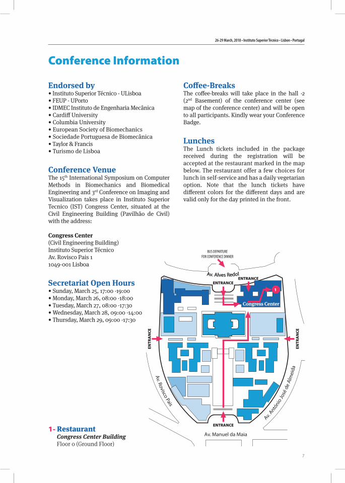
1- RestaurantCongress Center BuildingFloor 0 (Ground Floor)
Endorsed by• Instituto Superior Técnico - ULisboa• FEUP - UPorto• IDMEC Instituto de Engenharia Mecânica• Cardiff University• Columbia University• European Society of Biomechanics• Sociedade Portuguesa de Biomecânica• Taylor & Francis• Turismo de Lisboa
Conference VenueThe 15th International Symposium on Computer Methods in Biomechanics and Biomedical Engineering and 3rd Conference on Imaging and Visualization takes place in Instituto Superior Tecnico (IST) Congress Center, situated at the Civil Engineering Building (Pavilhão de Civil) with the address:
Congress Center(Civil Engineering Building)Instituto Superior TécnicoAv. Rovisco Pais 11049-001 Lisboa
Secretariat Open Hours• Sunday, March 25, 17:00 -19:00• Monday, March 26, 08:00 -18:00• Tuesday, March 27, 08:00 -17:30• Wednesday, March 28, 09:00 -14:00• Thursday, March 29, 09:00 -17:30
Coffee-BreaksThe coffee-breaks will take place in the hall -2 (2nd Basement) of the conference center (see map of the conference center) and will be open to all participants. Kindly wear your Conference Badge.
LunchesThe Lunch tickets included in the package received during the registration will be accepted at the restaurant marked in the map below. The restaurant offer a few choices for lunch in self-service and has a daily vegetarian option. Note that the lunch tickets have different colors for the different days and are valid only for the day printed in the front.
BUS DEPARTUREFOR CONFERENCE DINNER
Av. Rovisco Pais
Av. Antó
nio
José
de
Alm
eida
ENTRANCE
ENTR
AN
CE
ENTR
AN
CE
Av. Manuel da Maia
ENTRANCE
ENTRANCEAv. Alves RedolAv. Alves Redol
1
Congress Center
15th International Symposium on Computer Methods in Biomechanics and Biomedical Engineering and 3rd Conference on Imaging and Visualization
8
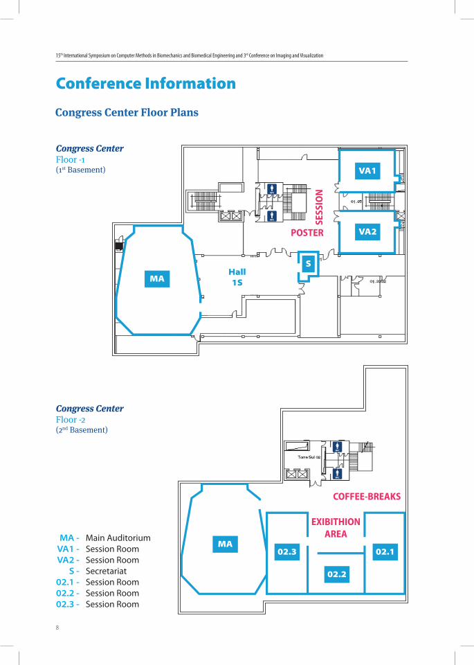
MA -VA1 -VA2 -
S -02.1 -02.2 -02.3 -
Main AuditoriumSession RoomSession RoomSecretariatSession RoomSession RoomSession Room
Conference Information
Congress CenterFloor -1(1st Basement)
Congress CenterFloor -2(2nd Basement)
Congress Center Floor Plans
S
MAHall 1S
POSTER
02.102.3
02.2
MA
SESS
ION
COFFEE-BREAKS
EXIBITHIONAREA
VA2
VA1
26-29 March, 2018 • Instituto Superior Técnico • Lisbon • Portugal
9
Conference Information
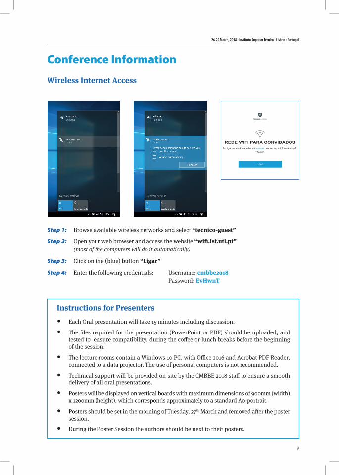
Wireless Internet Access
Instructions for Presenters• Each Oral presentation will take 15 minutes including discussion.
• The files required for the presentation (PowerPoint or PDF) should be uploaded, and tested to ensure compatibility, during the coffee or lunch breaks before the beginning of the session.
• The lecture rooms contain a Windows 10 PC, with Office 2016 and Acrobat PDF Reader, connected to a data projector. The use of personal computers is not recommended.
• Technical support will be provided on-site by the CMBBE 2018 staff to ensure a smooth delivery of all oral presentations.
• Posters will be displayed on vertical boards with maximum dimensions of 900mm (width) x 1200mm (height), which corresponds approximately to a standard A0-portrait.
• Posters should be set in the morning of Tuesday, 27th March and removed after the poster session.
• During the Poster Session the authors should be next to their posters.
Step 1:
Step 2:
Step 3:
Step 4:
Browse available wireless networks and select “tecnico-guest”
Open your web browser and access the website “wifi.ist.utl.pt” (most of the computers will do it automatically)
Click on the (blue) button “Ligar”
Enter the following credentials: Username: cmbbe2018 Password: EvHwnT

15th International Symposium on Computer Methods in Biomechanics and Biomedical Engineering and 3rd Conference on Imaging and Visualization
10
Social Programme
Tour visit to Cascais/Sintra - Wednesday, 28th March - 14:30Buses will depart from IST (Rua Alves Redol) at 14:30. Please be there 10 minutes before the departure time and don’t forget to bring your Tour/ Dinner Voucher.We will depart to Cascais a cosmopolitan former fish-ing village, that in the 1940’s was chosen as residence by exiled European Royalty, a stop will be made by the lovely bay filled with fishing boats.
The tour will continue by coast line of Guincho, with its superb beach areas, cliffs and where, close by, is located Cape Roca, the westernmost point of Conti-nental Europe. It continues to Sintra, a small delight-ful town in the forest covered Mountain of Sintra, im-mortalized as “Glorious Eden” by the World famous poet Lord Byron.
Welcome Reception - Monday, 26th March - 18:30Welcome reception will take place at conference site.
The dinner will take place at Penha Longa Monastery. (Quinta da Penha Longa, Estrada da Lagoa Azul, Sintra)
The Monastery was founded in 1355 by Friar Vasco Martins that introduced the St. Jeronimos Order in Portugal.In 1400 a church was built consecrated to Our Lady
of Health. During the 15th and 16th centuries, Penha Longa was chosen by the Royal Family as a place for hunting, during the summer.Memories of strong presences, such as King D. Manuel (1496) and King D. Sebastião (1580). During this period, a Palace to host the Kings and their guests, fountains and gardens, oratories and watermills were built.
Conference Dinner
26-29 March, 2018 • Instituto Superior Técnico • Lisbon • Portugal
11
General Tourist Information
Getting to Lisbon by airDirect flights from most of European cities, North or South America and Africa land at the Portela Airport, terminal 1. A taxi ride from the airport to IST is about 4-5 km that takes 10-15 min, depending on traffic, and should cost around 8€. To downtown the taxi ride is about 7 km and should cost around 10€. 1.60€ is charged for the transportation of luggage or animals. A sure option is the “Taxi Voucher” a prepaid taxi service starting at 16.40€, on sale at the “Information Desk” in the arrival terminal. Lisbon Airport has its own Metro Station, Aeroporto - red line (see map of Lisbon with subway lines). Other options are the AeroBus and the Aeroshutle (3.5€).
Getting to Lisbon by carDrivers can use highway A1 when coming from the North, highway A2, through the 25 de Abril bridge, when coming from the South, and highway A12, through Vasco da Gama bridge, when coming from the Northeast.
Getting to Lisbon by trainThe St. Apolónia station is the terminal for trains arriving from the North of Portugal. Another option is to use the train station Oriente. From the South of Portugal an option is to use the train station Oriente. Connections to the metro lines exist at both stations (St. Apolónia - blue line, Oriente - red line).
Moving aroundTaxi:Lisbon is served by an extensive network of public transportation that can take you anywhere in the city and to its surroundings. Taxis (black and green or beige) are cheap when comparing to most of the European countries. They can be called by phone, picked-up on taxi plazas or stopped on the street. The fare on the taxi meter should start at 3.25€ (daytime pick-up) or 3.90€ (nighttime). Outside the city limits, city fares are charged per kilometer. 1.60€ is charged
for the transportation of luggage or animals. Before taking a taxi, inquire about the fare.
Metro:The Lisbon Metro is a very comfortable and easy way to reach most of the city, from 6:30 to 1:00. The Metro lines reach most of the city being the Metro stations close to IST:• Alameda (red and green line)• Saldanha (red and yellow line)
BusThe bus routes cover all Lisbon and extend to its outskirts. The tickets can be pre-paid, at the counters of Carris, the surface transportation operator for Lisbon, or bought aboard the bus, electric cars or funiculars.For IST hop off on one of the following bus stops:Av. Manuel da MaiaAv. Rovisco PaisArco do Cego
Metro and Bus Fares:Reusable card – 0.50 €METRO/CARRIS – 1.45 €CARRIS Bus – 1.80 € (on board fare) Tram – 2.85 € (on board fare)
TrainsSuburban trains to Estoril and Cascais depart from the Cais do Sodré train station, to the south of the river cities from Roma-Areeiro (Entrecampos) while to Sintra the trains depart from Rossio train station or Oriente (Entrecampos). The ride to Cascais or to Sintra should take about 35-45 min, each way. The train ride to south of the river is a highlight as the train will cross the 25 de Abril bridge with magnificent views of Lisbon. For IST the nearby train stations are: Roma-AreeiroEntrecampos
15th International Symposium on Computer Methods in Biomechanics and Biomedical Engineering and 3rd Conference on Imaging and Visualization
12
Other general information• National emergency number: 112
• Time zone: GMT +1 summer time
• Electricity: 220V, 50 Hz with standard European power sockets
• Temperature: Average high 19ºC, Average low 13ºC
• Currency: Euro (€)
• Banks: working hours are 8:30 – 15:00 (Monday-Friday)
• Pharmacies: 9:00 – 19:00
• Shops: 9:00 – 19:00
• Shopping Malls: 10:00 – 23:00
Main Museums in Lisbon:• CentrodeArteModerna
(Modern Art Museum)
• MuseudoOriente (Oriente Museum)
• MuseuCalousteGulbenkian (Calouste Gulbenkian Museum)
• MuseudosCoches (Coach Museum)
• MuseuNacionaldeArteAntiga (National Museum for Ancient Art)
• ColecçãoBerardo (The Berardo Collection)
• MuseudoAzulejo (Tile Museum)
Main Monuments in Lisbon:• AquedutodasÁguasLivres
(Águas Livres’ Aqueduct)
• BasílicadaEstrela (Estrela Basilica)
• CastelodeSãoJorge (Saint George’s Castle)
• SéPatriarcal (Patriarchal Church)
• MosteirodosJerónimos (Jerónimos Monastery)
• PadrãodosDescobrimentos (Monument to the Overseas Discoveries)
• TorredeBelém (Belém Tower)
26-29 March, 2018 • Instituto Superior Técnico • Lisbon • Portugal
13
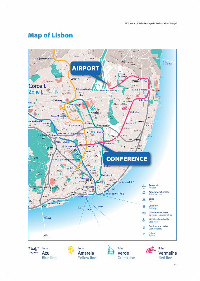
Map of Lisbon
AIRPORT
CONFERENCE


ScientificProgramme
26-29 March, 2018
Instituto Superior Técnico Lisbon • Portugal
CMBBE201815th International Symposium on Computer Methods
in Biomechanics and Biomedical Engineering
and
3rd Conference on Imaging and Visualization

26-29 March, 2018 • Instituto Superior Técnico • Lisbon • Portugal
17
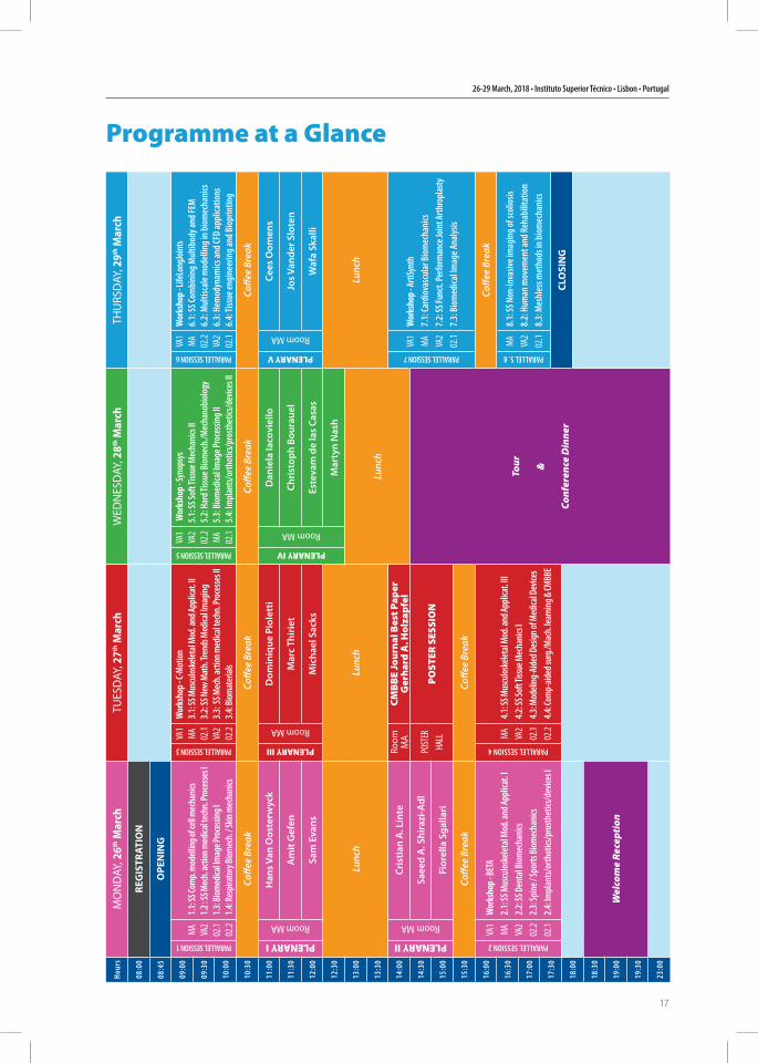
Programme at a GlanceHo
urs
MO
ND
AY, 2
6th M
arch
TUES
DAY
, 27th
Mar
chW
EDN
ESD
AY, 2
8th M
arch
THU
RSD
AY, 2
9th M
arch
08:00
REG
ISTR
ATIO
N
08:45
OPE
NIN
G
09:00
PARALLEL SESSION 1
MA VA2
02.1
02.2
1.1: S
S Com
p. m
odell
ing of
cell m
echa
nics
1.2 : S
S Mec
h. ac
tion m
edica
l tech
n. Pr
oces
ses I
1.3: B
iomed
ical Im
age P
roce
ssing
I1.4
: Res
pirat
ory B
iomec
h. / S
kin m
echa
nics
PARALLEL SESSION 3
VA1
MA 02.1
VA2
02.2
Wor
ksho
p - C-
Mot
ion
3.1: S
S Mus
culos
kelet
al M
od. a
nd Ap
plica
t. II
3.2: S
S New
Mat
h. Tr
ends
Med
ical Im
agin
g3.3
: SS M
ech.
actio
n med
ical te
chn.
Proc
esse
s II
3.4: B
iomat
erial
s
PARALLEL SESSION 5
VA1
VA2
02.2 MA 02.1
Wor
ksho
p - Sy
nops
ys
5.1:
SS So
ft Tis
sue M
echa
nics
II5.
2: H
ard T
issue
Biom
ech.
/Mec
hano
biol
ogy
5.3:
Biom
edica
l Imag
e Pro
cessi
ng II
5.4:
Impl
ants/
orth
otics
/pro
sthet
ics/d
evice
s II
PARALLEL SESSION 6
VA1
MA 02.2
VA2
02.1
Wor
ksho
p - Li
feLo
ngJo
ints
6.1:
SS Co
mbi
ning
Mul
tibod
y and
FEM
6.2:
Mul
tisca
le m
odel
ling i
n bio
mec
hani
cs
6.3:
Hem
odyn
amics
and C
FD ap
plica
tions
6.4:
Tiss
ue en
gine
erin
g and
Bio
prin
ting
09:30
10:00
10:30
Coffe
e Br
eak
Coffe
e Br
eak
Coffe
e Br
eak
Coffe
e Br
eak
11:00
PLENARY I
Room MA
Han
s Van
Oos
terw
yck
PLENARY III
Room MA
Dom
iniq
ue P
iole
tti
PLENARY IV
Room MA
Dan
iela
Iaco
viel
lo
PLENARY V
Room MA
Cees
Oom
ens
11:30
Am
it G
efen
Mar
c Th
irie
tCh
rist
oph
Bour
auel
Jos V
ande
r Slo
ten
12:00
Sam
Eva
nsM
icha
el S
acks
Este
vam
de
las
Casa
sW
afa
Skal
li
12:30
Lunc
hLu
nch
Mar
tyn
Nas
h
Lunc
h13:00
Lunc
h13:30
14:00
PLENARY II
Room MA
Cris
tian
A. L
inte
Room
M
ACM
BB
E Jo
urna
l Bes
t Pap
erG
erha
rd A
. Hol
zapf
el
PARALLEL SESSION 7
VA1
MA VA2
02.1
Wor
ksho
p - Ar
tiSyn
th7.
1: Ca
rdio
vasc
ular
Bio
mec
hani
cs7.
2: SS
Func
t. Pe
rform
ance
Join
t Arth
ropl
asty
7.3:
Bio
med
ical Im
age A
nalys
is
14:30
Saee
d A
. Shi
razi
-Adl
POST
ERHA
LLPO
STER
SES
SIO
N
Tour &
Conf
eren
ce D
inne
r
15:00
Fior
ella
Sga
llari
15:30
Coffe
e Br
eak
Coffe
e Br
eak
16:00
PARALLEL SESSION 2
VA1
MA VA2
02.2
02.1
Wor
ksho
p - BE
TA2.
1: SS
Mus
culo
skele
tal M
od. a
nd Ap
plica
t. I
2.2:
SS D
enta
l Biom
echa
nics
2.3:
Spin
e / Sp
orts
Biom
echa
nics
2.
4: Im
plan
ts/or
thot
ics/p
rosth
etics
/dev
ices I
PARALLEL SESSION 4
MA VA2
02.1
02.2
4.1: S
S Mus
culos
kelet
al M
od. a
nd Ap
plica
t. III
4.2: S
S Sof
t Tiss
ue M
echa
nics
I4.3
: Mod
eling
-Aide
d Des
ign of
Med
ical D
evice
s4.4
: Com
p-aid
ed su
rg./M
ach.
lear
ning
& CM
BBE
Coffe
e Br
eak
16:30
PARALLEL S. 8
MA VA2
02.1
8.1:
SS N
on-in
vasiv
e im
agin
g of s
colio
sis8.
2: H
uman
mov
emen
t and
Reh
abili
tatio
n 8.
3: M
eshl
ess m
etho
ds in
biom
echa
nics
17:0
0
17:3
0CL
OSI
NG
18:00
18:30
Wel
com
e Re
cept
ion
19:00
19:30
23:00
15th International Symposium on Computer Methods in Biomechanics and Biomedical Engineering and 3rd Conference on Imaging and Visualization
18
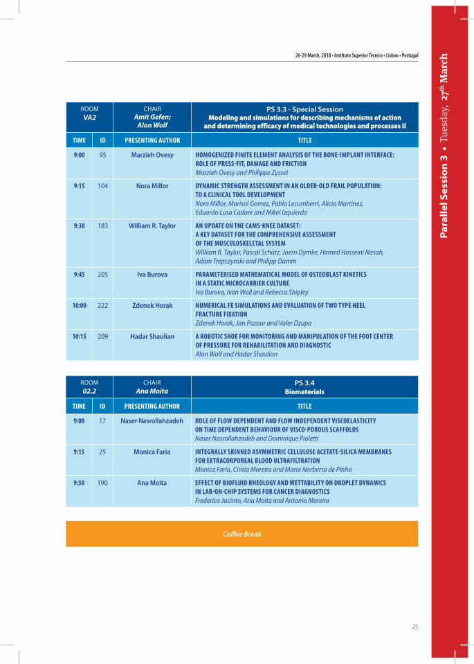
ROOM
MA
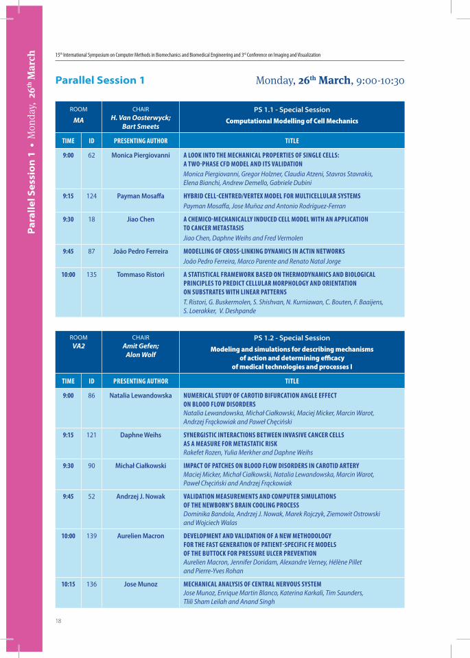
CHAIRH. Van Oosterwyck;
Bart Smeets
PS 1.1 - Special Session
Computational Modelling of Cell Mechanics
Time iD PresenTing AuThor TiTLe
9:00 62 Monica Piergiovanni A Look inTo The mechAnicAL ProPerTies of singLe ceLLs: A TWo-PhAse cfD moDeL AnD iTs vALiDATionMonica Piergiovanni, Gregor Holzner, Claudia Atzeni, Stavros Stavrakis, Elena Bianchi, Andrew Demello, Gabriele Dubini
9:15 124 Payman Mosaffa hybriD ceLL-cenTreD/verTex moDeL for muLTiceLLuLAr sysTemsPayman Mosaffa, Jose Muñoz and Antonio Rodríguez-Ferran
9:30 18 Jiao Chen A chemico-mechAnicALLy inDuceD ceLL moDeL WiTh An APPLicATion To cAncer meTAsTAsisJiao Chen, Daphne Weihs and Fred Vermolen
9:45 87 João Pedro Ferreira moDeLLing of cross-Linking DynAmics in AcTin neTWorksJoão Pedro Ferreira, Marco Parente and Renato Natal Jorge
10:00 135 Tommaso Ristori A sTATisTicAL frAmeWork bAseD on ThermoDynAmics AnD bioLogicAL PrinciPLes To PreDicT ceLLuLAr morPhoLogy AnD orienTATion on subsTrATes WiTh LineAr PATTernsT. Ristori, G. Buskermolen, S. Shishvan, N. Kurniawan, C. Bouten, F. Baaijens, S. Loerakker, V. Deshpande
Parallel Session 1 Monday, 26th March, 9:00-10:30
Par
alle
l Ses
sio
n 1
• M
onda
y, 2
6th M
arch
ROOMVA2
CHAIRAmit Gefen;
Alon Wolf
PS 1.2 - Special Session
Modeling and simulations for describing mechanisms of action and determining efficacy
of medical technologies and processes I
Time iD PresenTing AuThor TiTLe
9:00 86 Natalia Lewandowska numericAL sTuDy of cAroTiD bifurcATion AngLe effecT on bLooD fLoW DisorDersNatalia Lewandowska, Michał Ciałkowski, Maciej Micker, Marcin Warot, Andrzej Frąckowiak and Paweł Chęciński
9:15 121 Daphne Weihs synergisTic inTerAcTions beTWeen invAsive cAncer ceLLs As A meAsure for meTAsTATic riskRakefet Rozen, Yulia Merkher and Daphne Weihs
9:30 90 Michał Ciałkowski imPAcT of PATches on bLooD fLoW DisorDers in cAroTiD ArTeryMaciej Micker, Michał Ciałkowski, Natalia Lewandowska, Marcin Warot, Paweł Chęciński and Andrzej Frąckowiak
9:45 52 Andrzej J. Nowak vALiDATion meAsuremenTs AnD comPuTer simuLATions of The neWborn’s brAin cooLing ProcessDominika Bandola, Andrzej J. Nowak, Marek Rojczyk, Ziemowit Ostrowski and Wojciech Walas
10:00 139 Aurelien Macron DeveLoPmenT AnD vALiDATion of A neW meThoDoLogy for The fAsT generATion of PATienT-sPecific fe moDeLs of The buTTock for Pressure uLcer PrevenTionAurelien Macron, Jennifer Doridam, Alexandre Verney, Hélène Pillet and Pierre-Yves Rohan
10:15 136 Jose Munoz mechAnicAL AnALysis of cenTrAL nervous sysTemJose Munoz, Enrique Martin Blanco, Katerina Karkali, Tim Saunders, Tlili Sham Leilah and Anand Singh
26-29 March, 2018 • Instituto Superior Técnico • Lisbon • Portugal
19
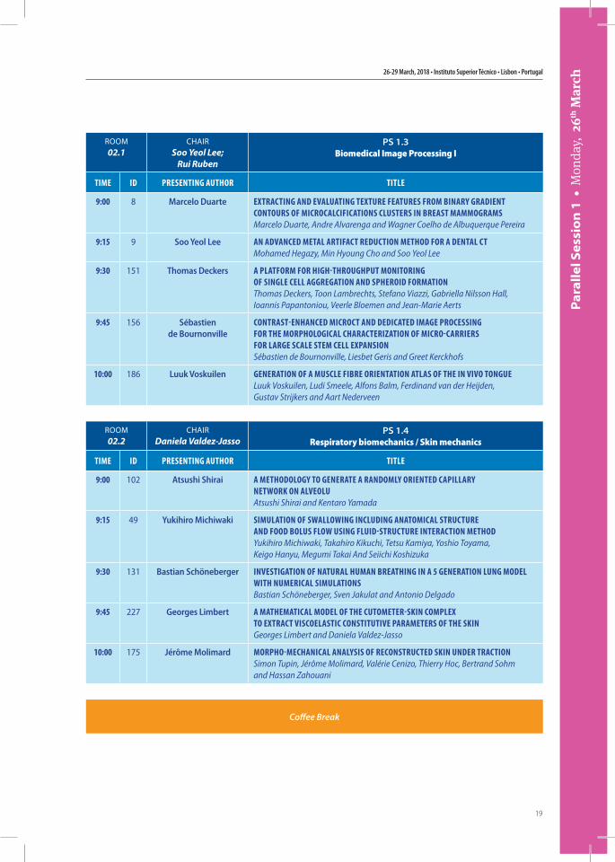
Par
alle
l Ses
sio
n 1
• M
onda
y, 2
6th M
arch
ROOM02.1
CHAIRSoo Yeol Lee;
Rui Ruben
PS 1.3 Biomedical Image Processing I
Time iD PresenTing AuThor TiTLe
9:00 8 Marcelo Duarte exTrAcTing AnD evALuATing TexTure feATures from binAry grADienT conTours of microcALcificATions cLusTers in breAsT mAmmogrAmsMarcelo Duarte, Andre Alvarenga and Wagner Coelho de Albuquerque Pereira
9:15 9 Soo Yeol Lee An ADvAnceD meTAL ArTifAcT reDucTion meThoD for A DenTAL cTMohamed Hegazy, Min Hyoung Cho and Soo Yeol Lee
9:30 151 Thomas Deckers A PLATform for high-ThroughPuT moniToring of singLe ceLL AggregATion AnD sPheroiD formATionThomas Deckers, Toon Lambrechts, Stefano Viazzi, Gabriella Nilsson Hall, Ioannis Papantoniou, Veerle Bloemen and Jean-Marie Aerts
9:45 156 Sébastien de Bournonville
conTrAsT-enhAnceD microcT AnD DeDicATeD imAge Processing for The morPhoLogicAL chArAcTerizATion of micro-cArriers for LArge scALe sTem ceLL exPAnsionSébastien de Bournonville, Liesbet Geris and Greet Kerckhofs
10:00 186 Luuk Voskuilen generATion of A muscLe fibre orienTATion ATLAs of The in vivo TongueLuuk Voskuilen, Ludi Smeele, Alfons Balm, Ferdinand van der Heijden, Gustav Strijkers and Aart Nederveen
ROOM02.2
CHAIRDaniela Valdez-Jasso
PS 1.4 Respiratory biomechanics / Skin mechanics
Time iD PresenTing AuThor TiTLe
9:00 102 Atsushi Shirai A meThoDoLogy To generATe A rAnDomLy orienTeD cAPiLLAry neTWork on ALveoLuAtsushi Shirai and Kentaro Yamada
9:15 49 Yukihiro Michiwaki simuLATion of sWALLoWing incLuDing AnATomicAL sTrucTure AnD fooD boLus fLoW using fLuiD-sTrucTure inTerAcTion meThoDYukihiro Michiwaki, Takahiro Kikuchi, Tetsu Kamiya, Yoshio Toyama, Keigo Hanyu, Megumi Takai And Seiichi Koshizuka
9:30 131 Bastian Schöneberger invesTigATion of nATurAL humAn breAThing in A 5 generATion Lung moDeL WiTh numericAL simuLATionsBastian Schöneberger, Sven Jakulat and Antonio Delgado
9:45 227 Georges Limbert A mAThemATicAL moDeL of The cuTomeTer-skin comPLex To exTrAcT viscoeLAsTic consTiTuTive PArAmeTers of The skinGeorges Limbert and Daniela Valdez-Jasso
10:00 175 Jérôme Molimard morPho-mechAnicAL AnALysis of reconsTrucTeD skin unDer TrAcTionSimon Tupin, Jérôme Molimard, Valérie Cenizo, Thierry Hoc, Bertrand Sohm and Hassan Zahouani
Coffee Break

15th International Symposium on Computer Methods in Biomechanics and Biomedical Engineering and 3rd Conference on Imaging and Visualization
20
Ple
nar
y Le
ctu
res
• M
onda
y, 2
6th M
arch
ROOMMA
CHAIRPaulo Fernandes
Plenary I
Time iD PLenAry sPeAker TiTLe
11:00 261 Hans Van Oosterwyck moDeLLing AnD meAsuring ceLL-mATrix mechAnicAL inTerAcTionsHans Van Oosterwyck
11:30 57 Amit Gefen meDicAL Device-reLATeD Pressure uLcers: Where biomechAnics shouLD come To The rescueAmit Gefen
12:00 263 Sam Evans moDeLs of sofT mATeriALs unDer muLTiAxiAL LoADingSam Evans
Lunch
Coffee Break
Plenary Lectures Monday, 26th March, 11:00-12:30
ROOMMA
CHAIRJoão Tavares
Plenary II
Time iD PLenAry sPeAker TiTLe
14:00 251 Cristian A. Linte from meDicAL imAge comPuTing To comPuTer-AiDeD DiAgnosis TooLs: successes, chALLenges, guiDeLines AnD Lessons LeArneDCristian Linte
14:30 244 Saeed A. Shirazi-Adl on The equiLibrium AnD sTAbiLiTy of The knee joinT in gAiTSaeed A. Shirazi-Adl, Hafedh Marouane, Masoud Sharifi and Malek Adouni
15:00 245 Fiorella Sgallari enhAncing sPArsiTy beyonD convexiTy: APPLicATions To The resTorATion AnD segmenTATion of meDicAL imAges AnD surfAcesFiorella Sgallari
Plenary Lectures Monday, 26th March, 14:00-15:30
26-29 March, 2018 • Instituto Superior Técnico • Lisbon • Portugal
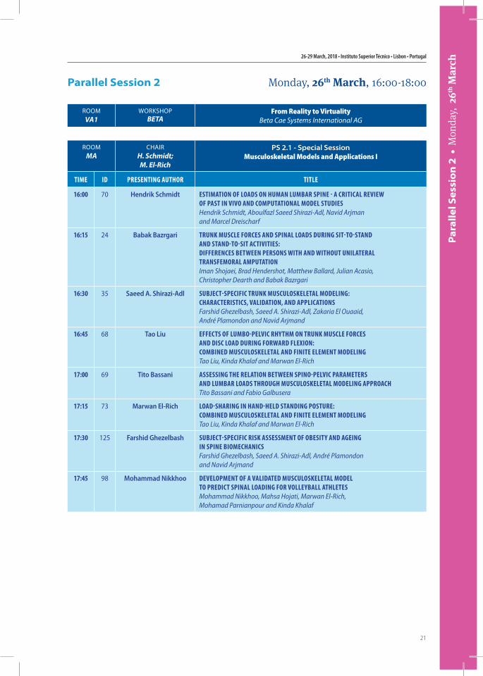
21
Par
alle
l Ses
sio
n 2
• M
onda
y, 2
6th M
arch
ROOMMA
CHAIRH. Schmidt;M. El-Rich
PS 2.1 - Special SessionMusculoskeletal Models and Applications I
Time iD PresenTing AuThor TiTLe
16:00 70 Hendrik Schmidt esTimATion of LoADs on humAn LumbAr sPine - A criTicAL revieW of PAsT in vivo AnD comPuTATionAL moDeL sTuDiesHendrik Schmidt, Aboulfazl Saeed Shirazi-Adl, Navid Arjman and Marcel Dreischarf
16:15 24 Babak Bazrgari Trunk muscLe forces AnD sPinAL LoADs During siT-To-sTAnD AnD sTAnD-To-siT AcTiviTies: Differences beTWeen Persons WiTh AnD WiThouT uniLATerAL TrAnsfemorAL AmPuTATionIman Shojaei, Brad Hendershot, Matthew Ballard, Julian Acasio, Christopher Dearth and Babak Bazrgari
16:30 35 Saeed A. Shirazi-Adl subjecT-sPecific Trunk muscuLoskeLeTAL moDeLing: chArAcTerisTics, vALiDATion, AnD APPLicATionsFarshid Ghezelbash, Saeed A. Shirazi-Adl, Zakaria El Ouaaid, André Plamondon and Navid Arjmand
16:45 68 Tao Liu effecTs of Lumbo-PeLvic rhyThm on Trunk muscLe forces AnD Disc LoAD During forWArD fLexion: combineD muscuLoskeLeTAL AnD finiTe eLemenT moDeLingTao Liu, Kinda Khalaf and Marwan El-Rich
17:00 69 Tito Bassani Assessing The reLATion beTWeen sPino-PeLvic PArAmeTers AnD LumbAr LoADs Through muscuLoskeLeTAL moDeLing APProAchTito Bassani and Fabio Galbusera
17:15 73 Marwan El-Rich LoAD-shAring in hAnD-heLD sTAnDing PosTure: combineD muscuLoskeLeTAL AnD finiTe eLemenT moDeLingTao Liu, Kinda Khalaf and Marwan El-Rich
17:30 125 Farshid Ghezelbash subjecT-sPecific risk AssessmenT of obesiTy AnD Ageing in sPine biomechAnicsFarshid Ghezelbash, Saeed A. Shirazi-Adl, André Plamondon and Navid Arjmand
17:45 98 Mohammad Nikkhoo DeveLoPmenT of A vALiDATeD muscuLoskeLeTAL moDeL To PreDicT sPinAL LoADing for voLLeybALL AThLeTesMohammad Nikkhoo, Mahsa Hojati, Marwan El-Rich, Mohamad Parnianpour and Kinda Khalaf
Parallel Session 2 Monday, 26th March, 16:00-18:00
ROOMVA1
WORKSHOPBETA
From Reality to VirtualityBeta Cae Systems International AG
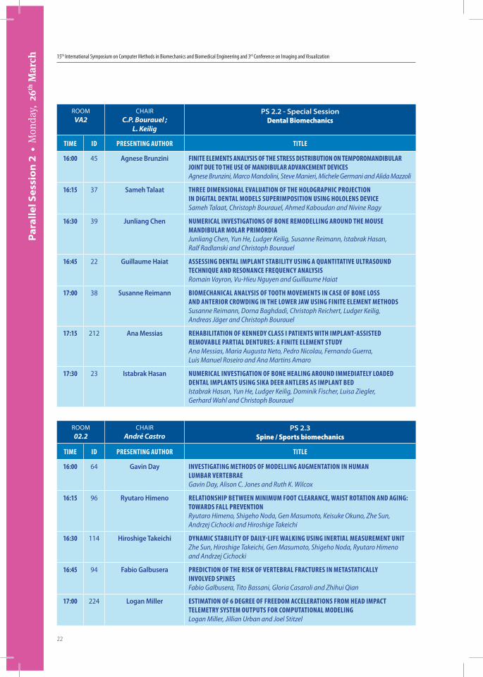
15th International Symposium on Computer Methods in Biomechanics and Biomedical Engineering and 3rd Conference on Imaging and Visualization
22
Par
alle
l Ses
sio
n 2
• M
onda
y, 2
6th M
arch
ROOMVA2
CHAIRC.P. Bourauel ;
L. Keilig
PS 2.2 - Special SessionDental Biomechanics
Time iD PresenTing AuThor TiTLe
16:00 45 Agnese Brunzini finiTe eLemenTs AnALysis of The sTress DisTribuTion on TemPoromAnDibuLAr joinT Due To The use of mAnDibuLAr ADvAncemenT DevicesAgnese Brunzini, Marco Mandolini, Steve Manieri, Michele Germani and Alida Mazzoli
16:15 37 Sameh Talaat Three DimensionAL evALuATion of The hoLogrAPhic ProjecTion in DigiTAL DenTAL moDeLs suPerimPosiTion using hoLoLens DeviceSameh Talaat, Christoph Bourauel, Ahmed Kaboudan and Nivine Ragy
16:30 39 Junliang Chen numericAL invesTigATions of bone remoDeLLing ArounD The mouse mAnDibuLAr moLAr PrimorDiAJunliang Chen, Yun He, Ludger Keilig, Susanne Reimann, Istabrak Hasan, Ralf Radlanski and Christoph Bourauel
16:45 22 Guillaume Haiat Assessing DenTAL imPLAnT sTAbiLiTy using A quAnTiTATive uLTrAsounD Technique AnD resonAnce frequency AnALysisRomain Vayron, Vu-Hieu Nguyen and Guillaume Haiat
17:00 38 Susanne Reimann biomechAnicAL AnALysis of TooTh movemenTs in cAse of bone Loss AnD AnTerior croWDing in The LoWer jAW using finiTe eLemenT meThoDsSusanne Reimann, Dorna Baghdadi, Christoph Reichert, Ludger Keilig, Andreas Jäger and Christoph Bourauel
17:15 212 Ana Messias rehAbiLiTATion of kenneDy cLAss i PATienTs WiTh imPLAnT-AssisTeD removAbLe PArTiAL DenTures: A finiTe eLemenT sTuDyAna Messias, Maria Augusta Neto, Pedro Nicolau, Fernando Guerra, Luis Manuel Roseiro and Ana Martins Amaro
17:30 23 Istabrak Hasan numericAL invesTigATion of bone heALing ArounD immeDiATeLy LoADeD DenTAL imPLAnTs using sikA Deer AnTLers As imPLAnT beDIstabrak Hasan, Yun He, Ludger Keilig, Dominik Fischer, Luisa Ziegler, Gerhard Wahl and Christoph Bourauel
ROOM02.2
CHAIRAndré Castro
PS 2.3Spine / Sports biomechanics
Time iD PresenTing AuThor TiTLe
16:00 64 Gavin Day invesTigATing meThoDs of moDeLLing AugmenTATion in humAn LumbAr verTebrAeGavin Day, Alison C. Jones and Ruth K. Wilcox
16:15 96 Ryutaro Himeno reLATionshiP beTWeen minimum fooT cLeArAnce, WAisT roTATion AnD Aging: ToWArDs fALL PrevenTionRyutaro Himeno, Shigeho Noda, Gen Masumoto, Keisuke Okuno, Zhe Sun, Andrzej Cichocki and Hiroshige Takeichi
16:30 114 Hiroshige Takeichi DynAmic sTAbiLiTy of DAiLy-Life WALking using inerTiAL meAsuremenT uniTZhe Sun, Hiroshige Takeichi, Gen Masumoto, Shigeho Noda, Ryutaro Himeno and Andrzej Cichocki
16:45 94 Fabio Galbusera PreDicTion of The risk of verTebrAL frAcTures in meTAsTATicALLy invoLveD sPinesFabio Galbusera, Tito Bassani, Gloria Casaroli and Zhihui Qian
17:00 224 Logan Miller esTimATion of 6 Degree of freeDom AcceLerATions from heAD imPAcT TeLemeTry sysTem ouTPuTs for comPuTATionAL moDeLingLogan Miller, Jillian Urban and Joel Stitzel
26-29 March, 2018 • Instituto Superior Técnico • Lisbon • Portugal
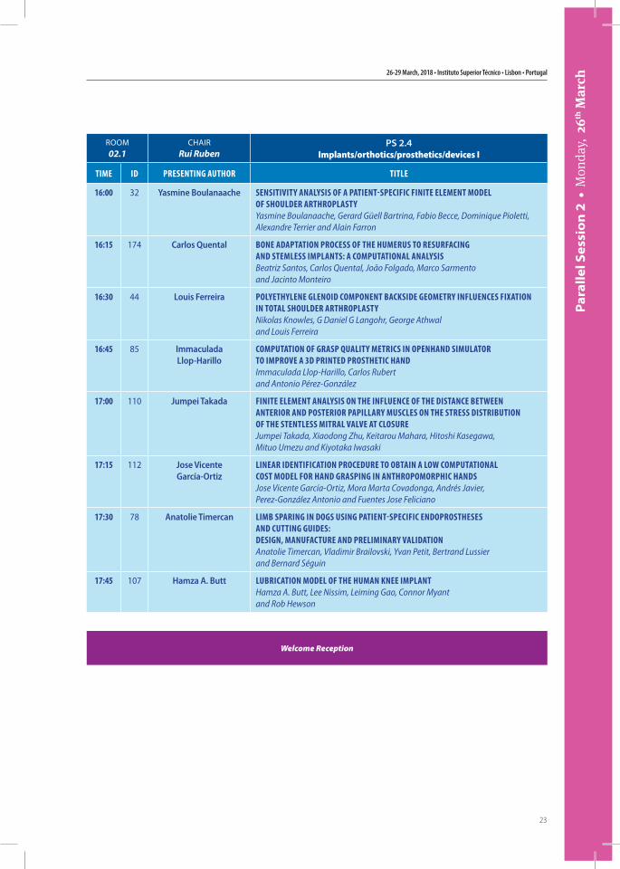
23
Par
alle
l Ses
sio
n 2
• M
onda
y, 2
6th M
arch
ROOM02.1
CHAIRRui Ruben
PS 2.4Implants/orthotics/prosthetics/devices I
Time iD PresenTing AuThor TiTLe
16:00 32 Yasmine Boulanaache sensiTiviTy AnALysis of A PATienT-sPecific finiTe eLemenT moDeL of shouLDer ArThroPLAsTyYasmine Boulanaache, Gerard Güell Bartrina, Fabio Becce, Dominique Pioletti, Alexandre Terrier and Alain Farron
16:15 174 Carlos Quental bone ADAPTATion Process of The humerus To resurfAcing AnD sTemLess imPLAnTs: A comPuTATionAL AnALysisBeatriz Santos, Carlos Quental, João Folgado, Marco Sarmento and Jacinto Monteiro
16:30 44 Louis Ferreira PoLyeThyLene gLenoiD comPonenT bAcksiDe geomeTry infLuences fixATion in ToTAL shouLDer ArThroPLAsTyNikolas Knowles, G Daniel G Langohr, George Athwal and Louis Ferreira
16:45 85 Immaculada Llop-Harillo
comPuTATion of grAsP quALiTy meTrics in oPenhAnD simuLATor To imProve A 3D PrinTeD ProsTheTic hAnDImmaculada Llop-Harillo, Carlos Rubert and Antonio Pérez-González
17:00 110 Jumpei Takada finiTe eLemenT AnALysis on The infLuence of The DisTAnce beTWeen AnTerior AnD PosTerior PAPiLLAry muscLes on The sTress DisTribuTion of The sTenTLess miTrAL vALve AT cLosureJumpei Takada, Xiaodong Zhu, Keitarou Mahara, Hitoshi Kasegawa, Mituo Umezu and Kiyotaka Iwasaki
17:15 112 Jose Vicente García-Ortiz
LineAr iDenTificATion ProceDure To obTAin A LoW comPuTATionAL cosT moDeL for hAnD grAsPing in AnThroPomorPhic hAnDsJose Vicente García-Ortiz, Mora Marta Covadonga, Andrés Javier, Perez-González Antonio and Fuentes Jose Feliciano
17:30 78 Anatolie Timercan Limb sPAring in Dogs using PATienT-sPecific enDoProsTheses AnD cuTTing guiDes: Design, mAnufAcTure AnD PreLiminAry vALiDATionAnatolie Timercan, Vladimir Brailovski, Yvan Petit, Bertrand Lussier and Bernard Séguin
17:45 107 Hamza A. Butt LubricATion moDeL of The humAn knee imPLAnTHamza A. Butt, Lee Nissim, Leiming Gao, Connor Myant and Rob Hewson
Welcome Reception
15th International Symposium on Computer Methods in Biomechanics and Biomedical Engineering and 3rd Conference on Imaging and Visualization
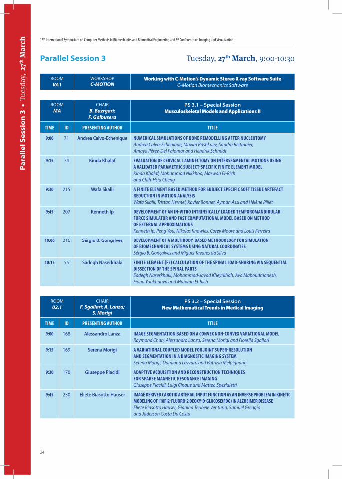
24
Par
alle
l Ses
sio
n 3
• T
uesd
ay,
27th
Mar
ch
ROOMMA
CHAIRB. Bazrgari;
F. Galbusera
PS 3.1 – Special SessionMusculoskeletal Models and Applications II
Time iD PresenTing AuThor TiTLe
9:00 71 Andrea Calvo-Echenique numericAL simuLATions of bone remoDeLLing AfTer nucLeoTomyAndrea Calvo-Echenique, Maxim Bashkuev, Sandra Reitmaier, Amaya Pérez-Del Palomar and Hendrik Schmidt
9:15 74 Kinda Khalaf evALuATion of cervicAL LAminecTomy on inTersegmenTAL moTions using A vALiDATeD PArAmeTric subjecT-sPecific finiTe eLemenT moDeLKinda Khalaf, Mohammad Nikkhoo, Marwan El-Rich and Chih-Hsiu Cheng
9:30 215 Wafa Skalli A finiTe eLemenT bAseD meThoD for subjecT sPecific sofT Tissue ArTefAcT reDucTion in moTion AnALysisWafa Skalli, Tristan Hermel, Xavier Bonnet, Ayman Assi and Hélène Pillet
9:45 207 Kenneth Ip DeveLoPmenT of An in-viTro inTrinsicALLy LoADeD TemPoromAnDibuLAr force simuLATor AnD fAsT comPuTATionAL moDeL bAseD on meThoD of exTernAL APProximATionsKenneth Ip, Peng You, Nikolas Knowles, Corey Moore and Louis Ferreira
10:00 216 Sérgio B. Gonçalves DeveLoPmenT of A muLTiboDy-bAseD meThoDoLogy for simuLATion of biomechAnicAL sysTems using nATurAL coorDinATesSérgio B. Gonçalves and Miguel Tavares da Silva
10:15 55 Sadegh Naserkhaki finiTe eLemenT (fe) cALcuLATion of The sPinAL LoAD-shAring viA sequenTiAL DissecTion of The sPinAL PArTsSadegh Naserkhaki, Mohammad-Javad Kheyrkhah, Ava Maboudmanesh, Fiona Youkhanva and Marwan El-Rich
ROOM02.1
CHAIRF. Sgallari; A. Lanza;
S. Morigi
PS 3.2 – Special SessionNew Mathematical Trends in Medical Imaging
Time iD PresenTing AuThor TiTLe
9:00 168 Alessandro Lanza imAge segmenTATion bAseD on A convex non-convex vAriATionAL moDeLRaymond Chan, Alessandro Lanza, Serena Morigi and Fiorella Sgallari
9:15 169 Serena Morigi A vAriATionAL couPLeD moDeL for joinT suPer-resoLuTion AnD segmenTATion in A DiAgnosTic imAging sysTemSerena Morigi, Damiana Lazzaro and Patrizia Melpignano
9:30 170 Giuseppe Placidi ADAPTive AcquisiTion AnD reconsTrucTion Techniques for sPArse mAgneTic resonAnce imAgingGiuseppe Placidi, Luigi Cinque and Matteo Spezialetti
9:45 230 Eliete Biasotto Hauser imAge DeriveD cAroTiD ArTeriAL inPuT funcTion As An inverse ProbLem in kineTic moDeLing of [18f]2-fLuoro-2 Deoxy-D-gLucose(fDg) in ALzheimer DiseAseEliete Biasotto Hauser, Gianina Teribele Venturin, Samuel Greggio and Jaderson Costa Da Costa
Parallel Session 3 Tuesday, 27th March, 9:00-10:30
ROOMVA1
WORKSHOPC-MOTION
Working with C-Motion’s Dynamic Stereo X-ray Software SuiteC-Motion Biomechanics Software
26-29 March, 2018 • Instituto Superior Técnico • Lisbon • Portugal
25
Par
alle
l Ses
sio
n 3
• T
uesd
ay,
27th
Mar
ch
ROOMVA2
CHAIRAmit Gefen;
Alon Wolf
PS 3.3 - Special SessionModeling and simulations for describing mechanisms of action
and determining efficacy of medical technologies and processes II
Time iD PresenTing AuThor TiTLe
9:00 95 Marzieh Ovesy homogenizeD finiTe eLemenT AnALysis of The bone-imPLAnT inTerfAce: roLe of Press-fiT, DAmAge AnD fricTionMarzieh Ovesy and Philippe Zysset
9:15 104 Nora Millor DynAmic sTrengTh AssessmenT in An oLDer-oLD frAiL PoPuLATion: To A cLinicAL TooL DeveLoPmenTNora Millor, Marisol Gomez, Pablo Lecumberri, Alicia Martinez, Eduardo Lusa Cadore and Mikel Izquierdo
9:30 183 William R. Taylor An uPDATe on The cAms-knee DATAseT: A key DATAseT for The comPrehensive AssessmenT of The muscuLoskeLeTAL sysTemWilliam R. Taylor, Pascal Schütz, Joern Dymke, Hamed Hosseini Nasab, Adam Trepczynski and Philipp Damm
9:45 205 Iva Burova PArAmeTeriseD mAThemATicAL moDeL of osTeobLAsT kineTics in A sTATic microcArrier cuLTureIva Burova, Ivan Wall and Rebecca Shipley
10:00 222 Zdenek Horak numericAL fe simuLATions AnD evALuATion of TWo TyPe heeL frAcTure fixATionZdenek Horak, Jan Pazour and Valer Dzupa
10:15 209 Hadar Shaulian A roboTic shoe for moniToring AnD mAniPuLATion of The fooT cenTer of Pressure for rehAbiLiTATion AnD DiAgnosTicAlon Wolf and Hadar Shaulian
ROOM02.2
CHAIRAna Moita
PS 3.4Biomaterials
Time iD PresenTing AuThor TiTLe
9:00 17 Naser Nasrollahzadeh roLe of fLoW DePenDenT AnD fLoW inDePenDenT viscoeLAsTiciTy on Time DePenDenT behAviour of visco-Porous scAffoLDsNaser Nasrollahzadeh and Dominique Pioletti
9:15 25 Monica Faria inTegrALLy skinneD AsymmeTric ceLLuLose AceTATe-siLicA membrAnes for exTrAcorPoreAL bLooD uLTrAfiLTrATionMonica Faria, Cintia Moreira and Maria Norberta de Pinho
9:30 190 Ana Moita effecT of biofLuiD rheoLogy AnD WeTTAbiLiTy on DroPLeT DynAmics in LAb-on-chiP sysTems for cAncer DiAgnosTicsFrederico Jacinto, Ana Moita and Antonio Moreira
Coffee Break
15th International Symposium on Computer Methods in Biomechanics and Biomedical Engineering and 3rd Conference on Imaging and Visualization
26
Plen
ary
Lect
ure
s •
Tue
sday
, 27
th M
arch
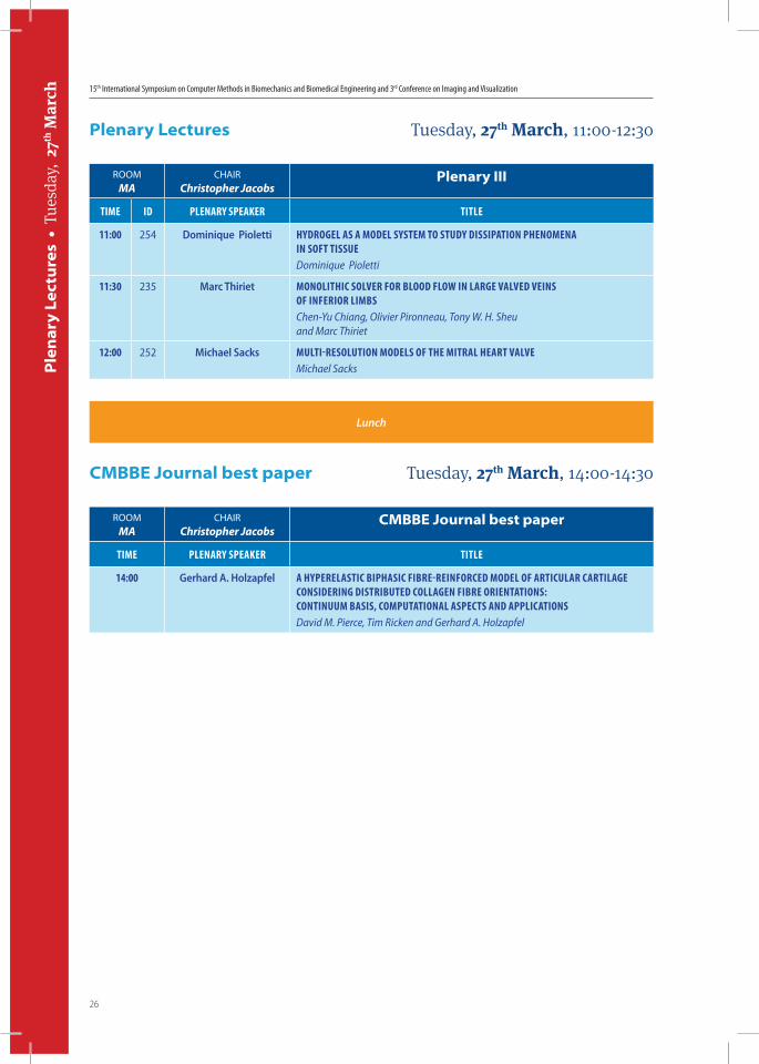
ROOMMA
CHAIRChristopher Jacobs
Plenary III
Time iD PLenAry sPeAker TiTLe
11:00 254 Dominique Pioletti hyDrogeL As A moDeL sysTem To sTuDy DissiPATion PhenomenA in sofT TissueDominique Pioletti
11:30 235 Marc Thiriet monoLiThic soLver for bLooD fLoW in LArge vALveD veins of inferior LimbsChen-Yu Chiang, Olivier Pironneau, Tony W. H. Sheu and Marc Thiriet
12:00 252 Michael Sacks muLTi-resoLuTion moDeLs of The miTrAL heArT vALveMichael Sacks
Lunch
ROOMMA
CHAIRChristopher Jacobs
CMBBE Journal best paper
Time PLenAry sPeAker TiTLe
14:00 Gerhard A. Holzapfel A hyPereLAsTic biPhAsic fibre-reinforceD moDeL of ArTicuLAr cArTiLAge consiDering DisTribuTeD coLLAgen fibre orienTATions: conTinuum bAsis, comPuTATionAL AsPecTs AnD APPLicATionsDavid M. Pierce, Tim Ricken and Gerhard A. Holzapfel
Plenary Lectures Tuesday, 27th March, 11:00-12:30
CMBBE Journal best paper Tuesday, 27th March, 14:00-14:30
26-29 March, 2018 • Instituto Superior Técnico • Lisbon • Portugal
27
Post
er S
essi
on
• T
uesd
ay,
27th
Mar
ch
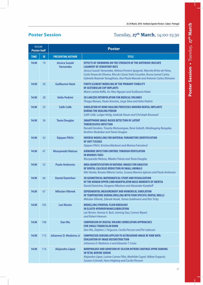
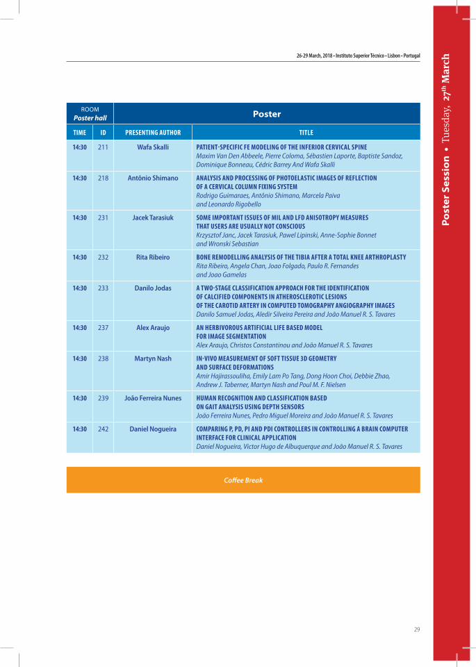
ROOMPoster hall Poster
Time iD PresenTing AuThor TiTLe
14:30 19 Jéssica Suzuki Yamanaka
effecTs of sWimming on The sTrengTh of The AnTerior cruciATe LigAmenT of seDenTAry rATsJéssica Suzuki Yamanaka, Heloisa Ferreira Spagnoli, Marcela Britto de Paiva, Carla Tereza de Oliveira, Rita de Cássia Stela Cossalter, Bruna Leonel Carlos, Gabriela Rezende Yanagihara, Ana Paula Macedo and Antonio Carlos Shimano
14:30 20 Guillaume Haiat finiTe eLemenT moDeLing of The PrimAry sTAbiLiTy of AceTAbuLAr cuP imPLAnTsMaria-Letizia Raffa, Vu-Hieu Nguyen and Guillaume Haiat
14:30 28 Helio Pedrini 3D LAnczos inTerPoLATion for meDicAL voLumesThiago Moraes, Paulo Amorim, Jorge Silva and Helio Pedrini
14:30 33 Salih Celik simuLATion of bone heALing Processes ArounD DenTAL imPLAnTs During The heALing PerioDSalih Celik, Ludger Keilig, Istabrak Hasan and Christoph Bourauel
14:30 36 Tania Douglas smArTPhone imAge-bAseD DeTecTion of LATenT TubercuLosis infecTionRonald Dendere, Tinashe Mutsvangwa, Rene Goliath, Molebogeng Rangaka, Ibrahim Abubakar and Tania Douglas
14:30 42 Stjepan Piličić inverse moDeLLing for mATeriAL PArAmeTers iDenTificATion of sofT TissuesStjepan Piličić, Kristina Marković and Marina Franulović
14:30 47 Munyaradzi Matose Airborne infecTion conTroL Through venTiLATion in minibus TAxisMunyaradzi Matose, Mladen Poluta and Tania Douglas
14:30 53 Paulo Ambrosio AreA quAnTificATion in nATurAL imAges for AnALysis of DenTAL cALcuLus reDucTion in smALL AnimALsNilo Varela, Renata Alberto Carlos, Susana Marrero Iglesias and Paulo Ambrosio
14:30 66 Daniel Dantchev 3D geomeTricAL mAThemATicAL sTuDy AnD visuALizATion of The humAn uPPer Limb mAniPuLATor mAss momenTs of inerTiADaniel Dantchev, Gergana Nikolova and Alexander Kazakoff
14:30 67 Miloslav Vilimek exPerimenTAL meAsuremenT AnD numericAL simuLATion of TemPerATure During DriLLing WiTh four sPecific DenTAL DriLLsMiloslav Vilimek, Zdenek Horak, Tomas Goldmann and Petr Tichy
14:30 105 Lee Nissim moDeLLing synoviAL fLuiD rheoLogy in eLAsTo-hyDroDynAmicLubricATionLee Nissim, Hamza A. Butt, Leiming Gao, Connor Myant and Robert Hewson
14:30 108 Dan Wu comPArison of DigiTAL voLume correLATion APProAches for singLe TrAbecuLAr boneDan Wu, Stephen J. Ferguson, Cecilia Persson and Per Isaksson
14:30 115 Johannes D. Medeiros Jr comPresseD sensing APPLieD To uLTrAsounD imAge rf rAW DATA: evALuATion of imAge reconsTrucTionJohannes D. Medeiros Jr and Eduardo T. Costa
14:30 116 Alejandro López morPhoLogy AnD ADhesion of siLicon niTriDe coATings uPon soAking in feTAL bovine serumAlejandro López, Luimar Correa Filho, Mathilde Cogrel, Håkan Engqvist, Susann Schmidt, Hans Högberg and Cecilia Persson
Poster Session Tuesday, 27th March, 14:00-15:30
15th International Symposium on Computer Methods in Biomechanics and Biomedical Engineering and 3rd Conference on Imaging and Visualization
28
Post
er S
essi
on
• T
uesd
ay,
27th
Mar
ch
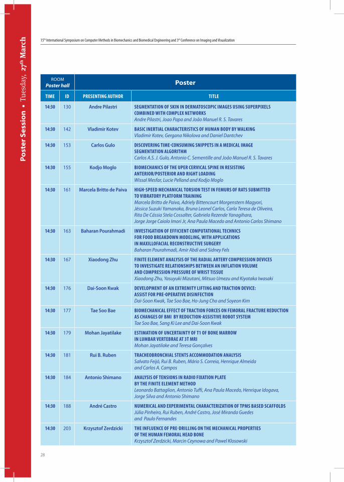
ROOMPoster hall Poster
Time iD PresenTing AuThor TiTLe
14:30 130 Andre Pilastri segmenTATion of skin in DermAToscoPic imAges using suPerPixeLs combineD WiTh comPLex neTWorksAndre Pilastri, Joao Papa and João Manuel R. S. Tavares
14:30 142 Vladimir Kotev bAsic inerTiAL chArAcTerisTics of humAn boDy by WALkingVladimir Kotev, Gergana Nikolova and Daniel Dantchev
14:30 153 Carlos Gulo Discovering Time-consuming sniPPeTs in A meDicAL imAge segmenTATion ALgoriThmCarlos A.S. J. Gulo, Antonio C. Sementille and João Manuel R. S. Tavares
14:30 155 Kodjo Moglo biomechAnics of The uPer cervicAL sPine in resisTing AnTerior/PosTerior AnD righT LoADingWissal Mesfar, Lucie Pelland and Kodjo Moglo
14:30 161 Marcela Britto de Paiva high-sPeeD mechAnicAL Torsion TesT in femurs of rATs submiTTeD To vibrATory PLATform TrAiningMarcela Britto de Paiva, Adriely Bittencourt Morgenstern Magyori, Jéssica Suzuki Yamanaka, Bruna Leonel Carlos, Carla Teresa de Oliveira, Rita De Cássia Stela Cossalter, Gabriela Rezende Yanagihara, Jorge Jorge Caiolo Imori Jr, Ana Paula Macedo and Antonio Carlos Shimano
14:30 163 Baharan Pourahmadi invesTigATion of efficienT comPuTATionAL Technics for fooD breAkDoWn moDeLing, WiTh APPLicATions in mAxiLLofAciAL reconsTrucTive surgeryBaharan Pourahmadi, Amir Abdi and Sidney Fels
14:30 167 Xiaodong Zhu finiTe eLemenT AnALysis of The rADiAL ArTery comPression Devices To invesTigATe reLATionshiPs beTWeen An infLATion voLume AnD comPression Pressure of WrisT TissueXiaodong Zhu, Yasuyuki Mizutani, Mitsuo Umezu and Kiyotaka Iwasaki
14:30 176 Dai-Soon Kwak DeveLoPmenT of An exTremiTy LifTing AnD TrAcTion Device: AssisT for Pre-oPerATive DisinfecTionDai-Soon Kwak, Tae Soo Bae, Ho-Jung Cho and Soyeon Kim
14:30 177 Tae Soo Bae biomechAnicAL effecT of TrAcTion forces on femorAL frAcTure reDucTion As chAnges of bmi by reDucTion-AssisTive roboT sysTemTae Soo Bae, Sang Ki Lee and Dai-Soon Kwak
14:30 179 Mohan Jayatilake esTimATion of uncerTAinTy of T1 of bone mArroW in LumbAr verTebrAe AT 3T mriMohan Jayatilake and Teresa Gonçalves
14:30 181 Rui B. Ruben TrAcheobronchiAL sTenTs AccommoDATion AnALysisSalvato Feijó, Rui B. Ruben, Mário S. Correia, Henrique Almeida and Carlos A. Campos
14:30 184 Antonio Shimano AnALysis of Tensions in rADio fixATion PLATe by The finiTe eLemenT meThoDLeonardo Battaglion, Antonio Tuffi, Ana Paula Macedo, Henrique Idogava, Jorge Silva and Antonio Shimano
14:30 188 André Castro numericAL AnD exPerimenTAL chArAcTerizATion of TPms bAseD scAffoLDsJúlia Pinheiro, Rui Ruben, André Castro, José Miranda Guedes and Paulo Fernandes
14:30 203 Krzysztof Zerdzicki The infLuence of Pre-DriLLing on The mechAnicAL ProPerTies of The humAn femorAL heAD boneKrzysztof Zerdzicki, Marcin Ceynowa and Pawel Klosowski
26-29 March, 2018 • Instituto Superior Técnico • Lisbon • Portugal
29
Post
er S
essi
on
• T
uesd
ay,
27th
Mar
ch
ROOMPoster hall Poster
Time iD PresenTing AuThor TiTLe
14:30 211 Wafa Skalli PATienT-sPecific fe moDeLing of The inferior cervicAL sPineMaxim Van Den Abbeele, Pierre Coloma, Sébastien Laporte, Baptiste Sandoz, Dominique Bonneau, Cédric Barrey And Wafa Skalli
14:30 218 Antônio Shimano AnALysis AnD Processing of PhoToeLAsTic imAges of refLecTion of A cervicAL coLumn fixing sysTemRodrigo Guimaraes, Antônio Shimano, Marcela Paiva and Leonardo Rigobello
14:30 231 Jacek Tarasiuk some imPorTAnT issues of miL AnD LfD AnisoTroPy meAsures ThAT users Are usuALLy noT consciousKrzysztof Janc, Jacek Tarasiuk, Pawel Lipinski, Anne-Sophie Bonnet and Wronski Sebastian
14:30 232 Rita Ribeiro bone remoDeLLing AnALysis of The TibiA AfTer A ToTAL knee ArThroPLAsTyRita Ribeiro, Angela Chan, Joao Folgado, Paulo R. Fernandes and Joao Gamelas
14:30 233 Danilo Jodas A TWo-sTAge cLAssificATion APProAch for The iDenTificATion of cALcifieD comPonenTs in ATheroscLeroTic Lesions of The cAroTiD ArTery in comPuTeD TomogrAPhy AngiogrAPhy imAgesDanilo Samuel Jodas, Aledir Silveira Pereira and João Manuel R. S. Tavares
14:30 237 Alex Araujo An herbivorous ArTificiAL Life bAseD moDeL for imAge segmenTATionAlex Araujo, Christos Constantinou and João Manuel R. S. Tavares
14:30 238 Martyn Nash in-vivo meAsuremenT of sofT Tissue 3D geomeTry AnD surfAce DeformATionsAmir Hajirassouliha, Emily Lam Po Tang, Dong Hoon Choi, Debbie Zhao, Andrew J. Taberner, Martyn Nash and Poul M. F. Nielsen
14:30 239 João Ferreira Nunes humAn recogniTion AnD cLAssificATion bAseD on gAiT AnALysis using DePTh sensorsJoão Ferreira Nunes, Pedro Miguel Moreira and João Manuel R. S. Tavares
14:30 242 Daniel Nogueira comPAring P, PD, Pi AnD PDi conTroLLers in conTroLLing A brAin comPuTer inTerfAce for cLinicAL APPLicATionDaniel Nogueira, Victor Hugo de Albuquerque and João Manuel R. S. Tavares
Coffee Break
15th International Symposium on Computer Methods in Biomechanics and Biomedical Engineering and 3rd Conference on Imaging and Visualization
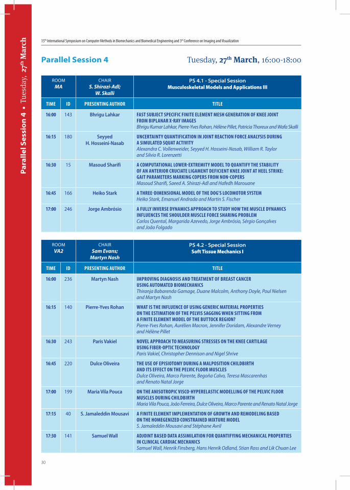
30
Par
alle
l Ses
sio
n 4
• T
uesd
ay,
27th
Mar
ch
ROOMMA
CHAIRS. Shirazi-Adl;
W. Skalli
PS 4.1 - Special SessionMusculoskeletal Models and Applications III
Time iD PresenTing AuThor TiTLe
16:00 143 Bhrigu Lahkar fAsT subjecT sPecific finiTe eLemenT mesh generATion of knee joinT from biPLAnAr x-rAy imAgesBhrigu Kumar Lahkar, Pierre-Yves Rohan, Hélène Pillet, Patricia Thoreux and Wafa Skalli
16:15 180 Seyyed H. Hosseini-Nasab
uncerTAinTy quAnTificATion in joinT reAcTion force AnALysis During A simuLATeD squAT AcTiviTyAlexandra C. Vollenweider, Seyyed H. Hosseini-Nasab, William R. Taylor and Silvio R. Lorenzetti
16:30 15 Masoud Sharifi A comPuTATionAL LoWer-exTremiTy moDeL To quAnTify The sTAbiLiTy of An AnTerior cruciATe LigAmenT DeficienT knee joinT AT heeL sTrike: gAiT PArAmeTers mArking coPers from non-coPersMasoud Sharifi, Saeed A. Shirazi-Adl and Hafedh Marouane
16:45 166 Heiko Stark A Three-DimensionAL moDeL of The Dog’s LocomoTor sysTemHeiko Stark, Emanuel Andrada and Martin S. Fischer
17:00 246 Jorge Ambrósio A fuLLy inverse DynAmics APProAch To sTuDy hoW The muscLe DynAmics infLuences The shouLDer muscLe force shAring ProbLemCarlos Quental, Margarida Azevedo, Jorge Ambrósio, Sérgio Gonçalves and João Folgado
Parallel Session 4 Tuesday, 27th March, 16:00-18:00
ROOMVA2
CHAIRSam Evans;
Martyn Nash
PS 4.2 - Special SessionSoft Tissue Mechanics I
Time iD PresenTing AuThor TiTLe
16:00 236 Martyn Nash imProving DiAgnosis AnD TreATmenT of breAsT cAncer using AuTomATeD biomechAnicsThiranja Babarenda Gamage, Duane Malcolm, Anthony Doyle, Poul Nielsen and Martyn Nash
16:15 140 Pierre-Yves Rohan WhAT is The infLuence of using generic mATeriAL ProPerTies on The esTimATion of The PeLvis sAgging When siTTing from A finiTe eLemenT moDeL of The buTTock region?Pierre-Yves Rohan, Aurélien Macron, Jennifer Doridam, Alexandre Verney and Hélène Pillet
16:30 243 Paris Vakiel noveL APProAch To meAsuring sTresses on The knee cArTiLAge using fiber-oPTic TechnoLogyParis Vakiel, Christopher Dennison and Nigel Shrive
16:45 220 Dulce Oliveira The use of ePisioTomy During A mALPosiTion chiLDbirTh AnD iTs effecT on The PeLvic fLoor muscLesDulce Oliveira, Marco Parente, Begoña Calvo, Teresa Mascarenhas and Renato Natal Jorge
17:00 199 Maria Vila Pouca on The AnisoTroPic visco-hyPereLAsTic moDeLLing of The PeLvic fLoor muscLes During chiLDbirThMaria Vila Pouca, João Ferreira, Dulce Oliveira, Marco Parente and Renato Natal Jorge
17:15 40 S. Jamaleddin Mousavi A finiTe eLemenT imPLemenTATion of groWTh AnD remoDeLing bAseD on The homegenizeD consTrAineD mixTure moDeLS. Jamaleddin Mousavi and Stéphane Avril
17:30 141 Samuel Wall ADjoinT bAseD DATA AssimiLATion for quAnTifying mechAnicAL ProPerTies in cLinicAL cArDiAc mechAnicsSamuel Wall, Henrik Finsberg, Hans Henrik Odland, Stian Ross and Lik Chuan Lee
26-29 March, 2018 • Instituto Superior Técnico • Lisbon • Portugal
31
Par
alle
l Ses
sio
n 4
• T
uesd
ay,
27th
Mar
ch
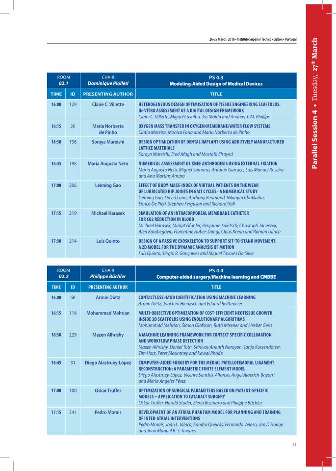
ROOM02.1
CHAIRDominique Piolleti
PS 4.3Modeling-Aided Design of Medical Devices
TIME ID PrESEnTIng AuThor TITLE
16:00 129 Claire C. Villette heTerogeneous Design oPTimisATion of Tissue engineering scAffoLDs: in-viTro AssessmenT of A DigiTAL Design frAmeWorkClaire C. Villette, Miguel Castilho, Jos Malda and Andrew T. M. Phillips
16:15 26 Maria Norberta de Pinho
oxygen mAss TrAnsfer in oxygen/membrAne/WATer fLoW sysTemsCintia Moreira, Monica Faria and Maria Norberta de Pinho
16:30 196 Soraya Mareishi Design oPTimizATion of DenTAL imPLAnT using ADDiTiveLy mAnufAcTureD LATTice mATeriALsSoraya Mareishi, Fred Afagh and Mostafa Elsayed
16:45 198 Maria Augusta Neto numericAL AssessmenT of knee ArThroDesis using exTernAL fixATionMaria Augusta Neto, Miguel Samarra, António Garruço, Luis Manuel Roseiro and Ana Martins Amaro
17:00 206 Leiming Gao effecT of boDy-mAss-inDex of virTuAL PATienTs on The WeAr of LubricATeD hiP joinTs in gAiT cycLes - A numericAL sTuDyLeiming Gao, David Lunn, Anthony Redmond, Nilanjan Chakladar, Enrico De Pieri, Stephen Ferguson and Richard Hall
17:15 219 Michael Harasek simuLATion of An inTrAcorPoreAL membrAne cATheTer for co2 reDucTion in bLooDMichael Harasek, Margit Gföhler, Benjamin Lukitsch, Christoph Janeczek, Alen Karabegovic, Florentine Huber-Dangl, Claus Krenn and Roman Ullrich
17:30 214 Luís Quinto Design of A PAssive exoskeLeTon To suPPorT siT-To-sTAnD movemenT: A 2D moDeL for The DynAmic AnALysis of moTionLuís Quinto, Sérgio B. Gonçalves and Miguel Tavares Da Silva
ROOM02.2
CHAIRPhilippe Büchler
PS 4.4 Computer-aided surgery/Machine learning and CMBBE
Time iD PresenTing AuThor TiTLe
16:00 60 Armin Dietz conTAcTLess hAnD iDenTificATion using mAchine LeArningArmin Dietz, Joachim Hienzsch and Eduard Reithmeier
16:15 118 Mohammad Mehrian muLTi-objecTive oPTimizATion of cosT-efficienT neoTissue groWTh insiDe 3D scAffoLDs using evoLuTionAry ALgoriThmsMohammad Mehrian, Simon Olofsson, Ruth Misener and Liesbet Geris
16:30 229 Mazen Alhrishy A mAchine LeArning frAmeWork for conTexT sPecific coLLimATion AnD WorkfLoW PhAse DeTecTionMazen Alhrishy, Daniel Toth, Srinivas Ananth Narayan, Tanja Kurzendorfer, Tim Horz, Peter Mountney and Kawal Rhode
16:45 31 Diego Alastruey-López comPuTer-AiDeD surgery for The meDiAL PATeLLofemorAL LigAmenT reconsTrucTion: A PArAmeTric finiTe eLemenT moDeLDiego Alastruey-López, Vicente Sanchis-Alfonso, Angel Alberich-Bayarri and María Angeles Pérez
17:00 100 Oskar Truffer oPTimizATion of surgicAL PArAmeTers bAseD on PATienT-sPecific moDeLs – APPLicATion To cATArAcT surgeryOskar Truffer, Harald Studer, Elena Businaro and Philippe Büchler
17:15 241 Pedro Morais DeveLoPmenT of An ATriAL PhAnTom moDeL for PLAnning AnD TrAining of inTer-ATriAL inTervenTionsPedro Morais, João L. Vilaça, Sandro Queirós, Fernando Veloso, Jan D’Hooge and João Manuel R. S. Tavares
15th International Symposium on Computer Methods in Biomechanics and Biomedical Engineering and 3rd Conference on Imaging and Visualization
32
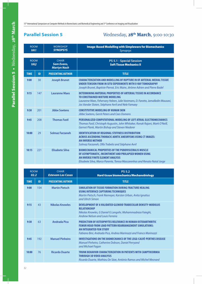
ROOMVA2
CHAIRSam Evans,
Martyn Nash
PS 5.1 - Special SessionSoft Tissue Mechanics II
Time iD PresenTing AuThor TiTLe
9:00 30 Joseph Brunet chArAcTerizATion AnD moDeLLing of ruPTure in of ArTeriAL meDiAL Tissue unDer Tension from in siTu exPerimenTs WiTh x-rAy TomogrAPhyJoseph Brunet, Baptiste Pierrat, Eric Maire, Jérôme Adrien and Pierre Badel
9:15 147 Lauranne Maes DeTermining mATeriAL ProPerTies of ArTeriAL Tissue in AccorDAnce To consTrAineD mixTure moDeLingLauranne Maes, Fehervary Heleen, Julie Vastmans, D. Farotto, Jamalledin Mousavi, Jos Vander Sloten, Stéphane Avril and Nele Famaey
9:30 201 Jibbe Soetens consTiTuTive moDeLing of humAn skinJibbe Soetens, Gerrit Peters and Cees Oomens
9:45 208 Thomas Fastl PersonALizeD comPuTATionAL moDeLing of LefT ATriAL eLecTromechAnicsThomas Fastl, Christoph Augustin, John Whitaker, Ronak Rajani, Mark O’Neill, Gernot Plank, Martin Bishop and Steven Niederer
10:00 29 Solmaz Farzaneh iDenTificATion of regionAL sTiffness DisTribuTion Across AscenDing ThorAcic AorTic Aneurysms using cT imAges: An inverse meThoDSolmaz Farzaneh, Olfa Trabelsi and Stéphane Avril
10:15 221 Elisabete Silva biomechAnicAL ProPerTies of The PuboviscerALis muscLe of AsymPTomATic, inconTinenT AnD ProLAPseD Women using An inverse finiTe eLemenT AnALysisElisabete Silva, Marco Parente, Teresa Mascarenhas and Renato Natal Jorge
Parallel Session 5 Wednesday, 28th March, 9:00-10:30
Par
alle
l Ses
sio
n 5
• W
edne
sday
, 28
th M
arch
ROOM02.2
CHAIREstevam Las Casas
PS 5.2Hard tissue biomechanics/Mechanobiology
Time iD PresenTing AuThor TiTLe
9:00 154 Martin Pietsch simuLATion of Tissue formATion During frAcTure heALing using inTerfAce cAPTuring TechniquesMartin Pietsch, Frank Niemeyer, Karsten Urban, Anita Ignatius and Ulrich Simon
9:15 43 Nikolas Knowles DeveLoPmenT of A vALiDATeD gLenoiD TrAbecuLAr DensiTy-moDuLus reLATionshiPNikolas Knowles, G Daniel G Langohr, Mohammadreza Faieghi, Andrew Nelson and Louis Ferreira
9:30 63 Andrada Pica PreDicTion of osTeoPhyTes reLevAnce in humAn osTeoArThriTic femur heAD from LoAD PATTern reArrAngemenT simuLATions: An inTegrATeD fem sTuDyFabiano Bini, Andrada Pica, Andrea Marinozzi and Franco Marinozzi
9:45 192 Manuel Pinheiro invesTigATions on The biomechAnics of The Legg-cALvé-PerThes DiseAseManuel Pinheiro, Catherine Dobson, Daniel Perryand and Michael Fagan
10:00 76 Ricardo Duarte Trunk behAvior chArAcTerizATion in PATienTs WiTh cAmPTocormiA Through 3D viDeo AnALysisRicardo Duarte, Mathieu De Sèze, António Ramos and Michel Mesnard
ROOMVA1
WORKSHOPSYNOPSYS
Image-Based Modelling with Simpleware for BiomechanicsSynopsys
26-29 March, 2018 • Instituto Superior Técnico • Lisbon • Portugal
33
Par
alle
l Ses
sio
n 5
• W
edne
sday
, 28
th M
arch
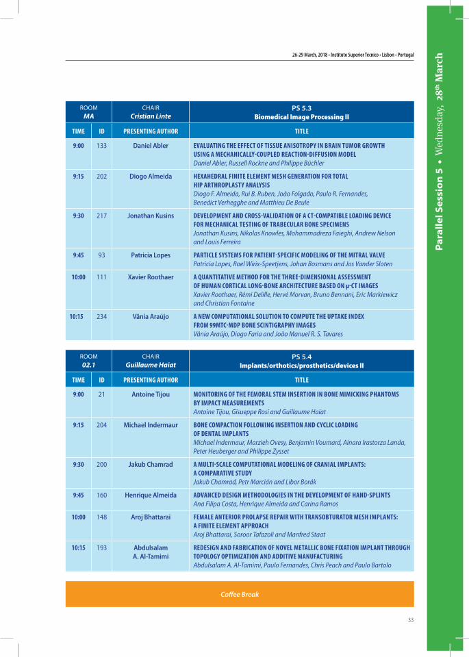
ROOMMA
CHAIRCristian Linte
PS 5.3Biomedical Image Processing II
Time iD PresenTing AuThor TiTLe
9:00 133 Daniel Abler evALuATing The effecT of Tissue AnisoTroPy in brAin Tumor groWTh using A mechAnicALLy-couPLeD reAcTion-Diffusion moDeLDaniel Abler, Russell Rockne and Philippe Büchler
9:15 202 Diogo Almeida hexAheDrAL finiTe eLemenT mesh generATion for ToTAL hiP ArThroPLAsTy AnALysisDiogo F. Almeida, Rui B. Ruben, João Folgado, Paulo R. Fernandes, Benedict Verhegghe and Matthieu De Beule
9:30 217 Jonathan Kusins DeveLoPmenT AnD cross-vALiDATion of A cT-comPATibLe LoADing Device for mechAnicAL TesTing of TrAbecuLAr bone sPecimensJonathan Kusins, Nikolas Knowles, Mohammadreza Faieghi, Andrew Nelson and Louis Ferreira
9:45 93 Patricia Lopes PArTicLe sysTems for PATienT-sPecific moDeLing of The miTrAL vALvePatricia Lopes, Roel Wirix-Speetjens, Johan Bosmans and Jos Vander Sloten
10:00 111 Xavier Roothaer A quAnTiTATive meThoD for The Three-DimensionAL AssessmenT of humAn corTicAL Long-bone ArchiTecTure bAseD on μ-cT imAgesXavier Roothaer, Rémi Delille, Hervé Morvan, Bruno Bennani, Eric Markiewicz and Christian Fontaine
10:15 234 Vânia Araújo A neW comPuTATionAL soLuTion To comPuTe The uPTAke inDex from 99mTc-mDP bone scinTigrAPhy imAgesVânia Araújo, Diogo Faria and João Manuel R. S. Tavares
ROOM02.1
CHAIRGuillaume Haiat
PS 5.4Implants/orthotics/prosthetics/devices II
Time iD PresenTing AuThor TiTLe
9:00 21 Antoine Tijou moniToring of The femorAL sTem inserTion in bone mimicking PhAnToms by imPAcT meAsuremenTsAntoine Tijou, Gisueppe Rosi and Guillaume Haiat
9:15 204 Michael Indermaur bone comPAcTion foLLoWing inserTion AnD cycLic LoADing of DenTAL imPLAnTsMichael Indermaur, Marzieh Ovesy, Benjamin Voumard, Ainara Irastorza Landa, Peter Heuberger and Philippe Zysset
9:30 200 Jakub Chamrad A muLTi-scALe comPuTATionAL moDeLing of crAniAL imPLAnTs: A comPArATive sTuDyJakub Chamrad, Petr Marcián and Libor Borák
9:45 160 Henrique Almeida ADvAnceD Design meThoDoLogies in The DeveLoPmenT of hAnD-sPLinTsAna Filipa Costa, Henrique Almeida and Carina Ramos
10:00 148 Aroj Bhattarai femALe AnTerior ProLAPse rePAir WiTh TrAnsobTurATor mesh imPLAnTs: A finiTe eLemenT APProAchAroj Bhattarai, Soroor Tafazoli and Manfred Staat
10:15 193 Abdulsalam A. Al-Tamimi
reDesign AnD fAbricATion of noveL meTALLic bone fixATion imPLAnT Through ToPoLogy oPTimizATion AnD ADDiTive mAnufAcTuringAbdulsalam A. Al-Tamimi, Paulo Fernandes, Chris Peach and Paulo Bartolo
Coffee Break

15th International Symposium on Computer Methods in Biomechanics and Biomedical Engineering and 3rd Conference on Imaging and Visualization
34
Plen
ary
Lect
ure
s •
Wed
nesd
ay,
28th
Mar
ch
ROOM
MA
CHAIR
John MiddletonPlenary IV
Time iD PLenAry sPeAker TiTLe
11:00 248 Daniela Iacoviello PhysioLogicAL cyberneTics: meThoDs AnD APPLicATionsDaniela Iacoviello
11:30 258 Christoph Bourauel high PerformAnce PoLymers in DenTisTry - biomechAnicAL AnD cLinicAL AsPecTsChristoph Bourauel, Ludger Keilig, Istabrak Hasan, Tobias Klur,Anne Kartzenbach and Helmut Stark
12:00 255 Estevam de las Casas Designing inTrAmeDuLLAr PosTs for veTerinAry APPLicATionsEstevam B. Las Casas, Leopoldo Paolucci, Rafael Faleiros, Sergio Rocha Jr., Paulo Fernandes, João Folgado, Luciano Rodrigues and Luciana Gomides
12:30 253 Martyn Nash biomechAnicAL mechAnisms of heArT fAiLureMartyn Nash
Plenary Lectures Wednesday, 28th March, 11:00-12:30
Lunch
Tour & Conference Dinner
26-29 March, 2018 • Instituto Superior Técnico • Lisbon • Portugal
35
Para
llel S
essi
on
6 •
Thu
rsda
y, 2
9th M
arch
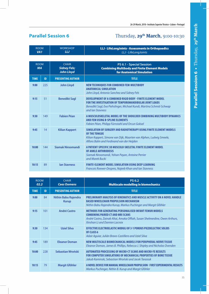
Parallel Session 6 Thursday, 29th March, 9:00-10:30
ROOMMA
CHAIRSidney Fels; John Lloyd
PS 6.1 - Special SessionCombining Multibody and Finite Element Models
for Anatomical Simulation
Time iD PresenTing AuThor TiTLe
9:00 225 John Lloyd neW Techniques for combineD fem-muLTiboDy AnATomicAL simuLATionJohn Lloyd, Antonio Sanchez and Sidney Fels
9:15 51 Benedikt Sagl DeveLoPmenT of A combineD rigiD boDy - finiTe eLemenT moDeL for The invesTigATion of TemPoromAnDibuLAr joinT LoADsBenedikt Sagl, Eva Piehslinger, Michael Kundi, Martina Schmid-Schwap and Ian Stavness
9:30 149 Fabien Péan A muscuLoskeLeTAL moDeL of The shouLDer combining muLTiboDy DynAmics AnD fem using b-sPLine eLemenTsFabien Péan, Philipp Fürnstahl and Orcun Goksel
9:45 14 Kilian Kappert simuLATion of surgery AnD rADioTherAPy using finiTe eLemenT moDeLs of The TongueKilian Kappert, Simone van Dijk, Maarten van Alphen, Ludwig Smeele, Alfons Balm and Ferdinand van der Heijden
10:00 144 Siamak Niroomandi A PATienT-sPecific 3D muscuLo-skeLeTAL finiTe eLemenT moDeL of AnkLe ArThroDesisSiamak Niroomandi, Yohan Payan, Antoine Perrier and Marek Bucki
10:15 89 Ian Stavness finiTe-eLemenT moDeL simuLATion using DeeP LeArningFrancois Roewer-Despres, Najeeb Khan and Ian Stavness
ROOM02.2
CHAIRCees Oomens
PS 6.2Multiscale modelling in biomechanics
Time iD PresenTing AuThor TiTLe
9:00 84 Nithin Babu Rajendra Kurup
PreLiminAry AnALysis of kinemATics AnD muscLe AcTiviTy on A noveL hAnDLe bAseD WheeLchAir ProPuLsion mechAnismNithin Babu Rajendra Kurup, Markus Puchinger and Margit Gföhler
9:15 101 André Castro meThoDs for generATing PersonALiseD infAnT femur moDeLs combining PAireD cT AnD mri scAnsAndré Castro, Zainab Altai, Amaka Offiah, Susan Shelmerdine, Owen Arthurs, Xinshan Li and Damien Lacroix
9:30 134 Uziel Silva effecTive eLecTroeLAsTic moDuLi of 3-1 Porous PiezoeLecTric soLiDs of cLAss 6Adair Aguiar, Julián Bravo-Castillero and Uziel Silva
9:45 189 Eleanor Doman neW muLTiscALe biomechAnicAL moDeLs for PeriPherAL nerve TissueEleanor Doman, James B. Phillips, Rebecca J. Shipley and Nicholas Ovenden
10:00 228 Sebastian Wroński AuTomATeD Processing of micro-cT scAns AnD micro-fe resuLTs for comPuTer simuLATions of mechAnicAL ProPerTies of bone TissueJakub Kaminski, Sebastian Wroński and Jacek Tarasiuk
10:15 79 Margit Gföhler A noveL Device for mAnuAL WheeLchAir ProPuLsion - firsT exPerimenTAL resuLTsMarkus Puchinger, Nithin B. Kurup and Margit Gföhler
ROOMVA1
WORKSHOPLLJ
LLJ - LifeLongJoints - Assessments in OrthopaedicsLLJ - LifeLongJoints
15th International Symposium on Computer Methods in Biomechanics and Biomedical Engineering and 3rd Conference on Imaging and Visualization
36
Par
alle
l Ses
sio
n 6
• T
hurs
day,
29th
Mar
ch
ROOMVA2
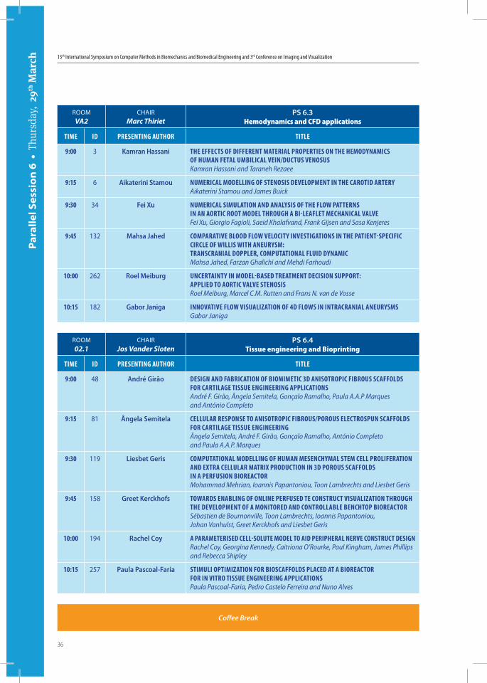
CHAIRMarc Thiriet
PS 6.3Hemodynamics and CFD applications
Time iD PresenTing AuThor TiTLe
9:00 3 Kamran Hassani The effecTs of DifferenT mATeriAL ProPerTies on The hemoDynAmics of humAn feTAL umbiLicAL vein/DucTus venosusKamran Hassani and Taraneh Rezaee
9:15 6 Aikaterini Stamou numericAL moDeLLing of sTenosis DeveLoPmenT in The cAroTiD ArTeryAikaterini Stamou and James Buick
9:30 34 Fei Xu numericAL simuLATion AnD AnALysis of The fLoW PATTerns in An AorTic rooT moDeL Through A bi-LeAfLeT mechAnicAL vALveFei Xu, Giorgio Fagioli, Saeid Khalafvand, Frank Gijsen and Sasa Kenjeres
9:45 132 Mahsa Jahed comPArATive bLooD fLoW veLociTy invesTigATions in The PATienT-sPecific circLe of WiLLis WiTh Aneurysm: TrAnscrAniAL DoPPLer, comPuTATionAL fLuiD DynAmicMahsa Jahed, Farzan Ghalichi and Mehdi Farhoudi
10:00 262 Roel Meiburg uncerTAinTy in moDeL-bAseD TreATmenT Decision suPPorT: APPLieD To AorTic vALve sTenosisRoel Meiburg, Marcel C.M. Rutten and Frans N. van de Vosse
10:15 182 Gabor Janiga innovATive fLoW visuALizATion of 4D fLoWs in inTrAcrAniAL AneurysmsGabor Janiga
ROOM02.1
CHAIRJos Vander Sloten
PS 6.4Tissue engineering and Bioprinting
Time iD PresenTing AuThor TiTLe
9:00 48 André Girão Design AnD fAbricATion of biomimeTic 3D AnisoTroPic fibrous scAffoLDs for cArTiLAge Tissue engineering APPLicATionsAndré F. Girão, Ângela Semitela, Gonçalo Ramalho, Paula A.A.P Marques and António Completo
9:15 81 Ângela Semitela ceLLuLAr resPonse To AnisoTroPic fibrous/Porous eLecTrosPun scAffoLDs for cArTiLAge Tissue engineeringÂngela Semitela, André F. Girão, Gonçalo Ramalho, António Completo and Paula A.A.P. Marques
9:30 119 Liesbet Geris comPuTATionAL moDeLLing of humAn mesenchymAL sTem ceLL ProLiferATion AnD exTrA ceLLuLAr mATrix ProDucTion in 3D Porous scAffoLDs in A Perfusion bioreAcTorMohammad Mehrian, Ioannis Papantoniou, Toon Lambrechts and Liesbet Geris
9:45 158 Greet Kerckhofs ToWArDs enAbLing of onLine PerfuseD Te consTrucT visuALizATion Through The DeveLoPmenT of A moniToreD AnD conTroLLAbLe benchToP bioreAcTorSébastien de Bournonville, Toon Lambrechts, Ioannis Papantoniou, Johan Vanhulst, Greet Kerckhofs and Liesbet Geris
10:00 194 Rachel Coy A PArAmeTeriseD ceLL-soLuTe moDeL To AiD PeriPherAL nerve consTrucT DesignRachel Coy, Georgina Kennedy, Caitriona O’Rourke, Paul Kingham, James Phillips and Rebecca Shipley
10:15 257 Paula Pascoal-Faria sTimuLi oPTimizATion for bioscAffoLDs PLAceD AT A bioreAcTor for in viTro Tissue engineering APPLicATionsPaula Pascoal-Faria, Pedro Castelo Ferreira and Nuno Alves
Coffee Break
26-29 March, 2018 • Instituto Superior Técnico • Lisbon • Portugal
37
Plen
ary
Lect
ure
s •
Thu
rsda
y, 2
9th M
arch
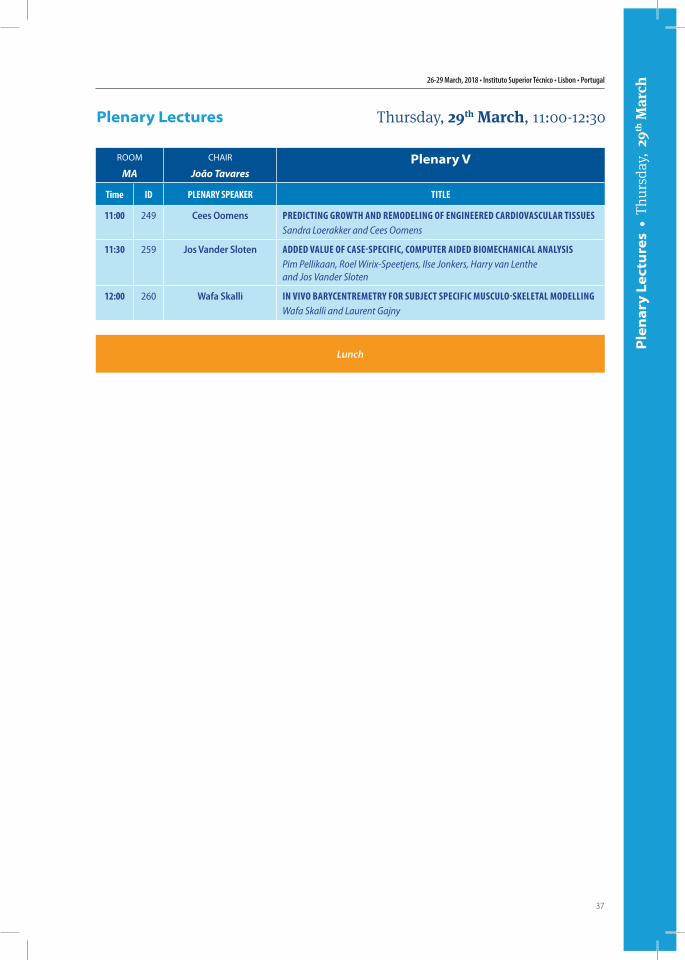
Plenary Lectures Thursday, 29th March, 11:00-12:30
ROOM
MA
CHAIR
João TavaresPlenary V
Time iD PLenAry sPeAker TiTLe
11:00 249 Cees Oomens PreDicTing groWTh AnD remoDeLing of engineereD cArDiovAscuLAr TissuesSandra Loerakker and Cees Oomens
11:30 259 Jos Vander Sloten ADDeD vALue of cAse-sPecific, comPuTer AiDeD biomechAnicAL AnALysisPim Pellikaan, Roel Wirix-Speetjens, Ilse Jonkers, Harry van Lenthe and Jos Vander Sloten
12:00 260 Wafa Skalli in vivo bArycenTremeTry for subjecT sPecific muscuLo-skeLeTAL moDeLLingWafa Skalli and Laurent Gajny
Lunch
15th International Symposium on Computer Methods in Biomechanics and Biomedical Engineering and 3rd Conference on Imaging and Visualization
38
Par
alle
l Ses
sio
n 7
• T
hurs
day,
29th
Mar
ch
ROOMMA
CHAIRMichael Sacks
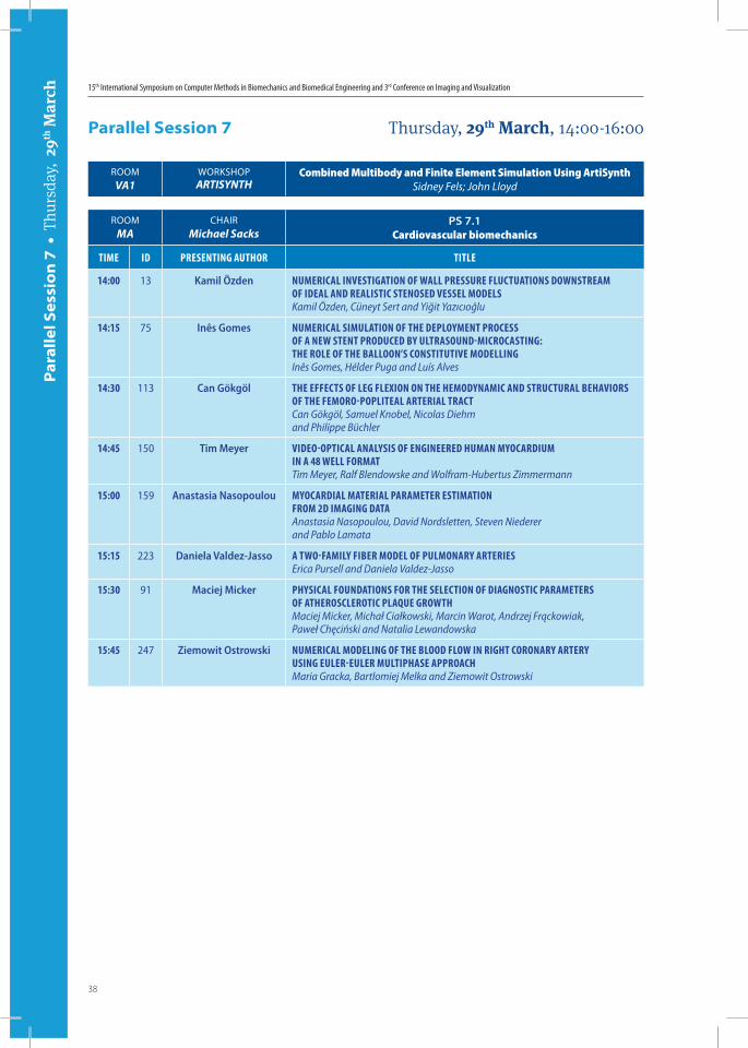
PS 7.1Cardiovascular biomechanics
Time iD PresenTing AuThor TiTLe
14:00 13 Kamil Özden numericAL invesTigATion of WALL Pressure fLucTuATions DoWnsTreAm of iDeAL AnD reALisTic sTenoseD vesseL moDeLsKamil Özden, Cüneyt Sert and Yiğit Yazıcıoğlu
14:15 75 Inês Gomes numericAL simuLATion of The DePLoymenT Process of A neW sTenT ProDuceD by uLTrAsounD-microcAsTing: The roLe of The bALLoon’s consTiTuTive moDeLLingInês Gomes, Hélder Puga and Luís Alves
14:30 113 Can Gökgöl The effecTs of Leg fLexion on The hemoDynAmic AnD sTrucTurAL behAviors of The femoro-PoPLiTeAL ArTeriAL TrAcTCan Gökgöl, Samuel Knobel, Nicolas Diehm and Philippe Büchler
14:45 150 Tim Meyer viDeo-oPTicAL AnALysis of engineereD humAn myocArDium in A 48 WeLL formATTim Meyer, Ralf Blendowske and Wolfram-Hubertus Zimmermann
15:00 159 Anastasia Nasopoulou myocArDiAL mATeriAL PArAmeTer esTimATion from 2D imAging DATAAnastasia Nasopoulou, David Nordsletten, Steven Niederer and Pablo Lamata
15:15 223 Daniela Valdez-Jasso A TWo-fAmiLy fiber moDeL of PuLmonAry ArTeriesErica Pursell and Daniela Valdez-Jasso
15:30 91 Maciej Micker PhysicAL founDATions for The seLecTion of DiAgnosTic PArAmeTers of ATheroscLeroTic PLAque groWThMaciej Micker, Michał Ciałkowski, Marcin Warot, Andrzej Frąckowiak, Paweł Chęciński and Natalia Lewandowska
15:45 247 Ziemowit Ostrowski numericAL moDeLing of The bLooD fLoW in righT coronAry ArTery using euLer-euLer muLTiPhAse APProAchMaria Gracka, Bartlomiej Melka and Ziemowit Ostrowski
Parallel Session 7 Thursday, 29th March, 14:00-16:00
ROOMVA1
WORKSHOPARTISYNTH
Combined Multibody and Finite Element Simulation Using ArtiSynthSidney Fels; John Lloyd
26-29 March, 2018 • Instituto Superior Técnico • Lisbon • Portugal
39
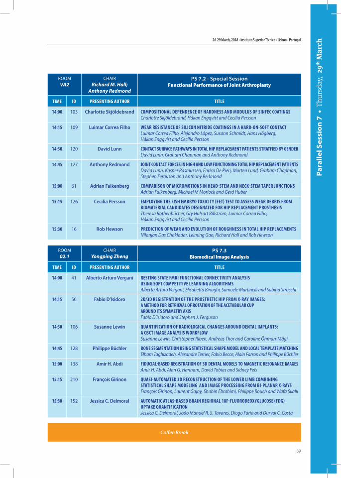
ROOMVA2
CHAIRRichard M. Hall;
Anthony Redmond
PS 7.2 - Special SessionFunctional Performance of Joint Arthroplasty
Time iD PresenTing AuThor TiTLe
14:00 103 Charlotte Skjöldebrand comPosiTionAL DePenDence of hArDness AnD moDuLus of sinfec coATingsCharlotte Skjöldebrand, Håkan Engqvist and Cecilia Persson
14:15 109 Luimar Correa Filho WeAr resisTAnce of siLicon niTriDe coATings in A hArD-on-sofT conTAcTLuimar Correa Filho, Alejandro López, Susann Schmidt, Hans Högberg, Håkan Engqvist and Cecilia Persson
14:30 120 David Lunn conTAcT surfAce PAThWAys in ToTAL hiP rePLAcemenT PATienTs sTrATfieD by genDerDavid Lunn, Graham Chapman and Anthony Redmond
14:45 127 Anthony Redmond joinT conTAcT forces in high AnD LoW funcTioning ToTAL hiP rePLAcemenT PATienTsDavid Lunn, Kasper Rasmussen, Enrico De Pieri, Morten Lund, Graham Chapman, Stephen Ferguson and Anthony Redmond
15:00 61 Adrian Falkenberg comPArison of micromoTions in heAD-sTem AnD neck-sTem TAPer juncTionsAdrian Falkenberg, Michael M Morlock and Gerd Huber
15:15 126 Cecilia Persson emPLoying The fish embryo ToxiciTy (feT) TesT To Assess WeAr Debris from biomATeriAL cAnDiDATes DesignATeD for hiP rePLAcemenT ProsThesisTheresa Rothenbücher, Gry Hulsart Billström, Luimar Correa Filho, Håkan Engqvist and Cecilia Persson
15:30 16 Rob Hewson PreDicTion of WeAr AnD evoLuTion of roughness in ToTAL hiP rePLAcemenTsNilanjan Das Chakladar, Leiming Gao, Richard Hall and Rob Hewson
ROOM02.1
CHAIRYongping Zheng
PS 7.3Biomedical Image Analysis
Time iD PresenTing AuThor TiTLe
14:00 41 Alberto Arturo Vergani resTing sTATe fmri funcTionAL connecTiviTy AnALysis using sofT comPeTiTive LeArning ALgoriThmsAlberto Arturo Vergani, Elisabetta Binaghi, Samuele Martinelli and Sabina Strocchi
14:15 50 Fabio D’Isidoro 2D/3D regisTrATion of The ProsTheTic hiP from x-rAy imAges: A meThoD for reTrievAL of roTATion of The AceTAbuLAr cuP ArounD iTs symmeTry AxisFabio D’Isidoro and Stephen J. Ferguson
14:30 106 Susanne Lewin quAnTificATion of rADioLogicAL chAnges ArounD DenTAL imPLAnTs: A cbcT imAge AnALysis WorkfLoWSusanne Lewin, Christopher Riben, Andreas Thor and Caroline Öhman-Mägi
14:45 128 Philippe Büchler bone segmenTATion using sTATisTicAL shAPe moDeL AnD LocAL TemPLATe mATchingElham Taghizadeh, Alexandre Terrier, Fabio Becce, Alain Farron and Philippe Büchler
15:00 138 Amir H. Abdi fiDuciAL-bAseD regisTrATion of 3D DenTAL moDeLs To mAgneTic resonAnce imAgesAmir H. Abdi, Alan G. Hannam, David Tobias and Sidney Fels
15:15 210 François Girinon quAsi-AuTomATeD 3D reconsTrucTion of The LoWer Limb combining sTATisTicAL shAPe moDeLing AnD imAge Processing from bi-PLAnAr x-rAysFrançois Girinon, Laurent Gajny, Shahin Ebrahimi, Philippe Rouch and Wafa Skalli
15:30 152 Jessica C. Delmoral AuTomATic ATLAs-bAseD brAin regionAL 18f-fLuoroDeoxygLucose (fDg) uPTAke quAnTificATionJessica C. Delmoral, João Manuel R. S. Tavares, Diogo Faria and Durval C. Costa
Par
alle
l Ses
sio
n 7
• T
hurs
day,
29th
Mar
ch
Coffee Break
15th International Symposium on Computer Methods in Biomechanics and Biomedical Engineering and 3rd Conference on Imaging and Visualization
40
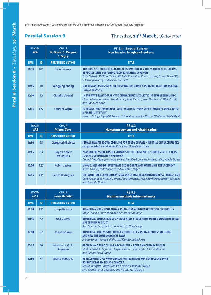
ROOMMA
CHAIRW. Skalli; C. Vergari;
L. Gajny
PS 8.1 - Special SessionNon-invasive imaging of scoliosis
Time iD PresenTing AuThor TiTLe
16:30 185 Saša Ćuković non-ionizing Three-DimensionAL esTimATion of AxiAL verTebrAL roTATions in ADoLescenTs suffering from iDioPAThic scoLiosisSaša Ćuković, William Taylor, Michele Fiorentino, Vanja Luković, Goran Devedžić, S. Karupppasamy and Silvio Lorenzetti
16:45 10 Yongping Zheng scoLioscAn: AssessmenT of 3D sPinAL DeformiTy using uLTrAsounD imAgingYongping Zheng
17:00 12 Claudio Vergari sheAr WAve eLAsTogrAPhy To chArAcTerize scoLioTic inTerverTebrAL DiscClaudio Vergari, Tristan Langlais, Raphaël Pietton, Jean Dubousset, Wafa Skalli and Raphaël Vialle
17:15 122 Laurent Gajny 3D reconsTrucTion of ADoLescenT scoLioTic Trunk shAPe from biPLAnAr x-rAys: A feAsibiLiTy sTuDyLaurent Gajny, Léopold Robichon, Thibault Hernandez, Raphaël Vialle and Wafa Skalli
ROOMVA2
CHAIRMiguel Silva
PS 8.2Human movement and rehabilitation
Time iD PresenTing AuThor TiTLe
16:30 65 Gergana Nikolova femALe humAn boDy moDeLLing for sTuDy of mAss - inerTiAL chArAcTerisTicsGergana Nikolova, Vladimir Kotev and Daniel Dantchev
16:45 83 Tiago de Melo Malaquias
PLAnTAr Pressure bAseD esTimATes of fooT kinemATics During gAiT - A LeAsT squAres oPTimizATion APProAchTiago de Melo Malaquias, Wouter Aerts, Friedl De Groote, Ilse Jonkers and Jos Vander Sloten
17:00 123 Robin Layton A noveL meThoD To invesTigATe cross-sheAr moTion in A hiP rePLAcemenTRobin Layton, Todd Stewart and Neil Messenger
17:15 145 Carlos Rodrigues sofTWAre TooL for significAnT AnALysis of comPLemenTAry DomAins AT humAn gAiTCarlos Rodrigues, Miguel Correia, João Abrantes, Marco Aurélio Benedetti Rodrigues and Jurandir Nadal
ROOM02.1
CHAIRJorge Belinha
PS 8.3Meshless methods in biomechanics
Time iD PresenTing AuThor TiTLe
16:30 195 Jorge Belinha biomechAnicAL APPLicATions using ADvAnceD DiscreTizATion TechniquesJorge Belinha, Lúcia Dinis and Renato Natal Jorge
16:45 72 Ana Guerra numericAL simuLATion of Angiogenesis sTimuLATion During WounD heALing: A PreLiminAry sTuDyAna Guerra, Jorge Belinha and Renato Natal Jorge
17:00 97 Joana Gomes numericAL AnALysis of chiTosAn guiDe Tubes using meshLess meThoDs AnD neW PhenomenoLogicAL LAWsJoana Gomes, Jorge Belinha and Renato Natal Jorge
17:15 99 Madalena M. A. Peyroteo
groWTh AnD remoDeLing mechAnisms – bone AnD cArDiAc TissuesMadalena M. A. Peyroteo, Jorge Belinha, Joaquim A.C.F. Leite Moreira and Renato Natal Jorge
17:30 77 Marco Marques DeveLoPmenT of A homogenizATion Technique for TrAbecuLAr bone using The fAbric Tensor concePTMarco Marques, Jorge Belinha, António Fonseca Oliveira, M.C. Manzanares Céspedes and Renato Natal Jorge
Par
alle
l Ses
sio
n 8
• T
hurs
day,
29th
Mar
ch
Parallel Session 8 Thursday, 29th March, 16:30-17:45

41
Book of Abstracts
26-29 March, 2018
Instituto Superior Técnico Lisbon • Portugal
CMBBE201815th International Symposium on Computer Methods
in Biomechanics and Biomedical Engineering
and
3rd Conference on Imaging and Visualization

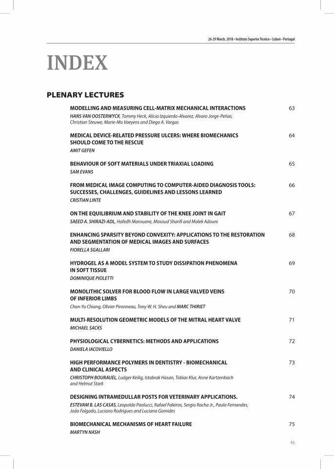
26-29 March, 2018 • Instituto Superior Técnico • Lisbon • Portugal
43
INDEXPLENARY LECTURES
MODELLING AND MEASURING CELL-MATRIX MECHANICAL INTERACTIONSHANS VAN OOSTERWYCK, Tommy Heck, Alicia Izquierdo-Alvarez, Alvaro Jorge-Peñas,
Christian Steuwe, Marie-Mo Vaeyens and Diego A. Vargas
MEDICAL DEVICE-RELATED PRESSURE ULCERS: WHERE BIOMECHANICS SHOULD COME TO THE RESCUE
AMIT GEFEN
BEHAVIOUR OF SOFT MATERIALS UNDER TRIAXIAL LOADINGSAM EVANS
FROM MEDICAL IMAGE COMPUTING TO COMPUTER-AIDED DIAGNOSIS TOOLS: SUCCESSES, CHALLENGES, GUIDELINES AND LESSONS LEARNED
CRISTIAN LINTE
ON THE EQUILIBRIUM AND STABILITY OF THE KNEE JOINT IN GAITSAEED A. SHIRAZI-ADL, Hafedh Marouane, Masoud Sharifi and Malek Adouni
ENHANCING SPARSITY BEYOND CONVEXITY: APPLICATIONS TO THE RESTORATION AND SEGMENTATION OF MEDICAL IMAGES AND SURFACES
FIORELLA SGALLARI
HYDROGEL AS A MODEL SYSTEM TO STUDY DISSIPATION PHENOMENA IN SOFT TISSUE
DOMINIQUE PIOLETTI
MONOLITHIC SOLVER FOR BLOOD FLOW IN LARGE VALVED VEINS OF INFERIOR LIMBS
Chen-Yu Chiang, Olivier Pironneau, Tony W. H. Sheu and MARC THIRIET
MULTI-RESOLUTION GEOMETRIC MODELS OF THE MITRAL HEART VALVEMICHAEL SACKS
PHYSIOLOGICAL CYBERNETICS: METHODS AND APPLICATIONSDANIELA IACOVIELLO
HIGH PERFORMANCE POLYMERS IN DENTISTRY - BIOMECHANICAL AND CLINICAL ASPECTS
CHRISTOPH BOURAUEL, Ludger Keilig, Istabrak Hasan, Tobias Klur, Anne Kartzenbach and Helmut Stark
DESIGNING INTRAMEDULLAR POSTS FOR VETERINARY APPLICATIONS.ESTEVAM B. LAS CASAS, Leopoldo Paolucci, Rafael Faleiros, Sergio Rocha Jr., Paulo Fernandes,
João Folgado, Luciano Rodrigues and Luciana Gomides
BIOMECHANICAL MECHANISMS OF HEART FAILUREMARTYN NASH
63
64
65
66
67
68
69
70
71
72
73
74
75
15th International Symposium on Computer Methods in Biomechanics and Biomedical Engineering and 3rd Conference on Imaging and Visualization
44
PREDICTING GROWTH AND REMODELING OF ENGINEERED CARDIOVASCULAR TISSUESSANDRA LOERAKKER
ADDED VALUE OF CASE-SPECIFIC, COMPUTER AIDED BIOMECHANICAL ANALYSISPim Pellikaan, Roel Wirix-Speetjens, Ilse Jonkers, Harry van Lenthe and JOS VANDER SLOTEN
IN VIVO BARYCENTREMETRY FOR SUBJECT SPECIFIC MUSCULO-SKELETAL MODELLING
WAFA SKALLI and Laurent Gajny
WORKSHOPS
FROM REALITY TO VIRTUALITYBETA CAE Systems International AG
WORKING WITH C-MOTION’S DYNAMIC STEREO X-RAY SOFTWARE SUITEC-Motion Biomechanics Software
IMAGE-BASED MODELLING WITH SIMPLEWARE FOR BIOMECHANICSSynopsys
ASSESSMENTS IN ORTHOPAEDICSLLJ - LifeLongJoints
COMBINED MULTIBODY AND FINITE ELEMENT SIMULATION USING ARTISYNTHArtiSynth
ABSTRACTS
ID3 THE EFFECTS OF DIFFERENT MATERIAL PROPERTIES ON THE HEMODYNAMICS OF HUMAN FETAL UMBILICAL VEIN/DUCTUS VENOSUS
Kamran Hassani and Taraneh Rezaee
ID6 NUMERICAL MODELLING OF STENOSIS DEVELOPMENT IN THE CAROTID ARTERYAikaterini Stamou and James Buick
ID8 EXTRACTING AND EVALUATING TEXTURE FEATURES FROM BINARY GRADIENT CONTOURS OF MICROCALCIFICATIONS CLUSTERS IN BREAST MAMMOGRAMS
Marcelo Duarte, Andre Alvarenga and Wagner Coelho de Albuquerque Pereira
ID9 AN ADVANCED METAL ARTIFACT REDUCTION METHOD FOR A DENTAL CTMohamed Hegazy, Min Hyoung Cho and Soo Yeol Lee
ID10 SCOLIOSCAN: ASSESSMENT OF 3D SPINAL DEFORMITY USING ULTRASOUND IMAGING
Yongping Zheng
ID12 SHEAR WAVE ELASTOGRAPHY TO CHARACTERIZE SCOLIOTIC INTERVERTEBRAL DISC
Claudio Vergari, Tristan Langlais, Raphaël Pietton, Jean Dubousset, Wafa Skalli and Raphaël Vialle
76
77
78
81
81
81
82
82
85
86
87
88
89
90
26-29 March, 2018 • Instituto Superior Técnico • Lisbon • Portugal
45
ID13 NUMERICAL INVESTIGATION OF WALL PRESSURE FLUCTUATIONS DOWNSTREAM OF IDEAL AND REALISTIC STENOSED VESSEL MODELS
Kamil Özden, Cüneyt Sert and Yiğit Yazıcıoğlu
ID14 SIMULATION OF SURGERY AND RADIOTHERAPY USING FINITE ELEMENT MODELS OF THE TONGUE
Kilian Kappert, Simone van Dijk, Maarten van Alphen, Ludwig Smeele, Alfons Balm and Ferdinand van der Heijden
ID15 A COMPUTATIONAL LOWER-EXTREMITY MODEL TO QUANTIFY THE STABILITY OF AN ANTERIOR CRUCIATE LIGAMENT DEFICIENT KNEE JOINT AT HEEL STRIKE : GAIT PARAMETERS MARKING COPERS FROM NON-COPERS
Masoud Sharifi, Saeed A. Shirazi-Adl and Hafedh Marouane
ID16 PREDICTION OF WEAR AND EVOLUTION OF ROUGHNESS IN TOTAL HIP REPLACEMENTSNilanjan Das Chakladar, Leiming Gao, Richard Hall and Rob Hewson
ID17 ROLE OF FLOW DEPENDENT AND FLOW INDEPENDENT VISCOELASTICITY ON TIME DEPENDENT BEHAVIOUR OF VISCO-POROUS SCAFFOLDS
Naser Nasrollahzadeh and Dominique Pioletti
ID18 A CHEMICO-MECHANICALLY INDUCED CELL MODEL WITH AN APPLICATION TO CANCER METASTASIS
Jiao Chen, Daphne Weihs and Fred Vermolen
ID21 MONITORING OF THE FEMORAL STEM INSERTION IN BONE MIMICKING PHANTOMS BY IMPACT MEASUREMENTS
Antoine Tijou, Gisueppe Rosi and Guillaume Haiat
ID22 ASSESSING DENTAL IMPLANT STABILITY USING A QUANTITATIVE ULTRASOUND TECHNIQUE AND RESONANCE FREQUENCY ANALYSIS
Romain Vayron, Vu-Hieu Nguyen and Guillaume Haiat
ID23 NUMERICAL INVESTIGATION OF BONE HEALING AROUND IMMEDIATELY LOADED DENTAL IMPLANTS USING SIKA DEER ANTLERS AS IMPLANT BED
Istabrak Hasan, Yun He, Ludger Keilig, Dominik Fischer, Luisa Ziegler, Gerhard Wahl and Christoph Bourauel
ID24 TRUNK MUSCLE FORCES AND SPINAL LOADS DURING SIT-TO-STAND AND STAND-TO-SIT ACTIVITIES: DIFFERENCES BETWEEN PERSONS WITH AND WITHOUT UNILATERAL TRANSFEMORAL AMPUTATION
Iman Shojaei, Brad Hendershot, Matthew Ballard, Julian Acasio, Christopher Dearth and Babak Bazrgari
ID25 INTEGRALLY SKINNED ASYMMETRIC CELLULOSE ACETATE-SILICA MEMBRANES FOR EXTRACORPOREAL BLOOD ULTRAFILTRATION.
Monica Faria, Cintia Moreira and Maria Norberta de Pinho
ID26 OXYGEN MASS TRANSFER IN OXYGEN/MEMBRANE/WATER FLOW SYSTEMSCintia Moreira, Monica Faria and Maria Norberta de Pinho
ID29 IDENTIFICATION OF REGIONAL STIFFNESS DISTRIBUTION ACROSS ASCENDING THORACIC AORTIC ANEURYSMS USING CT IMAGES: AN INVERSE METHOD
Solmaz Farzaneh, Olfa Trabelsi and Stéphane Avril
91
92
93
94
95
96
97
98
99
100
101
102
103
15th International Symposium on Computer Methods in Biomechanics and Biomedical Engineering and 3rd Conference on Imaging and Visualization
46
ID30 CHARACTERIZATION AND MODELLING OF RUPTURE IN OF ARTERIAL MEDIAL TISSUE UNDER TENSION FROM IN SITU EXPERIMENTS WITH X-RAY TOMOGRAPHY
Joseph Brunet, Baptiste Pierrat, Eric Maire, Jérôme Adrien and Pierre Badel
ID31 COMPUTER-AIDED SURGERY FOR THE MEDIAL PATELLOFEMORAL LIGAMENT RECONSTRUCTION: A PARAMETRIC FINITE ELEMENT MODEL
Diego Alastruey-López, Vicente Sanchis-Alfonso, Angel Alberich-Bayarri and María Angeles Pérez
ID32 SENSITIVITY ANALYSIS OF A PATIENT-SPECIFIC FINITE ELEMENT MODEL OF SHOULDER ARTHROPLASTY
Yasmine Boulanaache, Gerard Güell Bartrina, Fabio Becce, Dominique Pioletti, Alexandre Terrier and Alain Farron
ID34 NUMERICAL SIMULATION AND ANALYSIS OF THE FLOW PATTERNS IN AN AORTIC ROOT MODEL THROUGH A BI-LEAFLET MECHANICAL VALVE
Fei Xu, Giorgio Fagioli, Saeid Khalafvand, Frank Gijsen and Sasa Kenjeres
ID35 SUBJECT-SPECIFIC TRUNK MUSCULOSKELETAL MODELING: CHARACTERISTICS, VALIDATION, AND APPLICATIONS
Farshid Ghezelbash, Saeed A. Shirazi-Adl, Zakaria El Ouaaid, André Plamondon and Navid Arjmand
ID37 THREE DIMENSIONAL EVALUATION OF THE HOLOGRAPHIC PROJECTION IN DIGITAL DENTAL MODELS SUPERIMPOSITION USING HOLOLENS DEVICE
Sameh Talaat, Christoph Bourauel, Ahmed Kaboudan and Nivine Ragy
ID38 BIOMECHANICAL ANALYSIS OF TOOTH MOVEMENTS IN CASE OF BONE LOSS AND ANTERIOR CROWDING IN THE LOWER JAW USING FINITE ELEMENT METHODS
Susanne Reimann, Dorna Baghdadi, Christoph Reichert, Ludger Keilig, Andreas Jäger and Christoph Bourauel
ID39 NUMERICAL INVESTIGATIONS OF BONE REMODELLING AROUND THE MOUSE MANDIBULAR MOLAR PRIMORDIA
Junliang Chen, Yun He, Ludger Keilig, Susanne Reimann, Istabrak Hasan, Ralf Radlanski and Christoph Bourauel
ID40 A FINITE ELEMENT IMPLEMENTATION OF GROWTH AND REMODELING BASED ON THE HOMEGENIZED CONSTRAINED MIXTURE MODEL
S. Jamaleddin Mousavi and Stéphane Avril
ID41 RESTING STATE FMRI FUNCTIONAL CONNECTIVITY ANALYSIS USING SOFT COMPETITIVE LEARNING ALGORITHMS
Alberto Arturo Vergani, Elisabetta Binaghi, Samuele Martinelli and Sabina Strocchi
ID43 DEVELOPMENT OF A VALIDATED GLENOID TRABECULAR DENSITY-MODULUS RELATIONSHIP
Nikolas Knowles, G Daniel G Langohr, Mohammadreza Faieghi, Andrew Nelson and Louis Ferreira
ID44 POLYETHYLENE GLENOID COMPONENT BACKSIDE GEOMETRY INFLUENCES FIXATION IN TOTAL SHOULDER ARTHROPLASTY
Nikolas Knowles, G Daniel G Langohr, George Athwal and Louis Ferreira
ID45 FINITE ELEMENTS ANALYSIS OF THE STRESS DISTRIBUTION ON TEMPOROMANDIBULAR OF MANDIBULAR ADVANCEMENT DEVICES
Agnese Brunzini, Marco Mandolini, Steve Manieri, Michele Germani and Alida Mazzoli
104
105
106
107
108
109
110
111
112
113
114
115
116
26-29 March, 2018 • Instituto Superior Técnico • Lisbon • Portugal
47
ID48 DESIGN AND FABRICATION OF BIOMIMETIC 3D ANISOTROPIC FIBROUS SCAFFOLDS FOR CARTILAGE TISSUE ENGINEERING APPLICATIONS
André F. Girão, Ângela Semitela, Gonçalo Ramalho, Paula A.A.P Marques and António Completo
ID49 SIMULATION OF SWALLOWING INCLUDING ANATOMICAL STRUCTURE AND FOOD BOLUS FLOW USING FLUID-STRUCTURE INTERACTION METHOD
Yukihiro Michiwaki, Takahiro Kikuchi, Tetsu Kamiya, Yoshio Toyama, Keigo Hanyu, Megumi Takai and Seiichi Koshizuka
ID50 2D/3D REGISTRATION OF THE PROSTHETIC HIP FROM X-RAY IMAGES: A METHOD FOR RETRIEVAL OF ROTATION OF THE ACETABULAR CUP AROUND ITS SYMMETRY AXIS
Fabio D’Isidoro and Stephen J. Ferguson
ID51 TOWARDS THE DEVELOPMENT OF A COMBINED RIGID BODY - FINITE ELEMENT MODEL FOR THE INVESTIGATION OF TEMPOROMANDIBULAR JOINT LOADS
Benedikt Sagl, Eva Piehslinger, Michael Kundi, Martina Schmid-Schwap and Ian Stavness
ID52 VALIDATION MEASUREMENTS AND COMPUTER SIMULATIONS OF THE NEWBORN’S BRAIN COOLING PROCESS
Dominika Bandola, Andrzej J. Nowak, Marek Rojczyk, Ziemowit Ostrowski and Wojciech Walas
ID55 FINITE ELEMENT (FE) CALCULATION OF THE SPINAL LOAD-SHARING VIA SEQUENTIAL DISSECTION OF THE SPINAL PARTS
Sadegh Naserkhaki, Mohammad-Javad Kheyrkhah, Ava Maboudmanesh, Fiona Youkhanva and Marwan El-Rich
ID60 CONTACTLESS HAND IDENTIFICATION USING MACHINE LEARNINGArmin Dietz, Joachim Hienzsch and Eduard Reithmeier
ID61 COMPARISON OF MICROMOTIONS IN HEAD-STEM AND NECK-STEM TAPER JUNCTIONSAdrian Falkenberg, Michael M Morlock and Gerd Huber
ID62 A LOOK INTO THE MECHANICAL PROPERTIES OF SINGLE CELLS: A TWO-PHASE CFD MODEL AND ITS VALIDATION
Monica Piergiovanni, Gregor Holzner, Claudia Atzeni, Stavros Stavrakis, Elena Bianchi, Andrew Demello and Gabriele Dubini
ID63 PREDICTION OF OSTEOPHYTES RELEVANCE IN HUMAN OSTEOARTHRITIC FEMUR HEAD FROM LOAD PATTERN REARRANGEMENT SIMULATIONS: AN INTEGRATED FEM STUDY
Fabiano Bini, Andrada Pica, Andrea Marinozzi and Franco Marinozzi
ID64 INVESTIGATING METHODS OF MODELLING AUGMENTATION IN HUMAN LUMBAR VERTEBRAE
Gavin Day, Alison C. Jones and Ruth K. Wilcox
ID65 FEMALE HUMAN BODY MODELLING FOR STUDY OF MASS-INERTIAL CHARACTERISTICSGergana Nikolova, Vladimir Kotev and Daniel Dantchev
ID68 EFFECTS OF LUMBO-PELVIC RHYTHM ON TRUNK MUSCLE FORCES AND DISC LOAD DURING FORWARD FLEXION: COMBINED MUSCULOSKELETAL AND FINITE ELEMENT MODELING
Tao Liu, Kinda Khalaf and Marwan El-Rich
117
118
119
120
121
122
123
124
125
126
127
128
129
15th International Symposium on Computer Methods in Biomechanics and Biomedical Engineering and 3rd Conference on Imaging and Visualization
48
ID69 ASSESSING THE RELATION BETWEEN SPINO-PELVIC PARAMETERS AND LUMBAR LOADS THROUGH MUSCULOSKELETAL MODELING APPROACH
Tito Bassani and Fabio Galbusera
ID70 ESTIMATION OF LOADS ON HUMAN LUMBAR SPINE - A CRITICAL REVIEW OF PAST IN VIVO AND COMPUTATIONAL MODEL STUDIES
Hendrik Schmidt, Aboulfazl Saeed Shirazi-Adl, Navid Arjman and Marcel Dreischarf
ID71 NUMERICAL SIMULATIONS OF BONE REMODELLING AFTER NUCLEOTOMYAndrea Calvo-Echenique, Maxim Bashkuev, Sandra Reitmaier, Amaya Pérez-Del Palomar
and Hendrik Schmidt
ID72 NUMERICAL SIMULATION OF ANGIOGENESIS STIMULATION DURING WOUND HEALING: A PRELIMINARY STUDY
Ana Guerra, Jorge Belinha and Renato Natal Jorge
ID73 LOAD-SHARING IN HAND-HELD STANDING POSTURE: COMBINED MUSCULOSKELETAL AND FINITE ELEMENT MODELING
Tao Liu, Kinda Khalaf and Marwan El-Rich
ID74 EVALUATION OF CERVICAL LAMINECTOMY ON INTERSEGMENTAL MOTIONS USING A VALIDATED PARAMETRIC SUBJECT-SPECIFIC FINITE ELEMENT MODEL
Kinda Khalaf, Mohammad Nikkhoo, Marwan El-Rich and Chih-Hsiu Cheng
ID75 NUMERICAL SIMULATION OF THE DEPLOYMENT PROCESS OF A NEW STENT PRODUCED BY ULTRASOUND-MICROCASTING: THE ROLE OF THE BALLOON’S CONSTITUTIVE MODELLING
Inês Gomes, Hélder Puga and Luís Alves
ID76 TRUNK BEHAVIOR CHARACTERIZATION IN PATIENTS WITH CAMPTOCORMIA THROUGH 3D VIDEO ANALYSIS
Ricardo Duarte, Mathieu De Sèze, António Ramos and Michel Mesnard
ID77 DEVELOPMENT OF A HOMOGENIZATION TECHNIQUE FOR TRABECULAR BONE USING THE FABRIC TENSOR CONCEPT
Marco Marques, Jorge Belinha, António Fonseca Oliveira, M.C. Manzanares Céspedes and Renato Natal Jorge
ID78 LIMB SPARING IN DOGS USING PATIENT-SPECIFIC ENDOPROSTHESES AND CUTTING GUIDES: DESIGN, MANUFACTURE AND PRELIMINARY VALIDATION
Anatolie Timercan, Vladimir Brailovski, Yvan Petit, Bertrand Lussier and Bernard Séguin
ID79 A NOVEL DEVICE FOR MANUAL WHEELCHAIR PROPULSION - FIRST EXPERIMENTAL RESULTS
Markus Puchinger, Nithin B. Kurup and Margit Gföhler
ID81 CELLULAR RESPONSE TO ANISOTROPIC FIBROUS/POROUS ELECTROSPUN SCAFFOLDS FOR CARTILAGE TISSUE ENGINEERING
Ângela Semitela, André F. Girão, Gonçalo Ramalho, António Completo and Paula A.A.P. Marques
ID83 PLANTAR PRESSURE BASED ESTIMATES OF FOOT KINEMATICS DURING GAIT - A LEAST SQUARES OPTIMIZATION APPROACH
Tiago de Melo Malaquias, Wouter Aerts, Friedl De Groote, Ilse Jonkers and Jos Vander Sloten
130
131
132
133
134
135
136
137
138
139
140
141
142
26-29 March, 2018 • Instituto Superior Técnico • Lisbon • Portugal
49
ID84 PRELIMINARY ANALYSIS OF KINEMATICS AND MUSCLE ACTIVITY ON A NOVEL HANDLE BASED WHEELCHAIR PROPULSION MECHANISM
Nithin Babu Rajendra Kurup, Markus Puchingerand Margit Gföhler
ID85 COMPUTATION OF GRASP QUALITY METRICS IN OPENHAND SIMULATOR TO IMPROVE A 3D PRINTED PROSTHETIC HAND
Immaculada Llop-Harillo, Carlos Rubert and Antonio Pérez-González
ID86 NUMERICAL STUDY OF CAROTID BIFURCATION ANGLE EFFECT ON BLOOD FLOW DISORDERS
Natalia Lewandowska, Michał Ciałkowski, Maciej Micker, Marcin Warot, Andrzej Frąckowiak and Paweł Chęciński
ID87 MODELLING OF CROSS-LINKING DYNAMICS IN ACTIN NETWORKSJoão Pedro Ferreira, Marco Parente and Renato Natal Jorge
ID89 TOWARDS FINITE-ELEMENT SIMULATION USING DEEP LEARNINGFrancois Roewer-Despres, Najeeb Khan and Ian Stavness
ID90 IMPACT OF PATCHES ON BLOOD FLOW DISORDERS IN CAROTID ARTERY
Michał Ciałkowski, Natalia Lewandowska, Maciej Micker, Marcin Warot, Andrzej Frąckowiak and Paweł Chęciński
ID91 PHYSICAL FOUNDATIONS FOR THE SELECTION OF DIAGNOSTIC PARAMETERS OF ATHEROSCLEROTIC PLAQUE GROWTH
Maciej Micker, Michał Ciałkowski, Natalia Lewandowska, Marcin Warot, Andrzej Frąckowiak and Paweł Chęciński
ID93 PARTICLE SYSTEMS FOR PATIENT-SPECIFIC MODELING OF THE MITRAL VALVE
Patricia Lopes, Roel Wirix-Speetjens, Johan Bosmans and Jos Vander Sloten
ID94 PREDICTION OF THE RISK OF VERTEBRAL FRACTURES IN METASTATICALLY INVOLVED SPINES
Fabio Galbusera, Tito Bassani, Gloria Casaroli and Zhihui Qian
ID95 HOMOGENIZED FINITE ELEMENT ANALYSIS OF THE BONE-IMPLANT INTERFACE: ROLE OF PRESS-FIT, DAMAGE AND FRICTION
Marzieh Ovesy and Philippe Zysset
ID96 RELATIONSHIP BETWEEN MINIMUM FOOT CLEARANCE, WAIST ROTATION AND AGING: TOWARDS FALL PREVENTION
Ryutaro Himeno, Shigeho Noda, Gen Masumoto, Keisuke Okuno, Zhe Sun, Andrzej Cichocki and Hiroshige Takeichi
ID97 NUMERICAL ANALYSIS OF CHITOSAN GUIDE TUBES USING MESHLESS METHODS AND NEW PHENOMENOLOGICAL LAWS
Joana Gomes, Jorge Belinha and Renato Natal Jorge
ID98 DEVELOPMENT OF A VALIDATED MUSCULOSKELETAL MODEL TO PREDICT SPINAL LOADING FOR VOLLEYBALL ATHLETES
Mohammad Nikkhoo, Mahsa Hojati, Marwan El-Rich, Mohamad Parnianpour and Kinda Khalaf
143
144
145
146
147
148
149
150
151
152
153
154
155
15th International Symposium on Computer Methods in Biomechanics and Biomedical Engineering and 3rd Conference on Imaging and Visualization
50
ID99 GROWTH AND REMODELING MECHANISMS - BONE AND CARDIAC TISSUESMadalena M. A. Peyroteo, Jorge Belinha, Joaquim A.C.F. Leite Moreira and Renato Natal Jorge
ID100 OPTIMIZATION OF SURGICAL PARAMETERS BASED ON PATIENT-SPECIFIC MODELS - APPLICATION TO CATARACT SURGERY
Oskar Truffer, Harald Studer, Elena Businaro and Philippe Büchler
ID101 METHODS FOR GENERATING PERSONALISED INFANT FEMUR MODELS COMBINING PAIRED CT AND MRI SCANS
André Castro, Zainab Altai, Amaka Offiah, Susan Shelmerdine, Owen Arthurs, Xinshan Li and Damien Lacroix
ID102 A METHODOLOGY TO GENERATE A RANDOMLY ORIENTED CAPILLARY NETWORK ON ALVEOLUS
Atsushi Shirai and Kentaro Yamada
ID103 COMPOSITIONAL DEPENDENCE OF HARDNESS AND MODULUS OF SINFEC COATINGS
Charlotte Skjöldebrand, Håkan Engqvist and Cecilia Persson
ID104 DYNAMIC STRENGTH ASSESSMENT IN AN OLDER-OLD FRAIL POPULATION: TO A CLINICAL TOOL DEVELOPMENT
Nora Millor, Marisol Gomez, Pablo Lecumberri, Alicia Martinez, Eduardo Lusa Cadore and Mikel Izquierdo
ID106 QUANTIFICATION OF RADIOLOGICAL CHANGES AROUND DENTAL IMPLANTS: A CBCT IMAGE ANALYSIS WORKFLOW
Susanne Lewin, Christopher Riben, Andreas Thor and Caroline Öhman-Mägi
ID107 LUBRICATION MODEL OF THE HUMAN KNEE IMPLANTHamza A. Butt, Lee Nissim, Rob Hewson, Leiming Gao and Connor Myant
ID109 WEAR RESISTANCE OF SILICON NITRIDE COATINGS IN A HARD-ON-SOFT CONTACT
Luimar Correa Filho, Alejandro López, Susann Schmidt, Hans Högberg, Håkan Engqvist and Cecilia Persson
ID110 FINITE ELEMENT ANALYSIS ON THE INFLUENCE OF THE DISTANCE BETWEEN ANTERIOR AND POSTERIOR PAPILLARY MUSCLES ON THE STRESS DISTRIBUTION OF THE STENTLESS MITRAL VALVE AT CLOSURE
Jumpei Takada, Xiaodong Zhu, Keitarou Mahara, Hitoshi Kasegawa, Mituo Umezu and Kiyotaka Iwasaki
ID111 A QUANTITATIVE METHOD FOR THE THREE-DIMENSIONAL ASSESSMENT OF HUMAN CORTICAL LONG-BONE ARCHITECTURE BASED ON µ-CT IMAGES
Xavier Roothaer, Rémi Delille, Hervé Morvan, Bruno Bennani, Eric Markiewicz and Christian Fontaine
ID112 LINEAR IDENTIFICATION PROCEDURE TO OBTAIN A LOW COMPUTATIONAL COST MODEL FOR HAND GRASPING IN ANTHROPOMORPHIC HANDS
García-Ortiz Jose Vicente, Mora Marta Covadonga, Andrés Javier, Perez-González Antonio and Fuentes Jose Feliciano
ID113 THE EFFECTS OF LEG FLEXION ON THE HEMODYNAMIC AND STRUCTURAL BEHAVIORS OF THE FEMORO-POPLITEAL ARTERIAL TRACT
Can Gökgöl, Samuel Knobel, Nicolas Diehm and Philippe Büchler
156
157
158
159
160
161
162
163
164
165
166
167
168
26-29 March, 2018 • Instituto Superior Técnico • Lisbon • Portugal
51
ID114 DYNAMIC STABILITY OF DAILY-LIFE WALKING USING INERTIAL MEASUREMENT UNIT
Zhe Sun, Hiroshige Takeichi, Gen Masumoto, Shigeho Noda, Ryutaro Himeno and Andrzej Cichocki
ID118 MULTI-OBJECTIVE OPTIMIZATION OF COST-EFFICIENT NEOTISSUE GROWTH INSIDE 3D SCAFFOLDS USING EVOLUTIONARY ALGORITHMS
Mohammad Mehrian, Simon Olofsson, Ruth Misener and Liesbet Geris
ID119 COMPUTATIONAL MODELLING OF HUMAN MESENCHYMAL STEM CELL PROLIFERATION AND EXTRA CELLULAR MATRIX PRODUCTION IN 3D POROUS SCAFFOLDS IN A PERFUSION BIOREACTOR
Mohammad Mehrian, Ioannis Papantoniou, Toon Lambrechts and Liesbet Geris
ID120 CONTACT SURFACE PATHWAYS IN TOTAL HIP REPLACEMENT PATIENTS STRATFIED BY GENDER
David Lunn, Graham Chapman and Anthony Redmond
ID121 SYNERGISTIC INTERACTIONS BETWEEN INVASIVE CANCER CELLS AS A MEASURE FOR METASTATIC RISK
Rakefet Rozen, Yulia Merkher and Daphne Weihs
ID122 3D RECONSTRUCTION OF ADOLESCENT SCOLIOTIC TRUNK SHAPE FROM BIPLANAR X-RAYS: A FEASIBILITY STUDY
Laurent Gajny, Léopold Robichon, Thibault Hernandez, Raphaël Vialle and Wafa Skalli
ID123 A NOVEL METHOD TO INVESTIGATE CROSS-SHEAR MOTION IN A HIP REPLACEMENT
Robin Layton, Todd Stewart and Neil Messenger
ID124 HYBRID CELL-CENTRED/VERTEX MODEL FOR MULTICELLULAR SYSTEMSPayman Mosaffa, Jose Muñoz and Antonio Rodríguez-Ferran
ID125 SUBJECT-SPECIFIC RISK ASSESSMENT OF OBESITY AND AGEING IN SPINE BIOMECHANICS
Farshid Ghezelbash, Saeed A. Shirazi-Adl, André Plamondon and Navid Arjmand
ID126 EMPLOYING THE FISH EMBRYO TOXICITY (FET) TEST TO ASSESS WEAR DEBRIS FROM BIOMATERIAL CANDIDATES DESIGNATED FOR HIP REPLACEMENT PROSTHESIS
Theresa Rothenbücher, Gry Hulsart Billström, Luimar Correa Filho, Håkan Engqvist and Cecilia Persson
ID127 JOINT CONTACT FORCES IN HIGH AND LOW FUNCTIONING TOTAL HIP REPLACEMENT PATIENTS
David Lunn, Kasper Rasmussen, Enrico De Pieri, Morten Lund, Graham Chapman, Stephen Ferguson and Anthony Redmond
ID128 BONE SEGMENTATION USING STATISTICAL SHAPE MODEL AND LOCAL TEMPLATE MATCHING
Elham Taghizadeh, Alexandre Terrier, Fabio Becce, Alain Farron and Philippe Büchler
ID129 HETEROGENEOUS DESIGN OPTIMISATION OF TISSUE ENGINEERING SCAFFOLDS: IN-VITRO ASSESSMENT OF A DIGITAL DESIGN FRAMEWORK
Claire C. Villette, Miguel Castilho, Jos Malda and Andrew T. M. Phillips
169
170
171
172
173
174
175
176
177
178
179
180
181
15th International Symposium on Computer Methods in Biomechanics and Biomedical Engineering and 3rd Conference on Imaging and Visualization
52
ID131 INVESTIGATION OF NATURAL HUMAN BREATHING IN A 5 GENERATION LUNG MODEL WITH NUMERICAL SIMULATIONS
Bastian Schöneberger, Sven Jakulat and Antonio Delgado
ID132 COMPARATIVE BLOOD FLOW VELOCITY INVESTIGATIONS IN THE PATIENT-SPECIFIC CIRCLE OF WILLIS WITH ANEURYSM: TRANSCRANIAL DOPPLER, COMPUTATIONAL FLUID DYNAMIC
Mahsa Jahed, Farzan Ghalichi and Mehdi Farhoudi
ID133 EVALUATING THE EFFECT OF TISSUE ANISOTROPY IN BRAIN TUMOR GROWTH USING A MECHANICALLY-COUPLED REACTION-DIFFUSION MODEL
Daniel Abler, Russell Rockne and Philippe Büchler
ID134 EFFECTIVE ELECTROELASTIC MODULI OF 3-1 POROUS PIEZOELECTRIC SOLIDS OF CLASS 6
Adair Aguiar, Julián Bravo-Castillero and Uziel Silva
ID135 A STATISTICAL FRAMEWORK BASED ON THERMODYNAMICS AND BIOLOGICAL PRINCIPLES TO PREDICT CELLULAR MORPHOLOGY AND ORIENTATION ON SUBSTRATES WITH LINEAR PATTERNS
Tommaso Ristori, Gitta Buskermolen, Siamak Shishvan, Nicholas Kurniawan, Carlijn Bouten,Frank Baaijens, Sandra Loerakker and Vikram Deshpande
ID136 MECHANICAL ANALYSIS OF CENTRAL NERVOUS SYSTEMJose Munoz, Enrique Martin Blanco, Katerina Karkali, Tim Saunders, Tlili Sham Leilahand Anand Singh
ID138 FIDUCIAL-BASED REGISTRATION OF 3D DENTAL MODELS TO MAGNETIC RESONANCE IMAGES
Amir H. Abdi, Alan G. Hannam, David Tobias and Sidney Fels
ID139 DEVELOPMENT AND VALIDATION OF A NEW METHODOLOGY FOR THE FAST GENERATION OF PATIENT-SPECIFIC FE MODELS OF THE BUTTOCK FOR PRESSURE ULCER PREVENTION
Aurelien Macron, Jennifer Doridam, Alexandre Verney, Hélène Pillet and Pierre-Yves Rohan
ID140 WHAT IS THE INFLUENCE OF USING GENERIC MATERIAL PROPERTIES ON THE ESTIMATION OF THE PELVIS SAGGING WHEN SITTING FROM A FINITE ELEMENT MODEL OF THE BUTTOCK REGION?
Pierre-Yves Rohan, Aurélien Macron, Jennifer Doridam, Alexandre Verney and Hélène Pillet
ID141 ADJOINT BASED DATA ASSIMILATION FOR QUANTIFYING MECHANICAL PROPERTIES IN CLINICAL CARDIAC MECHANICS
Samuel Wall, Henrik Finsberg, Hans Henrik Odland, Stian Rossand Lik Chuan Lee
ID143 FAST SUBJECT SPECIFIC FINITE ELEMENT MESH GENERATION OF KNEE JOINT FROM BIPLANAR X-RAY IMAGES
Bhrigu Kumar Lahkar, Pierre-Yves Rohan, Hélène Pillet, Patricia Thoreux and Wafa Skalli
ID144 A PATIENT-SPECIFIC 3D MUSCULO-SKELETAL FINITE ELEMENT MODEL OF ANKLE ARTHRODESIS
Siamak Niroomandi, Yohan Payan, Antoine Perrier and Marek Bucki
182
183
184
185
186
187
188
189
190
191
192
193
26-29 March, 2018 • Instituto Superior Técnico • Lisbon • Portugal
53
ID145 SOFTWARE TOOL FOR SIGNIFICANT ANALYSIS OF COMPLEMENTARY DOMAINS AT HUMAN GAIT
Carlos Rodrigues, Miguel Correia, João Abrantes, Marco Aurélio Benedetti Rodrigues and Jurandir Nadal
ID147 DETERMINING MATERIAL PROPERTIES OF ARTERIAL TISSUE IN ACCORDANCE TO CONSTRAINED MIXTURE MODELING
Lauranne Maes, Fehervary Heleen, Julie Vastmans, Dario Farotto, Jamalledin S. Mousavi, Jos Vander Sloten, Stéphane Avril and Nele Famaey
ID148 PECTOPEXY TO REPAIR VAGINAL VAULT PROLAPSE: A FINITE ELEMENT APPROACH
Aroj Bhattarai and Manfred Staat
ID149 A MUSCULOSKELETAL MODEL OF THE SHOULDER COMBINING MULTIBODY DYNAMICS AND FEM USING B-SPLINE ELEMENTS
Fabien Péan, Philipp Fürnstahl and Orcun Goksel
ID150 VIDEO-OPTICAL ANALYSIS OF ENGINEERED HUMAN MYOCARDIUM IN A 48 WELL FORMAT
Tim Meyer, Ralf Blendowske and Wolfram-Hubertus Zimmermann
ID151 A PLATFORM FOR HIGH-THROUGHPUT MONITORING OF SINGLE CELL AGGREGATION AND SPHEROID FORMATION
Thomas Deckers, Toon Lambrechts, Stefano Viazzi, Gabriella Nilsson Hall, Ioannis Papantoniou, Veerle Bloemen and Jean-Marie Aert
ID152 AUTOMATIC ATLAS-BASED BRAIN REGIONAL 18F-FLUORODEOXYGLUCOSE (FDG) UPTAKE QUANTIFICATION
Jessica C. Delmoral, Diogo Faria, Durval C. Costa and João Manuel R.S. Tavares
ID154 SIMULATION OF TISSUE FORMATION DURING FRACTURE HEALING USING INTERFACE CAPTURING TECHNIQUES
Martin Pietsch, Frank Niemeyer, Karsten Urban, Anita Ignatiusand Ulrich Simon
ID156 CONTRAST-ENHANCED MICROCT AND DEDICATED IMAGE PROCESSING FOR THE MORPHOLOGICAL CHARACTERIZATION OF MICRO-CARRIERS FOR LARGE SCALE STEM CELL EXPANSION
Sébastien de Bournonville, Liesbet Geris and Greet Kerckhofs
ID158 TOWARDS ENABLING OF ONLINE PERFUSED TE CONSTRUCT VISUALIZATION THROUGH THE DEVELOPMENT OF A MONITORED AND CONTROLLABLE BENCHTOP BIOREACTOR.
Sébastien de Bournonville, Toon Lambrechts, Ioannis Papantoniou, Johan Vanhulst, Greet Kerckhofs and Liesbet Geris
ID159 MYOCARDIAL MATERIAL PARAMETER ESTIMATION FROM 2D IMAGING DATAAnastasia Nasopoulou, David Nordsletten, Steven Niederer and Pablo Lamata
ID160 ADVANCED DESIGN METHODOLOGIES IN THE DEVELOPMENT OF HAND-SPLINTSAna Filipa Costa, Henrique Almeida and Carina Ramos
ID166 A THREE-DIMENSIONAL MODEL OF THE DOG’S LOCOMOTOR SYSTEMHeiko Stark, Emanuel Andrada and Martin S. Fischer
194
195
196
197
198
199
200
201
202
203
204
205
206
15th International Symposium on Computer Methods in Biomechanics and Biomedical Engineering and 3rd Conference on Imaging and Visualization
54
ID168 CONVEX NON-CONVEX SEGMENTATION OVER SURFACESMartin Huska, Alessandro Lanza, Serena Morigi and Fiorella Sgallari
ID169 A VARIATIONAL COUPLED MODEL FOR JOINT SUPER-RESOLUTION AND SEGMENTATION IN A DIAGNOSTIC IMAGING SYSTEM
Serena Morigi, Damiana Lazzaro and Patrizia Melpignano
ID170 SPARSE SAMPLING FOR SPARSE MAGNETIC RESONANCE IMAGINGGiuseppe Placidi, Luigi Cinque, Filippo Mignosi, Matteo Polsinelli and Matteo Spezialetti
ID174 BONE ADAPTATION PROCESS OF THE HUMERUS TO RESURFACING AND STEMLESS IMPLANTS: A COMPUTATIONAL ANALYSIS
Beatriz Santos, Carlos Quental, João Folgado, Marco Sarmento and Jacinto Monteiro
ID175 MORPHO-MECHANICAL ANALYSIS OF RECONSTRUCTED SKIN UNDER TRACTIONSimon Tupin, Jérôme Molimard, Valérie Cenizo, Thierry Hoc, Bertrand Sohm
and Hassan Zahouani
ID180 UNCERTAINTY QUANTIFICATION IN JOINT REACTION FORCE ANALYSIS DURING A SIMULATED SQUAT ACTIVITY
Alexandra C. Vollenweider, Seyyed H. Hosseini-Nasab, William R. Taylor and Silvio R. Lorenzetti
ID182 INNOVATIVE FLOW VISUALIZATION OF 4D FLOWS IN INTRACRANIAL ANEURYSMS
Gabor Janiga
ID183 AN UPDATE ON THE CAMS-KNEE DATASET: A KEY DATASET FOR THE COMPREHENSIVE ASSESSMENT OF THE MUSCULOSKELETAL SYSTEM
William R. Taylor, Pascal Schütz, Joern Dymke, Hamed Hosseini Nasab, Adam Trepczynski and Philipp Damm
ID185 NON-IONIZING THREE-DIMENSIONAL ESTIMATION OF AXIAL VERTEBRAL ROTATIONS IN ADOLESCENTS SUFFERING FROM IDIOPATHIC SCOLIOSIS
Saša Ćuković, William Taylor, Michele Fiorentino, Vanja Luković, Goran Devedžić, Subburaj Karupppasamy and Silvio Lorenzetti
ID186 GENERATION OF A MUSCLE FIBRE ORIENTATION ATLAS OF THE IN VIVO TONGUELuuk Voskuilen, Ludi Smeele, Alfons Balm, Ferdinand van der Heijden, Gustav Strijkers
and Aart Nederveen
ID189 NEW MULTISCALE BIOMECHANICAL MODELS FOR PERIPHERAL NERVE TISSUEEleanor Doman, James B. Phillips, Rebecca J. Shipley and Nicholas Ovenden
ID190 EFFECT OF BIOFLUID RHEOLOGY AND WETTABILITY ON DROPLET DYNAMICS IN LAB-ON-CHIP SYSTEMS FOR CANCER DIAGNOSTICS
Frederico Jacinto, Ana Moita and Antonio Moreira
ID192 INVESTIGATIONS ON THE BIOMECHANICS OF THE LEGG-CALVÉ-PERTHES DISEASEManuel Pinheiro, Catherine Dobson, Daniel Perry and Michael Fagan
ID193 REDESIGN AND FABRICATION OF NOVEL METALLIC BONE FIXATION IMPLANT THROUGH TOPOLOGY OPTIMIZATION AND ADDITIVE MANUFACTURING
Abdulsalam A. Al-Tamimi, Paulo Fernandes, Chris Peach and Paulo Bartolo
207
208
209
210
211
212
213
214
215
216
217
218
219
220
26-29 March, 2018 • Instituto Superior Técnico • Lisbon • Portugal
55
ID194 A PARAMETERISED CELL-SOLUTE MODEL TO AID PERIPHERAL NERVE CONSTRUCT DESIGN
Rachel Coy, Georgina Kennedy, Caitriona O’Rourke, Paul Kingham, James Phillips and Rebecca Shipley
ID195 BIOMECHANICAL APPLICATIONS USING ADVANCED DISCRETIZATION ECHNIQUES
Jorge Belinha, Lúcia Dinis and Renato Natal Jorge
ID196 DESIGN OPTIMIZATION OF DENTAL IMPLANT USING ADDITIVELY MANUFACTURED LATTICE MATERIALS
Soraya Mareishi, Fred Afagh and Mostafa Elsayed
ID198 NUMERICAL ASSESSMENT OF KNEE ARTHRODESIS USING EXTERNAL FIXATIONMaria Augusta Neto, Miguel Samarra, António Garruço, Luis Manuel Roseiro and Ana Martins Amaro
ID199 ON THE ANISOTROPIC VISCO-HYPERELASTIC MODELLING OF THE PELVIC FLOOR MUSCLES DURING CHILDBIRTH
Maria Vila Pouca, João Ferreira, Dulce Oliveira, Marco Parente and Renato Natal Jorge
ID200 A MULTI-SCALE COMPUTATIONAL MODELING OF CRANIAL IMPLANTS: A COMPARATIVE STUDY
Jakub Chamrad, Petr Marcián and Libor Borák
ID201 CONSTITUTIVE MODELING OF HUMAN SKINJibbe Soetens, Gerrit Peters and Cees Oomens
ID202 HEXAHEDRAL FINITE ELEMENT MESH GENERATION FOR TOTAL HIP ARTHROPLASTY ANALYSIS
Diogo F. Almeida, Rui B. Ruben, João Folgado, Paulo R. Fernandes, Benedict Verhegghe and Matthieu De Beule
ID204 BONE COMPACTION FOLLOWING INSERTION AND CYCLIC LOADING OF DENTAL IMPLANTS
Michael Indermaur, Marzieh Ovesy, Benjamin Voumard, Ainara Irastorza Landa, Peter Heuberger and Philippe Zysset
ID205 PARAMETERISED MATHEMATICAL MODEL OF OSTEOBLAST KINETICS IN A STATIC MICROCARRIER CULTURE
Iva Burova, Ivan Wall and Rebecca Shipley
ID206 EFFECT OF BODY-MASS-INDEX OF VIRTUAL PATIENTS ON THE WEAR OF LUBRICATED HIP JOINTS IN GAIT CYCLES - A NUMERICAL STUDY
Leiming Gao, David Lunn, Anthony Redmond, Nilanjan Chakladar, Enrico De Pieri, Stephen Ferguson and Richard Hall
ID207 DEVELOPMENT OF AN IN-VITRO INTRINSICALLY LOADED TEMPOROMANDIBULAR FORCE SIMULATOR AND FAST COMPUTATIONAL MODEL BASED ON METHOD OF EXTERNAL APPROXIMATIONS
Kenneth Ip, Peng You, Nikolas Knowles, Corey Moore and Louis Ferreira
ID208 PERSONALIZED COMPUTATIONAL MODELING OF LEFT ATRIAL ELECTROMECHANICSThomas Fastl, Christoph Augustin, John Whitaker, Ronak Rajani, Mark O’Neill, Gernot Plank,
Martin Bishop and Steven Niederer
221
222
223
224
225
226
227
228
229
230
231
232
233
15th International Symposium on Computer Methods in Biomechanics and Biomedical Engineering and 3rd Conference on Imaging and Visualization
56
ID209 A ROBOTIC SHOE FOR MONITORING AND MANIPULATION OF THE FOOT CENTER OF PRESSURE FOR REHABILITATION AND DIAGNOSTIC
Alon Wolf and Hadar Shaulian
ID210 QUASI-AUTOMATED 3D RECONSTRUCTION OF THE LOWER LIMB COMBINING STATISTICAL SHAPE MODELING AND IMAGE PROCESSING FROM BI-PLANAR X-RAYS
François Girinon, Laurent Gajny, Shahin Ebrahimi, Philippe Rouch and Wafa Skalli
ID212 REHABILITATION OF KENNEDY CLASS I PATIENTS WITH IMPLANT-ASSISTED REMOVABLE PARTIAL DENTURES: A FINITE ELEMENT STUDY
Ana Messias, Maria Augusta Neto, Pedro Nicolau, Fernando Guerra, Luis Manuel Roseiro and Ana Martins Amaro
ID214 DESIGN OF A PASSIVE EXOSKELETON TO SUPPORT SIT-TO-STAND MOVEMENT: A 2D MODEL FOR THE DYNAMIC ANALYSIS OF MOTION
Luís Quinto, Sérgio B. Gonçalves and Miguel Tavares Da Silva
ID215 A FINITE ELEMENT BASED METHOD FOR SUBJECT SPECIFIC SOFT TISSUE ARTEFACT REDUCTION IN MOTION ANALYSIS
Wafa Skalli, Tristan Hermel, Xavier Bonnet, Ayman Assi and Hélène Pillet
ID216 DEVELOPMENT OF A MULTIBODY-BASED METHODOLOGY FOR SIMULATION OF BIOMECHANICAL SYSTEMS USING NATURAL COORDINATES
Sérgio B. Gonçalves and Miguel Tavares Da Silva
ID217 DEVELOPMENT AND CROSS-VALIDATION OF A CT-COMPATIBLE LOADING DEVICE FOR MECHANICAL TESTING OF TRABECULAR BONE SPECIMENS
Jonathan Kusins, Nikolas Knowles, Mohammadreza Faieghi, Andrew Nelson and Louis Ferreira
ID219 SIMULATION OF AN INTRACORPOREAL MEMBRANE CATHETER FOR CO2 REDUCTION IN BLOOD
Michael Harasek, Margit Gföhler, Benjamin Lukitsch, Christoph Janeczek, Alen Karabegovic, Florentine Huber-Dangl, Claus Krenn and Roman Ullrich
ID220 THE USE OF EPISIOTOMY DURING A MALPOSITION CHILDBIRTH AND ITS EFFECT ON THE PELVIC FLOOR MUSCLES
Dulce Oliveira, Marco Parente, Begoña Calvo, Teresa Mascarenhas and Renato Natal
ID221 BIOMECHANICAL PROPERTIES OF THE PUBOVISCERALIS MUSCLE OF ASYMPTOMATIC, INCONTINENT AND PROLAPSED WOMEN USING AN INVERSE FINITE ELEMENT ANALYSIS
Elisabete Silva, Marco Parente, Teresa Mascarenhas and Renato Natal Jorge
ID222 NUMERICAL FE SIMULATIONS AND EVALUATION OF TWO TYPE HEEL FRACTURE FIXATION
Zdenek Horak, Jan Pazour and Valer Dzupa
ID223 A TWO-FAMILY FIBER MODEL OF PULMONARY ARTERIESErica Pursell and Daniela Valdez-Jasso
ID224 ESTIMATION OF 6 DEGREE OF FREEDOM ACCELERATIONS FROM HEAD IMPACT TELEMETRY SYSTEM OUTPUTS FOR COMPUTATIONAL MODELING
Logan Miller, Jillian Urban and Joel Stitzel
234
235
236
237
238
239
240
241
242
243
244
245
246
26-29 March, 2018 • Instituto Superior Técnico • Lisbon • Portugal
57
ID225 NEW TECHNIQUES FOR COMBINED FEM-MULTIBODY ANATOMICAL SIMULATIONJohn Lloyd, Antonio Sanchez, Erik Widing, Ian Stavness and Sidney Fels
ID227 A MATHEMATICAL MODEL OF THE CUTOMETER-SKIN COMPLEX TO EXTRACT VISCOELASTIC CONSTITUTIVE PARAMETERS OF THE SKIN
Georges Limbert and Daniela Valdez-Jasso
ID228 AUTOMATED PROCESSING OF MICRO-CT SCANS AND MICRO-FE RESULTS FOR COMPUTER SIMULATIONS OF MECHANICAL PROPERTIES OF BONE TISSUE
Jakub Kaminski, Sebastian Wroński and Jacek Tarasiuk
ID229 A MACHINE LEARNING FRAMEWORK FOR CONTEXT SPECIFIC COLLIMATION AND WORKFLOW PHASE DETECTION
Mazen Alhrishy, Daniel Toth, Srinivas Ananth Narayan, Tanja Kurzendorfer, Peter Mountney, Kawal Rhode and Yingliang Ma
ID230 IMAGE DERIVED CAROTID ARTERIAL INPUT FUNCTION AS AN INVERSE PROBLEM IN KINETIC MODELING OF [18F]2-FLUORO-2 DEOXY-D-GLUCOSE(FDG) IN ALZHEIMER DISEASE
Eliete Biasotto Hauser, Gianina Teribele Venturin, Samuel Greggio, Jaderson Costa Da Costa and Wyllians Vendramini Borelli
ID234 A NEW COMPUTATIONAL SOLUTION TO COMPUTE THE UPTAKE INDEX FROM 99MTC-MDP BONE SCINTIGRAPHY IMAGES
Vânia Araújo, Diogo Faria and João Tavares
ID236 IMPROVING DIAGNOSIS AND TREATMENT OF BREAST CANCER USING AUTOMATED BIOMECHANICS
Thiranja Babarenda Gamage, Duane Malcolm, Anthony Doyle, Poul Nielsen and Martyn Nash
ID241 DEVELOPMENT OF AN ATRIAL PHANTOM MODEL FOR PLANNING AND TRAINING OF INTER-ATRIAL INTERVENTIONS
Pedro Morais, João L. Vilaça, Sandro Queirós, Fernando Veloso, Jan D’Hooge and João Manuel R. S. Tavares
ID243 A NOVEL APPROACH TO MEASURING STRESSES ON THE KNEE CARTILAGE USING FIBER-OPTIC TECHNOLOGY
Paris Vakiel, Christopher Dennison and Nigel Shrive
ID246 A FULLY INVERSE DYNAMICS APPROACH TO STUDY HOW THE MUSCLE DYNAMICS INFLUENCES THE SHOULDER MUSCLE FORCE SHARING PROBLEM
Carlos Quental, Margarida Azevedo, Jorge Ambrósio, Sérgio Gonçalves and João Folgado
ID247 NUMERICAL MODELLING OF THE BLOOD FLOW IN RIGHT CORONARY ARTERY USING EULER-EULER MULTIPHASE APPROACH
Maria Gracka, Bartlomiej Melka and Ziemowit Ostrowski
ID257 STIMULI OPTIMIZATION FOR BIOSCAFFOLDS PLACED AT A BIOREACTOR FOR IN VITRO TISSUE ENGINEERING APPLICATIONS
Paula Pascoal-Faria, Pedro Castelo-Ferreira, Abhishek Datta and Nuno Alves
ID262 UNCERTAINTY IN MODEL- BASED TREATMENT DECISION SUPPORT: APPLIED TO AORTIC VALVE STENOSIS
Roel Meiburg, Marcel Rutten and Frans van de Vosse
247
248
249
250
251
252
253
254
255
256
257
258
259
15th International Symposium on Computer Methods in Biomechanics and Biomedical Engineering and 3rd Conference on Imaging and Visualization
58
POSTERS
ID19 EFFECTS OF SWIMMING ON THE STRENGTH OF THE ANTERIOR CRUCIATE LIGAMENT OF SEDENTARY RATS
Jéssica Suzuki Yamanaka, Heloisa Ferreira Spagnoli, Marcela Britto de Paiva, Carla Tereza de Oliveira, Rita de Cássia Stela Cossalter, Bruna Leonel Carlos, Gabriela Rezende Yanagihara, Ana Paula Macedo and Antonio Carlos Shimano
ID20 FINITE ELEMENT MODELING OF THE PRIMARY STABILITY OF ACETABULAR CUP IMPLANTS
Maria-Letizia Raffa, Vu-Hieu Nguyen and Guillaume Haiat
ID28 3D LANCZOS INTERPOLATION FOR MEDICAL VOLUMESThiago Moraes, Paulo Amorim, Jorge Silva and Helio Pedrini
ID33 SIMULATION OF BONE HEALING PROCESSES AROUND DENTAL IMPLANTS DURING THE HEALING PERIOD
Salih Celik, Ludger Keilig, Istabrak Hasan and Christoph Bourauel
ID36 SMARTPHONE IMAGE-BASED DETECTION OF LATENT TUBERCULOSIS INFECTIONRonald Dendere, Tinashe Mutsvangwa, Rene Goliath, Molebogeng Rangaka, Ibrahim Abubakar
and Tania Douglas
ID42 INVERSE MODELLING FOR MATERIAL PARAMETERS IDENTIFICATION OF SOFT TISSUES
Stjepan Piličić, Kristina Marković and Marina Franulović
ID47 AIRBORNE INFECTION CONTROL THROUGH VENTILATION IN MINIBUS TAXIS
Munyaradzi Matose, Mladen Poluta and Tania Douglas
ID53 AREA QUANTIFICATION IN NATURAL IMAGES FOR ANALYSIS OF DENTAL CALCULUS REDUCTION IN SMALL ANIMALS
Nilo Varela, Renata Alberto Carlos, Susana Marrero Iglesias and Paulo Ambrosio
ID66 3D GEOMETRICAL MATHEMATICAL STUDY AND VISUALIZATION OF THE HUMAN UPPER LIMB MANIPULATOR MASS MOMENTS OF INERTIA
Daniel Dantchev, Gergana Nikolova and Alexander Kazakoff
ID67 EXPERIMENTAL MEASUREMENT AND NUMERICAL SIMULATION OF TEMPERATURE DURING DRILLING WITH FOUR SPECIFIC DENTAL DRILLS
Miloslav Vilimek, Zdenek Horak, Tomas Goldmann and Petr Tichy
ID105 MODELLING SYNOVIAL FLUID RHEOLOGY IN ELASTO-HYDRODYNAMIC LUBRICATION
Lee Nissim, Hamza A. Butt, Leiming Gao, Connor Myant and Robert Hewson
ID108 COMPARISON OF DIGITAL VOLUME CORRELATION APPROACHES FOR SINGLE TRABECULAR BONE
Dan Wu, Stephen J. Ferguson, Cecilia Persson and Per Isaksson
ID115 COMPRESSED SENSING APPLIED TO ULTRASOUND IMAGE RF RAW DATA: EVALUATION OF IMAGE RECONSTRUCTION
Johannes D. Medeiros Jr and Eduardo T. Costa
263
264
265
266
267
268
269
270
271
272
273
274
275
26-29 March, 2018 • Instituto Superior Técnico • Lisbon • Portugal
59
ID116 MORPHOLOGY AND ADHESION OF SILICON NITRIDE COATINGS UPON SOAKING IN FETAL BOVINE SERUM
Alejandro López, Luimar Correa Filho, Mathilde Cogrel, Håkan Engqvist, Susann Schmidt, Hans Högberg and Cecilia Persson
ID130 SEGMENTATION OF SKIN IN DERMATOSCOPIC IMAGES USING SUPERPIXELS COMBINED WITH COMPLEX NETWORKS
Andre Pilastri, Joao Papa and Joao Tavares
ID142 BASIC INERTIAL CHARACTERISTICS OF HUMAN BODY BY WALKINGVladimir Kotev, Gergana Nikolova and Daniel Dantchev
ID153 DISCOVERING TIME-CONSUMING SNIPPETS IN A MEDICAL IMAGE SEGMENTATION ALGORITHM
Carlos A.S. J. Gulo, Antonio C. Sementille and João Manuel R. S. Tavares
ID155 BIOMECHANICS OF THE UPER CERVICAL SPINE IN RESISTING ANTEIOR/POSTERIOR AND RIGHT LOADING
Wissal Mesfar, Lucie Pelland and Kodjo Moglo
ID161 HIGH-SPEED MECHANICAL TORSION TEST IN FEMURS OF RATS SUBMITTED TO VIBRATORY PLATFORM TRAINING
Marcela Britto de Paiva, Adriely Bittencourt Morgenstern Magyori, Jéssica Suzuki Yamanaka, Bruna Leonel Carlos, Carla Teresa de Oliveira, Rita De Cássia Stela Cossalter, Gabriela Rezende Yanagihara, Jorge Jorge Caiolo Imori Jr, Ana Paula Macedo and Antoni
ID163 INVESTIGATION OF EFFICIENT COMPUTATIONAL TECHNICS FOR FOOD BREAKDOWN MODELING, WITH APPLICATIONS IN MAXILLOFACIAL RECONSTRUCTIVE SURGERY
Baharan Pourahmadi, Amir Abdi and Sidney Fels
ID167 FINITE ELEMENT ANALYSIS OF THE RADIAL ARTERY COMPRESSION DEVICES TO INVESTIGATE RELATIONSHIPS BETWEEN AN INFLATION VOLUME AND COMPRESSION PRESSURE OF WRIST TISSUE
Xiaodong Zhu, Yasuyuki Mizutani, Mitsuo Umezu and Kiyotaka Iwasaki
ID176 DEVELOPMENT OF AN EXTREMITY LIFTING AND TRACTION DEVICE: ASSIST FOR PRE-OPERATIVE DISINFECTION
Dai-Soon Kwak, Tae Soo Bae, Ho-Jung Cho and Soyeon Kim
ID177 BIOMECHANICAL EFFECT OF TRACTION FORCES ON FEMORAL FRACTURE REDUCTION AS CHANGES OF BMI BY REDUCTION-ASSISTIVE ROBOT SYSTEM
Tae Soo Bae, Sang Ki Lee and Dai-Soon Kwak
ID179 T1 UNCERTAINTY ESTIMATION OF BONE MARROW IN LUMBAR VERTEBRAE USING MAGNETIC RESONANCE IMAGING
Mohan Jayatilake and Teresa Gonçalves
ID181 TRACHEOBRONCHIAL STENTS ACCOMMODATION ANALYSISSalvato Feijó, Rui B. Ruben, Mário S. Correia, Henrique Almeidaand Carlos A. Campos
ID184 ANALYSIS OF TENSIONS IN RADIO FIXATION PLATE BY THE FINITE ELEMENT METHOD
Leonardo Battaglion, Antonio Tuffi, Ana Paula Macedo, Henrique Idogava, Jorge Silva and Antonio Shimano
276
277
278
279
280
281
282
283
284
285
286
287
288
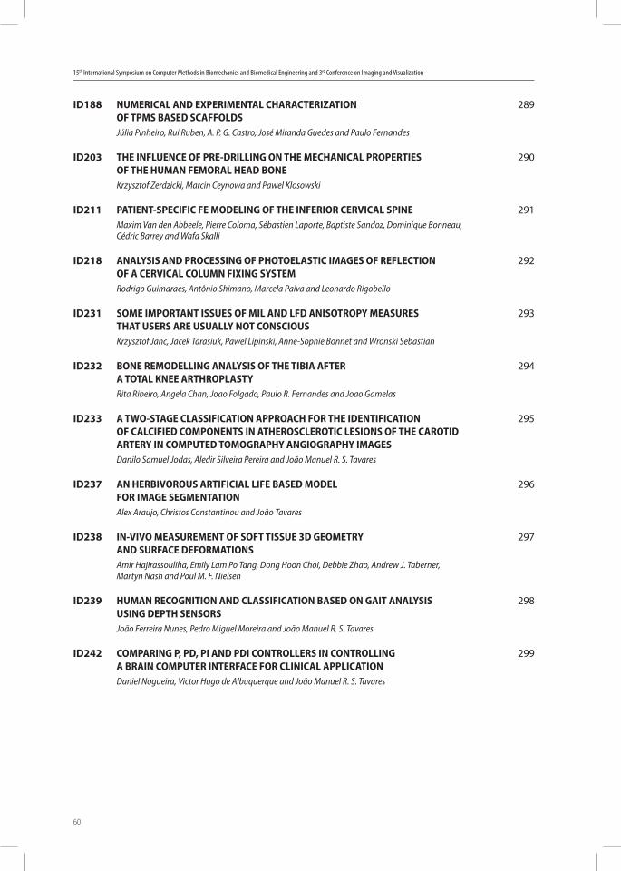
15th International Symposium on Computer Methods in Biomechanics and Biomedical Engineering and 3rd Conference on Imaging and Visualization
60
ID188 NUMERICAL AND EXPERIMENTAL CHARACTERIZATION OF TPMS BASED SCAFFOLDS
Júlia Pinheiro, Rui Ruben, A. P. G. Castro, José Miranda Guedes and Paulo Fernandes
ID203 THE INFLUENCE OF PRE-DRILLING ON THE MECHANICAL PROPERTIES OF THE HUMAN FEMORAL HEAD BONE
Krzysztof Zerdzicki, Marcin Ceynowa and Pawel Klosowski
ID211 PATIENT-SPECIFIC FE MODELING OF THE INFERIOR CERVICAL SPINEMaxim Van den Abbeele, Pierre Coloma, Sébastien Laporte, Baptiste Sandoz, Dominique Bonneau,
Cédric Barrey and Wafa Skalli
ID218 ANALYSIS AND PROCESSING OF PHOTOELASTIC IMAGES OF REFLECTION OF A CERVICAL COLUMN FIXING SYSTEM
Rodrigo Guimaraes, Antônio Shimano, Marcela Paiva and Leonardo Rigobello
ID231 SOME IMPORTANT ISSUES OF MIL AND LFD ANISOTROPY MEASURES THAT USERS ARE USUALLY NOT CONSCIOUS
Krzysztof Janc, Jacek Tarasiuk, Pawel Lipinski, Anne-Sophie Bonnet and Wronski Sebastian
ID232 BONE REMODELLING ANALYSIS OF THE TIBIA AFTER A TOTAL KNEE ARTHROPLASTY
Rita Ribeiro, Angela Chan, Joao Folgado, Paulo R. Fernandes and Joao Gamelas
ID233 A TWO-STAGE CLASSIFICATION APPROACH FOR THE IDENTIFICATION OF CALCIFIED COMPONENTS IN ATHEROSCLEROTIC LESIONS OF THE CAROTID ARTERY IN COMPUTED TOMOGRAPHY ANGIOGRAPHY IMAGES
Danilo Samuel Jodas, Aledir Silveira Pereira and João Manuel R. S. Tavares
ID237 AN HERBIVOROUS ARTIFICIAL LIFE BASED MODEL FOR IMAGE SEGMENTATION
Alex Araujo, Christos Constantinou and João Tavares
ID238 IN-VIVO MEASUREMENT OF SOFT TISSUE 3D GEOMETRY AND SURFACE DEFORMATIONS
Amir Hajirassouliha, Emily Lam Po Tang, Dong Hoon Choi, Debbie Zhao, Andrew J. Taberner, Martyn Nash and Poul M. F. Nielsen
ID239 HUMAN RECOGNITION AND CLASSIFICATION BASED ON GAIT ANALYSIS USING DEPTH SENSORS
João Ferreira Nunes, Pedro Miguel Moreira and João Manuel R. S. Tavares
ID242 COMPARING P, PD, PI AND PDI CONTROLLERS IN CONTROLLING A BRAIN COMPUTER INTERFACE FOR CLINICAL APPLICATION
Daniel Nogueira, Victor Hugo de Albuquerque and João Manuel R. S. Tavares
289
290
291
292
293
294
295
296
297
298
299

26-29 March, 2018 • Instituto Superior Técnico • Lisbon • Portugal
61
CMBBE2018Plenary Lectures


26-29 March, 2018 • Instituto Superior Técnico • Lisbon • Portugal
63
Plenary Lecture
MODELLING AND MEASURING CELL-MATRIX MECHANICAL INTERACTIONS
Hans Van Oosterwyck(1), Tommy Heck(1), Alicia Izquierdo-Alvarez(1), Alvaro Jorge-Peñas(1),Christian Steuwe(2), Marie-Mo Vaeyens(3), Diego A. Vargas(1)
(1)Biomechanics Section, Department of Mechanical Engineering, KU Leuven, [email protected], [email protected], [email protected],
[email protected], [email protected](2)Center for Surface Chemistry and Catalysis, KU Leuven, Belgium
[email protected](3)Biomechanics Section, Department of Mechanical Engineering, KU Leuven, Bahrain
Keywords: angiogenesis, cell mechanics, particle-based models
Summary: Cells mechanically interact with their extracellular matrix by adhering and applying force to it. These cellular forces play an important role in the dynamics of cell adhesion and migration as part of single cell or multicellular, collective behaviour and more in general can modulate cell signaling in a process called mechanotransduction. We have worked on microscopy-based methods of quantifying these cellular forces during single endothelial cell migration and vascular invasion. In vitro models compatible with live cell fluorescence microscopy imaging were established and time lapses of cell and matrix movements were recorded. The extracellular matrix was mimicked by the use of deformable natural and synthetic hydrogels. Cell-induced hydrogel deformations were calculated by registering positional information, coming either from hydrogel-embedded fluorescent beads or hydrogel fibers that were imaged label-free.2D endothelial cell migration assays on polyacrylamide gels suggested that cell traction magnitude was affected by gel stiffness and adhesion proteins (collagen versus fibronectin), while force polarity was only affected by adhesion proteins and not by stiffness. When analyzing traction exertion at a subcellular scale, adhesion proteins were found to affect the distribution and dynamics of traction focal areas. These results are further analysed by computational models of single cell mechanics that deal with the discrete, spatially non-uniform nature of focal adhesion turnover, protrusion and stress fiber formation. 3D vascular invasion assays demonstrated complex, spatially non-uniform hydrogel deformation patterns around angiogenic sprouts into collagen. Highest deformations were found at the base and the tip of the sprouts and suggested that sprouts were mechanically interacting with the hydrogel as a force dipole along the sprout principal direction. Computational models of sprout-hydrogel mechanics and dynamics are created in order to further quantify force patterns. The models are based on smoothed particle hydrodynamics (SPH), resulting in a discretization of hydrogel viscoelasticity and degradation, cell cortex viscoelasticity and contractility, protrusion and adhesion dynamics. By model fitting the measured deformation fields, the model is able to estimate the location and magnitude of cellular tractions. Simulation results also suggest that the observed long-range deformations require strain stiffening of the collagen hydrogels.Acknowledgements: FP7/2007-2013)/ERC n° 308223
15th International Symposium on Computer Methods in Biomechanics and Biomedical Engineering and 3rd Conference on Imaging and Visualization
64
Plenary Lecture
MEDICAL DEVICE-RELATED PRESSURE ULCERS: WHERE BIOMECHANICS SHOULD COME TO THE RESCUE
Amit Gefen
Tel Aviv University, [email protected]
Keywords: Tissue biomechanics, pressure injury, deformation damage, physical phantoms, finite element modeling
Summary: We have developed multiple physical (phantom) and computational (finite element, FE) three-dimensional anatomical model systems to investigate commonly encountered conditions and scenarios at which medical device-related pressure ulcers (injuries) may occur. Medical device-related pressure ulcers (MDRPUs) are injuries associated with use of devices and equipment applied for diagnostic or therapeutic purposes, where the injury has the same configuration as the applied device. In intensive care units (ICUs), MDRPUs caused by endotracheal and nasogastric tubes are common, both in adult and in pediatric settings. Studying the root causes of MDRPUs and effective means to mitigate their risk will lead to improved quality of life of patients and considerable saving of costs which are otherwise invested in treatment. Development of experimental and computational biomechanical models is essential for creating laboratory standards for testing the safety of medical devices which are in contact with the surface of the body. We have developed experimental head phantom systems equipped with force sensors as well as FE models of adult and pediatric patient heads to simulate strains and stresses in head soft tissues during interactions with devices, particularly tubing, electrodes and head supports. Based on our findings, we feel that the design of medical devices and equipment used in ICUs should be re-visited, since currently, there appears to be no attention to the safety of use with regard to the pressure ulcer risk. Much can be done concerning the design of device structures, selection of materials and integration of mechanisms that minimize the risk, e.g. of misplacement under the body, so that tubes, wires, electrodes and other equipment can be made safer, even if forgotten or misplaced between the patient and support surface. For example, selection of more adequate, softer materials and devices, e.g. development of soft electrodes made of conducting textiles and similar ideas can vastly minimize the occurrence of MDRPUs. These examples will be discussed in the talk, based on data from our recent experiments and FE simulations of scenarios where there is high risk for MDRPUs.
26-29 March, 2018 • Instituto Superior Técnico • Lisbon • Portugal
65
Plenary Lecture
BEHAVIOUR OF SOFT MATERIALS UNDER TRIAXIAL LOADING
Sam Evans
Cardiff University, United [email protected]
Keywords: Soft tissue, Triaxial, brain
Summary: Materials are usually tested uniaxially, but in many real life situations they are subjected to multiaxial loading. For example, cartilage in a joint is loaded in triaxial compression, as is a seal or an o-ring in a hydraulic system. Tissues such as blood vessels are loaded in biaxial tension, and triaxial tension occurs at notches and crack tips. Models of soft materials usually separate their behaviour into volumetric and deviatoric components, for numerical reasons, and we usually make crude assumptions about the volumetric part. It is common to say that the material was assumed to be nearly incompressible, and then to use a relatively low bulk modulus in order to ensure a stable solution. In reality many soft tissues undergo large volume changes associated with fluid flow, which have not generally been measured or well understood. Models that separate volumetric and deviatoric stresses have been shown by Ni Annaidh et al to be incorrect, at least for anisotropic materials, and are probably also wrong for isotropic materials. In other areas of mechanics it is well known that high triaxial stresses cause significant changes in deviatoric stiffness, for example in rock mechanics where high triaxial stresses increase the velocity of seismic waves. There is some theory that predicts this but it does not work for soft tissues.We have modelled deformation of the brain under gravitational loading in different positions, and measured it using MRI. A parametric study shows that the deviatoric stiffness has little effect on the displacement, but the bulk modulus is critically important, and must be quite low to match the behaviour that is observed in real life. It seems that not only are there large changes in volume but that they are critical in determining how the material actually deforms. We have developed a system for testing soft materials under triaxial loading. Initial results show that there is significant coupling between volumetric and deviatoric deformation and the conventional assumption that they are independent is incorrect. This has important implications for predicting the behaviour of soft materials under triaxial loading.
15th International Symposium on Computer Methods in Biomechanics and Biomedical Engineering and 3rd Conference on Imaging and Visualization
66
Plenary Lecture
FROM MEDICAL IMAGE COMPUTING TO COMPUTER-AIDED DIAGNOSIS TOOLS: SUCCESSES, CHALLENGES, GUIDELINES
AND LESSONS LEARNED
Cristian Linte
Rochester Institute of Technology, United [email protected]
Keywords: medical image computing, computer-aided diagnosis, guidance and navigation, characterization and visualization
Summary: With advanced in computing technology and data acquisition, medical imaging has enabled clinicians to see inside the human body in less invasive ways, visualize anatomy with much more fidelity and enable more accurate, precise and on time diagnosis of diseases. Medical image computing has played a significant role in the development of computer-aided diagnosis (CAD) tools. Nevertheless, despite the extensive research in medical image localization, segmentation, registration as critical tools for biomarker quantification, the use of CAD tools in clinical practice could be much enhanced. This lecture will outline some of the challenges associated with the slow “translation” of medical image computing tools and their adoption in clinical practice. Several examples encompassing different diagnostic radiology modalities will be showcased, along with a set of guidelines that could be followed to ensure the development of CAD tools with an increased likeliness of clinical use.
26-29 March, 2018 • Instituto Superior Técnico • Lisbon • Portugal
67
Plenary Lecture
ON THE EQUILIBRIUM AND STABILITY OF THE KNEE JOINT IN GAIT
Saeed A. Shirazi-Adl, Hafedh Marouane, Masoud Sharifi, Malek Adouni
Ecole Polytechnique, [email protected]; [email protected]; [email protected];
Keywords: Knee joint, Musculoskeletal model, Gait, Stability, Anterior cruciate ligament, Contact forces
Summary: Improved understanding of the knee joint biomechanics and factors affecting it in gait as the most common activity in human life is essential in prevention and treatment managements of joint disorders. Musculoskeletal models often make a number of crucial assumptions when attempting to estimate muscle forces and joint loads. The knee is routinely idealized as a planar 2D joint in the sagittal plane with no consideration of out-of-plane motions and equilibrium, the joint passive properties are overlooked, the joint center is assumed to coincide with the joint center of reaction at all gait periods, fixed locations for contact points at both condyles are taken when estimating medial/lateral contact forces, and the joint stability in intact and injured conditions is totally neglected. Large scatter exists in reported gait kinematics-kinetics that are due to inter-subject differences, marker arrangements-placements, joint coordinate system and skin artifact. We present here a lower extremity hybrid musculoskeletal model driven by gait kinematics-kinetics to compute muscle forces and internal loads in the stance phase of gait. Effects of changes in tibial posterior slope, coactivity in muscles, joint planar assumption, input kinematics-kinetics and ACL presence on muscle/contact/ligament forces, centers of pressure on each plateau and joint stability are then computed.Results highlight the substantial effect of variations in foregoing parameters on equilibrium and stability results. Large unbalanced out-of-sagittal plane moments reaching peaks of 30 Nm abduction moment and 12 Nm internal moment at 25% stance period are overlooked in a planar 2D model resulting in lower muscle, ACL and tibiofemoral contact forces when compared to the 3D reference model. The location of contact centers on each plateau also noticeably alters. Changes (by ±SD) in the knee flexion-extension and varus-valgus rotations have more effects on results compared to similar changes in their moments which are due partly to the substantial alterations in the knee passive resistances. The computed effects on ACL forces of changes in flexion-extension rotation are similar to those of alterations in the posterior tibial slope. The joint stability is mainly affected by input flexion-extension rotations and moments and joint compression forces.Acknowledgement: supported by NSERC-Canada.
15th International Symposium on Computer Methods in Biomechanics and Biomedical Engineering and 3rd Conference on Imaging and Visualization
68
Plenary Lecture
ENHANCING SPARSITY BEYOND CONVEXITY: APPLICATIONS TO THE RESTORATION AND SEGMENTATION
OF MEDICAL IMAGES AND SURFACES
Fiorella Sgallari
University of Bologna, [email protected]
Keywords: Variational models, Image Restoration, Image Segmentation, Medical Application
Summary: This talk concerns the design and numerical solution of variational models for image processing. In particular we present models containing non-convex non-smooth regularization terms involving a sparsity-inducing prior which improves the solution quality. These models arise in a wide variety of research areas, and we are interested in their application to the restoration of images corrupted by blur and noise, and the segmentation of images as well as surfaces, i.e. the partitioning of the data into regions that are homogeneous according to a certain feature.We first introduce the Convex-NonConvex strategy which relies on the idea of constructing and then optimizing convex functionals containing non-convex (sparsity-promoting) regularization terms. This allows for using reliable convex minimization approaches to compute the (unique) solution.We then deal with the pure non-convex regime by briefly presenting a new space-variant regularization which holds the potential for better modeling space variant properties of real images. We discuss how to solve numerically these models and highlight their effectiveness in practical applications such as image restoration and segmentation.Based on joint works with Raymond Chan, Martin Huska, Alessandro Lanza, Serena Morigi, Lothar Reichel, Ivan Selesnick.
26-29 March, 2018 • Instituto Superior Técnico • Lisbon • Portugal
69
Plenary Lecture
HYDROGEL AS A MODEL SYSTEM TO STUDY DISSIPATION PHENOMENA IN SOFT TISSUE
Dominique Pioletti
Ecole Polytechnique Fédérale de Lausanne, [email protected]
Keywords: hydrogel, mechano-biology, dissipation
Summary: The relaxation behaviour of alginate-based hydrogel was shown to influence the differentiation of mesenchimal stem cells. Rapidly relaxing hydrogel favors osteogenic differentiation [1]. We recently observed that dissipation of cartilage can locally induce a temperature increase in this tissue [2] and as a consequence supports chondrogenic gene expression [3]. These two examples highlight a new paradigm on the role of dissipative phenomena in the field of soft tissues and biomaterials. While dissipation has long been identified to play an important role in the mechanical and functional behaviours of musculo-skeletal tissues through viscoelasticity or poro-elasticity considerations, the relation between dissipation and mechanobiology only recently emerges. In parallel to what has been developed with biomaterials considering only elastic (static) parameters such as substrate stiffness to control cell differentiation, the development of new biomaterials incorporating dissipation aspects can allow to engineer in a more refined way the interaction between cells and materials. Indeed dynamical aspects related to the material loading can be uniquely modulated with the consideration of dissipation. This is demonstrated with the two examples mentioned above where the rate (for relaxation) or the combination of duration and frequency (for loading) affect osteogenic differentiation or chondrogenesis. The use of dissipation was also proposed to trigger the controlled release of a drug from a hydrogel [4].By developing different composite hydrogels based on PEG or HEMA, we were able to control the dissipation properties of these materials and obtain properties that could favour a particular cell differentiation pathway such as chondrogenesis for example as well as tuning in parallel the mechanical properties of the hydrogels.
References[1]Chaudhuri et al. Nature materials 2016; 15:326-334.[2]Abdel-Sayed et al. J Mech Behav Biomed Mat 2014; 30:123-130.[3]Abdel-Sayed et al. Biomaterials 2014; 35:1890-1897.[4]Moghadam et al. Biomaterials 2014; 35:450-455.
15th International Symposium on Computer Methods in Biomechanics and Biomedical Engineering and 3rd Conference on Imaging and Visualization
70
Plenary Lecture
MONOLITHIC SOLVER FOR BLOOD FLOW IN LARGE VALVED VEINS OF INFERIOR LIMBS
Chen-Yu Chiang(1), Olivier Pironneau(1), Tony W. H. Sheu(3), Marc Thiriet(4)
(1)Laboratoire Jacques-Louis-Lions (LJLL), Sorbonne University, Paris, [email protected] ; [email protected]
(3)Department of Engineering Science and Ocean Engineering, National Taiwan University; TaiwanCASTS, NTU, [email protected]
(4)Laboratoire Jacques-Louis-Lions, Sorbonne University, Paris, FranceINRIA-Paris, [email protected]
Keywords: Finite element method, fluid-structure interaction, large displacement, monolithic solver, venous valve
Summary: Arteries are distensible, whereas veins are both distensible, enabling blood storage, and collapsible. Walk enhances venous return from extremities of inferior limbs, but also provokes backflow. Venous valves are aimed at limiting backflow magnitude. A monolithic formulation based on an Eulerian formulation of the full coupling system and a fluid-structure solver have been developed to model blood flow in deformable valved veins. A hyperelastic incompressible model is used to represent behavior of venous valves and wall.
26-29 March, 2018 • Instituto Superior Técnico • Lisbon • Portugal
71
Plenary Lecture
MULTI-RESOLUTION GEOMETRIC MODELS OF THE MITRAL HEART VALVE
Michael Sacks
Center for Cardiovascular Simulation, Department of Biomedical EngineeringThe University of Texas at Austin, United States
Keywords: Heart Function, Mitral Valve, Computational Simulations
Summary: An essential element of the heart function, the mitral valve (MV) ensures proper directional blood flow between the left heart chambers. Over the past two decades, computational simulations have made marked advancements towards providing powerful predictive tools to better understand valvular function and improve treatments for MV disease. However, challenges remain in the development of robust means for the quantification and representation of MV leaflet geometry. In this study, we present a novel modeling pipeline to quantitatively characterize and represent MV leaflet surface geometry. Our methodology utilized a two-part additive decomposition of the MV geometric features to decouple the macro-level general leaflet shape descriptors from the leaflet fine-scale features. First, the general shapes of five ovine MV leaflets were modeled using superquadric surfaces. Second, the finer-scale geometric details were captured, quantified, and reconstructed via a 2D Fourier analysis with an additional sparsity constraint. This spectral approach allowed us to easily control the level of geometric details in the reconstructed geometry. The results revealed that our methodology provided a robust and accurate approach to develop MV-specific models with an adjustable level of spatial resolution and geometric detail. Such fully customizable models provide the necessary means to perform computational simulations of the MV in a range of geometric detail, allowing identification of the complexity required to achieve predictive MV simulations to a desired accuracy level. We then extend these approaches to develop novel in-vivo methods for patient specific model development.
15th International Symposium on Computer Methods in Biomechanics and Biomedical Engineering and 3rd Conference on Imaging and Visualization
72
Plenary Lecture
PHYSIOLOGICAL CYBERNETICS: METHODS AND APPLICATIONS
Daniela Iacoviello
Sapienza University of Rome, [email protected]
Keywords: Physiological systems, automatic control, homeostasis
Summary: Cybernetics is the term used by Wiener in 1948 to denote “the unity of the set of problems centering about communication, control and statistical mechanics, whether in the machine or in the living tissue”. Physiological cybernetics is a research field aiming at an interdisciplinary approach to face biomedical problems; it considers models able to describe physiological systems using the feedback theory and, more in general, the system analysis. Homeostasis is the capacity of the body to preserve relatively constant physiological conditions and control actions are the basis for its maintenance; the control theory is the natural framework to describe physiological systems and their regulation mechanisms.The research on physiological cybernetics applies mathematics to provide a support for testing therapeutic protocols and helping medical diagnosis.This talk will discuss the methods of control theory applied to physiological systems; they are generally nonlinear and time invariant, thus requiring the hard task of model balance complexity. Important key differences between engineering and physiological control systems will be discussed; in particular, the “components” of a physiological control system are generally unknown or difficult to analyze and, moreover, the system to be controlled is a controller itself.Examples and applications will be presented.
26-29 March, 2018 • Instituto Superior Técnico • Lisbon • Portugal
73
Plenary Lecture
HIGH PERFORMANCE POLYMERS IN DENTISTRY - BIOMECHANICAL AND CLINICAL ASPECTS
Christoph Bourauel(1), Ludger Keilig(1), Istabrak Hasan(1), Tobias Klur(2), Anne Kartzenbach(1), Helmut Stark(2)
(1)Oral Technology, University of Bonn, [email protected] ; [email protected] ; [email protected] ; [email protected]
(2)Department of Prosthetic Dentistry, University of Bonn, Germany [email protected] ; [email protected]
Keywords: Prosthetic Dentistry, High Performance Polymer, Finite Element Analysis, Permanent Fracture Resistance
Summary: A large variety of different materials, such as ceramics, precious and non-precious alloys, is currently available for use in dental prosthetics. Recently, new high performance polymers provide an additional alternative for the framework material. It was the aim of the presented study to determine the mechanical and biomechanical behaviour of dental bridges made of a recently introduced high performance polymer (polyether-ketone-ketone, Pekkton®, Cendres + Métaux SA, Switzerland) using the finite element (FE) method and to compare these results with different well-established and proven framework materials that are used for dental bridges.In this paper we present results of experimental, numerical and clinical studies showing the potential of Pekkton as restorative material in dental prosthetics. Permanent fracture resistance of single tooth crowns made of Pekkton showed a fracture resistance higher than 600 N, which is clearly above normal occlusal forces. In finite element simulations, bridges and framework made of Pekkton showed slightly differing biomechanical behaviour compared to conventional framework and bridge materials like titanium, gold or ceramics. However they proved to be recommendable for clinical application. Clinical studies including single crowns, bridges and retention elements showed comparable or better results than those made of nonprecious metals.Concluding, the new high performance polymer Pekkton shows promising new possibilities for application in all fields of dental prosthetics.
15th International Symposium on Computer Methods in Biomechanics and Biomedical Engineering and 3rd Conference on Imaging and Visualization
74
Plenary Lecture
DESIGNING INTRAMEDULLAR POSTS FOR VETERINARY APPLICATIONS
Estevam B. Las Casas(1), Leopoldo Paolucci(1), Rafael Faleiros(1), Sergio Rocha Jr.(1), Paulo Fernandes(2), João Folgado(2), Luciano Rodrigues(3), Luciana Gomides(4)
(1)UFMG, [email protected] ; [email protected] ; [email protected] ; [email protected]
(2)IST, ULisboa, [email protected] ; [email protected]
(3)UESB, [email protected]
(4)UFI, [email protected]
Keywords: Biomechanics, Interlocking Nail, Finite Element, Bovine Femur
Summary: The objective of this study is to describe the development of a low cost interlocking nail for young calves. Biomechanical parameters were measured for the numerical analysis of the bovine femoral reduction system. Different polymeric and composite in silico materials were tested to investigate their mechanical performance . analyze polyacetal (POM), polypropylene (PP), polyamide (PA) and a glass fiber-reinforced polymer (GFRP) Twelve femur models, divided into three groups, each one associated with a different fixation strategy, were used for simulation of an oblique simple fracture. Model loading conditions corresponded to a calf in the transition (decubitus position to static position). The most critical stresses in the implant were found in the screws and at the interface between screw and nail. A numerical model demonstrated that all polymeric materials analyzed provided sufficient resistance to tolerate the loading forces imposed on the femur when an adequate fixation strategy was applied. After testing the biocompatibility of the material, in vivo tests were conducted to validate the proposed design. Preliminary results indicate the efficiency of the proposed nail design.
26-29 March, 2018 • Instituto Superior Técnico • Lisbon • Portugal
75
Plenary Lecture
BIOMECHANICAL MECHANISMS OF HEART FAILURE
Martyn Nash
The University of Auckland, New [email protected]
Keywords: Cardiac mechanics, Heart failure modelling, Microstructural remodelling
Summary: Diagnosis and treatment of heart failure is hampered by a lack of knowledge of the underlying pathophysiological mechanisms on an individualised basis. Tissue-apecific biomechanical factors, such as diastolic myocardial stiffness and stress, are known to have important influences on heart function, but these factors cannot be measured directly. Mathematical modelling provides a rational basis for identifying these biomarkers by integrating the rich variety of physiological data that are now available in the laboratory and clinical settings. This presentation will discuss how image-based, individualised biomechanical models of the heart can be used to characterise the relative roles of anatomical, microstructural and functional remodelling in heart failure. Data from pre-clinical and clinical studies will be presented to demonstrate this approach. In the clinic, patient-specific mathematical models of this kind have the potential to more specifically stratify the different forms of heart pathology, and thus to help inform and monitor patient therapy.
15th International Symposium on Computer Methods in Biomechanics and Biomedical Engineering and 3rd Conference on Imaging and Visualization
76
Plenary Lecture
PREDICTING GROWTH AND REMODELING OF ENGINEERED CARDIOVASCULAR TISSUES
Sandra Loerakker (Presented by Cees Oomens)
Eindhoven University of Technology, [email protected]
Keywords: Growth, Remodeling, Cardiovascular, Heart valves, Tissue engineering
Summary: Cardiovascular tissues are widely known to grow and remodel in response to changes in hemodynamics, in an attempt to restore or preserve mechanical homeostasis. For the field of cardiovascular tissue engineering, understanding the responsible growth and remodeling mechanisms is essential for developing living cardiovascular replacements that can last for a life-time. Due to the dynamic interplay between tissue adaptation and mechanical cues, computational modeling plays an important role in addressing this challenge. We have developed a computational model to predict soft tissue remodeling [1-3], inspired by experimental data on the individual mechanisms, which has contributed significantly to obtaining a mechanistic understanding of in vivo (engineered) valve remodeling. Model predictions showed that TEHVs may remodel differently at aortic pressure conditions compared to pulmonary conditions [4], and a comparison with in vivo remodeling at pulmonary conditions demonstrated that our model could closely mimic the experimentally observed changes in valve characteristics. Besides focusing on TEHVs, we also aim to improve our understanding of the postnatal development of human native heart valves, as these present the benchmark for TEHVs. Using computational-experimental analyses, we investigated the presence of mechanical homeostasis in human heart valves, and dissected the individual contributions of growth and remodeling in preserving mechanical homeostasis. Our data suggest that mechanical homeostasis for human semilunar heart valves is determined by a certain stretch [5]. Interestingly, growth and remodeling appear to play opposing roles in the preservation of this mechanical homeostasis [6].This research has been supported by the European Union’s FP7 program (LifeValve), the Dutch Heart Foundation (CVON, 1Valve), and a Marie Curie Individual Fellowship.
References[1]Obbink-Huizer et al., BMMB 2014.[2]Loerakker et al., BMMB 2014.[3]Ristori et al., CMBBE 2016.[4]Loerakker et al., JMBBM 2016.[5]Oomen et al., Acta Biomater 2016.[6]Oomen et al., Sci Rep 2018.
26-29 March, 2018 • Instituto Superior Técnico • Lisbon • Portugal
77
Plenary Lecture
ADDED VALUE OF CASE-SPECIFIC, COMPUTER AIDED BIOMECHANICAL ANALYSIS
Pim Pellikaan(1), Roel Wirix-Speetjens(2), Ilse Jonkers(3), Harry van Lenthe(4), Jos Vander Sloten(4)
(1)Biomechanics Section, KU Leuven, [email protected]
(2)Materialise NV. Leuven, Belgium [email protected]
(3)Human Movement Biomechanics Research Group, KU Leuven, [email protected]
(4)Biomechanics Section, KU Leuven, [email protected] ; [email protected]
Keywords: subject-specific, arthroplasty, finite elemento analysis
Summary: FEA has become a common tool to investigate biomechanical problems. However, most of the models used are generic in nature and try to reflect the situation of an average patient. Modern software tools allow for biomechanical analysis in a patient-specific way, however often at a higher computational cost. We investigate, using two representative case studies, the potential added value of subject-specific biomechanical modeling.Firstly, the aim was to quantify the effects of subject-specific geometry, density, and loading conditions on the strains at the femur neck. CT scans and gait data at several speeds of walking and running were collected from 9 elderly women, 70.1 ± 3.6 years old. Full-body generic and subject-specific musculoskeletal models were used to calculate hip contact and muscle forces. CT-based FE models were generated; material properties were estimated from a template or subject’s CT. Principal strains for the generic models were around twice as high compared to the subject-specific models. Furthermore, the geometry and bone density from the subject’s CT had a greater influence on the calculated strains than the loading conditions. Results showed a need for subject-specific models to obtain reliable results for each individual. Then, it was investigated to what extent a subject-specific knee model could be a reliable predictor of post-operative behavior. Subject specific models of four cadaveric knees were constructed from CT images. The models were validated by comparing experimental cadaveric measures to model based kinematics and ligament elongations. First the native knees were mounted onto a mechanical rig to perform squatting, measuring kinematics and ligament elongations with optical markers and extensometers. Next total knee replacement surgery was performed on each knee by inserting a posterior stabilized implant and the same squat simulation performed with the implanted knees. Coronal malrotation was also introduced using tibial inserts with a built in slope. The models could accurately predict the tibio femoral kinematics and ligament elongations, showing excellent correlations and small root mean square errors in the same order of magnitude as other studies. Native knee results presented slightly higher deviations than the predictions for the implanted knees as a consequence of model simplifications.
15th International Symposium on Computer Methods in Biomechanics and Biomedical Engineering and 3rd Conference on Imaging and Visualization
78
Plenary Lecture
IN VIVO BARYCENTREMETRY FOR SUBJECT SPECIFIC MUSCULO-SKELETAL MODELLING
Wafa Skalli(1), Laurent Gajny(2)
(1)Institut de Biomecanique Humaine Georges Charpak, Arts et Métiers ParisTech, [email protected]
(2)Arts et Métiers Paristech, France [email protected]
Keywords: barycentremetry, subject specific, musculoskeletal model, body segment parameters, gravitational loads, postural analysis
Summary: Subject specific musculo-skeletal modelling is of paramount importance in various areas of biomechanics including sport, ergonomics and medicine. Among the various issues raised by such modelling, there are two main scientific and technical challenges : the first one is dealing with the well known muscle redundancy problem : too many unknown muscle forces with regard to the limited number of equilibrium equations. This issue drives very active research on quantification of muscles activation both using various modelling methods, and/or experimental techniques. The second one, which is the object of this talk, is feeding the models with subject specific data, related both to internal information such as vertebrae locations or muscle geometry, and to body segments inertial parameters (BSIPs). Complex methods involving data fusion between different imaging modalities (among which MRI) are progressively set to get subject specific external and internal geometry. Using the resuting datasets, further methods consider deformation of templates and/or statistical analysis and machine learning to simplify the problem and get internal information from external envelope. BSIPs are generally estimated from databases collected on a limited number of samples : the mass is defined as a given percentage of global body mass. Both center of mass (COM) location and inertial parameters are generic or roughly scaled.Recent development of bodyscanners and of biplanar X-Rays, associated to 3D reconstruction methods including the external envelope, open new perspectives for subject specific modeling. Mass, COM and other BSIPs scan be computed from the external envelope using automatic accurate processing to delimitate body segments, together with density models per body segment (revisited for the thorax). Computation of barycenter of body segment above a given body segment and direct postural or dynamic analysis allows subject specific quantification of gravitational loads that apply at each body level and for each individual, providing powerfull means of analysis. This talk will focus mainly on barycentremetry, with examples of clinical and basic applications related to spine and musculoskeletal disorders.

26-29 March, 2018 • Instituto Superior Técnico • Lisbon • Portugal
79
CMBBE2018Workshops

26-29 March, 2018 • Instituto Superior Técnico • Lisbon • Portugal
81
From Reality to VirtualityBETA CAE Systems International AG
When reality is ahead of the simulation reverse engineering is necessary to create the virtual model out of the physical one. During this workshop the reverse engineering process will be presented with the aid of the tools of BETA CAE SYSTEMS AG, starting from the output data of an industrial tomograph and resulting to a ready to run solver file. If internal structures are equally important as the external surfaces, the use of a tomograph for the 3D scanning is a necessity. RETOMO is the reliable tool for going from tomography voxels to virtual prototypes. With its streamed image processing technology it can process large datasets and deliver effortlessly large FE-Models, ready for pre-processing. Pre-processing involves the creation of a high quality FE-Mesh and the definition of material properties, initial and boundary conditions for the simulation. In our case the simulation will be the virtual compression test of a cubic sample of a cancellous bone. During this workshop we will not only focus on the creation of a simple loadcase, but advanced modelling techniques will be demonstrated as well, by presenting the capabilities of ANSA for model reusability and automation.
Working with C-Motion’s Dynamic Stereo X-ray Software SuiteC-Motion Biomechanics Software
Advances in diagnostic imaging have greatly improved our ability to detect structural changes in musculoskeletal tissues. There is now evidence that subtle joint translations of only a few millimeters are critical to estimating key clinical measures such as tissue stress, joint impingement, or implant kinematics during loaded functional movements. Dynamic Stereo X-ray (DSX) is the only currently available technology that can achieve sub-millimeter accuracy for a wide variety of functional movements. This workshop shows attendees how to use C-Motion’s new DSX Software Suite to calibrate their equipment, correct X-ray images, and track multiple bones in the images. This workshop will expain:1. Calculating the 3D pose of the X-ray sources and image planes from images of the calibration object, including uniformity-corrections, distortion-corrections, and X-ray image resizing.2. Using bone models extracted from CT, define anatomically meaningful reference frames, add landmarks, and define regions of interest. Track bones in X-ray trials using single frame or 4D optimization. 3. Export bone tracking to Visual3D for joint animations and kinematic analysis, including the calculation of joint congruency and ligament lengths. Validate markerless bone tracking with bead tracking.
Image-Based Modelling with Simpleware for BiomechanicsSynopsys
Image-based modelling from 3D scans (MRI, CT, mCT and more) enables detailed analysis and simulation of complex biomechanics problems. Join this workshop to learn about the key techniques, benefits, and opportunities offered by image-based modelling using Simpleware software. Explore the software’s many applications in biomechanics, including patient-specific implant designs, orthopaedics research, human body models, and many more. Discover common workflows and see how Simpleware has been used for specific biomechanics case studies to solve a variety of modelling challenges.
15th International Symposium on Computer Methods in Biomechanics and Biomedical Engineering and 3rd Conference on Imaging and Visualization
82
Assessments in OrthopaedicsLLJ – LifeLongJoints
Recent results from metal on metal prostheses demonstrated that standards used to assess such prostheses provided little indication of the resulting failure in vivo. Whilst new standards have been agreed they are formed in retrospective manner and denote one of many perhaps possible failure modes. However, the global capacity of simulator testing in insufficient for testing all possible scenarios. Computational simulation may provide a methodology aiding the assessment of joint replacement performance in terms of identifying deleterious scenarios and predicting the levels of wear in these cases. The workshop will discuss these and other issues.
Combined Multibody and Finite Element Simulation Using ArtiSynthArtiSynth
ArtiSynth is a free, open source 3D modeling system developed at the University of British Columbia that combines multibody and finite element (FEM) simulation capabilities, including contact and constraints. It provides a highly interactive platform that lets users view and inspect their models while simultaneously running them. It is currently being used by a diverse set of research groups for a variety of applications including musculo-skeletal modeling of the foot, lower limb, spinal region, arm, and shoulder; function modeling of swallowing, mastication and speech; and preliminary studies of surgical treatment planning involving the head and neck region. This workshop will provide an overview of ArtiSynth and its capabilities, combined with a hands-on tutorial in which attendees can evaluate how it may be used to further their research goals. Specific use cases will be presented to provide concrete illustrations of how it may be used. Attendees will be able to download ArtiSynth and experiment with it beforehand, and we will be available to answer any specific questions that they may have. ArtiSynth models are primarily implemented using the Java programming language, providing users with a very powerful means to assemble complex and detailed models containing highly customized components and methods. Complete information about ArtiSynth, along with instructions for installing and using it, is available at www.artisynth.org.

26-29 March, 2018 • Instituto Superior Técnico • Lisbon • Portugal
83
CMBBE2018Abstracts

26-29 March, 2018 • Instituto Superior Técnico • Lisbon • Portugal
85
Abstract ID-3
THE EFFECTS OF DIFFERENT MATERIAL PROPERTIES ON THE HEMODYNAMICS OF HUMAN FETAL UMBILICAL
VEIN/DUCTUS VENOSUS
Kamran Hassani, Taraneh Rezaee
Science and Research Branch,Islamic Azad University, [email protected] ; [email protected]
Keywords: Umbilical vein, Ductus Venosus, Material properties
Summary: The umbilical vein is a vein which exist during development of the fetus. It carries oxygenated blood to the growing fetus. The ductus venosus connects intra-abdominal portion of the UV and the inferior vena cava (IVC) at the inlet of the right atrium. It sends one-third of the blood flow of the UV to the IVC . Due to increasing rate of abortion of the fetus, it is maybe interesting to study the possible abnormities of blood flow in UV/DV. Therefore, the studying of hemodynamics of blood in UV/DV could predict the possibility some congenital heart diseases.In this study, we have investigated the hemodynamics of blood flow based on four different mechanical properties of UV/DV including elastic (uniaxial test), elastic (circumferential), viscoelastic, and rigid. The results were compared to each other to see the effects of different we have assumed four different mechanical properties for UV including elastic 1 (uniaxial test method), elastic 2 (circumferential) , viscoelastic ( Ogden), and rigid material properties of UV/DV on the hemodynamic parameters on blood.The velocity of blood is maximum in hyper elastic model comparing to lowest one in rigid case.The shear stress values are maximum again in hyper elastic model and minimum in rigid vein. The calculated isthmus pressure was maximum (403.2 Pa) in elastic 2 case where it was minimum in hyper elastic model (387.8 Pa).Our results including the hemodynamics factors suggest that hyper elastic model has had more conformity to the relevant clinical and experimental results. In this study, we aimed to compare the hemodynamics parameters of the blood flow in UV/DV among two elastic models, hyper elastic, and rigid one. In this regard, the finite element analysis for the models were performed and the results were obtained for different vein material properties.
15th International Symposium on Computer Methods in Biomechanics and Biomedical Engineering and 3rd Conference on Imaging and Visualization
86
Abstract ID-6
NUMERICAL MODELLING OF STENOSIS DEVELOPMENT IN THE CAROTID ARTERY
Aikaterini Stamou(1), James Buick(2)
(1)University of Hertfordshire, United [email protected]
(2)University of Portsmouth, United Kingdom [email protected]
Keywords: Atherosclerosis, carotid bifurcation, numerical modelling, haemodynamic properties, stenosis development
Summary: Stroke is the third-leading cause of death in the developed world, and carotid artery stenosis is one of the leading risk factors for stroke, accounting for about 20 percent of strokes. There are about 152,000 strokes in the UK alone every year where about 1.2 million people live with the after effects.The carotid arteries are located on each side of the neck and are the blood vessels which supply blood to the large front part of the brain which is where thinking, speech, personality, and sensory and motor functions reside. Each common carotid artery divides in two branches: the internal carotid artery and the external carotid artery. Normal these arteries are smooth; however, overtime plaques, made of cholesterol, calcium, destructed cells and fibrous tissue, build up on the walls - a process known as atherosclerosis. Extensive atherosclerosis may cause stenosis (narrowing) of the artery or even complete blockage. In the carotid artery, this typically occurs at the bifurcationwhere the common carotid artery divided into the internal and external arteries.Many studies have considered the blood flow, as well as associated parameters such as wall shear stress, in arteries with varying degrees of stenosis; however, this only provides a snap-shot in time and does not give any information on the progression of the disease. Here we consider a numerical model for stenosis development, based on the haemodynamic properties inside the artery. This enables the development of an understanding of the role of the blood flow in stenosis development, and the two-way interaction between the interaction between the blood flow and the developing stenosis. Blood flow is simulated using the Lattice Boltzmann Method, while the development of the stenosis is governed by the simulated haemodynamic conditions in the wall region. Details of the stenosis growth model will be presented, including details of its development and application. Simulation result will also be presented indicating how the stenosis develops and how the corresponding geometrical changes affects the near wall flow parameters.
26-29 March, 2018 • Instituto Superior Técnico • Lisbon • Portugal
87
Abstract ID-8
EXTRACTING AND EVALUATING TEXTURE FEATURES FROM BINARY GRADIENT CONTOURS OF MICROCALCIFICATIONS CLUSTERS
IN BREAST MAMMOGRAMS
Marcelo Duarte(1), Andre Alvarenga(2), Wagner Coelho de Albuquerque Pereira(3)
(1)UniCarioca University Centre, [email protected]
(2)Inmetro, Brazil [email protected]
(3)Biomedical Engineering Program, COPPE/UFRJ, [email protected]
Keywords: CAD systems, Microcalcifications, Breast cancer, Mammograms, Texture features
Summary: Death women rates related to breast cancer are high worldwide, mainly because of late diagnosis. The best methods for detecting the early signs of breast cancer are clinical and mammographic examinations.Microcalcifications (MCs) are small granular deposits of calcium that can be found in mammographic routine exams in most breast cancer cases. Despite their frequent occurrence, although 60%–80% are detected via histological examination, only 30%–50% of MCs in breast carcinoma are detected via mammographic examination.Computer-aided diagnosis (CADx) systems have been developed in an effort to assist MC diagnosis. CADx can be used to provide a second opinion, thereby increasing the accuracy of a radiologist’s final diagnosis, and basically involve three steps: (i) segmentation, (ii) features extraction and selection from the segmented MCs and their clusters, and (iii) classification. Such systems are usually based on features extracted from MCs, as compactness, roughness, orientation, and they can help minimizing false-positive and false-negative rates in breast cancer diagnosis.Texture features combined to morphological features have been studied worldwide to characterize MCs clusters, as well as texture features alone.In this work, the binary gradient contours (BGC) and local binary pattern (LBP) techniques were applied to calculate 991 texture features from MCs clusters presented on 190 images from Digital Database for Screening Mammography (DDSM). Hence, texture features were ranked, based on mutual information technique. Finally, an incremental procedure sequentially adds the top m-ranked features to the Fisher discriminant analysis to identify the best set of texture features in classifying benign or malignant clusters.For the 190 images, the procedure determined that 21 texture features are capable of attaining the best classification performance with Area under ROC curve (AUC) =0.928±0.022. It was also found that five texture features are enough to reach AUC =0.891±0.021, which represents the best classification performance considering a minimum of 30 sample images for each texture feature studied. The results achieved using just texture features are encouraging and in accordance with literature. In future, other texture features will be studied in order to improve classification performance.
15th International Symposium on Computer Methods in Biomechanics and Biomedical Engineering and 3rd Conference on Imaging and Visualization
88
Abstract ID-9
AN ADVANCED METAL ARTIFACT REDUCTION METHOD FOR A DENTAL CT
Mohamed Hegazy, Min Hyoung Cho, Soo Yeol Lee
Kyung Hee University, South [email protected], [email protected], [email protected]
Keywords: Metal artifact reduction, Dental CT, Dual-energy imaging
Summary: In a dental CT scan, the presence of dental fillings or dental implants generates severe metal artifacts that often compromise readability of the CT images. Since teeth may have attenuation coefficients similar to those of dental implants, the conventional metal artifact reduction (MAR) techniques for a medical CT often fail for a dental CT. We propose a new MAR method for a dental CT that is based on dual-energy imaging with a narrow energy gap. We acquired two projection data sets at two close tube voltages (80kVp and 90kVp) using a cone-beam CT consisting of a flat-panel detector and a micro-focus x-ray tube. Then, we computed the weighted difference projection data between the two projection data taken at different energies. We reconstructed 3D CT images from the weighted difference projection data to better identify the metallic region in the 3D space than we did from the single-energy projection data. By forward projecting the identified metallic region, we could identify the metal trace on the projection data that should be modified for MAR. By applying the region filling to the identified metal trace, we replaced the high-intensity pixels, stemming from the metallic objects, with the pixels surrounding the metallic objects. We reconstructed final CT images from the region-filled projection data. We applied the proposed method (the dual-energy-based method) to the projection data of a dental phantom and a human skull phantom with comparing the conventional single-energy-based method. Owing to better identification of the metallic regions on the projection data, the proposed method showed better MAR performances in all cases than the conventional method in terms of the major MAR performance metrics, that is, the relative error (REL), the sum of squared difference (SSD) and the normalized absolute difference (NAD). To apply the proposed method to the clinical dental CT, we need to alternate the tube voltage, between the high and low voltages every other projection view, to speed up the scan. If the high speed voltage switching is adopted in a dental CT, we expect the proposed MAR method can be greatly used to improve the MAR performance in dental imaging.
26-29 March, 2018 • Instituto Superior Técnico • Lisbon • Portugal
89
Abstract ID-10
SCOLIOSCAN: ASSESSMENT OF 3D SPINAL DEFORMITY USING ULTRASOUND IMAGING
Yongping Zheng;
The Hong Kong Polytechnic University, Hong [email protected]
Keywords: scoliosis, ultrasound, Cobb angle, spinous process, coronal view, sagittal view, transerve rotation, 3D imaging
Summary: Scoliosis is a medical condition defined as a 3D spine deformity with curvature of more than 10 degrees in the coronal plane. Scoliosis is usually seen in teenagers and known as adolescent idiopathic scoliosis (AIS). Recently, a number of organizations, including American Academy of Pediatrics (AAP), have endorsed a position statement on “Screening for the Early Detection for Idiopathic Scoliosis in Adolescents”, indicating the importance of earlier diagnosis and non-surgical management of AIS. (http://pediatrics.aappublications.org/)The traditional scoliosis examination is X-ray radiography. However, there are some health risks posed by the radiation exposure – including an increased incidence of lung and breast cancers. Although there are several radiation-free screening approaches, but none of them are accurate enough, thus using X-ray us inevitable for AIS during diagnosis, curve progression monitoring, and treatment outcome evaluation, as well as during brace treatment. Accordingly, we developed a novel technology that enables safer and more frequent monitoring for scoliosis.Scolioscan takes the advantage of 3D ultrasound imaging techniques and can provide 3D view of spine without any radiation. Ultrasound probe is scanned over the spine to collect a series of image together with spatial information, and advanced imaging processing methods are used to form images in coronal views as well as in 3D. Scolioscan has been used for scanning over 3000 scoliosis patients in Hong Kong, China, Macau, and The Netherlands. In this talk, we will introduce the principle of Scolioscan, its automatic angle measurement function, 3D spinal model formation, and its excellent intra- and inter-rater repeatability of spinous process angle (ICC>0.8) and good correlation with X-ray Cobb’s angle (R>0.85) in human subject trials. Its applications for sagittal analysis, transverse rotation analysis, forward-bending study, lateral flexibility test, brace design and fitting, longitudinal follow-up as well monitoring during Halo traction will also be introduced. A series of study have demonstrated that Scolioscan, using 3D ultrasound imaging, is a very promising technique for providing radiation-free while accurate assessment of scoliosis, and has the ability for evaluating spine deformity in 3D, thus is a unique tool for scoliosis screening and monitoring.
15th International Symposium on Computer Methods in Biomechanics and Biomedical Engineering and 3rd Conference on Imaging and Visualization
90
Abstract ID-12
SHEAR WAVE ELASTOGRAPHY TO CHARACTERIZE SCOLIOTIC INTERVERTEBRAL DISC
Claudio Vergari(1), Tristan Langlais(2), Raphaël Pietton(2), Jean Dubousset(1), Wafa Skalli(1), Raphaël Vialle(2)
(1)Institut de Biomécanique Humaine Georges Charpak, [email protected]
(2)DHU MAMUTH - Department of Pediatric Orthopaedic, Armand Trousseau Hospital, France
Keywords: scoliosis, imaging, mechanical properties
Summary: Adolescent idiopathic scoliosis (AIS) is a 3D deformity of the spine that can rapidly progress during adolescent’s growth spurt; an unchecked progression can lead to respiratory or locomotion impairment that can only be corrected by spinal surgery. Nevertheless, if progressive cases are detected early, they can be efficiently treated by bracing. Intervertebral disc (IVD) is a main component of spine mechanics, and could play a biomechanical role in the vicious cycle leading to curve progression. Moreover, personalization of its mechanical properties in numerical models would improve their realism. Shearwave ultrasound elastography was recently applied to measure shear wave speed (SWS) in IVD in vitro, in animal model, and in vivo in healthy subjects. SWS measurement proved reliable, and it correlated to disc mechanical properties. In the present work, elastography was applied to compare SWS in scoliotic and healthy lumbar IVDs.Thirty healthy adolescents (13.0 ± 2 years old) and thirty scoliotic patients (13 ± 2 years old, Cobb angles 28.8° ± 10.5°, range 13-50°) were included. Of the latter, twenty were progressive (Cobb > 25°) and 10 were stable scoliosis (i.e., Cobb angle < 25° and Risser sign > 2). SWS was measured in L3-L4, L4-L5 and L5-S1 discs with the patient lying in supine position.SWS was 3.0 ± 0.3 m/s in healthy discs and 3.5 ± 0.3 m/s in scoliotic discs; the difference was significant (p < 0.001). The difference was also significant at each disc level independently (p < 0.05). Results suggest that a junctional disc with a high SWS is associated with a risk of progression accrued by 4.6 times.Measurement was feasible and did not present particular difficulties. Moreover, SWS in the control group was similar to those previously measured in healthy children (2.9 ± 0.5 m/s). These results should be confirmed on a larger cohort; in particular, the effect of scoliosis topology, which was not controlled in the present work, should be investigated. Nevertheless, this study confirmed the potential interest of IVD elastography as a biomarker of scoliosis, but also of its use in the personalization of numerical biomechanical models.
26-29 March, 2018 • Instituto Superior Técnico • Lisbon • Portugal
91
Abstract ID-13
NUMERICAL INVESTIGATION OF WALL PRESSURE FLUCTUATIONS DOWNSTREAM OF IDEAL AND REALISTIC STENOSED VESSEL MODELS
Kamil Özden, Cüneyt Sert, Yiğit Yazıcıoğlu
Middle East Technical University, [email protected], [email protected], [email protected]
Keywords: Stenosis, pressure fluctuations, acoustic radiation, blood flow, computational fluid dynamics
Summary: Cardiovascular diseases, the most common of which is atherosclerosis, are the leading global cause of death. Atherosclerosis leads to a plaque built up inside an artery, narrowing it down and forming a stenosis. It may lead to coronary artery disease, stroke or peripheral artery occlusive disease, depending on the location of the lesion. The flow turning into turbulent regime after passing the stenotic obstruction leads to pressure fluctuations at the arterial wall. The generated sound is transmitted through the surrounding tissue and reaches the skin. This acoustic radiation may give important information about the stenotic region. In this study, the effect of using real and ideal stenosed vessel models on the generated acoustic radiation is investigated using numerical simulations. The idealized vessel-like model with an eccentric elliptical stenosis and a real vessel model with a realistic stenosis shape inspired by the MR image of a stenosis. Inlet diameters are 6.4 mm for ideal and realistic models. Both these models have a stenosis severity of 87%.Steady flow simulations at a Reynolds number of 1000 (based on average velocity and un-constricted vessel diameter) are performed with dynamic Smagorinsky LES turbulence model of OpenFOAM. After the mean wall pressures reach steady-state, time history of fluctuating pressure data is recorded on the vessel wall downstream of the stenosis exit and converted into acoustic pressures, which are investigated in terms of amplitude and frequency content. It is seen that although spectral behavior shows similarities, both acoustic pressure levels and maximum excitation points are different. Maximum activity point of flow in realistic vessel is just at the exit of stenotic region whereas it is located at about 10 mm after the exit of the idealized stenosis geometry. Furthermore, changing vessel and stenosis geometry from ideal to realistic leads to up to 10 fold increase in the acoustic pressure level.
15th International Symposium on Computer Methods in Biomechanics and Biomedical Engineering and 3rd Conference on Imaging and Visualization
92
Abstract ID-14
SIMULATION OF SURGERY AND RADIOTHERAPY USING FINITE ELEMENT MODELS OF THE TONGUE
Kilian Kappert(1), Simone van Dijk(1), Maarten van Alphen(1), Ludwig Smeele(2), Alfons Balm(2), Ferdinand van der Heijden(3)
(1)Netherlands Cancer Institute, [email protected], [email protected], [email protected]
(2)Netherlands Cancer Institute, Academic Medical Center Amsterdam, Netherlands [email protected], [email protected]
(3)University of Twente, Netherlands Cancer Institute, [email protected]
Keywords: Partial glossectomy, Radiotherapy, Bio-mechanical model, Tongue surgery, Virtual Therapy, Finite Element Method, Finite Element Analysis, Post-operative motion impairment, Functional impairment.
Summary: Introduction: Tongue cancer is notorious for its effect on swallowing and speech after treatment. Therefore, shared decision making and evidence based patient counselling is important when considering treatment options. However, because of the complex structures and systems involved in oral and oropharyngeal functions, it is challenging for a physician to predict the functional consequences of treatment by experience and reasoning alone. We therefore developed a tool to simulate surgery and radiotherapy on a finite element (FE) model of the tongue. This includes the simulation of closure of the resected volume with suturing, scar formation and post radiotherapy fibrosis. Methods: Using the 3D modelling platform ArtiSynth (Lloyd J.E. et al. 2012), a tool was created to remove parts of a (segmented) tongue mesh and its muscle fiber locations defined by vector fields. A FE mesh using only cubic hexahedral elements is automatically generated to approach the shape of the edited surface mesh, and facilitates the mesh and muscle deformation during closure of the created defect. In the post-surgery and post-radiotherapy model, elements are stiffened at the location of respectively the defect and the radiated area to simulate scar tissue.Results: A generic model of the tongue based on the model of Buchaillard S. et al.(2007) was created with our new approach and showed comparable movements upon activation. Tissue removal at the right lateral side of the model showed an impaired motion to the left in accordance with the literature and one of our case studies. Also an impaired downwards motion was visible in both our model and the patient of our case study. Conclusion & Future Perspective: A tool was developed to simulate surgery and radiotherapy on a finite element (FE) model of the tongue. Our generic model showed an impaired motion to the left in accordance with the literature and one of our case studies upon tissue removal. A prospective study is started to gather data to create and validate personalized models of patients receiving surgery or radiotherapy.
26-29 March, 2018 • Instituto Superior Técnico • Lisbon • Portugal
93
Abstract ID-15
A COMPUTATIONAL LOWER-EXTREMITY MODEL TO QUANTIFY THE STABILITY OF AN ANTERIOR CRUCIATE LIGAMENT DEFICIENT
KNEE JOINT AT HEEL STRIKE: GAIT PARAMETERS MARKING COPERS FROM NON-COPERS
Masoud Sharifi, Saeed A. Shirazi-Adl, Hafedh Marouane
Ecole Polytechnique, [email protected], [email protected], [email protected]
Keywords: Knee joint, Musculoskeletal model, Anterior cruciate ligament, Gait, Stability
Summary: Anterior cruciate ligament (ACL) is a primary ligament of the knee joint. Its rupture, as a common knee injury especially in younger population, results in the joint instability (subluxation, giving way) and subsequent compensatory actions leading to increased risk of re-injuries and degeneration. Unfortunately, ACL reconstruction surgery can neither insure return to pre-injury activities nor protect the joint against long-term degeneration. Nevertheless and unlike non-copers, a small portion of ACL deficient patients (copers) can with no reconstruction surgery continue with their pre-injury activities even with no episodes of instability. The underlying mechanisms in play for such distinct performances remain yet unknown. Here we investigated the stability of ACL-Deficient (ACL-D) knee joint at heel strike (HS) of gait using a computational hybrid musculoskeletal model of the lower extremity. Role of alterations in joint rotations-moments, posterior tibial slope (PTS), and muscle cocontraction, within their variations reported in the literature, on the joint stability (critical muscle stiffness coefficient (qcr) (as a surrogate of stability margin) as well as anterior tibial translation (ATT)) was investigated. Results demonstrated that small extension (and not flexion) moments which activate hamstrings more than quadriceps are necessary to maintain the stability of ACL-D joint at HS. Results showed that flexion rotations are also essential to increase the stability (smaller qcr and ATT) of the ACL-D joint. With flexion rotations >5 deg, knee joint stability substantially improved to levels similar to the intact knee. Reduced PTS acted on the joint stability exactly in line with higher flexion angles. Low cocontraction levels of 1-3% (in hamstrings and quads and not in gastrocnemii) could also improve the stability of the ACL-D joint in flexion angles > 3 deg. Overall, to maintain the stability of ACL-D joint at the pre-injury intact levels (i.e., ATT <3mm and qcr <20-30), presence of small extension moments and higher flexion angles (>5 deg) or lower PTS are required at HS. These act as markers to differentiate copers from non-copers. Acknowledgement: supported by NSERC-Canada.
15th International Symposium on Computer Methods in Biomechanics and Biomedical Engineering and 3rd Conference on Imaging and Visualization
94
Abstract ID-16
PREDICTION OF WEAR AND EVOLUTION OF ROUGHNESS IN TOTAL HIP REPLACEMENTS
Nilanjan Das Chakladar(1), Leiming Gao(2), Richard Hall(1), Rob Hewson(2)
(1)University of Leeds, United [email protected], [email protected]
(2)Imperial College London, United Kingdom [email protected], [email protected]
Keywords: Wear, Mixed lubrication, Total hip replacement, Contact mechanics
Summary: Wear plays a pivotal role in the performance of lubricated total hip replacements (THR), particularly in the boundary and mixed lubrication regimes where the surfaces come in the areas of direct solid contact. Minimum film thickness of the synovial fluid at these joints lies in the nanometer scale and is comparable to the local asperity dimensions. This requires solving the mechanics of fluid flow along with the contact mechanics to predict wear. Thus, the time of computation of wear, accompanied with the update of the evolved hip surface geometry becomes one of the challenges due to a deterministic definition of micro-scale roughness on a macro-scale geometry. Researchers since 1970s, proposed solutions to mixed lubrication problems by conceptualizing a flow factor approach to consider the influence of roughness at the asperity scale on the fluid flow and adapted the Reynolds Equation. During the same time, asperity contact models based on the Hertzian contact of probabilistically described asperities were developed. The current approach builds on that concept and characterizes the wear of a simple pin-on-plate system and then extends it to hip replacements. The THR model considers the load and motions based on a human walking cycle. The model uses the cumulative distribution function (CDF) of roughness of the two surfaces in contact to solve the mixed lubricated problem.The evolution of load partition ratio in the mixed lubrication regime indicates an increase in load sharing by the fluid as wear occurs, accompanied by a reduction in the contact area ratio. The evolution of the CDF of the wearing surface reflects gradual smoothening of the asperities and the transition from run-in to steady wear. A friction study is carried out to identify the regimes of lubrication of the total system which showed a friction drop from the solid friction coefficient by 96% , followed by a little increase due to the viscous shear of the fluid. A numerical study on a CoCrMo(cup) - CoCrMo(head) reports comparable wear volume results up to five million walking cycles with experimental findings, the slight overestimate may be attributed to neglecting the asperity deformation and viscosity effects.
26-29 March, 2018 • Instituto Superior Técnico • Lisbon • Portugal
95
Abstract ID-17
ROLE OF FLOW DEPENDENT AND FLOW INDEPENDENT VISCOELASTICITY ON TIME DEPENDENT BEHAVIOUR
OF VISCO-POROUS SCAFFOLDS
Naser Nasrollahzadeh, Dominique Pioletti
Ecole Polytechnique Fédérale de Lausanne, [email protected], [email protected]
Keywords: Biomaterials, Visco-porous scaffold, Flow-dependent viscoelasticity, Flow-independent viscoelasticity, Poro-viscoelastic model, Strain dependent permeability, Relaxation function, Particle swarm optimization
Summary: Three dimensional scaffolds can provide a controlled in vitro model for mechanobiological studies. However, determining the local mechanical environment surrounding cells is complicated due to the nonlinear and viscoelastic behavior of these biomaterials under loading. In particular, the mechanics of visco-porous scaffolds is influenced, among other factors, by flow-dependent and flow-independent viscoelasticity. By developing a reliable poro-viscoelastic model, we can separate flow-dependent and flow-independent mechanisms and numerically evaluate their contribution to the local mechanical environment. For this purpose, we combined experimental characterization and computational optimization to develop an accurate semi-inverse poro-viscoelastic model contrary to highly variable method of estimating flow-dependent and flow-independent contributing parameters together.Different types of polymeric scaffold were fabricated and characterized. We developed a poro-viscoelastic finite element model using COMSOL Multiphysics to study time dependent behavior of the scaffolds. Different parameters for a poro-viscoelastic model are required including Equilibrium-Elastic modulus (Eeq), Poisson ratio (ν), strain-dependent permeability (k(ε)), porosity (φ) and relaxation function (G(t)) which is usually defined by Prony Series. Having measured Eeq, ν, k(ε) & φ, we defined an optimization routine using particle swarm optimization (PSO), to estimate relaxation moduli and times (flow-independent contributing parameters) to have the same trend of stress relaxation for model and experiment. The objective function for PSO was defined to cover peak and equilibrium stresses as well as stress rate at critical time points. The release of loading-induced fluid pressure takes a longer time in scaffolds with lower permeability contrary to the instantaneous relief in highly permeable scaffolds. By increasing the loading rate, the flow dependent viscoelasticity becomes the dominant contributing factor on transient local mechanical environment of poorly permeable scaffold. The relaxation moduli are directly correlated to crosslinking density of the scaffold, while there is an inverse correlation between relaxation times and crosslinking density of the polymeric network. We showed that the interaction and rearrangement of polymer chains under loading would be stabilized slower in a low-crosslinked scaffold compared to a constrained network. Indeed, the effective mechanism and time window for flow-dependent and flow-independent viscoelasticity can be tuned by permeability and crosslinking density of the biomaterials for different mechanobiological studies.
15th International Symposium on Computer Methods in Biomechanics and Biomedical Engineering and 3rd Conference on Imaging and Visualization
96
Abstract ID-18
A CHEMICO-MECHANICALLY INDUCED CELL MODEL WITH AN APPLICATION TO CANCER METASTASIS
Jiao Chen(1), Daphne Weihs(2), Fred Vermolen(1)
(1)Delft University of Technology, [email protected], [email protected]
(2)Technion-Israel Institute of Technology, Israel [email protected]
Keywords: Cell deformation, Nucleus deformation, Cancer metastasis, Cell-based model
Summary: In many biomedical processes like wound healing, organ development and cancer metastasis, cells are migrating. Cell migration could be classified mainly into amoeboid movement and mesenchymal movement, which is necessary to for instance close a wound opening and is detrimental in many cases like cancer metastasis. When cancer cells are spreading, they are able to deform to fulfill penetration and this deformation is often driven by external signals such as chemotaxis, durotaxis or tensotaxis. In our work, we develop a phenomenological model for chemico-mechanically induced cell and nucleus deformation with an application to cancer spread in 2D or/and 3D. Taking a generic signal into account, the emitting source has been incorporated such that it allows a simple treatment using Green’s Fundamental solutions. Next to the signal, the interaction between the cell membrane and its nucleus proceeds via the stiffness of the cell’s cytoskeleton, which is dealt with using a collection of springs. Furthermore, stochastic processes are considered to simulate the random movements of cell and nucleus. To determine the positions of one cell and its nucleus, we use an IMEX time-integration method to update the positions such that the linear parts are treated using an Euler backward scheme, whereas the nonlinear parts are treated in a forward Euler method. Since many of the input parameters are not know well, the quantification of the propagation of uncertainty in the data is crucially important. To this extent, we carry out Monte Carlo Simulation to estimate likelihoods of (cancer) cell penetration.As far as we know, it will be the first description considering the interactive deformation between cell and its nucleus during cancer metastasis. Moreover, this cell-based model is able to quantify the influence of the stiffness of the cell nucleus to the deformability and its ability to migrate through narrow cavities. Therefore, the model is expected to predict the microenvironmental behavior of cell and nucleus and to aid biological experiments as well as clinical trials for researching cancer metastasis inhibition and new drug developments.
26-29 March, 2018 • Instituto Superior Técnico • Lisbon • Portugal
97
Abstract ID-21
MONITORING OF THE FEMORAL STEM INSERTION IN BONE MIMICKING PHANTOMS BY IMPACT MEASUREMENTS
Antoine Tijou(1), Gisueppe Rosi(2), Guillaume Haiat(1)
(1)CNRS, [email protected], [email protected]
(2)UPEC, France [email protected]
Keywords: Total hip arthroplasty, Implant stability, Femoral stem
Summary: The primary stability of the femoral stem (FS) implant is an important parameter for the surgical success of cementless hip arthroplasty. During the press-fit FS insertion, a compromise must be found regarding the number and the energy of impacts that should be sufficiently large to obtain a good primary stability of the stem, but that should not be too high to avoid risk of fracture. The aim of this study is to determine whether a hammer instrumented with a force sensor can be used to monitor the insertion of cementless FS. FS of different sizes were impacted in four artificial femurs with the instrumented hammer, leading to 44 configurations. The impact number when the surgeon empirically felt that the FS implant was in an optimal position was noted Rsurg. The insertion depth E was assessed using digital image correlation and the impact number Rvid corresponding to the end of the insertion was determined. For each impact, two indicators noted I and D were determined based on the analysis of the variation of the force as a function of time. The impact number Rd corresponding to the first time when D<0.53 ms was determined. Then, the pull-out force F was measured. For all configurations, the variation of D and E as a function of the impact number was are qualitative similar and an indicator based on this evolution is highly close to Rsurg. The average difference between Rsurg and Rd (respectively Rvid and Rd ) was equal to 0.07 (respectively 0.09). Moreover, the pull-out force F was significantly correlated with the indicator I (R² = 0.70). The method developed herein allows to determine the moment when the surgeon should stop the impaction procedure in order to obtain an optimal insertion of the FS and to assess the FS implant primary stability. This study paves the way towards the development of a decision support system to assist the surgeon in hip arthroplasty.
15th International Symposium on Computer Methods in Biomechanics and Biomedical Engineering and 3rd Conference on Imaging and Visualization
98
Abstract ID-22
ASSESSING DENTAL IMPLANT STABILITY USING A QUANTITATIVE ULTRASOUND TECHNIQUE AND RESONANCE FREQUENCY ANALYSIS
Romain Vayron(1), Vu-Hieu Nguyen(2), Guillaume Haiat(1)
(1)CNRS, [email protected], [email protected]
(2)UPEC, France [email protected]
Keywords: Dental implant, Stability, Quantitative ultrasound, Resonnance frequency analysis
Summary: The implant primary stability is an important factor for the implant success and is determined by the biomechanical quality of bone tissue around the implant. Radiofrequency analyses (RFA) and quantitative ultrasound (QUS) methods have been suggested to assess implant stability. The purpose of this study was to compare the results obtained using these two techniques applied to the same dental implants inserted in bone mimicking phantoms. The reproducibility of the two techniques was determined for each implant. Different values of trabecular bone density (#10, #20, #30 PCF) and cortical thickness were considered to assess the effect of bone quality on the ultrasonic indicator (UI) and on the ISQ values. The effect of the implant insertion depth and of the final drill diameter (1 or 2 mm) was also investigated. ISQ values increase and UI values decrease when i) the bone density increase, ii) cortical thickness increase, and iii) the implant is screwed in the phantom. The UI values are significantly different for all final drill diameters except for 2.8 and 2.9 mm, while the ISQ values are similar for all final drill diameters lower than 3.2 and higher than 3.3. The error realized on the estimation of the trabecular density (respectively cortical thickness and insertion depth) with the QUS device is around 4 (respectively 8 and 4) times lower compared to that made with the RFA technique.The results show that ultrasound technique provides a better estimation of different parameters related to the implant stability compared to the RFA technique.
26-29 March, 2018 • Instituto Superior Técnico • Lisbon • Portugal
99
Abstract ID-23
NUMERICAL INVESTIGATION OF BONE HEALING AROUND IMMEDIATELY LOADED DENTAL IMPLANTS USING
SIKA DEER ANTLERS AS IMPLANT BED
Istabrak Hasan(1), Yun He(2), Ludger Keilig(1), Dominik Fischer(3), Luisa Ziegler(3), Gerhard Wahl(1), Christoph Bourauel(1)
(1)University of Bonn, [email protected], [email protected], [email protected], [email protected]
(2)Southwest Medical University, China [email protected]
(3)Justus Liebig University Giessen, [email protected], [email protected]
Keywords: Sika deer, implants, immediate loading, osseointegration
Summary: Objective: The aim of this study was to analyse the bone healing of antler tissue around dental implants in submerged and unsubmerged conditions and to compare the reaction of the antler tissue with human bone healing process.Method: Two implants per antler were inserted into four sika deer antlers with a distance of 2.5 cm. One implant was loaded immediately via a self-developed loading device1; the other was submerged and unloaded as a control implant. The immediately loaded implants and surrounding tissue were harvested after 3, 4, 5 and 6 weeks. The unloaded implants were collected after the shedding of antlers. Specimens were scanned in a μCT scanner (Skyscan 1174, Skyscan, Belgium) and bone mineral density was analysed. Finally, finite element models were generated for loaded and unloaded specimens. A vertical force of 10 N was applied on the implant. The mean values of maximum displacements, stresses and strains were recorded and compared. Results: During the healing time, the density of antler tissue around the implant increased significantly. The bone mineral density of antler tissue around immediately loaded implants was much higher than that around unloaded implants after full osseointegration. The highest values of implant’s displacement (6.2 µm) were observed in the 3-week immediately loaded model. The 6-week osseointegrated model showed the lowest values of maximum displacement of the implant (0.3 µm). Stresses in the bone were significantly decreased and concentrated in small area while the healing time was increasing. As the healing time increased, strains in the antler tissue around the implants were reduced. The 3-week immediately loading model showed the highest values of maximum strains (9,878 µstrain) in the antler tissue.Conclusions: Our findings showed that antler tissue has similar biomechanical properties as human bone and can be used as a novel model for studying bone remodelling around dental implants.
References[1]Rahimi A, Klein R, Keilig L, Abboud M, Wahl G, Bourauel C. Development and design of a novel loading device for the investigation of bone adaptation around immediately loaded dental implants using the reindeer antler as implant bed. J Biomech 2009;42:2415-2418.
15th International Symposium on Computer Methods in Biomechanics and Biomedical Engineering and 3rd Conference on Imaging and Visualization
100
Abstract ID-24
TRUNK MUSCLE FORCES AND SPINAL LOADS DURING SIT-TO-STAND AND STAND-TO-SIT ACTIVITIES: DIFFERENCES BETWEEN PERSONS WITH AND WITHOUT UNILATERAL TRANSFEMORAL AMPUTATION
Iman Shojaei(1), Brad Hendershot(2), Matthew Ballard(1), Julian Acasio(2), Christopher Dearth(2), Babak Bazrgari(1)
(1)University of Kentucky, United [email protected], [email protected], [email protected]
(2)Walter Reed National Military Medical Center, United [email protected], [email protected], [email protected]
Keywords: Spinal loads, Lower limb amputation, Sit-to-stand, Finite element model
Summary: Low back pain (LBP) is a significant secondary health problem in persons with unilateral lower limb amputation. In particular, persons with versus without transfemoral amputation (TFA) often adopt different trunk postures/motions when performing activities of daily living to overcome the physical limitation(s) imposed by amputation. Such differences in trunk postures/motions, if associated with even moderate increases in spinal loads across all activities of daily living, can lead to LBP via cumulative damages in spinal tissues. The objective of this study was to compare spinal loads between persons with (n=10) and without (n=10) TFA when performing sit-to-stand and stand-to-sit activities. A non-linear finite element model of the lumbar spine and trunk muscles, adjusted for participant height and weight, was used to calculate trunk muscle forces and the resultant spinal loads. Model inputs were kinematics of thorax and pelvis measured when participants performed sit-to-stand and stand-to-sit activities. Forces within superficial muscles (attached between pelvis and thorax spine) were 145 N larger* in person with versus without TFA, while forces within deeper muscles (attached between pelvis and lumbar spine) were 57 N larger during stand-to-sit versus sit-to-stand. The resultant mean and peak values of compression force at L5-S1 were respectively 171 N (~12%) and 348 N (~16%) larger in persons with TFA. The maximum value of anterior-posterior shear force at L5-S1 was also 217 N (~24%) larger in persons with TFA. Finally, in persons with TFA the mean and maximum values of lateral shear force at L5-S1 were respectively 68 N (~92%) and 215 N (~81%) larger during stand-to-sit versus sit-to-stand. The peak value of shear force experienced at L5-S1 (~1.1 kN) among persons with TFA during sit-to-stand was within the reported range of threshold of injury (i.e., 1-2 kN) for lumbar spine motion segments. Considering we have recently reported persons with versus without TFA experience larger spinal loads during walking, characterization of these loads during (other) activities of daily living further highlights their potential role in LBP after TFA, and may assist with the development of trunk-specific movement retraining or other preventative therapies. *p<0.05 in all reported results

26-29 March, 2018 • Instituto Superior Técnico • Lisbon • Portugal
101
Abstract ID-25
INTEGRALLY SKINNED ASYMMETRIC CELLULOSE ACETATE-SILICA MEMBRANES FOR EXTRACORPOREAL BLOOD ULTRAFILTRATION
Monica Faria, Cintia Moreira, Maria Norberta de Pinho
Universidade de Lisboa, [email protected], [email protected], [email protected]
Keywords: Integral asymmetrical hybrid cellulose acetate silica membrane, Synthesis via the coupling of the phase inversion and the sol- gel techniques, Fluid volume management and toxin depletion in patients with End Stage Renal Disease (ESRD)
Summary: Fluid volume management is one of the most important problems of contemporary End Stage Renal Disease (ESRD) and decompensated Congestive Heart Failure (CHF). Blood ultrafiltration (UF) is capable of the physical removal of fluid, cytokines and other toxins by convection.This study reports the synthesis and characterization of high flux integrally skinned asymmetric Cellulose Acetate-Silica (CA-Si) membranes for extracorporeal blood ultrafiltration.The CA-Si hybrid membranes were synthesised via the coupling of the phase inversion and the sol- gel techniques. The tetraethoxysilane (TEOS) is used as a precursor of the sol-gel reactions in acidic conditions and at room temperature. Casting solutions containing 5%, 11%, 18% and 25% (w/w) silica rendered, CA-Si5, CA-Si11, CA-Si18 and CA-Si25 membranes. The membranes were characterized by Scanning Electron Microscopy (SEM), Zeta potential, ATR-FTIR and RMN. Permeation experiments were carried out to yield Hydraulic Permeability (Lp), Molecular Weight Cut-Off (MWCO) and rejection coefficients to a set of reference solutes. In-vitro hemocompatibility was evaluated in terms of hemolysis index, thrombosis degree and platelet adhesion and activation according to the ISO 10993-4:2002 standard using pooled sheep blood anticoagulated with acid citrate dextrose (ACD) at a blood/ACD ratio of 9:1.The CA-Si11, CA-Si18 and CA-Si25 membranes have hydraulic permeabilities of 81 kg/(hm2bar), 59 kg/(hm2bar) and 57 kg/(hm2bar), respectively. This represents an increase in hydraulic permeability compared to the CA membrane (32 kg/(hm2bar)). The corresponding MWCOs of the CA-Si hybrid membranes are 96 kDa, 111 kDa and 79 kDa. The MWCO of the pristine CA membrane is 31 kDa.All of the CA-Si hybrid membranes can be selected for water removal with simultaneous total rejection of albumin and total permeation of urea. The efficiency of water removal and selective permeation to middle size molecules can be assessed.
15th International Symposium on Computer Methods in Biomechanics and Biomedical Engineering and 3rd Conference on Imaging and Visualization
102
Abstract ID-26
OXYGEN MASS TRANSFER IN OXYGEN/MEMBRANE/WATER FLOW SYSTEMS
Cintia Moreira, Monica Faria, Maria Norberta de Pinho
Universidade de Lisboa, [email protected], [email protected], [email protected]
Keywords: surrogate system of membrane blood oxygenators, membrane gas permeation, unidimensional convection/diffusion model
Summary: This work addresses the quantification of oxygen mass transfer in an oxygen/membrane/water flow experimental set up as a design tool of extracorporeal membrane blood oxygenators, in terms of fluid dynamics and membrane surface area arrangement.The set up consists of an oxygen chamber at constant pressure and a slit for water circulation (2X×2B×Z where 2B≪Z,2X) as a surrogate of the blood chamber, separated by integral asymmetric poly(ester urethane urea) PEUU membrane. The bi-soft segment membranes designated by PEUU 100, PEUU 95, PEUU 90 and PEUU 85 have 0, 5, 10 and 15% of polycaprolactone respectively [1]. They display increasing degrees of hemocompatibility and decreasing oxygen permeation flow rates. The oxygen concentration, C(O2), was measured as a function of time, t, by a sensor at oxygen pressures of 22.5 and 45 cmHg and water flow rates of 2.0, 2.5 and 3.0 L/min. A global mass transfer coefficient, K(O2), was determined by K(O2)=(dC(O2 ))/dt)x(V/(A∙C*(O2)), where V is the reservoir volume, A is the membrane permeation area, C*(O2) is the equilibrium oxygen concentration at the liquid/membrane interface and (dC(O2 ))/dt is the slope of the straight line of C(O2 ) vs t, for the short times range.The resistances to oxygen transfer in the liquid stream and in the membrane were predicted respectively by convection/diffusion and solution/diffusion models. Neglecting the resistance in the oxygen chamber, a three resistances in series model was used to predict a global mass transfer coefficient. The good agreement of predictions with experimental values was observed mainly for PEUU 90 and PEUU 95 membranes, with approximate values of 4 to 4.5E m/s. The partial mass transfer of oxygen in the liquid phase was correlated to geometrical and flow parameters [2].Acknowledgment: PTDC/CTM-BIO/6178/2014 project sponsored by Fundação para a Ciência e Tecnologia, Portugal
References[1 Faria M, Brogueira P, de Pinho MN. Sub-micron tailoring of bi-soft segment asymmetric polyurethane membrane surfaces
with enhanced hemocompatibility properties. Colloids Surf B Biointerfaces. 2011. 86(1): p. 21-27.[2] Faria, M., Development of poly(ester urethane urea) membranes for Extracorporeal Membrane Blood Oxygenators, PhD
thesis, IST, ULisboa, 2012.
26-29 March, 2018 • Instituto Superior Técnico • Lisbon • Portugal
103
Abstract ID-29
IDENTIFICATION OF REGIONAL STIFFNESS DISTRIBUTION ACROSS ASCENDING THORACIC AORTIC ANEURYSMS
USING CT IMAGES: AN INVERSE METHOD
Solmaz Farzaneh, Olfa Trabelsi, Stéphane Avril
Mines Saint-Étienne, CIS-EMSE, [email protected], [email protected], [email protected]
Keywords: material property, ascending thoracic aortic aneurysm, inverse method, finite–elements
Summary: Ascending thoracic aortic aneurysms (ATAAs) are abnormal bulge or ballooning of the aorta. ATAA without any symptoms may undergo dissection or rupture of the aneurysm and may end with instantaneous death. Type A dissection is often associated with an ATAA. The treatment of ATAA is a timely surgical repair by replacement of the bulged aortic segment with synthetic grafts. Elective surgical intervention of ATAA is recommended when its diameter is larger than 5.5 cm or when it is considered as a fast growing aneurysms (growth>1 cm per year) [1]. However, it is extensively proved that the risk of type A dissection cannot be predicted simply by measuring the ATAA diameter and there is an urgent need for more reliable risk factors. According to the results of the previous studies we know that there is a significant correlation between a rupture criterion based on the ultimate stretch of the ATAA and the local membrane stiffness of the aorta [2]. Therefore, reconstructing local variations of the membrane stiffness across the aorta seems very important. In this research, we propose a novel non–invasive inverse method to identify the patient–specific local membrane stiffness of aortic walls based on preoperative gated CT scans. Using these images, a structural mesh is generated across the aorta with a group of nodes attached to the same material points at different time steps throughout the cardiac cycle. Fourier series is used to analyze time variations of the position of each node, providing the reconstruction of the local strain distribution (fundamental term). Afterwards, obtained strains are related to tensions with the membrane stiffness, and writing the local equilibrium satisfied by the tensions, the local membrane stiffness is eventually obtained at every position. The methodology is applied onto the ascending and descending aorta of three patients. Interestingly, the regional distribution of identified stiffness properties appears heterogeneous across the ATAA, including hot spots in bulging regions. Averagely, the identified stiffness is also compared with the values obtained from other methodologies. The results support the possible non-invasive prediction of stretch-based rupture criteria in clinical practice using local stiffness reconstruction.
15th International Symposium on Computer Methods in Biomechanics and Biomedical Engineering and 3rd Conference on Imaging and Visualization
104
Abstract ID-30
CHARACTERIZATION AND MODELLING OF RUPTURE IN OF ARTERIAL MEDIAL TISSUE UNDER TENSION FROM
IN SITU EXPERIMENTS WITH X-RAY TOMOGRAPHY
Joseph Brunet(1), Baptiste Pierrat(1), Eric Maire(2), Jérôme Adrien(2), Pierre Badel(1)
(1)Mines Saint-Etienne, [email protected], [email protected], [email protected]
(2)INSA Lyon, France [email protected], [email protected]
Keywords: Aorta, dissection, X-ray tomography, tensile test, numerical modeling
Summary: Aortic dissection (a sudden delamination of the aortic wall) is a life-threatening event associated with a very poor outcome, and requires rapid diagnosis and decision-making. Current knowledge and clinical criteria to predict its occurrence and evolution would greatly benefit from advanced mechanical analyses of the underlying mechanisms. Indeed, many studies have investigated global wall properties but only a few of them have been focusing on micro-scale damage initiation, and little is known about what triggers dissection and rupture. In an attempt to alleviate the scarcity of data at this scale, uni-axial tests were performed on porcine medial arterial tissue under x-ray micro-tomography in a previous study. The analysis presented in the present study consists of a numerical model that reproduces the experimental uni-axial tests, and that was developed based on an analytical model, first, and a finite element (FE) model, then. The analytical model is composed of several layers representing the media of the arterial wall (the layer in which dissection happens), each having their own elastic and damage (longitudinal type 1 rupture) properties. The elastic properties are modelled with a hyperelastic constitutive law (Gasser-Holzapfel-Ogden) and the damage properties with a bi-linear cohesive law. The cohesive properties were assumed to represent the physiological defects present in each layer. Hyperelastic parameters and critical fracture energy were assumed to be the same for all the layers. Two FE models were created to validate the analytical model using user-set parameters (comparing the stress-strain responses). Then, an inverse analysis was performed to fit the damage model parameters on experimental curves from the described experiments. A limitation of this analytical model is that shear delamination between layers is not taken into account. Consequently, a FE model that additionally includes shear delamination was developed and used to investigate the influence of the delamination on the tensile stress-strain response. In conclusion, this simple model was able to reproduce the successive delamination of the media sheets as observed in the experiments and showed promising insight towards a better understanding of the underlying rupture mechanisms.
26-29 March, 2018 • Instituto Superior Técnico • Lisbon • Portugal
105
Abstract ID-31
COMPUTER-AIDED SURGERY FOR THE MEDIAL PATELLOFEMORAL LIGAMENT RECONSTRUCTION:
A PARAMETRIC FINITE ELEMENT MODEL
Diego Alastruey-López(1), Vicente Sanchis-Alfonso(2), Angel Alberich-Bayarri(3), María Angeles Pérez(4)
(1)University of Zaragoza, [email protected]
(2)Department of Orthopaedic Surgery, Hospital Arnau de Vilanova and Hospital 9 de Octubre, Spain(3)QUIBIM SL, Biomedical Imaging Research Group, La Fe Health Research Institute, Spain
(4)Multiscale in Mechanical and Biological Engineering, Aragón Institute of Engineering Research, University of Zaragoza, Spain
Keywords: Patellofemoral Joint, Finite Element Methodology, 3D parametric FE Model, MPFL Reconstruction, Patellofemoral Contact Pressure, MPFL Stress
Summary: Lateral patellar instability has become an important clinical issue among young people, being the medial patellofemoral ligament (MPFL) tear its main cause. The most common clinical solution is the MPFL reconstruction through a surgical procedure that replaces the damaged ligament with a graft. This reconstruction has been described through several surgical techniques, with different insertion points and grafts, which have obtained good initial results. However, although stability is reestablished and pain seems to disappear, these techniques should also avoid any damage on the patellar cartilage over the years. The success of these techniques is also strongly influenced by the knee joint geometry, especially in the cases that show any kind of osseous abnormality. Therefore, the main goal of this work is the development of a parametric finite element (FE) model of the patellofemoral joint (PFJ) enabling us to simulate different surgical techniques for MPFL reconstructions in different patient-specific cases.A 3D parametric FE model was developed based on the knee joint geometry (femur, femoral cartilage, patella and patellar cartilage). Related muscles and ligaments were also included. Four MPFL reconstruction techniques were considered for analyzing the stresses generated on the patellar cartilages and the ligaments: anatomical insertion, posterior non-anatomical insertion with osseous tunnel, anterior non-anatomical insertion with osseous tunnel and posterior non-anatomical insertion without osseous tunnel. These techniques were analyzed in five main knee flexion positions: 0º, 30º, 60º, 90º and 120º.Important differences among these techniques were estimated. Additionally, the parametric model was validated by the simulation of several patient-specific cases. The computational predictions were compared with the clinical evaluation. A good correlation between both results was obtained. To conclude, the use of a 3D parametric FE model of the PFJ enables us to evaluate different types of surgical techniques for MPFL reconstruction, with regard to its consequence on the patellofemoral contact pressure and ligament stress.
15th International Symposium on Computer Methods in Biomechanics and Biomedical Engineering and 3rd Conference on Imaging and Visualization
106
Abstract ID-32
SENSITIVITY ANALYSIS OF A PATIENT-SPECIFIC FINITE ELEMENT MODEL OF SHOULDER ARTHROPLASTY
Yasmine Boulanaache(1), Gerard Güell Bartrina(1), Fabio Becce(2), Dominique Pioletti(1), Alexandre Terrier(1), Alain Farron(2)
(1)Ecole Polytechnique Fédérale de Lausanne, EPFL, [email protected], [email protected], [email protected],
[email protected](2)Lausanne University Hospital (CHUV), Switzerland
[email protected], [email protected]
Keywords: Finite element modeling, Patient-specific, Shoulder arthroplasty, Sensitivity analysis, Computed tomography
Summary: Finite element models replicating total shoulder arthroplasty (TSA) are getting more and more sophisticated, with a clear tendency towards patient-specific modeling. This approach requires several steps, with specific uncertainties for each of them. Our objective was to investigate the sensitivity of a CT-based model replicating an 80-year-old female patient with glenohumeral osteoarthritis treated with TSA.The sensitivity analysis focused on assumed uncertainties in the model creation process: 1) systematic error (-2%, 0, +2%) of CT numbers (in Hounsfield Units), 2) Gaussian filter parameter (0.1, 5, 20), 3) trabecular bone constitutive law (Keller’s law [1], Zysset’s law [2]). A full factorial design of experiment (DOE) method was used to investigate variations in bone octahedral shear strain, bone axial strain, cement von Mises stress, and cement axial strain. In total, 8 simulations and four DOE runs were performed. The DOE factors (DOEf) ranking the input parameters in order of importance were extracted.Bone octahedral shear and axial strains were highly sensitive to constitutive law: DOEf = -30% and -18%, respectively. Bone strain was much less sensitive to Gaussian filter (DOEf <= 4%) and to CT number errors (DOEf <= 6%). For most tests, Zysset’s law predicted lower bone strain values than Keller’s. Cement stress was slightly altered by all parameters (DOEf <= 3%). Cement strain was mostly altered by the combination of constitutive law and CT number errors (DOEf <= 10%), as well as the combination of Gaussian filter and CT number errors (DOEf <= 10%). The analysis of variance performed with 95% confidence interval revealed significant results for bone octahedral shear strain (p < 0.001), bone axial strain (p = 0.028), cement stress (p = 0.006), but not significant for cement axial strain (p = 0.22).The present work showed that the trabecular bone constitutive law had the largest impact on bone strain, while cement strain was nearly not altered. Our study helped identify sources of highest uncertainties in a multi-step patient-specific model generation. We conclude that the CT imaging process is sufficiently accurate, while efforts need to be focused towards a validated constitutive law of trabecular bone.
References[1]Keller1994 [2]Latyopva 2016.JBiomech
26-29 March, 2018 • Instituto Superior Técnico • Lisbon • Portugal
107
Abstract ID-34
NUMERICAL SIMULATION AND ANALYSIS OF THE FLOW PATTERNS IN AN AORTIC ROOT MODEL THROUGH
A BI-LEAFLET MECHANICAL VALVE
Fei Xu(1), Giorgio Fagioli(1), Saeid Khalafvand(1), Frank Gijsen(2), Sasa Kenjeres(1)
(1)Delft University of Technology, [email protected], [email protected], [email protected], [email protected]
(2)Erasmus Medical Center Rotterdam, Netherlands [email protected]
Keywords: CFD, Heart valve, vortical structure, DNS, LES
Summary: The aortic root (AR) is the region which connects the left ventricle (LV) to the ascending aorta that is the main artery which delivers oxygenated blood to the rest of the body.The current work aims at providing a credible and cost-effective finite-volume based numerical simulations of the flow patterns within an aortic root model with the bi-leaflet mechanical valves in realistic conditions. The aortic model and the bi-leaflet mechanical valve is based on the experiment of Romano (2008) from which we acquire the geometry of the aortic model and the leaflets as well as the time dependent flow rate at inlet.In first approach, the kinematics of the mechanical bi-leaflet is predefined from the experiments. Since the considered problem has two independently moving leaflets, proper meshing strategies need to be applied. In order to properly resolve the leaflet wall-boundary layers as well as the central region, the dynamic re-meshing and Chimera overset methods were applied. For approximately same total number of control volumes (~6M CVs) , the Chimera overset method was able to provide a better mesh quality (less skewed) in the proximity of leaflets compared to the dynamic re-meshing.In second approach, the dynamics of the bi-leaflet is represented as a result of the fluid-structure interactions (FSI) in which leaflets are considered as solid rotating objects with a limited degree of freedom. The forces imposed by the fluid has been computed into rotation torques to rotate the leaflets.For both approaches, we applied the direct numerical simulations (DNS). Results were compared with available PIV measurements of Romano (2008) and immersed boundary method (IBM) simulations of De Tullio et al. (2009). The obtained results are in good agreement with experiment and the IBM method.The full paper will address in great details also comparisons between DNS, LES and unsteady RANS in predicting the stream-wise velocity profiles as well as the behavior of intermittent flow structures triggered by the valve motion.
15th International Symposium on Computer Methods in Biomechanics and Biomedical Engineering and 3rd Conference on Imaging and Visualization
108
Abstract ID-35
SUBJECT-SPECIFIC TRUNK MUSCULOSKELETAL MODELING: CHARACTERISTICS, VALIDATION, AND APPLICATIONS
Farshid Ghezelbash(1), Saeed A. Shirazi-Adl(1), Zakaria El Ouaaid(1), André Plamondon(2), Navid Arjmand(3)
(1)Ecole Polytechnique, [email protected], [email protected], [email protected]
(2)IRSST, Canada [email protected]
(3)Sharif University of Technology, [email protected]
Keywords: Spine, Biomechanics, Compression, Musculoskeletal model, Maximum voluntary exertion, Motion segments
Summary: Due to limitations, cost and invasiveness of measurements, musculoskeletal (MS) models are recognized as crucial tools in improving our understanding of functional biomechanics of human articulations in intact and perturbed conditions. Here a trunk MS model is evaluated, validated and applied in various activities. Using the subject-specific model accounting (based on scaling protocols and datasets in the literature) for variations in age, sex, body weight and body height, the effects of motion segment modeling and their positioning on muscle forces and spinal loads are evaluated. Each motion segment is represented either as a shear deformable beam or a spherical joint with/without linear/nonlinear stiffness properties. Model estimations are compared with kinematics in our passive finite element model as well as recorded EMGs in 19 volunteers during maximum voluntary exertions in all directions and symmetric lifting of loads at different heights/magnitudes. Finally, regression equations to estimate compression and shear forces at the lowermost L4-S1 levels are developed for the lifting tasks. Nonlinear deformable beams and spherical joints with offsets from 2 mm anterior to 4 mm posterior to the disc center predicted more accurate kinematics (versus the passive model) and spinal loads (versus in vivo IDP measurements) although spherical joints failed to accurately estimate axial displacements. Shifting joints posteriorly in general increased spinal loads (up to 17% in compression and 26% in shear) and delayed flexion relaxation (by 40 deg) during forward flexion. For a unique estimation of muscle forces and spinal loads, passive properties should increase as joint models shift posteriorly from the center of reaction. The trunk maximum exertion moment in both sexes was highest in extension (236 Nm in males and 190 Nm in females) and least in left axial torque (97and 64 Nm). Maximum muscle stress was computed at 0.80±0.42 MPa and varied among muscles. Overall, good agreements were found between computed forces and recorded EMGs. Regression equations predicted spinal loads in satisfactory agreement with IDP measurements (r=0.92). Spinal loads were most influenced by changes in trunk rotation, body weight and load magnitude/lever arm and least by body height and sex. Acknowledgement: Supported by IRSST & FRQNT (Quebec).
26-29 March, 2018 • Instituto Superior Técnico • Lisbon • Portugal
109
Abstract ID-37
THREE DIMENSIONAL EVALUATION OF THE HOLOGRAPHIC PROJECTION IN DIGITAL DENTAL MODELS SUPERIMPOSITION USING
HOLOLENS DEVICE
Sameh Talaat(1), Christoph Bourauel(2), Ahmed Kaboudan(3), Nivine Ragy(1)
(1)Future University in egypt, [email protected], [email protected]
(2)Oral Technology department, Bonn University, Germany [email protected]
(3)Elshorouk Academy, new Cairo, Egypt, [email protected]
Keywords: Hololens, superimposition, digital dental models
Summary: Objective: The aim of this study was to assess validity and reliability of 3D palatal superimposition of holograms of 3D digital dental models using a customized software (Ortho Mechanics Sequential Analyzer OMSA) installed on Microsoft Hololens device1-3, compared to the conventional OMSA application running on a regular computer screen. The OMSA software is developed to enable orthodontists to superimpose pre and post treatment digital dental models by selecting specific registration points selected by the user on stable anatomical landmarks. Then the orthodontist shall be able to analyze the achieved orthodontic tooth movement from the superimposed pre and post treatment 3D digital dental models. Methods: The sample consisted of pre- and post-treatment digital maxillary dental models of 20 orthodontic cases treated by maxillary expansion. For each case the pre- and post-treatment digital models were superimposed using hand gestures for marking the dental models holograms in mixed reality using the Microsoft Hololens (Figure 1). Then the same models were superimposed using the conventional landmark based method using OMSA software running on a regular computer 2 D screen. The same set of dental arch parameters was measured on the superimposed 3D data by the two software versions for comparison. Agreement in the superimposition outcomes among the two superimposition methods was evaluated with Dahlberg error (DE), intra class correlation coefficients (ICCs) using two way ANOVA mixed model for absolute agreement and Bland-Altman agreement limits (LOA). Results: Repeatability was acceptable for all variables based on the high obtained values of ICCs over 0.99 with a lower 95 % confidence limit over 0.95 for any variable. Also, the Dahlberg error (DE) ranged from as low as 0.14 mm up to 0.36 mm. The absolute error did not exceed 0.5 mm for any variable. Conclusions: Using the depth vision capabilities of the Microsoft HoloLens, 3D digital maxillary dental models can be visualized, get landmarks selected by stereovision and can be superimposed to interactively assess 3D orthodontic treatment outcomes.
References[1]Hirogaki et al., IEEE Trans Med Img, 20:1093-1101, 2001[2]Fleming et al., Orthod Craniofac Res,14:1-16, 2011[3]Azuma, Teleop Virt Env,6:355-385, 1997
15th International Symposium on Computer Methods in Biomechanics and Biomedical Engineering and 3rd Conference on Imaging and Visualization
110
Abstract ID-38
BIOMECHANICAL ANALYSIS OF TOOTH MOVEMENTS IN CASE OF BONE LOSS AND ANTERIOR CROWDING IN THE LOWER JAW
USING FINITE ELEMENT METHODS
Susanne Reimann(1), Dorna Baghdadi(1), Christoph Reichert(2), Ludger Keilig(1), Andreas Jäger(2), Christoph Bourauel(1)
(1)Oral Technology, University of Bonn, [email protected], [email protected], [email protected],
[email protected](2)Department of Orthodontics, University of Bonn, Germany
[email protected], [email protected]
Keywords: Dental Biomechanics, Periodontitis, Orthodontics
Summary: Objective: In order to eliminate plaque niches remaining after periodontal treatment of affected teeth and prevent a recurrence of the periodontal infection, pronounced anterior crowding is treated orthodontically. The treatment indication results from functional and aesthetic aspects. The aim of this study was to examine the biomechanical behaviour of the mandibular incisors in the presence of bone loss and crowding using numerical models.Methods: The developed finite element (FE) model is based on an idealised geometry, in which an anterior crowding of about 4 mm and a bone loss of 4 to 5 mm were generated and discretised with degenerated tetrahedral elements. Also, treatment elements adapted to the situation were modelled with forces of 0.2 N per incisor to level the crowding. The material parameters for bone and teeth (homogeneous, isotropic, E = 2 GPa and E = 20 GPa) and healthy periodontal ligament (PDL) were integrated into the FE models from previous investigations. Pure couples of forces were applied to determine the location of the centres of resistance (CR). The results were compared with those of a model with reduced attachment but unchanged PDL and, on the other hand, with a morphologically healthy patient.Results and Discussion: The initial tooth mobility is significantly increased by the reduced attachment. The anterior teeth with periodontal defects show higher strains (up to 50%) of the PDL, especially in the apical region. Although the teeth from first premolar to second molar were combined to form an anchorage unit, mesialising, rotating and tipping movements occurred. It is of particular importance to control the force levels to prevent further damage of the PDL and root resorption.Conclusion: Clinically this treatment is easy to accomplish. The numerical simulation however has to be decomposed into several subsequent individual load cases, thus complicating the calculations. In case of periodontitis, significant reductions in orthodontic forces per tooth are recommended. Presumably due to the initial high deflections in such a reduced PDL, pain, further attachment loss and root resorption occur.
26-29 March, 2018 • Instituto Superior Técnico • Lisbon • Portugal
111
Abstract ID-39
NUMERICAL INVESTIGATIONS OF BONE REMODELLING AROUND THE MOUSE MANDIBULAR MOLAR PRIMORDIA
Junliang Chen(1), Yun He(1), Ludger Keilig(1), Susanne Reimann(1), Istabrak Hasan(1), Ralf Radlanski(2), Christoph Bourauel(3)
(1)Endowed Chair of Oral Technology, University of Bonn, [email protected], [email protected], [email protected],
[email protected], [email protected](2)Department of Craniofacial Developmental Biology, Charité - Free University of Berlin, Germany
[email protected](3)Endowed Chair of Oral Technology, University of Bonn, Germany, Germany
Keywords: Numerical investigation, 3D finite element, Bone remodelling, Molar primordia, Tooth eruption
Summary: Objective: Regeneration of the alveolar crests degrading over time, a common clinical finding in most human adults, has been a challenge in periodontal regenerative therapy for years. The challenge is not only the regeneration of the alveolar bone, but also its formation. The formation of the alveolar bone, which houses the dental primordia, and later the dental roots, may serve as a model to approach general questions of bone formation. This study aimed to investigate the interaction between the alveolar formation and tooth eruption and their biomechanical mechanism by studying bone remodelling around the mouse mandibular molar primordia.Method: 38 heads of mice (C57 Bl/6J) ranging from stages E13–P20 were used to prepare histological serial sections. For each stage, 3D reconstructions were made to study the morphogenesis of the mandibular molar primordia concomitantly with their surrounding bone. 3D finite element models were generated from the 3D data of the specimens using the software Mimics and 3-Matic (Materialise, Leuven, Belgium). Cortical bone, spongious bone, dentin, enamel and dental follicle around the primordia were generated and converted into 3D FE models using 4-noded tetrahedral elements. Models were imported into the FE software package MSC.Marc/Mentat (MSC.software, Santa Ana, CA). Surface loads were applied to the surface of dental follicle ranging from 0.1cN to 10cN and the results were compared with the histological results.Results and Discussion: The validity of the model was analysed by comparing the density pattern of the alveolar bone as determined in the histological study with the loading pattern from the numerical analysis. Maximum shear stresses (1.5 N/mm²) and strains (>300 µe) as well as strain energy density (>0,5*10-3) were observed in exactly those regions were the histological study revealed highest remodelling activity with bone apposition. The numerical model showed that an eruptive force of 1 cN fits the continuously growing Molar Primordia, which was consistent with the results of animal experiments.
15th International Symposium on Computer Methods in Biomechanics and Biomedical Engineering and 3rd Conference on Imaging and Visualization
112
Abstract ID-40
A FINITE ELEMENT IMPLEMENTATION OF GROWTH AND REMODELING BASED ON THE HOMEGENIZED CONSTRAINED MIXTURE MODEL
S. Jamaleddin Mousavi, Stéphane Avril
Mines Saint-Etienne, CIS-EMSE, [email protected], [email protected]
Keywords: ascending thoracic aortic aneurysm, homogenized constrained mixture theorem, anisotropic growth and remodeling, numerical implementation, Bilayer arterial wall
Summary: Experimental observations suggest that arterial growth occurs anisotropic and most likely in thickness direction of the arterial wall. It may be because anisotropic growth can more effectively provide the stability of blood vessels under perturbations [1]. Recently growth and remodeling (G&R) has been increasingly modeled based on constrained mixture theorem (CMT) to predict a variety of arterial behavior [1, 2]. Mostly previously published work has been limited to simplified cases as isotropic growth, axisymmetric motions, mono-layer wall and/or membrane approximations. Therefore, a 3D anisotropic bilayer model has the potential to consider more realistic cases. Thus, herein, a 3D numerical model based on homogenized CMT is implemented in ABAQUS through a coupled UEL to predict anisotropic G&R of arteries. At the Gauss points level, the passive behavior is assumed hyperelastic and a strain energy function (SEF) is assumed for each constituent with decoupled contributions of the purely volumetric and isochoric parts. Although the same SEF is assumed for every element across the geometry of the artery, different material properties and mass fraction can be applied at each layer. It is considered that the arterial wall is composed of a constrained mixture of elastin, collagen fibers and smooth muscle cells and includes the in situ stresses existing in the reference configuration. Four collagen fibers with different mass fractions in media and adventitia in the axial, circumferential and angular directions are considered. The contractility and growth of the muscle and turnover of collagen fibers are assumed stress dependent. To show application of the model, simulations were performed on a bilayer thick-wall tube subjected to different boundary conditions in homeostatic conditions, as in the ascending thoracic aortic aneurysm (ATAA). Different rates of elastin degradations and gain parameters of collagen fibers are consider. As realized in the case of aneurysms leading to rupture, the model was able to predict unbounded increase of the tube diameter and the wall stress. Our findings indicate the determinant role of collagen mass deposition during thickening of arterial wall. The implemented numerical model can be considered as an appropriate alternative to study anisotropic growth and remodeling in the vasculature.
26-29 March, 2018 • Instituto Superior Técnico • Lisbon • Portugal
113
Abstract ID-41
RESTING STATE FMRI FUNCTIONAL CONNECTIVITY ANALYSIS USING SOFT COMPETITIVE LEARNING ALGORITHMS
Alberto Arturo Vergani(1), Elisabetta Binaghi(1), Samuele Martinelli(1), Sabina Strocchi(2)
(1)University of Insubria, [email protected], [email protected], [email protected]
(2)Ospedale di Circolo e Fondazione Macchi, Italy [email protected]
Keywords: resting state fMRI, brain functional connectivity, clustering analysis, competitive learning
Summary: Resting state fMRI data analysis for functional connectivity explorations is a challenging topic in computational neuroimaging. Several approaches have been investigated to discover whole-brain data features. Among these, clustering techniques based on Soft Competitive Learning (SCL) have been shown effective in providing useful information in various context; however, although significant achievements have been reached, these techniques still present critical aspects that require further investigations. We selected three clustering algorithms, i.e. Self-Organizing Maps (SOM), Neural Gas (NG) and Growing Neural Gas (GNG), to study the intrinsic functional properties of images coming from a shared repository of resting state fMRI experiments (1000 Functional Connectome Project); specially, we used Oxford dataset [N=22 healthy subjects; 12M/10F; ages:20-35; TR=2, slices=34; timepoints: 175; magnet: 3T) because it has a nice gender balance and a small age spread. Before starting the analysis, we processed data using FSL standard tools to made filtering, motion correction, standard registration and brain segmentation; to compute the data reduction, we extracted the BOLD signal with the semantics of Harvard-Oxford atlas (96 ROIs). To test the gender difference, we used parametric and non-parametric statistical methods (one-way ANOVA and Kruskall-Wallis test). Furthermore, we investigated the within gender variability with algebraic metrics such as Manhattan/Taxi-cub (L1 distance) and Euclidean (L2 distance). Also, to compare the functional connectivity based on soft clustering, we calculated the Seed Based Linear Correlation (SBLC) to study the Default Mode Network (DMN) functionality, i.e. we found that Precuneus L/R has the higher Correlations Coefficients (CC > 0.80) with its controlateral part and with the posterior division of Cingulate Gyrus. The differences among the three soft clustering algorithms adopted were deeply analyzed and measured basing on Jaccard Similarity Coefficient (JSC), whereas the quality of clusters has been evaluated with Davies-Bouldin separation measure (DB Index). The preliminary results obtained show, globally, a good behavior of the clustering techniques adopted with mutual advantages and disadvantages. In the final paper, we will compare the brain’s temporal properties, i.e. the DMN functional connectivity computed by Seed Based Linear Correlation analysis (SBLC), with the discovered whole-brain functional features, clustered by Soft Competitive Learning algorithms (SCL).
15th International Symposium on Computer Methods in Biomechanics and Biomedical Engineering and 3rd Conference on Imaging and Visualization
114
Abstract ID-43
DEVELOPMENT OF A VALIDATED GLENOID TRABECULAR DENSITY-MODULUS RELATIONSHIP
Nikolas Knowles, G Daniel G Langohr, Mohammadreza Faieghi, Andrew Nelson, Louis Ferreira
UWO, [email protected], [email protected], [email protected], [email protected],
Keywords: Glenoid, Trabecular Bone, Mechanical Properties, Density-Modulus
Summary: INTRODUCTION: Subject-specific finite element (FE) models are an valuable tool in biomechanics research. Highly correlated relationships exist between CT intensity and bones mechanical properties. These relationships depend on bone architecture and mineralization, and are therefore site-specific. A validated density-modulus relationship does not exist for the glenoid, potentially limiting the accuracy of these FE studies. As such, the objective of this study was to develop a validated density-modulus relationship specific to the glenoid.METHODS: Fourteen cadaveric scapulae (7 male, 7 female) were QCT scanned (0.625 mm isotropic voxels), and micro-CT scanned (0.032 mm isotropic voxels). Direct conversion to eight-node brick elements created micro-FE models from 98 virtual ‘cores.’ Apparent modulus (Eapp) was determined by compressively loading each core to 0.5% strain. Co-registered QCT-FE models were applied one of three density-modulus equations to map heterogeneous material properties to the QCT-FE models. Equation 1 used ordinary least squares regression power fit. Equation 2 was identical, but passed through minimum and maximum values of 0 and 20 GPa, respectively. Equation 3 was a power fit with coefficients derived from log-transformed data. Apparent strain energy density (SEDapp) between micro-FE and QCT-FE models were used as validation. To account for more samples than donors, restricted maximum likelihood estimation (REML) linear fits compared micro-FE SEDapp and QCT-FE SEDapp for each equation.RESULTS: The REML linear fits showed high correlations for all three equations: equation 1 (R2 = 0.940; QCT-FE SEDapp = 0.862Micro-FE SEDapp + 0.0026), equation 2 (R2 = 0.945; QCT-FE SEDapp = 0.753Micro-FE SEDapp + 0.0048), equation 3 (R2 = 0.945; QCT-FE SEDapp = 0.748Micro-FE SEDapp + 0.0045). Although equations 2 and 3 had slightly higher R2 values, less variation of the residuals occurred with equation 1, and the linear fit equation had a slope which was closer to the ideal value of 1. CONCLUSION: This study presented a validated density-modulus relationship of the glenoid, with the potential to greatly improve accuracy in biomechanical studies of the shoulder derived from clinical-CT images. A glenoid-specific density-modulus relationship accounting for trabecular bone architecture is essential to properly model the load transfer.
26-29 March, 2018 • Instituto Superior Técnico • Lisbon • Portugal
115
Abstract ID-44
POLYETHYLENE GLENOID COMPONENT BACKSIDE GEOMETRY INFLUENCES FIXATION IN TOTAL SHOULDER ARTHROPLASTY
Nikolas Knowles, G Daniel G Langohr, George Athwal, Louis Ferreira
UWO, [email protected], [email protected], [email protected],
Keywords: Glenoid Component, Total Shoulder Arthroplasty, Finite Element Analysis
Summary: PURPOSE: Stability of the glenoid component is essential to ensure successful long-term outcomes following TSA, and may be improved through better glenoid component design. As such, this study assessed identical all-polyethylene glenoid components stability, having various fixation types, using component micromotion under simulated joint loading in an osteoarthritic patient cohort.METHODS: Five all-polyethylene glenoid component designs were compared (Keel, Central-Finned 4-Peg, Peripheral 4-Peg, Cross-Keel, and Inverted-Y). Scapular models of six osteoarthritic male patients were assigned heterogeneous bone properties based on CT-intensity. A ‘worst case’ load magnitude of 125% BW of a 50th percentile male was used. A humeral component with a non-conforming radius of curvature applied a purely compressive load, followed by superior, superior-posterior, posterior, inferior-posterior, and inferior loads. Stability of the glenoid component was determined using the maximum normal and tangential micromotion in six regions of the glenoid component.RESULTS: The greatest maximum normal micromotion occurred for the Inverted-Y component (109±43µm) in the anterior-inferior region of the glenoid component under a posterior-superior directed load. This was significantly greater than the other four components (Peripheral 4-peg, 61 ± 25 µm; p < .001, Keel, 89 ± 36 µm; p < .001, Central-Finned 4-Peg, 47 ± 19 µm; p < .001, and Cross-Keel, 92 ± 37 µm; p = .002).The greatest maximum tangential micromotion occurred for the Cross-Keel component (146±46µm) in the posterior-superior region of the glenoid component under a posterior-superior directed load. This was significantly different (p < .001) from the other four components (Peripheral 4-peg, 111 ± 21 µm, Keel, 115 ± 34 µm, Central-Finned 4-Peg, 111 ± 39 µm, and Inverted-Y, 117 ± 34 µm). CONCLUSION: This study addressed the contribution of all-polyethylene glenoid component fixation on component stability under simulated joint loading. Pegged components were significantly more stable than the keeled components. An inverse relationship between normal and tangential micromotion, with the greatest sliding (tangential micromotion) occurring in the direction of the applied load, and the greatest liftoff (normal micromotion) occurring opposite the applied load. This likely occurs due to polyethylene deformation of both the fixation features and articular surfaces.
15th International Symposium on Computer Methods in Biomechanics and Biomedical Engineering and 3rd Conference on Imaging and Visualization
116
Abstract ID-45
FINITE ELEMENTS ANALYSIS OF THE STRESS DISTRIBUTION ON TEMPOROMANDIBULAR JOINT DUE TO THE USE
OF MANDIBULAR ADVANCEMENT DEVICES
Agnese Brunzini, Marco Mandolini, Steve Manieri, Michele Germani, Alida Mazzoli
Università Politecnica delle Marche, [email protected], [email protected], [email protected], [email protected],
Keywords: Obstructive Sleep Apnea Syndrome, Finite Elements Model, Temporomandibular Joint, Temporomandibular Ligaments, Articular Disc, Mandibular Advancement Device
Summary: Mandibular Advancement Devices (MADs) are therapeutic tools frequently used for the treatment of Obstructive Sleep Apnoea Syndrome (OSAS). Patients suffering from OSAS show repeated phenomena of oropharynx obstruction during sleep, which alter the airway volume and the breathing airflow. By advancing the mandible, MAD increases the airway volume and allows the patient to breathe better and consequently to sleep better. However, the use of MAD, forcing the mandible forward, causes the development of not negligible stresses on temporomandibular joint (TMJ). The main goal of this study is to analyse the stress distribution on temporomandibular joint by means of finite elements simulations.The 3D reconstruction of TMJ begins with the extraction of anatomical 3D models from the CT images of the patient’s skull. The 3D meshes of the mandible and temporal bones are then smoothed, defeatured and transformed in NURBS surface models by mean of reverse engineering techniques. Soft tissues (articular disc and ligaments), which cannot be identified from CT images, are modelled according to anatomical atlas and by using geometric reconstruction tools of specific CAD software. The roto-translation of the mandible, due to the use of MAD, is experimentally determined from the scans of the moulds of dental arches (closed mouth) with and without MAD. The mechanical properties for each component of the mandibular system are derived from previous studies. Simulations are conducted by imposing two different displacements (by advancing the lower plate of MAD) and without imposing external loads. Preliminary results show the qualitative stress distribution on condyle, ligaments and articular disc. Quantitative results are comparable to those obtained in literature with simulations of non-pathological normal joint. The proposed simulation model will allow to compare the stress distribution on soft and hard tissues, due to the use of different MAD. For this reason, future work will include the design of MAD and periodontal ligament, in order to study the tensile state of the anatomical parts, on the basis of different MAD’s materials and fulcrum positioning.
26-29 March, 2018 • Instituto Superior Técnico • Lisbon • Portugal
117
Abstract ID-48
DESIGN AND FABRICATION OF BIOMIMETIC 3D ANISOTROPIC FIBROUS SCAFFOLDS FOR CARTILAGE TISSUE ENGINEERING
APPLICATIONS
André F. Girão, Ângela Semitela, Gonçalo Ramalho, Paula A.A.P Marques, António Completo
University of Aveiro, [email protected], [email protected], [email protected], [email protected] , [email protected]
Keywords: cartilage tissue engineering, anisotropic scaffold, electrospinning
Summary: The major challenge and also the main opportunity of cartilage tissue engineering is to recreate in vitro the depth dependent nanostructural organization of the fibrous collagen network that comprises the native cartilaginous tissue. In fact, the fibre size and orientation rearrangement of the cartilage natural extracellular matrix progresses from perpendicular to the subchondral bone surface in the deepest zone, to random in the middle zone and to parallel in the superficial region, leading to anatomically and functionally complementary features that are responsible for the biochemical and mechanical properties of this tissue. Though the encouraging results of both fibrous and porous scaffolds during the past few years, none of the followed methodologies is currently capable of guaranteeing an optimal balance between biological features, mechanical properties and suitable topographic cues.Thus, in this study, with the purpose of accurately recreating each cartilaginous zone, we purpose a versatile design and fabrication strategy involving the combination of different electrospinning set ups that are sequentially used to control the size and alignment of the Polycaprolactone (PCL) fibres towards a 3D structure. In this way, we were able to adapt the methodology in order to develop alternative fibrous scaffolds with distinct anisotropy properties capable of offering singular mechanical and topographic characteristics and therefore theoretically influence cell behaviour differentially. The morphology of the 3D electrospun scaffolds together with their individual fibrous zones were analysed via SEM and their mechanical properties were evaluated via static and dynamic (via a bioreactor) compressive and tensile tests. The results confirmed that although the mechanical and swelling properties of the electrospun scaffolds are related with the specific anisotropic organization used in each design, all the scaffolds show compatible features for cartilage cell culture protocols and therefore the potential of being used as alternative enhanced cellular microenvironments capable of promoting cartilage regeneration using different pathways. This work was supported by the funding of Program COMPETE-FEDER, Programa Operacional Competitividade e Internacionalização through the project POCI-01-0145-FEDER-016574 and by Fundação para a Ciencia e Tecologia I.P. (FCT, IP) through the project PTDC/EMS-TEC/3263/2014. The authors thank to FCT for the PhD grants SFRH/BD/130287/2017 and SFRH/BD/133129/2017.
15th International Symposium on Computer Methods in Biomechanics and Biomedical Engineering and 3rd Conference on Imaging and Visualization
118
Abstract ID-49
SIMULATION OF SWALLOWING INCLUDING ANATOMICAL STRUCTURE AND FOOD BOLUS FLOW USING FLUID-STRUCTURE
INTERACTION METHOD
Yukihiro Michiwaki(1), Takahiro Kikuchi(1), Tetsu Kamiya(2), Yoshio Toyama(2), Keigo Hanyu(2), Megumi Takai(2), Seiichi Koshizuka(3)
(1)Japanese Red Cross Musashino Hospital, [email protected], [email protected]
(2)R&D Division, Meiji Co., Ltd., Japan [email protected], [email protected], [email protected], [email protected]
(3)The University of Tokyo, [email protected]
Keywords: swallowing simulation, anatomical structure, fluid-structure interaction
Summary: Swallowing is the process of transporting food masticated in the mouth through the pharynx to the esophagus. The transported food is often a mixture of solid and liquid. Organs involved are multiple soft tissues such as tongue, soft palate, pharyngeal wall, epiglottis, arytenoid, vocal cord and esophagus.Therefore, swallowing simulation essentially needs a model of solid and liquid mixture for food and an elastic model for living body. In this study, we apply a method of coupled analysis of fluid and elastic body using newly developed fluid-structure interaction (FSI) technique in particle method. The technique leads us to analyze the interaction between organs or between organs and bolus.In the structural analysis, the behavior of particles arranged in organs was analyzed by elastic force, artificial force, force of viscosity, and contact force. The elastic force was analyzed using Hamiltonian Moving Particle Simulation (HMPS) method usually applied to non-linear elastic body. In the HMPS method, displacement mode with specificity and local vibration of particles are likely to occur, thereby providing artificial potential as a stabilizer. The viscous force was introduced not to reproduce strict visco-elasticity but to stabilize the calculation. The contact force consists of a normal force and frictional force. The wall surface represented by particles was smoothed by the metaball technique and then the repulsive force was calculated by the penalty method. The soft tissue was applied nearly incompressible Mooney-Rivlin model.From the medical CT image and the video-fluorography of swallowing, the tongue, soft palate, pharyngeal wall, epiglottis, arytenoid, vocal cord and esophagus were modeled to make it anatomically refined form. For foods, the solid part was analyzed as an elastic body, and the liquid part was analyzed as a fluid using the Explicit MPS method. Physical property values were determined based on actual measurement data. This study made it possible to model not only accurate anatomical structures and movements but a mixture of solid and liquid of food for simulating swallowing. In the future, we plan to develop it for swallowing simulation to elucidate the mechanism of swallowing and aspiration.
26-29 March, 2018 • Instituto Superior Técnico • Lisbon • Portugal
119
Abstract ID-50
2D/3D REGISTRATION OF THE PROSTHETIC HIP FROM X-RAY IMAGES: A METHOD FOR RETRIEVAL OF ROTATION OF THE ACETABULAR CUP
AROUND ITS SYMMETRY AXIS.
Fabio D’Isidoro, Stephen J. Ferguson
ETH Zurich, [email protected], [email protected]
Keywords: 2D/3D registration, Fluoroscopy, Hip arthroplasty kinematics
Summary: Video fluoroscopy is used for the accurate analysis of in vivo kinematics of the hip joint, especially in the context of hip arthroplasty. A sequence of X-ray images of the joint of interest is collected with a high temporal resolution, while the subject performs a motion task . For each frame, the 3D pose of the joint is retrieved by matching a 3D model of each joint segment to its 2D projection in the corresponding image, a process called 2D/3D registration. Hip prosthesis motion is reconstructed from the sequence of registered poses, by computing the relative motion between the femoral and the acetabular component.2D/3D registration for prosthetic implants relies on the extraction of silhouettes of each component in the image, and accuracy improves with the amount of available contour details. Since the acetabular cup is symmetric, its rotation around the symmetry axis can not be uniquely determined with the sole use of contours. For this reason, the 3D pose of the acetabular cup, and thus the anatomical kinematics of the hip joint, cannot be fully retrieved by standard registration methods. This issue was not addressed by previous fluoroscopy studies of hip joint kinematics.In this study, a solution of the problem is described, based on additional information from the pelvic bone, if an appropriate 3D model is available (e.g. segmented pelvic CT or MRI scan, statistical shape model, 3D bony landmarks, or otherwise skin markers attached to the pelvis of the patient). The rigid position of the pelvic bone model relative to the acetabular cup is determined from a reference image. For each new image, the acetabular cup is partially registered from contours. The unknown rotation is finally determined as the rotation around its symmetry axis needed for the pelvic bone model to match with its projection in the image. The accuracy in retrieval of the unknown rotation of the cup is limited by the accuracy of the rigid relative position between cup and pelvic bone model. This method was successfully implemented on in vivo fluoroscopy measurements of hip arthroplasty patients performing activities of daily living.
15th International Symposium on Computer Methods in Biomechanics and Biomedical Engineering and 3rd Conference on Imaging and Visualization
120
Abstract ID-51
TOWARDS THE DEVELOPMENT OF A COMBINED RIGID BODY – FINITE ELEMENT MODEL FOR THE INVESTIGATION
OF TEMPOROMANDIBULAR JOINT LOADS
Benedikt Sagl(1), Eva Piehslinger(1), Michael Kundi(1), Martina Schmid-Schwap(1), Ian Stavness(2)
(1)Medical University of Vienna, [email protected], [email protected], [email protected],
[email protected](2)University of Saskatchewan, Canada
Keywords: Temporomandibular Joint, Biomechanical Modeling, Masticatory System, Dental Biomechanics
Summary: Temporomandibular joint disorders (TMD) are among the most prevalent human syndromes. Due to the complexity of the masticatory system, the development of TMD is not fully understood. Investigations of joint loads could lead to a better understanding of TMD. Hence, this project aims to use a novel biomechanical model of the masticatory region for the investigation of temporomandibular joint (TMJ) loads. CT data of a healthy person were acquired to create detailed models of the bony structures of the masticatory region. Additionally, MRI scans using a special TMJ imaging sequence were performed to acquire a high-resolution representation of the soft tissue structures of the TMJ for different static postures. The maxilla and mandible were represented as rigid bodies. The condylar and articular cartilage and the TMJ disc were represented as finite element (FEM) structures. In the future, we aim to use the combined Rigidbody/FEM model to gather insight into the mechanisms that underlie pathologies of the TMJ.
26-29 March, 2018 • Instituto Superior Técnico • Lisbon • Portugal
121
Abstract ID-52
VALIDATION MEASUREMENTS AND COMPUTER SIMULATIONS OF THE NEWBORN’S BRAIN COOLING PROCESS
Dominika Bandola(1), Andrzej J. Nowak(1), Marek Rojczyk(1), Ziemowit Ostrowski(1), Wojciech Walas(2)
(1)Institute of TThermal Technology, Silesian University of Technology, [email protected], [email protected], [email protected], [email protected]
(2)University Clinical Hospital in Opole, Poland [email protected]
Keywords: brain cooling process, cool cap, hipothermic therapy, Pennes bioheat equation, inverse analysis, validation measurements
Summary: The purpose of this work is to present the computational and experimental analysis of the neonate’s brain cooling process. The brain cooling can be nowadays considered as a hypothermic therapy which allows to reduce significantly or even fully eliminate negative results of the hypoxic-ischaemic encephalopathy (HIE) which is still a relatively frequent problem encountered during childbirths (2 cases for every 1000 births). The fully 3-D real geometrical model of the newborn’s body is built using Mimics software and the Design Modeler and utilizing available MRI and CT scans. The developed model is based on the Pennes bioheat equation which allows one to determine temperature field within all neonate’s tissues. The blood perfusion rate, metabolic heat generation rate as well as arterial and venous blood temperature are all dependent on the tissue temperature. In order to determine proper values of the model parameters an attempt to experimental measurements and inverse analysis, based on the standard least-square method, is also carried out. Those measurements include experiments with the own thermal mannequin, specially designed stand to register the heat rate within a cooling cap and the thermographic camera. Obtained model parameters were also compared to the data obtained from neonatologists and medical literature.To implement the whole model, the Ansys Fluent with its User Defined Function capability was used. Tuned model was then applied to simulate the neonates’ brain cooling process with a good accuracy and to determine of the proper time of the therapy individual for a patient. Obtained results are also compared to real hypothermic therapy. In this way the new protocol of the therapy and particularly its rewarming phase can be established in the safe way.
15th International Symposium on Computer Methods in Biomechanics and Biomedical Engineering and 3rd Conference on Imaging and Visualization
122
Abstract ID-55
FINITE ELEMENT (FE) CALCULATION OF THE SPINAL LOAD-SHARING VIA SEQUENTIAL DISSECTION OF THE SPINAL PARTS
Sadegh Naserkhaki(1), Mohammad-Javad Kheyrkhah(1), Ava Maboudmanesh(1), Fiona Youkhanva(1), Marwan El-Rich(2)
(1)Science and Research Branch, Islamic Azad University, [email protected], [email protected], [email protected], [email protected]
(2)Khalifa University, University of Alberta, United Arab Emirates [email protected]
Keywords: Lumbar Spine, Load-Sharing, Finite Element (FE) Analysis
Summary: Study of mechanical behavior of the spine is essential to understand Low Back Pain (LBP). Determining spinal load-sharing enables us understand how applied loads distribute among discs, ligaments and facet joints. Previous investigations are focused mostly on in-vitro experiments which are unable to quantify spinal load-sharing due to the indeterminacy of the system. One well-known method of determining spinal load-sharing is applying moment until achieving a unique rotation. The moment is applied to the intact functional spinal unit and rotation is measured. Then, one spinal structure is removed (e.g. supraspinous ligament) and a moment is applied until the same rotation is achieved. Difference between the applied moments in these two consecutive steps gives the load supported by the removed structure i.e. its load-sharing. This method does not give the force developed in the removed structure and it is also doubtful that similar result will be obtained if the order of the removed parts is changed. In this study it is aimed to compare the load-sharing resulted from sequential removal of the spinal parts while changing the removal order.A three-dimensional nonlinear finite element (FE) model of the L4-L5 lumbar functional unit was developed and subjected to a moment of 10 Nm applied in various anatomical planes. Spinal parts were then removed sequentially following two scenarios and the moment was applied again. In the first scenario (P-A-1), removal started from posterior towards anterior structures; supraspinous (SSL), interspinous (ISL), ligamentum flavum (LF), intertransverse (ITL), capsular (CL), facet joint (FJ), posterior (PLL) and anterior longitudinal (ALL) ligaments. Reverse order was used in the second scenario (A-P-2) while the disc was left to the end in both scenarios. Results revealed that the removal order does affect load-sharing results. When the model rotates in sagittal plane similar structures were recruited to resist load in both scenarios but with different load-sharing. Although in lateral and transverse plane rotations, posterior parts resisted load in one scenario, they were silent during the other scenario which affected load-sharing. This confirmed that the model kinematics and kinetics are dissection-order dependent due to the redundancy of the spinal system.
26-29 March, 2018 • Instituto Superior Técnico • Lisbon • Portugal
123
Abstract ID-60
CONTACTLESS HAND IDENTIFICATION USING MACHINE LEARNING
Armin Dietz(1), Joachim Hienzsch(1), Eduard Reithmeier(2)
(1)Institute of Measurement and Automatic Control, Leibniz Universität Hannover, [email protected], [email protected]
(2)Institute of Measurement and Automatic Control, Leibniz Universität Hannover, [email protected]
Keywords: user identification, hand identification, hand detection, machine learning, human machine interfaces
Summary: Hand gesture recognition systems are becoming more popular for human machine interfaces (HMI) in consumer devices, as well as in industrial and medical applications. Therefore, it is desirable to control access rights by users or user groups. We propose a user identification system that is able to recognize users by their hands. Four concepts are presented, that vary in the degree of required user cooperation - ranging from high cooperation by performing a specific pose holding the hand spread out, to no specific cooperation by only performing the usual hand gestures of the HMI. A depth image of a time-of-flight sensor is used to detect hands and to segment them from the background. After transformations on the region of interest, an infrared image of the same sensor serves as input to a convolutional neural network for classification. Experimental results indicate the feasibility of the user identification system, with rates above 96% of success in classification of up to 23 users.
15th International Symposium on Computer Methods in Biomechanics and Biomedical Engineering and 3rd Conference on Imaging and Visualization
124
Abstract ID-61
COMPARISON OF MICROMOTIONS IN HEAD-STEM AND NECK-STEM TAPER JUNCTIONS
Adrian Falkenberg, Michael M Morlock, Gerd Huber
Hamburg University of Technology, [email protected], [email protected], [email protected]
Keywords: hip, corrosion, taper junction, micromotion
Summary: Metal debris released from taper junctions of modular hip arthroplasties caused by tribocorrosion can promote implant failure. Considerably high taper wear rates due to corrosion were observed in bi-modular implant designs. Less frequent corrosion-related failure is stated for head-stem taper junctions. Micromotion between the taper surfaces is assumed to be a notable factor contributing to fretting and crevice corrosion.The aim was to predict whether the differences in susceptibility to wear can be attributed to distinct differences in micromotion between the two types of taper junctions.Finite element models (Abaqus 6.14, DassaultSystemes, France) of a neck-stem junction of a bi-modular THA prosthesis (Rejuvenate, Stryker, USA) and a head-stem taper junction (Metha, Aesculap, Germany) were generated, incorporating specific taper parameters (angle, diameter) obtained from tactile measurements (BHN 805, Mitutoyo, Japan). Micromotions and changing contact areas were investigated for loads of daily activities. Model validation was performed based on optical micromotion measurements. Small windows (< 2.5 mm²) were cut through the female tapers to expose the male taper surface for microscopic topographic measurements (Infinite Focus Microscope, Alicona Imaging, Austria). Feature matching (Matlab 2016b, MathWorks Inc., USA) was applied to the images, determining the local micromotion.Neck-stem micromotion exceeded 30 µm at the medial taper face. Initial loading revealed a permanent tilt of the neck adapter, which shifted taper engagement from distal to diagonal contact. Furthermore, a rocking motion of the neck adapter within the junction was observed, changing the taper contact conditions during cyclic loading. Micromotion within the head-stem junction was substantially lower (2 µm). Nevertheless, head toggling was also revealed. Results from finite element analyses underestimated experimentally measured micromotion by about 10 %.Continuous changes in contact area may cause the repetitive disruption of the passivation layer of the metals, making them susceptible for fretting corrosion. The higher micromotion at the neck-stem taper is presumably owed to the larger lever arm (20-fold) between load application and taper engagement. These findings might also apply for prostheses with large head lengths, which showed higher failure rates in-vivo.Acknowledgements. Funding from the European Union’s Seventh Framework Program (FP7/2007-2013) under grant agreement GA-310477 (Life-Long Joints).
26-29 March, 2018 • Instituto Superior Técnico • Lisbon • Portugal
125
Abstract ID-62
A LOOK INTO THE MECHANICAL PROPERTIES OF SINGLE CELLS: A TWO-PHASE CFD MODEL AND ITS VALIDATION
Monica Piergiovanni(1), Gregor Holzner(2), Claudia Atzeni(1), Stavros Stavrakis(2), Elena Bianchi(1), Andrew Demello(2), Gabriele Dubini(1)
(1)Politecnico di Milano, [email protected], [email protected], [email protected], [email protected]
(2)ETHZ - Eidgenössische Technische Hochschule Zürich, Switzerland [email protected], [email protected], [email protected]
Keywords: Single cell mechanics, Two-phase CFD model, microfluidics, extensional flow
Summary: IntroductionUnderstanding the mechanical properties of cells is one of the most challenging topics in microfluidics. The development of numerical simulations able to represent the mechanical behaviour of different kinds of cells can help better understand altered or pathological conditions.Herein, we describe a computational fluid dynamic (CFD) model of a single cell flowing in a microfluidic channel at different conditions. The computational results are then compared with experimental data on HL60 and Jurkat cells on a specific set-up. Materials and MethodsThe cell is modelled as a fluid droplet, where the surface tension represents the membrane tension. A fluid-fluid interaction is solved using the Volume of Fluid (VoF) model in Fluent 16.0 (ANSYS Inc., Canonsburg, PA) for a two-phase flow. Boundary conditions are prescribed that represent both the physiological condition in the microcirculation and the experimental procedure. The setup for the experimental validation is composed of a cross-sectioned microchannel, a syringe pump, a high speed camera (IDT MotionPro Y5) and a plasma source light (HPLS200, ThorLab). Cells are deformed into an elliptical shape by the fluid flow in a specific location of the channel. Image processing is performed with MATLAB2017.Results and DiscussionThe model here presented can represent the behaviour of different cell types, according to the droplet properties. It is possible to identify two different cell behaviours in microcirculation. At low viscosity (0.0045 Pa·s) the cell is deformed into a parachute shape, reaching an equilibrium near the centre of the channel, as typical of Red Blood Cells (RBCs). At high viscosity (100 Pa·s) the cell remains spherical and migrates towards the wall of the channel, as it is characteristic of nucleated cells. The CFD model can accurately represent the experimental data, where HL60 and Jurkat cells can reach values of deformability – distance from the spherical configuration - of 0.025 – 0.05. ConclusionIn this work, we have used the VoF method to successfully model two different cellular haemodynamic situations, both typical of the microcirculation. The CFD results have been validated with an ad hoc experimental campaign, designed to mechanically stretch and deform single cells.
15th International Symposium on Computer Methods in Biomechanics and Biomedical Engineering and 3rd Conference on Imaging and Visualization
126
Abstract ID-63
PREDICTION OF OSTEOPHYTES RELEVANCE IN HUMAN OSTEOARTHRITIC FEMUR HEAD FROM LOAD PATTERN
REARRANGEMENT SIMULATIONS: AN INTEGRATED FEM STUDY
Fabiano Bini(1), Andrada Pica(1), Andrea Marinozzi(2), Franco Marinozzi(1)
(1)Department of Mechanical and Aerospace Engineering-Sapienza University of Rome, [email protected], [email protected], [email protected]
(2)Orthopedy and Traumatology Area-Campus Bio-Medico University, Italy [email protected]
Keywords: Osteoarthritis, Osteophytes, 3D model, Finite Element Analysis, Loading conditions
Summary: Introduction: Osteoarthritis (OA) is the most common degenerative joint disease and it is mainly characterized by articular cartilage damage, synovial fibrosis and osteophyte formation. Osteophytes are osteo-cartilaginous outgrowths that involve the bone structure of osteoarthritic joints. In this study we analyzed how osteophytes evolution leads to a rearrangement of the stresses and strains within the subchondral trabecular bone.Methods: A 3D, isotropic, homogeneous and linearly elastic model of the proximal half of the human femur head was implemented from radiographic images. The osteophytes formation is achieved by introducing different loading distributions that mimic concentrated loads on the femur head surface. By means of the finite element analysis (FEA), we explored the circumstance that osteophytes growth alters the physiological load pattern in such a way that large zones of reduced load can cause resorption and the formation of bone cysts (geodes), surrounded by areas of overstimulated tissue (eburnation). Results: The outcome of the FEA provides Von Mises Stresses (VMs) and strain energy density (SED) for four different scenarios (Healthy, Early, Intermediate and Advanced Osteoarthritis) characterized by different osteophytes spatial distributions on the femur head. According to our simulation, osteophytes growth lead to abnormal contact between bony extremities and alters loading conditions in the femoral head which in turn will induce the re-arrangement of trabecular subchondral bone. The simulations of VMs and SED were compared to clinical studies to validate the effectiveness of the model.Conclusion: The parametric study conducted can result particularly useful not only for the clinical assessment of bone failure but also for the design of patient-specific scaffold, especially if combined with 3D-printing technique.
26-29 March, 2018 • Instituto Superior Técnico • Lisbon • Portugal
127
Abstract ID-64
INVESTIGATING METHODS OF MODELLING AUGMENTATION IN HUMAN LUMBAR VERTEBRAE
Gavin Day, Alison C. Jones, Ruth K. Wilcox
University of Leeds, United [email protected], [email protected], [email protected]
Keywords: Spine, Vertebroplasty, Finite Element, Augmentation, Vertebrae, FE, Model
Summary: There are an estimated 65,000 osteoporotic vertebral compression fractures reported annually in the UK, with 27% of women over 70 years of age affected globally. Vertebroplasty is a method of treating such fractures, however, questions about the efficacy of the procedure have been raised. The aims of this study are to accurately model the bone-cement interface, validate augmented models against experimental data and to understand the effects of patient and procedure variation on the mechanical outcomes of vertebroplasty.Fourteen cadaveric lumbar vertebrae were μCT scanned, tested, augmented, rescanned and retested in the laboratory. Finite element (FE) models were generated to understand the effect of variation in vertebral structure and geometry, and variations in procedure, for example cement fill volume and location. Models were made pre-/post- augmentation in order to understand and capture the mechanical response to vertebroplasty. An accurate description of the bone-cement interface was an important factor in modelling augmentation, hence, different approaches were investigated: a yielding material interface, representing trabecular bone partially captured in injected cement, a reduced Young’s modulus, representing the air-gaps created from cement shrinkage, and using image registration combining the structure and material properties from pre-/post- augmentation. Validation of the models was achieved through a comparison of the computational and experimental stiffness.The results indicated good agreement for non-augmented models between computational and experimental stiffness (Concordance Correlation Coefficient, CCC = 0.85). The method developed here reduced the scanner dependence on the material properties of models and increased the definition of the internal bone structure, while keeping the computational cost benefits of 1 mm3 voxel size models. The methods investigated for modelling augmentation and specifically modelling the bone-cement interface, suggested simple descriptions of the injected cement were not enough to provide an accurate representation of vertebroplasty. An improved agreement was observed when using registered images of pre/post augmentation scans for augmented models along with using more sophisticated material properties.This study will allow further work to investigate the effects of vertebroplasty over larger quantities of vertebrae to assess the effects of patient and procedure variations.
15th International Symposium on Computer Methods in Biomechanics and Biomedical Engineering and 3rd Conference on Imaging and Visualization
128
Abstract ID-65
FEMALE HUMAN BODY MODELLING FOR STUDY OF MASS–INERTIAL CHARACTERISTICS
Gergana Nikolova, Vladimir Kotev, Daniel Dantchev
Bulgarian Academy of Sciences, [email protected], [email protected], [email protected]
Keywords: Human Body Modelling, Mass-inertial Parameters, Anthropometry, Human movement, Links between robotics and human modeling, CAD Design
Summary: The knowledge of mass-inertial characteristics of the human body is a basic prerequisite for the human movement analysis in many biomechanical activities. Unfortunately, most of the data about mass-inertial parameters available in literature concerns males. In additions, in the rear cases when such data for women are reported, almost all of them are about the different segments, not for the body as a whole. In the current article we will present a 3D model of the female body and with the help of it computer realization within the SolidWorks media will provide data for the mass-inertial parameters of the female body in few basic positions. The aim of the current work is: 1) to propose 16-segmental biomechanical model of the female body and to generate that model within a SolidWorks medium 2) to verify the model via comparing the results for the mass-inertial characteristics of the body obtained within the model with the analytical results from our previous investigation; 3) to obtain new results for the mass-inertial characteristics of the whole female human body of the average Bulgarian female on the basis of the model in various body positions. In the current article, we accomplished the above program and report data for the mass-inertial parameters of the female body in few basic positions, e.g. the standing position and the sitting position. The comparison performed between our model results and data reported in literature gives us confidence that this model could be reliably used to calculate these characteristics at any another posture of the body of interest when studying these parameters related to problems appearing in the everyday live, work, leisure, sport, criminology, in human movement analysis, rehabilitation, in space exploration with the participation of female astronauts, to properly design wearable or rehabilitation robots and devices etc.
26-29 March, 2018 • Instituto Superior Técnico • Lisbon • Portugal
129
Abstract ID-68
EFFECTS OF LUMBO-PELVIC RHYTHM ON TRUNK MUSCLE FORCES AND DISC LOAD DURING FORWARD FLEXION: COMBINED
MUSCULOSKELETAL AND FINITE ELEMENT MODELING
Tao Liu(1), Kinda Khalaf(2), Marwan El-Rich(2)
(1)University of Alberta, [email protected]
(2)Khalifa University, United Arab Emirates [email protected], [email protected]
Keywords: Lumbo-pelvic rhythm, load-sharing, Finite Element Model, Musculoskeletal Model, Spinal Load
Summary: Frequent forward flexion in daily life involves flexion of the lumbar spine and rotation of the pelvis. The total rotation of the lumbar spine and pelvis is referred to as lumbo-pelvic coordination or rhythm. Literature on spinal kinematics during forward flexion includes a wide range of in-vivo lumbar spine- and lumbo-pelvic rhythm data. It was reported that lumbo-pelvic coordination during forward flexion differs from the backward extension and varies with rotation angle. On the other hand, musculoskeletal models of the thorax routinely use spinal rhythm to predict muscle forces and joint loads in the spine. Thus, understanding the effects of lumbo-pelvic rhythm on spinal load prediction is of prime importance. A recent study found that the effects of lumbar spine rhythms and intra-abdominal pressure on the muscle forces and spinal load in the lumbar disc L4-L5 increase with large flexion angles. This study aims to quantify the effects of various lumbo-plevic ratios on muscle forces and disc loads using a musculoskeletal model of the thorax combined with a nonlinear 3D Finite Element (FE) model of the ligamentous lumbosacral spine L5-S1.This novel global/local combined modeling technique has been recently validated in our previous study. The musculoskeletal model was developed using standard male anthropometry (height: 186cm, weight: 70 kg), and was subjected to 60 deg forward flexion using three different lumbo-pelvic rotation ratios of 1.5, 3, and ∞ (pelvis fixed). The lumbo-pelvic rotation ratio was defined as the ratio of lumbar rotation to pelvic rotation. The muscle and joint reaction forces predicted by the musculoskeletal model, in addition to boundary conditions, were then applied to the FE model. Our results revealed that pelvic rotation during flexion with a lumbo-pelvic ratio of 1.5 decreased local muscle forces by 32% and increased global muscle forces by 53% for angles between 0 and 50°. The muscle forces were decreased by 32% after 50 deg relative to flexion with a fixed pelvis. The lumbo pelvic ratio of 1.5 also reduced the intradiscal pressure (IDP) in the intervertebral discs at levels L1-L3 by about 12% but increased it by 52% at level L4-L5.
15th International Symposium on Computer Methods in Biomechanics and Biomedical Engineering and 3rd Conference on Imaging and Visualization
130
Abstract ID-69
ASSESSING THE RELATION BETWEEN SPINO-PELVIC PARAMETERS AND LUMBAR LOADS THROUGH MUSCULOSKELETAL MODELING
APPROACH
Tito Bassani, Fabio Galbusera
IRCCS Istituto Ortopedico Galeazzi, [email protected], [email protected]
Keywords: spine, musculoskeletal modeling, biomechanical loads
Summary: Introduction: Spinal loads distribution and muscles activations are expected to be in relation with the spino-pelvic anatomical parameters. Unfortunately, assessing this relation accounting for in vivo measurements results unfeasible since acquiring internal loads is highly invasive and identifying the anatomical parameters requires radiographic examination. Conversely, musculoskeletal modeling allows to non-invasively investigate this relation through the so-called inverse dynamic approach, which provides loads and muscles activations in assigned postures. To this aim, the present work exploits the AnyBody full-body model to assess the relation between lumbar loads and spino-pelvic parameters in the lateral plane. Methods: The model was evaluated in standing position. The simulated postures were set by accounting for spino-pelvic parameters obtained from the literature and characterizing adult healthy populations. The four Roussouly lumbar type (RT) curves were considered. For each RT, three sagittal balance (SB) postures (backward, medium and frontward) were assessed. The pelvic parameters, i.e. sacral slope (SS) and pelvic incidence (PI), ranged from 25° to 55° and from 30° to 70°, respectively, with specific limits imposed in relation with RT. Overall, 2771 configurations were simulated. The following measurements were computed: axial force and postero-anterior shear at L4L5 level; multifidus (MF), longissimus spinae (LS) and rectus abdominis (RA) muscles forces.Results: Axial force values were lowest in RT3 (ranging from 310N to 440N) and largest in RT1 (545N to 760N). Generally, medium SB provided lower axial forces. Postero-anterior shears were lowest in RT4 (45N to 60N) and largest in RT1 (70N to 170N). Frontward SB provided larger shears in RT1 and RT2, whereas backward SB increased shears in RT3 and RT4. RA and ES resulted activated in backward and frontward SB postures, respectively. MF was found more activated in frontward SB, with the only exception of RT3. The computed measurements resulted affected by SS changes but not by PI variations.Discussion: Musculoskeletal modeling approach confirmed to be a valuable tool to non-invasively investigate the relation between internal loads and spino-pelvic parameters. The L4L5 loads were found dependent on RT. As expected, medium SB guaranteed lower load values. Surprisingly, PI variations did not affect load distribution and muscles activations.
26-29 March, 2018 • Instituto Superior Técnico • Lisbon • Portugal
131
Abstract ID-70
ESTIMATION OF LOADS ON HUMAN LUMBAR SPINE-A CRITICAL REVIEW OF PAST IN VIVO AND COMPUTATIONAL MODEL STUDIES
Hendrik Schmidt(1), Aboulfazl Saeed Shirazi-Adl(2), Navid Arjman(3), Marcel Dreischarf(1)
(1)Julius Wolff Institut, [email protected], [email protected]
(2)École Polytechnique, Montréal, Canada [email protected]
(3)Department of Mechanical Engineering, Sharif University of Technology, [email protected]
Keywords: in vivo measurements, computational model studies, lumbar spinal loads, static tasks, dynamic activities, intradiscal pressure, muscle activation
Summary: Excessive and repetitive loads on human lumbar spine during diurnal activities are recognized to play a major role in the etiology of back disorders and pain. A comprehensive knowledge of these loads in static and dynamic activities is a prerequisite for proper management of various spinal disorders, effective risk prevention and assessment in workplace activities, sports and rehabilitation, realistic testing of spinal implants as well as adequate loading in in vitro and in silico studies. During the last five decades, a variety of in vivo techniques have been employed to estimate spinal loads by measuring surface electromyography activity in limited trunk muscles, changes in the body height, the intradiscal pressure or forces and moments transmitted via instrumented implants. In addition, computational models have been employed as alternative powerful means to directly compute spinal loading and tissue-level stresses-strains under various static and dynamic activities. Limitations and invasiveness in the former and assumptions in the latter remain as major concerns in these investigations. This work aims to systematically review, compare and critically evaluate the existing literature on in vivo measurement and computational model studies of lumbar spinal loads. Towards this goal, models dealing with static postures (standing, sitting, lying), slow dynamic tasks (walking, stair climbing, lifting) as well as faster dynamic activities (lifting, sudden perturbations vibrations and impact) are separately evaluated. Validation of model predictions with recorded electromyography, maximum voluntary exertion moments and intradiscal pressures is treated. The findings are beneficial in many areas in work place design and ergonomics, biomechanics, and clinical environments.
15th International Symposium on Computer Methods in Biomechanics and Biomedical Engineering and 3rd Conference on Imaging and Visualization
132
Abstract ID-71
NUMERICAL SIMULATIONS OF BONE REMODELLING AFTER NUCLEOTOMY
Andrea Calvo-Echenique(1), Maxim Bashkuev(2), Sandra Reitmaier(2), Amaya Pérez-Del Palomar(1), Hendrik Schmidt(2)
(1)University of Zaragoza, [email protected], [email protected]
(2)Julius Wolff Institut, [email protected], [email protected], [email protected]
Keywords: lumbar spine, nucleotomy, finite element analysis, bone remodelling
Summary: Nucleotomy is a frequently used surgical treatment of lumbar disc herniation. The clinical outcome varies from 30% to almost 100% good/very good results, depending on authors and studies. In recent years, great efforts have thus been made to understand the impact of this surgery on lumbar spine biomechanics. However, possible responses of the bony structures due to instabilities have been usually disregarded. This work aims to shed light on bone adaption after nucleotomy with special focus on the role of collagen fibres in the annulus fibrosus.A finite element model of a L4-L5 functional spinal unit (FSU) was developed. An adaptative bone remodelling process was initiated in the intact FSU from a uniform vertebral bone mineral density (BMD) according to Huiskes law. After reaching the equilibrium, a nucleotomy was performed removing the whole nucleus pulposus. The effect of different loading scenarios combining pure compression and flexion-extension was studied. To investigate the role of collagen fibres, all simulations were run with and without annular fibres.The distribution of BMD inside the vertebral bodies was highly dependent on the loading conditions; denser bone was found in the anterior part of the endplates and in the cortical shell when flexion was considered compared to pure compression. After nucleotomy the bone was highly resorbed in the central part of the endplates and the underlying cancellous bone. In contrast, the cortical shells became denser. Furthermore, the absence of fibres provoked a change in load sharing between annulus and nucleus in the intact model leading to higher BMD in the outer part of the vertebral body and the posterior elements and lower BMD in the body core. However, this influence was not seen after nucleotomy.In this study, the changes after nucleotomy have been shown to be comparable to those previously seen in clinical studies. The annular fibres had a mechanical influence on the bony structures of an intact FSU but, in contrast to our expectations, not after nucleotomy. In the present simulations only the bone remodelling was considered. In future simulations, the healing potential of the nucleotomized intervertebral disc will be also taken into account.
26-29 March, 2018 • Instituto Superior Técnico • Lisbon • Portugal
133
Abstract ID-72
NUMERICAL SIMULATION OF ANGIOGENESIS STIMULATION DURING WOUND HEALING: A PRELIMINARY STUDY
Ana Guerra(1), Jorge Belinha(2), Renato Natal Jorge(2)
(1)INEGI, [email protected]
(2)INEGI, FEUP, [email protected], [email protected]
Keywords: Angiogenesis, Chemical diffusion, Meshless methods
Summary: Skin wounds are very frequent during human life and may have a negative impact in health. Angiogenesis, the formation of new blood vessels from pre-existent ones, is a fundamental process in wound healing since it allows the reestablishment of the normal blood flow and the sufficient exchange of oxygen and nutrients, essential for cell proliferation and viability. This biological process is stimulated by several chemical molecules such as the vascular endothelial growth factor [1]. This study aimed to construct a computational model to analyze the effect of chemical diffusion in the formation of new blood vessels. The presented model uses meshless methods which allow to discretize the problem domain using only a set of nodes without any pre-established relations. The nodal connectivity is achieved by means of the ‘influence-domain’ concept. The interpolation functions are constructed using the Radial Point Interpolators techniques, which combines radial basis functions with polynomial functions to obtain the approximation [2]. Since this is a preliminary study, only small-strain elastic-static assumptions are considered. The typical geometry, mechanical properties of the several tissues involved in the simulation and chemical diffusion gradients are obtained from the available literature [3]. The experience acquired with the development of this work permits to better understand the effect of chemical molecules in the angiogenesis process in order to complement experimental research.AcknowledgementsThe authors truly acknowledge the funding provided by Ministério da Ciência, Tecnologia e Ensino Superior – Fundação para a Ciência e a Tecnologia (Portugal), under grants: SFRH/BD/133894/2017 and SFRH/BPD/111020/2015 and project UID/EMS/50022/2013; and project NORTE-01-0145-FEDER-000022 – SciTech.
References[1] Carmeliet P, Jain RK. Molecular mechanisms and clinical applications of angiogenesis. Nature. 2011; 473(7347):298-307.[2] Belinha J. Meshless Methods in Biomechanics - Bone Tissue Remodelling Analysis, Vol.16. Lecture Notes in Computational
Vision and Biomechanics, Springer Netherlands, 2014.[3] Levy A, Kopplin K, Gefen A. Simulations of skin and subcutaneous tissue loading in the buttocks while regaining weight-
bearing after a push-up in wheelchair users. J Mech Behav Biomed Mater. 2013; 28 (2013), 436–447.
15th International Symposium on Computer Methods in Biomechanics and Biomedical Engineering and 3rd Conference on Imaging and Visualization
134
Abstract ID-73
LOAD-SHARING IN HAND-HELD STANDING POSTURE: COMBINED MUSCULOSKELETAL AND FINITE ELEMENT MODELING
Tao Liu(1), Kinda Khalaf(2), Marwan El-Rich(2)
(1)University of Alberta, [email protected]
(2)Khalifa University, United Arab Emirates [email protected], [email protected]
Keywords: Load-sharing, Hand-Held Load, Standing Posture, Finite Element Model, Musculoskeletal Model
Summary: Understanding load-sharing among passive and active components of human lumbar spine during various daily and sport activities is vital to injury prevention, implant design, as well as the evaluation and treatment of spinal disorders. Load-sharing at segment L4-L5 has been previously investigated using a combined passive and active musculoskeletal model based on in-vivo load [1]. Recently, we investigated load-sharing over the entire lumbar spine in upright standing and forward flexion postures also using a combined FE and musculoskeletal model [2]. The current study extends this work by quantifying changes in load-sharing while holding a load of 198N with the arms extended parallel to the trunk. The body height and weight of the musculoskeletal model were specified at 168 cm and 70 kg, respectively. The musculoskeletal model predicts the muscle forces and joint reactions, which are then input into the FE model to predict load-sharing, intradiscal pressure, and disc loads. Our results reveal that adding the load, in comparison with the no load upright posture, increases the total local muscle forces by 10%, and decreases the total global muscle forces by 23%. The IDP also increased at all levels, such that the highest increase (45%) occurred at the L5-S1. Disc shear, compression and moment also increased considerably at all levels except the L5-S1, where the disc moment was reduced to zero. The role typically played by the disc as a major load-bearer in the upright posture was not affected by the hand-held load. On the other hand, the contribution of the ligaments in resisting disc shear and moment, although minimum, was slightly affected by adding the load. The facet joints did not contribute to the load sharing in both loading scenarios. Such detailed information of the spinal load-sharing is of high relevance to research and clinical communities, alike.
References[1] Azari F et al. “A combined passive and active musculoskeletal model study to estimate L4-L5 load sharing” J Biomech. 2017:
S0021-9290(17)30227-0[2] Liu et al. “Load-Sharing in the Lumbosacral Spine in Neutral Standing & Flexed Postures-A combined Finite Element and
Inverse Static Study” J Biomech in press BM-D-17-00603
26-29 March, 2018 • Instituto Superior Técnico • Lisbon • Portugal
135
Abstract ID-74
EVALUATION OF CERVICAL LAMINECTOMY ON INTERSEGMENTAL MOTIONS USING A VALIDATED PARAMETRIC SUBJECT-SPECIFIC
FINITE ELEMENT MODEL
Kinda Khalaf(1), Mohammad Nikkhoo(2), Marwan El-Rich(3), Chih-Hsiu Cheng(4)
(1)Khalifa University, United Arab [email protected]
(2)Science and Research Branch, Islamic Azad University, Iran [email protected]
(3)Khalifa University of Science and Technology, United Arab [email protected]
(4)Chang Gung University, [email protected]
Keywords: Cervical Laminectomy, Spinal Stenosis, Parametric Subject-Specific Finite Element Model, Stability
Summary: Spinal stenosis, or the abnormal narrowing of the spinal canal, is one of the most commonly diagnosed and treated pathological conditions affecting the spine at all levels. Decompression of the spinal canal through cervical laminectomy is currently the standard treatment for cervical stenosis. However, studies have shown that laminectomy may increase segmental instability unless fusion is performed. Spinal fusion also comes with its own set of disadvantages, including altered spinal biomechanics and increased risk of adjacent disc degeneration. Biomechanical investigations of cervical intersegmental motion patterns and stability associated with laminectomy are hence critical to provide surgeons with additional tools for informed decision making. This study presents a novel experimentally-based parametric subject-specific Finite Element (FE) model approach to analyze the cervical intersegmental ranges of motion associated with increased level of laminectomy. Nine patients who have undergone 2-level (C3-C4, N=5) and 3-level laminectomy (C3-C5, N=4) participated in this study upon informed consent. All parameters were extracted from Lateral and anterior-posterior (AP) X-Ray radiographs pre-operation and again at the 3 months post-op and exported to our previously validated parametric FE model to generate 18 subject-specific models (9 intact and 9 laminectomy). The lamina, yellow ligament, spinous process, and interspinous ligaments were removed in the laminectomy models, while the facet joints remained intact. One Nm pure sagittal moment was applied to the top endplate of the laminectomy models (N=9). The average rotation of the intact models was 29.8 (±5.94), and 27.34 (±6.31) degrees, for flexion and extension, respectively, in alignment with literature. In the laminectomy FE models, the average intersegmental rotation in flexion/extension at the upper levels (C2-3, C3-4) increased by 17.93% (±4.72), while the intersegmental rotation at the lower levels (C5-6, C6-7) decreased by 22.37% (±6.36). On the other hand, the rotation did not change after surgery during axial rotation and lateral bending. Significantly altered intersegmental rotation in flexion/extension after laminectomy may affect the stability of the cervical spine, hence potentially influencing the clinical decision-making process. Our preliminary results demonstrate that the novel validated subject-specific FE model presented here can provide surgeons with valuable quantitative data for surgery planning towards better clinical outcomes.
15th International Symposium on Computer Methods in Biomechanics and Biomedical Engineering and 3rd Conference on Imaging and Visualization
136
Abstract ID-75
NUMERICAL SIMULATION OF THE DEPLOYMENT PROCESS OF A NEW STENT PRODUCED BY ULTRASOUND-MICROCASTING:
THE ROLE OF THE BALLOON’S CONSTITUTIVE MODELLING
Inês Gomes, Hélder Puga, Luís Alves
Universidade do Minho - CMEMS, [email protected], [email protected], [email protected]
Keywords: ultrasonic-microcasting, stent, FEM analysis, balloon constitutive modelling
Summary: A stent is a tiny wire mesh tube-like structure made of metallic alloys or biopolymers, whose radial deformation within a blocked blood vessel allows its reopening, re-establishing the normal blood flow. Generally, the expansion of this device is promoted by the inflation of a balloon inside the stent until a target diameter is reached. This procedure is considered minimally invasive and presents good results in the treatment of coronary heart diseases. The application of the finite element method (FEM) allows to predict the behavior of a stent during the deployment process and when in service, being a powerful tool to use in its design and development. To guarantee that the obtained results are trustworthy, it is crucial a correct definition of the system, namely in terms of the material constitutive models applied to the involved elements. As the promoter of the stent’s expansion, the balloon plays a very important role, offering a strong influence on its performance, mainly during the deployment process. This element is usually built in a rubber-like material such as polyurethane, being modelled as linear elastic or hyperelastic with a Mooney-Rivlin description This work aims, through FEM analysis, the study of the influence of the adopted material formulation – linear elastic or hyperelastic -, as well as the respective material constants and properties for the balloon modelling on the performance of a biocompatible magnesium stent regarding a set of metrics. Furthermore, a comparison is established between those results and the obtained ones in the scenario of application of pressure directly in the inner surface of the stent, neglecting the balloon. The obtained results suggest that material formulation has direct influence on the stent deployment process. Concerning to hyperelastic models, three different combinations of parameter values were tested, showing a similar behavior in terms of dogboning and foreshortening, while the required expansion pressure was significantly different. The same relation was found between the results obtained with the tested linear elastic models, while the scenario of neglecting the balloon suggests providing the highest values of dogboning, foreshortening and recoil, with an expansion pressure comparable to that of hyperelastic models.
26-29 March, 2018 • Instituto Superior Técnico • Lisbon • Portugal
137
Abstract ID-76
TRUNK BEHAVIOR CHARACTERIZATION IN PATIENTS WITH CAMPTOCORMIA THROUGH 3D VIDEO ANALYSIS
Ricardo Duarte(1), Mathieu De Sèze(2), António Ramos(3), Michel Mesnard(4)
(1)Universite de Bordeaux, [email protected]
(2)Université de Bordeaux, [email protected](3)University of Aveiro, Portugal
[email protected](4)Université de Bordeaux - Institut de Mécanique et d’Ingénierie, France
Keywords: Imaging, Displacements analysis, Spine posture, Camptocormia
Summary: The objective of this study was the evaluation of the two types of postural evolution in patients with camptocormia.Camptocormia or bent spine syndrome, characterized by the antero-flexion of the trunk, is a postural disease that affects the elderly population, around 70 years old. This pathology is highly incapacitating and usually developing lower back pain. This pain and posture precludes patients to walk for long periods of time but also in a social way, making difficult the social exchanges [1]-[3].The camptocormia origins are not well known, however a treatment using orthosis has shown significant medical results [4] [5]. However, it was observed that the trunk behavior is not the same for all patients, and that to design an orthosis to treat camptocormia, the trunk postural evolution has to be characterized.The most common way to obtain postural evolution information is the use of 3D video analysis techniques. Then, to obtain these characteristics, six vertebral processes (C7, T3, T7, T12, L3 and S1) were equipped with six reflective markers (Optitrack).The results confirmed the existence of two postural behaviors in patients with camptocormia. In a first group, patients presented only one rotation axis and were named as “pivoting patients”; in the second group patients presented several rotation axes along the spine and were named “rolling patients”.Additionally one could observe that for pivoting patients, the vertebral level in which the rotation axis is situated may change depending on the patient. This fact presents an important and necessary information during the treatment of camptocormia as well as during the orthosis development process.
15th International Symposium on Computer Methods in Biomechanics and Biomedical Engineering and 3rd Conference on Imaging and Visualization
138
Abstract ID-77
DEVELOPMENT OF A HOMOGENIZATION TECHNIQUE FOR TRABECULAR BONE USING THE FABRIC TENSOR CONCEPT
Marco Marques(1), Jorge Belinha(2), António Fonseca Oliveira(3), M.C. Manzanares Céspedes(4), Renato Natal Jorge(2)
(1)Inegi - Instituto de Ciencia e Inovacao em Engenharia Mecanica e Engenharia Industrial, [email protected]
(2)FEUP - Faculty of Engineer of University of Porto, Portugal [email protected], [email protected]
(3)ICBAS – Institute of Biomedical Sciences Abel Salazar, Porto, [email protected]
(4)Muscular and Skeletal Pathology Research at Human Anatomy and Embryology Unit, Universitat de Barcelona, Spain
Keywords: Homogenization technique, Trabecular bone, Fabric tensor, Phenomenological material law
Summary: In the literature, it is possible to find several homogenization techniques capable to assist an efficient multiscale analysis. These homogenization techniques allow to predict the anisotropic macro-scale mechanical properties of heterogeneous materials (at their micro-scale level). Bone can be classified as hierarchical structure, where the different structural levels can be identified from the microscale to macroscale. In this work is presented a new homogenization technique for trabecular bone tissue. This technique uses as input a gray level medical image in which is used the Fabric Tensor concept. Using an orientation distribution function (ODF), that is provided by the fabric tensor concept, is possible to define the material preferential direction, that supplemented with a Phenomenological material law concept allows to define the mechanical properties for each principal direction of the material. Finally, with the material orientation and the mechanical properties for each principal direction, the material anisotropic constitutive matrix is defined. This proposed methodology efficiently homogenize the trabecular bone highly heterogeneous medium what allows to define its homogenized microscale mechanical properties and still to reduce the analysis computational cost (when compared with classical homogenization techniques). In order to verify the efficiency of homogenization technique several examples were solved using meshless methods, using a confined square patch of trabecular bone under compression.
26-29 March, 2018 • Instituto Superior Técnico • Lisbon • Portugal
139
Abstract ID-78
LIMB SPARING IN DOGS USING PATIENT-SPECIFIC ENDOPROSTHESES AND CUTTING GUIDES: DESIGN, MANUFACTURE
AND PRELIMINARY VALIDATION
Anatolie Timercan(1), Vladimir Brailovski(1), Yvan Petit(1), Bertrand Lussier(2), Bernard Séguin(3)
(1)École de technologie supérieure, [email protected], [email protected], [email protected]
(2)Université de Montréal, Faculté de médecine vétérinaire, [email protected]
(3)Colorado State University, Flint Animal Cancer Center, United [email protected]
Keywords: Additive manufacturing, Personalized implant, CT-scan reconstruction, Limb-sparing surgery
Summary: Osteosarcoma, the most common type of bone tumor, affects over 10 000 dogs each year in the USA. For appendicular skeleton cases, the two available treatments are amputation of the limb and limb sparing surgery. Although amputation remains the standard of care, some owners are opposed to this approach. Limb sparing consists in removing the tumorous segment of bone and using a fixation system such as a metallic spacer-plate construct screwed to the remaining bony structures. This technique results in a functionally good outcome, nevertheless, the surgery is time-consuming and the post-surgery complication rate remains significant.This project focuses on the limb sparing treatment of dogs clinically afflicted with osteosarcoma of the distal radius using 3D-printed patient-specific (personalized) endoprostheses (PE) and cutting guides (CG).CT-scan data of the patient’s affected and contralateral limbs are sent by the veterinary surgeon to the engineering team. Specialized software is used to build the bone models of the radius, ulna, carpal and metacarpal bones on the affected limb and of the radius on the healthy limb. The PE and the CG are designed in a CAD environment using the bone models as scaffolding. The affected radius is used to define the implant contours and the healthy radius is mirrored and positioned to replace the osteotomized radius. The designs of PE and CG are validated with the surgeon prior to 3D printing. The PE is manufactured from Ti6Al4V powder using a commercial laser powder bed fusion system, while the CG is 3D printed from ABS plastic using a commercial fused deposition modeling system. Several post-processing steps are undertaken: a) PE: stress-relief heat treatment, part/build plate separation, support removal and surface finishing, b) CG: support removal. The PE/CG kit is shipped to the surgery site where sterilization is performed. A total turnover time ranges from 65 to 85 hours. Preliminary biomechanical testing indicates similar post-operative stability of the PE and the standard spacer-plate construct. The ongoing clinical study shows that the patient specific implants allow at least a threefold reduction of surgery time and promises to decrease the risk of post-operative infection and implant failure.
15th International Symposium on Computer Methods in Biomechanics and Biomedical Engineering and 3rd Conference on Imaging and Visualization
140
Abstract ID-79
A NOVEL DEVICE FOR MANUAL WHEELCHAIR PROPULSION - FIRST EXPERIMENTAL RESULTS
Markus Puchinger, Nithin B. Kurup, Margit Gföhler
Vienna University of Technology, [email protected], [email protected], [email protected]
Keywords: Rehabilitation, Propulsion, Wheelchair, Handle Drive
Summary: Long term manual wheelchair users are at high risk of upper-limb injuries due to high repetitive loads applied to the upper limbs. [1] [2]To reduce this impact a novel manual wheelchair propulsion device was developed based on a dynamically optimized handle based propulsion movement developed by Kurup et. al. [3]Starting point was the prescribed propulsion path with 0.89 circularity ratio which was realized as a sliding guide for the handle mounted at the side of a conventional wheelchair instead of the armrest. A crank is mounted at the centre of the path and during the rotation it follows the sliding guide by adapting the crank length. With timing belts the propulsion is transmitted from the handle to the back wheel. A special pulley layout allows easy adaptation of the crank centre position to the individual user’s anthropometric size.The novel hand propulsion device was mounted on a wheelchair based test rig that generates adjustable resistance power. With a force measurement handle the applied forces during the exercise were captured. Also the geometrical data were recorded with a motion capturing system. First tests with three healthy subjects at 30 Watt resistance have shown an effective force component (FEF) at the handle of 79% in comparison to up to 75% for conventional pushrim propulsion. [4]Currently, shape design and drivetrain are optimized to further increase the efficiency of the hand propulsion device. Next step will be tests on a group of long term wheelchair users to confirm the potential of this development.
References[1] Jensen MP et al., (2005) Chronic pain in individuals with spinal cord injury: a survey and longitudinal study. Spinal Cord
2005, 43:704-12.[2] Cooper RA et al., (2006) Engineering better wheelchairs to enhance community participation. IEEE Trans Neural Syst Rehabil
Eng., 14(4):438-55.[3] Kurup et al., (2017) Dynamically optimized muscle activity patterns from a novel handle based propulsion movement for a
wheelchair. International Society of Biomechanics Conference Proceedings, 35(1), 698-701.[4] Arnet et al., (2013) Force Application During Handcycling and Handrim Wheelchair Propulsion: An Initial Comparison,
Journal of Applied Biomechanics 2013, 29:687-695.

26-29 March, 2018 • Instituto Superior Técnico • Lisbon • Portugal
141
Abstract ID-81
CELLULAR RESPONSE TO ANISOTROPIC FIBROUS/POROUS ELECTROSPUN SCAFFOLDS FOR CARTILAGE TISSUE ENGINEERING
Ângela Semitela, André F. Girão, Gonçalo Ramalho, António Completo, Paula A.A.P. Marques
University of Aveiro, [email protected], [email protected], [email protected], [email protected], [email protected]
Keywords: Cartilage tissue engineering, Electrospun scaffolds, Anisotropic, Biocompatibility
Summary: Tissue engineering (TE) strategies for repairing and regenerating articular cartilage face critical challenges to approximate the biochemical and biomechanical microenvironment of native tissues. The major challenge of TE cartilage is to mimic their mechanical properties to the native ones. The importance of the arcade-like collagen structure for the load-bearing properties in native cartilage is well emphasized in literature, but few studies have assessed the importance of collagen fibril depth-orientation on the mechanical properties of engineered-cartilage. To generate spatially-varying properties into TE-cartilage scaffolds, several combined cell-scaffold methods have been reported. Electrospinning allows the formation of arrays of aligned and random polymer-based nanofibers mats that can be assembled to mimic the structure of the native cartilaginous extracellular matrix. Mechanical stimulation can also be performed in order to create an anisotropic distribution of collagen in engineered-cartilage. Thus, a new series of anisotropic fibrous/porous electrospun scaffolds of polycaprolactone/collagen/graphene oxide were developed and their biocompatibility evaluated with and without mechanical stimulation using a bioreactor. First, anisotropic fibrous layers of PCL with depth-dependent variations in the fibrillar size and orientation were electrospun and then assembled and incorporated within a microporous graphene oxide/collagen structure. Several architectures were produced and tested in vitro. For this, a cartilage progenitor cell line was used and the cell metabolic activity, morphology and distribution throughout the scaffolds were accessed. The results, both static and dynamic, revealed that the scaffolds could not only allow cells’ adhesion, but also cell proliferation. Overall, polycaprolactone/collagen/graphene oxide scaffolds generated a good cellular response and were able to support cell proliferation. The effect of mechanical stimulation under physiological conditions is discussed. This work was supported by the funding of Program COMPETE-FEDER, Programa Operacional Competitividade e Internacionalização through the project POCI-01-0145-FEDER-016574 and by Fundação para a Ciência e Tecnologia I.P. (FCT, IP) through the project PTDC/EMS-TEC/3263/2014. The authors thank to FCT for the PhD grants SFRH/BD/133129/2017 and SFRH/BD/130287/2017.
15th International Symposium on Computer Methods in Biomechanics and Biomedical Engineering and 3rd Conference on Imaging and Visualization
142
Abstract ID-83
PLANTAR PRESSURE BASED ESTIMATES OF FOOT KINEMATICS DURING GAIT – A LEAST SQUARES OPTIMIZATION APPROACH
Tiago de Melo Malaquias, Wouter Aerts, Friedl De Groote, Ilse Jonkers, Jos Vander Sloten
Katholieke Universiteit Leuven, [email protected], [email protected], [email protected],
[email protected], [email protected]
Keywords: Musculoskeletal Model, Foot Kinematics, Plantar Pressure, Multibody, Inverse Kinematics
Summary: Despite the availability of marker-based 3D motion capture systems that allow accurate description of foot kinematics and kinetics, ankle-foot specialists tend to rely mainly on plantar pressure data to evaluate foot pathologies and to describe treatment strategies. The use of pressure systems is also related to the high economical cost and long preparation times of marker-based technologies. We hypothesized that it is possible to enhance the potential of plantar pressure evaluation to compute ankle-foot kinematics, requiring less preparation time favoring its use in clinical practice. We developed a model-based algorithm to estimate ankle-foot kinematics based on plantar pressure data and the trajectories of a limited amount of markers. The marker set consisted of a cluster of three shank markers and either (A) two markers, one positioned at the upper posterior part of the calcaneus and one at the first phalanx or (B) one marker, positioned at the head of the second metatarsal. The algorithm is based on a least squares optimization approach that minimizes the weighted difference between simulated and measured plantar pressure and marker data. Marker positions and plantar pressures are simulated in OpenSim using a musculoskeletal shank and foot model with six bodies and fourteen degrees of freedom coupled with an elastic foundation contact model. Plantar pressure based estimates of foot kinematics were evaluated by comparison to kinematics estimated using an extended marker set with fourteen markers on the foot and six markers on the shank. The average root mean square difference was 3.86° for marker set B and 3.16° for marker set A. The maximum root mean square difference was 14.34° for the forefoot-toes plantarflexion/dorsiflexion using marker set B and 6.98° for calcaneus-midfoot eversion/inversion using marker set A. Within this work we showed the feasibility to quantify ankle-foot kinematics based on plantar pressure measurements, thereby increasing its potential to assess pathologies and evaluate treatments in standard clinical practice. In the future, it should be explored if the motion capture markers can be replaced by inertial measurement units or even by skin-base markers tracked by video cameras in order to further simplify the acquisition process.
26-29 March, 2018 • Instituto Superior Técnico • Lisbon • Portugal
143
Abstract ID-84
PRELIMINARY ANALYSIS OF KINEMATICS AND MUSCLE ACTIVITY ON A NOVEL HANDLE BASED WHEELCHAIR
PROPULSION MECHANISM
Nithin Babu Rajendra Kurup, Markus Puchinger, Margit Gföhler
Vienna University of Technology, [email protected], [email protected], [email protected]
Keywords: Wheelchair propulsion, Kinematic analysis, Hand propulsion
Summary: Frequent use of push-rim wheelchairs often leads to upper extremity injuries, partly because in this propulsion technique joint excursion is not within the ergonomic ranges of human joint motion.Kurup et al., (2017) investigated on a new propulsion technique for wheelchairs based on computational optimization of a musculoskeletal model and found a propulsion pattern which works within the anthropometric joint ranges, consequently reducing the risk of injuries. The optimized propulsion movement was realized as a propulsion device consisting of a crank linked to a sliding guide and a handle. This propulsion mechanism with the crank joint as origin is attached to the lateral side of the wheelchair. During propulsion, the crank changes its effective length forced by the sliding guide results in the novel propulsion movement for the handle.In this study, motion capture data and EMG data of the Biceps and Triceps muscle groups were collected from 3 healthy subjects during wheelchair propulsion with the novel mechanism at an average power output of 30W. A 7 DOF human musculoskeletal model was then used to perform an inverse kinematic simulation using the OpenSim software. The kinematic results from the model show that the joint motions were within the ergonomic ranges for all three subjects. The subjects showed a semi-circular hand propulsion pattern, which resembles the stroke pattern during push-rim propulsion [2]. Biceps and triceps muscle groups were found to be active during both pull and push phases of propulsion.This preliminary study indicates that the novel propulsion device may help to reduce the occurrence of injuries when compared to push-rim propulsion [1] and thus improve the quality of life of wheelchair users.
Reference:[1] Kurup et al (2017) Dynamically optimized muscle activity patterns from a novel handle based propulsion movement for a
wheelchair. ISBS Conference Proceedings 35(1), 698-701.[2] Kwarciak et al (2009)Redefining the Manual Wheelchair Stroke Cycle: Identification and Impact of Nonpropulsive Pushrim
Contact. Arch Med Rehabil;90(1)20-26
15th International Symposium on Computer Methods in Biomechanics and Biomedical Engineering and 3rd Conference on Imaging and Visualization
144
Abstract ID-85
COMPUTATION OF GRASP QUALITY METRICS IN OPENHAND SIMULATOR TO IMPROVE A 3D PRINTED PROSTHETIC HAND
Immaculada Llop-Harillo, Carlos Rubert, Antonio Pérez-González
Universitat Jaume I (UJI), [email protected], [email protected], [email protected]
Keywords: Anthropomorphic hand, Grasping evaluation, Quality metrics
Summary: The evaluation of anthropomorphic hand designs should be focused mainly in their grasping ability. In the robotics community, many grasp quality metrics (GQM) that can be used for evaluating this grasping ability have been proposed in the research literature.The aim of this work is to use the most relevant GQM to improve a 3D printed prosthetic hand, the IMMA hand [1]. The OpenHand Simulator [2] was used to simulate virtually and evaluate the grasps performed with the hand on 24 objects of the “Yale-CMU-Berkeley Object and Model Set” [3] that the authors have modeled in SolidWorks.Different configurations of the IMMA hand were compared, evaluating the differences in the GQM, in order to improve its current design. Particularly, the thumb metacarpophalangeal (MCP) joint orientation, the addition of the abduction degree of freedom (DOF) in the MCP joints of the four fingers and the fixation of the distal interphalangeal (DIP) joints of the four fingers instead of having the current flexion/extension DOF, were analysed.Analysing the results for up to 100 randomly selected successful grasps per object, we observed that changes in a range of 30º in the orientation of the MCP joint of the thumb had little effect on the GQM. The abduction of the MCP joints of the fingers in a range of 15º improved few of the GQM. Fixing the DIP joints of the fingers at 20º worsened most of the GQM.This study shows that OpenHand Simulator is a powerful tool for evaluating the grasping ability of different configurations of anthropomorphic hands in order to obtain the best configuration for further redesigns. Moreover, having the models of the objects of the YCB set, provides a way to compare the analytical and the experimental evaluations of the anthropomorphic hands.
References:[1]IMMA Hand, 2017, https://sites.google.com/a/uji.es/devalhand/imma-hand[2]OpenHand Simulator, 2017, https://sites.google.com/a/uji.es/devalhand/openhand-simulation[3]Calli, B. et al., 2015, http://arxiv.org/abs/1502.03143
26-29 March, 2018 • Instituto Superior Técnico • Lisbon • Portugal
145
Abstract ID-86
NUMERICAL STUDY OF CAROTID BIFURCATION ANGLE EFFECT ON BLOOD FLOW DISORDERS
Natalia Lewandowska(1), Michał Ciałkowski(1), Maciej Micker(2), Marcin Warot(2), Andrzej Frąckowiak(1), Paweł Chęciński(2)
(1)Poznan University of Technology, [email protected], [email protected], [email protected]
(2)Medical University of Poznan, Poland [email protected], [email protected]
Keywords: carotid artery, bifurcation angle, internal carotid artery, external carotid artery, plaques, non-Newtonian fluid, atherosclerosis, numerical modeling, numerical methods, CFD, computational fluid mechanics, Fluent, turbulence, blood flow disorders, whirlp
Summary: The plaques depositing in the carotid artery, in addition to their biological-chemical source, have a mechanical cause related to blood flow geometry and nature. The flow of fluid through a divergent channel may lead to separation of the parietal layer. In consequence, this may cause occurrence of whirlpools that affect deposition of solid particles near the arterial walls. The paper presents study results of the impact of the common carotid artery bifurcation angle on the flow disorders. The studies were carried out using numerical methods. Based on actual images, geometry was made of the diffuser channel with bifurcation and predetermined angle. The analysis of the flow field results was based on the following flow parameters: kinetic energy of turbulence, velocity profiles, Reynolds number and boundary layers thickness. The simulation results showed that for bifurcation angles exceeding 60 degree the whirlpools near the bulb start to occur – at that time almost a double increase of the parameter values takes place, related to flow disorders. The whirlpools become increasingly larger and grow proportionally to the value of the bifurcation angle. Thanks to the studies carried out, three areas have been shown, in which plaques may deposit, due to disadvantageous geometry. Based on the simulation results, arteries have been divided into three groups of risk. It has been proven that the bifurcations exceeding 50 degree significantly disturb the flow and the points of whirlpool occurrence represent frequent points of plaque depositions. The results obtained in the studies justify why the patients with greater bifurcation angle are more susceptible to occurrence of plaques in the arteries. The results of the presented study conform to the cases observed in practice – the largest group of patients with arterial plaques diagnosed has an artery with bifurcation angle above 50 degree. Based on the above prerequisites, the mechanical justification has been obtained why people with large carotid artery bifurcation angle are more exposed to the development of the atherosclerotic plaque. The studies represent the starting point for a geometrical base of carotid arteries with geometry inducing plaque deposition.
15th International Symposium on Computer Methods in Biomechanics and Biomedical Engineering and 3rd Conference on Imaging and Visualization
146
Abstract ID-87
MODELLING OF CROSS-LINKING DYNAMICS IN ACTIN NETWORKS
João Pedro Ferreira(1), Marco Parente(2), Renato Natal Jorge(1)
(1)University of Porto, [email protected], [email protected]
(2)IDMEC - Faculdade de Engenharia da Universidade do Porto, Portugal [email protected]
Keywords: Actin, Cross-linked Network, Binding Dynamics
Summary: Networks of filamentous proteins such as F-actin play a crucial role in cell mechanics, while the properties of this quite unique biological system still remain underdescribed through physical models. This physically robust network with a spatiotemporal organization, together with crosslinkers and other binding proteins such as myosin, adapt its local microstructure via dynamic self-assembly processes with integration and synchronization upon environmental changes. With a primer into the basic physics of individual filaments and the networks formed by them, a continuum constitutive model for actin networks in the context of current in vitro experiments is discussed in the present work. In the developed model, the dynamics of the actin-binding proteins (ABPs) via diffusion through the interstitial spaces of the network is incorporated. The obtained results show a viscoelastic cross-linked actin network due to the ABPs diffusion. It is observed higher densities of ABPs in the most mechanical solicited zones if the local deformed state is within the binding and contraction thresholds limits of the ABPs.
26-29 March, 2018 • Instituto Superior Técnico • Lisbon • Portugal
147
Abstract ID-89
TOWARDS FINITE-ELEMENT SIMULATION USING DEEP LEARNING
Francois Roewer-Despres, Najeeb Khan, Ian Stavness
University of Saskatchewan, [email protected], [email protected], [email protected]
Keywords: Deep learning, Finite-element modeling, Autoencoder, Principal component analysis, Biomechanics simulation, Dimensionality reduction
Summary: Finite-element modeling is commonly used to simulate soft-tissue biomechanics, but is too computationally burdensome for use in real-time applications. Various forms of dimensionality reduction have been investigated to reduce the computational cost of finite-element simulation, such as surrogate models, principal-component analysis, and model-order reduction, however linear dimensionality reduction techniques may be insufficient to capture the high degree of non-linearity in biological soft-tissue materials. Recent advances in deep learning have the potential to represent a highly complex and non-linear model deformation space in a compact form. In this paper, we use a deep-autoencoder to approximate the large deformations of a non-linear, muscle actuated beam. We found that the autoencoder consistently produced lower reconstruction error than the equivalently sized principal-component analysis model. These results are a preliminary step towards modeling more fulsome biomechanical soft-tissue models with deep learning approaches.
15th International Symposium on Computer Methods in Biomechanics and Biomedical Engineering and 3rd Conference on Imaging and Visualization
148
Abstract ID-90
IMPACT OF PATCHES ON BLOOD FLOW DISORDERS IN CAROTID ARTERY
Michał Ciałkowski(1), Natalia Lewandowska(1), Maciej Micker(2), Marcin Warot(2), Andrzej Frąckowiak(1), Paweł Chęciński(2)
(1)Poznan University of Technology, [email protected], [email protected], [email protected]
(2)Medical University of Poznan, Poland [email protected]
Keywords: atherosclerotic plaques, internal carotid artery, common carotid artery, arterial lumen stenosis, patch width, surgical removal of atherosclerotic plaques, numerical simulations, Fluent, non-Newtonian fluid, atherosclerosis, numerical modeling, numerical
Summary: The atherosclerotic plaques are surgically removed by endarterectomy of the common and internal carotid artery wall, removal of lesions and suturing the artery again. This is a primary and secondary proactive method to prevent the ischemic stroke. To avoid arterial lumen stenosis, sewing a patch in the incision area is indicated, which will cause a slight expansion of the flow lumen. It has been proven in numerous studies that implantation of a patch statistically significantly reduces the frequency of post-operative cerebral strokes and restenoses. The channel expansion causes a positive tension gradient, enhancing separation of the parietal layer and occurrence of whirlpools. The latter may cause plaque redeposition. The selection of the patch size is not described in detail in literature and is based on the surgeon’s experience and intuition. The purpose of the studies is to determine the maximum patch width per surgical incision at which no flow separation will occur. To determine the geometry of the channel with patch sewn in, an equation was determined to reflect the course of the arterial wall curves by math functions. The artery radius, maximum expansion radius and length of the patch sewn in have been assumed as input parameters that define the boundary conditions necessary for determination of i.a. polynomial coefficients. By gradual increase of the maximum radius a geometry group was determined, which was the starting point for numerical simulations.. The simulations were made with the use of Fluent. The increasing of the maximum radius was continued until the separation of the parietal layer was detected and whirlpools occurred. The results showed that when the maximum radius is 50% greater in relation to the arterial radius, whirlpools occur, which in consequence may lead to plaque redeposition. The study result will comprise development of software, which, upon introduction of input data related to arterial geometry, patch length and patient’s blood parameters (affecting the fluid density and viscosity), shall generate accurate contour of the patch of width causing no flow disorders. The finished patch will also have a marked fold, which is related to the wound suturing technique applied by the surgeons.
26-29 March, 2018 • Instituto Superior Técnico • Lisbon • Portugal
149
Abstract ID-91
PHYSICAL FOUNDATIONS FOR THE SELECTION OF DIAGNOSTIC PARAMETERS OF ATHEROSCLEROTIC PLAQUE GROWTH
Maciej Micker(1), Michał Ciałkowski(2), Natalia Lewandowska(2), Marcin Warot(1), Andrzej Frąckowiak(2), Paweł Chęciński(1)
(1)Medical University of Poznan, [email protected], [email protected]
(2)Poznan University of Technology, [email protected], [email protected], [email protected]
Keywords: atherosclerotic plaques, internal carotid artery, common carotid artery, arterial lumen stenosis, numerical simulations, Fluent, atherosclerosis, numerical modeling, numerical methods, CFD, computational fluid mechanics, turbulence, blood flow disorders,
Summary: The paper presents derivation of a diagnostic parameter that may become the basis for estimation of the level of blocking the carotid artery lumen with plaques. The diagnostic parameter is defined by relative increase of plaques on the radius. The mathematical analysis showed that with a 60% increment in the atherosclerotic lesions, the diagnostic parameter changes rapidly – above that value total blockage of the arterial lumen might occur. The deposition of the plaques may take various forms. It depends on the shape of the plaques deposited on the arterial walls. 4 cases have been analyzed: circular, laminar, lenticular and eccentric growth of plaques. It appeared that the determined parameter is independent of the shape or geometry of the depositing plaques. Such independence has been obtained through reducing the variable radius of artery with plaques to hydraulic radius. The results obtained through analysis were compared to numerical studies. A straight-line channel of 8 mm diameter was modeled with plaques depositing in it and reducing the diameter to a range between 7 mm (12.5%) to 1 mm (87.5%). The purpose of the simulations was to find out the impact of the deposit thickness on significant reduction of the flow through the channel and whether the value determined following the numerical studies will converge with that of the analytical studies. The growths were modeled as a turbulence occurring on one side of the channel or as a channel with bilateral turbulence. The analysis of the simulation results has shown that with a ca. 60% stenosis, the energy of turbulence grew rapidly and the whirlpools that occurred were sufficiently large to cause destabilization of the flow. Above 75% of blocking of the arterial lumen, the whirlpools occupied most of the area after the turbulence, which could significantly reduce further arterial flow. The studies carried out represent analytical evidence that if a patient’s deposits on the arterial wall exceed 60% of the arterial wall lumen, such patient will be explicitly qualified for operation of the plaque removal, because any further growth of the plaques is very rapid and may cause blood flow blockage
15th International Symposium on Computer Methods in Biomechanics and Biomedical Engineering and 3rd Conference on Imaging and Visualization
150
Abstract ID-93
PARTICLE SYSTEMS FOR PATIENT-SPECIFIC MODELING OF THE MITRAL VALVE
Patricia Lopes(1), Roel Wirix-Speetjens(1), Johan Bosmans(2), Jos Vander Sloten(3)
(1)Materialise NV, [email protected]
(2)University Hospital Antwerp, Belgium(3)Katholieke Universiteit Leuven, Belgium
Keywords: mitral valve, particle systems, transesophageal echocardiography (3DTEE)
Summary: With the rise in percutaneous mitral valve repair and replacement procedures, the focus on computational models to simulate and predict the outcome of such interventions has increased in the past few years. Various methods have been reported for the generation of finite element (FE) meshes from medical images. Nonetheless, two main limitations are noticeable: 1) the level of automation for extracting the mitral valve geometry, and 2) the inclusion of local thickness information for the leaflets. While a few authors have described highly automated methods to obtain these meshes [1, 2], most publications alluded to the need for considerable user-interaction. Additionally, leaflet thickness information, an essential boundary condition for the accurate simulation of healthy and pathological mitral valve biomechanics, is commonly simplified. The leaflet thickness is either considered uniform, or is linearly interpolated along the leaflet surface based on literature values. In this work, we evaluate particle system methods for automatically generating surface representations of the mitral valve leaflets, including local information on leaflet thickness. Five three-dimensional transesophageal echocardiographic (3D TEE) studies were acquired for patients with different types of mitral valve disease. After selecting the end-diastolic phase, we increased the contrast between leaflets and background, and defined a region of interest around the mitral valve. A particle system implementation was subsequently applied to the original images and limited to the region of interest. The resulting point clouds were meshed using a surface reconstruction filter, and subsequently overlaid onto the 3D TEE images to verify the location of the mitral leaflets. The results indicate that detailed representations of the mitral valve containing local information on the leaflet thickness can be automatically obtained from particle systems, in order to be used as input for computer simulations.
References[1] Pouch, A. et al., “Semi-Automated Mitral Valve Morphometry and Computational Stress Analysis Using 3D Ultrasound”,
Journal of Biomechanics, Vol. 45, No. 5, pp. 903-907, 2012.[2] Drach, A. et al, “A comprehensive pipeline for multi-resolution modeling of the mitral valve: Validation, computational
efficiency, and predictive capability”, International Journal for Numerical Methods in Biomedical Engineering, 2017.
26-29 March, 2018 • Instituto Superior Técnico • Lisbon • Portugal
151
Abstract ID-94
PREDICTION OF THE RISK OF VERTEBRAL FRACTURES IN METASTATICALLY INVOLVED SPINES
Fabio Galbusera(1), Tito Bassani(1), Gloria Casaroli(1), Zhihui Qian(2)
(1)IRCCS Istituto Ortopedico Galeazzi, [email protected], [email protected], [email protected]
(2)Jilin University, [email protected]
Keywords: metastasis, vertebral fracture, patient-specific model, musculoskeletal modeling
Summary: Introduction. The presence of bone metastases in the spine usually involves an increase of the risk of vertebral fractures even in low energy trauma, with possible catastrophic consequences on the patient condition which is already severely compromised by cancer. In this work, a methodology to predict the risk of vertebral fractures in specific patients with bone metastases based on CT images and on numerical biomechanical models was developed.Materials and Methods. We created finite element models of thoracolumbar vertebrae based on CT scans which take into account the patient-specific anatomy and the local bone densities. A set of such models (20 thoracolumbar vertebrae from 3 spines) was used to run a sensitivity analysis in which bone metastases have been artificially added to the models. For each vertebra, 30 different models including randomized metastases with variable size, location, shape and material properties were generated. All the resulting 600 models were loaded in simple compression as well as with spinal loads during daily activities such as standing, walking and weight lifting predicted by means of validated musculoskeletal models of the whole body. Results. Statistical analysis of the numerical results highlighted the fundamental role of the size of the metastasis in determining the degree of vertebral collapse under the action of physiological loads. The craniocaudal position of the tumor was a stronger predictor of the loss of vertebral height, with respect to the tumor location in the anteroposterior and lateral directions. Poor bone quality also induced a significantly higher vertebral collapse, whereas the vertebral level had a negligible influence.Discussion. The limitations of the models included the lack of the surrounding structures, such as intervertebral discs and ligaments, the simplified loading scenario as well as the linear material properties assumed for the bone tissue, which impeded a proper simulation of vertebral failure. Despite these limitations, the simulations allowed determining the most relevant biomechanical risk factors for metastatic fractures, which can be useful in the selecting the most appropriate medical management options (such as the use of bracing) for patients with vertebral metastases.
15th International Symposium on Computer Methods in Biomechanics and Biomedical Engineering and 3rd Conference on Imaging and Visualization
152
Abstract ID-95
HOMOGENIZED FINITE ELEMENT ANALYSIS OF THE BONE-IMPLANT INTERFACE: ROLE OF PRESS-FIT, DAMAGE AND FRICTION
Marzieh Ovesy, Philippe Zysset
Institute for Surgical Technology and Biomechanics, University of Bern, [email protected], [email protected]
Keywords: Bone, Implant, Interface, Damage, Friction
Summary: IntroductionPrimary stability is important to prevent failure of implants in osteoporotic bone. Homogenized finite element (hFE) models account for non-linear material properties of bone, for frictional contact at the bone-implant interface and may be instrumental in exploring the determinants of primary stability. The objective of this study is to simulate a previous experiment of insertion and cyclic overloading of an implant in trabecular bone using hFE analysis. MethodsThe FE model consists of a cylinder made of continuous, homogenized, low-density (BV/TV=10.9 %) trabecular bone with a drilled hole in the centre. Trabecular bone was modelled as an orthotropic, linear elastic, perfectly plastic material exhibiting damage and densification. The insertion of a rigid cylindrical implant (ø 3.5 mm) was studied under three different press-fit conditions namely, soft (2 mm hole), dense (3.2 mm hole) and intact (3.5 mm hole). A cyclic displacement along a tilted direction of 30° with respect to the implant axis was then imposed. Frictional contact with small sliding was used at the interface.ResultsThe damaged boundary of bone reduced from 1.68 mm to 1.12 mm when changing from soft to dense protocol. The change of friction coefficient from 0.05 to 0.3 increased the initial stiffness and ultimate force. The initial stiffness computed for soft, dense and intact were 390, 560 and 860 N/mm, respectively, while the median experimental values for soft and dense protocols were 500 and 550 N/mm, respectively. The computed ultimate loads were 32, 31 and 35 N, respectively, while the experimental values were 32 and 28 N.DiscussionThe high induced damage in soft protocol at implantation results in lower stiffness and less maximum force in the first cycle thus lowering the primary stability. The ultimate loads computed for the different protocols compare favourably with the experiment, but the simulated stiffness values seem more sensitive to the damage history of implantation. hFE proves to be a promising approach for the investigation of implant primary stability as reproduces successfully the key biomechanical features of the insertion and the cyclic overloading of the implant.AcknowledgementRobert Mathys Foundation (RMS) grant no E16_0001/HOM-FEM.
26-29 March, 2018 • Instituto Superior Técnico • Lisbon • Portugal
153
Abstract ID-96
RELATIONSHIP BETWEEN MINIMUM FOOT CLEARANCE, WAIST ROTATION AND AGING: TOWARDS FALL PREVENTION
Ryutaro Himeno, Shigeho Noda, Gen Masumoto, Keisuke Okuno, Zhe Sun, Andrzej Cichocki, Hiroshige Takeichi
RIKEN, [email protected], [email protected], [email protected], [email protected], [email protected],
[email protected], [email protected]
Keywords: Gait analysis, Inertial measurement unit, Waist rotation
Summary: In order to develop a practical device to reduce risk of falls of elderly people, their gait patterns were analyzed and processed. The gait measurements were performed on healthy elderly adults (n=28) with age over 55 during normal walking using an optical motion capture system (Optitrack) and inertial measurement unit sensors with 3-axes gyro sensors and 3-axes accelerometers (ATR-Promotions). Various stages of gait patterns were investigated, especially on the insteps and hip over the sacrum, in order to estimate and compare among subjects’ feet trajectories and body movements. It was found that chronological age did not correlate with minimum foot clearance during the swing phase (MFC; correlation coefficient, r=0.22, p>0.05) as an index of likelihood of potential falls. Moreover, one-leg standing duration with vision did not correlate with MFC (r=0.16, p>0.05) neither, suggesting their independence. On the other hand, medium correlations were found between MFC and waist rotation (r=0.40, p<0.05), between body mass index and upper-body rotation (r=-0.44, p<0.05), and one-leg standing duration and lateral body movements (r=0.44, p<0.05). Taken together, these observations suggest that a major predictor of falls can be formed in terms of individual gait patterns, waist and upper body movements, and overall physique, in addition to aging and associated muscle weakness. A follow-up study (n=23) successfully replicated a part of these observations. In addition, successful identification of the gait phases of the gait cycle was achieved, and the estimated MFC and mean absolute error (MAE) between the measured and estimated trajectories showed significant agreements between the measurements using the optical motion capture system and the inertial sensors (r=0.73,p<0.05, MAE = 0.071 ± 0.069 (standard deviation) for the left foot; r=0.66, p<0.05, MAE = 0.072 ± 0.072 for the right foot). These provide basic and fundamental characteristic data markers and lead to new techniques required for the implementation of the fall prevention devices.
15th International Symposium on Computer Methods in Biomechanics and Biomedical Engineering and 3rd Conference on Imaging and Visualization
154
Abstract ID-97
NUMERICAL ANALYSIS OF CHITOSAN GUIDE TUBES USING MESHLESS METHODS AND NEW PHENOMENOLOGICAL LAWS
Joana Gomes(1), Jorge Belinha(2), Renato Natal Jorge(2)
(1)Institute of Mechanical Engineering and Industrial Management (INEGI), [email protected]
(2)Faculty of Engineering of Porto University (FEUP), [email protected], [email protected]
Keywords: Meshless methods, Chitosan, Constitutive model
Summary: Injuries in the peripheral nervous system affect 3% of trauma patients worldwide and they are a major cause of morbidity and life-long disabilities. There are different strategies that can be implemented in order to repair a nerve gap. One of them includes the use of a guide tube that is sutured to the nerve stumps, creating in its lumen optimal conditions for the regeneration process of the peripheral nerve. As a biomaterial, chitosan is a preferable candidate for the fabrication of these guide tubes, since it has many of the required characteristics.In order to allow the simulation of the non-linear elasto-plastic behavior of chitosan, a constitutive model was developed, where both the yield criterion and the corresponding yield surface were defined. For this, relevant mechanical properties of chitosan were obtained from the literature, such as the Young’s modulus, the yield stress and the strain for both compression and tension tests.The constitutive model was combined with discrete numerical methods, such as the finite element method (FEM) and the radial point interpolation method (RPIM), which is a meshless method. Several simple 3D models of chitosan tubes were constructed and simulated using the proposed methodology. In the end, the nonlinear relation between stress and strain fields was obtained, allowing to understand the nonlinear structural response of the guide tubes when subjected to external loads.The authors truly acknowledge the funding provided by Ministério das Ciência, Tecnologia e Ensino Superior – Fundação para a Ciência e a Tecnologia (Portugal), under grants: SFRH/BPD/111020/2015 and by project funding UID/EMS/50022/2013 (funding provided by the inter-institutional projects from LAETA). Additionally, the authors gratefully acknowledge the funding of Project NORTE-01-0145-FEDER-000022 – SciTech – Science and Technology for Competitive and Sustainable Industries, co-financed by Programa Operacional Regional do Norte (NORTE2020), through Fundo Europeu de Desenvolvimento Regional (FEDER).
26-29 March, 2018 • Instituto Superior Técnico • Lisbon • Portugal
155
Abstract ID-98
DEVELOPMENT OF A VALIDATED MUSCULOSKELETAL MODEL TO PREDICT SPINAL LOADING FOR VOLLEYBALL ATHLETES
Mohammad Nikkhoo(1), Mahsa Hojati(1), Marwan El-Rich(2), Mohamad Parnianpour(3), Kinda Khalaf(4)
(1)Science and Research Branch, Islamic Azad University, [email protected], [email protected]
(2)Khalifa University of Science and Technology, United Arab Emirates [email protected]
(3)Sharif University of Technology, [email protected]
(4)Khalifa University of Science Technology and Research, United Arab [email protected]
Keywords: Spinal loading, Musculoskeletal Model, Volleyball Athletes, EMG, Kinematics
Summary: Volleyball is a demanding sport with high bearing on the lumbar spine. Studies have reported the high prevalence of low back pain (LBP) in volleyball, mostly associated with degenerated intervertebral discs. Enhanced knowledge of spinal loading in volleyball and its variation during specific movements is important towards injury prevention and professional training. Therefore, the objective of this study was to provide a validated biomechanical model to predict the muscular forces and spinal loading during specific movements in volleyball. Three male volleyball players of regional level (age: 20.67(±2.52) yrs., mass: 84(±7.55) kg and height: 1.91(±0.05) m) participated in this study upon informed consent. The participants were asked to perform typical movements: upright standing, spike, bump set and block, which were executed using a ball passed by a third player. Forty markers were placed on reference points, and the kinematic data were captured using six VICON T-Series infrared cameras (VICON, Oxford, UK) at 500 Hz. The electrical activity of the muscles (EMG) was also monitored using surface electrodes. An instrumented force plate was used to measure the ground reaction forces/moments. A musculoskeletal model was developed using Standing Model from AnyBody (AnyBody Technology, Denmark). All kinematic data was obtained from the model in MATLAB under the relevant conditions and input to AnyBody. Inverse dynamics were then used to balance the external loads based on minimum energy principles. The average EMG data and GRF were used to check the validity of the model. For each activity, the calculated forces in the intervertebral disc were extracted. The results demonstrated that the maximum force was observed in proximo-distal region of the L5-S1, in alignment with literature. For all lumbar discs, including L5-S1, the minimal force occurred in the medio-lateral region. Compared to the upright standing, the force in proximo-distal direction of the L5-S1 increased by 428 (±85.7)%, 342 (±56.3)% and 118 (±23.9)% for spike, bump set and block, respectively. This work provides a novel tool that can be used to estimate spinal loading during sports. Prediction of spinal loading associated with movements executed in particular sports is invaluable towards injury prevention and performance optimization.
15th International Symposium on Computer Methods in Biomechanics and Biomedical Engineering and 3rd Conference on Imaging and Visualization
156
Abstract ID-99
GROWTH AND REMODELING MECHANISMS - BONE AND CARDIAC TISSUES
Madalena M. A. Peyroteo(1), Jorge Belinha(2), Joaquim A.C.F. Leite Moreira(2), Renato Natal Jorge(2)
(1)INEGI - Institute of Mechanical Engineering and Industrial Management, [email protected]
(2)University of Porto, Portugal [email protected], [email protected], [email protected]
Keywords: biomechanics, mathematical modeling, bone, heart
Summary: Growth and remodeling (G&R) consist in two mechanical procedures essential to the healthy and pathological development of several tissues. In this work, two distinct G&R processes are addressed, one occurring in bone and the other in the cardiac tissue. Although research regarding G&R of bone tissue is extensive, determining the mechanical cues that trigger this process is still challenging. Thus, this work proposes a model that reproduces bone’s G&R combining both the mechanical and biological components of the process. The numerical example used to test the implemented algorithm is a two-dimensional bone patch, allowing a micro-scale analysis in time and space. The novel approach proposed correlates the von Mises effective stress field with the autocrine and paracrine signaling pathways with very promising results. Regarding G&R of cardiac tissue, the type of the triggering mechanical stimulus is still undefined and there is a lack of models reproducing large elastic deformations. Therefore, this work implements a mechanical model combining large deformations with advanced discretization techniques supported by experimental data. The mathematical formulation created is a G&R model reproducing a left ventricular diastolic dysfunction caused by aortic stenosis. Therefore, this work presents two very distinct G&R processes and proposes new approaches to overcome the main limitations of the in silico models already existing in the literature.
26-29 March, 2018 • Instituto Superior Técnico • Lisbon • Portugal
157
Abstract ID-100
OPTIMIZATION OF SURGICAL PARAMETERS BASED ON PATIENT-SPECIFIC MODELS - APPLICATION
TO CATARACT SURGERY
Oskar Truffer(1), Harald Studer(2), Elena Businaro(2), Philippe Büchler(1)
(1)University of Bern, [email protected], [email protected]
(2)Optimo Medical AG, Switzerland [email protected], [email protected]
Keywords: Arcuate Keratotomy, Opthalmology, Optimization, Nomogram, Refractive Surgery, Cataract, AK, Mechanical Simulation, Cornea, parameter-optimization
Summary: Introduction: Degradation or loss of vision greatly impacts quality of life. Almost 250 million people worldwide are visually impaired, which in 2009 had an estimated economic impact of $268.8B.The optical properties of the cornea are majorly determined by the mechanical balance between intraocular pressure and the internal stresses of the corneal tissue. Interventions such as cataract surgery (~4.3 million per year in the US) alter this balance and can thereby compromise visual acuity by local incisions performed in the cornea as part of the intervention. Typically, Arcuate Keratotomy (AK) consists of additional incisions in the cornea to correct astigmatism following replacement of the pathologic crystalline lens.Clinically, the cutting parameters, such as the depth or length of the incisions, are derived from clinical nomograms, which are lookup tables based on statistical models. Unfortunately, in most cases (~50%) astigmatism remains under-corrected (>0.5D).Methods: We propose to rely on a previously validated numerical model of the cornea to optimize the visual outcome for each individual patient. A simulation pipeline has been developed to perform AK on a large number of patient datasets; more than 600 patients representing candidates for cataract intervention have been included. For each patient dataset, the simulation was performed using the cutting parameters suggested by a nomogram and compared with the results obtained using a parameter-optimization routine.Results: Surgery outcome is more reliable using model-based optimization; while the resulting post-simulation residual astigmatism for the nomogram group was +0.36D±0.29 (SD), the group with optimized parameters had a residual astigmatism of +0.33±0.06 (SD).Discussion: Results showed some key advantages of the numerically optimized parameters over nomograms. First, the numerical optimization controls the steep axis of the astigmatism, preventing over-correction. Only ~1% of simulations changed the steep axis more than 30°. In addition, in contrast to nomograms, the optimization allows targeting a specific post-operative astigmatism, which provides the surgeon with more control. Patient-specific optimization of surgical parameters showed promising results for AK surgery. However, the study is purely numerical and clinical validation is needed to demonstrate clinical applicability.
15th International Symposium on Computer Methods in Biomechanics and Biomedical Engineering and 3rd Conference on Imaging and Visualization
158
Abstract ID-101
METHODS FOR GENERATING PERSONALISED INFANT FEMUR MODELS COMBINING PAIRED CT AND MRI SCANS
André Castro(1), Zainab Altai(2), Amaka Offiah(2), Susan Shelmerdine(3), Owen Arthurs(3), Xinshan Li(2), Damien Lacroix(2)
(1)Instituto Superior Técnico/University of Sheffield, [email protected]
(2)The University of Sheffield, United Kingdom [email protected], [email protected], [email protected], [email protected]
(3)Great Ormond Street Hospital for Children, United [email protected], [email protected]
Keywords: Bone, Soft Tissue, Infant Femur, Personalised Models, Finite Element Method
Summary: Most fractures seen in child abuse occur in children younger than 3 years old, with 80% occurring before 18 months. Reports have shown that at least 20% of cases of child abuse, under the age of 3 years old, are misdiagnosed as due to other causes, implying a need to improve diagnostic methods. The growing skeleton contains non-ossified cartilaginous regions at the epiphyses, which may play an important role in infant bone biomechanical behaviour, specifically in fracture mechanisms. The goal of this work was to investigate the contribution of these non-mineralised regions to the mechanical response of the whole infant bone, considering the hard-soft tissue transition and boundary conditions that may lead to fracture of long bones. Previous finite element (FE) studies based post-mortem computer tomography (CT) scans of infant bones were limited in terms of soft tissue characterisation. Therefore this current study introduces the use of magnetic resonance imaging (MRI) and corresponding CT scans, to allow for the anatomical and constitutive characterisation of both mineralised and non-mineralised tissues subject-specific FE models of the infant femur. In summary, this work used anonymised paired CT and MR images of post mortem infant femurs at two different stages of development (4 and 7 months). Images were reconstructed into FE models and simulated using a new functional workflow involving Amira® (independent CT and MRI segmentation, followed by spatial alignment of the images), ScanIP® (merging of layers and FE meshing), Bonemat® (subject-specific material properties assignment based on CT attenuation) and Abaqus® (porohyperelastic time-dependent simulation). The simulation outcomes suggest that the biomechanical behaviour of the femoral diaphysis is not visibly altered when compared with previous bone-only FE studies, particularly under torsional loads. However, the more accurate boundary conditions made possible by the presence of the non-ossified femoral head allowed for a deeper insight into the developmental stages of the infant femur and its risk of injury when subjected to potentially harming external loads. This work also has the potential to contribute for the future study of long bone metaphyseal fractures.
26-29 March, 2018 • Instituto Superior Técnico • Lisbon • Portugal
159
Abstract ID-102
A METHODOLOGY TO GENERATE A RANDOMLY ORIENTED CAPILLARY NETWORK ON ALVEOLUS
Atsushi Shirai, Kentaro Yamada
Tohoku University, [email protected], [email protected]
Keywords: Alveolar capillary network, Bubble mesh method, Voronoi diagram, Modeling
Summary: Alveoli are covered by a dense capillary network for the gas exchange between outer air and blood. The alveolar capillary network is also known as a site of the marginated pool of neutrophils where neutrophils retain with a high concentration for the host defense against infectious substances coming from the outer air. The authors have numerically investigated behavior of neutrophils in a simple lattice capillary network, and found that geometry of the meshwork could affect spatial distribution of the cells. Therefore, it is crucial to generate a more realistic randomly oriented capillary network with a variety of capillary length for investigation of the neutrophils behavior.The proposed method is based on the babble mesh method (BMM) to generate finite element meshes. In BMM, a computational domain is filled with nodes with a finite size and the mesh is generated by the Delaunay triangulation which connects centers of the neighbor nodes. We generate a capillary meshwork by edges of the Voronoi diagram which is the geometric dual of the Delaunay triangulation.We first generated a capillary network on a spherical alveolus with this method. The capillary network, however, did not have experimentally measured variation of the capillary length reported in a literature. We then interposed irregular nodes with a variety of the node size among the regular nodes of BMM and replaced the Voronoi vertices by weighted centroids of the Delaunay triangles taking into account the node size, to give more variety to the capillary length.We investigated contributions of the concentration of the irregular nodes and variation range of their size to the variations in the capillary length and bifurcation angle. It was found that they both increase with the increase in either of the concentration and the range of the node size. Determining a square of the inverse of the node radius as the vertex load to obtain the weighted centroid of the Delaunay triangles, 75% of the concentration of the irregular nodes with ±75% of the variation in the node radius from that of the regular nodes reproduced the reported variation in the capillary length.
15th International Symposium on Computer Methods in Biomechanics and Biomedical Engineering and 3rd Conference on Imaging and Visualization
160
Abstract ID-103
COMPOSITIONAL DEPENDENCE OF HARDNESS AND MODULUS OF SINFEC COATINGS
Charlotte Skjöldebrand, Håkan Engqvist, Cecilia Persson
(1)Materials in Medicine Group, Department of Engineering Sciences, Uppsala University, [email protected], [email protected], [email protected]
Keywords: ceramic coating, silicon nitride, hardness, modulus, joint implant, iron, carbon
Summary: Total hip replacement is a largely successful procedure where the implants have a survival rate of at least 95% after 10 years. The main limiting factors include release of ions and wear debris from the implant materials. Coating the implant metal parts with a ceramic coating has been suggested as a way to decrease the wear rate and the metal ion release, thereby increasing the implant lifetime. Promising candidates include silicon nitride based coatings due to their wear resistance, biocompatibility and particle solubility. Incorporating carbon and iron could reduce oxygen contamination and be a means of adjusting the dissolution rate and mechanical properties, while maintaining biocompatibility.In this work, angled targets of silicon, iron and carbon were sputtered using nitrogen as a reactive gas to create compositional gradients, on silicon wafers. During deposition the pressure was 3 mTorr, the argon gas flow was 10 sccm and the nitrogen gas flow 3 sccm, resulting in a ratio ƒN2/Ar of 0.3. The silicon target was powered by a pulsed DC aggregate with 200 W, 200 kHz and 2 µs while the iron target and carbon target were powered by DC aggregates with 25 W and 65 W, respectively. The coating composition was evaluated with ERDA, the surface roughness with AFM, the coating thickness was measured in SEM, and hardness and modulus was determined with nanoindentation. Each method was used in five points composed of the four corners in a square (40mmx40mm) and the middle point. The ERDA results show gradients with silicon contents from 26 to 34 at.%, iron content from 10 to 20 at.% and carbon content from 8 to 14 at.%. The nitrogen content also varied from 40 to 46 at.% and the oxygen impurities were low (0.3 to 0.6 at.%). The thickness ranged from 470 to 630 nm and the cross-sectional morphology was slightly columnar. The hardness and modulus obtained with nanoindentation were between 13.9 and 18.1 GPa and 203 and 224 GPa, respectively. All five examined points had a low surface roughness as well as a high hardness and modulus, making them promising for further investigation.
26-29 March, 2018 • Instituto Superior Técnico • Lisbon • Portugal
161
Abstract ID-104
DYNAMIC STRENGTH ASSESSMENT IN AN OLDER-OLD FRAIL POPULATION: TO A CLINICAL TOOL DEVELOPMENT
Nora Millor(1), Marisol Gomez(1), Pablo Lecumberri(1), Alicia Martinez(1), Eduardo Lusa Cadore(2), Mikel Izquierdo(1)
(1)UPNA, [email protected], [email protected], [email protected],
[email protected], [email protected](2)UPNA, Brazil
Keywords: Dynamic strength, frailty, functional status, gait, 30-s chair stand test
Summary: Physical frailty in later life is costly in terms of both money spent on medical care and the diminished quality of life of the patient and their relatives [1]. Early detection of weakness and proper adjustments in physical activity behaviors make it possible to prevent the frailty syndrome [2]. A decline in lower body function has been identified as a major predictor of subsequent disability [3]. Therefore, measures lower body strength and endurance are the keys to assess older adults’ physical performance. In clinical practice, functional tests (i.e. strength, gait and 30-s chair stand test “30-s CST”) are used but based on time or number of repetitions. However, latest advances of technology have demonstrated that inertial units are able to provide major information related to how the movements are performed [4].In this paper, the relationship between maximal dynamic strength, measured with a leg press machine, was compared to both clinically-used and kinematic parameters of gait and the 30-s CST through the use of a single inertial sensor. A group of seven frail oldest-old was tested to assess their physical status. The idea here is to develop a tool that could be suitable in the clinical practice to help to identify the most relevant physical outcomes to prevent disability. Our results show that values of dynamic strength are significantly associated with chair-kinematics (i.e. sit-to-stand “SiSt” duration, power values during the SiSt and stand-to-sit “StSi” transfers). In particular, R-values ranges from 0,82 for the max. power during SiSt and -0,76 for the SiSt duration, p<0.05. On the other hand, normally used parameters such as the time required to perform the 5-m gait test, and the number of repetitions in the 30-s CST not present associations with maximal dynamic strength. As a conclusion, the use of inertial units can provide a tool to assess dynamic strength in clinical settings in an easy and affordable manner. This may lead to an important help tool to clinicians and physiotherapist to undergo the corresponding actions to promote independent living in later years of life.
15th International Symposium on Computer Methods in Biomechanics and Biomedical Engineering and 3rd Conference on Imaging and Visualization
162
Abstract ID-106
QUANTIFICATION OF RADIOLOGICAL CHANGES AROUND DENTAL IMPLANTS: A CBCT IMAGE ANALYSIS WORKFLOW
Susanne Lewin(1), Christopher Riben(2), Andreas Thor(2), Caroline Öhman-Mägi(1)
(1)Materials in Medicine Group, Department of Engineering Sciences, Uppsala University, Sweden, [email protected], [email protected]
(2)Oral & Maxillofacial Surgery, Department of Surgical Sciences, Uppsala University, Sweden, Sweden [email protected], [email protected]
Keywords: Image analysis, Cone beam computed tomography (CBCT), Dental implants, Bone formation
Summary: The last decades, the use of cone-beam computed tomography (CBCT) for maxillofacial imaging has increased rapidly. However, application-specific image analysis methods for extracting quantitative information remain to be developed. Long-term tooth loss near the maxillary sinus may lead to a decrease in alveolar bone. Hence, a sinus lift surgery may be needed to increase the amount of bone to support dental implants. The functionality of dental implants depends on stability and bone integration, which is related to bone volume. The number of previous studies, where bone formation after sinus lift has been quantified in 3D, are limited. Furthermore, the image analysis methods in existing studies are often based on manual segmentation[1]. Consequently, available image analysis steps such as registration and semi-automatic segmentation would highly improve these evaluations. The present study aims to develop an image analysis workflow to quantify the bone volume around implants after a graftless sinus lift surgery.The study was made retrospectively on six patients. All scans were made with the same CBCT (J.Morita, Kyoto, Japan). The patients were scanned preoperatively, at baseline (closely after surgery) and 6 months postoperatively. The workflow was solely based on the baseline and postoperative scans (generally patients are only scanned at these time-points). The preoperative scans were only used for validation. The image analysis contained metal artefact reduction, registration and a standardized protocol for semi-automatic segmentation. Validations of different steps of the method were conducted by comparing scans from all three time-points. Comparison of constant volumes (e.g. screws, bony parts not subjected to change) was used to validate the registration and segmentation. Additionally, the dice similarity coefficient (DSC) was used.The DSC showed accurate results with values >0.92. Furthermore, no significant differences were found for the constant volumes between the different scanning time-points. In the present study, a robust and objective workflow was successfully developed to determine the volume of new bone formed after a sinus lift. This methodology can also be applied to other research questions in dental CBCT images, e.g. to compare grafting materials or surgical strategies.
References[1].Marković A et al. (2016). Clin Implant Dent Relat Res,18(5),873-882.
26-29 March, 2018 • Instituto Superior Técnico • Lisbon • Portugal
163
Abstract ID-107
LUBRICATION MODEL OF THE HUMAN KNEE IMPLANT
Hamza A. Butt, Lee Nissim, Rob Hewson, Leiming Gao, Connor Myant
Imperial College London, United [email protected]
Keywords: Knee Implant, Elastohydrodynamic Lubrication, Multigrid Methods, Computational Methods, Tribology, Wear Modelling
Summary: Degenerative action, whether due to natural ageing, disease or injury, is a common occurrence during the lifetime of a human joint. 3.48 million patients in the US alone are expected to undergo knee replacement surgeries by 2030. However, these implants are not without complications. There are numerous areas of incompetency in current designs as they suffer from wear and loosening, together accounting for more than half of knee implant failures. While there are numerous experimental studies into wear of implants, numerical modelling is a less explored aspect to implant design. Both are necessary for the validation of an implant design.Further to the difficulty in modelling the geometry of the knee implant, the system involves elastohydrodynamic lubrication, due to the presence of synovial fluid between the contact zones being modelled. This involves a highly non-linear set of equations that prove difficult to solve. The rheology and composition of synovial fluid also presents a challenge to the modelling of implants, as fluid behaviour derived from experimental results are complex to replicate numerically. A stable lubrication mechanism for the implant is required as a pre-requisite to explore wear. Current models are based on approximations of the geometry, simplifying to spherical and elliptical contact models. A new model of the knee implant will be presented, with the intent to provide a basis for simulating realistic wear modelling of the implant. This model will attempt to capture the complex geometry of the knee implant, and move beyond current geometric simplifications, resulting in a more accurate representation of the knee implant, while retaining a stable solution scheme.
15th International Symposium on Computer Methods in Biomechanics and Biomedical Engineering and 3rd Conference on Imaging and Visualization
164
Abstract ID-109
WEAR RESISTANCE OF SILICON NITRIDE COATINGS IN A HARD-ON-SOFT CONTACT
Luimar Correa Filho(1), Alejandro López(1), Susann Schmidt(2), Hans Högberg(2), Håkan Engqvist(1), Cecilia Persson(1)
(1)Uppsala University, [email protected], [email protected],
[email protected], [email protected](2)Linköping University, Sweden
[email protected], [email protected]
Keywords: Silicon Nitride, Coating, Hip Implant, Wear, Ion release
Summary: Ultra high molecular weight polyethylene (UHMWPE) and CoCrMo alloy are widely used as bearing couple in hip implants. However, the release of metal ions and wear particles from the materials may lead to inflammation and in the worst case implant revision. Silicon nitride based (SiNx) coatings have been proposed as a means to reduce metal ion release. This study aimed to investigate the ion release and wear resistance of SiNx coatings deposited onto CoCrMo full head implants, when worn against UHMWPE. A HIPIMS process was used to sputter a CrN interlayer followed by a SiNx top layer using 3-fold rotation in an industrial deposition system (CemeCon AG, Wurselen, Germany). An AMTI Ortho POD wear tester was adapted to run 32 and 36mm hip heads against flat polymer discs made of UHMWPE. Tests were run in a 25 vol.% fetal bovine serum solution at 37±3ºC. Vertical Scanning Interferometry, Stylus profilometry and Scanning Electron Microscopy were used to characterize wear tracks and worn surfaces. Cross-sections of coated implants were obtained by Focused Ion Beam to visualize the present layers. Metal ion release was measured by ICP-OES. The SiNx coatings showed low wear and reduced metal ion release, hence demonstrating a potential for improving the biological response in future studies.
26-29 March, 2018 • Instituto Superior Técnico • Lisbon • Portugal
165
Abstract ID-110
FINITE ELEMENT ANALYSIS ON THE INFLUENCE OF THE DISTANCE BETWEEN ANTERIOR
AND POSTERIOR PAPILLARY MUSCLES ON THE STRESS DISTRIBUTION OF THE STENTLESS MITRAL VALVE AT CLOSURE
Jumpei Takada(1), Xiaodong Zhu(1), Keitarou Mahara(2), Hitoshi Kasegawa(2), Mituo Umezu(1), Kiyotaka Iwasaki(1)
(1)Waseda University TWIns, [email protected], [email protected], [email protected], [email protected]
(2)Sakakibara Heart Institute, Japan [email protected], [email protected]
Keywords: Stentless mitral valve, Function of chordae tendineae, Finite element analysis
Summary: BACKGROUND: We have developed a stentless mitral valve (NORMO valve). The valve consists of anterior and posterior leaflets sutured along an annuloplasty ring. Each leaflet of the valve has two legs as a function of chordae and is sutured on papillary muscle in left ventricle. In clinical settings, the distance between anterior and posterior papillary (A-P) muscles varies from patient to patient. Therefore, the degree of stress concentration on the NORMO valve at closure can be influenced by the distance.AIMS: The aim of this study was to investigate influences of the distance between A-P papillary muscles on stress distribution of the NORMO valve at closure and to assess validity of the valve design.METHOD: The NORMO valve with the annulus diameter of 27 mm was prepared. The three dimensional structure of the valve was taken under applying 10 mmHg from the ventricle side using a micro CT. The primary tetra-element was used for the finite element analysis using ADINA 9.2.3. The pressure load of 120 mmHg was applied to each element with the increment of 6 mmHg. Elastic modulus and poisons ratio of the valve leaflet were obtained using a uni-axial tensile test. Von Mises stress distributions of the leaflet under the conditions that the distances between A-P papillary muscles are18, 24, 30, and 36 mm, respectively, were analyzed considering the variation in patients.RESULT & DISCCUSION: The higher Von Mises stress concentrations were observed when the distance between A-P papillary muscles were 18 mm and 36 mm (18 mm: 0.58 MPa, 24 mm: 0.21 MPa, 30 mm: 0.36 MPa, 36 mm:0.62 MPa). These differences were caused by the design of the NORMO valve that the distance of the two legs of the valve is 25 mm. However, the stress concentrations were found to be less than the ultimate stress of the leaflet material by 10 times.CONCLUSION: The Finite element analysis reveals that the stress concentration on the NORMO valve increases when the distance between the anterior and posterior papillary muscles is 18mm (smaller) or 36 mm (larger) and that the values were are sufficiently low.
15th International Symposium on Computer Methods in Biomechanics and Biomedical Engineering and 3rd Conference on Imaging and Visualization
166
Abstract ID-111
A QUANTITATIVE METHOD FOR THE THREE-DIMENSIONAL ASSESSMENT OF HUMAN CORTICAL LONG-BONE ARCHITECTURE
BASED ON µ-CT IMAGES
Xavier Roothaer(1), Rémi Delille(1), Hervé Morvan(1), Bruno Bennani(1), Eric Markiewicz(1), Christian Fontaine(2)
(1)University of Valenciennnes - LAMIH UMR CNRS 8201, [email protected], [email protected], [email protected],
[email protected], [email protected](2)Laboratory of Anatomy, University of Lille 2, 59000 Lille, France
Keywords: Bone remodeling, Canals orientation, Connectivities, Geometrical features, Micro-CT
Summary: Long human bones are mostly divided in two distinct portions: a highly porous portion called trabecular bone located at each ends of the bone (epiphyses), and a fewer porous portion called cortical bone located in the central part (diaphysis) which mainly provides the general stiffness of the bone. In spite of the mechanical functions, the cortical bone has also a highly connected porous architecture which ensures the blood vessel supply inside the bone. For several decades, it is widely admitted that a cellular activity, called BMU activity, which ensures the dynamic bone remodeling, is closely related to mechanical stimulus. The BMUs structure can be interpreted as a 3D oriented canal which presents a closing cone on one side and connectivity on the other side. Thus, the investigation of the bone architecture would permit bone remodeling quantification. The aim of this study is to provide a method which is able to identify the 3D features of the canals network and its connectivities using µ-CT images in order to quantify the remodeling activity and to supply cortical bone architectural datas. An original algorithm, based on Python, is developed. It extracts the contours from the thresholded images and identifies 3D link between consecutives images in order to reconstruct the canals and computes the geometrical characteristics. Particular attention was paid to the threshold method by using an adaptive Otsu thresholding coupled with bilateral and morphological filters in order to reduce image noise. Furthermore, the algorithm is able to detect connectivities and thereby defines the beginning or the end of canals. Hence, each canal can be described by the length, the equivalent diameter, the volume (and then the ductal volume porosity), aspect ratio, and orientation. Likewise, some connectivities features are computed as the opening angle, and the diameters ratio of the connected canals. Therefore, this study is the first one to propose an automated method for the connectivity detection and thus a clear definition of cortical canals which may quantifies the remodeling activity of bone sample.
26-29 March, 2018 • Instituto Superior Técnico • Lisbon • Portugal
167
Abstract ID-112
LINEAR IDENTIFICATION PROCEDURE TO OBTAIN A LOW COMPUTATIONAL COST MODEL FOR HAND GRASPING
IN ANTHROPOMORPHIC HANDS
García-Ortiz Jose Vicente, Mora Marta Covadonga, Andrés Javier, Perez-González Antonio, Fuentes Jose Feliciano
Universitat Jaume I, [email protected], [email protected], [email protected], [email protected], [email protected]
Keywords: anthropomorphic prothesis, Identificaction, videogrammetric, Control
Summary: Human hand grasping is a very complex process that has been studied for years under different perspectives [1]. In the field of prosthetics, artificial hands have to accurately emulate human grasping at the lowest possible cost. Currently, control algorithms tends to program in on-board commercial microcontrollers, preventing the design of high performance systems. Mechanical models based on physical equations, such as Lagrange equations, provide accuracy and allow the design of high performance control systems, requiring high computational effort when discretized.Linear models are widely known in the literature [2] as their computational effort is low. Linear algorithms are easy to program and can be used in real-time. However, they lose the physical meaning of the system and sometimes the valid range of use is not wide enough. Many times, the identification procedure and algorithms need high quality data to obtain a valid model. In this paper we present a first step to obtain a control model for the BRUJA hand, a 5-finger 6 DoF low-cost prosthetic hand developed by the R&D group (an improved version of our Devalhand hand [3]). In this work, we focus on the index finger in order to define: a) a videogrammetric test to obtain high quality data for identification purposes; b) a linear model structure that fits the dynamics equations and c) a linear model and a valid range of use for the index finger. We have defined a test to sufficiently excite the system to get valuable data. We have studied different identification algorithms. The model structure is fit considering the system physical characteristics. Finally, linear models have been developed to design low computational effort control algorithms for anthropomorphic hand prosthesis.
References[1] Sancho-Bru J.L. et al. (2011), Towards a Realistic and Self-Contained Biomechanical Model of the Hand, Theoretical
Biomechanics InTech Ed.[2] Zhu Y. and Backx T. Identification of Multivariable Industrial Processes for simulation, diagnosis and control. Springer-Verlag
(1993). [3] Rubert C. and A. Morales (2016). “Comparison between grasp quality metrics and the anthropomorphism index for the
evaluation of artificial hands”. 6th IEEE RAS/EMBS International Conference on Biomedical Robotics and Biomechatronics.
15th International Symposium on Computer Methods in Biomechanics and Biomedical Engineering and 3rd Conference on Imaging and Visualization
168
Abstract ID-113
THE EFFECTS OF LEG FLEXION ON THE HEMODYNAMIC AND STRUCTURAL BEHAVIORS OF THE FEMORO-POPLITEAL
ARTERIAL TRACT
Can Gökgöl(1), Samuel Knobel(1), Nicolas Diehm(2), Philippe Büchler(1)
(1)Inst. Surg. Tech. Biomech. / University of Bern, [email protected], [email protected], [email protected]
(2)Vascular Institute Central Switzerland, Switzerland [email protected]
Keywords: Femoro-popliteal Artery, Peripheral Arterial Disease, Restenosis, Leg Flexion, Arterial Deformations, Arterial Kinking, Computational Fluid Dynamics (CFD), Finite Element Analysis (FEA)
Summary: The long-term outcomes of endovascular therapy in patients with Peripheral Arterial Disease (PAD) show high restenosis rates, which may be explained by the demanding mechanical environment related to leg movements. Stents that are implanted towards the popliteal artery (PA) can lead to unphysiological arterial deformations during leg flexion, such as arterial kinking. Although the presence of these extreme deformations have been qualitatively linked with restenosis, their effects on either the flow behaviors or structural stress distributions of the PA have only been investigated using idealized arterial geometries and simple material models. Therefore, the objective of this work was to perform Computational Fluid Dynamics (CFD) and Finite Element (FE) analyses on patient-specific models of arteries in straight and flexed positions, in order to investigate the changes in hemodynamic and structural properties of the PAs with leg flexion.3D patient-specific arterial geometries in straight and flexed positions (hip/knee flexion of 20°/70°) were reconstructed from 2D angiographic images of 5 patients with PAD. The arteries were assumed to have a constant lumen diameter of 5 mm and a wall thickness of 0.5 mm. The stenoses/stents were represented by increasing the stiffness of the artery wall. The FE analyses were performed with Abaqus/Explicit. The arteries were modeled as isotropic, hyperelastic (polynomial). The flexion was simulated by calculating the displacement between the two configurations and applying them to a rigid tool around the straight artery. Thus, there was no risk of over-constraining the artery. The CFD analyses were conducted with Abaqus/CFD. Blood was modeled as a Newtonian fluid and the flow was simulated by applying an MRI measured volumetric flow rate at the inlet of the artery. A zero pressure condition was applied at the outlet and a no-slip condition was prescribed for the artery wall. The FE analyses showed localized stress distributions distal and proximal to the heavily curved regions; while the CFD analyses resulted in areas of low Wall Shear Stress within the kinked segments. As such, both analyses suggest that arterial kinking due to leg flexion would adversely affect the structural and hemodynamic properties of the arteries and may trigger arterial remodeling.
26-29 March, 2018 • Instituto Superior Técnico • Lisbon • Portugal
169
Abstract ID-114
DYNAMIC STABILITY OF DAILY-LIFE WALKING USING INERTIAL MEASUREMENT UNIT
Zhe Sun, Hiroshige Takeichi, Gen Masumoto, Shigeho Noda, Ryutaro Himeno, Andrzej Cichocki
RIKEN, [email protected], [email protected], [email protected], [email protected], [email protected],
Keywords: Gait Analysis, Gait Dynamics, Exponential Stability
Summary: One of the most frequent activities of our daily-life is walking activities, which provides a lot of information about biodynamics and kinematics of individuals. Usually, gait stability decreases with ages which indicates increasing risk of falling down. In this research, we evaluate qualitatively gait stability and its non-stationarity by using Inertial Measurement Unit sensors to record the 3-directions acceleration and 3-axes angular velocity signals of the subjects’ feet. We developed new algorithms for processing the very noisy raw gait data in order to analyze de-noised and de-drifted accelerations of anterior-poster, vertical and mediolateral of foot movements. The perturbed gait dynamics x(t) is constructed from accelerations, and parameter d(t) is the Euclidean distance between x(t) and unperturbed gait dynamics. Parameter λ estimates the linear regression slope of the ln (d(t)). We calculated exponential stability for our dataset for different age groups, and the exponential stability has demonstrated its potential suitability for gait analysis and to enhance our previous results. With the exponential stability, we introduced one single parameter λ_s , which indicates whether the average distance between perturbed gait dynamics and the unperturbed gait dynamics increases exponentially (λ_s>0) or decreases exponentially (λ_s<0). In our extensive experiments, we used totally 21 groups of gait datasets for subjects of age of 60s, 70s, 80s. In the results, we found that for the subjects in their 60s and 70s, the standard deviations of λ_s are 0.61 and 0.58. But for the subjects 80s old, the standard deviations of λ_s increased dramatically to 1.75. We conjecture that the standard deviations of the parameter λ_s has a strong correlation with gait dynamics stability. We believe that exponential stability helps us to predict the risk of falling.
15th International Symposium on Computer Methods in Biomechanics and Biomedical Engineering and 3rd Conference on Imaging and Visualization
170
Abstract ID-118
MULTI-OBJECTIVE OPTIMIZATION OF COST-EFFICIENT NEOTISSUE GROWTH INSIDE 3D SCAFFOLDS USING EVOLUTIONARY
ALGORITHMS
Mohammad Mehrian(1), Simon Olofsson(2), Ruth Misener(2), Liesbet Geris(1)
(1)Biomechanics Research Unit, GIGA In silico Medicine, University of Liège, Belgium, [email protected], [email protected]
(2)Imperial College London, United [email protected], [email protected]
Keywords: Multi-objective optimization, Evolutionary algorithms, Tissue engineering, Computational model, 3D scaffolds, Perfusion bioreactor
Summary: IntroductionTissue engineering is a fast progressing domain where solutions are provided for organ failure or tissue damage. Computer models can facilitate the design of optimal production process conditions leading to robust and economically viable products. We developed a computational model describing the neotissue growth (cells + their ECM) inside 3D scaffolds in a perfusion bioreactor [1]. Here we apply multi-objective optimization (MOO) to maximize neotissue growth whilst minimizing the cost coming from medium refreshment and associated labor [2]. MethodsThe model describes neotissue growth inside 3D scaffolds in a perfusion bioreactor with the speed of said neotissue growth depending on the flow-induced shear stress, curvature and the local concentrations of oxygen, glucose and lactate. Culture conditions can be varied by changing the frequency of medium refreshments and changing the amount of medium that is replaced at every refreshment step. In a single objective optimization study [1], it has been shown that frequent refreshment with a full medium replacement yields the highest neotissue volume. However, these frequent refreshments lead to a very high culture cost. In MOO, the goal is to reach a compromise between conflicting objectives (i.e. maximal neotissue volume and minimal cost). Here, we used four evolutionary algorithms: genetic algorithms, particle swarm optimization, multi-objective evolutionary algorithm based on decomposition, and differential evolution. In each algorithm, the MOO problem is solved using three different candidate solutions. Results are visualized by computing the Pareto-frontier, which is the border between suboptimal and infeasible solutions.Results and DiscussionAccording to the obtained Pareto front, the most cost effective answer to the problem results in 84.5% filling of the scaffold in 21 days of culture at a cost of 46 euro. This point corresponds to refreshing the medium every 90 hours by 99%. There are other interesting points on the Pareto front resulting in higher neotissue filling, but at a dramatically increased total cost. The proposed optimal refreshment strategy now needs to be investigated in the laboratory to verify the model predictions.
Reference[1] Mehrian M. et al., Biotech & Bioeng, revised version submitted (2017).[2] Olofsson S. et al., ESCAPE Proceedings (2017).
26-29 March, 2018 • Instituto Superior Técnico • Lisbon • Portugal
171
Abstract ID-119
COMPUTATIONAL MODELLING OF HUMAN MESENCHYMAL STEM CELL PROLIFERATION AND EXTRA CELLULAR MATRIX PRODUCTION
IN 3D POROUS SCAFFOLDS IN A PERFUSION BIOREACTOR
Mohammad Mehrian(1), Ioannis Papantoniou(2), Toon Lambrechts(3), Liesbet Geris(1)
(1)Biomechanics Research Unit, GIGA In silico Medicine, University of Liège, [email protected], [email protected]
(2)Skeletal Biology and Engineering Research Center, KU Leuven, [email protected]
(3)M3-BIORES, KU Leuven, [email protected]
Keywords: Computational modelling, Tissue engineering, Human mesenchymal stem cell, Matrix production, Perfusion bioreactor, 3D porous scaffolds
Summary: Introduction3D porous scaffolds are frequently used in tissue engineering (TE) applications in combination with bioreactor systems because of their ability to induce reproducible culture conditions that can control specific cell behavior such as proliferation and extracellular matrix (ECM) production. A computational model describing neotissue growth inside 3D scaffolds in a perfusion bioreactor was developed [1], with neotissue being considered the combination of cells and their extra cellular matrix. In the model, the speed of neotissue growth depends on the flow-induced shear stress, curvature and the local concentrations of oxygen, glucose and lactate. The goal of this study is to make a distinction between the cell and the ECM fraction within the neotissue in the model [2] to allow for a more detailed validation and optimization of the process.MethodsThe neotissue variable (and corresponding equation) was separated into two variables – one for the cell compartment and one for the ECM compartment. The density of the cells is modelled to be affected by the presence of ECM and the total available space. The final model was composed of five model variables and implemented in MATLAB®. The model was calibrated using previously obtained experimental results [3] where human mesenchymal stem cells seeded on 3D printed titanium scaffolds were cultured for 28 days. The combined ECM and cell volume was measured using contrast-enhanced nanofocus computed tomography imaging of the scaffold filling and the cell fraction was quantified through DNA measurements. Results and Discussion The simulation results showed a good correspondence for cell and ECM compartments, as well as the total filling percentage between the model predictions and the experimental data. Applying the previously implemented optimization routines [1] to this extended model now allows to design culture strategies that will favor cell expansion over matrix production whilst limiting the overall cost of culture. This modeling endeavor will assist in the transition from flask-based expansion to cost efficient culture in perfusion bioreactors.
References[1] Mehrian , et al., Biotech & Bioeng, revised version submitted, 2017.[2] Lemon, et al., J Theoretical Biol, 2007.[3] Sonnaert, et al., J Tis Eng Reg Med, 2017.
15th International Symposium on Computer Methods in Biomechanics and Biomedical Engineering and 3rd Conference on Imaging and Visualization
172
Abstract ID-120
CONTACT SURFACE PATHWAYS IN TOTAL HIP REPLACEMENT PATIENTS STRATFIED BY GENDER
David Lunn(1), Graham Chapman(2), Anthony Redmond(2)
(1)Leeds Teaching Hospital NHS Trust, United [email protected]
(2)University of Leeds, United Kingdom [email protected], [email protected]
Keywords: Total Hip Replacements, Contact Surface Pathways, Gait
Summary: IntroductionTotal hip replacement (THR) patients usually have good clinical outcomes postoperatively but remain slightly compromised functionally(1). Patterns of failure with the ASR hip highlighted the potential importance of patient-specific characteristics in wear of hip implants(2). The aim of this study was to better understand the effect of patient-specific characteristics such as gender on hip motions and to explore the possible impact on wear.Methods137 THR patients, at least 12 months post-surgery, underwent 3D kinematic (Vicon, Oxford, UK) and kinetic (AMTI, USA) analysis whilst walking at self-selected walking speed. 3D kinematic data were then mapped onto a modelled femoral cup at 20 pre-determined points to create pathways for femoral head contact, which were then quantified by deriving the aspect ratio. Comparisons were made using independent t-tests and 95% confidence intervals(CI).ResultsPatients were grouped by gender, 70 female (age 71.6±7.6 years, BMI 27.8±4.3) 67 male patients (age 71.0±7.9 years, BMI 28.6±3.5). Neither group demonstrated full hip extension with the female group exhibiting a mean minimum flexion angle of 1.9° (CI 0.0 to 3.76) and the male group of 0.2° (CI -1.5 to 1.9) (p=0.197). The female group also exhibited an increased mean abduction angle of 0.8° (CI 0.1 to 1.6) (p<0.001) and internal rotation angle of -5.5° (CI -7.0 to -4.0) (p=0.013) compared to -2.0° (-2.8 to -1.2) and -8.4° (CI -10.1 to -6.7) respectively in the male group. The female patients had a greater mean aspect ratio of 3.56 (CI 3.40 to 3.71) (p=0.003) compared to the male group mean of 3.21 (CI 3.05 to 3.37).DiscussionThere were systematic differences between male and female patients, particularly in the frontal and transverse plane kinematics, and these kinematic differences between the groups were reflected in the aspect ratios. The increased aspect ratio in the female patients might assist long molecule entrainment and hence reduce risk of polyethylene wear for equivalent levels of activity and contact force. AcknowledgementFunding from the European Union’s Seventh Framework Program (FP7/2007-2013), grant agreement GA-310477(Life-Long Joints)
References[1]Ewen et al Gait Posture. 2012 May;36(1):1-6[2]Langton et al J Bone Joint Surg [Br] 2010;92-B:38-46.
26-29 March, 2018 • Instituto Superior Técnico • Lisbon • Portugal
173
Abstract ID-121
SYNERGISTIC INTERACTIONS BETWEEN INVASIVE CANCER CELLS AS A MEASURE FOR METASTATIC RISK
Rakefet Rozen, Yulia Merkher, Daphne Weihs
Technion-Israel Institute of Technology, [email protected], [email protected], [email protected]
Keywords: Mechanobiology, Cancer metastasis, Finite element modeling, Predictive prognosis
Summary: The main cause (90%) of cancer-related deaths is due to metastasis, spreading of cancer to distant sites in the body. Metastasis requires cells to detach from the tumor and invade through neighboring and distant tissues, composed of cells and matrix. To do that, the cell needs to reshape itself, modify its environment, and apply forces to push through narrow regions. The Weihs lab has recently correlated the ability of cells to apply invasive forces outside of the body with the metastatic risk in patients, concurrently revealing novel mechanisms of synergistic force application by the cancer cells. The origin and mechanisms underlying these synergistic interactions are the focus of the current talk, and will be addressed experimentally and through finite elements modeling.We have previously shown that a subpopulation of single, metastatic breast cancer cells from cell-lines will rapidly (<2 hours) and forcefully indent an elastic, synthetic, impenetrable gel to depths of 1-10mm, whereas benign breast cells do not indent. Interestingly, when the cancer cells are in high density, close to many neighbors, they are able to synergistically interact and indent more deeply. Specifically, we observe a bimodal distribution of indentation depths, overlapping single cell capabilities and at larger depths. This synergistic phenomenon/capability is lost when cells are treated with chemotherapy, undergo mechanical perturbation, or are seeded on gels with stiffness outside their preferred range; the stiffness range varies with cancer type. To determine the mechanisms leading to this synergistic interaction we combine experiments and finite element modeling of indenting cells on different gels and with varying environmental cues. In this talk we will discuss the cues and mechanisms that are available to the cells, and how that will affect the ability to predict the metastatic risk and to potentially negate it.
15th International Symposium on Computer Methods in Biomechanics and Biomedical Engineering and 3rd Conference on Imaging and Visualization
174
Abstract ID-122
3D RECONSTRUCTION OF ADOLESCENT SCOLIOTIC TRUNK SHAPE FROM BIPLANAR X-RAYS: A FEASIBILITY STUDY
Laurent Gajny(1), Léopold Robichon(1), Thibault Hernandez(1), Raphaël Vialle(2), Wafa Skalli(1)
(1)Arts et Métiers ParisTech, [email protected], [email protected], [email protected], [email protected]
(2)Hôpital Armand Trousseau, France [email protected]
Keywords: biplanar X-rays, 3D reconstruction, trunk, scoliosis
Summary: Adolescent idiopathic scoliosis (AIS) is a 3D deformity of the spine detectable by trunk asymmetry. As a decision-aid tool, a body scanner can help assessing non-invasively the external shape of the trunk. However if AIS is diagnosed, additional personalized 3D reconstructions of the spine, rib cage and pelvis are useful to have a complete understanding of the pathology and plan a treatment. For instance, both internal and external geometry are required for computer-aided brace design. Since a body scanner cannot currently guarantee reliable bones reconstruction, low-dose biplanar X-rays (BXR) is a relevant alternative. Indeed, it enables to compute reliable reconstructions of bony structures using validated methods daily used in clinical routine. More recently, 3D body shape reconstruction of asymptomatic subjects from BXR was also proved to be feasible with good accuracy. In order to prevent young scoliotic subjects from multiple examinations, we investigated the feasibility of trunk shape reconstruction from BXR. The proposed method relies on the 3D reconstructions of the spine, rib cage and pelvis priory performed and 10 radio-opaque markers placed on the subject on which we fit a statistical shape model (SSM). This model was built on a training set of 50 asymptomatic and 15 scoliotic female subjects for whom spine, rib cage, pelvis and trunk shape were reconstructed. The trunk shape reconstructions were assessed using the above mentioned method of body shape reconstruction. For the training on scoliotic subjects, this solution was corrected by 100 radio-opaque markers placed on the subjects and detected on the X-rays. During testing, after regression using the SSM, the trunk shape is registered automatically on apparent radiographic contours. Finally, few manual adjustments can be performed. This method has been evaluated on the 15 scoliotic subjects (13.7 ± 1.3 years, Cobb = 23.3° ± 8.5°) using a leave-one-out procedure. Signed marker-to-surface errors were computed on several trunk regions. The bias was everywhere lower than 0.7mm in absolute value and the standard deviation lower than 6mm. These results are promising and could be improved with a larger database. Thus, this study is a first step toward computer-aided brace design with a single examination.
26-29 March, 2018 • Instituto Superior Técnico • Lisbon • Portugal
175
Abstract ID-123
A NOVEL METHOD TO INVESTIGATE CROSS-SHEAR MOTION IN A HIP REPLACEMENT
Robin Layton, Todd Stewart, Neil Messenger
University of Leeds, United [email protected], [email protected], [email protected]
Keywords: Cross-shear, Motion paths, Gait analysis, Visual 3D, Hip replacement
Summary: IntroductionCross-shear forces between bearing surfaces at the hip have been identified as a key contributor to prosthesis wear. Previous analysis of this interaction has relied on computer-aided engineering software and MATLAB to simulate relative motion occurring between the femoral head and acetabular cup (MathWorks, Inc., Natick, MA, USA) (Budenberg et al., 2012; Saikko and Calonius et al., 2002; Ramamurti et al., 1996). Understanding and utilising these programmes is reliant on the users understanding of complex scripts. Additionally, without further programming, batch processing is not possible. Developing a user friendly program, with a simple yet flexible graphical user interface, would improve usability and cut analysis time. MethodVisual 3D (Visual 3D standard, v5.01.18, C-Motion, Germantown, MD, USA) is an advanced research software package for the biomechanical analysis of 3D movement data. It is synonymous with all motion capture software, allowing for a smooth transition of data from its native software. The method put forward has manipulated the basic analytical capabilities of Visual 3D, allowing for the relative motion between surfaces at the hip to be estimated. Similar to previous work, 20 points were defined on the femoral head. The three dimensional displacement of each point can be calculated, therefore providing the hip motion paths occurring throughout an imported motion file. A number of pipelines calculate simple metrics to quantify the data, which can be graphed and exported easily for large data sets.ResultsThe Visual 3D method will be validated against the computational script utilised by Budenberg et al. (2012). Initial comparisons indicate an average difference of < 0.00 mm across the twenty defined points. Following validation, potential improvements to the programme can be explored.ConclusionThe developed method in Visual 3D allows the average user, with minimal computational experience, to easily process and analyse motions occurring between bearing surfaces at the hip. The method is applicable to any C3D motion file and will ultimately reduce analyse time when working with large sets of data. The method is likely to benefit those within the field of gait analysis who do not have a computational background.
15th International Symposium on Computer Methods in Biomechanics and Biomedical Engineering and 3rd Conference on Imaging and Visualization
176
Abstract ID-124
HYBRID CELL-CENTRED/VERTEX MODEL FOR MULTICELLULAR SYSTEMS
Payman Mosaffa, Jose Muñoz, Antonio Rodríguez-Ferran
Polytecnic University of Catalonia, [email protected], [email protected], [email protected]
Keywords: cell-centred, vertex model, equilibrium-preserving mapping, biomechanics, wound healing, tissue
Summary: We present a hybrid cell-centred/vertex approach to simulate the mechanics of cellular monolayers undergoing cell reorganisation. Cell centres are represented by a triangular nodal network, while the cell boundaries are formed by an associated vertex network. The two networks are coupled through a kinematic constraint, which we allow to relax progressively. This approach allows to independently controlling the material properties of the cell boundaries and the cell cytoplasm [1].The method resorts to a rheological law that is based on an evolution law of the resting length [2,3]. This evolution is controlled through a material parameter that we call the remodelling rate and that mimics viscous effects. When the remodelling is high, the tissue relaxes and adapts its reference free configuration rapidly, while for very low values of a purely elastic response is recovered. Cell-cell connectivity changes due to cell reorganisation or remodelling events are also simulated. These situations are handled by resorting to an Equilibrium-Preserving Mapping (EPM) on the new connectivity, which computes a new set of resting lengths that preserve nodal and vertex equilibrium [1]. The map aims to smooth the force jumps between connectivity changes. The proposed technique enables to recover fully vertex or fully cell-centred models in a seamless manner by modifying a numerical parameter of the model. The properties of the model are illustrated by simulating monolayers subjected to an imposed extension and during a wound healing process [1]. The evolution of forces and the EPM are analysed during the remodelling events.
References[1 ]P Mosaffa et al., Hybrid cell-centred/vertex model for multicellular systems with equilibrium-preserving remodeling. Int. J.
Num. Meth. Biomed. Eng, 2017 (In press).[2] K Doubrovinski et al., Measurement of cortical elasticity in Drosophila melanogaster embryos using ferrofluids. Proc. Nat.
Acad. Sci. USA, 114 (5):1051–1056, 2017.[3] J J Muñoz & S Albo, Physiology-based model of cell viscoelasticity. Phys. Rev. E, 88(012708), 2013.
26-29 March, 2018 • Instituto Superior Técnico • Lisbon • Portugal
177
Abstract ID-125
SUBJECT-SPECIFIC RISK ASSESSMENT OF OBESITY AND AGEING IN SPINE BIOMECHANICS
Farshid Ghezelbash(1), Saeed A. Shirazi-Adl(1), André Plamondon(2), Navid Arjmand(3)
(1)Ecole Polytechnique, [email protected], [email protected]
(2)IRSST, [email protected]
(3)Sharif University of Technology, [email protected]
Keywords: Musculoskeletal model, Obesity, Ageing, Spine loads, Subject specific, Vertebral fracture
Summary: As escalating health care concerns in western societies, obesity and ageing are recognized as risk factors of back pain. Although biomechanical factors (overloading and instability) directly influence the risk of back injuries and pain, little is yet known about the biomechanical risks associated with the obesity and ageing. We aim to investigate the effects of obesity and ageing on spine biomechanics in forward flexion by using a subject-specific trunk musculoskeletal finite element model. Age and obesity related changes in the muscle architecture, posture, segmental masses (head, arms and trunk), passive properties of the ligamentous spine, trunk kinematics and bone mineral density are considered. Using National Health and Nutrition Examination Survey (NHANES) dataset, two random sample groups (with the same size at each age cohort) including 1000 individuals from the general population and 1000 individuals with osteoporosis/osteopenia (T-score <-1) were selected and analyzed. To further investigate the effect of obesity and obesity shapes, we reconstructed and analyzed apple and pear body shapes corresponding to maximum and minimum waist circumferences. Age, body weight, waist circumference, body height and sex significantly (p<0.01) affected spinal loads. The 50th percentile spinal loads substantially increased due to ageing in females (flexion<50o; L5-S1 compression of 613 N at 21 years vs 785 N at 69 years) and males (flexion<30o; L5-S1 compression of 810 N at 22 years vs 979 N at 70 years).Though individuals with osteoporosis/osteopenia and females had smaller spinal loads in comparison with males, higher risk of vertebral compression fracture (i.e. compression over areal bone mineral density) was found in those with osteoporosis/osteopenia followed by females beyond ~50 years particularly at the uppermost lumbar levers. In obese individuals, at identical body weight and height, greater waist circumference (min vs max) substantially increased spinal loads at lower lumbar levels (as a ~20 kg additional body weight) and the risk of vertebral fracture due to larger spinal loads as well as smaller bone mineral density. In summary, this study quantifies the significant effects of obesity and its associated body shape as well as ageing in spine biomechanics and spinal loads.Acknowledgement: supported by IRSST & FRQNT (Quebec).
15th International Symposium on Computer Methods in Biomechanics and Biomedical Engineering and 3rd Conference on Imaging and Visualization
178
Abstract ID-126
EMPLOYING THE FISH EMBRYO TOXICITY (FET) TEST TO ASSESS WEAR DEBRIS FROM BIOMATERIAL CANDIDATES
DESIGNATED FOR HIP REPLACEMENT PROSTHESIS
Theresa Rothenbücher, Gry Hulsart Billström, Luimar Correa Filho, Håkan Engqvist, Cecilia Persson
Uppsala University, [email protected], [email protected], [email protected],
[email protected], [email protected]
Keywords: Nano particles, Cobalt chromium, Silicon nitride, Zebra fish, Replacement model, Prosthesis, Wear debris
Summary: Total hip replacement is a common surgical therapy for patients who suffer from hip arthrosis or femoral head necrosis. The femoral head is commonly replaced by a cobalt chromium (CoCr) alloy, articulating against a polymer liner. These materials have to resist a high load, have high fracture toughness and demonstrate a low frictional contact combination. Still, the main cause for prosthesis failure is the biological reaction to wear debris. This reaction is dependent on the particle size, the chemical properties and the quantity of debris. For example, CoCr ions and wear debris may, in rare cases, cause severe side effects such as hypersensitivity, metallosis and pseudotumours.This prompts for a further need to develop novel materials with high biocompatibility and preferably bioactivity, promoting healing and osteogenesis that simultaneously minimises inflammatory responses. Newly developed biomaterials need to be thoroughly assessed preclinically both in vitro and in vivo, which burdens the use of animal in research. Therefore, we herein employ the fish embryo toxicity (FET) test as a bridge between in vitro and in vivo, for wear particle toxicity assessment. This Zebrafish model is ideal for imaging due to the transparency of the embryos and larvae. In this study, embryos were exposed to increasing concentrations of CoCr or silicon nitride nanoparticles, the latter being a recently investigated material for joint implants. The embryos were later assessed in terms of malformations and survival in correlation to increasing exposure. The fate of the nanoparticle was visualised with confocal microscopy, light sheet imaging and supplementary electron microscopy. The nanoparticles aggregated quickly and adhered to the chorion with minimal migration across the membrane. Malformations and low survival rates of the embryos were seen to a higher extent in the silicon nitride group, at the same (high) concentration. In conclusion, the FET test proved to be a valuable method for biocompatibility testing of nanoparticles, where the complexity of the growing embryo and exposure to a whole organism enabled a more thorough understanding of the in situ behaviour of the biomaterial.
26-29 March, 2018 • Instituto Superior Técnico • Lisbon • Portugal
179
Abstract ID-127
JOINT CONTACT FORCES IN HIGH AND LOW FUNCTIONING TOTAL HIP REPLACEMENT PATIENTS
David Lunn(1), Kasper Rasmussen(2), Enrico De Pieri(3), Morten Lund(2), Graham Chapman(4), Stephen Ferguson(3), Anthony Redmond(4)
(1)Leeds Teaching Hospital NHS Trust, United [email protected]
(2)AnyBody Technology, [email protected], [email protected]
(3)ETH Zurich, [email protected], [email protected]
(4)University of Leeds, United [email protected], [email protected]
Keywords: Total Hip Replacement, Joint Contact Force’s, Gait
Summary: IntroductionIn pre-clinical testing of total hip replacement (THR) implants, inter-patient functional differences are usually ignored. Some cohort studies have explored stratification and identified that THR patients function differently [1]. The aim of this study was to better understand the effect of patient characteristics on hip motions and forces. Methods140 THR patients, at least 12 months post-surgery, underwent 3D kinematic (Vicon, Oxford, UK) and kinetic (AMTI, USA) analysis whilst walking at self-selected walking speed. From this cohort, two subsets were identified representing low and high functioning patients. The low functioning group (LF) comprised all cases with a gait speed <0.93 m.s-1 (i.e. the cohort mean of 1.1m/s ≤1SD) and the high functioning group (HF) comprised cases walking ≥1.26ms-1. Joint contact forces (JCF’s) were calculated through multibody modeling (AnyBody Technology, Aalborg Denmark). Comparisons were made between groups using descriptive statistics and 95% confidence intervals (CI). ResultsNineteen patients (12 male, age 68.7±6.7 years, BMI 27.3±3.0) were HF and nineteen patients (6 male, age 77.0±5.9 years, BMI 28.3±4.8) were LF. The LF group exhibited reduced total joint excursion for sagittal plane hip motion, 31.89° (CI 29.19 to 34.58) compared to 36.93° (CI 34.50 to 39.39) for the HF group. LF patients demonstrated reduced GRF’s, which was reflected in the hip moments. Peak resultant hip contact forces were 318.9N/BW in the LF group compared to 369.8N\BW in HF cases. The characteristic double hump pattern usually seen was present in the HF group but absent in the LF group. DiscussionThere were systematic kinematic and kinetic differences between the groups, which contribute to differences in modeled JCF’s. The HF patients achieved hip extension, absent in the LF group, who also had reduced sagittal ROM and adduction angle, consistent with previous literature [1]. The JCF’s were non-normal in the LF group compared to the HF. THR patients are heterogeneous and preclinical testing should reflect differences better than currently required by the ISO-14242 standard.Acknowledgements. Funding from the European Union’s Seventh Framework Program (FP7/2007-2013), grant agreement GA-310477 (Life-Long Joints).
References[1]Bennett et al. Gait Posture. 2017 Mar; 53:173-178
15th International Symposium on Computer Methods in Biomechanics and Biomedical Engineering and 3rd Conference on Imaging and Visualization
180
Abstract ID-128
BONE SEGMENTATION USING STATISTICAL SHAPE MODEL AND LOCAL TEMPLATE MATCHING
Elham Taghizadeh(1), Alexandre Terrier(2), Fabio Becce(3), Alain Farron(4), Philippe Büchler(1)
(1)Institute for Surgical Technology and Biomechanics, University of Bern, Bern, [email protected], [email protected]
(2)Laboratory of Biomechanical Orthopedics, EPFL, Lausanne, Switzerland [email protected]
(3)Department of Diagnostic and Interventional Radiology, Lausanne University Hospital, Lausanne, Switzerland
[email protected](4)Service d’orthopédie et de traumatologie, Lausanne, Switzerland
Keywords: Image Segmentation, Statistical Shape Model, Template Matching, Scapula, Orbit, Mandible
Summary: ObjectiveAccurate bone segmentation is necessary to develop patient-specific models for in-silico clinical trials or personalized surgical implants. Various automatic segmentation techniques have been proposed to streamline the process (e.g. graph-cut or deep-learning), but these techniques do not provide any anatomical correspondences during the segmentation step, which makes exploitation of segmentation more difficult for subsequent biomedical purposes.Bone segmentation using active shape model (ASM) would provide anatomical correspondences. However, this technique is error prone for thin structures, such as the scapular blade or orbital walls.MethodWe developed a new method relying on shape model fitting and local correction using image similarities. In the first step, statistical shape model (SSM) fitting was used to approximate the overall shape of the patient’s bone. Then, at each node of the shape model, a 3D image patch was retrieved and compared with three corresponding templates extracted from a pre-segmented database. These patches were locally registered to the patient’s scan, providing a local correction of the initial segmentation; the position of the nodes was corrected by majority voting between the three templates based on normalized correlation coefficient metric. The size of the patch was determined by the quality of the SSM fitting.ResultsThe method was tested on three anatomical locations: i) scapula (80 CTs) ii) orbital bone (95 CBCTs) and iii) mandible (160 CTs). On average, results were accurate with surface distance smaller or equal to 0.5mm (0.32 (0.03) mm for scapulae, 0.51 (0.08) mm for orbits and 0.47 (0.18) mm for mandibles). The average Dice coefficient (DSC) was also high (94.2% (0.9%) and 93.2% (1.9%) respectively for scapular and mandibular bones). Since only inner surfaces of the orbits were segmented, it was not possible to compute DSCs.DiscussionThis approach results in accurately segmented bones while maintaining anatomical correspondence. It is able to separate joint surfaces, even in challenging pathological situations that include osteoarthritis. However, the initial SSM fitting needs to be sufficiently close to the target, otherwise, the image-based correction is not able to correct the segmentation. Therefore, the SSM must include a sufficient number of representative samples.
26-29 March, 2018 • Instituto Superior Técnico • Lisbon • Portugal
181
Abstract ID-129
HETEROGENEOUS DESIGN OPTIMISATION OF TISSUE ENGINEERING SCAFFOLDS:
IN-VITRO ASSESSMENT OF A DIGITAL DESIGN FRAMEWORK
Claire C. Villette(1), Miguel Castilho(2), Jos Malda(3), Andrew T. M. Phillips(1)
(1)Imperial College London, United [email protected]
(2)Eindhoven University of Technology, Netherlands(3)Utrecht University, Netherlands
Keywords: Digital design, heuristic optimisation, bone tissue engineering, heterogenous scaffold, additive manufacturing
Summary: Introduction There is a compelling argument in favour of designing bone tissue engineering (BTE) scaffolds able to drive optimised bone formation by stimulating the mechanotransduction processes of the osteogenic cells in a controlled manner. However, in current BTE techniques, local control over the mechanical environment created within the scaffold is insufficient. The importance of scaffold design parameters such as stiffness, pore size and pore shape for tissue growth has been established [1] but most scaffold designs still neglect the considerable heterogeneity observed in native bone architecture. In addition, scaffold design routinely relies on trial and error approaches, associated with substantial costs in time and expenses. The main objective of this work was to establish a proof of concept for automated digital design and additive manufacturing of BTE scaffolds with tailored heterogeneous local properties. Methods and resultsThe 3D design software Rhinoceros 3D and its algorithmic modelling platform Grasshopper were used for the scaffold design, via the implementation of a plugin written in C#. This plugin supports automatic cellular topology generation and optimisation of thicknesses of the individual cell struts to meet both local strain and porosity targets under a specified load case, building on a heuristic strain-based optimisation algorithm derived by the authors [2]. The capabilities of this design framework were assessed in-silico and in-vitro for compression and three-point bending scenarios by comparing the resulting optimized designs with controls, defined as homogeneous scaffolds with same outer shape and same mass as the optimized designs. Finite element comparative analyses were run using Abaqus. CAD models of all designs were manufactured in a photocurable acrylic resin using a SLA 3D printing process and mechanically tested using the simulated load cases.The control samples presented catastrophic fracture under loading when the optimized samples preserved their structural integrity. In addition, finite element analyses indicate that the lightest homogeneous scaffold achieving mechanical integrity under the three-point bending scenario simulated contains seven times more material and presents a 43% decrease in porosity compared to the optimized heterogeneous design.
References[1] Hollister SJ, Nature Materials, 4, 2005. [2] Phillips ATM, et al., International Biomechanics, 2, 2015.
15th International Symposium on Computer Methods in Biomechanics and Biomedical Engineering and 3rd Conference on Imaging and Visualization
182
Abstract ID-131
INVESTIGATION OF NATURAL HUMAN BREATHING IN A 5 GENERATION LUNG MODEL WITH NUMERICAL SIMULATIONS
Bastian Schöneberger, Sven Jakulat, Antonio Delgado
Institute of Fluid Mechanics (LSTM), Friedrich-Alexander-University Erlangen-Nurmberg, [email protected], [email protected], [email protected]
Keywords: pressure drop, numerical investigation, lung, breathing
Summary: To understand the human breathing and get better mechanical ventilation systems it is important to understand pressure drops and volume flow. To investigate a 5 generation lung model with instationary numerical simulations is created.The whole lung with 23 generations is nearly impossible to simulate, because it would imply to implement the 223 branches of the bronchail tree. Knowledge of pressure drop and volume flow in a single branch aids in reducing the extend of the whole model. For this the impact from the pressure losses on one furcation to another must be known.The geometry of the simulation is based on the lung model of weibel and is fully parametrized. Depending on the volume flow the flow conditions will change, therefor a SST with gamma-theta turbulence model is used.The zero crossing of the volume flow at the inlet is difficult to realize, because veolcity of the pressure is sonic speed. Consequently the pressure will work in opposite direction then the volume flow. To challenge this problem, at every opening in the 5th generation a closed box will be placed. The pressure decreases in this box and only the incoming air can be exhaleted, so no opening is neccesary, only a inlet will be used for in- and outflow. The outflow is only pressure driven, like the natural human breathing.The results were validated with an analytical solution. With the results oft this numerical simualtions the whole bronchail tree can be reduced to a model of only one branch.
26-29 March, 2018 • Instituto Superior Técnico • Lisbon • Portugal
183
Abstract ID-132
COMPARATIVE BLOOD FLOW VELOCITY INVESTIGATIONS IN THE PATIENT-SPECIFIC CIRCLE OF WILLIS WITH ANEURYSM:
TRANSCRANIAL DOPPLER, COMPUTATIONAL FLUID DYNAMIC
Mahsa Jahed(1), Farzan Ghalichi(1), Mehdi Farhoudi(2)
(1)Sahand University of Technology, [email protected], [email protected]
(2)Tabriz University of Medical Science, [email protected]
Keywords: Circle of Willis, Computational Fluid Dynamic, Transcranial Doppler
Summary: The main center of the brain blood distribution is the Circle of Willis (COW). Obtaining information of hemodynamic parameters is very important for diagnosing of cardiovascular diseases, such as aneurysm in cerebral arteries. Non-invasive or semi-invasive Clinical methods for diagnosing any vascular diseases in this area can only measure blood velocity. One of them is Transcranial Doppler. Several factors influence the accuracy of TCD such as arteries wall motion, probe positions and angle and even mistake of operator. The existence of these errors can lead to wrong estimations and affect the treatment planning. Computational fluid dynamic is one of the most important approaches for obtaining exact hemodynamic information. In this study, realistic three-dimensional models have been produced from angiography images. A domain of the blood flow has been simulated by the ANSYS.CFX software. The velocity in the Circle of Willis has been calculated and compared with the velocity obtained from Transcranial Doppler, and the accuracy of the measured parameters has been considered. Results showed that the correspondence between computational fluid dynamics (CFD) and Doppler test (TCD) in different vessels of the circle of Willis are different, and in some arteries it increases. Maximum differences in the arteries are achieved to 80 cm/s and on the opposite side 40 cm/s. Comparison of the results shows that in arteries which are near the surface of the skull and slight inflexion, the accordance between the TCD and the CFD data is appropriate.
15th International Symposium on Computer Methods in Biomechanics and Biomedical Engineering and 3rd Conference on Imaging and Visualization
184
Abstract ID-133
EVALUATING THE EFFECT OF TISSUE ANISOTROPY IN BRAIN TUMOR GROWTH USING A MECHANICALLY-COUPLED
REACTION-DIFFUSION MODEL
Daniel Abler(1), Russell Rockne(2), Philippe Büchler(1)
(1)University of Bern, [email protected], [email protected]
(2)Beckman Research Institute, City of Hope, United States [email protected]
Keywords: glioma, cancer, reaction diffusion, brain tissue mechanics
Summary: Glioblastoma (GBM), the most frequent malignant brain tumor in adults, is characterized by rapid growth and healthy tissue invasion. Long-term prognosis for GBM remains poor with median overall survival between 1 y to 2 y [1]. GBM presents with different growth phenotypes, ranging from invasive lesions without notable mass-effect to strongly displacing lesions that induce mechanical stresses and result in healthy-tissue deformation, midline shift or herniation. Biomechanical forces, such as those resulting from displacive tumour growth, shape the tumor environment and contribute to tumor progression [2]. We therefore expect that mechanical forces exerted by lesions on the brain parenchyma have implications on the biophysical level, and that they may affect treatment response and outcome.Previously, we presented a mechanically–coupled reaction-diffusion model of brain tissue that computes tumor-induced strains based on local tumor cell concentration [3]. The framework simulates tumor evolution over time and across different brain regions using literature-based parameter estimates for tumor cell proliferation, as well as isotropic motility, and mechanical tissue properties. This model yielded realistic estimates of the mechanical impact of a growing tumor on intra-cranial pressure. However, comparison to imaging data showed that asymmetric shapes could not be reproduced by isotropic growth assumptions.Here we present an extended version of the model that accounts for tissue anisotropy, based on MRI diffusion tensor imaging (MR-DTI), which is known to affect the mechanical behavior of brain tissue and the directionality of tumor cell migration. Tumors were seeded at multiple locations in a human MR-DTI brain atlas and the spatio-temporal evolution of tumor cell concentration and mechanical impact was simulated using the Finite-Element Method. We evaluate the impact of tissue anisotropy on the model’s ability to reproduce features of real pathologies by comparing predicted lesions to publicly available GBM imaging datasets. We plan to use the model to characterize GBM growth phenotypes and study implications for treatment.
References[1] D. Ricard et al. The Lancet 379.9830 (2012), pp. 1984–1996. [2] R. K. Jain et al. Annu. Rev. Biomed. Eng. 16.1 (2014), pp. 321–346. [3] D. Abler and P. Büchler. CMBBE. Ed. by A. Gefen and D. Weihs (2018), pp. 57–64.
26-29 March, 2018 • Instituto Superior Técnico • Lisbon • Portugal
185
Abstract ID-134
EFFECTIVE ELECTROELASTIC MODULI OF 3-1 POROUS PIEZOELECTRIC SOLIDS OF CLASS 6
Adair Aguiar(1), Julián Bravo-Castillero(2), Uziel Silva(1)
(1)University of São Paulo, [email protected], [email protected](2)Universidad de La Habana, Cuba
Keywords: Eshelby Tensor, Mori-Tanaka Method, Homogenization, Electroelastic Properties, Porous Solid
Summary: Abstract. Bone can be modeled as a heterogeneous solid with a hierarchical structure. At the mesoscale, dry cortical bone can be modeled as a porous medium containing unidirectional cylindrical holes embedded in a piezoelectric solid of crystal class 6. In this work, we consider a piezoelectric medium of hexagonal crystal system of class 6 containing a uniform distribution of elliptical cylindrical inclusions aligned with the axis of material symmetry and obtain closed-form expressions for the components of the Eshelby tensor. The only known expressions for the components of the Eshelby tensor are those for the particular cases of hexagonal crystals of classes 6mm and 622. We then consider the particular case of cylindrical holes and use the Eshelby tensor together with the Mori-Tanaka Method to obtain closed-form expressions for the effective electroelastic properties of the porous medium. These expressions depend upon both the electroelastic properties of the solid material and the volume fraction of the cylindrical holes. Using electroelastic properties reported in the literature, we obtain graphs of the effective properties of the porous medium versus the volume fraction of the pores and show that these effective electroelastic properties decrease for increasing porous volume fraction, as expected. In particular, for the case of the crystal subclass 6mm and circular cylindrical holes, the results obtained via Mori-Tanaka Method are in good agreement with results obtained via Asymptotic Homogenization Method and Finite Element Method. This work could be useful in the evaluation of the effective electroelastic properties of heterogeneous solids with hierarchical structures, such as bone.Acknowledgements. The authors gratefully acknowledge the financial support from Brazilian National Council for Scientific and Technological Development (CNPq), grant #444896/2014-7, and from Coordination for the Improvement of Higher Education Personnel (CAPES) and São Paulo Research Foundation (FAPESP), grant #2014/21836-2.
15th International Symposium on Computer Methods in Biomechanics and Biomedical Engineering and 3rd Conference on Imaging and Visualization
186
Abstract ID-135
A STATISTICAL FRAMEWORK BASED ON THERMODYNAMICS AND BIOLOGICAL PRINCIPLES TO PREDICT CELLULAR MORPHOLOGY
AND ORIENTATION ON SUBSTRATES WITH LINEAR PATTERNS
Tommaso Ristori(1), Gitta Buskermolen(1), Siamak Shishvan(2), Nicholas Kurniawan(1), Carlijn Bouten(1), Frank Baaijens(1), Sandra Loerakker(1), Vikram Deshpande(3)
(1)Eindhoven University of Technology, [email protected], [email protected], [email protected], [email protected],
[email protected], [email protected](2)University of Tabriz, Iran
[email protected](3)University of Cambridge, United Kingdom
Keywords: Statistical modelling, Contact guidance, Cell orientation, Cell morphology
Summary: It is well known that cells on substrates with alternating adhesive and non-adhesive lines manufactured with the microcontact printing technique tend to align in the direction of these linear patterns. Nevertheless, the underlying mechanisms determining such cellular response are currently unclear.In this study, we propose a new computational approach to study cells on substrates with alternating adhesive and non-adhesive lines. This computational approach stems from a recent study by Shishvan et al. [1], who developed a new statistical mechanics theory to study the behavior of single cells. As in their study, the focus was on the system comprising the cell and the substrate to model. Each configuration of the system, identified by the contact points between the cell and the substrate, was associated with a magnitude of the Gibbs free-energy. The statistical distributions of the cellular orientation, aspect ratio, and number of adhesive lines touched by the cell were then calculated with the Monte Carlo method, by assuming that cells prefer configurations with low values of the Gibbs free-energy and that, during their life, they strive to maintain a constant homeostatic value of this free-energy. The Gibbs free-energy for every configuration was computed by accounting for the contributions of stress fibers, other passive cellular components (such as cytoplasm and nucleus) and, in addition to what was proposed in Shishvan et al. [1], the adhesions of the cell with the substrate. The comparison between the computational results and experiments performed in our lab confirmed that considering the cellular adhesion contribution to the Gibbs free-energy enables the qualitative prediction of the shape and orientation of cells on substrates having linear patterns with different widths. Therefore, the computational approach of this study has a high predictive potential and can provide valuable information about the behavior and mechanics of cells on substrates with linear patterns. For example, it can be used to investigate the relative importance that the width of non-adhesive lines has on cellular (re)orientation, compared with the width of adhesive lines.
References[1] Shishvan, S.S., Vigliotti, A. & Deshpande, V.S. The homeostatic ensemble for cells. submitted (2017).
26-29 March, 2018 • Instituto Superior Técnico • Lisbon • Portugal
187
Abstract ID-136
MECHANICAL ANALYSIS OF CENTRAL NERVOUS SYSTEM
Jose Munoz(1), Enrique Martin Blanco(2), Katerina Karkali(3), Tim Saunders(4), Tlili Sham Leilah(4), Anand Singh(4)
(1)Universitat Politècnica de Catalunya, [email protected]
(2)Insitut de Biologia Molecular de Barcelona, [email protected]
(3)Insitut de Biologia Molecular de Barcelona, [email protected]
(4)National University of Singapore, [email protected], [email protected], [email protected]
Keywords: inverse, embryo, development, Central Nervous System
Summary: During embryo development, the Central Nervous System (CNS) of Drosophila undergoes a condensation process, which is preceded by a retraction of the germ band. We here analyse the three dimensional displacement field of the CNS from images obtained through confocal microscopy of stained embryos. We mechanically analysis of different regions of the CNS and compute the plausible tractions that enable such deformations in a quasi-static analysis between measured time-steps. This computation involves the solution of an inverse problem that takes the displacement field and retrieves the forces that better match mechanical equilibrium for an active material [1,2].The results show the central and lateral regions of he CNS synchronise their horizontal displacements along the ventral-posterior axis in a different manner, the traction fields are relatively similar, and exhibit an oscillatory behaviour.
References[1] JJ Muñoz. “Non-regularised Inverse Finite Element Analysis for 3D Traction Force Microscopy”. International Journal of
Numerical Analysis and Modeling. 13 (5): 763-781, 2016[2] J.J. Muñoz, D. Amat, V. Conte Computation of forces from deformed visco-elastic biological tissues. Inverse Problems.
Accepted.
15th International Symposium on Computer Methods in Biomechanics and Biomedical Engineering and 3rd Conference on Imaging and Visualization
188
Abstract ID-138
FIDUCIAL-BASED REGISTRATION OF 3D DENTAL MODELS TO MAGNETIC RESONANCE IMAGES
Amir H. Abdi, Alan G. Hannam, David Tobias, Sidney Fels
The University of British Columbia, [email protected], [email protected], [email protected], [email protected]
Keywords: MRI, Registration, Fiducials, Dental model, Computer-aided design, Masticatory system
Summary: Background: Many clinical contexts require representation of the 3-dimensional anatomy of the stomatognathic system, including the teeth, jaw, muscles, and soft tissues. Capturing this anatomy often requires fusion of different imaging modalities. For example, Magnetic Resonance Imaging (MRI) captures the anatomy of soft tissues, but the low hydrogen content of hard tissues compromises their representation by MRI. In the dentition, the only components which create a strong signal are the dental pulps; generally, the enamel, dentine, and cementum are invisible unless special steps are taken. Objective: The goal of this research is to enhance the value of masticatory system MR imaging by enabling superimposition of high-resolution virtual dental models, acquired by intra-oral 3D surface scanning, on the MR volume. Methods: A subject-specific intraoral dental splint, connected to a semi-circular extra-oral attachment extending towards the temporomandibular joints, is fabricated and 3D printed. The design is generated semi-automatically based on 3D models of the subject’s dentition and direct anatomical measurements made from the subject’s skull. The extra-oral attachment holds 10-20 semi-elliptical concavities in predetermined positions where ellipsoidal fiducial markers comprised of vitamin E capsules are placed. The markers resonate during MR imaging, and their spatial positions are used as references for registration. The MRI is acquired while the subject bites on the dental splint. The spatial position of the center of mass (COM) of each fiducial marker in the resulting image set is calculated by averaging its voxels’ coordinates. The COMs of the fiducials on the extra-oral attachment are the center of semi-elliptical concavities, which are known components in the computer-aided design. This enables calculation of a rigid transformation based on corresponding fiducials via singular-value decomposition, and the virtual dental models can be superimposed on the MRI volume.Conclusion: With this approach, detailed anatomy of dental surfaces can be incorporated into MRI-based reconstructions of the masticatory system. The combined data would be helpful in modelling the biomechanics of mastication and swallowing, and could be used in clinically-related software involving dental articulation, implant design and orthognathic surgery.
26-29 March, 2018 • Instituto Superior Técnico • Lisbon • Portugal
189
Abstract ID-139
DEVELOPMENT AND VALIDATION OF A NEW METHODOLOGY FOR THE FAST GENERATION OF PATIENT-SPECIFIC FE MODELS
OF THE BUTTOCK FOR PRESSURE ULCER PREVENTION
Aurelien Macron(1), Jennifer Doridam(2), Alexandre Verney(3), Hélène Pillet(2), Pierre-Yves Rohan(2)
(1)Institut de Biomécanique Humaine Georges Charpak and CEA, CLINATEC, [email protected]
(2)Institut de Biomécanique Humaine Georges Charpak, [email protected], [email protected], [email protected]
(3)CEA, LIST, Interactive Robotics Laboratory, [email protected]
Keywords: Deep tissue injury, Pressure sore modeling, Biomechanical model, Model validation
Summary: Ischial pressure sores are painful, slow healing wounds that develop during prolonged sitting. Its formation is associated with the high internal strains induced by the compression of the soft tissues under the ischium. Although, many 3D Finite Element (FE) models have been developed to predict the mechanical response of the subdermal soft tissues, they are always constructed from segmentation of MRI or CT-Scan acquisitions limiting the studies to only one individual and overlooking the inter-individual variability. In this contribution, we present a new methodology for a fast 3D FE model generation of the buttock for PU prevention. The 3D subject-specific FE model was generated from the combination of bi-planar Radiography, ultrasound imaging and optical scanner and is composed of the pelvis (rigid body) and 3 homogeneous layers representing the muscle tissue, fat and skin. The adipose tissue and the muscle layer were modelled as an Ogden quasi-incompressible hyperelastic material and the material properties were calibrated to fit the experimental data. The validation of the model was performed from external pressure measurement on a population of 6 healthy subjects. The mean difference of the median pressure was 0.32kPa (std 0.8kPa), showing good agreement between the experiments and FE models and representing 2% of the mean value. The low generation time of this model compared to existing methodologies will allow to investigate the influence of pelvis and buttock geometry on the biomechanical response of the subdermal soft tissues under the ischium during sitting.
15th International Symposium on Computer Methods in Biomechanics and Biomedical Engineering and 3rd Conference on Imaging and Visualization
190
Abstract ID-140
WHAT IS THE INFLUENCE OF USING GENERIC MATERIAL PROPERTIES ON THE ESTIMATION OF THE PELVIS SAGGING WHEN SITTING FROM A FINITE ELEMENT MODEL OF THE BUTTOCK REGION?
Pierre-Yves Rohan(1), Aurélien Macron(2), Jennifer Doridam(1), Alexandre Verney(3), Hélène Pillet(1)
(1)Institut de Biomécanique Humaine Georges Charpak, [email protected], [email protected], [email protected]
(2)Institut de Biomécanique Humaine Georges Charpak and CEA, Clinatec, [email protected]
(3)CEA, LIST, Interactive Robotics Laboratory, [email protected]
Keywords: Deep tissue injury, Pressure sore modeling, Biomechanical model, material properties calibration
Summary: Ischial pressure sores are painful, slow healing wounds that develop during prolonged sitting. Its formation is associated with the high internal strains induced by the compression of the soft tissues under the ischium. 3D Finite Element (FE) models have been developed to estimate internal strains in the subdermal soft tissues. Some authors have also investigated the influence of the material properties of the soft tissues. However, the interval of variation of the parameters in these sensitivity studies are not necessarily representative of the variability of subgroups of population.In this contribution, we investigate the influence of using the material properties of one given individual (generic material properties) as representative of a population. The generic material properties were obtained by Finite Element Updating to fit the experimental sagging of the pelvis of one subject when sitting. The 3D subject-specific FE model was generated from the combination of bi-planar Radiography, ultrasound imaging and optical scanner and is composed of the pelvis (rigid body) and 3 homogeneous layers representing the muscle tissue, fat and skin. The adipose tissue and the muscle layer were modelled as an Ogden quasi-incompressible hyperelastic material. The same material parameters were used to estimate the pelvis sagging of 7 healthy subjects. The estimated sagging was compared to the experimental one measured by computing the vertical displacements of both ischial tuberosities visible on the radiographs before and after sitting (Figure 1). For 5 subjects, the differences between both were below 1mm. For the two other subjects, the differences were 4 and 6 mm. These findings suggest that using generic material properties allow to reproduce the biomechanical response of the buttock when sitting for healthy subjects. The same approach could be applied to spinal cord injury population, which will allow to clarify the necessity of personalizing the material properties in models developed for this population.
26-29 March, 2018 • Instituto Superior Técnico • Lisbon • Portugal
191
Abstract ID-141
ADJOINT BASED DATA ASSIMILATION FOR QUANTIFYING MECHANICAL PROPERTIES IN CLINICAL CARDIAC MECHANICS
Samuel Wall(1), Henrik Finsberg(1), Hans Henrik Odland(2), Stian Ross(2), Lik Chuan Lee(3)
(1)Simula Research Laboratory, [email protected], [email protected]
(2)Oslo University Hospital, Norway [email protected], [email protected]
(3)Michigan State University, United [email protected]
Keywords: Cardiac Mechanics, Computational Cardiology, Optimization
Summary: Computational mechanical analysis of the heart has received much attention in the last decades, as accurate biophysical models offer promise of allowing greater understanding of cardiac functionality. However, the myocardium is a complex material, and while detailed models and constitutive laws have been developed, properly fitting these models to obtained data is made difficult by the number of interacting parameters that are often needed. Although numerous techniques, from trial and error to advanced optimization, can be used to fit data, challenges still exist, often due to the computational requirements when many control parameters are needed. This is particularly a challenge in the case of computational cardiology, where heterogeneous noisy data sets are used, and time constraints on analysis are present. Here we discuss the use of adjoint methods as an attractive, efficient means to rapidly assimilate large data sets into personalized models of cardiac mechanics derived from clinical data. These adjoint-based optimization techniques allow us to fit models at a cost that does not significantly depend upon the numbers of parameters to be fit, and thereby provide an excellent means to assimilate high dimensional parameter spaces at a relatively low computational cost. These methods are enabled by the new generation of software tools that automatically create physical models and derive adjoint equations for problems of interest.We show the utility of this method in a range of clinical settings, from heart failure, to pulmonary hypertension, to myocardial infarction. In all these cases, we use an efficient pipeline to create cardiac models directly from medical imaging and assimilate detailed strain data and pressure measurements and estimates. Parameterized mechanical models are used to demonstrate differences in active and passive properties of the myocardium in disease states, which may have diagnostic use in clinical applications. Reducing the computational cost of accurate computational models is a key step towards translating modelling into greater utility, both for basic science and for clinical adoption. Adjoint based PDE constrained optimization methods, with their applicability in data assimilation for cardiac mechanics, offer the means to accelerate the use of biophysical models in both experimental and clinical cardiac applications.
15th International Symposium on Computer Methods in Biomechanics and Biomedical Engineering and 3rd Conference on Imaging and Visualization
192
Abstract ID-143
FAST SUBJECT SPECIFIC FINITE ELEMENT MESH GENERATION OF KNEE JOINT FROM BIPLANAR X-RAY IMAGES
Bhrigu Kumar Lahkar(1), Pierre-Yves Rohan(1), Hélène Pillet(1), Patricia Thoreux(2), Wafa Skalli(1)
(1)Institut de Biomécanique Humaine Georges Charpak, Arts et Métiers ParisTech, Paris, [email protected], [email protected], [email protected], [email protected]
(2)Institut de Biomécanique Humaine Georges Charpak and Université Paris 13, France [email protected]
Keywords: FEM, 3D reconstruction, subject specific mesh, knee joint, biplanar X-ray
Summary: Numerous finite element models of the knee joint have been developed to investigate knee pathology, post-surgery assessment and knee biomechanics. However, because of extensive computational effort required for preparing subject specific model from CT-scan or MRI data, most of the models in literature are done only for one subject resulting in poor validation of the model, limiting the predictive power of conclusions. Biplanar X-ray is a promising alternative to perform 3D reconstruction of bony structures because of low radiation dose and very less reconstruction time, of about 10 min per subject [1]. However, an accurate and fast computational mesh is a prerequisite for generating subject specific mesh in order to perform personalized FE analysis. Traditionally, both triangular/tetrahedral and quadrilateral/hexahedral FE elements are used for 3D mesh generation. But because of distinct numerical advantages quadrilateral/hexahedral elements are preferred to avoid numerical instability, specifically for problems involving high strains at soft tissues [2]. The aim of the current study is to develop fast and automatic subject specific mesh for knee joint from biplanar X-ray images. This approach was successfully tested for 11 cadaveric specimens, where from the biplanar radiographic image of each, 3D reconstruction models were built by adapting the strategy of [1]. From the reconstruction models, subject specific mesh (shell) for bony and cartilage structures were generated based on the mapping from the generic model to subject specific model with about 10 sec of time for each specimen. Both the meniscus were meshed (hex elements) using the nodes of femoral and tibial cartilage with numerical cost less than 1 min in a non-optimized matlab code. So, a total of about 12 min computational time was required to build each knee from 3D reconstruction to mesh generation. Quality of mesh for individual specimen was checked using mesh quality indicators (Jacobian ratio etc.), which showed less than 0.35% warning and no error. As for surface representation accuracy for all specimens, mean (RMS) errors in mm were respectively less than or equal to 0.2 (0.3), 0.3 (0.55) and 0.0 (0.1) for femur, tibia and patella which are less than the uncertainties of 3D reconstruction.

26-29 March, 2018 • Instituto Superior Técnico • Lisbon • Portugal
193
Abstract ID-144
A PATIENT-SPECIFIC 3D MUSCULO-SKELETAL FINITE ELEMENT MODEL OF ANKLE ARTHRODESIS
Siamak Niroomandi(1), Yohan Payan(1), Antoine Perrier(2), Marek Bucki(3)
(1)University of Grenoble Alpes, [email protected], [email protected]
(2)University of Grenoble Alpes, TexiSense, [email protected]
(3)Texisense, [email protected]
Keywords: Ankle Arthrodesis, Musculo-skeletal model, Patient-specific, Finite Element
Summary: In the context of advanced ankle arthrosis, arthrodesis is a popular surgery option. After arthrodesis the motion of other ankle joints can be modified under muscle activation and this modification was investigated in this study. Apart from that, the muscle functions may vary as a result of the constrained ankle. In this study a combination of Finite Element (FE) and rigid body was used to model the foot taking into account contact and muscle activation. To simulate such complex model, the ArtiSynth platform (artisynth.org) was used thus providing powerful tools to efficiently combine rigid body and soft tissue. A high-resolution CT volume acquired on the unloaded right foot of a volunteer was used to segment the 30 bones and reconstruct their 3D shapes. Rigid body contact constraints were implemented in the Artisynth framework to model joint interactions. All 33 joint motions were further constrained by 210 ligaments modeled by cables and inserted on the bones using CT images of the subject. Such contacts between bones and ligaments attached to the bones were thus used guiding the foot kinematics. The Aponeurosis was modeled using five ligaments linked by transverse structures. Finally, 15 Hill’s model muscles were positioned according to their anatomical course and can be independently activated. Soft tissues were modeled by a FE mesh comprising three sub-domains representing skin, fat and muscles tissues. Neo-Hookean material model was used to account for large deformations and all the modeled elements are subject to gravity. This generic model of the whole foot allows simulating a virtual arthrodesis and predicting the post-operative foot kinematics. Since the computation time for running such complex model is still very high, we are currently implementing model order reduction techniques to decrease such computation time to make the simulations compatible with clinical routine.
15th International Symposium on Computer Methods in Biomechanics and Biomedical Engineering and 3rd Conference on Imaging and Visualization
194
Abstract ID-145
SOFTWARE TOOL FOR SIGNIFICANT ANALYSIS OF COMPLEMENTARY DOMAINS AT HUMAN GAIT
Carlos Rodrigues(1), Miguel Correia(1), João Abrantes(2), Marco Aurélio Benedetti Rodrigues(3), Jurandir Nadal(4)
(1)University of Porto, [email protected], [email protected]
(2)Universidade Lusófona de Humanidades e tecnologia, [email protected]
(3)PPGEE-ProgramadePós-GraduaçãoemEngenhariaElétrica,UFPE,[email protected]
(4)Universidade Federal do Rio de Janeiro, [email protected]
Keywords: Computer, Methods, Biomechanics, Gait, Translational, Research
Summary: Over time many important contributions have been made at the methodological and technological level to the study of human movement with possible impact and applications at ergonomics, diagnosis of neuro-musculoskeletal diseases, treatment, rehabilitation and improvement of human movement performance. State of the art on human movement study increasingly points to the need of subject specific models and extraction of significant information for the intended objective. Recent developments on imageology and modeling have greatly contributed to detection of human kinematic patterns with the underlying causes of registered movement and translational research an open issue. Proposed tool presents as an upper layer software application taking subject specific entire series of internal and external kinematic, kinetic and electromyographic data from human movement acquisition software and musculoskeletal modeling and simulation tools, extracting and exhibiting in a user friendly graphic environment hidden features and relations of biomechanical signal at complementary time, frequency and phase domains. Developed software application is presented using modified and normal gait case study of subject specific model, grouping head, arms and trunk as the passenger of the locomotion system composed by the lower limbs interconnected through the pelvis. Presented features include 2D and 3D visualization, continuous and step by step animation of lower limb stick-figures according to joint cartesian coordinates, ground reaction and resultant force vectors during feet contact, hip, knee and ankle joint sagittal angular displacement, angular velocity and acceleration, flexion / extension joint force moments presentation with dynamic time line during normal gait, stiff knee and slow running tests. Time series analysis include boxplot, histogram, time profile, descriptive local and dispersion measurements of signals during selected time period, linear and cross-correlation, maximum correlation, time delay and animation, Fourier transform analysis, phase and amplitude, signal reconstruction and root mean square deviation, cross-spectral analysis, phase plane analysis, Rayleigh test, histogram and rose diagram, selected muscle action representation and analysis with magnitude, phase and combined measures. Presented tool aims to contribute for increase of translational research of human movement analysis into clinical gait diagnosis and rehabilitation, overcoming big data management complexity and unveiling hidden data relations on time, frequency and phase domains.
26-29 March, 2018 • Instituto Superior Técnico • Lisbon • Portugal
195
Abstract ID-147
DETERMINING MATERIAL PROPERTIES OF ARTERIAL TISSUE IN ACCORDANCE TO CONSTRAINED MIXTURE MODELING
Lauranne Maes(1), Fehervary Heleen(1), Julie Vastmans(1), Dario Farotto(1), Jamalledin S. Mousavi(2), Jos Vander Sloten(1), Stéphane Avril(2), Nele Famaey(1)
(1)Katholieke Universiteit Leuven, [email protected]
(2)MINES Saint-Etienne, France
Keywords: arterial tissue, residual stresses, constrained mixture, parameter fitting
Summary: Arterial tissue is constantly remodeling in order to maintain homeostasis or after disease or injury. In that process, the different constituents in the tissue each renew at their own pace. For example, collagen is constantly degraded and deposited, while elastin is present from birth throughout adulthood. This results in a particular mechanical interaction, causing residual stresses in the tissue. It is often assumed that excising an arterial segment and making a radial cut, brings it to its stress-free configuration, considered as reference. However, as a transition to patient-specific modeling is occurring, this configuration is most often unknown. The use of such a stress-free reference configuration is avoided with the constrained mixture modeling approach where constituent-specific deformations are considered, as well as a physiological reference state of the artery [1].We present a method to determine material properties of arteries in accordance to the constrained mixture theory to reliably integrate residual stresses in the modeling of arterial behavior. The material model is defined by integrating so-called deposition stretches in the Gasser-Ogden-Holzapfel hyperelastic constitutive model [2] and shifting the reference state to the arterial configuration at diastole. Material parameters are obtained in an iterative way by subsequently fitting this material definition to experimental data and determining deposition stretches. The parameter fitting is performed in Matlab by minimizing the difference between model and experimental data from biaxial or extension-inflation mechanical tests. The resulting material parameters are used in a finite element simulation in Abaqus that determines the deposition stretches, assuming that they balance out the physiological pressure and geometry.This new parameter fitting approach is validated against constructed sets of biaxial and extension-inflation test data and tested with experimentally obtained data. The results show a clear discrepancy between material parameters compatible with the constrained mixture approach and material parameters obtained by considering a stress-free reference state.
References[1] C. Bellini, et al. A microstructurally motivated model of arterial wall mechanics with mechanobiological implications. Ann Biomed Eng, 42:488-502, 2014.[2] T.C. Gasser, et al. Hyperelastic modeling of arterial layers with distributed collagen fiber orientations. J Royal Soc Interface, 3:15–35, 2006.
15th International Symposium on Computer Methods in Biomechanics and Biomedical Engineering and 3rd Conference on Imaging and Visualization
196
Abstract ID-148
PECTOPEXY TO REPAIR VAGINAL VAULT PROLAPSE: A FINITE ELEMENT APPROACH
Aroj Bhattarai, Manfred Staat
Institute of Bioengineering, FH Aachen University of Applied Sciences, [email protected], [email protected]
Keywords: Vaginal Vault, Prosthetic mesh implants, Minimally invasive surgery, Pectopexy, Sacrocolpopexy
Summary: Vaginal vault prolapse is the descent of the vaginal apex which can occur either in conjunction with uterine prolapse or post-hysterectomy and can coexist with the prolapse of the bladder, urethra, rectum or small bowel [1]. Minimally invasive surgery such as laparoscopic sacrocolpopexy and pectopexy are widely performed using mesh implants to substitute the function of the lax apical ligaments. Especially in obese patients, the sacrocolpopexy mesh implanted between the sacrum and the vaginal stump narrows the pelvis to result defecation disorders, adhesions or trauma of the hypogastric nerves [2]. Laparoscopic pectopexy eases such surgical difficulties and has rapidly proved to be an alternative technique against sacrocolpopexy with good postoperative outcomes [3]. This method uses the lateral parts of the iliopectineal ligament for a bilateral mesh fixation. The mesh implant carefully follows the direction of the round and broad ligaments without crossing or interfering sensitive areas such as the ureter, bowel or hypogastric trunk which offers zero restriction to the organ function by the implant.The purpose of this study is to compare the effectiveness and functionality of the pectopexy surgical technique against our previous sacrocolpopexy simulation results [4] within the 3D female pelvic floor finite element model constructed from sheet plastination technique to correct vaginal vault prolapse. The Dynamesh-PRS® soft and the Dynamesh-PRP® soft from FEG Textiltechnik mbH, Aachen, Germany, Gynecare Prolift® and Ultrapro® from Ethicon Inc. Johnson & Johnson are tested for both surgical techniques. Numerical simulations are conducted at rest after surgery and during Valsalva maneuver with weakened tissues modeled by reduced tissue stiffness. Assuming the pelvic tissues to be isotropic, hyperelastic and incompressible materials, a three term Mooney Rivlin type strain energy function is used to model the mechanical behavior of tissues. Furthermore, the mechanical composition of the fascia tissue composed of smooth muscles, elastin-collagen and adipose tissue is considered using multiscale mechanics adopting their respective volume fraction. The positions of the organs are calculated with respect to the pubococcygeal line for female pelvic floor at rest after repair and during Valsalva maneuver using the all meshes.
26-29 March, 2018 • Instituto Superior Técnico • Lisbon • Portugal
197
Abstract ID-149
A MUSCULOSKELETAL MODEL OF THE SHOULDER COMBINING MULTIBODY DYNAMICS AND FEM USING B-SPLINE ELEMENTS
Fabien Péan(1), Philipp Fürnstahl(2), Orcun Goksel(3)
(1)Computer-assisted Applications in Medicine, ETH Zurich, [email protected]
(2)Computer Assisted Research and Development Group, Balgrist University Hospital Zurich, [email protected]
(3)ETH Zurich, [email protected]
Keywords: higher-order fem, b-spline, musculoskeletal simulation, muscle modelling, shoulder model
Summary: Research in the biomechanics of motion relies on the use of musculoskeletal models to gather clinical insights. The usual approach is to model a bone as a rigid body, while a muscle consists of lumping its volume in one or several line-segments representing its various lines-of-action. However, due to internal force transfer between fascicles, the size of attachment, and the collisions with its surroundings, a muscle cannot be reduced to the sum of individual lines.Using the Finite Element Method, one can model the volumetric extent of the muscles, allowing to capture 3D deformations and afford complex collisions between different parts. However, present models are highly detailed, which severely limits the scope of possible studies due to simulation time summing up in hours for a simple motion.Our goal is to build a musculoskeletal model of the shoulder directed toward clinicians in the context of surgical planning. We evaluated multiple frameworks fulfilling the following technical specifications: proposing a graphical interface, a physics engine solving in a unified manner the finite element method, rigid body mechanics, and contact mechanics. We present a model of the shoulder allowing us to perform inverse dynamics in minutes. To remain near real-time, we use coarse meshes of the muscles made up of hexahedra elements equipped with linear basis functions. However, a small amount of such elements may not be suitable for models undergoing large deformations such as muscles, e.g. due to shear-locking. To overcome this limitation, we propose to model muscles using higher-order elements, with B-Spline volumes through an embedded approach, and B-Spline surfaces for muscles which fall under the shell assumption, i.e. thickness much smaller than the other two dimensions. Higher order basis functions are computationally more demanding than linear elements, however, the fewer number of degrees of freedom needed to capture the same deformations modes may be a good tradeoff. We compare the usage of these higher-order elements over line-segments and linear elements in the context of a muscusloskeletal simulation of the shoulder.

15th International Symposium on Computer Methods in Biomechanics and Biomedical Engineering and 3rd Conference on Imaging and Visualization
198
Abstract ID-150
VIDEO-OPTICAL ANALYSIS OF ENGINEERED HUMAN MYOCARDIUM IN A 48 WELL FORMAT
Tim Meyer(1), Ralf Blendowske(2), Wolfram-Hubertus Zimmermann(1)
(1)Universitätsmedizin Göttingen, [email protected], [email protected]
(2)Hochschule Darmstadt, [email protected]
Keywords: Engineered Human Myocardium, Video-optical Analysis, high throughput screening, drug screening, pharmaceutics
Summary: Engineered human myocardium (EHM) was recently developed is used as an advanced patient-centric model for preclinical safety and efficacy screens, replacing animal research. To address the need for We have developed a high-throughput in drug screening applications, we have developed a 48-well screening platform for automated handling and analysis using video-optic devices. In this platform, for chronic and acute screens of potentially cardioactive compounds based on the auxotonic contraction exhibited by EHM. EHM are suspended on fluorescent rubber flexible poles, generating a defined restoring force. , Ccontractions are recorded at high spatial (20 µm) and temporal (20 ms) resolution and analyzed in real time using special purpose hardware (Silicon Software VQ8-CXP6D). Based on contraction facilitated pole movement frequency and amplitude we determine, besides others, inotropic, chronotropic, lusiotropic and arrhythmogenic effects after acute (after seconds to minutes) or chronic (up to several months) treatment of EHMresponses to treatment. Controlled electrical stimulation is integrated into the screening platform to condition EHM development and assess contractile performance at defined beating frequencies. Optical analysis are sterile and non-perturbing allowing multiple measurements to follow tissue maturation and chronic effects over several weeks of treatment. Continuous electrical stimulation may be applied for improved maturation and as a tachycardia model.The platform is validated against reference compounds, comparison to our classical isometric organ bath measurements and clinical data. Taken together, we have developed a novel screening platform to analyse cardioactive compounds using dedicated special-purpose hard and software.
26-29 March, 2018 • Instituto Superior Técnico • Lisbon • Portugal
199
Abstract ID-151
A PLATFORM FOR HIGH-THROUGHPUT MONITORING OF SINGLE CELL AGGREGATION AND SPHEROID FORMATION
Thomas Deckers, Toon Lambrechts, Stefano Viazzi, Gabriella Nilsson Hall, Ioannis Papantoniou, Veerle Bloemen, Jean-Marie Aerts
Katholieke Universiteit Leuven, [email protected], [email protected], [email protected],
[email protected], [email protected], [email protected], [email protected]
Keywords: Cell aggregation and spheroid formation, High-throughput monitoring, Micro-well format, Image processing, Bright-field microscopy
Summary: It has been increasingly shown that monolayer cultures and whole-animal models are associated with limitations for the prediction of in vivo tissue responses. Therefore, laboratories are tending towards three-dimensional (3D) cell cultures grown in vitro, and especially spheroid cultures, as a model of native tissues. Moreover, in the field of tissue engineering, spheroids are being investigated for the in vitro fabrication of functional tissues and organs. During spheroid formation and maturation, spheroid morphology is considered an important characteristic, and is often captured using microscopy. In literature, several image analysis strategies have been proposed for the automatic extraction of morphological features of readily formed spheroids. However, these are still limited in their throughput and are not adapted to continuous monitoring of the cell aggregation kinetics present at the onset spheroid formation. Therefore, our goal is to develop an automated system for non-invasive, high-throughput monitoring of the morphological changes during this initial state of single cell aggregation to spheroids.Agarose inserts, patterned with cylindrical, flat-bottomed micro-wells, were used for spheroid production. The morphological changes were captured using a wide field microscope equipped with a bright-field objective (4X), incubator chamber and motorized stage. A software tool was developed to automatically process the acquired images, thereby extracting morphological features (i.e. area and circularity) of each spheroid. The segmentation approach was quantitatively validated on four sets of ± 60 spheroids, including 100 and 250 cell-sized spheroids after seeding and 16 hours of aggregation. As a proof of concept, more than 1000 individual aggregation processes were monitored over time for three different types of agarose and the abovementioned spheroid sizes. The sensitivity and precision of the automatic segmentation process, obtained by averaging over the predefined sets, were respectively 97.11 ± 2.14% and 97.28 ± 1.00%. In addition, the relative errors for the area and circularity were 1.82 ± 1.79% and 3.24 ± 2.76%, respectively. The accuracy of our method was comparable to other systems, while the achieved throughput significantly outperformed them. Therefore, the developed system has the potential to provide more insight in cell aggregation kinetics and allow for monitoring of spheroid formation during large-scale production.

15th International Symposium on Computer Methods in Biomechanics and Biomedical Engineering and 3rd Conference on Imaging and Visualization
200
Abstract ID-152
AUTOMATIC ATLAS-BASED BRAIN REGIONAL 18F-FLUORODEOXYGLUCOSE (FDG) UPTAKE QUANTIFICATION
Jessica C. Delmoral(1), Diogo Faria(2), Durval C. Costa(3), João Manuel R.S. Tavares(1)
(1)FEUP & INEGI, [email protected], [email protected]
(2)HPP & Lenitudes, [email protected]
(3)FundaçãoChampalimaud,[email protected]
Keywords: Medical Image Analysis, phantom, PET quantification
Summary: Brain metabolism is affected by a multitude of health factors. The healthy brain requires high glucose metabolism, which can be affected by ageing, neuro-degenerative disease or metabolic alterations of pharmacological origin. With brain diseases, such as Alzheimer’s and Parkinson’s disease, becoming serious social issues, brain Positron Emission Tomography (PET) studies of [18F] fluorodeoxyglucose with higher performance in terms of both spatial resolution and sensitivity have been proposed and developed over the past two decades. In this paper, a phantom PET volume is used to validate a quantification method of regional brain metabolism. The 3-D Hoffman phantom has become a standard tool in Nuclear Medicine, used for research purposes such as performance characterization of PET scanners or image analysis algorithms. We present the design and evaluation of an atlas based segmentation algorithm for regional brain metabolism based on PET images. The atlas-based segmentation of PET volumes is based on brain tissue segmentation followed by co-registration with the atlas brain tissue probability map. The registration transform is subsequently used to co-register the brain region atlas with the PET segmented brain volumes. The result is an individual pixel labelling of the PET brain volumes according to the corresponding brain regions. Two automatic segmentation algorithms, HMRF (Hidden Markov Random Fields) and Chan-Vese method, were used to perform brain tissue segmentations from PET images into white and grey matter. A non-rigid Affine registration based on mutual information algorithm was used for the volume-to-atlas co-registration. The adequacy of the two segmentation algorithms is validated with a ground truth volume given by a Digital Reference Object (DRO) of the brain phantom. The optimal registration matrix of the two images is calculated, and used for the final registration of the PET volume with the brain region atlas. The segmentation method is then used to extract brain regional Standard Uptake Values (SUVs) from an [18F] FDG PET human study. The results validate the usage of such a method to perform statistical evaluation of regional metabolic differences over different subjects.
26-29 March, 2018 • Instituto Superior Técnico • Lisbon • Portugal
201
Abstract ID-154
SIMULATION OF TISSUE FORMATION DURING FRACTURE HEALING USING INTERFACE CAPTURING TECHNIQUES
Martin Pietsch(1), Frank Niemeyer(2), Karsten Urban(3), Anita Ignatius(2), Ulrich Simon(1)
(1)Scientific Computing Centre Ulm, [email protected], [email protected]
(2)Institute of Orthopaedic Research and Biomechanics, Ulm, [email protected], [email protected](3)Institute for Numerical Mathematics, Ulm, Germany
Keywords: fracture healing, tissue differentiation, mechanobiology, volume of fluid, level-set method, finite-element method
Summary: Tissue formation during the fracture healing process is regulated non-trivially by biological and biomechanical factors. One way to increase our understanding is the development of computational methods that capture the healing process on different tissue scales. The numerical method we present is characterized by the usage of numerically well-established interface capturing techniques. In order to apply these techniques we combine the level-set and volume of fluid method to obtain the interfaces between different tissue types. In this way, osteogenesis, chondrogenesis and revascularization can be interpreted as tissue surface processes that are triggered via mechano-transduction according to the hypotheses of Claes and Heigele. Here, the mechanical stimulation is governed by a stress-strain equation that can be solved by standard finite-element methods. Subsequently, strain invariants corresponding to Pauwel’s hypotheses are evaluated and the type of tissue formation is determined by them due to the tissue differentiation hypotheses. A benefit of this procedure is that tissue formation is modeled via an advection equation which provides the growth velocity related to the different types of genesis as a natural parameter. After analyzing the convergence behavior, we benchmark our model with the previous presented results of the Ulm healing model. Furthermore, we show that the predicted tissue distributions and the time-dependent behavior of the interfragmentary movement resemble observations of past animal experiments. Finally, we give an outlook on future extensions of the model to include osteoconductivity and osteoinductivity which play a major role in the simulation of implants.
15th International Symposium on Computer Methods in Biomechanics and Biomedical Engineering and 3rd Conference on Imaging and Visualization
202
Abstract ID-156
CONTRAST-ENHANCED MICROCT AND DEDICATED IMAGE PROCESSING FOR THE MORPHOLOGICAL CHARACTERIZATION
OF MICRO-CARRIERS FOR LARGE SCALE STEM CELL EXPANSION
Sébastien de Bournonville(1), Liesbet Geris(2), Greet Kerckhofs(3)
(1)Katholieke Universiteit Leuven, [email protected]
(2)Katholieke Universiteit Leuven & Université de Liège, [email protected]
(3)Katholieke Universiteit Leuven & Université Catholique de Louvain, [email protected]
Keywords: Micro-carriers, Stem cell expansion, Large scale expansion, Tissue engineering
Summary: Tissue Engineering (TE) is an interdisciplinary field aiming to provide solutions for the regeneration of organs and tissues. Many typical TE processes make use of stem cells due to their pluripotent behavior and their self-renewal capacities. For large scale stem cell expansion, micro-carriers are commonly used. These are typically degradable porous or non-porous beads on which cells are seeded and expanded during culture in a spinner flask. Only limited information is provided by the manufacturers on the morphological characteristics of the micro-carriers even though this information is crucial to improve process parameters for cell expansion, such as the cell seeding density, culture time, etc [Sart et al, Methods Mol Biol 2016;1502:87-102]. X-ray microfocus computed tomography (microCT) could provide a solution, as it allows for non-invasive analysis of the 3D morphology of porous materials. However, many commercially available micro-carriers are polymeric and, as they should be characterized in wet state to obtain the proper morphometric characteristics, there is no or negligible image contrast difference between the micro-carriers and the surrounding liquid. Therefore, we investigated the potential of contrast-enhanced X-ray computed tomography (CE-CT), combined with dedicated image processing and analysis, for the 3D morphological characterization of polymeric micro-carriers.In this study we used a polyoxometalate (POM) as CE-CT contrast agent for non-invasive staining of the micro-carriers. For each individual micro-carrier, a binary volume was determined (Otsu’s method) and its convex hull was computed with a pseudo 3D algorithm. Then, the micro-carrier volume, convex volume, open pore network, closed pore network, inner surface and throat size distribution were computed for each individual micro-carrier in wet state. This enabled accurate analysis of the statistical distribution of these parameters along samples containing tens to hundreds of micro-carriers.To conclude, POM-based CE-CT, combined with dedicated image processing, is a highly valuable tool for 3D morphological characterization of polymeric micro-carriers in wet state.
26-29 March, 2018 • Instituto Superior Técnico • Lisbon • Portugal
203
Abstract ID-158
TOWARDS ENABLING OF ONLINE PERFUSED TE CONSTRUCT VISUALIZATION THROUGH THE DEVELOPMENT OF A MONITORED
AND CONTROLLABLE BENCHTOP BIOREACTOR
Sébastien de Bournonville(1), Toon Lambrechts(1), Ioannis Papantoniou(2), Johan Vanhulst(1), Greet Kerckhofs(3), Liesbet Geris(4)
(1)Katholieke Universiteit Leuven, [email protected], [email protected], [email protected]
(2)Katholieke Universiteit Leuven, [email protected]
(3)Katholieke Universiteit Leuven & Université Catholique de Louvain, [email protected]
(4)Katholieke Universiteit Leuven & Université de Liège, [email protected]
Keywords: Bioreactor, Contrast enhanced computed tomography, Tissue engineering, Bioprocess control
Summary: Tissue engineering (TE) aims to provide solutions for the regeneration or replacement of damaged organs and tissues. It has now long been recognized that technological innovations are lacking in the field, which hampers the robust production of high quality TE constructs [Talò et al, 2017]. In the case of bone TE, a common approach involves the perfusion culture of mesenchymal stem cell seeded on 3D scaffolds since perfusion enhances nutrient delivery to the cells [Martin et al, 2004]. However, the lack of monitoring and visualization of the structurally complex neo-tissue (cells + extracellular matrix) growing inside these constructs hampers the optimization and quality control of those bioprocesses. In the past we have shown that contrast-enhanced X-ray computed tomography (CE-CT) could be used as a non-destructive tool for neo-tissue visualization [Papantoniou et al, 2014]. However, that was in an off-line context whereas quality control relies on on-line measurements. In this study, our aim was to establish new tools for controlled and monitored 3D cell culturing.In a first step, we developed a portable, benchtop perfusion bioreactor. This bioreactor allows precise monitoring and controllability of critical process parameters (pH, dO2 and T°), and fits inside an X-ray nanoCT device. Concomitantly, we tested the effect of an in-house developed X-ray contrast agent (CA) on 2D cultures of human periosteum derived cells (hPDCs). No significant effect on the metabolic activity and differentiation potential of the hPDCs was observed for different CA concentrations and staining times. Also for the cell exposure to a wide range of X-ray doses (40-60kV, 100-140µA, 8-20 min), no effect on cell proliferation was observed, not even for the highest X-ray dose. These results suggest that online administration of the CA during culture is feasible, and that the online exposition of hPDCs to X-rays does not lead to significant changes in cell proliferation.In a following step, these technologies will be combined and, together, will go one step further in the development of new solutions for online monitoring, control and visualization of perfused TE constructs, as well as in the development of automated bioprocesses.
15th International Symposium on Computer Methods in Biomechanics and Biomedical Engineering and 3rd Conference on Imaging and Visualization
204
Abstract ID-159
MYOCARDIAL MATERIAL PARAMETER ESTIMATION FROM 2D IMAGING DATA
Anastasia Nasopoulou, David Nordsletten, Steven Niederer, Pablo Lamata
King’s College London, United [email protected], [email protected], [email protected],
Keywords: parameter estimation from 2D images, virtual fields method, myocardium, patient specific modelling
Summary: Myocardial material parameter estimation is a necessary step towards both the estimation of the material stiffness of the myocardial wall (a recommended biomarker for cardiac disease stratification), and the generation of predictive cardiac biomechanical models of clinical value. Currently the process of estimating myocardial material parameters relies on 3D models of the myocardium and therefore requires the use of costly and time consuming 3D imaging protocols (i.e. from magnetic resonance or computer tomography), which are not universally available bedside. Conversely, 2D ultrasound images are rapidly obtainable and ubiquitous in clinical practice, thus providing motivation for the development of a parameter estimation strategy from 2D images presented in this study.The proposed methodology builds up from recent work [1], where a novel energy based cost function was introduced in order to address the known problem of parameter ‘coupling’, whereby multiple parameter combinations can provide equally valid solutions in the inverse problem. Using long axis images of the myocardium and cavity pressure measurements, the formulation of the virtual work principle over a myocardial ‘slice’ allows for unique estimation of the parameter set. The methodology was tested against two synthetic data sets of the left ventricle in diastole; one comprising of pressure, 2D geometry and 3D deformation data (mimicking the availability of 3D speckle tracking data) and another where the deformation data are 2D instead. For both data sets a popular transversely isotropic material model [2] was used and the ground truth parameters were recovered, demonstrating the feasibility of the technique. The accompanying sensitivity analysis to data noise and sources of data-model incompatibility (i.e. choice of reference frame, fitted microstructural profile in the model, offset in imaging plane) demonstrated a sufficiently robust behavior of the method, identifying the quality of the deformation data as the most critical factor for accuracy. As a next step, the pipeline is applied to clinical data from heart failure patients.
References[1] Nasopoulou et al. Improved identifiability of myocardial material parameters by an energy-based cost function. BMMB (2017).[2] Guccione et al. Passive material properties of intact ventricular myocardium determined from a cylindrical model. J Biomech Eng (1991).
26-29 March, 2018 • Instituto Superior Técnico • Lisbon • Portugal
205
Abstract ID-160
ADVANCED DESIGN METHODOLOGIES IN THE DEVELOPMENT OF HAND-SPLINTS
Ana Filipa Costa, Henrique Almeida, Carina Ramos
School of Technology and Management, Polytechnic Institute of Leiria, [email protected], [email protected], [email protected]
Keywords: Hand splints, 3D Scanning, Topological Optimisation, Additive Manufacturing
Summary: Hand splints come in a varied assortment of configurations and sizes to accomplish many different functions for support and immobilization. Athletic trainers, occupational therapists, physical therapists, orthopaedic doctors and emergency room and ambulance professional personnel often require specific hand splints to help their patients recover or rehab from injuries, or to assist in deformities or spasticity caused by certain health conditions. In many occasions, the patients are forced to use the hand splints during some time and due to their aesthetics, the patients aren’t always comfortable in using them in every occasion.The main objective of this paper is to present novel design methodologies composed of advanced digital and physical manufacturing systems in order to produce good aesthetic light-weight hand splints. The steps are composed of 3D scanning the patients arm and then producing a digital model which is then optimised by using topological optimisation algorithms. After obtaining the optimised model, the physical model is then produced in an additive manufacturing system. The final result is an optimised light-weight hand splints with a design that the patient will feel comfortable in using in any occasion, even ceremonies, without compromising its objective functionality, helping them recover or rehab from injuries, or to assist in their deformities or spasticity.
15th International Symposium on Computer Methods in Biomechanics and Biomedical Engineering and 3rd Conference on Imaging and Visualization
206
Abstract ID-166
A THREE-DIMENSIONAL MODEL OF THE DOG’S LOCOMOTOR SYSTEM
Heiko Stark, Emanuel Andrada, Martin S. Fischer
Friedrich Schiller University Jena, [email protected]
Keywords: Dog, Model, Kinematic, Joint load, Muscles
Summary: Dogs are an interesting object of investigation due to their different body sizes (chihuahua – great dane) and their variable body type (sighthound – bulldog). Interestingly, the connections between body structure and joint load during locomotion, as well as between joint load and degenerative diseases of the musculoskeletal system (e.g. dysplasia) are not sufficiently investigated and understood. Above all, we want to understand how body size, physique and agility as well as diseases affect the gait-related mechanics and control of the joints in dogs. To understand joint load, a simulation model was used for the evaluation of the internal and thus invisible power transmission.This requires the exact morphometric data of the skeleton. For the indirect calculation of muscle activities, the description of the muscles via a muscle model is needed. We used the open source software “OpenSim” to create the model. For the construction of the model we used computer tomography (CT) data of a beagle and an available digital musculoskeletal reconstruction based on cadaver data of a shepherd dog. CT data were used to reconstruct the skeletal elements and care was taken to get a precise representation of the muscle attachment points. In addition, the masses, moments of inertia, and centre of rotations were calculated from the CT data. Furthermore, boundary conditions have been included to prevent the muscles from penetrating the bones. The model is fully 3D and include 67 muscles per side. The result is a model that can be used to verify EMG data or generate new data insights.The model is now being checked for plausibility and with external data. Because the model is scalable, we will be able to better understand the control and load of the joints on the locomotion of dogs. Therefore, we have collected kinematic and kinetic data of more than 20 dogs with different physiques (beagles, whippets, french bulldog, mallinois) at walk, trot, gallop, and different jumps.
26-29 March, 2018 • Instituto Superior Técnico • Lisbon • Portugal
207
Abstract ID-168
CONVEX NON-CONVEX SEGMENTATION OVER SURFACES
Martin Huska(1), Alessandro Lanza(2), Serena Morigi(2), Fiorella Sgallari(2)
(1)University of Padova, Department of Mathematics, [email protected]
(2)University of Bologna, Department of Mathematics, [email protected], [email protected], [email protected]
Keywords: Image/surface segmentation, Variational methods, Convex non-convex strategy, alternating direction method of multipliers
Summary: The use of variational methods for the mathematical modelling of image/surface processing tasks has become extremely popular over the last decades. Under this modelling, the processed output is obtained as the minimiser of a suitable energy functional typically made of the weighted sum of two terms: a fidelity term, which measures the fit with the observed data, and a regularizer encoding a-priori information on the desired solution in terms, for instance, of its regularity. It is well known that using non-convex regularizers holds the potential for more accurate results than using convex regularizers. However, the optimization problem becomes typically non-convex (and non-smooth) thus presenting all the associated mathematical and numerical complexities. A recent solution to this problem is the so-called Convex Non-Convex (CNC) strategy, consisting in constructing and then optimizing convex functionals containing non-convex regularizers. We present a CNC variational model for multiphase segmentation of real-valued functions defined on surfaces. More precisely, we present a three-stage segmentation algorithm that first computes a piecewise smooth multi-phase partition function, then applies clusterization on its values, and finally tracks the boundary curves to obtain the segmentation on the surface.An appropriate numerical scheme based on the Alternating Directions Methods of Multipliers is proposed to efficiently solve the nonlinear optimization problem. Experimental results show the effectiveness of this three-stage segmentation procedure.
15th International Symposium on Computer Methods in Biomechanics and Biomedical Engineering and 3rd Conference on Imaging and Visualization
208
Abstract ID-169
A VARIATIONAL COUPLED MODEL FOR JOINT SUPER-RESOLUTION AND SEGMENTATION IN A DIAGNOSTIC IMAGING SYSTEM
Serena Morigi(1), Damiana Lazzaro(1), Patrizia Melpignano(2)
(1)University of Bologna, [email protected], [email protected]
(2)OREAL, [email protected]
Keywords: Variational image processing, image segmentation, image super-resolution, immunofluorescence technique, CMOS image sensors
Summary: Objective:Immunofluorescence diagnostic systems cost is often dominated by high-sensitivity, low-noise CCD-based cameras which are used to acquire the fluorescence images. In this paper we investigate the useof low-cost CMOS sensors in a point-of-care immunofluorescence diagnostic application for the detection and discrimination of four different serotypes of the Dengue virus in a set of human samples. Methods:A two-phase post-processing software pipeline is proposed which consists in a first image enhancement stage for resolution increasing and segmentation, and a second diagnosis stage for the computation of the output concentrations. Results: We present a novel variational coupled model for the joint super-resolution and segmentation stage, and an automatic innovative image analysis for the diagnosis purpose. A specially designed Forward Backward-based numerical algorithm is introduced and its convergence is proved under mild conditions. We present results on a cheap prototype CMOS camera compared with the results of a more expensive CCD device, for the detection of the Dengue virus with a low-cost OLED light source. The combination of the CMOS sensor and the developed post-processing software allows to correctly identify the different Dengue serotype using an automatized procedure. Conclusions: The results demonstrate that our diagnostic imaging system enables camera cost reduction up to 99%, at an acceptable diagnostic accuracy,with respect to the reference CCD-based camera system. The correct detection and identification of the Dengue serotypes has been confirmed by standard diagnostic methods (RT-PCR and ELISA).
26-29 March, 2018 • Instituto Superior Técnico • Lisbon • Portugal
209
Abstract ID-170
SPARSE SAMPLING FOR SPARSE MAGNETIC RESONANCE IMAGING
Giuseppe Placidi(1), Luigi Cinque(2), Filippo Mignosi(1), Matteo Polsinelli(1), Matteo Spezialetti(1)
(1)University of L’Aquila, [email protected], [email protected], [email protected],
[email protected](2)Sapienza University of Rome, Italy
Keywords: MRI, k-space, Undersampling, Artefacts, Compressed Sensing, Adaptive Sampling, Image Reconstruction
Summary: Several medical imaging techniques, especially regarding Magnetic Resonance Imaging (MRI), require to accelerate data acquisition for allowing the measurement of very fast physiological dynamic processes. Experimental data are collected in the k-space by following different trajectories to cover the whole space. Complete data acquisition necessitates waiting for a fixed and long temporal interval: the sparsity which is implicit in MRI images can be traduced in some under-sampling. Compressed Sensing (CS) allows to recover images from randomly sampled under-sampled k-space data and non-linear optimization for reconstruction. We discuss the possibility of improving further image quality and/or acquisition time reduction by using adaptive acquisition methods, for collecting an under-sampled dataset consisting of “the most informative” k-space data, and non-linear reconstruction. In this way, the collected dataset is supposed to be almost minimal, adapted to the sample shape, and, at the same time, apparently random. The goal is to get advantage both from the adaptive acquisition and from non-linear reconstruction methods used in classical CS. Comparisons between classical CS and adaptive CS are reported and discussed.
15th International Symposium on Computer Methods in Biomechanics and Biomedical Engineering and 3rd Conference on Imaging and Visualization
210
Abstract ID-174
BONE ADAPTATION PROCESS OF THE HUMERUS TO RESURFACING AND STEMLESS IMPLANTS: A COMPUTATIONAL ANALYSIS
Beatriz Santos(1), Carlos Quental(1), João Folgado(1), Marco Sarmento(2), Jacinto Monteiro(2)
(1)Universidade de Lisboa, [email protected], [email protected], [email protected]
(2)Faculdade de Medicina, Universidade de Lisboa, [email protected], [email protected]
Keywords: Shoulder arthroplasty, Stemless implant, Resurfacing implant, Bone remodelling, Finite element method
Summary: The shoulder arthroplasty has undergone evident advances over the last years. However, several complications still limit its success. The introduction of an implant into the bone changes the load environment, which may cause adverse changes in the bone structure, and thus compromise the long-term stability of the implant. Resurfacing and stemless implants are novel implant designs that have been developed to improve the long-term outcomes of the shoulder arthroplasty and to provide an improved bone quality in revision operations. The objective of this work is to analyze the bone adaptation process of the humerus due to resurfacing and stemless implants, based on the Global C.A.P. and the Sidus Stem-Free systems, respectively. The geometry of the humerus was generated from the Visible Human Project, while the geometries of the two implants were modelled in Solidworks. The bone remodeling model applied is based on an optimization criterion that considers a balance between structural stiffness and the metabolic cost of bone maintenance. Six load cases, related to different positions of abduction in the frontal plane and anterior flexion in the sagittal plane, are considered, being the applied muscle and joint forces estimated by a multibody model of the upper limb. Healthy and poor bone quality conditions were simulated. The bone adaptation process is evaluated by comparing the bone density distribution predicted for the humerus without an implant and that obtained for the implanted humeri. For comparable regions of the humeral head, similar levels of bone resorption were observed for the resurfacing and stemless implants. However, for the stemless implant, the reduction in bone stock at the implant fixation was smaller, which suggests that the stemless implants may be better supported in the long-term. For the poor bone quality condition, the amount of resorption increased for both implants, which supports the limited performance of these implants under reduced bone stock conditions.

26-29 March, 2018 • Instituto Superior Técnico • Lisbon • Portugal
211
Abstract ID-175
MORPHO-MECHANICAL ANALYSIS OF RECONSTRUCTED SKIN UNDER TRACTION
Simon Tupin(1), Jérôme Molimard(2), Valérie Cenizo(3), Thierry Hoc(1), Bertrand Sohm(3), Hassan Zahouani(1)
(1)LTDS, CNRS UMR5513, Ecole Centrale de Lyon, Université de Lyon, [email protected], [email protected], [email protected]
(2)SAINBIOSE, INSERM U1059, Mines Saint Etienne, Université de Lyon, [email protected]
(3)BASF Beauty Care Solutions, [email protected], [email protected]
Keywords: Tissue engineered skin, Biphoton confocal microscopy, Mechanical characterization
Summary: Introduction - Tissue engineered skin usually consist of a multi-layered visco-elastic material composed of a fibrillar matrix and cells. The complete mechanical characterization of these tissues, which would facilitate the development of skin equivalents that better mimic human skin, has not yet been accomplished. The purpose of this study was to develop a mechanical microscale approach to perform this characterization. As a proof-of-concept, tissue engineered skin samples were characterized at different stages of manufacturing.Methods - This study was performed using reconstructed skin samples licensed by BASF Beauty Care Solutions, France. To evaluate the effect of sample structure on the mechanical properties, we mechanically characterized the acellular matrix (Mimedisc™), the reconstructed dermis and the reconstructed dermo-epidermis composite (Mimeskin™). Moreover, to understand the effect of aging, the entire process was performed using cells from donors of 18- and 61-year-old. Fives samples of each type were produced. Images of the internal network of the samples under stretching were acquired by combining confocal microscopy (A1R MP PlusTM, Nikon) with a tensile device (500N Compact MicrotestTM, DEBEN). Loading is performed at constant speed and stopped every 2.5% stretch for imaging after relaxation time. Image processing strategy consists in quantifying locally 1st/ the fiber orientation 2nd/ local and global movements of the fiber network.Results - The acellular matrix network has been used to check the orientation during loading from 0% to 20% stretch. The local re-orientation is quantified, and results show fiber re-orientating in the direction of loading. Strain component in the direction loading and transverse to it are calculated from Digital Image Correlation-based displacements. When seeding fibroblasts, the tissue becomes stiffer, from 8.2 to 122.2 kPa and the Poisson ratio smaller, from 0.375 to 0.214 (1.5% stretch).Discussion - Results revealed that adding cells during manufacturing induced structural changes, which provided higher mechanical properties. Moreover, senescence models exhibited lower mechanical properties. This multiscale approach was efficient to characterize and compare skin equivalent samples and permits the first experimental assessment of the Poisson’s ratio for such tissues.
15th International Symposium on Computer Methods in Biomechanics and Biomedical Engineering and 3rd Conference on Imaging and Visualization
212
Abstract ID-180
UNCERTAINTY QUANTIFICATION IN JOINT REACTION FORCE ANALYSIS DURING A SIMULATED SQUAT ACTIVITY
Alexandra C. Vollenweider, Seyyed H. Hosseini-Nasab, William R. Taylor, Silvio R. Lorenzetti
Institute of Biomechanics, ETH Zurich, [email protected], [email protected], [email protected], [email protected]
Keywords: maximum isometric force, muscle pathway, muscle properties, musculoskeletal modelling, pennation angle, tendon slack length, tibio-femoral force, uncertainty quantification
Summary: For a better understanding of total knee arthroplasty functionality, detailed knowledge of the loads applied to the implant and surrounding structures is crucial. Musculoskeletal (MS) models now open perspectives for the non-invasive assessment of in-vivo loading conditions, but such models remain limited in their predictive accuracy. Here, one factor affecting their joint reaction force (JRF) estimations is the uncertainty of the input parameters originating from inter-subject variability. These parameters can be assessed using probabilistic modelling, thus allowing studies to investigate the impact of muscle properties on JRF estimates. However, in addition to the lack of reliable data for model validation, such studies have generally been limited to level walking and cannot be generalised to activities involving deep knee bending. Within the recent CAMS-Knee project, detailed internal loading conditions (telemetry), whole body kinematics (Vicon) and ground reaction forces have become available for multiple subjects, and therefore now opens perspectives for understanding MS modelling inaccuracies.The current study aimed to quantify the impact of uncertainties in muscle parameters on the JRF predictions of a scaled MS model while simulating weight-bearing knee flexion. Skin-marker trajectories and ground reaction forces were taken from the CAMS-Knee datasets for a subject performing squats. The input parameters (maximum isometric force (MIF), pennation angle, tendon slack length, muscle pathway) of twelve knee extensor/flexors were perturbed within the MS model (OpenSim) using Monte Carlo analyses in order to assess their impact on JRF distributions, which were compared against the measured in-vivo loads.The results indicated JRF errors of up to 76% when compared to the known in-vivo forces. Probabilistic analysis revealed a relatively large variation in the maximum JRF output (SD= 13% of the max. JRF), when considering the combined effect of all sources of uncertainty. MIF of the semimembranosus and vastus lateralis were found to contribute up to four times more to the overall JRF variation than other studied parameters and were therefore identified as the most influential factors. This study confirmed that uncertainty in muscle parameters can partially explain errors in the JRF estimates of a generic musculoskeletal model when simulating a quasi-static squat activity.
26-29 March, 2018 • Instituto Superior Técnico • Lisbon • Portugal
213
Abstract ID-182
INNOVATIVE FLOW VISUALIZATION OF 4D FLOWS IN INTRACRANIAL ANEURYSMS
Gabor Janiga
University of Magdeburg ‘’Otto von Guericke’’, [email protected]
Keywords: Intracranial aneurysms, Computational fluid dynamics (CFD), Phase-Contrast Magnetic Resonance Imaging (PC-MRI), Proper orthogonal decomposition (POD), Flow visualization
Summary: Intracranial aneurysms are balloon-type dilatations of the diseased brain arteries. It is believed that the hemodynamics play a paramount role in the development of this disease, including the growing process as well as the aneurysm rupture.Patient-specific computational fluid dynamics (CFD) is a well-established tool to investigate the time-dependent three-dimensional (1D+3D) blood flows, but also Phase-Contrast Magnetic Resonance Imaging (PC-MRI) measurements produce 4D flow velocity vectors.The simple and meaningful representation of such information is challenging. Standard visualization techniques – such as velocity vectors, 2D contour plots, iso-surfaces or streamlines – can be used to depict a given time snapshot or the temporal mean information. Furthermore, animation can be used to portray the temporal changes. However, the perception of the entire information is not easy. This is particularly true for non-experts in flow visualization, e.g., for physicians.Proper orthogonal decomposition (POD) is a mathematical tool to analyze complex spatial-temporal information. It can be efficiently used to separate the main primary flow from underlying secondary and tertiary flow structures. This POD technique is applied in the present work for 4D blood flows in an intracranial aneurysm obtained by PC-MRI measurements and CFD simulations.Innovative techniques that can be used to represent and combine the primary and the secondary flow will be presented at the conference. This leads to a faster and easier perception for non-experts in flow visualization and reduces the need for unsteady flow animations.
References: [1] Abdelsamie, A., Janiga, G. and Thévenin, D., Spectral entropy as a flow state indicator. Int. J. Heat Fluid Flow 68, (2017) 102-
113.[2] Berg, P., Stucht, D., Janiga, G., Beuing, O., Speck, O. and Thévenin, D., Cerebral blood flow in a healthy circle of Willis and two
intracranial aneurysms: Computational Fluid Dynamics versus 4D Phase-Contrast Magnetic Resonance Imaging. J. Biomech. Eng. 136(4), (2014) 041003/1-9.
15th International Symposium on Computer Methods in Biomechanics and Biomedical Engineering and 3rd Conference on Imaging and Visualization
214
Abstract ID-183
AN UPDATE ON THE CAMS-KNEE DATASET: A KEY DATASET FOR THE COMPREHENSIVE ASSESSMENT
OF THE MUSCULOSKELETAL SYSTEM
William R. Taylor(1), Pascal Schütz(1), Joern Dymke(2), Hamed Hosseini Nasab(1), Adam Trepczynski(2), Philipp Damm(2)
(1)ETH Zurich, [email protected], [email protected], [email protected]
(2)Julius Wolff Institute, Charité - Universitätsmedizin Berlin, [email protected], [email protected], [email protected]
Keywords: Internal loading conditions, In vivo kinematics, Moving fluoroscope, EMG, Ground reaction forces, Telemetry, Tibio-femoral joint contact forces
Summary: Combined knowledge of the functional kinematics and kinetics of the human body is critical for understanding a wide range of biomechanical processes including musculoskeletal adaptation, injury mechanics, and orthopaedic treatment outcome, but also for validation of musculoskeletal models. Until now, however, no datasets that include internal loading conditions (kinetics), synchronized with advanced kinematic analyses in multiple subjects have been available. Our goal was to provide such datasets and thereby foster a new understanding of how in vivo knee joint movement and contact forces are interlinked – and thereby impact biomechanical interpretation of any new knee replacement design. In this collaborative study, we have created unique kinematic and kinetic datasets of the lower limb musculoskeletal system for worldwide dissemination by assessing a unique cohort of 6 subjects with instrumented knee implants (Charité – Universitätsmedizin Berlin) synchronized with a moving fluoroscope (ETH Zurich) and other measurement techniques (including whole body kinematics, ground reaction forces, video data, and electromyography data) for multiple complete cycles of 5 activities of daily living. The cohort of subjects presented mean peak tibio-femoral joint contact forces during walking of 2.74 BW, 2.73 BW during sit-to-stand, 2.57 BW during stand-to-sit, 2.64 BW during squats, 3.38 BW during stair descent, and 3.39 BW during ramp descent. Internal rotation of the tibia ranged from 3° external to 9.3° internal. The greatest range of anterio-posterior translation was measured during stair descent (medial 9.3 ± 1.0 mm, lateral 7.5 ± 1.6 mm), and the lowest during stand-to-sit (medial 4.5 ± 1.1 mm, lateral 3.7 ± 1.4 mm). The first sample dataset is now available online for public use in biomechanical and orthopaedic research and development at https://cams-knee.orthoload.com/. The complete CAMS-Knee datasets are planned for public release in late 2018.
26-29 March, 2018 • Instituto Superior Técnico • Lisbon • Portugal
215
Abstract ID-185
NON-IONIZING THREE-DIMENSIONAL ESTIMATION OF AXIAL VERTEBRAL ROTATIONS IN ADOLESCENTS SUFFERING
FROM IDIOPATHIC SCOLIOSIS
Saša Ćuković(1), William Taylor(1), Michele Fiorentino(2), Vanja Luković(3), Goran Devedžić(4), Subburaj Karupppasamy(5), Silvio Lorenzetti(1)
(1)Swiss Federal Institute of Technology ETH, Department of Health Sciences and Technology, Institute for Biomechanics, Switzerland
[email protected], [email protected], [email protected](2)Politehnico di Bari, Politecnico di Bari, DMMM, Italy
[email protected](3)University of Kragujevac, Faculty of Technical Sciences, Serbia
[email protected](4)University of Kragujevac, Faculty of Engineering, Serbia
[email protected](5)Singapore University of Technology and Design - SUTD, Singapore
Keywords: Axial Vertebral Rotation - AVR, Non-ionizing Diagnosis of AIS, Surface Topography, Patient-Specific CAD Models
Summary: Three-dimensional deformity and axial rotation of vertebrae or a section of the vertebral column are considered risk factors for scoliosis development, but also predictors of curve progression, prognosis, and treatment outcomes. Clinically, measurement of axial rotation of vertebrae is performed manually from 2D anterior-posterior radiographs by estimating the relative positions of vertebral pedicles from the vertebral symmetry line. However, an inability to measure the 3D Euclidean distance between anatomical features of the vertebrae and the asymmetric nature of the vertebral column due to rotational deformity, leads to poor accuracy and reproducibility of the measurement. In this article, we investigate the use of an integrated method, by combining a 3D non-invasive optical method with a surface topography (ST) technique, to measure 3D absolute vertebral axial rotations (VAR) of three sections of the vertebral column: lumbar (L1-L5), thoracic (T1-T12), and cervical (C5-C7) in a patient with adolescent idiopathic scoliosis (AIS), and quantify standing posture in terms of extracting clinically meaningful 3D anatomical measures. The steps involved in this method are: 3D digitalization of the dorsal surface of the patient, patient-specific 3D CAD model of the spine, and ontology-based knowledge-aided system (KAx) to map and quantify VAR and other measures. We conducted a retrospective analysis of 372 subjects (141 males and 231 females) with major thoracic AIS who were optically scanned using a 3D digitizer between 2009 and 2015. We evaluated axial rotations and processed them statistically, with the primary aim of studying apical (most dislocated) vertebral rotation in the primary curve of the deformity. The thoracic primary curve with apical vertebra occurred mostly between T1 and T11. The T1 apical vertebra was identified as the most prevalent and found in 272 patients with axial rotations between 3.08 and 21.93 degrees. Due to its non-ionizing nature, the integrated method of data acquisition and analysis has enormous potential to be safely used in clinical settings, for monitoring AIS, and to predict important indicators of the underlying structures and the associations between the vertebral rotations and other spinal deformity variables.
15th International Symposium on Computer Methods in Biomechanics and Biomedical Engineering and 3rd Conference on Imaging and Visualization
216
Abstract ID-186
GENERATION OF A MUSCLE FIBRE ORIENTATION ATLAS OF THE IN VIVO TONGUE
Luuk Voskuilen(1), Ludi Smeele(2), Alfons Balm(2), Ferdinand van der Heijden(3), Gustav Strijkers(4), Aart Nederveen(4)
(1)Netherlands Cancer Institute; Academic Medical Center; Academic Centre for Dentistry Asterdam, Netherlands
[email protected](2)Netherlands Cancer Institute; Academic Medical Center, Netherlands
[email protected], [email protected](3)Netherlands Cancer Institute; Universty of Twente, Netherlands
[email protected](4)Academic Medical Center, Netherlands
[email protected], [email protected]
Keywords: Tongue, Atlas, Muscle Architecture, Constrained Spherical Deconvolution, Registration, Tractography, Diffusion
Summary: Purpose: While diffusion tensor imaging based brain atlases are common, no attempts have been made to generate tongue atlases. An atlas based on tongue muscle fibre orientation may automate segmentation of tongue muscles in order to predict tongue functionality after surgery. In this work, we aim to generate a tongue atlas, based on in vivo muscle fibre architecture.Methods: Diffusion-weighted images (DWIs) were acquired of 10 healthy volunteers in a 3T MRI. Two repetitions were made with opposing phase-encoding directions, voxel size of 3mm isotropic, b-value of 700s/mm2 in 64 directions, and scan time of 10 minutes. The acquisition was repeated after repositioning the subject within an hour. DWIs were corrected for B0-inhomogeneity, eddy current distortion, and rigid motion. Fibre orientation distributions (FODs) were calculated using constrained spherical deconvolution. An initial template atlas was generated by alignment of all 10 FOD volumes on centre of mass. Subsequently, all FOD volumes were registered to the current template using an FOD-based registration. The template was replaced by the average of registered FOD volumes, and afterwards the registration was repeated on the updated template. To assess reproducibility, the processing was repeated using the second acquisitions. Finally, to determine the improvement of FOD-based registration compared to DWI-based registration, a DWI-based template was generated similarly by registering averaged b0-images instead of FODs. Template quality was assessed by calculating similarity on a voxel-level of FODs between ten subjects transformed to the template and the template using the spherical harmonic L2-norm (L2) and angular correlation coefficient (ACC), and paired t-tests (5% significance level). Finally, fibre tractography was calculated from the atlas.Results: Both repetitions of FOD-based templates display significantly better similarity in both repetitions, compared to the DWI-based template (P<0.001). The templates from both repetitions were not significantly different (P>0.05). Tractography of the atlas displays several tongue muscles visually in agreement with an anatomical atlas.Conclusion: For the first time, an atlas based on muscle fibre orientation has been created of the in vivo human tongue. This tongue atlas may facilitate automated segmentation of tongue muscles in subjects to evaluate impaired tongue function after tumour resection.
26-29 March, 2018 • Instituto Superior Técnico • Lisbon • Portugal
217
Abstract ID-189
NEW MULTISCALE BIOMECHANICAL MODELS FOR PERIPHERAL NERVE TISSUE
Eleanor Doman, James B. Phillips, Rebecca J. Shipley, Nicholas Ovenden
UCL, United [email protected], [email protected], [email protected], [email protected]
Keywords: Nerve Engineering, Mathematical Modelling, Asymptotic Homogenisation
Summary: It is estimated that 2.8% of trauma patients [1] suffer damage to the peripheral nervous system, leading to loss of function, chronic pain and permanent disability. The current gold standard treatment of nerve autographs is estimated to have a success rate of 40-50% [2] depending on the scale of the injury. The UCL Centre for Nerve Engineering is developing new treatments for peripheral nerve injury as well as improving our understanding of the function and regeneration of nerves.Treatments being developed include the use of a collagen scaffold to replace the lost tissue and improve nerve regeneration. For this to be an effective treatment the scaffold’s mechanical properties must closely match those of the native nerve tissue to prevent localised regions of excessive strain prone to failure. Previous experimental work [3] has shown a link between the mechanical properties of the nerve and the structural features of the nerve. Additionally, there are ultrastructural data on collagen fibril density and diameter in nerves, however, the extent to which these affect the mechanical properties of the nerves remains unclear. There seems to be little evidence in the current literature of mathematical models of the mechanical properties of peripheral nerves. Asymptotic homogenisation is an analytical mathematical technique that incorporates the microscale geometry and interactions of a composite material into homogenised macroscale equations. It has been used in a variety of engineering problems. In biomedical modelling, asymptotic homogenisation has been used in a range of situations including light absorption in tissues [4], and drug transport in tumours [5]. Here, the approach is used to describe macroscale mechanical properties of the nerves in relation to the collagen sub-structure. This allows for models to be developed which explore the ultrastructural data to gain insight into the mechanical properties of peripheral nerves.
References[1] Noble, J. et al (1998). J. Trauma 45, 116-122.[2] Lee, S.K., et al (2000). J. Am. Aced. Orthop. Surg. 8, 243-252.[3] Phillips, J.B., et al (2004). J. Physiology, 557(3), 879–887.[4] Mottin, S., et al (2010). PLOS ONE 5(12), 1.[5] Shipley, R.J., Chapman, S.J., (2010). Bull. Math. Biol. 72(6):1464-1491.
15th International Symposium on Computer Methods in Biomechanics and Biomedical Engineering and 3rd Conference on Imaging and Visualization
218
Abstract ID-190
EFFECT OF BIOFLUID RHEOLOGY AND WETTABILITY ON DROPLET DYNAMICS IN LAB-ON-CHIP SYSTEMS
FOR CANCER DIAGNOSTICS
Frederico Jacinto, Ana Moita, Antonio Moreira
IN+ Instituto Superior Tecnico, Universidade de Lisboa, [email protected], [email protected], [email protected]
Keywords: Lab-on-chip, biofluids, droplet dynamics, rheology, cell stiffness, wettability, image post-processing
Summary: Innovative diagnostic tools are vital for early and prompt identification of many diseases. In this context, the fast development of microfluidics opened a wide range of possibilities to explore as diagnostic tools, the so-called lab-on-chips. Change in cell stiffness is a new characteristic of cancer cells and different types of cancer cells depict similar stiffness e.g. [1]. Detailed research on cell deformation is therefore proposed in more recent literature to play a vital role in the identification of label free biomarkers towards the early diagnostics of malignancy in lab-on-chip systems. This paper addresses the test and design of a lab-on-a-chip system, for cancer diagnostics, based on the analysis of droplet dynamics of sample biofluids, which is affected by the deformability ratio of the cells. This in turn is expected to be correlated to different stages of malignancy.The design of the lab-on-chip promoting an efficient transport of the biofluid droplet is strongly dependent on the wetting and rheological properties of the biosamples, which affect droplet flow and dynamics. An experimental approach is followed to infer on the basic chip configuration, allowing its best performance, to handle the biosamples, using electrostatic actuation. This performance is evaluated based on high-speed visualization and image post-processing to characterize droplet dynamics, quantifying the spreading/receding diameters, the dynamic contact angles and the contact line velocities. Droplet dynamics is then modelled and simulated in COMSOL Multiphysics 4.3b. The numerical results are confronted with the experimental data. Once the model is validated, preliminary results are then presented and discussed, as the rheological properties of the biofluid droplets are changed, following the rheological models as e.g. [2], to relate at a primary level, the biofluid rheology with different ratios of cell deformability and cell stiffness.
References[1] Cross, S.E., Jin, Y.-S, Rao, J., Gimzewski, J.K. (2005) Nanomechanical analysis of cells from cancer patients. Nature
Nanotechnology Letters, 2, pp. 780-783.[2] Gosset, G.R., Tse, H.T.K., Lee, S.A., Ying, Y., Lidgren, A.G., Yang, O.O., Rao, J., Clark, A.T., Di Carlo, D. (2010) Hydrodynamic
stretching of single cells for large population mechanical phenotyping, PNAS, 109(20), pp. 7630-7635.
26-29 March, 2018 • Instituto Superior Técnico • Lisbon • Portugal
219
Abstract ID-192
INVESTIGATIONS ON THE BIOMECHANICS OF THE LEGG-CALVÉ-PERTHES DISEASE
Manuel Pinheiro(1), Catherine Dobson(1), Daniel Perry(2), Michael Fagan(1)
(1)University of Hull, United [email protected], [email protected], [email protected]
(2)University of Liverpool, United [email protected]
Keywords: Biomechanics, Perthes’ disease, Finite element analysis, Hip morphology
Summary: Legg-Calvé-Perthes’ (Perthes’) is one of the most common diseases in paediatric orthopaedics, and affects children between the ages of 4-8 years old. Perthes’ disease is more common in boys and is characterized by the avascular necrosis of the femoral epiphysis, and consequent collapse and flattening of the femoral head. A Perthes’ child also has significant morphological variations in the affected hip, for example, an enlarged femoral head, a shorter femoral neck and have a 1-2 years skeletal retardation when compared with their healthy peers. Approximately 90% of Perthes’ cases are unilateral and the permanent flattening of the femoral head can lead to degeneration of the articular cartilage, and early osteoarthritis.The factors that trigger the disease are still unclear, but the following have been suggested: single or multiple ischaemic events, vascular deficiency or obstruction, coagulation disorders, deviations in geometry, growth impairment and skeletal immaturity, socio-economic conditions and social deprivation, and genetic factors. As a result, several hypotheses describing the possible mechanics that lead to Perthes’ disease have recently been proposed, varying from either pure femoral head collapse due to biomechanical overload as a direct consequence of altered morphology of the hip, to epiphyseal vessel obstruction within the intra-articular space. Our work uses a unique finite element model (FEM) that incorporates the blood vessels to the developing epiphysis of a juvenile subject. First, a finite element model of a juvenile hemi-pelvis and femur was simulated under single-leg stance and drop landing. Nodal displacements obtained from it were then mapped onto a high-resolution FEM of the femoral head, acetabulum, and blood vessels to assess the precise behaviour of the blood vessels under loading. The results obtained so far reveal femoral head collapse due to pure overload is unlikely to occur even in the presence of a skeletally immature epiphysis. In addition, the results show that a skeletally immature hip may experience a significant reduction in the cross-section of the blood vessels supplying the femoral head even in single-leg stance loading. The reduction of the blood flow may lead to necrosis, collapse of the femoral head and to the onset of Perthes’ disease.
15th International Symposium on Computer Methods in Biomechanics and Biomedical Engineering and 3rd Conference on Imaging and Visualization
220
Abstract ID-193
REDESIGN AND FABRICATION OF NOVEL METALLIC BONE FIXATION IMPLANT THROUGH TOPOLOGY OPTIMIZATION
AND ADDITIVE MANUFACTURING
Abdulsalam A. Al-Tamimi(1), Paulo Fernandes(2), Chris Peach(3), Paulo Bartolo(1)
(1)School of Mechanical, Aerospace and Civil Engineering, University of Manchester, United [email protected], [email protected]
(2)IST, Universidade de Lisboa, [email protected]
(3)Department of shoulder and elbow surgery, Manchester university foundation trust, United [email protected]
Keywords: Bone fixation implants, Topology optimization, Electron beam melting, Finite element analysis, Orthopedic devices
Summary: Bone fixation currently used to treat traumatic fractured bones and to promote fracture healing are built with metallic materials such as stainless steel, cobalt and titanium and its alloys (e.g. CoCrMo and Ti6Al4V). However, due to significant differences between the mechanical properties of these plates and native bone, stress shielding problems causing bone loss lead to deficient orthopedic treatment. This paper, part of a major research program to design the next generation of bone fixation implants, describes the combined use of 3D Topology Optimization and additive manufacturing (Electron Beam Melting) to redesign and fabricate novel plates based on a standard one, minimizing the stress shielding phenomenon, by considering compliance minimization, different mechanical loading (tension, bending, torsion and combined loads) and volume reduction conditions (25-75%). The resulting plates, due to the optimal distribution of material, present a decrease in volume and the consequent reduction of stiffness while maintaining the structural integrity, this will lead to minimize stress shielding. The optimized plates fabricated using additive manufacturing showed adequate shapes and proved the possibility of fabricating designs developed using topology optimization. This work highlights the potential of topology optimization integrated with additive manufacturing to develop orthopedic devices.
26-29 March, 2018 • Instituto Superior Técnico • Lisbon • Portugal
221
Abstract ID-194
A PARAMETERISED CELL-SOLUTE MODEL TO AID PERIPHERAL NERVE CONSTRUCT DESIGN
Rachel Coy(1), Georgina Kennedy(1), Caitriona O’Rourke(1), Paul Kingham(2), James Phillips(1), Rebecca Shipley(1)
(1)University College London, United [email protected], [email protected], [email protected]
(2)Umea University, Sweden
Keywords: tissue engineering, peripheral nerve repair, mathematical modelling, interdisciplinary research, translational research, partial differential equations
Summary: Peripheral nerve injuries have a high global economic cost and are often debilitating for patients. The current gold-standard solution is to bridge the severed nerve ends with an autograft, but the supply of adequate tissue is limited and the procedure often results in comorbidities. Tissue engineered constructs are a promising alternative, but currently do not match the clinical outcomes of the autograft. So far, experimental studies have been used to refine the design of constructs, with varying degrees of success. Due to the multitude of variables involved in nerve repair, and the high costs associated with experimental work, mathematical and computational modelling has great potential for application in this field. Here we demonstrate this potential with a model to describe the interactions between the seeded cell density and oxygen and VEGF (vascular endothelial growth factor) concentrations in a collagen construct. Vascularisation and cell survival are vitally important for effective nerve repair, and this model will provide spatio-temporal estimates of variables crucial to controlling these processes within a nerve repair construct. The model consists of a set of coupled partial differential equations to describe cell proliferation and death, VEGF diffusion, secretion and decay, and oxygen diffusion and consumption. In vitro data was collected to enable parameterisation of the model. Collagen gels with different initial seeded cell densities were incubated under different oxygen conditions, and measurements of VEGF concentration and cell density were taken after 24h. COMSOL Multiphysics finite element software was used to conduct parameterisation against the data using a 2D geometry representative of the experimental set up.Parameters extracted based on the in vitro data are input into a model simulation in a cylindrical geometry representative of a nerve repair construct. Boundary (e.g. Neumann/Dirichlet) and initial conditions (e.g. seeded cell distribution) are varied to mimic the effect of different construct designs upon the distributions of the variables over time. The most promising designs based on cell survival are identified and prioritised for future in vivo testing. In combination with the traditional experimental approach, the model will help to streamline and accelerate the improvement of nerve repair construct designs.
15th International Symposium on Computer Methods in Biomechanics and Biomedical Engineering and 3rd Conference on Imaging and Visualization
222
Abstract ID-195
BIOMECHANICAL APPLICATIONS USING ADVANCED DISCRETIZATION TECHNIQUES
Jorge Belinha(1), Lúcia Dinis(2), Renato Natal Jorge(2)
(1)Institute of Mechanical Engineering and Industrial Management, [email protected]
(2)University of Porto, [email protected], [email protected]
Keywords: Computational biomechanics, meshless methods, finite element method
Summary: ABSTRACT: Presently, it is possible to find in the literature several advanced discretization numerical techniques capable to simulate in-silico the structural behaviour of biological structures. Within the computational mechanics research community, the finite element method (FEM) is recognize as the most popular numerical technique. FEM is a robust technique, easy to program and allows to obtain fair approximations. In order to discretize the problem domain, FEM uses a grid of nodes organized with a structured element mesh.Nevertheless, in the last two decades, new mature advanced discretization techniques started to appear – the meshless methods. The most attracting feature of meshless methods is their unique capability to discretize the problem domain with an unstructured nodal distribution. With meshless methods it is possible to obtain a discrete geometrical model directly from medical images. Thus, this meshing advantage is a true asset in computational biomechanics.There are numerous discrete meshless techniques capable to perform a biomechanical structural analysis [1]. However, since the several numerical approaches described in the literature are fundamentally very dissimilar, their computational performances are different. Consequently, the computational mechanics research community is continuously seeking the best numerical approach to reproduce and simulate in-silico biological phenomena.In this work, radial point interpolation meshless methods are used to analyse nonlinear biomechanical problems, assuming for instances: the transient behaviour of bone tissue; the elastoplastic behaviour of biological tissues, the blood fluid flow and the structural response of implants and bio-structures. The meshless results are compared with FEM solution, allowing to understand the efficiency and accuracy of meshless methods.Acknowledgements: The authors truly acknowledge the funding provided by: Fundação para a Ciência e a Tecnologia, under grants: SFRH/BPD/111020/2015 and project UID/EMS/50022/2013; and project NORTE-01-0145-FEDER-000022 – SciTech.
References[1] Belinha, Jorge. Meshless Methods in Biomechanics – Bone Tissue Remodelling Analysis. (J. M. R. S. Tavares & R. M. Natal
Jorge, Eds). 2014. Volume (16): 320. Notes in Computational Vision and Biomechanics, Springer Netherlands.
26-29 March, 2018 • Instituto Superior Técnico • Lisbon • Portugal
223
Abstract ID-196
DESIGN OPTIMIZATION OF DENTAL IMPLANT USING ADDITIVELY MANUFACTURED LATTICE MATERIALS
Soraya Mareishi, Fred Afagh, Mostafa Elsayed
Carleton University, [email protected], [email protected], [email protected]
Keywords: dental implant, design optimization, lattice structure, bone loss, interface failure, lattice implant
Summary: A Dental implant is a biocompatible surgical component placed into the jawbone to support dental prosthesis including bridges, crowns, or denture replacements. It might also be used in facial prosthesis operations or orthodontic anchoring.Currently, dental implants are constructed employing solid materials, coated with biocompatible layers. Since bone is a living tissue which is constantly modified in response to external loading, redistributed or reduced mechanical loading might cause bone resorption, implant loosening or interface failure, all of which have been notable problems for orthopedic implants. To address these issues, this paper presents a novel design for dental implant employing lattice materials. A lattice material is a class of open cell engineered cellular solid that is periodically structured and optimized for different applications. A multiscale and multi-objective design optimization framework based on Finite Element Method was constructed to, primarily, minimize the bone/implant interface failure and bone loss and secondary, to minimize the implant weight. Here, we assumed the implant as made of a lattice part (interface zone with the bone) and a solid part (implant core). The design variables included the microscopic parameters of the lattice unit cell as well as thickness of the interface zone. Simulation results show that the proposed design is capable of reducing the interface failure and the bone loss. Additively manufactured Titanium Ti-6Al-4V, a biocompatible material, is used for the new implant manufacturing, eliminating the need for a biocompatible coating.
15th International Symposium on Computer Methods in Biomechanics and Biomedical Engineering and 3rd Conference on Imaging and Visualization
224
Abstract ID-198
NUMERICAL ASSESSMENT OF KNEE ARTHRODESIS USING EXTERNAL FIXATION
Maria Augusta Neto(1), Miguel Samarra(1), António Garruço(2), Luis Manuel Roseiro(3), Ana Martins Amaro(1)
(1)University of Coimbra, [email protected], [email protected], [email protected]
(2)Centro Hospitalr e Universitário de Coimbra, [email protected]
(3)Instituto Politecnico de Coimbra, ISEC/DEM, [email protected]
Keywords: Knee arthrodesis, finite element method, biomechanics
Summary: This work present the numerical results of arthrodesis using a bilateral-monoplanar external fixator for the treatment of septic sequelae of the knee joint. The knee arthrodesis serves as an option of salvage treatment for failed total knee arthroplasty procedures [1].The arthrodesis technique used in this work was promoted with external fixation consisting on the union of two or more femoral pins with two or more tibial pins through lateral bars. Biplanar fixation has higher sagittal stability and higher fusion rates than the monoplanar fixation, but the level of compression on the fusion area as well as the extension of contact area can have a great impact on the bone union [2]. Hence, in this work, the influence of orthostatic positioning on factors that contribute to the compression process of femur and tibia cutting surfaces was evaluated. The arthrodesis technique was implemented using the CAD Solidworks ® software and the numerical analysis was carried out on ADINA® software. With this methodology it was possible to recreate the procedure performed in knee arthrodesis and the results indicate that parameters which provide the best conditions to the surgery success is to perform the simultaneous load of Steinman pins and to use a correct adjustment of the orthostatic positioning of lower limbs.
References[1] Kim, K. snir, N. and Schwarzkopf, R., Modern techniques in knee arthrodesis, International Journal of Orthopedics, 3(1), 2016[2] J. C. Charnley, Positive pressure in arthrodesis of the knee joint, Journal of Bone & Joint Surgery, British Volume, Vol. 30-B(3), 478-486, 1948.
26-29 March, 2018 • Instituto Superior Técnico • Lisbon • Portugal
225
Abstract ID-199
ON THE ANISOTROPIC VISCO-HYPERELASTIC MODELLING OF THE PELVIC FLOOR MUSCLES DURING CHILDBIRTH
Maria Vila Pouca(1), João Ferreira(2), Dulce Oliveira(1), Marco Parente(2), Renato Natal Jorge(2)
(1)INEGI, [email protected], [email protected]
(2)FEUP, [email protected], [email protected], [email protected]
Keywords: Constitutive Model, Finite Element Method, Pelvic Floor Muscles, Childbirth
Summary: During vaginal delivery women sustain stretching of their pelvic floor, risking tissue injury and adverse outcomes. Since studies in pregnant women are limited with ethical constraints, in recent years, computational models have become an interesting alternative to elucidate the pregnancy mechanisms and analyse its effects of the pelvic floor muscles. Still, these numerical frameworks can be improved by using more realistic biomechanical properties, which constitutes a key ingredient to achieve accurate simulations. It is known that biological tissues present an anisotropic visco-hyperelastic behaviour and that viscoelasticity, in particular, can assume an important functional significance. Tissues exhibiting a more pronounced creep behaviour will stretch more under a constant load, and tissues presenting a higher relaxation behaviour will show a higher decrease in the stresses over time, when held at a constant length. As such, the ultimate goal of this research was to study the pelvic floor tissue during a vaginal delivery, complementing the work developed by Parente, M. et al 2008, 2009 [1], [2] by considering the pelvic floor tissue as an anisotropic visco-hyperelastic material. The inclusion of viscoelasticity allows to investigate the impact of time dependent obstetrical factors on the efforts sustained by the pelvic floor, namely the duration of labour and the resting stages existent between contractions. Finally, it was intended to study the damage mechanism in this procedure, analysing the effect and influence of relaxation caused by the viscoelastic properties of the tissue.
References[1] M. P. L. Parente, R. M. N. Jorge, T. Mascarenhas, A. A. Fernandes, and J. A. C. Martins, “Deformation of the pelvic floor muscles
during a vaginal delivery,” Int. Urogynecol. J. Pelvic Floor Dysfunct., vol. 19, no. 1, pp. 65–71, 2008.[2] M. P. L. Parente, R. M. Natal Jorge, T. Mascarenhas, A. A. Fernandes, and J. A. C. Martins, “The influence of the material
properties on the biomechanical behavior of the pelvic floor muscles during vaginal delivery,” J. Biomech., vol. 42, no. 9, pp. 1301–1306, 2009.
15th International Symposium on Computer Methods in Biomechanics and Biomedical Engineering and 3rd Conference on Imaging and Visualization
226
Abstract ID-200
A MULTI-SCALE COMPUTATIONAL MODELING OF CRANIAL IMPLANTS: A COMPARATIVE STUDY
Jakub Chamrad, Petr Marcián, Libor Borák
Institute of Solid Mechanics, Mechatronics and Biomechanics, Brno University of Technology, Czech Republic
[email protected], [email protected], [email protected]
Keywords: Skull, implant, 3D modeling, patient specific, computational modeling, finite element method
Summary: Since the 3D printing expanded to medicine, implantology has undergone a huge progress due to possibility of manufacturing patient-specific implants. Thanks to this modern technology, it is possible to achieve almost any implant shape with a very high accuracy. This is particularly advantageous in cranial surgery where implants should meet not only functional but also aesthetic requirements. Any new technology is associated with new challenges. New possibilities in designing and manufacturing of implants also increase expectations for high-level structural analyses of the implants and for better predictions of corresponding stresses and strains. This is one of new challenges for cranial biomechanics. A valuable tool in biomechanics is the computational modeling based on numerical methods, especially the finite element method (FEM).The quality of the analysis directly depends on the quality of computational model and on the availability of reliable input data. In case of stress-strain analyses of cranial implants, the input data include information about the subject geometry, material characteristics, loading and boundary conditions. In present study, models of human skull were created based on computed tomography (CT) dataset. Material properties of bone were simulated using two different approaches: 1) Using homogenized linear-isotropic model; 2) Using non-homogeneous, linear-isotropic model based on experimentally-obtained characteristics from previously published studies. The material characteristics in the second approach were mapped onto the FE model using a special in-house software from µCT dataset utilizing the established correlation between HU, bone tissue density and Young’s modulus. The submodeling strategy is used to significantly shorten the calculation times.The aim of the present study is to introduce our approach to multi-scale modeling of cranial implants and to perform a comparative analysis of relevant biomechanical variables (such as displacements and von Mises stress in the implant and fixation mini-plates and von Mises strains in skull). The main purpose is, however, to demonstrate the differences of the results when computational models of various levels are used.
26-29 March, 2018 • Instituto Superior Técnico • Lisbon • Portugal
227
Abstract ID-201
CONSTITUTIVE MODELING OF HUMAN SKIN
Jibbe Soetens, Gerrit Peters, Cees Oomens
Eindhoven University of Technology, [email protected], [email protected], [email protected]
Keywords: Constitutive, Modeling, Human, Skin, LAOS, Biaxial, Indentation
Summary: Skin mechanics is important for various fields of research. This includes research on pressure ulcer etiology and the interaction between skin and devices or materials such as shaving appliances, prosthetic liners and bed linen. For this research, prediction of mechanical response of skin is essential. From a mechanical point of view, skin should be considered as a highly dynamic and complex composite that has non-linear viscoelastic, anisotropic and heterogeneous properties. Because of this complexity its mechanical response is difficult to understand and predict, varying orders of magnitude depending on the type of loading. Therefore the aim of this work was to develop a constitutive material model that is based on experimental evidence and capable of capturing the complex mechanical behavior of skin.The Marc/Mentat FEM software package was used for the implementation of the constitutive model. In the HYPELA2 user subroutine skin is modelled as a fiber-reinforced matrix, with an elastic fibrous component and an isotropic, non-linear viscoelastic matrix. The fibers only contribute in extension and provide anisotropic properties. Heterogeneity was included by gradually varying the stiffness over depth. Parameter optimization with respect to the experimental results was performed using an iterative parameter estimation method in Matlab. Three intrinsically different experiments were performed on ex-vivo human skin. First, highly controlled large amplitude oscillatory shear (LAOS) was performed on a rheometer to determine non-linear viscoelastic properties of the matrix. It was combined with digital image correlation (DIC) on the cross-sectional area to assess heterogeneity. Secondly, biaxial tensile tests were performed to determine the fiber contribution, combined with DIC to determine local deformations and assess anisotropy. Finally, micro indentation experiments were performed to determine the contribution of volume change.The model showed to be able to describe the non-linear viscoelastic response to LAOS. The anisotropic response of human skin to biaxial tensile loading was described more accurate than the well-established Ogden model. The response to indentation is slightly overestimated with the current set of parameters.Overall our constitutive model is capable of describing the complex mechanical behavior of human skin under shear, biaxial tension and indentation with a single parameter set.
15th International Symposium on Computer Methods in Biomechanics and Biomedical Engineering and 3rd Conference on Imaging and Visualization
228
Abstract ID-202
HEXAHEDRAL FINITE ELEMENT MESH GENERATION FOR TOTAL HIP ARTHROPLASTY ANALYSIS
Diogo F. Almeida(1), Rui B. Ruben(2), João Folgado(3), Paulo R. Fernandes(3), Benedict Verhegghe(4), Matthieu De Beule(4)
(1)IDMEC, Instituto Superior Técnico, Universidade de Lisboa; iBiTech-bioMMeda, Ghent University, Portugal
[email protected](2)ESTG, CDRSP, Polytechnic Institute of Leiria, Portugal
[email protected](3)Universidade de Lisboa, Portugal
[email protected], [email protected](4)iBiTech-bioMMeda, Ghent University, Belgium
[email protected], [email protected]
Keywords: Mesh Generation, Image Based Simulation, Hip implants
Summary: In the last decade, our group has been working in the development of a new software for total hip arthroplasty (THA) preparation. From a CT-scan, the developed computational toolbox is able to create a 3D femur model, extract important landmarks, propose an optimal prosthesis placement and compute important performance criteria. This toolbox is fully automatic, i.e., no human intervention is necessary. However, a user interface is provided for the doctor to test or even to change the software’s suggestion. Automatic mesh generation for THA finite element (FE) analysis is one of the last steps of the toolbox. With a finite element analysis, the toolbox can compare some performance criteria between several prostheses. The developed toolbox is able to work with low-resolution CT-scans and also with low-dose CT-scan protocols.Since, hexahedral meshes present several numerical advantages over tetrahedral meshes, a method for automatically generate hexahedral FE meshes of the femur-implant coupling was implemented. Contrarily to prostheses with rectangular cross-sections, where the meshing is straightforward, the meshing of the prostheses with round cross-sections is done combining two techniques: grid-based and receding front. Implants have a smooth geometry. However, femur has a non-smooth exterior surface. To reduce elements distortion an optimization procedure was also implemented to rearrange femur tissue nodes.In the interface, nodes of the implant are made coincident with the femur tissue nodes, in order to increase contact analysis quality. To each element of the mesh of the femur there is a type of material associated, medullary cavity, cortical or trabecular bone, depending on location. Every section has the same number of quadrilateral elements and therefore the hexahedral elements are formed by the consecutive quadrilateral elements. There is the possibility of interactively choosing the number of elements per section and the number of sections along the implant, increasing or decreasing the mesh refinement. The condition number of the Jacobian matrix was used to compute the FE mesh quality. To illustrate the automatic mesh generation procedure, a comparative FE analysis was successfully performed in order to compute important THA performance criteria.
26-29 March, 2018 • Instituto Superior Técnico • Lisbon • Portugal
229
Abstract ID-204
BONE COMPACTION FOLLOWING INSERTION AND CYCLIC LOADING OF DENTAL IMPLANTS
Michael Indermaur(1), Marzieh Ovesy(1), Benjamin Voumard(1), Ainara Irastorza Landa(2), Peter Heuberger(2), Philippe Zysset(1)
(1)Institute for Surgical Technology and Biomechanics, University of Bern, [email protected], [email protected], [email protected], [email protected]
(2)Nobel Biocare, Kloten, [email protected], [email protected]
Keywords: PEEK implant, Bone compaction, Primary stability, Image registration
Summary: IntroductionThe bone-implant interface plays a key role in the primary stability of dental implants. A biomechanical investigation of a tapered dental implant comparing two distinct drilling protocols suggested a marginal difference in primary stability. In order to understand this finding into more details, biomechanical tests were repeated in the present study with radiolucent PEEK (Polyether ether ketone) implants of identical geometry to explore trabecular bone compaction after insertion and after cyclic loading. MethodsFollowing a soft (Ø 2mm) or a dense (Ø 2.8/3.2mm) drilling protocol, implants in PEEK with the replicated geometry of a variable-thread tapered implant (Nobel Active, Nobel Biocare AB, Göteborg, Sweden) were inserted into 12 cylindrical human trabecular bone samples (BV/TV=11%) and subjected to off-axis cyclic loading. Micro-computed tomography scans (MicroCT) were taken with a resolution of 36 mm at four time points: 1) before drilling, 2) before implantation, 3) before mechanical testing, and 4) after mechanical testing. The images were registered, segmented and subtracted to evaluate compaction of trabecular bone around the implant between each step. Primary stability was assessed by extracting stiffness and ultimate force from the cyclic force-displacement curves.ResultsAfter drilling, most debris were located at the bottom of the hole for both protocols. Following insertion, the radius of trabecular bone compaction was higher for the soft (1 mm) than for the dense (0.5 mm) protocol. During compaction produced by cyclic loading the implants tilted around a point located approximately at 60% of the implant length from the top surface. However, no statistically significant differences in either stiffness or ultimate force were observed between soft and dense protocols.DiscussionThe use of PEEK implants and proper image registration techniques allow to visualize and analyse compaction of the surrounding bone at each step of the implantation procedure. This study showed that distinct drilling protocols influence the compaction of the bone around the implant, but not the primary stability.AcknowledgementFunded by Nobel Biocare.
15th International Symposium on Computer Methods in Biomechanics and Biomedical Engineering and 3rd Conference on Imaging and Visualization
230
Abstract ID-205
PARAMETERISED MATHEMATICAL MODEL OF OSTEOBLAST KINETICS IN A STATIC MICROCARRIER CULTURE
Iva Burova, Ivan Wall, Rebecca Shipley
University College London, United [email protected], [email protected], [email protected]
Keywords: mathematical modelling, static culture, osteoblasts, tissue engineering, bone tissue, parameterisation, microcarriers, bioactive glass, cell proliferation, oxygen-dependent
Summary: Tissue engineering is a promising bone trauma treatment when the self-regeneration process is impaired due to extensive trauma or compromised by diabetes or osteoporosis. Tissue engineering and cell therapies have the potential to deliver clinically-relevant cell numbers without the drawbacks of allografting or autografting.However, reliable protocols capable of expanding the patient’s cell sample in vitro to therapeutic numbers and producing tissue engineered grafts of sufficient functionality are yet to be developed. Mathematical modelling provides a complimentary tool to gain insight into and control of different factors in the tissue culture. It is a highly time and cost effective tool to simulate tissue culture outcomes under different operating conditions. Modelling can be used to propose protocols which promote the functionality of the engineered bone tissue and scale its production to clinically-relevant size.Here we develop a mathematical model to simulate the behaviour of cells cultured in static conditions on bioactive glass microcarriers. The use of microcarriers is a suitable expansion strategy as it limits the need for passaging and preserves the cell phenotype by providing 3D cellular interaction. The model is based on a set of coupled differential equations describing cell population dynamics and nutrient distribution. We use reaction-diffusion equations to model the transport of oxygen, glucose and lactic acid in the system. Metabolic consumption of glucose and oxygen is modelled by Michaelis-Menten kinetics equations and is coupled to lactic acid production. The cell species are modelled through a variation of the logistic growth law in which proliferation is linearly dependent on local oxygen concentration.We successfully parameterise the model by data fitting to experimental data obtained by our group and define coefficients for the cell expansion potential of each microcarrier biomaterial used experimentally. We use this predictive model to inform future in vitro culture settings and identify more efficient combinations of biomaterials, cell seeding numbers and culture duration. Within the context of obtaining stem cells by bone marrow aspiration, we propose a material type capable of expanding the lowest seeding number.The simulation results demonstrate that the model can be used to improve the culture protocol.
26-29 March, 2018 • Instituto Superior Técnico • Lisbon • Portugal
231
Abstract ID-206
EFFECT OF BODY-MASS-INDEX OF VIRTUAL PATIENTS ON THE WEAR OF LUBRICATED HIP JOINTS IN GAIT CYCLES - A NUMERICAL STUDY
Leiming Gao(1), David Lunn(2), Anthony Redmond(2), Nilanjan Chakladar(3), Enrico De Pieri(4), Stephen Ferguson(4), Richard Hall(3)
(1)Imperial College London, United [email protected]
(2)Leeds Teaching Hospital, United [email protected], [email protected]
(3)University of Leeds, United [email protected], [email protected]
(4)ETH Zurich, [email protected], [email protected]
Keywords: Hip joint replacement, lubrication modeling, wear modeling, body-mass-index, gait cycle
Summary: This work aims to provide a numerical tool to predict the wear performance of hip joint replacements considering more specific patient groups in terms of the body-mass-index (BMI). Total Hip Replacement (THR) is generally a highly successful treatment for late stage hip joint diseases and wear, however, wear continues to be one of the major causes of THR’s failure. In this paper, a numerical wear simulation of metal-on-polyethylene hip replacements is presented, under the walking conditions of various BMI. The wear model is fully coupled with the mixed lubrication regimes (boundary lubrication and elastohydrodynamic lubrication), rather than a dry contact used in most of other studies of wear modelling in the literature. The mechanical wear is described by an adapted Archard wear formula by introducing a power term of the ‘lambda ratio’ (the ratio of film thickness to surface roughness). Non-Newtonian shear-thinning properties of the synovial fluid is addressed in the model. The loading and motion applied on the hip joint bearing surface in gait cycles of virtual patients with both high and low BMI are considered. The numerical wear simulation is carried out until steady-state wear rate is observed. During this procedure, the change of worn profiles are updated regularly. The two stage wear is found, i.e., the running-in wear in the beginning with high wear rate followed by a steady-state wear after around one million cycle with much lower wear rate. The wear evolution process (wear scar profiles and wear depth variation) are presented in the results. The predicted wear are compared under different BMI values and the effect of the different loading and motion patterns are discussed. This work could provide an important numerical tool to aid the design of hip joint replacements towards more specific patient or patient groups.
15th International Symposium on Computer Methods in Biomechanics and Biomedical Engineering and 3rd Conference on Imaging and Visualization
232
Abstract ID-207
DEVELOPMENT OF AN IN-VITRO INTRINSICALLY LOADED TEMPOROMANDIBULAR FORCE SIMULATOR
AND FAST COMPUTATIONAL MODEL BASED ON METHOD OF EXTERNAL APPROXIMATIONS
Kenneth Ip, Peng You, Nikolas Knowles, Corey Moore, Louis Ferreira
University of Western Ontario, [email protected], [email protected], [email protected], [email protected], [email protected]
Keywords: In-vitro simulator, In-silico, micro-CT, meshless, craniofacial, biomechanics, strain, muscle loading
Summary: IntroductionThe biomechanics of the maxillofacial skeleton are generally described as a system of columns and buttresses through which bone stresses are channeled, and it is important to restore the integrity of these structures during fracture repair or major reconstruction. Ideally, the design and evaluation of surgical techniques and implants, such as fracture plates, should be informed by validated biomechanics models. However, contemporary in-vitro models lack basic physiological intrinsic muscle loading patterns.MethodsAn in-vitro simulator was developed to replicate intrinsic masticatory loads from six individually and simultaneously controlled muscle load vectors. Subject-specific muscle attachments were reverse-engineered from CT scan (isotropic voxel: 0.625 mm) and 3D-printed to produce physiologic lines of action. Ten strain gauges measured bone strains and a load cell measured bite force. One human cadaveric head was tested with temporalis and masseter muscles simulated. Ten uniaxial strain gauges were cemented to craniofacial bones to obtain in-vitro strain measurements. The head was also scanned using micro-CT (isotropic voxel: 0.108 mm) and the high-resolution model was combined with the clinical resolution model to create a solid model with high resolution in the face, where strain gauges were located. The complete experiment, including bone and actuators, was computer modelled in SIMSOLID and used for cross validation.ResultsComputer simulations required on average 15 minutes. The eight strain gauges away from muscle attachments showed good correlation (R^2=0.833). The two strain gauges near muscle attachments showed poor correlation (R^2=0.231) and strain inflections. A sensitivity analysis revealed that these locations were sensitive to small changes in muscle attachment sites. Pairwise T-test showed no significant difference between in-vitro and in-silico normalised changes in strain between loads levels. The in-vitro bite force correlated with simulation values (R^2=0.987).ConclusionThe in-vitro and in-silico models revealed complex bone stress/strain distributions as a function of both complex and simple masticatory loading patterns. Locations of stress/strain inflections were found in both models and determined to be sensitive to small changes in muscle loading vectors. The novel application of intrinsic muscle loading to produce complex masticatory load patterns will be useful to simulate in-vivo biomechanics in future studies.
26-29 March, 2018 • Instituto Superior Técnico • Lisbon • Portugal
233
Abstract ID-208
PERSONALIZED COMPUTATIONAL MODELING OF LEFT ATRIAL ELECTROMECHANICS
Thomas Fastl(1), Christoph Augustin(2), John Whitaker(1), Ronak Rajani(1), Mark O’Neill(1), Gernot Plank(3), Martin Bishop(1), Steven Niederer(1)
(1)King’s College London, United [email protected], [email protected], [email protected], [email protected],
[email protected], [email protected](2)University of California, Berkeley, United States
[email protected](3)Medical University of Graz, Austria
Keywords: Personalized Computational Modeling, Finite Element Method, Left Atrial Electromechanics
Summary: Atrial fibrillation (AF) is a supraventricular tachyarrhythmia characterized by uncoordinated atrial activation with consequent deterioration of mechanical function and associated with increased morbidity and mortality. The complex interactions between electrics and mechanics provide multiple potential pathways through which an altered electromechanical environment adversely affects atrial physiology and function. Personalized computational modeling provides a novel framework for integrating and interpreting the combined role of atrial electrophysiology and biomechanics in AF development and sustenance.Personalized computational models were generated from high-resolution coronary computed tomography angiography (CTA) data and discretized into high-resolution tetrahedral finite element meshes. The complex left atrial myofiber architecture was estimated using an automated approach informed by anatomical and morphological images and based on local solutions of Laplace’s equation. Cellular electrophysiology was represented using a biophysically-based human atrial cell model, while the propagation of the electrical activity was described by the monodomain model. Experimental biaxial mechanical tension test data of human atrial tissue were reinterpreted using a microstructurally-based anisotropic strain-energy function and represented the mechanical response of the left atrial myocardium. The coupling between electrophysiology and biomechanics was achieved using a biophysically-based active contraction model adapted to human atrial cell measurements. The hemodynamic response at the pulmonary veins and the mitral valve was governed by a phase-dependent Windkessel model, while the effect of the ventricles was incorporated using displacement trajectories.Personalized computational models generated from high-resolution coronary CTA data included the heterogeneous thickness distribution of the left atrial myocardium and qualitatively reflected the complex left atrial myofiber architecture. The specific impact of an altered electromechanical environment, i.e., changes in myocardial stiffness, blood pressure, and ventricular deformation, on left atrial function over the individual phases of the cardiac cycle were quantified. This allows the comparison between healthy controls and patients with different pathological conditions to quantitatively investigate the link between electrophysiology and biomechanics and identify the capacity of the atria to sustain AF.
15th International Symposium on Computer Methods in Biomechanics and Biomedical Engineering and 3rd Conference on Imaging and Visualization
234
Abstract ID-209
A ROBOTIC SHOE FOR MONITORING AND MANIPULATION OF THE FOOT CENTER OF PRESSURE
FOR REHABILITATION AND DIAGNOSTIC
Alon Wolf, Hadar Shaulian
Technion I.I.T, [email protected], [email protected]
Keywords: COP manipulation, Biomechanics, Robotic Shoe, Gait Rehabilitation
Summary: The use of biomechanical devices in a form of a shoe for rehabilitation and clinical treatment in various fields is a common, well-established approach. However, this approach has several drawbacks and limitations as it requires a long learning cycle to determine the optimal shoe adjustment for specific patient needs. The biomechanical device adjustment usually requires many iterations to achieve the patient-specific optimal solution, which leads to a less accurate solution, suffer the lack of dynamic changes, and misses the flexibility to support the patient’s specific situation, such as different stepping surfaces and fatigue.The system developed is a robotic-biomechanics shoe composed of a special in-soles that is equipped with embedded pressure sensors enabling it to continuously monitor the ground reaction forces (GRF) and the foot center of pressure (COP) while standing, or walking/running. The COP and GRF information can be stored and later be used for analyzing diagnosing gait and instability events accruing during locomotion. In addition, we developed a robotic shoe-sole that contains miniature electrical motors that are manipulating two biomechanical hemispheres that changes and manipulate the forces acting on the foot and the lower limb. We then use the shoe insole COP readings as a feedback to manipulate the two hemispheres in the robotic sole resulting in a manipulation of the GRF. We have develop a Robotic Rehabilitation System (RRS) in the form of a shoe which allows shifting of the Center of Pressure (COP) trajectory dynamically and fit patient specific needs. The COP is being measured continuously by an in-soles system (SoleMate) enabling an accurate dynamic fit of an optimal COP, autonomously and continuously using the RRS in a controlled closed loop to enable an efficient novel treatment method for patients suffering with neuromuscular and orthopeadic disorders.
26-29 March, 2018 • Instituto Superior Técnico • Lisbon • Portugal
235
Abstract ID-210
QUASI-AUTOMATED 3D RECONSTRUCTION OF THE LOWER LIMB COMBINING STATISTICAL SHAPE MODELING
AND IMAGE PROCESSING FROM BI-PLANAR X-RAYS
François Girinon(1), Laurent Gajny(1), Shahin Ebrahimi(2), Philippe Rouch(1), Wafa Skalli(1)
(1)IBHGC - Arts et Métiers Paristech, [email protected], [email protected], [email protected], [email protected]
(2)IBHGC, [email protected]
Keywords: biplanar X-rays, lower limb, 3D modelling, clinical 3D measurements, statistical shape model, minimal path, gaussian process
Summary: 3D reconstruction from low dose bi-planar x-rays (BPXR) has become common practice in clinical routine. The aim of this study is to partially automate the process, thus decreasing reconstruction time and increasing robustness.As a training set 50 femurs and tibias were segmented from CT scans together with 80 BPXR reconstructions. From this data and 13 numerized landmarks, bony shapes are initialized through Gaussian process regression (GPR). This initial solution is retro-projected on both x-rays, then an automatic adjustment is performed based on image processing. A ribbon is defined along the retro-projected contours (RC) and an adapted minimal path algorithm is applied to recover a contour which sticks to the corresponding image contour. To ensure robustness, we introduced in the graph some prior costs related to the initial annotations and to the Gaussian process.Once the vertices belonging to the RC have been paired to the detected image contours, two successive GPR are applied to deform the bones. The first one is built from the training set and enforces a probable shape, the second one is created as a Gaussian kernel and allows finer deformations. This process (detection, matching and two steps deformation) was iteratively repeated and combined with a decreasing ribbon area as a regularization process.The proposed method has been applied to the femur and tibia parts. A preliminary evaluation has been realized on the femur comparing 20 cadaveric CT scans (1 mm resolution) from which we have simultaneously generated digital radiographs and their bony surfaces. The projected Euclidean distances between femur reconstructions and the segmented CT data were on average 1.0 mm with a RMSE of 0.8 mm. Femoral torsions errors were also assessed: the bias was lower than 0,1° with a 95% confidence interval of 4.8°. Full validation is in progress considering a large range of normal and pathological lower limbs.Once fully validated, such a method should drastically improve 3D reconstructions from BPXR since it allows to obtain a fast and reliable reconstruction without any further manual adjustments, essential in clinical routine.
15th International Symposium on Computer Methods in Biomechanics and Biomedical Engineering and 3rd Conference on Imaging and Visualization
236
Abstract ID-212
REHABILITATION OF KENNEDY CLASS I PATIENTS WITH IMPLANT-ASSISTED REMOVABLE PARTIAL DENTURES:
A FINITE ELEMENT STUDY
Ana Messias(1), Maria Augusta Neto(1), Pedro Nicolau(1), Fernando Guerra(1), Luis Manuel Roseiro(2), Ana Martins Amaro(1)
(1)University of Coimbra, [email protected], [email protected], [email protected], [email protected],
[email protected](2)Instituto Politecnico de Coimbra, ISEC/DEM, Portugal
Keywords: Rehabilitation of Kennedy Class I, removable partial dentures, finite element analysis
Summary: Mandibular distal extension conventional removable partial dentures are associated with progressive resorption of the residual ridge, which compromises the stability and masticatory efficiency of the prosthesis and subsequent impairment of the oral health-related quality of life. The installation of two implants to support and/or retain the prosthesis has been suggested as a viable option to improve the prognosis of removable rehabilitations in Kennedy class I patients. The main goal of this work was to compare conventional and implant-assisted mandibular distal extension removable partial dentures in terms of displacements, stress and pressure distributions in the underlying edentulous bone.Three finite element models were built using the CBCT data and plaster models scan of a patient with mandibular bilateral posterior edentulism missing 3 teeth per edentulous site considering: 1. Rehabilitation with conventional removable partial denture (CONVENTIONAL RPD); 2. Rehabilitation with a removable partial denture assisted with 2 implants in the premolar region (IARPD PM); 3. Rehabilitation with a removable partial denture assisted with 2 implants in the premolar region (IARPD M). Loading was applied as homogenous pressure over the acrylic portion of the prosthesis equivalent to 120N force. The CONVENTIONAL RPD model revealed the highest vertical and anterior-posterior displacements of the prosthesis. The displacements of the IARPD PM and IARPD M prosthesis is 1000 to 10000 times lower, respectively.The highest stresses were registered in the region of the direct retainers (clasp and occlusal rest) of the CONVENTIONAL RPD framework. In the implant-assisted frameworks, the highest stresses were located in the region of the connection to the implant (matrix) but well within the elastic limit of the material. The CONVENTIONAL RPD and IARPD PM models revealed similar negative pressure distribution, corresponding to compressive stresses in the bone underlying the unsupported portions of the prosthesis (distal edentulous area). In the IARPD M model negative pressure was almost inexistent but some positive pressure, corresponding to tensile stresses was notorious surrounding the implants.Both implant-assisted models appear to be suitable to provide support for a RPD. The IARPD M model is more favourable biomechanically than the IARPD PM model.
26-29 March, 2018 • Instituto Superior Técnico • Lisbon • Portugal
237
Abstract ID-214
DESIGN OF A PASSIVE EXOSKELETON TO SUPPORT SIT-TO-STAND MOVEMENT:
A 2D MODEL FOR THE DYNAMIC ANALYSIS OF MOTION
Luís Quinto(1), Sérgio B. Gonçalves(2), Miguel Tavares Da Silva(2)
(1)CINAMIL, LAETA, IDMEC, IST, [email protected](2)LAETA, IDMEC, IST, Portugal
[email protected], [email protected]
Keywords: Multibody system dynamics, sit-to-stand, exoskeleton design, planar motion, 2D biomechanical model.
Summary: A significant number of people worldwide suffer from musculoskeletal pathologies, which result in limitations during locomotion and even while performing sit-to-stand (STS) movement. Causes can be associated not only to traumatic events, but also to degenerative diseases or stroke, and its severity tends to aggravate with age. Allowing disabled people to stand, even if during small periods, can reduce secondary conditions and improve emotional factors, resulting in an increase of their life expectancy and reducing healthcare costs.The aim of this project is to develop a passive exoskeleton to support sit-to-stand movement. In order to design and develop this solution, a study of its kinematics and dynamics is required, so that reaction forces and moments at joints can be estimated. For that purpose, a computational tool based on two-dimensional multibody dynamics was developed and its results compared with validated software using reference models.Data concerning STS movement, specifically kinematics and kinetics, was collected in a biomechanics laboratory using a motion capture system. Two movements were considered, namely STS with arm support and STS without arm support. A two-dimensional simplified biomechanical model was developed so that the collected data could be integrated in the computational tool. Outcomes include reaction forces and moments calculated at the ankle, knee and hip joints, giving insights about the torque and power requirements for the exoskeleton design.Preliminary studies revealed that 10% of the force required to perform the standing motion can be granted through the user’s arms action force. The obtained results will be used for the optimization of the model, concerning its structural design, actuation and control methodologies.
15th International Symposium on Computer Methods in Biomechanics and Biomedical Engineering and 3rd Conference on Imaging and Visualization
238
Abstract ID-215
A FINITE ELEMENT BASED METHOD FOR SUBJECT SPECIFIC SOFT TISSUE ARTEFACT REDUCTION IN MOTION ANALYSIS
Wafa Skalli(1), Tristan Hermel(1), Xavier Bonnet(1), Ayman Assi(2), Hélène Pillet(1)
(1)Institut de Biomecanique Humaine Georges Charpak, Arts et Métiers ParisTech, [email protected], [email protected], [email protected], [email protected]
(2)Laboratory of Biomechanics and Medical Imaging,University of Saint-Joseph, Beirut, [email protected]
Keywords: gait analysis, finite element, soft tissue artefact, lower limb, subject specific model
Summary: Motion analysis based on skin markers tracking is widely used,particularly in clinical and sports musculoskeletal modelling. A main source of error is the soft tissue artefact, due to deformable connection between skin and bones, yielding non-consistent joint kinematics. Optimization methods are widely used, with described limitations (Cereatti et al. 2017). We propose an alternative finite element based approach which was first implemented for the lower limb.This first model included pelvis, femur and tibia. Bones were represented by a set of high stiffness beams (E=12GPa) connecting functional parts, for example for pelvis left/right acetabulum and left/right iliac spines, respectively anterior and posterior. Two elements types connected each skin marker Msi to its corresponding bone Bi: combination of springs connected Msi to a subcutaneous point Msci (1 mm distant), and accounted for all the soft tissue deformability, while a stiff beam connected Msci to Bi. Each hip joint was modelled using a stiff bar (HJ) joining centers of acetabulum and femoral head, thus allowing free rotation while controlling translation using the HJ length (0,001 mm in this first model). The same approach was considered for knee joints. Ms (skin markers) displacements were incrementally introduced (for each time frame of the gait cycle) and the resulting Msc and B nodes displacements were computed using ANSYS software taking into account geometric non linearities. The model was applied on 85 subjects (age 18-59 yrs) that performed both EOS biplanar X-rays and gait analysis using plug-in-gait markers protocol. The whole model comprised 62 nodes and 88 elements. Computation was fast, robust, with consistent joint motion as controlled by the joint bars length.The main advantages of such approach are simplicity and versatility, allowing further investigations of various parameters, particularly soft tissue deformability (which could vary according to the anatomic region and patient age or status), and joint characteristics (such as knee hyperlaxity). Such approach also allows further refined joint modelling, as well as extension to the whole body. Coding in a dedicated solver is in progress to use the model in routine gait analysis.
26-29 March, 2018 • Instituto Superior Técnico • Lisbon • Portugal
239
Abstract ID-216
DEVELOPMENT OF A MULTIBODY-BASED METHODOLOGY FOR SIMULATION OF BIOMECHANICAL SYSTEMS
USING NATURAL COORDINATES
Sérgio B. Gonçalves, Miguel Tavares Da Silva
LAETA, IDMEC, IST, [email protected], [email protected]
Keywords: Motion Simulation, Optimization, Multibody System Dynamics, Natural Coordinates, Biomechanics
Summary: Methodologies based on movement simulation have being used to predict with success the outcome of surgical procedures, design new rehabilitation protocols, among other applications, as these allow to test in silico the outcome of new variables and inputs without the constraints associated with a motion lab. However, and despite the recent evolution in technology, the motion simulation of more intricate systems, such as the human body, is still an issue due to the complexity of the problem to solve.This work aims the development of a methodology to simulate three-dimensional human movement using a multibody dynamics formulation with natural coordinates. For that purpose an optimal control approach is considered, enabling to estimate simultaneously the kinematics and dynamics of the system, while minimizing a cost function based on physiological criteria. The equations of motion, as well as other path constraints will be treated as equality or inequality constraints of the optimization problem. In order to decrease the computational requirements, the state variables, associated with the model kinematics, will be defined as the drivers of the mechanical system. During the simulation, the generalized coordinates are computed using an inverse kinematic analysis considering as inputs the optimized drivers. Both the state and control variables are also discretized and interpolated using B-Splines, decreasing the number of variables to optimize. The influence of different optimizations strategies and cost functions will also be evaluated by comparing the output of the simulations with data acquired experimentally.The methodology was assessed by applying it in the study of two simplified biomechanical models representative of the human arm. The first model considers two degrees-of-freedom to depict the extension-flexion movement of the elbow and shoulder and the second one 11 to represent the three-dimensional movement of the upper arm, lower arm and trunk. Preliminary results indicate that the proposed methodology allow to depict with success the movement in study, predicting also its dynamics. The simulation outputs also show that cost functions based on dynamic effort criteria tend to produce good results when studying daily human movements.
15th International Symposium on Computer Methods in Biomechanics and Biomedical Engineering and 3rd Conference on Imaging and Visualization
240
Abstract ID-217
DEVELOPMENT AND CROSS-VALIDATION OF A CT-COMPATIBLE LOADING DEVICE FOR MECHANICAL TESTING
OF TRABECULAR BONE SPECIMENS
Jonathan Kusins, Nikolas Knowles, Mohammadreza Faieghi, Andrew Nelson, Louis Ferreira
Western University, [email protected], [email protected], [email protected], [email protected], [email protected]
Keywords: MicroCT, MicroFE, Cross-Validation
Summary: Introduction: MicroFE models, derived from micro-CT imaging, are commonly used to characterize load transfer of trabecular bone. However, experimental methods to validate these computational models are lacking – primarily at the microscopic level. The present work entails the development and cross-validation of an experimental micro-CT compatible hexapod loading robot coupled with microFE simulation. Methods: A custom-designed six degree-of-freedom hexapod robot was fabricated with six carbon fiber strut sections for compatibility with a cone beam micro-CT scanner. Two specimens were cored (⌀≈9.8 mm) from cancellous-grade sawbone analog (density=0.32 g/cm3) and subsequently potted in polymethyl methacrylate. A compressive 54N axial load, equivalent to 0.5% strain, was applied to both specimens. Micro-CT scans (31.5 µm resolution) were captured for the pre- and post-loaded conditions. Additionally, microFE models were derived from the corresponding pre-loaded scan and meshed with eight-node brick elements. A threshold value, used to segment the images, was selected to match volume fraction reported by the manufacturer to the microFE models. Homogeneous and isotropic material properties (E=2156 MPa, v=0.3) were assigned and the experimental loading conditions were modeled. Overall strain calculated by the microFE model was compared to the experimentally prescribed 0.5% strain. Direct comparison between the post-loaded micro-CT scan and microFE model was performed using linear regression in the X, Y, and Z directions. Slope (m), coefficient of determination (R2) and root-mean-square error (RMSE) were determined.Results: Overall strain determined by the microFE model resulted in errors of 6% and 24% compared to the predicted strain for specimens 1&2, respectively. For specimen 1, the microFE model closely matched the experimental results in the X (m=0.9990, R2=1.0, RMSE=17.0 µm), Y (m=0.9989, R2=1.0, RMSE=17.3 µm), and Z (m=1.0003, R2=1.0, RMSE=19.8 µm) directions. Similar results were observed for specimen 2 in the X (m=1.0004, R2=1.0, RMSE=16.9 µm), Y (m=0.9998, R2=0.99, RMSE=21.4 µm), and Z (m=1.0001, R2=1.0, RMSE=17.3 µm) directions.Conclusion: The current work encompassed the development of a multi-directional CT compatible mechanical testing device and its performance evaluation compared to a microFE model. Initial results show promise for this novel loading mechanism in acquiring high-resolution micro-CT scans of cancellous bone samples under multi-directional loads.
26-29 March, 2018 • Instituto Superior Técnico • Lisbon • Portugal
241
Abstract ID-219
SIMULATION OF AN INTRACORPOREAL MEMBRANE CATHETER FOR CO2 REDUCTION IN BLOOD
Michael Harasek(1), Margit Gföhler(1), Benjamin Lukitsch(1), Christoph Janeczek(1), Alen Karabegovic(1), Florentine Huber-Dangl(1), Claus Krenn(2), Roman Ullrich(2)
(1)Vienna University of Technology, [email protected], [email protected], [email protected], christoph.
[email protected], [email protected], [email protected](2)CCORE Technology GmbH, Austria
[email protected], [email protected]
Keywords: mass transfer modeling, intracorporeal membrsne catheter, membrane device, computational fluid dynamics, artificial lung, acute respiratory distress syndrome
Summary: An approach to prevent the acute respiratory distress syndrome (ARDS) is to reduce the blood CO2 concentration before the blood reaches the lungs. Intracorporeal membrane catheters have recently been proposed as a novel approach. The catheter is implanted into a large body vein where CO2 can be transferred from blood side to an take-up fluid which is pumped to an extracorporeal regeneration device. Here, CO2 is removed from the secondary fluid and the fluid is ready to be pumped back into the body in a closed loop.The main challenge is to design the membrane module in a way that it is small enough to be implanted as catheter into the patient body still providing the required CO2 removal capacity.The influence of key parameters on the performance of membrane catheter designs is critical. Using experimental methods is usually very time consuming and expensive. They provide point-wise information which might not be sufficient for perfect optimization since local dead zones or other flow irregularities might be missed. Simulation tools can provide a rather fast and flexible platform for investigation of different designs and operational parameters. Computational fluid dynamics (CFD) is a method using the solution of fluid flow equations (Navier-Stokes and continuity equations) based on numerical approaches with time and space resolution. In this study an open-source CFD platform (OpenFOAM®) was used for development of a new solver for 3D membrane modeling. Driving forces for mass transfer - partial pressure differences - are calculated based on local information. The code was validated against measurements and other 1D simulation tools for both carbon dioxide and oxygen transfer.The flow structure in the shell side of a newly designed membrane module for CO2 removal was simulated and the flow pattern and residence time of the blood in the shell was studied. The simulation gives insight to flow and pressure fields and residence time distribution which can be used for improvements and re-design of device such as membrane fiber arrangement, turbulence promotion, reduction of concentration polarization effects and prevention of dead zones.
15th International Symposium on Computer Methods in Biomechanics and Biomedical Engineering and 3rd Conference on Imaging and Visualization
242
Abstract ID-220
THE USE OF EPISIOTOMY DURING A MALPOSITION CHILDBIRTH AND ITS EFFECT ON THE PELVIC FLOOR MUSCLES
Dulce Oliveira(1), Marco Parente(2), Begoña Calvo(3), Teresa Mascarenhas(4), Renato Natal(2)
(1)INEGI - LAETA, [email protected]
(2)INEGI - LAETA, FEUP, Portugal [email protected], [email protected]
(3)University of Zaragoza, [email protected]
(4)Hospital São João, FMUP, [email protected]
Keywords: Occipitoposterior fetal position, Finite element method, Numerical simulation, Pelvic floor muscles injuries, Vaginal delivery
Summary: Occipitoposterior position, referred as a fetal malposition, results in a larger presenting diameter, increasing the risk of pelvic floor muscles injury. Such injuries are considered a significant factor in the development of Pelvic Floor Dysfunction (PFD), affecting women’s lives in several domains. Many insights relevant to the understanding of the pelvic floor biomechanics and subsequent dysfunction can be provided by accurate numerical simulation of vaginal delivery. The present work investigates the influence of performing mediolateral episiotomies in the mechanics of the pelvic floor, simulating a vaginal delivery with the fetus in occipitoposterior position. The numerical simulations of vaginal deliveries, with and without episiotomy, are performed based on the Finite Element Method. The biomechanical model includes the pelvic floor muscles, a surface to delimit the anterior region of the birth canal and a fetus. The study shows that fetal malposition induces a greater extension of the muscles compared to the normal position (occipitoanterior position), leading to increases of stretch. The anteroposterior diameter increases 5%. Likewise, in occipitoposterior position, the maximal enlargement of the muscular structure, for both diameters analyzed (anteroposterior and transverse), occurs for a smaller vertical descent of the fetus comparing with the occipitoanterior position. This faster enlargement may be responsible for a prolonged second stage of labor. Similarly, the enlargement of the incision is always superior in occipitoposterior position than in occipitoanterior position, being always greater for mediolateral episiotomies carried out at 30°. In addition, the force required to achieve delivery is almost 20% higher when we have a fetal malposition. Applying episiotomy, the values may decrease almost 60%. Furthermore, episiotomy is essential in reducing the damage to values near the ones obtained with normal position, making the fetal position irrelevant. The study provides evidence about the importance of performing episiotomy in specific angles in order to reduce muscle injuries during delivery. These biomechanical models are becoming non-invasive procedures to estimate how obstetrical factors influence labor and its outcomes, helping in the development of preventative strategies.
26-29 March, 2018 • Instituto Superior Técnico • Lisbon • Portugal
243
Abstract ID-221
BIOMECHANICAL PROPERTIES OF THE PUBOVISCERALIS MUSCLE OF ASYMPTOMATIC, INCONTINENT AND PROLAPSED WOMEN
USING AN INVERSE FINITE ELEMENT ANALYSIS
Elisabete Silva(1), Marco Parente(1), Teresa Mascarenhas(2), Renato Natal Jorge(3)
(1)LAETA, INEGI, Faculty of Engineering, University of Porto, [email protected], [email protected]
(2)Department of Gynecology and Obstetrics, Centro Hospitalar de São João-EPE, Faculty of Medicine, University of Porto, Portugal
[email protected](3)LAETA, INEGI, Faculty of Engineering, University of Porto, Portugal
Keywords: Pubovisceralis Muscle, Biomechanical Properties, Computational models, Inverse Finite Element Analysis
Summary: Pelvic floor dysfunction (PFD), such as stress urinary incontinence (SUI) and pelvic organ prolapse (POP), can be associated with changes in the biomechanical properties of the supportive structures such as pelvic floor muscles (PFM), ligaments or pelvic fascia. Normally, the PFD are related to the weakness or direct injuries of the PFM associated with different risk factors, such as genetic predisposition, age, menopause, obesity - increased body-mass index (BMI), among others [1]–[3]. In this sense, the biomechanical assessment of the pelvic floor tissues is important to understand the PFD, allowing to understand their structure and function, which may contribute to improve clinical outcomes.The aim of this study was to establish the biomechanical properties of the pubovisceralis muscle (PVM) of asymptomatic, incontinent and prolapsed women, using an inverse finite element analysis (FEA). The numerical models, including the pubovisceralis muscle and pelvic bones were built from magnetic resonance (MR) images acquired at rest. The numerical simulation of Valsalva maneuver was based on the finite element method and the material constants were determined for different constitutive models (Neo-Hookean, Mooney-Rivlin and Yeoh) using an iterative process.The values of the material constants were significantly higher for the asymptomatic than for the incontinent women. The variation for asymptomatic vs. incontinent group was approximately 38.46% for the c1 for the Neo-Hookean, 38.46 and 64.29% for c1 and c2 of the Mooney-Rivlin, and 48.39, 84.00 and 95.65% for c1, c2 and c3 of the Yeoh. When comparing asymptomatic vs. prolapsed women, the ratio between the values of the material constants for women without and with prolapse was approximately 43%for the c1 parameter of the Neo-Hookean constitutive model, 57% and 24% for c1 and c2 of the Mooney-Rivlin constitutive model, and 35%, 21% and 14% for c1, c2 and c3 of the Yeoh constitutive model, meaning that women with prolapse presented a higher stiffness.Using an inverse FEA coupled with MR images allowed to obtain the in vivo biomechanical properties of the pelvic floor muscles, leading to a relationship between them for the continent and incontinent women in a non-invasive manner.
15th International Symposium on Computer Methods in Biomechanics and Biomedical Engineering and 3rd Conference on Imaging and Visualization
244
Abstract ID-222
NUMERICAL FE SIMULATIONS AND EVALUATION OF TWO TYPE HEEL FRACTURE FIXATION
Zdenek Horak(1), Jan Pazour(2), Valer Dzupa(2)
(1)College of Polytechnics Jihlava, Czech [email protected]
(2)Department of Orthopaedics and Traumatology, 3rd Faculty of Medicine, Charles University in Prague, Czech Republic
[email protected], [email protected]
Keywords: heel fracture, fracture fixation, finite element analyses
Summary: Heel fractures are complex injuries with variable prognoses that depend upon many factors. The aim of the treatment is to restore the heel biomechanical stability and fracture fixation stiffness. The aim of this study was to simulate numerically several fixation techniques of the heel fractures, evaluate their stability, determine their impact on surrounding tissue load, and correlate the results to clinical treatment experience. Six models of heel fracture fixation were used: plate fixation with locked or unlocked screws and with lag screw on three positions. All fracture fixation models were analyzed according to their use in both healthy physiological bone and osteoporotic bone tissue. Based on the results of Finite Element Analysis for these simulations, we found that fixation method for heel fractures was independent to use locked or unlocked screws. Stability of fixations is influenced only by the lag screws position for patients with physiological and osteoporotic bone tissue.
26-29 March, 2018 • Instituto Superior Técnico • Lisbon • Portugal
245
Abstract ID-223
A TWO-FAMILY FIBER MODEL OF PULMONARY ARTERIES
Erica Pursell(1), Daniela Valdez-Jasso(2)
(1)University of Illinois at Chicago, United [email protected]
(2)University of California San Diego, United [email protected]
Keywords: pulmonary vascular mechanics, structural-based constitutive modeling, pulmonary arterial hypertension, tubular biaxial mechanics, multiphoton microscopy
Summary: During pulmonary arterial hypertension (PAH), the pulmonary vasculature undergoes structural remodeling that compromise its normal physiological function. In particular, the pulmonary blood vessels undergo currently untreatable changes and develop lesions that alter their biomechanics and mechanobiology. Structural changes supporting the vessel mechanics are here investigated using a two-fiber model. Distal right pulmonary artery segments were harvested from male Sprague-Dawley rats four weeks after being treated with monocrotaline to induce PAH. These segments were cannulated and mechanically tested in a tubular biaxial setting where circumferential and axial protocols were executed to simulate near in-vivo conditions. Subsequently, the vessels were imaged under a multiphoton microscope to obtain the image projections of collagen fibers. Fiber orientation distributions were obtained in FIJI from 21 images of the center of the vascular wall. Based on the measured intramural pressure, diameter, and axial length and load from the unloading phase, stress and strain were computed for modeling. Two model parameters from axial data, two from circumferential, and one parameter that covers both were simultaneously minimized via a least-squares method in MATLAB. The model was able to closely reproduce simultaneously the axial and circumferential stress-stretch data. The orientation of the preferred fiber direction varied about 40 degrees in the population studied. This in turn led to a large variation in optimized model parameter values. To verify if the model predictions are robust, a group of arteries with a similar structural organization will be used to investigate how this influences the vessel mechanics.
15th International Symposium on Computer Methods in Biomechanics and Biomedical Engineering and 3rd Conference on Imaging and Visualization
246
Abstract ID-224
ESTIMATION OF 6 DEGREE OF FREEDOM ACCELERATIONS FROM HEAD IMPACT TELEMETRY SYSTEM OUTPUTS
FOR COMPUTATIONAL MODELING
Logan Miller, Jillian Urban, Joel Stitzel
Wake Forest University School of Medicine, United [email protected], [email protected], [email protected]
Keywords: Brain modeling, Brain injury, Subconcussive head impact, HITS, Strain, Finite element model
Summary: Head impact exposure in contact sports has been extensively studied; however, the biomechanical basis of subconcussive head impacts is not well-understood.1,3,4,6–8 Finite element (FE) modeling may be used to further study this. FE simulation of head motion requires 6 degree of freedom (6DOF) curves defining the boundary conditions, which is not available from the Head Impact Telemetry (HIT) System, a common head impact sensor. The goal of this study was to develop a transformation algorithm to determine 6DOF acceleration curves based on the corresponding HITS output data.The transformation algorithm was developed from a dataset of 14,767 head impacts collected with the HIT System. HITS output is limited to peak XYZ linear acceleration values, peak XY rotational acceleration values, a 40 ms linear resultant time trace, and azimuth and elevation of each impact. For this set of impacts, Simbex (Lebanon, NH) provided the 6DOF information.The 6DOF data was used to calculate characteristic curves corresponding to impact location and polarity of XYZ accelerations peaks. First, the impacts were sorted into 1 of 192 impact regions defined by approximately equal divisions of azimuth and elevation, then classified by polarity of peak accelerations. Polarity was described by a 1x6 vector of positive or negative ones corresponding to the polarity of XYZ linear and rotational acceleration. Then, characteristic curves for each unique polarity combination were calculated by averaging aligned normalized acceleration curves. The characteristic curves for the region in Figure 1 are shown in Figure 2. 6DOF curves are generated for each impact by scaling the characteristic curves to the peak values output by the HIT System given the impact region and polarity. The algorithm was validated against 50 random impacts by comparing predicted and true acceleration curves (Figure 3). CORA, an objective curve comparison metric, was used to quantify error.5 CORA scores were calculated for all 6 acceleration curves and averaged to get a single rating for each tested impact. The mean, minimum, and maximum CORA scores of the 50 validation impacts were 0.497, 0.267, and 0.733, respectively. These results demonstrate the algorithm accurately estimates 6DOF motion characteristics from 5DOF inputs.
26-29 March, 2018 • Instituto Superior Técnico • Lisbon • Portugal
247
Abstract ID-225
NEW TECHNIQUES FOR COMBINED FEM-MULTIBODY ANATOMICAL SIMULATION
John Lloyd(1), Antonio Sanchez(1), Erik Widing(2), Ian Stavness(2), Sidney Fels(1)
(1)The University of British Columbia, [email protected], [email protected], [email protected]
(2)University of Saskatchewan, Canada [email protected], [email protected]
Keywords: finite element methods, multibody dynamics, anatomical simulation, hybrid multibody/FEM modeling
Summary: Effective simulation of human anatomical structure and function often requires combining low-fidelity models with fast computation times and high-fidelity models that emulate detailed tissue dynamics but have slower computation times. Multibody methods are typically used for the former, modeling structures such as bones, joints and point-to-point muscles, while finite element methods (FEM) are typically used for the latter, modeling deformable tissues and capturing internal stress/strain dynamics. In this presentation, we describe new techniques that are being introduced into ArtiSynth (www.artisynth.org), an open source simulation platform that permits researchers to combine multibody and FEM techniques and hence leverage the advantages of both. These include:Reduced coordinate modeling. This is a technique in which a deformable body is modeled using a global deformation basis instead of finite elements. It spans the gap between FEM methods and rigid bodies (themselves reduced models condensed to purely rigid motions), and can be very effective in speeding up simulation times for models in which the range of typical deformations is constrained (such as tongue motions in speech production).Skinning and embedded meshes. This entails attaching a passive mesh to a set of dynamically active bodies so that it deforms in accordance with the motion of those bodies. ArtiSynth allows meshes to be attached to collections of both rigid bodies and FEM models, facilitating the creation of structures embedded in, connecting, or enveloping a set of underlying components. For example, when modeling the vocal tract, separate components describing the tongue, jaw, palate and pharynx can be connected together with a surface skin to form an airtight mesh.Deformable body attachments. Artisynth supports various ways to attach both rigid bodies and points directly to deformable bodies, providing different means for connecting model components together, and allowing (for example) the force of a point-to-point muscle to be distributed across a prescribed volume of deformable tissue.Our presentation will also describe other new features, such as muscle wrapping, and review some of the basic theory by which multibody methods may be extended to the FEM domain.
15th International Symposium on Computer Methods in Biomechanics and Biomedical Engineering and 3rd Conference on Imaging and Visualization
248
Abstract ID-227
A MATHEMATICAL MODEL OF THE CUTOMETER-SKIN COMPLEX TO EXTRACT VISCOELASTIC CONSTITUTIVE PARAMETERS OF THE SKIN
Georges Limbert(1), Daniela Valdez-Jasso(2)
(1)University of Southampton, United [email protected]
(2)University of California San Diego, United States [email protected]
Keywords: skin, mechanics, viscoelasticity, cutometer, mathematical model, constitutive parameters, inverse methods
Summary: INTRODUCTIONIn skin sciences in general, and in the pharmaceutic and cosmetic industries in particular, evolution of the mechanical properties of human skin as a result of ageing, application of a topical product or altered environmental conditions are typically assessed in vivo using a wide range of devices such as cutometers. These mechanical devices measure certain implicit aspects of the mechanical properties of the skin (e.g. elasticity, viscoelasticity) but it is not clear how these measured parameters are correlated to scientifically meaningful engineering constitutive parameters such as Young’s modulus, Poisson’s ratio or characteristic relaxation times.The objective of this research was to devise a mathematical model of a typical cutometer measurement test so that physical measurements could be directly linked to the constitutive parameters of a quasi-linear viscoelastic skin model.METHODSAn analytical model of a skin patch subjected to cyclic negative pressure profiles was devised in order to express the skin bulge deformation as a function of the pressure profile and the viscoelastic properties of the skin (e.g. Young’s moduli and characteristic relaxation times of a Prony series). An identification procedure between in vivo experimental measurements on human skin obtained using a Cutometer® MPA 580 (Courage and Khazaka, Köln, Germany) and a closed-form expression of the skin deflection was designed and implemented in Mathematica® (Wolfram Research, Inc., Champaign, IL, USA).RESULTS AND CONCLUSIONThe model was shown to provide excellent agreement with experimental measurements and to offer tunable accuracy depending on the number of terms in the Prony series. The approach proposed is a very computationally-economical alternative to inverse identification procedures based on finite element methods.
26-29 March, 2018 • Instituto Superior Técnico • Lisbon • Portugal
249
Abstract ID-228
AUTOMATED PROCESSING OF MICRO-CT SCANS AND MICRO-FE RESULTS FOR COMPUTER SIMULATIONS
OF MECHANICAL PROPERTIES OF BONE TISSUE
Jakub Kaminski, Sebastian Wroński, Jacek Tarasiuk
AGH University of Science and Technology, [email protected], [email protected], [email protected]
Keywords: bone tissue, 3D image processing, registration micro-CT, micro-FE
Summary: Processing of micro-CT scans was successfully applied to determine mechanical properties of cancellous bone tissue using linear micro-FE simulations. Descriptor-based approach was used for registration of 70 dissected cuboid specimens of cancellous bone in 6 bovine femur heads, using local geometric descriptors based on 3D Laplace filter and nearest neighbours identification. The developed methodology allows for verification of mechanical properties of bone tissue, enabling precise determination of local anisotropy and therefore the study of behaviour of the bone under load. It can be valuable tool for ex vivo and in vivo studies in the near future for the purpose of measurements from CBCT and HR-pQCT.
15th International Symposium on Computer Methods in Biomechanics and Biomedical Engineering and 3rd Conference on Imaging and Visualization
250
Abstract ID-229
A MACHINE LEARNING FRAMEWORK FOR CONTEXT SPECIFIC COLLIMATION AND WORKFLOW PHASE DETECTION
Mazen Alhrishy(1), Daniel Toth(2), Srinivas Ananth Narayan(1), Tanja Kurzendorfer(3), Peter Mountney(2), Kawal Rhode(1), Yingliang Ma(4)
(1)King’s College London, United [email protected], [email protected], [email protected]
(2)Siemens, United [email protected], [email protected]
(3)Siemens, [email protected]
(4)Coventry University, United [email protected]
Keywords: Machine Learning, Context Specific Collimation, Workflow Phase Detection, Image Guided Interventions, Congenital Cardiac Interventions
Summary: Collimators control the field of view (FoV) by using thick blades to block X-rays leaving the source to image the patient. When the blades are adjusted to reduce the FoV, the area of the patient receiving radiation is reduced. Current fluoroscopy systems allow only for manual collimation by the operator. This can be done from the control panel using physical controls. Nonetheless, manual collimation is time consuming, causes interruption to the clinical Workflow, and is operator dependant. This is because the operator has to first identify a region of interest (RoI), then collimate around the RoI depending on the type of the procedure, workflow phase, and interventionist`s preferences. In this work, we propose a learning based framework that can autonomously predict the workflow phase and localize an object of interest during congenital cardiac interventions (CCIs). In particular, we propose to learn the task of workflow recognition by using a convolutional neural network model. For training and evaluating our model, 4500 images from 25 clinical cases acquired during Biplane CCIs at Evelina London Children’s Hospital, UK, were used. A training accuracy of 99% and an evaluation accuracy of 86% were achieved. The framework allows for optimal and automatic adjustment of collimation depending on the predicted workflow around the localized devices, which we refer to as context specific collimation.
26-29 March, 2018 • Instituto Superior Técnico • Lisbon • Portugal
251
Abstract ID-230
IMAGE DERIVED CAROTID ARTERIAL INPUT FUNCTION AS AN INVERSE PROBLEM IN KINETIC MODELING
OF [18F]2-FLUORO-2 DEOXY-D-GLUCOSE(FDG) IN ALZHEIMER DISEASE
Eliete Biasotto Hauser(1), Gianina Teribele Venturin(2), Samuel Greggio(2), Jaderson Costa Da Costa(2), Wyllians Vendramini Borelli(2)
(1)Brain Institute of Rio Grande do Sul (BraIns)/School of Sciences, PUCRS, [email protected]
(2)Brain Institute of Rio Grande do Sul (BraIns), [email protected], [email protected], [email protected], [email protected]
Keywords: Carotid Arterial Input Function, Kinetic Modeling, Laplace Transform
Summary: A two-tissue reversible compartment model is solved by Laplace transform method for kinetic modeling of [18F]2-fluor-2deoxy-D-glucose(FDG), in order to quantify amyloid in Positron Emission Tomography(PET) image. A reverse engineer technic is applied to determine the input function(Ca(t)), that represents the time-course of tracer concentration arterial blood. Ca(t) is obtained by non-linear regression, and, noninvasively from the time–activity curve in a carotid volume of interest (VOI). After calculating a convolution integral, the analytical solution is completely described.

15th International Symposium on Computer Methods in Biomechanics and Biomedical Engineering and 3rd Conference on Imaging and Visualization
252
Abstract ID-234
A NEW COMPUTATIONAL SOLUTION TO COMPUTE THE UPTAKE INDEX FROM 99MTC-MDP BONE SCINTIGRAPHY IMAGES
Vânia Araújo(1), Diogo Faria(2), João Tavares(3)
(1)Escola Superior de Biotecnologia-Universidade Católica Portuguesa, [email protected]
(2)Universidade Católica Portuguesa, Lenitudes – Medical Center & Research, [email protected]
(3)University of Porto, [email protected]
Keywords: Medical Imaging, Image segmentation, Bone Metastasis, Object Modulation, Uptake Index
Summary: The appearance of bone metastasis in patients with breast or prostate cancer makes the skeleton the organ most affected by metastatic cancer. It is estimated that these two cancers lead in 80% of the cases to the appearance of bone metastasis, which is considered the main cause of death. 99mTc-methylene diphosphonate (99mTc-MDP) bone scintigraphy is the most commonly used radionuclide imaging technique for the detection and prognosis of bone carcinoma. With this work, it was intended to develop a computational solution to extract from 99mTc-MDP bone scintigraphy images quantitative measurements of the affected regions in relation to the non-pathological regions. Hence, the computed uptake indexes from a new imaging exam were compared with the indexes computed from a previous exam of the same patient. We evaluated the scintigraphic images of 15 patients (7 females and 8 males) with bone carcinoma in two distinct time exams. The bone scans were obtained approximately 3 hours after the injection of 740MBq of 99mTc-MDP. Using active shape models, it was possible to segment the regions of the skeleton more prone to be affected by the bone carcinoma.The metastasis were obtained using the region growing algorithm. The uptake rate was calculated from the relation between the maximum intensity pixel of the metastatic region in relation to the maximum intensity pixel of the skeletal region where the metastasis was located. The obtained indexes were compared against the evaluations in the clinical reports of the patients. It was possible to verify that the indexes obtained are inline with the clinical evaluations of the 30 exams analyzed. However, there were 2 cases where the clinical evaluation was unclear as to the progression or regression of the disease, and when comparing the indexes, it is suggested the progression of the disease in one case and the regression in the other case. Based on the present results, it is possible to verify that the computed indexes allow a quantitative analysis to evaluate the response to the prescribed therapy. Thus, the developed solution is promising to be used as tool to help the technician at the time of clinical evaluation.
26-29 March, 2018 • Instituto Superior Técnico • Lisbon • Portugal
253
Abstract ID-236
IMPROVING DIAGNOSIS AND TREATMENT OF BREAST CANCER USING AUTOMATED BIOMECHANICS
Thiranja Babarenda Gamage(1), Duane Malcolm(1), Anthony Doyle(2), Poul Nielsen(1), Martyn Nash(1)
(1)Auckland Bioengineering Institute, University of Auckland, New [email protected], [email protected], [email protected],
[email protected](2)Department of Anatomy and Medical Imaging, Faculty of Medical and Health Sciences,
University of Auckland, New [email protected]
Keywords: breast cancer, breast biomechanics, statistical shape modelling, medical image analysis
Summary: Breast cancer affects 1 in 9 New Zealand women. Early detection is key to improving the likelihood of survival. We have developed an innovative breast image analysis technology to help with breast cancer diagnosis. This technology integrates state-of-the-art image processing techniques, personalised 3D biomechanical modelling, and population-based statistical analysis to create a fully automated clinical pipeline designed to help address clinical challenges in the interpretation of medical images to improve diagnosis and treatment of breast cancer. This pipeline is being implemented in collaboration with breast radiologists at Auckland City Hospital. Pilot studies are underway using clinical magnetic resonance images to assess the efficacy of this technology for predicting the motion of breast tumours from the prone position to the supine position, in which treatment procedures such as surgery and radiotherapy procedures are performed. Our long-term vision is to address a variety of clinical issues for breast cancer patients using automated construction and application of personalised biomechanical models. This research has the potential to lead to technological advancements in the breast cancer imaging field, which would directly translate into better health outcomes for New Zealand women and improve breast care practices world-wide.
15th International Symposium on Computer Methods in Biomechanics and Biomedical Engineering and 3rd Conference on Imaging and Visualization
254
Abstract ID-241
DEVELOPMENT OF AN ATRIAL PHANTOM MODEL FOR PLANNING AND TRAINING OF INTER-ATRIAL INTERVENTIONS
Pedro Morais(1), João L. Vilaça(2), Sandro Queirós(3), Fernando Veloso(4), Jan D’Hooge(5), João Manuel R. S. Tavares(1)
(1)InstitutodeCiênciaeInovaçãoemEngenhariaMecânicaeEngenhariaIndustrial, Universidade do Porto, Portugal
[email protected], [email protected](2)DIGARC – Polytechnic Institute of Cávado and Ave, Barcelos, Portugal
[email protected](3)ICVS/3B’s - PT Government Associate Laboratory, Portugal
[email protected](4)DIGARC – Polytechnic Institute of Cávado and Ave, Barcelos, Portugal, New Caledonia
[email protected](5)Lab on Cardiovascular Imaging & Dynamics, Department of Cardiovascular Sciences, KULeuven, Portugal
Keywords: Patient-specific phantom models, cardiac atria, inter-atrial septal wall, 3D-printing
Summary: Background: Several researchers have presented cardiac phantoms to mimic the particularities of the heart in order to assist the medical training and surgical planning. Although the initial models were mainly focused on the ventricles, atrial phantoms were recently proposed. However, such models are typically rigid, the atrial wall is not realistic and it is not compatible with ultrasound, being sub-optimal for planning/training of several interventions [1]. Methods: In this work, we propose a strategy to build a patient-specific atrial phantom model. Specifically, this model is built from a 3D computed tomography (CT) dataset, by manual delineation of the atria and inter-atrial septal (IAS) wall. The obtained surfaces are then used to generate a mold that is physically materialized using a 3D-printer. The flexible phantom is built through the pouring of a flexible material inside the mold, followed by the elimination of the inner structures. Two phantoms were built using different flexible materials (silicone and polyvinyl alcohol-PVA), which were then compared to assess their appropriateness for ultrasound imaging and for the generation of complex anatomies. Results: Two experiments were set up to validate the phantom model. First, the ultrasound imaging of each model was performed and the acquired images visually compared. Both models showed a similar performance, with easy detection of the left and right atria (LA|RA) and the IAS. Secondly, the accuracy of the manufacturing approach was assessed through the comparison between a post-production CT dataset and the virtual model. The results proved that the silicone-based phantom was more accurate than the PVA-based one, with an error of 1.68±0.79, 1.36±0.94, 1.45±0.77 mm for the LA, RA and IAS, respectively.Conclusions: The proposed strategy proved to be accurate and feasible for the correct generation of personalized atrial models. Acknowledgement: The authors acknowledge Fundacão para a Ciência e a Tecnologia, Portugal, and the European Social Found, for funding support through the “Programa Operacional Capital Humano” in the scope of the grants SFRH/BD/95438/2013 and SFRH/BD/93443/2013.
References[1] P. Morais, J. L. Vilaça, J. Ector, J. D’hooge, and J. MRS Tavares, “Novel solutions applied in transseptal puncture: a systematic
review,” Journal of Medical Devices, 2017.
26-29 March, 2018 • Instituto Superior Técnico • Lisbon • Portugal
255
Abstract ID-243
A NOVEL APPROACH TO MEASURING STRESSES ON THE KNEE CARTILAGE USING FIBER-OPTIC TECHNOLOGY
Paris Vakiel(1), Christopher Dennison(2), Nigel Shrive(1)
(1)University of Calgary, [email protected], [email protected]
(2)University of Alberta, [email protected]
Keywords: biomechanics, osteoarthritis, stress, cartilage, fibre-optics
Summary: Change in stress has been hypothesized to be a significant factor in the initiation and progression of knee osteoarthritis (OA). Without a reliable method for measuring stress this hypothesis has largely gone untested; Understanding stresses within the joint is central to understanding the etiology and progression of degenerative osteoarthritis, as well as the effects of clinical interventions meant to slow or halt OA progression. Clinical symptoms such as pain and cartilage degeneration are also widely believed to be related to changes in stress magnitude and distribution across the joint. This study is the first accurate direct measurement of in-vivo stress in a joint during gait accomplished by combining our capability to reproduce in-vivo motions accurately and improvements in fibre optic technology,Currently the most widely used method for measuring contact stresses are Prescale Film (Fuji Photo Film Co) and I-Scan (Tekscan Inc.) These methods have substantial limitations. Both are stress-sensitive films that are inserted into the joint thus altering natural mechanics and lubrication. They require a significant amount of dissection and the removal of biomechanically relevant structures. Significant errors are also associated with their thickness, curvature, and modulus dependency resulting in large errors and unreliable data.To address the existing knowledge gap, we have used fibre optic sensors specifically designed to be inserted into ovine knee joints to measure in-vivo stresses on the surface of the cartilage healthy and damaged joints (at 10 and 20 weeks post injury). With a diameter of 125-292 μm and sensing length of 1 mm these fibre optic sensors can be inserted into the joint space without the removal of biomechanically relevant structures; and with minimal disturbance to natural load-bearing mechanics. They are both compliant and biocompatible, allowing for more accurate measurements. These sensors address many of the limitations associated with stress sensitive films and while there are challenges associated with their use, we have successfully obtained repeatable and reliable in vivo stresses measurements. We will explain the physics and computational requirements to obtain data and our results which show a clear change in stress distribution patterns within the joint as a result of injury.
15th International Symposium on Computer Methods in Biomechanics and Biomedical Engineering and 3rd Conference on Imaging and Visualization
256
Abstract ID-246
A FULLY INVERSE DYNAMICS APPROACH TO STUDY HOW THE MUSCLE DYNAMICS INFLUENCES THE SHOULDER MUSCLE FORCE
SHARING PROBLEM
Carlos Quental, Margarida Azevedo, Jorge Ambrósio, Sérgio Gonçalves, João Folgado
IDMEC, Instituto Superior Técnico, [email protected], [email protected], [email protected],
[email protected], [email protected]
Keywords: Musculotendon model, Hill-type model, Inverse Dynamics, Window Moving Inverse Dynamics Optimization
Summary: Due to numerical challenges related to the optimization methods applied, inverse dynamic simulations of the human movement hardly ever consider the muscle dynamics. Recently, a novel method was proposed to overcome the limitations of the commonly applied optimization methods and allow the analysis of complex biomechanical models including the muscle dynamics, i.e., the activation and muscle-tendon contraction dynamics. Considering that the influence of the muscle dynamics on the muscle force predictions is not fully understood, the aim of this study is to evaluate how the shoulder muscle force sharing problem changes due to the muscle dynamics using a musculoskeletal model of the upper limb. The biomechanical model applied is composed of 7 rigid bodies that present 9 degrees-of-freedom. The mechanical behavior of 22 muscles, described by 74 bundles, is described by a three-element Hill-type muscle model. Depending on the simulation of the activation and muscle-tendon contraction dynamics, four musculotendon models are defined. Kinematic and EMG data were acquired synchronously at the Laboratory of Biomechanics of Lisbon for abduction and anterior flexion motions of the upper limb. The optimization problem associated with the solution of all muscle and joint reaction forces was formulated as the minimization of the muscle metabolic energy consumption subjected to the boundary constraints of the muscle activations, to the equilibrium of the equations of motion, and to the stability of the shoulder and scapulothoracic joints. Boundary constraints on the muscle excitations were also defined when the activation dynamics was simulated. The results showed no major differences on the solutions of the muscle force sharing problem for all four muscle models, which suggests that the muscle dynamics on the shoulder joint can be neglected without compromising the muscle force sharing problem when slow-speed, standard movements of the upper limb are studied.
26-29 March, 2018 • Instituto Superior Técnico • Lisbon • Portugal
257
Abstract ID-247
NUMERICAL MODELLING OF THE BLOOD FLOW IN RIGHT CORONARY ARTERY USING EULER-EULER MULTIPHASE APPROACH
Maria Gracka, Bartlomiej Melka, Ziemowit Ostrowski
Biomedical Engineering Laboratory, Institute of Thermal Technology, Silesian University of Technology, Poland
[email protected], [email protected], [email protected]
Keywords: hemodynamics, CFD simulations, cardiovascular blood flow, multiphase approach
Summary: In recent years Computational Fluid Dynamics (CFD) methods proved their applicability in bio-engineering. The cardiovascular diseases presently have become the leading causes of death in the world. Accurate model and understanding of the basic mechanisms and phenomena occurring in the cardiovascular system may be useful for early detection and diagnosis of developing lesions. The blood flow is related to the geometry of the vessels and to its rheological properties. To better understand cardiovascular diseases the hemodynamic date on the roles of physiologically critical blood particulates is needed. The numerical simulation could provide the blood flow patterns and the particulate buildup that could be useful in predicting the most vulnerable spaces to the accumulation of the atherosclerotic plaques.The scope of this work was a numerical analysis of the blood flow within the human blood vessel. The multifluid transient CFD simulation for describing the hemodynamics in the realistic right coronary artery has been performed. The Eulerian multiphase approach was used in the model of the blood flow which assumed blood properties as a nonhomogenous mixture of its two main components: plasma and Red Blood Cells (RBC). The volume concentration of RBC was defined on the level of 45% and plasma constituted 55% of volume. The numerical model assumed rigid walls of the blood vessel. Using User Defined Functions (UDFs) the pulsatile boundary condition has been implemented to mimic periodic cardiac cycle. The velocity profile using typical cardiac waveform with a cardiac period of 0.735s has been set as an inlet boundary condition (BC) and the constant pressure condition as an outlet BC has been used. To develop numerical models of the blood flow within human coronary artery the commercial software ANSYS Fluent (ANSYS Inc., USA) has been used.Acknowledgements: The research of was supported by Faculty of Energy and Environmental Engineering within Ministry of Science and Higher Education (Poland) statutory research funding scheme. This help is gratefully acknowledged herewith.
15th International Symposium on Computer Methods in Biomechanics and Biomedical Engineering and 3rd Conference on Imaging and Visualization
258
Abstract ID-257
STIMULI OPTIMIZATION FOR BIOSCAFFOLDS PLACED AT A BIOREACTOR FOR IN VITRO TISSUE ENGINEERING APPLICATIONS
Paula Pascoal-Faria(1), Pedro Castelo-Ferreira(2), Abhishek Datta(3), Nuno Alves(1)
(1)CDRSP-IPLeiria & ESTG-IPLeiria, [email protected], [email protected]
(2)CDRSP-IPLeiria, [email protected]
(3)Soterix Medical, Inc. & City College of New York, NY, United [email protected]
Keywords: tissue engineering, bioreactor, scaffold, electrical stimulation, electro-mechanical strain stimulation, optimization
Summary: Tissue engineering is today applied to distinct cell types including bone, cartilage, muscle, neural and cardiac tissue. Both in vitro and in vivo implementations typically require a scaffold to nucleate the cell growth which is immersed in the liquid culture medium. One relevant problem within tissue engineering is to identify and implement the optimal conditions for each cell type. To ensure optimal cell replication and survival the mechanical stimulation of the cells must ensure an adequate vascularization of the scaffold. However typically this is not enough as there is experimental evidence that optimal tissue growth requires also the presence of low intensity electric current. The specific direction of the electric current and whether it is continuous or alternate depends on the specific tissue type. To induce these currents several solutions exist both considering conductor and nonconductor scaffolds and directly applying electric fields in the culture medium far from, or close to, the scaffolds as well as externally applying magnetic fields that induce the desired electric current. Also piezoelectric scaffolds have been considered that react to mechanical stimulus such that a direct induction of electrical current is not required. In this work we will focus in the analytical and numerical analysis of a bioreactor for in vitro tissue engineering aiming at giving optimal (1) mechanical vascularization, (2)electric stimulation and (3)measurement access for process control. To achieve these aims we present a topological optimization of the geometry and movement of the bioreactor, the placement of electrodes or electromagnets as well as the electromagnetic properties of scaffolds.
26-29 March, 2018 • Instituto Superior Técnico • Lisbon • Portugal
259
Abstract ID-262
UNCERTAINTY IN MODEL-BASED TREATMENT DECISION SUPPORT: APPLIED TO AORTIC VALVE STENOSIS
Roel Meiburg, Marcel Rutten, Frans van de Vosse
Eindhoven University of Technology, [email protected], [email protected], [email protected]
Keywords: aortic stenosis, sensitivity analysis, decision support
Summary: Mathematical methods have been applied to gain insight into pathophysiology of the cardiovascular system as early as the 1900’s, and are maturing to the point where clinical application may be feasible in the form of treatment outcome prediction. However, prior to use in a clinical environment, three essential points must be considered. Firstly, these models must be tailored to describe patient-specific haemodynamics, which is done by tuning model input parameters. Secondly, parameter tuning must be robust enough to handle the limited, noisy data available clinically. Finally, the predictive power of the model must be quantified to support clinical decision making.Complex haemodynamic models require many parameters, many of which are often hard (if not impossible) to measure. As such, the patient is described with a lumped parameter model containing a minimal description of the circulation, including only the left heart, valves and left circulation. The model should be complex enough to determine patient haemodynamics, but simple enough for patient-specificity to remain feasible.To increase parameter tuning robustness, first the most important model input parameters are determined via adaptive generalized sparse polynomial chaos expansion (agPCE). agPCE is a computationally efficient method, which computes a orthogonal polynomial-based meta-model, from which sensitivity indices can be determined analytically. Important input parameters must be tuned to describe patient measurements, while less important parameters can be kept at population-wide values. Parameter optimization is performed via an adapted unscented Kalman filter (UKF). The UKF not only filters the noisy clinical signals, but simultaneously produces an estimate of important model input parameters for each time-step during the heart cycle. The variation of input parameters across the heart cycle can then be interpreted as the estimation uncertainty or the quality of the patient-specific model, which used as input to determine output uncertainty via the agPCE method.The method has been applied to test-data; results indicate the method is viable and will shortly be applied to clinical data which contain patient haemodynamics before and after a valve replacement procedure.


26-29 March, 2018 • Instituto Superior Técnico • Lisbon • Portugal
261
CMBBE2018Posters

26-29 March, 2018 • Instituto Superior Técnico • Lisbon • Portugal
263
Abstract ID-19
EFFECTS OF SWIMMING ON THE STRENGTH OF THE ANTERIOR CRUCIATE LIGAMENT OF SEDENTARY RATS
Jéssica Suzuki Yamanaka, Heloisa Ferreira Spagnoli, Marcela Britto de Paiva, Carla Tereza de Oliveira, Rita de Cássia Stela Cossalter, Bruna Leonel Carlos, Gabriela Rezende Yanagihara, Ana
Paula Macedo, Antonio Carlos Shima
University of São Paulo, [email protected], [email protected], [email protected], [email protected]
[email protected], [email protected], [email protected] [email protected], [email protected]
Keywords: anterior cruciate ligament, mechanical test, swimming, sedentary life style
Summary: Introduction and objective: Anterior cruciate ligament (ACL) is an important structure to stability of knee and has higher risk of lesions during sport practice. The instability of knee can progress to functional and other structural lesions. Exercise is considered important to good health but not prepared people that initiate exercise may undergo lesions. So, it is important to investigate if sedentary life style can also be a factor risk to anterior cruciate ligament lesions.Knowledge about mechanical characteristics of biological tissues are important to biomechanics, medicine and sports. The purpose of the present study was to evaluate the effects of exercise in the mechanical strength of ACL in rats submitted to sedentary life style.Methods: It was an experimental study previously approved by the Ethics Committee on Animal Experiments of the Ribeirão Preto Medical School at University of São Paulo, nº X/2012. Sixteen male Wistar rats were divided into two groups (n=8): TR (trained) and NT (not trained). The animals were induced to sedentary life style using cages with reduced internal space (19x19x12 cm). The animals of TR group were submitted to a swimming protocol adapted to rats. The training consisted in 60 minutes daily during 8 weeks. Animals body mass were evaluated weekly during the experiment. After experimental period the animals were submitted to euthanasia with an overdose of anesthetic, ACL was removed and analyzed by mechanical test. Data were compared using test T Student, and adopting significance level of 5%. Results: Animals of TR group showed body weight mean of 504.28 (SD 53.73) and NT group 452.86 (SD 23.78) with difference between groups (p=0.039). Mean of maximal force was decreased in TR group (5.68 SD 0.62) when compared to NT group (6.98 SD 0.90) (p=0.008) but there was no difference in displacement between groups (0.271). Conclusion: High body weight observed in TR group can be suggestive of muscle mass gain with swimming training. Sedentary life style may have caused fragility of ACL. So, animals submitted to swimming training may have undergone more intense microlesions of ACL which was more susceptible to rupture in the mechanical test.
15th International Symposium on Computer Methods in Biomechanics and Biomedical Engineering and 3rd Conference on Imaging and Visualization
264
Abstract ID-20
FINITE ELEMENT MODELING OF THE PRIMARY STABILITY OF ACETABULAR CUP IMPLANTS
Maria-Letizia Raffa(1), Vu-Hieu Nguyen(2), Guillaume Haiat(1)
(1)CNRS, [email protected], [email protected]
(2)UPEC, [email protected]
Keywords: Hip arthroplasty, finite element model, friction, contact, acetabular cup, implant stability
Summary: Biomechanical phenomena occurring at the bone-implant interface during the press-fit insertion of acetabular cup implants are poorly understood because of the complexity of this multiscale problem. A 2D axisymmetric finite element model assuming large displacements and aiming at modeling the biomechanical behavior of the AC implant as a function of the bone Young’s modulus Eb, of the diametric interference fit (IF) and of the friction coefficient µ is proposed. The maximal contact pressure tN is localized at the peri-equatorial rim of the acetabular cavity and reaches a value of 15 MPa for μ = 0.4, Eb= 0.2 GPa and IF = 1 mm. The behavior of the AC implant depends linearly on Eb. Increasing µ from 0.2 to 1 (for IF = 1 mm) leads to an increase of the pull-out force from 121 to 1821.5 N, which is reached at the transition between the non-sliding and sliding regimes. The main finding of this study is that for a given value of µ, the pull-out force reaches a maximum value F0 at an interference fit IF0, corresponding to an optimal primary stability condition. When µ varies from 0.1 to 1, F0 increases from 1.21 to 2576.7 N, while IF0 first increases from 0.1 to 1.7 mm for μ comprised between 0.1 and 0.4 and then stays equal to 1.7 mm for µ > 0.4. The value of IF0 does not depend on Eb. The results may be useful to understand the biomechanical determinant of the AC implant primary stability.
26-29 March, 2018 • Instituto Superior Técnico • Lisbon • Portugal
265
Abstract ID-28
3D LANCZOS INTERPOLATION FOR MEDICAL VOLUMES
Thiago Moraes(1), Paulo Amorim(1), Jorge Silva(2), Helio Pedrini(3)
(1)Center for Information Technology Renato Archer - CTI, [email protected], [email protected]
(2)CentrodeTecnologiadaInformaçãoRenatoArcher-CTI,[email protected]
(3)Institute of Computing, University of Campinas, [email protected]
Keywords: image resampling, medical volumes, image interpolation
Summary: Medical imaging techniques have been widely used in the diagnosis and treatment of many diseases, such as magnetic resonance imaging, computed tomography, mammography, ultrasonography, and photon emission tomography. Their main purpose is to enable the visualization of internal anatomic structures, such as organs and tissues, for clinical procedures. Resampling is required for certain medical tasks, for instance, registration and reconstruction, which occur when images or volumes are scaled, translated or rotated. The result of the resampling depends on the interpolation filter. In this paper, we develop and analyze a novel three-dimensional Lanczos resampling method in the context of medical imaging.Lanczos resampling is a 1-D interpolation method based on the Lanczos kernel, which is dynamic and must be calculated for each value to be interpolated. Lanczos resampling is a separable filter, which means that it is possible to first apply it in the horizontal direction, then in the vertical direction and, finally, in the depth direction to be used in the 3D interpolation. Since the kernel may have negative values, the range of values of the output image may be wider than the input image. Therefore, the output image must be rescaled after the interpolation process in order to obtain the same value interval as the input values.In our experiments, we observed that a=2 (the filter size) can generate images with ringing artifacts, which did not occurred with a=3 or a=4. Several data sets are used to demonstrate the effectiveness of the proposed approach. Results are compared with nearest neighbor, trilinear and tricubic interpolation methods.
15th International Symposium on Computer Methods in Biomechanics and Biomedical Engineering and 3rd Conference on Imaging and Visualization
266
Abstract ID-33
SIMULATION OF BONE HEALING PROCESSES AROUND DENTAL IMPLANTS DURING THE HEALING PERIOD
Salih Celik(1), Ludger Keilig(2), Istabrak Hasan(2), Christoph Bourauel(1)
(1)Endowed Chair of Oral Technology, University of Bonn, [email protected], [email protected]
(2)Department of Prosthetic Dentistry, Preclinical Education and Materials Science, University of Bonn, Germany
[email protected], [email protected]
Keywords: Bone Remodeling, Dental Implant, Finite Element Analysis
Summary: Objective: The healing process of dental implants after insertion is complex. It was assumed that implant healing is comparable to indirect fracture healing of long bones. Hence, the aim of the present study was to simulate the remodelling process of the bone bed surrounding dental implants, considering different tissue layers until the osseointegrated state is reached.Methods: The remodelling theory presented by Li et al. [1] was used in our remodelling simulations. A two-dimensional model was created in a bone segment which has 1.0 mm layer of cortical bone surrounding a core of trabecular bone. Three different layers with three different thicknesses were added around the implant in the models. Model 1: Layers of 0.1, 0.2 and 0.3 mm, respectively, of connective tissue (CT) surround the implant. Model 2: Layers of 0.1, 0.2 and 0.3 mm CT, Soft callus (SOC), and intermediate soft callus (MSC) surround the implant. Model 3: Layers of 0.1, 0.2 and 0.3 mm SOC, MSC and stiff callus (SC) surround the implant. A Young’s modulus of 20 GPa for cortical bone and 20-1000 MPa for trabecular bone were considered. Different forces (100 N-150 N) were applied on the implant at 20° from its long axis. The model was subjected to a compression pressure with 1.0-5.0 MPa on the lingual and the buccal side to simulate muscle pressure.Results: Changes in bone density with the different mechanical parameters are presented after 100 and 300 time steps. Comparing the muscle pressure on the model, the bone density reached the maximum value on the cortical bone and outside of the spongious bone at 3 MPa. New bone formation was observed with a layer of 0.1 mm thickness. With a layer of 0.3 mm simulation resulted in bone resorption. Discussion: A stable region for all remodelling parameters could be determined such that bone density resulted in an equilibrium state with a soft tissue layer of 0.1 mm, which is in accordance with clinical findings. Similar boundary conditions will be applied in future 3D modelling.
References[1] Li et al., Dent Mater, 23:1073–1078, 2007
26-29 March, 2018 • Instituto Superior Técnico • Lisbon • Portugal
267
Abstract ID-36
SMARTPHONE IMAGE-BASED DETECTION OF LATENT TUBERCULOSIS INFECTION
Ronald Dendere(1), Tinashe Mutsvangwa(1), Rene Goliath(1), Molebogeng Rangaka(2), Ibrahim Abubakar(2), Tania Douglas(1)
(1)University of Cape Town, South [email protected], [email protected], [email protected], [email protected]
(2)University College London, United [email protected], [email protected]
Keywords: latent tuberculosis infection, mobile health, computer-aided diagnosis, photogrammetry, image reconstruction
Summary: Latent tuberculosis infection (LTBI) is a sign of previous infection and is associated with high risk of progression to active tuberculosis (TB), particularly in young children. The tuberculin skin test (TST) is the most common method used to detect LTBI. The TST entails intradermal injection of tuberculin purified protein derivative into a patient’s arm, followed by assessment of the skin response 48-72 hours later. A positive TST response is a skin induration beyond a size threshold, as typically measured with a ruler. Patients are required to return to health facilities for this measurement to be taken, but many do not, leaving their TST unassessed. We developed and tested a smartphone-based solution for measurement of the skin induration. The solution included image capture using the primary camera of an HTC One M8 (HTC Corporation, Taiwan) smartphone. The HTC has a 4MP primary camera with a 2µm pixel size, 1/3 inch sensor size and a f/2.0, 28mm lens. A custom-designed rig enabled images to be captured at predetermined positions/angles around the arm. Images were transferred to a personal computer for photogrammetric processing with Agisoft PhotoScan (Agisoft LLC, Russia); this software is able to generate 3D spatial data for indirect measurements of distance, area and volume. For testing, mock indurations were created on the arms of 10 volunteers by a make-up artist. A scale-bar was placed on the arm prior to image capture for calibration of the capture volume. After 3D reconstruction, image-based measurements were taken by mouse-clicks on the borders of the induration. Image-based measurements were compared to direct manual measurements by an experienced TST reader. The intra-class correlation coefficient showed excellent agreement (0.96, 95% CI 0.84-0.99). Thus the smartphone based solution shows potential for improving the efficiency of screening for latent TB. We ultimately envision free-hand imaging (without a rig) which could be done by a patient at their home, followed by electronic transfer of images to a central facility. This would remove the need for patients to return to healthcare facilities for assessment of the TST response.
15th International Symposium on Computer Methods in Biomechanics and Biomedical Engineering and 3rd Conference on Imaging and Visualization
268
Abstract ID-42
INVERSE MODELLING FOR MATERIAL PARAMETERS IDENTIFICATION OF SOFT TISSUES
Stjepan Piličić, Kristina Marković, Marina Franulović
Faculty of Engineering, University of Rijeka, [email protected], [email protected], [email protected]
Keywords: soft tissues, biomaterials, inverse modelling, parameter identification, genetic algorithm
Summary: Soft tissue material behaviour modelling has become a significant area of interest of numerous researchers in technical materials. In order to make prediction of the mentioned materials behaviour as accurate as possible, adequate material models must be used. Since soft tissues are nonlinear elastic materials that can undergo large deformations when subjected to loading, it is suitable to take into account application of hyperelastic material models. Some well-known models that are in wide use are Yeoh, Mooney-Rivlin, Odgen, neo-Hookean, Weronda-Vestmann, Humphrey, Arruda-Boyce, Gent and polynomial material models. Those models differ from each other by the number of constants which have to be identified as meaningful material parameters. Some material models even have few variants in order to capture more or less phenomena in material, which correspond to the number of parameters. Also, there are complex material models which are comprised from several components which originate from simpler models. In order to describe the behaviour of soft tissues as accurately as possible, it is not only crucial to select appropriate material model, but also the calibration of the chosen model must be performed. Excluding very simple material models, like those with one (for example, neo-Hookean or the first order Yeoh model, which are equivalent) or two (for example, Mooney and the second order Yeoh model) parameters, calibration of the models is not a trivial task and adequate optimization procedures, like evolutionary algorithms, need to be applied. It especially comes to expression when working with previously mentioned complex material models. This paper proposes solution for the mentioned calibration as an inverse modelling in the application of genetic algorithm with specially developed genetic operators. The algorithm has proved itself to be a suitable tool for automatization of the calibration process and also to be applicable within the scope of the widely available numerical computing environments. Effective genetic algorithm enables the achievement of appropriate values of material parameters for the chosen models and, consequently, the more accurate modelling of the behaviour of soft tissues.
26-29 March, 2018 • Instituto Superior Técnico • Lisbon • Portugal
269
Abstract ID-47
AIRBORNE INFECTION CONTROL THROUGH VENTILATION IN MINIBUS TAXIS
Munyaradzi Matose(1), Mladen Poluta(2), Tania Douglas(1)
(1)University of Cape Town, South [email protected], [email protected]
(2)Western Cape Department of Health, South [email protected]
Keywords: airborne infection control, tuberculosis, infectious disease, public transportation
Summary: Airborne infection control (AIC) measures are used extensively in healthcare settings to curtail the spread of airborne infectious diseases; these measures include administrative, architectural, engineering (e.g. ventilation) and personal protective interventions, serving either to reduce the concentration of airborne infectious particles or to protect individuals from direct exposure to airborne infection. Few such measures are applied in public congregate spaces outside of health facilities, such as those associated with public transport. Existing AIC measures have yet to be studied in the context of public transport modalities. This study explores the role of ventilation as an AIC measure in minibus taxis in Cape Town, South Africa, to determine its potential role in reducing airborne infectious disease transmission.The minibus taxi model chosen for the study was the Toyota Quantum Ses’fikile , which is commonly used in the Cape Town metropole. The Ses’fikile taxi has 6 windows, 2 at the front, 2 in line with the main passenger door and 2 towards the rear of the taxi. Ultrasonic anemometers were placed at key positions throughout the taxi-interior to measure and log airflow patterns, under different widow-open/close configurations and at different taxi speeds. The configurations were tested in an occupied taxi, with occupants comprising the driver, a researcher, and 14 volunteer participants.Ventilation rates were found to depend on interior airflow as a result of the window configuration, as well as on the number of open windows, although the ventilation rate was not highest with the highest number of open windows. The best ventilation rates were found with four open windows, which included the front windows on both sides of the vehicle, and either the middle windows on both sides or the rear windows on both sides. The ventilation rates produced by these configurations at all tested taxi speeds (40 km/h, 80 km/h and 100 km/h) ranged from 108 to 316 L/s and exceeded the World Health Organization recommendation for new healthcare facilities, airborne precaution rooms, as well as for general wards and outpatient departments.
15th International Symposium on Computer Methods in Biomechanics and Biomedical Engineering and 3rd Conference on Imaging and Visualization
270
Abstract ID-53
AREA QUANTIFICATION IN NATURAL IMAGES FOR ANALYSIS OF DENTAL CALCULUS REDUCTION IN SMALL ANIMALS
Nilo Varela, Renata Alberto Carlos, Susana Marrero Iglesias, Paulo Ambrosio
Universidade Estadual de Santa Cruz, [email protected]
Keywords: natural image processing, dental calculus, animal health
Summary: The digital processing of natural images is a great challenge because it deals with inconsistent images, with a lot of noise and false edges. The problem intensifies when a large database of non-standardized images needs to be processed, and specific points must be identified. This work presents an approach to this kind of scenario that allows images to be processed with a higher level of precision. The context of this study is related to the automated estimation of the area affected by dental calculus in small animals, based on photographic images of the teeth produced during a treatment cycle with tartar removal gel. The study involved a total of 120 dogs at a 90-day treatment, with the images captured at 7-day intervals to measure the percentage of tartar removal at each stage. Visual measurement is not feasible because of the large volume of data, and the difficulty in dealing with animals because manipulation can cause pain. Thus, the photographic registration of the region of the teeth of the animals was performed for automated analysis and quantification. Among the used techniques are the active contour for segmentation, and the gradient analysis to support a process of image reduction to the area of interest. The developed technique shows itself as an advance under manual measurement, solving previous problems, and can generate numeric data that can be analyzed in detail. The results obtained were validated by a qualified veterinarian, and the technique presented was considered capable of successfully quantifying the condition.
26-29 March, 2018 • Instituto Superior Técnico • Lisbon • Portugal
271
Abstract ID-66
3D GEOMETRICAL MATHEMATICAL STUDY AND VISUALIZATION OF THE HUMAN UPPER LIMB
MANIPULATOR MASS MOMENTS OF INERTIA
Daniel Dantchev, Gergana Nikolova, Alexander Kazakoff
Bulgarian Academy of Sciences, [email protected], [email protected], [email protected]
Keywords: Human movement, Links between robotics and human modeling, Mass moments of inertia, human upper limb model, three-dimensional geometrical mathematical modelling manipulation system, body segment parameters
Summary: Two joints upper human limb manipulator is designed and its mass properties are investigated, by using a 3D geometrical mathematical modeling of the human upper limb. In principle, the computer realization of the model can provide data for upper limb’s mass, volume, surface area, center of mass coordinates, principal axes of inertia and principal moments of inertia, as well as moments of inertia taken at the origin of the laboratory coordinate system.In this paper, if z is the vertical and x the horizontal axes, we take that the upper arm can move in the (z, x) plane with angle α with respect to the z-axis characterizing its position. The lower arm then moves in the plane formed by the upper and the lower arm with angle β between the both determining their mutual positio. Within the computer model developed, we determine and present data how the center of mass of the upper limb changes when it moves so that its end reaches, say, the area of the mouth of the human. In addition, we present corresponding data for the changes of the moment of inertia along such movement. Visualisation of the motion of manipulator’s mass center is performed under the variation of both investigated angles generating a particular saddle-like surface in the original laboratory coordinate system. Based on this graphics, mass moments of inertia are visualized. Ellipsoids of mass moments of inertia are generated and visualized.The presented study can be helpful in the design of manipulation systems with application in rehabilitation. More generally, the method provides a handy opportunity to calculate the mass-inertial parameters of the upper limb of a given individual, provided the anthropometric easily measurable data for that individual are known.
15th International Symposium on Computer Methods in Biomechanics and Biomedical Engineering and 3rd Conference on Imaging and Visualization
272
Abstract ID-67
EXPERIMENTAL MEASUREMENT AND NUMERICAL SIMULATION OF TEMPERATURE DURING DRILLING
WITH FOUR SPECIFIC DENTAL DRILLS
Miloslav Vilimek(1), Zdenek Horak(2), Tomas Goldmann(3), Petr Tichy(3)
(1)Czech Technical University in Prague, Faculty of Mechanical Engineering, Czech [email protected]
(2)college of Polytechnic Jihlava, Czech [email protected]
(3)CTU in Prague, Faculty of Mechanical Engineering, Czech [email protected], [email protected]
Keywords: bone drilling, dental drill, temperature
Summary: High temperature during bone drilling is not required. The aim of presented study was experimentally measure and simulate thermal diffusion differences in the surrounding of the two specific drills during hole drilling into the polyurethane (PUR) foam block (from Sawbones Europe AB). Polyurethane (PUR) block was (“artificial bone”) with 6 holes in 5mm depth for semiconductor termoelements. Holes for thermocouple were distributed in perpendicular direction along the drilling direction. These thermocouples measure thermal diffusion in according to the drill depth into the PUR blocks. Experiment was performed with and without cooling and in three different revolution speeds, 800rpm, 3000rpm and 5000rpm. Experimental investigation was realized on four types of drills. Two cylindrical and two conical drills from two different manufacturers: i) cylindrical Drill no.1610.928b Ihde Dental; ii) cylindrical Drill no.1610.928b-k Dentamechanik.The aim of created numerical FE simulations was heat production analysis of drills during hole drilling into the polyurethane (PUR) foam cylinder. Final recommendation for the design of drills is based on obtained results of the heating production analysis.Created FE simulations analyzed influence of the drill geometry to the heat production during drilling (friction drill on PUR foam). FE analyzes were focused only to friction problem and drill geometry optimization towards to heat reduction. Generally drilling is more complicated process, where are acted more factors (material removing, chip transport, drill geometry, drill size, cutting face cooling etc.
26-29 March, 2018 • Instituto Superior Técnico • Lisbon • Portugal
273
Abstract ID-105
MODELLING SYNOVIAL FLUID RHEOLOGY IN ELASTO-HYDRODYNAMIC LUBRICATION
Lee Nissim, Hamza A. Butt, Leiming Gao, Connor Myant, Robert Hewson
Imperial College London, United [email protected]
Keywords: Elasto-Hydrodynamic Lubrication, Protein-Aggregation Lubrication, Synovial Fluid, Implants, Prosthetics
Summary: Joint replacements have been performed since the 1960s, the most common being hip and knee implants. Data collected from 2014 shows that, across the EU, on average 319 hip and knee replacements are carried out per 100 000 people. This equates to over 1.6 million surgeries annually. A number of factors including ageing populations, increasing life expectancy and improving joint designs mean that the number of replacement and revision procedures is only set to continue rising.Current computational models for hip and knee prostheses utilise the Elasto-Hydrodynamic Lubrication (EHL) equations to predict fluid pressure and lubricant film thickness within the joints. Experimental results, show that these models are not solving the problem in its entirety when used to describe synovial joints because of the complex and multi-component nature of the fluid. Synovial fluid is protein rich and these proteins induce complex rheological behaviour which appears to be geometry specific where the length scale of the protein is of the same order as the fluid film thickness. A number of approaches have been considered in the field to improve the accuracy of simulations such as including non-Newtonian behaviour, piezo-viscosity or fluid compressibility, however further improvements are still needed.It can be seen in the experimental work of others that protein matter collects at the inlet of the lubricated contact area and this aggregated matter drives rheological changes locally within the fluid. In this work, to model this behaviour computationally, protein concentration is tracked using an advection-diffusion equation with modified terms to simulate aggregation. Concentration predictions are used to alter the local viscosity of the fluid via a number of rheological models, giving rise to observed rheopectic behaviour. This study captures the nature of Protein-Aggregation Lubrication (PAL) alongside EHL to obtain computational results that better agree with observed phenomenon.
15th International Symposium on Computer Methods in Biomechanics and Biomedical Engineering and 3rd Conference on Imaging and Visualization
274
Abstract ID-108
COMPARISON OF DIGITAL VOLUME CORRELATION APPROACHES FOR SINGLE TRABECULAR BONE
Dan Wu(1), Stephen J. Ferguson(2), Cecilia Persson(1), Per Isaksson(1)
(1)Department of Engineering Sciences, Uppsala University, [email protected], [email protected], [email protected]
(2)ETH Zurich, [email protected]
Keywords: Digital volume correlation, Trabecular bone, Micro-CT, Strain uncertainties
Summary: Digital volume correlation (DVC) has in earlier studies been applied to X-ray computed tomography (micro-CT) images of trabecular bone to estimate the 3D deformation fields. However, to our knowledge, no DVC estimations have been performed on single trabeculae to capture their deformation when they are subject to compressvie load. Compared with trabecular bone, single trabeculae have less inherent patterns, which might affect the ability of the codes to track material motion, and DVC codes based on different strategies might show dependency on the patterns. The aim of this study is to compare the reliability of two DVC approaches when applied to single trabeculae. Single slender trabeculae, isolated from human femoral cores, were subject to uniaxial compression in a loading stage within a synchrotron radiation micro-CT device (TOMCAT, Paul Scherrer Institute). The acquired high-resolution images were reconstructed into 3D volumes and virtually shifted to generate pairs of volumes with known rigid body displacements (2.5-10 voxels) and vanishing strains. Uncertainties due to different sub-volume sizes (28 to 60 voxels) and virtual displacements were tested with two DVC approaches: a classical local correlation algorithm (CLDVC) and an iterative DVC algorithm (FIDVC). Spatial resolutions of the two DVC approaches were evaluated with a pair of artificial volumes. The results showed that for the CLDVC both spatial resolution and uncertainties of the displacement and strain fields were very sensitive to the sub-volume size. Lager sub-volume size produced more stable results in the zero strain tests, but worse spatial resolution when compared to FIDVC. Dependency of uncertainties on virtual displacements was also demonstrated as both DVC approaches gave more uncertainties when the virtual displacement was relatively large (7-10 voxels).In conclusion, this study provides an evaluation of the DVC approaches applied to human bone tissue on the microscale. Preliminary results on their spatial resolution and reliability proved that caution should be taken when applying DVC-codes and interpreting their results.
26-29 March, 2018 • Instituto Superior Técnico • Lisbon • Portugal
275
Abstract ID-115
COMPRESSED SENSING APPLIED TO ULTRASOUND IMAGE RF RAW DATA:
EVALUATION OF IMAGE RECONSTRUCTION
Johannes D. Medeiros Jr(1), Eduardo T. Costa(2)
(1)School of Electrical and Computer Engineering – University of Campinas – UNICAMP, [email protected]
(2)School of Electrical and Computer Engineering/Center for Biomedical Engineering – University of Campinas – UNICAMP, Brazil
Keywords: Compressed Sensing, Ultrasound imaging, RF raw data
Summary: In the last few years, compressed sensing (CS) has attracted much attention from different research areas, like biomedical image processing, radar technology and seismology. Compressed sensing has found different applications in ultrasound imaging, like 3D imaging, ultrasound computed tomography and in the standard B-Mode imaging. In this work, we evaluate the application of CS to the ultrasound RF raw-data (pre-beamforming RF) of each individual channel. Thus, instead of applying CS techniques to the matrix of N channels together, we apply it to each nth vector of the matrix (each RF A-line). Prior reported works, to the best of our knowledge, only evaluate the error in each individual vector, not the quality degradation in the resulting reconstructed imaging. In this work, we used the Structural Similarity Index – SSIM to compare the original image to the image built with the data recovered with the lower sampled-rate data. We used Field II cyst phantom example as data input with sampling frequency of 35 MHz (a value close to the sampling frequency of real ultrasound imaging systems) and transducer center frequency of 3.5 MHz. Then we acquired a reduced number of points of each channel using a random matrix with normal distribution, recovered the data of each channel using optimization methods and built the B-Mode imaging following the example script provided by Field II. During this evaluation, we used DFT and DCT transforms as representation bases and l1-MAGIC MATLAB® toolbox to recover the data. We performed simulations with vectors with length equivalent to those we would obtain when using sampling frequencies of 28, 21, 14, 7 and 3.5 MHz and compared the reconstructed images with the example image that uses data sampled at 35 MHz. The best results (higher SSIM) were achieved using DCT transform. We obtain images visually similar to the original image when their reconstruction use signals acquired with sampling frequencies above or equal to 14 MHz, which do not represent a good data rate reduction (7 MHz instead of 35 MHz). These images have a SSIM greater than 0.85.
15th International Symposium on Computer Methods in Biomechanics and Biomedical Engineering and 3rd Conference on Imaging and Visualization
276
Abstract ID-116
MORPHOLOGY AND ADHESION OF SILICON NITRIDE COATINGS UPON SOAKING IN FETAL BOVINE SERUM
Alejandro López(1), Luimar Correa Filho(1), Mathilde Cogrel(1), Håkan Engqvist(1), Susann Schmidt(2), Hans Högberg(2), Cecilia Persson(1)
(1)Uppsala University, [email protected], [email protected], [email protected],
[email protected](2)Linköping University, Sweden
Keywords: silicon nitride, adhesion, coating, hip-joint, implant
Summary: Total hip joint replacements are considered successful in providing patients with close-to-normal lives; however, revision surgeries still represent an individual and socioeconomic burden due to wear and failure of the implants, which occurs at a rate of 3-10% at 10 years. Therefore, efforts are being made to increase the lifespan of artificial prostheses. Because of the impossibility to avoid wear debris, functional silicon nitride coatings are being developed due their low wearing, particle solubility and good biological response. However, a compromise needs to be found between coating reactivity and durability.A HIPIMS process was used to sputter a CrN interlayer followed by a SiNx top layer using 1- and 3-fold rotation in an industrial deposition system (CemeCon AG, Würselen, Germany). In order to measure the adhesion of the coatings to the cobalt-chromium-molybdenum substrates a scratch test was used, consisting of generating a scratch with a Rockwell C diamond stylus, at an increasing load from 0 to 100 N, at a displacement rate of 6mm/min. The samples were immersed in 25% fetal bovine serum solution to mimic synovial joint fluid and scratched after 0, 1, 3, 6 weeks periods in this solution. The cross-section of the coatings was assessed through Focused Ion Beam at 30kV for milling and 5kV for secondary electron imaging of the surface.The coatings demonstrated a high nitrogen content, previously shown to be beneficial in terms of low dissolution rates. The deposition conditions and coating morphology were found to have an effect on the dissolution rate and thereby also the spallation failure of the coatings. These results are informative for the further development of these coatings into parts of functional 3D implants.Acknowledgements. Funding from the European Union’s Seventh Framework Program (FP7/2007-2013), grant agreement GA-310477 (Life-Long Joints).
26-29 March, 2018 • Instituto Superior Técnico • Lisbon • Portugal
277
Abstract ID-130
SEGMENTATION OF SKIN IN DERMATOSCOPIC IMAGES USING SUPERPIXELS COMBINED WITH COMPLEX NETWORKS
Andre Pilastri(1), Joao Papa(2), Joao Tavares(3)
(1)University of Porto, [email protected]
(2)UNESP, [email protected]
(3)FEUP, [email protected]
Keywords: segmetation., complex networks., skin lesion., superpixels.
Summary: Studies of complex networks have been an important topic of interest to many researchers, in part due to its potential for a simple representation ofcomplex systems in various fields of science. Image segmentation is one of the most important tasks in image analysis with a large range application. However, some traditional techniques exhibit high computational costs, hindering their application, because the complexity of such approaches are intrinsically related to the nature of the image and also the desired accuracy at. Image segmentation accuracy, however, is a subjective concept and is often associated with how much it resembles segmentation produced by the human visual system. This work aims to develop and study some methods used for the characterization of complex networks. Thus, a new approach to the dermatoscopic image analysis that combines extraction of superpixels and detection of communities of a complex network is expected to be developed. To reduce the computational cost, a SLIC preprocessing algorithm will be used to group several pixels of the image into a uniform region (superpixels), which will decrease the size of the network and, consequently, minimize the computational cost of the cluster. The proposed network construction strategies will be analyzed through topological measurements and will be validated through supervised classification.
15th International Symposium on Computer Methods in Biomechanics and Biomedical Engineering and 3rd Conference on Imaging and Visualization
278
Abstract ID-142
BASIC INERTIAL CHARACTERISTICS OF HUMAN BODY BY WALKING
Vladimir Kotev, Gergana Nikolova, Daniel Dantchev
Institute of Mechanics, Bulgarian Academy of Sciences Acad. G. Bonchev, [email protected], [email protected], [email protected]
Keywords: Human body walking, Inertial characteristics, robotics, kinematics, simulations
Summary: Nowadays the interest to humanoid robots, exoskeletons, human – machine interface has been increasing. On the mechanical point of view Human body is composed of links connected by joints to form a kinematic chain. In machines and mechanisms joints are typically rotary (revolute) or linear (prismatic). However, in the human body hips are revolute joints and links are bones. The present research studies structure and kinematics of human body walking. More in particular mass-inertial properties of average Bulgarian male and females are insert to kinematic equations describing human motion. Moreover, geometrical model of human body walking is proposed. Simulations with 3D CAD software are done. The comparison performed between our model results and data reported in literature gives us confidence that this model is accurate. Finally, analogues between human body and mechanisms are discussed.
26-29 March, 2018 • Instituto Superior Técnico • Lisbon • Portugal
279
Abstract ID-153
DISCOVERING TIME-CONSUMING SNIPPETS IN A MEDICAL IMAGE SEGMENTATION ALGORITHM
Carlos A.S. J. Gulo(1), Antonio C. Sementille(2), João Manuel R. S. Tavares(3)
(1)CNPq & FEUP & INEGI, [email protected]
(2)Universidade Estadual Paulista, [email protected]
(3)FEUP & INEGI, [email protected]
Keywords: Medical Image Processing, Profiling Tools, Computer Performance Analysis
Summary: Image segmentation is one of the most important operations performed on medical images: it is responsible for delineating structures on images. Briefly, Active Contour Models (ACM) detect the structures whose boundaries are not necessarily associated to high gradient values by minimizing an energy, which can be seen as a particular case of the minimal partition problem. However, the high computation cost required by this type of model, which is commonly used in medical image segmentation, demands optimization strategies in order to reduce their computational runtime. High-performance computing techniques have contributed effectively to reduce the required runtime of many medical image processing algorithms, making them suitable for real-time diagnosis, by fully exploiting all the computational power available in recent computers. This article discusses the use of profiling tools on measuring the computational time demanded by the Chan-Vese’s image segmentation algorithm which is based on an ACM. Program profiling is commonly used to measure instruction set use, to evaluate and identify parts of the code that are responsible for excessive resource use. For measuring the performance of functions on the Chan-Vese’s algorithm implementation, we focused on the profiling tools: gprof and perf. Gathering profile data was the first step performed, it was responsible for collecting data while monitoring hardware interrupts, operating system calls and performance counters. The collected data was analyzed to extract performance statistics and also to record the arc in the call graph responsible for activating each implemented function. The generated call graph represents time-consuming functions and the number of times the functions were invoked. From the call graph obtained for the Chan-Vese’s algorithm, the profile showed that the function responsible for computing locally the signed distance function to its zero level set was the most called and the most time demanded, it required around 75% of the total running time. We have implemented an OpenMP-based parallel version of this costly function and, as result, the runtime was reduced by up to 4 times in comparison with the sequential version. Concluding, computational parallelization assisted by profiling tools increased the application performance and facilitates implementation efforts.
15th International Symposium on Computer Methods in Biomechanics and Biomedical Engineering and 3rd Conference on Imaging and Visualization
280
Abstract ID-155
BIOMECHANICS OF THE UPER CERVICAL SPINE IN RESISTING ANTEIOR/POSTERIOR AND RIGHT LOADING
Wissal Mesfar(1), Lucie Pelland(2), Kodjo Moglo(3)
(1)KING SAUD UNIVERSITY, Saudi [email protected]
(2)Queen’s University, [email protected]
(3)Royal Military College of Canada, [email protected]
Keywords: Cervical Spine, Head and Neck, Biomechanics, Net muscles moment, Contact forces
Summary: Computational studies of the biomechanical response of the head and neck (HN) is important to predict patterns of injury resulting from loads applied to the head in different directions. Characterizing the isometric muscle strength capacity as well as the contact forces (CF) between the facets joints at the different level is important for understanding the differential vulnerability of the cervical spine to injury with externally applied loads. For that, the objective of this study is to use our validated HN finite element model to primarily predict the moment developed by the upper cervical spine muscles as well as the CFs between the facets joints in resisting 100N external force applied separately to the head in anterior (ANT), posterior (POST) and right directions in presence of 40N head weight. First thoracic vertebra was fixed while the head and cervical vertebrae were free to translate but not to rotate to compute the reaction moment at each bony structure representing the net muscles moments required to counterbalance the applied force and to maintain static stability of the segment. Our predictions indicate that the net muscles moment at head was the highest in resisting the ANT and POST forces reaching 5.6N.m and 6.6N.m at 100N external force, respectively, following by the moment acting on C2 and then on C1. This trend was found to be different under the right loading case where the maximum net moment was computed at C1 (4.6N.m), at head and then at C2. On the other hand, total CFs were 26.5N, 37N and 12N, respectively at C0-C1, C1-C2 and C2-C3 levels under head weight. Under POST load, total CF increased at C0-C1 reaching 80N, diminishing at C1-C2 reaching 16.5N both under 100N POST and finally vanished at C2-C3 level after 20N. Under ANT load, the total CF increased at all levels and reaches 80N at C0-C1 and C2-C3 and 85.5N at C1-C2 level at 100N ANT load. This study elucidates biomechanical aspects of the upper CS which can be of great interest for understanding of the differential vulnerability of the segment to injury with externally applied loads in various directions.
26-29 March, 2018 • Instituto Superior Técnico • Lisbon • Portugal
281
Abstract ID-161
HIGH-SPEED MECHANICAL TORSION TEST IN FEMURS OF RATS SUBMITTED TO VIBRATORY PLATFORM TRAINING
Marcela Britto de Paiva(1), Adriely Bittencourt Morgenstern Magyori(1), Jéssica Suzuki Yamanaka(1), Bruna Leonel Carlos(1), Carla Teresa de Oliveira(1),
Rita De Cássia Stela Cossalter(1), Gabriela Rezende Yanagihara(1), Jorge Caiolo Imori Jr(1), Ana Paula Macedo(2), António Carlos Shimano(1)
(1)Faculdade de Medicina de Ribeirão Preto/USP, [email protected], [email protected], [email protected], [email protected], [email protected], [email protected], [email protected], [email protected]
(2)Faculdade de Odontologia de Ribeirão Preto/USP, [email protected]
Keywords: Activity on vibrating platform, Sedentary, lifestyle, Rats
Summary: Introduction: Considered a disease of the 21st century, sedentary life is considered as a lack or reduced practice of physical activity and is harmful to health and can lead to cardiovascular and musculoskeletal problems. The vibratory platform responds to the comfort and ease of physical activity bringing some benefits such as avoiding negative effects of functional incapacity, muscle atrophy, fractures and fragility of the skeletal system.Objectives:To evaluate bone mineral density and mechanical properties, the high-speed torsion of femurs of free healthy rats and induced to physical training on vibration platform at 60 Hz, during the post-neonatal period until reaching the adult stage at 12 weeks. Methods: Twenty Wistar rats with body mass ranging from 55 to 70 g at 21 days of age were divided into two groups (n = 10): Group A (free, control); Group B (free, trained at 60 Hz, for 20 minutes, 5 times per week). The training protocol was the same as Oxlund et al. (2003). The adaptations with three individual bays. Both groups remained under treatment for 12 weeks until euthanasia. The effects of vibration on the animals were evaluated through total body densitometry (DMO) by an X-ray densitometry device (Hologic, Discovery Wi®, USA), Laboratory of Endocrinology and Imaging Sciences and Medical Physics of Clinical Hospital of the Medical School of Ribeirão Preto. High-speed mechanical torsion tests were carried out on females in prototype of the test machine developed by the research group. For the tests the femur ends were included with methylmethacrylate cement, for ease of fixation and definition.Results: BMD of the femurs of the two groups analyzed did not present a statistically significant difference (p = 0,314). The comparison between the properties of energy absorbed before fracture, maximum torque and fracture angle. In the two groups analyzed, there was a statistically significant difference in all the comparisons. Conclusion: Although there was no significant difference in BMD, the results of the mechanical parameters showed that the platform training worsened the mechanical properties of the torsion at high speed when compared to the control group.
15th International Symposium on Computer Methods in Biomechanics and Biomedical Engineering and 3rd Conference on Imaging and Visualization
282
Abstract ID-163
INVESTIGATION OF EFFICIENT COMPUTATIONAL TECHNICS FOR FOOD BREAKDOWN MODELING, WITH APPLICATIONS
IN MAXILLOFACIAL RECONSTRUCTIVE SURGERY
Baharan Pourahmadi, Amir Abdi, Sidney Fels
The University of British Columbia, [email protected], [email protected], [email protected]
Keywords: maxillofacial reconstructive surgery, mastication, food breakdown, fracture mechanics, masticatory performance
Summary: Background: Maxillofacial reconstructive surgery is on the list of the top 5 common reconstructive surgeries in the US, with 202,000 procedures performed in 2016 [1]. The quality of mandibular reconstruction surgery depends on the fulfillment of cosmetic objectives as well as the restoration of mandibular functions: phonation, swallowing, and mastication [2]. However, the unknown state of force distribution on the mandible during mastication [3] leads to suboptimal surgical outcomes, preventing patients to retrieve their natural masticatory performance. Objective: Computational modeling of food breakdown is an important component missing from biomechanical simulation of mastication to accurately predict post-operative masticatory efficacyof a given treatment plan. Such a predictive tool can enhance the performance and success rate of jaw reconstructive surgeries by providing accurate insight on the force distribution on the subject’s dentition.Methods: Modeling food breakdown can be represented using a fracture mechanics model. Typical approaches used for fracture mechanics modeling include: finite-element method, boundary-element method, and smoothed-particle hydrodynamics. In order to represent the most realistic model of food breakdown, we compare different computational methods available for fracture mechanics modeling. Finally, we validate results against experimental data. In our case, we base our experiments on the clinical “almond test” method [4] as a measurement for masticatory performance; the computational methods then are rated based on their accuracy in evaluating the relevant masticatory performance.Conclusion: By finding the method that best matches all the physical restrictions and computational challenges for modeling mastication, we can make an effective virtual mastication set-up for a patient to enable clinicians to plan surgical procedures to optimize functional outcomes.
References:[1] Plastic Surgery Statistics Report 2016. American Society of Plastic Surgeons.[2] Carlos Navaro. Reconstructive Oral and Maxillofacial Surgery. Springer. 2015. p 2.[3] Wong RC, Tideman H, Kin L, Merkx MA. Biomechanics of mandibular reconstruction: a review. International Journal of Oral
Maxillofacial Surgey. 2010;39:313–9. [4] Oliveira NM, et. al. Methods for evaluation of masticatory efficiency in conventional complete denture wearers: a systematized
review. Oral Health Dent Manag. 2014 Sep;13(3):757-62.
26-29 March, 2018 • Instituto Superior Técnico • Lisbon • Portugal
283
Abstract ID-167
FINITE ELEMENT ANALYSIS OF THE RADIAL ARTERY COMPRESSION DEVICES TO INVESTIGATE RELATIONSHIPS BETWEEN AN INFLATION
VOLUME AND COMPRESSION PRESSURE OF WRIST TISSUE
Xiaodong Zhu, Yasuyuki Mizutani, Mitsuo Umezu, Kiyotaka Iwasaki
Waseda University TWIns, [email protected], [email protected], [email protected], [email protected]
Keywords: radial artery compression device, finite element analysis, radial catheterization
Summary: Background: The radial artery compression devices are used to achieve hemostasis after transradial catheterization procedures. The devices are applied around the wrist, and the balloon on the inward side is inflated with air to compress a puncture site. Although various design of compression devices are available, little is known about influences of air volume injected into the balloon on amount of compression of wrist tissue in different devices.Objective: The aim of this study was to investigate relationships between pressure and volume inside the balloon, and to investigate influences of the amount of air volume injected into the balloon on compression of wrist tissue using finite element analysis.Method: Two commercial compression devices, TR band (Terumo, Japan) and TRAcelet compression device (Medtronic, United States) were modeled. The devices consisted of dual compression balloons, and the curved plates and straps which were wrapped around the wrist. Membrane element was used for the balloons, and tetra element was used for the plates and straps. The models were analyzed as an elastic material. The wrist was modeled as an elliptic cylinder composed of four layered regions with skin, adipose tissue, muscle, and bone. Adipose tissue and muscle were modeled as Neo-Hookean solid. Skin and bone were modeled as elastic materials. Pressure was imposed on the internal surfaces of the balloons. Quasi-static analyses were performed using the ABAQUS/Explicit.Result: The relationship between the pressure and air volume inside the balloon, and the relationship between the pressure and amount of compression of the wrist tissue were revealed. The volume inside the balloon of the TR band was larger than that of the TRAcelet device. We calculated the injection volume of air into the balloon based on the ideal gas law. The results revealed that the larger compression of the wrist tissue was obtained by the TR band in comparison with the TRAcelet device.Conclusion: Regarding the two radial artery compression devices, the relationship between pressure and volume inside the balloon were successfully obtained. The finite element analysis presented here would be useful to determine an appropriate air volume injecting into each device with different design.
15th International Symposium on Computer Methods in Biomechanics and Biomedical Engineering and 3rd Conference on Imaging and Visualization
284
Abstract ID-176
DEVELOPMENT OF AN EXTREMITY LIFTING AND TRACTION DEVICE: ASSIST FOR PRE-OPERATIVE DISINFECTION
Dai-Soon Kwak(1), Tae Soo Bae(2), Ho-Jung Cho(1), Soyeon Kim(1)
(1)The Catholic University of Korea, South [email protected], [email protected], [email protected]
(2)JungWon University, South [email protected]
Keywords: Preoperative disinfection, Disinfection Assistant, Lifting Force, Traction Force
Summary: Introduction:Preoperative skin disinfection is a fundamental procedure performed to prevent infection. We intended to develop an assistant device that is able to lift the affected limb with additional traction assisting preoperative disinfection procedure. This study examined lifting and traction force using cadavers to establish basic data for the development of the disinfection assistant device.Methods: 39 fresh cadavers (mean age: 79.0 ± 8.5) were used and only one leg for each cadaver was measured. Lifting and traction force were measured in supine position, while fixing the ankle of the cadaver to a handy scale using a strap. Lifting force was recorded when the foot was lifted 70 cm above from the table. Traction force was measured in 22 cadavers. Incision was made at the middle of the femur length minimizing damage to the surrounding tissue and the femur was fractured using an osteotome. The traction force was then recorded while lifting the foot 70 cm above from the table and applying traction on the leg at the same time trying to minimize any damage on the fractured area.Results:Before the fracture, lifting force was 3.8 ± 1.2 kg, and after the fracture, the traction force was 11.9 ± 3.9 kg. Lifting force was 6.7% of the average weight of cadavers, and traction force was 20.8% of the average weight. Regression analysis equations for lifting (Equation 1) and traction force (Equation 2) are as below. (x: body weight, y: force, unit: kg)y = 0.0651x + 0.119 (R2 = 0.7424) (1)
y = 0.1722x + 1.852 (R2 = 0.5734) (2)Discussion: The disinfection assistant device, more efficient preoperative skin antisepsis is expected to be performed with one perioperative staff member, which currently involves at least two medical professionals. These test results from this study can be used to design an effective pre-operative disinfection assist device.Acknowledgements: This research was supported by the Ministry of Trade, Industry and Energy (MOTIE), Korea Institute for Advancement of Technology (KIAT) through the Encouragement Program for The Industries of Economic Cooperation Region (R0004486).
26-29 March, 2018 • Instituto Superior Técnico • Lisbon • Portugal
285
Abstract ID-177
BIOMECHANICAL EFFECT OF TRACTION FORCES ON FEMORAL FRACTURE REDUCTION AS CHANGES OF BMI BY REDUCTION-ASSISTIVE ROBOT SYSTEM
Tae Soo Bae(1), Sang Ki Lee(1), Dai-Soon Kwak(2)
(1)Jungwon University, South [email protected], [email protected]
(2)College of Medicine, The Catholic University of Korea, South [email protected]
Keywords: Reduction-Assistive Robot System, Biomechanical Effect, Traction Forces, BMI
Summary: Several robotic assist systems have been developed to support stable fracture reduction surgery while applying internal fixators to the fracture sites. It is important to provide adequate traction force to ensure alignment of the fracture site and to prevent further complications before and after surgery. Therefore, in this study, we investigated the biomechanical effects of traction force as changes of BMIs on femoral fracture reduction by using a human-like fracture dummy model considering the Korean Human Body Index (SizeKorea 2015). The range of BMIs (16.18~34.75) of HFD model was selected for body shapes to be applicable from underweight to obesity, and it was designed to be able to control length and weight as BMIs. In order to realize reduction assisting function through experiments using a 3-axis robot system (prototype), we performed the experiment by lifting the lower leg to some degrees (0~40) for disinfection and then applying the traction force to the tip of the foot for femoral fracture reduction. The magnitude of the traction force was increased by monitoring the angle of the fracture site with the motion analysis system so that the bending angle of the fracture site was less than 10 degrees and the joint load on each joint and the fracture site was measured. When a traction force was applied by the robot system, the bending angle of 9.05±1.06 was observed in lifting process at the fracture site. Considering the BMI range, the traction force measured on the foot connected to the robot system ranged from 22 to 38 kg and the joint force measured at the fracture site was 23 to 50 kg, with an average load greater than 28% at the fracture site. When using a reduction-assisting robotic system in the femoral fracture reduction surgeries for various BMIs, it is necessary to control the traction force considering BMI to avoid unexpected damage at the femoral fracture site. Also, it is desirable to limit the maximum traction force to less than 30 kg, considering that a larger load is generated on the femoral fracture site. (KIAT-MOTIE, #R0004486).
15th International Symposium on Computer Methods in Biomechanics and Biomedical Engineering and 3rd Conference on Imaging and Visualization
286
Abstract ID-179
T1 UNCERTAINTY ESTIMATION OF BONE MARROW IN LUMBAR VERTEBRAE USING MAGNETIC RESONANCE IMAGING
Mohan Jayatilake(1), Teresa Gonçalves(2)
(1)Department of Radiography/Rdiotherapy, Faculty of Allied Health Sciences, University of Peradeniya, Sri Lanka
[email protected](2)Departamento de Informática School of Science and Technologies, University of Évora, Portugal
Keywords: Magnetic Resonance Imaging, Longitudinal Relaxation Time, Acute Myeloid Leukaemia
Summary: The precise determination and analysis of longitudinal relaxation time (T1) is crucial for diagnosis, prognosis, and monitoring therapeutic response in a variety of diseases such as Acute Myeloid Leukemia either by comparing the native T1 values in longitudinal studies or by quantifying the physiological parameters in Magnetic Resonance Imaging (MRI). Therefore, in this study we optimize the accuracy of T1 using the derived uncertainty evaluation expression with the fixed two-flip angles and assess the error of T1 measurement in bone marrow of an Acute Myeloid Leukemia patient. MR image data were collected and MATLAB software was used in the image processing and data analysis. For quantitative MRI data analysis, Regions of Interest (ROI) on multiple image slices were drawn encompassing vertebral bodies of L3, L4 and L5. Both the T1 and the uncertainty of T1 were evaluated using the T1 maps obtained. Then the accurate bone marrow mean value of T1 was estimated as 747.3 (ms) at 3T. However, the reported T1 value of healthy subjects is significantly higher (946.0 ms) than the present finding. This suggests that the T1 for bone marrow can be considered as a potential prognostic bio-marker for Acute Myeloid Leukaemia patients.
26-29 March, 2018 • Instituto Superior Técnico • Lisbon • Portugal
287
Abstract ID-181
TRACHEOBRONCHIAL STENTS ACCOMMODATION ANALYSIS
Salvato Feijó(1), Rui B. Ruben(2), Mário S. Correia(3), Henrique Almeida(4), Carlos A. Campos(4)
(1)Centro Hospitalar de Leiria, [email protected]
(2)ESTG, CDRsp, Polytechnic Institute of Leiria, [email protected]
(3)ESTG, Polytechnic Institute of Leiria; CEMUC, University of Coimbra, [email protected]
(4)ESTG, Polytechnic Institute of Leiria, [email protected], [email protected]
Keywords: Tracheobronchial Stents, Respiratory Biomechanics, Solid Fluid interaction, Contact analysis
Summary: Medical interventions related with the implantation of tracheal and bronchial stents have become more common. However, tracheobronchial silicone prosthesis has some performance limitations related with implant migration, development of granulation tissue and accumulation of secretions. On the other hand, implant performance is strongly related with stent geometry and its accommodation with the patient. Additionally, this accommodation is also related with studs geometry, position and number. In fact, different studs are used by manufacturers, but stent performance limitations still persist. In order to study studs geometry, position and number, a comparative finite element analysis was performed. Granulation tissue formation is related with contact stresses between stent and tissue, migration is related with interface movements, and secretion accumulation is due to regions of low velocity air movement. So, a coupling fluid-structure finite element analysis is essential in stent accommodation analysis. Airway cartilage and smooth muscle tissues were both considered in the analysis. Results showed that granulation tissue formation and secretion accumulation could be minimized with less stiff stents. However, migration is minimized with more and bigger studs. So, the optimum performance is difficult to achieve, but a multi-criteria optimization procedure could be a good approach in order to develop new stud designs.
15th International Symposium on Computer Methods in Biomechanics and Biomedical Engineering and 3rd Conference on Imaging and Visualization
288
Abstract ID-184
ANALYSIS OF TENSIONS IN RADIO FIXATION PLATE BY THE FINITE ELEMENT METHOD.
Leonardo Battaglion(1), Antonio Tuffi(1), Ana Paula Macedo(1), Henrique Idogava(2), Jorge Silva(2), Antonio Shimano(1)
(1)University of São Paulo, [email protected], [email protected], [email protected], [email protected]
(2)CentrodatecnologiadainovaçãoRenatoArcher,[email protected], [email protected]
Keywords: Finite element method, distal fracture of the radio, unicortical and bicortical screw, axial compression
Summary: The purpose of this study was to determine and evaluate the tensions generated in models of fixation of intra-articular fractures of the distal extremity of the volar plate with two bolt lengths (Unicortical and bicortical), through static simulation, using the Method of finite elements. The Matrix SmartLock ™ system was used as the model of the implant to be studied, two models were prepared in synthetic bones, simulating the intra-articular fracture of the distal human radio. The first model, the plate is fixed with long bolts, in order to transfer the opposite cortical (Bicortical Locking Screws Ø2,7 mm x 24,0 mm). The second model was prepared with the plate fixed with flying screws, with 75% length of the first model, in order not to fix in the cortical opposite the plate (Unicortical Locking Screws Ø2,7 mm x 18,0 mm). Synthetic models of human radios were purchased from Sawbone ™, they are fourth-generation, suitable for biomechanical testing. According to the manufacturer the density resembles the bones of a woman of 60 years, which is the age of highest prevalence of distal radius fractures. It was carried out the survey of the geometric and mechanical properties of the radio, plate and screws of the fixation system. For analysis, a loading of compression was applied to the carpal articular face of the radio. After the simulations were carried out the comparisons of the maximum and minimum tensions generated by the uni and bicortical screws in the radius and the equivalent voltage of Von Mises in the plate and screws. The validation of the models use the MEF were performed by comparing the deformation obtained in the mechanical compression test. The differences found, with higher values obtained in the MEF, were 11.5% for the unicortical and 37.7% for the bicortical. After the validation of the models, it was possible observe, there is no great difference in the maximum and minimum principal stress observed in the two models and for the von Mises equivalent stresses observed in the plates and screws, it was verified that the higher tensions occur in the region of the screws.
26-29 March, 2018 • Instituto Superior Técnico • Lisbon • Portugal
289
Abstract ID-188
NUMERICAL AND EXPERIMENTAL CHARACTERIZATIONOF TPMS BASED SCAFFOLDS
Júlia Pinheiro(1), Rui Ruben(2), A. P. G. Castro(3), José Miranda Guedes(1), Paulo Fernandes(1)
(1)Universidade de Lisboa, [email protected], [email protected], [email protected]
(2)ESFG, CDRSP, Instituto Politécnico de Leiria, [email protected]
(3)IDMEC, Instituto Superior Técnico, University of Lisboa, [email protected]
Keywords: Scaffolds, Tissue Engineering, Triply Periodic Minimal Surfaces
Summary: Scaffolds are porous fabricated structures made for a specific cellular growth. The biological cell needs for a correct cell proliferation and differentiation require a structure with proper characteristics to permit diffusion of oxygen, nutrients and metabolic waste. In addition, applications such bone tissue engineering require scaffolds with enough structural integrity to maintain the bone shape and function. So, scaffolds must present right values for porosity, permeability and mechanical properties to satisfy these requisites. There are different design and manufacturing approaches to control the scaffold microstructure in order to obtain the right properties. The use of computer-aided tools followed by additive manufacturing is a promising approach. Lately, geometries obtained using triply periodic minimal surfaces (TPMS) have been used to computationally design the porous scaffolds. TPMS have the advantage of obtaining an interconnected structure by controlling the porosity. However, the actual permeability of the structure as well as the stiffness is not directly controlled. Moreover, the fabrication process has to assure that the properties assessed theoretical are verified in the obtained scaffolds.Thus, in this work the objective is to assess the properties of TPMS obtained using Schwartz P, Schwartz D, Gyroid and P W Hybrid surfaces. These properties are computed by the asymptotic homogenization method to obtain the effective permeability and stiffness. Different porosities were tested for each surface type and an analysis was also done in order to evaluate the influence of the unit cell dimensions on the mechanical properties of the scaffolds.The computed structures were fabricated using a MultiJet 3D printer. The resulting scaffolds were scanned using microCT in order to geometrically compare the obtained microstructure with the designed one.Results show that the obtained properties compare well with the properties of bone scaffolds presented in literature and obtained using different means. From the geometrical analysis, we observe that the fabricated structures reproduce well the designed feature of the microstructure.
15th International Symposium on Computer Methods in Biomechanics and Biomedical Engineering and 3rd Conference on Imaging and Visualization
290
Abstract ID-203
THE INFLUENCE OF PRE-DRILLING ON THE MECHANICAL PROPERTIES OF THE HUMAN FEMORAL HEAD BONE
Krzysztof Zerdzicki(1), Marcin Ceynowa(2), Pawel Klosowski(1)
(1)Gdansk University of Technology, Faculty of Civil and Environmental Engineering, [email protected], [email protected]
(2)Medical University of Gdańsk, Departament of Orthopedic Surgery, [email protected]
Keywords: femoral neck fracture, pre-drilling, mechanical testing, material properties
Summary: Femoral neck fracture is one of the most common surgically treated types of human bone fracture. The standard fixation method is closed reduction and stabilization with three parallel cannulated screws. Unfortunately, in over 30% of patients the fixation fails within 3 months post surgically. One of the possible reasons is that the screws can dislocate inside the fixed bone fragments when bone union is still in progress and enlarge the screw canals, what destabilizes the fracture. The main aim of our study is to analyze the biomechanics of femoral neck fracture, taking into account the different screws configurations and various fixation methods in a standardized fracture model. The first step concerns the identification of the strength properties of the femoral head/neck bone including analysis of the bones with and without the hole pre-drilled for the guide wire. The tests have been performed on the femoral head bones collected from 24 donors. From each femoral head two cuboid specimens of almost equal dimensions were prepared. The first part of samples was pre-drilled through with the drill bit of diameter 3.2 mm, while the second part stayed unchanged. Before testing, the apparent density of each specimen has been also recognized. Both types of samples from the same femoral head (pre-drilled and not drilled) underwent uniaxial compression tests on the Zwick/Roell Z020 strength machine. The displacements of specimens have been followed by the videoextensometer. The recorded data have been used to calculate basic mechanical properties of the both parts of each femoral head bone. The future step of the proposed analysis will be a comparative study of the whole fixation using the numerical model of the fixation and tests performed on the fixations made of artificial human bones. Therefore, to identify the required material properties used in numerical simulations, the proposed above experimental methodology has been realized upon the artificial femur heads as well. The utilized experimental methods, statistical analysis and necessary comparison between results obtained for natural and artificial human bones, both pre-drilled or not, will be included in the presented research.
26-29 March, 2018 • Instituto Superior Técnico • Lisbon • Portugal
291
Abstract ID-211
PATIENT-SPECIFIC FE MODELING OF THE INFERIOR CERVICAL SPINE
Maxim Van den Abbeele, Pierre Coloma, Sébastien Laporte, Baptiste Sandoz, Dominique Bonneau, Cédric Barrey, Wafa Skalli
Institut de Biomécanique Humaine Georges Charpak - Arts et Métiers ParisTech, [email protected], [email protected], [email protected],
[email protected], [email protected], [email protected], [email protected]
Keywords: Cervical Spine, Finite Element Modeling, Biomechanics, Subject-Specific Modeling, Experimental Validation
Summary: Neck pain, together with lower back pain, is the second most important cause of invalidity in high-income countries. To better understand spinal biomechanics and related issues, the use of finite element (FE) models becomes increasingly popular. However, due to the complex cervical spine morphology and the high inter-subject variability, the development and validation of such a model is proven very difficult. The aim of this study is to propose a patient-specific FE model of the inferior cervical spine and a protocol to validate the model behavior for different configurations. The model consists of four cervical vertebrae (C4 to C7), the associated intervertebral discs and the spinal ligaments. CT- and biplanar X-ray-based 3D reconstructions of six human cadaveric C3-T1 samples were performed. An initial FE mesh was generated from the biplanar X-ray data, as described in Laville et al. (2009), and adjusted automatically to match the CT data. A generic FE model was calculated from the averaged reconstruction. The vertebrae, the nucleus and the annulus matrix were meshed with hexahedral elements, while tension-only cable elements represented the ligaments and IVD fibers. The elastic and isotropic material properties were derived from the available literature and calibrated with the generic model. Flexion/extension, lateral bending and axial rotation motions were simulated. In-vitro experiments were performed on the same six cadaveric samples. The recorded load vs. displacement curves were compared with the numerical ones. The generic model reproduced the typical non-linear behavior and predicted consistent coupled motions. The mean RMSE was 0.8° [0.4° - 1.3°] for the principal and 0.6° [0.2° - 2.4°] for the coupled motions. A one-to-one comparison to assess the effect of geometry on spinal kinematics and a comparison between the CT-based and biplanar X-ray-based meshes, is currently ongoing.Besides providing experimental data useful for model validation, this study proposes a generic FE model of the C4-C7 segment of which the predictions were evaluated against experimental data. This model might provide a useful basis for the study of spinal biomechanics and for implant design. A subject-specific analysis is ongoing and should open the way for introduction in a clinical context.
15th International Symposium on Computer Methods in Biomechanics and Biomedical Engineering and 3rd Conference on Imaging and Visualization
292
Abstract ID-218
ANALYSIS AND PROCESSING OF PHOTOELASTIC IMAGES OF REFLECTION OF A CERVICAL COLUMN FIXING SYSTEM
Rodrigo Guimaraes, Antônio Shimano, Marcela Paiva, Leonardo Rigobello
University of São Paulo, [email protected], [email protected], [email protected], [email protected]
Keywords: Photoelasticity, Plate H, Vertebral spine, Stainless steel, Titanium alloy
Summary: The photoelastic analysis is an old technique and much used in engineering to evaluate the tensions and deformations in the materials, when submitted to a certain loading. Analyzing the mechanical behavior of the H plate used in the fixation of fractures is important for the quality of the stabilization system. In this way photoelasticity becomes an important technique to carry out comparative studies of this nature.The objective of this study is to analyze, by means of photoelasticity, the mechanical behavior of H - type plates subjected to the constant force of compression.The F138 stainless steel H-type and titanium alloy plates were used, also using standard F138 stainless steel cortical screws and titanium alloy expander screws. The simulation was carried out always applying compression force and another displacing 10 mm in relation to the plate position, simulating a flexion-compression of 300N in models of polyurethane and of 500N in vertebrae of pigs.When we compare the strain and strain values between the stainless steel and titanium plates we found that the stainless steel plate is more critical.In this way we conclude that the stainless steel plate is more susceptible to deformation when compared to the titanium plate, it was also possible to conclude that the homogeneity of the system is an important factor for the stabilization and integrity of the same.
26-29 March, 2018 • Instituto Superior Técnico • Lisbon • Portugal
293
Abstract ID-231
SOME IMPORTANT ISSUES OF MIL AND LFD ANISOTROPY MEASURES THAT USERS ARE USUALLY NOT CONSCIOUS
Krzysztof Janc(1), Jacek Tarasiuk(1), Pawel Lipinski(2), Anne-Sophie Bonnet(2), Wronski Sebastian(1)
(1)AGH - University of Science and Technology, [email protected], [email protected], [email protected]
(2)Ecole Nationale d’Ingénieurs de Metz, [email protected], [email protected]
Keywords: anisotropy measures, Line Fraction Deviation, Mean Intercept Length
Summary: Trabecular bone tissue, vascular system, alveoli, polymer scaffolds, metallic foams absorbing impact energy, some minerals and rocks are examples of porous objects with pronounced micro-architectures. Last years, thanks to sudden development of X-ray tomography and micro-tomography, methods to quantify its anisotropy are frequently applied. Classical 2D methods are also expanded to 3D situations. Two very widely used methods, namely Mean Intercept Length and Line Fraction Deviation, have some drawbacks that users are usually not conscious. The source of the problem lies in the discrete (rasterized or pixelized) nature of computer images. This work is dedicated to improvement of both methods and overcome their limitations.
15th International Symposium on Computer Methods in Biomechanics and Biomedical Engineering and 3rd Conference on Imaging and Visualization
294
Abstract ID-232
BONE REMODELLING ANALYSIS OF THE TIBIA AFTER A TOTAL KNEE ARTHROPLASTY
Rita Ribeiro(1), Angela Chan(1), Joao Folgado(1), Paulo R. Fernandes(1), Joao Gamelas(2)
(1)IDMEC, Instituto Superior Tecnico, [email protected], [email protected], [email protected],
[email protected](2)Faculdade Ciencias Medicas, Universidade Nova de Lisboa, Portugal
Keywords: total knee artroplasty, metaphyseal sleeves, stress shielding, bone remodelling
Summary: Total Knee Arthroplasty is one of the possible solution for osteoarthritis and other pathologies treatment. However, there are complications that can compromise its success. The stress shielding effect generated by the implant induce a reduction in the bone mass which can contribute to the prosthetic failure, more frequently in the tibial side. The tibial components currently used include the metallic tibial tray, the polyethylene insert and the stem. The stem can take different configurations depending on the bone condition. It can be the standard stem, normally used for primary surgeries, or longer stems. In some cases, to increase stability of the implant, metaphyseal sleeves can be added to tibial implant configuration. However, the option for different support systems changes the stress state on bone and the stress shielding effect can be more severe. Thus, the objective of this study is to compare the stress shielding effect for all these options, in order to have a pre-clinical evaluation of their biomechanical performance. For this purpose, in this study the bone remodeling process is analyzed after a total knee arthroplasty, using six different implant configurations, which include three different stem lengths, with or without the presence of a metaphyseal sleeve. First, a three-dimensional model of the intact tibia was obtained from CT images and the bone density at each site of the tibia was defined using a computational bone remodelling model. Then a virtual surgery was performed using prosthesis components modelled in Solidworks and the bone-remodeling model was applied again to evaluate how the bone density evolves in the presence of the implant. The model used in this work takes into account both structural stiffness and the metabolic cost of bone maintenance. The applied loads included the knee joint reaction forces and muscle forces at six different time instances of the walking gait. Results show that the standard stem leads to relatively low values of bone mass change, whereas long stems lead to bone apposition in the diaphysis and significant bone resorption in the proximal regions, and the metaphyseal sleeve increased the values of proximal bone resorption.
26-29 March, 2018 • Instituto Superior Técnico • Lisbon • Portugal
295
Abstract ID-233
A TWO-STAGE CLASSIFICATION APPROACH FOR THE IDENTIFICATION OF CALCIFIED COMPONENTS
IN ATHEROSCLEROTIC LESIONS OF THE CAROTID ARTERY IN COMPUTED TOMOGRAPHY ANGIOGRAPHY IMAGES
Danilo Samuel Jodas(1), Aledir Silveira Pereira(2), João Manuel R. S. Tavares(3)
(1)CAPES Foundation, Ministry of Education of Brazil, [email protected]
(2)Universidade Estadual Paulista “Júlio de Mesquita Filho”, [email protected]
(3)Inst.deCiênciaeInovaçãoemEngenhariaMecânicaeEngenhariaIndustrial, Fac. de Engenharia, Universidade do Porto, Portugal
Keywords: Medical Imaging, Cardiovascular Disease, Atherosclerosis, Classification, Computed Tomography Angiography
Summary: The assessment of atherosclerotic lesions in medical images represents an important step towards the evaluation of the disease progression. The identification of atherosclerotic plaque components and analysis of their morphology plays an important role in predicting the occurrence of future cardiovascular events. In this article, we present the classification of regions representing calcified components in Computed Tomography Angiography (CTA) images of the carotid artery. The proposed classification model consists of two main stages: the classification per pixel and the classification per region. The features extracted from each pixel within the carotid artery are submitted to four classifiers to determine the correct class, i.e., calcification or non-calcification. Then, geometric and intensity features extracted from each candidate region resulting from the pixel classification step are submitted to the classification per region step to determine the regions representing the correct calcified components of the atherosclerotic lesion under analysis. Additionally, four approaches regarding the removal of outliers from the training and testing datasets, and the use of the distances of each pixel to the contours of the lumen and carotid wall regions are performed to assess their influence on the final classification results. To evaluate the accuracy of the classification, the results of the proposed classification model were compared to ground truths of the calcified components obtained from micro Computed Tomography images of the excised atherosclerotic plaques that were registered with in vivo CTA images. The Spearman correlation coefficients obtained by the Linear Discriminant Classifier were greater than 0.80 for the relative volume of the calcified components. In addition, the mean values of the absolute error between the relative volumes of the classified calcifications and those calculated from the corresponding ground truths were less than 3%. Improvements in the classification results were also achieved by the proposed classification per region stage. Hence, the new classification model showed to be effective in identifying the regions that represent the true calcified components among the candidate regions previously classified by the pixel classification step, proving to be adequate as an auxiliary diagnostic tool to identify and evaluate calcifications in atherosclerotic lesions in CTA images.
15th International Symposium on Computer Methods in Biomechanics and Biomedical Engineering and 3rd Conference on Imaging and Visualization
296
Abstract ID-237
AN HERBIVOROUS ARTIFICIAL LIFE BASED MODEL FOR IMAGE SEGMENTATION
Alex Araujo(1), Christos Constantinou(2), João Tavares(3)
(1)InstitutoFederaldeEducaçãoCiênciaeTecnologiadeMatoGrossodoSul,[email protected]
(2)School of Medicine Stanford University, United [email protected]
(3)University of Porto, [email protected]
Keywords: Image Segmentation, ALife Model, Artificial Life Model, Noisy Image Segmentation
Summary: The image segmentation task is crucial in many computational pipelines of image processing and analysis and therefore, has gained high attention, with several efforts been made to develop efficient and robust segmentation methods. Different image difficulties, as, for example, the existence of image noise, low contrast between regions of interest and partial occlusion of regions, make more complex the segmentation problem and affect the segmentation quality. Although to be a very investigated researching area, those problems still have to be overcome in order to obtain more effective results. Therefore, various segmentation methods have been proposed in the literature, including based on thresholding, region growing, deformable models and organisms. In this work, it is proposed a segmentation method based on an artificial life model inspired on the behavior of herbivorous organisms during the process for selecting and consuming food in real environments. The proposed model was inspired on the fact that these organisms, in general, firstly search the food that have higher fitness to be consumed. This fitness depends on food features, as color intensity, size and age, and on the amount of food available in the environment. Making an analogy with digital images, the image pixels can represent the food, while the entire image represents the environment to be visited by the organisms. So, the “fitness” of each pixel was computed combining its color intensity with the average and homogeneity of its neighborhood. The experimental tests performed using synthetic and real images affected by different image noises, presented very promising results, which indicates that the proposed model can be interesting to segment images particularly affected by noise.
26-29 March, 2018 • Instituto Superior Técnico • Lisbon • Portugal
297
Abstract ID-238
IN-VIVO MEASUREMENT OF SOFT TISSUE 3D GEOMETRY AND SURFACE DEFORMATIONS
Amir Hajirassouliha(1), Emily Lam Po Tang(1), Dong Hoon Choi(1), Debbie Zhao(1), Andrew J. Taberner(2), Martyn Nash(1), Poul M. F. Nielsen(1)
(1)The University of Auckland, New [email protected], [email protected], [email protected], dzha915@aucklanduni.
ac.nz, [email protected], [email protected](2)The University of Auckland, New Zealand
Keywords: 3D deformation measurement, 3D shape, Soft tissue, Stretching
Summary: The development of realistic biomechanical models of soft tissues requires careful characterisation of tissue mechanical properties. Most of the methods used to characterise human soft tissues in-vivo are based on measuring 2D or 3D surface deformations. However, soft tissues exhibit complicated biomechanical behaviours, which cannot be fully characterised using 2D imaging systems. To address this, stereoscopic systems have been developed to measure the surface 3D geometry and deformation of soft tissues. The accuracy of 3D measurements in such stereoscopic systems is a key factor in determining how well a computational model can describe the real behaviour of tissues. Thus, it is essential to employ accurate and robust algorithms to measure the surface geometry and deformation fields when comparing these with those predicted by computational models of tissues.Multi-camera stereoscopic measurement of 3D surface geometry and deformations of soft tissues involves several steps, including: 1) measuring 2D deformations in each camera; 2) matching the corresponding points across the images of the multi-camera system; 3) calibration of the camera system; and 4) measuring 3D surface geometry and deformation by triangulation of the matched points. In this study, we developed a stereoscopic device that takes advantage of a set of recently developed accurate and robust image registration algorithms for each of the steps to measure surface 3D geometry and deformation. This device comprises four cameras that can acquire images of the field of view from four different locations. The capabilities of our device to accurately track 3D surface deformations were evaluated using rubber membranes and a series of rigid body translation experiments. The device was used to identify and characterise the surface deformations that occur with uniaxial stretching of rubber dam. Using the measured 3D displacement data, the Green-Lagrange strains were calculated. The device was then used to measure 3D surface deformations and strains of post-mortem pig skin. The results illustrated that the algorithms were successfully able to track the 3D surface deformations of pig skin using only the intrinsic features of the tissue. The mean displacements calculated from the reconstructed test object were consistently within 0.01 mm of the applied shift.
15th International Symposium on Computer Methods in Biomechanics and Biomedical Engineering and 3rd Conference on Imaging and Visualization
298
Abstract ID-239
HUMAN RECOGNITION AND CLASSIFICATION BASED ON GAIT ANALYSIS USING DEPTH SENSORS
João Ferreira Nunes(1), Pedro Miguel Moreira(1), João Manuel R. S. Tavares(2)
(1)ESTG-IPVC, [email protected], [email protected]
(2)FEUP, [email protected]
Keywords: Gait Analysis, Human Recognition, Human Classification, Depth Sensors
Summary: Human motion analysis is socially and economically relevant due to its wide spectrum of potential applications, ranging from clinical assessment, sport performance enhancement, smart surveillance systems, to pure entertainment, just to name a few. These examples demonstrate the growing usefulness of the human motion in people’s lives. In particular, the human gait, which can be an information source of great relevance in order to identify a person or even to infer some attributes of a person like gender, age, mental state, etc. This retrieved information can benefit numerous applications in many domains, such as oriented video games and human-computer interactions (HCI), and targeted advertising, etc. Many different approaches have been proposed for human gait analysis using depth sensors. In this paper we present a detailed, broad and up to date study focused in gait analysis using a low-cost motion sensing device, with applicability mainly in person identification and classification. We give an overview of recent works in this field, describing feature-extraction processes, conducted experiments and used classification methods. In addition we also describe an ongoing work that aims to design a portable, relatively inexpensive and easy to configure architecture, influenced by the knowledge in biomechanics and computer vision, capable to recognize persons by their gait and also to infer the person’s gender based on depth sensors. We describe experiments and present results that show that the proposed system, supported by the depth information, is able to be successfully used in gait recognition and classification problems.
26-29 March, 2018 • Instituto Superior Técnico • Lisbon • Portugal
299
Abstract ID-242
COMPARING P, PD, PI AND PDI CONTROLLERS IN CONTROLLING A BRAIN COMPUTER INTERFACE FOR CLINICAL APPLICATION
Daniel Nogueira(1), Victor Hugo de Albuquerque(2), João Manuel R. S. Tavares(3)
(1)University of Porto, [email protected]
(2)Universidade de Fortaleza, [email protected]
(3)InstitutodeCiênciaeInovaçãoemEngenhariaMecânicaeEngenhariaIndustrial,FEUP,[email protected]
Keywords: Neurobiofeedback., Computational Neuroscience., Occipital., EEG., SNR., SSVEP., Digital controller., Ziegler-Nichols
Summary: Neurobiofeedback based systems have given important contributions in psychological and rehabilitation treatments, correcting disturbances in brain functioning, and stimulating and developing the abilities of the individuals under treatment. Hence, these systems have gained high attention in many clinical applications, including in improvement motor performance, in the treatment of several disorders, such as depression, anxiety, attention deficit, hyperactivity, dyslexia, learning difficulties, seizures in epilepsy, chronic pain, headache and schizophrenia, and in the reduction of long-term symptoms in cancer survivors. To make these systems faster and more accurate, digital controllers are used in order to obtain better responses to the stimuli used throughout the treatment undergone, being the parameters of the controller drivers used of significant influence on the success achieved. This work focused on the automatic calculation of the parameters and the corresponding system response to an input having into account four types of digital controllers, which are the most used to control a Brain Computer Interface (BCI) with Neurobiofeedback using Steady State Visually Evoked Potential (SSVEP): proportional (P), proportional-derivative (PD), proportional-integrative (PI) and proportional-integral-derivative (PID) controllers. For the automatic determination of the PID controlers parameters, we performed tests using the Ziegler-Nichols Rule Reaction Curve method, and determined and analyzed the transfer functions of each controller under study. Thus, it was possible to decompose the Signal-Noise Ratio (SNR) data obtained using an electroencephalogram cap with 34 channels in eleven healthy individuals. As a result, all controlers parameters was calculated and it was possible to conclude that, for the studied samples, the PID and PI controllers generated more efficient system responses, since there was no significant difference between the stabilization values of these controllers regarding the desired output value. However, the PID controller presented responses faster than the PI controller, reaching the desired value (SNR = 1) in approximately one-third of the time of the PI controller. Therefore, the PID controller and the optimization of its parameters can contribute strongly to the efficient of a SSVEP system, leading to quite satisfactory clinical results.

Copyright © 2018