cloning, nucleotide sequence, and regulatory analysis of the lactococcus lactis dnaj gene
TRANSCRIPT

JOURNAL OF BACTERIOLOGY, Mar. 1993, p. 1637-1644 Vol. 175, No. 60021-9193/93/061637-08$02.00/0
Cloning, Nucleotide Sequence, and Regulatory Analysis ofthe Lactococcus lactis dnaJ Gene
MARTIEN vAN ASSELDONK, ANNET SIMONS, HANS VISSER, WILLEM M. DE VOS,AND GUUS SIMONS*
Molecular Genetics Group, Department of Biophysical Chemistry, Netherlands Institute for Dairy Research(NIZO), P.O. Box 20, 6710 BA, Ede, The Netherlands
Received 22 October 1992/Accepted 11 January 1993
The dncJ gene ofLactococcus lactis was isolated from a genomic library ofL. lactis NIZO R5 and cloned intopUC19. Nucleotide sequencing revealed an open reading frame of 1,137 bp in length, encoding a protein of 379amino acids. The deduced amino acid sequence showed homology to the DnaJ proteins of Escherichia coli,Mycobacterium tuberculosis, Bacillus subtilis, and Clostridium acetobutylicum. The level of the dnaJ monocis-tronic mRNA increased approximately threefold after heat shock. The transcription initiation site of the dnaJgene was determined and appeared to be preceded by a typical gram-positive vegetative promoter sequence(TTGCCA-17 bp-TAAAAT). Upstream of the promoter region, an inverted repeat is located that is identicalto those detected upstream of heat shock genes of other gram-positive organisms. A transcriptional fusionbetween the dnaJ expression signals and a usp45-amyS secretion cassette caused a significant increase ina-amylase activity after heat shock induction. Deletion mutagenesis showed that the inverted repeat is involvedin heat shock regulation of the dnaj gene. The conservation of this palindromic sequence in gram-positive heatshock genes suggests a common regulatory pathway distinct from the system used in gram-negative bacteria.
An abrupt increase in growth temperature usually causesthe induction of synthesis of a small group of proteins calledthe heat shock proteins. This response is a common featurein eubacterial, archaebacterial, and eukaryotic organisms.Not only is the reaction to heat shock similar, but thestructure and function of the induced proteins are highlyconserved (for a recent review, see reference 1).The dnaJ gene of Escherichia coli was originally discov-
ered to be essential for bacteriophage lambda replication(39). Recently, it was demonstrated that DnaJ is also in-volved in the replication of phage P1 (49) and oriC plasmids(22). One of the major activities of DnaJ is to stimulate theATPase activity of DnaK, the prokaryotic member of theHSP70 family. This enhanced ATPase activity may result inan efficient recycling of DnaK (20). Furthermore, DnaJ isalso believed to target other proteins for action by DnaK(48). Because of its cooperation with DnaK, DnaJ also playsa role in protein folding (11) and in the facilitation of exportof homologous and hybrid proteins (29, 50).
Analysis of the heat shock response of Lactococcus lactishas revealed the induction of 13 to 16 proteins after a shift intemperature from 30'C to 37 or 420C (2, 47). Immunologicalscreening of these induced proteins showed the presence ofGroEL- and DnaK-like heat shock proteins in L. lactis (2,47). In addition, the lactococcal counterparts of the heatshock proteins GrpE and DnaJ could also be detected (2).Recently, the groELS operon of L. lactis was cloned and itsnucleotide sequence was determined (17).
In this report, we describe the cloning and characteriza-tion of the dnaJ gene of L. lactis. We show that itsexpression is regulated at the transcriptional level and iscritically dependent on the presence of a palindromic struc-ture immediately preceding its promoter.
* Corresponding author.
MATERIALS AND METHODS
Bacterial strains, plasmids, media, and growth conditions.E. coli JM83 (45) and L. lactis MG1363 (12) and NIZO R5(30) were used. The plasmids used are listed in Table 1. E.coli was grown in TY broth (34) or on TY broth solidifiedwith 1.5% agar. L. lactis was grown in glucose M17 medium(40) or in whey-permeate broth (10). For the induction ofheat shock response, L. lactis cells were grown at 30'C to anoptical density at 600 nm of 0.6. Cells were pelleted bycentrifugation and resuspended in whey-permeate broth at30, 37, or 420C and incubated for 10, 20, or 30 min at thosetemperatures. For electroporation of L. lactis, cells werecultured, washed, and recovered as described previously(15) and plated on glucose M17 agar plates. The antibioticsused for selection in media were chloramphenicol (10 pug/ml)and ampicillin (50 ,ug/ml).DNA manipulations. Plasmid DNA was isolated as de-
scribed previously (4). For L. lactis cells, TMS buffer (43)containing 2% lysozyme was used for 30 min at 370C togenerate protoplasts. Enzymes were purchased from Be-thesda Research Laboratories (Gaithersburg, Md.) and NewEngland Biolabs Inc. (Beverly, Mass.) and were used asrecommended by the suppliers. DNA manipulations wereperformed essentially as described previously (35).
Cloning of the dnaj gene and immunological methods. Agenomic library of L. lactis NIZO R5 partial Sau3A frag-ments was prepared in E. coli MB406 (Promega, Madison,Wis.) by using the EMBL arms cloning system (PackageneLambda Packaging System; Promega, Madison, Wis.) asdescribed previously (33). Cellular extracts of L. lactis NIZOR5 were separated by sodium dodecyl sulfate-polyacrylamidegel electrophoresis (18). A mixture of proteins with molecularmasses of approximately 40 kDa, including glyceraldehyde-3-phosphate dehydrogenase, was excised from the gel andrecovered by isotachophoresis (27). Antibodies against thispartially purified protein fraction were raised and were usedto screen the genomic library. Immunoblotting was per-formed as described previously (42). Screening of the
1637
on June 7, 2015 by guesthttp://jb.asm
.org/D
ownloaded from
1638 vAN ASSELDONK ET AL.
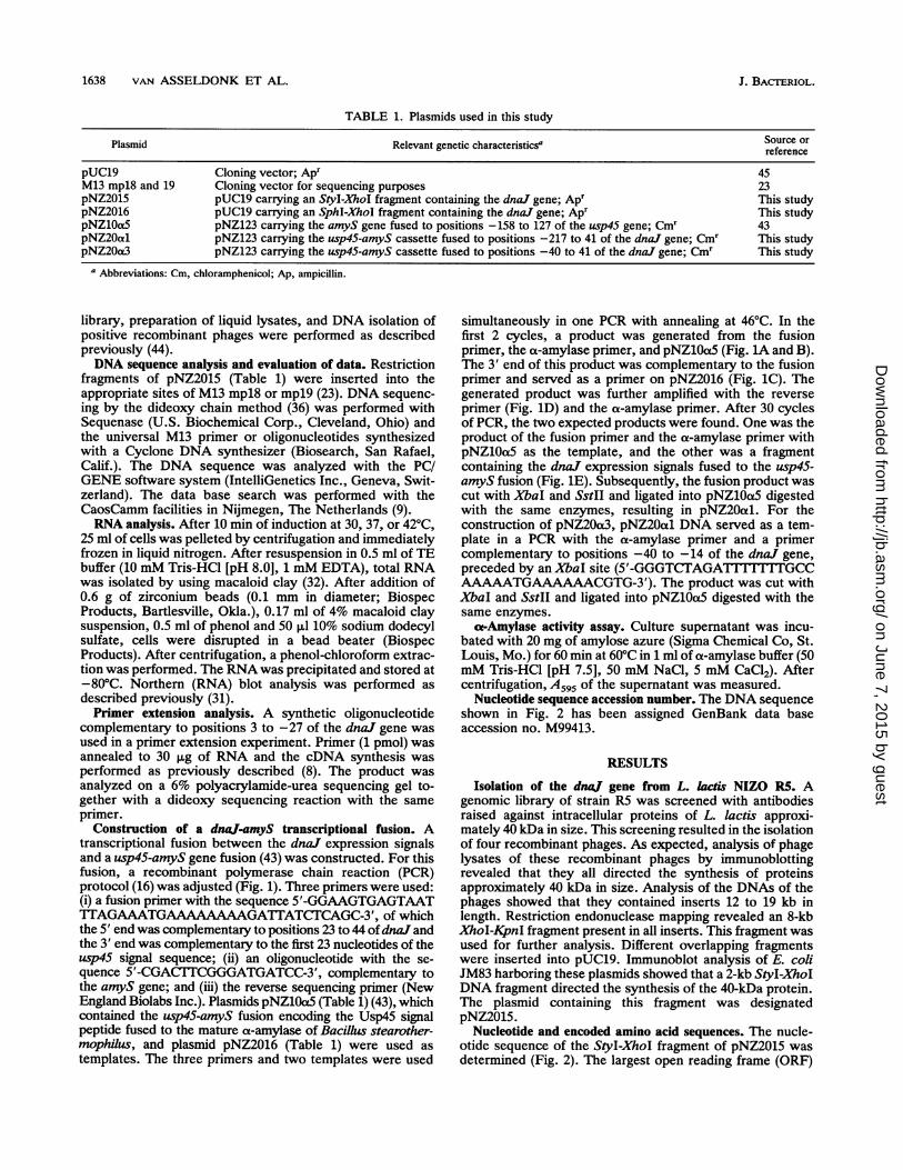
TABLE 1. Plasmids used in this study
Plasmid Relevant genetic characteristicsa Source orreference
pUC19 Cloning vector; Apr 45M13 mpl8 and 19 Cloning vector for sequencing purposes 23pNZ2015 pUC19 carrying an StyI-XhoI fragment containing the dnaJ gene; Apr This studypNZ2016 pUC19 carrying an SphI-XhoI fragment containing the dnaJ gene; Apr This studypNZlOa5 pNZ123 carrying the amyS gene fused to positions -158 to 127 of the usp45 gene; Cmr 43pNZ20al pNZ123 carrying the usp45-amyS cassette fused to positions -217 to 41 of the dnaJ gene; Cmr This studypNZ2Oa3 pNZ123 carrying the usp45-amyS cassette fused to positions -40 to 41 of the dnaJ gene; Cm' This study
a Abbreviations: Cm, chloramphenicol; Ap, ampicillin.
library, preparation of liquid lysates, and DNA isolation ofpositive recombinant phages were performed as describedpreviously (44).DNA sequence analysis and evaluation of data. Restriction
fragments of pNZ2015 (Table 1) were inserted into theappropriate sites of M13 mpl8 or mpl9 (23). DNA sequenc-ing by the dideoxy chain method (36) was performed withSequenase (U.S. Biochemical Corp., Cleveland, Ohio) andthe universal M13 primer or oligonucleotides synthesizedwith a Cyclone DNA synthesizer (Biosearch, San Rafael,Calif.). The DNA sequence was analyzed with the PC/GENE software system (IntelliGenetics Inc., Geneva, Swit-zerland). The data base search was performed with theCaosCamm facilities in Nijmegen, The Netherlands (9).RNA analysis. After 10 min of induction at 30, 37, or 42°C,
25 ml of cells was pelleted by centrifugation and immediatelyfrozen in liquid nitrogen. After resuspension in 0.5 ml of TEbuffer (10 mM Tris-HCl [pH 8.0], 1 mM EDTA), total RNAwas isolated by using macaloid clay (32). After addition of0.6 g of zirconium beads (0.1 mm in diameter; BiospecProducts, Bartlesville, Okla.), 0.17 ml of 4% macaloid claysuspension, 0.5 ml of phenol and 50 ,ul 10% sodium dodecylsulfate, cells were disrupted in a bead beater (BiospecProducts). After centrifugation, a phenol-chloroform extrac-tion was performed. The RNA was precipitated and stored at-80°C. Northern (RNA) blot analysis was performed asdescribed previously (31).Primer extension analysis. A synthetic oligonucleotide
complementary to positions 3 to -27 of the dnaJ gene wasused in a primer extension experiment. Primer (1 pmol) wasannealed to 30 ,ug of RNA and the cDNA synthesis wasperformed as previously described (8). The product wasanalyzed on a 6% polyacrylamide-urea sequencing gel to-gether with a dideoxy sequencing reaction with the sameprimer.
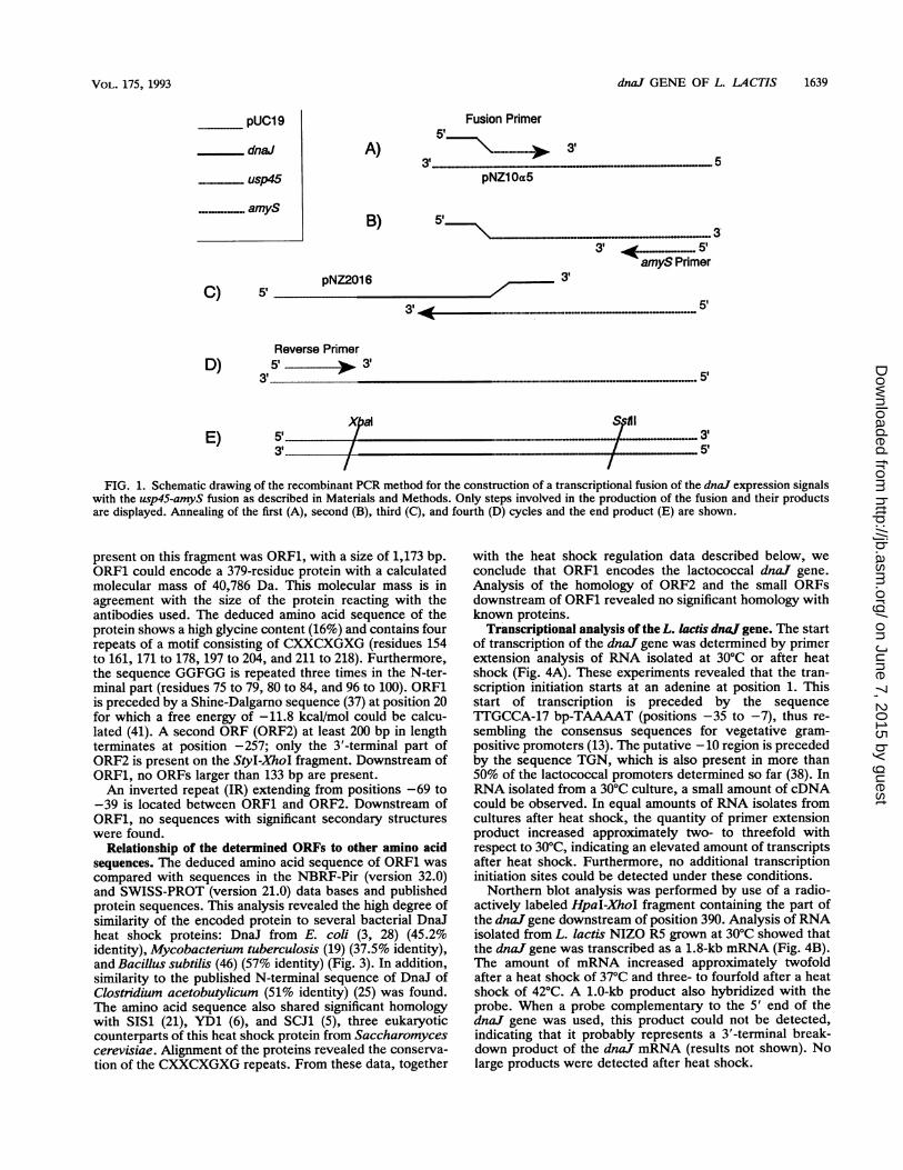
Construction of a dnaj-amyS transcriptional fusion. Atranscriptional fusion between the dnaJ expression signalsand a usp45-amyS gene fusion (43) was constructed. For thisfusion, a recombinant polymerase chain reaction (PCR)protocol (16) was adjusted (Fig. 1). Three primers were used:(i) a fusion primer with the sequence 5'-GGAAGTGAGTAATT1TAGAAATGAAAAAAAAGATTATCTCAGC-3', of whichthe 5' end was complementary to positions 23 to 44 ofdnal andthe 3' end was complementary to the first 23 nucleotides of theusp45 signal sequence; (ii) an oligonucleotide with the se-quence 5'-CGACTlCGGGATGATCC-3', complementary tothe amyS gene; and (iii) the reverse sequencing primer (NewEngland Biolabs Inc.). Plasmids pNZ10a5 (Table 1) (43), whichcontained the usp45-amyS fusion encoding the Usp45 signalpeptide fused to the mature a-amylase of Bacillus stearother-mophilus, and plasmid pNZ2016 (Table 1) were used astemplates. The three primers and two templates were used
simultaneously in one PCR with annealing at 46°C. In thefirst 2 cycles, a product was generated from the fusionprimer, the a-amylase primer, and pNZ10a5 (Fig. 1A and B).The 3' end of this product was complementary to the fusionprimer and served as a primer on pNZ2016 (Fig. 1C). Thegenerated product was further amplified with the reverseprimer (Fig. 1D) and the a-amylase primer. After 30 cyclesof PCR, the two expected products were found. One was theproduct of the fusion primer and the a-amylase primer withpNZ10cx5 as the template, and the other was a fragmentcontaining the dnaJ expression signals fused to the usp45-amyS fusion (Fig. 1E). Subsequently, the fusion product wascut with XbaI and SstII and ligated into pNZ10a5 digestedwith the same enzymes, resulting in pNZ20al. For theconstruction of pNZ2Oa3, pNZ20al DNA served as a tem-plate in a PCR with the a-amylase primer and a primercomplementary to positions -40 to -14 of the dnaJ gene,preceded by an XbaI site (5'-GGGTCTAGATI'T'ITTGCCAAAAATGAAAAAACGTG-3'). The product was cut withXbaI and SstII and ligated into pNZ10o5 digested with thesame enzymes.
cx-Amylase activity assay. Culture supernatant was incu-bated with 20 mg of amylose azure (Sigma Chemical Co, St.Louis, Mo.) for 60 min at 60°C in 1 ml of a-amylase buffer (50mM Tris-HCl [pH 7.5], 50 mM NaCl, 5 mM CaCl2). Aftercentrifugation, A595 of the supernatant was measured.
Nucleotide sequence accession number. The DNA sequenceshown in Fig. 2 has been assigned GenBank data baseaccession no. M99413.
RESULTS
Isolation of the dnaJ gene from L. lactis NIZO R5. Agenomic library of strain R5 was screened with antibodiesraised against intracellular proteins of L. lactis approxi-mately 40 kDa in size. This screening resulted in the isolationof four recombinant phages. As expected, analysis of phagelysates of these recombinant phages by immunoblottingrevealed that they all directed the synthesis of proteinsapproximately 40 kDa in size. Analysis of the DNAs of thephages showed that they contained inserts 12 to 19 kb inlength. Restriction endonuclease mapping revealed an 8-kbX7zoI-KpnI fragment present in all inserts. This fragment wasused for further analysis. Different overlapping fragmentswere inserted into pUC19. Immunoblot analysis of E. coliJM83 harboring these plasmids showed that a 2-kb StyI-XhoIDNA fragment directed the synthesis of the 40-kDa protein.The plasmid containing this fragment was designatedpNZ2015.
Nucleotide and encoded amino acid sequences. The nucle-otide sequence of the StyI-XhoI fragment of pNZ2015 wasdetermined (Fig. 2). The largest open reading frame (ORF)
J. BACTRIOL.
on June 7, 2015 by guesthttp://jb.asm
.org/D
ownloaded from
dna. GENE OF L. LACTIS 1639
pUC19
dnaJ
usp45
A)
Fusion Primer5'
3'
pNZ1 Oa5
B) 5'
pNZ2016 3,C)
3' ...... 5'amyS Primer
5'_-- ...............-----
Reverse PrimerD) 5 'b. 3'
31 Ap "."."."............ 51
E) 5 ....... 33' t
XpIa SPA I
FIG. 1. Schematic drawing of the recombinant PCR method for the construction of a transcriptional fusion of the dnaJ expression signalswith the usp45-amyS fusion as described in Materials and Methods. Only steps involved in the production of the fusion and their productsare displayed. Annealing of the first (A), second (B), third (C), and fourth (D) cycles and the end product (E) are shown.
present on this fragment was ORFi, with a size of 1,173 bp.ORFi could encode a 379-residue protein with a calculatedmolecular mass of 40,786 Da. This molecular mass is inagreement with the size of the protein reacting with theantibodies used. The deduced amino acid sequence of theprotein shows a high glycine content (16%) and contains fourrepeats of a motif consisting of CXXCXGXG (residues 154to 161, 171 to 178, 197 to 204, and 211 to 218). Furthermore,the sequence GGFGG is repeated three times in the N-ter-minal part (residues 75 to 79, 80 to 84, and 96 to 100). ORFiis preceded by a Shine-Dalgarno sequence (37) at position 20for which a free energy of -11.8 kcal/mol could be calcu-lated (41). A second ORF (ORF2) at least 200 bp in lengthterminates at position -257; only the 3'-terminal part ofORF2 is present on the StyI-XhoI fragment. Downstream ofORF1, no ORFs larger than 133 bp are present.An inverted repeat (IR) extending from positions -69 to
-39 is located between ORFi and ORF2. Downstream ofORF1, no sequences with significant secondary structureswere found.
Relationship of the determined ORFs to other amino acidsequences. The deduced amino acid sequence of ORFi wascompared with sequences in the NBRF-Pir (version 32.0)and SWISS-PROT (version 21.0) data bases and publishedprotein sequences. This analysis revealed the high degree ofsimilarity of the encoded protein to several bacterial DnaJheat shock proteins: DnaJ from E. coli (3, 28) (45.2%identity), Mycobactenium tuberculosis (19) (37.5% identity),and Bacillus subtilis (46) (57% identity) (Fig. 3). In addition,similarity to the published N-terminal sequence of DnaJ ofClostnidium acetobutylicum (51% identity) (25) was found.The amino acid sequence also shared significant homologywith SIS1 (21), YD1 (6), and SCJ1 (5), three eukaryoticcounterparts of this heat shock protein from Saccharomycescerevisiae. Alignment of the proteins revealed the conserva-tion of the CXXCXGXG repeats. From these data, together
with the heat shock regulation data described below, weconclude that ORFi encodes the lactococcal dnaJ gene.Analysis of the homology of ORF2 and the small ORFsdownstream of ORFi revealed no significant homology withknown proteins.
Transcriptional analysis of the L. lactis dnaj gene. The startof transcription of the dnaJ gene was determined by primerextension analysis of RNA isolated at 30°C or after heatshock (Fig. 4A). These experiments revealed that the tran-scription initiation starts at an adenine at position 1. Thisstart of transcription is preceded by the sequenceTTGCCA-17 bp-TAAAAT (positions -35 to -7), thus re-sembling the consensus sequences for vegetative gram-positive promoters (13). The putative - 10 region is precededby the sequence TGN, which is also present in more than50% of the lactococcal promoters determined so far (38). InRNA isolated from a 30°C culture, a small amount of cDNAcould be observed. In equal amounts of RNA isolates fromcultures after heat shock, the quantity of primer extensionproduct increased approximately two- to threefold withrespect to 30°C, indicating an elevated amount of transcriptsafter heat shock. Furthermore, no additional transcriptioninitiation sites could be detected under these conditions.Northern blot analysis was performed by use of a radio-
actively labeled HpaI-XhoI fragment containing the part ofthe dnaJ gene downstream of position 390. Analysis of RNAisolated from L. lactis NIZO R5 grown at 30°C showed thatthe dnaJ gene was transcribed as a 1.8-kb mRNA (Fig. 4B).The amount of mRNA increased approximately twofoldafter a heat shock of 37°C and three- to fourfold after a heatshock of 42°C. A 1.0-kb product also hybridized with theprobe. When a probe complementary to the 5' end of thednaJ gene was used, this product could not be detected,indicating that it probably represents a 3'-terminal break-down product of the dnaJ mRNA (results not shown). Nolarge products were detected after heat shock.
VOL. 175, 1993
5'
on June 7, 2015 by guesthttp://jb.asm
.org/D
ownloaded from

1640 VAN ASSELDONK ET AL. J. BACTERIOL.
-457
CCTTGGACGTCTGATGMGACATCCCGGATGGTGGTTTTCCMCGATT-409
TGGCCCACAAGTTTTG CTCAAGAAAATATATCCAAGTTGCTGACTTATCTGGAAAAAGTATTAGCGAGATTAAAGATGCT-319
GTTTTATCAGACATTTAG7TGAAA7GTGGGTGACGATTATTwTATGACCTAATATCACTTAGCATGATTATTACGGTTATAGGTT-229
GTAGCTMATACGGCA TCGTTATTTAGATr.#GTATAACGAAAAGAAAAAT'GAGTTTCAT'GTTACTGATCCAATAAAAGGAAAATAnwGSphI
-139
GTTGGCTMATCTACAGTTGAT~wGGTTTATAGTGGACAAATCAA~wTCATAGAA7TTTATAGTGTAATTAGCACT-lwTATAAAAA-49 1GAGTGCTA7 wM1CAAAAATGAAAAAACGTIGGTAAAATAGTGCTATTGAAAAATTGAT7AGTAAAGGAAGTGAGTAATTTAGAA<<<<<«««««< -35 -10 1
42
ATGAATATAC'MTATTATGCGACTAGGTGTCGATAAAAATCCAGTCAGATGAAATMAxAA GCTTATCGTAAAATTCCAAAM N N T E Y Y E R L G V D K N A S Q D E I K K A Y R K M S K 30132
AAATATCACCCCGATTTAAATAAAGAGAGGGTGCT'GGAAAAATATAAAGAAGTTCAGAGCATACGAAACGCITCGGATGACAK Y H P D L N K E E G A E E K Y K E V Q E A Y E T L S D E Q 60
222AGCGTCA TGACCATATGGTGAAGCTGGAGCCATGGCGGCTI"TGGCGGTGGGCTTCGGCGGAGCTTCTGG1 TK R A A Y D Q Y G E A G A N O O * 0 0 0 0 1 0 0 A S G F S G 90
312
TTCGGCGGTAGCTCAGGTIGGCTTTGG TTMAGATATATTCTCMGTTTCTTGAGGAGGTGGTGCACMGTTAACCCTMTGCAF G G S S 0 0 1 0 0 F E D I F S S F F G G G G A Q V N P N A 120
402
CCCCGTCAGGAGATGACTTACAGTATCGGATAAACTTAAAATTTGAGAAGCTAITMCGTGGAAAAACAGTCAAATATATCGTP R Q G D D L Q Y R I N L K F E E A I F G V E K Q V K Y N R 150
492
GMAGMCTCTIGTCACACTTGTGGAGGwrTAGCGAAACGT'GGCACACATCCAGAAAC'TCATAAATGTIGGTGGTCGTGGACAAATTE E L T... .C 0 A K R G T H P E T :. I 180
582
MTGTGTTCGTwGATACACCGCTIGGACGGATGCAAACACMAGTAACATGT'GATIGTTTGTMCGGMACAGGTAAAGAAATCAAAGAGAAAN V V R D T P L G R M Q T Q V T ¢ ..... .. ....................................................Y2; K E I K K.........10
672
TGTGAAACTTGTCATGGTTCAGGTCATGAAAAAGTGGCACATACTGTTAAGGTTACTGTACCTGCTGGTGTGAAACTGGACMAATGHH TK V K V T V P A G V H T G Q K M 240
762
CGTTTGCAGGACAGGTGATCTGTTAAATIGGTGACCTTAT'GGGATTTGTATGTTGTTwTCAAGTGAAGCTTCAGACAAATTTR L Q G Q G D A G V N G G P Y G D L Y V V F Q V E A S D K F 270852
GAGCGTGATGGTGCAGAAATTTACTACMGATGCCMTGGACTTTGTTCMGCTGCTTTAGGTATAAATTGMGTTCCGACTGTTCATE R D G A E I Y Y K M P M D F V Q A A L G D E I E V P T V H 300
942
GGAAATGTMAGTTGAAAATTCC73ISGACACAAACAGGGGCTATTTCCGTCTAAAAGGTAAAGGTGCGCCAAAACTCCGTGGTTCTG N V K L K I P A G T Q T G A N F R L K G K G A P K L R G S 3301032
GGTATGGTGACCATATGTTATTATCATATTGTCACTCCTAGATTTGATCAGCTCAAAAAGA GCGTCCAAG GCTAAAG N G D Q Y V I I N I V T P K N L N Q A Q K E A L Q A F A K 360
1122
GCAAGTGGTGwTAAGTCTCTGTTCAGGTAAAGGlil-lATAAGTTTAAATAAAAATAAAAGTAAAAGTCGCITGCTA S G V E V S G S G K K G F F D K F K -1212lIlI CTATCAGTA MTTle rCTAlI-liq!GACTGACAGTCAGTA--mAGGGAGAAAACAGGAATlT AGT
1302
ITIATATAATAGGGGCAMAAAATGATATTATAGCCTGATTA7qwTAGCGCTATCACTAGACCTACGCGTCAGTCGATGIWGC1392GATCTCATG
FIG. 2. Nucleotide and deduced amino acid sequence of the dnaJ gene of L. lactis. The putative Shine-Dalgarno sequence (double-underlined), the -10 and -35 sequences (underlined), and the IR (arrowheads below the sequence) are indicated. The 5' end of the mRNA,as identified by primer extension, is marked with an arrow. The repeated CXXCXGXG motif in the protein sequence is shaded, and therepeated GGFGG sequences are in boldface.
on June 7, 2015 by guesthttp://jb.asm
.org/D
ownloaded from

dna! GENE OF L. LACTIS 1641
Li MNNTE-----YYERLGVDKNASQDEIKKAYRKMSKKYHPDLNK-EEGAEE 44Bs MSKRD-----YYEVLGVSKSASKDEIKKAYRKLSKKYHPDINK-EAGSDE 44Ec MAKQD-----YYEILGVSKTAEEREIRKAYKRLAMKYHPDRNQGDKEAEA 45Mt MAQREWVEKDFYQELGVSSDASPEEIKRAYRKLARDLHPDANPGNPAAGE 50.. .. .. .. . . ... . . . . . . .. ...
Li KYKEVQEAYETLSDEQKRAAYDQYGEAG-ANGGFGGGGFGGA--SGFSGFBs KFKEVKEAYETLSDDQKRAHYDQFGHTD-PNQGFGGGGFGGG---DFGGFEc KFKEIKEAYEVLTDSQKRAAYDQYGHAAFEQGGMGGGGFGGG--ADFSDIMt RFKAVSEAHNVLSDPAKRKEYDE-TRRLFAGGGFGGRRFDSGFGGGFGGF
.. .. * *.*.... **. ..*.........
AAG CT 300 370 42°
:.:91909399
B
4.4 -
2.4 -
1.4 -
300370428
LI GGSSGGFGG----------------FEDIFSSFFGGGGAQVNPNAPRQGD 125Bs G---------------------FDDIFSSIFGGGTRRRDPKLRARGA 116Ec -------------------------FGDVFGDIFGGG---RGRQRAARGA 115Mt GVGGDGAEFNLNDLFDAASRTGGTTIGDLFGGLFGRGGSAR-PSRPRRGN 148
LIBEsEcMt
LiBsEcMt
175166165197
AHT 225RKK 216SKT 211TRT 243
LI VKVTVPAGVETGQKMRLQGQGDAGVNGGPYGDLYVVFQVEASDKFERDGA 275Bs INVTIPAGVDDGQQLRLSGQGEPGINGG-LPDLFVVFHVRAHEFFERDGD 265EC LSVKIPAGVDTGDRIRLAGEGEAGEHGAPAGDLYVQVQVKQHPIFEREGN 261Mt INVRIPPGVEDGQRIRLAGQGEAGLRGAPSGDLYVTVHVRPDKIFGRDGD 293
* * ** * ** * * * * *** * ***
LI EIYYKMPMDFVQAALGDEIEVPTVHGNVKLKIPAGTQT'GANFRLKGKGAP 325Bs DIYCEMPLTFAQAALGDEVEVPTLHG--KVKIPAGTQTGTKFRLRGKGVQ 313Ec NLYCEVPINFAMAALGGEIEVPTLDGRVKLKVPGETQTGKLFRMRGKGVK 311Mt DLTVTVPVSFTELALGSTLSVPTLDGTVGVRVPKGTADGRILRVRGRVCP 343
* ***.. * *** *. * * -*
Li KLRGSGNGDQYVIINIVTPKNLNQAQKEALQAFAKASGVEVSGSGK---K 372Bs NVRGYGQGDQHIVVRVVTPTNLTDKQKDIIREFAEVSG-NLPDEQE---M 359Ec SVRGGAQGDLLCRVVVETPVGLNERQKQLLQELQESFGGPTGEHNSPRSK 361Mt SAVGVAATYLSP 355
Li GFFDKFK-------- 379Bs SFFDKVKRAFKG--D 372Ec SFFDGVKKFFDDLTR 376
FIG. 3. Alignment of the deduced amino acid sequences of theDnaJ proteins of L. lactis (Li), B. subtilis (Bs) (46), E. coli (Ec) (3,28), and M. tuberculosis (Mt) (19). Identical amino acids areindicated by asterisks, conserved residues are indicated by points(14), gaps to obtain maximum fit are indicated by dashes, and theconserved CXXCXGXG repeats are shaded.
A palindromic DNA structure is involved in heat shockregulation of dnaJ. The IR found at position -69 showssimilarity to palindromic structures that are located at cor-responding positions upstream of the heat shock genes of B.subtilis, C acetobutylicum, Synechocystis sp., Synechococ-cus sp., Mycobacterium sp., and Chlamydia psittaci (46). Ithas been postulated that this IR could be involved in thetemperature-sensitive regulation of transcription of theseheat shock genes. To address this hypothesis, a constructionwas made in which the DnaJ-encoding region was exchangedwith a usp45-amyS gene fusion encoding the B. stearother-mophilus mature a-amylase preceded by the usp45 signalpeptide (43). In plasmid pNZ20al, the usp45-amyS fusion ispreceded by a region of dnaJ including positions -217 to 44which contain the IR (Fig. 2). In plasmid pNZ20a3, onlypositions -40 to 44 of the dnaJ gene are present, and hencethe IR is deleted. After introduction of pNZ20al orpNZ203 in L. lactis MG1363, a-amylase activities weremeasured 10, 20, or 30 min after heat shock at 37 or 420C(Fig. 5). The final optical density of the cells after heat shockinduction at 370C or 420C was not higher than the opticaldensity of cells grown at 300C at the indicated times. StrainMG1363 harboring pNZ20al resulted in two to four times
FIG. 4. Transcriptional analysis of the dnaJ gene. (A) Autora-diogram of the sequence gel used to analyze the primer extensionproducts of RNA of NIZO R5, isolated after 10 min of heat shockinduction at the indicated temperatures. The primer extensionproduct is indicated (arrow). The sequence ladder obtained with thesame primer is also shown. (B) Autoradiogram of a Northern blot oftotal RNA, isolated at 30'C or after heat shock, hybridized with aradioactively labeled HpaI-XhoI fragment of the dna. gene. The1.8-kb mRNA is indicated (arrow). The sizes of the RNA markers(Bethesda Research Laboratories) are shown in kilobases.
higher a-amylase production after heat shock induction thanbefore heat shock induction. However, strain MG1363 har-boring pNZ20a3 showed a constitutive ot-amylase produc-tion at 30'C and at elevated temperatures. The level ofa-amylase production of this strain was comparable to thatof MG1363 harboring pNZ20al after heat shock induction.
a-amylase activity (mU/ml)
40-
30 -
20 -
10
0I10
Time after heat shock (mi n)20 30
FIG. 5. Relative amounts of a-amylase activity produced by L.lactis MG1363 carrying a transcriptional dnaJ-amyS fusion.pNZ20al contains the dnaJ promoter region including the IR, whilein pNZ2Oa3, the IR was deleted. Heat shock was induced as
described in Materials and Methods. Samples were taken after thetimes indicated, and newly secreted a-amylase activity was assayed.MG1363(pNZ20al) at 30'C (E), 370C (U), and 420C (-) andMG1363(pNZ20a3) at 30'C (s), 370C (n), and 420C (g) are shown.
VOL. 175, 1993
TTr
on June 7, 2015 by guesthttp://jb.asm
.org/D
ownloaded from

1642 vAN ASSELDONK ET AL.
DISCUSSION
In this report, the cloning, sequencing, and characteriza-tion of the dnaJ gene of L. lactis are described. Besideshomology of the encoded protein with the DnaJ proteins ofE. coli (3, 28) and M. tuberculosis (19), identity with theDnaJ proteins from B. subtilis (46) and C. acetobutylicum(25) was found. The Met residue at position 1 fits perfectly inthe alignment of the different DnaJ proteins. Alignment ofthe reported proteins revealed a significant overall homologyand the conservation of a motif consisting of CXXCXGXG,which is repeated four times (Fig. 3). This motif was alsofound in the eukaryotic homologs of the DnaJ proteins: SCJ1(5), YD1 (6), and SIS1 (21). The biological meaning of thismotif is not yet clear. The organization into two largerrepeats, CXXCXGXG(X)8CXXCXGXG, as in YDJI (6), wasnot found in the other reported proteins and is unlikely to becharacteristic for DnaJ proteins. The conservation of theGGFGG sequence is less significant. Only one of the threelactococcal copies of this sequence is present in the bacterialDnaJ of E. coli and B. subtilis.
In prokaryotes, most dnaJ genes characterized so far arepreceded by dnaK, which encodes another heat shockprotein that is conserved among prokaryotes and eukaryotes(3, 25, 28, 46). Upstream of the L. lactis dnaJ gene, anotherORF, designated ORF2, was found, but its deduced aminoacid sequence shared no homology with known DnaK pro-teins, suggesting another genomic organization of these heatshock genes in L. lactis. The M. tuberculosis dnaJ gene islocated 788 bp downstream of the dnaK gene (19). Thisintergenic distance exceeds the length of DNA region se-quenced from dnaJ from L. lactis; hence, a conservation ingenetic organization of the dnaK and dnaJ genes between M.tuberculosis and L. lactis cannot be totally excluded. Thepossibility that the dnaK gene is situated downstream of thednaJ gene or elsewhere on the chromosome is also conceiv-able.
In B. subtilis (46), C. acetobutylicum (25), and E. coli (3,28), dnaJ is located in an operon that also includes dnaK. InL. lactis, however, the start of transcription of dnaJ islocated immediately upstream of the dnaJ gene. In addition,the size of the RNA messenger is 1.8 kb. This is too small tocontain both genes. From these data, it can be concludedthat the lactococcal dnaJ and dnaK genes are not organizedin a single operon. The same holds for the dnaK-dnaJ geneorganization of Synechocystis sp. (7). Transcriptional anal-ysis of the dnaK gene of this organism revealed that it istranscribed as a monocistronic messenger. Hence, a putativednaJ gene will also be present on a separate transcriptionalunit.The induction of expression of the dnaJ gene by heat
shock was determined by three methods. First, the primerextension carried out with RNA isolated at 30°C, or afterheat shock at 37 and 42°C, demonstrated a significant in-crease in dnaJ mRNA. Second, Northern blot analysisshowed a twofold increase in the amount of dnaJ RNA afterheat shock at 37°C. The amount of mRNA was even higherafter heat shock at 42°C. These results confirm that the heatshock response is controlled at the transcriptional level. Thesame has been found for the heat shock genes of othergram-positive bacteria such as B. subtilis (46) and C. aceto-butylicum (24, 25). Third, the fusion between the dnaJpromoter region and a usp45-amyS cassette caused a signif-icant increase in a-amylase production after heat shock.Similar results were obtained in a comparable experiment in
B. subtilis (46) with a transcriptional fusion between thednaK promoter and the amyL gene.
Analysis of the transcription initiation site of the dnaJgene revealed that it was preceded by gram-positive vegeta-tive -10 and -35 sequences (13). The IR, located upstreamof the -35 sequence, was also found upstream of the heatshock genes characterized thus far in gram-positive organ-isms, such as the groELS operons from L. lactis (17) and Cacetobutylicum (24) and the dnaK operons from B. subtilis(46) and C. acetobutylicum (25). Furthermore, an IR with thesame sequence is located upstream of the heat shock genesof Synechocystis and Synechococcus spp. and C. psittacci(46). However, the IR is, at least partially, as in Synechoc-occus sp., or entirely, as in B. subtilis or C. acetobutylicum,located on the 5' end of the mRNA in these operons,whereas in the L. lactis dnaJ gene, it is located upstream ofthe start of transcription. The IR at position -69 of thelactococcal dnaJ gene shares complete identity with theconsensus sequence, as proposed by Wetzstein et al. (46).To examine the function of this IR in heat shock regulation,pNZ20al and pNZ20a3 were constructed. These plasmidscontain a usp45-amyS fusion preceded by the promoterregion of the dnaJ gene. In MG1363 harboring pNZ2Oal,which contains the IR, the level of a-amylase activity is twoto four times higher after heat shock than before heat shock.MG1363 harboring pNZ20a3, in which the IR has beendeleted, showed no heat shock induction of ax-amylaseproduction. These results indicate a major role for the IR inthe heat shock regulation of the dnaJ gene of L. lactis.
In transcription of E. coli heat shock genes, a specificsigma factor ({r32) that recognizes a promoter sequencedeviant from the vegetative -35 and -10 sequences (26) isinvolved. In the heat shock genes from B. subtilis (46) and C.acetobutylicum (24, 25), the transcription start sites arepreceded by vegetative promoter sequences. The function ofthe IR in the heat shock regulation of the dnaJ gene of L.lactis and its conservation in sequence and location in heatshock genes of gram-positive bacteria strongly suggest asignificant difference in heat shock regulation between E.coli and gram-positive organisms. Moreover, in Synechocys-tis and Synechococcus spp. and C. psittacci, both an E. coliheat shock consensus promoter sequence and the IR arepresent in the promoter region, suggesting that the IR is notspecific for gram-positive heat shock genes. However, in theL. lactis dnaf gene, the IR is unlikely to protect against RNAdegradation, as suggested for B. subtilis by Wetzstein et al.(46), because it is located upstream of the start of transcrip-tion. For the same reason, it is unlikely to cause a pause inthe production of RNA, as proposed by Narberhaus andBahl (24). The amount of a-amylase produced by MG1363harboring pNZ2Oa3 at all tested conditions was comparablewith the amount produced by MG1363 harboring pNZ20alafter heat shock. This constitutive high level of a-amylaseproduction by MG1363 harboring pNZ2Oa3 suggests that therepeat is a target for a repressor, the activity of which isdisturbed after heat shock. However, to be conclusive aboutthis hypothesis, further analysis of the system is required.
ACKNOWLEDGMENTSWe are grateful to I. van Alen-Boerrigter for providing a sample of
the R5 genomic library. We also express thanks to C. A. Batt forsharing unpublished results on the characterization of the groELSoperon of L. lactis and W. Schumann for sharing unpublishedresults on the palindromic structure found upstream of the B.subtilis dnaK operon. We thank 0. Kuipers, P. Bruinenberg, R.Siezen, and R. van Rooijen for critical reading of the manuscript.
J. BACTERIOL.
on June 7, 2015 by guesthttp://jb.asm
.org/D
ownloaded from
dna. GENE OF L. LACTIS 1643
We thank Joop Mondria, Simon van der Laan, and Henk van Brakelfor artwork and photography.
This work was partly financed by the Mesdag Foundation, Leeu-warden, The Netherlands.
REFERENCES
1. Ang, D., K. Liberek, D. Skowyra, M. Zylicz, and C. Georgopo-ulos. 1991. Biological role and regulation of the universallyconserved heat shock proteins. J. Biol. Chem. 266:24233-24236.
2. Auffray, Y., X. Gansel, B. Thammavongs, and P. Boutibonnes.1992. Heat shock-induced protein synthesis in Lactococcuslactis subsp. lactis. Curr. Microbiol. 24:281-284.
3. Bardwell, J. C., K. Tilly, E. Craig, J. King, M. Zylicz, and C.Georgopoulos. 1986. The nucleotide sequence of the Escherichiacoli K12 dnaJ+ gene. J. Biol. Chem. 261:1782-1785.
4. Birnboim, H. C., and J. Doly. 1979. A rapid alkaline extractionprocedure for screening recombinant plasmid DNA. NucleicAcids Res. 7:1513-1523.
5. Blumberg, H., and P. A. Silver. 1991. A homologue of thebacterial heat-shock gene dnaJ that alters protein sorting inyeast. Nature (London) 349:627-630.
6. Caplan, A. J., and M. G. Douglas. 1991. Characterization ofYDJ1: a yeast homologue of the bacterial DnaJ protein. J. CellBiol. 114:609-621.
7. Chitnis, P. R., and N. Nelson. 1991. Molecular cloning of thegenes encoding two chaperone proteins of the cyanobacteriumSynechocystis sp. PCC 6803. J. Biol. Chem. 266:58-65.
8. Debarbouille, M., and 0. Raibaud. 1983. Expression of the E.coli malPQ operon remains unaffected after drastic alterationsof its promoter. J. Bacteriol. 153:1221-1227.
9. Devereux, J., P. Haeberli, and 0. Smithies. 1984. A comprehen-sive set of sequence analysis programs for the VAX. NucleicAcids Res. 12:387-395.
10. De Vos, W. M., P. Vos, H. De Haard, and I. Boerrigter. 1989.Cloning and expression of the Lactococcus lactis subsp. cremo-ris SK11 gene encoding an extracellular serine proteinase. Gene85:169-176.
11. Gaitanaris, G. A., A. G. Papavassiliou, P. Rubock, S. J. Silver-stein, and M. E. Gottesman. 1990. Renaturation of denaturedlambda repressor requires heat shock proteins. Cell 61:1013-1020.
12. Gasson, M. J. 1983. Plasmid complements of Streptococcuslactis NCDO 712 and other lactic streptococci after protoplast-induced curing. J. Bacteriol. 154:1-9.
13. Graves, M. C., and J. C. Rabinowitz. 1986. In vivo and in vitrotranscription of the C. pasteunanum ferredoxin gene. Evidencefor extended promoter elements in Gram-positive organisms. J.Biol. Chem. 261:11409-11415.
14. Higgins, D. G., and P. M. Sharp. 1989. CLUSTAL: a packagefor performing multiple sequence alignment on a micro com-puter. Gene 73:237-244.
15. Holo, H., and I. F. Nes. 1989. High-frequency transformation,by electroporation, of Lactococcus lactis subsp. cremonisgrown with glycine in osmotically stabilized media. Appl. En-viron. Microbiol. 55:3119-3123.
16. Horton, R. M., H. D. Hunt, S. N. Ho, J. K. Pullen, and L. R.Pease. 1989. Engineering hybrid genes without the use ofrestriction enzymes: gene splicing by overlap extension. Gene77:61-68.
17. Kim, S. G., and C. A. Batt. Cloning and sequencing theLactococcus lactis subsp. lactisgroELS operon. Gene, in press.
18. Laemmli, U. K. 1970. Cleavage of structural proteins during theassembly of the head of bacteriophage T4. Nature (London)227:680-685.
19. Lathigra, R. B., D. B. Young, D. Sweetser, and R. A. Young.1988. A gene from Mycobactenium tuberculosis which is homol-ogous to the DnaJ heat shock protein of E. coli. Nucleic AcidsRes. 16:1636.
20. Liberek, K., J. Marszalek, D. Ang, C. Georgopoulos, and M.Zylicz. 1991. Escherichia coli DnaJ and GrpE heat shockproteins jointly stimulate ATPase activity of DnaK. Proc. Natl.Acad. Sci. USA 88:2874-2878.
21. Luke, M. M., A. Sutton, and K. Arndt. 1991. Characterization ofSIS1, a Saccharomyces cerevisiae homologue of bacterial DnaJproteins. J. Cell Biol. 114:623-638.
22. Malki, A., P. Hughes, and M. Kohiyama. 1991. In vitro roles ofEscherichia coli DnaJ and DnaK heat shock proteins in thereplication oforiC plasmids. Mol. Gen. Genet. 225:420-426.
23. Messing, J. 1983. New M13 vectors for cloning. MethodsEnzymol. 101:20-78.
24. Narberhaus, F., and H. Bahl. 1992. Cloning, sequencing, andmolecular analysis of the groESL operon of Clostridium aceto-butylicum. J. Bacteriol. 174:3282-3289.
25. Narberhaus, F., K. Giebeler, and H. Bahl. 1992. Molecularcharacterization of the dnaK gene region of Clostridium aceto-butylicum, including grpE, dnaJ, and a new heat shock gene. J.Bacteriol. 174:3290-3299.
26. Neidhardt, F. C., and R. A. Vanbogelen. 1987. Heat shockresponse, p. 1334-1345. In F. C. Neidhardt, J. L. Ingraham,K. B. Low, B. Magasanik, M. Schaechter, and H. E. Umbarger(ed.), Eschenchia coli and Salmonella typhimurium: cellularand molecular biology. American Society for Microbiology,Washington, D.C.
27. Ofverstedt, L. G., J. Sandelin, and G. Johansson. 1983. Recov-ery of proteins on a milligram scale from polyacrylamide elec-trophoresis gels, exemplified by purification of a retinol-bindingprotein. Anal. Biochem. 134:361-365.
28. Ohki, M., F. Tamura, S. Nishimura, and H. Uchida. 1986.Nucleotide sequence of the Escherichia coli dnaJ gene andpurification of the gene product. J. Biol. Chem. 261:1778-1781.
29. Phillips, G. J., and T. J. Silhavy. 1990. Heat-shock proteinsDnaK and GroEL facilitate export of LacZ hybrid proteins in E.coli. Nature (London) 344:882-884.
30. Rauch, P. J. G., M. M. Beerthuyzen, and W. M. De Vos. 1990.Nucleotide sequence of IS904 from Lactococcus lactis subsp.lactis strain NIZO R5. Nucleic Acids Res. 18:4253.
31. Rauch, P. J. G., and W. M. De Vos. 1992. Transcriptionalregulation of the TnS267-located Lactococcus lactis sucroseoperon and characterization of the sacA gene encoding sucrose-6-phosphate hydrolase. Gene 121:55-61.
32. Raya, R. (Institut National de la Recherche Agronomique).Personal communication.
33. Rosenberg, S. M. 1987. Improved in vitro packaging of DNA.Methods Enzymol. 153:95-103.
34. Rottlander, E., and T. A. Trautner. 1970. Genetic and transfec-tion studies with Bacillus subtilis phage SP50. J. Mol. Biol.108:47-60.
35. Sambrook, J., E. F. Fritsch, and T. Maniatis. 1989. Molecularcloning: a laboratory manual. Cold Spring Harbor Laboratory,Cold Spring Harbor, N.Y.
36. Sanger, F., S. Nicklen, and A. R. Coulson. 1977. DNA sequenc-ing with chain-terminating inhibitors. Proc. Natl. Acad. Sci.USA 74:5463-5467.
37. Shine, J., and L. Dalgarno. 1974. The 3' terminal sequence of E.coli 16S RNA: complementary to nonsense triplets and ribo-some binding site. Proc. Natl. Acad. Sci. USA 71:1342-1346.
38. Simons, G., and W. M. de Vos. Unpublished data.39. Sunshine, M., M. Feiss, J. Stuart, and J. Yochem. 1977. A new
host gene (groPC) necessary for lambda DNA replication. Mol.Gen. Genet. 151:27-34.
40. Terzaghi, B. E., and W. E. Sandine. 1975. Improved medium forlactic streptococci and their bacteriophages. Appl. Microbiol.29:807-813.
41. Tinoco, I., P. N. Bore, B. Dengler, M. D. Levine, 0. C.Uhlenbeck, D. M. Crothers, and J. Gralla. 1973. Improvedestimation of secondary structure in ribonucleic acids. Nature(London) 246:40-41.
42. Towbin, H., T. Staehelin, and J. Gordon. 1979. Electrophoretictransfer of proteins from polyacrylamide gels to nitrocellulosesheets: procedure and some applications. Proc. Natl. Acad. Sci.USA 76:4350-4354.
43. Van Asseldonk, M., W. M. De Vos, and G. Simons. Submittedfor publication.
44. Van Asseldonk, M., G. Rutten, M. Oteman, R. J. Siezen, W. M.De Vos, and G. Simons. 1990. Cloning of usp45, a gene encoding
VOL. 175, 1993
on June 7, 2015 by guesthttp://jb.asm
.org/D
ownloaded from
1644 vAN ASSELDONK ET AL.
a secreted protein from Lactococcus lactis subsp. lactis. Gene95:155-160.
45. Vieira, J., and J. Messing. 1982. The pUC plasmids, anM13mp7-derived system for insertion mutagenesis and sequenc-ing with synthetic primers. Gene 19:259-268.
46. Wetzstein, M., U. Volker, J. Dedio, S. Lobau, U. Zuber, M.Schiesswohl, C. Herget, M. Hecker, and W. Schumann. 1992.Cloning, sequencing and molecular analysis of the dnaK locusfrom Bacillus subtilis. J. Bacteriol. 174:3300-3310.
47. Whitaker, R. D., and C. A. Batt. 1991. Characterization of theheat shock response in Lactococcus lactis subsp. lactis. Appl.
Environ. Microbiol. 57:1408-1412.48. Wickner, S., J. Hoskins, and K. McKenney. 1991. Function of
DnaJ and DnaK as chaperones in origin-specific DNA bindingby RepA. Nature (London) 350:165-167.
49. Wickner, S. H. 1990. Three Escherichia coli heat shock proteinsare required for P1 plasmid DNA replication: formation of anactive complex between E. coli DnaJ protein and the P1 initiatorprotein. Proc. Natl. Acad. Sci. USA 87:2690-2694.
50. Wild, J., E. Altman, T. Yura, and C. A. Gross. 1992. DnaK andDnaJ heat shock proteins participate in protein export in Esch-erichia coli. Genes Dev. 6:1165-1172.
J. BACrERIOL.
on June 7, 2015 by guesthttp://jb.asm
.org/D
ownloaded from