probing direct interactions between cody and the oppd promoter of lactococcus lactis
TRANSCRIPT
JOURNAL OF BACTERIOLOGY, Jan. 2005, p. 512–521 Vol. 187, No. 20021-9193/05/$08.00�0 doi:10.1128/JB.187.2.512–521.2005Copyright © 2005, American Society for Microbiology. All Rights Reserved.
Probing Direct Interactions between CodY and the oppD Promoter ofLactococcus lactis
Chris D. den Hengst,1† Peter Curley,2† Rasmus Larsen,1 Girbe Buist,1 Arjen Nauta,3
Douwe van Sinderen,2 Oscar P. Kuipers,1 and Jan Kok1*Department of Genetics, Groningen Biomolecular Sciences and Biotechnology Institute, Haren,1 and Corporate Research,
Friesland Coberco Dairy Foods, Deventer,3 The Netherlands, and Alimentary Pharmabiotic Centre and Departmentof Microbiology, National University of Ireland, Cork, Ireland2
Received 12 July 2004/Accepted 11 October 2004
CodY of Lactococcus lactis MG1363 is a transcriptional regulator that represses the expression of severalgenes encoding proteins of the proteolytic system. These genes include pepN, pepC, opp-pepO1, and probablyprtPM, pepX, and pepDA2, since the expression of the latter three genes relative to nitrogen availability issimilar to that of the former. By means of in vitro DNA binding assays and DNase I footprinting techniques,we demonstrate that L. lactis CodY interacts directly with a region upstream of the promoter of its major targetknown so far, the opp system. Our results indicate that multiple molecules of CodY interact with this promoterand that the amount of bound CodY molecules is affected by the presence of branched-chain amino acids andnot by GTP. Addition of these amino acids strongly affects the extent of the region protected by CodY in DNaseI footprints. Random and site-directed mutagenesis of the upstream region of oppD yielded variants that werederepressed in a medium with an excess of nitrogen sources. Binding studies revealed the importance ofspecific bases in the promoter region required for recognition by CodY.
Genetic and biochemical research over the past decades hasled to a clear picture of the proteolytic system of lactic acidbacteria. To ensure a proper nitrogen balance, several regula-tors are present that respond to changes in intracellular con-centrations of nitrogen-containing compounds (16, 24, 28, 30).The lactic acid bacterium Lactococcus lactis is auxotrophic forseveral amino acids (6). For optimal growth in milk, it has todegrade milk proteins (e.g., �S1 and �- and �-casein), becauseonly limited amounts of free amino acids are present in thisenvironment. An elaborate proteolytic system to release aminoacids from casein, involving a number of enzymatic activitiesthat are subject to medium-dependent regulation, has evolvedin L. lactis. Casein degradation by L. lactis is a process that canbe divided into three successive steps (25, 51). First, the extra-cellular cell wall-bound serine proteinase (PrtP) liberates pep-tides of various sizes from casein. In the second step, thecasein-derived peptides are transported into the cell by theoligopeptide transport system (Opp) or by the di- and tripep-tide transport systems (DtpP and DtpT, respectively) (45). Inthe last step, the internalized peptides are degraded intosmaller peptides and free amino acids by a large number ofcytoplasmic peptidases. Two major groups of peptidases can bediscerned: the endopeptidases (e.g., PepO and PepF), whichperform endolytic hydrolysis of their substrates, and the amin-opeptidases (e.g., PepN, PepX, and PepC), which cleave offone or two amino acids from the free N termini of their sub-strates (51).
Transcription of a number of lactococcal genes encoding the
proteins that constitute the proteolytic system is regulated sim-ilarly in response to peptide availability in the medium (17).Transcriptional luxAB fusions with the promoters of a numberof peptidase, protease, and transporter genes were used toshow that these genes are down-regulated in peptide-rich me-dium. More recently, it has been demonstrated that, at least fortranscription of the oligopeptide permease genes encoded byopp, a homologue of the nutritional repressor CodY of Bacillussubtilis is responsible for this peptide-dependent regulation(18). Evidence was found that the strength of repression by L.lactis CodY correlated with the intracellular pool of branched-chain amino acids (BCAAs) (18, 39). These findings are sup-ported by observations that the growth in milk of an L. lactisstrain lacking the aminotransferases AraT and BcaT, which areinvolved in the catabolism of BCAAs (42, 55), is severely af-fected when isoleucine (Ile) or a dipeptide containing thisamino acid is added. Since the growth rate of an L. lactis codYmutant was not altered by addition of Ile, inhibition by thisamino acid is probably due to CodY-mediated repression ofthe proteolytic system, which leads to retarded growth (5, 36).In fact, it has been shown recently that in the gram-positivebacterium B. subtilis, BCAAs directly interact with CodY andenhance the affinity for its targets (48). CodY was first identi-fied in this organism, where it serves as a nutritional repressorof the dipeptide permease operon (47, 49) and of genes in-volved in amino acid metabolism (7, 12, 13, 54), carbon andenergy metabolism (23), motility (1), antibiotic production(21), and competence development (38, 46). In addition, theaffinity of B. subtilis CodY for its targets is stimulated by itsinteraction with the cofactor GTP, independently of that withthe BCAAs (1, 23, 41, 48). The regulator is thereby thought tosense both the energy state and the nitrogen availability of thecell. In contrast, recent observations imply that lactococcalCodY probably does not respond to GTP, since addition of
* Corresponding author. Mailing address: Department of Genetics,Groningen Biomolecular Sciences and Biotechnology Institute, Kerk-laan 30, 9751 NN Haren, The Netherlands. Phone: 31 50 363 2111.Fax: 31 50 363 2348. E-mail: [email protected].
† C.D.D.H. and P.C. contributed equally to this work.
512
decoyinine to the medium, which evokes a rapid drop in intra-cellular GTP levels, did not result in derepression of a CodYtarget gene (39).
CodY contains a C-terminal helix-turn-helix motif, and theB. subtilus protein has been shown to bind to sequences over-lapping the �35 and �10 sequences of its target promoters(13, 47). Although the binding of CodY to several targets hasbeen demonstrated, no consensus recognition sequence forCodY binding has been deduced (12). The present study aimsto improve our understanding of L. lactis CodY by studying itsbinding to one of its DNA targets, the opp region. In order toexamine whether repression by CodY occurs by direct protein-DNA interactions, DNA binding and DNase I footprintingstudies were performed. By combining a random and a site-directed mutagenesis approach, we show the importance ofseveral nucleotides in the promoter region of opp for recogni-tion by CodY.
MATERIALS AND METHODS
Bacterial strains, plasmids, and growth conditions. The strains and plasmidsused in this study are listed in Table 1. Escherichia coli was grown in tryptone-yeast extract (TY) medium (Difco Laboratories, West Molesey, United King-dom) at 37°C with vigorous agitation or on TY medium (2) solidified with 1.5%agar, containing 100 �g of erythromycin per ml where needed. X-Gal (5-bromo-4-chloro-3-indolyl-�-D-galactopyranoside) was used at a final concentration of 40�g/ml. L. lactis was grown at 30°C in 0.5� M17 broth (50) or on 0.5� M17medium solidified with 1.5% agar and supplemented with 0.5% glucose (GM17).When appropriate, erythromycin, chloramphenicol, and X-Gal were added atfinal concentrations of 5, 5, and 80 �g per ml, respectively. Antibiotics wereobtained from Sigma Chemical Co. (St. Louis, Mo.). A chemically definedmedium (CDM) was prepared as described previously (29) and supplementedwith Casitone (Difco Laboratories) as a nitrogen source where indicated.
DNA preparation, molecular cloning, and transformation. Routine DNA ma-nipulations were performed as described elsewhere (43). Total chromosomalDNA from L. lactis MG1363 was extracted as described previously (33). PlasmidDNA was isolated by the alkaline lysis procedure as described elsewhere (43).Minipreparations of plasmid DNA from E. coli and L. lactis were essentiallymade by using the High Pure plasmid isolation kit from Roche MolecularBiochemicals (Mannheim, Germany). Restriction enzymes and T4 DNA ligasewere purchased from Roche Molecular Biochemicals. PCR amplifications werecarried out using either Pwo DNA polymerase for cloning purposes or Taq DNApolymerase (both from Roche Molecular Biochemicals) for checking plasmidDNA insert sizes from transformants. PCR products were purified with the HighPure PCR product purification kit (Roche Molecular Biochemicals). Electro-transformations of E. coli and L. lactis were performed using a Gene Pulser(Bio-Rad Laboratories, Richmond, Calif.) as described previously (9, 20).
RNA preparation and primer extension. The opp transcript was subjected toprimer extension analysis using oligonucleotide sto14 (see Table 2) essentially asdescribed previously (4). In the reactions, 30 �g of total RNA that was isolatedfrom L. lactis MG1363 cells as described previously (52) was used as a template.A DNA sequence ladder was obtained by using the T7 sequencing kit (Amer-sham Pharmacia Biotech, Little Chalfont, Buckinghamshire, United Kingdom)according to the manufacturer’s descriptions.
Cloning of oppD promoter fragments. Oligonucleotides used to amplify oppDpromoter fragments are listed in Table 2. Combinations of oligonucleotide opp1with oligonucleotides opp2, opp3, opp11, opp14, opp15(a), and opp15(b) wereused to amplify chromosomal DNA from L. lactis MG1363, in order to obtainvarious fragments encompassing the oppD promoter. The PCR products weredigested with XbaI and PstI and were transcriptionally fused upstream of thepromoterless lacZ gene in the pORI13 integration vector (44) digested with thesame enzymes. The resulting plasmids, pORIopp68, pORIopp162, pORIopp111,pORIopp14, pORIopp15(a), and pORIopp15(b), respectively, were all isolatedby using E. coli EC101, which contains a chromosomal copy of the lactococcalrepA gene, needed for pORI13 replication, as the cloning host. After isolation,the plasmids were introduced by electroporation into L. lactis LL302 and/or L.lactis LL108, which contain single and multiple chromosomal copies of repA,respectively, to facilitate replication of the pORI13 derivatives (32).
TABLE 1. Bacterial strains and plasmids used in this study
Strain or plasmid Relevant phenotype or genotype Source or reference
StrainsLactococcus lactis subsp. cremoris
MG1363 Lac� Prt�; plasmid-free derivative of NCDO712 15LL108 Cmr; MG1363 derivative containing multiple copies of the pWV01 repA gene in
the chromosome32
LL302 RepA� MG1363, carrying one copy of the pWV01 repA gene in thechromosome
32
NZ9000 MG1363 pepN::nisRK 27NZ9700 Nisin-producing transconjugant of MG1363 containing the nisin-sucrose
transposon Tn527626
E. coli EC101 Kmr; JM101 with repA from pWV01 integrated into the chromosome 31
PlasmidspNZ8048 Cmr; inducible expression vector carrying PnisA 27pNH6CodY his6-codY of L. lactis MG1363 behind PnisA This workpORI13 Emr; integration vector; promoterless lacZ; Ori� RepA derivative of pWV01 44pORIopp68 Emr; pORI13 carrying a 160-bp oppD promoter fragment amplified with
primers opp1 and opp2This work
pORIopp111 Emr; pORI13 carrying a 203-bp oppD promoter fragment amplified withprimers opp1 and opp11
This work
pORIopp162 Emr; pORI13 carrying a 254-bp oppD promoter fragment amplified withprimers opp1 and opp3
This work
pORIopp14 Emr; pORI13 carrying a 170-bp oppD promoter fragment amplified withprimers opp1 and opp14
This work
pORIopp15(a) Emr; pORI13 carrying a 170-bp oppD promoter fragment amplified withprimers opp1 and opp15(a)
This work
pORIopp15(b) Emr; pORI13 carrying a 170-bp oppD promoter fragment amplified withprimers opp1 and opp15(b)
This work
VOL. 187, 2005 CodY-MEDIATED REGULATION OF opp 513

Random mutagenesis of the oppD promoter region. PCR fragments encom-passing the oppD promoter region containing random base pair substitutionswere obtained by using the Diversify PCR random mutagenesis kit (ClontechLaboratories, San Jose, Calif.). L. lactis MG1363 chromosomal DNA was used asa template in the amplification step. Subsequently, the variants obtained werecloned into plasmid pORI13 and introduced into L. lactis LL108 as describedabove. Mutants showing differential blue coloring (compared to the unmutatedoppD region cloned into pORI13) on GM17 plates containing X-Gal wereselected and analyzed in more detail as described below.
Determination of �-galactosidase activity in L. lactis. Overnight cultures of L.lactis grown in GM17 were washed twice in 0.9% NaCl before inoculation to 1%in 50 ml of the appropriate medium containing erythromycin for the mainte-nance of pORI13 in L. lactis LL108 or LL302. Exponential-phase cells (opticaldensity at 600 nm, �1.0) were collected by centrifugation. �-Galactosidase ac-tivities were determined in permeabilized cell suspensions as described previ-ously (22). �-Galactosidase enzyme activities, calculated as averages from threeindependent experiments, were expressed in arbitrary units (37).
Purification of histidine-tagged CodY. The codY gene of L. lactis MG1363 wasamplified from the chromosome by PCR with oligonucleotides hc-5, introducingthe NcoI restriction enzyme site upstream of the sequence encoding the hexa-histidine tag, and hc-6, introducing the XbaI restriction enzyme site downstreamof the stop codon of the resulting His-tagged codY, designated H6-codY. Thepurified 833-bp PCR product was digested with NcoI and XbaI and ligated intothe corresponding sites in pNZ8048, resulting in pNH6CodY. This plasmid wasintroduced into L. lactis NZ9000 to enable nisin-induced expression from PnisAupstream of H6-codY, as described previously (8, 27). Following induction, H6-CodY was isolated from lysates of induced cells by affinity chromatography in aprocedure involving fast-performance protein liquid chromatography (Amer-sham Pharmacia Biotech) using nickel-nitrilotriacetic acid agarose (QIAGENGmbH, Hilden, Germany). The concentration of the purified protein was deter-mined by the Bradford procedure (3).
Electrophoretic mobility shift assays (EMSAs). Gel retardation experimentswere carried out essentially as described elsewhere (10). Purified PCR products(2 �g) were end labeled with polynucleotide kinase (Amersham PharmaciaBiotech) for 1 h at 37°C by using 30 �Ci of [-32P]ATP (Amersham PharmaciaBiotech) in a volume of 20 �l. Reactions were stopped by incubating the mixturesfor 10 min at 70°C. Binding studies were carried out in 20-�l reaction volumescontaining 20 mM Tris-HCl (pH 8.0), 8.7% (vol/vol) glycerol, 1 mM EDTA (pH8.0), 5 mM MgCl2, 100 mM KCl, 0.5 mM dithiothreitol, labeled DNA fragment(3,000 cpm), and purified H6-CodY protein (50 to 400 ng). Bovine serum albu-min (1 �g) and poly(dI-dC) (Amersham Pharmacia Biotech) were added to thereaction mixtures in order to reduce nonspecific interactions. After incubationfor 15 min at 30°C, samples were loaded onto a 4% polyacrylamide gel. Elec-trophoresis was performed in the Protean II Minigel system (Bio-Rad Labora-tories B.V., Veenendaal, The Netherlands) by using a gradient (0.5� to 2�) ofTris-acetate-EDTA buffer (43) at 150 V for 1.5 h. Gels were dried and used forautoradiography at �80°C by using Kodak XAR-5 film and intensifying screens.
DNase I footprinting analysis. DNase I footprinting was performed essentiallyaccording to the description supplied with the Sure Track footprinting kit (Am-ersham Pharmacia Biotech). The DNA fragments were prepared by PCR usingExpand DNA polymerase (Amersham Pharmacia Biotech) and oligonucleotidesopp1 and opp3, one of which was first end labeled with T4 polynucleotide kinase(Amersham Pharmacia Biotech) and [-32P]ATP as described by the manufac-turer. Binding reactions were identical to those used in EMSAs, in a total volumeof 40 �l and in the presence of approximately 150,000 cpm of the DNA probe.
Subsequently, DNase I footprinting experiments were performed as describedpreviously (19).
RESULTS
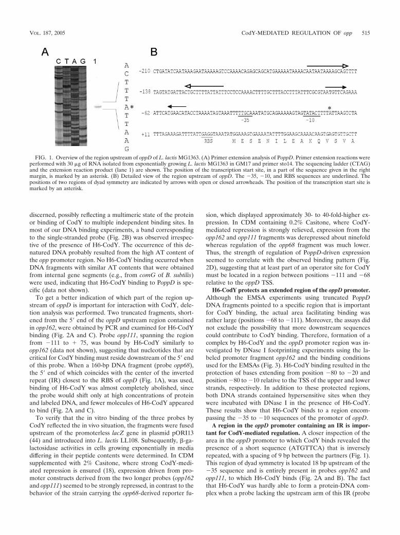
Determination of the TSS of oppD. It has been demonstratedthat the genes of the oligopeptide permease system, carried onthe oppDFBCA-pepO1 locus of L. lactis MG1363, are tran-scribed polycistronically (17, 51). Upstream of both oppD andoppA are regions that could serve as promoter elements. Toprecisely determine the location of the promoter upstream ofoppD, the transcription start site (TSS) was determined. Theopp transcript was analyzed by primer extension using RNAthat was isolated from exponentially growing MG1363 cells(Fig. 1A). The mRNA 5 end corresponds to an adenine res-idue located 35 bases upstream of the translation start codon(AUG) of oppD. The �35 sequence (TTGCAA) is separatedby a consensus 17 bp from the �10 region (TATACT), and aproper lactococcal ribosome binding site (RBS), with the se-quence GAGG, is also present (Fig. 1B). Although the dis-tance between the �10 region and the TSS is several base pairsshorter than those for most other lactococcal promoters (53),efficient transcription initiation is allowed, as evidenced by thefact that expression from PoppD was readily detectable (seeFig. 2D). The sequence upstream of oppD contains two regionsof dyad symmetry centered on positions �135 (�14.0 kcal/mol) and �62 (�5.6 kcal/mol) relative to the oppD TSS, re-spectively.
H6-CodY interacts with the region upstream of oppD. Inorder to examine whether L. lactis CodY directly interacts withits main target known so far, the opp-pepO1 region, in vitroDNA binding studies were performed using probes spanningPoppD. For this purpose, CodY carrying a histidine tag at its Nterminus (H6-CodY) was overexpressed in L. lactis by usingthe nisin-inducible gene expression system (8) and was subse-quently purified to apparent homogeneity (data not shown). A254-bp radioactively labeled DNA probe (opp162), spanningthe oppD promoter from position �162 to �75 relative to theTSS, was prepared by PCR as described in Materials andMethods (Fig. 2A). EMSAs clearly showed that purified H6-CodY was capable of binding directly to this fragment (Fig.2B). Multiple retarded fragments were present, indicating thatseveral molecules of H6-CodY can bind to the oppD fragment.When the amount of H6-CodY was gradually increased from 0to almost 1,000 nM, at least three distinct bands could be
TABLE 2. Oligonucleotides used in this study
Name Sequence (533)a
hc-5...................................................................CTAGACCACCATGGGGCATCACCATCACCATCACGTGGCTACATTACTTGAAAAAACACGhc-6...................................................................CTAGTCTAGATTAGAAATTACGTCCAGCAAGTTTATCopp1 .................................................................GCTCTAGACACTCACTTGTTTTGCTTCCopp2 .................................................................AACTGCAGGAAAATTCATGAACATACCopp3 .................................................................AACTGCAGTAAAACAATAATAAAAGCAGopp11 ...............................................................AACTGCAGCTCCAAAACTTTTGCTTTACopp14 ...............................................................AACTGCAGCGTAATGTTCAGAAAATTCopp15(a) ..........................................................AACTGCAGCGTAATATTTAGAAAATTCATGAACATACCopp15(b) ..........................................................AACTGCAGCGTACTGTGCCGAAAATTCATGAACATACCsto14.................................................................CTTGCCATGGAATCACCCG
a Restriction enzyme sites are underlined. Italicized sequence in hc-5 encodes the hexahistidine tag. Italicized sequence in hc-6 is the stop codon.
514 DEN HENGST ET AL. J. BACTERIOL.
discerned, possibly reflecting a multimeric state of the proteinor binding of CodY to multiple independent binding sites. Inmost of our DNA binding experiments, a band correspondingto the single-stranded probe (Fig. 2B) was observed irrespec-tive of the presence of H6-CodY. The occurrence of this de-natured DNA probably resulted from the high AT content ofthe opp promoter region. No H6-CodY binding occurred whenDNA fragments with similar AT contents that were obtainedfrom internal gene segments (e.g., from comG of B. subtilis)were used, indicating that H6-CodY binding to PoppD is spe-cific (data not shown).
To get a better indication of which part of the region up-stream of oppD is important for interaction with CodY, dele-tion analysis was performed. Two truncated fragments, short-ened from the 5 end of the oppD upstream region containedin opp162, were obtained by PCR and examined for H6-CodYbinding (Fig. 2A and C). Probe opp111, spanning the regionfrom �111 to � 75, was bound by H6-CodY similarly toopp162 (data not shown), suggesting that nucleotides that arecritical for CodY binding must reside downstream of the 5 endof this probe. When a 160-bp DNA fragment (probe opp68),the 5 end of which coincides with the center of the invertedrepeat (IR) closest to the RBS of oppD (Fig. 1A), was used,binding of H6-CodY was almost completely abolished, sincethe probe would shift only at high concentrations of proteinand labeled DNA, and fewer molecules of H6-CodY appearedto bind (Fig. 2A and C).
To verify that the in vitro binding of the three probes byCodY reflected the in vivo situation, the fragments were fusedupstream of the promoterless lacZ gene in plasmid pORI13(44) and introduced into L. lactis LL108. Subsequently, �-ga-lactosidase activities in cells growing exponentially in mediadiffering in their peptide contents were determined. In CDMsupplemented with 2% Casitone, where strong CodY-medi-ated repression is ensured (18), expression driven from pro-moter constructs derived from the two longer probes (opp162and opp111) seemed to be strongly repressed, in contrast to thebehavior of the strain carrying the opp68-derived reporter fu-
sion, which displayed approximately 30- to 40-fold-higher ex-pression. In CDM containing 0.2% Casitone, where CodY-mediated repression is strongly relieved, expression from theopp162 and opp111 fragments was derepressed about ninefoldwhereas regulation of the opp68 fragment was much lower.Thus, the strength of regulation of PoppD-driven expressionseemed to correlate with the observed binding pattern (Fig.2D), suggesting that at least part of an operator site for CodYmust be located in a region between positions �111 and �68relative to the oppD TSS.
H6-CodY protects an extended region of the oppD promoter.Although the EMSA experiments using truncated PoppDDNA fragments pointed to a specific region that is importantfor CodY binding, the actual area facilitating binding wasrather large (positions �68 to �111). Moreover, the assays didnot exclude the possibility that more downstream sequencescould contribute to CodY binding. Therefore, formation of acomplex by H6-CodY and the oppD promoter region was in-vestigated by DNase I footprinting experiments using the la-beled promoter fragment opp162 and the binding conditionsused for the EMSAs (Fig. 3). H6-CodY binding resulted in theprotection of bases extending from position �80 to �20 andposition �80 to �10 relative to the TSS of the upper and lowerstrands, respectively. In addition to these protected regions,both DNA strands contained hypersensitive sites when theywere incubated with DNase I in the presence of H6-CodY.These results show that H6-CodY binds to a region encom-passing the �35 to �10 sequences of the promoter of oppD.
A region in the oppD promoter containing an IR is impor-tant for CodY-mediated regulation. A closer inspection of thearea in the oppD promoter to which CodY binds revealed thepresence of a short sequence (ATGTTCA) that is inverselyrepeated, with a spacing of 9 bp between the partners (Fig. 1).This region of dyad symmetry is located 18 bp upstream of the�35 sequence and is entirely present in probes opp162 andopp111, to which H6-CodY binds (Fig. 2A and B). The factthat H6-CodY was hardly able to form a protein-DNA com-plex when a probe lacking the upstream arm of this IR (probe
FIG. 1. Overview of the region upstream of oppD of L. lactis MG1363. (A) Primer extension analysis of PoppD. Primer extension reactions wereperformed with 30 �g of RNA isolated from exponentially growing L. lactis MG1363 in GM17 and primer sto14. The sequencing ladder (CTAG)and the extension reaction product (lane 1) are shown. The position of the transcription start site, in a part of the sequence given in the rightmargin, is marked by an asterisk. (B) Detailed view of the region upstream of oppD. The �35, �10, and RBS sequences are underlined. Thepositions of two regions of dyad symmetry are indicated by arrows with open or closed arrowheads. The position of the transcription start site ismarked by an asterisk.
VOL. 187, 2005 CodY-MEDIATED REGULATION OF opp 515

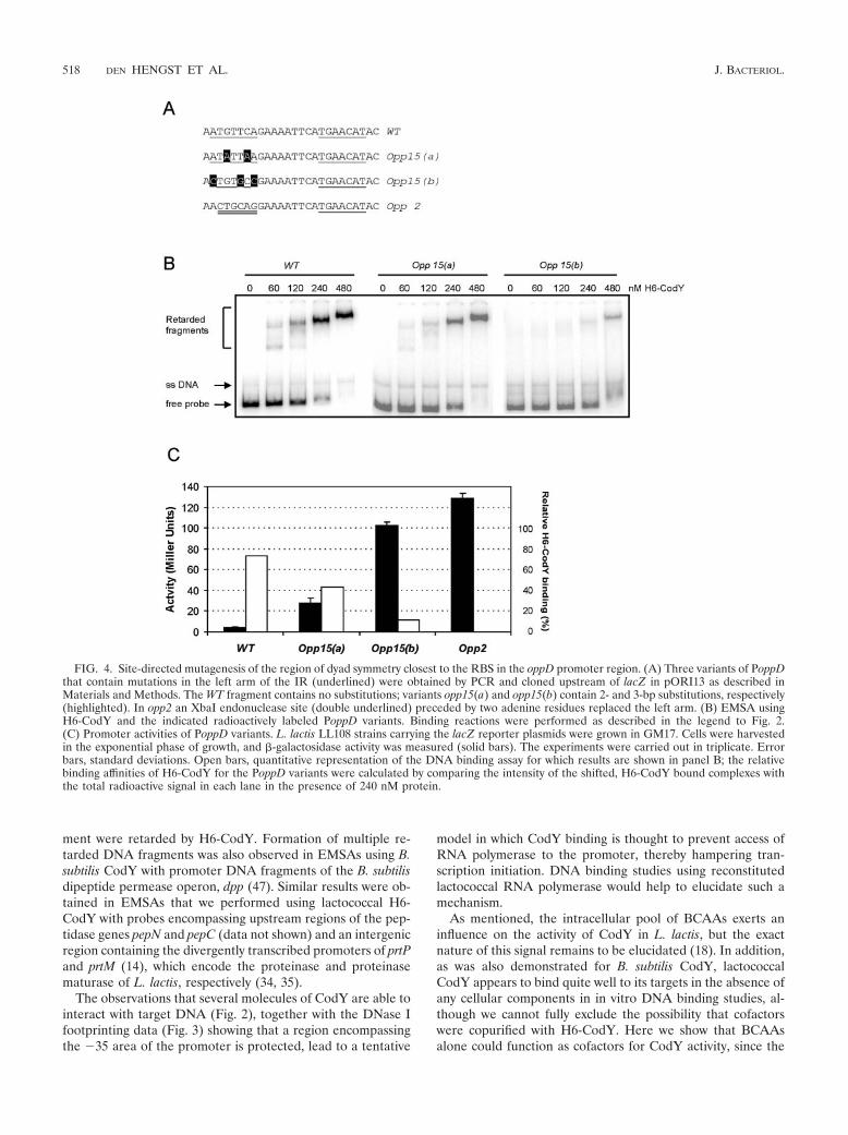
opp68 [Fig. 2A and C]) was used implies that this area mightserve as an operator site for CodY on PoppD. To study thisregion in more detail, site-directed mutations were introducedby PCR using combinations of oligonucleotide opp1 with oli-gonucleotide opp14 (wild type), opp15(a), opp(15b), or opp2(Fig. 4A). The PCR products were inserted upstream of thepromoterless lacZ reporter gene in pORI13 and introducedinto L. lactis LL108. Introducing base substitutions into theupstream arm of the repeat [opp15(a) and opp15(b)] resultedin both weaker binding of H6-CodY (Fig. 4B) and derepres-sion of PoppD-driven lacZ expression (Fig. 4C). When theunchanged half-site was replaced by an unrelated sequence (anXbaI endonuclease site, present in opp2), repression was re-duced approximately 30-fold (in cells growing exponentially ina rich medium) and H6-CodY binding was almost completelyabolished (as shown in Fig. 2C). Converting both the C and Gresidues in this region to adenines [opp15(a)] resulted in a�7-fold reduction of repression, whereas changing 3 out of 6bases [opp15(b)] led to a �20-fold derepression of expression.
These results were in accordance with those obtained from gelretardation analyses (Fig. 4B), where the affinity of H6-CodYwas highest for the wild-type probe, intermediate for theopp15(a) probe, and lowest for the opp15(b) probe. In the caseof the opp15(b) probe, hardly any protein-DNA complexeswere present and all intermediate complexes that were ob-served with the wild-type and opp15(a) fragments were absent.
Random mutation analysis of the oppD promoter area. Asshown above, a region close to the �35 sequence seems to beimportant for the binding of CodY to the oppD regulatoryregion. Since we could not find any obviously similar sequencesin the upstream regions of any of the other CodY-regulatedgenes identified so far, it is possible that structural determi-nants are required for CodY recognition of a promoter region.Therefore, PCR-based random mutagenesis was carried out onthe smallest oppD promoter fragment still showing strong re-pression of transcription by CodY (probe opp111 [Fig. 2]).PCR fragments containing random base pair substitutions orsmall deletions were restricted by using the appropriate restric-
FIG. 2. Interaction of H6-CodY with fragments of the oppD promoter region. (A) Schematic view of the probes used in panels B and C. Probesopp162, opp111, and opp68 were obtained by PCR on chromosomal DNA of L. lactis MG1363, using a combination of oligonucleotide opp1 witholigonucleotide opp3, opp11, or opp2, respectively. Arrows indicate the position of the region of dyad symmetry closest to the �35 sequence (seeFig. 1B). Vertical bars indicate the positions of the �35 and �10 sequences. Nucleotide positions are relative to the transcriptional start site(right-turn arrow at �1). (B) Interaction of H6-CodY with the upstream region of oppD. The radioactively labeled probe opp162 was incubatedalone or with increasing amounts of purified H6-CodY. The first lane contains the same probe, which was boiled in a 95% formamide solution inorder to obtain single-stranded (s.s.) DNA fragments. (C) H6-CodY binding to probe opp68. The same conditions were used as for probe opp162(B). (D) In vivo activities of PoppD variants. L. lactis LL108 strains carrying lacZ reporter plasmids fused to the opp fragments depictedschematically in panel A were grown in CDM containing either 0.2 or 2% Casitone (solid and open bars, respectively). Cells were harvested in theexponential phase of growth, and �-galactosidase activity was measured. Assays were carried out twice, in triplicate each time. Error bars, standarddeviations.
516 DEN HENGST ET AL. J. BACTERIOL.

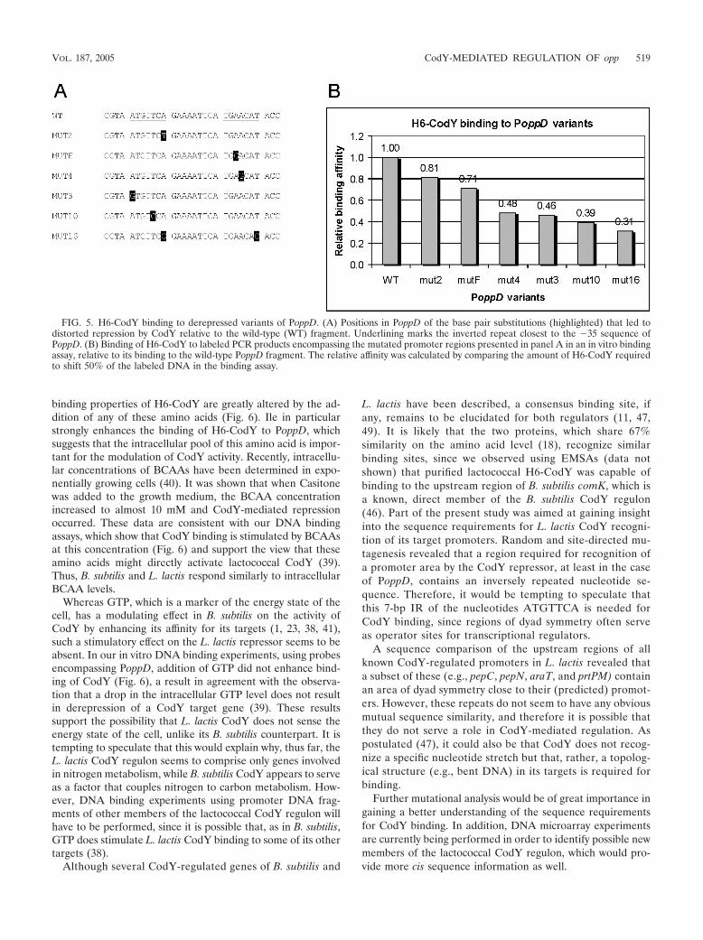
tion enzymes and ligated upstream of the promoterless lacZ inpORI13. The ligation mixture was used to transform L. lactisLL108. Cells carrying a pORI13 construct with a PoppD frag-ment containing a derepressing mutation formed light-blue orblue colonies on CDM plates containing X-Gal and excessnitrogen sources (in the form of Casitone), as opposed to thewhitish color of colonies formed by cells harboring pORI13with wild-type PoppD. The oppD fragments in cells with aderepressed phenotype were amplified by PCR, radioactivelylabeled, and tested by an EMSA for their abilities to formcomplexes with H6-CodY. The relative strength of H6-CodYbinding was examined by comparing the amount of the re-tarded mutated DNA fragment resulting from H6-CodY bind-ing to that of the unchanged PoppD fragment (Fig. 5). Weakerbinding to all of the mutated promoter fragments was ob-served. Sequence analyses of the PoppD variants obtained inthis study revealed that all of them carried one or more basepair substitutions, at least one of which was located in theregion from �82 to �56 relative to the TSS of oppD. This
finding, again, is an indication of the importance of this regionfor CodY binding.
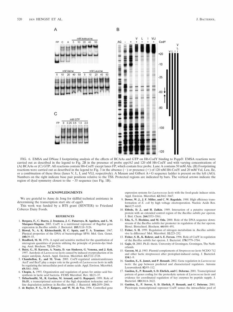
BCAAs stimulate stimulate H6-CodY binding. Evidence hasbeen presented that CodY senses the nitrogen supply of thecell as a function of the BCAA pool (18, 39). Although BCAAsact as direct effectors of CodY activity in B. subtilis (48), theexact nature of this signal in L. lactis remains to be established.Therefore, in vitro DNA binding of H6-CodY was examined inthe presence or absence of the three BCAAs Val, Leu, and Ile(Fig. 6A). Addition of any of the three BCAAs resulted inincreased binding of H6-CodY to the PoppD probe opp162compared to a situation in which no amino acid or another,aliphatic amino acid (i.e., alanine) was present. Stimulation ofH6-CodY binding by Val and Leu was observed at concentra-tions of these amino acids above 10 mM. In a titration of Ileand a constant amount of H6-CodY, most of the probe DNAwas already retarded in the presence of 5 mM Ile, showing thatthis BCAA stimulates the binding of H6-CodY to the highestextent.
The role of BCAAs in CodY binding was also investigatedby means of DNase I footprinting experiments, in which theopp162 probe was incubated with H6-CodY either alone; in thepresence of 20 mM Val, Leu, or Ile; or with a combination ofthese three BCAAs (Fig. 6B). Addition of any of these BCAAsresulted in extended protection of PoppD. As in the EMSAs,the effect of Ile addition was most severe. These results showthat the binding of lactococcal CodY to the target region ofoppD is directly stimulated by BCAAs and that, as in B. subtilis,Ile is most effective.
Similarly, we also tested whether GTP could stimulate thebinding of H6-CodY to lactococcal PoppD, since GTP serves asan effector molecule that enhances the binding of B. subtilisCodY to a number of its targets. As can be seen from Fig. 6C,the binding of L. lactis H6-CodY was not affected by thepresence of GTP at a range of concentrations between 0.25and 2.0 mM. These results are in good agreement with recentevidence showing that lactococcal CodY activity is indepen-dent of GTP at physiological concentrations, which do notexceed 0.55 mM in a medium containing Casitone (39).
DISCUSSION
L. lactis possesses a number of genes whose products areinvolved in the utilization of proteins present in milk, such asan extracellular protease, peptide transport systems, and intra-cellular peptidases (24). Most of these genes have been clonedand sequenced, and their enzymes have been biochemicallycharacterized (24). Recently, it has been established that CodYplays a pleiotropic role in the regulation of a number of thesegenes in response to nitrogen availability (17, 18).
The present study was conducted to gain insight into the roleof CodY in the regulation of the opp system and, more specif-ically, into the molecular interactions between the cis site andCodY. By combining data from in vivo and in vitro experi-ments, we have clearly demonstrated that repression by CodYis mediated by direct binding of this protein to the oppD pro-moter region. For this to occur most efficiently, binding ofseveral molecules of CodY is probably required, since severalprotein-DNA complexes were discerned in all EMSAs in whichvariants (in size or base composition) of this promoter frag-
FIG. 3. DNase I footprinting analysis of H6-CodY binding to theoppD promoter region. The left and right panels show the footprints ofthe upper and lower strands, respectively. Footprints, obtained in theabsence or presence of 60 or 240 nM H6-CodY by using radioactivelylabeled probe opp162, are flanked by Maxam and Gilbert A�G se-quence ladders (AG) on the left. Numbers on the left indicate basepair positions relative to the TSS. Protected regions are marked withbars, and horizontal arrows indicate the positions of hypersensitivebonds. Vertical arrows indicate the region of dyad symmetry closest tothe �35 sequence (see Fig. 1B).
VOL. 187, 2005 CodY-MEDIATED REGULATION OF opp 517
ment were retarded by H6-CodY. Formation of multiple re-tarded DNA fragments was also observed in EMSAs using B.subtilis CodY with promoter DNA fragments of the B. subtilisdipeptide permease operon, dpp (47). Similar results were ob-tained in EMSAs that we performed using lactococcal H6-CodY with probes encompassing upstream regions of the pep-tidase genes pepN and pepC (data not shown) and an intergenicregion containing the divergently transcribed promoters of prtPand prtM (14), which encode the proteinase and proteinasematurase of L. lactis, respectively (34, 35).
The observations that several molecules of CodY are able tointeract with target DNA (Fig. 2), together with the DNase Ifootprinting data (Fig. 3) showing that a region encompassingthe �35 area of the promoter is protected, lead to a tentative
model in which CodY binding is thought to prevent access ofRNA polymerase to the promoter, thereby hampering tran-scription initiation. DNA binding studies using reconstitutedlactococcal RNA polymerase would help to elucidate such amechanism.
As mentioned, the intracellular pool of BCAAs exerts aninfluence on the activity of CodY in L. lactis, but the exactnature of this signal remains to be elucidated (18). In addition,as was also demonstrated for B. subtilis CodY, lactococcalCodY appears to bind quite well to its targets in the absence ofany cellular components in in vitro DNA binding studies, al-though we cannot fully exclude the possibility that cofactorswere copurified with H6-CodY. Here we show that BCAAsalone could function as cofactors for CodY activity, since the
FIG. 4. Site-directed mutagenesis of the region of dyad symmetry closest to the RBS in the oppD promoter region. (A) Three variants of PoppDthat contain mutations in the left arm of the IR (underlined) were obtained by PCR and cloned upstream of lacZ in pORI13 as described inMaterials and Methods. The WT fragment contains no substitutions; variants opp15(a) and opp15(b) contain 2- and 3-bp substitutions, respectively(highlighted). In opp2 an XbaI endonuclease site (double underlined) preceded by two adenine residues replaced the left arm. (B) EMSA usingH6-CodY and the indicated radioactively labeled PoppD variants. Binding reactions were performed as described in the legend to Fig. 2.(C) Promoter activities of PoppD variants. L. lactis LL108 strains carrying the lacZ reporter plasmids were grown in GM17. Cells were harvestedin the exponential phase of growth, and �-galactosidase activity was measured (solid bars). The experiments were carried out in triplicate. Errorbars, standard deviations. Open bars, quantitative representation of the DNA binding assay for which results are shown in panel B; the relativebinding affinities of H6-CodY for the PoppD variants were calculated by comparing the intensity of the shifted, H6-CodY bound complexes withthe total radioactive signal in each lane in the presence of 240 nM protein.
518 DEN HENGST ET AL. J. BACTERIOL.
binding properties of H6-CodY are greatly altered by the ad-dition of any of these amino acids (Fig. 6). Ile in particularstrongly enhances the binding of H6-CodY to PoppD, whichsuggests that the intracellular pool of this amino acid is impor-tant for the modulation of CodY activity. Recently, intracellu-lar concentrations of BCAAs have been determined in expo-nentially growing cells (40). It was shown that when Casitonewas added to the growth medium, the BCAA concentrationincreased to almost 10 mM and CodY-mediated repressionoccurred. These data are consistent with our DNA bindingassays, which show that CodY binding is stimulated by BCAAsat this concentration (Fig. 6) and support the view that theseamino acids might directly activate lactococcal CodY (39).Thus, B. subtilis and L. lactis respond similarly to intracellularBCAA levels.
Whereas GTP, which is a marker of the energy state of thecell, has a modulating effect in B. subtilis on the activity ofCodY by enhancing its affinity for its targets (1, 23, 38, 41),such a stimulatory effect on the L. lactis repressor seems to beabsent. In our in vitro DNA binding experiments, using probesencompassing PoppD, addition of GTP did not enhance bind-ing of CodY (Fig. 6), a result in agreement with the observa-tion that a drop in the intracellular GTP level does not resultin derepression of a CodY target gene (39). These resultssupport the possibility that L. lactis CodY does not sense theenergy state of the cell, unlike its B. subtilis counterpart. It istempting to speculate that this would explain why, thus far, theL. lactis CodY regulon seems to comprise only genes involvedin nitrogen metabolism, while B. subtilis CodY appears to serveas a factor that couples nitrogen to carbon metabolism. How-ever, DNA binding experiments using promoter DNA frag-ments of other members of the lactococcal CodY regulon willhave to be performed, since it is possible that, as in B. subtilis,GTP does stimulate L. lactis CodY binding to some of its othertargets (38).
Although several CodY-regulated genes of B. subtilis and
L. lactis have been described, a consensus binding site, ifany, remains to be elucidated for both regulators (11, 47,49). It is likely that the two proteins, which share 67%similarity on the amino acid level (18), recognize similarbinding sites, since we observed using EMSAs (data notshown) that purified lactococcal H6-CodY was capable ofbinding to the upstream region of B. subtilis comK, which isa known, direct member of the B. subtilis CodY regulon(46). Part of the present study was aimed at gaining insightinto the sequence requirements for L. lactis CodY recogni-tion of its target promoters. Random and site-directed mu-tagenesis revealed that a region required for recognition ofa promoter area by the CodY repressor, at least in the caseof PoppD, contains an inversely repeated nucleotide se-quence. Therefore, it would be tempting to speculate thatthis 7-bp IR of the nucleotides ATGTTCA is needed forCodY binding, since regions of dyad symmetry often serveas operator sites for transcriptional regulators.
A sequence comparison of the upstream regions of allknown CodY-regulated promoters in L. lactis revealed thata subset of these (e.g., pepC, pepN, araT, and prtPM) containan area of dyad symmetry close to their (predicted) promot-ers. However, these repeats do not seem to have any obviousmutual sequence similarity, and therefore it is possible thatthey do not serve a role in CodY-mediated regulation. Aspostulated (47), it could also be that CodY does not recog-nize a specific nucleotide stretch but that, rather, a topolog-ical structure (e.g., bent DNA) in its targets is required forbinding.
Further mutational analysis would be of great importance ingaining a better understanding of the sequence requirementsfor CodY binding. In addition, DNA microarray experimentsare currently being performed in order to identify possible newmembers of the lactococcal CodY regulon, which would pro-vide more cis sequence information as well.
FIG. 5. H6-CodY binding to derepressed variants of PoppD. (A) Positions in PoppD of the base pair substitutions (highlighted) that led todistorted repression by CodY relative to the wild-type (WT) fragment. Underlining marks the inverted repeat closest to the �35 sequence ofPoppD. (B) Binding of H6-CodY to labeled PCR products encompassing the mutated promoter regions presented in panel A in an in vitro bindingassay, relative to its binding to the wild-type PoppD fragment. The relative affinity was calculated by comparing the amount of H6-CodY requiredto shift 50% of the labeled DNA in the binding assay.
VOL. 187, 2005 CodY-MEDIATED REGULATION OF opp 519
ACKNOWLEDGMENTS
We are grateful to Anne de Jong for skillful technical assistance indetermining the transcription start site of oppD.
This work was funded by a BTS grant (SENTER) to FrieslandCoberco Dairy Foods.
REFERENCES
1. Bergara, F., C. Ibarra, J. Iwamasa, J. C. Patarroyo, R. Aguilera, and L. M.Marquez-Magana. 2003. CodY is a nutritional repressor of flagellar geneexpression in Bacillus subtilis. J. Bacteriol. 185:3118–3126.
2. Biswal, N., A. K. Kleinschmidt, H. C. Spatz, and T. A. Trautner. 1967.Physical properties of the DNA of bacteriophage SP50. Mol. Gen. Genet.100:39–55.
3. Bradford, M. M. 1976. A rapid and sensitive method for the quantitation ofmicrogram quantities of protein utilizing the principle of protein-dye bind-ing. Anal. Biochem. 72:248–254.
4. Buist, G., H. Karsens, A. Nauta, D. van Sinderen, G. Venema, and J. Kok.1997. Autolysis of Lactococcus lactis caused by induced overproduction of itsmajor autolysin, AcmA. Appl. Environ. Microbiol. 63:2722–2728.
5. Chambellon, E., and M. Yvon. 2003. CodY-regulated aminotransferasesAraT and BcaT play a major role in the growth of Lactococcus lactis in milkby regulating the intracellular pool of amino acids. Appl. Environ. Microbiol.69:3061–3068.
6. Chopin, A. 1993. Organization and regulation of genes for amino acid bio-synthesis in lactic acid bacteria. FEMS Microbiol. Rev. 12:21–37.
7. Debarbouille, M., R. Gardan, M. Arnaud, and G. Rapoport. 1999. Role ofBkdR, a transcriptional activator of the SigL-dependent isoleucine and va-line degradation pathway in Bacillus subtilis. J. Bacteriol. 181:2059–2066.
8. de Ruyter, P. G., O. P. Kuipers, and W. M. de Vos. 1996. Controlled gene
expression systems for Lactococcus lactis with the food-grade inducer nisin.Appl. Environ. Microbiol. 62:3662–3667.
9. Dower, W. J., J. F. Miller, and C. W. Ragsdale. 1988. High efficiency trans-formation of E. coli by high voltage electroporation. Nucleic Acids Res.16:6127–6145.
10. Ebbole, D. J., and H. Zalkin. 1989. Interaction of a putative repressorprotein with an extended control region of the Bacillus subtilis pur operon.J. Biol. Chem. 264:3553–3561.
11. Eda, S., T. Hoshino, and M. Oda. 2000. Role of the DNA sequence down-stream of the Bacillus subtilis hut promoter in regulation of the hut operon.Biosci. Biotechnol. Biochem. 64:484–491.
12. Fisher, S. H. 1999. Regulation of nitrogen metabolism in. Bacillus subtilis:vive la difference! Mol. Microbiol. 32:223–232.
13. Fisher, S. H., K. Rohrer, and A. E. Ferson. 1996. Role of CodY in regulationof the Bacillus subtilis hut operon. J. Bacteriol. 178:3779–3784.
14. Gajic, O. 2003. Ph.D. thesis. University of Groningen, Groningen, The Neth-erlands.
15. Gasson, M. J. 1983. Plasmid complements of Streptococcus lactis NCDO 712and other lactic streptococci after protoplast-induced curing. J. Bacteriol.154:1–9.
16. Guedon, E., E. Jamet, and P. Renault. 2002. Gene regulation in Lactococcuslactis: the gap between predicted and characterized regulators. AntonieLeeuwenhoek 82:93–112.
17. Guedon, E., P. Renault, S. D. Ehrlich, and C. Delorme. 2001. Transcriptionalpattern of genes coding for the proteolytic system of Lactococcus lactis andevidence for coordinated regulation of key enzymes by peptide supply. J.Bacteriol. 183:3614–3622.
18. Guedon, E., P. Serror, S. D. Ehrlich, P. Renault, and C. Delorme. 2001.Pleiotropic transcriptional repressor CodY senses the intracellular pool of
FIG. 6. EMSA and DNase I footprinting analysis of the effects of BCAAs and GTP on H6-CodY binding to PoppD. EMSA reactions werecarried out as described in the legend to Fig. 2B in the presence of probe opp162 and 120 nM H6-CodY and with varying concentrations of(A) BCAAs or (C) GTP. All reactions contain H6-CodY except lanes FP, which contain free probe. Lane A contains 50 mM Ala. (B) Footprintingreactions were carried out as described in the legend to Fig. 3 in the absence (�) or presence (�) of 120 nM H6-CodY and 20 mM Val, Leu, Ile,or a combination of these three (lanes V, L, I, and VLI, respectively). A Maxam and Gilbert A�G sequence ladder is present on the left (AG).Numbers on the right indicate base pair positions relative to the TSS. Protected regions are indicated by bars. The vertical arrows indicate theregion of dyad symmetry closest to the �35 sequence (see Fig. 1B).
520 DEN HENGST ET AL. J. BACTERIOL.
branched-chain amino acids in Lactococcus lactis. Mol. Microbiol. 40:1227–1239.
19. Hamoen, L. W., A. F. Van Werkhoven, J. J. Bijlsma, D. Dubnau, and G.Venema. 1998. The competence transcription factor of Bacillus subtilis rec-ognizes short A/T-rich sequences arranged in a unique, flexible pattern alongthe DNA helix. Genes Dev. 12:1539–1550.
20. Holo, H., and I. F. Nes. 1995. Transformation of Lactococcus by electropo-ration. Methods Mol. Biol. 47:195–199.
21. Inaoka, T., K. Takahashi, M. Ohnishi-Kameyama, M. Yoshida, and K. Ochi.2003. Guanine nucleotides guanosine 5-diphosphate 3-diphosphate andGTP co-operatively regulate the production of an antibiotic bacilysin inBacillus subtilis. J. Biol. Chem. 278:2169–2176.
22. Israelsen, H., S. M. Madsen, A. Vrang, E. B. Hansen, and E. Johansen. 1995.Cloning and partial characterization of regulated promoters from Lactococ-cus lactis Tn917-lacZ integrants with the new promoter probe vector, pAK80.Appl. Environ. Microbiol. 61:2540–2547.
23. Kim, H. J., S. I. Kim, M. Ratnayake-Lecamwasam, K. Tachikawa, A. L.Sonenshein, and M. Strauch. 2003. Complex regulation of the Bacillus sub-tilis aconitase gene. J. Bacteriol. 185:1672–1680.
24. Kok, J., and G. Buist. 2003. Genetics of proteolysis in Lactococcus lactis, p.189–224. In B. J. B. Wood and W. M. de Vos (ed.), Genetics of lactic acidbacteria. Kluwer Academic/Plenum Publishers, New York, N.Y.
25. Kok, J., and W. M. de Vos. 1993. The proteolytic system of lactic acidbacteria, p. 169–210. In M. J. Gasson and W. M. de Vos (ed.), Genetics andbiotechnology of lactic acid bacteria. Blackie Academic and Professional,London, United Kingdom.
26. Kuipers, O. P., M. M. Beerthuyzen, R. J. Siezen, and W. M. de Vos. 1993.Characterization of the nisin gene cluster nisABTCIPR of Lactococcus lactis.Requirement of expression of the nisA and nisI genes for development ofimmunity. Eur. J. Biochem. 216:281–291.
27. Kuipers, O. P., P. G. de Ruyter, M. Kleerebezem, and W. M. de Vos. 1998.Quorum sensing controlled gene expression in lactic acid bacteria. J. Bio-technol. 64:15–21.
28. Kunji, E. R., I. Mierau, A. Hagting, B. Poolman, and W. N. Konings. 1996.The proteolytic systems of lactic acid bacteria. Antonie Leeuwenhoek 70:187–221.
29. Kunji, E. R., I. Mierau, B. Poolman, W. N. Konings, G. Venema, and J. Kok.1996. Fate of peptides in peptidase mutants of Lactococcus lactis. Mol.Microbiol. 21:123–131.
30. Larsen, R., G. Buist, O. P. Kuipers, and J. Kok. 2004. ArgR and AhrC areboth required for regulation of arginine metabolism in Lactococcus lactis. J.Bacteriol. 186:1147–1157.
31. Law, J., G. Buist, A. Haandrikman, J. Kok, G. Venema, and K. Leenhouts.1995. A system to generate chromosomal mutations in Lactococcus lactiswhich allows fast analysis of targeted genes. J. Bacteriol. 177:7011–7018.
32. Leenhouts, K., A. Bolhuis, G. Venema, and J. Kok. 1998. Construction of afood-grade multiple-copy integration system for Lactococcus lactis. Appl.Microbiol. Biotechnol. 49:417–423.
33. Leenhouts, K. J., J. Kok, and G. Venema. 1991. Replacement recombinationin Lactococcus lactis. J. Bacteriol. 173:4794–4798.
34. Marugg, J. D., W. Meijer, R. van Kranenburg, P. Laverman, P. G. Bruin-enberg, and W. M. de Vos. 1995. Medium-dependent regulation of protein-ase gene expression in Lactococcus lactis: control of transcription initiationby specific dipeptides. J. Bacteriol. 177:2982–2989.
35. Marugg, J. D., R. van Kranenburg, P. Laverman, G. A. Rutten, and W. M.de Vos. 1996. Identical transcriptional control of the divergently transcribedprtP and prtM genes that are required for proteinase production in Lacto-coccus lactis SK11. J. Bacteriol. 178:1525–1531.
36. Mierau, I., E. R. Kunji, K. J. Leenhouts, M. A. Hellendoorn, A. J. Haan-drikman, B. Poolman, W. N. Konings, G. Venema, and J. Kok. 1996. Mul-tiple-peptidase mutants of Lactococcus lactis are severely impaired in theirability to grow in milk. J. Bacteriol. 178:2794–2803.
37. Miller, J. H. 1972. Experiments in molecular genetics. Cold Spring HarborLaboratory Press, Cold Spring Harbor, N.Y.
38. Molle, V., Y. Nakaura, R. P. Shivers, H. Yamaguchi, R. Losick, Y. Fujita, andA. L. Sonenshein. 2003. Additional targets of the Bacillus subtilis globalregulator CodY identified by chromatin immunoprecipitation and genome-wide transcript analysis. J. Bacteriol. 185:1911–1922.
39. Petranovic, D., E. Guedon, B. Sperandio, C. Delorme, D. Ehrlich, and P.Renault. 2004. Intracellular effectors regulating the activity of the Lactococ-cus lactis CodY pleiotropic transcription regulator. Mol. Microbiol. 53:613–621.
40. Petranovic, D., and I. Mijakovic. 2004. Photometric assay for measuring theintracellular concentration of branched-chain amino acids in bacteria. J.Microbiol. Methods 56:133–136.
41. Ratnayake-Lecamwasam, M., P. Serror, K. W. Wong, and A. L. Sonenshein.2001. Bacillus subtilis CodY represses early-stationary-phase genes by sens-ing GTP levels. Genes Dev. 15:1093–1103.
42. Rijnen, L., S. Bonneau, and M. Yvon. 1999. Genetic characterization of themajor lactococcal aromatic aminotransferase and its involvement in conver-sion of amino acids to aroma compounds. Appl. Environ. Microbiol. 65:4873–4880.
43. Sambrook, J., E. F. Fritsch, and T. Maniatis. 1989. Molecular cloning: alaboratory manual, 2nd ed. Cold Spring Harbor Laboratory Press, ColdSpring Harbor, N.Y.
44. Sanders, J. W., G. Venema, J. Kok, and K. Leenhouts. 1998. Identification ofa sodium chloride-regulated promoter in Lactococcus lactis by single-copychromosomal fusion with a reporter gene. Mol. Gen. Genet. 257:681–685.
45. Sanz, Y., F. C. Lanfermeijer, P. Renault, A. Bolotin, W. N. Konings, and B.Poolman. 2001. Genetic and functional characterization of dpp genes encod-ing a dipeptide transport system in Lactococcus lactis. Arch. Microbiol.175:334–343.
46. Serror, P., and A. L. Sonenshein. 1996. CodY is required for nutritionalrepression of Bacillus subtilis genetic competence. J. Bacteriol. 178:5910–5915.
47. Serror, P., and A. L. Sonenshein. 1996. Interaction of CodY, a novel Bacillussubtilis DNA-binding protein, with the dpp promoter region. Mol. Microbiol.20:843–852.
48. Shivers, R. P., and A. L. Sonenshein. 2004. Activation of the Bacillus subtilisglobal regulator CodY by direct interaction with branched-chain aminoacids. Mol. Microbiol. 53:599–611.
49. Slack, F. J., P. Serror, E. Joyce, and A. L. Sonenshein. 1995. A gene requiredfor nutritional repression of the Bacillus subtilis dipeptide permease operon.Mol. Microbiol. 15:689–702.
50. Terzaghi, B. E., and W. E. Sandine. 1975. Improved medium for lacticstreptococci and their bacteriophages. Appl. Microbiol. 29:807–813.
51. Tynkkynen, S., G. Buist, E. Kunji, J. Kok, B. Poolman, G. Venema, and A.Haandrikman. 1993. Genetic and biochemical characterization of the oli-gopeptide transport system of Lactococcus lactis. J. Bacteriol. 175:7523–7532.
52. van Asseldonk, M., A. Simons, H. Visser, W. M. de Vos, and G. Simons. 1993.Cloning, nucleotide sequence, and regulatory analysis of the Lactococcuslactis dnaJ gene. J. Bacteriol. 175:1637–1644.
53. van de Guchte, M., J. Kok, and G. Venema. 1992. Gene expression inLactococcus lactis. FEMS Microbiol. Rev. 8:73–92.
54. Wray, L. V., Jr., A. E. Ferson, and S. H. Fisher. 1997. Expression of theBacillus subtilis ureABC operon is controlled by multiple regulatory factorsincluding CodY, GlnR, TnrA, and Spo0H. J. Bacteriol. 179:5494–5501.
55. Yvon, M., E. Chambellon, A. Bolotin, and F. Roudot-Algaron. 2000. Char-acterization and role of the branched-chain aminotransferase (BcaT) iso-lated from Lactococcus lactis subsp. cremoris NCDO 763. Appl. Environ.Microbiol. 66:571–577.
VOL. 187, 2005 CodY-MEDIATED REGULATION OF opp 521