cardiac baroreflex function and dynamic cerebral autoregulation in elderly masters athletes
TRANSCRIPT

1
Cardiac Baroreflex Function and Dynamic Cerebral Autoregulation In Elderly 1
Masters Athletes 2
3
Vincent L. Aengevaeren1, 2, Jurgen A.H.R. Claassen2, Benjamin D. Levine1, Rong 4
Zhang1 5
6
1Institute for Exercise and Environmental Medicine, Texas Health Presbyterian Hospital 7
Dallas and Department of Internal Medicine-Cardiology, University of Texas 8
Southwestern Medical Center, Dallas, TX 9
2Radboud University Nijmegen Medical Center, Nijmegen, Department of Geriatric 10
Medicine, The Netherlands 11
12
13
Running title: Baroreflex and cerebral autoregulation in Masters athletes 14
15
Corresponding Author: Rong Zhang 16
7232 Greenville Avenue, Dallas, TX 75231 17
Tel: (214) 345-8843 / Fax (214) 345-4618 18
E-mail: [email protected] 19
Articles in PresS. J Appl Physiol (November 8, 2012). doi:10.1152/japplphysiol.00402.2012
Copyright © 2012 by the American Physiological Society.
2
Abstract 20
Purpose: Cerebral blood flow (CBF) is maintained stable through combined effects of 21
blood pressure (BP) regulation and cerebral autoregulation. Previous studies suggest 22
that aerobic exercise training improves cardiac baroreflex function and beneficially 23
affects BP regulation, but may negatively affect cerebral autoregulation. The purpose of 24
this study was to reveal the impact of lifelong exercise on cardiac baroreflex function 25
and dynamic cerebral autoregulation (dCA) in older adults. Methods: Eleven Masters 26
athletes (MA) (8 males, 3 females, mean age 73±6 yrs, aerobic training >15 yrs) and 12 27
healthy sedentary elderly (SE) (7 males, 5 females, mean age 71±6 yrs) participated in 28
this study. BP, CBF velocity (CBFV) and heart rate were measured during resting 29
conditions and repeated sit-stand maneuvers to enhance BP variability. Baroreflex gain 30
was assessed using transfer function analysis of spontaneous changes in systolic BP 31
and R-R interval in the low frequency range (0.05-0.15 Hz). dCA was assessed during 32
sit-stand induced changes in mean BP and CBFV at 0.05 Hz (10s sit, 10s stand). 33
Results: Cardiac baroreflex gain was more than doubled in MA compared to SE (MA: 34
7.69±7.95, SE: 3.18±1.29, ms/mmHg, P=0.018). However, dCA was similar in the two 35
groups (normalized gain: MA: 1.50±0.56, SE: 1.56±0.42, %CBFV/mmHg, P=0.792). 36
Conclusion: These findings suggest that lifelong exercise improves cardiac baroreflex 37
function, but does not alter dCA. Thus, beneficial effects of exercise training on BP 38
regulation can be achieved in older adults without compromising dynamic regulation of 39
CBF. 40
Key words: baroreflex function, cerebral autoregulation, exercise 41

3
Introduction 42
Maintaining adequate cerebral perfusion during every day activities requires a 43
fine regulation of blood pressure (BP) and cerebral blood flow (CBF) (39). Short-term 44
BP and CBF regulation with time scales of several seconds to minutes are mainly 45
controlled by the baroreflex and dynamic cerebral autoregulation (dCA) (51). The 46
baroreflex controls BP by modulating heart rate (HR), cardiac inotropy and vascular 47
tone in response to BP changes (31). dCA refers to the capabilities of the cerebral 48
vascular bed to adjust its resistance to maintain adequate cerebral perfusion in 49
response to transient changes in BP (8). 50
Aging appears to differentially affect these regulation mechanisms: aging 51
reduces cardiac baroreflex function (14, 18, 40), however, dCA appears to be preserved 52
with aging (8, 9). Impairment of the baroreflex function is an adverse prognostic 53
indicator for cardiovascular diseases, and may lead to orthostatic hypotension, resulting 54
in brain hypoperfusion, falls or syncope if dCA cannot compensate for the augmented 55
fluctuations in BP (24-27, 30, 53, 54). 56
Physical activity is a modifiable factor which may affect the regulation of BP and 57
CBF (12, 35). For example, endurance training improves cardiac baroreflex function and 58
reduces BP in young and older adults (6, 11, 13, 23, 26, 32, 33, 38, 48, 49), and 59
previous studies from our group and others have shown marked cardiovascular benefits 60
accredited to lifelong endurance training (2, 15, 38, 46). Furthermore, high level of 61
physical fitness appears to be associated with increased CBF velocity (CBFV) across 62
the lifespan from young to older adults (1). However, the effects of exercise training on 63
the regulation of cerebral hemodynamics are not clear. In young endurance trained 64

4
subjects, Lind-Holst et al. (29) observed that dCA was less effective. Interestingly, 65
Tzeng et al. (50) found an inverse correlation between cardiac baroreflex function and 66
dCA, suggesting a negative interaction between BP and CBF regulation. Together these 67
studies raise the question if the benefits of exercise training for baroreflex function may 68
be counteracted by a reduced function of dCA. 69
Notably, these aspects have thus far only been investigated in young adults. The 70
purpose of this study was to assess the differential effects of lifelong exercise on 71
cardiac baroreflex function and dCA in the elderly. Masters athletes are a unique group 72
of elderly who have participated in lifelong exercise training and have competed in 73
sports at very high levels (2). In this study, cardiac baroreflex function and dCA were 74
measured in Masters athletes and sedentary elderly. Based on the previous findings in 75
younger subjects, we hypothesized that lifelong endurance training in Masters athletes 76
is associated with an improved cardiac baroreflex function but a less effective dCA. 77
78
Methods 79
Subjects population. Eleven Masters athletes (8 males, 3 females, mean age 80
73±6 yrs) and 12 sedentary elderly (7 males, 5 females, mean age 71±6 yrs) 81
participated in the study. All subjects were nonsmokers, not on any cardiovascular 82
medication, and normotensive. Female subjects were postmenopausal and not taking 83
hormone replacement therapy. All subjects were carefully screened for cardiovascular 84
and cerebrovascular diseases, diabetes, and hypertension with a detailed medical 85
history and a physical examination including a 12-lead ECG. Inclusion criterion for 86

5
Masters athletes was endurance training >15 years and still active (running about 20 to 87
50 miles weekly or equivalent cycling or swimming and participated in races) at the time 88
of this study. All Masters athletes were either regionally or nationally ranked runners 89
and were recruited from running clubs or the records of competitive running events (2). 90
Sedentary elderly were recruited locally with newsletters and from senior centers and 91
were excluded if they had exercised more than 30 minutes 3 times a week in the past 2 92
years. The Institutional Review Boards (IRB) of the University of Texas Southwestern 93
Medical Center and Texas Health Presbyterian Hospital Dallas approved the study 94
protocol. All subjects signed an informed consent approved by the IRBs which 95
conformed to the standards set by the latest revision of the Declaration of Helsinki. 96
Instrumentation. CBFV was measured in the right middle cerebral artery by 97
transcranial Doppler ultrasonography (Multi-Dop X2, Compumedics/ DWL, Germany). A 98
2 MHz Doppler probe was placed over the temporal window and fixed at a constant 99
angle with a probe holder (Spencer Technologies, USA). Doppler sampling depth 100
ranged from 42 to 55 mm and the angle of the Doppler probe and the sampling depth 101
for each individual subject were adjusted to optimize the signal quality according to the 102
standard procedures (5). This technique allows noninvasive and repeatable estimates of 103
changes in CBFV on a beat-to-beat basis. Continuous finger arterial BP was monitored 104
in the left hand by photoplethysmography (Finapres Medical Systems B.V., The 105
Netherlands). The hand was fixed at the level of the heart of the participant. This 106
method reliably assesses dynamic changes in beat-to-beat BP that correlate well with 107
intra-arterial recordings and can be used to quantify the dynamic pressure-flow 108
relationship of the cerebral circulation (44, 55, 56). Intermittent arterial pressure was 109
6
measured in the right arm by an electrosphygmomanometer (Tango+, Suntech, USA). 110
This pressure measurement was used to corroborate the BP measurements of the 111
finger pressure recordings throughout the experiment to make sure the finger cuff was 112
properly placed to record BP. Electrocardiogram (ECG) was recorded using a 3-lead 113
system (Hewlett-Packard, USA). End-tidal CO2 (EtCO2) was monitored with a nasal 114
cannula using capnography (Carpnogard, Novamatrix, USA) (4). 115
Study protocol. This was a cross-sectional study, consisting of 2 visits, one visit 116
to assess baroreflex function and dCA and one visit for a peak exercise test. The visits 117
were separated by at least 2 weeks. To assess baroreflex function and dCA, after at 118
least 10 minutes of rest in the sitting position, 5 minutes of data were recorded during 119
spontaneous breathing. These data were used to obtain spontaneous changes in BP, 120
R-R interval and CBFV as well as baseline steady-state hemodynamics. Next, repeated 121
sit-stand maneuvers were performed for 5 minutes with a duty cycle of 10s sit and 10s 122
stand. The purpose of these maneuvers was to induce larger oscillations in BP in order 123
to robustly (with high coherence) assess baroreflex function and dCA at 0.05Hz, in 124
addition to the estimation based on spontaneous oscillations, which is often associated 125
with low coherence in the very low frequency range (52). For the peak exercise test, a 126
modified Astrand-Saltin incremental treadmill protocol was used to determine peak 127
exercise capacity (3). Subjects jogged or ran at a constant speed on a leveled treadmill 128
first (grade=0). The treadmill speed was determined based on individual fitness level. 129
The grade was subsequently increased by 2% every 2 min until peak work rate was 130
achieved. Measures of ventilatory gas exchange were made using the Douglas bag 131
method. Gas fractions were analyzed by mass spectrometry (Marquette MGA1100), 132

7
and ventilatory volume was measured using a Tissot spirometer. HR was monitored 133
continuously via ECG. Maximal oxygen uptake (VO2 max) was defined as the highest 134
oxygen uptake (VO2) measured from at least a 40s Douglas bag immediately before the 135
end of the test. The criteria to confirm that VO2 max was achieved included an increase in 136
VO2 < 150 ml, despite increasing work rate of 2% grade; a respiratory exchange ratio > 137
1.1; and HR within 5 beats/min of age-predicted maximal values. In all cases, at least 138
two of these criteria were achieved. All experiments were performed in a quiet, 139
environmentally controlled laboratory with an ambient temperature of 22°C. Each 140
subject was asked to refrain from caffeinated beverages or alcohol at least 12 hours 141
prior to testing. 142
Data analysis. All signals were stored on a computer and analyzed off-line using 143
data acquisition and analysis software Acknowledge (BIOPAC Systems Inc, USA) and 144
Dadisp (DSP Development, Cambridge, MA). Arterial pressure, ECG and CBFV 145
waveforms were transferred into beat-to-beat values using a beat detection function in 146
Acknowledge and then linearly interpolated and resampled at 2 Hz for spectral and 147
transfer function analyses (55). The beat-to-beat time series were first detrended with 148
third-order polynomial fitting and then subdivided into 256-point segments with 50% 149
overlap for spectral estimation. Transfer functions gain, phase and coherence were 150
calculated between systolic BP (SBP) and R-R interval to assess cardiac baroreflex 151
function, and between mean BP (MBP) and CBFV to assess dCA (55). 152
Baroreflex function. Transfer functions gain, phase and coherence between 153
spontaneous oscillations in SBP and R-R interval were calculated in the low (0.05-0.15 154
Hz; LF) frequency range during baseline. We focused on the transfer function analysis 155

8
during spontaneous oscillations at the low frequencies in the specified frequency range 156
as that is the most commonly used method to assess cardiac baroreflex function (6, 9, 157
23, 38, 41, 43, 50). We however also assessed cardiac baroreflex function during 158
repeated sit-stand in the very low frequency (0.02-0.07 Hz; VLF) to concur with the 159
range used for dCA (see below). We did this because the increased coherence between 160
induced changes in SBP and R-R interval made the linear transfer function analysis 161
feasible under these conditions (Table 2) (54), whereas spontaneous oscillations in the 162
VLF often lead to low coherence and unreliable baroreflex estimates. Of note, although 163
perhaps somewhat confusing, the slight differences and some overlap in the LF and 164
VLF frequency range definitions under resting and sit-stand conditions provided 165
complementary data for assessing baroreflex function in this study. For baroreflex 166
estimation, transfer gain quantifies the magnitude relationship between changes in SBP 167
and R-R interval; a higher gain indicates higher baroreflex sensitivity as there is more R-168
R interval change for a given SBP change. The phase function shows the temporal 169
displacement of R-R interval to SBP, quantifying how SBP leads the R-R interval, with 170
less negative phase indicating faster changes in R-R interval in response to SBP. 171
Coherence function quantifies to what extent changes in R-R interval are linearly 172
correlated with changes in SBP. Coherence approaching unity in a specific frequency 173
range suggests a linear relationship between two signals in this frequency range, 174
whereas coherence approximating zero may indicate a nonlinear relationship, severe 175
extraneous noise in the signals, or simply no relationship between signals. 176
Cerebral autoregulation. Transfer functions gain, phase and coherence between 177
MBP and CBFV were calculated in the very low (0.02-0.07 Hz; VLF), low (0.07-0.2 Hz; 178

9
LF) and high frequency (0.2-0.35; HF) domains during baseline and in the VLF during 179
repeated sit-stand, using the methods described previously (10, 55). Both the absolute 180
and normalized gain are presented. Normalized gain was calculated as percentage 181
change in CBFV, relative to the individual mean values, divided by the absolute change 182
in MBP (CBFV%/mmHg). This corrects for the differences in individual baseline CBFV 183
values and, with that, the spectral power of CBFV. Briefly, transfer function gain 184
quantifies how changes in BP are transmitted into CBFV; a lower gain suggests that 185
oscillations in CBFV in response to changes in BP are either buffered by active changes 186
in cerebrovascular resistance and/or by increases in steady-state cerebrovascular 187
resistance (10). The phase function shows the temporal displacement of CBFV to BP. 188
This displacement may result from the fact that CBFV recovers faster than BP from 189
either internal or external perturbations. Thus, a reduction in phase may reflect less 190
effective autoregulation (52). Coherence has the same explanation as given for 191
baroreflex estimation. 192
Statistical analysis. All steady-state hemodynamic values during baseline and sit-193
stand maneuvers were obtained by averaging the beat-to-beat data. Data were 194
analyzed using Sigmaplot 11.0 (Systat Software Inc, CA). When data were normally 195
distributed, t-tests were performed. When data were not normally distributed (e.g. 196
baroreflex gain and phase, and other spectral measures), Mann-Whitney Rank Sum 197
tests were performed. Correlations between VO2max, baroreflex function and dCA were 198
performed using Pearson Product Moment. Data are presented as means ± SD. A P-199
value of < 0.05 was considered statistically significant. 200
201

10
Results 202
All subjects underwent baseline measurements, but one Masters athlete did not 203
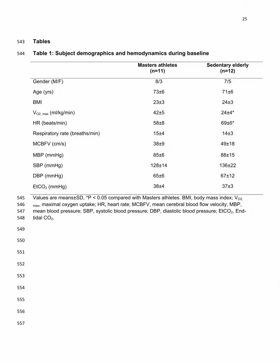
perform sit-stand maneuvers due to technical issues. The two groups had similar age, 204
body mass index (BMI), respiratory rate, CBFV, BP and EtCO2. As expected, VO2max 205
was higher and HR was lower in the Masters athletes (Table 1). Of note, VO2max in 206
Masters athletes was lower than expected from young elite endurance athletes because 207
of the decline in VO2max with age, even when maintaining a similar level of training (42). 208
There were no gender differences within or between groups (data not shown) and 209
therefore we present pooled data. 210
Baroreflex function. Baroreflex gain was higher in Masters athletes compared 211
with the sedentary elderly (Table 2) in the low frequency range (0.05-0.15 Hz) of 212
spontaneous changes in BP and R-R interval. As expected, under these baseline 213
(resting) conditions, spectral power of SBP and R-R interval were relatively small, 214
(Figure 1, Table 2). Forced BP changes at 0.05 Hz induced by repeated sit-stand 215
maneuvers augmented VLF spectral power in both groups; however, R-R interval 216
variability increased more in the Masters athletes. Although this suggests higher 217
baroreflex sensitivity consistent with findings in the LF, estimation of VLF transfer 218
function gain did not differ significantly between groups (Table 2). 219
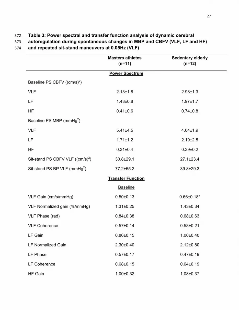
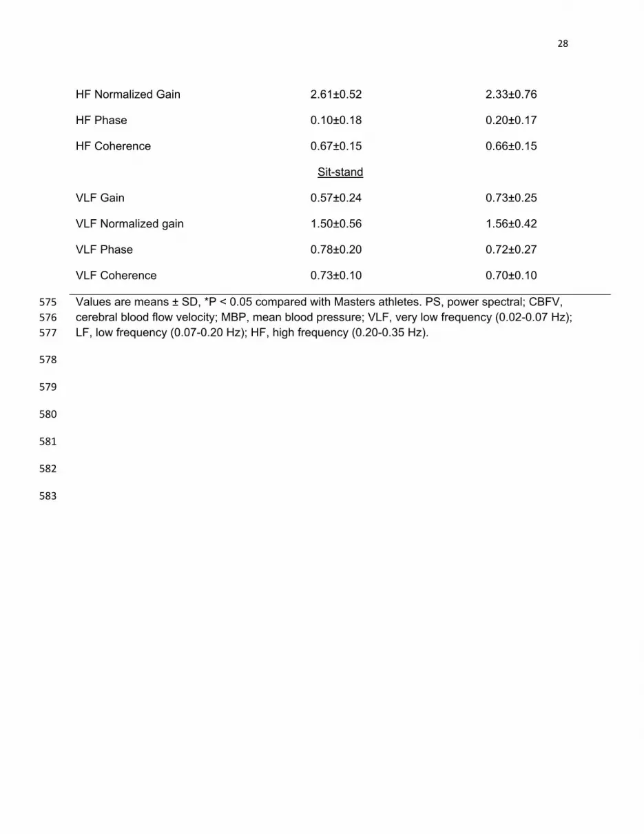
Cerebral autoregulation. Power spectral analysis (Figure 3, Table 3) showed no 220
differences in spontaneous or induced MBP and CBFV variability between the two 221
groups. Transfer function analysis (Figure 4, Table 3) showed a lower absolute, but not 222
normalized VLF gain in the Masters athletes during baseline. There were no differences 223
in phase, gain or coherence between the two groups during sit-stand maneuvers. 224
11
However, a trend toward increases in BP oscillations was observed in Masters athletes 225
when compared to sedentary elderly (Figure 3, Table 3). 226
No correlations were observed between dCA and baroreflex gain either during 227
baseline or sit-stand maneuvers. 228
229
Discussion 230
The main findings of this study are twofold. First, we did not find significant 231
differences in dynamic cerebral autoregulation between the Masters athletes and 232
sedentary elderly. Second, we found that cardiac baroreflex gain was more than 233
doubled in the Masters athletes when compared to the sedentary elderly. Moreover, no 234
correlations were observed between the estimates of baroreflex gain and dCA. 235
Collectively, these findings suggest that lifelong endurance training improves cardiac 236
baroreflex function, but does not alter dCA in older adults. 237
238
Effect of endurance training on cardiac baroreflex function 239
Previous studies suggest that endurance training improves (6, 13, 23, 26, 32, 38) 240
and aging reduces (8, 14, 18, 40) cardiac baroreflex function. The combined effects of 241
exercise and aging are therefore interesting. 242
Our finding that Masters athletes had a higher cardiac baroreflex gain than that 243
observed in the sedentary elderly during spontaneous BP changes is consistent with 244
previous studies which showed that endurance training from several months to a year 245
reduced the age-related decline in baroreflex function (6, 13, 32). 246

12
The underlying mechanisms through which exercise improves cardiac baroreflex 247
function are not completely understood. Exercise training may increase tonic vagal 248
activity, which could explain the higher cardiac baroreflex gain found in the Masters 249
athletes (41, 45). Consistent with this hypothesis, Masters athletes had a higher R-R 250
interval variability and lower resting heart rate than the sedentary elderly, indeed 251
suggesting increased vagal activity. Alternatively, exercise training may reduce age-252
related arterial stiffening and increase stroke volume, changing the transduction of 253
mechanical stimuli to the baroreceptors which in turn may increase cardiac baroreflex 254
gain (20). Finally, increases in cardiac cholinergic responsiveness associated with 255
exercise training may lead to increases in baroreflex sensitivity (31). 256
R-R interval variability was higher in Masters athletes during the augmented BP 257
oscillations induced by repeated sit-stand maneuvers when compared to the sedentary 258
elderly. However, calculation of baroreflex gain was not significantly higher in the 259
Masters athletes. In view of the high inter-individual variance, this may reflect a lack of 260
power for assessing baroreflex gain in the very low frequency range. 261
The higher SBP variations during repeated sit-stand maneuvers in the Masters 262
athletes may be explained by the training related changes in cardiovascular mechanics 263
(28). A greater peripheral vascular and/or left ventricular distensibility in combination 264
with a steeper slope of the Starling relationship between the left ventricular filling 265
pressure and stroke volume may result in large oscillations in stroke volume, and 266
therefore SBP, during sit-stand maneuvers in the Masters athletes, consistent with the 267
observation that athletes are more susceptible to orthostatic hypotension (28). 268
269

13
Effect of endurance training on cerebral autoregulation 270
Several studies have investigated acute effects of aerobic exercise on cerebral 271
circulation and dCA (7, 36, 37). Brys et al. (7) observed that dCA was maintained during 272
stepwise ergometric challenge at 50, 100, and 150 W. However, Ogoh et al. (36) 273
observed that dCA was impaired during ergometry at a mean workload of 168 W 274
continued to exhaustion. In addition, Ogoh et al. (37) found an unchanged normalized 275
transfer function gain between MBP and mean CBFV and between SBP and systolic 276
CBFV, but an increased gain between DBP and diastolic CBFV during cycling at a heart 277
rate of 150 beats/min, indicating reduced dCA during diastole. Both CBF and CBFV 278
may increase during aerobic exercise (19, 21, 37, 47). However, it has been argued that 279
increases in CBFV during exercise may reflect cerebral vasoconstriction of the 280
insonated artery instead of increases in blood flow (19). Clearly, differences in exercise 281
intensity, duration and modes used in these studies may have contributed to the above 282
discrepancies. However, collectively, these studies do suggest the possible presence of 283
acute effects of exercise on brain perfusion (7, 19, 21, 36, 37, 47). 284
Few studies have determined effects of exercise training on CBF regulation. In a 285
cross-sectional study, Ainslie et al. (1) showed that high aerobic fitness was associated 286
with a higher baseline CBFV, suggesting that exercise training may ameliorate age-287
related decline in CBF. Consistent with the proposed salutary effects of exercise training 288
on brain perfusion, endurance training for 3 months reduced cerebrovascular resistance 289
in elderly women over 60 years old (57) and increased cerebral vasomotor reactivity in 290
stroke survivors, but did not change baseline CBFV (22). Interestingly, Lind-Holst et al. 291

14
(29) found that dCA actually was less effective in the endurance trained young adults 292
relative to their sedentary controls. 293
Contrary to these findings, baseline CBFV was not higher in Masters athletes 294
when compared to the sedentary elderly. In addition, no group differences in dCA were 295
observed. Confounding factors, such as the limitations of cross-sectional study, 296
differences in the study population as well as the uncertainty about a potential dose-297
response relationship of exercise training and CBF regulation all may have contributed 298
to these inconsistent findings. However, the present study provides evidence that dCA 299
was not compromised in the Masters athletes who had participated in lifelong 300
endurance exercise training. 301
302
The relationship between cardiac baroreflex function and dCA 303
Short-term stability of the cerebral circulation is maintained primarily by two 304
mechanisms: the baroreflex control of arterial pressure and dCA regulation of CBF (51). 305
Whether there is a fundamental relationship between these regulatory mechanisms is 306
unclear. Tzeng et al. (50) found an inverse correlation between the cardiac baroreflex 307
function and dCA in young individuals. These findings have been interpreted to indicate 308
that there is a negative interaction between BP and CBF regulation (50), with higher 309
baroreflex sensitivity associated with less effective dCA. 310
In the present study, we did not observe any correlations between the estimates 311
of cardiac baroreflex and dCA gain in the Masters athletes nor in the sedentary elderly. 312
Consistently, Masters athletes had a higher cardiac baroreflex gain, but a similar dCA 313
when compared to the sedentary elderly. These findings indicate that higher baroreflex 314
15
sensitivity is not necessarily associated with a reduction in dCA in the elderly. These 315
findings also suggest that the overall effects of exercise on cerebral hemodynamics are 316
positive, with enhanced BP regulation combined with an unaffected regulation of CBF. 317
318
Study Limitations 319
The major limitations of the present study are its cross-sectional design and the 320
limited sample size. A causal relationship between lifelong endurance training in 321
Masters athletes and changes in baroreflex function and dCA cannot be determined. 322
However, conducting a longitudinal study to address these questions would be difficult, 323
if not impossible, to implement. The small sample size was due to the low prevalence 324
and hence difficulty to recruit Masters athletes. However, based on previous work (52), 325
we had > 80% power to detect a difference of 0.4 radians in the very low frequency 326
phase between the groups as an index of impaired dCA. 327
There are no “gold standard” methods to assess baroreflex function or dCA. 328
Therefore, direct comparisons between different studies are challenging. However, the 329
transfer function methods used in the present study are widely accepted and the results 330
of this study could be compared with a large number of studies using similar methods to 331
assess cardiac baroreflex function and dCA. 332
Our sedentary elderly do not reflect typically sedentarism as the chosen cut-off 333
level for physical activity may still have been relatively high. In addition, these elderly 334
subjects did not have hypertension and used no cardiovascular medication and did not 335
have other comorbidities that typically are associated with sedentarism. Therefore, the 336
sedentary elderly in this study most likely reflect normal aging of healthy controls. Thus, 337
16
the findings of this study primarily reflect the differences in cardiac baroreflex function 338
and dCA between normal aging and lifelong exercise training of Masters athletes. Using 339
a more sedentary control group could have enhanced contrasts between groups, 340
however, would also have complicated comparisons as comorbidities might have 341
confounded the results. 342
Finally, as with all studies using TCD to assess dCA, the assumption that 343
changes in CBFV reflect changes in CBF is valid only if the insonated MCA diameter did 344
not change during the experimental conditions. The MCA diameter is unlikely to change 345
under resting conditions or during moderate changes in BP induced during repeated sit-346
stand maneuvers (16, 34). However, lifelong endurance training may lead to 347
vasodilation in the basal cerebral arteries including the MCA (17). If this is the case, a 348
lower CBFV observed in the Masters athletes may underestimate CBF. To reduce the 349
effect of this potential limitation on the assessment of dCA, we normalized transfer 350
function gain. 351
In summary, in this cross-sectional study, we found that cardiac baroreflex gain 352
was more than doubled in the Masters athletes when compared to the sedentary 353
elderly. However, estimates of dCA were similar between the two groups. Furthermore, 354
no correlations were observed between the estimates of baroreflex gain and dCA. 355
Taken together, these findings suggest that lifelong endurance training improves 356
cardiac baroreflex function, but does not alter dCA in older adults. Thus, beneficial 357
effects of exercise training on BP regulation can be achieved in older adults without 358
compromising dynamic regulation of CBF. 359

17
Acknowledgments 360
The authors would like to thank the study participants for their willingness, time and 361
effort to make this project possible and Dean Palmer, Daniel Cresson and Kyle 362
Armstrong for their excellent technical support. 363
364
Grants 365
This project was supported in part by Texas Health Research & Education Institute Pilot 366
Study Award, Southwestern Medical Foundation, to RZ and by grants from the Dutch 367
Alzheimer’s Society and Radboud University Nijmegen to VA. 368
369
Disclosures 370
The authors have no financial conflict of interest to disclose. The content of this 371
manuscript is solely the responsibility of the authors. 372
373
Authors Contributions 374
V.A. analyzed data; V.A., J.C. and R.Z. interpreted results of experiments; V.A. 375
prepared tables and figures; V.A. and J.C. drafted manuscript; all authors edited and 376
revised manuscript; B.D.L. and R.Z. conception and design of research; B.D.L. and R.Z. 377
performed experiments; J.C. and R.Z. approved final version of manuscript. 378
379

18
References 380
1. Ainslie PN, Cotter JD, George KP, Lucas S, Murrell C, Shave R, Thomas KN, Williams MJ, 381
Atkinson G. Elevation in cerebral blood flow velocity with aerobic fitness throughout healthy human 382
ageing. J Physiol 586: 4005-4010, 2008. 383
2. Arbab-Zadeh A, Dijk E, Prasad A, Fu Q, Torres P, Zhang R, Thomas JD, Palmer D, Levine BD. 384
Effect of aging and physical activity on left ventricular compliance. Circulation 110: 1799-1805, 2004. 385
3. Balke B, Nagle FJ, Daniels J. Altitude and maximum performance in work and sports activity. 386
JAMA 194: 646-649, 1965. 387
4. Battisti-Charbonney A, Fisher J, Duffin J. The cerebrovascular response to carbon dioxide in 388
humans. J Physiol 589: 3039-3048, 2011. 389
5. Bishop CC, Powell S, Rutt D, Browse NL. Transcranial Doppler measurement of middle cerebral 390
artery blood flow velocity: a validation study. Stroke 17: 913-915, 1986. 391
6. Bowman AJ, Clayton RH, Murray A, Reed JW, Subhan MF, Ford GA. Baroreflex function in 392
sedentary and endurance-trained elderly people. Age Ageing 26: 289-294, 1997. 393
7. Brys M, Brown CM, Marthol H, Franta R, Hilz MJ. Dynamic cerebral autoregulation remains 394
stable during physical challenge in healthy persons. Am J Physiol Heart Circ Physiol 285: H1048-1054, 395
2003. 396
8. Burkhart CS, Rossi A, Dell-Kuster S, Gamberini M, Mockli A, Siegemund M, Czosnyka M, 397
Strebel SP, Steiner LA. Effect of age on intraoperative cerebrovascular autoregulation and near-infrared 398
spectroscopy-derived cerebral oxygenation. Br J Anaesth 107: 742-748, 2011. 399
9. Carey BJ, Eames PJ, Blake MJ, Panerai RB, Potter JF. Dynamic cerebral autoregulation is 400
unaffected by aging. Stroke 31: 2895-2900, 2000. 401

19
10. Claassen JA, Levine BD, Zhang R. Dynamic cerebral autoregulation during repeated squat-stand 402
maneuvers. J Appl Physiol 106: 153-160, 2009. 403
11. Cornelissen VA, Fagard RH. Effects of endurance training on blood pressure, blood pressure-404
regulating mechanisms, and cardiovascular risk factors. Hypertension 46: 667-675, 2005. 405
12. Cornelissen VA, Goetschalckx K, Verheyden B, Aubert AE, Arnout J, Persu A, Rademakers F, 406
Fagard RH. Effect of endurance training on blood pressure regulation, biomarkers and the heart in 407
subjects at a higher age. Scand J Med Sci Sports 21: 526-534, 2011. 408
13. Deley G, Picard G, Taylor JA. Arterial baroreflex control of cardiac vagal outflow in older 409
individuals can be enhanced by aerobic exercise training. Hypertension 53: 826-832, 2009. 410
14. Fisher JP, Ogoh S, Ahmed A, Aro MR, Gute D, Fadel PJ. Influence of age on cardiac baroreflex 411
function during dynamic exercise in humans. Am J Physiol Heart Circ Physiol 293: H777-783, 2007. 412
15. Fujimoto N, Prasad A, Hastings JL, Arbab-Zadeh A, Bhella PS, Shibata S, Palmer D, Levine BD. 413
Cardiovascular effects of 1 year of progressive and vigorous exercise training in previously sedentary 414
individuals older than 65 years of age. Circulation 122: 1797-1805, 2010. 415
16. Giller CA, Bowman G, Dyer H, Mootz L, Krippner W. Cerebral arterial diameters during changes 416
in blood pressure and carbon dioxide during craniotomy. Neurosurgery 32: 737-741; discussion 741-732, 417
1993. 418
17. Green DJ. Vascular adaptation in athletes: Is there an "Athlete's Artery"? Exp Physiol 2011. 419
18. Gribbin B, Pickering TG, Sleight P, Peto R. Effect of age and high blood pressure on baroreflex 420
sensitivity in man. Circ Res 29: 424-431, 1971. 421
19. Hellstrom G, Wahlgren NG. Physical exercise increases middle cerebral artery blood flow 422
velocity. Neurosurg Rev 16: 151-156, 1993. 423
20. Hunt BE, Farquhar WB, Taylor JA. Does reduced vascular stiffening fully explain preserved 424
cardiovagal baroreflex function in older, physically active men? Circulation 103: 2424-2427, 2001. 425

20
21. Ide K, Horn A, Secher NH. Cerebral metabolic response to submaximal exercise. J Appl Physiol 426
87: 1604-1608, 1999. 427
22. Ivey FM, Ryan AS, Hafer-Macko CE, Macko RF. Improved cerebral vasomotor reactivity after 428
exercise training in hemiparetic stroke survivors. Stroke 42: 1994-2000, 2011. 429
23. Iwasaki K, Zhang R, Zuckerman JH, Levine BD. Dose-response relationship of the cardiovascular 430
adaptation to endurance training in healthy adults: how much training for what benefit? J Appl Physiol 431
95: 1575-1583, 2003. 432
24. James MA, Potter JF. Orthostatic blood pressure changes and arterial baroreflex sensitivity in 433
elderly subjects. Age Ageing 28: 522-530, 1999. 434
25. Kenny RA, Kalaria R, Ballard C. Neurocardiovascular instability in cognitive impairment and 435
dementia. Ann N Y Acad Sci 977: 183-195, 2002. 436
26. Komine H, Sugawara J, Hayashi K, Yoshizawa M, Yokoi T. Regular endurance exercise in young 437
men increases arterial baroreflex sensitivity through neural alteration of baroreflex arc. J Appl Physiol 438
106: 1499-1505, 2009. 439
27. La Rovere MT, Bigger JT, Jr., Marcus FI, Mortara A, Schwartz PJ. Baroreflex sensitivity and 440
heart-rate variability in prediction of total cardiac mortality after myocardial infarction. ATRAMI 441
(Autonomic Tone and Reflexes After Myocardial Infarction) Investigators. Lancet 351: 478-484, 1998. 442
28. Levine BD. Regulation of central blood volume and cardiac filling in endurance athletes: the 443
Frank-Starling mechanism as a determinant of orthostatic tolerance. Med Sci Sports Exerc 25: 727-732, 444
1993. 445
29. Lind-Holst M, Cotter JD, Helge JW, Boushel R, Augustesen H, Van Lieshout JJ, Pott FC. Cerebral 446
autoregulation dynamics in endurance-trained individuals. J Appl Physiol 110: 1327-1333, 2011. 447
30. Mattace-Raso FU, van den Meiracker AH, Bos WJ, van der Cammen TJ, Westerhof BE, Elias-448
Smale S, Reneman RS, Hoeks AP, Hofman A, Witteman JC. Arterial stiffness, cardiovagal baroreflex 449

21
sensitivity and postural blood pressure changes in older adults: the Rotterdam Study. J Hypertens 25: 450
1421-1426, 2007. 451
31. Monahan KD. Effect of aging on baroreflex function in humans. Am J Physiol Regul Integr Comp 452
Physiol 293: R3-R12, 2007. 453
32. Monahan KD, Dinenno FA, Tanaka H, Clevenger CM, DeSouza CA, Seals DR. Regular aerobic 454
exercise modulates age-associated declines in cardiovagal baroreflex sensitivity in healthy men. J Physiol 455
529 Pt 1: 263-271, 2000. 456
33. Motoyama M, Sunami Y, Kinoshita F, Kiyonaga A, Tanaka H, Shindo M, Irie T, Urata H, Sasaki J, 457
Arakawa K. Blood pressure lowering effect of low intensity aerobic training in elderly hypertensive 458
patients. Med Sci Sports Exerc 30: 818-823, 1998. 459
34. Newell DW, Aaslid R, Lam A, Mayberg TS, Winn HR. Comparison of flow and velocity during 460
dynamic autoregulation testing in humans. Stroke 25: 793-797, 1994. 461
35. Ogoh S, Ainslie PN. Cerebral blood flow during exercise: mechanisms of regulation. J Appl 462
Physiol 107: 1370-1380, 2009. 463
36. Ogoh S, Dalsgaard MK, Yoshiga CC, Dawson EA, Keller DM, Raven PB, Secher NH. Dynamic 464
cerebral autoregulation during exhaustive exercise in humans. Am J Physiol Heart Circ Physiol 288: 465
H1461-1467, 2005. 466
37. Ogoh S, Fadel PJ, Zhang R, Selmer C, Jans O, Secher NH, Raven PB. Middle cerebral artery flow 467
velocity and pulse pressure during dynamic exercise in humans. Am J Physiol Heart Circ Physiol 288: 468
H1526-1531, 2005. 469
38. Okazaki K, Iwasaki K, Prasad A, Palmer MD, Martini ER, Fu Q, Arbab-Zadeh A, Zhang R, Levine 470
BD. Dose-response relationship of endurance training for autonomic circulatory control in healthy 471
seniors. J Appl Physiol 99: 1041-1049, 2005. 472

22
39. Panerai RB, White RP, Markus HS, Evans DH. Grading of cerebral dynamic autoregulation from 473
spontaneous fluctuations in arterial blood pressure. Stroke 29: 2341-2346, 1998. 474
40. Parati G, Frattola A, Di Rienzo M, Castiglioni P, Pedotti A, Mancia G. Effects of aging on 24-h 475
dynamic baroreceptor control of heart rate in ambulant subjects. Am J Physiol 268: H1606-1612, 1995. 476
41. Raczak G, Danilowicz-Szymanowicz L, Kobuszewska-Chwirot M, Ratkowski W, Figura-477
Chmielewska M, Szwoch M. Long-term exercise training improves autonomic nervous system profile in 478
professional runners. Kardiol Pol 64: 135-140; discussion 141-132, 2006. 479
42. Robinson S. Experimental studies of physical fitness in relation to age. European journal of 480
applied physiology and occupational physiology 10: 251-323, 1938. 481
43. Robinson TG, James M, Youde J, Panerai R, Potter J. Cardiac baroreceptor sensitivity is impaired 482
after acute stroke. Stroke 28: 1671-1676, 1997. 483
44. Sammons EL, Samani NJ, Smith SM, Rathbone WE, Bentley S, Potter JF, Panerai RB. Influence 484
of noninvasive peripheral arterial blood pressure measurements on assessment of dynamic cerebral 485
autoregulation. J Appl Physiol 103: 369-375, 2007. 486
45. Smith ML, Hudson DL, Graitzer HM, Raven PB. Exercise training bradycardia: the role of 487
autonomic balance. Med Sci Sports Exerc 21: 40-44, 1989. 488
46. Tanaka H, Dinenno FA, Monahan KD, Clevenger CM, DeSouza CA, Seals DR. Aging, habitual 489
exercise, and dynamic arterial compliance. Circulation 102: 1270-1275, 2000. 490
47. Thomas SN, Schroeder T, Secher NH, Mitchell JH. Cerebral blood flow during submaximal and 491
maximal dynamic exercise in humans. J Appl Physiol 67: 744-748, 1989. 492
48. Tsai JC, Chang WY, Kao CC, Lu MS, Chen YJ, Chan P. Beneficial effect on blood pressure and lipid 493
profile by programmed exercise training in Taiwanese patients with mild hypertension. Clin Exp 494
Hypertens 24: 315-324, 2002. 495

23
49. Tsai JC, Yang HY, Wang WH, Hsieh MH, Chen PT, Kao CC, Kao PF, Wang CH, Chan P. The 496
beneficial effect of regular endurance exercise training on blood pressure and quality of life in patients 497
with hypertension. Clin Exp Hypertens 26: 255-265, 2004. 498
50. Tzeng YC, Lucas SJ, Atkinson G, Willie CK, Ainslie PN. Fundamental relationships between 499
arterial baroreflex sensitivity and dynamic cerebral autoregulation in humans. J Appl Physiol 108: 1162-500
1168, 2010. 501
51. van Beek AH, Claassen JA, Rikkert MG, Jansen RW. Cerebral autoregulation: an overview of 502
current concepts and methodology with special focus on the elderly. J Cereb Blood Flow Metab 28: 503
1071-1085, 2008. 504
52. van Beek AH, Olde Rikkert MG, Pasman JW, Hopman MT, Claassen JA. Dynamic cerebral 505
autoregulation in the old using a repeated sit-stand maneuver. Ultrasound Med Biol 36: 192-201, 2010. 506
53. Wieling W, Krediet CT, van Dijk N, Linzer M, Tschakovsky ME. Initial orthostatic hypotension: 507
review of a forgotten condition. Clin Sci (Lond) 112: 157-165, 2007. 508
54. Zhang R, Claassen JA, Shibata S, Kilic S, Martin-Cook K, Diaz-Arrastia R, Levine BD. Arterial-509
cardiac baroreflex function: insights from repeated squat-stand maneuvers. Am J Physiol Regul Integr 510
Comp Physiol 297: R116-123, 2009. 511
55. Zhang R, Zuckerman JH, Giller CA, Levine BD. Transfer function analysis of dynamic cerebral 512
autoregulation in humans. Am J Physiol 274: H233-241, 1998. 513
56. Zhang R, Zuckerman JH, Iwasaki K, Wilson TE, Crandall CG, Levine BD. Autonomic neural 514
control of dynamic cerebral autoregulation in humans. Circulation 106: 1814-1820, 2002. 515
57. Zhu YS, Parker R, Tseng BY, Van Erkelens A, Coles G, Brunk E, Armstrong K, Rodrigue K, 516
Kennedy K, Park D, Zhang R. Abstract: Exercise Training Decreases Arterial Stiffness and Improves Brain 517
Perfusion in Sedentary Elderly Women. Circulation 124:A16151, 2011. 518

24
Figure captions 519
Figure 1: Group averaged power spectral density of spontaneous changes in systolic 520
blood pressure (SBP) (A) and R-R interval (B). 521
Figure 2: Group averaged transfer function gain (A), phase (B) and coherence (C) 522
between spontaneous changes in systolic blood pressure (SBP) and R-R interval. 523
Figure 3: Group averaged power spectral density of mean blood pressure (MBP) (A) 524
and cerebral blood flow velocity (CBFV) (B) during repeated sit-stand maneuvers at 525
0.05Hz. 526
Figure 4: Group averaged transfer function gain (A), phase (B) and coherence (C) 527
between mean blood pressure (MBP) and cerebral blood flow velocity (CBFV) during 528
repeated sit-stand maneuvers at 0.05Hz. 529
530
531
532
533
534
535
536
537
538
539
540
541
542
25
Tables 543
Table 1: Subject demographics and hemodynamics during baseline 544
Masters athletes (n=11)
Sedentary elderly (n=12)
Gender (M/F) 8/3 7/5
Age (yrs) 73±6 71±6
BMI 23±3 24±3
VO2, max (ml/kg/min) 42±5 24±4*
HR (beats/min) 58±8 69±6*
Respiratory rate (breaths/min) 15±4 14±3
MCBFV (cm/s) 38±9 49±18
MBP (mmHg) 85±6 88±15
SBP (mmHg) 128±14 136±22
DBP (mmHg) 65±6 67±12
EtCO2 (mmHg) 38±4 37±3
Values are means±SD, *P < 0.05 compared with Masters athletes. BMI, body mass index; VO2, 545
max, maximal oxygen uptake; HR, heart rate; MCBFV, mean cerebral blood flow velocity; MBP, 546 mean blood pressure; SBP, systolic blood pressure; DBP, diastolic blood pressure; EtCO2, End-547 tidal CO2. 548
549
550
551
552
553
554
555
556
557

26
Table 2: Power spectral and transfer function analysis of baroreflex function 558
during spontaneous changes in SBP and R-R interval (LF) and repeated sit-stand 559
maneuvers at 0.05 Hz (VLF) 560
Masters athletes (n=11)
Sedentary elderly (n=12)
Power Spectrum
Baseline LF PS SBP (mmHg2) 6.5±4.5 8.9±9.8
Sit-stand VLF PS SBP (mmHg²) 168±140 88±70
Baseline LF PS R-R interval (ms2) 411±424 157±161
Sit-stand VLF PS R-R interval (ms2) 2757±3578 769±770*
Transfer Function
Baseline LF Gain (ms/mmHg) 7.69±7.95 3.18±1.29*
Baseline LF Phase (rad) -1.01±0.35 -1.29±0.41
Baseline LF Coherence (units) 0.50±0.16 0.53±0.19
Sit-stand VLF Gain (ms/mmHg) 3.47±1.59 2.77±1.49
Sit-stand VLF Phase (rad) -0.86±0.64 -1.27±0.52
Sit-stand VLF Coherence (units) 0.67±0.09 0.61±0.09
Values are means±SD, *P < 0.05 compared with Masters athletes. PS, power spectral; SBP, 561 systolic blood pressure; LF, low frequency (0.05-0.15 Hz). VLF, very low frequency (0.02-0.07 Hz). 562
563
564
565
566
567
568
569
570
571
27
Table 3: Power spectral and transfer function analysis of dynamic cerebral 572
autoregulation during spontaneous changes in MBP and CBFV (VLF, LF and HF) 573
and repeated sit-stand maneuvers at 0.05Hz (VLF) 574
Masters athletes (n=11)
Sedentary elderly (n=12)
Power Spectrum
Baseline PS CBFV ((cm/s)2)
VLF 2.13±1.8 2.98±1.3
LF 1.43±0.8 1.97±1.7
HF 0.41±0.6 0.74±0.8
Baseline PS MBP (mmHg2)
VLF 5.41±4.5 4.04±1.9
LF 1.71±1.2 2.19±2.5
HF 0.31±0.4 0.39±0.2
Sit-stand PS CBFV VLF ((cm/s)2) 30.8±29.1 27.1±23.4
Sit-stand PS BP VLF (mmHg2) 77.2±55.2 39.8±29.3
Transfer Function
Baseline
VLF Gain (cm/s/mmHg) 0.50±0.13 0.66±0.18*
VLF Normalized gain (%/mmHg) 1.31±0.25 1.43±0.34
VLF Phase (rad) 0.84±0.38 0.68±0.63
VLF Coherence 0.57±0.14 0.58±0.21
LF Gain 0.86±0.15 1.00±0.40
LF Normalized Gain 2.30±0.40 2.12±0.80
LF Phase 0.57±0.17 0.47±0.19
LF Coherence 0.68±0.15 0.64±0.19
HF Gain 1.00±0.32 1.08±0.37
28
HF Normalized Gain 2.61±0.52 2.33±0.76
HF Phase 0.10±0.18 0.20±0.17
HF Coherence 0.67±0.15 0.66±0.15
Sit-stand
VLF Gain 0.57±0.24 0.73±0.25
VLF Normalized gain 1.50±0.56 1.56±0.42
VLF Phase 0.78±0.20 0.72±0.27
VLF Coherence 0.73±0.10 0.70±0.10
Values are means ± SD, *P < 0.05 compared with Masters athletes. PS, power spectral; CBFV, 575 cerebral blood flow velocity; MBP, mean blood pressure; VLF, very low frequency (0.02-0.07 Hz); 576 LF, low frequency (0.07-0.20 Hz); HF, high frequency (0.20-0.35 Hz). 577
578
579
580
581
582
583

0.05 0.15 0.25 0.35
PSD
of S
BP
(mm
Hg²
/Hz)
0
200
400
600
800
1000
Masters athletes Sedentary elderly
LF A

Frequency (Hz)
0.05 0.15 0.25 0.35
PSD
of R
-R in
terv
al (m
s²/H
z)
0
5000
10000
15000
20000
25000
30000
B

0.05 0.15 0.25 0.35
Gai
n (m
s/m
mH
g)
0
2
4
6
8
10
12
14
16
18
20
Masters athletesSedentary elderly
LF A

0.05 0.15 0.25 0.35
Phas
e (r
ad)
-3
-2
-1
0
1
2
3B

Frequency (Hz)
0.05 0.15 0.25 0.35
Coh
eren
ce (u
nits
)
0.2
0.3
0.4
0.5
0.6
0.7
0.8C

0.02 0.03 0.04 0.05 0.06 0.07 0.08
PSD
of M
BP
(mm
Hg²
/Hz)
0
1000
2000
3000
4000
5000
6000Masters athletesSedentary elderly
A VLF

Frequency (Hz)
0.02 0.03 0.04 0.05 0.06 0.07 0.08
PSD
of C
BFV
((cm
/s)²/
Hz)
0
500
1000
1500
2000
2500B

0.02 0.03 0.04 0.05 0.06 0.07 0.08
Nor
mal
ized
gai
n (%
/mm
Hg)
0.0
0.5
1.0
1.5
2.0
2.5
Masters athletesSedentary elderly
A VLF

0.02 0.03 0.04 0.05 0.06 0.07 0.08
Phas
e (r
ad)
0.0
0.2
0.4
0.6
0.8
1.0
1.2
1.4
B

Frequency (Hz)
0.02 0.03 0.04 0.05 0.06 0.07 0.08
Coh
eren
ce (u
nits
)
0.0
0.2
0.4
0.6
0.8
1.0
1.2C