c-kit immunopositive interstitial cells (cajal-type) in human myometrium
TRANSCRIPT

J. Cell. Mol. Med. Vol 9, No 2, 2005 pp. 407-420
During the last 20 years convincing data accumulatedshowing that the interstitial cells of Cajal (ICC) arelocated along the digestive tube [1–3]. Such cells
have an essential role as pacemakers and possible reg-ulators of neurotransmission [4–6]. More recently,Cajal-like interstitial cells have been described (basedon c-kit immunopositivity and/or TEM) outside thegastrointestinal tract: ureter [7, 8], bladder [9–11], ure-thra [12], vas deferens [13], fallopian tube [14], bloodvessels [15, 16], lymphatics [17], pancreas [18],prostate [19, 20], mammary gland [21]. Conflicting
Available online atwww.jcmm.ro www.jcmm.org
Published by:the CMM Foundation
Reprinted from: Journal of Cellular and Molecular Medicine
JCMMJCMM
C-kit immunopositive interstitial cells (Cajal-type) in human myometrium
Sanda M. Ciontea a, E. Radu a,c, T. Regalia a, Laura Ceafalan a, D. Cretoiu a,c, Mihaela Gherghiceanu c, R.I. Braga b, Mariana Malincenco a, L. Zagrean b,
M.E. Hinescu a,c, L.M. Popescu a,c *
a Department of Cellular and Molecular Medicine, b Department of Physiology,
“Carol Davila” University of Medicine and Pharmacy, Bucharest, Romaniac "Victor Babes" National Institute of Pathology, Bucharest, Romania
Received: April 28, 2005; Accepted: May 30, 2005
AbstractPrevious reports describing Cajal-like interstitial cells in human uterus are contradictory in terms of c-kit immunore-activity: either negative (but vimentin-positive) in pregnant myometrium, or positive, presumably in the endometri-um. The aim of this study was to verify the existence of human myometrial Cajal-like interstitial cells (m-CLIC).Six different, complementary approaches were used: 1) methylene-blue supravital staining of tissue samples(cryosections), 2) methylene blue and Janus green B vital staining (m-CLIC and mitochondrial markers, respective-ly), and 3) extracellular single-unit electrophysiological recordings in cell cultures, 4) non-conventional lightmicroscopy on glutaraldehyde/osmium fixed, Epon-embedded semi-thin sections (less than 1μm) stained with tolui-dine blue (TSM), 5) transmission electron microscopy (TEM), and 6) immunofluorescence (IF). We found m-CLICin myometrial cryosections and in cell cultures. In vitro, m-CLIC represented ~7% of the total cell number. m-CLIChad 2-3 characteristic processes which were very long (~60μm), very thin (<0.5μm) and moniliform. The dilated por-tions of processes usually accomodated mitochondria. In vitro, m-CLIC exhibited spontaneous electrical activity(62.4+7.22 mV field potentials, short duration: 1.197+0.04ms). Moreover, m-CLIC fulfilled the usual TEM criteria,the so-called ‘gold’ or ‘platinum’ standards (e.g. the presence of discontinuos basal lamina, caveolae, endoplasmicreticulum, and close contacts between each other, with myocytes, nerve fibers and/or capillaries etc.). IF showed thatm-CLIC express CD117/c-kit, sometimes associated with CD34 and with vimentin along their processes.
In conclusion, we describe myometrial Cajal-like interstitial cells that have affinity for methylene blue andJanus green B vital dyes, fulfill (all) TEM criteria, express CD117/c-kit and have spontaneous electric activity.
Keywords: CD117/c-kit • CD34 • immunofluorescence • human myometrium • smooth muscle α-actin •vimentin • methylene-blue vital staining • caveolae • interstitial cells • spontaneous electrical activity
* Correspondence to: L.M. POPESCU, M.D., Ph.D.Department of Cellular and Molecular Medicine, “Carol Davila” University of Medicine and Pharmacy, P.O. Box 35-29, Bucharest 35, Romania.E-mail: [email protected]
Introduction

reports have been published on c-kit expression byhuman m-CLIC. According to Duquette et al. [22]these cells tested negative for c-kit by immunohisto-chemistry, (but positive for vimentin) and are putativeinhibitors of intrinsic smooth muscle activity in preg-nant myometrium. Conversely, Shafik et al. [23]found, c-kit positive cells in cadaveric specimens ofhuman uterus but, located apparently in endometri-um, not in myometrium between myocytes.
We present here visual (morphologic) and func-tional (electrophysiological) evidence for the exis-tence of m-CLIC, in human pregnant and non-preg-nant myometria. These Cajal-like cells have affinityfor methylene blue (vital preparations), both in situand in cell culture, have spontaneous electric activity,fulfill all TEM criteria required for CLIC diagnostic,express CD-117/c-kit and form a network which con-tacts smooth muscle cells, nerve cells and capillaries.
Material and methods
Myometrial tissue samplesHuman tissue samples. Biopsies of human myometriumwere obtained from uteri of non-pregnant (undergoing hys-terectomies for benign gynaecological indications) and preg-nant women (between 17–40 weeks of gestation, at the timeof caesarean section, conducted either before or during labor).To avoid endometrium contamination, fragments of the exter-nal muscular layer from non-pregnant uteri were excised fromareas free of macroscopically visible anomalies. At the timeof caesarean delivery, a strip of uterine smooth muscle wastaken from the upper margin (in the midline) of the lower-seg-ment transverse incision. All tissue samples were obtained(after informed written consent from each woman) in accor-dance with a protocol approved by the local BioethicsCommittee, in accordance to generally accepted internationalpractice.
408
Fig. 1 Human pregnantmyometrium (17 weeksof gestation). Methylene-blue vital staining offresh specimens (cryosec-tions). A. Inset reveals atleast three selectivelystained m-CLIC, at lowermagnification (objective10x). B. Note the extrem-ly long (60–70 μm) ‘blue’processes of such cells,having a characteristicmoniliform aspect, whilethe surrounding smoothmuscle fibers are color-less. Photographic recon-struction; original magni-fication 100x, oil immer-sion.
A BMethylene blueMethylene blue
vitvitalalststainingaining

Animals. Eight-week-old Wistar female rats, body weight 200-250 g, with free access to food and water, were maintained in atemperature-controlled facility with a 12-h light/dark cycle forat least 1 week before use. All animal experiments were carriedout in accordance with the Guidelines for AnimalExperimentation of "Carol Davila" University of Medicine andPharmacy, Bucharest.
Methylene-blue vital stainingFresh human pregnant myometrium samples were vitally stainedwith methylene blue (Merck KGaA, Darmstadt, Germany),according to Niculescu’s protocol [26], presented in detail else-where [14]. Cryosections (3 μm thick) were cut with a Leica cry-omicrotome (Bensheim, Germany). Sections were dehydrated inabsolute ethanol, cleared in toluene and mounted on slides, insynthetic resin. Slides were examined under a Nikon 600E micro-scope. Micrographs were taken using Nikon Plan 10x or NikonPlan Fluor 100x/1.30 oil and digitally composed of three to fiveoptical sections, constructed with Adobe Photoshop (AdobeSystems) software.
Myometrial cells in cultureHuman myometrial samples were collected into sterile tubes con-taining Dulbecco's Modified Eagle's Medium (DMEM) supple-mented with fetal bovine serum (FBS) 2%, HEPES 1.5 mM, aswell as 10000 UI/ml penicillin, 0.2 mg/ml streptomycin and0.50 μg/ml amphotericin (Sigma Chemical, St. Louis, MO,USA), placed on ice, and transported to cell culture laboratory.Smooth muscle tissue was processed according to methodsdescribed by Morimoto et al. [24] and Li et al. [25] with somemodifications [14]. Briefly, tissue samples were minced into 0.5mm pieces, subsequently washed and incubated with gentle agi-tation for 30 min, at 37oC, with collagenase Ia (Sigma Chemical,St. Louis, MO, USA) 10 mg/ml and deoxyribonuclease I(0.1nm/mg) in DMEM supplemented with fetal bovine serum(FBS) 10%, HEPES 1.5 mM, 10000 UI/ml penicillin, 0.1 mg/mlstreptomycin and 0.25 μg/ml amphotericin. The dispersed cellswere separated from non-digested tissue by filtration through acell strainer (100 μm), collected by centrifugation of the filtrate at250g for 10 min, at room temperature (20oC), and suspended inculture medium. Cultures were initiated by plating 5x10⁴
cells/cm2 into 35 mm Petri dishes (BD Falcon, San Jose, CA,USA) or on glass inserts into 24-well plates (BD Labware, SanJose, CA, USA). Medium was changed every 48h. After asemi-confluent monolayer formed, cells were detached using
409
J. Cell. Mol. Med. Vol 9, No 2, 2005
Fig. 2 Human non-pregnantmyometrium in cell culture, the6th passage, day 3. Phase con-trast microscopy of a cell withtypical m-CLIC morphologyestablishing close contacts withsmooth muscle cells (arrows).One may observe 5 distinct pro-longations emerging from thecell body: a very short one (I),three having aproximately thesame length, 25–30 μm (II–IV),and a characteristic very longone, ~75 μm (V). Photographicreconstruction, original magni-fication 20x.
VV
Phase Phase contrastcontrast
II
IIII
IIIIIIIVIV
CajalCajal-- like like interstitial cellinterstitial cell
(m(m--CLIC)CLIC)

410
Fig. 3 Human pregnant myometrium. A, B. Primaryconfluent cultures (day 8) showing m-CLIC with atleast 7 ‘beads’ per process. C. The same aspect foundin semi-confluent cultures (day 4), where cells are inter-connected in a network-like pattern. D–G. m-CLICin culture (the 3rd passage, day 3), with characteristicmorphology, establishing contacts with smooth musclecells. D. Mitochondria revealed by Janus green B, atthe level of cell body and process dilations (arrowheads).Original magnification 20x (C), 40x (A, B, D–G).
A
D
E
F
G
B
C
MethyleneMethyleneblueblue
GiemsaGiemsa
GiemsaGiemsa
Safranin OSafranin O
Janus green BJanus green B
MethyleneMethyleneblueblue
MethyleneMethyleneblueblue
0.25% trypsin and 2mM EDTA and replated at a density of 5x10⁴
cells/cm2, the same technique being used for subsequent pas-sages. Experiments were performed in primary cultures andbetween passages 1–6.
Methylene blue vital staining. Cells were washed in pre-warmed phenol red-free DMEM (Sigma Chemical, St. Louis,MO, USA), and incubated for 20 min, in a 0.02% methylene-bluesolution, at 37°C. Methylene-blue solutions with dilutionsbetween 0.01–0.1% have been tested and 0.02% was chosen asbeing optimal. Cells were examined and photographed under aNikon inverted TE200 microscope equipped with a Nikon DN-5digital camera.
Janus green B vital staining. Cells in culture, the 3rd passage,were stained for 30 min in 0.02% Janus green B (SigmaChemical, St. Louis, MO, USA) [14] in DMEM and maintainedat 37°C, in a humidified atmosphere, 5% CO2 in air. After repeat-ed washes in DMEM, cells were examined under a heated object-stage inverted Nikon TE200 microscope and photographed.
Giemsa staining. The 35 mm Petri culture-dishes were emp-tied of the culture medium and cells were stained with 0.4%Giemsa solution (Sigma Chemical, St. Louis, MO, USA) inmethanol and distilled water (pH 6.9), for 30 min, at room tem-perature. This method is not a vital one, since the dye and the fix-ation agent (methanol) are acting concomitantly on live cells.Cells were washed three times in distilled water and examined.
Safranin O. Myometrial cultured cells, grown on coverslips,were fixed with paraformaldehyde 2%, for 30 min, at 4oC,washed 3 times with phosphate buffer saline (PBS), perme-abilised with methanol 70%, for 10 min, at 4oC and washed againwith PBS. Coverslips were immersed in 0.2% safranin O solution(Sigma Chemical, St. Louis, MO, USA), in PBS, for 2 h, at roomtemperature. After a rapid wash cells were examined.
ElectrophysiologyExtracellular single-unit recordings were performed on m-CLICcultures (human pregnant myometrium), at the 3rd passage. ANikon inverted TE200 microscope equipped with a Nikon DN-5digital camera and an Eppendorf (Eppendorf AG, Hamburg,Germany) NK I micromanipulator were used to position themicroelectrodes. On-site made glass microelectrodes (WPI1B150F-4) were filled with 3 mol/L potassium acetate and hadresistances between 4 and 8 MOhms. The field potential variationwas measured using a WPI Duo 773 electrometer (WorldPrecision Instruments, Sarasota, FL, USA), and outputs were dis-played on an oscilloscope. Electrical signals were digitized usinga Digidata 1322A data processor (Axon Instruments, Union City,CA, USA) and digitally stored. The Clampex 8.2 software wasused for data acquisition and analysis.
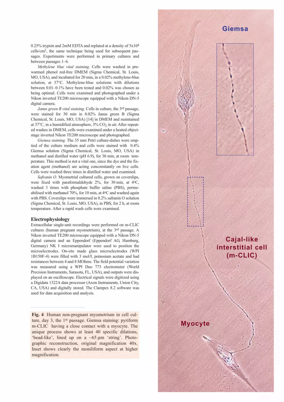
Fig. 4 Human non-pregnant myometrium in cell cul-ture, day 3, the 1st passage. Giemsa staining: pyriformm-CLIC having a close contact with a myocyte. Theunique process shows at least 40 specific dilations,‘bead-like’, lined up on a ~65 μm ‘string’. Photo-graphic reconstruction, original magnification 40x.Inset shows clearly the moniliform aspect at highermagnification.
GiemsaGiemsa
MyocyteMyocyte
CajalCajal-- like like interstitial cellinterstitial cell
(m(m--CLIC)CLIC)

Electron microscopy (TEM)Electron microscopy was performed on Epon-embedded tis-sue. Small tissue samples (Wistar rat myometrium), about 1mm3, were immersed in fresh 4% glutaraldhyde, for 4 h, at4°C. After fixation, the tissues were washed overnight in 0.1M sodium cacodylate buffer, at 4°C, post-fixed with 1%potassium ferrocyanide and 1% osmium tetroxide mixture in0.05 M sodium cacodylate buffer (pH 7.4), at room tempera-ture, for 60 min, dehydrated in graded ethanols and propy-lene oxide, and embedded in Epon 812. Slides with 1μmthick sections were stained with 1% toluidine blue for opticevaluation. After examination of the toluidine blue-stainedsections, ultrathin sections (60 nm) were cut using a MT-7000 ultramicrotome (Research Manufacturing CompanyInc., Tucson, AZ, USA), mounted on 50-mesh grids, anddouble stained with uranyl acetate and lead citrate.The gridswere examined in a Phillips 301 electron microscope or in aCM 12 Philips electron microscope, at an acceleration volt-age of 60 kV.
Thin-section microscopy (TSM)Control semi-thin sections (less than 1 μm) were stained with0.25% Toluidine blue and examined by light microscopy (NikonEclipse E600). Representative photomicrographs were takenusing Nikon Plan 40x and Nikon Plan Flour 100x/1.30 oil.
Immunofluorescence (IF)Immunofluorescent staining was performed using a protocoladapted after Mora et al. [27]. Cells grown on coverslips werefixed using 2% paraformaldehyde for 10 minutes at room temper-ature, then washed in PBS and permeabilized in PBS containing0.5% bovine serumalbumine (BSA) and 0.075% saponine(PBSSA), for 15 min. Primary antibodies (listed in Table 1, togeth-er with their respective clones, working dilutions and manufactur-ers) were applied for 4 h, at room temperature. Polyclonal FITC-labeled goat anti-mouse antibodies (working dilution 1:300, BDPharmingen, San Jose, CA, USA) were used to detect the primaryimmune reaction. For double staining experiments, the secondreaction was detected using monoclonal rat anti-mouse biotinylat-ed antibodies (1:200, clone A85-1, BD Pharmingen, San Jose, CA,USA) and streptavidin - Alexa Fluor 546 (working dilution 1:200,Molecular Probes, Eugene, OR, USA). Finally, nuclei were coun-terstained with 1 μg/ml Hoechst 33342 (Sigma Chemical, St.
412
50 ms
A B
10 mV1 ms
C
Fig. 5 A. Extracellular single-unit recording display-ing spontaneous electrical activity in m-CLIC. B. Spon-taneous field potential pattern. C. Phase contrastmicroscopy (40x) showing the recording glass-elec-trode at the level of m-CLIC body.
Table 1 Characteristics of primary antibodies used forimmunofluorescence.
Specificity Clone Conjugate Working dilution
Manufacturer
c-kit Ab81 none 1:100Santa CruzBiotechnology, SantaCruz, CA, USA
Vimentin V9(3) none 1:100 Dako, Glostrup,Denmark
CD34 8G12 FITC 1:10BD ImmunocytometrySystem, San Jose, CA,USA
SMA 1A4 FITC 1:100 Sigma Chemical, St.Louis, MO, USA

413
J. Cell. Mol. Med. Vol 9, No 2, 2005
Louis, MO, USA), and samples examined under a Nikon TE300microscope equipped with a Nikon DX1 camera, Nikon PlanApo40x and 60x objectives, and the appropriate fluorescence filters.Negative controls were prepared following the same protocol, butomitting the primary antibodies.
Results
Vital methylene blue staining, the method that ledto the initial discovery of Cajal cells in intestine[28], was used to look for Cajal-like interstitialcells in human myometrium. Selectively-stainedcells were found in both fresh tissue samples andcell cultures (Figs. 1 & 3A–C). These cells dis-
played particular morphology, defined by verylong, moniliform processes. In vivo, m-CLIC pro-longations were distributed among the intertwinedmyometrial fibers, and in vitro these cell process-es appeared interlaced in a network-like pattern(Fig. 3C). Typical m-CLIC (Fig. 2) were easilyrecognized in cell cultures (either primary or pas-sages 2–6) by phase contrast microscopy.Quantitatively, m-CLIC represented 6.9+0.9%SEM of myometrial cells grown in primary cul-tures (n = 651 cells).
Since in a previous study of Cajal-like cells inpancreas [18] we found by TEM that mitochondriaare preferentially located in the dilated portions ofthe cell processes, we used vital Janus green B dye,
Fig. 6 Human pregnant myometrium (40 weeks of gestation). Non-conventional light microscopy (TSM) onEpon-embedded samples, stained with toluidine blue. Three typical m-CLIC (red dashed lines) are seen embrac-ing the cross-sectioned smooth muscle cells (SMC; N = nucleus). Note very long m-CLIC processes (~ 60 μm).Original magnification 100x, oil immersion.
A B
Fig. 7 A, B Human myometrium cell culture (the 2nd passage); control phase-contrast microscopy (A) and IF for smoothmuscle α-actin (green, B) of the same microscopic fields; B, typical m-CLIC has one long SMA-negative process thatcontacts several SMA-positive myocytes. Although the m-CLIC cell body is reactive for SMA, no filamentous pattern isseen. Image reconstruction; original magnification 60x; nuclear counterstaining with Hoechst 33342 (blue).
A
B
SMC
SMC SMC
SMC
SMC
SMC
N
SMC
N

an established mitochondrial marker [29], to showmitochondrial localization in living cells in culture.Indeed, Fig. 3D reveals Janus green B positive‘dots’.
m-CLIC appeared also evident after using lessusual dyes. For instance, safranin O (Fig. 3E), adye that stains nuclei in red, actually coloredm-CLIC in orange (metachromasia).
Giemsa staining was used since this dye is essen-tially a mixture of two types of methylene blue dis-solved in methanol. Therefore, Giemsa solution actsconcomitantly as dye and fixative (Fig. 3F, G).Moreover, our experience suggests that Giemsastaining is the method of choice for revealing thetypical moniliform aspect of cell processes ofCajal-like interstitial cells in cultures (Fig. 4).
Extracellular single-unit recordings were donein order to determine whether m-CLIC in culturesexhibit (or not) spontaneous electrical activity.Field potentials averaged 62.4 + 7.22 mV ampli-tude (n = 30), apparently without a rhythmical pat-tern (Fig. 5). Compared to the electrophysiologicalproperties of smooth muscle cells [30], the record-ed potentials were rather short 1.197 + 0.04 ms.These results indicate that m-CLIC could generatespontaneous electrical activity.
TSM, the toluidine-blue staining of semi-thinsections from human pregnant myometrium depictsm-CLIC in close vicinity of smooth muscle cells.Fig. 6 shows that m-CLIC had an ovoid nucleussurrounded by a relatively small cytoplasm, fromwhich emerged two or three very long (50–75μm)processes.
Although putative m-CLIC can easily be dis-cerned in vitro by their long moniliform processes,we tested, by immunofluorescence, the presence ofthe specific smooth muscle α-actin (SMA) expres-sion. Fig. 7 shows that smooth muscle cells had acharacteristic positive reaction for actin stress fibers,while m-CLIC processes appeared negative forSMA (SMA not detectable), but a non-filamentousperinuclear positive immunoreaction is disclosed.
TEM (Figs. 8–10) prove at the ultrastructurallevel the characteristic features of Cajal-like intersti-tial cells (the so-called ‘gold standard’ [4] and /or‘platinum standard’ [14, 18]). For instance, Fig. 8shows clearly the basal lamina. m-CLIC establish,through their processes, vicinity relationships withcapillaries (Fig. 9B) and nerve fibers (Fig. 10B), aswell as specialized contacts, gap junctions with eachother and with the smooth muscle cells (Fig. 10A–D). Noteworthy, we found in myocytes typical‘Ca2+ release units’ (caveolae, sarcoplasmic reticu-lum and mitochondria) [31], vicinal of gap junctions.
Furthermore, to disclose the immunophenotype ofm-CLIC, IF assays for c-kit, CD34 and vimentin wereused. m-CLIC expressed CD117/c-kit (Fig. 11 A, B)in distinct membrane areas, sometimes associatedwith CD34 immunoreactivity (Fig. 11C). Cell con-tacts between m-CLIC processes and surroundingsmooth muscle cells were a constant finding, inaccordance with the role described for c-kit ligand(expressed on the myocyte) in the development ofICC in vitro [32]. Cell processes were also intenselypositive for vimentin (Fig. 11D, E), a featuredescribed in previously published reports [33].
414
Fig. 8 Human non-pregnant myometrium. TEM; fragment of a Cajal-like interstitial cell (m-CLIC); note the pres-ence of basal lamina on the extracellular side of the plasma membrane (arrows). The inset illustrates a higher mag-nification view of the basal lamina; N = nucleus.
INTERSTITIALCELLS
INTERSTITIALCELLS
Cajal-like cell body cell process
N
Cajal-like cell body cell process
Basal lamina

Fig. 9 TEM of human pregnant myometrium (A, B) andrat non-pregnant myometrium (C). m-CLIC processes areindicated by red dashed lines. A. Characteristic la-byrinthic system of m-CLIC processes. Note the strikingresemblance between the TEM image (A) and TSMimage (inset). B. This image shows a blood capillary astarget for m-CLIC processes. C. Typical Cajal-like inter-stitial cell with a very long process among the smoothmuscle cells (SMC). Note the presence of mitochondria(m) at the level of dilated segments of m-CLIC process.N = nucleus, Ht = heterochromatin; Eu = euchromatin.
415
J. Cell. Mol. Med. Vol 9, No 2, 2005
A
B
C
N
N
N
N
m
m
m
NN
SMC
Labyrinthic system
mm--CLICCLIC
InterstitialInterstitialCajalCajal-- like celllike cell
mm--CLICCLIC
SMC
SMC
SMC
SMC
SMC
Ht
Collagen
Eu
Eu
N
N
Capillary
Endothelium
N
NN
NN
N
N
N
HHHHtttt

Fig. 10 A–D Digitally-colored TEM images of m-CLIC in rat myometrium: Cajal-like interstitial cells (blue), smooth muscle cells(Sienna-brown), and nerve fiber fascicle (green). A. Low power micrograph showing three long, moniliform processes that encirclebundles of cross-cut smooth muscle cells. Original magnification x6,800. B. Gap junction between two processes of m-CLIC. The pro-cesses are in the proximity of unmyelinated nerve fibers. Original magnification x9,100. C, D. Cell junctions between myometrialsmooth muscle cells and m-CLIC processes. The two membranes are separated by a gap of about 30nm. One may observe‘Ca2+-release units’ (caveolae, sarcoplasmic reticulum and mitochondria) in the cytoplasmic region where smooth muscle sarcolem-ma comes in close contact with m-CLIC plasmalemma. Original magnification: x11,000 (C); x15,000 (D). Abbreviations: SMC =smooth muscle cells; ax = axon; db = dense bodies; N = nucleus; Ht = heterochromatin; Eu = euchromatin; rER = rough endoplasmicreticulum; sER = smooth endoplasmic reticulum; SR = sarcoplasmic reticulum; m = mitochondria; cav = caveolae (arrowheads).
416
Gap junction
CajalCajal-- like like interstitial cellsinterstitial cells
(m(m--CLIC)CLIC)
mm--CLICCLIC
rER
rER
SMC
SMC
SMC
SMC
ax
ax
ax ax
ax
m
ax
m
N
N
N
m m
mN
N
N
N
m
m
sER
rER
Nerve fiber Nerve fiber fasciclefascicle
ax
A
B

417
J. Cell. Mol. Med. Vol 9, No 2, 2005
C
D
Gap junction
CajalCajal-- like like interstitial cellsinterstitial cells
(m(m--CLIC)CLIC)
CajalCajal-- like like interstitial cell (minterstitial cell (m--CLIC)CLIC)
dense plaquedb
m
m
m
mm m
m
m
cavm
SR
rER
SR
SR
SR
sER
Nucleus
HtEuGap
junction

418
A
B
C
D
E
Fig. 11 A–E. Human myometrium cells in culture (the 2nd passage); IF for c-kit (green in A, B), c-kit and CD34 (redand green, respectively in C) and vimentin (green in D, E). Cells that display the m-CLIC morphologic feature (long,moniliform processes) express c-kit and contact adjacent cells (A–C). Some cells suggestive for m-CLIC coexpressc-kit and CD34 (C). The characteristic cell processes are immunoreactive for vimentin and establish connections withnearby cells (D and E). Original magnification 60x, nuclear counterstaining with Hoechst 33342 (blue).

419
J. Cell. Mol. Med. Vol 9, No 2, 2005
Discussion
This study provides evidence for the presence of a c-kit positive cell type, with Cajal-like morphology, inhuman pregnant and non-pregnant myometrium.However, a paper reported c-kit non-immunoreactiveICC in human and rat myometrium [22]. These appar-ently contradictory results could suggest the (co)exis-tence in myometrium of at least two subpopulations ofCajal-like cells, different in terms of c-kit expression,as reported for the human small intestine [34].
In order to deal with the issue of m-CLIC identity,besides c-kit-immunofluorescence, we used 'classical'vital staining with methylene blue and ultrastructuralstudies (TEM).
Although several sets of criteria have been assam-bled to identify ICC (‘gold standard’ [4]) or Cajal-likeinterstitial cells (‘platinum standard’ [14, 18]), weemphasize that not all criteria apply to each cell.
In our opinion, any debate on c-kit expressionshould start after a minimal set of criteria for Cajal-like cells is satisfied. Thus, we looked for Cajal-likeinterstitial cells, according to the following basiccheck-list:
– location in non-epithelial (interstitial) spaces;– vital staining with methylene blue;– multiple long, thin, moniliform processes.‘Our’ m-CLIC fulfill these conditions and, addi-
tionaly, have ultrastructural features specific for ICC,different from smooth muscle cells, fibroblasts ormyofibroblasts.
Since c-kit IHC is used in almost all current stud-ies on ICC pathology [35], the precise distribution ofc-kit expression in normal tissues has to be extensive-ly reconsidered. Examining 3,000 samples from morethan 120 different tumor categories, Went et al., pro-vided a list of normal tissues/organs found to be com-pletely c-kit-negative: "cerebrum, uterine cervix,colon, endometrium, esophagus, fat tissue, gall blad-der, heart, kidney, liver, lung, lymph node, myometri-um, oral cavity mucosa, ovary, pancreas, parathyroid,salivary gland, prostate, skeletal muscle, small intes-tine, intestinal smooth muscle, stomach mucosa, thy-roid, and urothelium" [36]. These data are in contra-diction with the ‘wave’ of papers identifying ICC orICC-like cells [7-21] in some of the so-called ‘com-pletely kit-negative tissues’. Possible explanationsmight be: a) differences in the affinity of the usedantibodies; b) the great sensitivity of ICC to fixa-tion [7].
An indirect argument for the existence of c-kitpositive cells in human myometrium might be theoccurrence of this antigen in myometrial sarcomas[37]. ICC loss was identified as a cause of bowelmotility disorders [38]. It remains to be establishedif such specific loss of Cajal-like cells could beidentified in the pathology of myometrium.
We noted spontaneous electrical activity ofm-CLIC in cultures. This m-CLIC behavior proba-bly reflects a closer relationship with typical ICCand a possible role of pace-maker cannot be over-looked.
Direct communication by cellular bridges mightbe a common morphological feature of stem cellsand cancer cells [39, 40]. It is tempting to speculatethat our IF data on m-CLIC might represent a similar'direct' cell to cell communication.
In conclusion, in our opinion, the search forCajal-like interstitial cells should begin in intersti-tium, after vital methylene-blue staining revealedcells with characteristic long, moniliform processes.Expression of c-kit and TEM should be the nextrequired steps of the algorythm for Cajal-like inter-stitial cell diagnostic. Electrophysiology could com-plete the functional profile of this still enigmatic cell.
References1. Thuneberg L, One hundred years of interstitial cells of Cajal.
Microsc Res Tech. 1999; 47: 223-38.2. Faussone-Pellegrini MS, Pantalone D, Cortesini C.
Smooth muscle cells, interstitial cells of Cajal and myentericplexus interrelationships in the human colon. Acta Anat(Basel). 1990; 139: 31-44.
3. Rumessen JJ, Peters S, Thuneberg L. Light- and electronmicroscopical studies of interstitial cells of Cajal (ICC) andmuscle cells at the submucosal border of human colon, LabInvest. 1993; 68: 481-95.
4. Huizinga JD, Thuneberg L, Vanderwinden JM,Rumessen JJ. Interstitial cells of Cajal as targets for phar-macological intervention in gastrointestinal motor disorders.Trends Pharmacol Sci. 1997; 18: 393-403.
5. Ward SM, Sanders KM. Physiology and pathophysiologyof the interstitial cell of Cajal: From Bench to Bedside I.Functional development and plasticity of interstitial cells ofCajal networks. Am J Physiol Gastrointest Liver Physiol.2001; 281: G602-G611.
6. Hirst GD, Ward SM. Interstitial cells: involvement in rhyth-micity and neural control of gut smooth muscle. J Physiol.2003; 550: 337-46.

7. Pezzone MA, Watkins SC, Alber SM, King WE, de GroatCW, Chancellor MB, Fraser MO. Identification of c-kit-pos-itive cells in the mouse ureter: the interstitial cells of Cajal of theurinary tract. Am J Physiol Renal Physiol. 2003; 284: 925-9.
8. Metzger R, Schuster T, Till H, Franke FE, Dietz HG.Cajal-like cells in the upper urinary tract: comparative studyin various species. Pediatr Surg Int. 2005; 21: 169-74.
9. McCloskey KD, Gurney AM. Kit positive cells in theguinea-pig bladder. J Urol. 2002; 168: 832-6.
10. Blyweert W, Aa F, Ost D, Stagnaro M, Ridder D.Interstitial cells of the bladder: the missing link? BJOG. 2004;111(s1):57-60.
11. Davidson RA, McCloskey KD. Morphology and localiza-tion of interstitial cells in the guinea pig bladder: structuralrelationships with smooth muscle and neurons. J Urol. 2005;173: 1385-90.
12. Sergeant GP, Hollywood MA, McCloskey KD,Thornbury KD, McHale NG. Specialized pacemaking cellsin the rabbit urethra. J Physiol. 2000; 526: 359-66.
13. Burton LD, Housley GD, Salih SG, Jaeenwood D.P2X2receptor expression by interstitial cells of Cajal in vasdeferens implicated in semen emission. Auton Neurosci BasicClin. 2000; 84: 147-61.
14. Popescu LM, Ciontea MS, Cretoiu D, Hinescu ME, RaduE, Ionescu N, Ceausu M, Gherghiceanu M, Braga RI,Vasilescu F, Zagrean L, Ardeleanu C. Novel type of inter-stitial cell (Cajal-like) in human fallopian tube. J Cell MolMed. 2005; 9: 479-523.
15. Harhun MI, Gordienko DV, Povstyan OV, Moss RF,Bolton TB. Function of interstitial cells of Cajal in the rabbitportal vein. Circ Res. 2004; 95: 619-26.
16. Harhun MI, Pucovsky V, Gordienko DV, Povstyan OV,Bolton TB. Interstitial cells in the vasculature. J Cell MolMed. 2005; 9: 232-43.
17. McCloskey KD, Hollywood MA, Thornbury KD, WardSM, McHale NG. Kit-like immunopositive cells in sheap me-senteric lymphatic vessels. Cell Tissue Res. 2002; 310: 77-84
18. Popescu LM, Hinescu ME, Ionescu N, Ciontea MS,Cretoiu D, Ardeleanu C. Interstitial cells of Cajal in pan-creas, J Cell Mol Med. 2005; 9: 169-90.
19. Exintaris B, Klemm MF, Lang RJ. Spontaneous slow waveand contractile activity of the guinea pig prostate. J Urol.2002; 168: 315-22.
20. Van der Aa F, Roskams T, Blyweert W, De Ridder D.Interstitial cells in the human prostate: a new therapeutic tar-get? Prostate. 2003; 56:250-5.
21. Popescu LM, Andrei F, Hinescu ME. Snapshots of mam-mary gland interstitial cells: methylene blue vital staining andc-kit immunopositivity. J Cell Mol Med. 2005; 9: 476-7.
22. Duquette RA, Shmygol A, Vaillant C, Mobasheri A, PopeM, Burdyga T, Wray S. Vimentin-positive, c-kit-negativeinterstitial cells in human and rat uterus: a role in pacemak-ing? Biol Reprod. 2005; 72: 276-83.
23. Shafik A, El-Sibai O, Shafik I. Identification of c-kit-positivecells in the uterus. Int J Gynaecol Obstet. 2004; 87: 254-5.
24. Morimoto T, Head JR, MacDonald PC, Casey ML.Thrombospondin-1 expression in human myometrium beforeand during pregnancy, before and during labor, and in humanmyometrial cells in culture. Biol Reprod. 1998; 59: 862-70.
25. Li CX, Liu BH, Tong WD, Zhang LY, Jiang YP. Dissociation,culture and morphologic changes of interstitial cells of Cajal invitro. World J Gastroenterol. 2005; 11: 2838-40.
26. Niculescu I. An atlas concerning morphological aspects ofvisceral nerve endings. 1st ed. Bucharest: Editura Medicala;1958.
27. Mora R, Bonilha VL, Marmorstein A, Scherer PE, BrownD, Lisanti MP, Rodriguez-Boulan E. Caveolin-2 localizesto the golgi complex but redistributes to plasma membrane,caveolae, and rafts when co-expressed with caveolin-1. J BiolChem. 1999; 274: 25708-17.
28. Cajal SR. Les nouvelles idees sur la structure du systemenerveux chez l’homme et chez les vertebres. 2eme ed. Paris:C. Reinwald & Cie, Libraires-Editeurs, 1895.
29. Novikoff AB. Mitochondria in: The Cell ed. By J. Brachet, A.Mirsky, Academic Press Inc., New York, 1961, 299-421.
30. Farrugia G. Ionic conductances in gastrointestinal smoothmuscles and interstitial cells of Cajal. Annu Rev Physiol.1999; 61: 45-84.
31. Moore ED, Voigt T, Kobayashi YM, Isenberg G, Fay FS,Gallitelli MF, Franzini-Armstrong C. Organization of Ca2+
release units in excitable smooth muscle of the guinea-pigurinary bladder. Biophys J. 2004; 87:1836-47. Erratum in:Biophys J. 2004; 87:2914.
32. Wu JJ, Rothman TP, Gershon MD, Development of theinterstitial cell of cajal: origin, kit dependence and nonneu-ronal sources of kit ligand. J Neurosci Res. 2000; 59: 384-401.
33. Torihashi S, Ward SM, Nishikawa S, Nishi K, KobayashiS, Sanders KM, c-kit-dependent development of interstitialcells and electrical activity in the murine gastrointestinal tract.Cell Tissue Res. 1995; 280:97.
34. Horiguchi K, Keef KD, Ward SM. Distribution of interstitialcells of Cajal in tunica muscularis of the canine rectoanal region.Am J Physiol Gastrointest Liver Physiol. 2003; 284: 756-67.
35. Wang XY, Berezin I, Mikkelsen HB, Der T, Bercik P,Collins SM, Huizinga JD. Pathology of interstitial cells ofCajal in relation to inflammation revealed by ultrastructurebut not immunohistochemistry. Am J Pathol. 2002; 160:1529-40.
36. Went PT, Dirnhofer S, Bundi M, Mirlacher M, SchramlP, Mangialaio S, Dimitrijevic S, Kononen J, Lugli A,Simon R, Sauter G. Prevalence of KIT expression in humantumors. J Clin Oncol. 2004; 22: 4514-22.
37. Rushing RS, Shajahan S, Chendil D, Wilder JL, Pulliam J,Lee EY, Ueland FR, van Nagell JR, Ahmed MM, Lele SM.Uterine sarcomas express KIT protein but lack mutation(s) inexon 11 or 17 of c-KIT. Gynecol Oncol. 2003; 91: 9-14.
38. Sanders KM, Ördög T, Ward SM. Physiology and patho-physiology of the interstitial cells of Cajal: from bench tobedside: IV. Genetic and animal models of GI motility disor-ders caused by loss of interstitial cells of Cajal. Am J PhysiolGastrointest Liver Physiol. 2002; 282: 747-56.
39. Rustom A, Saffrich R, Markovic I, Walther P, Gerdes H-H. Nanotubular highways for intercellular organelle trans-port. Science. 2004; 303: 1007-10.
40. Vidulescu C, Clejan S, O'Connor KC. Vesicle trafficthrough intercellular bridges in DU 145 human prostate can-cer cells. J Cell Mol Med. 2004; 8: 388-96.
420