c-fos and fos-related antigens as markers for neuronal activity: perspectives from neuroendocrine...
TRANSCRIPT

National Institute on Drug Abuse
RESEARCHMONOGRAPH SERIES
Activation of ImmediateEarly Genes by Drugs
of Abuse
125U.S. Department of Health and Human Services • Public Health Service • National Institutes of Health
Activation of ImmediateEarly Genes By Drugs ofAbuse
Editors:
Reinhard Grzanna, Ph.D.Roger M. Brown, Ph.D.
NIDA Research Monograph 1251993
U.S. DEPARTMENT OF HEALTH AND HUMAN SERVICESPublic Health ServiceNational Institutes of Health
National Institute on Drug Abuse5600 Fishers LaneRockville, MD 20857

ACKNOWLEDGMENT
This monograph is based on the papers and discussions from a technicalreview on “Activation of Immediate Early Genes by Drugs of Abuse” held onJune 3-4, 1991, in Rockville, MD. The technical review was sponsored by theNational Institute on Drug Abuse (NIDA).
COPYRIGHT STATUS
The National Institute on Drug Abuse has obtained permission from thecopyright holders to reproduce certain previously published material as noted inthe text. Further reproduction of this copyrighted material is permitted only aspart of a reprinting of the entire publication or chapter. For any other use, thecopyright holder’s permission is required. All other material in this volumeexcept quoted passages from copyrighted sources is in the public domain andmay be used or reproduced without permission from the Institute or the authors,Citation of the source is appreciated.
Opinions expressed in this volume are those of the authors and do notnecessarily reflect the opinions or official policy of the National Institute on DrugAbuse or any other part of the U.S. Department of Health and Human Services.
The U.S. Government does not endorse or favor any specific commercialproduct or company. Trade, proprietary, or company names appearing in thispublication are used only because they are considered essential in the context ofthe studies reported herein.
NIDA Research Monographs are indexed in the “Index Medicus.” They areselectively included in the coverage of “American Statistics Index,” “BioSciencesInformation Service,” “Chemical Abstracts,” “Current Contents,” “PsychologicalAbstracts,” and “Psychopharmacology Abstracts.”
National Institute on Drug AbuseNIH Publication No. 93-3504Printed 1993
i i

Contents
Page
Introduction 1Reinhard Grzanna and Roger M. Brown
Regulation of Immediate Early Gene Expression 3Brent H. Cochran
Everything Activates C-fos- How Can It Matter? 25Steven E. Hyman, Barry E. Kosofsky, Tuong V. Nguyen,Bruce M. Cohen, and Michael J. Comb
Immediate Early Genes: Their Involvement in Physiologicaland Pathological Responses in the Nervous System 39
Michael O. Hayward, Tom Curran, and James I. Morgan
Immediate Early Gene Activation and Long-Term Changesin Neural Function: A Possible Role in Addiction? 54
Harold A. Robertson
Acute Effects of Psychomotor Stimulant Drugs on GeneExpression in the Striatum 72
Ann M. Graybiel
Functional Organization of the Striatum: Relevance to Actionsof Psychostimulant Drugs of Abuse 82
Charles R. Gerfen
i i i

Regulation of Neural Gene Expression in Opiate andCocaine Addiction 92
Eric J. Nestler, Clare M. Bergson. Xavier Guitart,and Bruce T. Hope
C-Fos and Fos-Related Antigens as Markers for NeuronalActivity: Perspectives From Neuroendocrine Systems
Gloria E. Hoffman, Wen-Sen Lee, M. Susan Smith, Rula Abbud,Michelle M. Roberts, Alan G. Robinson, and Joseph G. Verbalis
117
Mechanisms of Opioid-Mediated Antinociception: Correlationof Fos Expression and Behavior 134
Kathleen R. Gogas, Jon D. Levine, and Allan I. Basbaum
The Ontogeny of Immediate Early Gene Response to Cocaine:A Molecular Analysis of the Effects of Cocaine on DevelopingRat Brain 181
Barry E. Kosofsky and Steven E. Hyman
NMDA Receptor Blockade Prevents Translation, but Not Transcription,of the C-fos Gene Following Stimulation With Multiple ExtracellularSignals in Cultured Cortical Neurons: Implications for Plasticity andMolecular Memory 172
Frank R. Sharp, Kinya Hisanaga, and Stephen M. Sagar
Induction and Suppression of Proto-Oncogenes in RatStriatum After Single or Multiple Treatments With Cocaineor GBR-12909 181
Michael J. ladaroia, Eric J. Chuang, Choh-Lun Yeung, Yin Hoo,Mayme Silverthorn, Jun Gu, and Gaetano Draisci
List of NIDA Research Monographs 212
iv
C-Fos and Fos-Related Antigensas Markers for Neuronal Activity:Perspectives From NeuroendocrineSystemsGloria E. Hoffman, Wen-Sen Lee, M. Susan Smith, Rula Abbud,Michelle M. Roberts, Alan G. Robinson, and Joseph G. Verbalis
INTRODUCTION
The alterations in neuronal function underlying drug abuse are complex, yetimportant. Several specific neuronal systems have been implicated in drug-seeking behavior, addiction, and withdrawal, but none has emerged as key.What may be needed is an approach to mark neurons throughout the brain thatare influenced by drugs of abuse. Once targeted neurons are identified, studiescan be extended to define their phenotype and projections and to determinetheir functions in drug-related processes. The use of immediate early gene(IEG) products may provide an important tool for labeling neurons whoseactivity has changed as a result of drug treatment. As is detailed below, c-Fosand Fog-related antigens (FRAs) serve as markers for identifying neuronalsystems whose activity is increased or decreased in response to a stimulus.
In situ hybridization of IEG messenger RNAs (mRNAs) as well asimmunocytochemical localization of protein products are useful for addressingthis issue. The use of the protein products in examining specific systems canhave advantages over the study of mRNA. First, molecular probes are notavailable for all IEGs, whereas antisera have been generated that recognizethe protein products of some genes not yet cloned. Second, IEG productssuch as c-Fos, c-Jun, and Zif/268, when present, concentrate within thecell nucleus (Sheng and Greenberg 1990). This feature makes it possible tolabel these proteins along with cytoplasmic markers for the neurotransmitter(Ceccatelli et al. 1969; Hoffman et al. 1990; Jacobson et al. 1990) and/orretrogradely transported tracers (Menetrey et al. 1969) using standard doubleimmunocytochemical techniques. For heterogenous neuronal populations, ordiffusely organized neuronal systems, this capability is imperative. In contrast,mRNA is located in the cytoplasm. Techniques for double labeling of either two
117

mRNAs or one mRNA and an immunocytochemically identified marker arenot yet sufficiently reliable to be easily applied; generally, there is greatcompromise in the quality of either the mRNA label, the immunocytochemicalmarker, or both. Another advantage to localizing the proteins is that theirappearance is delayed by at least 30 to 45 minutes after the stimulus isdelivered, unlike the mRNA, which increases within 5 to 10 minutes afterstimulation. This delay allows the investigator to move or manipulate theanimal prior to sacrifice without concern that such handling will result in IEGexpression. The case is well illustrated for studies of light activation. In thisinstance, for study of mRNA changes, animals must be sacrificed undersafelight conditions, whereas immunocytochemical analysis would allowan animal to be anesthetized and brought into the light for perfusionwithout allowing sufficient time for the light stimulus to be translated intonew IEG proteins. A disadvantage in using IEG products localized byimmunocytochemistry rather than by in situ hybridization of their mRNA isthe lack of precise quantitation inherent in the immunocytochemical methods.In this chapter, the authors present evidence that, in spite of this problem,localization of IEG products with standard immunohistochemical techniquespermits assessment of relative changes in gene expression. The studiespresented in this chapter use natural stimuli and document that the expressionof the IEGs is not only induced by drug treatment or other experimentalmanipulation but also is involved in normal homeostatic function.
C-FOS AS A MARKER FOR NEURON ACTIVATION: LUTEINIZINGHORMONE-RELEASING HORMONE NEURONS AS THE MODEL
Most of the data presented here focus on the authors’ studies of neuroendocrineregulation. In particular, study of the regulation of reproductive function hasbeen most revealing. The study of neuroendocrine-adenohypophyseal functionhas presented challenges to physiologists in that conventional means ofassessing neuronal activity are impractical owing to the small size and scattereddistribution of the neuroendocrine neurons regulating the anterior pituitary. Thecase in point is well illustrated for one neuroendocrine system: the luteinizinghormone-releasing hormone (LHRH) (also known as gonadotropin-releasinghormone or GnRH) neurons, which stimulate the release of both luteinizinghormone (LH) and follicle-stimulating hormone (FSH) from the anterior pituitary.LHRH neurons are small, few in number, and widely scattered. The rat braincontains only 1,200 LHRH neurons (Wray and Hoffman 1986a, 1986b), 70percent of which send their axons to the median eminence to effect LH and FSHrelease (Merchanthaler et al. 1989). The LHRH cells are scattered over the fullrostral-to-caudal extent of the forebrain and generally do not reside within classiccytoarchitectonic boundaries (Hoffman and Gibbs 1982; Silverman et al. 1982),To make matters worse, the LHRH somata typically measure only 8 to 10 µm indiameter: even in sites where the LHRH neurons are most numerous, they are
116

interspersed among non-LHRH neurons, making the possibility of successfullylocating the cells and then recording from them remote.
Means have been devised for monitoring LHRH secretory activity. The mostdirect method involves cannulation of the tiny portal blood vessels that carryLHRH released at the median eminence of the hypothalamus to the anteriorpituitary. With this approach, during an LH surge the output of LHRH increases(Sarkar et al. 1976). However, the necessity for anesthesia in performing thesestudies precludes accurate quantitative assessment of changes in LHRH thatoccur in the awake state. In sheep, portal blood collection is possible in theawake animal, and measurements of LHRH reveal increases in LHRH releasethat accompany LH pulses in the basal state, with dramatic increases during anLH surge (Moenter et al. 1990). Yet the methods employed in sheep cannot beeasily adapted to smaller mammals. An alternative has been the use of push-pull cannulae placed either in the median eminence or in the anterior pituitary.LHRH measured by push-pull techniques shows pulsatility during baselinestates and moderate, but clear, increases in LHRH output during an LH surge(Levine and Ramirez 1982; Park and Ramirez 1989). Unfortunately, damageto the axons that are under investigation invariably accompanies the collectionof samples. Moreover, although data from push-pull experiments convincinglylink increases in LHRH secretion with increases in LH secretion, they tell littleabout the stimulation of LHRH neurons at a cellular level. Measurement ofchanges in IEG expression provided the first direct evidence that cellular LHRHactivity accompanied LH surges (Hoffman et al. 1990; Lee et al. 1990a, 1990b).LHRH neurons normally do not express c-Fos. However, c-Fos expression isinduced within 45 minutes following electrochemical stimulation and duringinduced or spontaneous LH surges (Hoffman et al. 1990; Lee et al. 1990a,1990b). An analysis of the location of stimulated LHRH neurons expressingc-Fos during the peak of an LH surge identifies a subpopulation of LHRHneurons in the preoptic area in the vicinity of the organum vasculosum of thelamina terminalis (OVLT), below the anterior commissure, which extends intothe anterior hypothalamus (figure 1). Interestingly, the LHRH neurons locatedabove the anterior commissure and rostral to the OVLT remain “quiet” duringan LH surge. Yet these same cells will express c-Fos after electrochemicalstimulation. During an LH surge, the activated LHRH population may havebeen targeted by selective innervation of LHRH neurons, which, whencontacted by catecholamine or neurotensin axons (Hoffman 1985), aredistributed in a pattern similar to that observed for c-Fos expression in LHRHneurons during a surge (Hoffman et al. 1990; Lee et al. 1990a, 1990b).
In the authors’ studies, female rats were cannulated on the morning of theproestrus LH surge, and blood samples were monitored throughout the day at30- to 60-minute intervals until the time of sacrifice. These were assayed byradioimmunoassay for LH. Each animal then was anesthetized and perfused
119

FIGURE 1. Distribution of c-Fos-activated LHRH cells. The panels illustrate the location of LHRH neutrons thatexpress c-Fos and those that are devoid of c-Fos immunoreactivity throughout the rostra/forebrain. (A) The components of the LHRH cell field scattered within the vertical limb of the diagonalband of Broca, rostral to the OVLT, do not express c-Fos during an LH surge. (B) At a Ievel close to theOVLT, the LHRH neurons are most numerous, and approximately half express c-Fos. (C) Morecaudalty, within the preoptic area, LHRH neurons express c-Fos; again, approximate half the cells arestimulated. (D) At the junction of the preoptic area and anterior hypothalamus, c-Fos activation withinLHRH neurons continues.
SOURCE: Modified from Lee et al. 1990b

transaortically first with saline containing 2 percent sodium nitrite and followedby 4 percent buffered paraformaldehyde (pH 6.8) containing 2 to 2.5 percentacrolein (EM grade). The brains were removed, sunk in 25 percent aqueoussucrose solution, and cut into 12 series of 25 µm sections, which were storedin cryoprotectant solution (Watson et al. 1966) until immunocytochemicalprocedures for c-Fos and LHRH were initiated. Immunocytochemical stainingwas accomplished with specific antisera for LHRH (LR-1) and antiseragenerated against the amino acids within the N-terminus of c-Fos (CambridgeResearch Biochemicals sheep anti c-Fos, OA11-821; Dr. Tom Curran’s anti-aluFos). Sequential staining of first c-Fos followed by LHRH was performed withthe ABC “elite” procedure allowing primary antisera concentrations to be1:44,000-50,000 for the c-Fos antisera and 1:100,000 for anti-LHRH (whendouble immunoperoxidase methods were used; if immunofluorescence wasused instead, the concentration of anti-LHRH was increased to 1:30,000).The c-Fos staining following visualization of peroxidase activity with a nickeldiaminobenzidine chromogen appeared blue black; LHRH reactivity wasrevealed with either diaminobenzidine (immunoperoxidase reactions), whichstained golden brown, or Texas Red (immunofluorescence procedure), whichfluoroesced red after excitation in the green range. An example of LHRHneurons activated during an LH surge stained with the immunofluorescentprocedure is shown in figure 2.
On analysis of plasma LH levels, the authors noted that rats whose plasmaLH levels were high 30 to 60 minutes before the time of sacrifice had greaterlevels of c-Fos expression within their LHRH neurons than did rats whoseLH levels were low prior to sacrifice (figure 3). Analysis of plasma LH andc-Fos immunoreactivity in LHRH neurons revealed a highly significant linearrelationship (figure 4), indicating that the amplitude of LH secretion reflects thenumber of LHRH neurons activated. Further investigation of the relationshipbetween LHRH stimulation and LH secretion was aimed at the use of a model inwhich the amplitude of an LH surge was attenuated by prevention of the actionsof progesterone. Intact female rats were treated at 12:30 p.m. on the afternoonof proestrus with the progesterone antagonist RU 486 (5 mg, subcutaneously[SC]) and compared with untreated proestrus rats. In a separate series ofexperiments, castrated female rats treated with estradiol or with progesteroneprovided a second similar model. Rats were ovariectomized and 2 weekslater were administered 1 µg estradiol benzoate subcutaneously (SC) at 9:00a.m. Twenty-four hours later a second injection of estradiol benzoate (50 µgSC) was administered (also at 9:00 a.m.) followed by an SC injection ofprogesterone (5 mg) or vehicle at 12:30 p.m. Plasma LH was monitored asdescribed above. In animals treated with RU 486 and in castrates receivingonly estrogen, the blunting of the LH peak was accompanied by a decrease inthe degree of LHRH c-Fos expression (figure 5) (Lee et al. 1990b). Closeexamination of the LHRH neurons revealed that, in addition to showing fewer
121

FIGURE 2. C-Fos expression in LHRH neurons. C-Fos (arrows) is expressedin LHRH neurons during an LH surge. (A) Bright-field imageshows that the nuclei of two LHRH neurons in the field containc-Fos. (B) The same section viewed under fluorescencemicroscopy reveals immunofluorescent LHRH neurons (arrows)close to the OVLT that express c-Fos in their nuclei.
LHRH neurons activated when the actions of progesterone were prevented orblocked, progesterone-deprived rats showed a reduced intensity of c-Fosprotein immunoreactivity within their nuclei (figure 6). Thus, progesteroneappeared not only to recruit more LHRH neurons into the active state but alsoincreased the extent of stimulation of individual cells. More generally, thesedata indicate that c-Fos expression is graded to the intensity of the stimulusand, as was revealed by progesterone deprivation, is not all or none. Furtherconfirmation of this feature has been obtained for hypovolemic stimulation ofmagnocellular neurons of the hypothalamus (Roberts et al., submitted forpublication), in which, following delivery of increasing hemorrhagic stimuli, c-Fosimmunoreactivity within vasopressin and oxytocin neurons increased both interms of the number of neurons expressing c-Fos and the intensity of thestaining within each stimulated neuron. An example showing the generalchanges in c-Fos expression in the paraventricular nucleus is shown in figure 7.The changes in c-Fos expression paralleled the changes in hormone secretion.
FRAs AS MARKERS FOR BASELINE ACTIVITY: THETUBEROINFUNDIBULAR DOPAMINE NEURONS AS THE MODEL SYSTEM
The analysis of LHRH, vasopressin, and oxytocin neurons, althoughdemonstrating that c-Fos can effectively serve as a marker for stimulated
122

FIGURE 3. C-Fos activation in LHRH neurons during the rising phase ofan LH surge. A plot from an animal whose plasma LH hadonly begun to rise within the hour prior to sacrifice (A) iscompared with a plot from a rat whose plasma LH had nearlyreached peak values (B). LHRH neurons expressing c-Fos aredepicted as closed squares LHRH neurons devoid of c-Fosimmunoreactivity are shown as open squares Note that thehigher degree of c-Fos activation in LHRH is associated with agreater elevation of plasma LH.
SOURCE: Lee et al., in press, copyright 1992, by permission of OxfordUniversity Press (Oxford, England).
activity, did not allow the authors to analyze changes in baseline activity.Screening antisera that might allow a broader picture of IEG expression andexamining other neuroendocrine systems provided some insight into thisproblem. Recently, Dragunow and Faull (1990) as well as Jacobsen andcolleagues (1990) observed that staining with antisera generated against theM-peptide region of c-Fos revealed many nuclei in the cerebral cortex of controlanimals, whereas staining with an N-terminally directed c-Fos antiserum (or insitu hybridization of c-Fos mRNA) showed little or no baseline c-Fos expression.Since Western blots of the M-peptide antiserum indicated recognition of multipleproteins, it was reasoned that, in baseline conditions, some FRAs, but notc-Fos, are expressed. Consequently, the authors sought to determine if thebasal FRA expression could be manipulated in a fashion to allow theexamination of decreases in activity following stimulus delivery.
The test system was the tuberoinfundibular dopamine system. Thisneuroendocrine system, located within the arcuate nucleus of the
123

FIGURE 4. Relationship between the percent of LHRH neurons thatexpressed c-Fos and plasma LH values 30 to 60 minutes priorto sacrifice on the afternoon of proestrus. Each point representsone animal. R=0.957; p<0.001.
SOURCE: Modified from Lee et al., in press, copyright 1992, by permission ofOxford University Press (Oxford, England).
hypothalamus, tonically inhibits prolactin release. Following the onsetof suckling, prolactin release markedly increases, due at least in part toan inhibition of the dopamine’s inhibitory tone (Moore 1987). Groups oflactating rats (postpartum day 12) continually suckling their pups anddiestrus rats were compared for the presence or absence of FRA proteinswithin dopamine neurons of the arcuate nucleus. An antiserum generatedagainst the M-peptide region of c-Fos (generated by Dr. Michael ladarola)served as the FRA marker and was used at a concentration of 1:15,000.Staining for the enzyme tyrosine hydroxylase (TH) with an anti-TH serumserved as a marker for dopamine in the hypothalamic neurons. Staining
124

FIGURE 5. Effects of progesterone on the magnitude of the LH surge andactivation of LHRH neurons. (A) Effect of administration of theprogesterone antagonist RU 486 on the amplitude of the LHsurge (shaded bar) and degree of c-Fos expression within LHRHneurons (striped bar) at the time of the peak of the LH surge. Forcomparison, the values of the RU 486-treated rats are depictedalong with untreated proestrus rats. (B) Effect of administrationof estrogen only or estrogen plus progesterone to ovariectomizedrats on the magnitude of the LH surge (shaded bar) and degreeof c-Fos expression (striped bar) in LHRH neurons at the time ofthe peak of the LH surge.
SOURCE: Lee et al. 1990a
125

FIGURE 6. Effect of removal of progesterone’s influence on intensity ofc-Fos protein in activated LHRH neurons. In addition toreducing the number of neurons activated during an LH surge,ovariectomized rats replaced with estrogen only showed amarked reduction in the intensity of c-Fos staining in LHRHneurons. (A) Ovariectomized rat treated with estrogen andprogesterone; (B) ovariectomized rat treated only with estrogen.Similar results were obtained following treatment of intact cyclingrats with RU 486.
SOURCE: Lee et al. 1990a
126
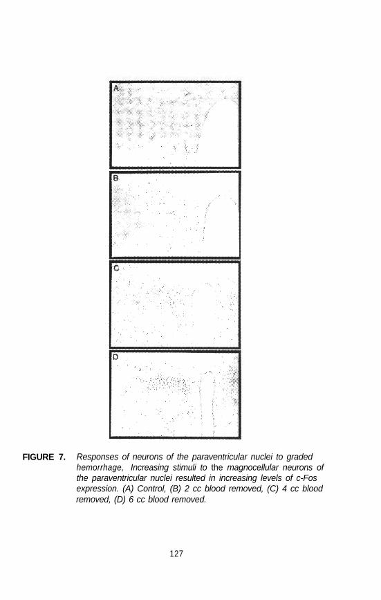
FIGURE 7. Responses of neurons of the paraventricular nuclei to gradedhemorrhage, Increasing stimuli to the magnocellular neurons ofthe paraventricular nuclei resulted in increasing levels of c-Fosexpression. (A) Control, (B) 2 cc blood removed, (C) 4 cc bloodremoved, (D) 6 cc blood removed.
127

strategies were identical to those described above for LHRH and c-Fos.Approximately half the dopamine neurons within the arcuate nucleus of diestrusrats expressed FRA immunoreactivity (figure 8, panel A). In contrast, FRAimmunoreactivity was absent in arcuate dopamine neurons from lactating ratssuckling their pups (figure 8, panel B). These data demonstrate that FRAimmunoreactivity can reveal reductions in activity as a result of the sucklingstimulus within a specific inhibitory neuron population. The suckling stimulus,or consequences of hormonal changes resulting from suckling, appears toaffect a great many brain systems in addition to the dopamine neurons.Analysis of FRA immunoreactivity throughout the brain, including the neocortexand hippocampus, revealed a marked reduction in baseline levels in lactatingrats compared with cycling rats. An example of the pattern of FRA staining in
FIGURE 8. FRA expression in the dopamine neurons of the arcuate nucleusin (A) a diestrus and (B) a lactating female rat. FRA proteins,normally expressed within the dopamine neurons, are no longerexpressed in suckling rats.
128

FIGURE 9. Baseline FRA expression in the somatomotor cortex. (A) In thediestrus rat many neurons thoughout all cell layers express FRAs.(B) In the lactating rat the baseline FRA expression in the cerebralcortex is markedly reduced.
the neocortex of a diestrus rat and a lactating rat is shown in figure 9. Thesechanges are consistent with the attenuation of many responses to stressobserved in lactating rats and offer another example of the use of FRA stainingas a means of identifying neuronal populations whose activity diminishes inresponse to neuronal stimuli and/or hormonal changes,
REPRODUCTIVE STATE AS A VARIABLE IN STUDYING BRAINACTIVATION: LACTATION AND STIMULATION BY NMA
Lactating rats, in addition to showing blunted behavioral changes to stress, failto possess spontaneous pulses of LH release. In investigating the possiblemechanisms of this reproductive quiescence, the authors treated lactatinganimals with the excitatory amino acid N-methyl-aspartate (NMA) at doses thatstimulate LH secretion in cycling rats (Smith and Lee 1990). Attention in thesestudies was returned to c-Fos as a marker for stimulated activity. Lactating ratsand a group of diestrus female rats were fitted with jugular cannulae, and four
129

FIGURE 10. Induction of c-Fos in the cerebral cortex of an NMA-treated rat(A) Diestrus female rats show strong induction of c-Fos in thesomatomotor cortex following subconvulsive doses of NMA. (B)Similar treatment of lactating rats fails to induce c-Fos in thecerebral cortex. The patterns of c-Fos expression appeared tomirror the behavioral effects of NMA.
pulses of 40 mg/kg NMA were administered at 10-minute intervals. Ninetyminutes following the last dose of NMA, the rats were anesthetized andperfused for localization of c-Fos. In the diestrus rats, administration of NMAelicited signs of hyperactivity, but no seizures. In contrast, administration ofNMA to lactating rats did not induce behavioral changes. Examination of c-Fosrevealed marked induction of c-Fos in neurons throughout the neural axis of thediestrus rats, whereas the same treatment of lactating rats did not induce c-Fosimmunoreactivity. Examples of c-Fos staining within the neocortex of a diestrusand lactating rat treated with NMA are shown in figure 10. Initial studiesexploring the recovery of excitability in lactating rats reveal that both thesuckling stimulus as well as progesterone participate in the dampening of NMAresponsiveness (Abbud et al. 1991). These studies not only provide anotherexample of IEG products as effective tools for studying neuronal activation butalso illustrate the importance of the animal’s endocrine state in determining theeffects of drugs on the nervous system.
130

REFERENCES
Abbud, R.; Lee, W.-S.; Hoffman, G.E.; and Smith, MS. Hippocampal andneocortical refractoriness to NMA stimulation in the lactating rat as revealedby c-Fos expression: Recovery after pup removal and blockade ofprogesterone receptors. Soc Neurosci Abstr 17:907, 1991.
Ceccatelli, S.; Villar, M.J.; Goldstein, M.; and Hokfelt, T. Expression of c-fosimmunoreactivity in transmitter characterized neurons after stress. Proc Nat/Acad Sci U S A 86:9569-9573, 1989.
Dragunow, M., and Faull, R.L.M. MK-801 induces c-fos protein in thalamicand neocortical neurons of rat brain. Neurosci Lett 11:39-45, 1990.
Hoffman, G.E. Organization of LHRH cells: Differential apposition ofneurotensin, substance P and catecholamine axons. Peptides 6:439-461,1985.
Hoffman, G.E., and Gibbs, F.P. LHRH pathways in the rat brain:Deafferentiation spares a subchiasmatic LHRH projection to the medianeminence. Neuroscience 7:1979-1993, 1982.
Hoffman, G.E.; Lee, W.; Attardi, B.; Yann, V.; and Fitzsimmons, M.D. LHRHneurons express c-fos after steroid activation. Endocrinology 126: 1736.1741, 1990.
Jacobson, L.; Sharp, F.R.; and Dallman, M.F. Induction of fos-likeimmunoreactivity in hypothalamic corticotropin-releasing factor neuronsafter adrenalectomy in the rat. Endocrinology 126:1709-1719, 1990.
Lee, W.-S.; Smith, M.S.; and Hoffman, G.E. Progesterone enhances the surgeof luteinizing hormone by increasing the activation of luteinizing hormone-releasing hormone neurons, Endocrinology 127:2604-2606, 1990a.
Lee, W.-S.; Smith, M.S.; and Hoffman, G.E. Luteinizing hormone-releasinghormone (LHRH) neurons express c-Fos during the proestrous LH surge.Proc Natl Acad Sci U S A 87:5163.5167, 1990b.
Lee, W.-S.; Smith, MS.; and Hoffman, G.E. c-Fos activity identifies recruitmentof LHRH neurons during ascending phase of proestrous luteinizing hormonesurge. J Neuroendocrinol, in press.
Levine, J.E., and Ramirez, V.D. Luteinizing hormone-releasing hormonerelease during the rat estrous cycle and after ovariectomy, as estimatedwith push-pull cannulae. Endocrinology 111:1439-1448, 1982.
Menetrey, D.; Gannon, A.; Levine, J.D.; and Basbaum, A.I. Expression of c-fosprotein in interneurons and projection neurons of the rat spinal cord inresponse to noxious somatic, articular, and visceral stimulation. J CompNeurol 285:177-195, 1989.
Merchenthaler, I.; Setalo, G.; Gores, T.; Petrusz, P.; and Flerko, B. Combinedretrogradetracing and immunocytochemical identification of luteinizinghormone-releasing hormone- and somatostatin-containing neurons projectingto the median eminence of the rat. Endocrinology 125:2812-2821, 1989.
131

Moenter, S.M.; Caraty, A.; and Karsch, F.J. The estradiol-induced surge ofgonadotropin-releasing hormone in the ewe. Endocrinology 127:1375-1384,1990.
Moore, K.E. Interactions between prolactin and dopaminergic neurons. BiolReprod 36:47-56, 1987.
Park, O., and Ramirez, V. Spontaneous changes in LHRH release during therat estrous cycle, as measured with repetitive push-pull perfusions of thepituitary gland in the same female rats. Neuroendocrinology 50:66-72, 1989.
Roberts, M.M.; Robinson, A.G.; Fitzsimmons, M.D.; Lee, W.-S.; and Hoffman,G.E. c-Fos expression in vasopressin and oxytocin neurons revealsfunctional heterogeneity within magnocellular neurons. Neuroendocrinology,submitted for publication.
Sarkar, D.K.; Chiappa, S.A.; and Fink, G. Gonadotropin-releasing hormonesurge in pro-oestrous rats. Nature 264:461-466, 1976.
Sheng, M., and Greenberg, M. The regulation and function of c-fos andother IEGs in the nervous system. Neuron 4:477-485, 1990.
Smith, M.S., and Lee, W.-S. c-Fos expression as a marker of neuronalactivation in response to the suckling stimulus. (Abstract 1057.) EndocrineSoc 72:289, 1990.
Watson, R.E.; Wiegand, S.J.; Clough, R.W.; and Hoffman, G.E. Use ofcryoprotectant to maintain long-term peptide immunoreactivity and tissuemorphology. Peptides 7:155-159, 1986.
Witkin, J.W.; Paden, CM.; and Silverman, A.J. The luteinizing hormone-releasing hormone (LHRH) systems in the rat brain, Neuroendocrinology35:429-438, 1982.
Wray, S., and Hoffman, G.E. A developmental study of the quantitativedistribution of LHRH neurons in postnatal male and female rats. J CampNeural 252:522-531, 1986a.
Wray, S., and Hoffman, G.E. Postnatal morphological changes in rat LHRHneurons correlated with sexual maturation, Neuroendocrinology 43:93-97,1986b.
ACKNOWLEDGMENTS
The studies reported in this manuscript were supported by National Institutes ofHealth grants NS-27014, NS-28730, HD-14643, and HD-21350 and NationalScience Foundation grant BNS-8919953. Thomas C. Waters providedphotographic assistance.
AUTHORS
Gloria E. Hoffman, Ph.D.Associate Professor
132
Wen-Sen Lee, Ph.D.Research Associate
M. Susan Smith, Ph.D.Professor
Rula Abbud
Department of PhysiologyUniversity of Pittsburgh School of MedicineEl 440 Biomedical Science Tower3500 Terrace StreetPittsburgh, PA 15261
Michelle M. Roberts, M.D.Associate Professor
Alan G. Robinson, M.D.Professor
Joseph G. Verbalis, M.A.Professor
Department of MedicineUniversity of Pittsburgh School of MedicineEl 140 Biomedical Science Tower3500 Terrace StreetPittsburgh, PA 15261
133