structure and mapping of the fos b gene. fosb downregulates the activity of the fos b promotero
TRANSCRIPT
Nucleic Acids Research, Vol. 20, No. 2 343-350
Structure and mapping of the fosB gene. FosBdownregulates the activity of the fosB promoter
Pedro S.Lazo+, Karen Dorfman, Tetsuro Noguchi, Marie-Genevieve Matteil and Rodrigo Bravo*Department of Molecular Biology, Bristol-Myers Squibb Pharmaceutical Research Institute,PO Box 4000, Princeton, NJ 08543-74000, USA and 'Institut National de la Sante et de laRecherche Medicale U242, Centre de Genetique Medicale, HOpital d'Enfants de la Timone,13385 Marseille, France
Received August 13, 1991; Revised and Accepted November 11, 1991
ABSTRACT
We have determined the genomic structure of the fosBgene and shown that it consists of 4 exons and 3introns at positions also found in the c-fos gene. Bydeletion analysis we have characterized a regionupstream of the TATA box which is the promoter regionof the gene. Several consensus sequences have beenidentified, including an SRE and AP-1 binding sitewhose relative positions are identical to those in the5' upstream region of the c-fos gene. We have alsoshown that FosB and c-Fos can downregulate theactivity of the fosB promoter to a similar extent. ThefosB gene is located in the [Al -B1] region of mousechromosome 7.
INTRODUCTION
Growth factors and other mitogens are capable of rapidly inducingthe expression of a set of genes in quiescent fibroblasts. Thesegenes which have been named immediate early genes encode fortranscription factors, cytoskeletal proteins, cytokines and otherproteins of yet unknown functions (for review 1, 3). The bonafide or putative transcription factors encoded by the immediateearly genes that have been identified so far include two relatedproteins containing zinc fingers, Krox-20/Egr-2 (4-6) andKrox-24 (7, 8), also named Zif/268 (9-11), NGFIA (12) andEgr-1 (13), a nuclear receptor, N1O (14) also named Nur/77 (15)and NGFIB (16), c-Rel (17), and the different members of theJun and Fos families, such as c-Jun (18, 19), JunB (20), JunD(21, 22), c-Fos (23, 24), FosB (25), Fra-1 (26) and Fra-2 (27,28). The initial observation that c-Jun and c-Fos can formheterodimers (29- 34) has been extended to the differentmembers of these families. c-Fos (35) as well as FosB (25) andFra-l (36, 37) can associate with the three Jun proteins enhancingtheir DNA binding to AP- 1 sites indicating that the transcriptionfactor AP-1 comprises a collection of related inducible proteincomplexes that interacts with similar sequence motifs. Thereforeit would be of interest to understand the mechanisms controlling
GenBank accession no. M77748
the expression of the different components of the AP-1 activityespecially considering that stimulation of cell proliferation inducesthe co-expression of these proteins leading to the formation ofdifferent Jun:Fos heterodimers in the cell (38). The c-fos genehas been the most extensively studied of all the immediate earlygenes and it has been demonstrated that its activation is in partmediated by upstream cis-elements, some of which have beenalso found in other immediate early genes (reviewed in 39).One of the members of the fos family, fosB (25), presents a
rapid and transient kinetics of induction similar to c-fos (24), incontrast to fra-1 (26) and fra-2 (27, 28) which have a moredelayed response. Therefore it was of interest to characterize thegene structure offosB in order to determine its possible similaritywith c-fos and to study the upstream elements involved in thecontrol of its expression. We have shown that the structure ofthe fosB gene is very similar to that of c-fos and that they sharethe cis-elements CRE, SRE, AP-1 and NF-1 in the 5' upstreamregion. The expression of thefosB gene is transrepressed by FosBand c-Fos proteins.
MATERIALS AND METHODSCell cultureNIH 3T3 cells were routinely grown in Dulbecco's modifiedEagle's medium (DMEM) supplemented with 10% fetal calfserum (FCS) and antibiotics (100 U/ml penicillin, 50 ug/mlstreptomycin). Confluent cells were made quiescent by incubatingthem for 48 h in 0.5% FCS. For stimulation, quiescent cells wereincubated in 20% FCS for the indicated periods of time.
Cell transfectionsNIH 3T3 cells were plated in DMEM supplemented with 10%FCS at 4x 10⁵ cells per 100 mm diameter dishes 20 h beforeDNA transfection. Cells were transfected with the different CAT-constructs by the calcium phosphate coprecipitation method (40)and exposed to the precipitate for 12 to 16 h, washed twice inPBS and kept in 0.5% FCS for 36 to 48 h. Cells were stimulated
* To whom correspondence should be addressed
'Present address: Departmento Biologia Funcional, Facultad de Medicina, E-33071 Oviedo, Spain
.=) 1992 Oxford University Press

344 Nucleic Acids Research, Vol. 20, No. 2
for 3 h with 20% FCS before harvesting. In all cases the plasmidpCH110 containing the 3-galactosidase gene under the controlof the simian virus 40 early promoter (41) was used as internalcontrol of the efficiency of transfection. Typically, 2 ,ug ofreporter plasmid and 3 ,lg of pCH110 were used. The totalamount of DNA used for the transfections was kept at 20 ,ug byadding pUC 18.For transrepression experiments, cells were exposed to the
precipitate for 4 to 6 h, followed by a 2 min 15% glycerol shockand then washed twice with PBS. In this case, the total amountof transfected DNA was always adjusted to 15 ,ug with pUC 18.After transfection, cells were made quiescent by incubating inDMEM containing 0.5 % FCS for 36 to 48 h. Three hours beforeharvesting, cells were stimulated with 20% FCS.The CAT and ,B-galactosidase activities were determined as
described (42, 43). The results were quantitated by cutting outand counting the spots in a liquid scintillation counter. All resultswere standardized according to the internal levels of f-galactosidase and/or to protein content.
Genomic library screening and Southern blottingTwo hundred micrograms of mouse liver genomic DNA werepartially digested with EcoRI. The digested DNA was sizefractionated in a linear 15% to 40% sucrose gradient bycentrifugation in a SW28 (Beckman) rotor for 20 h at 22,000rpm at 15°C. Ten micrograms of size-fractioned DNA (25 to45 kb) were used for ligation into the vector pHC79 (GIBCO-BRL; EcoRI digested and phosphatase treated) and the librarywas packaged using in vitro lambda packaging system(Amersham). The bacterial strain used for transformation wasDH5a MCR (GIBCO-BRL). A total of 1.5 x 10⁵ colonies werescreened as previously described (44). A 3.1 kb fosB cDNArandom labeled to a specific activity of 1 x 10⁹ cpm/Ijg was usedas a probe (45, 46). For Southern blotting experiments, DNArestriction fragments were separated on 1.2 % agarose gels andtransferred to a nitrocellulose membrane (47). Hybridization wascarried out in 50% formamide, 0.5 % SDS, Sx SSC (1 x SSC =150 mM sodium chloride, 15 mM sodium citrate) andS x Denhardts solution at 42°C for 12 to 16 h. Filters wereextensively washed in 0.1 x SSC containing 0.5 % SDS at 60°C.
Ribonuclease protection assayA BamHI-HindIII fragment comprising nucleotides -380 to+ 309 offosB genomic DNA was cloned into Bluescript KS (+)plasmid. 0.5 ,ug of cloned DNA was in vitro transcribed by T3RNA polymerase in the presence of 100 ICi of [32P]-UTP (800Ci/m mol) for 60 min at 37°C. Then the DNA was digested with50 jtg of RNase-free DNase I for 15 min at 37°C andsubsequently the RNA was extracted withphenol/chloroform/isoamyl alcohol and precipitated with ethanolusing 20 ,ug of tRNA as a carrier. The specific activity of theRNA obtained was approximately 2 x 10⁸ cpm/,ug. The probeRNA (2 x 10⁶ cpm total) was then hybridized with 10 yg of totalRNA from serum stimulated NIH 3T3 cells or 10 tig of tRNAas a control at 45°C overnight after being denatured at 90°Cfor 5 min. The hybridized RNAs were then digested with 2 Agof DNase-free Ribonuclease A and 1.2 /Ag of Ribonuclease T1for 60 min at 30°C. The reaction was terminated by additionof SDS (0.5% final) and digestion with 0.1 ,ug of proteinase Kfor 15 min at 37°C. After extraction and precipitation theribonuclease digested products were analyzed on a denaturating6% polyacrylamide sequencing gel.
Construction of fosB-CAT plasmidsTo construct the -962-fosB-CAT plasmid, a PstI-HindIIIfragment of approximately 1.3 kb comprising nucleotides -962to 308 of the fosB genomic DNA was cloned into the vectorpBLCAT3 (48). The otherfosB-CAT plasmids were constructedby PCR amplifying the desired fragments and then cloning inPstI-KpnI sites of the pBLCAT vector. The oligonucleotidescorresponding to the promoter region of thefosB gene used forPCR amplification were: 5'AGCACATTTTCAAGCTCACCG-AG 3' (nucleotides - 884 to - 862); 5'AGCGGCGGGCGGG-TTGCCACAG 3' (nucleotides -535 to -514);5'AGGATACCAAACAAACACTGGG 3' (nucleotides -465 to-444); 5'ATATGGCTAATTGCGTCACAGG 3' (nucleotides-424 to -403); 5'CAGGAACTCCGGGGAGGGCGGGG 3'(nucleotides -406 to -384); 5'CAGAACGCAGCCTTGGG-GACCCCCG 3' (nucleotides - 358 to -334); 5'AGTTGCGG-GTGACCGAAGCGCGGG 3' (nucleotides -225 to -202);5'GCAGCTCCGTTTCATTCATAAGACTG 3' (nucleotides- 170 to - 145); 3'GAGGCTTGGCCTCTGATT 5' (nucleotides292 to 309).
All these oligonucleotides contain in their 5' end an extra PstIsite and in their 3' end an extra KpnI site. The PCR productswere ligated into PstI-KpnI sites of the vector pBLCAT2. Allconstructs were confirmed by sequencing (49).The c-fos DSE- andfosB SRE-CAT constructs were obtained
by cloning into Sall sites of the pBLCAT2 vector (48) thefollowing oligonucleotides:5'TCGACAGGATGTCCATATTAGGACATCTGC3'3'GTCCTACAGGTATAATCCTGTAGACGCAGCT5'and5'TCGACTGGCGAGCTCCTTATATGGCTAATTGCG3'3'GACCGCTCGAGGAATATACCGATTAACGCAGCT5'respectively.
Chromosome spread preparationIn situ hybridization experiments were carried out usingmetaphase spreads from a WMP male mouse in which all theautosomes except 19 were in the form of metacentric robertsoniantranslocations. Concanavalin A-stimulated lymphocytes werecultivated at 37°C for 72 h with 5-bromodeoxyuridine added forthe final 6 h of culture (60 yIg/ml of medium), to ensure achromosomal R-banding of good quality. The 2.6 kbfosB clonein pGEM1 was tritium labeled by nick-translation (50) to aspecific activity of 1.3 x10⁸ dpm/,tg. The radiolabeled probewas hybridized to metaphase spreads at a final concentration of100 yig/ml of hybridization solution as previously described (51).After coating with nuclear track emulsion (NTB2, KODAK),
the slides were exposed for 21 days at 4°C, then developed. Toavoid any slipping of silver grains during the banding procedure,chromosome spreads were first stained with buffered Giemsasolution and metaphases photographed. R-banding was thenperformed by the fluorochrome-photolysis-Giemsa method andmetaphases rephotographed before analysis.
Gel retardation assayThe annealing of the two strands of a corresponding double-stranded oligonucleotide was performed in 10 mM Tris-HCl pH7.5, 1 mM EDTA and 50 mM NaCl, starting at 95°C for 10min and cooling down slowly to room temperature. The sequenceof the DSE and SRE oligonucleotides used for the gel retardationassays, have been described above under construction offosB-CAT plasmids.

Nucleic Acids Research, Vol. 20, No. 2 345
One pmol of the oligonucleotide was end-labeled with[32P]-dCTP by filling in the overlapping ends using the Klenowfragment of DNA polymerase I. Six pmoles of the radioactivedeoxynucleotide were used to assure complete labeling of thedouble-stranded oligonucleotide. In vitro translated proteins (0.5to 1 ll) were added to Dignam's buffer D (18 /il) containing 5mM spermidine and 1 mg/ml BSA and if not otherwise indicated,1 itg of pUC 19. The DNA-protein complexes were resolved ona 7% polyacrylamide gel (39:1; acrylamide: bisacrylamide) in0.25 xTBE (20 mM Tris-boric acid pH 8.3, 0.25 mM EDTA)run at room temperature for 16 h at IOOV.
-967 CTGCAGTCCCCCAGATTCAGGGACCCGGAAAAGCCACCTATCCTTTGCTTAGCYP&TT-907 CMGAAGTTTAGATCAGGGAGGGAGCACAT.TTCAAGCTCACCGAGTCACACAGAATCGC
-847 TGACAAACCTAGTGGGACGCACTGCATCCTTACCCATCCCTACACCTGGAGATATCACAG
-767 ACTCTCCAATTCCTCCTGAGCCTCAGTTTCCCCACCTAGACTGATTATACCCCTCTCGCC
-727 AGAATCTGCACTGGCCGACCT?OCTCTTTCACCAGCAGGGGCGCTGCACCGGTTTGGGG
-667 AGGGTGGGGTCCAGGGGTATAAGCAGACC?TG06ACTGGATTGCACC?TCTCCAACCCG
-607 GGTCACCACGGGCCTTCTOCAGGGAGTTAGGCGCCTGTCAATCTCAGCTCCGGGACAGCG
-547 TGGAACTGCGCAAGCGCC TrCCACA GCACArCGCCGC A c
-487 GATTCCCTC GT SAGCCAAACCCTGCCCCCCTGCGAGCC
-427 _T7I --fp427C-ATAT7At [gT &AGGCAACTCC CC_CCTCCCCTCCG
-367 GAA-CCCCTCAGAAC-C-CCCTTYGGGACCCCCCACOACCCCCAGGCTCACACTATGGGCA
-307 GGTGGCAGGTGCACGAGGCCCTGGAGGCCACAACGCTCAGTCGGCGGCAAGCGAG-247 AGTTTGGCAGGTTTGTGCATGOAO?TGCGGGTGACC CCCGGA
-107 GGCATAAATTCAGCCCGGCAGCTCC 0TTTCATTCATAAGACTGGAGCG
-127 GCG CTTGCTGGACTTGACTACTGGTGACTCTTT?TrTTCTTGGAGC GT*G s TATAtAAAASttTSAA_TCTCTT
-67 GTGGGTTTC
-7 CTTTTTGCAAGIACTCTTTCTAGATCGTTCTTCCGATTCGTCCGGAGATAGACTTTCTTTC
53
113
173
233
293
353
413
473
533
593653713773133193953
1013107311331193125316131373143314931SS316131673173317931531913197320332093216322132273
2333
2393
245S3
2513
2573
2633
CACAGGGGCCCCCCTGTGCCCAGGGAAATGTTTCAAGCTTTTCCCGGAGACTACGACTCCa Ga a 1 6 3D Y D a
GGCTCCCGGTGTAGCTCATCACCCTCCGCCGAGTCTCAGTACCTGTCTTCGGTGGACTCC0 S a C 8 8 S * 8 a 2 8 2 Y L S S V D 2
TTCGGCAGTCCACCCACCGCCGCCGCCTCCCAGGTAAGTTTTAGATCGTAAAGATTCTACIP 0 9 P * 7 a a a 9 2
TTTAGTGGTTGGGGGGGGGGGTGTTTCTTTCAGGCTAAAGrTAGATTGAGCATCTCTCAAATAGAGACGCGCCAAAACCAGGATCTGGATTGCAATACTTCGGTTTGCACTTTGGTTTTTTGTTGTTTGCAAACTGCAAAGAATGGAGTGATGTTTGCAAAAGGTTATTTCGGAGCGCGGAAAGGAATGCACCTGGGCAAACGTT0CCGATGCCGGTGCAAAGTATATACCCGCTGCGTTAGCAGAAGCTGAGAACTTTTAGCCGAAGCCGGCTCCCTAAGCCGAACCTAGGCAACTAGGOGAAGAAAAAGAAAAAAAAAATCACACAOAAGS?TCCACAGCCTCSTCCTCTTCCCTCTTCCTTCAAAGGACTGCAAGTCCGCAGTCACCCTCCACCCAGCAACAGTTAOGGCCTCGAACCCCGCTCACGCTGCCTCCCCTCCT0CCGAACGTAACGGGGGACCCGTGCGTAAAGCGTGACGCOCTGGAATCCTCCGTCTGACGCGGGGCACGCACAGGCCGCAGCCCCTCCGCCCGCCCCGCCCCTGACGTCCGGGCACGTTCTATTTTGCAACGCCGAGCGCCACGTTGCTAAGGAGGGGGCAGCGTGGCTTTGTGATTGGCTGTCGCGCGCACCTTTACCCAATCAGCGTTCCCTTCCTATTTGTAGAGCGTAGCTCCCTTCCTTGC??TTTTTGGTTCTTCCCGTGCTGGGGGTCTCCAAGAGGAGAGCTAGGATTCTTGTC0CGATCGGGACTCGTTGTCACCCCATGGTCTGCGACGACTTGTGTGGACCTGGTCTGTTGTCATAAGCTAGACOCTmGGCTGACTGTTAGCGCCTCTAAGGGGGAACTGAAGGCCTCATCCTTCTCAGGCACACATATACGTGCTCCTGAGCTCTAGACACTCAGTCCTTCCGAOGTGTTCAAACACTAGATGAGCTAGCCTACGGAGAGGCAGCCAGGTGGTCTCTAAAAGGTCTGCCTTCCCTTACTTCCCAGGCTCTGATTGGCCAGGGATTCAGCCCTTCCCTCGCCACGCCCCCTACASTAGTTAAGCCTCtACTATTCCACTTGCGGGAAGGCGGGGGGGGGGGGCGTGATGGACGCTCCTTGGGGACGCAGATCCTATGTCACCCCATCCCCTGCAAGACAGTCTGAAGATTCTCGCTGTCACTTTTCTCTGCCTATCAGTTCACTGAAACCTGTCACTCTCACT0GCAAGAfACAGACACTCGGAAGGGATGCTCTCAACTCTTAGOCCGGTCCCCCAACACCGITTGAACTGGGATCTCCGCCTGCGGGAGCCCTCATGCAGTCGGGGGTGTGTTTCGTGTT AG GGG 11 CTCT CCCTCTCCCTCCCTCTGTGGTGGGGGTTGGGGGGTTTTGGCTCTCSATGTGTGTGAATGTCTGTCGCTCCATCCCGGGACTT0TCACCAOGTTCTCCAGCCTCCTCTCCCACCCACCCCCCCACACCTAAGAGTCACCAACCCGGGOTGTGATTCACCACCCGCTGGAACCGTGCAACCTTTCCCCAA AACC A AAACAGAAAC?TCCQTTAACCACTGC0TCACG?GThAGTGGAAGGOTGGGT0TTTGTC TTGCCT0TGACACACACATCCACACCCGCTCACCCTG?GCTCACTCACACGGTCGGTCTCTCTTATCTCTCTCTTGGCCTGCTCTG
TCGGTGGCTTTGTTTGTGTGTCTACGCCTGTGTGTGTATGTCTCACCCCGTAGGAGTGCG3 C A
CCGGTCTCGGGGAAATGCCCGGCTCCTTCOTOCCAACGGTCACCGCAATCACAACCAGCCa L a as *aO s 1 v *1 v I a I * a 2Q
AGGATCTTCAGTGGCTCGTGCAACCCACCCTCATCTCTTCCATGCOCCCAGTCCCAGGGGCD L W11 V Q M AL I 8 8 N a 038 6 2
AGCCACTGGCCTCCCAGCCTCCAGCTOTTGACCCTTATGACATGCCAGGAACCAGCTACT1 L A O 01 1 A V D 1 3D 3 1 0 T ST13
CAACCCCAGGCCTGAGTGCCTACAGCACTGGCGGGGCAAGCGGAAGTGGTGGGCCTTCAAT P1 L0 a Y TA 3 aA 0 86 0 P S3
CCAGCACAACCACCAGTGGACCTGTGTCTGCCCGTCCAGCCAGAGCCAGGCCTAGAAOAC*IIT361V3A3PARA31 3RS* t
RESULTS
Genomic structure of fosBTo isolate the fosB gene, a 2 kbfosB cDNA was used to screena mouse genomic cosmid library. Two clones were isolated from1.5 x 105 colonies screened. One of them, cosmid B2, whichcross-hybridized to probes from the 5' and 3' region of fosBcDNA, was further characterized. The region containing thefosBgene was defined by restriction mapping analysis and Southernblotting. The complete fosB cDNA clone was demonstrated tobe included in a 8 kb EcoRI fragment. This was subcloned and
2693
275328132873293329933053
3113
3173
3233
32933353341334733533359336533713377338333.93395340134073413341934253431343734433
4493
11
4553
314613
424673
4733
4793
4853
4913
4 973
503350935153S21352735333539354535513
46 1573563356936753
665713567359335993
S5 6053611361736233
lOS 6293635364136473
125S 6533659366536713
145 67736933
6893
CCCGAGAAGAGACAGTAAGTATGAGGCCTCAGGAGTTGGGTGGAGGAGCCTAGCTAGCG33as3
ATGTGGGCTCAGTTTGTACAGTGCCTTGCTGCCAT.CATGAAGATCCCTAGCACACCATAAGCCAGGAGTGGTTATGCAGACCTGTAACCCCAGCTCTCAGAAGGTGGAGGCAGGAGGAGCAGGAGTTCGAGGCCAGCCTGTGCTACTTATGGAGTCCAGCCTGCACTGCAAGAGATCATTATTTTCAAAAGTTGGCCTTGGGGGGAGGTGGGTGAGGGAAGTAAGAGAAAGTCACTAGATTTTGTGACTTAATAGTTGGAGGTTCCTCTGAGGCCTCAAGTCTGAAGGAACTTTACCATTCSGCCAGTGAGGAGTAGGGGTTATTATTGGGGTTCGC G
GGGCTGATAAGGTACCCCCAGATCTCATGGTCCTTATCTCTGACTCAGCTTACCCCAGA
ACGAAAACCCAAGCGAAGCGTTCGCAGAGAGCGGAACAAGCT¢GCTGCAGCTAAGTGCAG3 3 3 3R a V R a R N3 3 L Aa aZ C R
GAACCGTCGGAGGGAGCTGACAGATCGACTTCAGGCGGTAAGGAGGAGTCTGGCCCTGTC3 R R A 3 L3 D a L Q A
TTGAGGCCGTGCTGGGAGCACTCTGCCTTGTTCTTCCCCCGTTTCTCACTGTGCCTGTGTCCTAAACGAGGAAACCCCCTCTTAGGGAACAGGGGTCAGTATAGGCTGATGGAGCOGCTCCATATGCATQCTCAGACCCAT.CCCACTTACTTTCGACTGTTCCCCACTTTCCCTGAATATGTCCCCACATGTCACCCTCCTGGCTTTCTCTCAGCCTAAGGAGACAACTAGAGGAGGTAATTCTCTCACCTTCTTTTCTTCACTAAATAATAATCCATTTTGCCTTCCTGCCTCCATTTTTTTTTCCTGAGCTGGGCTACCTGTCGTAGTTCAGCCCTCCTCCCCCAACTTGATAGCCTCAAGTTTCAGCCCTTGGCTGAGATGCCATCATCCTGACTGGCCTGCGCTGGAAACTATTTTGTGCTAATCAATTCCTTGTCTGCTACTTCAGCTATCTACCCTCCCGAACTTGAGCTGGTGGCGCCCACCAAGCCCACTTCTTTCTCTCTTTTTTACCTCAGTGCAACCCCCCACACACAAAACT?CATGCCTGCCCTTGAAACCAGGGT@CGTCTCTGACTCCCCGTCGGGAGGC?GAA AACAACCTCA TTAAMACAACTAAtTACCTAC1GACTCAACAAACTGTAGTGTTTTTCTTTTTTCCTCTCAAAAATTAATTCGTTTGTTTATTTATTATTTGCTTATGTTTGAGTCACTGCTGGTGCACCACAGCACACATACGAGGTCACAGGGAAATTTTCATACTTTGTTCTCTCCTTCCGTGTTGTGGCTGCTTGCTGGCAATCTCCTTCACTCAGTGAGCTACAATGCCCCCTTCTGCCCTTTAAGGCAGAGTACTCCTTACTACAGGGGGACCCTTTCCTCGGCCTCTCAAAGTTGAGATTACAAATGTTCACCATCACACCAGGCTTGGAGTTCTTGCCTATCAGTGACGTCCACTCCTGCCTAGCTCTTCCCAACCATCTTTAGTCTGATGGGGAAACCGAGGCACGAGTAGCATGGTCTACCAGGATTTCCTCTTAGGGGACGCTCCCCTCAGTTGGCAGGGAOCTGTCCAGCCCCCTGGATCAGCAGCAAOAATGTATGAGTGTGGGGTTGGGCGGGTGAAGCTACTCTGTGTGGTCGCTGACCAGCAATTCTCCTTTCTCTGTCTCCTATGACCTGGCCCTGCTGGGATCCATTAAACATCAGCTTGAAGAGGA
3 1 D 3L 3 3a
AAAGGCAGAGCTGGAGTCGGAGATCGCCGAGCTGCAAAAAGAGAAGGAACGCCTGGAGTT3 A 3 L 3 * I a3 L Q 3 3 K3 R L 3 r
TGTCCTGGTGGCCCACAAACCGGGCTGCAAGATCCCCTACGAAGAGGGGCCGGGGCCAGGv L v Aa* x I aC *I 1 3 3 1 0 1aO a
CCCGCTGGCCGAIiTGAGAGATTTGCCAGGGTCAACATCCGCTAAGGAAGACGGCTTCGG1 L Aa3 v a D L 1 6 8s J 3 3 D a3 a
CTGGCTGCTGCCCCCCCCTCCACCACCGCCCCTGCCCTTCCAGAGCAGCCGAGACGCACC3 L L * P P P P * L 1*3 3Q 8 3a Aa
CCCCAACCTGACGGCTTCTCTCTTTACACACA1GTAAGTTCAACTCCTCGGCGACCCCTT* 3 L S A 3 L T a a a v 0 v L a D *1
CCCCGTTGTTAGCCCTTCGTACACTTCCTCGTTTGTCCTCACCTCCCCGGAGGTCTCCGC* v v a * a SY a a1 v L I C 1 a v3 A
GTTCGCCGGCGCCCAACGCACCAGCGGCAGCGAGCAGCCGTCCGACCCGCTGAACTCGCC1 A 0 A o R * *1 * * Q P * D * L NP11
CTCCCTTCTTGCTCTGTAAACTCTTTAGACAAACAAAACAAACAAACCCGCAAGGAACAAS L L A L *
V
TTTTACAATCTATATCGTTGAG.AATTC
Figure 1. Nucleotide sequence of the fosB gene and of the 5' flanking region. The site of transcriptional initiation is indicated by an open triangle. The first and
last nucleotides corresponding to the fosB cDNA are indicated by closed triangles. The 3' untranslated region of exon 4 is not complete and ends in a natural EcoRIsite. The intron-exon boundaries are indicated by broken arrows. The TATA box and other elements are boxed. The arrowheads indicate the site for alternativesplicing which would generate fosB/sf.
149
153
173
185
193
213
233
253
273
293
313
333
339
346 Nucleic Acids Research, Vol. 20, No. 2
by detailed restriction mapping the presence of several intronswas detected. Then the 8 kb fragment was completely sequencedconfirming the presence of the introns (Figure 1). The preciselocation of the introns was established by comparison of thegenomic sequence tofosB cDNA. ThefosB gene encloses 4 exonsand 3 introns. The complete coding sequence is distributed inthe 4 exons in a genomic fragment of approximately 4.6 kb.The putative 5'-cap-nucleotide is located 40 nucleotides
downstream from a complex TATA box. As shown in Figure1, within the first 500 bases 5' upstream of the starting site ofthefosB gene a few cis-acting elements can be identified by theirsimilarity with consensus DNA motifs. There are one KRE-likesequence (Krox responsive element, 8), one AP- I -like sequence,one SRE, one CRE-like sequence, four Sp-1 binding sites andone NF-1-like site. There is another AP-1 like sequence atposition -909. It is interesting to note that the relative locationsof the SRE (-428 to -419) and the AP-1-like sequences (-413to -407) resemble very much that found in the 5' upstreamregion of the c-fos, Krox-20 and Krox-24 genes (11, 52, 53).The strict conservation of these elements possibly reflects theirimportance in the regulation of these genes.Comparison of the 5' untranslated regions of thefosB cDNA
clone AC 1 13-1 (61) and the homologous genomic regionrevealed a difference in the sequence. The first 881 nucleotidesof the AC 113-1 cDNA are not present in the corresponding5' genomic sequence, likely due to a cloning artifact. The identitybetween the 5' untranslated region of AC 113 -1 cDNA and thecorresponding genomic region ends in nucleotide 123 of thegenomic sequence. In light of this discrepancy we determined
ld
450 --
zn1,;
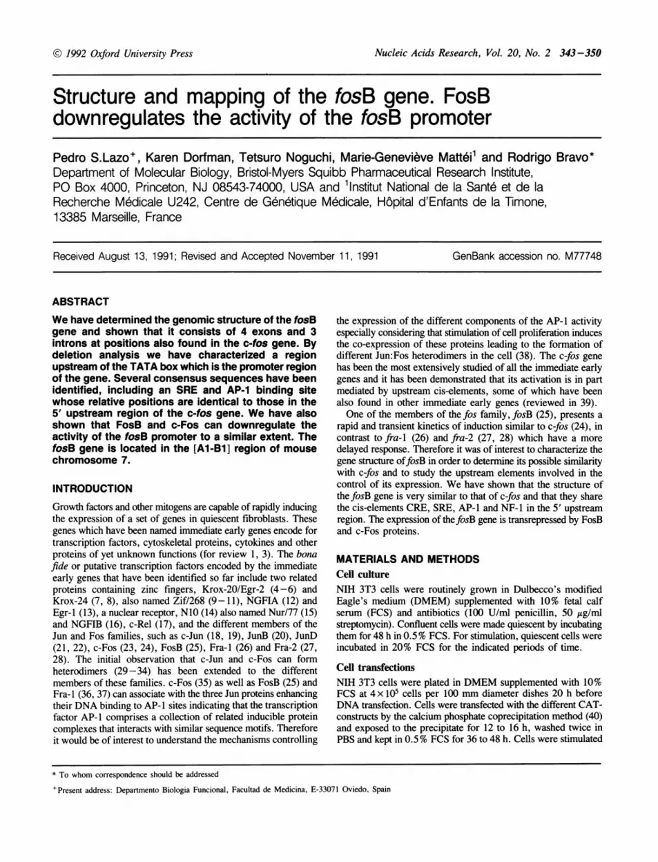
the starting site of fosB by ribonuclease protection assay. Forthis, a single-stranded RNA probe comprising nucleotides - 380to + 309 of the genomicfosB generated by in vitro transcriptionin the presence of [32P]-UTP (Figure 2, lane 1) was hybridizedagainst total RNA of serum-stimulated NIH 3T3 cells and thendigested with ribonuclease as described in Materials and Methods.As shown in Figure 2, lane 2, ribonuclease treatment of thehybridized labeled RNA generates a major protected fragmentof approximately 320 nucleotides, which is consistent with thelength expected considering the genomic fosB sequence. Thisconfirms that the major 5' cap-nucleotide is located 40 nucleotidesdownstream from a complex TATA box and demonstrates thatthe discrepancy found between the size of the 5' untranslatedregion of the AC 113 -1 cDNA clone (61) and that predicted fromthe genomic fosB sequence is the result of an artifact producedduring the construction of the cDNA library. These results alsoindicate that, although weaker, there are other possibletranscription initiation sites further upstream of the complexTATA box, as illustrated by the presence of protected fragmentsof approximately 420 and 470 nucleotides. Indeed, there are twosequences in thefosB genomic sequence located at positions -184to -175 and -157 to -150, approximately 120 and 90nucleotides upstream of the complex TATA box (-63 to -41)which resemble a TATA box. The protected fragments smaller320 nucleotides are possibly generated by the presence ofincomplete labeled transcripts.The first exon of thefosB gene, which is 565 nucleotides long,
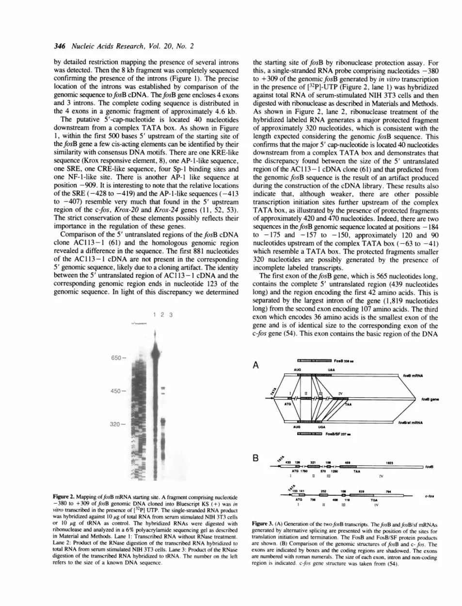
contains the complete 5' untranslated region (439 nucleotideslong) and the region encoding the first 42 amino acids. This isseparated by the largest intron of the gene (1,819 nucleotideslong) from the second exon encoding 107 amino acids. The thirdexon which encodes 36 amino acids is the smallest exon of thegene and is of identical size to the corresponding exon of thec-fos gene (54). This exon contains the basic region of the DNA
A FosB 338 as
AUG UAA
tosB gene
AUG UGA
==3=*****Z.3 FoB/SF 237_m
#a*
B I44433 126 321 106 459
ATG 1780 570 1250I U iu
tos/el mRNA
1923
TAAfosS
Figure 2. Mapping offosB mRNA starting site. A fragment comprising nucleotide-380 to +309 of fosB genomic DNA cloned into Bluescript KS (+) was invitro transcribed in the presence of [32p] UTP. The single-stranded RNA productwas hybridized against 10 1sg of total RNA from serum stimulated NIH 3T3 cellsor 10 isg of tRNA as control. The hybridized RNAs were digested withribonuclease and analyzed in a 6% polyacrylamide sequencing gel as describedin Material and Methods. Lane 1: Transcribed RNA without RNase treatment.Lane 2: Product of the RNase digestion of the transcribed RNA hybridized tototal RNA from serum stimulated NIH 3T3 cells. Lane 3: Product of the RNasedigestion of the transcribed RNA hybridized to tRNA. The number on the leftrefers to the size of a known DNA sequence.
53 141
ATG 756l
252 106 639
406 11611 IIn
- ct-osTGA
IV
Figure 3. (A) Generation of the twofosB transcripts. ThefosB andfosB/sf mRNAsgenerated by alternative splicing are presented with the position of the sites fortranslation initiation and termination. The FosB and FosB/SF protein productsare shown. (B) Comparison of the genomic structures offosB and c- fos. Theexons are indicated by boxes and the coding regions are shadowed. The exonsare numbered with roman numerals. The size of each exon, intron and non-codingregion is indicated. c-fos gene structure was taken from (54).
---

Nucleic Acids Research, Vol. 20, No. 2 347
binding domain and presents an 89% identity with c-fos. Thefourth exon is the longest of all. It encodes the last 153 aminoacids offosB protein and contains a fragment 1,923 nucleotideslong of the 3' untranslated region which is incomplete in ourgenomic subclone. Considering that the size of thefosB mRNAis approximately 5 kb, the genomic clone is missing nearly 1.7kb of the 3' region. Therefore the complete exon 4 would beapproximately 4 kb long, of which 3.6 kb would correspond tothe 3' untranslated region. This exon can be alternatively splicedat positions 4,685 and 4,825, giving rise to a truncated form offosB, fosBlsf, which generates a protein of 237 amino acids
A. 12
10
z
0 4
IO.2
0
B.
- 4z
2 22
0
C.
3z0F
0za-i
0
FCS
N0
pCATPO P1 P2 P3 P4 P5 i P6 P7 P8
PnGFlU)~~F.Pm
U)
N ,. CY
pCATPO P1 P2 P3 P4 P5 PS P7 PS
PMA
pCATPO P1 P2 P3 P4 PS P6 P7 P8DELETIONS
Figure 4. Induction ofCAT activity in cells transfected with differentfosB-CATplasmids after treatment with (A) 20% FCS, (B) 10 ng/ml of PDGF, and (C)100 ng/ml of PMA. NIH 3T3 cells were cotransfected with the indicatedfosB-CAT plasmids and the a-galactosidase plasmid pCH I 10 and were incubated for36 to 48 h in 0.5% FCS before stimulation for 3 h with the indicated agents.The CAT activity of each construct was determined using equivalent amountsof nuclear extracts and was normalized to the ,B-galactosidase activity of eachextract. The graphs indicate the fold induction of CAT activity respect to thenon-stimulated cells for each construct. The results represent the average of 6independent transfections.
(FosB/SF) and includes the basic and leucine zipper domain (55,56). The 5' and 3' deletion endpoints contain a typical acceptorand donor splice site respectively (57). Although the fosBlsftranscript is only missing 140 nucleotides, it encodes a protein101 amino acids shorter due to an in frame stop codon generatedin the alternative spliced site (Figure 1 and Figure 3A). Thisalternative splicing in exon 4 has not been observed in the othermembers of thefos family. That the overall structure of thefosBgene is very similar to that of c-fos strengthens the notion thatthey have evolved from a common ancestor gene. A similarstructure has been also reported for fra-2 (28). Both contain 4exons and 3 introns and their relative size has been conserved(Figure 3B). It is interesting to mention that the first 40 aminoacids encoded in exon 4 comprise the leucine zipper of FosBwhich has a 67% identity with the corresponding leucine zipperof c-Fos, also encoded in exon 4. In contrast, the remaining 113amino acids of FosB encoded in this exon present no significanthomology with c-Fos with the exception of the last 12 aminoacids that are highly conserved in the Fos protein family.
Effect of progressive deletions of fosB 5' upstream cis-elements in gene inductionTo determine if the 1 kb 5' upstream sequence of thefosB geneis sufficient for gene activation, a recombinantfosB-CAT fusiongene was used. To construct this plasmid (PO) the genomicfosBsequence (nucleotides -962 to 308) was ligated into the PstI-HindIll sites of the vector pBLCAT3 (48). NIH 3T3 cells weretransfected and tested for inducibility as described in Materialsand Methods. As shown in Figure 4 the -962-fosB-CAT (PO)presents marked stimulation of CAT expression by serum andplatelet-derived growth factor (PDGF) and to a lesser extent byphorbol 12-myristate 13-acetate (PMA). To investigate which isthe minimal 5' upstream fragment offosB that confers inducibilityto these different agents, several progressive deletionfosB-CATfusion genes were constructed (see Figure 5). The -884-fosB-CAT plasmid (P1), which lacks the AP-1-like sequence inposition -912 to -906, presents an approximately 6-foldstimulation of CAT expression by serum and PDGF comparedto only 2-fold stimulation with PMA. The induction of P1 issignificantly lower than that of the -962-fosB-CAT plasmid(PO), specially with serum, indicating that under these conditionsthe AP-1-like sequence plays a role in the induction of the gene.The stimulation of CAT expression in the -535-fosB-CATplasmid (P2) by the three agents is comparable to that of P1.This indicates that the region between nucleotides -883 and-536, of the 5' upstream region of the fosB gene, containingno recognizable consensus element, is not essential for the
AP-1
-W2 A
SRECRE AP-1
n &An&
KRE TATA boxA OA OA O
.kA A 0
A A 0
A c)A 0-
Figure 5. Schematic representation of the upstream elements of the fosB geneand the various fosB deletion mutants used. The fosB-CAT constructs weregenerated by standard recombinant DNA procedures as described in Materialsand Methods.
POP1P2P3P4P5PSP7PS
N
IF Mr%
W.C-i
CY
10': Cl Cj Ci
C)

348 Nucleic Acids Research, Vol. 20, No. 2
induction of the gene under these conditions. However, theconstruct -465-fosB-CAT (P3) which lacks the CRE-likesequence located in position -479 to -470 presents a modestbut reproducible reduction in its stimulation, indicating that thiselement contributes to the induction of thefosB gene. The mostdramatic decrease in stimulation is shown by the construct-424-fosB-CAT (P4), where the SRE has been eliminated, itshows a slight or no stimulation of CAT expression by thedifferent stimulants suggesting that this element plays an importantrole in the induction of the gene. This also demonstrates that theelements such as AP- 1, Sp- 1 and KRE-like which still remainin -424-fosB-CAT are not sufficient to confer inducibility tothe gene. Further deletion mutants show no induction of CATexpression.Activity and binding of fosB SREThe results presented above suggested that the SRE located inposition -428 to -419 may play an important role in theinduction of thefosB gene, therefore we determined whether theSRE was functional, and compared its activity with that of theSRE present in the c-fos gene (39). The oligonucleotide containingthe c-fos SRE also included the flanking symmetrical sequences,therefore we will refer to it as dyad symmetry element (DSE).Cells were transfected with the corresponding constructs andinduction of CAT activity by serum was determined. The fosB-SRECAT shows a 5-fold induction in CAT activity by serum(Figure 6A). This induction is similar to that observed with theconstruct -465-fosB-CAT which contains the SRE confirmingthe above observation that deletion of the SRE, as in the-424-fosB-CAT construct, eliminates the serum response.Comparison of the CAT activity to serum between thefosB-SRE-CAT and -962-fosB-CAT construct (PO) indicates that the SREcontributes to nearly half of the total induction by serum. Theresponse to serum confered by the c-fos DSE to CAT isconsistently higher than thefosB SRE, with 8-fold activation bythe DSE compared to a 5-fold by the SRE. This correlates withthe higher level of induction of the c-fos gene compared to thefosB gene in vivo.
As the activity of thefosB SRE is lower than that of the c-fosDSE, and as they share the core sequence CC(A or T)6GG, butthefosB SRE lacks the flanking symmetrical sequences presentin the c-fos DSE (reviewed in 39), we found it of interest todetermine whether the fosB SRE binds the serum responsivefactor (SRF) with a similar affinity to that of the c-fos DSE. Toperform these studies, in vitro translated SRF was incubated witha [32P] -labeled SRE- or DSE-containing oligonucleotide and theprotein/DNA complex was analyzed at room temperature usingthe gel retardation assay. The results show that fosB SRE bindsSRF very efficiently and competition studies using an unlabeledoligonucleotide containing SRE unrelated sequences demonstratethat the complex formed is specific (Figure SB). Competitionexperiments using different concentrations of unlabeled SRE orDSE show that they similarly compete the binding of the labeledSRE indicating that the binding affinity of SRF for both, DSE
z
0
z00
z
z0U
PO PO PO
c-aos fo*B
SRE SRE SRE
cfos fosB
Figure 7. (A) Transrepression of fosB promoter by FosB and c-Fos. The-962-fosB-CAT construct was transfected into NIH 3T3 cells alone orcotransfected with the expression vector pMexNeo containing eitherfosB or c-fos. After 36 to 48 h in 0.5% FCS cells were stimulated for 3 h with 20% FCS.(B) Transrepression offosB SRE by FosB and c-Fos. ThefosB-SRE-CAT plasmidwas transfected alone or cotransfected with either afosB or c-fos expressing vector.Cells were kept for 36 to 48 h in 0.5% FCS before stimulation with 20% FCS.An equivalent amount of nuclear extract was used and the percentage of conversionwas nominalized to that of galactosidase activity in each case.
*. 4
_ 1* _
Figure 6. (A) Induction of CAT activity in cells transfected with -962-fosB-CAT (PO), fosB-SRE or c-fos-DSE. NIH 3T3 cells were cotransfected with theindicated constructs and 13-galactosidase plasmid pCH 110 and were kept in 0.5%FCS for 36 h before stimulation for 3 h with 20% FCS. Equivalent amountsof nuclear extracts were used. The CAT activity was normalized to thegalactosidase activity of each extract. (B) Competition between fosB-SRE andc-fos-DSE for SRF. Binding reactions were carried out as described in Materialsand Methods using 1 P1 of in vitro translated SRF and 0.01 pmol of labeledfosB-SRE oligonucleotide per reaction. For competition studies the competitoroligonucleotide was premixed with the labeled oligonucleotide before adding SRF.C = binding of SRE in the absence of competitor oligonucleotide.
C
4.S~....
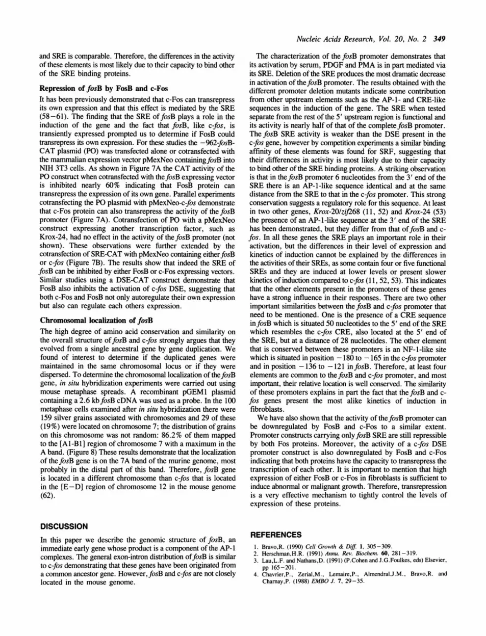
Figure 8. Localization of the fosB gene to mouse chromosome 7 by in situhybridization. (A) Two partial WMP mouse metaphases, showing the specificsite of hybridization to chromosome 7. Top, arrowheads indicate silver grainson Giemsa-stained chromosomes after autoradiography. Bottom, chromosomeswith silver grains were subsequently identified by R-banding. (B) Diagram ofWMP mouse Rb (7; 18) chromosome indicating the distribution of labeled sites.
9
M."a.
I..
Ei
Nucleic Acids Research, Vol. 20, No. 2 349
and SRE is comparable. Therefore, the differences in the activityof these elements is most likely due to their capacity to bind otherof the SRE binding proteins.
Repression of fosB by FosB and c-FosIt has been previously demonstrated that c-Fos can transrepressits own expression and that this effect is mediated by the SRE(58-61). The finding that the SRE offosB plays a role in theinduction of the gene and the fact that fosB, like c-fos, istransiently expressed prompted us to determine if FosB couldtransrepress its own expression. For these studies the -962-fosB-CAT plasmid (PO) was transfected alone or cotransfected withthe mammalian expression vector pMexNeo containingfosB intoNIH 3T3 cells. As shown in Figure 7A the CAT activity of thePO construct when cotransfected with thefosB expressing vectoris inhibited nearly 60% indicating that FosB protein cantransrepress the expression of its own gene. Parallel experimentscotransfecting the PO plasmid with pMexNeo-c-fos demonstratethat c-Fos protein can also transrepress the activity of the fosBpromoter (Figure 7A). Cotransfection of PO with a pMexNeoconstruct expressing another transcription factor, such as
Krox-24, had no effect in the activity of thefosB promoter (notshown). These observations were further extended by thecotransfection of SRE-CAT with pMexNeo containing eitherfosBor c-fos (Figure 7B). The results show that indeed the SRE offosB can be inhibited by either FosB or c-Fos expressing vectors.Similar studies using a DSE-CAT construct demonstrate thatFosB also inhibits the activation of c-fos DSE, suggesting thatboth c-Fos and FosB not only autoregulate their own expressionbut also can regulate each others expression.
Chromosomal localization of fosBThe high degree of amino acid conservation and similarity on
the overall structure offosB and c-fos strongly argues that theyevolved from a single ancestral gene by gene duplication. Wefound of interest to determine if the duplicated genes weremaintained in the same chromosomal locus or if they were
dispersed. To determine the chromosomal localization of thefosBgene, in situ hybridization experiments were carried out usingmouse metaphase spreads. A recombinant pGEM1 plasmidcontaining a 2.6 kbfosB cDNA was used as a probe. In the 100metaphase cells examined after in situ hybridization there were159 silver grains associated with chromosomes and 29 of these(19 %) were located on chromosome 7; the distribution of grainson this chromosome was not random: 86.2% of them mappedto the [Al-Bi ] region of chromosome 7 with a maximum in theA band. (Figure 8) These results demonstrate that the localizationof the fosB gene is on the 7A band of the murine genome, mostprobably in the distal part of this band. Therefore, fosB geneis located in a different chromosome than c-fos that is locatedin the [E-D] region of chromosome 12 in the mouse genome(62).
DISCUSSION
In this paper we describe the genomic structure of fosB, an
immediate early gene whose product is a component of the AP-1complexes. The general exon-intron distribution offosB is similarto c-fos demonstrating that these genes have been originated froma common ancestor gene. However, fosB and c-fos are not closelylocated in the mouse genome.
The characterization of the fosB promoter demonstrates thatits activation by serum, PDGF and PMA is in part mediated viaits SRE. Deletion of the SRE produces the most dramatic decreasein activation of thefosB promoter. The results obtained with thedifferent promoter deletion mutants indicate some contributionfrom other upstream elements such as the AP-1- and CRE-likesequences in the induction of the gene. The SRE when testedseparate from the rest of the 5' upstream region is functional andits activity is nearly half of that of the complete fosB promoter.The fosB SRE activity is weaker than the DSE present in thec-fos gene, however by competition experiments a similar bindingaffinity of these elements was found for SRF, suggesting thattheir differences in activity is most likely due to their capacityto bind other of the SRE binding proteins. A striking observationis that in thefosB promoter 6 nucleotides from the 3' end of theSRE there is an AP-l-like sequence identical and at the samedistance from the SRE to that in the c-fos promoter. This strongconservation suggests a regulatory role for this sequence. At leastin two other genes, Krox-201zif268 (11, 52) and Krox-24 (53)the presence of an AP-1-like sequence at the 3' end of the SREhas been demonstrated, but they differ from that offosB and c-fos. In all these genes the SRE plays an important role in theiractivation, but the differences in their level of expression andkinetics of induction cannot be explained by the differences inthe activities of their SREs, as some contain four or five functionalSREs and they are induced at lower levels or present slowerkinetics of induction compared to c-fos (11, 52, 53). This indicatesthat the other elements present in the promoters of these geneshave a strong influence in their responses. There are two otherimportant similarities between thefosB and c-fos promoter thatneed to be mentioned. One is the presence of a CRE sequenceinfosB which is situated 50 nucleotides to the 5' end of the SREwhich resembles the c-fos CRE, also located at the 5' end ofthe SRE, but at a distance of 28 nucleotides. The other elementthat is conserved between these promoters is an NF-1-like sitewhich is situated in position -180 to -165 in the c-fos promoterand in position -136 to -121 infosB. Therefore, at least fourelements are common to thefosB and c-fos promoter, and mostimportant, their relative location is well conserved. The similarityof these promoters explains in part the fact that thefosB and c-fos genes present the most alike kinetics of induction infibroblasts.We have also shown that the activity of thefosB promoter can
be downregulated by FosB and c-Fos to a similar extent.Promoter constructs carrying onlyfosB SRE are still repressibleby both Fos proteins. Moreover, the activity of a c-fos DSEpromoter construct is also downregulated by FosB and c-Fosindicating that both proteins have the capacity to transrepress thetranscription of each other. It is important to mention that highexpression of either FosB or c-Fos in fibroblasts is sufficient toinduce abnormal or malignant growth. Therefore, transrepressionis a very effective mechanism to tightly control the levels ofexpression of these proteins.
REFERENCES1. Bravo,R. (1990) Cell Growth & Diff 1, 305-309.2. Herschman,H.R. (1991) Annu. Rev. Biochem. 60, 281-319.3. Lau,L.F. and Nathans,D. (1991) (P.Cohen and J.G.Foulkes, eds) Elsevier,
pp 165-201.4. Chavrier,P., Zerial,M., Lemaire,P., Almendral,J.M., Bravo,R. and
Charnay,P. (1988) EMBO J. 7, 29-35.

350 Nucleic Acids Research, Vol. 20, No. 2
5. Chavrier,P., Vesque,C., Galliot,B., Vigneron,M., Dolle,P., Duboule,D.and Charnay,P. (1990) EMBO J. 9, 1209 -1218.
6. Joseph,L.J., Le Beau,M.M., Jamieson,G.A., Acharya,S., Shows,T.B.,Rowley,J.D. and Sukhatine,V.P. (1988) Proc. Natl. Acad. Sci. USA 85,7164-7168.
7. Lemaire,P., Revelant,O., Bravo,R. and Charnay,P. (1988) Proc. Natl. Acad.Sci. USA 85, 4691-4695.
8. Lemaire,P., Vesque,C., Schmitt,J., Stunnenberg,H., Frank, R. andCharnay,P. (1990) Mol. Cell. Biol. 10, 3456-3467.
9. Christy,B.A., Lau,L.F. and Nathans,D. (1988) Proc. Natl. Acad. Sci. USA85, 7857-7861.
10. Christy,B. and Nathans,D. (1989) Proc. Natl. Acad. Sci. USA 86,8737 -8741.
11. Christy,B. and Nathans,D. (1989) Mol. Cell. Biol. 9, 4889-4895.12. Milbrandt,J. (1987) Science 238, 797-799.13. Sukhatme,V.P., Cao,X., Chang,L.C., Tsai-Morris,C.H., Stamenkovich,D.,
Ferreira,P.C.P., Cohen,D.R., Edwards,S.A., Shows,T.B., Curran,T., LeBeau,M.M. and Adamson,E.D. (1988) Cell 53, 37-43.
14. Ryseck,R.-P., Macdonald-Bravo,H., Mattei,M.-G., Ruppert,S. and Bravo,R.(1989) EMBO J. 8, 3327-3335.
15. Hazel,T.G., Nathans,D. and Lau,L.F. (1988) Proc. Natl. Acad. Sci. USA85, 8444-8448.
16. Milbrandt,J. (1988) Neuron 1, 183-188.17. Bull,P., Hunter,T. and Verma,I.M. (1989) Mol. Cell. Biol. 9, 5239-5243.18. Ryder,K. and Nathans,D. (1988) Proc. Natl. Acad. Sci. USA 85, 8464-8467.19. Ryseck,R.-P., Hirai,S.I., Yaniv,M. and Bravo,R. (1988) Nature 334,
535 -537.20 Ryder,K., Lau,L.F. and Nathans,D. (1988) Proc. Natl. Acad. Sci. USA 85,
1487-1491.21. Hirai,S.I., Ryseck,R.-P., Mechte,F., Bravo,R. and Yaniv,M. (1989) EMBO
J. 8, 1433-1439.22. Ryder,K., Lanahan,A., Perez-Albueme,E. and Nathans,D. (1989) Proc. Natl.
Acad. Sci. USA 86, 1500-1503.23. Greenberg,M.E. and Ziff,E.B. (1984) Nature 311, 433-438.24. Maller,R., Bravo,R., Burckhardt,J. and Curran,T. (1984) Nature 312,
716-720.25. Zerial,M., Toschi,L., Ryseck,R.-P., Schuermann,M., Maller,R. and
Bravo,R. (1989) EMBO J. 8, 805-813.26. Cohen,D.R. and Curran,T. (1988) Mol. Cell. Biol. 8, 2063-2069.27. Matsui,M., Tokuhare,M., Konuma,Y., Nomura,N. and Ishizaki,R. (1990)
Oncogene 5, 249-255.28. Nishina,H., Sato,H., Suzuke,T., Sato,M. and Iba,H. (1990) Proc. Natl. Acad.
Sci. USA 87, 3619-3623.29. Chiu,R., Boyle,W.J., Meek,J., Smeal,T., Hunter,T. and Karin,M. (1988)
Cell 54, 541-552.30. Franza Jr.,B.R., Rauscher 1J,F.J., Josephs,S.F. and Curran,T. (1988) Science
239, 1150-1153.31. Halazonetis,T.D., Georgopoulos,K., Greenberg,M.E. and Leder,P. (1988)
Cell 55, 917-924.32. Kouzarides,T. and Ziff,E. (1988) Nature 336, 646-651.33. Rauscher,F.J., Cohen,D.R., Curran,T., Bos,T.J., Vogt,P.K., Bohmann,D.,
Tijian,R. and Franza,B.R. (1988) Science 240, 1010- 1016.34. Rauscher,F.J., Voulalas,P.J., Franza,B.R. and Curran,T. (1988) Genes Dev.
21, 1687-1699.35. Nakabeppu,Y., Ryder,K. and Nathans,D. (1988) Cell 55, 907-915.36. Cohen,D.R., Ferreira,P.C.P., Gentz,R., Franza, Jr.B.R. and Curran,T.
(1989) Genes Dev. 3, 173-184.37. Ryseck,R.-P. and Bravo,R. (1991) Oncogene 6, 533-542.38. Kovary,K. and Bravo,R. (1991) Mol. Cell. Biol. 11, 2451-2459.39. Treisman,R. (1990) Sem. in Cancer Biol. 1, 47-58.40. Van der Eb,A.J. and Graham,F.L. (1980) Meth. Enzymol. 65, 826-839.
41. Hall,C., Jacob,E., Ringold,G. and Lee,F. (1983) J. Mol. AppI. Genet. 2,
101- 109.42. Gorman,C.M., Moffat,L.F. and Howard,B.H. (1982) Mol. Cell. Biol. 2.
1044-1051.43. Miller,J.H. (1972) In Experiments in Molecular Genetics, pp. 352-355,
Cold Spring Harbor, N.Y.44. Maniatis,T., Fristsch,E.F. and Sambrook,J. (1982) Molecular Cloning, Cold
Spring Harbor Laboratory, Cold Spring Harbor, N.Y.
45. D'Eustachio,P. (1984) J. Exp. Med. 160, 827-838.46. Feinberg,A.P. and Vogelstein,B. (1984) Anal. Biochem. 132, 266-267.
47. Southern,E. (1979) Meth. Enzymol. 68, 152-176.48. Luckow,B. and Schiutz,G. (1987) Nucl. Acids Res. 1, 5490.
49. Sanger,F., Nicklen,S. and Coulson,A.R. (1977) Proc. Natl. Acad. Sci. USA
74, 5463-5467.
50. Rigby,P.W.J., Dieckmann,M., Rhodes,C. and Berg,P. (1977) J. Mol. Biol.113, 237-251.
51. Mattei,M.-G., Philip,N., Passage,E., Moisan,J.P., Mandel,J.L. andMattei,J.F. (1985) Hum. Genet. 69, 268-271.
52. Chavrier,P., Janssen-Timmen,U., Mattei,M.-G., Zerial,M., Bravo.R. andCharnay,P. (1989) Mol. Cell. Biol. 9, 787-797.
53. Janssen-Tinnnen,U., Lemaire,P., Mattei,M.-G., Revelant,O. and Charnay,P.(1989) Gene 80, 325-336.
54. Van Beveren,C., Van Straaten,F., Curran,T., Maller,R. and Vennaj. (1981)Cell 27, 97-108.
55. Nakabeppu,Y. and Nathans,D. (1991) Cell 64, 751 -759.56. Dobrzanski,P., Noguchi,T., Kovary,K., Rizzo,C.A., Lazo,P.S. and Bravo,R.
(1991) Mol. Cell. Biol. 11, 5470-5478.57. Mount,S.M. (1982) Nucl. Acids Res. 10, 459-472.58. Konig,H., Ponta,H., Rahmsdorf,U., Buscher,M., Schonthal,A.,
Rahmsdorf,H.J. and Herrlich,P. (1989) EMBO J. 8, 2559-2566.59. Lucibello,F.C., Lowag,C., Neuberg,M. and Muller,R. (1989) Cell 59,
999-1007.60. Ofir,R., Dwarki,V.J., Rashid,D. and Verina,I.M. (1990) Nature 348, 80-82.61. Sassone-Corsi,P., Sisson,J.C. and Venna,I.M. (1988) Nature 334, 314-319.62. D'Eustachio,P. (1984) J. Exp. Med. 160, 827-838.