brit j pharma res - smeera
TRANSCRIPT
____________________________________________________________________________________________
*Corresponding author: Email: [email protected];
British Journal of Pharmaceutical Research3(4): 536-547, 2013
SCIENCEDOMAIN internationalwww.sciencedomain.org
Free Radical Scavenging Activities ofNyctanthes arbor-tristis. L on Adjuvant Induced
Arthritis in Rats
Smeera Thomas1, J. Grace Nirmala2 and R. T. Narendhirakannan2*
1Department of Biotechnology Engineering, Sahrdaya college of Engineering andTechnology, Kodakara, Thrissur, Kerala, India.
2Department of Biotechnology, School of Biotechnology and Health Sciences, KarunyaUniversity, (Karunya Institute of Technology and Sciences), Coimbatore, Tamil Nadu, India.
Authors’ contributions
This work was carried out in collaboration between all authors. Author ST performed theexperiments and wrote the first draft of the manuscript. Author JGN performed the second
draft, statistical analysis and done all the corrections mentioned by the reviewers. AuthorRTN designed the study and wrote the protocol. All authors read and approved the final
manuscript.
Received 15th February 2013Accepted 6th May 2013
Published 15th May 2013
ABSTRACT
Aims: The present study was undertaken to explore in vivo antioxidant potential ofethanol extracts of Nyctanthes arbor-tristis leaf and stem in adjuvant induced arthritic rats.Methodology: Arthritis induced rats were administered with extract of Nyctanthes arbor-tristis leaf and stem. (150 mg/kg body Weight/rat/day for 30 days.Results: A significant decrease in paw edema was observed following oral administrationof the leaf and stem extracts. A significant (p<0.05) increase in the level of tissue TBARS,GPx and catalase was seen in arthritis induced rats (group II) and NAT treated rats (groupIII and group IV) showed a significant decrease in lipid peroxides, GPx and catalase levelto near normalcy. The activity of total tissue SOD was found significantly (p<0.05) low inarthritis induced rats (group II) while a substantial increase in the activity to near normallevel was noticed in NAT administered rats. The alterations in hematological and otherbiochemical parameters were restored to near normal levels after a treatment period of 30days. The structural changes of the tissues shows the therapeutic ability of Nyctanthesarbor-tristis stem and leaf in experimental animals which were further evidenced by
Research Article
British Journal of Pharmaceutical Research, 3(4): 536-547, 2013
537
histological observations made on the hind limb tissue.Conclusion: As Nyctanthes arbor-tristis is of natural origin, it is a safe and effectiveintervention for free radical mediated diseases.
Keywords: Anti-inflammatory; Arthritis; In vivo antioxidant; lipid peroxidation; Superoxidedismutase.
ABBREVIATIONS
RA: Rheumatoid arthritis; AIA: Adjuvant induced arthritis; NAT-L : Nyctanthes arbor-tristisleaf ; NAT-S: Nyctanthes arbor-tristis stem; ROS: Reactive oxygen species; GPx:Glutathione peroxidase; SOD: Superoxide dismutase; NSAID: Non-steroidal anti-inflammatory drug; RBC: Red blood cell; WBC: White blood cell; ESR: Erythrocytesedimentation rate; ALP: Alkaline phosphatase; LDH: Lactate dehydrogenase.
1. INTRODUCTION
Over 50% of all modern clinical drugs are of natural product origin and natural products playan important role in drug development programs in the pharmaceutical industry. Herbaldrugs have gained importance in recent years because of their efficacy and costeffectiveness. Nyctanthes arbor-tristis L. commonly known as Night flowering Jasmine)a shrub or a small tree growing to 10 m tall, with flaky grey bark. The seeds, flowers andleaves possess immunostimulant, hepatoprotective [1], antileishmanial, antiviral andantifungal activities [2]. The decoctions of leaves are extensively used by Ayurvedicphysicians for the treatment of arthritis, obstinate sciatica, malaria, intestinal worms and as atonic, cholagogue and laxative [2,3]. The water soluble portion of the alcoholic extract ofleaves of N. arbor tristis (NAT) has been reported to possess anti-inflammatory activity in avariety of experimental models. In addition, analgesics, antipyretic along with ulcerogenicpotency have also been observed [3]. Our previous study exhibits the in vitro anti-oxidantactivity of N. arbor tristis leaf extract [4,5]. The arbortristoside A isolated from the seedsfound to have antitumor activity [6].
Adjuvant induced arthritis (AIA) in experimental rats; a chronic inflammatory diseasecharacterized by infiltration of the synovial membrane and associated with destruction of thejoints resembles closely to the human rheumatoid arthritis [7]. The role of reactive oxygenspecies in the pathogenesis of degenerative joint disease has already been documented.Both steroidal and non-steroidal anti-inflammatory drugs currently used for the ameliorationof the symptoms of the disease offer only temporary relief and often cause severe sideeffects like peptic ulcer and renal failure [8]. Therefore, new drugs without side effects arebeing studied all over the world as an alternate to NSAIDs and opiates [9]. Hence, thepresent study aims to investigate the in vivo antioxidant property and tissue defencemechanism of the leaf and stem extracts of Nycranthes arbor-tristis in AIA byhistopathological studies (20X).
2. MATERIALS AND METHODS
2.1 Plant Materials: Collection
Nyctanthes arbor-tristis (NAT-L (leaf) and NAT- S (stem)) were collected from Coimbatore
British Journal of Pharmaceutical Research, 3(4): 536-547, 2013
538
district Tamilnadu (India) and authenticated (Voucher No- KU/2009/025/12) by Dr. V.S.Ramachandran, Bharathiar University, Coimbatore, India.
2.2 Preparation of the Plant Extracts
Collected materials were washed thoroughly; shade dried, powdered coarsely, and named itas NAT-L (leaf) and NAT- S (stem). The powder obtained (25 g) were extracted with ethanol(250 mL) in a Soxhlet extractor for 18-20 hrs. The extracts were concentrated using rotaryflash evaporator and preserved at 4ºC in air tight container. The yield from the extract was12% for leaf and 10% for stem.
2.3 Test Animals
Male albino rats of Wister strain weighing around 160–180 g were procured from College ofVeterinary and Animal Sciences, Mannuthy, Thrissur, Kerala for the present study. The ratswere fed with commercial rat diet (Hindustan Lever Limited, Mumbai, India) and water adlibitum. The experiments were designed and conducted in accordance with the ethicalnorms. Once arthritis developed, food was served on the bottom of the cages as severelyarthritic rats have difficulty in feeding from the cage top.
2.4 Induction of Arthritis
Arthritis was induced by a single intradermal injection of 0.1 ml of Freund’s completeadjuvant (FCA) containing 10 mg/ml dry heat-killed Mycobacterium tuberculosis per millilitersterile paraffin oil (Difco Laboratories, USA) into a foot pad of the left hind paw of male rats[10].
2.5 Experimental Set Up
The optimum dosage of NAT S and NAT L were fixed as 150mg/kg based on previoustoxicity studies. Animals were divided into four groups of six animals in each group asfollows:
Group I - Control ratsGroup II - Adjuvant induced arthritic ratsGroup III - Arthritis induced rats administered with extract of NAT leaf. (150mg/kg Body
Weight/rat/day for 30 days by intubations starting 10 days after adjuvantInjection)
Group IV - Arthritis induced rats administered with extract of NAT stem. (150mg/kg bodyWeight/rat/day for 30 days by intubations starting 10 days after adjuvantInjection)
Group V - Control rats administered with extract of NAT leaf (150mg/kg bodyWeight/rat/day for 30 days).
Group VI - Control rats administered with extract of NAT stem (150mg/kg bodyWeight/rat/day for 30 days).
2.6 Measurement of Paw Volume
The paw volume was measured using plethysmometer. During the experimental period,scoring of paw was restricted to once in a week to avoid excessive handling of the animals
British Journal of Pharmaceutical Research, 3(4): 536-547, 2013
539
as this can reduce the severity or incidence of arthritis after adjuvant injection and the pawedema was calculated.
2.7 Collection of Blood and Tissue and Preparation of Tissue Homogenate
Animals were sacrificed on the day 30 by cervical decapitation; blood was collected with andwithout EDTA for plasma and serum separation. Joint cartilage tissue is removedimmediately and these samples were used for further investigations. Joint cartilage tissuesamples were homogenized in a solution of ice-cold 0.1 M Tris-HCl buffer (pH 7.4) at 4ºCand centrifuged for 10 min at 15,000 g in 4ºC [11].
2.8 Biochemical Assays
The hematological parameters like hemoglobin, RBC, WBC, Platelets, ESR, and PCV [12]were determined by usual standardized laboratory method. The biochemical parameters,blood glucose [13], urea [14], uric acid [15], creatinine [16], total protein [17], were alsodetermined in serum. Marker enzymes such as glutamate oxaloacetate transaminase /aspartate aminotransferase (GOT/AST), and glutamate pyruvate transaminase/alanineaminotransferase (GPT/ALT), [18] ALP [19] and LDH [20] were analyzed.
2.9 In vivo Antioxidant Assays
Superoxide dismutase (SOD) was assayed by the method of Misra and Fridovich [21]. A 0.1ml of tissue homogenate was added to the tube containing 0.75 ml ethanol and 0.15 mlchloroform (chilled in ice) and centrifuged. To 0.5 ml of supernatant, added 0.5 ml of EDTAsolution and 1 ml of buffer. The reaction was initiated by the addition of 0.5 ml of epinephrineand the increase in absorbance at 480 nm was measured in in UV-VisibleSpectrophotometer (Systronics UV-Visible Spectrophotometer 117, India). The enzymeactivity was expressed as 50% inhibition of epinephrine auto oxidation/min. Glutathioneperoxidase (GPx) was assayed by following Rotruck et al. method [22]. The reaction mixtureconsisted of 0.2 ml of EDTA, 0.1 ml of sodium azide, 0.1 ml of H2O2, 0.2 ml of reducedglutathione, 0.4 ml of phosphate buffer, and 0.2 ml of tissue homogenate was incubated at37 ºC for 10 min. The reaction was arrested by the addition of 0.5 ml of TCA and the tubeswere centrifuged at 2000 rpm. To the supernatant, 3ml of disodium hydrogen phosphate and1.0 ml of DTNB were added and the colour developed was read at 420 nm immediately. Theenzyme activity was expressed as micromoles of glutathione oxidized/min/mg protein. TheCatalase activity was measured by the method of Takahara et al. [23]. To 1.2 ml ofphosphate buffer, 0.2 ml of the tissue homogenate was added and the enzyme reaction wasstarted by the addition of 1.0 ml of H2O2 solution. The decrease in absorbance wasmeasured at 240 nm at 30 sec intervals for 3 min. The enzyme blank was runsimultaneously with 1.0 ml of distilled water instead of hydrogen peroxide. The enzymeactivity is expressed as micromoles of H2O2 decomposed/min/mg protein. The level of Lipidperoxides was estimated using thiobarbituric acid reactive substances by the method ofOhkawa et al. [24]. To 0.2 ml of plasma, 0.2 ml of SDS, 1.5 ml of acetic acid and 1.5 ml ofTBA were added. The mixture was diluted to 4 ml with water and then heated in a water bathat 95 ºC for 60 min using glass ball as a condenser. After cooling, 1ml of water and 5ml of n-butanol/pyridine mixture were added and shaken vigorously. After centrifugation at 4000 rpmfor 10 min, the organic layer was taken and its absorbance at 532 nm was measured in UVspectrophotometer. 1, 1, 3, 3-tetramethoxypropane was used as standard. The level of lipid
British Journal of Pharmaceutical Research, 3(4): 536-547, 2013
540
peroxides was expressed as n moles of TBA /100 g of tissue. The tissue homogenates wereused for these studies.
2.10 Histological Studies
Hind limbs were removed and fixed in 10% buffered formalin. The tissue was sectioned to 4µm thickness and subsequently stained with haematoxylin eosin for histological examination[25].
2.11 Statistical Analysis
All the grouped data were statistically evaluated with SPSS/10 software. Hypothesis testingmethods included one way analysis of variance (ANOVA) followed by least significantdifference (LSD) test. P values of less than 0.05 were considered to indicate statisticalsignificance. All the results were expressed as mean ± S.D. for five experiments in each.
3. RESULTS AND DISCUSSION
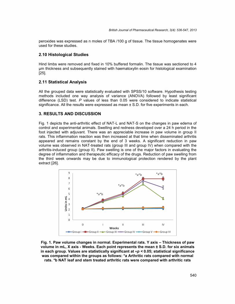
Fig. 1 depicts the anti-arthritic effect of NAT-L and NAT-S on the changes in paw edema ofcontrol and experimental animals. Swelling and redness developed over a 24 h period in thefoot injected with adjuvant. There was an appreciable increase in paw volume in group IIrats. This inflammation reaction was then increased at that time when disseminated arthritisappeared and remains constant by the end of 3 weeks. A significant reduction in pawvolume was observed in NAT-treated rats (group III and group IV) when compared with thearthritis-induced group (group II). Paw swelling is one of the major factors in evaluating thedegree of inflammation and therapeutic efficacy of the drugs. Reduction of paw swelling fromthe third week onwards may be due to immunological protection rendered by the plantextract [26].
Fig. 1. Paw volume changes in normal. Experimental rats. Y axis – Thickness of pawvolume in mL, X axis - Weeks. Each point represents the mean ± S.D. for six animalsin each group. Values are statistically significant at ∗p < 0.05; statistical significancewas compared within the groups as follows: *a Arthritic rats compared with normal
rats. *b NAT leaf and stem treated arthritic rats were compared with arthritic rats
British Journal of Pharmaceutical Research, 3(4): 536-547, 2013
541
It was observed that there was no significant change in hematological parameters suchas RBC, WBC, Hb, ESR, platelets, PCV and the changes in the induced group werereverted back to near normal levels on treating with NAT leaf and stem extracts (Table 1).
Table 1. Effect of NAT-L and NAT-S on hematological parameters in adjuvant inducedarthritic rats
Parameters Group I Group II Group III Group IV Group V Group VIHb (g/dL) 14.2±0.28 9.7±0.24*a 14.01±0.2*b 12.7±0.25*b 14.2±0.13 14.3±0.3RBC (x 10-6 /mm3) 5.1±0.258 3.6±0.24*a 4.8±0.155*b 4.2±0.158NS 5.2±0.21 5.3±0.20WBC(x103 /mm3) 7.6±020 12.3±0.24*a 7.9±0.14*b 9.0±0.19*b 7.6±0.19 7.7±0.22PLT (x103
cells/µL)2.7±0.24 3.9±0.196*a 2.8±0.243*b 3.2±0.22*b 2.7±0.04 2.7±0.1
PCV% 45±2.02 25±1.975*a 43±1.984*b 33±2.08NS 46±0.18 47±0.2ESR 30 Min 2.0±0.203 7.6±0.193*a 2.2±0.188*b 2.8±0.216*b 2.2±0.24 2.2±0.3ESR 60min 4.2±0.193 12.8±0.23*a 3.8±0.24*b 4.0±0.223*b 4.1±0.04 4.2±0.1Values are expressed as mean±S.D. for six animals. Comparisons are made between: *a, Group I vs. Groups II; *b,
Group II vs. Groups III and IV. The letters *a and *b represent the statistical significance at p < 0.05. NS- Nonsignificant. In all the experiments there were no significant changes found in extract alone administered rats (Group
V and Group VI).
The levels of blood glucose, urea, uric acid and creatinine of normal and experimentalgroups of rats are presented in Table 2. A significant (p<0.05) increase in the levels ofGlucose, urea and creatinine and decrease in uric acid level were observed in arthritis-induced rats (group II) when compared with normal rats. The increase in blood glucose inarthritic rats might be due to the decreased glycolytic and increased gluconeogenic enzymeactivities, since impaired hepatic biosynthetic activities were reported in AIA condition [27].The decrease in plasma uric acid in arthritic animals might be due to its continuous utilizationby the system during free radical quenching reaction [28, 29]. Renal dysfunction might be thecause of raised blood urea and creatinine levels in AIA rats.
Table 2. Effect of NAT-L and NAT-S on biochemical parameters in FCA inducedexperimental rats
Parameters(mg/dL)
Group I Group II Group III Group IV Group V Group VI
Blood glucose 84.2 ± 6.47 102.8±7.7*a 87.5 ± 6.8*b 90.4 ± 9.8*b 85±3.2 86±1.6Blood urea 17.3 ± 0.15 29.3 ±0.24*a 20.5 ±0.18*b 22.7 ± 0.1*b 17.6±0.2 18±1.4Uric acid 1.94 ± 0.13 1.34 ±0.14*a 1.83 ±0.14*b 1.62 ± 0.18 *b 1.9±0.5 1.83±2Creatinine 0.62 ± 0.02 1.82 ± 0.05*a 0.93 ± 0.06*b 0.92 ± 0.05 *b 0.63±0.4 0.64±0.1Values are expressed as mean ± S.D. for six animals. Comparisons are made between: *a, Group I vs. Groups II;*b, Group II vs. Groups III and IV. The letters *a and *b represent the Statistical significance at p < 0.05. In all theexperiments there were no significant changes found in extract alone administered rats (Group V and Group VI).
A marked increase in the activity of membrane marker enzymes (AST, ALP, and LDH) anddecrease in ALT were observed in the serum of arthritic rats (group II) when compared tocontrol rats (Group I). Arthritic rats treated with NAT-leaf extract showed a significant(p<0.05) change in the activity of membrane marker enzymes. Compared to NAT-S, NAT-Lshowed greater activity (Table 3). Assessment of the levels of AST, ALT, and ALP providesan excellent and simple tool to measure the anti-arthritic activity of the target drug. Theactivity of ALP was significantly increased in arthritic rats, since it is good index of liver andkidney impairment which is also considered a feature of adjuvant arthritis [30].
British Journal of Pharmaceutical Research, 3(4): 536-547, 2013
542
Table 3. Activities of membrane marker enzymes in the serum of control andexperimental rats
Parameters(U/L)
Group I Group II Group III Group IV Group V Group VI
AST 0.38±0.022 0.83±0.02*a 0.41±0.03*b 0.47±0.04*b 0.39±0.01 0.39±0.24ALT 0.45±0.03 0.012±0.002*a 0.41±0.04*b 0.33±0.04*b 0.44±0.23 0.45±0.37ALP 2.62±0.03 4.8±0.53*a 2.64±0.07*b 3.35±0.04*b 2.62±0.01 2.61±0.4LDH 9.4±0.24 17.9±0.29*a 9.8±0.25*b 11.1±1.62*b 9.5±0.22 9.4±0.31
Values are expressed as mean ± S.D. for six animals. Comparisons are made between: *a, Group I vs. Groups II;*b, Group II vs. Groups III and IV. The letters *a and *b represent the statistical significance at p < 0.05. In all theexperiments there were no significant changes found in extract alone administered rats (Group V and Group VI).
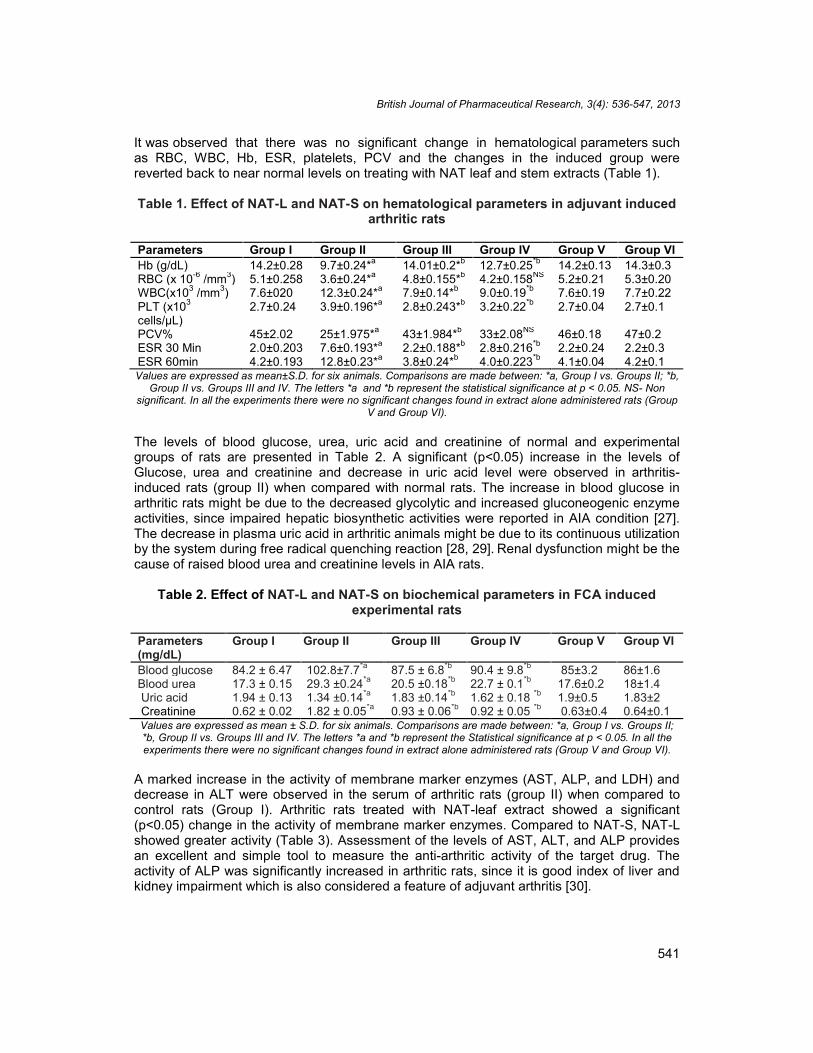
The activity of total tissue SOD was found significantly (p<0.05) low in arthritis induced rats(group II) than in control rats and a substantial increase in the activity to near normal levelwas noticed in NAT administered rats (Fig. 2). Activity was found to be higher in leaf extract(group III) than stem extract (group IV). The increased enzyme activity in NAT administeredrats suggests a response of the animals against possible damage caused by oxygen freeradicals. The superoxide radicals are the first product of molecular oxygen reduction. It actsas a catalyst for dismutations of superoxide radicals into H2O2 and into molecular oxygen toprotect cells and tissues from superoxide radicals and other peroxides such as lipidperoxides in vivo [31].
Fig. 2. Effect of NAT-L and NAT-S extract on the antioxidant activity in normal andexperimental rats. Values are expressed as mean ± S.D. for six animals. Comparisons
are made between: *a, Group I vs. Groups II; *b, Group II vs. Groups III and IV. Theletters *a and *b represent the Statistical significance at p < 0.05
Fig. 2 elucidates the significant (p<0.05) increase in the level of tissue TBARS, SOD andcatalase in arthritis induced rats (group II) when compared to control rats (group I). The NATtreated rats (group III and group IV) showed a significant decrease in lipid peroxides, SODand catalase level to near normalcy. Lipid peroxidation is considered as a critical mechanismof the injury that occurs during RA. Oxygen free radicals are potent lipid peroxidation-
British Journal of Pharmaceutical Research, 3(4): 536-547, 2013
543
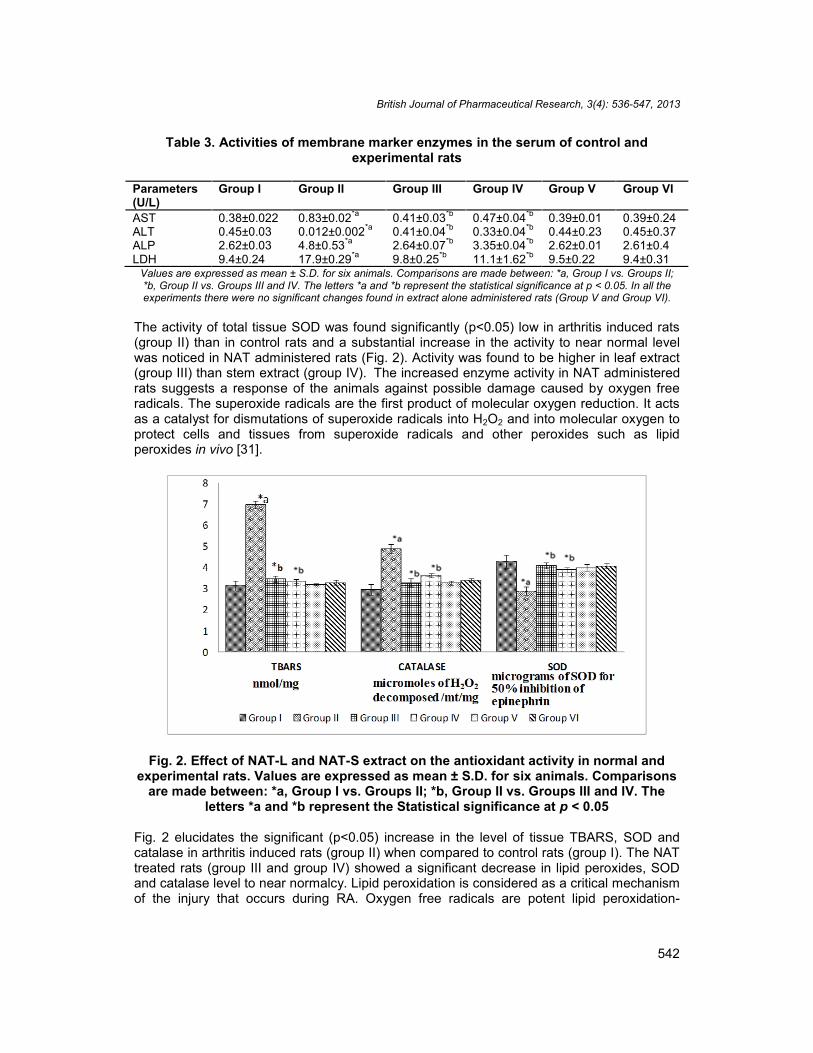
inducing agents that cause the depletion of unsaturated fatty acids of the cell membrane,thus inducing loss of cell integrity and functional alteration of cell receptors and enzymes, inmany diseases ,especially rheumatoid arthritis, membrane damage often occurs in someorgan or tissue, which provokes and accelerates the disorder structurally and functionally.The lack of antioxidant defense leads to an increase in lipid peroxidation and subsequentdeleterious effects. In the present study, the increased lipid peroxides may be due to poorantioxidant defense system. Catalase, which decomposes hydrogen peroxide and protectsthe tissues from highly reactive hydroxyl radicals and also inhibits other long-chainperoxides. A significant increase in catalase activity in arthritis induced group as comparedto the normal rats signifies over production of hydrogen peroxide. For a given concentrationof catalase, the initial rate of hydrogen peroxide removal is proportional to the hydrogenperoxide concentration. Fig. 3 explains the activity of Glutathione peroxidase in control andexperimental animals. The increased activity of GPx has been decreased in NAT leaf andstem treated animals. Glutathione peroxidase (GPx) is localized in the cytoplasm andmitochondria, which catalyses the degradation of various peroxides by oxidizing glutathionewith the formation of its conjugates. GPx has more affinity than catalase for H2O2. GPx isessential for the conversion of glutathione to oxidized glutathione during which H2O2 isconverted to water. The observed increase in the activity of GPx in joint tissue in arthritic ratsindicate the increased H2O2 concentration and may help understanding the pathogenesisassociated with arthritis [32-35]. In the previous study phytochemical screening of the extractof the leaves and stems of Nyctanthes arbor-tristis revealed the presence of flavonoids,tannins, saponins, glycosides, alkaloids, steroids, and phenolic compounds which act asfree radical scavengers [4] to combat the disease.
Fig. 3. Effect of NAT-L and NAT-S extract on the GPx activity in normal andexperimental rats. Values are expressed as mean ± S.D. for six animals. Comparisons
are made between: *a, Group I vs. Groups II; *b, Group II vs. Groups III and IV. Theletters *a and *b represent the Statistical significance at p < 0.05
British Journal of Pharmaceutical Research, 3(4): 536-547, 2013
544
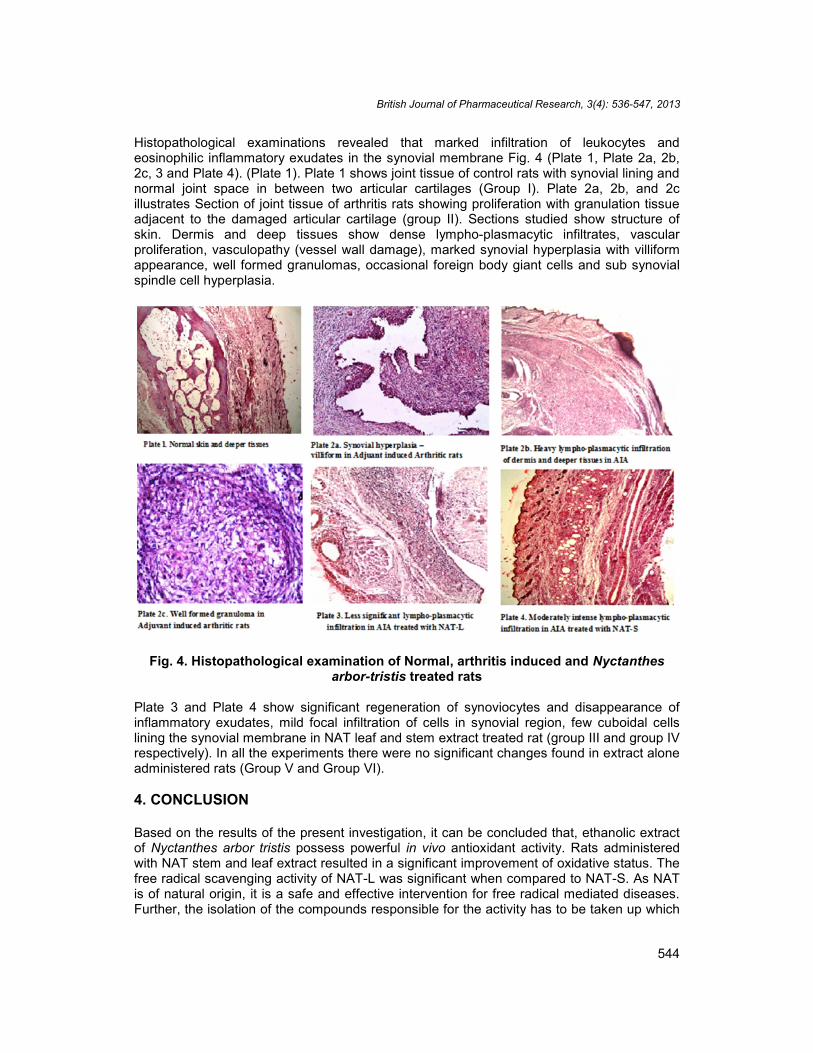
Histopathological examinations revealed that marked infiltration of leukocytes andeosinophilic inflammatory exudates in the synovial membrane Fig. 4 (Plate 1, Plate 2a, 2b,2c, 3 and Plate 4). (Plate 1). Plate 1 shows joint tissue of control rats with synovial lining andnormal joint space in between two articular cartilages (Group I). Plate 2a, 2b, and 2cillustrates Section of joint tissue of arthritis rats showing proliferation with granulation tissueadjacent to the damaged articular cartilage (group II). Sections studied show structure ofskin. Dermis and deep tissues show dense lympho-plasmacytic infiltrates, vascularproliferation, vasculopathy (vessel wall damage), marked synovial hyperplasia with villiformappearance, well formed granulomas, occasional foreign body giant cells and sub synovialspindle cell hyperplasia.
Fig. 4. Histopathological examination of Normal, arthritis induced and Nyctanthesarbor-tristis treated rats
Plate 3 and Plate 4 show significant regeneration of synoviocytes and disappearance ofinflammatory exudates, mild focal infiltration of cells in synovial region, few cuboidal cellslining the synovial membrane in NAT leaf and stem extract treated rat (group III and group IVrespectively). In all the experiments there were no significant changes found in extract aloneadministered rats (Group V and Group VI).
4. CONCLUSION
Based on the results of the present investigation, it can be concluded that, ethanolic extractof Nyctanthes arbor tristis possess powerful in vivo antioxidant activity. Rats administeredwith NAT stem and leaf extract resulted in a significant improvement of oxidative status. Thefree radical scavenging activity of NAT-L was significant when compared to NAT-S. As NATis of natural origin, it is a safe and effective intervention for free radical mediated diseases.Further, the isolation of the compounds responsible for the activity has to be taken up which
British Journal of Pharmaceutical Research, 3(4): 536-547, 2013
545
may result in a modern drug from this plant.
CONSENT
Not applicable.
ETHICAL APPROVAL
All authors hereby declare that "Principles of laboratory animal care" (NIH publication No.85-23, revised 1985) were followed, as well as specific national laws where applicable. Allexperiments have been examined and approved by the appropriate ethics committee.
ACKNOWLEDGMENTS
The authors expressed their gratitude to Dr. Paul Dhinakaran, Chancellor, Dr. James EJ,Vice chancellor, Dr. Joseph Kennedy, Registrar of Karunya University for providing thenecessary facilities for carrying out the experiments.
COMPETING INTERESTS
Authors have declared that no competing interests exist.
REFERENCES
1. Tuntiwachwuttikul P, Rayanil K, Taylor WC. Chemical Constituents from the Flower ofNyctanthes arbor-tristis. Sci Asia. 2003;29:21-30.
2. Puri A, Saxena R, Saxena RP, Saxena KC, Srivastava V, Tandon JS. Immunostimulant activity of Nyctanthes arbor-tristis L. J Ethnopharmacol. 1994;42(1):31–7.
3. Saxena RS, Gupta B, Saxena KK, Srivastava VK, Prasad DN. Analgesic, antipyretic,and ulcerogenic activity of Nyctanthes arbor tristis leaf extracts. J Ethnopharmacol.1987;19:193-200.
4. Narendhirakannan RT, Smeera T. In vitro anti-oxidant studies on ethanolic extracts ofleaves and stems of nyctanthes arbor-tristis, L (night-flowering jasmine). Int J Biol MedRes. 2010;1(4):188-92.
5. Tisdaie MJ, Mohrnoud MB. Activities of free radical metabolizing enzymes in tumors.Br J Cancer. 1983;47:809-12.
6. Susan T, Muzaffer A, Purushothaman KK. Inhibitory activity of arbortristoside A onfibrosarcoma in albino rats. Arogya. 1986;12:122-130.
7. Watlz DT, Drmartino JJ, Misher A. Adjuvant induced arthritis in rats, II Drug effects onphysiologic, biochemical and immunologic parameters. J Pharmacol Exp Ther.1971;178:223-31.
8. Begum VH, Sadique J. Long term effect of Withania somnifera on adjuvant inducedarthritis in rats. Indian J Exp Biol. 1988;26:877–82.
9. Narendhirakannan RT, Subramanian S, Kandaswamy M. Antiinflammatory activity ofCleome gynandra, L on hematological and cellular constituents in adjuvant inducedarthritic rats. J Med Food. 2005a;8:93–99.
10. Mizushima Y, Tsukada W, Akimoto T. A modification of rat adjuvant arthritis for testinganti-rheumatic drugs. J Pharm Pharmacol. 1972;24:781–85.
British Journal of Pharmaceutical Research, 3(4): 536-547, 2013
546
11. Campo GM, Angela A, Campo S, Ferlazzo AM, Altavilla DC. Efficacy of treatment withglycosaminoglycans on experimental collagen-induced arthritis in rats. Arth Res Ther.2003;5:122–31.
12. Docie JV. Practical Haemotology. London, J & A Churchill Ltd. 1958;38-42.13. Sasaki T, Matsui S. Effect of acetic acid concentration on the color reaction in the o-
toludine boric acid for blood glucose determination. RinshoKagaku. 1972;1:346–53.14. Natelson S, Scott ML, Beffa C. A rapid method for the estimation of urea in biological
fluid by means of the reaction between diacetely and urea. Am J Clin Pathol.1951;21:275–81.
15. Caraway WT. In: Seligson D, ed. Standard Methods of Clinical Chemistry. New York:Academic Press, 1963;4:239-47.
16. Owen JA, Iggo TB, Scandrett FJ, Stemart IP. Determination of creatinine in plasma(or) serum and in urine. Biochem J. 1954;58:426–37.
17. Lowry OH, Rosebrough NJ, Farr AL, Randall RJ. Protein measurement with the folinphenol reagent. J Biol Chem. 1951;193:265-275.
18. King J. The transferase – alanine and aspartate transaminase. In: Van, D (Ed),Practical Clinical Enzymology, Norstand Company Limited, London. 1965a;121–38.
19. Walter K, Schutt C. Acid and alkaline phosphatase in serum (two point method). In:Bergmeyer, H.U. (Ed.), In: Methods in Enzymatic Analysis, vol. 2. Academic Press,London; 1974.
20. King J. The dehydrogenase or oxide reductase – lactate dehydrogenase, In: Van, D(Ed), Practical Clinical Enzymology, Norstand company Limited, London. 1965b;83–93.
21. Misra HP, Fridovich I. The role of superoxide anion in the autooxidation of epinephrineand a simple assay for superoxide dismutase. J Biol Chem. 1972;247:3170–175.
22. Rotruck JT, Pope AL, Gasther HE, Hafeman DG, Hoekstra WG. Selenium-biochemical role as a component of glutathione peroxidase. Sci. 1973;179:588–90.
23. Ohkawa H, Oshishi N, Yag K. Assay of lipid peroxidation in animal tissue bythiobarbituric acid reaction. Anal Biochem.1979;95:351–358.
24. Takahara S, Hamilton BH, Nell JV, Ogura Y, Nishimura ET. Hypocatalasemia, a newgenetic carrier states. J Clin Invest. 1960;29:610–19.
25. Durie FH, Fava RA, Foy TM, Aruffo A, Ledbetter JA, Noelle RJ. Prevention ofcollagen-induced arthritis with an antibody to gp39, the ligand for CD40. Sci.1993;26:1328–330.
26. Narendhirakannan RT, Subramanian S, Kandaswamy M. Free radical scavengingactivity of Cleome gynandra L, leaves on adjuvant induced arthritis in rats. Mol CellBiochem. 2005b;276:71–80.
27. Cawthorne MA, Palmer ED, Green J. Adjuvant-induced arthritis and drug-metabolizingenzymes. Biochem Pharmacol. 1976;25:2683–2688.
28. Whiteman M, Ketsawatsakul U, Halliwell B. A reassessment of the peroxynitritescavenging activity of uric acid. Ann NY Acad Sci. 2002;962:242–59.
29. Grootveld M, Halliwell B. Measurement of allantoin and uric acid in human body fluids,A potential index of free-radical reactions in vivo. Biochem J. 1987;243:803–05.
30. Rehman Q, Lane NE, Bone loss. Therapeutic approaches for preventing bone loss ininflammatory arthritis. Arthritis Res. 2001;3:221–27.
31. Okabe T, Hamaguchi K, Inafuku T, Hara M. Aging and superoxide dismutase activityon cerebrospinal fluid. J Neurol Sci. 1996;141:100–04.
32. Alessio HM, Blasi ER. Physical activity as a natural antioxidant booster and its effecton healthy life style. Res Quart Exer Sport. 1997;68:292-02.
33. Darlington LG, Stone TW. Antioxidants and fatty acids in the amelioration ofrheumatoid arthritis and related disorders. Bri J Nutr. 2001;85:251-69.
British Journal of Pharmaceutical Research, 3(4): 536-547, 2013
547
34. Bowles DK, Torgan CE, Ebner S, Kehrer JP, Ivy JL, Starnes JW, Effect of acute, submaximal exercise on skeletal muscle vit E. Free Radic Res. 1991;14:139-43.
35. Scott MD, Eaton JW, Kuypers FA, Chiu DT, Lubin BH. Enhancement of erythrocyteSOD activity effects on cellular oxidant defence. Blood. 1989;74:2542-49.
_________________________________________________________________________© 2013 Thomas et al.; This is an Open Access article distributed under the terms of the Creative CommonsAttribution License (http://creativecommons.org/licenses/by/3.0), which permits unrestricted use, distribution, andreproduction in any medium, provided the original work is properly cited.
Peer-review history:The peer review history for this paper can be accessed here:
http://www.sciencedomain.org/review-history.php?iid=234&id=14&aid=1389