bond strength of self-etch adhesives to pre- etched enamel
TRANSCRIPT

Seediscussions,stats,andauthorprofilesforthispublicationat:http://www.researchgate.net/publication/26245059
Bondstrengthofself-etchadhesivestopre-etchedenamel
ARTICLEinDENTALMATERIALS:OFFICIALPUBLICATIONOFTHEACADEMYOFDENTALMATERIALS·MAY2009
ImpactFactor:4.16·DOI:10.1016/j.dental.2009.04.004·Source:PubMed
CITATIONS
28
DOWNLOADS
24
VIEWS
214
3AUTHORS,INCLUDING:
NicoleKimmes
OregonHealthandScienceUniversity
21PUBLICATIONS215CITATIONS
SEEPROFILE
Availablefrom:NicoleKimmes
Retrievedon:31July2015

B
RD
a
A
R
R
1
A
K
D
E
A
1
Bryesfee
f
0d
d e n t a l m a t e r i a l s 2 5 ( 2 0 0 9 ) 1187–1194
avai lab le at www.sc iencedi rec t .com
journa l homepage: www. int l .e lsev ierhea l th .com/ journa ls /dema
ond strength of self-etch adhesives to pre-etched enamel
obert L. Erickson, Wayne W. Barkmeier ∗, Nicole S. Kimmesepartment of General Dentistry, Creighton University School of Dentistry, Omaha, NE, USA
r t i c l e i n f o
rticle history:
eceived 10 February 2009
eceived in revised form
0 March 2009
ccepted 20 April 2009
eywords:
ental materials
namel
dhesion
a b s t r a c t
Objective. Bond strengths of composite resin to enamel using four self-etch adhesive (SEA)
systems were compared with the bond strength of an etch-and-rinse adhesive (ERA) system,
for both polished enamel and enamel pre-etched with phosphoric acid. The objective was
to determine if the pre-etching would increase the bond strengths of the SEA systems to
match the ERA system.
Methods. Ten specimens were used for each adhesive to determine 24-h resin composite
to enamel shear bond strengths (SBS) to polished (4000 grit) human enamel and this was
repeated for the SEA systems for enamel that was pre-etched with phosphoric acid for 15 s.
SEM analysis was made to assess the degree of etching and resin penetration into enamel
for each of the adhesive systems. Data were analyzed by a two factor ANOVA with a Tukey
HSD post hoc test.
Results. The SBS to polished enamel for all four SEA systems were statistically significantly
lower (p < 0.05) than the ERA control, but with pre-etched enamel there were no statistically
significant differences (p > 0.05) between any of the adhesive systems. All four of the SEA
systems demonstrated statistically significant increases in bond strength between bonding
to polished and pre-etched enamel, ranging from 27% to 86%. The results of SEM analysis
showed no differences in the resin penetration patterns of any of the adhesives for enamel
that was etched with phosphoric acid.
Significance. Pre-etching enamel may enhance the bond strength of SEA systems to values
comparable with those found with ERA adhesive systems, which may improve their overall
performance in clinical use.
emy
emphasized concern about the effectiveness of SEA systems
© 2009 Acad
. Introduction
onding procedures to enamel play an important role inestorative and preventive dental procedures, and for manyears the bonds obtained using phosphoric acid (PA) etching ofnamel were the mainstay of such procedures. In recent years,elf-etching adhesive (SEA) systems have been advocated
or these types of procedures as a suitable replacement fortch-and-rinse adhesive (ERA) systems that use PA etching ofnamel. However, there is reasonable cause for concern about∗ Corresponding author at: Creighton University School of Dentistry, 25ax: +1 402 280 5004.
E-mail address: [email protected] (W.W. Barkmeier).109-5641/$ – see front matter © 2009 Academy of Dental Materials. Puoi:10.1016/j.dental.2009.04.004
of Dental Materials. Published by Elsevier Ltd. All rights reserved.
the effectiveness of the SEA systems for bonding compositeto enamel [1]. Their ability to etch enamel is less than PA [2–4]and most studies find that the bond strengths of compositeto enamel provided with these SEA systems are significantlylower when compared to ERA systems [5–13]. Fatigue studiesof enamel bonds, comparing the two types of systems, have
00 California Plaza, Omaha, NE 68178, USA. Tel.: +1 402 280 5262;
for enamel bonding, and relative fatigue limit values suggestthat more enamel margin defects might occur with the SEAsystems than for ERA systems [14–16]. Clinical studies [17–21]
blished by Elsevier Ltd. All rights reserved.

s 2 5 ( 2 0 0 9 ) 1187–1194
Table 1 – Materials and procedures.
Adhesive Code System/etchant
Adper Single Bond PlusEtchant (Lot No. 7JL),Adhesive (Lot No. 7KY)
SBP Etch-and-rinse/35 wt.%phosphoric acid
Adper Prompt L-Pop (Lot No.273971)
PLP Self-etch/Twocomponents mixed –one-step system
Clearfil SE Primer (LotNo.00688A), Bond (Lot No.00985B)
CSE Self-etch/Primer andadhesive – two stepsystem
Clearfil S3 (Lot No. 00069A) CS3 Self-etch/Onecomponent – onestep system
UT, USA). The composite cylinders (2.35 mm diameter andapproximately 2 mm in length) were polymerized for 40 s. Thebonded specimens were stored in distilled water, at 37 ◦C, for
Table 2 – Bonding procedures.
Adhesive Procedure
SP 1. Etch enamel 15 s, water rinse, dry thoroughlywith air2. Apply 2 coats of adhesive 15 s with gentleagitation, air thin3. Light cure 10 s
PLP 1. Mix adhesive according to directions2. Apply adhesive with moderate pressure for 15 s3. Gentle air stream to thoroughly air dry4. Apply second coat of adhesive and thoroughly airdry5. Light cure 10 s
CSE 1. Apply primer 20 s, dry with mild air flow2. Apply adhesive followed by gentle air flow3. Light cure 10 s
CS3 1. Apply adhesive 20 s, dry with high pressure airflow2. Light cure 10 s
1188 d e n t a l m a t e r i a l
and laboratory studies [22,23] appear to corroborate a greaterincidence of enamel defects for restorations where SEAsystems were used compared with ERA systems.
Selective etching of enamel with PA is a potential tech-nique to use with SEA systems to improve their performanceon enamel. Fewer enamel margin defects were observed whenselective etching of enamel was employed in a clinical study[24] and in a laboratory study [23] where thermo-mechanicalloading was used on restorations. The success of this tech-nique presumes that the bond strength and durability of thebond for SEA systems, used on enamel pre-etched with PA,will be equivalent to those obtained with ERA systems bondedto enamel. Increased bond strengths have been shown for anumber of SEA systems used to bond composite to enamelpre-etched with PA [25–31], but only two of these studiesincluded ERA control systems. These two studies examinedfour commercial SEA systems between them and all four ofthese demonstrated statistically equivalent bond strengthsto their respective ERA control systems when pre-etching ofenamel was used. One experimental material did not reachthe level of the ERA control. These studies demonstrate thatselective etching may be a useful technique but it is still notclear that all SEA materials can achieve similar bond strengthsas their ERA counterparts.
It was the purpose of this study to examine the effectof pre-etching enamel on the bond strength of composite toenamel for four SEA adhesives that were examined previouslyin fatigue testing [16]. The hypothesis used is the null hypoth-esis, that there will be no statistically significant differencesin bond strength between the four SEA systems and an ERAsystem used as a control, at a 5% level of significance.
2. Materials and methods
Four SEA systems were examined for bonding resin compositeto human enamel: Adper Prompt L-Pop (3M ESPE, St Paul, MN,USA) (PLP), Clearfil SE Bond (Kuraray Medical Inc., Okayama,Japan) (CSE), Clearfil S3 (Kuraray Medical Inc., Okayama, Japan)(CS3) and Xeno IV (DENTSPLY Caulk, Milford, DE, USA) (X4).These were compared with an ERA system, Adper Single BondPlus (3M ESPE, St Paul, MN, USA) (BP), used as a control mate-rial. These materials are described in Table 1.
2.1. Bond strength testing
Extracted human molar teeth were sectioned bucco-linguallyand most of the root structure was removed. The mesial anddistal crown sections were then mounted with Triad DuaLine(DENTSPLY International, York, PA, USA) in custom brass fix-tures designed for use with an abrasive polishing system. Flatenamel bonding sites (approximately 4 mm in diameter) wereprepared using a sequence of polishing papers on a watercooled wheel to create a final polished surface with 4000 gritpaper (Struers Inc., Cleveland, OH, USA). This fine polish wasused to minimize smear layer challenges in the bonding of the
SEA systems.Ten enamel specimens were used with each adhesive sys-tem to bond Z100 resin composite (3M ESPE, St Paul, MN, USA– Lot 7PP) to the polished enamel. Manufacturers’ instructions
Xeno IV (Lot No. 061102) X4 Self-etch/Onecomponent – onestep system
were followed for bonding with the four SEA systems. For theERA system (SBP control group) the PA treated enamel surfacewas water rinsed and air dried (Table 2). Four more groupswere bonded with the SEA systems using the same proce-dures, except that the enamel surfaces had been pre-etchedwith 35% PA (Scotchbond Etchant, 3M ESPE, St Paul, MN, USA)for 15 s, using the gel etchant from the SBP system (Table 1).The adhesives were polymerized with a Spectrum 800 CuringUnit (DENTSPLY Caulk, Milford, DE, USA) set at 600 mW/cm2
for the time recommended by the manufacturers. Resin com-posite was bonded to the adhesive treated surface using anUltradent bonding fixture (Ultradent Products, South Jordan,
X4 1. Apply adhesive and actively scrub surface for 15 s2. Repeat adhesive application with scrubbing 15 s,gentle air stream to yield smooth glossy surface3. Light cure 10 s
5 ( 2 0 0 9 ) 1187–1194 1189
2(Iw(
2
Eadbwtnsa
selelaafdmff
Lit
2
AdtAt
3
Ttaissa(swc2ic
Table 3 – Shear bond strength for all groups (MPa).
Adhesive Procedure Increase
Recommended Pre-etch enamel
SBP 40.5 (6.1) a – –PLP 30.7 (4.3) b 39.1 (3.7) a* 27%CSE 29.1 (2.7) b 41.0 (6.0) a* 41%CS3 25.7 (3.1) bc 34.8 (2.8) a* 35%X4 20.9 (3.1) c 38.8 (5.2) a* 86%
Different letters in columns indicate differences at the 5% signifi-cance level.
d e n t a l m a t e r i a l s 2
4 h prior to testing. The specimens were loaded to failure1 mm per minute) using an Ultradent shearing fixture in annstron (Model 1123) test frame (Instron, Norwood, MA, USA),ith an MTS ReNew Upgrade Package and TestWorks software
MTS Systems Corporation, Eden Prairie, MN, USA).
.2. SEM analysis
ach adhesive was examined for its ability to etch enamelnd also for resin penetration into enamel. To examine theegree of etching, surfaces were prepared as described forond strength testing and the respective primer or conditioneras applied to polished enamel for the time recommended,
hen rinsed off either with water, as for PA, or by alter-ating water and acetone rinses for the acidic monomerystems. After drying, the specimens were processed for SEMnalysis.
To examine resin penetration into the enamel, adhesiveystems were applied to either polished enamel or pre-etchednamel according to recommended procedures followed by aayer of resin composite (about 2 mm thick) applied over thentire polished area and beyond. This was polymerized byight for 40 s. After storing these specimens in distilled water,t 37 ◦C, for 24 h, they were transferred to a solution of nitriccid to remove the enamel. When all the enamel was dissolvedrom the specimens they were gently rinsed in distilled water,ried and processed for SEM analysis. Three specimens wereade to correspond with each bond strength test group and
or each type of analysis. Representative images were obtainedrom the SEM.
A JEOL scanning electron microscope (Model 840A, JEOLtd., Tokyo, Japan) and Evex NanoAnalysis and Digital Imag-ng software (Evex Inc., Princeton, NJ, USA) were used to obtainhe SEM images.
.3. Data analysis
two factor ANOVA was used to analyze the bond strengthata for statistically significant differences and Tukey’s HSDest was used for multiple comparisons between groups.
5% level of statistical significance was used for allesting.
. Results
he ANOVA analysis demonstrated significant differences forhe factors adhesive system (p = 0.0001), technique (p = 0.0001)nd the interaction of the two factors (p = 0.0001). Compar-sons of means with the Tukey HSD test found differences ashown in Table 3. For bonding to polished enamel, all four SEAystems produced lower bond strengths than the SBP controldhesive and these differences were statistically significantp < 0.05). The three adhesive systems, PLP, CSE and CS3 weretatistically similar as were CS3 and X4. When the enamelas pre-etched, all four SEA systems demonstrated statisti-
ally significant improvements in bond strength ranging from7% to 86% and there were no significant differences (p > 0.05)n bond strength between any of the SEA systems and the SBPontrol adhesive system.
∗ Significant difference between recommended and pre-etchgroups.
Analysis by SEM of the etching and resin penetration pat-terns when recommended procedures were used are shown inFig. 1. The most acidic of the SEA systems, PLP, demonstratedetching primarily on the ends of the enamel prisms, with theinterprismatic regions less affected. The resin penetration isessentially a negative replica of the etch pattern with resinfound where the enamel prisms have been etched and lack ofsimilar resin penetration in the interprismatic regions. Withthe CSE system, the acidic primer produced a relatively weaketch of the enamel surface, but enough to just begin to definesome prism boundaries. Resin penetration is seen as shortresin projections across the entire surface with some areas ofgreater penetration, presumably, corresponding to the posi-tion of ends of enamel prisms. Both the CS3 system and theX4 system produced weaker etching, showing up as fine pit-ting of the enamel surface, but leaving the polishing groovesquite distinct. The resin patterns present for the most part asvery fine resin projections of the same order as the pitting ofenamel.
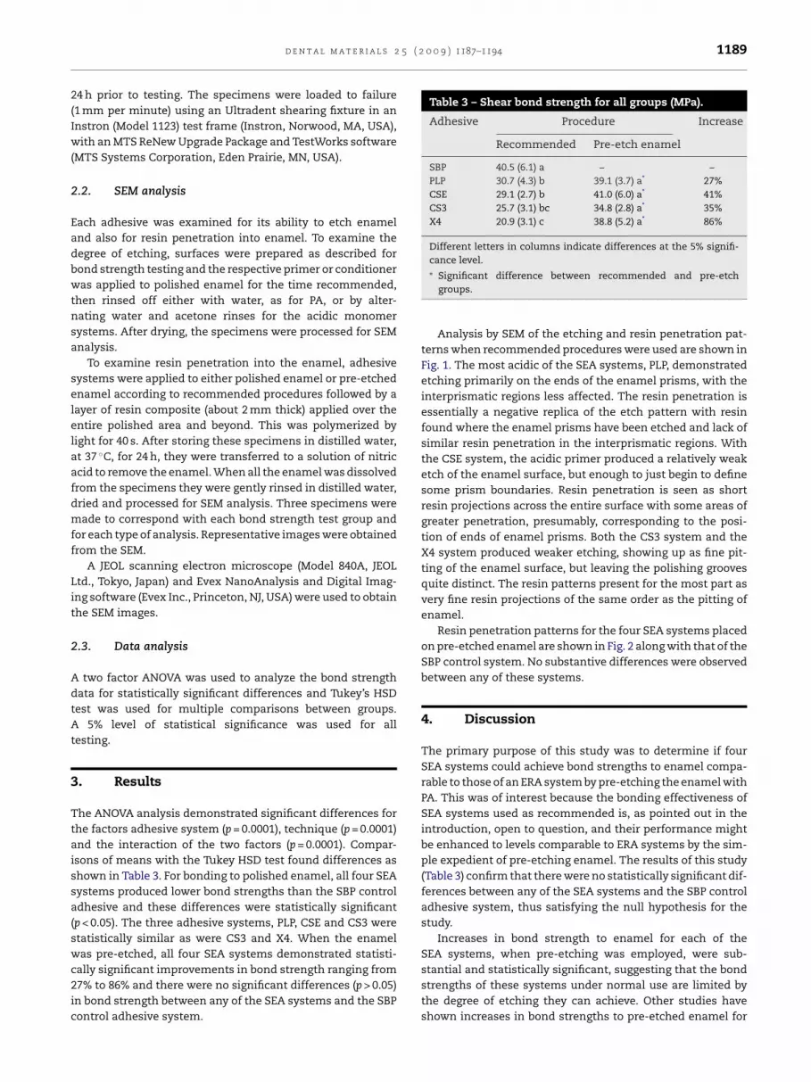
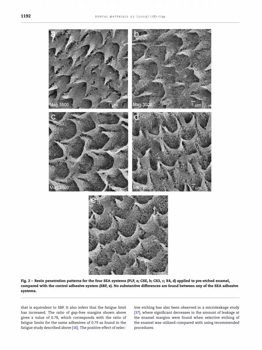
Resin penetration patterns for the four SEA systems placedon pre-etched enamel are shown in Fig. 2 along with that of theSBP control system. No substantive differences were observedbetween any of these systems.
4. Discussion
The primary purpose of this study was to determine if fourSEA systems could achieve bond strengths to enamel compa-rable to those of an ERA system by pre-etching the enamel withPA. This was of interest because the bonding effectiveness ofSEA systems used as recommended is, as pointed out in theintroduction, open to question, and their performance mightbe enhanced to levels comparable to ERA systems by the sim-ple expedient of pre-etching enamel. The results of this study(Table 3) confirm that there were no statistically significant dif-ferences between any of the SEA systems and the SBP controladhesive system, thus satisfying the null hypothesis for thestudy.
Increases in bond strength to enamel for each of theSEA systems, when pre-etching was employed, were sub-
stantial and statistically significant, suggesting that the bondstrengths of these systems under normal use are limited bythe degree of etching they can achieve. Other studies haveshown increases in bond strengths to pre-etched enamel for
1190 d e n t a l m a t e r i a l s 2 5 ( 2 0 0 9 ) 1187–1194

5 ( 2
vETwsoei8vsw
rsSdTitoaIfbbdputasoac
SttlaeIerepam
Fwwdaesr
d e n t a l m a t e r i a l s 2
arious SEA systems [25–31], but most have not included anRA control system to judge the effectiveness of the increases.wo studies that have used an ERA control [30,31] have found,ith the exception of one SEA system, that the increased bond
trengths are comparable to the ERA material examined. Onlyne of the SEA systems (CSE) used in this study has beenxamined in other studies where pre-etching of enamel wasnvestigated [27,28–30]. The increases in bond strength were0%, 43% and 46% for the three studies and although the 80%alue seems too large, perhaps because the reference bondtrength was too small, the other values are in good agreementith the 41% increase found in this study.
Results from the SEM analysis for degree of etching andesin penetration of the enamel surface (Figs. 1 and 2) giveupportive evidence that the lower bond strengths of theEA systems compared with SBP, when recommended proce-ures are used, is to a large extent due to etching differences.he similar resin penetration patterns (Fig. 2), when PA etch-
ng was used, correlated with similar bond strengths for allhe adhesive systems is highly suggestive that the degreef etching, or perhaps more correctly the etch morphologychieved with PA, is responsible for the higher bond strengths.t has also been shown that extending the application timeor these SEA systems does not increase the bond strengthsy a statistically significant amount [32]. Potential chemicalonding effects for some monomers [33,34] and differences inegree of polymerization of the various resin systems [35] maylay a role in the measured bond strengths for SEA systemssed as recommended, but they do not seem to account forhe higher bond strengths found with PA pre-etching. Also,lthough chemical bonding may play a role for better bondtrength of some ERA systems, it does not mitigate the findingf this study along with others [5–13] that SEA systems useds recommended do not generally produce bond strengthsomparable to ERA systems.
It should be pointed out that the manner in which someEA adhesives are applied to pre-etched enamel may affecthe bond strength obtained. In a previous study of the effecthat pre-etching enamel has on the bond strength and fatigueimit of the PLP system [15], it was found that the bond strengthnd fatigue limit were effectively unchanged by pre-etchingnamel from their values using recommended procedures.t was postulated that applying the acidic PLP system totched enamel, using moderate pressure in a scrubbing action,esulted in obliterating the porous structure of the etched
namel, resulting in an etch pattern and resin penetrationattern similar to that found without using the pre-etch. Inpproaching the present study, a series of specimens wereade to examine this effect using enamel specimens asig. 1 – Etch (a, c, e and g) and resin penetration (b, d, f and h) paas 3500× and for all others was 6500× (see data on images a, bas primarily confined to the enamel prism ends with the interpemonstrated roughening of the enamel surface with some enamre visible across the surface with longer projections presumablynamel was less than for CS3 (e and f) than for CSE, presenting atill distinct. The resin penetration presents as the reverse of theesin in the pitted surface. The etch pattern and resin penetratio
0 0 9 ) 1187–1194 1191
described in Section 2. The enamel surfaces were etched withPA for 15 s and the PLP system was applied in two ways; withmoderate pressure in a scrubbing action, according to man-ufacturer’s instructions, and with a lighter agitation of theadhesive over the surface. The specimens were prepared asdescribed earlier for resin penetration examination by SEM.Results are shown in Fig. 3, where the moderate pressuregives a resin penetration pattern similar to that found inFig. 1 for PLP used as recommended on polished enamel,whereas with light agitation the resin penetration is similarto that found in Fig. 2 where the resin penetration patternis similar to that for SBP. This tends to confirm the hypoth-esis proposed in the previous study and led to using theagitation method for the PLP/pre-etch group in the presentstudy.
The lower bond strengths found with SEA systems, usedas recommended, cannot be dismissed as being inconsequen-tial. Fatigue studies [14–16,36] have shown lower fatigue limitsfor bonds to enamel using SEA systems than when using anERA system. Such results are suggestive that the bonds toenamel with the SEA systems are more susceptible to fracture,and restorations bonded with these systems may have greateramounts of enamel margin defects. The same five adhesivesystems used in the present study were previously examinedin a fatigue study [16]. In this fatigue study the SEA systemswere also found to have lower bond strengths than the SBPcontrol adhesive, with their percentage of the control valuebeing 57–85%. However, for the fatigue limits the percentageswere 43–79% suggesting that the bond strength values maynot be as useful for judging bond performance as the fatiguelimits.
Cyclic loading of restorations is another way of examin-ing the effectiveness of an adhesive to maintain marginalintegrity. A good example of such a study [22] examinedcyclic loading of class II restorations in extracted teeth andfound that SEA systems had more enamel marginal gapsafter loading than did ERA systems. The ratios of gap-freemargin length for CSE and PLP to SBP were in good agree-ment with the ratios of fatigue limits for these adhesives inthe above mentioned fatigue study [16]. Recently, a similarstudy was done with the same cycling protocol [23] on class IIrestorations where the SEA adhesives were used as recom-mended and with selective etching of the enamel marginswith PA. For the CSE adhesive system the percentage of gap-free margins was 68.6% with recommended procedures but
increased to 88.8% when selective etching was used, whichwas similar to the 87.5% found with the SBP adhesive sys-tem. This is in agreement with the results of the presentstudy where pre-etching provides a bond strength for CSEtterns for the four SEA systems. Magnification for (a) and (b)and d). For PLP (a and b) the etching and resin penetrationrismatic areas less affected. The CSE (c and d) systemel prism delineation barely visible. Short resin projectionscorresponding to areas of enamel prism ends. Etching of
s a fine pitting of the surface and with polishing groovesetching pattern with a fine structure corresponding to
n for X4 (g and h) was similar to that of CS3.
1192 d e n t a l m a t e r i a l s 2 5 ( 2 0 0 9 ) 1187–1194
Fig. 2 – Resin penetration patterns for the four SEA systems (PLP, a; CSE, b; CS3, c; X4, d) applied to pre-etched enamel,stant
compared with the control adhesive system (SBP, e). No subsystems.
that is equivalent to SBP. It also infers that the fatigue limit
has increased. The ratio of gap-free margins shown abovegives a value of 0.78, which corresponds with the ratio offatigue limits for the same adhesives of 0.79 as found in thefatigue study described above [16]. The positive effect of selec-ive differences are found between any of the SEA adhesive
tive etching has also been observed in a microleakage study
[37], where significant decreases in the amount of leakage atthe enamel margins were found when selective etching ofthe enamel was utilized compared with using recommendedprocedures.
d e n t a l m a t e r i a l s 2 5 ( 2 0 0 9 ) 1187–1194 1193
Fig. 3 – Resin penetration patterns for the PLP adhesive system applied in two different ways to pre-etched enamel. (a)S essuw
tdTmottdcpmtcs
oibror3E1r
Ssttlttvrusy
hows the effect of applying the adhesive with moderate prith light agitation.
The above findings from in vitro studies were indicative ofhose found in clinical studies [17–21], where enamel marginefects were greater for SEA systems than for ERA systems.he importance of these observations in some cases wereinimized because the defect size had not reached the point
f requiring replacement of the restoration, but that seemso miss the point that the percentage of defects did not needo be that large, and the effect size by clinical research stan-ards was often quite large. One study [24] of 5 years durationompared restorations bonded with CSE using recommendedrocedures compared with selective etching of the enamelargins with PA. The results found that 36% of the restora-
ions with recommended procedures had no enamel defectsompared with 66% of the restorations having no defects whenelective etching was used.
Most of the above clinical studies examined restorationsf non-carious cervical lesions, but one study [21] exam-
ned Class I and II restorations, where fatigue stresses mighte greater on the enamel margins. This study comparedestorations bonded with three SEA systems compared withne ERA control system. After 1 year the percentage ofestorations with Alfa scores (no marginal defects) were 7.4%,1.0% and 59.3% for the SEA systems and 92.9% for theRA system. The material having the lowest score also had1% of the restorations with defects large enough to needeplacement.
The results of the present study demonstrated that fourEA systems, used with pre-etching enamel, provided bondtrengths to enamel that were comparable to that provided byhe ERA system they were tested against. It could be inferredhat the similar bond strength would mean that the fatigueimits would also be similar and that is supported by some ofhe other work discussed here, however, that testing remainso be done. It is likely that most SEA systems would pro-ide similar results when used on pre-etched enamel. These
esults support the use of selective etching of enamel whensing SEA systems to obtain better performance. Of course,elective etching is a technique that has been used for manyears but may have some drawbacks with regard to affect-r
re in a scrubbing action. In (b) the adhesive was applied
ing dentin bonding, as performance of SEA systems has beenshown to decrease when the dentin is etched with phospho-ric acid [23,38,39]. Selective etching also negates the purportedadvantage of SEA systems; that they are easier to use. How-ever, that claim is specious, as all adhesive systems arerelatively easy to use. The proper consideration is how effec-tive the various systems are in bonding to enamel as well asto dentin, and arguably enamel is the more important of thetwo.
It would seem that abundant information has been com-piled to persuasively argue that most if not all SEA systemsdo not bond to enamel as effectively as ERA systems that usePA etching of enamel. This is believed to be due to a funda-mental difference in the interface between enamel and theadhesive/restorative complex. With PA etching the interface isdistributed over many microns in depth with a complex mor-phology; providing an interface with graded properties thatmay be more resistant to crack propagation than the ratherplanar interface produced with SEA systems. Whether thisis the correct interpretation or not, the evidence seems clearthat SEA systems, used as recommended, are less effectivethan ERA systems for bonding composite resin to enamel. Thisposes an ethical issue for the dental profession. In view of allthe existing evidence, should SEA systems, as now used, belimited to dentin-only restorations until new methods, suchas selective etching of enamel, can be certified for more gen-eral use? This is an issue that has been put off for much toolong a time.
Acknowledgement
This study was supported in part by Bisco, Inc.
e f e r e n c e s
[1] De Munck J, Van Landuyt K, Peumans M, Poitevin A,Lambrechts P, Braem M, et al. A critical review of the

s 2 5
previously acid-etched dentin. Oper Dent 2008;33:702–9.
1194 d e n t a l m a t e r i a l
durability of adhesion to tooth tissue: methods and results. JDent Res 2005;84:118–32.
[2] Perdigão J, Geraldeli S. Bonding characteristics ofself-etching adhesives to intact versus prepared enamel. JEstht Restor Dent 2003;15:32–42.
[3] Hannig M, Bock H, Bott B, Hoth-Hannig W. Inter-crystallitenanoretention of self-etching adhesives at enamel imagedby transmission electron microscopy. Eur J Oral Sci2002;110:464–70.
[4] Salz U, Mucke A, Zimmermann J, Tay F, Pashley D. pKa valueand buffering capacity of acidic monomers commonlyused in self-etching primers. J Adhes Dent 2006;8:143–50.
[5] DeMunck J, Vargas M, Iracki J, Van Landuyt K, Poiterin A,Lambrechts P, et al. One-day bonding effectiveness of newself-etch adhesives to bur-cut enamel and dentin. Oper Dent2005;30(1):39–49.
[6] Ernst C, Holzmeier M, Willershousen B. In vitro bondstrength of self-etching adhesives in comparison to 4th and5th generation adhesives. J Adhes Dent 2004;6:293–9.
[7] Lopes G, Marson F, Vieira L, Andrada M, Baratieri L.Composite bond strength to enamel with self-etchingprimers. Oper Dent 2004;29(4):424–9.
[8] Goracci C, Sadek F, Monticelli F, Cardoso P, Ferrari M.Microtensile bond strength of self-etching adhesives toenamel and dentin. J Adhes Dent 2004;6:313–8.
[9] DeMunck J, Van Meerbeek B, Satoshi I, Vargas M, Yoshida Y,Armstrong S, et al. Microtensile bond strengths of one andtwo-step self-etch adhesives to bur-cut enamel and dentin.Am J Dent 2003;16:414–20.
[10] Inoue S, Vargas M, Abe Y, Yoshida Y, Lambrechts P, VanherleG, et al. Microtensile bond strength of eleven contemporaryadhesives to enamel. Am J Dent 2003;16:329–34.
[11] Brackett W, Ito S, Nishitani Y, Haisch L, Pashley D. Themicrotensile bond strength of self-etching adhesives toground enamel. Oper Dent 2006;31:332–7.
[12] Loguercio A, Moura S, Pellizzaro A, Dal-Bianco K, Patzlaff R,Grande R, et al. Durability of enamel bonding using two-stepself-etch systems on ground and unground enamel. OperDent 2008;33:79–88.
[13] Yazici A, Celik C, Ozgunaltay G, Dyangac B. Bond strength ofdifferent adhesive systems to dental hard tissues. Oper Dent2007;32:166–72.
[14] Erickson R, DeGee A, Feilzer A. Fatigue testing of enamelbonds with self-etching and total-etch adhesive systems.Dent Mater 2006;22:981–7.
[15] Erickson R, DeGee A, Feilzer A. Effect of pre-etching enamelon fatigue of self-etch adhesive bonds. Dent Mater2008;24:117–23.
[16] Erickson RL, Barkmeier WW, Kimmes NS. Fatigue of enamelbonds with self-etch adhesives. Dent Mater 2009;25:716–20.
[17] Van Landuyt KL, Peumans M, Fieuws S, De Munck J, CardosoMV, Ermis RB, et al. A randomized controlled clinical trial ofa HEMA-free all-in-one adhesive in non-carious cervicallesions at 1-year. J Dent 2008;36:847–55.
[18] Bittencourt D, Ezecelevski I, Reis A, Van Dijken J. An18-months’ evaluation of self-etch and etch-and-rinseadhesive in non-carious cervical lesions. Acta OdontolScand 2005;63:173–8.
[19] Loquercio A, Bittencourt D, Baratieri L, Reis A. A 36-monthevaluation of self-etch and etch-and-rinse adhesives innoncarious cervical lesions. J Am Dent Assoc2007;138:507–14.
[20] Ritter A, Heymann H, Swift E, Sturdevant J, Wilder Jr A.
Clinical evaluation of an all-in-one adhesive in non-cariouscervical lesions with different degrees of dentin sclerosis.Oper Dent 2008;33:370–8.[21] Perdigão J, Dutra-Corrêa M, Sastilhos N, Carmo A,Anauate-Netto C, Cordeiro H, et al. One-year clinical
( 2 0 0 9 ) 1187–1194
performance of self-etch adhesives in posterior restorations.Am J Dent 2007;20:125–33.
[22] Frankenberger R, Tay F. Self-etch vs etch-and-rinseadhesives: effect of thermo-mechanical fatigue loading onmarginal quality of bonded resin composite restorations.Dent Mater 2005;21:397–412.
[23] Frankenberger R, Lohbauer U, Roggendorf MJ, Naumann M,Taschner M. Selective enamel etching reconsidered: betterthan etch-and-rinse and self-etch? J Adhes Dent2008;10:339–44.
[24] Peumans M, De Munck J, Van Landuyt K, Lambrechts P, VanMeerbeek B. Five-year clinical effectiveness of a two-stepself-etching adhesive. J Adhes Dent 2007;9:7–10.
[25] Watanabe T, Tsubota K, Takamisawa T, Durokawa H, RikutaA, Ando S, et al. Effect of prior acid etching on bondingdurability of single-step adhesives. Oper Dent2008;33:426–33.
[26] Soares CJ, Castro CG, Filho P, Soares da Mota A. Effect ofprevious treatments on bond strength of two self-etchingadhesive systems to dental substrate. J Adhes Dent2007;9:291–6.
[27] Van Landuyt K, Kanumilli P, De Munck J, Peumans M,Lambrechts P, Van Meerbeek B. Bond strength of a mildself-etch adhesive with and without prior acid-etching. JDent 2006;34:77–85.
[28] Torii Y, Itou K, Nishitani Y, Ishikawa K, Suzuki K. Effect ofphosphoric acid etching prior to self-etching primerapplication on adhesion of resin composite to enamel anddentin. Am J Dent 2002;15:305–8.
[29] Erhardt M, Cavalcante L, Pimenta L. Influence of phosphoricacid pretreatment on self-etching bond strengths. J EsthetRestor Dent 2004;16:33–41.
[30] Miguez P, Castro P, Nunes M, Walter R, Pereira P. Effect ofacid-etching on the enamel bond of two self-etchingsystems. J Adhes Dent 2003;5:107–12.
[31] Lührs A-K, Guhr S, Schilke R, Borchers L, Geurtsen R, GünayH. Shear bond strength of self-etch adhesives to enamelwith additional phosphoric acid etching. Oper Dent2008;33:155–62.
[32] Barkmeier WW, Erickson RL, Kimmes NS, Latta MA,Wilwerding TM. Effect of enamel etching time on roughnessand bond strength. Oper Dent 2009;34:217–22.
[33] Van Landuyt KL, Yoshida Y, Hirata I, Snauwaert J, De MunckJ, Okazaki M, et al. Influence of the chemical structure offunctional monomers on their adhesive performance. J DentRes 2008;87:757–61.
[34] Yoshida Y, Van Meerbeek B, Nakayama Y, Snauwaert J,Hellemans L, Lambrechts P. Evidence of chemical bonding atbiomaterial-hard tissue interfaces. J Dent Res2000;79:709–14.
[35] Kanehira M, Finger WJ, Hoffman M, Endo T, Komatsu M.Relationship between degree of polymerization and enamelbonding strength with self-etching adhesives. J Adhes Dent2006;8:211–6.
[36] De Munck J, Van Meerbeek B, Wevers M, Lambrechts P,Braem M. Micro-rotary fatigue of tooth-biomaterialinterfaces. Biomaterials 2005;26:1145–53.
[37] Brackett MG, Brackett WW, Haisch LD. Microleakage ofclass V resin composites placed using self-etching resins:effect of prior enamel etching. Quintessence Int2006;37:109–13.
[38] Ikeda M, Tsubota K, Takamizawa T, Yoshida T, Miyazaki M,Platt JA. Bonding durability of single-step adhesives to
[39] Gokce K, Aykor A, Ersoy M, Ozel E, Soyman M. Effect ofphosphoric acid etching and self-etching primer applicationmethods on dentinal shear bond strength. J Adhes Dent2008;10:345–9.