state of the art etch-and-rinse adhesives
TRANSCRIPT
d e n t a l m a t e r i a l s 2 7 ( 2 0 1 1 ) 1–16
avai lab le at www.sc iencedi rec t .com
journa l homepage: www. int l .e lsev ierhea l th .com/ journa ls /dema
State of the art etch-and-rinse adhesives
David H. Pashleya,∗, Franklin R. Tayb, Lorenzo Breschi c,d, Leo Tjäderhane e,Ricardo M. Carvalho f, Marcela Carrilhog,h, Arzu Tezvergil-Mutluay i
a Department of Oral Biology, Medical College of Georgia, School of Dentistry, Augusta, GA, USAb Department of Endodontics, Medical College of Georgia, School of Dentistry, Augusta, GA, USAc Department of Biomedicine, University of Trieste, Trieste, Italyd IGM-CNR, Unit of Bologna c/o IOR, Bologna, Italye Institute of Dentistry, University of Oulu and Oulu University Hospital, Oulu, Finlandf Department of Prosthodontics, Bauru School of Dentistry, University of São Paulo, Bauru, Brazilg GEO/UNIBAN, Health Institute, Bandeirante University of São Paulo, São Paulo, Brazilh Piracicaba Dental School, State University of Campinas, Piracicaba, Brazili Department of Prosthetic Dentistry, Institute of Dentistry, University of Turku, Turku, Finland
a r t i c l e i n f o
Article history:
Received 7 October 2010
Accepted 22 October 2010
Keywords:
Acid-etchants
Primers
Adhesives
Durability
MMPs
a b s t r a c t
Objectives: The aim of this study was to explore the therapeutic opportunities of each
step of 3-step etch-and-rinse adhesives. Methods: Etch-and-rinse adhesive systems are the
oldest of the multi-generation evolution of resin bonding systems. In the 3-step version,
they involve acid-etching, priming and application of a separate adhesive. Each step can
accomplish multiple goals. Acid-etching, using 32–37% phosphoric acid (pH 0.1–0.4) not
only simultaneously etches enamel and dentin, but the low pH kills many residual bac-
teria. Results: Some etchants include anti-microbial compounds such as benzalkonium
chloride that also inhibits matrix metalloproteinases (MMPs) in dentin. Primers are usu-
ally water and HEMA-rich solutions that ensure complete expansion of the collagen fibril
meshwork and wet the collagen with hydrophilic monomers. However, water alone can
re-expand dried dentin and can also serve as a vehicle for protease inhibitors or pro-
tein cross-linking agents that may increase the durability of resin–dentin bonds. In the
future, ethanol or other water-free solvents may serve as dehydrating primers that may
also contain antibacterial quaternary ammonium methacrylates to inhibit dentin MMPs
and increase the durability of resin–dentin bonds. The complete evaporation of solvents
is nearly impossible. Significance: Manufacturers may need to optimize solvent concentra-
tions. Solvent-free adhesives can seal resin–dentin interfaces with hydrophobic resins that
may also contain fluoride and antimicrobial compounds. Etch-and-rinse adhesives produce
higher resin–dentin bonds that are more durable than most 1 and 2-step adhesives. Incorpo-
ration of protease inhibitors in etchants and/or cross-linking agents in primers may increase
the durability of resin–dentin bonds. The therapeutic potential of etch-and-rinse adhesives
has yet to be fully exploited.
© 2010 Academy of Dental Materials. Published by Elsevier Ltd. All rights reserved.
∗ Corresponding author.E-mail address: [email protected] (D.H. Pashley).
0109-5641/$ – see front matter © 2010 Academy of Dental Materials. Published by Elsevier Ltd. All rights reserved.doi:10.1016/j.dental.2010.10.016
2 d e n t a l m a t e r i a l s 2 7 ( 2 0 1 1 ) 1–16
1. Introduction to state of the artetch-and-rinse adhesives
Buonocore [1] was the first to demonstrate that acid-etching
enamel with phosphoric acid increased resin–enamel bond
strengths. He believed that acid-etching simply increased the
microscopic surface area available for resin retention. How-
ever, one of his students, John Gwinnett, who was a trained
electron microscopist, looked at the interface more closely. He
reported that adhesive resins could penetrate into acid-etched
enamel prisms where they could actually envelop apatite crys-
tallites [2] rendering them acid-resistant. This was the first
true hybrid layer, although that term had not yet been intro-
duced. Resin-treatment of acid-etched enamel created a new
structure that was neither enamel nor resin but a hybridiza-
tion of the two materials. It was the first example of in situ
dental tissue engineering.
Nakabayashi et al. [3] were the first to demonstrate true
hybrid layer formation in acid-etched dentin. This was best
observed by transmission electron microscopy but was later
demonstrated by scanning electron microscopy following
argon ion beam etching [4]. Nakabayashi’s group was the first
to demonstrate that resins could infiltrate into acid-etched
dentin to form a new structure composed of a resin-matrix
reinforced by collagen fibrils. He named this new biocomposite
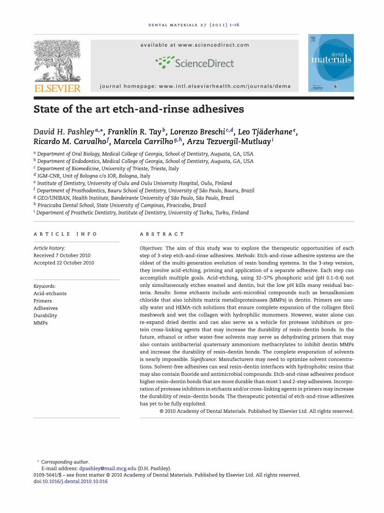
the hybrid layer (Fig. 1).
Evolution of etch-and-rinse adhesives When Fusayama [5]
introduced the revolutionary concept of total-etching of cav-
ities (i.e., simultaneous etching of enamel and dentin), the
technique was resisted by U.S. and European dentists. They
thought that 40% phosphoric acid would induce adverse pul-
pal reactions when allowed to etch dentin. Later work revealed
that acid-etching dentin more than 0.5 mm thick produced no
adverse pulpal reactions if the etched dentin could be sealed
from oral bacteria. The adverse pulpal reactions seen in the
U.S. and Europe were due to bacterial leakage, not acids per se
[6]. The lack of pulpal reactions to total-etching in Japan was
due to the fact that they only excavated carious dentin. As
part of their minimal invasive restorative philosophy, Japanese
dentists did not extend outline forms into normal dentin as
Fig. 1 – Schematic of a hybrid layer (HL) created by an
etch-and-rinse adhesive. Note that the depth of the hybrid
layer (green) is about four acid-etched tubule diameters (i.e.
ca. 8 �m). The collagen fibrils in the HL are continuous with
the underlying mineralized matrix. A single dentinal tubule
is shown devoid of a resin tag to illustrate its presence.
was the practice in the U.S. and Europe. Excavated caries-
affected dentin, unlike normal dentin, is almost impermeable
to all solutes and solvents [7], thereby protecting the pulp from
irritants.
The introduction of dry bonding The first marketed etch-and-
rinse adhesive was Clearfil Bond System-F (Kuraray Co., Ltd.,
Tokyo, Japan) in 1978. It utilized 40% phosphoric acid used
in the total-etch manner. Adverse pulpal reactions contin-
ued to be reported in the U.S. following acid-etching of dentin
with phosphoric acid because clinicians were performing “dry
bonding”. That is, after total-etching, they would dry the cav-
ity walls to confirm that the enamel margins were “frosty”
or had a chalk-like color. This meant that the enamel was
properly etched. What was not realized at that time was that
drying the cavity caused the acid-etched dentin to collapse.
Such collapsed demineralized dentin had lost the interfibril-
lar spaces between exposed collagen fibrils [8] that serve as
inward diffusion channels for monomer infiltration. Conse-
quently, resin–enamel bond strengths were high (ca. 20 MPa)
but resin–dentin bond strengths were very low (ca. 5 MPa).
Such low resin–dentin bond strengths were not sufficient to
resist the forces of polymerization shrinkage (about 24 MPa
in class I cavities) [9]. Thus, during polymerization of resin
composites, one or more of the bonded walls would debond,
creating bacterial leakage through normal permeable dentin
that could irritate the pulp.
The introduction of wet-bonding The low resin–dentin bond
strengths associated with “dry” bonding created dentin sen-
sitivity, microleakage, secondary caries and loss of bonded
restorations. Kanca found that water was an excellent rewet-
ting agent and this led to him [10] to introduce the concept
of “wet-bonding”. This technique increased the strength of
resin–dentin bonds, allowing good sealing of dentin and much
less post-operative pain. At this point resin–dentin bonds
equalled or exceeded resin–enamel bonds and the era of safe,
reproducible resin–dentin bonding began.
Dentin bonding as a form of tissue engineering In most tis-
sue engineering applications, one uses a 3-D scaffold (often
made of collagen) that is designed to be resorbed over sev-
eral weeks to months, to provide replacement by regenerating
tissues of the host [11,12]. Unlike classical tissue engineered
constructs, where the scaffold is designed to be resorbed and
replaced by normal tissue, in erupted teeth there are no tis-
sues available for regeneration of occlusal hard tissue surfaces
while teeth are in function. Instead, biocomposites must be
engineered within minutes, in situ, with the expectation that
they will last for decades! Because progress in adhesive den-
tistry has been incremental, we fail to recognize how far we
have come in 55 years since Buonocore [1] first acid-etched
enamel. Each day, practitioners bond relatively hydrophobic
resins to enamel and dentin within a few minutes and in
doing so have completely transformed the surface chemistry
of these hard tissues from wet, crystalline, hydrophilic sur-
faces that are acid-labile, to softer but tougher, hydrophobic,
drier dentin surfaces that are chemically compatible with
resin composites. These tooth colored biocomposites are also
acid-resistant. They can be made to be antibacterial [13,14].
Adhesive bonding begins by acid-etching to increase the
permeability of resins to enamel [2] and dentin [8]. In dentin,
this is a unique form of tissue engineering. Acid-etching with
d e n t a l m a t e r i a l s 2 7 ( 2 0 1 1 ) 1–16 3
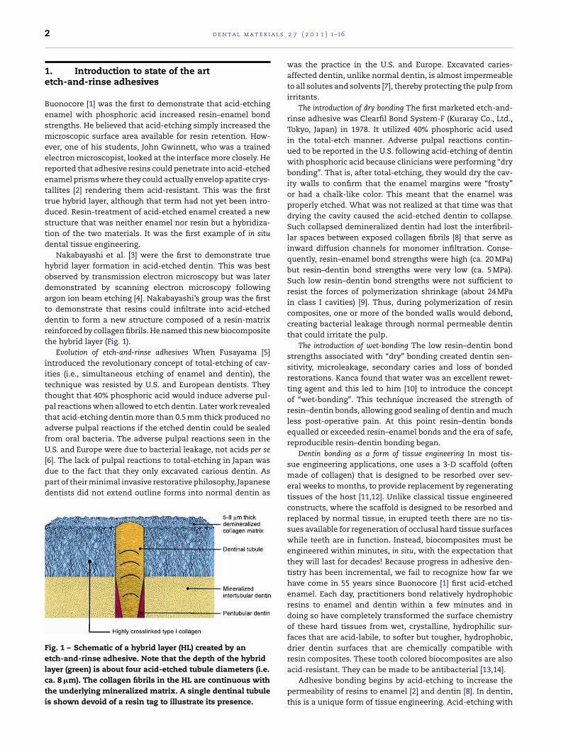
Fig. 2 – Scanning electron micrograph of acid-etched dentin
showing two dentinal tubules containing remnants of
peritubular dentin matrix. INSERT: High magnification of
branching collagen fibrils (ca. 75 nm in diameter) separated
by interfibrillar spaces that serve as channels for resin
infiltrations during bonding.
37 wt.% phosphoric acid completely demineralizes the surface
5–8 �m of the intertubular dentin matrix to create nanometer-
sized porosities (Fig. 2) within the underlying collagen fibrillar
matrix. This permits infiltration of solvated comonomers into
and around collagen fibrils to gain retention for tooth colored
resin-composite fillings [14]. Even more amazing is the con-
trast between the porosities of most bioengineered scaffolds
(5–20 �m) compared to the porosity of interfibrillar spaces
between resin-infiltrated collagen fibrils in hybrid layers that
are only 10–30 nm wide. Thus, the dental biocomposites that
are made by dentists in situ are created at a nanometer scale
over a distance of 5–8 �m!
Composition of mineralized vs. demineralized dentin vs. hybrid
layers Mineralized dentin is composed of approximately
50 vol.% mineral phase, 30 vol.% collagen and 20 vol.% water
[15] (Table 1). During the acid-etching process in etch-and-
rinse adhesives, the entire 50 vol.% of surface and subsurface
mineral is solubilized, extracted and is replaced by rinse-
water which, when combined with the intrinsic 20 vol.% of
water yields a new water content of 70 vol.% surrounding the
30 vol.% collagen fibrils that remain anchored into the under-
lying mineralized dentin. During the subsequent comonomer
infiltration phase of resin bonding, this 70 vol.% of water
should ideally be completely replaced by 70 vol.% of resin
comonomers that polymerize in situ to produce a hybridized
biocomposite of resin, reinforced with collagen fibrils known
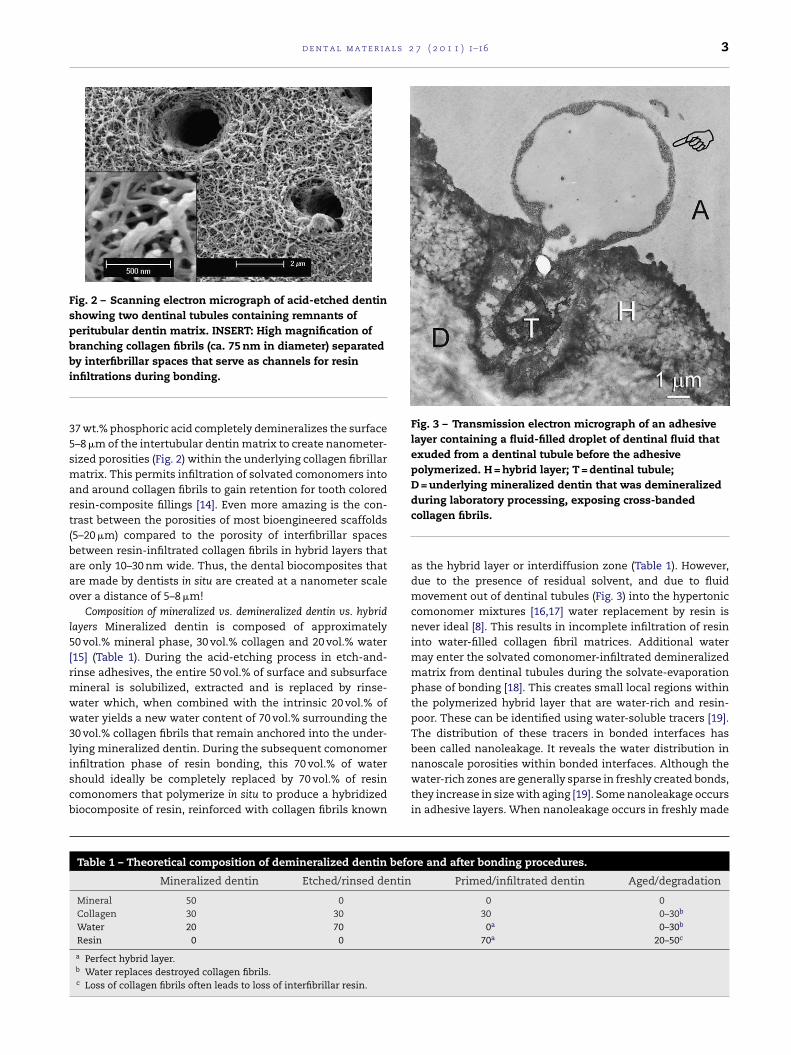
Fig. 3 – Transmission electron micrograph of an adhesive
layer containing a fluid-filled droplet of dentinal fluid that
exuded from a dentinal tubule before the adhesive
polymerized. H = hybrid layer; T = dentinal tubule;
D = underlying mineralized dentin that was demineralized
during laboratory processing, exposing cross-banded
collagen fibrils.
as the hybrid layer or interdiffusion zone (Table 1). However,
due to the presence of residual solvent, and due to fluid
movement out of dentinal tubules (Fig. 3) into the hypertonic
comonomer mixtures [16,17] water replacement by resin is
never ideal [8]. This results in incomplete infiltration of resin
into water-filled collagen fibril matrices. Additional water
may enter the solvated comonomer-infiltrated demineralized
matrix from dentinal tubules during the solvate-evaporation
phase of bonding [18]. This creates small local regions within
the polymerized hybrid layer that are water-rich and resin-
poor. These can be identified using water-soluble tracers [19].
The distribution of these tracers in bonded interfaces has
been called nanoleakage. It reveals the water distribution in
nanoscale porosities within bonded interfaces. Although the
water-rich zones are generally sparse in freshly created bonds,
they increase in size with aging [19]. Some nanoleakage occurs
in adhesive layers. When nanoleakage occurs in freshly made
Table 1 – Theoretical composition of demineralized dentin before and after bonding procedures.
Mineralized dentin Etched/rinsed dentin Primed/infiltrated dentin Aged/degradation
Mineral 50 0 0 0
Collagen 30 30 30 0–30b
Water 20 70 0a 0–30b
Resin 0 0 70a 20–50c
a Perfect hybrid layer.b Water replaces destroyed collagen fibrils.c Loss of collagen fibrils often leads to loss of interfibrillar resin.
4 d e n t a l m a t e r i a l s 2 7 ( 2 0 1 1 ) 1–16
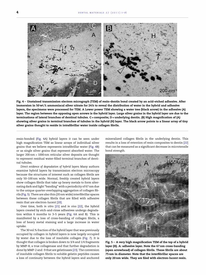
Fig. 4 – Unstained transmission electron micrograph (TEM) of resin–dentin bond created by an acid-etched adhesive. After
immersion in 50 wt.% ammoniacal silver nitrate for 24 h to reveal the distribution of water in the hybrid and adhesive
layers, the specimens were processed for TEM. A Lower power TEM showing a water tree (black arrow) in the adhesive (A)
layer. The region between the opposing open arrows is the hybrid layer. Large silver grains in the hybrid layer are due to the
terminations of lateral branches of dentinal tubules. C = composite; D = underlying dentin. (B) High magnification of (A)
showing silver grains in terminal branches of tubules in the hybrid (H) layer. The black arrow points to a linear array of tiny
silver grains thought to reside in intrafibrillar water inside collagen fibrils.
resin-bonded (Fig. 4A) hybrid layers it can be seen under
high magnification TEM as linear arrays of individual silver
grains that we believe represents intrafibrillar water (Fig. 4B)
or as single silver grains that represent absorbed water. The
larger 200 nm × 1000 nm reticular silver deposits are thought
to represent residual water-filled terminal branches of denti-
nal tubules.
Direct evidence of degradation of hybrid layers Many authors
examine hybrid layers by transmission electron microscopy
because the structures of interest such as collagen fibrils are
only 50–100 nm wide. Normal, freshly created hybrid layers
show collagen fibrils that take up heavy metals to form alter-
nating dark and light “banding” with a periodicity of 67 nm due
to the unique quarter-overlapping aggregation of collagen fib-
rils (Fig. 5). There are also thin (20 nm wide) interfibrillar spaces
between these collagen fibrils that are filled with adhesive
resin that are electron-lucent [20].
Over time, both in vitro [21] and in vivo [22], the hybrid
layers created by etch-and-rinse adhesives undergo degrada-
tion within 6 months to 3–5 years (Fig. 6A and B). This is
manifested by a loss of cross-banding of collagen fibrils, a
loss of heavy metal staining and a large increase in water
uptake.
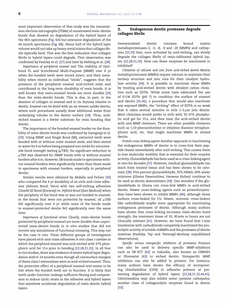
The 30 vol.% fraction of the hybrid layer that was previously
occupied by collagen in hybrid layers is now largely occupied
by water due to the loss of insoluble collagen (Fig. 7). It is
thought that collagen is broken down to 3/4 and 1/4 fragments
by MMP-8, a true collagenase and that further degradation is
done by MMP-2 and -9 that are gelatinases [23]. The conversion
of insoluble collagen fibrils to soluble gelatin peptides causes
a loss of continuity between the hybrid layers and anchored
mineralized collagen fibrils in the underlying dentin. This
results in a loss of retention of resin composites to dentin [22]
that can be measured as a significant decrease in microtensile
bond strength.
Fig. 5 – A very high magnification TEM of the top of a hybrid
layer (H). A: adhesive layer. Note the 67 nm cross-banding
(open arrowhead) of collagen fibrils. These fibrils are about
75 nm in diameter. Note that the interfibrillar spaces are
only 20 nm wide. They are filed with electron-lucent resin.
d e n t a l m a t e r i a l s 2 7 ( 2 0 1 1 ) 1–16 5
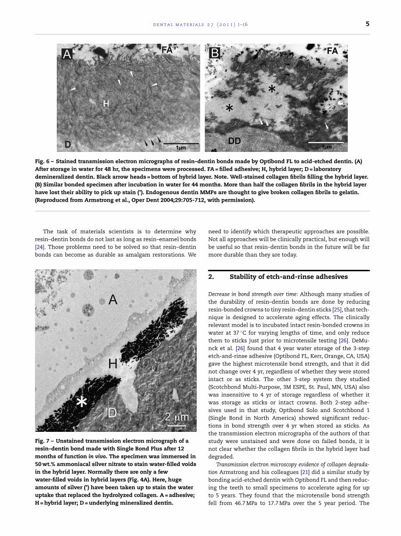
Fig. 6 – Stained transmission electron micrographs of resin–dentin bonds made by Optibond FL to acid-etched dentin. (A)
After storage in water for 48 hr, the specimens were processed. FA = filled adhesive; H, hybrid layer; D = laboratory
demineralized dentin. Black arrow heads = bottom of hybrid layer. Note. Well-stained collagen fibrils filling the hybrid layer.
(B) Similar bonded specimen after incubation in water for 44 months. More than half the collagen fibrils in the hybrid layer
have lost their ability to pick up stain (*). Endogenous dentin MMPs are thought to give broken collagen fibrils to gelatin.
(Reproduced from Armstrong et al., Oper Dent 2004;29:705-712, with permission).
The task of materials scientists is to determine why
resin–dentin bonds do not last as long as resin–enamel bonds
[24]. Those problems need to be solved so that resin–dentin
bonds can become as durable as amalgam restorations. We
Fig. 7 – Unstained transmission electron micrograph of a
resin–dentin bond made with Single Bond Plus after 12
months of function in vivo. The specimen was immersed in
50 wt.% ammoniacal silver nitrate to stain water-filled voids
in the hybrid layer. Normally there are only a few
water-filled voids in hybrid layers (Fig. 4A). Here, huge
amounts of silver (*) have been taken up to stain the water
uptake that replaced the hydrolyzed collagen. A = adhesive;
H = hybrid layer; D = underlying mineralized dentin.
need to identify which therapeutic approaches are possible.
Not all approaches will be clinically practical, but enough will
be useful so that resin–dentin bonds in the future will be far
more durable than they are today.
2. Stability of etch-and-rinse adhesives
Decrease in bond strength over time: Although many studies of
the durability of resin–dentin bonds are done by reducing
resin-bonded crowns to tiny resin–dentin sticks [25], that tech-
nique is designed to accelerate aging effects. The clinically
relevant model is to incubated intact resin-bonded crowns in
water at 37 ◦C for varying lengths of time, and only reduce
them to sticks just prior to microtensile testing [26]. DeMu-
nck et al. [26] found that 4 year water storage of the 3-step
etch-and-rinse adhesive (Optibond FL, Kerr, Orange, CA, USA)
gave the highest microtensile bond strength, and that it did
not change over 4 yr, regardless of whether they were stored
intact or as sticks. The other 3-step system they studied
(Scotchbond Multi-Purpose, 3M ESPE, St. Paul, MN, USA) also
was insensitive to 4 yr of storage regardless of whether it
was storage as sticks or intact crowns. Both 2-step adhe-
sives used in that study, Optibond Solo and Scotchbond 1
(Single Bond in North America) showed significant reduc-
tions in bond strength over 4 yr when stored as sticks. As
the transmission electron micrographs of the authors of that
study were unstained and were done on failed bonds, it is
not clear whether the collagen fibrils in the hybrid layer had
degraded.
Transmission electron microscopy evidence of collagen degrada-
tion Armstrong and his colleagues [21] did a similar study by
bonding acid-etched dentin with Optibond FL and then reduc-
ing the teeth to small specimens to accelerate aging for up
to 5 years. They found that the microtensile bond strength
fell from 46.7 MPa to 17.7 MPa over the 5 year period. The
6 d e n t a l m a t e r i a l s 2 7 ( 2 0 1 1 ) 1–16
most important observation of that study was the transmis-
sion electron micrographs (TEMs) of nonstressed resin–dentin
bonds that showed no degradation of the hybrid layers of
the 48 h specimens (Fig. 6A) but extensive degradation of the
44 month specimens (Fig. 6B). About half of the hybrid layer
volume would not take up heavy metal stains that collagen fib-
rils typically bind. This was the first indication that collagen
fibrils in hybrid layers could degrade. That observation was
confirmed by Pashley et al. [27] and later by Hebling et al. [28].
Importance of peripheral enamel seal The stability of Opti-
bond FL and Scotchbond Multi-Purpose (SBMP) over 4 yr
when the bonded teeth were stored intact, and their insta-
bility when stored as individual “sticks”, suggests that the
presence of the peripheral enamel acid-etched resin seal
contributed to the long-term durability of resin bonds. It is
well known that resin–enamel bonds are more durable [24]
than the resin–dentin bonds. This is due, in part, to the
absence of collagen in enamel and to its dryness relative to
dentin. Enamel can be dried with an air stream unlike dentin,
where such procedures usually draw additional water from
underlying tubules to the dentin surface [18]. Thus, acid-
etched enamel is a better substrate for resin bonding that
dentin.
The importance of the bonded enamel border on the dura-
bility of resin–dentin bonds was confirmed by Gamgorgi et al.
[29]. Using SBMP and Single Bond (SB), extracted teeth were
bonded with or without outer enamel seals, and then stored
in water for 6 m before being prepared into sticks for microten-
sile bond strength testing (�TBS). No significant reduction in
�TBS was observed for SBMP bonds with or without enamel
borders after 6 m. However, SB bonds made to specimens with-
out enamel borders were significantly lower than those made
to specimens with enamel borders, especially in peripheral
dentin.
Similar results were obtained by Abdalla and Feilzer [30]
who compared the 4 yr durability of an etch-and-rinse adhe-
sive (Admira Bond, Voco) with two self-etching adhesives
Clearfil SE Bond (Kuraray) vs. Hybrid Bond (Sun Medical) when
the periphery of the bond was or was not bonded to enamel.
In the bonds that were not protected by enamel, all �TBS
fell significantly over 4 yr while none of the bonds made
to enamel-protected dentin fell significantly over the same
time.
Importance of functional stress Clearly, resin–dentin bonds
protected by peripheral enamel are more durable than unpro-
tected resin–dentin bonds in in vitro studies that did not
involve any simulations of functional stressing. This may not
be the case in vivo. Three different groups of investigators
have placed etch-and-rinse adhesives in vivo class I cavities in
which the peripheral enamel was acid-etched with 37% phos-
phoric acid for 15 s prior to bonding [22,28,31,32]. In all four
in vivo studies, there was evidence of severe hybrid layer degra-
dation with 6–14 months even though all cavosurface margins
of these class I restorations were in acid-etched enamel. Thus,
the protective effect of a peripheral enamel seal seems to be
lost when the bonded teeth are in function. It is likely that
teeth under function undergo sufficient flexing and compres-
sion to induce cyclic strain in the adhesive and hybrid layers
that somehow accelerate degradation of resin–dentin hybrid
layers.
3. Endogenous dentin proteases degradecollagen fibrils
Demineralized dentin contains bound matrix
metalloproteinases-2, -3, -8, -9 and -20 (MMPs) and cathep-
sins [23,33] that, once activated by acid-etching, can slowly
degrade the collagen fibrils of resin-infiltrated hybrid lay-
ers [22,28,31,32]. How can these enzymes be inactivated or
inhibited?
Chelation of calcium and zinc from acid-etched dentin Matrix
metalloproteinases (MMPs) require calcium to maintain their
tertiary structure and zinc ions for their catalytic hydro-
lase activity [34]. It is possible to inactivate these MMPs
by treating acid-etched dentin with divalent cation chela-
tors such as EDTA. While some have advocated the use
of 0.5 M EDTA (pH 7) to condition the surface of enamel
and dentin [35,36], a procedure that would also inactivate
any exposed MMPs, the “etching” effect of EDTA is so weak
that it takes several minutes to etch 1–2 �m into dentin.
Most clinicians would prefer to etch with 32–37% phospho-
ric acid gel for 15 s, and then treat the acid-etched dentin
with anti-MMP chelators. There are other possible chelators
such as 1,10-phenanthroline or ethylene diamine tetraphos-
phonic acid, etc. that might inactivate MMPs in etched
dentin.
Protein cross-linking agents Another approach to inactivating
the endogenous MMPs of dentin is to cross-link their pep-
tide chains immediately after acid-etching. This causes them
to lose molecular mobility that is essential for their enzyme
activity. Glutaraldehyde has been used as a cross-linking agent
in vitro for decades [37]. However, residual glutaraldehyde can
leach from treated tissue and has been shown to be cyto-
toxic [38]. Five percent glutaraldehyde, 35% HEMA, 60% water
mixtures (Gluma Desensitizer, Heraeus Kulzer) continue to
be used as dentin desensitizers [39]. One wonders if the glu-
taraldehyde in Gluma can cross-link MMPs in acid-etched
dentin. Newer cross-linking agents such as proanthocyani-
dins have been shown to inhibit MMPs [40], although those
authors cross-linked for 2 h. Newer, nontoxic cross-linkers
like carbodiimide maybe more appropriate for inactivating
endogenous proteases of dentin. Although many authors
have shown that cross-linking increases resin–dentin bond
strength, the treatment times of 10, 30 min or hours are not
clinically relevant [41]. However, we have found that 1 min
treatments with carbodiimide completely inactivated the pro-
teolytic activity of soluble rhMMPs and the proteases of dentin
matrices [Pashley, Tay and Tezvergil-Mutluay unpublished
observations].
Specific versus nonspecific inhibitors of proteases Primers
can also be used to delivery specific MMP-inhibitors
such as SB-3CT [42] or Galardin, also known as GM6001
or Illomastat [43] to etched dentin. Nonspecific MMP
inhibitors can also be added to primers. For instance,
many authors have shown the efficacy of incorporat-
ing chlorhexidine (CHX) in adhesive primers at pre-
venting degradation of hybrid layers [22,28,31,32,44,45].
Chlorhexidine may also inhibit some cysteine cathepsins,
another class of collagenolytic enzymes found in dentin
[33].
d e n t a l m a t e r i a l s 2 7 ( 2 0 1 1 ) 1–16 7
Table 2 – Relative solubility (g comonomers/mL solvent) of comonomer blends and selected monomers in water vs.ethanol.
Water Ethanol Ethanol/water
70% ethoxy BisGMA/28% TEGDMA 0.0027 0.1735 64.3
70% BisGMA/28% TEGDMA 0.0038 0.1632 42.8
70% BisGMA/28% HEMA 0.0063 0.1268 20.1
40% BisGMA/30% TCDM/28% HEMA 0.0118 0.1296 11.0
40% BisGMA/30% BisMP/28% HEMA 0.0422 0.1515 3.6
100% HEMA 0.1120 0.1451 1.3
100% TEGDMA 0.0095 0.1692 17.8
100% BisGMA 0.0 0.1688 –
100% Bis-MP 0.1282 0.1890 1.5
4. The role of proteoglycan hydrogels ininterfibrillar spaces
Interfibrillar spaces in acid-etched dentin contain more than
water. They also contain highly hydrated negatively charged
proteoglycans that form a hydrogel within that space [46,47].
Removal of these molecules by prolonged enzymatic treat-
ment has been shown to produce large increases (49 and 63%,
respectively) in bond strength of Scotchbond Multi-Purpose
and Prime & Bond NT [48]. Those author’s used chondroitinase
ABC to remove interfibrillar chondroitin sulfate-containing
glycosaminoglycans (GAGs) that allowed better resin infiltra-
tion. Unfortunately, the time necessary for enzymatic removal
(24 h) of GAGs is not clinically relevant.
Molecular sieving within proteoglycan hydrogels If the GAG
“hydrogels” remain hydrated in interfibrillar spaces, they may
be responsible for “molecular sieving” of larger dimethacry-
lates like BisGMA, that cross-link polymers, from smaller
2-hydroxyethylmethacrylate (HEMA), that do not cross-link
polymers, restricting BisGMA to the upper half of hybrid lay-
ers [49,50]. HEMA-rich lower halves of hybrid layers might be
undergo large positive and negative strains during function
that could lead to fatigue failure of collagen fibrils.
Scott and Thomlinson [47] showed that organic solvents
(e.g., ethanol, acetone) cause a collapse of anionic gly-
cosamineglycans gels in connective tissues by removing their
water content.
Manufacturers of 2-step, etch-and-rinse adhesives dissolve
their comonomers in ethanol or acetone, not water. This is
because the dimethacrylates they use to toughen their poly-
mers by cross-linking are not miscible with water but are
soluble in ethanol (Table 2). Two-step etch-and-rinse adhesive
blends contain both primer (i.e., HEMA) and adhesive (i.e., Bis-
GMA) monomers in solvents containing low concentrations of
water in the same bottle. They are applied in two layers; the
first layer serves as a primer, while the second layer serves
as an adhesive layer. However, this formulation compromise
prevents the therapeutic opportunity provided by a separate
solvent-free adhesive in 3-step systems. Since comonomer
blends are dissolved in ethanol, can acid-etched dentin be
saturated with ethanol to enhance monomer penetration?
Use of an ethanol primer: When ethanol-solvated blends
are applied to water-saturated dentin, they often undergo
nanoscopic phase changes [51]. However, if after acid-etching
and rinsing, the water is replaced with an ethanol primer, the
matrix becomes saturated with ethanol, the same solvent the
comonomers are suspended in, and there can be no phase
changes [52].
Ethanol wet-bonding [52] was developed to enhance the
durability of etch-and-rinse adhesives. In this technique,
ethanol is used to chemically dehydrate acid-etched deminer-
alized dentin matrices [53]. This results in a lateral shrinkage
of collagen fibrils, causing an increase in the width of their
interfibrillar spaces and a reduction in the hydrophilicity of
the collagen matrix. Tay et al. [54] solvated BisGMA in ethanol
and applied it to ethanol-saturated acid-etched dentin. They
showed that excellent, high bond strengths could be achieved
between BisGMA and dentin. We do not recommend bonding
BisGMA to dentin but we did demonstrate the proof-of-
concept of ethanol wet-bonding using this water-insoluble
monomer.
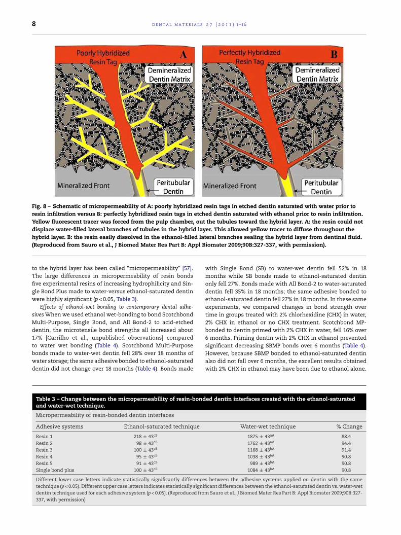
Clinical implications of ethanol wet-bonding We speculate that
bonding contemporary etch-and-rinse adhesives to water-
saturated dentin leaves a very thin layer of water or a hydrogel
between the infiltrated adhesive and the collagen fibrils of the
matrix. This layer seems to provide a fluid-filled continuum
from resin tags into the base of the hybrid layer through lat-
eral branches of dentinal tubules (Fig. 8) throughout the full
thickness of the hybrid layer. What evidence is there that this
is true? We have prepared resin-bonded crown segments con-
taining the top of the pulp chamber from extracted teeth,
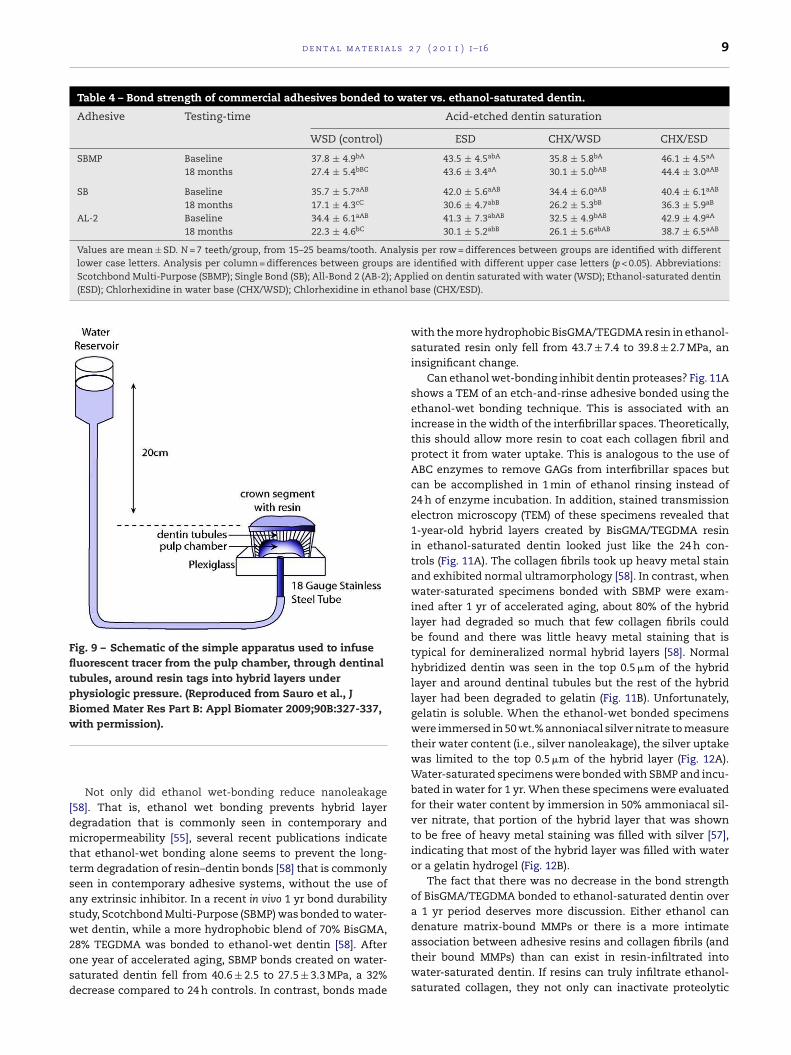
mounted on a plastic plate penetrated by an 18 ga. tube (Fig. 9).
The pulp chamber was filled with a water-soluble fluores-
cent dye under a physiologic pressure (20 cm H2O) for 3 h after
bonding with etch-and-rinse adhesives [55]. During this time,
an aqueous solution of fluorescent dye slowly seeped out the
dentinal tubules, permeated around poorly hybridized resin
tags, and continued to diffuse fluorescent dye outward via
lateral branches of dentinal tubules that ramify into smaller
branches that finally become continuous with interfibrillar
spaces around collagen fibrils in the hybrid layer. The end
result in specimens that were bonded while moist with water,
is that the entire hybrid layer becomes fluorescent, regard-
less of the adhesive resin (Fig. 10A). When the same adhesives
are applied to ethanol-saturated dentin (Fig. 10B), the hybrid
layers were not fluorescent. This means that fluorescent dye
can not permeate around resin tags to reach the hybrid layer
when resins were bonded to ethanol-saturated dentin. Rather,
the resin tags have hybridized with the surrounding collagen
fibrils and have sealed the tubules in ethanol-saturated dentin
much better than in water-saturated dentin [56].
The use of fluorescent dye under pressure to trace the
permeation pathways from the pulp, out dentinal tubules
8 d e n t a l m a t e r i a l s 2 7 ( 2 0 1 1 ) 1–16
Fig. 8 – Schematic of micropermeability of A: poorly hybridized resin tags in etched dentin saturated with water prior to
resin infiltration versus B: perfectly hybridized resin tags in etched dentin saturated with ethanol prior to resin infiltration.
Yellow fluorescent tracer was forced from the pulp chamber, out the tubules toward the hybrid layer. A: the resin could not
displace water-filled lateral branches of tubules in the hybrid layer. This allowed yellow tracer to diffuse throughout the
hybrid layer. B: the resin easily dissolved in the ethanol-filled lateral branches sealing the hybrid layer from dentinal fluid.
(Reproduced from Sauro et al., J Biomed Mater Res Part B: Appl Biomater 2009;90B:327-337, with permission).
to the hybrid layer has been called “micropermeability” [57].
The large differences in micropermeability of resin bonds
five experimental resins of increasing hydrophilicity and Sin-
gle Bond Plus made to water-versus ethanol-saturated dentin
were highly significant (p < 0.05, Table 3).
Effects of ethanol-wet bonding to contemporary dental adhe-
sives When we used ethanol wet-bonding to bond Scotchbond
Multi-Purpose, Single Bond, and All Bond-2 to acid-etched
dentin, the microtensile bond strengths all increased about
17% [Carrilho et al., unpublished observations] compared
to water wet bonding (Table 4). Scotchbond Multi-Purpose
bonds made to water-wet dentin fell 28% over 18 months of
water storage; the same adhesive bonded to ethanol-saturated
dentin did not change over 18 months (Table 4). Bonds made
with Single Bond (SB) to water-wet dentin fell 52% in 18
months while SB bonds made to ethanol-saturated dentin
only fell 27%. Bonds made with All Bond-2 to water-saturated
dentin fell 35% in 18 months; the same adhesive bonded to
ethanol-saturated dentin fell 27% in 18 months. In these same
experiments, we compared changes in bond strength over
time in groups treated with 2% chlorhexidine (CHX) in water,
2% CHX in ethanol or no CHX treatment. Scotchbond MP-
bonded to dentin primed with 2% CHX in water, fell 16% over
6 months. Priming dentin with 2% CHX in ethanol prevented
significant decreasing SBMP bonds over 6 months (Table 4).
However, because SBMP bonded to ethanol-saturated dentin
also did not fall over 6 months, the excellent results obtained
with 2% CHX in ethanol may have been due to ethanol alone.
Table 3 – Change between the micropermeability of resin-bonded dentin interfaces created with the ethanol-saturatedand water-wet technique.
Micropermeability of resin-bonded dentin interfaces
Adhesive systems Ethanol-saturated technique Water-wet technique % Change
Resin 1 218 ± 43cB 1875 ± 43aA 88.4
Resin 2 98 ± 43cB 1762 ± 43aA 94.4
Resin 3 100 ± 43cB 1168 ± 43bA 91.4
Resin 4 95 ± 43cB 1038 ± 43bA 90.8
Resin 5 91 ± 43cB 989 ± 43bA 90.8
Single bond plus 100 ± 43cB 1084 ± 43bA 90.8
Different lower case letters indicate statistically significantly differences between the adhesive systems applied on dentin with the same
technique (p < 0.05). Different upper case letters indicates statistically significant differences between the ethanol-saturated dentin vs. water-wet
dentin technique used for each adhesive system (p < 0.05). (Reproduced from Sauro et al., J Biomed Mater Res Part B: Appl Biomater 2009;90B:327-
337, with permission)
d e n t a l m a t e r i a l s 2 7 ( 2 0 1 1 ) 1–16 9
Table 4 – Bond strength of commercial adhesives bonded to water vs. ethanol-saturated dentin.
Adhesive Testing-time Acid-etched dentin saturation
WSD (control) ESD CHX/WSD CHX/ESD
SBMP Baseline 37.8 ± 4.9bA 43.5 ± 4.5abA 35.8 ± 5.8bA 46.1 ± 4.5aA
18 months 27.4 ± 5.4bBC 43.6 ± 3.4aA 30.1 ± 5.0bAB 44.4 ± 3.0aAB
SB Baseline 35.7 ± 5.7aAB 42.0 ± 5.6aAB 34.4 ± 6.0aAB 40.4 ± 6.1aAB
18 months 17.1 ± 4.3cC 30.6 ± 4.7abB 26.2 ± 5.3bB 36.3 ± 5.9aB
AL-2 Baseline 34.4 ± 6.1aAB 41.3 ± 7.3abAB 32.5 ± 4.9bAB 42.9 ± 4.9aA
18 months 22.3 ± 4.6bC 30.1 ± 5.2abB 26.1 ± 5.6abAB 38.7 ± 6.5aAB
Values are mean ± SD. N = 7 teeth/group, from 15–25 beams/tooth. Analysis per row = differences between groups are identified with different
lower case letters. Analysis per column = differences between groups are identified with different upper case letters (p < 0.05). Abbreviations:
Scotchbond Multi-Purpose (SBMP); Single Bond (SB); All-Bond 2 (AB-2); Applied on dentin saturated with water (WSD); Ethanol-saturated dentin
(ESD); Chlorhexidine in water base (CHX/WSD); Chlorhexidine in ethanol base (CHX/ESD).
Fig. 9 – Schematic of the simple apparatus used to infuse
fluorescent tracer from the pulp chamber, through dentinal
tubules, around resin tags into hybrid layers under
physiologic pressure. (Reproduced from Sauro et al., J
Biomed Mater Res Part B: Appl Biomater 2009;90B:327-337,
with permission).
Not only did ethanol wet-bonding reduce nanoleakage
[58]. That is, ethanol wet bonding prevents hybrid layer
degradation that is commonly seen in contemporary and
micropermeability [55], several recent publications indicate
that ethanol-wet bonding alone seems to prevent the long-
term degradation of resin–dentin bonds [58] that is commonly
seen in contemporary adhesive systems, without the use of
any extrinsic inhibitor. In a recent in vivo 1 yr bond durability
study, Scotchbond Multi-Purpose (SBMP) was bonded to water-
wet dentin, while a more hydrophobic blend of 70% BisGMA,
28% TEGDMA was bonded to ethanol-wet dentin [58]. After
one year of accelerated aging, SBMP bonds created on water-
saturated dentin fell from 40.6 ± 2.5 to 27.5 ± 3.3 MPa, a 32%
decrease compared to 24 h controls. In contrast, bonds made
with the more hydrophobic BisGMA/TEGDMA resin in ethanol-
saturated resin only fell from 43.7 ± 7.4 to 39.8 ± 2.7 MPa, an
insignificant change.
Can ethanol wet-bonding inhibit dentin proteases? Fig. 11A
shows a TEM of an etch-and-rinse adhesive bonded using the
ethanol-wet bonding technique. This is associated with an
increase in the width of the interfibrillar spaces. Theoretically,
this should allow more resin to coat each collagen fibril and
protect it from water uptake. This is analogous to the use of
ABC enzymes to remove GAGs from interfibrillar spaces but
can be accomplished in 1 min of ethanol rinsing instead of
24 h of enzyme incubation. In addition, stained transmission
electron microscopy (TEM) of these specimens revealed that
1-year-old hybrid layers created by BisGMA/TEGDMA resin
in ethanol-saturated dentin looked just like the 24 h con-
trols (Fig. 11A). The collagen fibrils took up heavy metal stain
and exhibited normal ultramorphology [58]. In contrast, when
water-saturated specimens bonded with SBMP were exam-
ined after 1 yr of accelerated aging, about 80% of the hybrid
layer had degraded so much that few collagen fibrils could
be found and there was little heavy metal staining that is
typical for demineralized normal hybrid layers [58]. Normal
hybridized dentin was seen in the top 0.5 �m of the hybrid
layer and around dentinal tubules but the rest of the hybrid
layer had been degraded to gelatin (Fig. 11B). Unfortunately,
gelatin is soluble. When the ethanol-wet bonded specimens
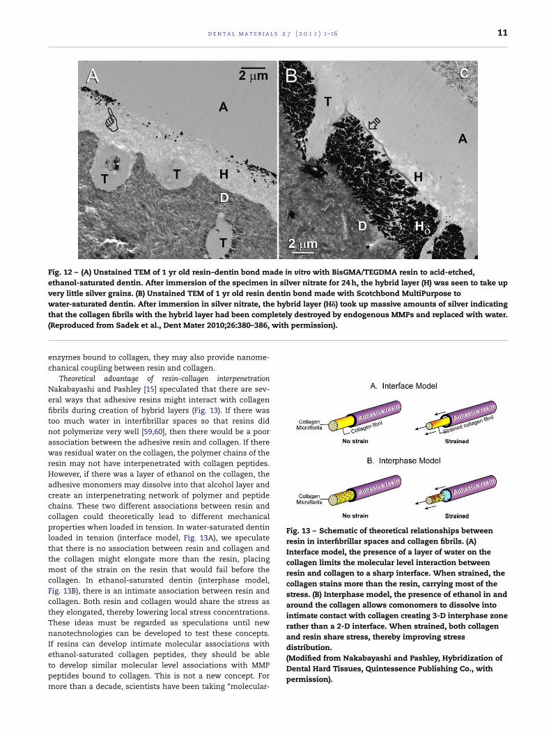
were immersed in 50 wt.% annoniacal silver nitrate to measure
their water content (i.e., silver nanoleakage), the silver uptake
was limited to the top 0.5 �m of the hybrid layer (Fig. 12A).
Water-saturated specimens were bonded with SBMP and incu-
bated in water for 1 yr. When these specimens were evaluated
for their water content by immersion in 50% ammoniacal sil-
ver nitrate, that portion of the hybrid layer that was shown
to be free of heavy metal staining was filled with silver [57],
indicating that most of the hybrid layer was filled with water
or a gelatin hydrogel (Fig. 12B).
The fact that there was no decrease in the bond strength
of BisGMA/TEGDMA bonded to ethanol-saturated dentin over
a 1 yr period deserves more discussion. Either ethanol can
denature matrix-bound MMPs or there is a more intimate
association between adhesive resins and collagen fibrils (and
their bound MMPs) than can exist in resin-infiltrated into
water-saturated dentin. If resins can truly infiltrate ethanol-
saturated collagen, they not only can inactivate proteolytic
10 d e n t a l m a t e r i a l s 2 7 ( 2 0 1 1 ) 1–16
Fig. 10 – A: Confocal laser scanning microscropy images of resin–dentin bonds made to crown segments. After
polymerizing the resin, the pulp chamber was filled with lucifer yellow and placed under 20 cm H2O pressure to allow the
fluorescent tracer to seep wherever there were water-filled submicron channels from the pulp to the hybrid layer. A:
resin–dentin bond made to acid-etched water-saturated dentin. Note that there is a fluorescence continuum from dentinal
tubules (t), around resin tags (rt), into the hybrid layer. Note that the entire hybrid layer was fluorescent B: When the same
resin was bonded to etched dentin saturated with ethanol, the lucifer yellow in the dentinal tubules (t) stopped when it
encountered the 10 �m thick zone of well-hybridized resin tags (rt), just below the hybrid layer. No lucifer yellow passed
around any resin tags, leaving the hybrid layer free of fluorescence.
Fig. 11 – (A) Stained TEM of acid-etched specimen bonded with BisGMA/TEGDMA resin under ethanol-wet bonding
conditions. A = adhesive resin; H = hybrid layer occupies space above the two open arrow heads. Appearance of bond after 1
yr of water storage. (B) Stained TEM of Scotchbond MultiPurpose Plus bond made to acid-etched dentin saturated with
water, after 1 yr of storage in water. C = hybrid composite; A = adhesive; H� = hybrid layer; D = underlying mineralized dentin
that was demineralized during TEM processing; T = resin tags in tubules. Note how much stain was taken up by laboratory
demineralized dentin and how little was taken up by hybrid layer due to degradation of collagen by endogenous MMPs.
(Reproduced from Sadek et al., Dent Mater 2010;26:380-386, with permission).
d e n t a l m a t e r i a l s 2 7 ( 2 0 1 1 ) 1–16 11
Fig. 12 – (A) Unstained TEM of 1 yr old resin–dentin bond made in vitro with BisGMA/TEGDMA resin to acid-etched,
ethanol-saturated dentin. After immersion of the specimen in silver nitrate for 24 h, the hybrid layer (H) was seen to take up
very little silver grains. (B) Unstained TEM of 1 yr old resin dentin bond made with Scotchbond MultiPurpose to
water-saturated dentin. After immersion in silver nitrate, the hybrid layer (H�) took up massive amounts of silver indicating
that the collagen fibrils with the hybrid layer had been completely destroyed by endogenous MMPs and replaced with water.
(Reproduced from Sadek et al., Dent Mater 2010;26:380–386, with permission).
enzymes bound to collagen, they may also provide nanome-
chanical coupling between resin and collagen.
Theoretical advantage of resin–collagen interpenetration
Nakabayashi and Pashley [15] speculated that there are sev-
eral ways that adhesive resins might interact with collagen
fibrils during creation of hybrid layers (Fig. 13). If there was
too much water in interfibrillar spaces so that resins did
not polymerize very well [59,60], then there would be a poor
association between the adhesive resin and collagen. If there
was residual water on the collagen, the polymer chains of the
resin may not have interpenetrated with collagen peptides.
However, if there was a layer of ethanol on the collagen, the
adhesive monomers may dissolve into that alcohol layer and
create an interpenetrating network of polymer and peptide
chains. These two different associations between resin and
collagen could theoretically lead to different mechanical
properties when loaded in tension. In water-saturated dentin
loaded in tension (interface model, Fig. 13A), we speculate
that there is no association between resin and collagen and
the collagen might elongate more than the resin, placing
most of the strain on the resin that would fail before the
collagen. In ethanol-saturated dentin (interphase model,
Fig. 13B), there is an intimate association between resin and
collagen. Both resin and collagen would share the stress as
they elongated, thereby lowering local stress concentrations.
These ideas must be regarded as speculations until new
nanotechnologies can be developed to test these concepts.
If resins can develop intimate molecular associations with
ethanol-saturated collagen peptides, they should be able
to develop similar molecular level associations with MMP
peptides bound to collagen. This is not a new concept. For
more than a decade, scientists have been taking “molecular-
Fig. 13 – Schematic of theoretical relationships between
resin in interfibrillar spaces and collagen fibrils. (A)
Interface model, the presence of a layer of water on the
collagen limits the molecular level interaction between
resin and collagen to a sharp interface. When strained, the
collagen stains more than the resin, carrying most of the
stress. (B) Interphase model, the presence of ethanol in and
around the collagen allows comonomers to dissolve into
intimate contact with collagen creating 3-D interphase zone
rather than a 2-D interface. When strained, both collagen
and resin share stress, thereby improving stress
distribution.
(Modified from Nakabayashi and Pashley, Hybridization of
Dental Hard Tissues, Quintessence Publishing Co., with
permission).
12 d e n t a l m a t e r i a l s 2 7 ( 2 0 1 1 ) 1–16
level” impressions of substrates and even enzymes, using
methacrylate monomers similar to those used in adhesive
dentistry. After free radical polymerization, the impres-
sions are separated from the substrate. These impressions
reproduce the 3-D structure of the substrate indicating that
these materials “wet” substrate molecules. This technique
is called “molecular imprinting” [61,62]. The phenomenon
has also been called “nano-interdigitation” [63] of polymer
interpenetration. We speculate that ethanol-saturated dentin
allows adhesive resins to flow into the catalytic sites of MMPs
and polymerize, thereby inactivating the enzymes. This is
another example of how therapeutic primers can increase
the durability of resin–dentin bonds.
5. Is there a role for therapeutics in dentinbonding?
Etch-and-rinse adhesives that utilize 3-steps are more durable
than 2-step etch-and-rinse adhesives [64]. By maintaining
separate acid-etching, priming and bonding steps in etch-
and-rinse adhesives, one can accomplish multiple therapeutic
goals in each step. By looking at 3-step etch-and-rinse adhe-
sive systems as a form of tissue engineering, one discovers
many opportunities for added value therapies. One advantage
of a three-step bonding procedure is the opportunity to use
each step for multiple purposes.
Acid etching For instance, 35–37% phosphoric acid etchant
can simultaneously etch enamel and dentin [5,10]. When acid-
etching enamel with 32–37% phosphoric acid, both the intra-
and interprismatic enamel can be well-etched at pH 0.4 that
is not possible with self-etching primers or all-in-one adhe-
sives that typically have pHs of 2–2.8. Studies have shown
that 32–37% phosphoric acid decimates residual bacteria in
caries-affected dentin [65]. Phosphoric acid also inactivates
MMP activity in dentin by 65–95% [27,66].
If that phosphoric acid also contains 1 wt.% benzalkonium
chloride (BAC) such as ETCH-37 w/BAC, marketed by Bisco, Inc.
(Schaumburg, IL, USA), then one can bind BAC to etched col-
lagen as it is being etched. We have recently found that BAC
is a good matrix metalloprotease (MMP) inhibitor [67] that can
withstand the low pH of phosphoric acid and does not lower
enamel or dentin bond strengths [68].
When using etchants that contain benzalkonium chloride,
a potent antimicrobial agent, one is also acid-etching enamel
and dentin, killing most of the residual bacterial in the dentin
and allowing BAC to bind to the demineralized matrix where
it exerts both an anti-microbial effect as well as an anti-MMP
effect [67]. This is a fine example of multi-role therapeutics.
Others have added 2% chlorhexidine to 37% phosphoric acid
for its anti-MMP effect [69].
It is important that acid-etched enamel and dentin by thor-
oughly rinsed to remove all reaction products. This water
also insures that the demineralized dentin matrix is fully
expanded [70]. If the excess water is not removed by blotting
or a half second air blast just prior to bonding, the residual
water may induce phase changes in etch-and-rinse adhesives
that contain BisGMA [51,71].
Application of primers The use of a primer was originally
designed to re-expand dried collapsed dentin and to coat the
wet collagen fibrils with a hydrophilic monomer like HEMA.
However, primers in etch-and-rinse adhesive systems offer
additional therapeutic opportunities. For instance, some clin-
icians treat acid-etched dentin with 0.2–2 wt.% chlorhexidine
in water or ethanol. Both ethanol and CHX tend to kill any
bacteria that survive acid-etching and then CHX binds to acid-
etched dentin [72] where it inhibits dentin MMPs and prolong
the durability of resin–dentin bonds [28]. Acid-etched water-
rinsed intertubular dentin matrix is almost 70% water and
30% organic matrix (Table 1). Thus, application of almost any
therapeutic agent will diffuse into that unbound water and
then bind to the organic matrix. Such binding has recently
been shown for chlorhexidine [72], benzalkonium chloride [67]
and polyvinylphosphonic acid, another potent MMP inhibitor
[73]. Both chlorhexidine and benzalkonuim chloride (BAC) are
positively charged molecules that bind to negatively charged
demineralized dentin matrix.
Interactions of acids and primers with matrix MMPs: In a
recent paper from the Van Meerbeek group [42], the authors
measured whether matrix-bound MMPs could be released
from acid-etched dentin matrices by phosphoric acid-etching
and/or etch-and-rinse primers. They showed that some MMP-
2 could be extracted from unetched dentin powder but that
more could be extracted from 35% phosphoric acid treated
dentin. Treatment of soluble MMP-2 by primers inactivated
the enzyme. Matrix-bound MMPs may be more resistant to the
primers since most adhesive bonds degrade over time when
they are treated with both primers and adhesives.
MMPs are relatively large enzymes having molecular
weights in the range of 60–90,000 Da. In the dentin matrix,
almost all of the MMPs are bound to collagen fibrils. The bind-
ing is so strong that extreme measures are required to extract
them such as the use of 4 moles/L quanidine HCl or 4 moles/L
urea for 24 h. When these extractions are repeated, more and
more MMPs can be extracted, indicating that extraction of
these tightly bound MMPs is an inefficient process and is not
a practical method of stabilizing resin–dentin bonds.
We have considered trying to extract MMPs from acid-
etched dentin just prior to bonding to see if the bonds are more
durable over time. Such attempts have not been successful.
While some MMPs can be extracted from the dentin matrix
by bonding procedures [42,74], it is likely that the largest frac-
tion of MMPs remain bound to the matrix, and are activated
by acid-etching. That is why we have concentrated our work
on the inhibition of endogenous MMPs of dentin in their nat-
ural bound form. Their active or catalytic site is adjacent to
their collagen binding site. We believe that endogenous dentin
MMPs attack the same collagen fibrils to which they are bound.
In our view of how MMPs degrade hybrid layers, to increase the
durability of resin–dentin bonds we must inhibit the bound
MMPs in situ.
Recent work has shown that the demineralized matrix of
mineralized tissues permit molecules smaller than 6 kDa to
diffuse in and out of the matrix water [75]. However, molecules
larger than 40 kDa can not diffuse in or out of the matrix.
This means that even if test agents are able to debind MMPs
with molecular weights between 60–90 kDa from the collagen
matrix, most of those MMPs will not be able to be extracted.
Only MMPs on the surface of the collagen fibrils may be
extractable.
d e n t a l m a t e r i a l s 2 7 ( 2 0 1 1 ) 1–16 13
One advantage of having MMPs bound to the matrix is that
the collagen matrix can serve as a binding “sink” for MMP-
inhibitors. When we compared the uptake of chlorhexidine
digluconate to demineralized versus mineralized dentin pow-
der, the uptake of demineralized dentin was 8–10 × higher
than to mineralized dentin [72].
Although there are many specific MMP inhibitors [42,43],
these inhibitors may also bind nonspecifically to collagen fib-
rils that can serve as a reservoir of bound inhibitors. This
arrangement may keep the bound MMPs saturated with the
inhibitors. This is true for chlorhexidine [72] and benzalko-
nium chloride [67]. Whether this is true of all MMP inhibitors
remains to be determined.
6. Solvated versus neat adhesives
The problem of residual solvents: Careful studies of the amount
of evaporation of solvents from adhesive resin blends indi-
cates that the 3–5 s advocated by manufacturers is too little
to even remove half of the solvent [76]. Both OptiBond FL
primer and Clearfil SE primer contain 45–47% water. When
the primers were air-dried with an air syringe for 30 s, only
38.2% and 31.8%, respectively of the water content of the
primers was evaporated for OptiBond FL and Clear SE primers
[77]. Similar results were obtained by Cadenaro et al. [17] in
ethanol-solvated resin blends. Thus, when one adds solvent
to resin comonomer blends to decrease their viscosity and
increase the wetting characteristics, one is unlikely to remove
more than half of the solvent by evaporation. After polymer-
ization the residual solvent will be replaced by water [78] that
will likely plasticize the polymer and reduce its mechanical
properties.
Use of solvent-free adhesives: When three-step etch-and-
rinse adhesive systems are used properly, the primed dentin
should be rich in methacrylates and relatively free of solvents.
Due to Raoult’s law, as the volatile solvents in the primer are
evaporated, the concentration of the nonvolatile monomers
rises rapidly. This causes changes in the colligative properties
of the residual solvent such as lowering its vapor pressure, the
driving force for solvent evaporation [17,79]. Thus, there is a
tendency for primed surfaces to contain residual solvent that
can weaken the strength of the primer [77] as well as decrease
its degree of conversion [17].
The therapeutic use of adhesives: In 3-step etch-and-rinse
adhesive systems, the solvent is in the primer, not the
adhesive. Most 3-step etch-and-rinse adhesives (OptiBond FL,
Scotchbond Multi-Purpose) use solvent-free adhesives. These
adhesives wet the solvent-rich primers, but cover them with
a more hydrophobic, dense adhesive that seals the primed
dentin. They can be used to seal bonds made with all-in-
one adhesives [80,81], thereby increasing bond strength and
reducing nanoleakage. Such treatment of all-in-one adhesives
with a solvent-free adhesive also increases the durability of
resin–dentin bonds [82]. Solvent-free adhesive blends gener-
ally have water sorption and solubility values that are less than
half that of solvated resin blends [82,83]. This means they will
take up less water over time and plasticize less than solvated
resin blends [84–86]. The solvent-free layers show very little
nanoleakage compared to solvated resin blends [81].
Attempts to add “therapeutic reagents” to adhesive blends
is more problematic because once polymerized, reagents
dissolved in adhesives can not diffuse out of the polymer-
ized resin. Polymerized dental resins are known to leach
monomers such as methacrylic acid (86 Da), HEMA (130 Da),
TEGDMA (286.3 Da) and BisGMA (512.6 Da). They do not leach
oligomers. Thus, it appears that dental polymers are unable to
release molecules larger than about 500 Da. Small molecules
like fluoride, calcium, hydroxyl ions, etc. can diffuse out,
but larger molecules like chlorhexidine have difficulty exiting
polymers. The release of fluoride from adhesives is regarded
as being therapeutically useful [87,88].
7. Summary
Thus, although 3-step etch-and-rinse adhesive systems are
the oldest of the marketed adhesives, their separation of key
ingredients offers more therapeutic flexibility than “simpler”
combination adhesives. Each of the 3-steps can accomplish
multiple tasks ending with sealing the bonded interface with
a relatively hydrophobic adhesive layer.
It remains to be seen whether manufacturers will uti-
lize the full potential of each of the 3-steps for maximum
therapeutic effect. The concept of therapeutic use of acidic
conditioners, primers and adhesives is a new exciting idea
whose time has come.
Acknowledgements
This study was supported, in part, by grants R01 DE 015306-
06 (PI. David H. Pashley) and R21 DE 019213-01 (PI. Franklin
R. Tay) from the National Institute of Dental and Cranio-
facial Research, grant #8126472 (PI. Arzu Tezvergil-Mutluay)
from Academy of Finland and grant FAPESP #07/54618-4 and
CNPq #300615/2007-8 (PI. Marcela Carrilho). This study was
performed during the Young Investigator Program (FAPESP) of
Dr. Marcela Carrilho at the Oral Biology Research Center of the
University of São Paulo/SP. The authors are grateful to Mrs.
Michelle Barnes for her secretarial support.
r e f e r e n c e s
[1] Buoncore MG. A simple method of increasing the adhesionof acrylic filling materials to enamel surfaces. J Dent Res1955;34:849–53.
[2] Gwinnett AJ, Matsui A. A study of enamel adhesives. Thephysical relationship between enamel and adhesives. ArchOral Biol 1967;12:1615–9.
[3] Nakabayashi N, Kojima K, Masuhara E. The promotion ofadhesion by the infiltration of monomers into toothsubstrates. J Biomed Mater Sci 1982;16:265–73.
[4] Inokoshi S, Hosoda H, Harnirattisai C, Shimida Y, Tatsumi T.A study on the resin-impregnated layer of dentin Part I. Acomparative study on the decalcified and undecalcifiedsections and the application of argon ion beam etching todisclose the resin-impregnated layer of dentin. Jpn J ConservDent 1990;33:427–42.
[5] Fusayama T. New Concepts in Operative Dentistry. Tokyo:Quitessence Publishing Co., Inc.; 1980. pp. 61–156.
14 d e n t a l m a t e r i a l s 2 7 ( 2 0 1 1 ) 1–16
[6] Pashley DH. The effects of acid-etching on the pulpodentincomplex. Buonocore Memorial Lecture, 1992. Oper Dent1992;17:229–42.
[7] Tagami J, Hosoda H, Burrow MF, Nakajima M. Effect of agingand caries on dentin permeability. Proc Finn Dent Soc1992;88(Suppl 1):149–54.
[8] Pashley DH, Ciucchi B, Sano H. Permeability of dentin toadhesive agents. Quintessence Int 1993;24:618–31.
[9] Asmussen E, Munksgaard EC. Adhesion of restorative resinsto dentinal tissues. In: Vanherle G, Smith DC, editors.Posterior composite resin dental restorative materials. PeterSzule Publishing Co.; 1985. Fig 25, p. 228.
[10] Kanca J. Improved bond strength through acid-etching ofdentin and bonding to wet dentin surfaces. J Am Dent Assoc1992;123:35–43.
[11] Baum BJ, Mooney DJ. The impact of tissue engineering ondentistry. J Am Dent Assoc 2000;131:309–18.
[12] Gotlieb EL, Murray PE, Namerow KN, Kuttler S, Garcia-GodoyF. An ultrastructural investigation of tissue-engineered pulpconstructs implanted within endodontically treated teeth. JAm Dent Assoc 2008;139:457–65.
[13] Kenawy E-R, Abdel-Hay FI, El-Raheem A, El-Shansboury R,El-Newehy MH. Biologically active polymers: synthesis andantimicrobial activity of modified glycidyl methacrylatepolymers having a quaternary ammonium andphosphorium groups. J Control Release 1998;50:145–52.
[14] Vaidyanathan M, Sheehy EC, Gilbert SC, Beighton D.Antimicrobial properties of dentine bonding agentsdetermined using in vitro and ex vivo methods. J Dent2009;37:514–21.
[15] Nakabayashi N, Pashley DH. Hybridization of Dental HardTissues. Chicago: Quintessence Publishing; 1998. pp. 65–67.
[16] Pashley DH, Horner JA, Brewer PO. Interactions ofconditioners on the dentin surfaces. Oper Dent1992;17(Suppl 5):127–50.
[17] Cadenaro M, Breschi L, Rueggeberg FA, Suchko M, Grodin E,Agee KA, Di Lenarda R, Tay FR, Pashley DH. Effects ofresidual ethanol on the rate and degree of conversion of fiveexperimental resins. Dent Mater 2009;25:621–8.
[18] Hashimoto M, Ito S, Tay FR, Svizero NR, Sano H, Kaga M,Pashley DH. Fluid movement across the resin–dentininterface during and after bonding. J Dent Res 2004;11:843–8.
[19] Tay FR, Hashimoto M, Pashley DH, Peters MC, Lai SCN, YiuCKY, Cheong C. Aging affects two modes of nanoleakageexpression in bonded dentin. J Dent Res 2003;82:537–41.
[20] Van Meerbeek B, Yoshida K, Lambrechts P, Vanherle G, DukeES, Eick JD, Robinson SI. A TEM study of two water-basedadhesive systems bonded to dry and wet dentin. J Dent Res1998;77:50–9.
[21] Armstrong SR, Vargas MA, Chung I, Pashley DH, CampbellJA, Laffoon JE, Qian F. Resin–dentin interfacial andmicortensile bond strength after five-year water storage.Oper Dent 2004;29:705–12.
[22] Carrilho MRO, Geraldeli S, Tay FR, de Goes MF, Carvalho RM,Tjäderhane L, Reis AF, Hebling J, Mazzoni A, Breschi L,Pashley DH. In vivo preservation of the hybrid layer bychlorhexidine. J Dent Res 2007;86:529–33.
[23] Zhang S-C, Kern M. The role of host-derived dentinal matrixmetalloproteinases in reducing dentin bonding of resinadhesives. Int J Oral Sci 2009;1:163–76.
[24] Loguercio AD, Moura SK, Pellizzaro A, Del-Bianco K, PatzlaffRT, Grande RHM, Rees A. Durability of enamel bonding usingtwo-step self-etch systems on ground and ungroundenamel. Oper Dent 2008;33(1):79–88.
[25] Shono Y, Terashita M, Shimada J, Kozono Y, Carvalho RM,Russell CM, Pashley DH. Durability of resin–dentin bonds. JAdhes Dent 1999;1:211–8.
[26] DeMunck J, Van Meerbeek B, Yoshida Y, Inoue S, Vargas M,Suzuki K. Four-year water degradation of total-etchadhesives bonded to dentin. J Dent Res 2003;82:136–40.
[27] Pashley DH, Tay FR, Yiu C, Hashimoto M, Breschi L, CarvalhoRM, Ito S. Collagen degradation by host-derived enzymesduring aging. J Dent Res 2004;83:216–21.
[28] Hebling J, Pashley DH, Tjäderhane L, Tay FR. Chlorhexidinearrests subclinical degradation of dentin hybrid layersin vivo. J Dent Res 2005;84:741–6.
[29] Gamborgi GP, Loguercio A, Reis A. Influence of enamelborder and regional variability on durability of resin–dentinbonds. J Dent 2007;35:371–6.
[30] Abdalla AI, Feilzer AJ. Four year water storage of a total-etchand two self etching adhesives bonded to dentin. J Dent2008;26:611–7.
[31] Brackett WW, Tay FR, Brackett MG, Dip A, Sword RJ, PashleyDH. The effect of chlorhexidine on dentin hybrid layersin vivo. Oper Dent 2007;32(2):107–11.
[32] Brackett MG, Tay FR, Brackett WW, Dip A, Dipp FA, Mai S,Pashley DH. In vivo chlorhexidine stabilization of anacetone-based dentin adhesives. Oper Dent 2009;34(4):381–5.
[33] Tersariol IL, Geraldeli S, Minciotti CL, Nascimento FD,Pääkkönen V, Martins MT, Carrilho MR, Pashley DH, Tay FR,Salo T, Tjäderhane L. Cysteine cathepsins in humandentin-pulp complex. J Endod 2010;36:475–81.
[34] Visse R, Nagase H. Matrix metalloproteases and tissueinhibitors of metalloproteinases. Cir Res 2003;92:827–39.
[35] Osorio R, Erhardt MCG, Pimenta LAF, Osorio F, Toledano M.EDTA treatment improves resin–dentin bonds resistance todegradation. J Dent Res 2005;85:736–40.
[36] Sauro S, Mannocci F, Toledano M, Osorio R, Pashley DH,Watson TF. EDTA or H3PO4/NaOCl dentin treatments mayincrease hybrid layer resistance to degradation: Amicrotensile bond strength and confocal micropermeabilitystudy. J Dent 2009;37:279–88.
[37] Cheung DT, Tong D, Perelman N, Ertl D, Nimhi ME.Mechanism of cross-linking proteins by glutaraldehyde IV.In vitro and in vivo stability of a cross-linked collagenmatrix. Conn Tiss Res 1990;25:27–34.
[38] Huang LL, Cheung DT, Nimni ME. Biochemical changes andcytotoxicity associated with the degradation of polymericglutaraldehyde derived cross-links. J Biomed Mater Res1990;24:1185–201.
[39] Pashley DH, Tay FR, Haywood VB, Collins MA, Drisko CL.Consensus-based recommendations for the diagnosis andmanagement of dentin hypersensitivity. Compend Cont EduDent 2008;29(Spec Ill 8):1–35.
[40] La VD, Howell AB, Grenier D. Cranberry proanthocyanidinsinhibit MMP production and activity. J Dent Res2009;88:627–32.
[41] Bedran-Russo AKB, Vidal CMP, Santos PHD, Castellan CS.Long-term effect of carbodiimide on dentin matri andresin–dentin bonds. J Biomed Mater Res B Appl Biomater2010;94B:250–5.
[42] DeMunck J, Vander Steen PE, Mine A, Van Landuyt KL,Poitevin A, Opdenakker G, Van Meerbeek B. Inhibition ofenzymatic degradation of adhesive-dentin interfaces. J DentRes 2009;88:1101–6.
[43] Breschi L, Martin P, Mazzoni A, Nato F, Carrilho M,Tjäderhane L, Visintini E, Cadenaro M, Tay FR, Dorigo EDS,Pashley DH. Use of a specific MMP inhibitor (Galardin) forpreservation of hybrid layer. Dent Mater 2010;26:571–8.
[44] Breschi L, Cammelli F, Visiniti E, Mazzoni A, Vita F, CarrilhoM, Cadenaro M, Foulger S, Tay FR, Pashley DH, Di Lenarda R.Influence of chlorhexidine concentration on the durability ofetch-and-rinse dentin bonds: a 12-month study. J AdhesDent 2009;11:191–8.
d e n t a l m a t e r i a l s 2 7 ( 2 0 1 1 ) 1–16 15
[45] Loguercio AD, Stanislawczuk R, Polli LG, Costa JA, MichelMD, Reis A. Influence of chlorhexidine digluconateconcentration and application time on resin–dentin bondstrength durability. Eur J Oral Sci 2009;117:587–96.
[46] Goldberg M, Takaji M. Dentine proteoglycans: composition,ultrastructure and functions. Histochem J 1993;25:781–806.
[47] Scott JE, Thomlinson AM. The structure of interfibrillarproteoglycan bridges (‘shape modules’) in extracellularmatrix of fibrous connective tissues and their stability invarious chemical environments. J Anat 1998;192:391–405.
[48] Mazzoni A, Pashley DH, Ruggeri A, Vita F, Falconi M, DiLenarda R, Breschi L. Adhesion to chondroitinase ABCtreated dentin. J Biomed Mater Res B: Appl Biomater2007;86B:228–36.
[49] Spencer P, Wang Y. Adhesive phase separation at the dentininterface under wet bonding conditions. J Biomed Mater Res2002;62:447–56.
[50] Shin TP, Yao X, Huenergardt R, Walker MP, Wang Y.Morphological and chemical characterization of bondinghydrophobic adhesive to dentin using ethanol wet bondingtechnique. Dent Mater 2009;25:1050–7.
[51] Ye Q, Wang Y, Spencer P. Nanophase separation of polymersexposed to simulated bonding conditions. J Biomed MaterRes Part B: Appl Biomater 2009;88B:339–48.
[52] Pashley DH, Tay FR, Carvalho RM, Rueggeberg FA, Agee KA,Carrilho M, Donnelly A, Garcia-Godoy F. From dry bonding towater-wet bonding to ethanol-wet bonding A review of theinteractions between dentin matrix and solvated resinsusing a macromodel of the hybrid layer. Am J Dent2007;20:7–21.
[53] Nishitani Y, Yoshiyama M, Donnelly AM, Agee KA, Sword J,Tay FR, Pashley DH. Effects of resin hydrophilicity on dentinbond strength. J Dent Res 2006;85:1016–21.
[54] Tay FR, Pashley DH, Kapur RR, Carrilho MRO, Hur YB, GarrettLV, Tay KCY. Bonding BisGMA to dentin a proof of concept. JDent Res 2007;86:1034–9.
[55] Sauro S, Watson TF, Mannocci F, Tay FR, Pashley DH.Two-photon laser confocal microscopy study of resin–dentininterfaces created with water or ethanol wet-bondingtechnique: qualitative and quantitative micropermeabilityassessment. J Biomed Mater Res: Part B Appl Biomater2009;90B:327–37.
[56] Carrilho MR, Tay FR, Sword J, Donnelly AM, Agee KA,Nishitani Y, Sadek FT, Carvalho RM, Pashley DH. Dentinesealing provided by smear layer/smear plugs versusadhesive resins/resin tags. Eur J Oral Sci 2007;115:321–9.
[57] Griffiths BM, Watson TF, Sheriff M. The influence of dentinbonding systems and their handling characteristics on themorphology and micropermeability of the dentine adhesiveinterface. J Dent 1999;27:63–71.
[58] Sadek FT, Castillan CS, Braga RR, Mai S, Tjäderhane L,Pashley DH, Tay FR. One-year stability of resin–dentin bondscreated with a hydrophobic ethanol-wet bonding technique.Dent Mater 2010;26:380–6.
[59] Jacobson T, Söderholm K-J. Some effects of water on dentinbonding. Dent Mater 1995;11:132–6.
[60] Paul SJ, Leach M, Rueggeberg FA, Pashley DH. Effect of watercontent on the physical properties of model dentine primerand bonding resin. J Dent 1999;27:209–14.
[61] Baggiani C, Anfossi L, Giovannoli C. Molecular imprintedpolymers: useful tools for pharmaceutical analysis. CurrPharm Anal 2006;2:219–47.
[62] Pradhan S, Boopathi M, Kumar O, Baghel A, Pandey P,Mahato TH, Singh B, Vijayaraghavan R. Molecularlyimprinted nanopatterns for the recognition of biologicalwarfare agent ricin. Biosens Bioelectron 2009;25:592–8.
[63] Marshall SJ, Bayne SC, Baier R, Tomsia AP, Marshall GW. Areview of adhesion science. Dent Mater 2010;26:e11–6.
[64] DeMunck J, Peumans M, Poitevin A, Lambrechts P, Braem M,Van Meerbeek B. A critical review of the durability ofadhesion to tooth tissue: methods and results. J Dent Res2005;84:118–32.
[65] Mertz-Fairhurst EJ, Curtis JW, Ergle JW, et al.Ultraconservative and cariostatic sealed restorations: resultsat year 10. J Am Dent Assoc 1998;129:55–66.
[66] Mazzoni A, Pashley DH, Nishitani Y, Breschi L, Mannello F,Tjäderhane L, Toledano M, Pashley EL, Tay FR. Reactivationof inactivated endogenous proteolytic activities inphosphoric acid-etched dentin by etch-and-rinse adhesives.Biomaterials 2006;27:4470–6.
[67] Tezvergil-Mutluay A, Mutluay MM, Gu L-S, Agee KA,Carvalho RM, Manso A, Carrilho M, Tay FR, Breschi L, Suh BI,Pashley DH. The anti-MMP activity of benzalkoniumchloride. J Dent, in press.
[68] Kanca J. One Step bond strength to enamel and dentin. Am JDent 1977;9:5–8.
[69] Stanislawczuk R, Amaral RC, Grande CZ, Gagler D, Reis A,Loguercio AD. Chlorhexidine containing acid conditionerpreserves the longevity of resin–dentin bonds. Oper Dent2009;34:483–92.
[70] Becker TD, Agee KA, Joyce AP, Rueggeberg FA, Borke JL,Waller JL, Tay FR, Pashley DH.Infiltration/evaporation-induced shrinkage of demineralizeddentin by solvated model adhesives. J Biomed Mater Res PartB: Appl Biomat 2007;80B:156–65.
[71] Van Landuyt KL, DeMunck J, Snauwaert J, Coutinho E,Poitevin A, Yoshida Y, Inoue S, Peumans M, Suzuki K,Lambrechts P, Van Meerbeek B. Monomer-solvent phaseseparation ine one-step self-etch adhesives. J Dent Res2005;84:183–8.
[72] Kim J, Uchiyama T, Carrilho M, Agee KA, Mazzoni A, BreschiL, Carvalho RM, Tjäderhane L, Looney S, Wimmer C,Tezvergil-Mutluay A, Tay FR, Pashley DH. Chlorhexidinebinding to mineralized versus demineralized dentin powder.Dent Mater 2010;26:771–8.
[73] Tezvergil-Mutluay A, Agee KA, Hoskika T, Tay FR, PashleyDH. The inhibitory effect of polyvinylphosphoric acid onfunctional MMP activities in human demineralized dentin.Acta Biomaterialia, in press.
[74] Breschi L, Mazzoni A, Nato F, Carrilho M, Visintini E,Tjäderhane L, Ruggeri A, Tay FR, Dorigo EDS, Pashley DH.Chlorhexidine stabilizes the adhesive inteface: a 2-yearin vitro study. Dent Mater 2010;26:320–5.
[75] Toroian D, Lim EL, Price PA. The size exclusioncharacteristics of type I collagen: implications for the role ofnoncollagenous bone constituents in mineralization. J BiolChem 2007;282:22437–47.
[76] Yiu CKY, Pashley EL, Hiraishi N, King NM, Goracci S, FerrariM, Carvalho RM, Pashley DH, Tay FR. Solvent and waterretention in dental adhesive blends after evaporation.Biomater 2005;26:6863–72.
[77] Ikeda T, DeMunck J, Shirai K, Hikita K, Inoue S, Sano H,Lambrechts P, Van Meerbeek B. Effect of evaporation ofprimer components on ultimate tensile strengths ofprimer-adhesive mixtures. Dent Mater 2005;21:1051–8.
[78] Malacarne-Zanon J, Pashley DH, Agee KA, Foulger S, AlvesMC, Breschi L, Cadenaro M, Garcia FP, Carrilho MR. Effects ofethanol addition on water sorption/solubility and percentconversion of monomers in model dental adhesives. DentMater 2009;25:1275–84.
[79] Pashley EL, Zhang Y, Lockwood P, Rueggeberg F, Pashley DH.Effects of HEMA on water evaporation from water-HEMAmixtures. Dent Mater 1998;14:6–10.
[80] King NM, Tay FR, Pashley DH, Hashimoto M, Ito S, BrackettWW, Garcia-Godoy F, Sunico M. Conversion of one-step to
16 d e n t a l m a t e r i a l s 2 7 ( 2 0 1 1 ) 1–16
two-step self-etch adhesives for improving efficacy andextended application. Am J Dent 2005;18:126–34.
[81] Brackett WW, Ito S, Tay FR, Haisch LD, Pashley DH.Microtensile dentin bond strength of self-etching resins:Effect of a hydrophobic layer. Oper Dent 2005;30(6):733–8.
[82] Reis A, Albuquerque M, Pegoraro M, Mattei G, Bauer JRO,Grande RHM, Klein-Junior CA, Baumhardt-Neto R, LoguercioAD. Can the durability of one-step self-etch adhesives beimproved by double application or by an extra layer ofhydrophobic resin? J Dent 2008;36:309–15.
[83] Malacarne J, Carvalho RM, De Goes MF, Svizero N, PashleyDH, Tay FR, Yiu CK, Carrilho MRO. Water sorption/solubilityof dental adhesive resins. Dent Mater 2006;22:973–80.
[84] Fabre HSC, Fabre S, Cefaly DFG, Carrilho MRO, Garcia FCP,Wang L. Water sorption and solubility of dentin bondingagents light-cured with different light sources. J Dent2007;35:253–8.
[85] Ito S, Hashimoto M, Wadgaonkar B, Svizero N, Carvalho RM,Yiu C, Rueggeberg F, Foulger S, Sato T, Nishitani Y,Yoshiyama M, Tay FR, Pashley DH. Effects of resinhydrophilicity on water sorption and changes in modulus ofelasticity. Biomaterials 2005;26:6449–59.
[86] Hosaka K, Nakajima M, Takahashi M, Itoh S, Ikeda M,Tagami J, Pashley DH. Relationship between mechanicalproperties of one-step, self-etch adhesives and watersorption. Dent Mater 2010;26:360–7.
[87] Nakajima M, Okuda M, Ogata M, Pereira PNR, Tagami J,Pashley DH. The durability of a fluoride-releasing resinadhesive system to dentin. Oper Dent 2003;28:186–92.
[88] Shinohara MS, De Goes MF, Schneider LFJ, Ferracane JL,Pereira PNR, Di Hipolito V, Nikaido T. Fluoride-containingadhesive: durability on dentin bonding. Dent Mater2009;25:1383–91.