boldine improves endothelial function in diabetic db/db mice through inhibition of angiotensin...
TRANSCRIPT
1
This article is protected by copyright. All rights reserved.
Boldine improves endothelial function in diabetic db/db mice through inhibition 1
of angiotensin II-mediated BMP4-oxidative stress cascade1 2
3
4
Yeh Siang Lau1, Xiao Yu Tian2, Mohd Rais Mustafa1*, Dharmani Murugan1, Jian Liu2, Yang 5
Zhang2, Chi Wai Lau2, Yu Huang2* 6
1Department of Pharmacology, Faculty of Medicine, University of Malaya, Kuala Lumpur 50603, 7
Malaysia; 2Institute of Vascular Medicine and Li Ka Shing Institute of Health Sciences, Chinese 8
University of Hong Kong, Hong Kong, China 9
10
*Corresponding Author: 11
Yu Huang, Ph.D 12
School of Biomedical Sciences, Chinese University of Hong Kong, Hong Kong SAR, China 13
Email: [email protected] 14
and 15
Mohd Rais Mustafa, Ph.D 16
Department of Pharmacology, University of Malaya, Kuala Lumpur, Malaysia 17
Email: [email protected] 18
19
20
21
22
This article has been accepted for publication and undergone full peer review but has not been through the copyediting, typesetting, pagination and proofreading process, which may lead to differences between this version and the Version of Record. Please cite this article as doi: 10.1111/bph.12350 A
ccep
ted
Arti
cle
2
This article is protected by copyright. All rights reserved.
Abstract 1
Background and Purpose 2
Boldine is a potent natural antioxidant present in the leaves and bark of Chilean boldo tree. The 3
present study investigated the endothelial protective effect of boldine in arteries of db/db diabetic 4
mice and in cultured mouse aortic endothelial cells receiving high glucose treatment. 5
Experimental Approach 6
Vascular reactivity was studied in mouse aortas. Reactive oxygen species (ROS) production, 7
angiotensin type 1 receptor (AT1R) localization and protein expression of oxidative stress markers 8
in the vascular wall were evaluated by DHE fluorescence, lucigenin enhanced-chemiluminescence, 9
immunohistochemistry and Western blot, respectively. The effect of boldine was also examined in 10
high glucose (30 mmol L-1)-treated primary mouse aortic endothelial cells. 11
Key Results 12
Both oral treatment (20 mg kg-1day-1, 7 days) and incubation in vitro with boldine (1 μmol L-1, 12 13
hours) enhanced endothelium-dependent aortic relaxations of db/db mice. Boldine reversed the 14
impaired relaxations induced by high glucose or angiotensin II (Ang II) in non-diabetic mouse 15
aortas while it reduced the ROS overproduction and increased the phosphorylation of eNOS in 16
db/db mouse aortas. The elevated expression of oxidative stress markers such bone morphogenic 17
protein 4 (BMP4), nitrotyrosine and AT1R was reduced in boldine-treated db/db mouse aortas. The 18
Ang II-stimulated BMP4 expression was also inhibited by treatment with boldine, tempol, noggin 19
and losartan. Boldine inhibited high glucose-stimulated ROS production and restored the lost 20
phosphorylation of eNOS in mouse aortic endothelial cells. 21
Conclusion and Implications 22
Boldine is effective to reduce oxidative stress and thus to improve endothelium-dependent 23
relaxations in aortas of diabetic mice largely through inhibiting ROS over-production associated 24
with Ang II-mediated BMP4-dependent mechanisms. 25 Acc
epte
d A
rticl
e

3
This article is protected by copyright. All rights reserved.
1
Keywords: Boldine, endothelial dysfunction, oxidative stress, diabetic mice 2
3
Abbreviations: ACh, acetylcholine; Ang II, angiotensin II; AT1R, angiotenin type 1 receptor; BMP4, 4
bone morphogenic protein 4; MAEC, mouse aortic endothelial cells; ROS, reactive oxygen species5
Acc
epte
d A
rticl
e
4
This article is protected by copyright. All rights reserved.
Introduction 1
Type II diabetes, a common metabolic disorder, is characterized by hyperglycemia and 2
hyperinsulinaemia which impair functions of both macro- and micro-circulation, and thus increases 3
risks for developing hypertension and atherosclerosis (Senador et al., 2009; Tranche et al., 2005). 4
Excessive oxidative stress or increased production of reactive oxygen species (ROS) damage 5
endothelial function as an early pathological event leading to cardiovascular diseases (Heitzer et al., 6
2001). For example, increased formation of NADPH oxidase-dependent superoxide anion has been 7
observed in diabetic animals including db/db mice, diet-induced obese mice, Otsuka Long-Evans 8
Tokishima Fatty (OLETF) rats, and Goto-Kakizaki (GK) rats (Gupte et al., 2010; Kim et al., 2002; 9
Tian et al., 2011). Activation of angiotensin II (Ang II) type 1 receptor (AT1R) plays a critical role 10
in mediating endothelial dysfunction through AT1R-dependent NADPH-derived ROS over-11
production in arteries of db/db diabetic mice (Tian et al., 2011; Wong et al., 2010b), while Ang II is 12
a potent vasoconstrictor with pro-inflammatory, mitogenic and profibrotic properties. 13
Bone morphogenetic protein (BMP), a member of transforming growth factor-β 14
superfamily, activates Smads as the immediate downstream molecules upon binding to BMP 15
receptors (Chen et al., 2004). The isoforms of BMP family include BMP2, BMP4 and BMP7 which 16
are up-regulated in diabetes and act as pro-inflammatory regulators in blood vessels (Bostrom et al., 17
2011; Nett et al., 2006). BMP4 impairs endothelial function in mouse aortas either by increased 18
ROS formation through NADPH oxidase or up-regulation of cyclooxygenase-2 (Miriyala et al., 19
2006; Wong et al., 2010a). An elevated expression of BMP4 and NAPDH oxidase in db/db mice 20
suggests a positive involvement of this redox-sensitive pro-inflammatory mechanism in diabetes 21
(San Martin et al., 2007). Therefore, natural products which improve endothelial function in 22
diabetes by favorable modulation of redox-sensitive mechanisms could be potentially useful for 23
treating diabetic vasculopathy. 24 Acc
epte
d A
rticl
e
5
This article is protected by copyright. All rights reserved.
Boldine ((S)-2,9-dihydroxy-1,10-dimethoxy-aporphine) is an aporphine alkaloid derived 1
from benzylisoquinoline family and found to be the major alkaloid in the leaves and bark of Chilean 2
boldo tree (Peumus boldus Molina) (O'Brien et al., 2006). Boldine possesses a potent anti-3
oxidative property (Cassels et al., 1995). Although the pharmacological effects of boldine were 4
reported a decade ago, it remains to be elucidated whether its antioxidant activity benefits vascular 5
function in type II diabetic mouse model. Therefore, the present study investigated the hypothesis 6
that in vivo and in vitro treatment with boldine ameliorates endothelial dysfunction in diabetic db/db 7
mice by inhibiting Ang II-mediated BMP4-depenent oxidative stress cascade. 8
9
Methods 10
Animals and experimental protocol 11
Male diabetic mice (C57BL/KSJ background) lacking the gene encoding for leptin receptor (db/db), 12
heterozygote (db/m+) and non-diabetic C57 mice were purchased from the Laboratory Animal 13
Service Center of Chinese University of Hong Kong (CUHK). The experimental procedures were 14
approved by the CUHK Animal Experimentation Ethics Committee. Mice were maintained in a 15
well-ventilated holding room at constant temperature of 24±1°C and received normal chow and tap 16
water ad libitum. The db/db mice (16-17 weeks old) were randomly assigned to control (vehicle), 17
boldine, and tempol treatment group, and they were treated daily with vehicle (20% ethanol, 0.8 ml 18
kg-1), or boldine (20 mg kg-1 day-1) or tempol (20 mg kg-1 day-1) by oral administration for 7 days. At 19
the end of the treatment period, mice were sacrificed by CO2 inhalation. 20
21
Artery preparation 22
The thoracic aorta was isolated, cleaned of surrounding connective tissues, and cut into several ring 23
segments, 2 mm each in length. Rings were suspended in a Multi Wire Myograph System (Danish 24
Myo Technology, Aarhus, Denmark) and bathed in oxygenated Krebs solution containing (in mmol 25 Acc
epte
d A
rticl
e
6
This article is protected by copyright. All rights reserved.
L-1) NaCl 119, NaHCO3 25, KCl 4.7, KH2PO4 1.2, MgSO4.7H2O 1.2, glucose 11.7, and 1
CaCl2.2H2O 2.5. Some arteries were snap-frozen in liquid nitrogen and stored in -80 °C for later 2
processing. All rings were maintained at 37 °C, stretched to an optimal baseline tension of 3 mN, 3
and continuously oxygenated by 95% O2 and 5% CO2. The changes of isometric tension were 4
recorded by a PowerLab LabChart 6.0 recording system (AD Instruments, Australia). 5
6
Experimental protocol 7
After 30-min equilibration, rings were first contracted by 60 mM KCl and washed in Krebs solution 8
three times before phenylephrine (1 μmol L-1) was added to induce a stable contraction. 9
Concentration-response curves for both endothelium-dependent relaxations in response to 10
acetylcholine (ACh, 3 nmol L-1 to 10 μmol L-1) and endothelium-independent relaxations to sodium 11
nitroprusside (SNP, 1 nmol L-1 to 10 μmol L-1) were obtained. 12
13
Detection of ROS formation in en face endothelium and cryostat section of mouse aortas 14
The oxidative stress level in en face endothelium and cryostat section of mouse aortas was assessed 15
by confocal microscopy using dihydroethidium (DHE) dye. The aortic segments and cryostat 16
sections (5 µm) of mouse aortas were pre-incubated in DHE (5 µmol L-1, invitrogen, CA, USA) for 17
15 min in normal physiological saline solution (NPSS: NaCl 140, KCl 5, CaCl2 1, MgCl2 1, 18
glucose 10 and HEPES 5 mmol L-1) (Tian et al., 2012). At the end of incubation period, the aortic 19
DHE dye was washed away and the fluorescence intensity at one optical section of the rings was 20
visualized using Olympus FV1000 laser scanning confocal system (Olympus, America Inc., 21
Melville, NY, USA). The fluorescence intensity was measured at 515 nm excitation and 585 nm 22
emission and the images were analyzed using Olympus Fluoview software (version 2.0). 23
24
Detection of vascular superoxide formation 25 Acc
epte
d A
rticl
e
7
This article is protected by copyright. All rights reserved.
The amount of superoxide anion formation was determined using lucigenin-enhanced 1
chemiluminescence method (Lau et al., 2013). Briefly, isolated mouse aortic rings were pre-2
incubated for 45 min at 37 °C in 2 ml of Krebs–HEPES buffer (in mmol L-1: NaCl 99.0, NaHCO3 3
25, KCl 4.7, KH2PO4 1.0, MgSO4.7H2O 1.2, glucose 11.0, CaCl2.2H2O 2.5 and Na-HEPES 20.0) in 4
the presence of diethylthiocarbamic acid (DETCA, 1 mmol L-1) to inactivate superoxide dismutase 5
(SOD) and β-nicotinamide adenine dinucleotide phosphate as NADPH oxidase substrate (0.1 mmol 6
L-1). The rings were then transferred into vials containing 300 uL of Krebs-HEPES buffer 7
containing 10 μmol L-1 lucigenin. Repeated measurements were made over 10 min in 1-min 8
intervals using luminometer (GloMax® 20/20 Luminometer, WI, USA). The data were expressed 9
as average counts per mg of tissue dry weight. 10
11
Detection of AT1R by immunohistochemical staining 12
The localization of AT1R in mouse aortas was determined by immunohistochemistry (Wong et al., 13
2010a). Briefly, aortic rings were fixed in 4% paraformaldehyde at 4 °C overnight and preceded to 14
dehydration and embedding in paraffin on the following day. The paraffin block was cut into 5-µm 15
thick sections on microtome (Leica Microsystems, Germany), followed by re-hydration. Sections 16
were then treated with 1.4% H2O2 in absolute methanol for 30 min at room temperature to block the 17
activity of endogenous peroxidase. To avoid false negative staining, sections were boiled in 0.01 18
mol L-1 sodium citrate buffer (pH 6) for 15 min to unmask antigenic sites in the specimens. After 19
washes in PBS, sections were blocked with 5% donkey serum (Jackson Immunoresearch, PA, USA) 20
for 1 hour and incubated with primary mouse monoclonal antibody AT1R (1: 50, Abcam, 21
Cambridge, UK) overnight in a humidified chamber at 4 °C. At the end of incubation period, 22
sections were incubated with biotin-SP conjugated goat anti-mouse secondary antibodies (1:200, 23
Jackson Immunoresearch, PA, USA) for 1 hour at room temperature and then for 30 min with 24
streptavidin-HRP conjugate (1:200, Zymed laboratory, CA, USA). Sections were washed in PBS 25 Acc
epte
d A
rticl
e
8
This article is protected by copyright. All rights reserved.
for three times and colors were developed using 3,3’-diamonobenzidine (DAB) peroxidase substrate 1
kit (Vector laboratory, CA, USA). The nuclei were counterstained with haematoxylin and the 2
section without primary antibody served as negative control. Images were captured using Leica 3
DMRBE microscope coupled to SPOT-RT cooled CCD color digital camera and SPOT Advanced 4
software (Version 3.5.5, Diagnostic Instruments, MI, USA). 5
6
Organ culture of isolated aortas 7
The isolated aortic rings were cultured in Dulbeco’s Modified Eagle’s Media (DMEM, Gibco, 8
Gaithersberg, MD) supplemented with 10% fetal bovine serum (FBS, Gibco), 100 U mL-1 penicillin 9
and 100 μg mL-1 streptomycin. Aortas from db/db mice were incubated for 12 hours in the presence 10
or absence of boldine (1 μmol L-1 in 0.001% DMSO), tempol (SOD mimetic, 100 μmol L-1), noggin 11
(BMP4 antagonist, 100 ng ml-1) or losartan (AT1R blocker, 3 μmol L-1). Aortic rings from C57 12
mice were incubated in normal glucose (NG, 5 mmol L-1 glucose and 25 mmol L-1 mannitol as 13
osmotic control of high glucose), high glucose (30 mmol L-1) and co-treatment with either boldine 14
(1 μmol L-1) or tempol (100 μmol L-1) for 36 hours in a 5% CO2 incubator at 37 °C. In another set 15
of experiments, rings were treated with Ang II (0.5 µmol L-1) for 24 hours and thereafter transferred 16
to wire myographs for functional examinations. 17
18
Primary culture of mouse aortic endothelial cells 19
Primary mouse aortic endothelial cells (MAECs) were isolated from two male mice (C57BL/6J at 20
age of 5-6 weeks) (Tian et al., 2012a). In brief, the aorta was isolated from C57 mice after single 21
intraperitoneal injection of pentobarbital sodium (40 mg kg-1) and perfusion of heparin (100 U mL-22
1) to the circulation from the left ventricle. The aortas were dissected in sterile ice-cold PBS to 23
remove adipose and connective tissues, incubated for 10 min in collagenase type 1A (Sigma, MO, 24
USA) solution at 37 °C with gentle shaking. Detached endothelial cells were centrifuged at 1500 25 Acc
epte
d A
rticl
e
9
This article is protected by copyright. All rights reserved.
rpm for 10 min and the cell pellets were re-suspended and cultured in endothelial cell growth 1
medium (EGM, Gibco, invitrogen, CA, USA) containing 20% FBS, 100 U mL-1 penicillin and 100 2
μg mL-1 streptomycin in addition of endothelial cell growth supplement (50 µg mL-1, BD 3
Transduction Laboratory, CA, USA). The cells were kept in a humidified atmosphere containing 4
5% CO2 at 37 °C for 45 min and culture medium was then replaced and allowed to grow into 5
confluence. Cells from passages between 1 and 3 were used for the present study. The endothelial 6
cells were verified by positive staining to PECAM-1 (Santa Cruz, CA, USA) and negative staining 7
to smooth muscle marker, β-actin (Dako, Denmark). 8
9
Measurement of intracellular ROS formation in MAECs 10
The intracellular ROS production in MAECs was measured by a fluorogenic probe, CM-H2DCFDA 11
and the fluorimetric signal was captured on an Olympus FV1000 laser scanning confocal system 12
(Olympus, America Inc., Melville, NY, USA). Briefly, the confluent MAECs were seeded on 13
circular cover slip and incubated in a low serum medium (EGM with 2% FBS) for 4 hours. The 14
cells were then exposed to high glucose (30 mmol L-1) for 36 hours with or without co-treatment 15
with boldine (1 μmol L-1) or tempol (100 μmol L-1). At the end of treatment, cells were washed 16
twice in NPSS and preloaded with CM-H2DCFDA (1 μmol L-1) for 20 min at 37 °C before the 17
fluorescence signal was measured at 488 nm excitation and 520 nm emission. 18
19
Western blotting 20
Aortas and MAECs were homogenized in ice-cold 1X RIPA buffer containing leupeptin 1 μg mL-1, 21
aprotonin 5 μg mL-1, PMSF 100 μg mL-1, sodium orthovanadate 1 mmol L-1, EGTA 1 mmol L-1, 22
EDTA 1 mmol L-1, NaF 1 mmol L-1, and β-glycerolphosphate 2 mg mL-1. The lysates were 23
centrifuged at 20,000 g for 20 min and supernatant was collected for Western blotting. Protein 24
concentrations of the supernatant were determined by modified Lowry assay (Bio-Rad Laboratories, 25 Acc
epte
d A
rticl
e
10
This article is protected by copyright. All rights reserved.
Hercules, CA, USA). A 15 μg of protein loaded in each lane was separated on 7.5% or 10% 1
sodium dodecyl sulphate (SDS)-polyacrylamide gel and then transferred to an immobilon-P 2
polyvinylidene difluoride membrane (Millipore, Billerica, MA, USA) at 100 V. The blots were 3
blocked for non-specific binding with 2% bovine serum albumin (BSA) or 5% non-fat milk in Tris-4
buffered saline containing 0.1 % Tween 20 (TBS) for 1 hour at room temperature with gentle 5
shaking. After it was rinsed in TBS-T, the blots were incubated with either primary polyclonal anti- 6
eNOS at Ser1177 (p-eNOSSer1177) (1:500, Cell Signaling Technology, MA, USA), monoclonal anti-7
eNOS (1:500, BD Transduction laboratory), anti-nitrotyrosine (1:1000, Milipore, MA, USA ), anti-8
AT1R (1:1000, Abcam, Cambridge, UK), anti-BMP4 (1:500, Sigma, MO, USA). After overnight 9
incubation at 4 °C, the membranes were washed three times and incubated with respective 10
secondary antibodies conjugated to horseradish peroxidase for 2 hours at room temperature. The 11
membranes were developed with AmershamTM ECL plus Western blotting detection system 12
(Amersham, Bukinghamshire, UK) and images were captured under ChemiDoc-It® Imaging 13
system (UVP, Cambridge, UK). The densitometric analysis was performed using VisionWorks®LS 14
analysis software and the respective protein expression levels were normalized to housekeeping 15
protein β-actin or glyceraldehyde 3-phosphate dehydrogenase (GAPDH). 16
17
Chemicals 18
Acetylcholine chloride (ACh), sodium nitroprusside (SNP), boldine, NG-nitro-L-arginine methyl 19
ester (L-NAME), phenylephrine, angiotension II (Ang II) were purchased from Sigma (St. Louis, 20
MO, USA). Tempol was purchased from Tocris (Avonmouth, UK). Noggin and BMP4 were 21
purchased from R&D System, Inc (Minneapolis, MN, USA). Losartan was purchased from Cayman 22
(Ann Arbor, MI, USA). Noggin and BMP4 were dissolved in PBS plus 0.1% BSA and 4 mmol L-1 23
HCl, respectively. Losartan was dissolved in DMSO. Boldine was dissolved in DMSO for in vitro 24
study or ethanol (20%) for oral feeding. Other drugs were dissolved in double distilled water. 25 Acc
epte
d A
rticl
e
11
This article is protected by copyright. All rights reserved.
1
Statistical analysis 2
Results represent means ± standard error of mean (S.E.M) for number (n) of mice. Concentration-3
response curves were fitted to a sigmoidal curve using non-linear regression with the aid of the 4
statistical software GraphPad Prism version 4, USA. Data were analyzed for statistical significance 5
using Student’s t-test for unpaired observations and the one-way analysis of variance (ANOVA) 6
followed by Bonferroni’s multiple comparison test for multiple value comparison (Prism 5.0, 7
GraphPad Software). A value of p < 0.05 was taken statistically significant. 8
9
Results 10
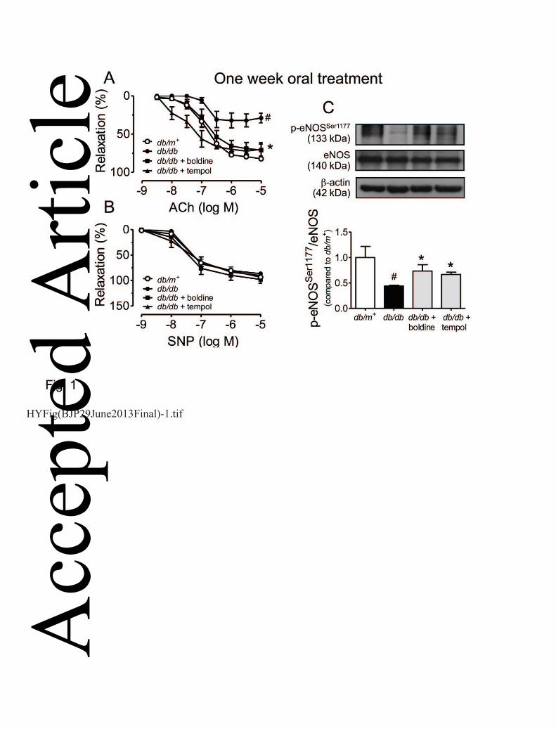
Boldine improves endothelial function in diabetic db/db mice 11
Figure 1 shows the impaired endothelium-dependent relaxations to ACh (Fig. 1A) but not 12
endothelium-independent relaxations to SNP (Fig. 1B) in aortic rings from db/db mice compared 13
with those from db/m+ mice. One-week oral administration of boldine (20 mg kg-1 day-1) or tempol 14
(20 mg kg-1day-1) to db/db mice rescued the impaired ACh-induced relaxations (Fig. 1A & Table 1) 15
without affecting SNP-induced responses in aortic rings (Fig. 1B). Such treatment did not modulate 16
plasma lipid profile or glucose levels (data not shown). In addition, treatment with either boldine or 17
tempol restored the lost phosphorylation of eNOS at Ser1177 in db/db mouse aortas (Fig. 1C). 18
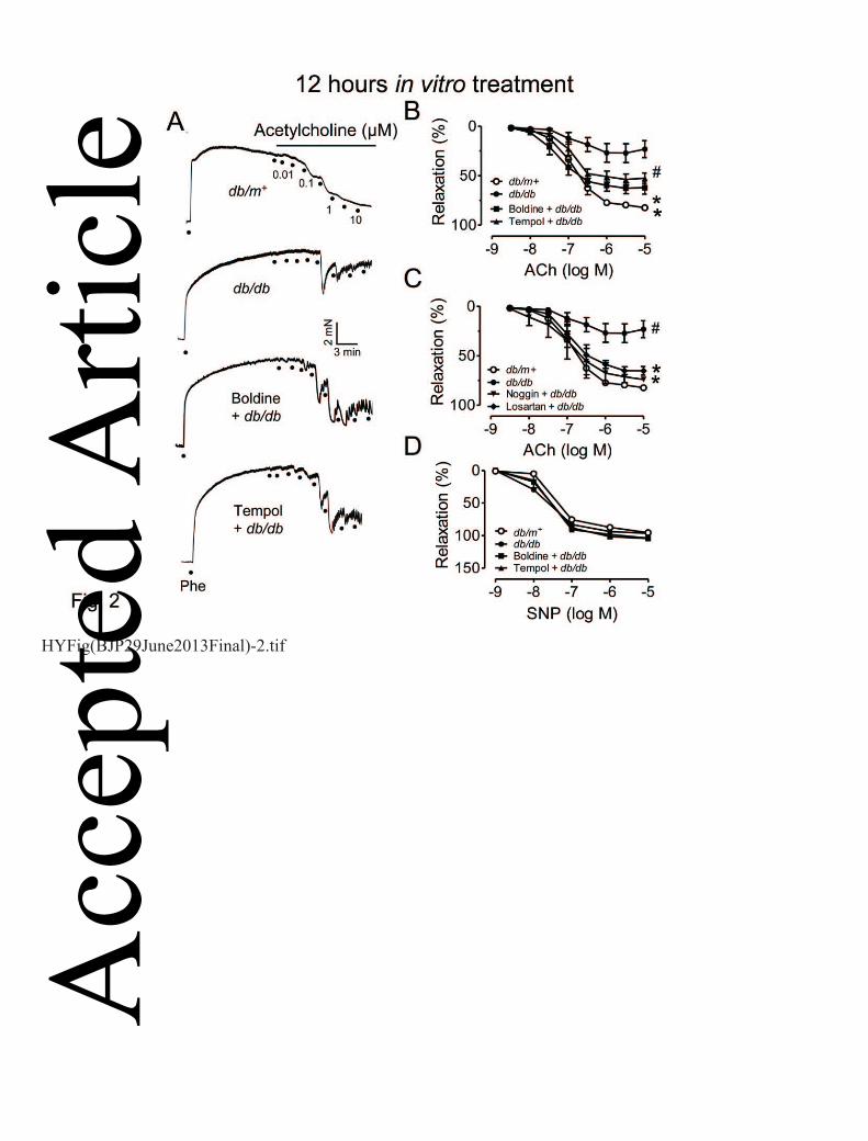
Likewise, 12-hour in vitro treatment with boldine (1 μmol L-1) or tempol (100 μmol L-1) also 19
rescued the impaired ACh-induced relaxations in db/db mouse aortas (Fig. 2A & B) without 20
affecting SNP-induced relaxations (Fig. 2C). The impaired relaxations were also reversed by 21
treatment with AT1R antagonist losartan (3 μmol L-1) and bone morphogenic protein-4 (BMP4) 22
antagonist, noggin (100 ng mL-1) (Fig. 2C). 23
24
Boldine reduces vascular oxidative stress in db/db mice 25 Acc
epte
d A
rticl
e
12
This article is protected by copyright. All rights reserved.
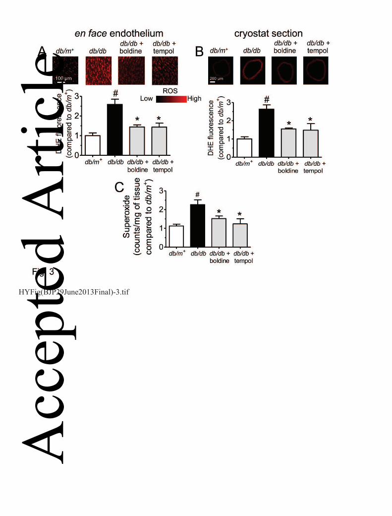
One-week treatment with boldine or tempol normalized the elevated ROS accumulation in en face 1
endothelium (Fig. 3A), across the vascular wall (Fig. 3B), and superoxide anion levels (Fig. 3C) in 2
aortas from db/db mice as reflected by DHE fluorescence dye and using lucigenin enhanced-3
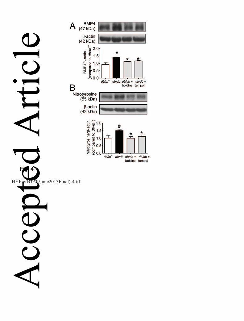
chemiluminescence method. The Western blot results show that the elevated protein expression of 4
BMP4 (Fig. 4A) and nitrotyrosine (another oxidative stress index, Fig. 4B) in db/db mouse aortas 5
was reversed by chronic treatment with boldine or tempol. 6
7
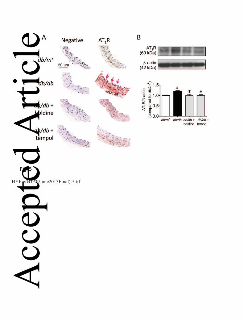
Boldine reduces the expression of AT1R in db/db mouse aortas 8
Immunohistochemistry staining showed the presence of AT1R in both endothelial cells and smooth 9
muscle cells in mouse aortas and chronic treatment with boldine or tempol reduced the expression 10
of AT1R in the aortas in db/db mice (Fig. 5A). Such effects were confirmed by the Western blot 11
data (Fig. 5B). 12
13
Boldine protects against Ang II-induced BMP4-dependent endothelial dysfunction 14
Ang II (500 nmol L-1) attenuated the ACh-induced relaxations in aortas from non-diabetic C57 mice 15
(Fig. 6A) and this impairment was reversed by co-treatment with BMP4 inhibitor noggin or AT1R 16
antagonist losartan (Fig. 6B). Ang II elevated the expression of BMP4 in cultured mouse aortas and 17
this effect was reversed by noggin or losartan (Fig. 6D). Like noggin or losartan, both boldine and 18
tempol also reversed the impairment of ACh-induced relaxations (Fig. 6C) and up-regulation of 19
BMP4 expression in Ang II-treated aortas from non-diabetic mice (Fig. 6E). 20
21
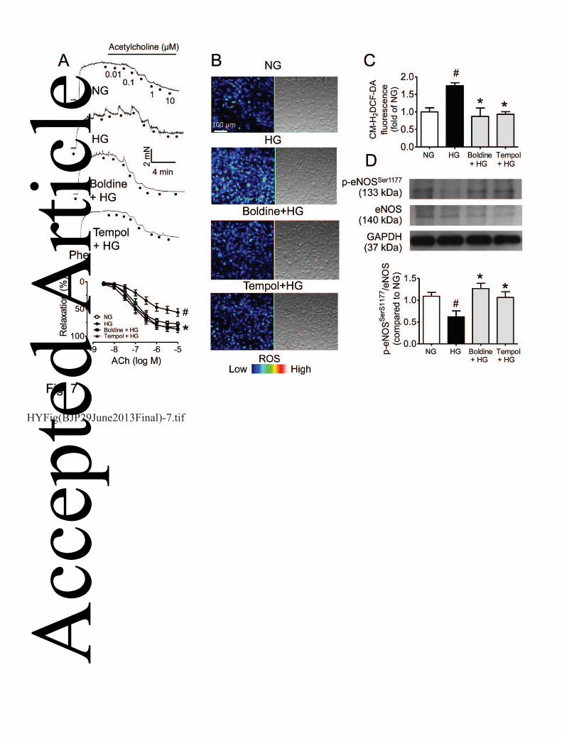
Boldine reverses high glucose-induced endothelial dysfunction in mouse aortas 22
Exposure to high glucose (36 hours) attenuated ACh-induced relaxations and this effect was 23
reversed by co-treatment with boldine (1 μmol L-1) or tempol (100 μmol L-1) (Fig. 7A). Treatment 24
with high glucose for 36 hours elevated the ROS production in MAECs as indicated by changes of 25 Acc
epte
d A
rticl
e
13
This article is protected by copyright. All rights reserved.
the DCF-DA fluorescence intensity, while ROS elevation was prevented by pre-treatment (8 hours, 1
data not shown) and co-treatment (Fig. 7B&C) with boldine or tempol. In addition, high glucose-2
induced reduction in eNOS phosphorylation in MAECs was reversed by co-treatment with boldine 3
or tempol (Fig. 7D). 4
5
Discussions 6
The present study provides experimental evidence showing that in vivo treatment with boldine 7
effectively restores the impaired endothelium-dependent relaxations in aortas of db/db mice and 8
reduces the expression of several oxidative stress markers, BMP4, nitrotyrosine and AT1R in 9
diabetic mouse arteries. The renin-angiotensin system and associated oxidative stress play a crucial 10
role in maintaining endothelial dysfunction in diabetic mice (Wong et al., 2010a) while BMP4 is a 11
novel important mediator of endothelial dysfunction in hypertension (Wong et al., 2010a; Tian et 12
al., 2012b; Wong et al., 2013). To further elucidate the inhibitory effect of boldine on Ang II-13
mediated vascular dysfunction, Ang II was used to trigger ROS generation and thus reduce the 14
bioavailability of nitric oxide in the vascular wall. As expected, in vitro Ang II treatment impairs 15
endothelium-dependent relaxations accompanied by the elevated BMP4 expression in aortas from 16
non-diabetic mice. Co-treatment with boldine reverses the harmful effects of Ang II on relaxations 17
and BMP4 up-regulation. It appears that boldine reduces Ang II-induced BMP expression mainly 18
through limiting ROS generation in the inflamed arteries as the known ROS inhibitor tempol 19
produces the same benefits as boldine. In addition, reducing the AT1R expression and associated 20
ROS over-production also play a positive role in boldine-induced improvement of endothelial 21
function in diabetic db/db mice. 22
Boldine was previously shown to ameliorate the development of diabetes in streptozotocin-23
treated rats by inhibiting oxidative stress-associated tissue damage and restoring the antioxidant 24
enzyme activities (Jang et al., 2000). We have recently described that treatment with boldine 25 Acc
epte
d A
rticl
e
14
This article is protected by copyright. All rights reserved.
reverses endothelial dysfunction in hypertensive rats through suppression of NADPH oxidase-1
mediated ROS over-production (Lau et al., 2012) and protects endothelial function in high glucose-2
induced oxidative stress through inhibiting the NADPH oxidase expression (Lau et al,. 2013). In 3
addition, boldine was reported to reduce the carrageenan-induced guinea pig paw oedema probably 4
through inhibiting the biosynthesis of pro-inflammatory prostaglandins (Backhouse et al., 1994). 5
However, the effect of boldine on endothelial dysfunction and vascular inflammatory response in 6
type II diabetes remains unclear. The present study provides novel findings on the endothelial cell 7
protective effect of boldine through inhibiting the Ang II-mediated BMP4 up-regulation and ROS 8
over-production under hyperglycemic conditions. 9
The renin-angiotensin system and associated NADPH oxidase-derived ROS mediate 10
endothelial dysfunction in the mouse model of type II diabetes through decreasing the NO 11
bioavailability (Wong et al., 2010b). The present study shows that the impaired endothelium-12
dependent relaxations in db/db mouse aortas or in high glucose-treated mouse aortas were reversed 13
by both in vivo and in vitro treatment with boldine or tempol. The improved relaxations correlate 14
with the restored phosphorylation of eNOS. Further support comes from experiments on cultured 15
mouse endothelial cells in which the high glucose-induced reduction in the eNOS phosphorylation 16
was reversed by in vitro treatment with boldine or tempol. 17
BMP4, an important matrix cytokine stimulates the expression of adhesion molecules and 18
induce endothelial dysfunction through NADPH oxidase-dependent mechanisms (San Martin et al., 19
2007). Up-regulation of BMP4 in db/db mouse aortas may involve ROS-dependent vascular 20
inflammation (San Martin et al., 2007). The present results demonstrate that endothelial dysfunction 21
in diabetic mice was accompanied by augmented oxidative/nitrosative stress and BMP4 up-22
regulation. Treatment with boldine rescued the impaired endothelial function in db/db mice and 23
inhibited ROS over-production and BMP4 up-regulation. The present study suggests that the 24
increased expression and activity of the renin-angiotensin system is likely associated with up-25 Acc
epte
d A
rticl
e
15
This article is protected by copyright. All rights reserved.
regulated expression of BMP4 and the latter causes ROS over-generation, thus impairing 1
endothelial function in diabetes, while boldine protects endothelial function through inhibiting this 2
AT1R-BMP4-ROS axis. In the present study, boldine reversed the Ang II-induced impairment of 3
ACh-induced relaxations and BMP4 over-expression in vitro and in vivo treatment with boldine 4
normalized the increased expression of AT1R in db/db mouse aortas. The harmful effects of Ang II 5
were inhibited by treatment with AT1R blocker losartan or BMP4 antagonist noggin, suggesting 6
that the improved endothelial function in db/db mice is more likely attributed to boldine-induced 7
inhibition of the Ang II-mediated BMP4 and associated oxidative stress in the vascular wall. A 8
recent study also indicates a pathophysiological role of BMP4 in Ang II-induced cardiomycyte 9
hypertrophy as Ang II stimulates the BMP4 expression in cultured cardiac fibroblasts (Sun et al., 10
2013). However, the present study can not discount the possible involvement of other BMP 11
isoforms such as BMP2, BMP6 and BMP7 in oxidative stress and vascular inflammation in diabetic 12
mice, which deserves future investigation. 13
Taken together, the present study provides novel evidence demonstrating that boldine is 14
effective in inhibiting the AT1R-mediated cellular signaling cascade and ameliorating endothelial 15
dysfunction in diabetic mice. Our findings further suggest a therapeutic potential of boldine-16
containing medicinal herbs in alleviating diabetic vasculopathy. 17
18
Acknowledgements 19
This study was supported by MOHE High Impact Research Grant H-20001-00-E000055, 20
postgraduate research fund PV008/2011B, Hong Kong General Research Fund, Chinese University 21
of Hong Kong Focused Investment Scheme B. 22
23
Conflicts of interest 24
None declared. 25
26 Acc
epte
d A
rticl
e
16
This article is protected by copyright. All rights reserved.
References 1
Backhouse N, Delporte C, Givernau M, Cassels BK, Valenzuela A, Speisky H (1994). Anti-2
inflammatory and antipyretic effects of boldine. Agents and actions 42(3-4): 114-117. 3
4
Bostrom KI, Jumabay M, Matveyenko A, Nicholas SB, Yao Y (2011). Activation of vascular bone 5
morphogenetic protein signaling in diabetes mellitus. Circulation research 108(4): 446-457. 6
7
Cassels BK, Asencio M, Conget P, Speisky H, Videla LA, Lissi EA (1995). Structure-antioxidative 8
activity relationships in benzylisoquinoline alkaloids. Pharmacol Res 31(2): 103-107. 9
10
Chen D, Zhao M, Mundy GR (2004). Bone morphogenetic proteins. Growth Factors 22(4): 233-11
241. 12
13
Gupte S, Labinskyy N, Gupte R, Csiszar A, Ungvari Z, Edwards JG (2010). Role of NAD(P)H 14
oxidase in superoxide generation and endothelial dysfunction in Goto-Kakizaki (GK) rats as a 15
model of nonobese NIDDM. PloS one 5(7): e11800. 16
17
Heitzer T, Schlinzig T, Krohn K, Meinertz T, Munzel T (2001). Endothelial dysfunction, oxidative 18
stress, and risk of cardiovascular events in patients with coronary artery disease. Circulation 19
104(22): 2673-2678. 20
21
Jang YY, Song JH, Shin YK, Han ES, Lee CS (2000). Protective effect of boldine on oxidative 22
mitochondrial damage in streptozotocin-induced diabetic rats. Pharmacol Res 42(4): 361-371. 23
24 Acc
epte
d A
rticl
e
17
This article is protected by copyright. All rights reserved.
Kim YK, Lee MS, Son SM, Kim IJ, Lee WS, Rhim BY, et al. (2002). Vascular NADH oxidase is 1
involved in impaired endothelium-dependent vasodilation in OLETF rats, a model of type 2 2
diabetes. Diabetes 51(2): 522-527. 3
4
Lau YS, Machha A, Achike FI, Murugan D, Mustafa MR (2012a). The aporphine alkaloid boldine 5
improves endothelial function in spontaneously hypertensive rats. Exp Biol Med (Maywood) 237(1): 6
93-98. 7
8
Lau YS, Tian XY, Huang Y, Murugan D, Achike FI, Mustafa MR (2013). Boldine protects 9
endothelial function in hyperglycemia-induced oxidative stress through an antioxidant mechanism. 10
Biochemical pharmacology 85(3):367-375. 11
12
Miriyala S, Gongora Nieto MC, Mingone C, Smith D, Dikalov S, Harrison DG, et al. (2006). Bone 13
morphogenic protein-4 induces hypertension in mice: role of noggin, vascular NADPH oxidases, 14
and impaired vasorelaxation. Circulation 113(24): 2818-2825. 15
16
Nett PC, Ortmann J, Celeiro J, Haas E, Hofmann-Lehmann R, Tornillo L, et al. (2006). 17
Transcriptional regulation of vascular bone morphogenetic protein by endothelin receptors in early 18
autoimmune diabetes mellitus. Life sciences 78(19): 2213-2218. 19
20
O'Brien P, Carrasco-Pozo C, Speisky H (2006). Boldine and its antioxidant or health-promoting 21
properties. Chem Biol Interact 159(1): 1-17. 22
23
Acc
epte
d A
rticl
e
18
This article is protected by copyright. All rights reserved.
San Martin A, Du P, Dikalova A, Lassegue B, Aleman M, Gongora MC, et al. (2007). Reactive 1
oxygen species-selective regulation of aortic inflammatory gene expression in Type 2 diabetes. 2
American journal of physiology. Heart and circulatory physiology 292(5): H2073-2082. 3
Senador D, Kanakamedala K, Irigoyen MC, Morris M, Elased KM (2009). Cardiovascular and 4
autonomic phenotype of db/db diabetic mice. Experimental physiology 94(6): 648-658. 5
6
Sun, B., Huo, R., Sheng, Y., Li, Y., Xie, X., Chen, C., Liu, H. B., Li, N., Li, C. B., Guo, W. T., Zhu, 7
J. X., Yang, B. F., & Dong, D. L. (2013). Bone morphogenetic protein-4 mediates cardiac 8
hypertrophy, apoptosis, and fibrosis in experimentally pathological cardiac hypertrophy. 9
Hypertension, 61(2), 352-360. 10
11
Tian XY, Wong WT, Xu A, Chen ZY, Lu Y, Liu LM, et al. (2011). Rosuvastatin improves 12
endothelial function in db/db mice: role of angiotensin II type 1 receptors and oxidative stress. Br J 13
Pharmacol 164(2b): 598-606. 14
15
Tian XY, Wong WT, Xu A, Lu Y, Zhang Y, Wang L, et al. (2012a). Uncoupling protein-2 protects 16
endothelial function in diet-induced obese mice. Circulation research 110(9): 1211-1216. 17
18
Tian XY, Yung LH, Wong WT, Liu J, Leung FP, Liu L, et al. (2012b). Bone morphogenic protein-19
4 induces endothelial cell apoptosis through oxidative stress-dependent p38MAPK and JNK 20
pathway. Journal of molecular and cellular cardiology 52(1): 237-244. 21
22
Tranche S, Galgo A, Mundet X, Sanchez-Zamorano MA (2005). Cardiovascular risk factors in type 23
2 diabetic patients: multifactorial intervention in primary care. Kidney international. 24
Supplement(93): S55-62. 25 Acc
epte
d A
rticl
e
19
This article is protected by copyright. All rights reserved.
1
Wong WT, Tian XY, Chen Y, Leung FP, Liu L, Lee HK, et al. (2010a). Bone morphogenic protein-2
4 impairs endothelial function through oxidative stress-dependent cyclooxygenase-2 upregulation: 3
implications on hypertension. Circulation research 107(8): 984-991. 4
5
Wong WT, Tian XY, Xu A, Ng CF, Lee HK, Chen ZY, et al. (2010b). Angiotensin II type 1 6
receptor-dependent oxidative stress mediates endothelial dysfunction in type 2 diabetic mice. 7
Antioxid Redox Signal 13(6): 757-768. 8
9
Wong WT, Tian XY, Huang Y (2013) Endothelial dysfunction in diabetes and hypertension: cross 10
talk in RAS, BMP4, and ROS-dependent COX-2-derived prostanoids. J Cardiovasc Pharmacol. 11
61(3):204-214. 12
13
14
15
16
17
Acc
epte
d A
rticl
e
20
This article is protected by copyright. All rights reserved.
Figure 1. ACh-induced endothelium-dependent (A) and SNP-induced endothelium-independent 1
relaxations (B) in aortic rings from db/db mice orally treated for one week with vehicle (20% 2
EtOH), boldine (20 mg-1 kg-1 day-1), or tempol (20 mg-1 kg-1 day-1). (C) Chronic boldine treatment 3
increased the level of eNOS phosphorylation at Ser1177 in aortas of db/db mice. Results are shown 4
as mean ± SEM of 6 separate experiments. # P<0.05 compared to db/m+ mice; * P<0.05 compared 5
to db/db mice. 6
7
Figure 2. In vitro exposure to boldine (1 μmol L-1) or tempol (100 μmol L-1) for 12 hours reversed 8
the impaired ACh-induced relaxations in db/db mouse aortas (A&B) without affecting SNP-induced 9
relaxations (D). Treatment (12 hours) with noggin (100 ng mL-1) or losartan (3 µmol L-1) also 10
rescued the impaired relaxation in these aortas (C). Results are means ± SEM of 6 separate 11
experiments. # P<0.05 compared to db/m+ mice; * P<0.05 compared to db/db mice. 12
13
Figure 3. Chronic boldine treatment reversed the elevated ROS in en face endothelium (A) and 14
cryostat section of db/db mouse aortas (B) as indicated by changes of the DHE fluorescence 15
intensity and inhibited the increased generation of superoxide anion in these aortas (C) as detected 16
using lucigenin-enhanced chemiluminescence method. Results are means ± SEM of 4-6 separate 17
experiments. # P<0.05 compared with db/m+ mice; * P<0.05 compared with db/db mice. 18
19
Figure 4. Western blotting showing the up-regulation for expressions of BMP4 (A) and 20
nitrotyrosine (B) in aortas from db/db mice with and without receiving one-week treatment with 21
boldine or tempol. Results are means ± SEM of 4 separate experiments. # P<0.05 compared with 22
db/m+ mice; * P<0.05 compared with db/db mice. 23
Figure 5. One-week treatment with boldine or tempol reduced the AT1R expression in db/db mouse 24
aortas as detected by immunohistochemistry (A) and Western blotting (B). Arrows indicate the 25 Acc
epte
d A
rticl
e
21
This article is protected by copyright. All rights reserved.
endothelial layer of the artery. Results are shown as mean ± SEM of 3-4 separate experiments. # 1
P<0.05 compared with db/m+ mice; * P<0.05 compared with db/db mice. 2
3
Figure 6. Ang II-induced impairment of ACh-induced relaxations was reversed by in vitro 4
treatment with boldine (1 µmol L-1, A&C), tempol (100 µM, A&C), noggin (100 ng ml-1, A&B) 5
and losartan (3 µmol L-1, A&B) in aortas from non-diabetic mice. These four inhibitors normalized 6
Ang II-induced increase in BMP4 expression (D&E). Results are means ± SEM of 4-6 separate 7
experiments. #P<0.05 compared with control; *P<0.05 compared with Ang II. 8
9
Figure 7. In vitro treatment with boldine or tempol reversed high glucose-induced impairment of 10
ACh-induced relaxations in non-diabetic mouse aortas (A) and normalized the elevated ROS 11
production in high glucose (HG)-treated MAECs (B&C). (D) Boldine and tempol increased the 12
eNOS phosphorylation in HG-treated MAECs. NG: normal glucose (5 mmol L-1 glucose and 25 13
mmol L-1 mannitol as osmotic control of HG). Results are means ± SEM of 4-6 separate 14
experiments. # P<0.05 compared with NG; *P< 0.05 compared with HG. 15
16
17
18
19
20
21
22
23
24 Acc
epte
d A
rticl
e

22
This article is protected by copyright. All rights reserved.
Table 1: The agonist sensitivity (pEC50) and percentage of maximum response (Rmax) of ACh-1
induced endothelium-dependent relaxations in aortic rings isolated from db/m+ and db/db 2
3
4
5
6
7
8
9
Results are means ± SEM of 6 separate experiments. # P< 0.01 compared to db/m+ 10
* P<0.01 compared to db/db 11
12
ACh
Group pEC50 (log M) Rmax (%)
db/m+ -6.60 ± 0.06 82.42 ± 2.03
db/db -4.35 ± 0.47 # 29.00 ± 6.25 #
db/db + Boldine -6.28 ± 0.12 * 71.00 ± 7.65 *
db/db + tempol -6.81 ± 0.15 * 70.00 ± 8.66 *
Acc
epte
d A
rticl
e
HYFig(BJP29June2013Final)-1.tif
Acc
epte
d A
rticl
e
HYFig(BJP29June2013Final)-2.tif
Acc
epte
d A
rticl
e
HYFig(BJP29June2013Final)-3.tif
Acc
epte
d A
rticl
e
HYFig(BJP29June2013Final)-4.tif
Acc
epte
d A
rticl
e
HYFig(BJP29June2013Final)-5.tif
Acc
epte
d A
rticl
e
HYFig(BJP29June2013Final)-6.tif
Acc
epte
d A
rticl
e
HYFig(BJP29June2013Final)-7.tif
Acc
epte
d A
rticl
e