biomechanical analysis of suture anchors and suture materials in the canine femur
TRANSCRIPT

BIOMECHANICAL ANALYSIS OF SUTURE
ANCHORS AND SUTURE MATERIALS
IN A CANINE FEMUR MODEL
By
JAMES THOMAS GILES III
Doctor of Veterinary Medicine
Oklahoma State University
Stillwater, Oklahoma
1998
Submitted to the Faculty of the Graduate College of the
Oklahoma State University in partial fulfillment of
the requirements for the Degree of
MASTER OF SCIENCE May, 2007

ii
BIOMECHANICAL ANALYSIS OF SUTURE
ANCHORS AND SUTURE MATERIALS
IN A CANINE FEMUR MODEL
Thesis Approved:
Kenneth E. Bartels, DVM, MS
Mark C. Rochat DVM, MS
Anthony Confer, DVM, PhD
A. Gordon Emslie, PhD
Dean of the Graduate College
iii
TABLE OF CONTENTS
Chapter Page I. INTRODUCTION......................................................................................................1
Overview..................................................................................................................1
Modes of Failure ......................................................................................................3 Suture Materials .......................................................................................................4 Objectives ................................................................................................................5
II. MATERIALS AND METHODS...............................................................................6
Objective 1- Acute Load to Failure .........................................................................6 Implants- Anchors.................................................................................................7 Specimens .............................................................................................................7 Construct Design...................................................................................................7 Mechanical Testing...............................................................................................8 Objective 2- Cyclic Testing ...................................................................................10 Implants- Anchors and Sutures...........................................................................10 Specimens ...........................................................................................................10 Construct Design.................................................................................................10 Mechanical Testing.............................................................................................11 Statistical Analysis.................................................................................................12
III. RESULTS...............................................................................................................14
Objective 1- Acute Load to Failure .......................................................................14 Objective 2- Cyclic Testing ...................................................................................14

iv
IV. DISCUSSION........................................................................................................18
Objective 1- Acute Load to Failure ... ...................................................................18 Suture Materials......................................................................................................21 Objective 2- Cyclic Testing ...................................................................................22 Study Limitations...................................................................................................23 Conclusion .............................................................................................................24
REFERENCES ............................................................................................................26

v
LIST OF TABLES
Table Page I. Mean acute load to failure for suture anchors in the cranial aspect of
femoral condyle...................................................................................................15
II. Mean acute load to failure for anchors in caudal aspect of femoral condyle..........15
III. Mean cycles to failure for anchor/suture combination, number completed
10K cycles and mean post-cycling acute failure load.......................................17

vi
LIST OF FIGURES
Figure Page
1. Photograph of the suture anchors tested..................................................................6 2. Photograph of an objective 1 construct...................................................................9 3. Photograph of an objective 2 construct...................................................................13 4. Load-displacement curve for acute load to failure test............................................16
5. Scanning electron microscope images of the suture anchor eyelets.........................25
1
CHAPTER I
INTRODUCTION
Suture anchors are metallic or absorbable orthopedic devices that facilitate the
attachment of suture or a suture-soft tissue union to bone. Suture anchors are designed to
provide temporary fixation of soft-tissue to bone or fixation of a prosthetic material until
functional healing or peri-articular fibrosis and stabilization occur.1-2 Suture anchors are
an effective alternative to the use of staples, transosseous tunnels and screw-washer
combinations for anchorage of suture-soft tissue to bone.2,3,5 The use of suture anchors
in human and veterinary surgery has been described.1-9 Suture anchors have been used
extensively in human surgery through arthroscopic and open techniques for ligament,
joint capsule and tendon reattachment or prosthesis placement, head and neck soft-tissue
reconstruction, urologic and gynecologic applications.3,5,10-18 Described veterinary
applications include tarsal and phalangeal ligament deficiencies, coxofemoral luxation,
shoulder luxation, elbow luxation, extracapsular stifle stabilization for cranial cruciate
and/or collateral ligament deficiency, common calcaneal tendon avulsion, triceps
avulsion, carpal extensor avulsion, gastrocnemius avulsion and acetabular fracture. 6-9,19
Defining features of suture anchors include their location within the bone, thread design,
eyelet design, deployability and composition (metallic vs. absorbable). Suture anchors

2
may have transcortical or subcortical location.2 Transcortical anchors are a screw-type
design with a suture eyelet and engage the cis-cortex. Subcortical anchors may be screw-
type or have prongs/flanges that resist pullout. While absorbable anchors are extensively
used in human surgery, their use is not reported in the veterinary literature. Decreased
mechanical strength currently makes absorbable suture anchors less useful in veterinary
patients.20 Eyelet design has a significant impact on suture abrasion and excess friction
lowers load to failure and cycles to failure.20,21 Eyelets of human metallic suture anchors
are generally round or streamlined with one or more suture protection channels.20
Features of eyelets that contribute to friction placed on the suture include the smoothness
of the surface, the shape and design of the eyelet, and the radius of curvature of the
eyelet. A larger radius will permit a smoother arc and limit abrasion. If the angle of
suture pull is out of a suture-protecting channel, the suture may be abraded on a relatively
sharp surface.20,21,22
The anchor location within a bone will vary with anchor type and clinical application.
Loads to failure vary with anchors placed in different bones and within regions of the
same bone.23-25 Biomechanical studies have been performed with veterinary suture
anchors in the proximal and distal tibial metaphyses, proximal humeral metaphysis,
acetabular wall, dorsal acetabular rim and femoral condyles.2,4,8,9 The studies involving
the acetabulum and femoral condyles utilized Bone Biter™ suture anchors (Innovative
Animal Products LLC™, Rochester, MN), which are subcortical flange-type anchors that
may have different failure loads than transcortical screw-type anchors.8,9 Applications
for suture anchor placement in the femoral condyle include the lateral suture technique

3
for cranial cruciate ligament rupture, collateral ligament repair and reattachment of the
long digital extensor tendon. To the authors’ knowledge, this is the only veterinary study
that evaluates transcortical suture anchor placement in the femoral condyle of a dog.
Modes of Failure
The angle of suture pull is a feature that may significantly effect suture abrasion and
failure.21,22,26,27 Many human suture anchors are designed such that a suture pull of 0°
(along the axis of insertion) results in the least suture abrasion. However, clinical
application may dictate that the suture pull angle is 45° or 90° to the angle of insertion.
In human rotator cuff repair, Burkhart concluded the optimal anchor to suture angle to
create an equilibrium between the anchor and the rotator cuff is 45°.28 The suture pull
angle is typically 90° in collateral or cranial cruciate ligament stabilizations in dogs.
Studies have demonstrated that with rigid eyelets, many anchor-suture combinations
experience reduced cycles and loads to failure at 45° and 90° suture pull angles when
compared to 0°.21,22,26 Interestingly, an absorbable suture anchor with a flexible polyaxial
suture serving as an eyelet demonstrated no statistical difference between different suture
pull directions.26 Another consideration for eyelet orientation is the alignment of the in-
plane axis of the eyelet with the direction of suture pull. Some metallic anchors/suture
combinations demonstrate reduced cycles and loads to failure with the eyelet in a coronal
orientation (out-of-plane) compared to a sagittal (in-plane) orientation with a 45° angle of
suture pull.22,26

4
Suture anchor constructs are susceptible to failure at the bone-anchor interface, anchor-
suture interface, suture-tissue interface or other abrasive areas on the suture, such as a
bone edge, knot or crimp-clamp.1,20,22 In suture anchor constructs in which the suture
engages soft tissue, the suture-tissue interface is typically the weakest link.28-30
Typically, the suture anchor and the anchor/bone interface have a higher load to failure
than the suture it accommodates.30-32
Suture anchor constructs may be tested in acute load to failure (ALF) or cyclic testing.
Acute load to failure is useful for direct comparison between anchors’ holding ability
during a single, supra-physiologic load. However, cyclic testing is a clinically more
useful model to accurately assess stress on the suture anchor construct during weight
bearing, range of motion, and activity.1,28
Suture Materials
Monofilament nylon leader line (NLL) secured with a crimp-clamp system is commonly
utilized for lateral fabella-tibial suture techniques in the canine cranial cruciate ligament
deficient stifles.33-37 Nylon leader line has also been recommended for use with a
veterinary suture anchor.4 It has been reported that monofilament nylon is more resistant
to cyclic loading than braided suture because individual strands cannot be serially
abraded,4 however, to the author’s knowledge, this claim is not supported in the
literature. Many human suture anchors are pre-loaded with a non-absorbable,
multifilament, polyester suture, Ethibond Excel™ (Ethicon Inc, Somerville, NJ).
Recently, a polyethylene-based multifilament suture, Fiberwire™ (Arthrex Inc., Naples,

5
FL) has been released and is used frequently with suture anchors. Mechanical testing
indicates that 2 USP Fiberwire is significantly stronger, stiffer and more abrasion
resistant than 2 USP Ethibond.26,27,38,39,40
Objectives
The first objective of this study was to evaluate the ALF at 0° to the angle of insertion in
cadaveric canine femoral condyles with the Securos® 3.5mm (Securos Inc., Charleston,
MA), FlexiTwist™ 3.5mm (Innovative Animal Products LLC™, Rochester, MN),
IMEX™ 4.0mm x 10mm (IMEX™ Veterinary Inc., Longview, TX) and Mitek Fastin™
4.0mm (DePuy-Mitek Inc., Raynham, MA). The first three are products designed for use
in veterinary medicine and the 4.0mm Fastin is designed for use in human medicine. The
null hypothesis was that all suture anchors tested would have the same load to failure.
The second objective of this study was to evaluate the cycles to failure of the above
suture anchors with 5 USP Fiberwire and 27kgt NLL (Securos Inc., Charleston, MA)
secured with two crimp-clamps and cycled at 90° to the angle of anchor insertion. Those
constructs that completed 10,000 cycles were subjected to ALF at 90° to the angle of
insertion. The null hypothesis was that all suture anchor constructs would have the same
cycles to failure and the same ALF.

6
CHAPTER II
MATERIALS AND METHODS
Objective 1- Acute Load to Failure
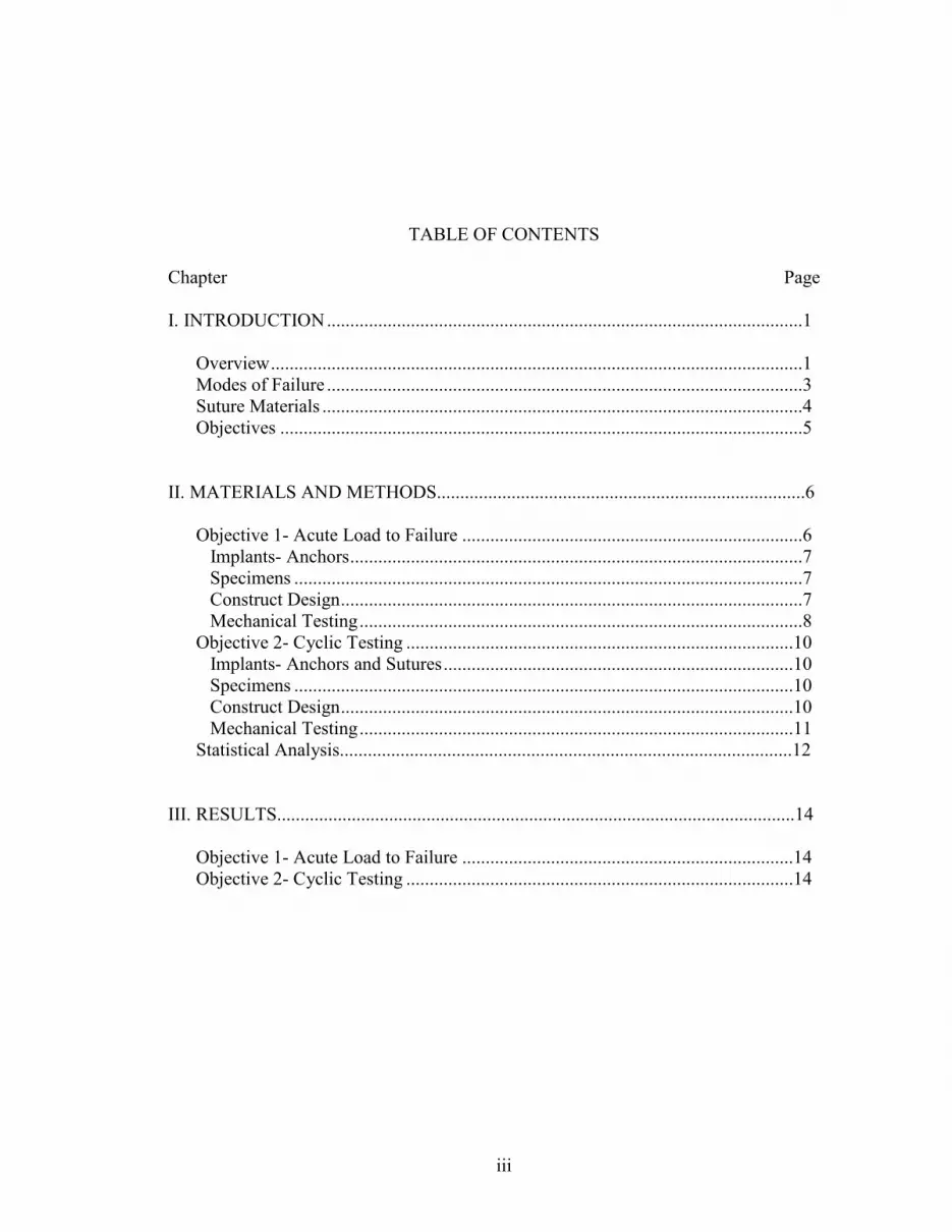
Implants- anchors: The following four anchors were examined in this experiment:
Securos 3.5mm x 19mm, FlexiTwist 3.5mm x 20mm, IMEX 4.0mm x 10mm, Fastin
4.0mm x 9.7mm (Figure 1).
Fig 1. Photograph of the suture anchors tested from left to right: Securos 3.5mm,
FlexiTwist 3.5mm, IMEX 4.0mm, Fastin 4.0mm.

7
Specimens: Femurs were harvested from 20-30kg, skeletally mature dogs immediately
following euthanasia for an unrelated project. The femurs were denuded of soft tissue,
wrapped in saline-soaked (.9%NaCl) gauze, placed in plastic bags and stored in a freezer
at –86°C until mechanical testing. Prior to testing, the bones were thawed to room
temperature. Throughout all stages, the bones were maintained in a moistened state with
saline-soaked gauze.
Construct Design: Prior to testing, the proximal one-third was removed from each
femur. The bone was inserted into a 14 gauge perforated-steel tube and secured with
three u-bolts. Two 2.4mm Steinman pins were placed through the tube and bone at
orthogonal angles to prevent rotation. Four anchors were placed in each femur; two in
each condyle separated by a minimum of 1cm between the caudal and central anchor to
prevent crack propagation, as previously recommended.1 Each brand of anchor was
rotated between cranial-caudal and medial-lateral positions to monitor for variation in
failure loads with respect to anchor position. The anchors were inserted perpendicular to
the femoral condyles according to the following manufacturers’ recommendations:
1. Securos 3.5mm anchor: A 3.2mm pilot hole was drilled by use of a power drill. The
spindle of the anchor was placed into a Jacobs chuck and inserted into the femoral
condyle and the chuck levered to break the insertion shaft free from the anchor.
2. FlexiTwist 3.5mm anchor: A 2.7mm pilot hole was drilled by use of a power drill.
The anchor was placed into the custom anchor driver and inserted into the femoral
condyle.
8
3. IMEX 4.0mm x 10mm anchor: A 2.7mm pilot hole was drilled by use of a power
drill. The anchor was placed into the custom anchor driver and inserted into the femoral
condyle.
4. Fastin 4.0mm: A 3.5mm pilot hole was drilled by use of a power drill. The anchor
was removed from the custom insertion spindle and the pre-loaded suture (2 USP
Ethibond) was removed. Five USP Fiberwire was placed through the anchor eyelet and
the anchor replaced in the insertion spindle. The spindle was placed in a power drill and
inserted at a low speed until the spindle detached from the anchor.
Each anchor was loaded with an appropriate wire or suture based upon eyelet size, with
the intent to eliminate suture breakage as a mode of failure. Since the various brands had
different eyelet sizes, a single material could not be used in all anchors. The following
anchor and wire or suture combinations were used: 3.5mm Securos and FlexiTwist with
1.2mm Kirschner wire, IMEX 4.0mm with 18 gauge (1.0mm) orthopedic wire and Fastin
4.0mm and 5 USP Fiberwire. Preliminary tests demonstrated that 18 gauge orthopedic
wire failed before pullout of the Securos and FlexiTwist anchors occurred. The largest
wire accommodated by the Fastin 4.0mm anchor is 22 gauge (.6mm), however, it failed
during preliminary tests. Number 5 USP Fiberwire consistently achieved anchor pullout
without suture failure. All specimens were prepared by one author (JTG).
Mechanical Testing: All testing was performed with a servohydraulic uniaxial testing
machine equipped with a 5kN load cell (MTS Systems Corporation, Eden Prairie, TX)
and Fastrack 8800D controller (Instron Corporation, Norwood, MA). Each femur was

9
placed in the bone-holder and secured in a vise, which was attached to the lower movable
jaw of the uniaxial test machine. The specimen was adjusted so the anchor was at the
center of the load cell allowing the applied load to be at 0° to the angle of insertion. The
appropriate coupler wire or suture was placed through the anchor eyelet and clamped to a
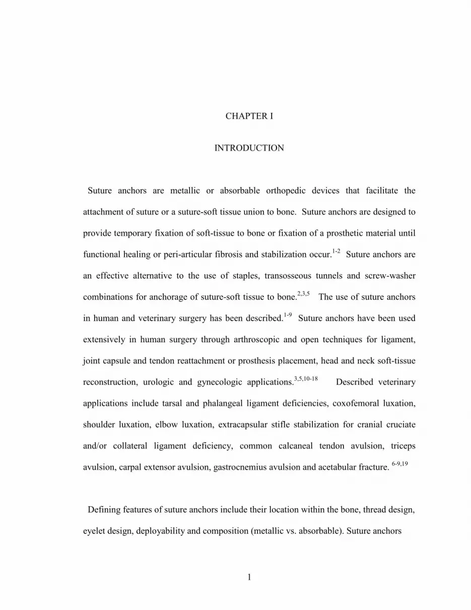
bolt connected to the load cell. Acute load to failure tests were performed in
displacement-control with an initial pre-load of 10N. The load was applied at 1mm/s and
data collected at 100 Samples/s. The load at failure was recorded as well as the mode of
failure. Figure 2 depicts a typical construct for the acute load to failure testing.
Fig 2. Photograph of the construct using a Securos suture anchor immediately
following an acute load to failure test.

10
Objective 2- Cyclic Testing
Implants- anchors and sutures: The same four anchor types were tested with two
different suture materials: 5 USP Fiberwire tied in a knot and 27kgt NLL secured with
two 36kg stainless steel crimp-clamps (Securos Inc., Charleston, MA). The 27kgt NLL
was selected because it was the largest size that would fit all the veterinary anchors.
Thirty-six kgt fits through the 3.5mm Securos and FlexiTwist anchors, but not the IMEX
4.0mm anchor. Due to the small eyelet size of the Fastin 4.0mm anchor, it was only
tested with 5 USP Fiberwire.
Specimens: The specimens were harvested and handled in an identical fashion to
objective 1.
Construct Design: Prior to testing, the proximal one-third was removed from each
femur. The femur was seated in a 14 gauge perforated-steel tube and two 2.4mm
Steinman pins inserted at orthogonal angles. The femur was potted within the tube with
Master® Dyna-Cast® (Kindt-Collins Company, LLC, Cleveland, Ohio). Anchors were
inserted into the caudal aspect of the femoral condyle according to previously described
manufacturers’ recommendations. The anchors were randomly assigned to different
femurs and medial or lateral condyle so they were distributed evenly. The anchors were
placed so the in-plane axis of the eyelet was in the same plane as the suture direction. To
achieve a uniform loop circumference, five USP Fiberwire was placed through the anchor
eyelet and tied around a 28cm circumference section of polyvinylchloride pipe by use of
a surgeon’s knot followed by four single overhand throws.40 The 27 kgt NLL was
11
inserted through the eyelets of the veterinary anchors and both ends passed through two
36kg crimp-clamps. The 27 kgt NLL was placed around the 28cm polyvinylchloride pipe
and the two crimp-clamps placed over a 3 cm rectangular notch in the pipe. Each of the
crimp-clamps was crimped in three places with a Securos crimping device according to
manufacturer recommendation. After creation of the loop, the polyvinylchloride pipe
was removed and a steel bar was secured within the specimen tube and placed in a 50K lb
gripper (MTS Systems Corporation, Eden Prairie, TX). The suture was placed through a
stainless steel chain anchor which was attached to the 5kN load cell. The construct was
adjusted so the angle of suture load was 90° to the angle of anchor insertion and in-plane
with the eyelet rotation angle. The rotation angle of the Fastin anchor could not be
controlled because the insertion spindle automatically detaches when the anchor is
inserted to the appropriate depth. Since this is how the Fastin anchor is employed
clinically and adjustments may damage the suture, no attempt was made to adjust the
rotation angle. The specimens were prepared by one author (JTG).
Mechanical Testing: All testing was performed with a uniaxial testing machine
equipped with a 5kN load cell (MTS Systems Corporation, Eden Prairie, TX) and
Fastrack 8800D controller (Instron Corporation, Norwood, MA). An initial pre-load of
10N was applied. Cyclic tests were performed under load control with a load range of
280-332N at 5 hertz. The maximum load of 332N (80% of the ultimate failure load) was
selected based upon an ultimate failure of 27 kgt NLL with a crimp-clamp of 416N as
reported by Banwell, et al.35 Preliminary tests yielded similar results and demonstrated 5
USP Fiberwire™ was stronger than the 27 kgt NLL. The use of a maximum load of 80%
12
of the ultimate failure load theoretically will allow failure of some of the constructs and
give a basis for comparison. The load was increased from 10N to 280N over 15 seconds.
The data collection rate was 20 Samples/s. Each sample was cycled for 10,000 cycles or
until the suture-anchor construct failed. The number of cycles at failure and mode of
failure were recorded. Constructs that achieved 10,000 cycles were subject to an ALF in
the same configuration, with the suture load at 90° to the anchor insertion. Acute load to
failure tests were performed in displacement-control with an initial pre-load of 10N. The
load was applied at 1mm/s and data collected at 100 Samples/s. The load at failure and
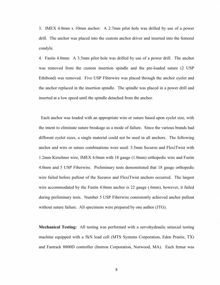
mode of failure were recorded. Figure 3 depicts a typical construct for the cyclic testing.
Statistical Analysis: All data were analyzed using PC SAS Version 9.1 (SAS Institute,
Cary, NC). Analysis of variance techniques were employed using SAS PROC MIXED.
A completely randomized two factor arrangement was used as the model in the ANOVA
with brand and location as the factors of interest for ALF (Objective 1) and brand and
suture as the factors of interest for cyclic testing (Objective 2). The response variables
for the ANOVA were pullout force for Objective 1 and cycles to failure for Objective 2.
Simple effects of brand for each location (or suture type) were assessed using a SLICE
option in an LSMEANS statement, and pair-wise t-tests performed if the overall simple
effects were significant.

13
Fig 3. Photographs of the construct utilized for cyclic testing. (A) Lateral view of a
FlexiTwist/NLL construct. Note: this was a preliminary test in which only a single
crimp-clamp was used to secure the NLL rather than two crimp-clamps used in the
actual experiments. (B) Cranial-caudal view of an IMEX/Fiberwire construct.

14
CHAPTER III
RESULTS
Objective 1- Acute Load to Failure
There was no statistical difference (p=.131) in acute load to failure among the four
anchors in the cranial aspect of the condyle (table 1). The veterinary anchors all had
significantly higher failure loads (p<.0001) in the caudal aspect of the femoral condyle
compared to the cranial aspect. There was no statistical difference in load to failure
between the caudal and cranial positions with the Fastin anchor (table 2). There was no
significant difference between the medial and lateral femoral condyle. Two of 10 of the
Flexitwist suture anchors failed by fracture of the eyelet. All of the other suture anchors
failed by pullout of the anchor from the bone. Figure 4 demonstrates a typical load-
displacement curve for an acute load to failure test.

15
Table 1: Mean acute load to failure in newtons (N) and standard error of the mean
(SEM) for suture anchors in the cranial aspect of femoral condyle.
Anchor n Mean Failure (N) SEM
Securos 5 611.70 a 55.68
FlexiTwist 6 582.58 a 80.29
IMEX 4 498.62 a 112.75
Fastin 4 359.86 a 68.55
Means with the same letter are not significantly different using a .05 significance
level.
Table 2: Mean acute load to failure in newtons (N) and standard error of the mean
(SEM) for anchors in caudal aspect of femoral condyle.
Anchor n Mean Failure (N) SEM
Securos 5 1192.74 a 105.7
FlexiTwist 5 1156.77 a 45.097
IMEX 5 736.79 b 86.645
Fastin 5 277.96 c 39.739
Means with the same letter are not significantly different using a .05 significance
level.

16
Figure 4: Load-displacement curve for acute load to failure test
Objective 2- Cyclic Testing and Post-Cycling Acute Load to Failure
All cyclic tests that failed did so by suture breakage at the eyelet. Constructs that
completed 10,000 cycles were subjected to an acute load to failure at 90° to the insertion
angle and all failed by suture breakage at the eyelet. The results of the cyclic tests and
acute load to failure for those that completed 10,000 cycles are summarized in table 3.
The p-value for mean cycles to failure was .0093. The Fiberwire (n=3) was statistically
stronger (p=.024) than NLL (n=6) in post-cycling ALF. Fiberwire only completed
10,000 cycles with the Securos anchor, while NLL completed the cycles with Securos
(n=3), FlexiTwist (n=2) and IMEX (n=1). Two of the Fastin anchors failed while being
Securos
0
0.2
0.4
0.6
0.8
0 1 2 3 4 5 6 7 8
Displacement (mm)
Load
(Kn)

17
ramped to 280N and completed no cycles. All of the Fastin anchors shifted in the
direction of the suture pull when the load was applied.
Table 3: Mean (± SEM) of cycles to failure for anchor/suture combination, number
completed 10K cycles and mean post-cycling acute failure load (N) (± SEM).
Anchor Suture n Mean Cycles
SEM # Completed 10K cycles
Mean Failure (N)
SEM
FlexiTwist NLL 5 7280.8 a 1727.56 3/5 440.411 b 3.277
Securos Fiberwire 5 6733.0 ab 2009.29 3/5 573.122 a 37.31
Securos NLL 5 6123.2 ab 2119.92 2/5 422.184 b 12.474
IMEX Fiberwire 5 2701.6 bc 774.64 0/5 NA
IMEX NLL 5 2559.6 bc 1874.22 1/5 416.610 b
FlexiTwist Fiberwire 5 1258.6 c 324.19 0/5 NA
Fastin Fiberwire 5 196.0 c 82.86 0/5 NA
Means with the same letter are not significantly different using a .05 significance
level.
18
CHAPTER IV
DISCUSSION
Acute Load to Failure
The objectives of this study were to compare failure loads with veterinary suture
anchors and a currently available human anchor in a canine femoral condyle model and
evaluate the anchors loaded two different suture materials in a cyclic model. Suture
anchors may be placed in the femoral condyle for repair of the cranial cruciate or
collateral ligaments or the long digital extensor tendon. To the authors’ knowledge,
Singer et al performed the only study evaluating a veterinary suture anchor (Bone Biter)
in a canine femoral condyle.8 Unlike the current study, their study demonstrated no
statistical difference between the cranial and caudal position of the femoral condyle. The
Bone Biter suture anchor is a flange-type, sub-cortical anchor that does not have thread-
interface with the cortex. Cortical thickness may have less of an impact on anchor
pullout strength with the subcortical Bone Biter than a screw-type anchor. All three
veterinary anchors in the current study were significantly stronger in the caudal aspect of
the femoral condyle, while the Fastin showed no statistical difference. Plausible
explanations include possible differences in cancellous and cortical bone mineral density
in the caudal and central aspects, engagement of the cis and trans-cortex by the
veterinary anchors, or both. Several studies demonstrated differences in anchor loads and
19
cycles to failure within different regions of the human proximal humerus which
corresponded to variations in bone mineral density.23-25, 41 While it is beyond the scope
of this study, a relationship of suture anchor failure loads and bone mineral density in
different locations within the femoral condyle may be investigated by the use of
peripheral quantitative computed tomography.24 The length of the veterinary suture
anchors allowed them to partially or fully engage the trans-cortex in caudal aspect of the
femoral condyle in many of the specimens. The Fastin suture anchor was not long
enough to engage the trans-cortex, which may partially explain the lack of difference in
failure loads between positions with this anchor. While engaging both cortices of the
femoral condyle increases the failure loads, caution should be used in selecting anchor
length and pre-drilling to avoid damage to the caudal and cranial (if present) cruciate
ligaments. The Fastin is inserted to a depth that places the eyelet below the level of the
cortex, which reduces soft-tissue irritation. However, that feature minimizes the threads
that engage the cortex and may lower failure loads. Mahar et al reported that anchors
inserted deeper in the human proximal humerus had more migration and no improved
strength over anchors at standard depth.42 The anchors were placed in the cranial and
caudal aspect of the femoral condyle in the ALF portion of this study to maximize the use
cadaveric limbs. However, in clinical application for CCL deficiency, anchor placement
should be in the caudal aspect of the femoral condyle.
In the current study, the mean ALF for the Securos 3.5mm suture anchor were 611.70
±55.68N and 1192.74 ±105.7N in the cranial and caudal aspect of the femoral condyles,
respectively. These values are statistically different. Balara et al reported a failure load

20
of 385 ±30N in the proximal canine humerus.4 Since the time of their study, Securos has
modified the 3.5mm anchor by increasing the length from 16mm to 19mm, which should
increase failure loads, as well as modifying the eyelet. The current study yielded
statistically different failure loads for the IMEX 4.0mm anchor of 498.62 ±112.75N and
736.79 ±86.65N in the cranial and caudal aspects of the condyle, respectively. Robb et al
reported a combined load to failure of IMEX 4.0mm anchor in the proximal and distal
tibial metaphyses of 661 ±163N.2 The current study demonstrated statistically different
failure loads for the FlexiTwist 3.5mm anchor of 582.58 ±80.29N and 1156.77 ± 45.1N
cranial and caudal aspects of the femoral condyle, respectively. To the authors’
knowledge, there are no published studies reporting the failure loads of the FlexiTwist
suture anchor. This study demonstrated statistically similar failure loads for the Fastin
4.0mm of 359.86 ±68.55N and 277.96 ±39.74N in the cranial and caudal aspect of the
condyles, respectively. Barber et al reported acute loads to failure of 431-449N in fresh
porcine femurs.43
Suture Materials
The suture is a component in a suture anchor construct that may fail. An objective of
this study was to compare 27kgt NLL secured with two steel crimp-clamps and 5 USP
Fiberwire secured with a knot. It has been recommended that a total of seven throws (1
surgeon’s knot followed by 4 overhand throws) be utilized to achieved maximum knot
security with 2 USP Fiberwire.40 To the author’s knowledge, the ideal number of throws
for 5 USP Fiberwire has not been published. Differences in suture anchor eyelet size
present a challenge in selecting suture size. The 3.5mm FlexiTwist and Securos anchors

21
will accommodate 36kgt NLL, while the IMEX 4.0mm anchor will not. The human
anchor selected, Fastin 4.0mm, has a small eyelet that will not accommodate 27kgt NLL.
While it did accommodate 5 USP Fiberwire, it is designed for a 2 USP suture. Friction
while placing the Fiberwire may have created defects that lowered the cycles to failure.
Securos does not recommend using 27kgt NLL with 36kg crimp-clamps. However, this
and previous studies demonstrate successful employment of this combination.35,37,44
Preliminary tests with a single crimp-clamp all failed by the NLL pulling through the
crimp. Following application of a second crimp-clamp, all NLL failed at the eyelet with
no slippage through the crimp. Two studies report successful use of 27kgt NLL with a
single crimp-clamp.35,44 Differences in the current study include the use of Securos NLL
rather than Mason Hard Type Leader Material (Mason Tackle Company, Otisville, MI)
and manufacturer changes in the crimper device, which limit the maximum pressure
applied to the crimp-clamp. With the current materials, use of two crimp-clamps is
advised.
Cyclic Testing
The exact strength needed to stabilize the canine CCL deficient stifle is unknown.
Ultimate forces of approximately 700N and 1300N have been reported in intact CCLs of
Labrador Retrievers and mixed-breed dogs, respectively.45,46 Caporn and Roe estimated
that the canine CCL can be estimated to resist loads of 50N at a walk and maximum loads
of 400-600N during vigorous activity.34 The load applied in cyclic testing in this study
ranged from 280-332N, which approaches the maximal loads that the canine CCL may

22
experience during vigorous activity. A higher load was selected to achieve failure in the
majority of the constructs and provide a basis for comparison in performance. However,
the load selected is higher than might be expected for typical activity in the post-
operative patient.
In the cyclic testing, four statistical categories were identified, which are summarized in
Table 3. The constructs in order of most statistically significant number of cycles
completed to least are as follows: FlexiTwist/NLL (7280.8 ± 1727.56) >
Securos/Fiberwire (6733 ± 2009.29) and Securos/NLL (6123 ± 2119.92) >
IMEX/Fiberwire (2701.6 ± 774.64) and IMEX/NLL (2559.6 ± 1874.22) >
FlexiTwist/Fiberwire (1258.6 ± 324.19) and Fastin/Fiberwire (196 ± 82.86).
Interestingly, the FlexiTwist anchor achieved the most cycles of all constructs with NLL
and the least of the veterinary constructs with Fiberwire. The Securos and IMEX anchors
each had statistically similar performance with NLL and Fiberwire (although Securos had
significantly more cycles than IMEX). While FlexiTwist/NLL achieved the most cycles,
all of the veterinary constructs exceeded 1200 cycles at a load of 280-332N and should be
adequate for a properly confined post-operative patient. The reduced cycles to failure of
the Fiberwire compared to NLL with the FlexiTwist anchor is interesting. The
FlexiTwist has a narrower eyelet than the Securos, which may create more abrasion that
identifies an enhanced abrasion resistance of the NLL. However, the IMEX anchor also
has a narrower eyelet than the Securos and there was no statistical difference in cycles to
failure between the Fiberwire and NLL with the IMEX. Additional studies to compare
abrasion resistance between Fiberwire and NLL would be useful. Given the superior

23
performance of NLL with the FlexiTwist anchor, Fiberwire can not be recommended
with this particular suture anchor. Since Fiberwire and NLL had similar results with the
Securos and IMEX anchors, either suture is acceptable. The loads applied in this study
exceeded the maximum recommended loads for the Fastin anchor. Additionally, this
anchor may be implanted with or without pre-drilling. The Fastin anchor may experience
different failure loads without pre-drilling or smaller diameter pre-drilling than the loads
achieved in this study. For the constructs that completed the cyclic testing, Fiberwire had
a statistically significant higher ALF (573N) when compared to NLL (416-440N). The
Fiberwire completed 10,000 cycles with the Securos anchor only (3/5), while NLL
completed the cycles with FlexiTwist (3/5), Securos (2/5) and IMEX (1/5).
Limitations
Limitations in this study include low numbers of tested specimens, use of dry and
unsterilized suture material and large standard error in cycles to failure. The number of
samples tested in both objectives was limited by the requirement for a large number of
cadaveric femurs. Cyclic testing of the constructs in a fluid environment will reduce
friction, increase heat dissipation and should increase the cycles to failure.27 The tested
materials in a moist, in vivo environment should have better performance than in the
current study. As reported by Banwell et al, conflicting data exist regarding the effect of
various sterilization methods on NLL.35 Therefore, we opted to utilize non-sterilized
suture specimens. Despite large standard error in the cyclic testing treatment groups,
statistical significance was achieved. The Fiberwire was all from the same lot and NLL
24
was from a single spool. The sutures and suture anchors were handled with great care,
but damage may still have occurred during implantation. Small defects on the suture
anchor eyelets from manufacturing or handling may create friction leading to early suture
failure. Scanning electron microscopy (SEM) images of the eyelets of tested anchors
demonstrate small defects on the eyelets’ surface that may create suture abrasion (Fig 5).
Microscopic imaging of the eyelets before and after testing may have allowed
conclusions regarding eyelet defects and cycles to failure.
Conclusions
In ALF testing, the veterinary anchors tested all exceeded the human anchor in the
caudal aspect of the femoral condyle. While there was no statistical difference detected
in any of the anchors in the cranial aspect of the condyle, the caudal aspect is the
appropriate insertion point for collateral or cruciate ligament deficiencies. In cyclic
testing, all veterinary suture/anchor combinations exceeded the human suture anchor
construct of Fastin/Fiberwire, with the exception of FlexiTwist/Fiberwire, which was
statistically similar. Fiberwire and NLL had statistically similar cycles to failure with
Securos and IMEX, but NLL achieved more cycles than Fiberwire with the FlexiTwist
anchor. For constructs that completed the cycles, Fiberwire was statistically stronger in
ALF than NLL. Both 27kgt NLL secured with two crimp-clamps and 5 USP Fiberwire
secured with a knot are suitable for use with the 3.5mm Securos anchor and 4.0mm
IMEX anchor in the femoral condyle. The 27kgt NLL appears to be a more suitable
material than 5 USP Fiberwire for the 3.5mm FlexiTwist anchor.

25
Fig 5. SEM images of the tested suture anchor eyelets: (A) IMEX, (B) FlexiTwist,
(C) Fastin, (D) Securos. Note: the Fastin anchor was explanted and has defects from
the explantation procedure.

26
REFERENCES
1. Rupp S, Georg T, Gauss C, et al: Fatigue testing of suture anchors. Am J Sports Med
30: 239-247, 2002.
2. Robb JL, Cook JL, Carson W: In vitro evaluation of screws and suture anchors in
metaphyseal bone of the canine tibia. Vet Surg 34:499-508, 2005.
3. Goble EM, Somers WK, Clark R, et al: The development of suture anchors for use in
soft tissue fixation to bone. Am J Sports Med 22:236-239, 1994.
4. Balara JM, McCarthy RJ, Boudrieau RJ, et al: Mechanical performance of a screw-
type veterinary suture anchor subjected to single load to failure and cyclic loads. Vet
Surg 33:615-619, 2004.
5. Barber FA, Deck MA: The in vivo histology of an absorbable suture anchor: a
preliminary report. Arthroscopy 11:77-81, 1995.
6. Edwards MR, Taylor RA, Franceschi RA: Clinical case applications of mitek tissue
anchors in veterinary orthopaedics. Vet Comp Orthop Traumatol 34:208-212, 1993.
7. Robinson A: Clinical application of prong-type tissue anchors in small animal surgery.
J Small Animal Practice 41:458-461, 2000.
8. Singer MJ, Pijanowski G, Wiley, Johnson AL, et al: Biomechanical evaluation of a
veterinary suture anchor in the canine cadaver pelvis and femur. Vet Comp Orthop
Traumatol 18:31-36, 2005.

27
9. Spranklin D, Elder S, Boyle C, et al: Comparison of a suture anchor and a toggle rod
for use in toggle pin fixation of coxofemoral luxations. J Am Vet Med Assoc 42:121-
126, 2006.
10. Pederson B, Tesoro D, Wertheimer SJ, et al : Mitek anchor system: a new technique
for tenodesis and ligamentous repair of the foot and ankle. J Foot Surg 30:48-51, 1991.
11. Barber FA, Herbert MA: Suture anchors- update 1999. Arthroscopy 15:719-725,
1999.
12. Dzwierzynski WW, Sanger JR, Larson DL: Use of Mitek suture anchors in head and
neck reconstruction. Ann Plast Surg 38:449-454, 1997.
13. Fields RT, Cardenas LE, Wolford LM: The pullout force for Mitek Mini and Micro
Suture anchor systems in human mandibular condyles. J Oral Maxillofac 55:483-487,
1997.
14. Ravin AG, Gonyon DL, Levin LS: Use of suture anchors in the reconstruction of
soft tissue defects with pedicled muscle flaps. Ann Plast Surg. 55:389-392, 2005.
15. Antonyshyn O,M, Weinberg MJ, Dagum AB: Use of a new anchoring device for
tendon reinsertion in medial canthopexy. Plast Reconstr Surg 98:520-523, 1996.
16. Dagan AB, Antonyshyn OM, Hearn T: Medial canthopexy: an experimental and
biomechanical study. Ann Plast Surg 35:262-265, 1995.
17. Shetty SD, Kirkemo AK: Bilateral bone anchor vaginal vault suspension: an initial
report of a new technique. Tech Urol 3:1-5, 1997.
18. Queck ML, Ginsberg DA, Wilson S, et al: Pubovaginal slings for stress urinary
incontinence following radical cystectomy and orthoptic neobladder reconstruction in
women. J Urology 172:219-221, 2004.

28
19. IMEX product instructions (www.imexvet.com). IMEX Veterinary Inc., Longview,
TX.
20. Meyer DC, Fucentese SF, Ruffieux K, et al: Mechanical testing of absorbable suture
anchors. Arthroscopy 19:188-193, 2003.
21. Meyer DC, Nyffler, Fucentese SF, et al: Failure of suture material at suture anchor
eyelets. Arthroscopy 18:1013-1019, 2002.
22. Bardana DD, Burks RT, West JR, et al: The effect of suture anchor design and
orientation on suture abrasion: an in vitro study. Arthroscopy 19: 274-281, 2003.
23. Tingart MJ, Aprelva M, Lehtinen J, et al: Pullout strength of suture anchors used in
rotator cuff repair. J Bone and joint Surg 85-A, 11:2190-2198, 2003
24. Tingart MJ, Aprelva M, Lehtinen J, et al: Anchor design and bone mineral density
affect the pull-out strength of suture anchors in rotator cuff repair: which anchors are best
to use in patients with low bone quality? Am J Sports Med 32, 6:1466-1473, 2004.
25. Wetzler, MJ; Bartolozzi AR, Gillespie MJ, et al: Fatigue properties of suture anchors
in anterior shoulder reconstructions: Mitek GII. Arthroscopy 12:687-693, 1996.
26. Deakin M, Stubbs, Bruce W, et al: Suture strength and angle of load application in a
suture anchor eyelet. Arthroscopy, 21:1447-1451, 2005.
27. Lo IK, Burkhart SS, Athanasiou K: Abrasion resistance of two types of
nonabsorbable braided suture. Arthroscopy 20:407-413, 2004.
28. Burkhart SS: The deadman theory of suture anchors: observations along a south
Texas fence line. Arthroscopy 11:119-123, 1995
29. Shea KP, O’Keefe RM Jr., Fulkerson JP. Comparison of initial pull-out strength of
arthroscopic suture and staple Bankart repair techniques. Arthroscopy 8:179-182, 1992.
29
30. Barber FA, Herbert MA, Richards DP: Suture and suture anchors: update 2003.
Arthroscopy 19:985-990, 2003.
31. Barber FA, Herbert MA, Click JN: Suture anchor strength revisited. Arthroscopy
12:32-38, 1996.
32. Barber FA, Herbert MA: Suture anchors- update 1999. Arthroscopy 15:719-725,
1999.
33. Nwadike BS, Roe SC: Mechanical comparison of suture material and knot type used
for fabello-tibial sutures. Vet Comp Orthop Traumatol 11:47-52, 1998.
34. Caporn TM, Roe SC: Biomechanical evaluation of the suitability of monofilament
nylon fishing and leader line for extra-articular stabilization of the canine cruciate-
deficient stifle. Vet Comp Orthop Traumatol 9:126-133, 1996.
35. Banwell MN, Kerwin SC, Hosgood G, et al: In vitro evaluation of the 18 and 36 kg
Securos cranial cruciate ligament repair system. Vet Surg 34:283-288, 2005.
36. Moores AP, Beck AL, Jespers KJM, et al: Mechanical evaluation of two crimp
clamp systems for extracapsular stabilization of the cranial cruciate ligament-deficient
canine stifle. Vet Surg 35:470-475, 2006
37. Vianna ML, Roe SC. Mechanical comparison of two knots and two crimp systems
for securing nylon line used for extra-articular stabilization of the canine stifle. Vet Surg
35:567-572, 2006
38. Carli AD, Vadala A, Monaco E, et al: Effect of cyclic loading on new polyblend
suture coupled with different anchors. Am J Sports Med 33:214-219, 2005.

30
39. Acton D, Perry A, Evans R, et al: The effect of two nonresorbable suture types on the
mechanical performance over a metal suture anchor eyelet. Knee Surg Sports Traumatol
Arthrosc 12:165-168, 2004.
40. Komatsu F, Mori R, Uchio Y: Optimum surgical material and methods to obtain high
tensile strength at knots: problems of conventional knots and the reinforcement effect of
adhesive agent. J of Orthop Science 11:70-74, 2006.
41. Roth CA, Bartolozzi AR, Ciccottii MG, et al: Failure properties of suture anchors in
the glenoid and the effects of cortical thickness. Arthroscopy 14:186-191, 1998
42. Mahar AT, Tucker BS, Upasani VV, et al: Increasing the insertion depth of suture
anchors for rotator cuff repair does not improve biomechanical stability. J Shoulder
Elbow Surg 14:626-630, 2005.
43. Barber FA, Herbert MA, Click JN: Internal fixation strength of suture anchors-
update 1997. Arthroscopy 13:355-362, 1997.
44. Peycke LE, Kerwin SC, Hosgood G, et al: Mechanical comparison of six loop
fixation methods with monofilament nylon leader line. Vet Comp Orthop Traumatol 15:
210-214, 2002.
45. Comerford EJ, Tarlton JF, Innes JF, et al: Metabolism and composition of the canine
anterior cruciate ligament relate to differences in knee joint mechanics and predisposition
to ligament rupture. J Orthop Res 23:61-66, 2005
46. Butler DL, Hulse DA, Kay MD, et al: Biomechanics of cranial cruciate ligament
reconstruction in the dog-II: Mechanical properties. Vet Surg 12:113-118, 1983

VITA
James Thomas Giles III
Candidate for the Degree of
Master of Science
Thesis: BIOMECHANICAL ANALYSIS OF SUTURE ANCHORS AND SUTURE MATERIALS IN A CANINE FEMUR
Major Field: Veterinary Medicine Biographical:
Personal Data: Education: Doctor of Veterinary Medicine, Oklahoma State University, 1998.
Completed the Requirements for the Masters of Science Degree at Oklahoma State University in May, 2007.
Experience: Professional Memberships: AVMA, Phi Zeta Honor Society

Name: James T. Giles III Date of Degree: May, 2007 Institution: Oklahoma State University Location: Stillwater, Oklahoma Title of Study: BIOMECHANICAL ANALYSIS OF SUTURE ANCHORS AND
SUTURE MATERIALS IN A CANINE FEMUR MODEL Pages in Study: 30 Candidate for the Degree of Master of Science
Major Field: Veterinary Medicine Scope and Method of Study: Biomechanical analysis of acute load to failure (ALF) of
three veterinary and one human suture anchor and cyclic load to failure with two suture-material/suture anchor constructs in cadaveric canine femoral condyles. Three veterinary and one human suture anchor were placed in the cranial and caudal aspects of the femoral condyle and subjected to a 0° ALF. The anchors were loaded with 5 USP Fiberwire or 27 kilogram test nylon leader line and subjected to 90° cyclic testing for 10,000 cycles followed by an ALF.
Findings and Conclusions: The veterinary anchors had higher ALF in the caudal aspect of the condyle, while position had no effect on the human anchor. In the caudal aspect of the condyle, all veterinary anchors had higher ALF than the human anchor. Differences were detected between anchor and suture brands in cycles to failure and in post-cycling ALF.
ADVISER’S APPROVAL: Kenneth E. Bartels