binding of protein kinase inhibitors to synapsin i inferred from pairwise binding site similarity...
TRANSCRIPT
Binding of Protein Kinase Inhibitors to Synapsin IInferred from Pair-Wise Binding Site SimilarityMeasurementsEnrico De Franchi1., Claire Schalon2., Mirko Messa1, Franco Onofri3, Fabio Benfenati1,3, Didier Rognan2*
1 Department of Neuroscience and Brain Technologies, The Italian Institute of Technology, Genova, Italy, 2 Structural Chemogenomics, Laboratory of Therapeutic
Innovation, CNRS UMR 7200, Universite de Strasbourg, Illkirch, France, 3 Department of Experimental Medicine, University of Genova and Istituto Nazionale di
Neuroscienze, Genova, Italy
Abstract
Predicting off-targets by computational methods is getting increasing importance in early drug discovery stages. Weherewith present a computational method based on binding site three-dimensional comparisons, which prompted us toinvestigate the cross-reaction of protein kinase inhibitors with synapsin I, an ATP-binding protein regulatingneurotransmitter release in the synapse. Systematic pair-wise comparison of the staurosporine-binding site of the proto-oncogene Pim-1 kinase with 6,412 druggable protein-ligand binding sites suggested that the ATP-binding site of synapsin Imay recognize the pan-kinase inhibitor staurosporine. Biochemical validation of this hypothesis was realized by competitionexperiments of staurosporine with ATP-c35S for binding to synapsin I. Staurosporine, as well as three other inhibitors ofprotein kinases (cdk2, Pim-1 and casein kinase type 2), effectively bound to synapsin I with nanomolar affinities andpromoted synapsin-induced F-actin bundling. The selective Pim-1 kinase inhibitor quercetagetin was shown to be the mostpotent synapsin I binder (IC50 = 0.15 mM), in agreement with the predicted binding site similarities between synapsin I andvarious protein kinases. Other protein kinase inhibitors (protein kinase A and chk1 inhibitor), kinase inhibitors(diacylglycerolkinase inhibitor) and various other ATP-competitors (DNA topoisomerase II and HSP-90a inhibitors) did notbind to synapsin I, as predicted from a lower similarity of their respective ATP-binding sites to that of synapsin I. The presentdata suggest that the observed downregulation of neurotransmitter release by some but not all protein kinase inhibitorsmay also be contributed by a direct binding to synapsin I and phosphorylation-independent perturbation of synapsin Ifunction. More generally, the data also demonstrate that cross-reactivity with various targets may be detected by systematicpair-wise similarity measurement of ligand-annotated binding sites.
Citation: De Franchi E, Schalon C, Messa M, Onofri F, Benfenati F, et al. (2010) Binding of Protein Kinase Inhibitors to Synapsin I Inferred from Pair-Wise BindingSite Similarity Measurements. PLoS ONE 5(8): e12214. doi:10.1371/journal.pone.0012214
Editor: Floyd Romesberg, The Scripps Research Institute, United States of America
Received June 10, 2010; Accepted July 26, 2010; Published August 16, 2010
Copyright: � 2010 De Franchi et al. This is an open-access article distributed under the terms of the Creative Commons Attribution License, which permitsunrestricted use, distribution, and reproduction in any medium, provided the original author and source are credited.
Funding: The project was supported by the French Ministry of Research and Technology (Ph.D. grant to C.S.), the Ministry of the University and Research (PRIN2008 grant), The Compagnia di San Paolo Torino and Telethon-Italy (GCP09134 grant to F.B.). The funders had no role in study design, data collection and analysis,decision to publish, or preparation of the manuscript.
Competing Interests: The authors have declared that no competing interests exist.
* E-mail: [email protected]
. These authors contributed equally to this work.
Introduction
For long, drug designers had been focusing on a single
macromolecular target and a single or very few chemical series
[1]. The selectivity of preclinical candidates for the intended target
was only addressed relatively at a late stage by profiling the
compound against neighboring targets (e.g. receptor subtypes).
Therefore, a significant attrition rate in clinical trials in the last
decades [2] was due to the unexpected binding of drug candidates
to additional targets (off-targets [3] or anti-targets [4]) resulting in
dubious pharmacological activities, side effects and sometimes
adverse drug reactions [5]. Remarkable advances in structural
genomics [6,7] and diversity-oriented chemistry [8,9] have
changed these practices. On the biological side, the Protein Data
Bank [10] which stores publicly available three-dimensional (3-D)
structures of macromolecules currently stores over 65 000 entries.
Outstanding efforts of structural genomic consortia to complete
the structural proteome let us anticipate an acceptable coverage of
the UniProt database [11] in only 15 years [7]. On the chemical
side, about 27 million unique structures and 435 000 bioactivity
screens are available in the PubChem repository [12]. Mapping
pharmacological space in 2006 [13] resulted in more than 1 300
targets with significant affinities (,10 mm) for small molecular-
weight ligands. Global chemogenomic approaches [14] targeting
arrays of ligands (rows) and proteins (columns) to generate huge
two-dimensional binding matrices enlarge our vision of how
chemical and biological spaces match [15]. Experimental
chemogenomics is however expensive, time-consuming and
addresses only a restricted subset of chemical (a few thousand
ligands) and biological space (a few hundred targets). Combining
bio- and chemoinformatic structural approaches [13,16,17] to fill
chemogenomic matrices presents the noticeable advantage to
considerably extend space coverage and limit the number of
supporting experimental validations. Predicting missing data in
chemogenomic matrices can be operated on a column-by-column
(virtual screening of ligand libraries [18]) or on a row-by-row basis
PLoS ONE | www.plosone.org 1 August 2010 | Volume 5 | Issue 8 | e12214
(virtual profiling of a ligand against an array of targets [19]). Two
main computational strategies are possible to profile a ligand
against a panel of putative targets. On one side, ligand-based
methods [9,20,21] aim at comparing chemical descriptors of
biologically-characterized ligands to transfer the target annotation
of similar molecules to the query ligand. To overcome structure-
activity cliffs [22] and gain statistical relevance, it is preferable to
compare sets of diverse ligands. Diverse descriptors and methods
have already been validated on existing data [23,24,25]. This
approach led to the discovery of several off-targets for known
drugs [20,21]. However, pure ligand-based methods have two
main drawbacks : (i) they are restricted by the incomplete coverage
of target space by known ligands and thus cannot be applied to
orphan proteins, (ii) the dogma stating that chemical similarity
implies biological similarity is only true in 30% of test cases [26].
On the other side, target-based approaches can also be used to
profile a ligand of interest. The most straightforward method is
docking a ligand to a collection of protein cavities [27,28,29,30].
This strategy led to the identification of novel targets for existing
ligands [5,31,32,33,34] or for a novel chemotype [35]. Molecular
docking is however notoriously hampered by the lack of reliable
binding free energy scoring functions [36] and the extreme
difficulty to automate the set-up of heterogeneous binding sites
[30]. Acknowledging that similar binding sites should recognize
similar ligands, a structure-based alternative to docking, is the 3-D
comparison of protein-ligand binding sites [37]. As for ligand-
based methods, structural descriptors of ligand-characterized
binding sites are used to transfer the ligand annotation of putative
targets to the query binding site. The method requires a proper
metric to compare binding sites in 3-D space and should be able to
detect global as well as local similarities among unrelated 3-D
structures. Despite the numerous methods described for measuring
3-D similarities between protein-ligand binding sites [37], there
are still very few reports of predictive target identifications by
systematic binding site comparisons (for a recent review see [38]).
We herewith present a predictive study supported by biochemical
and functional studies that successfully assigns an unexpected
target (synapsin I) to a series of therapeutically important bioactive
ligands (serine/threonine protein kinase inhibitors).
Results
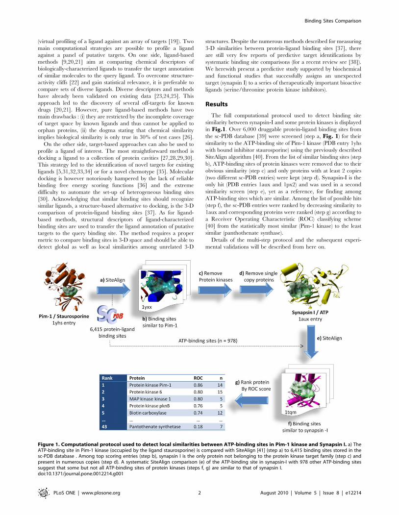
The full computational protocol used to detect binding site
similarity between synapsin-I and some protein kinases is displayed
in Fig.1. Over 6,000 druggable protein-ligand binding sites from
the sc-PDB database [39] were screened (step a, Fig. 1) for their
similarity to the ATP-binding site of Pim-1 kinase (PDB entry 1yhs
with bound inhibitor staurosporine) using the previously described
SiteAlign algorithm [40]. From the list of similar binding sites (step
b), ATP-binding sites of protein kinases were removed due to their
obvious similarity (step c) and only proteins with at least 2 copies
(two different sc-PDB entries) were kept (step d). Synapsin-I is the
only hit (PDB entries 1aux and 1px2) and was used in a second
similarity screen (step e), yet as a reference, for finding among
ATP-binding sites which are similar. Among the list of possible hits
(step f), the sc-PDB entries were ranked by decreasing similarity to
1aux and corresponding proteins were ranked (step g) according to
a Receiver Operating Characteristic (ROC) classifying scheme
[40] from the statistically most similar (Pim-1 kinase) to the least
similar (panthothenate synthase).
Details of the multi-step protocol and the subsequent experi-
mental validations will be described from here on.
Figure 1. Computational protocol used to detect local similarities between ATP-binding sites in Pim-1 kinase and Synapsin I. a) TheATP-binding site in Pim-1 kinase (occupied by the ligand staurosporine) is compared with SiteAlign [41] (step a) to 6,415 binding sites stored in thesc-PDB database . Among top scoring entries (step b), synapsin I is the only protein not belonging to the protein kinase target family (step c) andpresent in numerous copies (step d). A systematic SiteAlign comparison (e) of the ATP-binding site in synapsin-I with 978 other ATP-binding sitessuggest that some but not all ATP-binding sites of protein kinases (steps f, g) are similar to that of synapsin I.doi:10.1371/journal.pone.0012214.g001
Binding Sites Comparison
PLoS ONE | www.plosone.org 2 August 2010 | Volume 5 | Issue 8 | e12214
ATP-binding sites of synapsin I and of Pim-1 kinase sharestrikingly similar features
In benchmarking our 3-D binding site comparison algorithm
(SiteAlign) [41], we have previously compared ATP-binding sites
of protein kinases with other druggable protein-ligand cavities
from the sc-PDB database [39]. The ATP-binding site of synapsin
I was predicted to be similar to that of a pan-kinase inhibitor
(staurosporine) [42] with the proto-oncogene Pim-1 serine/
threonine protein kinase (Fig. 2A).
Protein kinases catalyze the reversible phosphorylation of
proteins and constitute a family of macromoleuclar targets of
utmost interest for their central implication in signal transduction
pathways [43]. Thanks to existing X-ray structures [44], various
inhibitors competing with the ATP substrate and exhibiting
different selectivity profiles [42] towards the 518 human protein
kinases have been designed, and some of them have reached the
market as anti-cancer drugs [45].
Synapsin I belongs to an evolutionary conserved family of
neuron-specific, synaptic vesicle-associated phosphoproteins in-
volved in the regulation of neurotransmitter release, synaptic
plasticity and synaptogenesis [46,47,48]. Synapsin isoforms are
composed of a mosaic of shared and individual domains, among
which the amino-terminal domain A and the large central domain
C are the most conserved across isoforms and species [49,50]. The
crystal structure of the recombinant C domain [51] or ABC
domains [52] of synapsin I revealed a high similarity to proteins of
the ATP-grasp superfamily, notably glutathione synthase, and the
presence of tightly associated dimers that can associate in a
tetramer. Indeed, in vitro studies showed that ATP binds to all
synapsins and that synapsins form homo- and hetero-oligomers
[53,54,55]. The binding of ATP affects the oligomerization state of
the synapsin ABC domains [52] and the interaction of synapsin I
with the immunophilin cyclophillin B [56]. Moreover, synapsin I is
a major presynaptic substrate of distinct protein kinases including
PKA, CaM kinases I/II/IV, MAPK/Erk, cdk5, PAK and Src
[46,57,58,59,60] that regulates synaptic vesicle trafficking, synap-
tic plasticity and neuronal development in a phosphorylation-
dependent fashion [61,62,63,64,65,66,67,68]. Based on the
structural similarity between the crystal structure of the synapsin
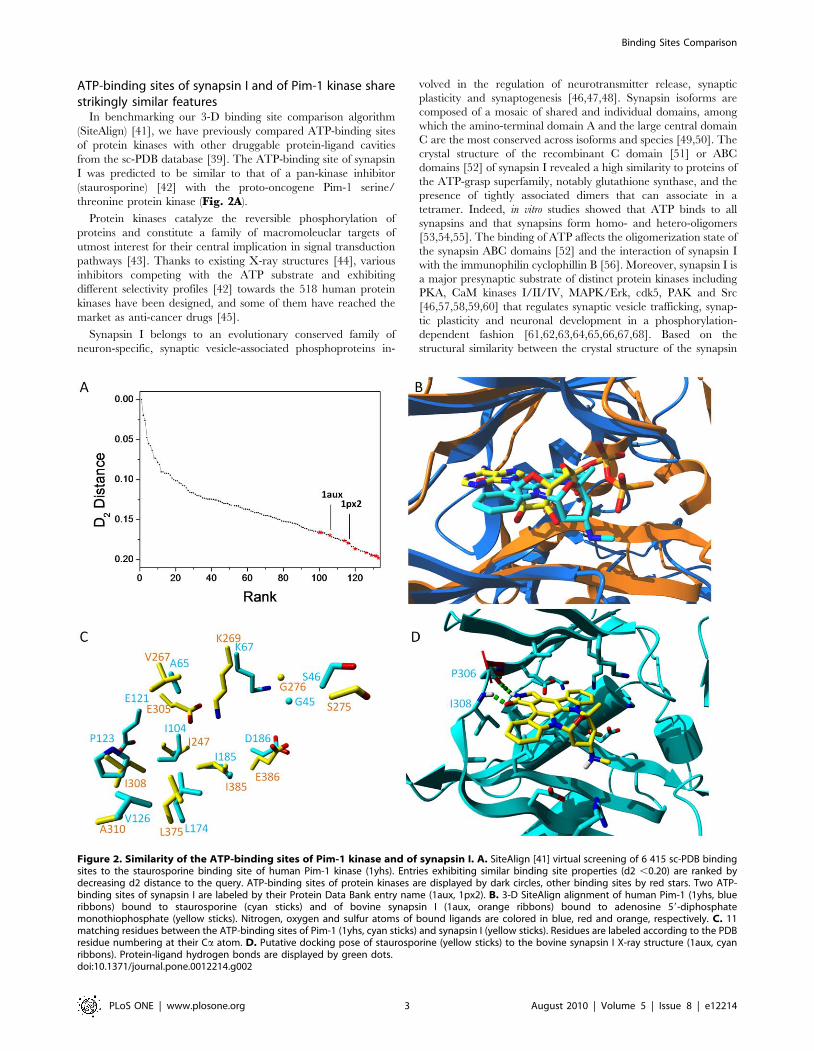
Figure 2. Similarity of the ATP-binding sites of Pim-1 kinase and of synapsin I. A. SiteAlign [41] virtual screening of 6 415 sc-PDB bindingsites to the staurosporine binding site of human Pim-1 kinase (1yhs). Entries exhibiting similar binding site properties (d2 ,0.20) are ranked bydecreasing d2 distance to the query. ATP-binding sites of protein kinases are displayed by dark circles, other binding sites by red stars. Two ATP-binding sites of synapsin I are labeled by their Protein Data Bank entry name (1aux, 1px2). B. 3-D SiteAlign alignment of human Pim-1 (1yhs, blueribbons) bound to staurosporine (cyan sticks) and of bovine synapsin I (1aux, orange ribbons) bound to adenosine 59-diphosphatemonothiophosphate (yellow sticks). Nitrogen, oxygen and sulfur atoms of bound ligands are colored in blue, red and orange, respectively. C. 11matching residues between the ATP-binding sites of Pim-1 (1yhs, cyan sticks) and synapsin I (yellow sticks). Residues are labeled according to the PDBresidue numbering at their Ca atom. D. Putative docking pose of staurosporine (yellow sticks) to the bovine synapsin I X-ray structure (1aux, cyanribbons). Protein-ligand hydrogen bonds are displayed by green dots.doi:10.1371/journal.pone.0012214.g002
Binding Sites Comparison
PLoS ONE | www.plosone.org 3 August 2010 | Volume 5 | Issue 8 | e12214
C domain with ATPases, a highly evolutionary conserved ATP
binding site has been mapped in domain C [50,51] and found to
bind ATP with nanomolar affinity in a Ca2+-dependent manner
[54]. Although very few data exist in the literature, it has been
reported that a domain C peptide corresponding to a sequence
between the ATP binding site and the Ca2+-binding site speci-
fically inhibits the binding of synapsin I to F-actin [69]. ATP
binding to synapsin I facilitates the transition from dimer to
tetramer [52] and inhibits cyclophilin B binding [56].
We showed in the first series of computations (Fig. 2A) that
ATP-binding sites of protein kinases do not resemble neither ATP-
binding sites of other kinases nor other ATP-binding cavities
[41,70]. It is therefore not surprising that 123 out of the 134
binding sites (92%) scored above an acceptable similarity threshold
(SiteAlign d2 score ,0.2)[41] are annotated as ATP-binding sites
in protein kinases (Fig. 2A, Supplementary Table S1). Out of
the 11 outliers, two entries (PDB entries 1aux and 1px2) drew our
attention since they both describe the ATP-binding site of synapsin
I. Despite a low homology (21%) between human Pim-1 (1yhs) and
bovine synapsin I (1aux) amino acid sequences, the proposed 3-D
alignment between both binding sites reveal remarkable shared
features. Although both proteins adopt distinct 3-D folds, their
bound ligands (staurosporine in Pim-1, ATP-cS in synapsin I) in
the cognate X-ray structures occupy a similar orientation in their
respective binding sites (Fig. 2B). Out of the 32 and 24 cavity-
lining residues in 1yhs and 1aux, respectively, 11 amino acids
matched in both their chemical properties and 3-D spatial
coordinates (Fig. 2C). 6 pairs of short side-chain aliphatic
residues, one pair of lysine residues, two pairs of negatively-
charged amino acids, one pair of glycine and one pair of serine
residues are absolutely conserved in both binding sites (Fig. 2C).
To be sure that non-conserved residues in the synapsin I site would
not impair staurosporine recognition, we attempted preliminary
docking experiments of the latter ligand to the 1aux structure.
Only the floppy Lys67 side chain which points inward the ATP-cS
binding site was rendered flexible during docking to putatively
enlarge the cavity. Docking staurosporine in synapsin I with the
GOLD software (see structure in Fig. 3A) provided a single set of
similar binding poses with mostly hydrophobic intermolecular
contacts and a bidentate hydrogen bond to main chain atoms of a
hinge region (Pro306, Ile308; see top-ranked pose Fig. 2D).
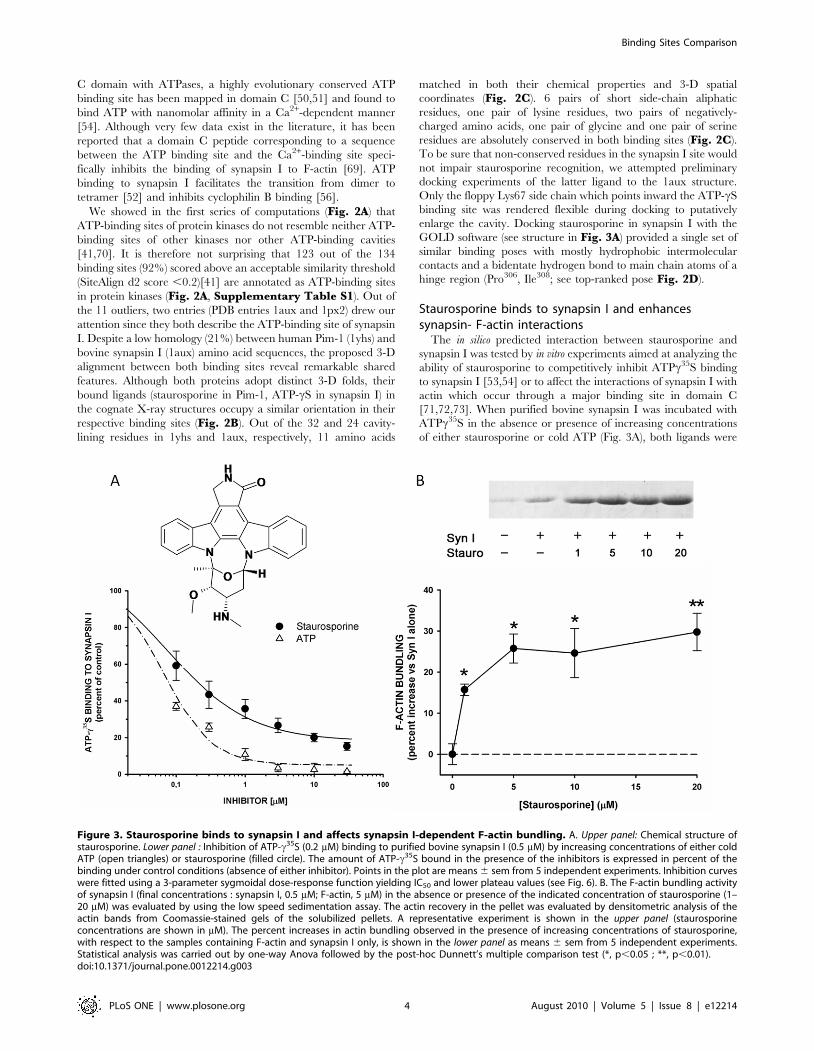
Staurosporine binds to synapsin I and enhancessynapsin- F-actin interactions
The in silico predicted interaction between staurosporine and
synapsin I was tested by in vitro experiments aimed at analyzing the
ability of staurosporine to competitively inhibit ATPc35S binding
to synapsin I [53,54] or to affect the interactions of synapsin I with
actin which occur through a major binding site in domain C
[71,72,73]. When purified bovine synapsin I was incubated with
ATPc35S in the absence or presence of increasing concentrations
of either staurosporine or cold ATP (Fig. 3A), both ligands were
Figure 3. Staurosporine binds to synapsin I and affects synapsin I-dependent F-actin bundling. A. Upper panel: Chemical structure ofstaurosporine. Lower panel : Inhibition of ATP-c35S (0.2 mM) binding to purified bovine synapsin I (0.5 mM) by increasing concentrations of either coldATP (open triangles) or staurosporine (filled circle). The amount of ATP-c35S bound in the presence of the inhibitors is expressed in percent of thebinding under control conditions (absence of either inhibitor). Points in the plot are means 6 sem from 5 independent experiments. Inhibition curveswere fitted using a 3-parameter sygmoidal dose-response function yielding IC50 and lower plateau values (see Fig. 6). B. The F-actin bundling activityof synapsin I (final concentrations : synapsin I, 0.5 mM; F-actin, 5 mM) in the absence or presence of the indicated concentration of staurosporine (1–20 mM) was evaluated by using the low speed sedimentation assay. The actin recovery in the pellet was evaluated by densitometric analysis of theactin bands from Coomassie-stained gels of the solubilized pellets. A representative experiment is shown in the upper panel (staurosporineconcentrations are shown in mM). The percent increases in actin bundling observed in the presence of increasing concentrations of staurosporine,with respect to the samples containing F-actin and synapsin I only, is shown in the lower panel as means 6 sem from 5 independent experiments.Statistical analysis was carried out by one-way Anova followed by the post-hoc Dunnett’s multiple comparison test (*, p,0.05 ; **, p,0.01).doi:10.1371/journal.pone.0012214.g003
Binding Sites Comparison
PLoS ONE | www.plosone.org 4 August 2010 | Volume 5 | Issue 8 | e12214
able to inhibit ATPc35S in a concentration-dependent fashion
(IC50 0.0760.01 mM and maximal inhibition 95.862.2% for
ATP; IC50 0.3160.09 mM and maximal inhibition 84.361.2% for
staurosporine), indicating that staurosporine bound synapsin I at
the ATP binding site.
Since the Synapsin I ATP binding pocket is localized in the
synapsin domain primarily involved in actin binding [69,72], we
investigated whether staurosporine binding is able to affect the
synapsin I-actin interaction. To this aim, we evaluated the F-actin
binding/bundling activity under conditions of ATP binding to
synapsin I in the presence of increasing concentrations of
staurosporine ranging from to 1 to 20 mM. The amount of F-
actin/synapsin I bundles recovered by low speed sedimentation
was increased by staurosporine by approximately 30% at 5 mM
(Fig. 3B), indicating that binding of the kinase inhibitor to the ATP
binding site of synapsin I modifies its molecular interactions with
the F-actin-based cytoskeleton.
Synapsin I is closer to Pim-1 than to other protein kinasesStaurosporine is a pan-kinase inhibitor exhibiting not only
nanomolar affinities to Pim-1 but also to a wide array of protein
kinases [42]. To ascertain whether the ATP-binding site of synapsin
I is equally close to all known ATP-binding sites or specifically
related to Pim-1, we computed with SiteAlign [41] the distance
between the ATP-binding site of bovine synapsin I (1aux) and 978
ATP-binding sites from the Protein Data Bank. The 978 ATP-
binding binding sites were extracted from the sc-PDB database [39]
and feature a total of 433 unique proteins among which 110 are
protein kinases. 113 entries describing 46 different protein kinases
present a binding site distance below 0.20, the previously-
determined computed threshold for discriminating similar from
dissimilar binding sites [41]. This result suggests that the ATP site of
synapsin I is similar to that of many other protein kinases. When
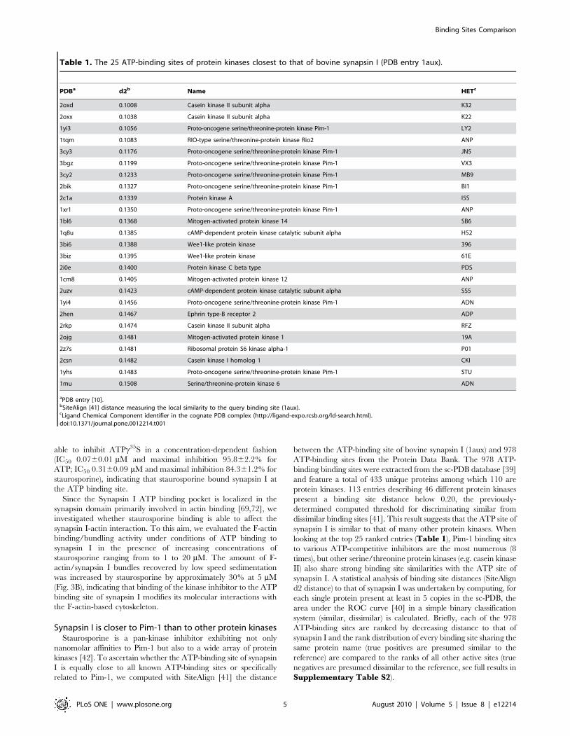
looking at the top 25 ranked entries (Table 1), Pim-1 binding sites
to various ATP-competitive inhibitors are the most numerous (8
times), but other serine/threonine protein kinases (e.g. casein kinase
II) also share strong binding site similarities with the ATP site of
synapsin I. A statistical analysis of binding site distances (SiteAlign
d2 distance) to that of synapsin I was undertaken by computing, for
each single protein present at least in 5 copies in the sc-PDB, the
area under the ROC curve [40] in a simple binary classification
system (similar, dissimilar) is calculated. Briefly, each of the 978
ATP-binding sites are ranked by decreasing distance to that of
synapsin I and the rank distribution of every binding site sharing the
same protein name (true positives are presumed similar to the
reference) are compared to the ranks of all other active sites (true
negatives are presumed dissimilar to the reference, see full results in
Supplementary Table S2).
Table 1. The 25 ATP-binding sites of protein kinases closest to that of bovine synapsin I (PDB entry 1aux).
PDBa d2b Name HETc
2oxd 0.1008 Casein kinase II subunit alpha K32
2oxx 0.1038 Casein kinase II subunit alpha K22
1yi3 0.1056 Proto-oncogene serine/threonine-protein kinase Pim-1 LY2
1tqm 0.1083 RIO-type serine/threonine-protein kinase Rio2 ANP
3cy3 0.1176 Proto-oncogene serine/threonine-protein kinase Pim-1 JN5
3bgz 0.1199 Proto-oncogene serine/threonine-protein kinase Pim-1 VX3
3cy2 0.1233 Proto-oncogene serine/threonine-protein kinase Pim-1 MB9
2bik 0.1327 Proto-oncogene serine/threonine-protein kinase Pim-1 BI1
2c1a 0.1339 Protein kinase A I5S
1xr1 0.1350 Proto-oncogene serine/threonine-protein kinase Pim-1 ANP
1bl6 0.1368 Mitogen-activated protein kinase 14 SB6
1q8u 0.1385 cAMP-dependent protein kinase catalytic subunit alpha H52
3bi6 0.1388 Wee1-like protein kinase 396
3biz 0.1395 Wee1-like protein kinase 61E
2i0e 0.1400 Protein kinase C beta type PDS
1cm8 0.1405 Mitogen-activated protein kinase 12 ANP
2uzv 0.1423 cAMP-dependent protein kinase catalytic subunit alpha SS5
1yi4 0.1456 Proto-oncogene serine/threonine-protein kinase Pim-1 ADN
2hen 0.1467 Ephrin type-B receptor 2 ADP
2rkp 0.1474 Casein kinase II subunit alpha RFZ
2ojg 0.1481 Mitogen-activated protein kinase 1 19A
2z7s 0.1481 Ribosomal protein S6 kinase alpha-1 P01
2csn 0.1482 Casein kinase I homolog 1 CKI
1yhs 0.1483 Proto-oncogene serine/threonine-protein kinase Pim-1 STU
1mu 0.1508 Serine/threonine-protein kinase 6 ADN
aPDB entry [10].bSiteAlign [41] distance measuring the local similarity to the query binding site (1aux).cLigand Chemical Component identifier in the cognate PDB complex (http://ligand-expo.rcsb.org/ld-search.html).doi:10.1371/journal.pone.0012214.t001
Binding Sites Comparison
PLoS ONE | www.plosone.org 5 August 2010 | Volume 5 | Issue 8 | e12214
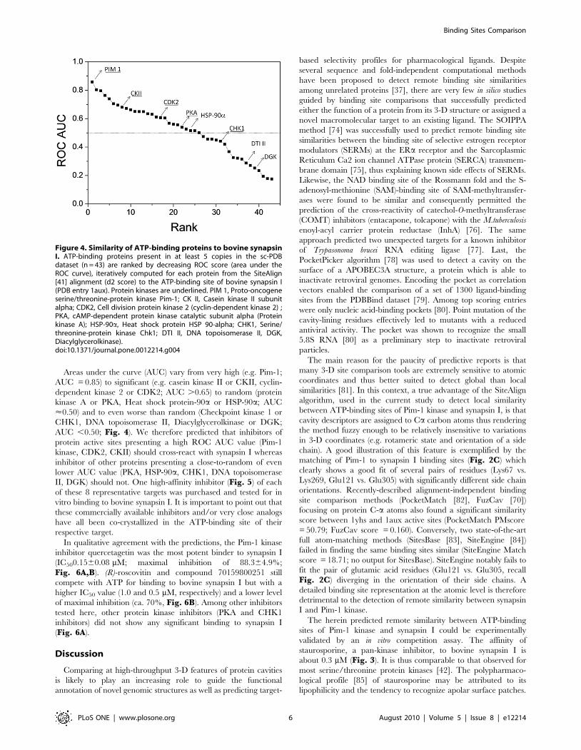
Areas under the curve (AUC) vary from very high (e.g. Pim-1;
AUC = 0.85) to significant (e.g. casein kinase II or CKII, cyclin-
dependent kinase 2 or CDK2; AUC .0.65) to random (protein
kinase A or PKA, Heat shock protein-90a or HSP-90a; AUC
<0.50) and to even worse than random (Checkpoint kinase 1 or
CHK1, DNA topoisomerase II, Diacylglycerolkinase or DGK;
AUC ,0.50; Fig. 4). We therefore predicted that inhibitors of
protein active sites presenting a high ROC AUC value (Pim-1
kinase, CDK2, CKII) should cross-react with synapsin I whereas
inhibitor of other proteins presenting a close-to-random of even
lower AUC value (PKA, HSP-90a, CHK1, DNA topoisomerase
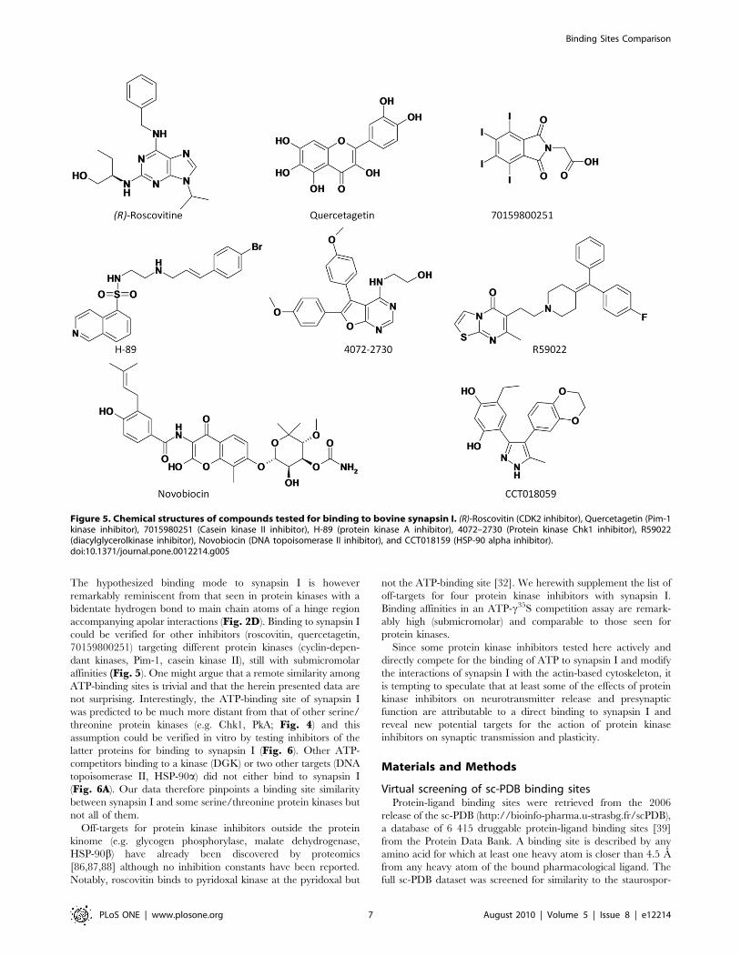
II, DGK) should not. One high-affinity inhibitor (Fig. 5) of each
of these 8 representative targets was purchased and tested for in
vitro binding to bovine synapsin I. It is important to point out that
these commercially available inhibitors and/or very close analogs
have all been co-crystallized in the ATP-binding site of their
respective target.
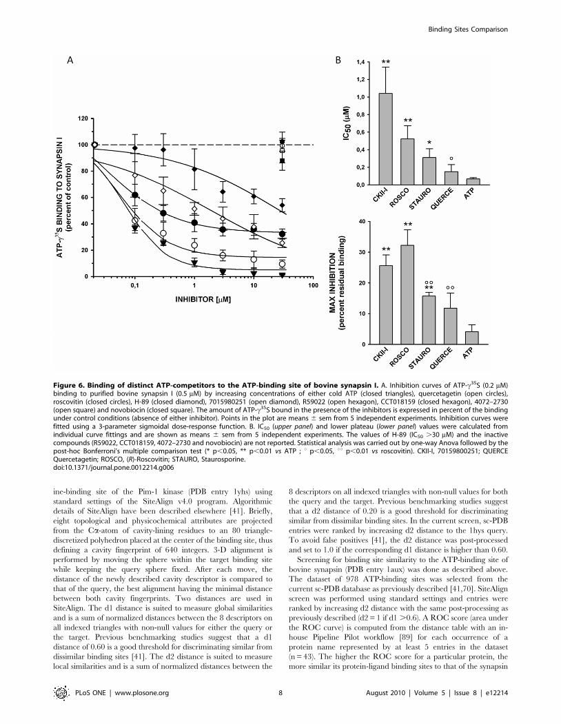
In qualitative agreement with the predictions, the Pim-1 kinase
inhibitor quercetagetin was the most potent binder to synapsin I
(IC500.1560.08 mM; maximal inhibition of 88.364.9%;
Fig. 6A,B). (R)-roscovitin and compound 70159800251 still
compete with ATP for binding to bovine synapsin I but with a
higher IC50 value (1.0 and 0.5 mM, respectively) and a lower level
of maximal inhibition (ca. 70%, Fig. 6B). Among other inhibitors
tested here, other protein kinase inhibitors (PKA and CHK1
inhibitors) did not show any significant binding to synapsin I
(Fig. 6A).
Discussion
Comparing at high-throughput 3-D features of protein cavities
is likely to play an increasing role to guide the functional
annotation of novel genomic structures as well as predicting target-
based selectivity profiles for pharmacological ligands. Despite
several sequence and fold-independent computational methods
have been proposed to detect remote binding site similarities
among unrelated proteins [37], there are very few in silico studies
guided by binding site comparisons that successfully predicted
either the function of a protein from its 3-D structure or assigned a
novel macromolecular target to an existing ligand. The SOIPPA
method [74] was successfully used to predict remote binding site
similarities between the binding site of selective estrogen receptor
modulators (SERMs) at the ERa receptor and the Sarcoplasmic
Reticulum Ca2 ion channel ATPase protein (SERCA) transmem-
brane domain [75], thus explaining known side effects of SERMs.
Likewise, the NAD binding site of the Rossmann fold and the S-
adenosyl-methionine (SAM)-binding site of SAM-methyltransfer-
ases were found to be similar and consequently permitted the
prediction of the cross-reactivity of catechol-O-methyltransferase
(COMT) inhibitors (entacapone, tolcapone) with the M.tuberculosis
enoyl-acyl carrier protein reductase (InhA) [76]. The same
approach predicted two unexpected targets for a known inhibitor
of Trypasonoma brucei RNA editing ligase [77]. Last, the
PocketPicker algorithm [78] was used to detect a cavity on the
surface of a APOBEC3A structure, a protein which is able to
inactivate retroviral genomes. Encoding the pocket as correlation
vectors enabled the comparison of a set of 1300 ligand-binding
sites from the PDBBind dataset [79]. Among top scoring entries
were only nucleic acid-binding pockets [80]. Point mutation of the
cavity-lining residues effectively led to mutants with a reduced
antiviral activity. The pocket was shown to recognize the small
5.8S RNA [80] as a preliminary step to inactivate retroviral
particles.
The main reason for the paucity of predictive reports is that
many 3-D site comparison tools are extremely sensitive to atomic
coordinates and thus better suited to detect global than local
similarities [81]. In this context, a true advantage of the SiteAlign
algorithm, used in the current study to detect local similarity
between ATP-binding sites of Pim-1 kinase and synapsin I, is that
cavity descriptors are assigned to Ca carbon atoms thus rendering
the method fuzzy enough to be relatively insensitive to variations
in 3-D coordinates (e.g. rotameric state and orientation of a side
chain). A good illustration of this feature is exemplified by the
matching of Pim-1 to synapsin I binding sites (Fig. 2C) which
clearly shows a good fit of several pairs of residues (Lys67 vs.
Lys269, Glu121 vs. Glu305) with significantly different side chain
orientations. Recently-described alignment-independent binding
site comparison methods (PocketMatch [82], FuzCav [70])
focusing on protein C-a atoms also found a significant similarity
score between 1yhs and 1aux active sites (PocketMatch PMscore
= 50.79; FuzCav score = 0.160). Conversely, two state-of-the-art
full atom-matching methods (SitesBase [83], SiteEngine [84])
failed in finding the same binding sites similar (SiteEngine Match
score = 18.71; no output for SitesBase). SiteEngine notably fails to
fit the pair of glutamic acid residues (Glu121 vs. Glu305, recall
Fig. 2C) diverging in the orientation of their side chains. A
detailed binding site representation at the atomic level is therefore
detrimental to the detection of remote similarity between synapsin
I and Pim-1 kinase.
The herein predicted remote similarity between ATP-binding
sites of Pim-1 kinase and synapsin I could be experimentally
validated by an in vitro competition assay. The affinity of
staurosporine, a pan-kinase inhibitor, to bovine synapsin I is
about 0.3 mM (Fig. 3). It is thus comparable to that observed for
most serine/threonine protein kinases [42]. The polypharmaco-
logical profile [85] of staurosporine may be attributed to its
lipophilicity and the tendency to recognize apolar surface patches.
Figure 4. Similarity of ATP-binding proteins to bovine synapsinI. ATP-binding proteins present in at least 5 copies in the sc-PDBdataset (n = 43) are ranked by decreasing ROC score (area under theROC curve), iteratively computed for each protein from the SiteAlign[41] alignment (d2 score) to the ATP-binding site of bovine synapsin I(PDB entry 1aux). Protein kinases are underlined. PIM 1, Proto-oncogeneserine/threonine-protein kinase Pim-1; CK II, Casein kinase II subunitalpha; CDK2, Cell division protein kinase 2 (cyclin-dependent kinase 2) ;PKA, cAMP-dependent protein kinase catalytic subunit alpha (Proteinkinase A); HSP-90a, Heat shock protein HSP 90-alpha; CHK1, Serine/threonine-protein kinase Chk1; DTI II, DNA topoisomerase II, DGK,Diacylglycerolkinase).doi:10.1371/journal.pone.0012214.g004
Binding Sites Comparison
PLoS ONE | www.plosone.org 6 August 2010 | Volume 5 | Issue 8 | e12214
The hypothesized binding mode to synapsin I is however
remarkably reminiscent from that seen in protein kinases with a
bidentate hydrogen bond to main chain atoms of a hinge region
accompanying apolar interactions (Fig. 2D). Binding to synapsin I
could be verified for other inhibitors (roscovitin, quercetagetin,
70159800251) targeting different protein kinases (cyclin-depen-
dant kinases, Pim-1, casein kinase II), still with submicromolar
affinities (Fig. 5). One might argue that a remote similarity among
ATP-binding sites is trivial and that the herein presented data are
not surprising. Interestingly, the ATP-binding site of synapsin I
was predicted to be much more distant from that of other serine/
threonine protein kinases (e.g. Chk1, PkA; Fig. 4) and this
assumption could be verified in vitro by testing inhibitors of the
latter proteins for binding to synapsin I (Fig. 6). Other ATP-
competitors binding to a kinase (DGK) or two other targets (DNA
topoisomerase II, HSP-90a) did not either bind to synapsin I
(Fig. 6A). Our data therefore pinpoints a binding site similarity
between synapsin I and some serine/threonine protein kinases but
not all of them.
Off-targets for protein kinase inhibitors outside the protein
kinome (e.g. glycogen phosphorylase, malate dehydrogenase,
HSP-90b) have already been discovered by proteomics
[86,87,88] although no inhibition constants have been reported.
Notably, roscovitin binds to pyridoxal kinase at the pyridoxal but
not the ATP-binding site [32]. We herewith supplement the list of
off-targets for four protein kinase inhibitors with synapsin I.
Binding affinities in an ATP-c35S competition assay are remark-
ably high (submicromolar) and comparable to those seen for
protein kinases.
Since some protein kinase inhibitors tested here actively and
directly compete for the binding of ATP to synapsin I and modify
the interactions of synapsin I with the actin-based cytoskeleton, it
is tempting to speculate that at least some of the effects of protein
kinase inhibitors on neurotransmitter release and presynaptic
function are attributable to a direct binding to synapsin I and
reveal new potential targets for the action of protein kinase
inhibitors on synaptic transmission and plasticity.
Materials and Methods
Virtual screening of sc-PDB binding sitesProtein-ligand binding sites were retrieved from the 2006
release of the sc-PDB (http://bioinfo-pharma.u-strasbg.fr/scPDB),
a database of 6 415 druggable protein-ligand binding sites [39]
from the Protein Data Bank. A binding site is described by any
amino acid for which at least one heavy atom is closer than 4.5 A
from any heavy atom of the bound pharmacological ligand. The
full sc-PDB dataset was screened for similarity to the staurospor-
Figure 5. Chemical structures of compounds tested for binding to bovine synapsin I. (R)-Roscovitin (CDK2 inhibitor), Quercetagetin (Pim-1kinase inhibitor), 7015980251 (Casein kinase II inhibitor), H-89 (protein kinase A inhibitor), 4072–2730 (Protein kinase Chk1 inhibitor), R59022(diacylglycerolkinase inhibitor), Novobiocin (DNA topoisomerase II inhibitor), and CCT018159 (HSP-90 alpha inhibitor).doi:10.1371/journal.pone.0012214.g005
Binding Sites Comparison
PLoS ONE | www.plosone.org 7 August 2010 | Volume 5 | Issue 8 | e12214
ine-binding site of the Pim-1 kinase (PDB entry 1yhs) using
standard settings of the SiteAlign v4.0 program. Algorithmic
details of SiteAlign have been described elsewhere [41]. Briefly,
eight topological and physicochemical attributes are projected
from the Ca-atom of cavity-lining residues to an 80 triangle-
discretized polyhedron placed at the center of the binding site, thus
defining a cavity fingerprint of 640 integers. 3-D alignment is
performed by moving the sphere within the target binding site
while keeping the query sphere fixed. After each move, the
distance of the newly described cavity descriptor is compared to
that of the query, the best alignment having the minimal distance
between both cavity fingerprints. Two distances are used in
SiteAlign. The d1 distance is suited to measure global similarities
and is a sum of normalized distances between the 8 descriptors on
all indexed triangles with non-null values for either the query or
the target. Previous benchmarking studies suggest that a d1
distance of 0.60 is a good threshold for discriminating similar from
dissimilar binding sites [41]. The d2 distance is suited to measure
local similarities and is a sum of normalized distances between the
8 descriptors on all indexed triangles with non-null values for both
the query and the target. Previous benchmarking studies suggest
that a d2 distance of 0.20 is a good threshold for discriminating
similar from dissimilar binding sites. In the current screen, sc-PDB
entries were ranked by increasing d2 distance to the 1hys query.
To avoid false positives [41], the d2 distance was post-processed
and set to 1.0 if the corresponding d1 distance is higher than 0.60.
Screening for binding site similarity to the ATP-binding site of
bovine synapsin (PDB entry 1aux) was done as described above.
The dataset of 978 ATP-binding sites was selected from the
current sc-PDB database as previously described [41,70]. SiteAlign
screen was performed using standard settings and entries were
ranked by increasing d2 distance with the same post-processing as
previously described (d2 = 1 if d1 .0.6). A ROC score (area under
the ROC curve) is computed from the distance table with an in-
house Pipeline Pilot workflow [89] for each occurrence of a
protein name represented by at least 5 entries in the dataset
(n = 43). The higher the ROC score for a particular protein, the
more similar its protein-ligand binding sites to that of the synapsin
Figure 6. Binding of distinct ATP-competitors to the ATP-binding site of bovine synapsin I. A. Inhibition curves of ATP-c35S (0.2 mM)binding to purified bovine synapsin I (0.5 mM) by increasing concentrations of either cold ATP (closed triangles), quercetagetin (open circles),roscovitin (closed circles), H-89 (closed diamond), 7015980251 (open diamond), R59022 (open hexagon), CCT018159 (closed hexagon), 4072–2730(open square) and novobiocin (closed square). The amount of ATP-c35S bound in the presence of the inhibitors is expressed in percent of the bindingunder control conditions (absence of either inhibitor). Points in the plot are means 6 sem from 5 independent experiments. Inhibition curves werefitted using a 3-parameter sigmoidal dose-response function. B. IC50 (upper panel) and lower plateau (lower panel) values were calculated fromindividual curve fittings and are shown as means 6 sem from 5 independent experiments. The values of H-89 (IC50 .30 mM) and the inactivecompounds (R59022, CCT018159, 4072–2730 and novobiocin) are not reported. Statistical analysis was carried out by one-way Anova followed by thepost-hoc Bonferroni’s multiple comparison test (* p,0.05, ** p,0.01 vs ATP ; u p,0.05, uu p,0.01 vs roscovitin). CKII-I, 70159800251; QUERCEQuercetagetin; ROSCO, (R)-Roscovitin; STAURO, Staurosporine.doi:10.1371/journal.pone.0012214.g006
Binding Sites Comparison
PLoS ONE | www.plosone.org 8 August 2010 | Volume 5 | Issue 8 | e12214
I reference. A ROC score of 0.5 indicate a random distribution of
d2 scores for a particular protein.
Automated docking of staurosporine to bovinesynapsin I
3-D atomic coordinates of staurosporine were obtained by
Corina v3.5[90] from a 2-D Marvin sketch [91]. Hydrogen atoms
were added using standard topological rules in Sybyl v.8.0 [92]
and coordinates were saved in mol2 format. Standard settings of
the Gold v4.1 program [93] were used to dock staurosporine to the
ATP-binding site of bovine synapsin (PDB entry 1aux) whose
coordinates were retrieved from the sc-PDB databank [30]. The
cavity was defined as any protein atom present in a 10 A-radius
sphere centered on the center of mass of the sc-PDB binding site.
The side chain of Lys67 was considered flexible during the docking
by explicit definition of 27 rotameric states from the standard Gold
rotamer library. Poses were scored with the Goldscore fitness
function.
Comparison of bovine synapsin I and human Pim-1 ATP-binding sites with other binding site matching methods
ATP-binding sites of 1aux (bovine synapsin I) and 1yhs (human
Pim-1 kinase) were retrieved from the sc-PDB website (http://
bioinfo-pharma.u-strasbg.fr/scPDB) and compared with the in-
house FuzCav algorithm with default parameters [70]. Web
interfaces to SitesBase [83] (http://www.modelling.leeds.ac.uk/
sb/), SiteEngine [84] (http://bioinfo3d.cs.tau.ac.il/SiteEngine/)
and PocketMatch [82] (http://proline.physics.iisc.ernet.in/
pocketmatch/) were used to compare the same entries. Active
site detection was here achieved by specifying the chemical
component HET code of the co-crystallized ligands (SAP for 1uax,
STU for 1yhs).
ATP-c35S binding assaysSynapsin I was purified from bovine brain [71] and stored in
liquid nitrogen in 200 mM NaCl, 25 mM TrisCl, pH 7.4.
Synapsin I (500 nM) was incubated with 200 nM ATP-c35S
(Perkin Elmer, Waltham, MA) in 50 mM HEPES-NaOH pH 7.4,
25 mM NaCl, 0.5 mM CaCl2 and 2 mM MgCl2 for 1 h at room
temperature in the absence or presence of increasing concentra-
tions (0.1–30 mM) of either cold ATP, staurosporine (Sigma,
Milan, Italy), (R)-roscovitin (Caiman, Ann Arbor, MI), querceta-
getin (Calbiochem, San Diego, CA), 70159800251 (Otava, kiev,
Ukraine), H-89 (LC Laboratories, Woburn, MA), 4072–7730
(ChemDiv, San Diego, CA), R59022 and novobiocin (MP
Biochemicals, Illkirch, France) and last CCT018059 (SPI-Bio,
Montigny Le Bretonneux, France). ATP-c35S binding was
quantified as previously described [94]. Briefly, aliquots of the
samples were spotted onto squares of phosphocellulose paper
(Upstate/Millipore, Billerica, MA). The paper squares were
extensively washed with deionized water for 30 min, air-dried
and analyzed for radioactivity by using the Perkin Elmer Cyclone
Plus Phosphor Imager. After subtraction of the background values
(samples with no synapsin I), data from individual competition
curves were fitted with a sigmoidal dose-response function (f = min +(max–min)/(1+10‘((logEC50-x)*Hillslope)) using the Sigmaplot 8.0
software (SPSS Inc., Chicago, IL) to yield IC50 and maximal
inhibition values. Data in the plots are the means 6 sem of at least 5
independent experiments.
Actin Bundling AssaysActin was purified from acetone powder of rabbit skeletal
muscles [95,96] and stored in liquid nitrogen in in 2 mM Tris
pH 8, 0.2 mM ATP, 0,2 mM CaCl2, 0.125 mM b-mercaptoeth-
anol and 0.005% NaN3 (G-buffer). Before the experiments, both
G-actin and synapsin I were prespun for 1 h at 4uC at 300,0006g
to remove large aggregates. G-actin was polymerized at room
temperature for 1 h in the presence of 100 mM KCl, 1.2 mM
MgCl2. Synapsin I (final concentration, 0.5 mM) was preincubated
with increasing concentrations (1–20 mM) of staurosporine for 1 h
at room temperature in 200 mM NaCl, 25 mM TrisCl pH 7.4.
Actin bundling was assessed by incubating the synapsin/stauro-
sporine samples with F-actin (final concentration, 5 mM) under
polymerization conditions (100 mM KCl, 1.2 mM MgCl2 in G-
buffer) for 1.5 h at room temperature followed by low-speed
centrifugation (10,0006 g for 15 min) to separate actin bundles
(Bahler & Greengard, 1987). Pellets were solubilized in sample
buffer [97] and analyzed by sodium dodecylsulfate polyacrylamide
gel electrophoresis (SDS-PAGE) using 10% acrylamide in the
resolving gel. Gels were fixed, stained with Coomassie Blue and
destained. Densitometric analysis of the actin bands was carried
out by using the ImageQuant system (GE Healthcare) followed by
densitometric analysis of the fluorograms and by data interpola-
tion into a standard curve of purified G-actin run in parallel with
the unknown samples.
Supporting Information
Table S1
Found at: doi:10.1371/journal.pone.0012214.s001 (0.04 MB
DOC)
Table S2
Found at: doi:10.1371/journal.pone.0012214.s002 (0.06 MB
DOC)
Acknowledgments
We acknowledge the supercomputing facilities of the CINES (Montpellier,
France) and IN2P3 (Villeurbanne, France) for allocation of computing time
to D.R. (Project x2010075024).
Author Contributions
Conceived and designed the experiments: FB DR. Performed the
experiments: EDF CS MM FO. Analyzed the data: EDF CS MM FO
DR. Wrote the paper: FB DR.
References
1. Wermuth CG (2006) Similarity in drugs: reflections on analogue design. Drug
Discov Today 11: 348–354.
2. Schuster D, Laggner C, Langer T (2005) Why drugs fail—a study on side effects
in new chemical entities. Curr Pharm Des 11: 3545–3559.
3. Shoshan MC, Linder S (2008) Target specificity and off-target effects as determinants
of cancer drug efficacy. Expert Opin Drug Metab Toxicol 4: 273–280.
4. Klabunde T, Evers A (2005) GPCR antitarget modeling: pharmacophore
models for biogenic amine binding GPCRs to avoid GPCR-mediated side
effects. Chembiochem 6: 876–889.
5. Yang L, Chen J, He L (2009) Harvesting candidate genes responsible for serious
adverse drug reactions from a chemical-protein interactome. PLoS Comput Biol
5: e1000441.
6. Dessailly BH, Nair R, Jaroszewski L, Fajardo JE, Kouranov A, et al. (2009) PSI-
2: structural genomics to cover protein domain family space. Structure 17:
869–881.
7. Nair R, Liu J, Soong TT, Acton TB, Everett JK, et al. (2009) Structural
genomics is the largest contributor of novel structural leverage. J Struct Funct
Genomics 10: 181–191.
Binding Sites Comparison
PLoS ONE | www.plosone.org 9 August 2010 | Volume 5 | Issue 8 | e12214
8. Daniel M, Stuart L, Christopher C, Stuart W, Adam N (2009) Synthesis ofNatural-Product-Like Molecules with Over Eighty Distinct Scaffolds. Angew
Chem Int Ed Engl 48: 104–109.
9. Nielsen TE, Schreiber SL (2008) Towards the optimal screening collection: a
synthesis strategy. Angew Chem Int Ed Engl 47: 48–56.
10. Berman HM, Westbrook J, Feng Z, Gilliland G, Bhat TN, et al. (2000) The
Protein Data Bank. Nucleic Acids Res 28: 235–242.
11. Schneider M, Lane L, Boutet E, Lieberherr D, Tognolli M, et al. (2009) The
UniProtKB/Swiss-Prot knowledgebase and its Plant Proteome Annotation
Program. J Proteomics 72: 567–573.
12. Wang Y, Xiao J, Suzek TO, Zhang J, Wang J, et al. (2009) PubChem: a publicinformation system for analyzing bioactivities of small molecules. Nucleic Acids
Res 37: W623–633.
13. Paolini GV, Shapland RH, van Hoorn WP, Mason JS, Hopkins AL (2006)Global mapping of pharmacological space. Nat Biotechnol 24: 805–815.
14. Caron PR, Mullican MD, Mashal RD, Wilson KP, Su MS, et al. (2001)
Chemogenomic approaches to drug discovery. Curr Opin Chem Biol 5:464–470.
15. Ong SE, Schenone M, Margolin AA, Li X, Do K, et al. (2009) Identifying the
proteins to which small-molecule probes and drugs bind in cells. Proc Natl AcadSci U S A 106: 4617–4622.
16. Bajorath J (2008) Computational analysis of ligand relationships within target
families. Curr Opin Chem Biol 12: 352–358.
17. Rognan D (2007) Chemogenomic approaches to rational drug design.
Br J Pharmacol 152: 38–52.
18. Shoichet BK (2004) Virtual screening of chemical libraries. Nature 432:862–865.
19. Ekins S, Mestres J, Testa B (2007) In silico pharmacology for drug discovery:
methods for virtual ligand screening and profiling. Br J Pharmacol 152: 9–20.
20. Keiser MJ, Roth BL, Armbruster BN, Ernsberger P, Irwin JJ, et al. (2007)
Relating protein pharmacology by ligand chemistry. Nat Biotechnol 25:
197–206.
21. Keiser MJ, Setola V, Irwin JJ, Laggner C, Abbas AI, et al. (2009) Predicting new
molecular targets for known drugs. Nature 462: 175–181.
22. Peltason L, Bajorath J (2007) SAR index: quantifying the nature of structure-activity relationships. J Med Chem 50: 5571–5578.
23. Mestres J, Martin-Couce L, Gregori-Puigjane E, Cases M, Boyer S (2006)
Ligand-based approach to in silico pharmacology: nuclear receptor profiling.
J Chem Inf Model 46: 2725–2736.
24. Nettles JH, Jenkins JL, Bender A, Deng Z, Davies JW, et al. (2006) Bridging
chemical and biological space: ‘‘target fishing’’ using 2D and 3D molecular
descriptors. J Med Chem 49: 6802–6810.
25. Nidhi, Glick M, Davies JW, Jenkins JL (2006) Prediction of biological targets for
compounds using multiple-category Bayesian models trained on chemogenomics
databases. J Chem Inf Model 46: 1124–1133.
26. Martin YC, Kofron JL, Traphagen LM (2002) Do structurally similar molecules
have similar biological activity? J Med Chem 45: 4350–4358.
27. Chen YZ, Zhi DG (2001) Ligand-protein inverse docking and its potential use inthe computer search of protein targets of a small molecule. Proteins 43: 217–226.
28. Li H, Gao Z, Kang L, Zhang H, Yang K, et al. (2006) TarFisDock: a web server
for identifying drug targets with docking approach. Nucleic Acids Res 34:W219–224.
29. Paul N, Kellenberger E, Bret G, Muller P, Rognan D (2004) Recovering the true
targets of specific ligands by virtual screening of the protein data bank. Proteins54: 671–680.
30. Kellenberger E, Foata N, Rognan D (2008) Ranking targets in structure-based
virtual screening of three-dimensional protein libraries: methods and problems.J Chem Inf Model 48: 1014–1025.
31. Zahler S, Tietze S, Totzke F, Kubbutat M, Meijer L, et al. (2007) Inverse in
silico screening for identification of kinase inhibitor targets. Chem Biol 14:
1207–1214.
32. Tang L, Li MH, Cao P, Wang F, Chang WR, et al. (2005) Crystal structure of
pyridoxal kinase in complex with roscovitin and derivatives. J Biol Chem 280:
31220–31229.
33. Do QT, Renimel I, Andre P, Lugnier C, Muller CD, et al. (2005) Reverse
pharmacognosy: application of selnergy, a new tool for lead discovery. The
example of epsilon-viniferin. Curr Drug Discov Technol 2: 161–167.
34. Cai J, Han C, Hu T, Zhang J, Wu D, et al. (2006) Peptide deformylase is a
potential target for anti-Helicobacter pylori drugs: reverse docking, enzymatic
assay, and X-ray crystallography validation. Protein Sci 15: 2071–2081.
35. Muller P, Lena G, Boilard E, Bezzine S, Lambeau G, et al. (2006) In silico-
guided target identification of a scaffold-focused library: 1,3,5-triazepan-2,6-
diones as novel phospholipase A2 inhibitors. J Med Chem 49: 6768–6778.
36. Ferrara P, Gohlke H, Price DJ, Klebe G, Brooks CL, 3rd (2004) Assessing
scoring functions for protein-ligand interactions. J Med Chem 47: 3032–3047.
37. Kellenberger E, Schalon C, Rognan D (2008) How to measure the similaritybetween protein ligand-binding sites? Current Computer-Aided Drug Design 4:
209–220.
38. Rognan D (2010) Structure-Based Approaches to Target Fishing and LigandProfiling. Mol Inf 29: 176–187.
39. Kellenberger E, Muller P, Schalon C, Bret G, Foata N, et al. (2006) sc-PDB: an
annotated database of druggable binding sites from the Protein Data Bank.
J Chem Inf Model 46: 717–727.
40. Triballeau N, Acher F, Brabet I, Pin JP, Bertrand HO (2005) Virtual screeningworkflow development guided by the ‘‘receiver operating characteristic’’ curve
approach. Application to high-throughput docking on metabotropic glutamate
receptor subtype 4. J Med Chem 48: 2534–2547.
41. Schalon C, Surgand JS, Kellenberger E, Rognan D (2008) A simple and fuzzymethod to align and compare druggable ligand-binding sites. Proteins 71:
1755–1778.
42. Fabian MA, Biggs WH, 3rd, Treiber DK, Atteridge CE, Azimioara MD, et al.(2005) A small molecule-kinase interaction map for clinical kinase inhibitors. Nat
Biotechnol 23: 329–336.
43. Manning G, Whyte DB, Martinez R, Hunter T, Sudarsanam S (2002) Theprotein kinase complement of the human genome. Science 298: 1912–1934.
44. Johnson LN (2009) Protein kinase inhibitors: contributions from structure to
clinical compounds. Q Rev Biophys 42: 1–40.
45. Grant SK (2009) Therapeutic protein kinase inhibitors. Cell Mol Life Sci 66:
1163–1177.
46. De Camilli P, Benfenati F, Valtorta F, Greengard P (1990) The synapsins. AnnuRev Cell Biol 6: 433–460.
47. Greengard P, Valtorta F, Czernik AJ, Benfenati F (1993) Synaptic vesicle
phosphoproteins and regulation of synaptic function. Science 259: 780–785.
48. Fdez E, Hilfiker S (2006) Vesicle pools and synapsins: new insights into old
enigmas. Brain Cell Biol 35: 107–115.
49. Sudhof TC, Czernik AJ, Kao HT, Takei K, Johnston PA, et al. (1989)Synapsins: mosaics of shared and individual domains in a family of synaptic
vesicle phosphoproteins. Science 245: 1474–1480.
50. Kao HT, Porton B, Hilfiker S, Stefani G, Pieribone VA, et al. (1999) Molecularevolution of the synapsin gene family. J Exp Zool 285: 360–377.
51. Esser L, Wang CR, Hosaka M, Smagula CS, Sudhof TC, et al. (1998) Synapsin I
is structurally similar to ATP-utilizing enzymes. EMBO J 17: 977–984.
52. Brautigam CA, Chelliah Y, Deisenhofer J (2004) Tetramerization and ATP
binding by a protein comprising the A, B, and C domains of rat synapsin I. J Biol
Chem 279: 11948–11956.
53. Hosaka M, Sudhof TC (1998) Synapsin III, a novel synapsin with an unusual
regulation by Ca2+. J Biol Chem 273: 13371–13374.
54. Hosaka M, Sudhof TC (1998) Synapsins I and II are ATP-binding proteins withdifferential Ca2+ regulation. J Biol Chem 273: 1425–1429.
55. Hosaka M, Sudhof TC (1999) Homo- and heterodimerization of synapsins. J Biol
Chem 274: 16747–16753.
56. Lane-Guermonprez L, Morot-Gaudry-Talarmain Y, Meunier FM, O’Regan S,
Onofri F, et al. (2005) Synapsin associates with cyclophilin B in an ATP- and
cyclosporin A-dependent manner. J Neurochem 93: 1401–1411.
57. Sakurada K, Kato H, Nagumo H, Hiraoka H, Furuya K, et al. (2002) Synapsin I
is phosphorylated at Ser603 by p21-activated kinases (PAKs) in vitro and in
PC12 cells stimulated with bradykinin. J Biol Chem 277: 45473–45479.
58. Jovanovic JN, Benfenati F, Siow YL, Sihra TS, Sanghera JS, et al. (1996)Neurotrophins stimulate phosphorylation of synapsin I by MAP kinase and
regulate synapsin I-actin interactions. Proc Natl Acad Sci U S A 93: 3679–3683.
59. Jovanovic JN, Sihra TS, Nairn AC, Hemmings HC, Jr., Greengard P, et al.(2001) Opposing changes in phosphorylation of specific sites in synapsin I during
Ca2+-dependent glutamate release in isolated nerve terminals. J Neurosci 21:
7944–7953.
60. Onofri F, Messa M, Matafora V, Bonanno G, Corradi A, et al. (2007) Synapsin
phosphorylation by SRC tyrosine kinase enhances SRC activity in synaptic
vesicles. J Biol Chem 282: 15754–15767.
61. Menegon A, Bonanomi D, Albertinazzi C, Lotti F, Ferrari G, et al. (2006)
Protein kinase A-mediated synapsin I phosphorylation is a central modulator of
Ca2+-dependent synaptic activity. J Neurosci 26: 11670–11681.
62. Benfenati F, Valtorta F, Chieregatti E, Greengard P (1992) Interaction of free
and synaptic vesicle-bound synapsin I with F-actin. Neuron 8: 377–386.
63. Benfenati F, Valtorta F, Rubenstein JL, Gorelick FS, Greengard P, et al. (1992)
Synaptic vesicle-associated Ca2+/calmodulin-dependent protein kinase II is abinding protein for synapsin I. Nature 359: 417–420.
64. Ceccaldi PE, Grohovaz F, Benfenati F, Chieregatti E, Greengard P, et al. (1995)
Dephosphorylated synapsin I anchors synaptic vesicles to actin cytoskeleton: ananalysis by videomicroscopy. J Cell Biol 128: 905–912.
65. Chi P, Greengard P, Ryan TA (2003) Synaptic vesicle mobilization is regulated
by distinct synapsin I phosphorylation pathways at different frequencies. Neuron38: 69–78.
66. Chi P, Greengard P, Ryan TA (2001) Synapsin dispersion and reclustering
during synaptic activity. Nat Neurosci 4: 1187–1193.
67. Kao HT, Song HJ, Porton B, Ming GL, Hoh J, et al. (2002) A protein kinase A-
dependent molecular switch in synapsins regulates neurite outgrowth. Nat
Neurosci 5: 431–437.
68. Bonanomi D, Menegon A, Miccio A, Ferrari G, Corradi A, et al. (2005)
Phosphorylation of synapsin I by cAMP-dependent protein kinase controls
synaptic vesicle dynamics in developing neurons. J Neurosci 25: 7299–7308.
69. Hilfiker S, Benfenati F, Doussau F, Nairn AC, Czernik AJ, et al. (2005)
Structural domains involved in the regulation of transmitter release by synapsins.
J Neurosci 25: 2658–2669.
70. Weill N, Rognan D (2010) Alignment-free ultra-high-throughput comparison of
druggable protein-ligand binding sites. J Chem Inf Model 50: 123–135.
71. Bahler M, Greengard P (1987) Synapsin I bundles F-actin in a phosphorylation-
dependent manner. Nature 326: 704–707.
Binding Sites Comparison
PLoS ONE | www.plosone.org 10 August 2010 | Volume 5 | Issue 8 | e12214
72. Bahler M, Benfenati F, Valtorta F, Czernik AJ, Greengard P (1989)
Characterization of synapsin I fragments produced by cysteine-specific cleavage:a study of their interactions with F-actin. J Cell Biol 108: 1841–1849.
73. Valtorta F, Greengard P, Fesce R, Chieregatti E, Benfenati F (1992) Effects of
the neuronal phosphoprotein synapsin I on actin polymerization. I. Evidence fora phosphorylation-dependent nucleating effect. J Biol Chem 267: 11281–11288.
74. Xie L, Bourne PE (2008) Detecting evolutionary relationships across existing foldspace, using sequence order-independent profile-profile alignments. Proc Natl
Acad Sci U S A 105: 5441–5446.
75. Xie L, Wang J, Bourne PE (2007) In silico elucidation of the molecularmechanism defining the adverse effect of selective estrogen receptor modulators.
PLoS Comput Biol 3: e217.76. Kinnings SL, Liu N, Buchmeier N, Tonge PJ, Xie L, et al. (2009) Drug discovery
using chemical systems biology: repositioning the safe medicine Comtan to treatmulti-drug and extensively drug resistant tuberculosis. PLoS Comput Biol 5:
e1000423.
77. Durrant JD, Amaro RE, Xie L, Urbaniak MD, Ferguson MA, et al. Amultidimensional strategy to detect polypharmacological targets in the absence
of structural and sequence homology. PLoS Comput Biol 6: e1000648.78. Weisel M, Proschak E, Schneider G (2007) PocketPicker: analysis of ligand
binding-sites with shape descriptors. Chem Cent J 1: 7.
79. Wang R, Fang X, Lu Y, Wang S (2004) The PDBbind database: collection ofbinding affinities for protein-ligand complexes with known three-dimensional
structures. J Med Chem 47: 2977–2980.80. Stauch B, Hofmann H, Perkovic M, Weisel M, Kopietz F, et al. (2009) Model
structure of APOBEC3C reveals a binding pocket modulating ribonucleic acidinteraction required for encapsidation. Proc Natl Acad Sci U S A 106:
12079–12084.
81. Wallach I, Lilien RH (2009) Prediction of sub-cavity binding preferences usingan adaptive physicochemical structure representation. Bioinformatics 25:
i296–304.82. Yeturu K, Chandra N (2008) PocketMatch: a new algorithm to compare binding
sites in protein structures. BMC Bioinformatics 9: 543.
83. Gold ND, Jackson RM (2006) Fold independent structural comparisons of
protein-ligand binding sites for exploring functional relationships. J Mol Biol
355: 1112–1124.
84. Shulman-Peleg A, Nussinov R, Wolfson HJ (2004) Recognition of functional
sites in protein structures. J Mol Biol 339: 607–633.
85. Hopkins AL (2008) Network pharmacology: the next paradigm in drug
discovery. Nat Chem Biol 4: 682–690.
86. Knockaert M, Wieking K, Schmitt S, Leost M, Grant KM, et al. (2002)
Intracellular Targets of Paullones. Identification following affinity purification on
immobilized inhibitor. J Biol Chem 277: 25493–25501.
87. Brehmer D, Greff Z, Godl K, Blencke S, Kurtenbach A, et al. (2005) Cellular
targets of gefitinib. Cancer Res 65: 379–382.
88. Bach S, Knockaert M, Reinhardt J, Lozach O, Schmitt S, et al. (2005)
Roscovitin targets, protein kinases and pyridoxal kinase. J Biol Chem 280:
31208–31219.
89. Pipeline Pilot v7.5, Accelrys Limited, San Diego, CA.
90. Molecular Networks GmbH, D-91052 Erlangen, Germany.
91. ChemAxon Kft., Budapest 1037, Hungary.
92. TRIPOS, Assoc., Inc., St-Louis, MO.
93. The Cambridge Crystallographic Data Centre, Cambridge, CB2 1EZ, UK.
94. McGuinness TL, Lai Y, Greengard P (1985) Ca2+/calmodulin-dependent
protein kinase II. Isozymic forms from rat forebrain and cerebellum. J Biol
Chem 260: 1696–1704.
95. MacLean-Fletcher S, Pollard TD (1980) Identification of a factor in
conventional muscle actin preparations which inhibits actin filament self-
association. Biochem Biophys Res Commun 96: 18–27.
96. Spudich JA, Watt S (1971) The regulation of rabbit skeletal muscle contraction.
I. Biochemical studies of the interaction of the tropomyosin-troponin complex
with actin and the proteolytic fragments of myosin. J Biol Chem 246:
4866–4871.
97. Laemmli UK (1970) Cleavage of structural proteins during the assembly of the
head of bacteriophage T4. Nature 227: 680–685.
Binding Sites Comparison
PLoS ONE | www.plosone.org 11 August 2010 | Volume 5 | Issue 8 | e12214