bdnf polymorphism predicts general intelligence after penetrating traumatic brain injury
TRANSCRIPT
BDNF Polymorphism Predicts General Intelligence afterPenetrating Traumatic Brain InjuryElham Rostami1, Frank Krueger2,3, Serguei Zoubak5,6,9, Olga Dal Monte4, Vanessa Raymont11,12,13,
Matteo Pardini7,8, Colin A. Hodgkinson5, David Goldman5, Marten Risling1, Jordan Grafman10*
1 Department of Neuroscience, Karolinska Institutet, Stockholm, Sweden, 2 Department of Molecular Neuroscience, George Mason University, Fairfax, Virginia, United
States of America, 3 Department of Psychology, George Mason University, Fairfax, Virginia, United States of America, 4 Cognitive Neuroscience Section, National Institute
of Neurological Disorders and Stroke, National Institutes of Health, Bethesda, Maryland, United States of America, 5 Laboratory of Neurogenetics, National Institute of
Alcohol Abuse and Alcoholism, National Institutes of Health, Bethesda, Maryland, United States of America, 6 Center for Neuroscience and Regenerative Medicine at the
Uniformed Services University of the Health Sciences, Bethesda, Maryland, United States of America, 7 Department of Neuroscience, Ophtalmology and Genetics,
University of Genoa, Genoa, Italy, 8 Magnetic Resonance Research Centre on Nervous System Diseases, University of Genoa, Genoa, Italy, 9 Henry Jackson Foundation for
Advancement in Military Medicine, Rockville, Maryland, United States of America, 10 Traumatic Brain Injury Research Laboratory, Kessler Foundation Research Center,
West Orange, New Jersey, United States of America, 11 Johns Hopkins University, Department of Radiology, Baltimore, Maryland, United States of America,
12 Department of Radiology, Johns Hopkins University, Baltimore, Maryland, United States of America, 13 Department of Medicine, Imperial College, London, United
Kingdom
Abstract
Neuronal plasticity is a fundamental factor in cognitive outcome following traumatic brain injury. Brain-derivedneurotrophic factor (BDNF), a member of the neurotrophin family, plays an important role in this process. While thereare many ways to measure cognitive outcome, general cognitive intelligence is a strong predictor of everyday decision-making, occupational attainment, social mobility and job performance. Thus it is an excellent measure of cognitive outcomefollowing traumatic brain injury (TBI). Although the importance of the single-nucleotide polymorphisms polymorphism oncognitive function has been previously addressed, its role in recovery of general intelligence following TBI is unknown. Wegenotyped male Caucasian Vietnam combat veterans with focal penetrating TBI (pTBI) (n = 109) and non-head injuredcontrols (n = 38) for 7 BDNF single-nucleotide polymorphisms. Subjects were administrated the Armed Forces QualificationTest (AFQT) at three different time periods: pre-injury on induction into the military, Phase II (10–15 years post-injury, andPhase III (30–35 years post-injury). Two single-nucleotide polymorphisms, rs7124442 and rs1519480, were significantlyassociated with post-injury recovery of general cognitive intelligence with the most pronounced effect at the Phase II timepoint, indicating lesion-induced plasticity. The genotypes accounted for 5% of the variance of the AFQT scores,independently of other significant predictors such as pre-injury intelligence and percentage of brain volume loss. Thesedata indicate that genetic variations in BDNF play a significant role in lesion-induced recovery following pTBI. Identifying theunderlying mechanism of this brain-derived neurotrophic factor effect could provide insight into an important aspect ofpost-traumatic cognitive recovery.
Citation: Rostami E, Krueger F, Zoubak S, Dal Monte O, Raymont V, et al. (2011) BDNF Polymorphism Predicts General Intelligence after Penetrating TraumaticBrain Injury. PLoS ONE 6(11): e27389. doi:10.1371/journal.pone.0027389
Editor: Yu-Feng Zang, Hangzhou Normal University, China
Received June 30, 2011; Accepted October 15, 2011; Published November 8, 2011
This is an open-access article, free of all copyright, and may be freely reproduced, distributed, transmitted, modified, built upon, or otherwise used by anyone forany lawful purpose. The work is made available under the Creative Commons CC0 public domain dedication.
Funding: This work was supported by the U.S. National Institute of Neurological Disorders and Stroke intramural research program and a project grant from theU.S. Army Medical Research and Material Command administrated by the Henry M. Jackson Foundation (Vietnam Head Injury Study Phase III: a 30-year post-injuryfollow-up study). No external funding was received for this study. The funders had no role in study design, data collection and analysis, decision to publish, orpreparation of the manuscript.
Competing Interests: The authors have declared that no competing interests exist.
* E-mail: [email protected]
Introduction
Traumatic brain injury (TBI) is a major public health concern
in industrialized countries. It is estimated that 1.7 million people
sustain a TBI annually in the United States. Young children,
adolescents and the elderly, predominantly male, suffer the highest
rates of TBI [1]. The treatment and improvement of outcome in
TBI subjects still remains a challenge [2]. Previous studies have
identified pre-injury general intelligence as the best predictor of
cognitive outcome after TBI [3,4,5]. Further, general intelligence
is a strong predictor of occupational attainment, social mobility
and job performance as well as everyday decision making [6,7]
and thus is an excellent measure of outcome and neuronal
plasticity in TBI subjects.
Brain-derived neurotrophic factor (BDNF), the most abundant
neurotrophin in the brain [8,9], plays an important role in the
survival, differentiation, synaptic plasticity and outgrowth of
peripheral and central neurons throughout adulthood [10,11,12].
The BDNF gene is located in the short arm of chromosome 11.
Several hundred polymorphisms in the BDNF gene have been
reported so far and two of them are shown to impact the expression
level of BDNF. The first one is BDNF-LCPR (BDNF-linked
complex polymorphic region), a 59-UTR located microsatellite
polymorphism, that is associated with an elevated risk of bipolar
disorder and a lower transcription level of BDNF [13]. The second
BDNF polymorphism is the SNP rs6265, which produces a missense
mutation Val66Met (196G/A) and affects the regulated secretion
and neuroplastic effect of mature BDNF [14].
PLoS ONE | www.plosone.org 1 November 2011 | Volume 6 | Issue 11 | e27389
Several studies have shown an association between the BDNF
polymorphism and episodic memory [14] and hippocampal
volumes [15,16,17,18]. An association between a BDNF poly-
morphism and different neuropathological conditions such as
Alzheimer’s disease [19], obsessive-compulsive disorder [20],
eating disorder [21] and bipolar disorder [22,23] has also been
documented, as well as an association with executive functions in
TBI subjects [24]. In addition, studies in experimental TBI have
shown increased expression of BDNF in hippocampus following
focal injury [25]. However, no association has yet been shown
between the BDNF polymorphism and general cognitive intelli-
gence in a healthy population [5,14,26]. Due to its strong influence
on brain plasticity we hypothesized that the BDNF polymorphism
would affect the recovery of general cognitive intelligence after
penetrating TBI (pTBI).
Military personnel represent an ideal population when studying
changes in cognitive functions after pTBI, since pre- and post-
injury data are usually available in the form of the Army Force
Qualification Test (AFQT) on which performance is associated
highly with scores on the Wechsler Adult Intelligence Scale
(WAIS) [3]. Thus, we investigated the association of 7 SNPs within
the BDNF gene region with recovery of general cognitive
intelligence after focal pTBI in male Vietnam combat veterans
10–15 years (Phase II) and 30–35 years after injury (Phase III).
Materials and Methods
SubjectsVeterans were drawn from Phase III of the W.F. Caveness
Vietnam Head Injury Study (VHIS) registry. The VHIS is a
prospective, long-term follow-up study of veterans with mostly
focal pTBIs, which has stretched over more than 3 decades [5,27]
The VHIS registry was collected during the Vietnam conflict by
William Caveness at the National Institutes of Health (NIH).
Simple registry forms outlining demographic, injury and initial
outcome data were completed by military physicians in Vietnam
on head injured soldiers who had survived the first week after a
severe head-injury including information about ‘‘type of penetrat-
ing head injury’’ and ‘‘classification of loss of consciousness’’.
About 2,000 subjects were entered in the registry between 1967
and 1970. Phase I (PI) of the VHIS was a medical records review
some 5 years post-injury using the military, VA medical and
personnel records of 1221 of these men, for whom adequate field,
hospital, rehabilitation and follow-up records were available.
Phase II (PII) was a collaborative project of the three Military
Services; the Department of Veterans Affairs, the National
Institutes of Health and the American Red Cross. It consisted of
a comprehensive, multidisciplinary inpatient evaluation at Walter
Reed Army Medical Center. Approximately 520 head injured
subjects from the original registry could be identified from VA
records, so these and 85 matched normal volunteers (recruited
through veteran publications) were evaluated between 1981 and
1984, some 12–15 years post-injury.
At Phase III (PIII), of the 520 subjects who were assessed in P2,
484 were still alive and 182 attended P3 of the study. In addition,
17 subjects identified in PI who did not attend PII were assessed.
Of 10 the original 80 control subjects without head injuries
recruited in PII, 32 attended P3 and a further 23 were recruited
for PIII through advertisements in veteran publications. There-
fore, a total of 199 subjects with head injuries attended PIII. No
significant differences in age were observed between PIII attendees
and non-attendees, in both the head-injured and control groups.
However, subjects (both head-injured and healthy controls) that
attended PIII did have more years of education (t = 3.06,
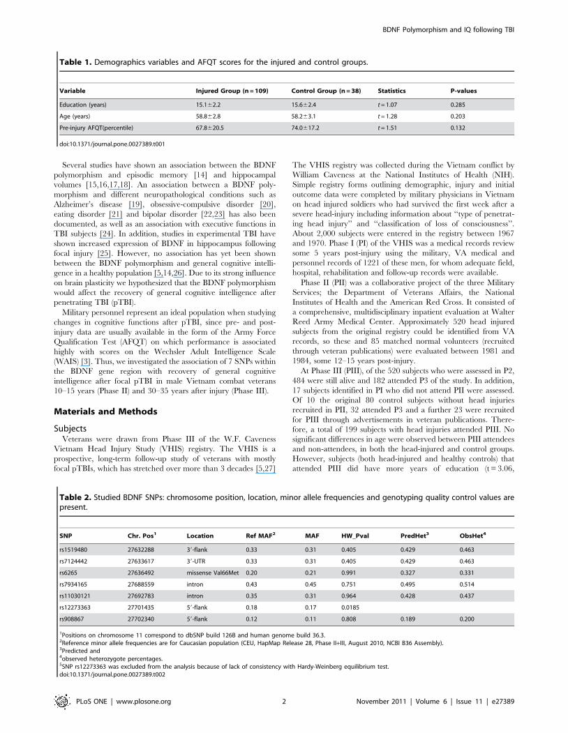
Table 1. Demographics variables and AFQT scores for the injured and control groups.
Variable Injured Group (n = 109) Control Group (n = 38) Statistics P-values
Education (years) 15.162.2 15.662.4 t = 1.07 0.285
Age (years) 58.862.8 58.263.1 t = 1.28 0.203
Pre-injury AFQT(percentile) 67.8620.5 74.0617.2 t = 1.51 0.132
doi:10.1371/journal.pone.0027389.t001
Table 2. Studied BDNF SNPs: chromosome position, location, minor allele frequencies and genotyping quality control values arepresent.
SNP Chr. Pos1 Location Ref MAF2 MAF HW_Pval PredHet3 ObsHet4
rs1519480 27632288 39-flank 0.33 0.31 0.405 0.429 0.463
rs7124442 27633617 39-UTR 0.33 0.31 0.405 0.429 0.463
rs6265 27636492 missense Val66Met 0.20 0.21 0.991 0.327 0.331
rs7934165 27688559 intron 0.43 0.45 0.751 0.495 0.514
rs11030121 27692783 intron 0.35 0.31 0.964 0.428 0.437
rs12273363 27701435 59-flank 0.18 0.17 0.0185
rs908867 27702340 59-flank 0.12 0.11 0.808 0.189 0.200
1Positions on chromosome 11 correspond to dbSNP build 126B and human genome build 36.3.2Reference minor allele frequencies are for Caucasian population (CEU, HapMap Release 28, Phase II+III, August 2010, NCBI B36 Assembly).3Predicted and4observed heterozygote percentages.5SNP rs12273363 was excluded from the analysis because of lack of consistency with Hardy-Weinberg equilibrium test.doi:10.1371/journal.pone.0027389.t002
BDNF Polymorphism and IQ following TBI
PLoS ONE | www.plosone.org 2 November 2011 | Volume 6 | Issue 11 | e27389
P = ,0.002), and higher AFQT scores (pre-injury: t = 4.85,
P,0.001, PII: t = 6.15, P,0.001) than PIII non-attendees. Since
those subjects attending PIII had a higher level of pre-injury
intelligence than those attending PII, as well as more years of
education, it is possible that those studied at PIII differed in other
ways from PIII non-attendees, which may have affected the
longitudinal results we report in this paper.
A further reduction of sample size in this study is explained by
several reasons: First, out of these 199 only 168 consented to
genotyping. Second, from the remaining subjects those who did
not complete all three phases of the study were excluded from the
analyses (n = 33). Third, as a majority of studied subjects were
Caucasian in ethnicity, those subjects who had Caucasian ethnicity
AIM scores ,0.5 were also excluded (n = 25). Finally, one subject
had to be excluded as an outlier due to his massive brain volume
loss. The final studied samples included male Caucasian combat
veterans with focal pTBIs (n = 109) and non-head-injured normal
control subjects who also served in Vietnam (n = 38). Importantly,
there were no significant differences in AFQT scores at PIII or
educational level attained in the group of 109 we studied compared
to the 90 excluded head-injured subjects from PIII (F(1,193) = 0.10,
P = 0.919). The type of pTBI injury was classified by neurosurgeons
at the time of injury into the following categories: Fragment (69.1%),
Gunshot (21.3%), Unclassified (1.5%) and Closed Head injury
(8.1%). Further, loss of consciousness (LOC) was classified as
following: No (42.6%) Yes, Momentary (17.6%), 1–15 min (14%),
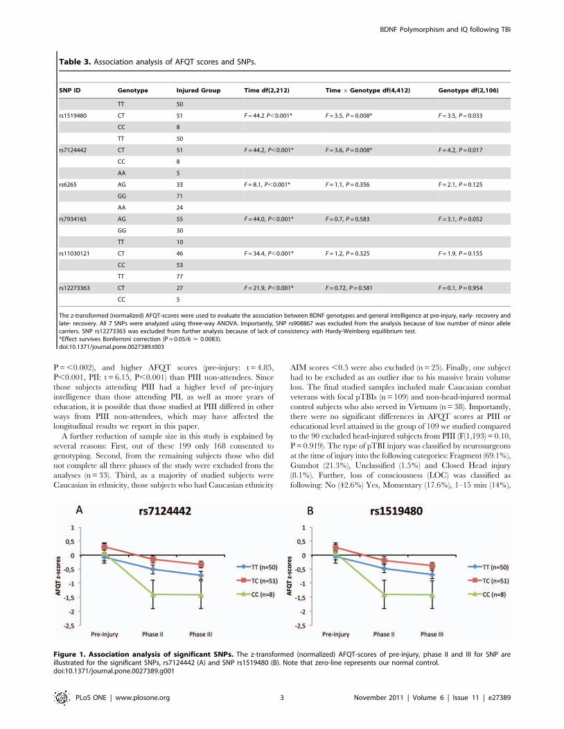
Table 3. Association analysis of AFQT scores and SNPs.
SNP ID Genotype Injured Group Time df(2,212) Time 6Genotype df(4,412) Genotype df(2,106)
TT 50
rs1519480 CT 51 F = 44.2 P,0.001* F = 3.5, P = 0.008* F = 3.5, P = 0.033
CC 8
TT 50
rs7124442 CT 51 F = 44.2, P,0.001* F = 3.6, P = 0.008* F = 4.2, P = 0.017
CC 8
AA 5
rs6265 AG 33 F = 8.1, P,0.001* F = 1.1, P = 0.356 F = 2.1, P = 0.125
GG 71
AA 24
rs7934165 AG 55 F = 44.0, P,0.001* F = 0.7, P = 0.583 F = 3.1, P = 0.052
GG 30
TT 10
rs11030121 CT 46 F = 34.4, P,0.001* F = 1.2, P = 0.325 F = 1.9, P = 0.155
CC 53
TT 77
rs12273363 CT 27 F = 21.9, P,0.001* F = 0.72, P = 0.581 F = 0.1, P = 0.954
CC 5
The z-transformed (normalized) AFQT-scores were used to evaluate the association between BDNF genotypes and general intelligence at pre-injury, early- recovery andlate- recovery. All 7 SNPs were analyzed using three-way ANOVA. Importantly, SNP rs908867 was excluded from the analysis because of low number of minor allelecarriers. SNP rs12273363 was excluded from further analysis because of lack of consistency with Hardy-Weinberg equilibrium test.*Effect survives Bonferroni correction (P = 0.05/6 < 0.0083).doi:10.1371/journal.pone.0027389.t003
Figure 1. Association analysis of significant SNPs. The z-transformed (normalized) AFQT-scores of pre-injury, phase II and III for SNP areillustrated for the significant SNPs, rs7124442 (A) and SNP rs1519480 (B). Note that zero-line represents our normal control.doi:10.1371/journal.pone.0027389.g001
BDNF Polymorphism and IQ following TBI
PLoS ONE | www.plosone.org 3 November 2011 | Volume 6 | Issue 11 | e27389
15 min – 1 day (11.8%), .1 day (11%), unknown (1.5%). The finals
groups were matched with respect to age, level of education, and
pre-injury intelligence (Table 1). All participants gave their written
informed consent, which was approved by the Institutional Review
Board at the National Naval Medical Center and the National
Institute of Neurological Disorders and Stroke, Bethesda, MD,
USA.
Neuropsychological TestingSubjects were admitted at the National Naval Medical Center in
Bethesda, MD, over a 5–7 day period and underwent a wide
variety of neuropsychological testing. The tests were designed to
measure cognitive abilities such as memory, language, social
cognition, and executive functioning. In this study, we focused on
the AFQT (AFQT-7A, Department of Defense, 1960), which is a
standardized multiple choice test of cognitive aptitude measuring
verbal ability, visual-spatial organization, arithmetic and function-
al associations via multiple choice questions. The total score range
is 0 to 100 and the subtest scores range from 0 to 25. AFQT scores
are reported as percentiles (1 to 99) and correlate highly with
WAIS scores [3]. It was the only pre-injury cognitive assessment
available in our sample and was also used in PII and PIII.
To determine the specific effect of BDNF genotype on the
recovery of general cognitive intelligence, two additional cognitive
control tasks were used in this study: First, the mini-mental state
examination test (MMSE) from PIII was used, which is a well-
validated standard test for cognitive impairment in adults, where
scores ,24 indicates cognitive impairment [28]. The purpose of its
inclusion was to separate out issues of exacerbated cognitive
decline from the onset of dementia [5]. Second, the delayed score
of the logical memory subtest of the Wechsler Memory Scale,
version III (WMS-III) was used to assess episodic memory, which
reflects the amount of information from stories that a subject can
recall after a 30 min delay [29].
Computed Tomography (CT) Acquisition and AnalysisThe axial CT scans were acquired without contrast in helical
mode on a GE Electric Medical Systems Light Speed Plus CT
scanner at the Bethesda Naval Hospital. Structural neuroimaging
data was reconstructed with an in-plane voxel size of
0.460.4 mm, an overlapping slice thickness of 2.5 mm and a
1 mm slice interval. The lesion location and volume were
determined from CT images using the interactive Analysis of
Brain Lesions (ABLe) software implemented in MEDx v3.44
(Medical Numerics) [30,31]. The analysis was performed on CT
images from Phase III. Lesion volume was calculated by
manually tracing the lesion in all relevant slices of the CT image
in native space, and then summing the trace areas and
multiplying by slice thickness. Manual tracing was performed
by a trained psychiatrist (V.R.) with clinical experience of reading
CT scans. The lesion tracing was then reviewed by an observer
that was blind to the results of the clinical evaluation and
neuropsychological testing (J.G.) enabling a consensus decision to
be reached regarding the limits of each lesion. The CT image of
each individual’s brain was normalized to a CT template brain
image in Montreal Neurological Institute (MNI) space. The
spatial normalization was performed with the AIR algorithm
[32], using a 12-parameter affine fit. Note that both the patient’s
brain and the MNI template’s brain are first skull-stripped in
order to maximize the efficacy of the AIR registration from native
space to MNI space. In addition, voxels inside the traced lesion
were not included in the spatial normalization procedure.
Afterwards, the percentage of Automated anatomical labeling
(AAL) structures that were intersected by the lesion was
determined by analyzing the overlap of the spatially normalized
lesion image with the AAL atlas [33].
Genotyping and Haplotype AnalysisWe used an addiction array designed by Hodgkinson et al [34].
The array is built on the Illumina GoldenGate platform and allows
for simultaneous genotyping of 1350 SNPs including 7 BDNF
SNPs (Table 2). The candidate genes for the array were selected
on the basis of their roles in the drug addictions and the related
phenotypes of anxiety and depression. These are the genes
important in signaling networks, stress/endocrine genes, and key
neurotransmitter systems including dopamine, serotonin, gluta-
mate, GABA and acetylcholine. As all these functional domains
are involved in the majority of brain functions, the addiction array
represents very convenient tool for our study. For each gene
Table 4. The SNPs that showed to have a significantassociation were further analyzed to see the significantchange from pre-injury to later and Phase II to Phase III.
SNP ID Phase Time df(1,106)Time6GenotypeDf(2,106)
rs7124442 Pre-injury to PII F = 66.7, P,0.001* F = 5.3, P = 0.006*
PII to PIII F = 2.3, P = 0.130 F = 0.3, P = 0.739
rs1519480 Pre-injury to PII F = 66.7, P,0.001* F = 5.3, P = 0.006*
PII to PIII F = 2.3, P = 0.130 F = 0.3, P = 0.734
*Effect survives Bonferroni correction (P = 0.05/6 < 0.0083).doi:10.1371/journal.pone.0027389.t004
Table 5. Results of t-test performed on the genotypes in the 2 significantly associated SNPs, comparing AFQT scores at each timepoint.
SNP ID Genotype Pre-injury Phase II Phase III
TT vs CT (df = 99) t = 21.95, P = 0.053; d = 0.39 t = 1.76, P = 0.081; d = 0.35 t = 21.95, P = 0.054; d = 1.12
rs7124442 CC vs CT (df = 56) t = 0.68, P = 0.495; d = 0.76 t = 23.24, P = 0.002*; d = 0.70 t = 22.99, P = 0.004; d = 0.58
CC vs TT (df = 57) t = 0.35, P = 0.727; d = 0.17 t = 22.03, P = 0.047; d = 0.38 t = 21.68, P = 0.097; d = 0.26
TT vs CT (df = 99) t = 21.61, P = 0.112; d = 0.32 t = 21.43, P = 0.156; d = 0.28 t = 21.63, P = 0.105; d = 0.32
rs1519480 CC vs CT (df = 56) t = 20.56,P = 0.573; d = 0.03 t = 23.08, P = 0.003*; d = 0.66 t = 22.86, P = 0.006; d = 0.63
CC vs TT (df = 57) t = 0.28, P = 0.780; d = 0.27 t = 22.11, P = 0.039; d = 0.40 t = 21.75, P = 0.085; d = 0.27
*Effect survives Bonferroni correction (P = 0.05/3 < 0.017).doi:10.1371/journal.pone.0027389.t005
BDNF Polymorphism and IQ following TBI
PLoS ONE | www.plosone.org 4 November 2011 | Volume 6 | Issue 11 | e27389
(including BDNF) array contains SNPs that tag common
haplotypes. In addition to 130 addiction-related genes, array
includes a panel of 186 ancestry information markers (AIMs) that
allows for determining subject’s ethnicity background. Each
marker represents a SNP with known frequencies of occurrence
in different ethnical groups. The AIM panel covers seven major
populations: African, European, Middle Eastern, Asian, Far East,
Oceania and Native American. Following genotyping, the
population assignment was performed for each individual
according to obtained AIM scores. Only Caucasians with a
‘‘European’’ AIM score .0.5 were included in this study.
Genotyping was performed according to the Illumina protocol
on 96 well-format Sentrix arrays. The completion rate of array
assay was .99%. The error rate of the assay was determined by
replicate genotyping, and was ,0.5%. Genotype frequencies were
tested for the Hardy-Weinberg equilibrium (HWE) applying
Fisher’s exact tests. Haplotype analysis was performed using a
Bayesian approach implemented with PHASE [35]. Haploview
4.2 (Broad Institute, USA,) was used to produce linkage
disequilibrium (LD) matrices. Haplotype blocks were constructed
by pairing the SNPs with the LD’s greater than 0.85, as described
by Gabriel et al [36]. We also investigated the effect of the
presence of the ApoE e4 allele and COMT Val158Met (rs4680) on
recovery of general intelligence to determine the relative specificity
of any BDNF effect.
Statistical AnalysisBehavioral data analysis was carried out using SPSS 15.0 with
an alpha level set to p,0.05 (two-tailed). Multiple comparisons
with Bonferroni correction were included in all analyzes. The
relationship between variations in the BDNF genotype and the
recovery of general cognitive intelligence was analyzed in several
ways:
First, the demographic variables between the injured and
controls groups were compared to ensure that the groups were
matched with respect to age, education, and pre-injury AFQT
using one-way analysis of variances (ANOVAs) with Group
(injured, control) as a between-subjects factor.
Second, the AQFT percentile score of the injured group was
normalized (z-transformation) in comparison to the performances
of the control group. For each of the 7 SNPs of the BDNF gene, a
mixed 363 analysis of variance (ANOVA) on AQFT z-scores was
performed with Time (pre-injury, PII, PIII) as a within-subjects
factor and Genotype (TT, CT, CC) as a between-subjects factor.
In planned follow-up analyses, the AFQT z- scores among the
different allele carriers in each SNP were compared using
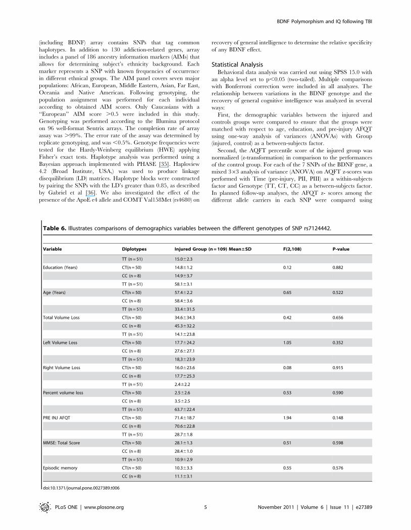
Table 6. Illustrates comparisons of demographics variables between the different genotypes of SNP rs7124442.
Variable Diplotypes Injured Group (n = 109) Mean±SD F(2,108) P-value
TT (n = 51) 15.062.3
Education (Years) CT(n = 50) 14.861.2 0.12 0.882
CC (n = 8) 14.963.7
TT (n = 51) 58.163.1
Age (Years) CT(n = 50) 57.462.2 0.65 0.522
CC (n = 8) 58.463.6
TT (n = 51) 33.4631.5
Total Volume Loss CT(n = 50) 34.6634.3 0.42 0.656
CC (n = 8) 45.3632.2
TT (n = 51) 14.1623.8
Left Volume Loss CT(n = 50) 17.7624.2 1.05 0.352
CC (n = 8) 27.6627.1
TT (n = 51) 18,3623.9
Right Volume Loss CT(n = 50) 16.0623.6 0.08 0.915
CC (n = 8) 17.7625.3
TT (n = 51) 2.462.2
Percent volume loss CT(n = 50) 2.562.6 0.53 0.590
CC (n = 8) 3.562.5
TT (n = 51) 63.7622.4
PRE INJ AFQT CT(n = 50) 71.4618.7 1.94 0.148
CC (n = 8) 70.6622.8
TT (n = 51) 28.761.8
MMSE: Total Score CT(n = 50) 28.161.3 0.51 0.598
CC (n = 8) 28.461.0
TT (n = 51) 10.962.9
Episodic memory CT(n = 50) 10.363.3 0.55 0.576
CC (n = 8) 11.163.1
doi:10.1371/journal.pone.0027389.t006
BDNF Polymorphism and IQ following TBI
PLoS ONE | www.plosone.org 5 November 2011 | Volume 6 | Issue 11 | e27389
between-subjects t-tests. In addition, effect sizes (Cohen’s d) that
represent the observed difference in the AFQT performance
between genotype groups were calculated (d = 0.2 indicates a small
effect size, d = 0.5 a medium effect size and d = 0.8 a large effect
size) [37].
Third, the specificity of the BDNF genotype effect on the
recovery of general cognitive intelligence was determined. Since
the BDNF polymorphism has been shown to modulate episodic
memory and hippocampal function (Egan et al., 2003; Dempster
et al., 2005), episodic memory scores were compared among the
different allele carriers in the injured and normal control groups
applying a 263 ANOVA with Group (injured, control) and
Genotype (TT, CT, CC) as between-subjects factors. In planned
follow-up analyses, the episodic memory scores among the
different allele carriers in each group were compared using
between-subjects t tests. Subjects within the genotype groups did
not differ in age, education, lesion size, pre-injury AFQT.
Fourth, the relative contribution of the BDNF genotype on the
recovery of general intelligence was estimated for PII and PIII. A
stepwise multiple linear regression analysis was applied including
the AFQT z-score as the dependent variable and BDNF genotype,
pre-injury intelligence, age, education, degree of atrophy,
percentage of total brain volume loss and brain volume loss
within each hemisphere as independent variables. This analysis
allowed for an estimation of the relative contribution of each
predictor to general intelligence. At the same time, it controls for
potential confounding factors that may influence general intelli-
gence.
Fifth, the influence of the ApoE e4 allele or COMT Val158Met
genotype on the recovery of general intelligence was determined
applying a mixed 363 analysis of variance (ANOVA) on AQFT z-
scores with Time (pre-injury, PII, PIII) as a within-subjects factor
and Genotype as a between-subjects factor.
Finally, we performed a haplotype analysis to increase the
chances of capturing gene-disease association by applying an
ANOVA on AQFT z-scores with Time (pre-injury, PII, PIII) as a
within-subjects factor and haplotypes (111222, 112122, 222211,
222212) as a between-subjects factor. The ANOVA was done for 2
haplotype blocks: block 1 included rs1519480, rs7124442, and
rs6265, whereas block 2 included rs7934165, rs11030121, and
rs908867.
Results
First, in terms of differences in demographic variables the one-
way ANOVAs showed no significant differences between the
injured and control groups regarding age, education, and pre-
injury AFQT (Table 1).
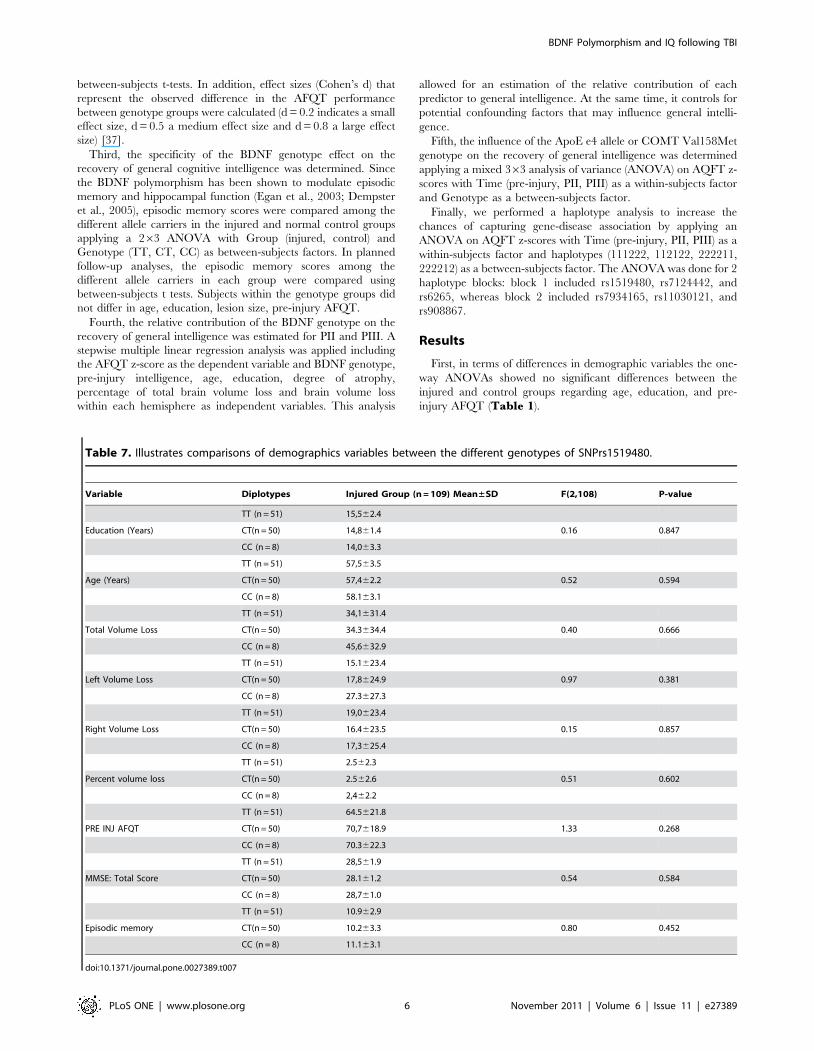
Table 7. Illustrates comparisons of demographics variables between the different genotypes of SNPrs1519480.
Variable Diplotypes Injured Group (n = 109) Mean±SD F(2,108) P-value
TT (n = 51) 15,562.4
Education (Years) CT(n = 50) 14,861.4 0.16 0.847
CC (n = 8) 14,063.3
TT (n = 51) 57,563.5
Age (Years) CT(n = 50) 57,462.2 0.52 0.594
CC (n = 8) 58.163.1
TT (n = 51) 34,1631.4
Total Volume Loss CT(n = 50) 34.3634.4 0.40 0.666
CC (n = 8) 45,6632.9
TT (n = 51) 15.1623.4
Left Volume Loss CT(n = 50) 17,8624.9 0.97 0.381
CC (n = 8) 27.3627.3
TT (n = 51) 19,0623.4
Right Volume Loss CT(n = 50) 16.4623.5 0.15 0.857
CC (n = 8) 17,3625.4
TT (n = 51) 2.562.3
Percent volume loss CT(n = 50) 2.562.6 0.51 0.602
CC (n = 8) 2,462.2
TT (n = 51) 64.5621.8
PRE INJ AFQT CT(n = 50) 70,7618.9 1.33 0.268
CC (n = 8) 70.3622.3
TT (n = 51) 28,561.9
MMSE: Total Score CT(n = 50) 28.161.2 0.54 0.584
CC (n = 8) 28,761.0
TT (n = 51) 10.962.9
Episodic memory CT(n = 50) 10.263.3 0.80 0.452
CC (n = 8) 11.163.1
doi:10.1371/journal.pone.0027389.t007
BDNF Polymorphism and IQ following TBI
PLoS ONE | www.plosone.org 6 November 2011 | Volume 6 | Issue 11 | e27389
Second, the association between BDNF genotype and general
cognitive intelligence at each evaluation Phase and for each of the
7 BDNF SNPs was evaluated. The mixed 363 ANOVAs on
AQFT z-scores with Time (pre-injury, PII, PIII) and Genotype
(TT, CT, CC) revealed a significant main effect of Time
(P,0.001) but no significant main effect of Genotype for all
SNPs (Table 3). The interaction effect for Time 6 Genotype
was only significant for SNPs rs7124442 and rs1519480. For both
SNPs, no differences were found regarding type of injury
(rs1519480: F(2,108) = 0.105, P = 0.900; rs7124442: F(2,108) =
0.228, P = 0.797) and loss of consciousness (rs1519480: F(2,106) =
1,101, P = 0.336; rs7124442: F(2,106) = 1,043, P = 0.356).
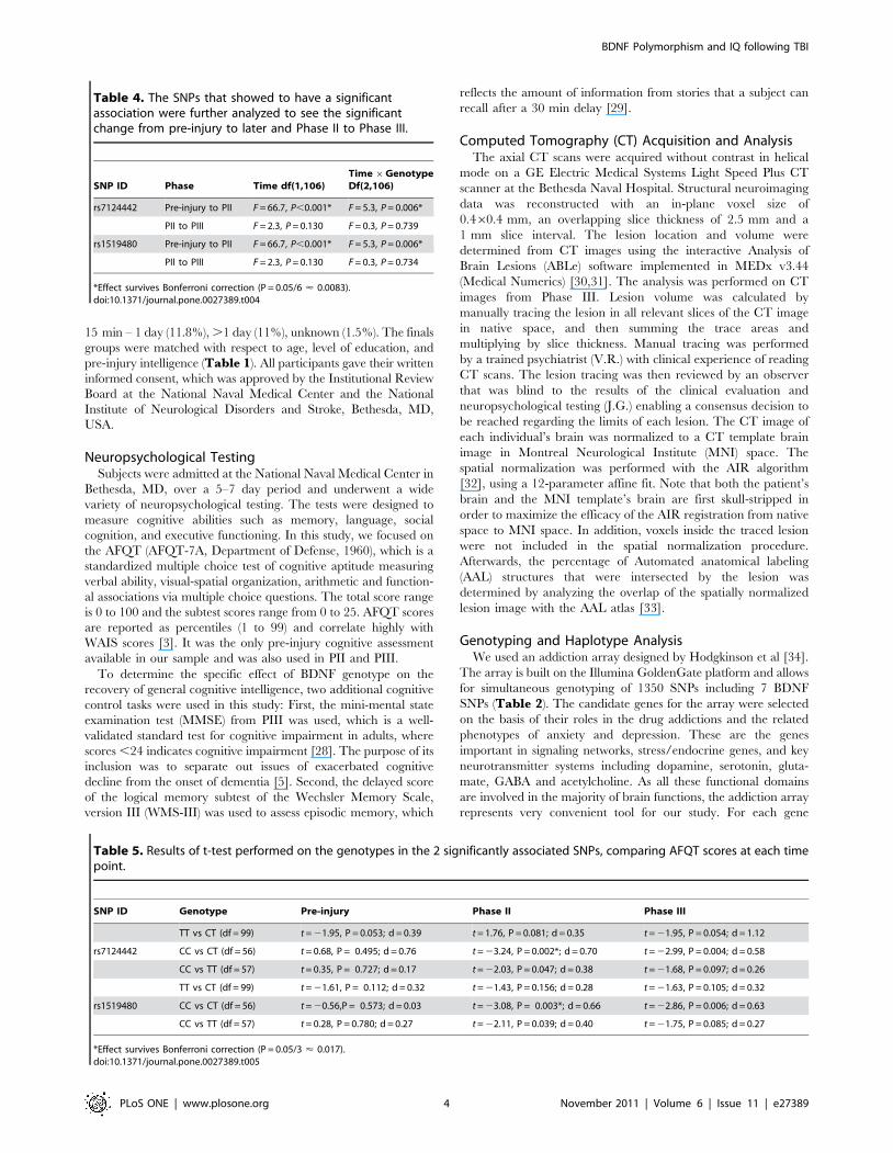
Figure 1 illustrates the changes in AFQT z-scores across time
(pre-injury, PII, PIII) among carriers of different genotypes of
those SNPs rs7124442 and rs1519480. The genotype groups that
performed best were CT (n = 51), in both SNP rs7124442 and
rs1519480, and the worst groups were genotype CC (n = 8). For
SNPs rs7124442 and rs1519480, the ANOVAs analysis showed
only a significant decline of general cognitive intelligence from
pre-injury to PII but not from PII to PIII (Table 4), indicating
that those SNPs are more prominent during the early post-injury
period, For both SNPs at PII, follow up t-tests revealed only
significant differences between the CC and CT genotypes
(Table 5).
Third, the specificity of the BDNF genotype effect on the
recovery of general cognitive intelligence was determined and no
significant differences were observed for both SNPs (rs7124442,
rs1519480) regarding demographic variables, volume loss, degree
of atrophy, episodic memory and MMSE (SNP rs7124442:
Table 6, SNP rs1519480: Table 7).
Fourth, the relative contribution of the significant SNPs to the
recovery of general cognitive intelligence at PII and PIII was
investigated using stepwise multiple linear regression analyzes. For
phase II, variance in cognitive intelligence was explained by
pre-injury intelligence (47.0%, P,0.001, Beta = 0.68), to SNP
rs7124442 and genotype CC (4.9%, P,0.001, Beta = 20.221),
percentage of total brain volume loss (2.1%, P,0.041, Be-
ta = 20.152), and education (1.5%, P,0.018, Beta = 0.130). For
phase III, variance in cognitive intelligence was explained by pre-
injury intelligence (41.8%, P = ,0.001, R2 = 0.414, Beta = 0.647),
percentage of total brain volume loss (6.4%, P,0.001, Be-
ta = 20.253), and SNP rs7124442 and genotype CC (2.4%,
P,0.018, Beta = 20.160). No significant associations were found
for age, education, atrophy ratings or right or left hemisphere
brain volume loss for both time-points.
Fifth, we determined whether the presence of the ApoE e4 allele
or COMT Val158Met genotype has an effect on the recovery of
general cognitive intelligence. This genetic analysis for ApoE and
COMT was available for 94 of the subjects. In the injured group,
22 subjects showed presence of the ApoEe4 allele. The ANOVA
revealed that there was a main effect on Time (F(2,182) = 25.2
P,0.001) but no main effect for Genotype (F(1,91) = 0.95,
P,0.332) and no interaction effect for Time 6 Genotype
(F(2,182) = 1.11, P,0.328). The influence for the COMT
polymorphism (Val/Val = 23, Val/Met = 38, Met/Met = 33) was
similar: significant main effect for Time (F(2,182) = 34.4, P,0.001)
but no significant main effect for Genotype (F(4,182) = 2.5,
P,0.086) and not significant interaction effect for Time 6Genotype (F(2,91) = 1.4, P,0.247).
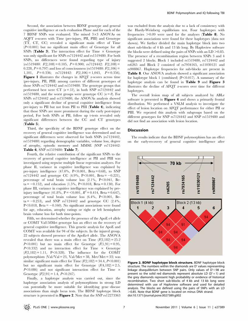
Finally, a haplotype analysis was carried out, since the
haplotype association analysis of polymorphisms in strong LD
can potentially be more suitable for identifying gene–disease
associations than single locus tests. The BDNF haplotype block
structure is presented in Figure 2. Note that the SNP rs12273363
was excluded from the analysis due to a lack of consistency with
the Hardy-Weinberg equilibrium test. Four haplotypes with
frequencies .0.09 were used for the analyses (Table 8). No
significant associations were found for these haplotypes (data not
shown). We further divided the main haplotype block into two
short sub-blocks of 4 kb and 13 kb long. By Haploview software
the blocks were defined using the pairs of SNPs with an LD .0.85.
The presence of a recombination region between SNPs 3 and 4
suggested 2 blocks. Block 1 included rs1519480, rs7124442 and
rs6265 and Block 2 consisted of rs7934165, rs11030121 and
rs908867. Haplotype frequencies for sub-blocks are present in
Table 8. Our ANOVA analysis showed a significant association
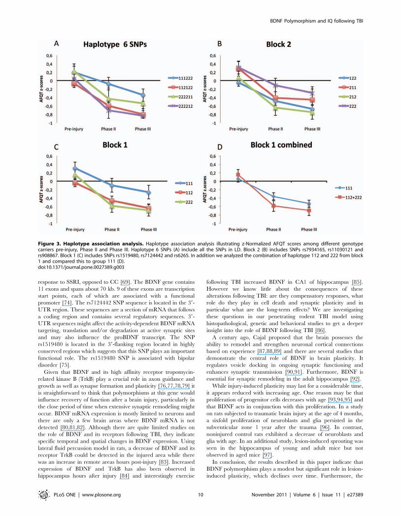
for haplotype block 1 (combined) (P,0.017). A summary of the
haplotype analysis can be found in Table 9 and Figure 3illustrates the decline of AFQT z-scores over time for different
haplotypes.
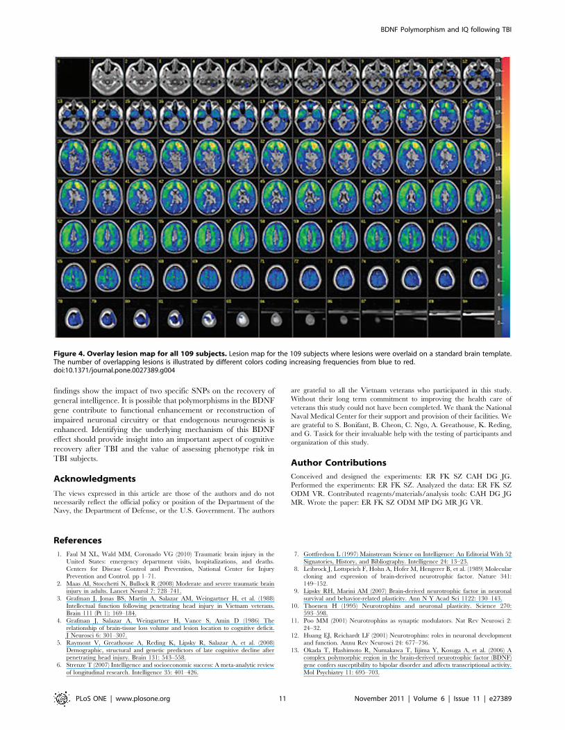
The overall lesion map of all subjects analyzed by ABLe
software is presented in Figure 4 and shows a primarily frontal
distribution. We performed a VSLM analysis to investigate the
effect of lesion location on AFQT performance for either PII or
PIII. We repeated this analysis with subgroups based on the
different genotypes for SNP rs7124442 and SNP rs1519480 and
did not find an association with lesion location.
Discussion
The results indicate that the BDNF polymorphism has an effect
on the early-recovery of general cognitive intelligence after
Figure 2. BDNF haplotype block structure. BDNF haplotype blockstructure. The numbers within the diamonds are D’ values representinglinkage disequilibrium between SNP pairs. Only values of D’,98 arepresent so the solid red diamonds represent absolute LD (D’ = 1) andthe grey diamonds represent high probability or evidence of historicalrecombination. Two short sub-blocks of 4 kb and 13 kb long weredetermined with use of Haploview software and used for detailedanalysis. The blocks are defined using the pairs of SNPs with an LD.0.85. Note that BDNF gene is located on minus DNA strand.doi:10.1371/journal.pone.0027389.g002
BDNF Polymorphism and IQ following TBI
PLoS ONE | www.plosone.org 7 November 2011 | Volume 6 | Issue 11 | e27389
penetrating TBI, although it has no influence on pre-injury
intelligence or the stability of general intelligence following
recovery of function. Prior studies could not find a significant
association between the Val66met BDNF genotype and overall
intelligence in a healthy population [14,26]. A previous study from
our group did not find a significant association between post-injury
intelligence and the Val66met BDNF genotype [5]. However,
looking at additional SNPs we provide evidence that rs7124442
and rs1519480 have an effect on recovery of general cognitive
intelligence and that this influence is probably a lesion-induced
plasticity that is decreased overtime. Our haplotype analysis
confirmed this significant association.
As found in prior studies, we confirmed that pre-injury
intelligence is the most consistent and vital predictor of cognitive
outcome after TBI [4,5,38,39]. The next best predictor at the
Phase II evaluation was the BDNF genotype. There were no
effects of the ApoE e4 allele or COMT Val158Met on recovery of
general cognitive intelligence. The ApoE e4 allele has shown to be
one of the strongest genetic predictors of poor outcome following
TBI [40,41,42]. Several association studies have investigated the
role of the ApoE gene polymorphism in subjects with closed TBI
but overall the findings are inconclusive [43,44].
An analysis of the association of lesion location and overall
AFQT score did not show any specific location to be significant.
Previous studies have shown lesion location to be the least
predictive of performance outcome of overall post injury
intelligence [3,4]. Relations have mainly been found between
subtest outcomes and specific injured lobe or hemisphere and not
full-scale IQ or general intelligence [3,45,46]. However in healthy
people, studies show full-scale IQ to be significantly correlated
with intracranial, cerebral, temporal lobe and hippocampal
volume as well as to overall gray matter volume [47,48,49].
Several neuroimaging studies on intelligence and reasoning has
generated a suggestive model of the Parieto-Frontal Integration
Theory (P-FIT) [50]. This model explains the individual
differences in intelligence by the interaction between association
cortices within parietal and frontal brain regions that are
effectively linked by white matter structures. While we did not
find a specific lesion location to be significant, the percentage of
total volume loss was one of the main predictors of general
intelligence at Phases II and III.
Prior studies have shown that aging decreases the brain’s
volume of gray and white matter and age-related cognitive
decline is associated with atrophic changes in the brain
[51,52,53]. Aging is also associated with decreasing neurotrans-
mitter levels [54], neuronal loss and shrinking neuronal size
[55,56] as well as reduced synaptic density [57]. However there is
very little known about BDNF in normal aging brain. A study in
monkey showed low levels of BDNF during aging [58]. A recent
study in healthy individuals revealed a faster rate of executive
control decline on a task-switching task that varied as a function
of the val66met BDNF polymorphism in age .65 [59]. IQ
measurements using the Wechsler Adult Intelligence Scales
(WAIS) have shown that IQ declines with age and that age-
related decline is more pronounced on certain subtests of WAIS
[60,61,62,63,64]. In the current study there were no significant
differences between the genotypes with respect to degree of
atrophy and total brain volume loss. None of the subjects with
penetrating head injury had MMSE scores lower than 25. Scores
below 25 are associated with mild dementia and age-related
cognitive impairment [28]. There was no age difference between
the different genotypes.
Furthermore, at the time of the Phase II AFQT testing, the
subjects were in their forties, long before the estimated age of 50 at
which accelerated cell loss occurs [65,66]. More importantly the
key BDNF genotypes contributed 5% of the variance in recovery
of general intelligence even when controlling for age and
education. Thus, we do not believe that our findings at Phase II
can be explained by age-related cognitive decline. Rather, we
speculate that it is indicative of lesion-induced plasticity in which
BDNF is known to play a crucial role.
Previous studies have investigated the association of the
rs7124442 SNP to eating disorders, cognitive abilities in the
elderly, major depression, susceptibility to asthma, Parkinson’s
disease, autism, Alzheimer’s disease and migraine
[67,68,69,70,71,72,73]. The only significant associations identified
were to eating disorders and high plasma BDNF levels [67] as well
as a significant negative influence of TT genotype on treatment
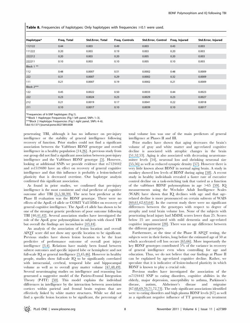
Table 8. Frequencies of haplotypes: Only haplotypes with frequencies $0.1 were used.
Haplotype* Freq, Total Std.Error, Total Freq, Controls Std.Error, Control Freq, Injured Std.Error, Injured
112122 0.44 0.003 0.49 0.003 0.43 0.003
111222 0.20 0.003 0.19 0.003 0.20 0.003
222212 0.20 0.003 0.20 0.005 0.20 0.003
222211 0.10 0.003 0.10 0.005 0.10 0.003
Block 1 **
112 0.48 0.0007 0.51 0.0002 0.48 0.0009
222 0.31 0.0007 0.29 0.0002 0.32 0.0009
111 0.21 0.0007 0.19 0.0002 0.21 0.0009
Block 2***
122 0.45 0.0022 0.50 0.0033 0.44 0.0023
222 0.23 0.0024 0.23 0.0029 0.23 0.0027
212 0.21 0.0019 0.17 0.0041 0.22 0.0018
211 0.10 0.0017 0.10 0.0039 0.10 0.0017
*Frequencies of 6-SNP haplotypes (Fig.1).**Block 1 Haplotype Frequencies (Fig.1 left panel, SNPs 1–3).***Block 2 Haplotype Frequencies (Fig.1 right panel, SNPs 4–6).doi:10.1371/journal.pone.0027389.t008
BDNF Polymorphism and IQ following TBI
PLoS ONE | www.plosone.org 8 November 2011 | Volume 6 | Issue 11 | e27389
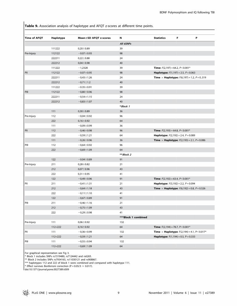
Table 9. Association analysis of haplotype and AFQT z-scores at different time points.
Time of AFQT Haplotype Mean±SD AFQT z-scores N Statistics F P
All 6SNPs
111222 0,2060.89 39
Pre-Injury 112122 20.0760.93 98
222211 0,2260.88 24
222212 0,0460.98 40
111222 21,2328 39 Time: F(2,197) = 64.2, P,0.001*
PII 112122 20.0760.95 98 Haplotype: F(1,197) = 2.5, P = 0.063
222211 20,4361.26 24 Time 6Haplotype: F(6,197) = 1.2, P = 0..319
222212 20,7161.2 40
111222 20.3560.91 39
PIII 112122 20,8060.96 98
222211 20,5461.15 24
222212 20,8361.07 40
*Block 1
111 0,3060.89 36
Pre-Injury 112 20,0460.92 96
222 0,1660.92 64
111 20,0960.99 36
PII 112 20,4660.98 96 Time: F(2,193) = 64.8, P,0.001*
222 20,5961.21 64 Haplotype: F(2,192) = 2.4, P = 0.089
111 20,2660.96 36 Time 6Haplotype: F(2,193) = 2.1, P = 0.086
PIII 112 20,6460.92 96
222 20,6961.09 64
**Block 2
122 20.0460.89 91
Pre-Injury 211 0,2860.82 21
212 0,0760.96 43
222 0,3160.95 41
122 20,4960.96 91 Time: F(2,192) = 63.4, P,0.001*
PII 211 20,4161.31 21 Haplotype: F(3,192) = 2.2, P = 0.094
212 20,6461.18 43 Time 6Haplotype: F(6,192) = 0.8, P = 0.526
222 20,1161.10 41
122 20,6760.89 91
PIII 211 20,4661.16 21
212 20,7561.09 43
222 20,2960.98 41
***Block 1 combined
Pre-Injury 111 0,0660.92 132
112+222 0,1660.92 64 Time: F(2,194) = 78.7, P,0.001*
PII 111 20,3660.99 132 Time 6Haplotype: F(2,194) = 4.1, P,0.017*
112+222 20,5961.21 64 Haplotype: F(1,194) = 0.5, P = 0.333
PIII 111 20,5560.94 132
112+222 20,6961.09 64
For graphical representation see Fig 3.* Block 1 includes SNPs rs1519480, rs7124442 and rs6265.** Block 2 includes SNPs rs7934165, rs11030121 and rs908867.*** haplotypes 112 and 222 of block 1 were combined and compared with haplotype 111.* Effect survives Bonferroni correction (P = 0.05/3 < 0.017).doi:10.1371/journal.pone.0027389.t009
BDNF Polymorphism and IQ following TBI
PLoS ONE | www.plosone.org 9 November 2011 | Volume 6 | Issue 11 | e27389
response to SSRI, opposed to CC [69]. The BDNF gene contains
11 exons and spans about 70 kb. 9 of these exons are transcription
start points, each of which are associated with a functional
promoter [74]. The rs7124442 SNP sequence is located in the 39-
UTR region. These sequences are a section of mRNA that follows
a coding region and contains several regulatory sequences. 39-
UTR sequences might affect the activity-dependent BDNF mRNA
targeting, translation and/or degradation at active synaptic sites
and may also influence the proBDNF transcript. The SNP
rs1519480 is located in the 39-flanking region located in highly
conserved regions which suggests that this SNP plays an important
functional role. The rs1519480 SNP is associated with bipolar
disorder [75].
Given that BDNF and its high affinity receptor tropomycin-
related kinase B (TrkB) play a crucial role in axon guidance and
growth as well as synapse formation and plasticity [76,77,78,79] it
is straightforward to think that polymorphisms at this gene would
influence recovery of function after a brain injury, particularly in
the close period of time when extensive synaptic remodeling might
occur. BDNF mRNA expression is mostly limited to neurons and
there are only a few brain areas where BDNF mRNA is not
detected [80,81,82]. Although there are quite limited studies on
the role of BDNF and its receptors following TBI, they indicate
specific temporal and spatial changes in BDNF expression. Using
lateral fluid percussion model in rats, a decrease of BDNF and its
receptor TrkB could be detected in the injured area while there
was an increase in remote areas hours post-injury [83]. Increased
expression of BDNF and TrkB has also been observed in
hippocampus hours after injury [84] and interestingly exercise
following TBI increased BDNF in CA1 of hippocampus [85].
However we know little about the consequences of these
alterations following TBI: are they compensatory responses, what
role do they play in cell death and synaptic plasticity and in
particular what are the long-term effects? We are investigating
these questions in our penetrating rodent TBI model using
histopathological, genetic and behavioral studies to get a deeper
insight into the role of BDNF following TBI [86].
A century ago, Cajal proposed that the brain possesses the
ability to remodel and strengthen neuronal cortical connections
based on experience [87,88,89] and there are several studies that
demonstrate the central role of BDNF in brain plasticity. It
regulates vesicle docking in ongoing synaptic functioning and
enhances synaptic transmission [90,91]. Furthermore, BDNF is
essential for synaptic remodeling in the adult hippocampus [92].
While injury-induced plasticity may last for a considerable time,
it appears reduced with increasing age. One reason may be that
proliferation of progenitor cells decreases with age [93,94,95] and
that BDNF acts in conjunction with this proliferation. In a study
on rats subjected to traumatic brain injury at the age of 4 months,
a sixfold proliferation of neuroblasts and glia persisted in the
subventicular zone 1 year after the trauma [96]. In contrast,
noninjured control rats exhibited a decrease of neuroblasts and
glia with age. In an additional study, lesion-induced sprouting was
seen in the hippocampus of young and adult mice but not
observed in aged mice [97].
In conclusion, the results described in this paper indicate that
BDNF polymorphism plays a modest but significant role in lesion-
induced plasticity, which declines over time. Furthermore, the
Figure 3. Haplotype association analysis. Haplotype association analysis illustrating z-Normalized AFQT scores among different genotypecarriers pre-injury, Phase II and Phase III. Haplotype 6 SNPs (A) include all the SNPs in LD. Block 2 (B) includes SNPs rs7934165, rs11030121 andrs908867. Block 1 (C) includes SNPs rs1519480, rs7124442 and rs6265. In addition we analyzed the combination of haplotype 112 and 222 from block1 and compared this to group 111 (D).doi:10.1371/journal.pone.0027389.g003
BDNF Polymorphism and IQ following TBI
PLoS ONE | www.plosone.org 10 November 2011 | Volume 6 | Issue 11 | e27389
findings show the impact of two specific SNPs on the recovery of
general intelligence. It is possible that polymorphisms in the BDNF
gene contribute to functional enhancement or reconstruction of
impaired neuronal circuitry or that endogenous neurogenesis is
enhanced. Identifying the underlying mechanism of this BDNF
effect should provide insight into an important aspect of cognitive
recovery after TBI and the value of assessing phenotype risk in
TBI subjects.
Acknowledgments
The views expressed in this article are those of the authors and do not
necessarily reflect the official policy or position of the Department of the
Navy, the Department of Defense, or the U.S. Government. The authors
are grateful to all the Vietnam veterans who participated in this study.
Without their long term commitment to improving the health care of
veterans this study could not have been completed. We thank the National
Naval Medical Center for their support and provision of their facilities. We
are grateful to S. Bonifant, B. Cheon, C. Ngo, A. Greathouse, K. Reding,
and G. Tasick for their invaluable help with the testing of participants and
organization of this study.
Author Contributions
Conceived and designed the experiments: ER FK SZ CAH DG JG.
Performed the experiments: ER FK SZ. Analyzed the data: ER FK SZ
ODM VR. Contributed reagents/materials/analysis tools: CAH DG JG
MR. Wrote the paper: ER FK SZ ODM MP DG MR JG VR.
References
1. Faul M XL, Wald MM, Coronado VG (2010) Traumatic brain injury in the
United States: emergency department visits, hospitalizations, and deaths.Centers for Disease Control and Prevention, National Center for Injury
Prevention and Control. pp 1–71.
2. Maas AI, Stocchetti N, Bullock R (2008) Moderate and severe traumatic braininjury in adults. Lancet Neurol 7: 728–741.
3. Grafman J, Jonas BS, Martin A, Salazar AM, Weingartner H, et al. (1988)
Intellectual function following penetrating head injury in Vietnam veterans.
Brain 111 (Pt 1): 169–184.
4. Grafman J, Salazar A, Weingartner H, Vance S, Amin D (1986) Therelationship of brain-tissue loss volume and lesion location to cognitive deficit.
J Neurosci 6: 301–307.
5. Raymont V, Greathouse A, Reding K, Lipsky R, Salazar A, et al. (2008)Demographic, structural and genetic predictors of late cognitive decline after
penetrating head injury. Brain 131: 543–558.
6. Strenze T (2007) Intelligence and socioeconomic success: A meta-analytic review
of longitudinal research. Intelligence 35: 401–426.
7. Gottfredson L (1997) Mainstream Science on Intelligence: An Editorial With 52
Signatories, History, and Bibliography. Intelligence 24: 13–23.
8. Leibrock J, Lottspeich F, Hohn A, Hofer M, Hengerer B, et al. (1989) Molecular
cloning and expression of brain-derived neurotrophic factor. Nature 341:
149–152.
9. Lipsky RH, Marini AM (2007) Brain-derived neurotrophic factor in neuronal
survival and behavior-related plasticity. Ann N Y Acad Sci 1122: 130–143.
10. Thoenen H (1995) Neurotrophins and neuronal plasticity. Science 270:593–598.
11. Poo MM (2001) Neurotrophins as synaptic modulators. Nat Rev Neurosci 2:
24–32.
12. Huang EJ, Reichardt LF (2001) Neurotrophins: roles in neuronal development
and function. Annu Rev Neurosci 24: 677–736.
13. Okada T, Hashimoto R, Numakawa T, Iijima Y, Kosuga A, et al. (2006) Acomplex polymorphic region in the brain-derived neurotrophic factor (BDNF)
gene confers susceptibility to bipolar disorder and affects transcriptional activity.
Mol Psychiatry 11: 695–703.
Figure 4. Overlay lesion map for all 109 subjects. Lesion map for the 109 subjects where lesions were overlaid on a standard brain template.The number of overlapping lesions is illustrated by different colors coding increasing frequencies from blue to red.doi:10.1371/journal.pone.0027389.g004
BDNF Polymorphism and IQ following TBI
PLoS ONE | www.plosone.org 11 November 2011 | Volume 6 | Issue 11 | e27389
14. Egan MF, Kojima M, Callicott JH, Goldberg TE, Kolachana BS, et al. (2003)
The BDNF val66met polymorphism affects activity-dependent secretion of
BDNF and human memory and hippocampal function. Cell 112: 257–269.
15. Hariri AR, Goldberg TE, Mattay VS, Kolachana BS, Callicott JH, et al. (2003)
Brain-derived neurotrophic factor val66met polymorphism affects human
memory-related hippocampal activity and predicts memory performance.
J Neurosci 23: 6690–6694.
16. Pezawas L, Verchinski BA, Mattay VS, Callicott JH, Kolachana BS, et al. (2004)
The brain-derived neurotrophic factor val66met polymorphism and variation in
human cortical morphology. J Neurosci 24: 10099–10102.
17. Szeszko PR, Lipsky R, Mentschel C, Robinson D, Gunduz-Bruce H, et al.
(2005) Brain-derived neurotrophic factor val66met polymorphism and volume of
the hippocampal formation. Mol Psychiatry 10: 631–636.
18. Bueller JA, Aftab M, Sen S, Gomez-Hassan D, Burmeister M, et al. (2006)
BDNF Val66Met allele is associated with reduced hippocampal volume in
healthy subjects. Biol Psychiatry 59: 812–815.
19. Huang R, Huang J, Cathcart H, Smith S, Poduslo SE (2007) Genetic variants in
brain-derived neurotrophic factor associated with Alzheimer’s disease. J Med
Genet 44: e66.
20. Hall D, Dhilla A, Charalambous A, Gogos JA, Karayiorgou M (2003) Sequence
variants of the brain-derived neurotrophic factor (BDNF) gene are strongly
associated with obsessive-compulsive disorder. Am J Hum Genet 73: 370–376.
21. Ribases M, Gratacos M, Armengol L, de Cid R, Badia A, et al. (2003) Met66 in
the brain-derived neurotrophic factor (BDNF) precursor is associated with
anorexia nervosa restrictive type. Mol Psychiatry 8: 745–751.
22. Sklar P, Gabriel SB, McInnis MG, Bennett P, Lim YM, et al. (2002) Family-
based association study of 76 candidate genes in bipolar disorder: BDNF is a
potential risk locus. Brain-derived neutrophic factor. Mol Psychiatry 7: 579–593.
23. Neves-Pereira M, Mundo E, Muglia P, King N, Macciardi F, et al. (2002) The
brain-derived neurotrophic factor gene confers susceptibility to bipolar disorder:
evidence from a family-based association study. Am J Hum Genet 71: 651–655.
24. Krueger F, Pardini M, Huey ED, Raymont V, Solomon J, et al. (2011) The role
of the Met66 brain-derived neurotrophic factor allele in the recovery of
executive functioning after combat-related traumatic brain injury. J Neurosci 31:
598–606.
25. Hicks RR, Numan S, Dhillon HS, Prasad MR, Seroogy KB (1997) Alterations in
BDNF and NT-3 mRNAs in rat hippocampus after experimental brain trauma.
Brain Res Mol Brain Res 48: 401–406.
26. Tsai SJ, Hong CJ, Yu YW, Chen TJ (2004) Association study of a brain-derived
neurotrophic factor (BDNF) Val66Met polymorphism and personality trait and
intelligence in healthy young females. Neuropsychobiology 49: 13–16.
27. Raymont V, Salazar AM, Krueger F, Grafman J (2011) "Studying injured
minds" - the Vietnam head injury study and 40 years of brain injury research.
Frontiers in neurology (Neurotrauma) 2(15): 1–11.
28. Folstein MF, Folstein SE, McHugh PR (1975) "Mini-mental state". A practical
method for grading the cognitive state of patients for the clinician. J Psychiatr
Res 12: 189–198.
29. Wechsler D (1997) Wechsler Memory Scale - III. New York: Psychological
Corporation.
30. Makale M, Solomon J, Patronas NJ, Danek A, Butman JA, et al. (2002)
Quantification of brain lesions using interactive automated software. Behav Res
Methods Instrum Comput 34: 6–18.
31. Solomon J, Raymont V, Braun A, Butman JA, Grafman J (2007) User-friendly
software for the analysis of brain lesions (ABLe). Comput Methods Programs
Biomed 86: 245–254.
32. Woods RP, Mazziotta JC, Cherry SR (1993) MRI-PET registration with
automated algorithm. J Comput Assist Tomogr 17: 536–546.
33. Tzourio-Mazoyer N, Landeau B, Papathanassiou D, Crivello F, Etard O, et al.
(2002) Automated anatomical labeling of activations in SPM using a
macroscopic anatomical parcellation of the MNI MRI single-subject brain.
Neuroimage 15: 273–289.
34. Hodgkinson CA, Yuan Q, Xu K, Shen PH, Heinz E, et al. (2008) Addictions
biology: haplotype-based analysis for 130 candidate genes on a single array.
Alcohol Alcohol 43: 505–515.
35. Stephens M, Smith NJ, Donnelly P (2001) A new statistical method for haplotype
reconstruction from population data. Am J Hum Genet 68: 978–989.
36. Gabriel SB, Schaffner SF, Nguyen H, Moore JM, Roy J, et al. (2002) The
structure of haplotype blocks in the human genome. Science 296: 2225–2229.
37. Cohen J (1988) Statistical power analysis for behavioral sciences. NJ: Lawrence
Earlbaum Associates.
38. Gao B, Jiang S, Wang X, Chen J (2000) The role of pre-injury IQ in the
determination of intellectual impairment from traumatic head injury.
J Neuropsychiatry Clin Neurosci 12: 385–388.
39. Kesler SR, Adams HF, Blasey CM, Bigler ED (2003) Premorbid intellectual
functioning, education, and brain size in traumatic brain injury: an investigation
of the cognitive reserve hypothesis. Appl Neuropsychol 10: 153–162.
40. Teasdale GM, Nicoll JA, Murray G, Fiddes M (1997) Association of
apolipoprotein E polymorphism with outcome after head injury. Lancet 350:
1069–1071.
41. Lichtman SW, Seliger G, Tycko B, Marder K (2000) Apolipoprotein E and
functional recovery from brain injury following postacute rehabilitation.
Neurology 55: 1536–1539.
42. Nathoo N, Chetty R, van Dellen JR, Barnett GH (2003) Genetic vulnerability
following traumatic brain injury: the role of apolipoprotein E. Mol Pathol 56:132–136.
43. Jordan BD (2007) Genetic influences on outcome following traumatic brain
injury. Neurochem Res 32: 905–915.
44. Dardiotis E, Fountas KN, Dardioti M, Xiromerisiou G, Kapsalaki E, et al.(2010) Genetic association studies in patients with traumatic brain injury.
Neurosurg Focus 28: E9.
45. Warrington EK, James M, Maciejewski C (1986) The WAIS as a lateralizing
and localizing diagnostic instrument: a study of 656 patients with unilateral
cerebral lesions. Neuropsychologia 24: 223–239.
46. Glascher J, Rudrauf D, Colom R, Paul LK, Tranel D, et al. (2010) Distributed
neural system for general intelligence revealed by lesion mapping. Proc Natl
Acad Sci U S A 107: 4705–4709.
47. Andreasen NC, Flaum M, Swayze V, 2nd, O’Leary DS, Alliger R, et al. (1993)
Intelligence and brain structure in normal individuals. Am J Psychiatry 150:
130–134.
48. Toulopoulou T, Grech A, Morris RG, Schulze K, McDonald C, et al. (2004)
The relationship between volumetric brain changes and cognitive function: a
family study on schizophrenia. Biol Psychiatry 56: 447–453.
49. Witelson SF, Beresh H, Kigar DL (2006) Intelligence and brain size in 100
postmortem brains: sex, lateralization and age factors. Brain 129: 386–398.
50. Jung RE, Haier RJ (2007) The Parieto-Frontal Integration Theory (P-FIT) ofintelligence: converging neuroimaging evidence. Behav Brain Sci 30: 135–154.
discussion 154-187.
51. Good CD, Johnsrude IS, Ashburner J, Henson RN, Friston KJ, et al. (2001) A
voxel-based morphometric study of ageing in 465 normal adult human brains.
Neuroimage 14: 21–36.
52. Resnick SM, Pham DL, Kraut MA, Zonderman AB, Davatzikos C (2003)
Longitudinal magnetic resonance imaging studies of older adults: a shrinking
brain. J Neurosci 23: 3295–3301.
53. Raz N, Rodrigue KM (2006) Differential aging of the brain: patterns, cognitivecorrelates and modifiers. Neurosci Biobehav Rev 30: 730–748.
54. Volkow ND, Wang GJ, Fowler JS, Ding YS, Gur RC, et al. (1998) Parallel loss of
presynaptic and postsynaptic dopamine markers in normal aging. Ann Neurol
44: 143–147.
55. Flood DG, Coleman PD (1988) Neuron numbers and sizes in aging brain:
comparisons of human, monkey, and rodent data. Neurobiol Aging 9: 453–463.
56. Shimada A (1999) Age-dependent cerebral atrophy and cognitive dysfunction in
SAMP10 mice. Neurobiol Aging 20: 125–136.
57. Terry RD, Katzman R (2001) Life span and synapses: will there be a primary
senile dementia? Neurobiol Aging 22: 347–348. discussion 353-344.
58. Hayashi M, Mistunaga F, Ohira K, Shimizu K (2001) Changes in BDNF-
immunoreactive structures in the hippocampal formation of the aged macaquemonkey. Brain Res 918: 191–196.
59. Erickson KI, Kim JS, Suever BL, Voss MW, Francis BM, et al. (2008) Genetic
contributions to age-related decline in executive function: a 10-year longitudinal
study of COMT and BDNF polymorphisms. Front Hum Neurosci 2: 11.
60. Ronnlund M, Nilsson L (2006) Adult life-span patterns in WAIS-R Block Design
performance: Cross-sectional versus longitudinal age gradients and relations to
demographic factors. Intelligence 34: 6378.
61. Wechsler D (1939) The Measurement of Adult Intelligence. Baltimore: Williams& Wilkins.
62. Wechsler D (1958) The measurement and appraisal of adult intelligence.
Baltimore: Williams & Wilkins.
63. Ryan JJ, Sattler JM, Lopez SJ (2000) Age effects on Wechsler Adult IntelligenceScale-III subtests. Arch Clin Neuropsychol 15: 311–317.
64. Kaufman A, Lichtenberger E (1990) Assessing adolescent and adult intelligence.
Boston: Allyn and Bacon.
65. DeCarli C, Massaro J, Harvey D, Hald J, Tullberg M, et al. (2005) Measures ofbrain morphology and infarction in the framingham heart study: establishing
what is normal. Neurobiol Aging 26: 491–510.
66. Scahill RI, Frost C, Jenkins R, Whitwell JL, Rossor MN, et al. (2003) A
longitudinal study of brain volume changes in normal aging using serial
registered magnetic resonance imaging. Arch Neurol 60: 989–994.
67. Mercader JM, Ribases M, Gratacos M, Gonzalez JR, Bayes M, et al. (2007)
Altered brain-derived neurotrophic factor blood levels and gene variability are
associated with anorexia and bulimia. Genes Brain Behav 6: 706–716.
68. Miyajima F, Ollier W, Mayes A, Jackson A, Thacker N, et al. (2008) Brain-
derived neurotrophic factor polymorphism Val66Met influences cognitive
abilities in the elderly. Genes Brain Behav 7: 411–417.
69. Domschke K, Lawford B, Laje G, Berger K, Young R, et al. (2010) Brain-
derived neurotrophic factor ( BDNF) gene: no major impact on antidepressanttreatment response. Int J Neuropsychopharmacol 13: 93–101.
70. Szczepankiewicz A, Rose-Zerilli MJ, Barton SJ, Holgate ST, Holloway JW
(2009) Association analysis of brain-derived neurotrophic factor gene polymor-
phisms in asthmatic families. Int Arch Allergy Immunol 149: 343–349.
71. Xiromerisiou G, Hadjigeorgiou GM, Eerola J, Fernandez HH, Tsimourtou V,
et al. (2007) BDNF tagging polymorphisms and haplotype analysis in sporadic
Parkinson’s disease in diverse ethnic groups. Neurosci Lett 415: 59–63.
72. Nishimura K, Nakamura K, Anitha A, Yamada K, Tsujii M, et al. (2007)
Genetic analyses of the brain-derived neurotrophic factor (BDNF) gene inautism. Biochem Biophys Res Commun 356: 200–206.
BDNF Polymorphism and IQ following TBI
PLoS ONE | www.plosone.org 12 November 2011 | Volume 6 | Issue 11 | e27389
73. Lemos C, Mendonca D, Pereira-Monteiro J, Barros J, Sequeiros J, et al. (2010)
BDNF and CGRP interaction: implications in migraine susceptibility.
Cephalalgia 30: 1375–1382.
74. Pruunsild P, Kazantseva A, Aid T, Palm K, Timmusk T (2007) Dissecting the
human BDNF locus: bidirectional transcription, complex splicing, and multiple
promoters. Genomics 90: 397–406.
75. Liu L, Foroud T, Xuei X, Berrettini W, Byerley W, et al. (2008) Evidence of
association between brain-derived neurotrophic factor gene and bipolar
disorder. Psychiatr Genet 18: 267–274.
76. Frisen J, Verge VM, Cullheim S, Persson H, Fried K, et al. (1992) Increased
levels of trkB mRNA and trkB protein-like immunoreactivity in the injured rat
and cat spinal cord. Proc Natl Acad Sci U S A 89: 11282–11286.
77. Huang EJ, Reichardt LF (2003) Trk receptors: roles in neuronal signal
transduction. Annu Rev Biochem 72: 609–642.
78. Luikart BW, Parada LF (2006) Receptor tyrosine kinase B-mediated excitatory
synaptogenesis. Prog Brain Res 157: 15–24.
79. Lu B (2003) Pro-region of neurotrophins: role in synaptic modulation. Neuron
39: 735–738.
80. Katoh-Semba R, Takeuchi IK, Semba R, Kato K (1997) Distribution of brain-
derived neurotrophic factor in rats and its changes with development in the
brain. J Neurochem 69: 34–42.
81. Conner JM, Lauterborn JC, Yan Q, Gall CM, Varon S (1997) Distribution of
brain-derived neurotrophic factor (BDNF) protein and mRNA in the normal
adult rat CNS: evidence for anterograde axonal transport. J Neurosci 17:
2295–2313.
82. Ernfors P, Ibanez CF, Ebendal T, Olson L, Persson H (1990) Molecular cloning
and neurotrophic activities of a protein with structural similarities to nerve
growth factor: developmental and topographical expression in the brain. Proc
Natl Acad Sci U S A 87: 5454–5458.
83. Hicks RR, Li C, Zhang L, Dhillon HS, Prasad MR, et al. (1999) Alterations in
BDNF and trkB mRNA levels in the cerebral cortex following experimental
brain trauma in rats. J Neurotrauma 16: 501–510.
84. Hicks RR, Martin VB, Zhang L, Seroogy KB (1999) Mild experimental brain
injury differentially alters the expression of neurotrophin and neurotrophin
receptor mRNAs in the hippocampus. Exp Neurol 160: 469–478.
85. Hicks RR, Boggs A, Leider D, Kraemer P, Brown R, et al. (1998) Effects of
Exercise Following Lateral Fluid Percussion Brain Injury in Rats. Restor NeurolNeurosci 12: 41–47.
86. Risling M, Plantman S, Angeria M, Rostami E, Bellander BM, et al. (2011)
Mechanisms of blast induced brain injuries, experimental studies in rats.Neuroimage 54 Suppl 1: S89–97.
87. Pascual-Leone A, Amedi A, Fregni F, Merabet LB (2005) The plastic humanbrain cortex. Annu Rev Neurosci 28: 377–401.
88. Will B, Galani R, Kelche C, Rosenzweig MR (2004) Recovery from brain injury
in animals: relative efficacy of environmental enrichment, physical exercise orformal training (1990-2002). Prog Neurobiol 72: 167–182.
89. Seitz RJ, Kleiser R, Butefisch CM (2005) Reorganization of cerebral circuits inhuman brain lesion. Acta Neurochir Suppl 93: 65–70.
90. Pozzo-Miller LD, Gottschalk W, Zhang L, McDermott K, Du J, et al. (1999)Impairments in high-frequency transmission, synaptic vesicle docking, and
synaptic protein distribution in the hippocampus of BDNF knockout mice.
J Neurosci 19: 4972–4983.91. Tyler WJ, Pozzo-Miller LD (2001) BDNF enhances quantal neurotransmitter
release and increases the number of docked vesicles at the active zones ofhippocampal excitatory synapses. J Neurosci 21: 4249–4258.
92. Heldt SA, Stanek L, Chhatwal JP, Ressler KJ (2007) Hippocampus-specific
deletion of BDNF in adult mice impairs spatial memory and extinction ofaversive memories. Mol Psychiatry 12: 656–670.
93. Kolb B (1995) Brain plasticity and behavior. NJ: Mahwah, Erlbaum.94. Luebke JI, Rosene DL (2003) Aging alters dendritic morphology, input
resistance, and inhibitory signaling in dentate granule cells of the rhesusmonkey. J Comp Neurol 460: 573–584.
95. Kuhn HG, Dickinson-Anson H, Gage FH (1996) Neurogenesis in the dentate
gyrus of the adult rat: age-related decrease of neuronal progenitor proliferation.J Neurosci 16: 2027–2033.
96. Chen XH, Iwata A, Nonaka M, Browne KD, Smith DH (2003) Neurogenesisand glial proliferation persist for at least one year in the subventricular zone
following brain trauma in rats. J Neurotrauma 20: 623–631.
97. Kadish I, van Groen T (2009) Lesion-induced hippocampal plasticity intransgenic Alzheimer’s disease mouse models: influences of age, genotype, and
estrogen. J Alzheimers Dis 18: 429–445.
BDNF Polymorphism and IQ following TBI
PLoS ONE | www.plosone.org 13 November 2011 | Volume 6 | Issue 11 | e27389