basis for sudden cardiac death prediction by t-wave alternans from an integrative physiology...
TRANSCRIPT

Basis for Sudden Cardiac Death Prediction by T-Wave Alternansfrom an Integrative Physiology Perspective
Richard L. Verrier, Ph.D., F.A.C.C., Kapil Kumar, M.D., and Bruce D. Nearing, Ph.D.Harvard Medical School, Beth Israel Deaconess Medical Center, Department of Medicine,Cardiovascular Institute, Boston MA
AbstractDetection of microvolt levels of T-wave alternans (TWA) has been shown to be useful in identifyingindividuals at heightened risk for sudden cardiac death. The mechanistic bases for TWA are complex,at the cellular level involving multiple mechanisms, particularly instabilities in membrane voltage(i.e., steep action potential duration restitution slope) and disruptions in intracellular calcium cyclingdynamics. The integrative factors influencing TWA at the systemic level are also multifold. We focuson three main variables, namely, heart rate, autonomic nervous system activities, and myocardialischemia. Clinically, there is growing interest in extending TWA testing to include ambulatory ECGmonitoring as well as exercise. The former modality permits assessment of the influence of diverseprovocative stimuli of daily life, including circadian factors, mental stress, and sleep-state relateddisturbances in respiratory and cardiovascular function. Two major emerging concepts in clinicalTWA testing are discussed, namely, quantitative analysis of TWA level, to complement the currentbinary classification scheme, and risk stratification of patients with preserved left ventricularfunction, the population with the largest absolute number of sudden cardiac deaths.
KeywordsT-wave alternans; sudden cardiac death; cardiac arrest; ventricular fibrillation; ventriculartachycardia; risk stratification
INTRODUCTIONIdentification of individuals who are at risk for sudden cardiac death (SCD), which claims anestimated 325,000 Americans annually, remains an elusive challenge. The mainstaycontemporary noninvasive marker of elevated risk is left ventricular ejection fraction (LVEF).1 However, LVEF has limited sensitivity, as the majority of SCDs occur in patients withrelatively preserved mechanical function. In addition, LVEF has limited specificity, since allpatients with depressed LVEF do not have the same risk for SCD and thus do not gain the samebenefit from an implantable cardioverter-defibrillator (ICD).
Corresponding author: Richard L. Verrier, Ph.D., F.A.C.C., Associate Professor of Medicine, Harvard Medical School, Beth IsraelDeaconess Medical Center, Harvard-Thorndike Electrophysiology Institute, Harvard Institutes of Medicine, 77 Avenue Louis Pasteur,Room 148, Boston MA 02115, Phone: 617-667-0730; FAX: 617-975-5270, Email address: E-mail: [email protected]'s Disclaimer: This is a PDF file of an unedited manuscript that has been accepted for publication. As a service to our customerswe are providing this early version of the manuscript. The manuscript will undergo copyediting, typesetting, and review of the resultingproof before it is published in its final citable form. Please note that during the production process errors may be discovered which couldaffect the content, and all legal disclaimers that apply to the journal pertain.
NIH Public AccessAuthor ManuscriptHeart Rhythm. Author manuscript; available in PMC 2010 March 1.
Published in final edited form as:Heart Rhythm. 2009 March ; 6(3): 416–422. doi:10.1016/j.hrthm.2008.11.019.
NIH
-PA Author Manuscript
NIH
-PA Author Manuscript
NIH
-PA Author Manuscript

The need for more reliable prognostic indicators has prompted extensive investigations ofelectrocardiographic markers, particularly T-wave alternans (TWA), defined as a beat-to-beatfluctuation in the morphology and amplitude of the ST segment and/or T wave. The presentreview will focus on physiologic principles derived from intact animal studies and clinicalinvestigations to discuss the scientific underpinnings for the use of TWA as a SCD riskstratification tool.
Basic Cellular Electrophysiologic Mechanisms Underlying TWAThe underlying cellular mechanisms of TWA have been reviewed in detail elsewhere.2–7 Herewe briefly summarize some of the highlights. TWA on the surface ECG arises from beat-to-beat alternation of action potential duration (APD) at the level of the cardiac myocyte. Twomajor conceptual frameworks, namely, APD restitution and calcium cycling dynamics, havebeen advanced. According to the first, dynamic instabilities in the form of TWA can resultfrom changes in membrane voltage (Vm) due to steep APD restitution (the relationship betweenAPD and the preceding diastolic interval).5,8 Flattening the APD restitution curvepharmacologically or otherwise is expected to diminish the propensity fortachyarrhythmias9 by reducing the likelihood of progression from spatially concordant TWAto discordant TWA, in which APD alternates out-of-phase in adjoining regions.10
Discordant alternans is thought to be highly arrhythmogenic because it establishes steep,heterogeneous repolarization gradients and is conducive to reentry and wavebreak.11 It isfacilitated by changes in APD and conduction velocity restitution, premature beats, andfunctional and anatomically based gradients in APD. Among the most important clinicallysignificant anatomical barriers is myocardial scar associated with ischemic heart disease andinfarction. In patients with hypertrophic cardiomyopathy, abnormal myocardial fiberorientation and/or fibrosis may constitute potentially arrhythmogenic anatomical barriers, asa positive TWA test has been link to the severity of histopathological changes.12
Alterations in intracellular calcium cycling are an important basis for repolarization alternans.7 In normal ventricular myocytes, Ca2+ is released from the sarcoplasmic reticulum (SR)through the ryanodine receptor type-2 (RyR2) complex to initiate myocardial contraction.Relaxation develops upon reuptake of Ca2+ into the SR by the Ca2+-adenosine triphosphatase(SERCA2a), regulated by phospholamban, and extrusion of Ca2+ to the external mediumthrough the action of the Na+/Ca2+ exchanger and the plasma membrane Ca2+ ATPase. TheNa+/Ca2+ exchanger removes Ca2+ from the cytosol, with an exchange of 3 Na+ in per eachCa2+ out, leading to a net positive charge in. As a result, during normal conditions and heartrates, the amount of Ca2+ released from the SR equals SR reuptake by SERCA2a.Repolarization alternans can occur whenever the myocyte’s capabilities to maintain the balancebetween release and reuptake is compromised, such as when heart rate is elevated. Even in thenormal heart, rapid pacing can elicit concordant TWA with progression to discordant TWA,wherein long/short APDs alternate out of phase with neighboring APDs.
Physiologic influences on TWADiverse physiologic interventions have been shown to alter TWA magnitude in parallel withtheir influence on vulnerability to ventricular tachyarrhythmias. Specifically, these includeelevations in heart rate, coronary artery occlusion and reperfusion,13,14 and sympathetic nervestimulation,13 as previously reviewed.15 Conversely, vagus nerve stimulation, blockade ofbeta-adrenergic receptors, sympathetic denervation, and spinal cord stimulation,16 whichreduce susceptibility to ventricular tachyarrhythmias,15 have been shown to decrease TWAmagnitude.15,17 These series of observations underscore the fundamental link between TWAand vulnerability to lethal arrhythmias, which underlies the utility of this parameter in assessing
Verrier et al. Page 2
Heart Rhythm. Author manuscript; available in PMC 2010 March 1.
NIH
-PA Author Manuscript
NIH
-PA Author Manuscript
NIH
-PA Author Manuscript
propensity for life-threatening ventricular arrhythmias. Because of their clinical importance,the roles of heart rate, the autonomic nervous system, and myocardial ischemia are reviewed.
Roles of heart rate and autonomic factorsHeart rate is an integral factor in TWA both experimentally10 and clinically.18 However, aswill be discussed, heart rate is not the sole determinant, as autonomic neurotransmitters canexert a heart-rate independent influence on TWA magnitude.
Changes in autonomic nervous system activity, particularly beta-adrenergic activation andblockade, can significantly alter the magnitude of TWA. During myocardial ischemia inanesthetized canines, quantifiable increases in TWA are inducible by left stellate ganglionstimulation and ameliorated by surgical interruption of tonic influences of the two ganglia.13 Importantly, the sympathetic nervous system results were independent of the effect of heartrate, which was maintained constant by right atrial pacing. In conscious animals, elicitation ofan angerlike state significantly increased TWA with and without concurrent myocardialischemia, and this effect was significantly lessened by acute beta-adrenergic blockade withintravenous metoprolol.19 Heart-rate independence from adrenergic influences wasunderscored by the demonstration that rapid pacing alone to a comparable level did not replicatethe enhancement in TWA.
In humans, Kaufmann and colleagues18 compared the effects of increasing heart rate by pacingto ~100 beats/min to beta-adrenergic stimulation with isoproterenol to the same heart rate onTWA test results in normal subjects, in patients with monomorphic ventricular tachycardia,and in patients with a history of sudden cardiac arrest. The results of the combined groupanalysis, which suggested no difference in TWA positivity between the two protocols, led theinvestigators to conclude that increased heart rate rather than sympathetic activation isresponsible for TWA. However, a rate-independent effect of beta-adrenergic stimulation isevident in the fact that in all of the patients with a history of cardiac arrest, although not in theother groups, beta-adrenergic stimulation with isoproterenol elicited a 2.8-fold increase inTWA magnitude (means from 4.44 to 12.44μV) compared to pacing alone. This finding isconsistent with the results of electrophysiologic studies conducted by Klingenheben andcoworkers20 with metoprolol and by Rashba et al21 with esmolol. They demonstrated thatbeta-blockade significantly reduced both the number of positive tests and the mean TWAmagnitude, further implicating adrenergic factors in TWA in humans. Finally, either by angerrecall or by mental arithmetic, mental stress can elicit prognostically significant, elevated TWAin patients with an ICD.22,23 By comparison, mental stress exerted only minor changes inTWA magnitude in age-matched healthy volunteers.22 The increase in TWA in both groupsoccurred with only mild changes in heart rate (<15 beats/min) and therefore was unlikely tobe accounted for solely by chronotropic changes.
The mechanisms responsible for the heart-rate independent increase in TWA during beta-adrenergic activation have not been sufficiently studied. It is reasonable to anticipate that theremay be indirect influences as a result of increased metabolic demands, which exacerbatesmyocardial ischemia, decreasing supplies of circulating fatty acids and glucose. As aconsequence, cellular ATP levels, which are critical for calcium reuptake by the ATP-dependent SERCA2a pump, would be reduced. No doubt, other complex changes occur.5
The effects of parasympathetic nerve activation on TWA are less studied. Pilot data indicatethat direct electrical stimulation of the vagus nerve, which decreases susceptibility toventricular fibrillation, reduced myocardial ischemia-induced TWA in canines during fixedrate pacing.15 In humans, Rashba and coworkers21 have shown that parasympathetic nerveblockade with atropine did not affect a positive or negative TWA determinations during atrialpacing in patients undergoing electrophysiologic testing. However, in this anxiogenic
Verrier et al. Page 3
Heart Rhythm. Author manuscript; available in PMC 2010 March 1.
NIH
-PA Author Manuscript
NIH
-PA Author Manuscript
NIH
-PA Author Manuscript

environment, vagal tone may have been low, and its blockade may underestimate the capacityof vagus nerve activity to influence TWA.
Myocardial ischemiaExtensive evidence in animals during coronary artery occlusion13, 14 and in humans duringangioplasty14 indicates that provocation of myocardial ischemia can increase TWAmagnitude. In experimental studies in which heart rate was maintained constant, it wasdemonstrated that myocardial ischemia provokes increases in TWA magnitude in parallel withincreased susceptibility to ischemia-induced ventricular fibrillation.14 This crescendo in TWAwas accompanied by marked parallel changes in T-wave complexity and heterogeneity (Fig.1).24,25 The progression in electrical instability was orderly, with a transition from concordantto discordant TWA.
The ionic bases for these changes are not well understood. It is likely although unproven thatderangements in both calcium cycling and conduction are involved. It is germane that Clusinand coworkers6,26 using luminescent dyes demonstrated both concordant and discordantalternation in calcium transients during myocardial ischemia (Fig. 2). The potentialinvolvement of calcium is also suggested by the observation that calcium channel blockadereversed ischemia-induced TWA in parallel with suppression of arrhythmia in anesthetizedcanines.27
Potassium channels also appear to be involved in TWA during ischemia, with different spatial(epicardial vs. endocardial) sensitivity of KATP channel activation.28 Alternans of actionpotential amplitude was associated with alternation of ST segment, and alternans of actionpotential upstroke velocity was associated with alternating QRS morphology.
Exercise-Based TWA Prediction of SCDMost clinical TWA studies for prediction of sudden cardiac death have utilized exercise as aprovocative stimulus in diverse patient populations including those with ischemic heart disease,heart failure, or ischemic or nonischemic cardiomyopathy.29–34 The basic rationale is thatphysiologic factors associated with exercise, namely increased heart rate, workload, andsympathetic nerve activity, with concurrent withdrawal of parasympathetic tone, help todisclose latent electrical instability within the vulnerable substrate. The main analyticalapproach introduced by Cohen, Smith, Rosenbaum and their coworkers and employed for morethan a decade is the spectral method, which applies a Fast Fourier Transform to compute theaverage TWA magnitude throughout the JT interval and in comparison to a mean beat over128 beats at a constant heart rate during exercise or fixed rate pacing to meet data stationarityrequirements.35
Gehi and coworkers performed an extensive meta-analysis to summarize the results ofnumerous prospective, observational studies with the spectral method, which demonstratedthat TWA is a powerful predictor of cardiovascular and arrhythmic mortality with a hazardratio of 3.77 (95% CI: 2.39–5.95).29 These studies enrolled patients with coronary arterydisease, preserved or depressed LVEF, congestive heart failure, and non-ischemiccardiomyopathy. Although most studies using spectral analysis of TWA have concluded thatit is valuable in estimating risk for SCD and ventricular tachyarrhythmias, the negative outcomeof the recent sizeable MASTER I trial in ICD-treated patients with left ventricular ejectionfraction ≤ 30% and history of myocardial infarction has raised questions.36 This study foundthat the rate of appropriate ICD shocks was not different in patients who were TWA negativecompared to those who were non-negative. Two significant limitations of that study couldaccount for this lack of predictivity. These include adoption of ICD shocks as a surrogateendpoint for SCD, despite the facts that shocks have been shown to overestimate arrhythmic
Verrier et al. Page 4
Heart Rhythm. Author manuscript; available in PMC 2010 March 1.
NIH
-PA Author Manuscript
NIH
-PA Author Manuscript
NIH
-PA Author Manuscript
mortality by a factor of two and that the devices themselves may potentially be proarrhythmic.Hohnloser and coworkers37 performed a meta-analysis of studies enrolling nearly 6,000patients to address the potential confounding influence of ICDs on TWA’s predictivity. Inprospective primary prevention studies in which ICD use was high, including the MASTER Itrial and the recently published TWA substudy of Sudden Cardiac Death in Heart Failure Trial(SCD-HeFT),38 they found that the hazard ratio for predicting SCD including appropriate ICDtherapies was low, 1.6 (95% CI: 1.2–2.1). By contrast, in studies in which ICD use was lowand a low percentage of ICD treatments were employed as endpoints, the predictive capacityof TWA for SCD was high, with a hazard ratio of 13.6 (95% CI: 8.5–30.4). Hohnloser andcoworkers concluded that ICD therapy appears to be an unreliable surrogate endpoint for SCD,and this fact may in part account for TWA’s apparent lack of predictivity in studies with highuse of ICDs.
Recently, interest has developed in applying the time-domain modified moving average(MMA) method to analyze TWA during both routine exercise and ambulatory ECGmonitoring.39 The technique is based on the powerful noise-rejection principle of recursiveaveraging, and respiration and motion artifacts have been further reduced by cubic alignmentand other filters. MMA computes TWA as the peak difference between A and B beats in anABAB beat stream at any point within the JT interval. The predictive capacity of this techniqueduring exercise has been examined in consecutive patients referred for exercise testing enrolledin the Finnish Cardiovascular Study (FINCAVAS).32,33 The main indications for the exercisetest were to confirm suspicion of coronary heart disease (frequency 46%), test vulnerability toarrhythmia during exercise (18%), and evaluate work capacity (19%) and adequacy of the CHDtreatment (24%), as well as to obtain an exercise test profile prior to an invasive operation(13%) or after an MI (10%). Nieminen and coworkers32 found in multivariate analysis in thislow-risk population that the relative risk of TWA≥65 μV for SCD was 7.4 (95% CI, 2.8–19.4;P<0.001), for cardiovascular mortality was 6.0 (95% CI, 2.8–12.8; P<0.001), and for all-causemortality was 3.3 (95% CI, 1.8–6.3; P=0.001), indicating that TWA exhibits specificity forSCD.
Pathophysiologic Rationale and Clinical Evidence for AECG-Based TWA Prediction of SCDUntil a few years ago, TWA was primarily assessed in conjunction with exercise testing.29,31 However, factors other than exercise can trigger arrhythmias during daily activities, inparticular, circadian factors, mental stress, and sleep states, which can function as an autonomicstress test for the heart due to adrenergic surges during rapid eye movement (“REM”)sleep40 and the occurrence of sleep apnea. Approximately 15% of SCDs occur at night, andpatients with disturbed nighttime breathing, particularly 50% of advanced heart failure patients,are at increased risk. Certain channelopathies, including LQT341 and Brugada syndromes,42 are associated with an increased risk of arrhythmic events during sleep, when reduced heartrates expose the arrhythmogenic effects of their channel defects. AECG monitoring may beadvantageous, as these specific disruptions in autonomic and respiratory patterns, which maylead to nocturnal SCD, are not replicated during an exercise test. Moreover, AECG-based TWAtesting provides an opportunity for assessment of patients who cannot perform an exercise test.
The utility of AECG monitoring for TWA testing with MMA was examined in two case-controlstudies of post-myocardial infarction patients. The Autonomic Tone and Reflexes afterMyocardial Infarction (ATRAMI) study43 enrolled patients with preserved LVEF. The oddsratios were calculated based on an a priori cutpoint at the 75th percentile of TWA magnitudein ATRAMI patients without events during followup. Risk of lethal arrhythmia or cardiac arrestwas elevated by 4- to 7-fold when TWA exceeded this cutpoint during peak heart rate,potentially reflecting the influence of enhanced physical and mental activity, and at 8:00 a.m.,coinciding with the circadian period of elevated risk for SCD. The MMA-based 47μV TWA
Verrier et al. Page 5
Heart Rhythm. Author manuscript; available in PMC 2010 March 1.
NIH
-PA Author Manuscript
NIH
-PA Author Manuscript
NIH
-PA Author Manuscript

cutpoint was recently validated in the Eplerenone Post-Acute Myocardial Infarction HeartFailure Efficacy and Survival Study (EPHESUS) study, as receiver-operator characteristiccurves determined that this TWA magnitude resulted in the most significant separation betweenhospitalized heart failure patients with left ventricular dysfunction who died suddenly orsurvived during followup, with relative risk exceeding 5.44 Recently, Exner and colleaguesdetermined that the spectral method assessed during exercise and the MMA method monitoredduring the recovery phase yielded significant odds ratios, 2.75 and 2.94, respectively, in post-myocardial infarction patients with moderately depressed LVEF but without ICDs.45
FUTURE OF TWA-BASED ASSESSMENT OF CARDIAC ELECTRICALINSTABILITYQuantitative TWA testing
TWA’s capacity to predict SCD rests on sound physiologic principles, as this ECGphenomenon reflects the degree of heterogeneity of repolarization, a fundamental trigger ofarrhythmias in diverse disease conditions. Accordingly, TWA magnitude can provide ameasure of the extent of vulnerability to ventricular fibrillation within the continuum of cardiacelectrical instability. Experimental13,14,39 and clinical studies using either the spectralmethod20,48 or MMA32,33,43,44 reveal that higher TWA magnitudes indicate increased riskfor ventricular tachyarrhythmias. Klingenheben and colleagues found in patients with infarct-related or nonischemic cardiomyopathy that TWA magnitude, not just its presence, wasassociated with tachyarrhythmic complications (Fig. 3).46 In a recent analysis of the expanded2000+-patient FINCAVAS database, Minkinnen and coworkers found that the risk for SCDand cardiovascular mortality rose sharply when the a priori 46μV TWA cutpoint was exceeded(Fig. 4).33 Analysis of AECG recordings from ambulatory subjects47 as well as hospitalizedpatients48 reveals a crescendo in the TWA magnitude prior to onset of life-threateningventricular tachyarrhythmias. These observations lend credibility to employing quantitativeanalysis, as independent of the analytical methodology, information on TWA magnitude cancomplement the results of a single cutpoint for a positive TWA test.
Progressing from the current binary all-or-nothing approach to incorporate quantitative TWAtesting carries important advantages in terms of gauging risk and guiding therapy. Theseinclude the possibility of tracking changes in risk over time, as patients recover and themyocardium remodels, or as cardiac disease or heart failure status are altered. The magnitudeof TWA also reflects the effects of pharmacologic therapy without reducing the phenomenon’spredictive capacity. The finding that beta-adrenergic blockade with metoprolol, an agentknown to reduce SCD, substantially reduces TWA magnitude without affecting its prognosticutility20,21,49 is illustrative. As with most clinical measures, such as blood pressure, lipidlevels, and LVEF, knowing TWA values within a range can be important in evaluating thepatient’s status, the urgency of intervention, and effectiveness of therapy.
Quantitative TWA analysis may also prove useful in developing ICD-based therapy forpreemptive interventions. Intracardiac recordings both in animals13,14 and humans50–52 havedemonstrated that marked increases in TWA magnitude herald the development of ventriculartachyarrhythmias. Thus, there is interest in developing algorithms for incorporation intoimplantable devices to monitor TWA magnitude with the goal of device-initiated therapy,51,52 including burst-pacing, activation of autonomic pathways by vagal or spinal cordstimulation,16,17,53 or administration of antifibrillatory agents.
Risk Stratification in Patients with Preserved Ejection FractionAn important frontier for TWA lies in improving arrhythmia risk stratification among patientswith preserved LVEF. This is the population in which the majority of SCDs occur although
Verrier et al. Page 6
Heart Rhythm. Author manuscript; available in PMC 2010 March 1.
NIH
-PA Author Manuscript
NIH
-PA Author Manuscript
NIH
-PA Author Manuscript

the incidence is low. Ikeda and colleagues employing the spectral method54 and theFINCAVAS and ATRAMI investigators using MMA32,33,43 have provided encouragingevidence that TWA can identify individuals at heightened risk for SCD whose LVEF ispreserved and whose risk is not indicated by other factors. TWA has high negative predictivevalue of ≥98%, but, as is typical for markers in low-risk populations, its positive predictivevalue is 8–10%.32,33,54 To address this concern, there is growing interest in pursuing acombination of noninvasive parameters, particularly autonomic measures, such as heart rateturbulence,55 which stratifies risk of cardiovascular death in its own right and fits within themechanistic framework of SCD as resulting from transient triggers acting on a vulnerablesubstrate, with cardiac electrical instability quantifiable by TWA.14,33,46,47 This approachhas gained support from improved prediction with the combination of TWA, heart rateturbulence, and LVEF in the recent investigation by Exner and coworkers.45
Thus, TWA testing is based on sound physiologic underpinnings and has an intriguing futurein helping to address the elusive challenge of SCD risk stratification and development ofmechanistically based anti-SCD therapy.
AcknowledgementsFUNDING
Supported by grants from Center for Integration of Medicine and Innovative Technology, the National Institutes ofHealth, and the American Heart Association.
ABBREVIATIONSAECG
ambulatory ECG
APD action potential duration
ICD implantable cardioverter defibrillator
LQT long QT
LVEF left ventricular ejection fraction
MI myocardial ischemia
MMA modified moving average
SCD sudden cardiac death
SR sarcoplasmic reticulum
TWA T-wave alternans
Verrier et al. Page 7
Heart Rhythm. Author manuscript; available in PMC 2010 March 1.
NIH
-PA Author Manuscript
NIH
-PA Author Manuscript
NIH
-PA Author Manuscript

References1. Goldberger JJ, Cain ME, Hohnloser SH, et al. American Heart Association/American College of
Cardiology Foundation/Heart Rhythm Society Scientific Statement on Noninvasive Risk StratificationTechniques for Identifying Patients at Risk for Sudden Cardiac Death. A scientific statement from theAmerican Heart Association Council on Clinical Cardiology Committee on Electrocardiography andArrhythmias and Council on Epidemiology and Prevention. Circulation. 200810.1161/CIRCULATIONAHA.107.189375published online Aug 25
2. Shimizu W, Antzelevitch C. Cellular and ionic basis for T-wave alternans under long-QT conditions.Circulation 1999;99:1499–507. [PubMed: 10086976]
3. Fox JJ, McHarg JL, Gilmour RF. Ionic mechanism of electrical alternans. Am J Physiol Heart CircPhysiol 2002;282:H516–30. [PubMed: 11788399]
4. Diaz ME, O’Neill SC, Eisner DA. Sarcoplasmic reticulum calcium content fluctuation is the key tocardiac alternans. Circ Res 2004;94:650–56. [PubMed: 14752033]
5. Weiss JN, Karma A, Shiferaw Y, et al. From pulsus to pulseless: the saga of cardiac alternans. CircRes 2006;98:1244–53. [PubMed: 16728670]
6. Clusin WT. Mechanisms of calcium transient and action potential alternans in cardiac cells and tissues.Am J Physiol Heart Circ Physiol 2008;294:H1–H10. [PubMed: 17951365]
7. Cutler MJ, Rosenbaum DS. Explaining the clinical manifestations of T-wave alternans in patients atrisk for sudden cardiac death. Heart Rhythm. [suppl], published online
8. Sato D, Shiferaw Y, Garfinkel A, et al. Spatially discordant alternans in cardiac tissue: Role of calciumcycling. Circ Res 2006;99:520–27. [PubMed: 16902177]
9. Garfinkel A, Kim Y-H, Voroshilovsky O, et al. Preventing ventricular fibrillation by flattening cardiacrestitution. PNAS 2000;97:6061–66. [PubMed: 10811880]
10. Pastore JM, Girouard SD, Laurita KR, et al. Mechanism linking T-wave alternans to the genesis ofcardiac fibrillation. Circulation 1999;99:1385–94. [PubMed: 10077525]
11. Tolkacheva EG, Anumonwo JM, Jalife J. Action potential duration restitution portraits of mammalianventricular myocytes: role of calcium current. Biophys J 2006;91:2735–45. [PubMed: 16844743]
12. Kon-No Y, Watanabe J, Koseki Y, et al. Microvolt T wave alternans in human cardiac hypertrophy:electrical instability and abnormal myocardial arrangement. J Cardiovasc Electrophysiol2001;12:759–63. [PubMed: 11469422]
13. Nearing BD, Huang AH, Verrier RL. Dynamic tracking of cardiac vulnerability by complexdemodulation of the T-wave. Science 1991;252:437–40. [PubMed: 2017682]
14. Nearing BD, Oesterle SN, Verrier RL. Quantification of ischaemia induced vulnerability by precordialT wave alternans analysis in dog and human. Cardiovasc Res 1994;28:1440–49. [PubMed: 7954657]
15. Verrier RL, Nearing BD. Electrophysiologic basis for T-wave alternans as an index of vulnerabilityto ventricular fibrillation. J Cardiovasc Electrophysiol 1994;5:445–61. [PubMed: 8055149]
16. Issa ZF, Zhou X, Ujhelyi MR, et al. Thoracic spinal cord stimulation reduces the risk of ischemicventricular arrhythmias in a postinfarction heart failure canine model. Circulation 2005;111:3217–20. [PubMed: 15956128]
17. Ferrero P, Castagno D, Massa R, et al. Spinal cord stimulation affects T-wave alternans in patientswith ischaemic cardiomyopathy: a pilot study. Europace 2008;10:506–08. [PubMed: 18339613]
18. Kaufman ES, Mackall JA, Julka B, et al. Influence of heart rate and sympathetic stimulation onarrhythmogenic T wave alternans. Am J Physiol Heart Circ Physiol 2000;279:H1248–55. [PubMed:10993791]
19. Kovach JA, Nearing BD, Verrier RL. An angerlike behavioral state potentiates myocardial ischemia-induced T-wave alternans in canines. J Am Coll Cardiol 2001;37:1719–25. [PubMed: 11345390]
20. Klingenheben T, Gronefeld G, Li YG, et al. Effect of metoprolol and d,l-sotalol on microvolt-levelT-wave alternans. Results of a prospective, double-blind, randomized study. J Am Coll Cardiol2001;38:2013–19. [PubMed: 11738309]
21. Rashba EJ, Cooklin M, MacMurdy K, et al. Effects of selective autonomic blockade on T-wavealternans in humans. Circulation 2002;105:837–42. [PubMed: 11854124]
Verrier et al. Page 8
Heart Rhythm. Author manuscript; available in PMC 2010 March 1.
NIH
-PA Author Manuscript
NIH
-PA Author Manuscript
NIH
-PA Author Manuscript

22. Kop WJ, Krantz DS, Nearing BD, et al. Effects of acute mental stress and exercise on T-wave alternansin patients with implantable cardioverter defibrillators and controls. Circulation 2004;109:1864–69.[PubMed: 15037524]
23. Lampert R, Shusterman V, Burg MM, et al. Anger-induced T-wave alternans predicts futureventricular arrhythmias in patients with implantable cardioverter-defibrillators. J Am Coll Cardiol.in press
24. Nearing BD, Verrier RL. Tracking heightened cardiac electrical instability by computing interleadheterogeneity of T-wave morphology. J Appl Physiol 2003;95:2265–72. [PubMed: 12897035]
25. Nearing BD, Verrier RL. Progressive increases in complexity of T-wave oscillations herald ischemia-induced ventricular fibrillation. Circ Res 2002;91:727–32. [PubMed: 12386150]
26. Qian YW, Clusin WT, Lin SF, et al. Spatial heterogeneity of calcium transient alternans during theearly phase of myocardial ischemia in the blood-perfused rabbit heart. Circulation 2001;104:2082–87. [PubMed: 11673350]
27. Nearing B, Hutter J, Verrier RL. Potent antifibrillatory effect of combined blockade of calciumchannels and 5-HT2 receptors with nexopamil during myocardial ischemia and reperfusion incanines: comparison to diltiazem. J Cardiovasc Pharmacol 1996;27:777–87. [PubMed: 8761843]
28. Hua F, Gilmour RF. Contribution of IKr to rate-dependent action potential dynamics in canineendocardium. Circ Res 2004;94:810–819. [PubMed: 14963001]
29. Gehi AK, Stein RH, Metz LD, et al. Microvolt T-wave alternans for the risk stratification of ventriculartachyarrhythmic events: A meta-analysis. J Am Coll Cardiol 2005;46:75–82. [PubMed: 15992639]
30. Narayan SM. T-wave alternans and the susceptibility to ventricular arrhythmias. J Am Coll Cardiol2006;47:269–81. [PubMed: 16412847]
31. Kaufman ES, Bloomfield DM, Steinman RC, et al. “Indeterminate” microvolt T-wave alternans testspredict high risk of death or sustained ventricular arrhythmias in patients with left ventriculardysfunction. J Am Coll Cardiol 2006;48:1399–04. [PubMed: 17010802]
32. Nieminen T, Lehtimäki T, Viik J, et al. T-wave alternans predicts mortality in a population undergoinga clinically indicated exercise test. Eur Heart J 2007;28:2332–37. [PubMed: 17652105]
33. Minkkinen M, Kähönen M, Viik J, et al. Enhanced predictive power of quantitative TWA duringroutine exercise testing in the Finnish Cardiovascular (FINCAVAS) Study. J CardiovascElectrophysiol. in press
34. De Ferrari GM, Sanzo A. -wave alternans in risk stratification of patients with nonischemic dilatedcardiomyopathy: Can it help to better select candidates for ICD implantation? Heart Rhythm. [suppl],published online
35. Rosenbaum DS, Jackson LE, Smith JM, et al. Electrical alternans and vulnerability to ventriculararrhythmia. N Engl J Med 1994;330:235–41. [PubMed: 8272084]
36. Chow T, Kereiakes DJ, Onufer J, et al. Primary results from the Microvolt T Wave AlternanS TEstingfor Risk stratification of post MI patients (MASTER I) trial. Circulation 2007;116:2631.[abstract]
37. Hohnloser SH, Ikeda T, Cohen RJ. Predictive accuracy of microvolt T-wave alternans testing inprimary prevention patients with and without ICDs. Heart Rhythm. [suppl], published online
38. Gold MR, Ip JH, Costantini O, et al. Role of Microvolt T-wave alternans in assessment of arrhythmiavulnerability among patients with heart failure and systolic dysfunction. Primary results from the T-Wave Alternans Sudden Cardiac Death in Heart Failure Trial Substudy. Circulation2008;11810.1161/CIRCULATIONAHA.107.748962online publication
39. Nearing BD, Verrier RL. Modified moving average analysis of T-wave alternans to predict ventricularfibrillation with high accuracy. J Appl Physiol 2002;92:541–49. [PubMed: 11796662]
40. Verrier, RL.; Josephson, ME. Cardiac arrhythmogenesis during sleep: mechanisms, diagnosis, andtherapy. In: Kryger, MH.; Roth, T.; Dement, WC., editors. Principles and Practice of Sleep Medicine.Vol. 4. Philadelphia: WB Saunders; 2005. p. 1171-1179.
41. Schwartz PJ, Priori SG, Spazzolini C, et al. Genotype-phenotype correlation in the long-QT syndrome:Gene-specific triggers for life-threatening arrhythmias. Circulation 2001;103:89–95. [PubMed:11136691]
42. Tada T, Kusano KF, Nagase S, et al. Clinical significance of macroscopic T-wave alternans aftersodium channel blocker administration in patients with Brugada syndrome. J CardiovascElectrophysiol 2008;19:56–61. [PubMed: 17916151]
Verrier et al. Page 9
Heart Rhythm. Author manuscript; available in PMC 2010 March 1.
NIH
-PA Author Manuscript
NIH
-PA Author Manuscript
NIH
-PA Author Manuscript
43. Verrier RL, Nearing BD, LaRovere MT, et al. Ambulatory ECG-based tracking of T-wave alternansin post-myocardial infarction patients to assess risk of cardiac arrest or arrhythmic death. J CardiovascElectrophysiol 2003;14:705–11. [PubMed: 12930249]
44. Stein PK, Sanghavi D, Domitrovich PP, et al. Ambulatory ECG-based T-wave alternans predictssudden cardiac death in high-risk post-MI patients with left ventricular dysfunction in the EPHESUSStudy. J Cardiovasc Electrophysiol 2008;19:1037–42. [PubMed: 18554193]
45. Exner DV, Kavanagh KM, Slawnych MP, et al. Noninvasive risk assessment early after a myocardialinfarction the REFINE study. J Am Coll Cardiol 2007;50:2275–84. [PubMed: 18068035]
46. Klingenheben T, Ptaszynski P, Hohnloser SH. Quantitative assessment of microvolt T-wave alternansin patients with congestive heart failure. J Cardiovasc Electrophysiol 2005;16:620–24. [PubMed:15946361]
47. Shusterman V, Goldberg A, London B. Upsurge in T-wave alternans and nonalternating repolarizationinstability precedes spontaneous initiation of ventricular tachyarrhythmias in humans. Circulation2006;113:2880–87. [PubMed: 16785339]
48. Nearing BD, Wellenius GA, Mittleman MA, et al. Crescendo T-wave alternans prior to VT inhospitalized decompensated heart failure patients in the PRECEDENT trial [abstract]. Circulation2008;118:S833.
49. Zacks ES, Morin DP, Ageno S, et al. Effect of oral beta-blocker therapy on microvolt T-wave alternansand electrophysiology testing in patients with ischemic cardiomyopathy. Am Heart J 2007;153:392–97. [PubMed: 17307418]
50. Selvaraj RJ, Picton P, Nanthakumar K, et al. Endocardial and epicardial repolarization alternans inhuman cardiomyopathy: Evidence for spatiotemporal heterogeneity and correlation with bodysurface T-wave alternans. J Am Coll Cardiol 2007;49:338–46. [PubMed: 17239715]
51. Swerdlow CD, Zhou X, Voroshilovsky O, et al. High amplitude T-wave alternans precedesspontaneous ventricular tachycardia or fibrillation in ICD electrograms. Heart Rhythm 2008;5:670–76. [PubMed: 18452868]
52. Sandhu RK, Costantini O, Cummings JE, et al. Intracardiac alternans compared to surface T-wavealternans as a predictor of ventricular arrhythmias in humans. Heart Rhythm 2008;5:1003–8.[PubMed: 18598955]
53. Schwartz PJ, De Ferrari G, Sanzo A, et al. Long term vagal stimulation in patients with advancedheart failure: First experience in man. Eur J Heart Failure 2008;10:884–91.
54. Ikeda T, Yoshino H, Sugi K, et al. Predictive value of microvolt T-wave alternans for sudden cardiacdeath in patients with preserved cardiac function after acute myocardial infarction: Results of acollaborative cohort study. J Am Coll Cardiol 2006;48:2268–74. [PubMed: 17161258]
55. Schmidt G, Malik M, Barthel P, et al. Heart-rate turbulence after ventricular premature beats as apredictor of mortality after acute myocardial infarction. Lancet 1999;353:1390–96. [PubMed:10227219]
Verrier et al. Page 10
Heart Rhythm. Author manuscript; available in PMC 2010 March 1.
NIH
-PA Author Manuscript
NIH
-PA Author Manuscript
NIH
-PA Author Manuscript

Fig. 1.Progression of T-wave complexity in electrograms monitored from a 4-electrode plaquepreceding ventricular fibrillation is paralleled by the increasing magnitude of T-waveheterogeneity, assessed by analysis of second central moment, a measure of variance in T-wavemorphology among the electrodes.28
Verrier et al. Page 11
Heart Rhythm. Author manuscript; available in PMC 2010 March 1.
NIH
-PA Author Manuscript
NIH
-PA Author Manuscript
NIH
-PA Author Manuscript
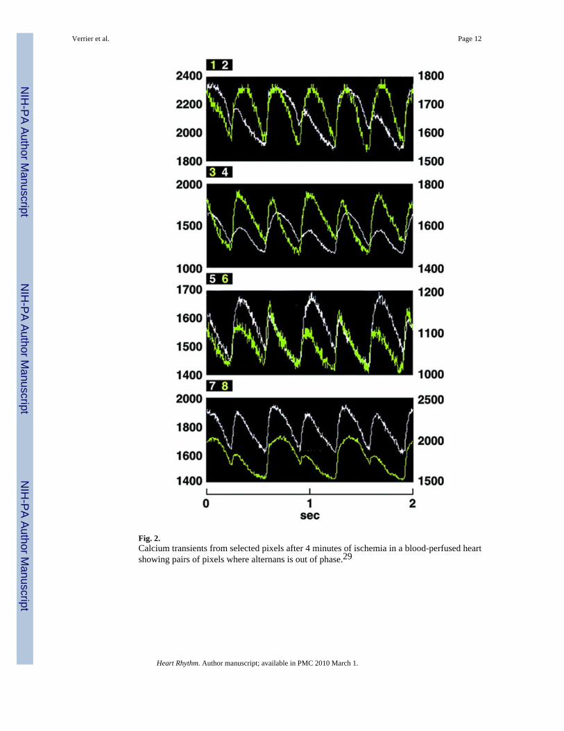
Fig. 2.Calcium transients from selected pixels after 4 minutes of ischemia in a blood-perfused heartshowing pairs of pixels where alternans is out of phase.29
Verrier et al. Page 12
Heart Rhythm. Author manuscript; available in PMC 2010 March 1.
NIH
-PA Author Manuscript
NIH
-PA Author Manuscript
NIH
-PA Author Manuscript
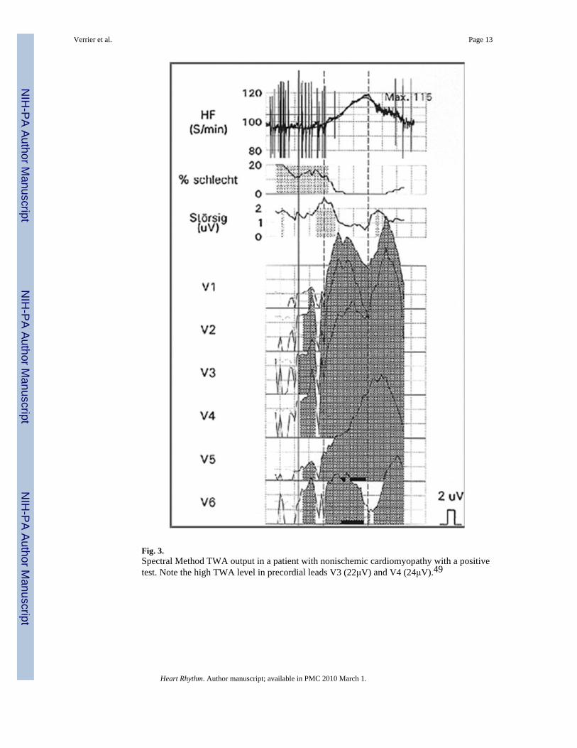
Fig. 3.Spectral Method TWA output in a patient with nonischemic cardiomyopathy with a positivetest. Note the high TWA level in precordial leads V3 (22μV) and V4 (24μV).49
Verrier et al. Page 13
Heart Rhythm. Author manuscript; available in PMC 2010 March 1.
NIH
-PA Author Manuscript
NIH
-PA Author Manuscript
NIH
-PA Author Manuscript
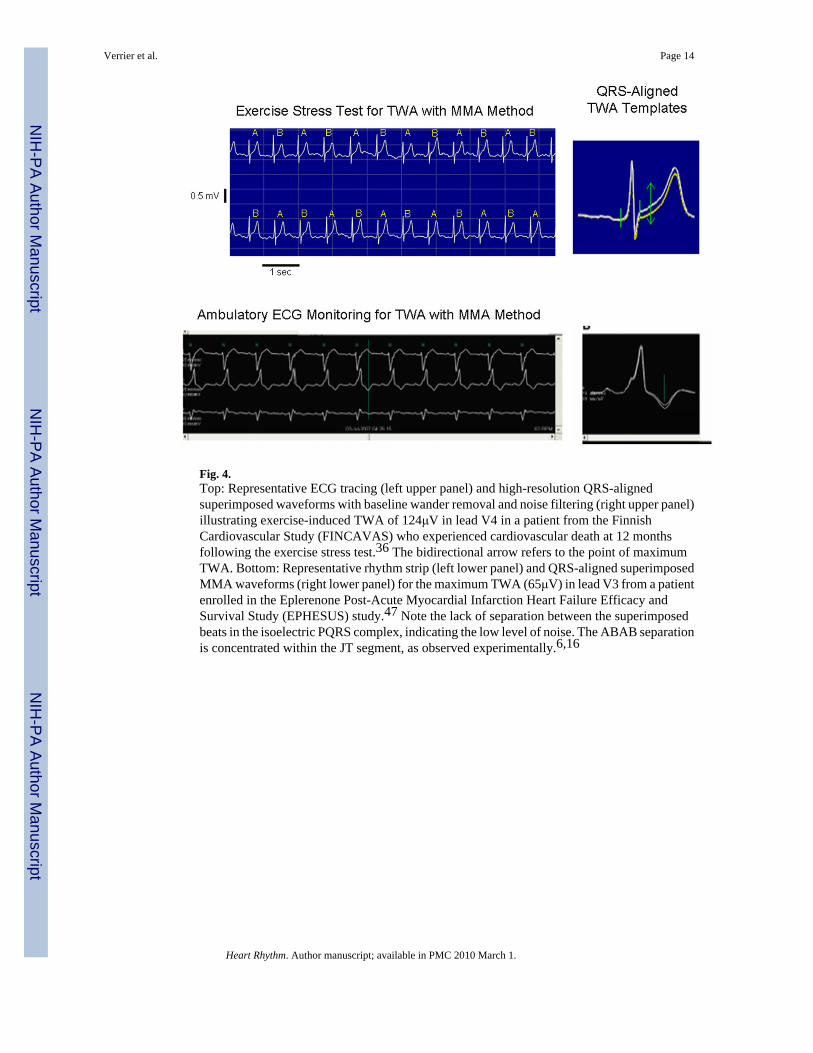
Fig. 4.Top: Representative ECG tracing (left upper panel) and high-resolution QRS-alignedsuperimposed waveforms with baseline wander removal and noise filtering (right upper panel)illustrating exercise-induced TWA of 124μV in lead V4 in a patient from the FinnishCardiovascular Study (FINCAVAS) who experienced cardiovascular death at 12 monthsfollowing the exercise stress test.36 The bidirectional arrow refers to the point of maximumTWA. Bottom: Representative rhythm strip (left lower panel) and QRS-aligned superimposedMMA waveforms (right lower panel) for the maximum TWA (65μV) in lead V3 from a patientenrolled in the Eplerenone Post-Acute Myocardial Infarction Heart Failure Efficacy andSurvival Study (EPHESUS) study.47 Note the lack of separation between the superimposedbeats in the isoelectric PQRS complex, indicating the low level of noise. The ABAB separationis concentrated within the JT segment, as observed experimentally.6,16
Verrier et al. Page 14
Heart Rhythm. Author manuscript; available in PMC 2010 March 1.
NIH
-PA Author Manuscript
NIH
-PA Author Manuscript
NIH
-PA Author Manuscript