aza-bodipy: a new vector for enhanced theranostic boron
TRANSCRIPT
cells
Article
Aza-BODIPY: A New Vector for Enhanced TheranosticBoron Neutron Capture Therapy Applications
Ghadir Kalot 1,† , Amélie Godard 2,†, Benoît Busser 1,3, Jacques Pliquett 2,Mans Broekgaarden 1 , Vincent Motto-Ros 4, Karl David Wegner 5, Ute Resch-Genger 5,Ulli Köster 6 , Franck Denat 2 , Jean-Luc Coll 1 , Ewen Bodio 2,* , Christine Goze 2,* andLucie Sancey 1,*
1 Institute for Advanced Biosciences, UGA INSERM U1209 CNRS UMR5309, 38700 La Tronche, France;[email protected] (G.K.); [email protected] (B.B.);[email protected] (M.B.); [email protected] (J.-L.C.)
2 Institut de Chimie Moléculaire de l’Université de Bourgogne, ICMUB CNRS, UMR 6302, UniversitéBourgogne Franche-Comté, 21078 Dijon, France; [email protected] (A.G.);[email protected] (J.P.); [email protected] (F.D.)
3 Grenoble Alpes University Hospital, 38043 Grenoble, France4 Institut Lumière Matière UMR 5306, Université Lyon 1-CNRS, Université de Lyon,
69622 Villeurbanne, France; [email protected] Division Biophotonics, Federal Institute for Materials Research and Testing (BAM), Richard-Willstaetter-Str.
11, 12489 Berlin, Germany; [email protected] (K.D.W.); [email protected] (U.R.-G.)6 Institut Laue Langevin, 38042 Grenoble, France; [email protected]* Correspondence: [email protected] (E.B.); [email protected] (C.G.);
[email protected] (L.S.); Tel.: +33-380-396-076 (E.B.); +33-380-399-043 (C.G.);+33-476-549-410 (L.S.)
† G.K. and A.G. contributed equally to this work.
Received: 29 July 2020; Accepted: 20 August 2020; Published: 25 August 2020�����������������
Abstract: Boron neutron capture therapy (BNCT) is a radiotherapeutic modality based on thenuclear capture of slow neutrons by stable 10B atoms followed by charged particle emissionthat inducing extensive damage on a very localized level (<10 µm). To be efficient, a sufficientamount of 10B should accumulate in the tumor area while being almost cleared from the normalsurroundings. A water-soluble aza-boron-dipyrromethene dyes (BODIPY) fluorophore was reportedto strongly accumulate in the tumor area with high and BNCT compatible Tumor/Healthy Tissue ratios.The clinically used 10B-BSH (sodium borocaptate) was coupled to the water-soluble aza-BODIPYplatform for enhanced 10B-BSH tumor vectorization. We demonstrated a strong uptake of thecompound in tumor cells and determined its biodistribution in mice-bearing tumors. A model ofchorioallantoic membrane-bearing glioblastoma xenograft was developed to evidence the BNCTpotential of such compound, by subjecting it to slow neutrons. We demonstrated the tumoraccumulation of the compound in real-time using optical imaging and ex vivo using elementalimaging based on laser-induced breakdown spectroscopy. The tumor growth was significantlyreduced as compared to BNCT with 10B-BSH. Altogether, the fluorescent aza-BODIPY/10B-BSHcompound is able to vectorize and image the 10B-BSH in the tumor area, increasing its theranosticpotential for efficient approach of BNCT.
Keywords: aza-BODIPY; BNCT; in ovo model; theranostic; boron compound; 10B-BSH; opticalimaging; NIR-I; SWIR
Cells 2020, 9, 1953; doi:10.3390/cells9091953 www.mdpi.com/journal/cells
Cells 2020, 9, 1953 2 of 14
1. Introduction
Boron neutron capture therapy (BNCT) is a cancer treatment modality based on the vectorizationof 10B-rich compounds in tumor tissues before neutron exposure to selectively destroy cancer cells.Under low-energy neutron irradiation, the stable 10B atoms may capture neutrons producing energeticalpha and 7Li particles. The generated high-linear energy transfer (LET) particles have a cell killingeffect within a 10 µm-range [1,2]. Such type of cancer treatment may not only spare the surroundinghealthy tissues but may be efficient to treat recurrent, or radioresistant to conventional X-ray photontherapy tumors [3–6].
BNCT has obtained promising clinical results for several pathologies as head and neck tumorsincluding recurrent pathologies [3,5,7], malignant brain tumors [8,9], and malignant melanoma [10,11].Regardless of these reports, there are still several limitations. The first limitation of this treatmentmodality meanwhile overcome was the lack of hospital neutron sources that limited the clinical practice toresearch nuclear reactor sites. Recent developments have permitted the installation of accelerator-basedneutron sources [12,13], opening new perspectives to BNCT. In the last 4 decades, only two compoundshave been used as 10B-sources for BNCT in patients: sodium mercaptoundecahydrododecaborate(also called sodium borocaptate, Na2
10B12H11SH; Na210BSH; or BSH) and L-p-boronophenylalanine
(L-10BPA). While not used in clinical trials, GB-10 ([closo-B10H10]2−, dodecahydrododecaborate) isalso an FDA approved molecule [14]. Despite their clinical use, both compounds do not fulfill all therequired criteria. In particular, to be successful, 20 to 50 µg of 10B per gram of tumor is necessary,with a tumor-to-normal tissue and tumor-to-blood ratio > 3:1 [15].
To deliver boron-containing compounds in tumors, various low molecular weight molecules havebeen developed for preclinical research including boron clusters [16,17], amino acid derivatives [18],RGD-BSH conjugates for integrinαvβ3 targeting [19], and several other specific targeted systems [20–22].The clinical application of BNCT requires the tracking of the compound in vivo to predict the 10Bconcentration in the tumor and surrounding non-tumor tissues. Blood samples are required for indirect10B tumor amount estimation [23,24] and adjustment of the treatment planning system that permits todeliver the optimal neutron exposure time and to determine the dose delivered to the patient. Positronemission tomography (PET) or single-photon emission computed tomography (SPECT), which arehighly sensitive nuclear imaging techniques, are non-invasive quantification procedures used to trackand quantify boron carriers in vivo, for personalized treatment protocols.
For preclinical studies, optical imaging (OI) can be also used [25,26]. Among the different opticalprobes, boron-dipyrromethene dyes (BODIPYs, Figure 1a) are very versatile organic fluorophoreswith tunable optical properties from the visible to the NIR-I (Near Infrared) and SWIR (Short WaveInfrared) optical windows [27–29], which display excellent photophysical properties controlled bysubstitution pattern conferring a strong interest for these compounds as theranostic tools [30,31].Moreover, the fluorine atoms on the boron could be substituted by acetylides bearing ammoniumgroups for engrafting functional groups to increase the water-solubility and to conjugate them withchelates for OI/PET, OI/SPECT bimodal imaging [32–35], and therapeutic moieties [31,36,37]. BODIPYfluorophores are also interesting for BNCT, as they possess a boron atom. However, the presence of onlya single boron atom in the BODIPY core and the limited number of commercially available 10B-boronreagents make it difficult to synthesize enriched 10B-BODIPYs. For this reason, BODIPY derivativesreported for a BNCT use implied their conjugation to a 10B-boron cluster [38–41]. Up-to-now, none ofthese studies described either in vivo BNCT investigations or OI distribution studies due to the factthat BODIPYs emit in the visible region of the light spectrum while preclinical OI studies requirefluorescence emission in the NIR-I and, if possible, in the NIR-II/SWIR optical window. To bridgethis gap, we decided to tether a 10B-BSH on a NIR-emitting BODIPY derivative named aza-BODIPY(Figure 1b), thereby creating a theranostic system gathering the advantages of both aza-BODIPY and10B-BSH components.
Cells 2020, 9, 1953 3 of 14
Cells 2020, 9, x FOR PEER REVIEW 6 of 14
3. Results
3.1. Rationale, Design, and Characterization of the Compounds
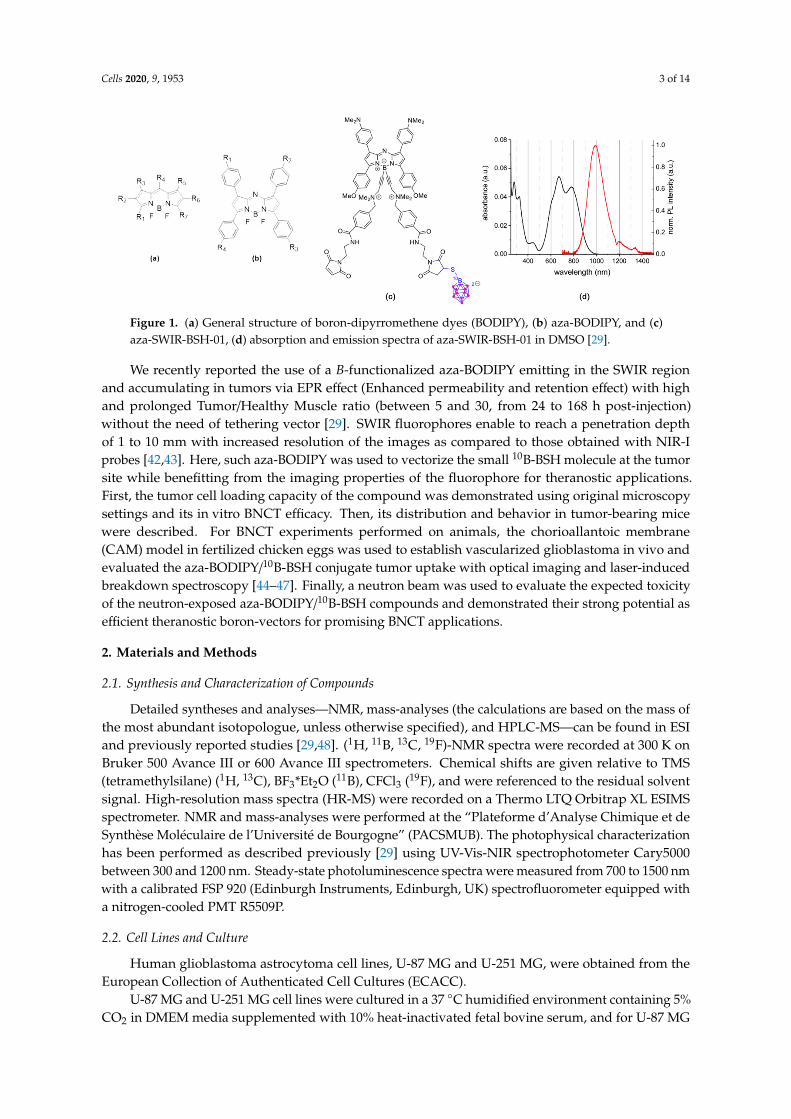
To design an efficient theranostic boron vector, we used a fluorescent reporter platform based on the versatile BODIPY family (Figure 1a) displaying (i) a strong tumor accumulation and (ii) a biocompatible water-solubility, (iii) which can be easily functionalized. In particular, we used B-functionalized aza-BODIPYs (Figure 1b,c), as they have numerous advantages, such as a very high chemical and photochemical photostability, and excellent photophysical properties, rendering them suitable fluorophore for the NIR-I to the SWIR region (Figure 1d). Previously, we developed a strategy, which favored their solubilization and limited their aggregation in biologically relevant media (i.e., water, PBS, or serum), by substituting the fluorine atoms on the boron by alkyne ammonium groups [34,35,48]. Very recently, we used this strategy on a particular Donor-Acceptor-Donor’ aza-BODIPY structure, which emits in the SWIR region, yielding a water-soluble derivative SWIR-WAZABY-01 (See the ESI and Reference [29]; WAZABY for Water-Soluble aza-BODIPY). SWIR-WAZABY-01 could accumulate in vivo very efficiently at the tumor site, without any vectorization, showing high SWIR contrast for up to one week [29].
Figure 1. (a) General structure of boron-dipyrromethene dyes (BODIPY), (b) aza-BODIPY, and (c) aza-SWIR-BSH-01, (d) absorption and emission spectra of aza-SWIR-BSH-01 in DMSO [29].
In this study, we took advantage of the SWIR-WAZABY-01 structure to deliver the 10B-BSH at the tumor site, while enabling the tracking of the molecule in vivo by optical imaging. In order to tether the 10B-BSH on the SWIR-WAZABY-01, we slightly modified its structure and adapted the synthetic method previously reported by us in order to synthesize aza-SWIR-BSH-01. It is functionalized with one 10B BSH unit and emits in the near-infrared region (Figure 1c and see ESI section for the synthetic pathway of aza-SWIR-BSH-01).
3.2. In Vitro Distribution and BNCT Efficacy
To investigate the tumor cell accumulation capacity of aza-SWIR-BSH-01 compounds, the first set of experiments was conducted on brain tumor cells U-251 MG and U-87 MG using SWIR-WAZABY-01, a SWIR emitting aza-BODIPY structurally close to aza-SWIR-BSH-01 but without BSH (see Godard et al. [29]). While the fluorescent signal of SWIR-WAZABY-01 was more compatible with in vivo experiments, due to its emission in the SWIR optical region, it was still detectable by conventional fluorescence microscopy for in vitro investigations. SWIR-WAZABY-01 was incubated with human brain tumor cells U-251 MG and U-87 MG cultured in 2D, but also in 3D spheroids mimicking small tumors. This compound displayed a very fast tumor cell internalization in both 2D and 3D cultures.
Therefore, similar experiments were conducted using the new compound aza-SWIR-BSH-01 on U-87 MG cells. As displayed in Figure 2 after 3 h of incubation, aza-SWIR-BSH-01 accumulated massively in tumor cells, into small cytoplasmic vesicles. The main challenge of this compound was its fluorescence emission properties that can mostly be measured after 800 nm. A dedicated confocal
Figure 1. (a) General structure of boron-dipyrromethene dyes (BODIPY), (b) aza-BODIPY, and (c)aza-SWIR-BSH-01, (d) absorption and emission spectra of aza-SWIR-BSH-01 in DMSO [29].
We recently reported the use of a B-functionalized aza-BODIPY emitting in the SWIR regionand accumulating in tumors via EPR effect (Enhanced permeability and retention effect) with highand prolonged Tumor/Healthy Muscle ratio (between 5 and 30, from 24 to 168 h post-injection)without the need of tethering vector [29]. SWIR fluorophores enable to reach a penetration depthof 1 to 10 mm with increased resolution of the images as compared to those obtained with NIR-Iprobes [42,43]. Here, such aza-BODIPY was used to vectorize the small 10B-BSH molecule at the tumorsite while benefitting from the imaging properties of the fluorophore for theranostic applications.First, the tumor cell loading capacity of the compound was demonstrated using original microscopysettings and its in vitro BNCT efficacy. Then, its distribution and behavior in tumor-bearing micewere described. For BNCT experiments performed on animals, the chorioallantoic membrane(CAM) model in fertilized chicken eggs was used to establish vascularized glioblastoma in vivo andevaluated the aza-BODIPY/10B-BSH conjugate tumor uptake with optical imaging and laser-inducedbreakdown spectroscopy [44–47]. Finally, a neutron beam was used to evaluate the expected toxicityof the neutron-exposed aza-BODIPY/10B-BSH compounds and demonstrated their strong potential asefficient theranostic boron-vectors for promising BNCT applications.
2. Materials and Methods
2.1. Synthesis and Characterization of Compounds
Detailed syntheses and analyses—NMR, mass-analyses (the calculations are based on the mass ofthe most abundant isotopologue, unless otherwise specified), and HPLC-MS—can be found in ESIand previously reported studies [29,48]. (1H, 11B, 13C, 19F)-NMR spectra were recorded at 300 K onBruker 500 Avance III or 600 Avance III spectrometers. Chemical shifts are given relative to TMS(tetramethylsilane) (1H, 13C), BF3*Et2O (11B), CFCl3 (19F), and were referenced to the residual solventsignal. High-resolution mass spectra (HR-MS) were recorded on a Thermo LTQ Orbitrap XL ESIMSspectrometer. NMR and mass-analyses were performed at the “Plateforme d’Analyse Chimique et deSynthèse Moléculaire de l’Université de Bourgogne” (PACSMUB). The photophysical characterizationhas been performed as described previously [29] using UV-Vis-NIR spectrophotometer Cary5000between 300 and 1200 nm. Steady-state photoluminescence spectra were measured from 700 to 1500 nmwith a calibrated FSP 920 (Edinburgh Instruments, Edinburgh, UK) spectrofluorometer equipped witha nitrogen-cooled PMT R5509P.
2.2. Cell Lines and Culture
Human glioblastoma astrocytoma cell lines, U-87 MG and U-251 MG, were obtained from theEuropean Collection of Authenticated Cell Cultures (ECACC).
U-87 MG and U-251 MG cell lines were cultured in a 37 ◦C humidified environment containing 5%CO2 in DMEM media supplemented with 10% heat-inactivated fetal bovine serum, and for U-87 MG
Cells 2020, 9, 1953 4 of 14
cell cultures 1% non-essential amino acids. A total of 10,000 cells were plated into 4-well chamberedcover glass NuncTM Lab-TekTM II (Roskilde, Denmark) for 48 h to be used for fluorescence microscopy.
2.3. Fluorescence Microscopy
Previously prepared 2D cell cultures were kept at 37 ◦C, 5% CO2 and incubated with 10 µMaza-SWIR-BSH-01 compound solution diluted in cell culture medium. Fluorescence microscopy imageswere acquired using a confocal laser-scanning microscope (LSM 710 Carl Zeiss, Jena, Germany) incombi mode. Plan apochromat 63× in oil objective was used. Hoechst 33,342 was used to counterstainthe cell nuclei (1 µM). Optimal fluorescent signal would be obtained with a 680 nm excitation laser,and a collection wavelength between 800–1200 nm would be required. However, such settings arenot available in our current microscopy system. Therefore, the fluorescent signal of the water-solubleaza-BODIPY was collected after excitation with 1.5% 633 nm laser (pinhole aperture 200 µm, gain 1000)using a LP736 filter, in APD (avalanche photodiode) mode.
Images were processed using ImageJ software. The experiments were performed at the MicroCell(Optical Microscopy-Cell Imaging) platform, IAB Grenoble.
2.4. In Vivo Imaging Experiments
All animal experiments were performed in accordance with the Institutional Animal Care and UseCommittee at Grenoble Alpes University. These experiments were also approved by the Animal EthicsCommittee of the French Ministry, under the agreement number APAFIS #8782. The experiments wereperformed at the Optimal (Small Animal Imaging Platform) platform, IAB Grenoble.
U-87 MG cancer cells (5 million cells per 100 µL PBS) were injected subcutaneously on theright flank of female NMRI nude mice (6-8-week-old) (Janvier Labs, Le Genest-Saint Isle, France).After tumor growth (~two weeks), 6 mice were anesthetized (air/isoflurane 4% for induction and 2%thereafter) and injected intravenously in the tail vein with 200 µL of aza-SWIR-BSH-01 (600 µM in PBS).Whole-body NIR fluorescence images were acquired before and 2, 5, 24, and 48 h post-administration.Three mice were euthanized at 24 and 48 h, respectively, and their organs were sampled for ex vivofluorescence imaging. Acquired images were analyzed using ImageJ software. Semi-quantitative datawere obtained by drawing regions of interest (ROI) around the organs. The fluorescence imaging wasperformed using a Pear Trilogy LI-COR system with a laser excitation source of 785 nm and a CCD(charge couple device) collecting fluorescence > 820 nm.
2.5. Neutron Exposure at the Institut Laue-Langevin
2.5.1. Neutron Beam Characteristics
Neutron irradiation experiments were performed at the Institut Laue-Langevin (ILL) (Grenoble,France). The beam line PF1b [49] provides cold neutrons that were collimated through a 3 m longsystem to obtain a final circular beam of 2 cm diameter. Before the experiment, the thermal neutroncapture equivalent flux was measured by activation of thin Au foils, at 2.85 × 109 cm−2 s−1. For cellirradiations, this cold neutron beam provides results equivalent to a thermal neutron irradiation asexplained in detail in Pedrosa-Riviera et al. [50], but for egg irradiations, a significant attenuationtowards deeper layers has to be considered.
2.5.2. BNCT Experiment on Cells
Exponentially growing glioblastoma cancer cells (U251 MG/U87 MG cells) were incubated withmedia alone, media containing BSH, or media containing aza-SWIR-BSH-01, at a 10B concentrationof 40 µg/mL. Cells incubated with media alone were used as a control. After 2 h, the cells (in eachincubation condition) were washed, collected, and divided into 4 quartz cuvettes (Hellma, 110-QS,1-mm layer thickness). These cuvettes were kept as control or exposed to the neutron beam for 1,5, and 10 min, respectively. Following the neutron irradiation, cells were counted, diluted, and re-plated
Cells 2020, 9, 1953 5 of 14
in T25 flasks in triplicates (500 cells/flask) for the colony formation assay. When 128-cell-colonieswere formed in the control condition, the flasks were washed with PBS, fixed with 4% formol(U-87 MG) (for 15 min) or with 4% glutaraldehyde (U-251 MG) (for 5 min), stained with methyleneblue (for 15–30 min), and dried. Finally, the colonies were counted and the results were normalized tothe control condition in each cell line and treatment condition.
2.5.3. In Ovo BNCT Experiment
According to the French and European regulations, no ethical approval is needed for the scientificexperimentations using oviparous embryos. This model was developed to restrict the use of animalsand to facilitate BNCT experiments, according to the following procedure: fertilized white leghornchicken eggs (Couvoir de Cerveloup, Vourey, France) were incubated at 38 ◦C with 60% relativehumidity. At day-3 of chicken embryo development, 3 mL albumin was removed from the eggs anda small window was made into the eggshell above the chorioallantoic membrane (CAM). At day7, the CAM was gently lacerated and 5 × 10⁶ pelleted glioma cells mixed with 30 µL of matrigel(Growth Factor Reduced (GFR) Basement Membrane Matrix 354,230, Corning® Matrigel® (Wiesbaden,Germany)) were deposited on the lacerated region. At day 10, 100 µL of 10B-containing formulations(10B-BSH or aza-SWIR-BSH-01) were added on top of the grown tumors (1.35 µg of 10B/egg) 1 h priorto irradiation, untreated eggs served as a control. The eggs were then exposed to neutron beam for 1 h,split in 2 sessions of 30 min to expose each tumor on both sides (n ≤ 3/condition). Following neutronexposure, the eggs were re-incubated for additional 6 days after which they were terminated and thetumors were carefully collected, weighed, and analyzed.
2.5.4. Statistical Analysis
In vitro data passed the Shapiro-Wilk normality test and were analyzed using a One-way ANOVAand a Sidak’s test for multiple comparisons (Graphpad Prism 7.0 (La Jolla, CA, USA)). CAM datapassed the Shapiro-Wilk normality test and the indicated treatment groups were compared using astudent’s t-test.
2.6. Elemental Imaging Using Laser-Induced Breakdown Spectroscopy (LIBS)
Frozen tumor samples were cut into 7 µm-thick sections before mounting onto plastic slides.The samples were analyzed with a LIBS system to determine their elemental composition, i.e., boron (B (I)208.8 nm) and phosphorus (P (I) 214.9 nm) elements in our case. The homemade LIBS setup was basedon an optical microscope that combined a LIBS laser injection line, a standard optical-imaging apparatus,and a three-dimensional motorized platform for sample positioning [45]. In brief, the ablation wascreated using a quadruple Nd:YAG laser pulses of 1064 nm. The pulse duration was 8 ns, the pulseenergy 1 mJ, and the repetition rate 100 Hz. During the sample scan, the objective to sample distancewas carefully controlled to compensate for any flatness anomalies. The light emitted by the plasmawas collected and connected by an optical fiber to a Czerny-Turner spectrometer equipped with anintensified charge-coupled device camera (Shamrock 303 and iStar, Andor Technology, Belfast, UK).The experiments were performed by Ablatom S.A.S.
2.7. In Ovo Fluorescence Imaging
The experiments were performed at the OPTIMAL (Small animal optical imaging) platform,IAB Grenoble. In order to determine the in ovo tumoral accumulation kinetics, in ovo glioma modelswere prepared. Eggs bearing well-developed and vascularized glioma xenografted tumors wereincubated with 20 µL of aza-SWIR-BSH-01 compound. Fluorescence images of the eggs were acquiredbefore and at specific time intervals following the incubation. Obtained images were analyzed usingImageJ software. Semi-quantitative data regarding the tumoral accumulation were obtained bydrawing regions of interest (ROI) around the tumors. NIR-I 2D-fluorescence reflectance imaging device(Fluobeam 800®, Fluoptics, France) was used to image the eggs incubated with aza-SWIR-BSH-01
Cells 2020, 9, 1953 6 of 14
compound. Fluobeam 800 was provided by a class 1 expanded laser source at 780 nm delivering10 mW/cm2 on the imaging field. The resulting fluorescence signals were collected by a CCD througha high pass filter > 830 nm.
2.8. Quantification of B Content by Inductively Coupled Plasma—Atomic Emission Spectrometry (ICP-AES)
Mice bearing U-87 MG tumors were injected with 40 µg of 10B equivalent from aza-SWIR-BSH-01or 10B-BSH condition (n = 3/group). Determination of the boron content in the samples was performedby ICP-AES analyses (Agilent 720 ES) with a detection limit of 0.1 mg/L. The samples were mineralizedusing 1 mL of aqua regia (mixture of acids: nitric and hydrochloric). After complete mineralization,the samples were diluted with HNO3 (5%, w/w) to reach a 3 mL volume and finally filtered at 0.2 µmfor the measurements. The results were expressed as µg of 10B/g of tumor. The experiments wereperformed at the ISTerre platform, Grenoble.
3. Results
3.1. Rationale, Design, and Characterization of the Compounds
To design an efficient theranostic boron vector, we used a fluorescent reporter platform basedon the versatile BODIPY family (Figure 1a) displaying (i) a strong tumor accumulation and (ii)a biocompatible water-solubility, (iii) which can be easily functionalized. In particular, we usedB-functionalized aza-BODIPYs (Figure 1b,c), as they have numerous advantages, such as a veryhigh chemical and photochemical photostability, and excellent photophysical properties, renderingthem suitable fluorophore for the NIR-I to the SWIR region (Figure 1d). Previously, we developed astrategy, which favored their solubilization and limited their aggregation in biologically relevant media(i.e., water, PBS, or serum), by substituting the fluorine atoms on the boron by alkyne ammoniumgroups [34,35,48]. Very recently, we used this strategy on a particular Donor-Acceptor-Donor’aza-BODIPY structure, which emits in the SWIR region, yielding a water-soluble derivativeSWIR-WAZABY-01 (See the ESI and Reference [29]; WAZABY for Water-Soluble aza-BODIPY).SWIR-WAZABY-01 could accumulate in vivo very efficiently at the tumor site, without any vectorization,showing high SWIR contrast for up to one week [29].
In this study, we took advantage of the SWIR-WAZABY-01 structure to deliver the 10B-BSH at thetumor site, while enabling the tracking of the molecule in vivo by optical imaging. In order to tetherthe 10B-BSH on the SWIR-WAZABY-01, we slightly modified its structure and adapted the syntheticmethod previously reported by us in order to synthesize aza-SWIR-BSH-01. It is functionalized withone 10B BSH unit and emits in the near-infrared region (Figure 1c and see ESI section for the syntheticpathway of aza-SWIR-BSH-01).
3.2. In Vitro Distribution and BNCT Efficacy
To investigate the tumor cell accumulation capacity of aza-SWIR-BSH-01 compounds, the firstset of experiments was conducted on brain tumor cells U-251 MG and U-87 MG usingSWIR-WAZABY-01, a SWIR emitting aza-BODIPY structurally close to aza-SWIR-BSH-01 but withoutBSH (see Godard et al. [29]). While the fluorescent signal of SWIR-WAZABY-01 was more compatiblewith in vivo experiments, due to its emission in the SWIR optical region, it was still detectable byconventional fluorescence microscopy for in vitro investigations. SWIR-WAZABY-01 was incubatedwith human brain tumor cells U-251 MG and U-87 MG cultured in 2D, but also in 3D spheroidsmimicking small tumors. This compound displayed a very fast tumor cell internalization in both 2Dand 3D cultures.
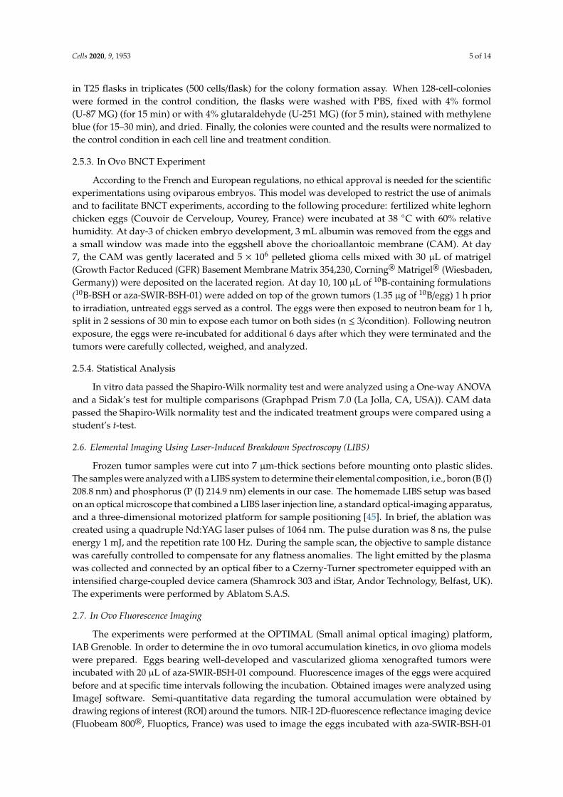
Therefore, similar experiments were conducted using the new compound aza-SWIR-BSH-01on U-87 MG cells. As displayed in Figure 2 after 3 h of incubation, aza-SWIR-BSH-01 accumulatedmassively in tumor cells, into small cytoplasmic vesicles. The main challenge of this compound wasits fluorescence emission properties that can mostly be measured after 800 nm. A dedicated confocal
Cells 2020, 9, 1953 7 of 14
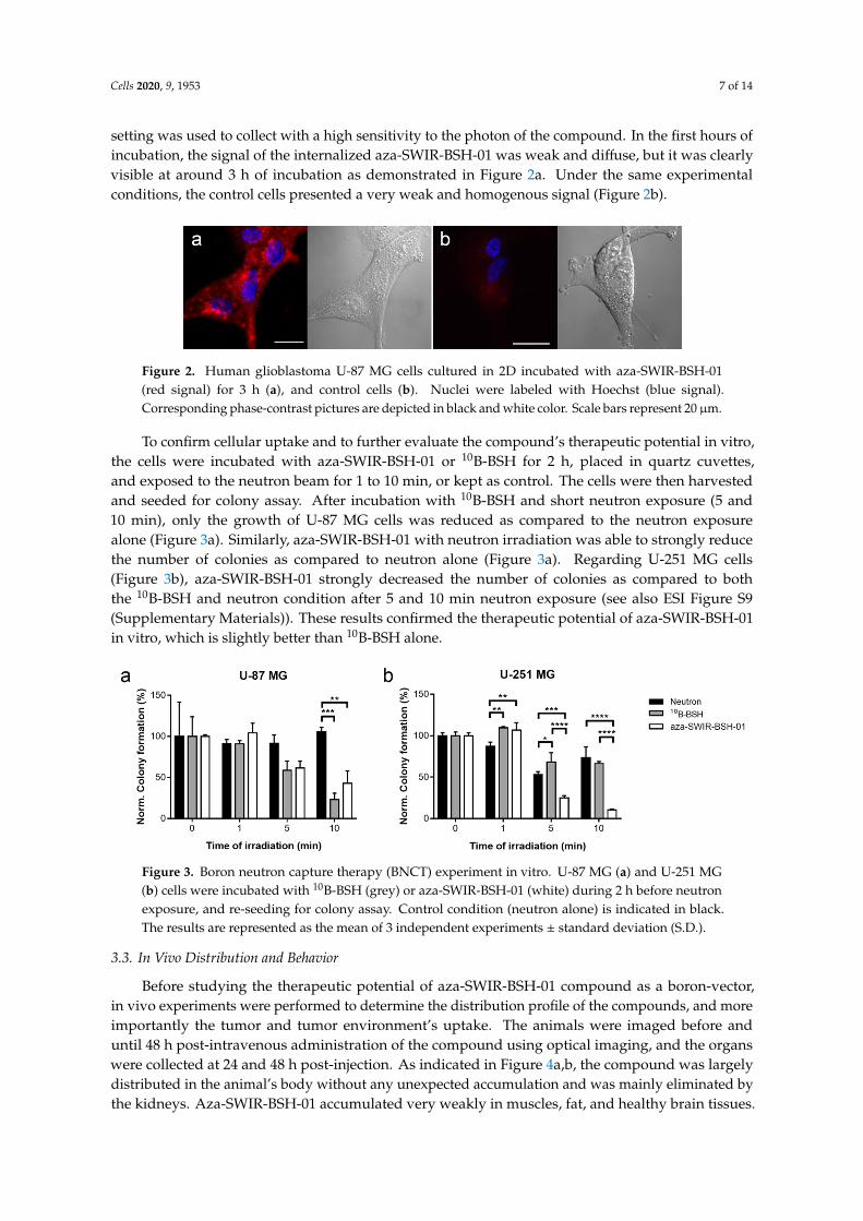
setting was used to collect with a high sensitivity to the photon of the compound. In the first hours ofincubation, the signal of the internalized aza-SWIR-BSH-01 was weak and diffuse, but it was clearlyvisible at around 3 h of incubation as demonstrated in Figure 2a. Under the same experimentalconditions, the control cells presented a very weak and homogenous signal (Figure 2b).
Cells 2020, 9, x FOR PEER REVIEW 7 of 14
setting was used to collect with a high sensitivity to the photon of the compound. In the first hours of incubation, the signal of the internalized aza-SWIR-BSH-01 was weak and diffuse, but it was clearly visible at around 3 h of incubation as demonstrated in Figure 2a. Under the same experimental conditions, the control cells presented a very weak and homogenous signal (Figure 2b).
Figure 2. Human glioblastoma U-87 MG cells cultured in 2D incubated with aza-SWIR-BSH-01 (red signal) for 3 h (a), and control cells (b). Nuclei were labeled with Hoechst (blue signal). Corresponding phase-contrast pictures are depicted in black and white color. Scale bars represent 20 μm.
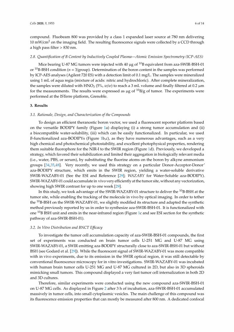
To confirm cellular uptake and to further evaluate the compound’s therapeutic potential in vitro, the cells were incubated with aza-SWIR-BSH-01 or 10B-BSH for 2 h, placed in quartz cuvettes, and exposed to the neutron beam for 1 to 10 min, or kept as control. The cells were then harvested and seeded for colony assay. After incubation with 10B-BSH and short neutron exposure (5 and 10 min), only the growth of U-87 MG cells was reduced as compared to the neutron exposure alone (Figure 3a). Similarly, aza-SWIR-BSH-01 with neutron irradiation was able to strongly reduce the number of colonies as compared to neutron alone (Figure 3a). Regarding U-251 MG cells (Figure 3b), aza-SWIR-BSH-01 strongly decreased the number of colonies as compared to both the 10B-BSH and neutron condition after 5 and 10 min neutron exposure (see also ESI Figure S9 (Supplementary Materials)). These results confirmed the therapeutic potential of aza-SWIR-BSH-01 in vitro, which is slightly better than 10B-BSH alone.
Figure 3. Boron neutron capture therapy (BNCT) experiment in vitro. U-87 MG (a) and U-251 MG (b) cells were incubated with 10B-BSH (grey) or aza-SWIR-BSH-01 (white) during 2 h before neutron exposure, and re-seeding for colony assay. Control condition (neutron alone) is indicated in black. The results are represented as the mean of 3 independent experiments ± standard deviation (S.D.).
3.3. In Vivo Distribution and Behavior
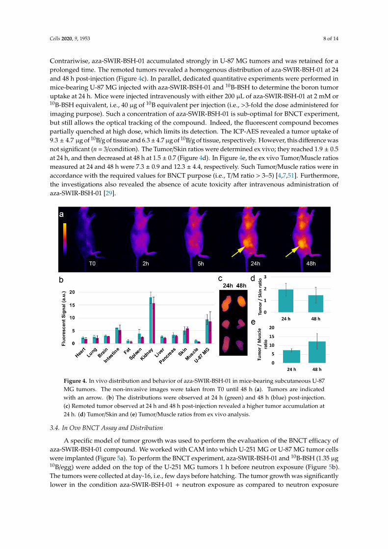
Before studying the therapeutic potential of aza-SWIR-BSH-01 compound as a boron-vector, in vivo experiments were performed to determine the distribution profile of the compounds, and more importantly the tumor and tumor environment’s uptake. The animals were imaged before and until 48 h post-intravenous administration of the compound using optical imaging, and the organs were collected at 24 and 48 h post-injection. As indicated in Figure 4a,b, the compound was largely distributed in the animal’s body without any unexpected accumulation and was mainly eliminated by the kidneys. Aza-SWIR-BSH-01 accumulated very weakly in muscles, fat, and healthy brain tissues. Contrariwise, aza-SWIR-BSH-01 accumulated strongly in U-87 MG tumors and was retained
Figure 2. Human glioblastoma U-87 MG cells cultured in 2D incubated with aza-SWIR-BSH-01(red signal) for 3 h (a), and control cells (b). Nuclei were labeled with Hoechst (blue signal).Corresponding phase-contrast pictures are depicted in black and white color. Scale bars represent 20µm.
To confirm cellular uptake and to further evaluate the compound’s therapeutic potential in vitro,the cells were incubated with aza-SWIR-BSH-01 or 10B-BSH for 2 h, placed in quartz cuvettes,and exposed to the neutron beam for 1 to 10 min, or kept as control. The cells were then harvestedand seeded for colony assay. After incubation with 10B-BSH and short neutron exposure (5 and10 min), only the growth of U-87 MG cells was reduced as compared to the neutron exposurealone (Figure 3a). Similarly, aza-SWIR-BSH-01 with neutron irradiation was able to strongly reducethe number of colonies as compared to neutron alone (Figure 3a). Regarding U-251 MG cells(Figure 3b), aza-SWIR-BSH-01 strongly decreased the number of colonies as compared to boththe 10B-BSH and neutron condition after 5 and 10 min neutron exposure (see also ESI Figure S9(Supplementary Materials)). These results confirmed the therapeutic potential of aza-SWIR-BSH-01in vitro, which is slightly better than 10B-BSH alone.
Cells 2020, 9, x FOR PEER REVIEW 7 of 14
setting was used to collect with a high sensitivity to the photon of the compound. In the first hours of incubation, the signal of the internalized aza-SWIR-BSH-01 was weak and diffuse, but it was clearly visible at around 3 h of incubation as demonstrated in Figure 2a. Under the same experimental conditions, the control cells presented a very weak and homogenous signal (Figure 2b).
Figure 2. Human glioblastoma U-87 MG cells cultured in 2D incubated with aza-SWIR-BSH-01 (red signal) for 3 h (a), and control cells (b). Nuclei were labeled with Hoechst (blue signal). Corresponding phase-contrast pictures are depicted in black and white color. Scale bars represent 20 μm.
To confirm cellular uptake and to further evaluate the compound’s therapeutic potential in vitro, the cells were incubated with aza-SWIR-BSH-01 or 10B-BSH for 2 h, placed in quartz cuvettes, and exposed to the neutron beam for 1 to 10 min, or kept as control. The cells were then harvested and seeded for colony assay. After incubation with 10B-BSH and short neutron exposure (5 and 10 min), only the growth of U-87 MG cells was reduced as compared to the neutron exposure alone (Figure 3a). Similarly, aza-SWIR-BSH-01 with neutron irradiation was able to strongly reduce the number of colonies as compared to neutron alone (Figure 3a). Regarding U-251 MG cells (Figure 3b), aza-SWIR-BSH-01 strongly decreased the number of colonies as compared to both the 10B-BSH and neutron condition after 5 and 10 min neutron exposure (see also ESI Figure S9 (Supplementary Materials)). These results confirmed the therapeutic potential of aza-SWIR-BSH-01 in vitro, which is slightly better than 10B-BSH alone.
Figure 3. Boron neutron capture therapy (BNCT) experiment in vitro. U-87 MG (a) and U-251 MG (b) cells were incubated with 10B-BSH (grey) or aza-SWIR-BSH-01 (white) during 2 h before neutron exposure, and re-seeding for colony assay. Control condition (neutron alone) is indicated in black. The results are represented as the mean of 3 independent experiments ± standard deviation (S.D.).
3.3. In Vivo Distribution and Behavior
Before studying the therapeutic potential of aza-SWIR-BSH-01 compound as a boron-vector, in vivo experiments were performed to determine the distribution profile of the compounds, and more importantly the tumor and tumor environment’s uptake. The animals were imaged before and until 48 h post-intravenous administration of the compound using optical imaging, and the organs were collected at 24 and 48 h post-injection. As indicated in Figure 4a,b, the compound was largely distributed in the animal’s body without any unexpected accumulation and was mainly eliminated by the kidneys. Aza-SWIR-BSH-01 accumulated very weakly in muscles, fat, and healthy brain tissues. Contrariwise, aza-SWIR-BSH-01 accumulated strongly in U-87 MG tumors and was retained
Figure 3. Boron neutron capture therapy (BNCT) experiment in vitro. U-87 MG (a) and U-251 MG(b) cells were incubated with 10B-BSH (grey) or aza-SWIR-BSH-01 (white) during 2 h before neutronexposure, and re-seeding for colony assay. Control condition (neutron alone) is indicated in black.The results are represented as the mean of 3 independent experiments ± standard deviation (S.D.).
3.3. In Vivo Distribution and Behavior
Before studying the therapeutic potential of aza-SWIR-BSH-01 compound as a boron-vector,in vivo experiments were performed to determine the distribution profile of the compounds, and moreimportantly the tumor and tumor environment’s uptake. The animals were imaged before anduntil 48 h post-intravenous administration of the compound using optical imaging, and the organswere collected at 24 and 48 h post-injection. As indicated in Figure 4a,b, the compound was largelydistributed in the animal’s body without any unexpected accumulation and was mainly eliminated bythe kidneys. Aza-SWIR-BSH-01 accumulated very weakly in muscles, fat, and healthy brain tissues.
Cells 2020, 9, 1953 8 of 14
Contrariwise, aza-SWIR-BSH-01 accumulated strongly in U-87 MG tumors and was retained for aprolonged time. The remoted tumors revealed a homogenous distribution of aza-SWIR-BSH-01 at 24and 48 h post-injection (Figure 4c). In parallel, dedicated quantitative experiments were performed inmice-bearing U-87 MG injected with aza-SWIR-BSH-01 and 10B-BSH to determine the boron tumoruptake at 24 h. Mice were injected intravenously with either 200 µL of aza-SWIR-BSH-01 at 2 mM or10B-BSH equivalent, i.e., 40 µg of 10B equivalent per injection (i.e., >3-fold the dose administered forimaging purpose). Such a concentration of aza-SWIR-BSH-01 is sub-optimal for BNCT experiment,but still allows the optical tracking of the compound. Indeed, the fluorescent compound becomespartially quenched at high dose, which limits its detection. The ICP-AES revealed a tumor uptake of9.3 ± 4.7 µg of 10B/g of tissue and 6.3± 4.7µg of 10B/g of tissue, respectively. However, this difference wasnot significant (n = 3/condition). The Tumor/Skin ratios were determined ex vivo; they reached 1.9 ± 0.5at 24 h, and then decreased at 48 h at 1.5 ± 0.7 (Figure 4d). In Figure 4e, the ex vivo Tumor/Muscle ratiosmeasured at 24 and 48 h were 7.3 ± 0.9 and 12.3 ± 4.4, respectively. Such Tumor/Muscle ratios were inaccordance with the required values for BNCT purpose (i.e., T/M ratio > 3–5) [4,7,51]. Furthermore,the investigations also revealed the absence of acute toxicity after intravenous administration ofaza-SWIR-BSH-01 [29].
Cells 2020, 9, x FOR PEER REVIEW 8 of 14
for a prolonged time. The remoted tumors revealed a homogenous distribution of aza-SWIR-BSH-01 at 24 and 48 h post-injection (Figure 4c). In parallel, dedicated quantitative experiments were performed in mice-bearing U-87 MG injected with aza-SWIR-BSH-01 and 10B-BSH to determine the boron tumor uptake at 24 h. Mice were injected intravenously with either 200 μL of aza-SWIR-BSH-01 at 2 mM or 10B-BSH equivalent, i.e., 40 μg of 10B equivalent per injection (i.e., >3-fold the dose administered for imaging purpose). Such a concentration of aza-SWIR-BSH-01 is sub-optimal for BNCT experiment, but still allows the optical tracking of the compound. Indeed, the fluorescent compound becomes partially quenched at high dose, which limits its detection. The ICP-AES revealed a tumor uptake of 9.3 ± 4.7 μg of 10B/g of tissue and 6.3 ± 4.7 μg of 10B/g of tissue, respectively. However, this difference was not significant (n = 3/condition). The Tumor/Skin ratios were determined ex vivo; they reached 1.9 ± 0.5 at 24 h, and then decreased at 48 h at 1.5 ± 0.7 (Figure 4d). In Figure 4e, the ex vivo Tumor/Muscle ratios measured at 24 and 48 h were 7.3 ± 0.9 and 12.3 ± 4.4, respectively. Such Tumor/Muscle ratios were in accordance with the required values for BNCT purpose (i.e., T/M ratio > 3–5) [4,7,51]. Furthermore, the investigations also revealed the absence of acute toxicity after intravenous administration of aza-SWIR-BSH-01 [29].
Figure 4. In vivo distribution and behavior of aza-SWIR-BSH-01 in mice-bearing subcutaneous U-87 MG tumors. The non-invasive images were taken from T0 until 48 h (a). Tumors are indicated with an arrow. (b) The distributions were observed at 24 h (green) and 48 h (blue) post-injection. (c) Remoted tumor observed at 24 h and 48 h post-injection revealed a higher tumor accumulation at 24 h. (d) Tumor/Skin and (e) Tumor/Muscle ratios from ex vivo analysis.
3.4. In Ovo BNCT Assay and Distribution
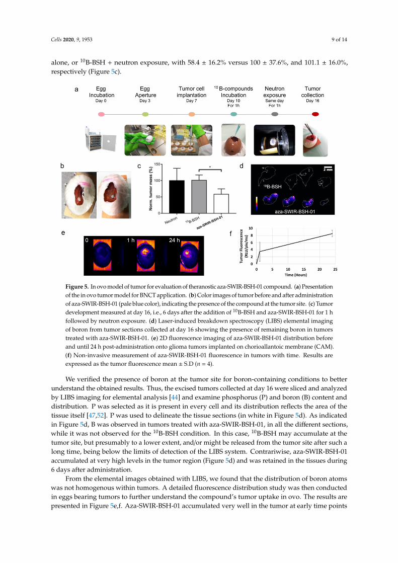
A specific model of tumor growth was used to perform the evaluation of the BNCT efficacy of aza-SWIR-BSH-01 compound. We worked with CAM into which U-251 MG or U-87 MG tumor cells were implanted (Figure 5a). To perform the BNCT experiment, aza-SWIR-BSH-01 and 10B-BSH (1.35 μg 10B/egg) were added on the top of the U-251 MG tumors 1 h before neutron exposure (Figure 5b). The tumors were collected at day-16, i.e., few days before hatching. The tumor growth was significantly lower in the condition aza-SWIR-BSH-01 + neutron exposure as compared to neutron exposure alone, or 10B-BSH + neutron exposure, with 58.4 ± 16.2% versus 100 ± 37.6%, and 101.1 ± 16.0%, respectively (Figure 5c).
We verified the presence of boron at the tumor site for boron-containing conditions to better understand the obtained results. Thus, the excised tumors collected at day 16 were sliced and
Figure 4. In vivo distribution and behavior of aza-SWIR-BSH-01 in mice-bearing subcutaneous U-87MG tumors. The non-invasive images were taken from T0 until 48 h (a). Tumors are indicatedwith an arrow. (b) The distributions were observed at 24 h (green) and 48 h (blue) post-injection.(c) Remoted tumor observed at 24 h and 48 h post-injection revealed a higher tumor accumulation at24 h. (d) Tumor/Skin and (e) Tumor/Muscle ratios from ex vivo analysis.
3.4. In Ovo BNCT Assay and Distribution
A specific model of tumor growth was used to perform the evaluation of the BNCT efficacy ofaza-SWIR-BSH-01 compound. We worked with CAM into which U-251 MG or U-87 MG tumor cellswere implanted (Figure 5a). To perform the BNCT experiment, aza-SWIR-BSH-01 and 10B-BSH (1.35 µg10B/egg) were added on the top of the U-251 MG tumors 1 h before neutron exposure (Figure 5b).The tumors were collected at day-16, i.e., few days before hatching. The tumor growth was significantlylower in the condition aza-SWIR-BSH-01 + neutron exposure as compared to neutron exposure
Cells 2020, 9, 1953 9 of 14
alone, or 10B-BSH + neutron exposure, with 58.4 ± 16.2% versus 100 ± 37.6%, and 101.1 ± 16.0%,respectively (Figure 5c).
Cells 2020, 9, x FOR PEER REVIEW 9 of 14
analyzed by LIBS imaging for elemental analysis [44] and examine phosphorus (P) and boron (B) content and distribution. P was selected as it is present in every cell and its distribution reflects the area of the tissue itself [47,52]. P was used to delineate the tissue sections (in white in Figure 5d). As indicated in Figure 5d, B was observed in tumors treated with aza-SWIR-BSH-01, in all the different sections, while it was not observed for the 10B-BSH condition. In this case, 10B-BSH may accumulate at the tumor site, but presumably to a lower extent, and/or might be released from the tumor site after such a long time, being below the limits of detection of the LIBS system. Contrariwise, aza-SWIR-BSH-01 accumulated at very high levels in the tumor region (Figure 5d) and was retained in the tissues during 6 days after administration.
From the elemental images obtained with LIBS, we found that the distribution of boron atoms was not homogenous within tumors. A detailed fluorescence distribution study was then conducted in eggs bearing tumors to further understand the compound’s tumor uptake in ovo. The results are presented in Figure 5e,f. Aza-SWIR-BSH-01 accumulated very well in the tumor at early time points (1 h), evidencing some diffusion into vessels at time points with prolonged exposure, suggesting a long-lasting circulation in the blood vessels. The fluorescence signal increased with time, reaching a massive uptake peak at 24 h. As compared to the fluorescence signal obtained after 1 h, the fluorescent signal at 24 h was enhanced by a factor of ~2.5, confirming that the optimal and minimal time required for optimal aza-SWIR-BSH-01 tumor uptake was 24 h. These results also suggest that 24 h post-treatment could be the ideal delay for starting neutron irradiation for a more effective BNCT.
Figure 5. In ovo model of tumor for evaluation of theranostic aza-SWIR-BSH-01 compound. (a) Presentation of the in ovo tumor model for BNCT application. (b) Color images of tumor before and after administration of aza-SWIR-BSH-01 (pale blue color), indicating the presence of the compound at the tumor site. (c) Tumor development measured at day 16, i.e., 6 days after the addition of 10B-BSH and aza-SWIR-BSH-01 for 1 h followed by neutron exposure. (d) Laser-induced breakdown spectroscopy (LIBS) elemental imaging of boron from tumor sections collected at day 16 showing the presence of remaining boron in tumors treated with aza-SWIR-BSH-01. (e) 2D fluorescence imaging of aza-SWIR-BSH-01 distribution before and until 24 h post-administration onto glioma tumors implanted on chorioallantoic membrane (CAM). (f) Non-invasive measurement of aza-SWIR-BSH-01 fluorescence in tumors with time. Results are expressed as the tumor fluorescence mean ± S.D (n = 4).
Figure 5. In ovo model of tumor for evaluation of theranostic aza-SWIR-BSH-01 compound. (a) Presentationof the in ovo tumor model for BNCT application. (b) Color images of tumor before and after administrationof aza-SWIR-BSH-01 (pale blue color), indicating the presence of the compound at the tumor site. (c) Tumordevelopment measured at day 16, i.e., 6 days after the addition of 10B-BSH and aza-SWIR-BSH-01 for 1 hfollowed by neutron exposure. (d) Laser-induced breakdown spectroscopy (LIBS) elemental imagingof boron from tumor sections collected at day 16 showing the presence of remaining boron in tumorstreated with aza-SWIR-BSH-01. (e) 2D fluorescence imaging of aza-SWIR-BSH-01 distribution beforeand until 24 h post-administration onto glioma tumors implanted on chorioallantoic membrane (CAM).(f) Non-invasive measurement of aza-SWIR-BSH-01 fluorescence in tumors with time. Results areexpressed as the tumor fluorescence mean ± S.D (n = 4).
We verified the presence of boron at the tumor site for boron-containing conditions to betterunderstand the obtained results. Thus, the excised tumors collected at day 16 were sliced and analyzedby LIBS imaging for elemental analysis [44] and examine phosphorus (P) and boron (B) content anddistribution. P was selected as it is present in every cell and its distribution reflects the area of thetissue itself [47,52]. P was used to delineate the tissue sections (in white in Figure 5d). As indicatedin Figure 5d, B was observed in tumors treated with aza-SWIR-BSH-01, in all the different sections,while it was not observed for the 10B-BSH condition. In this case, 10B-BSH may accumulate at thetumor site, but presumably to a lower extent, and/or might be released from the tumor site after such along time, being below the limits of detection of the LIBS system. Contrariwise, aza-SWIR-BSH-01accumulated at very high levels in the tumor region (Figure 5d) and was retained in the tissues during6 days after administration.
From the elemental images obtained with LIBS, we found that the distribution of boron atomswas not homogenous within tumors. A detailed fluorescence distribution study was then conductedin eggs bearing tumors to further understand the compound’s tumor uptake in ovo. The results arepresented in Figure 5e,f. Aza-SWIR-BSH-01 accumulated very well in the tumor at early time points
Cells 2020, 9, 1953 10 of 14
(1 h), evidencing some diffusion into vessels at time points with prolonged exposure, suggesting along-lasting circulation in the blood vessels. The fluorescence signal increased with time, reaching amassive uptake peak at 24 h. As compared to the fluorescence signal obtained after 1 h, the fluorescentsignal at 24 h was enhanced by a factor of ~2.5, confirming that the optimal and minimal timerequired for optimal aza-SWIR-BSH-01 tumor uptake was 24 h. These results also suggest that 24 hpost-treatment could be the ideal delay for starting neutron irradiation for a more effective BNCT.
4. Discussion
BODIPY derivatives have already been developed for BNCT application [38–41], but none ofthem, to our best knowledge, have been tested for BNCT anti-tumor development in vitro and in vivo.This study is the first to report the use and evaluation of aza-BODIPY-based compounds as theranosticcompounds for BNCT purpose.
The delivery of 10B into tumors for BNCT requires the production and evaluation of biocompatibleand water-soluble vectors, with no or only marginal and reversible toxicity, and preferential tumoraccumulation while clearing from the blood and tumor micro-environment. As recently reported,some SWIR-WAZABY compounds possess a strong, time-dependent tumor uptake, and weak muscleaccumulation [29]. Contrariwise, 10B-BSH, a molecule approved for BNCT, is rapidly washed outfrom the body and should be administrated at high dose [8,53]. Here, the tumor uptake kineticsand the elemental imaging indicated that the 10B-BSH part of the novel aza-SWIR-BSH compoundadvantageously followed the BNCT-compatible kinetics of the SWIR-WAZABY vector, rather than thatof the 10B-BSH moiety itself. Therefore, the engraftment of the small 10B-BSH entity did not impact thetumor accumulation capacity and water-solubility of the SWIR-WAZABY, even when administeredat high concentrations (200 µL at 2 mM as a bolus intravenous administration). The substitution ofthe fluorine atoms on the boron allowing to increase the water-solubility is a key factor for efficientdistribution and tumor targeting.
To evaluate the properties of the theranostic aza-SWIR-BSH compound in vivo, the CAM modelwas used. This model is particularly interesting for drug evaluation as (i) the tumors grow rapidly dueto the nutrients-rich environment of the CAM, (ii) the vasculature is well developed and accessible,allowing an efficient tumor vascularization, (iii) the absence of a well-established immune system atthis point of embryo development contributes to the tumor growth, even from patient-derived cells,and (iv) the utilization of this experimental model contributes to the reduction of animal’s use in anethical aspect of the research [54]. Therefore, this model has been widely used for the evaluation ofanti-angiogenic or antitumor compounds [54–56], the evaluation of sensitizers for radiotherapy andphotodynamic therapy [57,58], and imaging using MRI and PET/CT [59,60]; however, the CAM modelhas never been reported to demonstrate the potential of boron compounds for BNCT application.Using optical imaging, the distribution of aza-SWIR-BSH compound in this CAM model indicated anoptimized tumor uptake after prolonged exposure time (24 h, Figure 5), while the 1-h administrationfollowed by neutron exposure was already able to reduce tumor growth; all these results suggestedthat BNCT could increase tumor shrinkage after prolonged incubations with these compounds andneutron exposure.
5. Conclusions
Altogether, we demonstrated that water-soluble aza-BODIPYs can be used as theranostic vectorsfor boron complexes, opening a new perspective for compound development for BNCT applications.
6. Patents
To be added during the reviewing process when official patent numbers will be known.
Supplementary Materials: The following are available online at http://www.mdpi.com/2073-4409/9/9/1953/s1,Table S1: HPLC analytical gradient. Table S2: Detailed of gradient. Scheme S1: Synthetic pathway of
Cells 2020, 9, 1953 11 of 14
aza-SWIR-BSH-01. Figure S1: Analytical HPLC of SWIR-WAZABY-02. Figure S2: 1H NMR of SWIR-WAZABY-02(500 MHz, 298 K, MeOD-d4). Figure S3: 13C NMR of SWIR-WAZABY-02 (125 MHz, 298 K, MeOD-d4). Figure S4:Analytical HPLC of aza-SWIR-03. Figure S5: 1H NMR of aza-SWIR-03 (500 MHz, 298 K, DMSO-d6). Figure S6:13C NMR of aza-SWIR-03 (150 MHz, 298 K, DMSO-d6). Figure S7: 1H NMR of aza-SWIR-BSH-01 (500 MHz, 298K, DMSO-d6). Figure S8: 13C NMR of aza-SWIR-BSH-01 (150 MHz, 343 K, DMSO-d6). Figure S9: U-251 cellsincubated with aza-SWIR-BSH-01 before (a) and after (b) 10 min of neutron exposure colored with Trypan blue.
Author Contributions: Conceptualization, F.D., J.-L.C., and L.S.; methodology, G.K., A.G., V.M.-R., M.B., and U.K.;validation, A.G., J.P., C.G., and E.B.; formal analysis, G.K., B.B., M.B., and L.S.; investigation, G.K., A.G., B.B., J.P.,K.D.W., U.R.-G., and L.S.; resources, U.K.; writing—original draft preparation, G.K., A.G., B.B., E.B., C.G., and L.S.;writing—review and editing, all the authors; supervision, E.B., C.G., and L.S.; funding acquisition, F.D., E.B., C.G.,and L.S. All authors have read and agreed to the published version of the manuscript.
Funding: This work was supported by the GEFLUC Grenoble Dauphiné Savoie, and the French funding agencies:FLI (France Life Imaging) for the project Thera-BODIPY, the CNRS Mission for Transversal and InterdisciplinaryInitiatives for the project BREVET-ISOTOP, the Fondation pour la Recherche Médicale (FRM) for the specificsupport of G.K. (FRM ECO201806006861). This research was funded by The Ministère de l’Enseignement Supérieuret de la Recherche, the Centre National de la Recherche Scientifique (CNRS), the Conseil Régional de Bourgogne(PhD JCE Grant # 2015-9205AAO033S04139/BG0003203), and the French Research National Agency (ANR) viaproject JCJC “SPID” ANR-16- CE07-0020 and project JCJC “WazaBY” ANR-18-CE18-0012. This work is part of theprojects “Pharmacoimagerie et agents théranostiques” et “Chimie durable, environnement et agroalimentaire”supported by the Université de Bourgogne and the Conseil Régional de Bourgogne through the Plan d’ActionsRégional pour l’Innovation (PARI) and the European Union through the PO FEDER-FSE Bourgogne 2014/2020programs. It was performed within the Dijon’s pharmaco-imaging consortium, a regional center of excellencein pharmaco-imaging.
Acknowledgments: G.K. and L.S. would like to thank F. Trichard from Ablatom SAS for LIBS experiments andM. Couvet and A. Grichine (µCell platform IAB, Grenoble, France) for technical advice. The authors are gratefulto V. Josserand from OPTIMAL platform (platform responsible) for technical access and optical experiments.We also thank the ISTerre facility (S.B., Grenoble, France) for ICP measurements. We are grateful to T. Soldner(ILL, PF1b instrument responsible) for invaluable help with the setup of the beam line. A.G., J.P., F.D., E.B.,and C.G. gratefully acknowledge the Université de Bourgogne, SATT Sayens, Conseil Régional de BourgogneFranche-Comté and the French Ministère de l’Enseignement Supérieur et de la Recherche for financial support ofthe analytical platform PACSMUB.
Conflicts of Interest: The authors declare no conflict of interest.
References
1. Locher, G. Biological effects and therapeutic possibilities of neutrons. Am. J. Roentgenol. Radium Ther. 1936,36, 1–13.
2. Barth, R.F.; Coderre, J.A.; Vicente, M.G.; Blue, T.E. Boron neutron capture therapy of cancer: Current statusand future prospects. Clin. Cancer Res. 2005, 11, 3987–4002. [CrossRef] [PubMed]
3. Aihara, T.; Morita, N.; Kamitani, N.; Kumada, H.; Ono, K.; Hiratsuka, J.; Harada, T. BNCT for advanced orrecurrent head and neck cancer. Appl. Radiat. Isot. 2014, 88, 12–15. [CrossRef] [PubMed]
4. Aiyama, H.; Nakai, K.; Yamamoto, T.; Nariai, T.; Kumada, H.; Ishikawa, E.; Isobe, T.; Endo, K.; Takada, T.;Yoshida, F.; et al. A clinical trial protocol for second line treatment of malignant brain tumors with BNCT atUniversity of Tsukuba. Appl. Radiat. Isot. 2011, 69, 1819–1822. [CrossRef]
5. Kato, I.; Ono, K.; Sakurai, Y.; Ohmae, M.; Maruhashi, A.; Imahori, Y.; Kirihata, M.; Nakazawa, M.; Yura, Y.Effectiveness of BNCT for recurrent head and neck malignancies. Appl. Radiat. Isot. 2004, 61, 1069–1073.[CrossRef]
6. Sauerwein, W.; Moss, R.; Rassow, J.; Stecher-Rasmussen, F.; Hideghety, K.; Wolbers, J.G.; Sack, H. Organisationand management of the first clinical trial of BNCT in Europe (EORTC protocol 11961).EORTC BNCT studygroup. Strahlenther. Onkol. 1999, 175 (Suppl. 2), 108–111. [CrossRef]
7. Wang, L.W.; Chen, Y.W.; Ho, C.Y.; Hsueh Liu, Y.W.; Chou, F.I.; Liu, Y.H.; Liu, H.M.; Peir, J.J.; Jiang, S.H.;Chang, C.W.; et al. Fractionated BNCT for locally recurrent head and neck cancer: Experience from a phaseI/II clinical trial at Tsing Hua Open-Pool Reactor. Appl. Radiat. Isot. 2014, 88, 23–27. [CrossRef]
8. Haritz, D.; Gabel, D.; Huiskamp, R. Clinical phase-I study of Na2B12H11SH (BSH) in patients with malignantglioma as precondition for boron neutron capture therapy (BNCT). Int. J. Radiat. Oncol. Biol. Phys. 1994, 28,1175–1181. [CrossRef]
Cells 2020, 9, 1953 12 of 14
9. Takagaki, M.; Oda, Y.; Miyatake, S.; Kikuchi, H.; Kobayashi, T.; Sakurai, Y.; Osawa, M.; Mori, K.; Ono, K.Boron neutron capture therapy: Preliminary study of BNCT with sodium borocaptate (Na2B1 2H1 1SH) onglioblastoma. J. Neurooncol. 1997, 35, 177–185. [CrossRef]
10. Gonzalez, S.J.; Bonomi, M.R.; Santa Cruz, G.A.; Blaumann, H.R.; Calzetta Larrieu, O.A.; Menendez, P.;Jimenez Rebagliati, R.; Longhino, J.; Feld, D.B.; Dagrosa, M.A.; et al. First BNCT treatment of a skin melanomain Argentina: Dosimetric analysis and clinical outcome. Appl. Radiat. Isot. 2004, 61, 1101–1105. [CrossRef]
11. Mishima, Y.; Honda, C.; Ichihashi, M.; Obara, H.; Hiratsuka, J.; Fukuda, H.; Karashima, H.; Kobayashi, T.;Kanda, K.; Yoshino, K. Treatment of malignant melanoma by single thermal neutron capture therapy withmelanoma-seeking 10B-compound. Lancet 1989, 2, 388–389. [CrossRef]
12. Kato, T.; Hirose, K.; Tanaka, H.; Mitsumoto, T.; Motoyanagi, T.; Arai, K.; Harada, T.; Takeuchi, A.; Kato, R.;Yajima, S.; et al. Design and construction of an accelerator-based boron neutron capture therapy (AB-BNCT)facility with multiple treatment rooms at the Southern Tohoku BNCT Research Center. Appl. Radiat. Isot.2020, 156, 108961. [CrossRef] [PubMed]
13. Kreiner, A.J.; Baldo, M.; Bergueiro, J.R.; Cartelli, D.; Castell, W.; Thatar Vento, V.; Gomez Asoia, J.; Mercuri, D.;Padulo, J.; Suarez Sandin, J.C.; et al. Accelerator-based BNCT. Appl. Radiat. Isot. 2014, 88, 185–189. [CrossRef][PubMed]
14. Barth, R.F.; Mi, P.; Yang, W. Boron delivery agents for neutron capture therapy of cancer. Cancer Commun.2018, 38, 35. [CrossRef] [PubMed]
15. Slatkin, D.N. A history of boron neutron capture therapy of brain tumours. Postulation of a brain radiationdose tolerance limit. Brain 1991, 114 Pt 4, 1609–1629. [CrossRef]
16. Chandra, S.; Barth, R.F.; Haider, S.A.; Yang, W.; Huo, T.; Shaikh, A.L.; Kabalka, G.W. Biodistribution andsubcellular localization of an unnatural boron-containing amino acid (cis-ABCPC) by imaging secondaryion mass spectrometry for neutron capture therapy of melanomas and gliomas. PLoS ONE 2013, 8, e75377.[CrossRef]
17. Kabalka, G.W.; Shaikh, A.L.; Barth, R.F.; Huo, T.; Yang, W.; Gordnier, P.M.; Chandra, S. Boronated unnaturalcyclic amino acids as potential delivery agents for neutron capture therapy. Appl. Radiat. Isot. 2011, 69,1778–1781. [CrossRef]
18. Futamura, G.; Kawabata, S.; Nonoguchi, N.; Hiramatsu, R.; Toho, T.; Tanaka, H.; Masunaga, S.I.; Hattori, Y.;Kirihata, M.; Ono, K.; et al. Evaluation of a novel sodium borocaptate-containing unnatural amino acid as aboron delivery agent for neutron capture therapy of the F98 rat glioma. Radiat. Oncol. 2017, 12, 26. [CrossRef]
19. Kimura, S.; Masunaga, S.; Harada, T.; Kawamura, Y.; Ueda, S.; Okuda, K.; Nagasawa, H. Synthesis andevaluation of cyclic RGD-boron cluster conjugates to develop tumor-selective boron carriers for boronneutron capture therapy. Bioorg. Med. Chem. 2011, 19, 1721–1728. [CrossRef]
20. Hoppenz, P.; Els-Heindl, S.; Kellert, M.; Kuhnert, R.; Saretz, S.; Lerchen, H.G.; Kobberling, J.; Riedl, B.;Hey-Hawkins, E.; Beck-Sickinger, A.G. A Selective Carborane-Functionalized Gastrin-Releasing PeptideReceptor Agonist as Boron Delivery Agent for Boron Neutron Capture Therapy. J. Org. Chem. 2020, 85,1446–1457. [CrossRef]
21. Kellert, M.; Hoppenz, P.; Lonnecke, P.; Worm, D.J.; Riedl, B.; Koebberling, J.; Beck-Sickinger, A.G.;Hey-Hawkins, E. Tuning a modular system-synthesis and characterisation of a boron-rich s-triazine-basedcarboxylic acid and amine bearing a galactopyranosyl moiety. Dalton Trans. 2020, 49, 57–69. [CrossRef][PubMed]
22. Kellert, M.; Worm, D.J.; Hoppenz, P.; Sarosi, M.B.; Lonnecke, P.; Riedl, B.; Koebberling, J.; Beck-Sickinger, A.G.;Hey-Hawkins, E. Modular triazine-based carborane-containing carboxylic acids-synthesis and characterisationof potential boron neutron capture therapy agents made of readily accessible building blocks. Dalton Trans.2019, 48, 10834–10844. [CrossRef] [PubMed]
23. Savolainen, S.; Kortesniemi, M.; Timonen, M.; Reijonen, V.; Kuusela, L.; Uusi-Simola, J.; Salli, E.; Koivunoro, H.;Seppala, T.; Lonnroth, N.; et al. Boron neutron capture therapy (BNCT) in Finland: Technological andphysical prospects after 20 years of experiences. Phys. Med. 2013, 29, 233–248. [CrossRef] [PubMed]
24. Koivunoro, H.; Hippelainen, E.; Auterinen, I.; Kankaanranta, L.; Kulvik, M.; Laakso, J.; Seppala, T.;Savolainen, S.; Joensuu, H. Biokinetic analysis of tissue boron ((1)(0)B) concentrations of glioma patientstreated with BNCT in Finland. Appl. Radiat. Isot. 2015, 106, 189–194. [CrossRef]
Cells 2020, 9, 1953 13 of 14
25. Atallah, I.; Milet, C.; Henry, M.; Josserand, V.; Reyt, E.; Coll, J.L.; Hurbin, A.; Righini, C.A. Near-infraredfluorescence imaging-guided surgery improves recurrence-free survival rate in novel orthotopic animalmodel of head and neck squamous cell carcinoma. Head Neck 2016, 38 (Suppl. 1), E246–E255. [CrossRef]
26. Jacquart, A.; Keramidas, M.; Vollaire, J.; Boisgard, R.; Pottier, G.; Rustique, E.; Mittler, F.; Navarro, F.P.;Boutet, J.; Coll, J.L.; et al. LipImage 815: Novel dye-loaded lipid nanoparticles for long-term and sensitivein vivo near-infrared fluorescence imaging. J. Biomed. Opt. 2013, 18, 101311. [CrossRef]
27. Bai, L.; Sun, P.; Liu, Y.; Zhang, H.; Hu, W.; Zhang, W.; Liu, Z.; Fan, Q.; Li, L.; Huang, W. Novel aza-BODIPYbased small molecular NIR-II fluorophores for in vivo imaging. Chem. Commun. 2019, 55, 10920–10923.[CrossRef]
28. Ge, Y.; O’Shea, D.F. Azadipyrromethenes: From traditional dye chemistry to leading edge applications.Chem. Soc. Rev. 2016, 45, 3846–3864. [CrossRef] [PubMed]
29. Godard, A.; Kalot, G.; Pliquett, J.; Busser, B.; Le Guevel, X.; Wegner, K.D.; Resch-Genger, U.; Rousselin, Y.;Coll, J.L.; Denat, F.; et al. Water-Soluble Aza-BODIPYs: Biocompatible Organic Dyes for High Contrast InVivo NIR-II Imaging. Bioconj. Chem. 2020, 31, 1088–1092. [CrossRef] [PubMed]
30. Bodio, E.; Denat, F.; Goze, C. BODIPYS and aza-BODIPY derivatives as promising fluorophores for in vivomolecular imaging and theranostic applications. J. Porphyr. Phthalocyanines 2019, 23, 1159–1183. [CrossRef]
31. Bertrand, B.; Passador, K.; Goze, C.; Denat, F.; Bodio, E.; Salmain, M. Metal-based BODIPY derivatives asmultimodal tools for life sciences. Coord. Chem. Rev. 2018, 358, 108–124. [CrossRef]
32. Lhenry, D.; Larrouy, M.; Bernhard, C.; Goncalves, V.; Raguin, O.; Provent, P.; Moreau, M.; Collin, B.; Oudot, A.;Vrigneaud, J.M.; et al. BODIPY: A Highly Versatile Platform for the Design of Bimodal Imaging Probes.Chem. Eur. J. 2015, 21, 13091–13099. [CrossRef] [PubMed]
33. Bernhard, C.; Goze, C.; Rousselin, Y.; Denat, F. First bodipy-DOTA derivatives as probes for bimodal imaging.Chem. Commun. 2010, 46, 8267–8269. [CrossRef] [PubMed]
34. Bodio, E.; Goze, C. Investigation of B-F substitution on BODIPY and aza-BODIPY dyes: Development of B-Oand B-C BODIPYs. Dye. Pigment 2019, 160, 700–710. [CrossRef]
35. Flores, O.; Pliquett, J.; Abad Galan, L.; Lescure, R.; Denat, F.; Maury, O.; Pallier, A.; Bellaye, P.S.; Collin, B.;Meme, S.; et al. Aza-BODIPY Platform: Toward an Efficient Water-Soluble Bimodal Imaging Probe for MRIand Near-Infrared Fluorescence. Inorg. Chem. 2020, 59, 1306–1314. [CrossRef] [PubMed]
36. Pliquett, J.; Amor, S.; Ponce-Vargas, M.; Laly, M.; Racoeur, C.; Rousselin, Y.; Denat, F.; Bettaieb, A.;Fleurat-Lessard, P.; Paul, C.; et al. Design of a multifunctionalizable BODIPY platform for the facileelaboration of a large series of gold(i)-based optical theranostics. Dalton Trans. 2018, 47, 11203–11218.[CrossRef]
37. Flores, O.; Trommenschlager, A.; Amor, S.; Marques, F.; Silva, F.; Gano, L.; Denat, F.; Cabral Campello, M.;Goze, C.; Bodio, E.; et al. In vitro and in vivo trackable titanocene-based complexes using optical imaging orSPECT. Dalton Trans. 2017, 46, 14548–14555. [CrossRef]
38. Xuan, S.; Zhao, N.; Zhou, Z.; Fronczek, F.R.; Vicente, M.G. Synthesis and in Vitro Studies of a Series ofCarborane-Containing Boron Dipyrromethenes (BODIPYs). J. Med. Chem. 2016, 59, 2109–2117. [CrossRef]
39. Nakase, I.; Katayama, M.; Hattori, Y.; Ishimura, M.; Inaura, S.; Fujiwara, D.; Takatani-Nakase, T.; Fujii, I.;Futaki, S.; Kirihata, M. Intracellular target delivery of cell-penetrating peptide-conjugated dodecaborate forboron neutron capture therapy (BNCT). Chem. Commun. 2019, 55, 13955–13958. [CrossRef]
40. Gibbs, J.H.; Wang, H.; Bhupathiraju, N.V.; Fronczek, F.R.; Smith, K.M.; Vicente, M.G. Synthesis and propertiesof a series of carboranyl-BODIPYs. J. Organomet. Chem. 2015, 798, 209–213. [CrossRef]
41. Nakata, E.; Koizumi, M.; Yamashita, Y.; Onaka, K.; Sakurai, Y.; Kondo, N.; Ono, K.; Uto, Y.; Hori, H. Design,synthesis and destructive dynamic effects of BODIPY-containing and curcuminoid boron tracedrugs forneutron dynamic therapy. Anticancer Res. 2011, 31, 2477–2481. [PubMed]
42. Musnier, B.; Wegner, K.D.; Comby-Zerbino, C.; Trouillet, V.; Jourdan, M.; Hausler, I.; Antoine, R.; Coll, J.L.;Resch-Genger, U.; Le Guevel, X. High photoluminescence of shortwave infrared-emitting anisotropic surfacecharged gold nanoclusters. Nanoscale 2019, 11, 12092–12096. [CrossRef] [PubMed]
43. Shen, Q.; Wang, S.; Yang, N.D.; Zhang, C.; Wu, Q.; Yu, C. Recent development of small-molecule organicfluorophores for multifunctional bioimaging in the second near-infrared window. J. Lumin. 2020, 225, 117338.[CrossRef]
Cells 2020, 9, 1953 14 of 14
44. Sancey, L.; Motto-Ros, V.; Kotb, S.; Wang, X.; Lux, F.; Panczer, G.; Yu, J.; Tillement, O. Laser-inducedbreakdown spectroscopy: A new approach for nanoparticle’s mapping and quantification in organ tissue.J. Vis. Exp. 2014. [CrossRef] [PubMed]
45. Sancey, L.; Motto-Ros, V.; Busser, B.; Kotb, S.; Benoit, J.M.; Piednoir, A.; Lux, F.; Tillement, O.; Panczer, G.;Yu, J. Laser spectrometry for multi-elemental imaging of biological tissues. Sci. Rep. 2014, 4, 6065. [CrossRef][PubMed]
46. Gimenez, Y.; Busser, B.; Trichard, F.; Kulesza, A.; Laurent, J.M.; Zaun, V.; Lux, F.; Benoit, J.M.; Panczer, G.;Dugourd, P.; et al. 3D Imaging of Nanoparticle Distribution in Biological Tissue by Laser-Induced BreakdownSpectroscopy. Sci. Rep. 2016, 6, 29936. [CrossRef]
47. Busser, B.; Moncayo, S.; Trichard, F.; Bonneterre, V.; Pinel, N.; Pelascini, F.; Dugourd, P.; Coll, J.L.; D’Incan, M.;Charles, J.; et al. Characterization of foreign materials in paraffin-embedded pathological specimens using insitu multi-elemental imaging with laser spectroscopy. Mod. Pathol. 2018, 31, 378–384. [CrossRef]
48. Pliquett, J.; Dubois, A.; Racoeur, C.; Mabrouk, N.; Amor, S.; Lescure, R.; Bettaieb, A.; Collin, B.; Bernhard, C.;Denat, F.; et al. A Promising Family of Fluorescent Water-Soluble aza-BODIPY Dyes for in Vivo MolecularImaging. Bioconj. Chem. 2019, 30, 1061–1066. [CrossRef]
49. Abele, H.; Dubbers, D.; Haese, H.; Klein, M.; Knoepfler, A.; Kreuz, M.; Lauer, T.; Maerkisch, B.; Mund, D.;Nesvizhevsky, V.; et al. Characterization of a ballistic supermirror neutron guide. Nucl. Instrum. MethodsPhys. Res. Sect. A 2006, 562, 407–417. [CrossRef]
50. Pedrosa-Rivera, M.; Ruiz-Magana, M.J.; Porroas, I.; Praena, J.; Torres-Sanchez, P.; Sabariego, M.P.; Köester, U.;Forsyth, T.; Solden, T.; Haertlein, M.; et al. Neutron radiobiology studies with a pure cold neutron beam.Nucl. Instrum. Methods Phys. Res. Sect. B 2020, 462, 24–31. [CrossRef]
51. Hideghety, K.; Sauerwein, W.; Wittig, A.; Gotz, C.; Paquis, P.; Grochulla, F.; Haselsberger, K.; Wolbers, J.;Moss, R.; Huiskamp, R.; et al. Tissue uptake of BSH in patients with glioblastoma in the EORTC 11961 phaseI BNCT trial. J. Neurooncol. 2003, 62, 145–156. [CrossRef]
52. Busser, B.; Moncayo, S.; Coll, J.L.; Sancey, L.; Motto-Ros, V. Elemental imaging using laser-induced breakdownspectroscopy: A new and promising approach for biological and medical applications. Coord. Chem. Rev.2018, 358, 70–79. [CrossRef]
53. Yokoyama, K.; Miyatake, S.; Kajimoto, Y.; Kawabata, S.; Doi, A.; Yoshida, T.; Asano, T.; Kirihata, M.; Ono, K.;Kuroiwa, T. Pharmacokinetic study of BSH and BPA in simultaneous use for BNCT. J. Neurooncol. 2006, 78,227–232. [CrossRef] [PubMed]
54. Tamanoi, F. Recent excitements in the study of the CAM assay. Enzymes 2019, 46, 1–9. [CrossRef]55. Steiner, R. Angiostatic activity of anticancer agents in the chick embryo chorioallantoic membrane (CHE-CAM)
assay. EXS 1992, 61, 449–454. [CrossRef]56. Tanaka, N.G.; Sakamoto, N.; Tohgo, A.; Nishiyama, Y.; Ogawa, H. Inhibitory effects of anti-angiogenic agents
on neovascularization and growth of the chorioallantoic membrane (CAM). The possibility of a new CAMassay for angiogenesis inhibition. Exp. Pathol. 1986, 30, 143–150. [CrossRef]
57. Dunker, N.; Jendrossek, V. Implementation of the Chick Chorioallantoic Membrane (CAM) Model in RadiationBiology and Experimental Radiation Oncology Research. Cancers 2019, 11, 1499. [CrossRef] [PubMed]
58. Park, J.H.; Moon, Y.H.; Kim, D.J.; Kim, S.A.; Lee, J.B.; Ahn, S.G.; Yoon, J.H. Photodynamic therapy withhexenyl ester of 5-aminolevulinic acid induces necrotic cell death in salivary gland adenocarcinoma cells.Oncol. Rep. 2010, 24, 177–181.
59. Zuo, Z.; Syrovets, T.; Wu, Y.; Hafner, S.; Vernikouskaya, I.; Liu, W.; Ma, G.; Weil, T.; Simmet, T.; Rasche, V.The CAM cancer xenograft as a model for initial evaluation of MR labelled compounds. Sci. Rep. 2017,7, 46690. [CrossRef]
60. Warnock, G.; Turtoi, A.; Blomme, A.; Bretin, F.; Bahri, M.A.; Lemaire, C.; Libert, L.C.; Seret, A.E.; Luxen, A.;Castronovo, V.; et al. In vivo PET/CT in a human glioblastoma chicken chorioallantoic membrane model:A new tool for oncology and radiotracer development. J. Nucl. Med. 2013, 54, 1782–1788. [CrossRef]
© 2020 by the authors. Licensee MDPI, Basel, Switzerland. This article is an open accessarticle distributed under the terms and conditions of the Creative Commons Attribution(CC BY) license (http://creativecommons.org/licenses/by/4.0/).