nanoengineered photoactive theranostic agents for cancer
TRANSCRIPT
Review
Nishant K. Jain, Bavya M. Chathoth, Vinil S. Bhaskar, Himanshu Meena, Rajendra Prasad*and Rohit Srivastava*
Nanoengineered photoactive theranostic agentsfor cancer
https://doi.org/10.1515/nanoph-2021-0205Received April 30, 2021; accepted June 29, 2021;published online July 14, 2021
Abstract: Cancer has gained much attention because ofslow development of advanced diagnostics and therapeuticstrategies. So far, conventional procedures like surgery, ra-diation therapy and chemotherapy are only available optionsfor cancer treatment which have various limitations. Toovercome the limitations of conventional procedures, nano-diagnostics, and therapeutics are emerging approaches forlocalizeddiagnosis and treatmentof cancernowadays. So far,various bio-mimicking and stimuli active cancer theranosticplatforms have been established but they are limited only foranimal studies and their clinical translational progress isslow. Among various cancer theranostics platforms, photo-responsive systems have shown promising outcomes forcancer theranostics applications due to their specific physi-cochemical properties, biocompatibility, multifunctionalityetc. Moreover, these photothermal agents in combinationwith diagnostics probes and surface functional targetingmoieties demonstrate their synergistic response for site se-lective imaging and ablating cancer cells/tumor. Photoactiveprinciples are rife and with increasing access to light irradi-ation setups, more the discovery of photoactive products,more would be the success reaped in cancer battle. This re-view highlights recent developments in cancer nano-theranostics with a special focus on photoactive functional
nanotheranostics. Moreover, the challenges involved in
clinical translation of photoactive materials along with their
application in vivid areas of cancer nanomedicine and
elucidate the future implicationsonphotoactive therapyhave
been addressed here.
Keywords: cancer therapy; nanotheranostics; photoactivematerials; photodynamic probes; photothermal agents.
1 Introduction
Cancer stills remains as one of the leading causes of deathworldwide and accounts for about 10 million deaths per year[1, 2]. Approximately 70%of deaths from cancer occur in low-and middle-income countries mainly due to delayed diag-nosis and lackof access to diagnosis or treatment facilities [3].Further, the monetary expenses involved in treating cancerare quite high and results in economic burden. Conventionaltreatment strategies such as surgery, radiotherapy, andchemotherapy are still widely used in spite of several draw-backs that include nonspecificity, toxicity, and poor thera-peutic efficacy [4]. Hence, various researchers across theglobe have been trying to explore novel strategies that canovercome these limitations. Nanotechnology has emerged asan innovative technology to cater theunmet needs in thefieldof cancer therapy [5–7]. Several materials i.e., organic as wellas inorganic in origin have been designed to exploit theirunique properties at nanoscale dimension. These nanoplat-forms exhibit diverse physicochemical properties anddemonstrate significant therapeutic activity in treating cancerby active or passive targetingmechanism. Furthermore, thesenanosystems can be surface functionalized with targetingligands and can also be made bioresponsive resulting inlocalized action at the tumor site yielding better therapeuticresponse with less side effects [5–11]. Photothermal therapy(PTT) has emerged as oneof the potentialmodality in treatingcancer offering several advantages that includes non-invasiveness, low toxicity, and localized action [12, 13]. Itmakes use of photoactive agents that generate heat uponirradiation with specific wavelength of light leading to
Nishant K. Jain and Bavya M. Chathoth have contributed equally tothis work.
*Corresponding authors: Rohit Srivastava and Rajendra Prasad,Department of Biosciences and Bioengineering, Indian Institute ofTechnology Bombay, Powai, Mumbai, 400076, India,E-mail: [email protected] (R. Srivastava), [email protected](R. Prasad). https://orcid.org/0000-0002-3937-5139 (R. Srivastava).https://orcid.org/0000-0001-9851-8630 (R. Prasad)Nishant K. Jain, Bavya M. Chathoth and Himanshu Meena,Department of Biosciences and Bioengineering, Indian Institute ofTechnology Bombay, Powai, Mumbai, 400076, IndiaVinil S. Bhaskar, Department of Anaesthesia, Saifee Hospital,Girgaon, Mumbai, 400004, India
Nanophotonics 2021; 10(12): 2973–2997
Open Access. © 2021 Nishant K. Jain et al., published by De Gruyter. This work is licensed under the Creative Commons Attribution 4.0International License.

denaturation of proteins, DNA damage and cellular mem-brane destruction [14, 15]. This results in ablation of cancercell with subsequent reduction in size of tumor tissue.Further, the most widely used light for PTT is near infrared(NIR-I) (650–950 nm) often mentioned as first biologicalwindow because the specified wavelength reduces NIRabsorption of biological tissues mainly blood and water[16, 17]. While, some studies have indicated 1000–1700 nmas second wavelength (NIR-II) or second biological windowfor which bioimaging of deep rooted tumors show immenseprogress with improved signal to noise ratio [17]. Recently,several materials of organic and inorganic origin aredesigned with an aim to improve the photothermal con-version efficiency with high biocompatibility and enhancedlocalization of nanomedicine at target site for better in vivoPTT performance [18, 19]. Additionally, PTT mediatednanomaterials are used in combinationwith other strategieslike chemotherapy, photodynamic therapy, and immuno-therapy to achieve synergistic effect for better tumorregression [16, 17, 20]. Moreover, these hybrid systems canbe designed as stimuli responsive nanocarriers andfunctionalized with ligands for on-demand release ofchemotherapeutic drug and site-specific localization of thenanomedicine, respectively [21, 22]. The heat generatedduring the PTT activates the antitumor immunitythrough immune-stimulatory molecules and release ofantigens from the ablating tumor cells [23–25]. Further, italso alters the enzymatic activity and gene expressionof living cells that regulates the biological events [26, 27].
In this review, we have highlighted the recent de-
velopments in the field of photo-activated nanomaterials
and their applications as cancer nanotheranostics. Design of
various advanced NIR active hybrid materials for localized
imaging and synergistic PTT have been discussed. Further,
the principle of heat generation from photoactive materials
upon irradiation with NIR light and mechanism of cancer
cell death from generated heat has been deciphered. The
role of these nanohybrids as an efficientmultimode-imaging
agent for visualization of tumor area has also been reflected.
Besides thermal effect, the synergistic effect achieved with
nanohybrids through multimodal therapies with delivery of
drug, gene or enzyme, and photodynamic therapy (PDT) has
been elaborated. Further, we have summarized the
challenges in clinical translation and future perspectives of
these photoactive nanotheranostic agents in the field of
cancer.
2 Approaches in cancer diagnosisand therapy
2.1 Traditional treatment approaches
Treatment strategies for cancer have undergone hugeprogress in bringing back patients to live quality lifeovershadowing the holy grail. According to expert opinion,the advancement in screening and treatment has largelyimproved or doubled the survival rates in decades afterdiagnosis. Cancer diagnosis has broadly been divided intotraditional and modern screening, with a focus to under-stand the cell morphology at microscopic level. Traditionalprocedure includes biopsy, scans (X-ray, computerizedtomography, ultrasonography, and magnetic resonanceimaging), and endoscopy [28]. Among the above, biopsy isregarded as the gold standard confirmatory test for cancer,for which the abnormal tissue is removed and furtherdelved to understand the cellular pathology. Once thedisease is confirmed treatment proceeds based on theseverity/degree of the ailment. The treatment options usedin the past and still relied to date are radiation, chemo-therapy, immunotherapy or a combinatorial approach ofchemo-immunotherapy, and surgery [29].
2.2 Modernized techniques for cancertreatment
2.2.1 Photodynamic therapy (PDT)
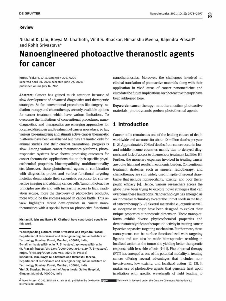
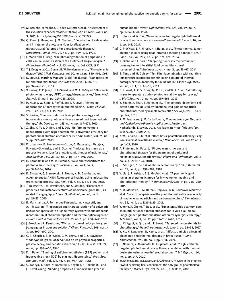
PDT is a procedure by which photosensitizing agent istargeted onto tumor cells which upon stimulation withvisible light of specific wavelength produces reactivesinglet oxygen species (Figure 1a) [30]. The procedure re-quires vigilant selection of photoactive agents capable oftumor localization and metabolic synthesis resulting inirremediable cytolysis of cancer cells [31]. Additionally,tumor vasculature adds to protumorigenic and immuno-suppressive microenvironment creating physical barrier toT cell infiltration favoring PDT [32]. The major challenge inthis therapy is the photobleaching of sensitizers. Therefore,loading of these photosensitizers in nanocarriers couldovercome this issue [33]. Commonly employed sensitizersbeing hydrophobic, use of nanocarriers has shown toimprove the bioavailability [29]. Mainly noble metals aresuitable for PDT owing to their absorption capacitycompared to that of photoabsorbing dyes [34].
2974 N.K. Jain et al.: Nanoengineered photoactive theranostic agents for cancer
2.2.1.1 Commonly used agents for PDTThe commonly used photosensitizers in PDTmainly belongto tetrapyrrole family. Among them the most used are theones found in nature; e.g.: phthalocyanines and porphy-rins [35]. Besides, many other compounds also serve asphotosensitizers (PS). The suitable commonly used dyesinclude phenothiazines, phenalenones, squaraines, andindocyanine green (ICG). ICG is negatively charged poly-methine dye which serves in eradicating multiple cancerson irradiationwith NIR laser at a wavelength range of 800–810 nm and has reduced toxicity to nonsubject host tissue[36, 37]. Studies suggest ICG successfully absorbs lightabove 800 nm, and the photochemical nature of the com-pound makes it a suitable candidate for PDT [29, 38, 39].However, ICG has certain reported drawbacks as well.Stability of ICG differs in different solvents when used forclinical applications, which could be accounted due to itsphysiochemical nature [40]. Presence of sulfonyl groupspromotes water solubility of ICG, while hydrophobicpolycyclic groups make it lipophilic [41]. While ICG810 isseen to aggregate or degrade in water which may furtherresult in selfquenching and fluorescent reduction, whilethe optical properties strictly depend on the dye
concentration [42]. Owing to the amphiphilic nature, ICGhas tendency to adsorb lipids [43]. Therefore, in certaincases ICGmay interact with normal tissues andmay lead tochaos during surgical intervention. Notably, ICG in contactwith living tissues bind and exhibits in vivo lipoproteindynamics. Therefore, it is hard to predict the materialmovement completely using ICG [44–46]. Studies havedescribed that ICG enabled deep light tissue permeabilityimprovising PDT [38, 39]. Apart natural products are alsouseful as PS [33]. Further, with advancing technologyinorganic nanoparticles has contributed enormouslywhich is described in the later part.
2.2.2 Photothermal therapy (PTT)
PTT aims to sentence cancer cell death via engineering heatexposure (electromagnetic radiation) to near infrared light(Figure 1b) [47, 48]. Instilling contrast agents or dyes withweaker emission converts photo energy to thermal energyand results in tumor cell necrosis or apoptosis [39].Recently, tremendous progress is witnessed in cancertherapy using PTT. NIR absorbents for efficient heat pro-duction and incorporating drug delivery system (DDS) for
Figure 1: Mechanism of action for photoactive nanotheranostic agent resulting in tumor regression.(a) Photodynamic therapy (PDT) involving generation of reactive oxygen species (ROS) resulting in cytolysis of tumor cells [78]. (b)Photothermal therapy (PTT) involving generation of heat upon irradiation resulting in ablation of tumor cells [79].
N.K. Jain et al.: Nanoengineered photoactive theranostic agents for cancer 2975
enhanced heat exposure to abnormal cells has helped PTTto attain success. The notable merit of DDS in PTT isimprovising the efficacy and ensuring safety to healthycells from photothermal damage. The only concern beforestarting the therapy is the optimization of fluence rate andirradiation time. Later, DDS NIR absorbent and simulta-neous PTT is performed irradiating tumor for specified timefollowed by absorbent administration [49]. However, hur-dles exist in determining tumor degree, size, and hetero-geneity for which maximum therapeutic effect is hard toexert. At 50 °C or above the risk of thermal damage inhealthy tissue raises concern as it might denature proteinand may likely cause healthy cell necrosis [50]. Generally,the temperature threshold for PTT therapy is chosen from40 to 60 °C above which will lead to cell death by coagu-lation necrosis [51, 52]. Above this temperature, it mayaffect the integrity of cell membrane and induce the releaseof cellular contents [51, 53, 54]. When a comparison of PTTis made over PDT, it could be concluded that PTT is morepreferred than PDT due to its longer wavelength of lightused and is well tolerated by normal cells; additionally theoxygen requirement for the procedure is very minimal [55].Well, the specificity of photosensitizers restricts extensionof PDT [56].
2.2.2.1 Commonly used agents for PTTPhotothermal agents are selected based on their size,shape, and ability to transfer energy from one form toanother [33]. Mostly used agents comes in the category ofinorganic (metallic, carbon nanostructures, and quantumdots) or organic–inorganic nanohybrids. Inorganic metal-based photoactive agents are selected due to the desiredshape and size favoring heat and optical properties [57].Whereas, various forms of carbon based nanostructuresdemonstrate ability to transfer energy from one form toanother upon light irradiation [58]. Importantly, quan-tum confinement make quantum dots suitable opticallyactive probes for theranostic applications [59]. Inorganicnanoparticles are not widely used because of the chal-lenges in biodegradability/clearance issue, bioavail-ability, and toxicity caused for long term [60]. However,hybrids derived from combination of organic and inor-ganic has demonstrated good biocompatibility, highrenal clearance, less toxicity, and high photothermalperformance.
2.2.2.2 Principle of photoactive agentsPhotoactive agent is the basic element for contrastingphototherapy and need to be selected judiciously. There-fore, these agents basically absorb light of particularwavelength, precisely in near infrared for improved light
penetration. The second criteria for selection; thematerialsshould not interact with cells and should be able to triggerimmune responses without external stimuli. Further, fortargeted therapies the agents should be capable ofclearing from blood and normal tissues for assuredphototherapy agent. To brief all these characteristicswith good photostability make the material apt forphotoactive therapies [60].
The principle involved in the generation of heat by thephotoactive materials upon irradiation with NIR light in-cludes absorption of photon resulting in excitement of elec-tron. The excited electron shifts from low excited singlet state(S0) to highly unstable excited singlet state (S1) that subse-quently comes back to stable S0 state with release of energyduring the relaxation process [61–65]. The energy releasedcan be in the form of fluorescence emission or nonradiativevibrational emission in the form of photothermal energy.Various inorganic structures made from gold, silver, ironoxide, graphene, carbon nanotubes, and upconversionnanoparticles have been explored for photothermal therapy[12, 13, 17, 65, 66], whereas, organic materials like dyes andpolymers are also used in photothermal based treatment ofcancer. All these nanostructures upon irradiation with NIRlight generate heat that ablates the cancer cells. Further,these materials due to their diverse physicochemical prop-erties also aid in themultimodal imaging of the tumor areaas well image guided tumor regression. The optical,electronic, and magnetic properties of these materialsplay a crucial role in contributing to this application[67, 68]. Such nanomaterials which are used for therapyas well as diagnosis of a disease are known as nano-theranostic agents [69]. The field of nanotheranostics israpidly emerging in cancer with different novel materialsbeing explored by various researchers cross the world.Their application in cancer has revolutionized thetreatment regime as they offer several advantages overthe conventional treatment like biocompatibility,biodegradability, minimal dose requirement with lessfrequency, and low systemic toxicity [70].
The most widely used light source for PTT is NIR due tohigh penetration and less toxicity. The first window of NIR(NIR-I, 650–950 nm) demonstrates lesser penetration whencompared to second NIR window (NIR-II, 1000–1700 nm)through biological tissue [68, 71]. Additionally, themaximalpermissible exposure (MPE) for skin is higher with NIR-II(1W/cm2 for 1064 nm) when compared to NIR-I (0.33W/cm2
for 808 nm) [72]. Hence, materials with photoexcitation inNIR-II windowhave recently attractedwide attention in PTTbased cancer nanomedicine. This includes inorganic aswellorganic nanomaterials that are tuned to exhibit distinctivephotoexcitation in the NIR window. These photoactive
2976 N.K. Jain et al.: Nanoengineered photoactive theranostic agents for cancer
carriers can be elements or derived as the products ofchemical bond with temperature sensitive material. Thegenerated heat from these photoactive materials not onlykills the malignant cells but also alters the enzyme activityand gene expression. Further, these NIR photoactivablenanosystems when used in combination with chemothera-peutic drugs, enzymes or gene exhibit synergistic effect withbetter therapeutic action [73]. Additionally, some of thesematerials exhibit fluorescence or contrast due to theirunique physicochemical properties that aids in imaging ofthe tumor tissue which are recognized as nanotheranosticagents. Recently, such photoactive nanotheranostic agentsare gaining attention and are actively reported for diverseapplications in cancer therapy [74].
2.2.3 Combinatorial agents for PDT and PTT
The efficacy could be pronounced with the use of combi-natorial therapy of PDT and PTT. The dual therapy isgenerally made use of two different phosphatidylserine
(PS), which further needs wavelength lasers for activationof two PS [75]. While with the advancement in studies it isrecognized that somePS is able to function both in PDT andPTT, e.g.; carbon dots [76]. Besides, ICG loaded liposomeswere also equipped in PDT and PTT assisted therapies [77].An overview of different photoactive agents used in cancerfor PDT, PTT and their combinatorial therapy has beenprovided in Table 1.
2.2.4 PDT versus PTT: curbs in promoting to clinics
PDT has been introduced to clinics in the last 40 years andholds propitious treatment options for cancers of head,neck, breast, bladder, bile duct, pancreas and other vitalorgans [103]. The therapy is affirmedwith the production ofreactive oxygen species (ROS). Irradiation triggers thephotosensitizer to absorb photons and to get excited tohigher electronic states. The singlet state photosensitizertries to achieve triplet state and further emits energy in theform of fluorescence, heat or light. In this state, the ROS isproduced via two mechanisms. The triplet molecular
Table : Different photoactive materials used in cancer.
Chemical family of photosensitizerused in PDT
Cancer type Wavelength (nm) References
Pyrolipid T murine breast tumor []Murine colorectal cancer (MC , CT ) [, ]
Porphyrin Skin, bladder, esophagus, breast, lung, and brain [–]Basal cell White light []
Chlorin Superficial, skin, breast, and colon
[–]
Nanomaterials for cancer treatmentvia PTTCarbon nanotubes RKO (rectal carcinoma cell line), HCT (human colon cancer cells) []AuNPs CC (colon carcinoma cells) []Graphene oxide NPs HCT (human colon cancer cells), human dermal fibroblast []PLGA-ICG-R T breast cancer cell line, CT colon rectal cancer cell line [, ]Gold nanostars MB bladder tumor []Combination therapies for cancerPTT + CTPolydopamine/rGo/MSN
HeLa cells (human cervix cancer cell line) LTH () + drugrelease
[]
PTT + CTCD AuNP
Breast cancer LTH () + drugrelease
[]
PTT + CTPolypyrrole/MIL/DOX
HeLa cells (human cervix cancer cell line) LTH () + drugrelease
[]
PTT + PDTTe ND
TBreast cancer cell line
LTH + ROS () []
GNcHyNA MDA-MB-Breast cancer cell line
LTH () + ROS()
[]
UCNP-NGO/ZnPC KB cells (human nasopharyngeal epidermal carcinoma), HeLa cells(human cervix cancer cell line)
LTH () + ROS()
[]
CP-TPP/Au/PEG nanospheres HeLa cells (human cervix cancer cell line) LTH () + ROS()
[]
N.K. Jain et al.: Nanoengineered photoactive theranostic agents for cancer 2977
oxygen in ground state stimulates to produce singlet oxy-gen species [104–106]. An ideal photosensitizer is equip-ped with ample biocompatibility and absorbs high lightlevels. PS can be classified into several categories likemetals, organic, inorganic, polymer based and others [107].Apart from PS selection, it is often a cumbersome task todecide drug to device match and the mode to which thelight needs to be applied, or in brief PDT dosimetry (irra-diation geometry with parameters which includes fluencerate, wavelength, and total fluence), light source andexposure, PS dose and intervals for drug to light adminis-tration is a topic which needs to be worked up diligently[108, 109].
PTT is a technique for treating tumors in local arena.Here, the tissue temperature is raised above 60 °C and re-sults in protein denaturation followed by plasma mem-brane destruction. The mechanism is that photosensitizerswhen activated by light of particular wavelength absorbsphotons and excites from singlet ground state to excitedsinglet state. A cascade of event takes place, the electronicexcitation energy undergoes vibrational relaxation, anonradiative decay and comes back to ground state whichis mediated by collisions from photothermal agents andother molecules. During this event, tissue temperature el-evates as a result of kinetic energy, were heat shock pro-teins and gene expression changes occurs to overcome thethermal damage [110]. Further, as the temperature goeshigher it causes microvascular thrombosis and ischemia[111]. Both PDT and PTT are superior compared to othertreatments due to selectivity of PS. In addition, the tech-nique provides less scar formation and can be utilized fortreating multiple times [112, 113].
2.2.5 Laser and suitability
Light absorption for PTT and PDT in visible light rangesfrom 400 to 700 nm while for near infra-red from 700 to
1350 nm. Skin tumors are treated using light emittingdiode (LED) with regulated exposure to sunlight [114].While, most of the lasers are available commercially(Table 2).
2.3 Role of nanoparticles in cancer cellscreening and therapy
Nanoparticles (NPs) have growing interest in cancer ther-apies. Certain nanoparticles are capable of absorbing lightand get heated up parallelly with their plasmon band. Theoptical transmission in tissue is optimal due to possiblemanipulation of plasmonic band in NPs from visible toinfrared favoring deep tissue treatment [121]. Therefore,NPs need to be well orchestrated for; heat generation afterabsorption of innocuous light in the near infrared range.The efficacy of PTT is largely dependent upon the accu-mulation of light responsive NPs, excitation time with po-wer density of light, and effectiveness of light energy toheat conversion [47].
Recent studies have demonstrated the use of nano-shells and nanocages of metallic and noble metals inorganic polymeric shells to combat cancer [49]. Studyconducted by Hirsch et al., revealed that gold–silicananoshell by thermal ablative therapy on tumor cellsdemonstrated effective outcomes in breast carcinoma cells.The studywas performed tailoring the nanoshells to absorbnear infrared where optical transmission was optimallycontrolled. Photothermal morbidity (with 820 nm, 35 W/cm2) was further confirmed with viable fluorescent stain-ing. Simultaneously the in vivo studies conducted sub-stantiated the irreversible thermal damage precisely to thetumor arena, using magnetic resonance imaging (MRI) forconcordant planning and temperature monitoring byphase sensitive gradient-echo MRI. Further, magneticresonance temperature imaging (MRTI) evidence wassupported with histopathological findings, proving thatnanoshells can be used as potential candidate for thermalablative tumor destruction [122]. Recent study conductedby Norton et al. on PTT effects of plasmonic metal nano-particles summarized that it is apt to consider NP whoseplasmon resonance is near to therapeutic window formaximum possible light exposure onto soft tissues.Though it is possible to tune the plasmon resonance ofnanoshells the absorption is narrow. Therefore,maximum efficacy is obtained when optical wavelengthgets along with each other. The study has given moreinsights to which the NP diffusion can be confined to thetumor region [123].
Table : List of commercially available lasers along with theirwavelength range [–].
Laser Wavelength range(nm)
Green laser
Alexandrite laser –Dye laser –Diode laser –Neodymium doped yttrium aluminium garnetlaser
LED (red light)
2978 N.K. Jain et al.: Nanoengineered photoactive theranostic agents for cancer
3 Nanotheranostic agents incancer
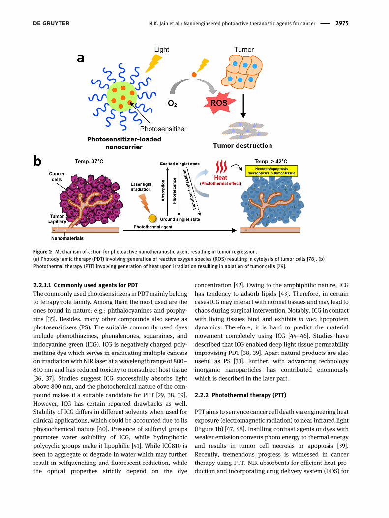
Conventional treatment of cancer involves chemotherapy,radiation, and surgery. Based on the stage of cancer andextent of tumor area, combination of these strategies is alsoused for better therapeutic action. The widely used cancerchemotherapy suffers from the drawbacks of non-specificity of drug action leading to poor drug concentra-tion levels in tumor area, toxicity of nontargeted tissue,and multiple dose requirements [124, 125]. However, theemergence of nanotechnology has added new dimensionto cancer treatment approach with minimal side effects.They exhibit unique physicochemical properties due totheir nanoscale dimension that plays a crucial role incancer nanomedicine. Various nanotheranostics agentsderived from inorganic as well as organic origin have beendesigned for cancer applications. However, photoactivematerials have gained much attention due to their prom-ising potential to overcome the drawbacks of conventionalchemotherapy. In addition, the delivery of therapeuticmolecules like drugs, enzymes or genes along with thesenanohybrids results in synergistic therapy at minimal dosewith reduced side effects. Different materials developed forPTT includes metallic nanoparticles (NPs) (gold NPs, silverNPs, and platinum NPs), carbon based NPs (carbon nano-tubes and nanodots, graphene based NPs, and Mxene),noncarbon based NPs (upconversion NPs and black
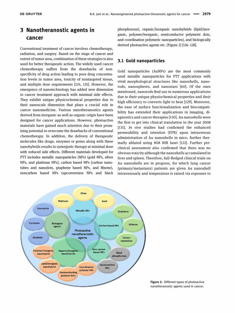
phosphorous), organic/inorganic nanohybrids (lipid/inor-ganic, polymer/inorganic, semiconductor polymeric dots,and coordination polymeric nanoparticles), and biologicallyderived photoactive agents etc. (Figure 2) [126–128].
3.1 Gold nanoparticles
Gold nanoparticles (AuNPs) are the most commonlyused metallic nanoparticles for PTT applications withvivid morphological structures like nanoshells, nano-rods, nanospheres, and nanostars [60]. Of the onesmentioned, nanorods find use in numerous applicationsdue to their unique physiochemical properties and theirhigh efficiency to converts light to heat [129]. Moreover,the ease of surface functionalization and biocompati-bility has extended their applications in imaging, di-agnostics and cancer therapies [130]. Au nanoshells werethe first to get into clinical translation in the year 2008[131]. In vivo studies had confirmed the enhancedpermeability and retention (EPR) upon intravenousadministration of Au nanoshells in mice, further ther-mally ablated using 808 NIR laser [122]. Further pre-clinical assessment also confirmed that there was noobvious toxicity although the nanoshells accumulated inliver and spleen. Therefore, full-fledged clinical trials onAu nanoshells are in progress, for which lung cancer(primary/metastasis) patients are given Au nanoshellintravenously and temperature is raised via exposure to
Figure 2: Different types of photoactivenanotheranostic agents used in cancer.
N.K. Jain et al.: Nanoengineered photoactive theranostic agents for cancer 2979
radiation through bronchoscopy. Investigations are stillon the adult stage for treatment tumors of head and neckusing Au nanoshells [47]. Mooney et al., were successfulin showing the distribution of Au nanorods throughoutthe tumors when transported by neural stem cells inbreast cancer xenografts in mice in vivo. In doing so, thegold nanorods were able to improve the tumor ablationwith tremendous reduction in tumor recurrence. Thework also compared the results from free Au nanorods.The authors concluded that a combination of nano-technology with cell therapy would benefit for cancercuration [132]. Vijayaraghavan et al. [133], showed thecomplete elimination of deep tumors by the combinato-rial treatment with gene silencing (ultralow NIR light)
and PDT employing Au nanoechinus both in in vivo andon HeLa cells.
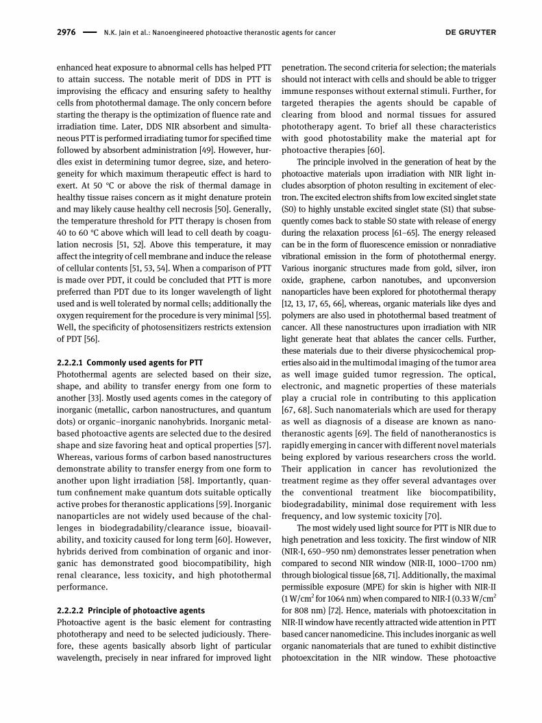
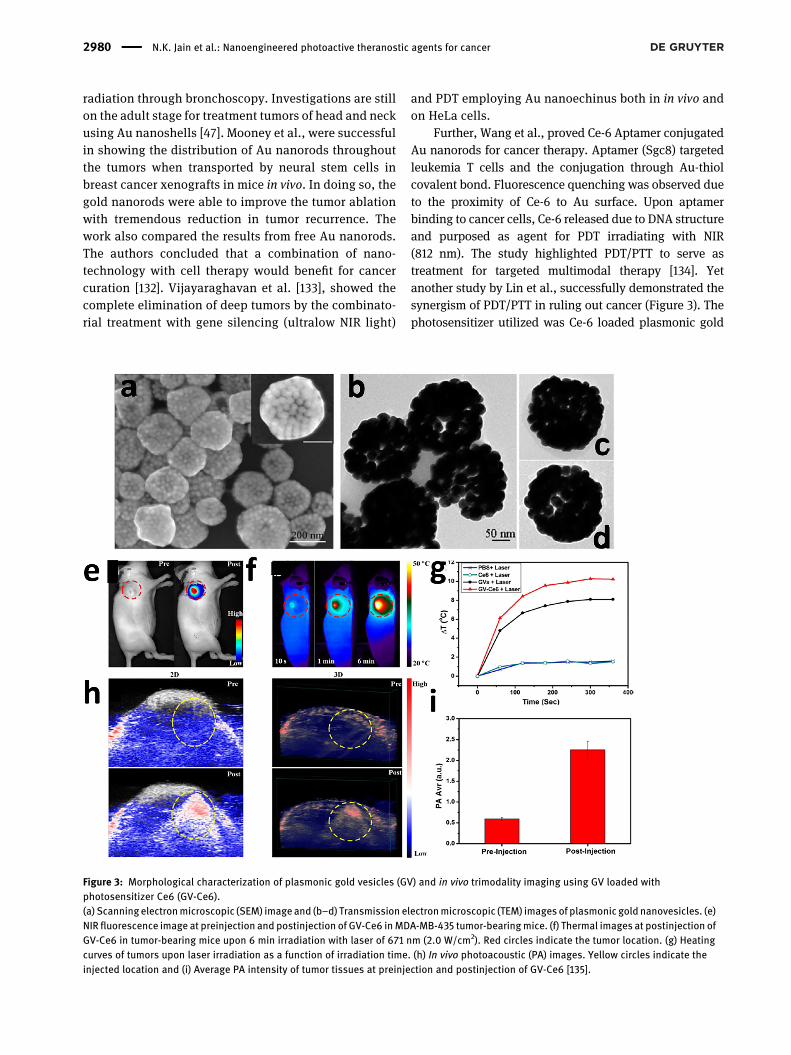
Further, Wang et al., proved Ce-6 Aptamer conjugatedAu nanorods for cancer therapy. Aptamer (Sgc8) targetedleukemia T cells and the conjugation through Au-thiolcovalent bond. Fluorescence quenching was observed dueto the proximity of Ce-6 to Au surface. Upon aptamerbinding to cancer cells, Ce-6 released due to DNA structureand purposed as agent for PDT irradiating with NIR(812 nm). The study highlighted PDT/PTT to serve astreatment for targeted multimodal therapy [134]. Yetanother study by Lin et al., successfully demonstrated thesynergism of PDT/PTT in ruling out cancer (Figure 3). Thephotosensitizer utilized was Ce-6 loaded plasmonic gold
Figure 3: Morphological characterization of plasmonic gold vesicles (GV) and in vivo trimodality imaging using GV loaded withphotosensitizer Ce6 (GV-Ce6).(a) Scanningelectronmicroscopic (SEM) imageand (b–d) Transmission electronmicroscopic (TEM) images of plasmonic gold nanovesicles. (e)NIR fluorescence image at preinjection and postinjection of GV-Ce6 inMDA-MB-435 tumor-bearingmice. (f) Thermal images at postinjection ofGV-Ce6 in tumor-bearing mice upon 6 min irradiation with laser of 671 nm (2.0 W/cm2). Red circles indicate the tumor location. (g) Heatingcurves of tumors upon laser irradiation as a function of irradiation time. (h) In vivo photoacoustic (PA) images. Yellow circles indicate theinjected location and (i) Average PA intensity of tumor tissues at preinjection and postinjection of GV-Ce6 [135].
2980 N.K. Jain et al.: Nanoengineered photoactive theranostic agents for cancer
vesicles. The technology used was trimodality assistedfluorescence/thermal/acoustic image guided combinationwith PDT/PTT. The study reported that the absorbance byvesicles in NIR region was strong, and the NIR irradiation(671 nm) was successful in exciting Ce-6 along with Auvesicles producing singlet oxygen species together withheat and eradicated cancer cells. When investigated invivo, the results were fruitful and were visualized viafluorescence/thermal/acoustic signals. There the studyclearly descries the importance of combinatorial approachwith PDT/PTT in cancer cell destruction [135].
3.2 Silver nanoparticles
Silver nanoparticles (Ag NPs) are popular due to theirantibacterial and tumor destructing properties. Besides,their plasmon tunability in the NIR biological windowrange (650–1200 nm) have paved for increased applica-tions of Ag in medical field [136]. More interesting is itsantitumor properties. However, Ag is shown to exhibitcancer killing properties only at higher concentrations[137]. Ag in photoactive therapy seems to aid as NPs foreffective drug loading or photosensitizers to improveantitumor therapy [138]. Ag NPs are suitable to load heatlabile or water soluble drugs because of the synthesismethod (reduction of Ag ions which is devoid of organicsolvent or heat) [34]. Studies conducted by Bose et al.,demonstrated folate receptor targeted plasmonic Ag NPsintended for breast cancer where, Ag NPs to purport asefficient nanocarrier system for delivery of quercetinthereby inducing PTT. The quercetin folate receptor AgNPswere synthesized by one pot method and later thehydrogen bond between stabilizer and reductant weretuned according to the need. The outcome of the study wasthat PTT induced Ag NPs complemented to the antitumorefficiency by hyperthermia induction resulting in selectivelysis of abnormal cells with a fate of apoptosis. Further,quercetin incorporated Ag NPs showed double antitumoreffect in lab and animal studies. The study successfullyproved that quercetin loaded Ag NPs are more effectivethan free quercetin and can be beneficial for breast cancers[139]. While recent study conducted by Park et al., tried todelve effectiveness and applications of indocyanine greenloaded Ag NPs. The study was successful in high loadingcapacity of indocyanine with good stability against lightinduceddegradation andhepatic clearance. The compositedelivery system addressed significant tumor accumulationcombining local irradiation of laser at tumor site resultingin tremendous inhibition of tumor growth. The author was
successful to prove that the nanocomposite system pur-poses to be a promising PTT agent for treating cancer [137].
3.3 Platinum nanoparticles
Platinum (Pt) is a metal that absorbs light in biologicalrange and generates DNA strand breaks [140]. Though PtNPs act as antioxidants at higher concentration, thetoxicity reported is also high in literature [141]. Whilecancer therapies have also evidenced synergistic effectwith the use of these NP’s further reducing the side effects,which is a factor of longer time period patient prognosis[142]. To date Pt NPs when used in reduced concentrationare apt in biological stability and tolerance [143]. Studyconducted by Depciuch et al., was on assessment of size ofPt NPs for PTT therapy. The study concluded that theantitumor activity of Pt NPs like DNA damage and enzymeactivity, apart the smaller size could serve as best PS agent[144]. Another study conducted by Song et al., demon-strated Au–Pt NPs uptake with cell targeting folic acid andtriphenylphosphine targeting mitochondria in tumor cells.Mitochondria targeting was achieved via loading PS (Ce6)onto Au–Pt NPs. The authors were successful in designingmultifunctional theranostic approach with ability totarget mitochondria using combinatorial PDT and PTT[145]. While the study conducted by Phan et al., revealedthat the prepared Iron–Platinum NPs (Fe–Pt NPs) withpolypyrrole coating with evidence of high biocompati-bility, NIR absorbance, and photothermal stability canexcellently serve as multifunctional system for photodiagnostic application like PDT and photo acoustic im-aging (PAI) [141].
3.4 Graphene
Graphene based materials are of major importance inbiomedical applications due to their uniqueness in prog-nosis, diagnosis, and therapeutic agents in cancer thera-pies [146]. This is due to their easiness in surfacemodification, for being rich in carboxyl and hydroxylgroups [147]. Graphene lays superior position in cancertreatment due to its toxic effects in tumor cells [148]. Hence,graphene is used actively in battling cancer in PTT, PDT,and imaging [149]. On the other hand, there needs an un-met research to address its biocompatibility and biode-gradability [150]. Wang et al., demonstrated in his studiesby covalent grafting of nanographene oxide as core shelland upconversion through bifunctional polyethylene gly-col, further, loading phthalocyanines on graphene oxide
N.K. Jain et al.: Nanoengineered photoactive theranostic agents for cancer 2981
surface. The study reports the greater efficacy of the pre-pared nanocomposite system with good biocompatibility.The study leaves a remark that the system can be a greatsuccess to be used as upconversion luminescence probe ofcells and to image whole body with greater contrast [101].Recently, a notable study conducted by Thapa et al.,revealed that graphene was suitable for prostate cancertreatment, where the authors intratumorally-injectedpalladium nanoparticle decorated with graphene oxide.The outcome was that the graphene oxide decoratednanoparticulate system showed enhanced local distribu-tion, photothermal ablation, and inhibited cancer cells inPC3 xenograft mouse models, with reduced organ toxicity[151]. Besides, these a study conducted in 2016 by Zhanget al., proved that BaGdF5 nanoparticleswere seemed to getattached intact on graphene oxide surface nanosheets toform the GO/BaGdF5/PEG nanocomposites. This nano-composite systemexhibited low toxicity, efficientmagneticcontrast. The author with evidence suggested that thesystem could serve as dual imaging (MR and X-ray) modelsfor in vivo tumor models [152]. Study conducted by Nur-unnabi et al. [153], was yet a further confirmation thatgraphene nanoparticles being photoluminescent wassuitable candidate for PTT and imaging. Notably Nafiujja-man et al. [154], through his work elucidated the use ofgraphene quantum dots in targeted cancer therapy (PDT)and imaging. In short, graphene will be a promising com-pound in future for treating and imaging cancer, except thechallenges faced for its biocompatibility.
3.5 Carbon nanostructures
Carbon dots are basically diamond or graphitic core, whichis sp2 or sp3 hybridized. Low dimensional carbon is clas-sified as carbon nanoparticle, carbon nanodot or carbonquantum dot, and fullerenes [155–157]. The ease of surfacefunctionalization of carbon dots with amine (NH2), car-boxylic acid (–COOH–), alcohol (OH), aldehyde (CHO) andproduction tuning their size together with the ability todeliver photoluminescence has made them popular incancer therapeutics and imaging [158]. Besides, they havegainedmuch attention in biomedical applications owing totheir optical absorption, photostability, high penetration,good electrical conductivity, biocompatibility, highchemical stability, and low toxicity [159, 160]. Study con-ducted by Serda et al., in 2018 has affirmed the use offullerene in PDT applications. The authors have developedglycoconjugated C60 derivative for adenocarcinoma. As aresult, it was seen that fullerenes completely accumulatedin nucleus of stellate pancreatic cells, which is not toxic
upto 1 mg/mL, and was shown to exhibit photoactiveability upon green, blue light activation [161]. Zheng et al.,in his studies a solution for hypoxia condition caused insolid tumors. He designed a multifunctional nano-composite comprising of carbon dots decorated with car-bon nitride NPs against hypoxic tumor water splitting. Theresults were so promising that improved intracellular ox-ygen concentration with reactive oxygen species underhypoxia and normoxia with light irradiation. While in vivostudies confirm that the multifunctional nanosystem sur-passed tumor hypoxic condition. The author suggests us-ing water-splitting materials which has the capability ofenhancing oxygen level and reversing hypoxia inducedPDT resistance and metastasis [162]. A recent study bySundaram et al., in 2020, was successful in coating hyal-uronic acid and Ce6 onto carbon nanotubes which pur-ported as photosensitizer. This further on when tested onCaCo-2 colorectal cancer cells via PDT approach (660 nm)inhibited apoptotic cell death, the study confers thenanocomposite system to be an efficient vehicle forphotosensitizer localization in Colorectal cancer cells [163].
3.6 Mxene
MXenes are two-dimensional (2D) novel structures derivedfrom transition metal carbides and nitrides [164]. They arecharacterized by the presence of layered arrays of transi-tion metal atoms that are interconnected by a carbon ornitrogen atom. The general formula for MXenes isMn + 1XnTx (n = 1–3), where M stands for early transitionmetal carbides (Ti, Hf, Zr, Nb, Ta, V, Mo Sc etc.), X denotescarbon or nitrogen and Tx represent functional groups(–Cl, –F, –OH, and O) terminating the M surface [165–167].They have attracted increased attention as they exhibitexcellent electrical, optical, magnetic, and mechanicalproperties [168, 169]. These properties can be tuned bymodulating the transition metal atom and surface func-tional groups. Further, they demonstrate thermal stabilityand extreme biocompatibility that makes them suitable forcatalysis, energy storage and biomedical applications [164,166, 170, 171]. They have structural similarity with gra-phene and their morphology as well as size can be alteredas per the application requirement. Recently, MXenes haveemerged as a promising photothermal agent for cancertherapy due to their high surface area, hydrophilic nature,broad absorption band in UV/NIR region and high photo-thermal conversion efficiency (PCE) [172, 173]. The hydro-philic nature of MXenes can attributed to the presence ofhydroxyl (OH), oxygen (O), and fluorine (F). Additionally,their surface can be functionalized to achieve active
2982 N.K. Jain et al.: Nanoengineered photoactive theranostic agents for cancer
targeting as well as combinatorial therapy. Moreover, theplanar structure of MXenes can acts as a carrier for cargodelivery [172]. Highly photostable MXene QDs with goodquantum yield and tunable wavelength demonstratingluminescent properties have been studied extensively forPTT. Therefore, they can be used for imaging guided syn-ergistic PTT integrated with chemotherapy/photodynamictherapy in cancer. The potential of MXene as theranosticsagents in cancer has been explored by researchers acrossthe globe.
For instance, the surface of Ti3C2 MXenes were beencoated with superparamagnetic iron oxide nanoparticles(IONPs) for cancer theranostic applications [174]. Theformed composite of Ti3C2-IONPs demonstrated goodbiocompatibility and high PCE of 48.6% revealed throughsystemic in vitro and in vivo studies. Further, the largesurface area provided by the nanosheets of MXene acts as acarrier for loading of therapeutic molecules or nano-particles. Herein, the functionalization of MXene withsuperparamagnetic IONPs resulted in imaging-guided PTTagainst cancer through contrast-enhanced T2-weightedMRI and efficient photothermal ablation of cancer cells.Hence, such unique design of safe and efficient photo-thermal based composite system with high therapeuticefficacy holds promising potential for clinical translation.
Additionally, recent research has been widely focusedon photoactive active materials that are triggered in NIR-II(1000–1350 nm) biowindow as it shows high tissue pene-tration with less adverse effects. Lin et al. reported thedesign of biodegradable Nb2C nanosheets (150 nm) thatdemonstrated absorption in both NIR-I (808 nm, PCE36.4%) andNIR-II (1064 nm, PCE 45.65%) biowindow [175].Modification of these Nb2C nanosheets with poly-vinylpyrrolidone (PVP) resulted in composite (Nb2C-PVP)with increased biocompatibility and less toxicity. Resultsof both in vitro as well as in vivo experiments confirmed thenon-toxic nature and excellent photothermal performanceof composite in the NIR-I and NIR-II biological windows.Effective photothermal conversion with considerable tu-mor regression composite in the NIR-I andNIR-II biologicalwindows was seen in vivo and such systems can be highlybeneficial for deep tissue PTT applications.
3.7 Upconversion nanomaterials
Upconversion nanomaterials abbreviated as UCNPs makesit unique due to the ability of generating shorter wave-length emissions in longer wavelength excitations [176].This requires two ormore than two low energyNIR photonsfor high-energy photon generation that ranges from NIR to
ultraviolet/visible. Large stokes shift, photo-blinking andstable energy levels in micro/milli seconds makes UCNPsoutstanding compared to other techniques [177]. Besides,minimal scattering and absorption levels improved pene-tration depth favors UCNP for in vivo biological applica-tions [178, 179]. The host materials selected for UCNPshould preferably be of low lattice energy, to lessen non-radiative loss and deliver maximum radiative emission.Nonradiative energy loss needs the presence of phonons inhost lattice [180]. UCNP when excited to long wavelengths(e.g.: 980 or 808 nm) the upconversion is to shorterwavelength ranging from deep ultraviolet to near infrared.In turnNIR light excitation results in lowautofluorescence,reducing photo damage and enhancing the penetrationdepth, and benefitting cell label and imaging in live or-ganisms [181, 182].
Upconversion mechanisms are mainly classified intofive types namely; energy transfer, photon avalanche,migration mediated energy upconversion, excited stateconversion, and cooperative energy transfer upconversion.Although there is no gold standard procedure for upcon-version mechanisms these are purely based on host matrixtype and concentration of doped activator [181, 183].Lanthanide doped UCNPs will be able to get excited withnegligible background providing future possibilities in thefield of bioimaging. Surface modification of UCNP opens agood possibility for better biocompatibility (silica coating).In addition, efforts to remove the capping ligand on hy-drophobic UCNPswere also successful [184]. ThoughUCNPhas high possibility to excel in bio-applications, immenseeffort is needed to explore and tune the biological andphysiochemical properties especially in cellular applica-tions [121, 177, 185].
3.8 Black phosphorous quantum dots(BPQDs)
Black phosphorous (BP) is two-dimensional (2D) materialwith high surface area and serves as a carrier for drugloading [186, 187]. It also acts as a photosensitizer due to itselectronic properties and generates singlet oxygen for PDT[188, 189]. The bulk properties of BP widely differ whenthey are brought down to single layered structure. Addi-tionally, the nanoparticles (NPs) and quantum dots (QDs)of BP exhibits wide absorption spectrum that can be usedfor the near infrared (NIR) light triggered PTT [190]. Hence,BP shows huge potential in the field of biomedicine andphoto-electronics due to its diverse properties [186, 190–192]. Black phosphorous quantum dots (BPQDs) arenonmetallic, optically active and semiconductor based
N.K. Jain et al.: Nanoengineered photoactive theranostic agents for cancer 2983
material with tunable band gap. They demonstrate absor-bance in the NIR region of spectrum. They exhibit diag-nostic as well as therapeutic properties for cancerapplications. Their surface can be functionalized and canserve as carrier for chemotherapeutic drug. Hence, theycan be used to achieve image guided synergistic photo-dynamic–photothermal–chemotherapy [193–196]. Func-tionalization of these systemswith targetingmoieties helpsin selective localization in tumor area resulting in precisekilling of the cancer cells. Moreover, the BPQDs degrade inaqueous medium yielding nontoxic and biocompatiblephosphate and phosphonate [197, 198].
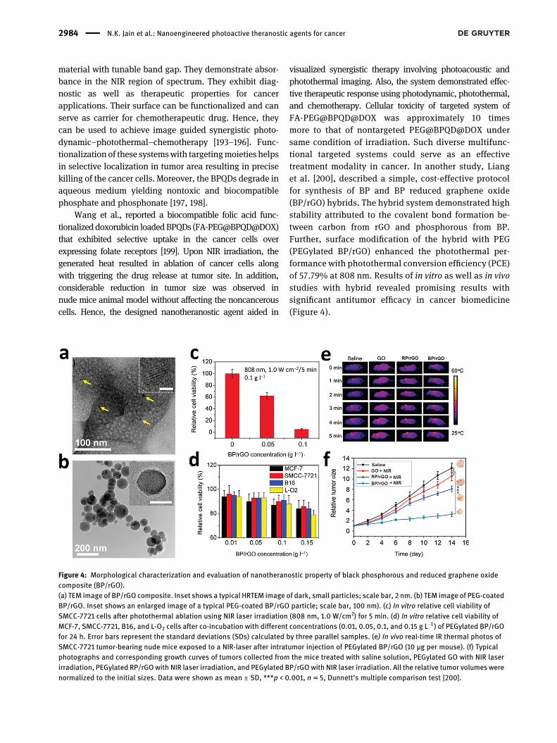
Wang et al., reported a biocompatible folic acid func-tionalizeddoxorubicin loadedBPQDs (FA-PEG@BPQD@DOX)that exhibited selective uptake in the cancer cells overexpressing folate receptors [199]. Upon NIR irradiation, thegenerated heat resulted in ablation of cancer cells alongwith triggering the drug release at tumor site. In addition,considerable reduction in tumor size was observed innude mice animal model without affecting the noncancerouscells. Hence, the designed nanotheranostic agent aided in
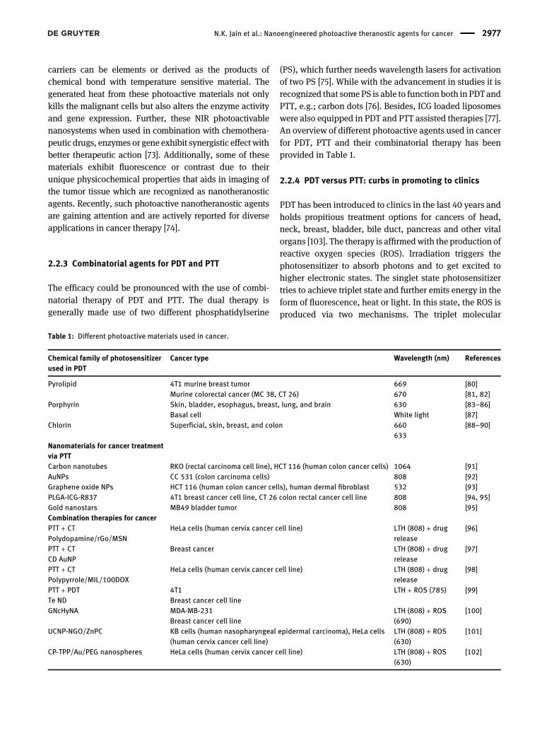
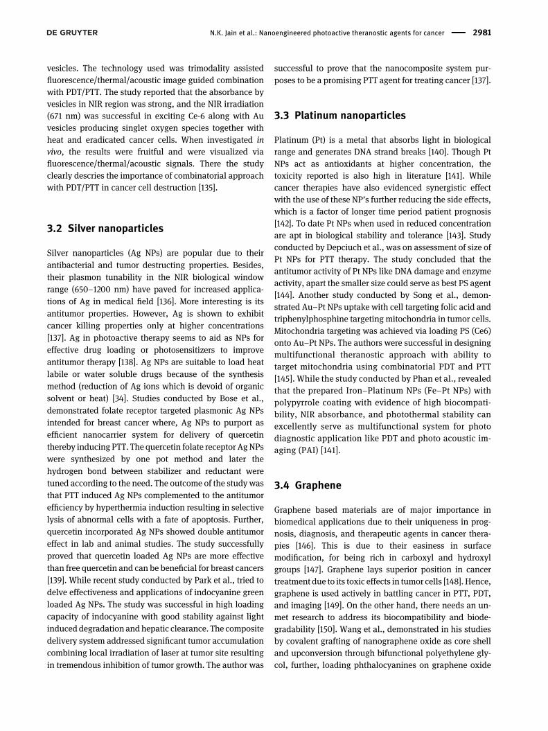
visualized synergistic therapy involving photoacoustic andphotothermal imaging. Also, the system demonstrated effec-tive therapeutic response using photodynamic, photothermal,and chemotherapy. Cellular toxicity of targeted system ofFA-PEG@BPQD@DOX was approximately 10 timesmore to that of nontargeted PEG@BPQD@DOX undersame condition of irradiation. Such diverse multifunc-tional targeted systems could serve as an effectivetreatment modality in cancer. In another study, Lianget al. [200], described a simple, cost-effective protocolfor synthesis of BP and BP reduced graphene oxide(BP/rGO) hybrids. The hybrid system demonstrated highstability attributed to the covalent bond formation be-tween carbon from rGO and phosphorous from BP.Further, surface modification of the hybrid with PEG(PEGylated BP/rGO) enhanced the photothermal per-formance with photothermal conversion efficiency (PCE)of 57.79% at 808 nm. Results of in vitro as well as in vivostudies with hybrid revealed promising results withsignificant antitumor efficacy in cancer biomedicine(Figure 4).
Figure 4: Morphological characterization and evaluation of nanotheranostic property of black phosphorous and reduced graphene oxidecomposite (BP/rGO).(a) TEM image of BP/rGO composite. Inset shows a typical HRTEM image of dark, small particles; scale bar, 2 nm. (b) TEM image of PEG-coatedBP/rGO. Inset shows an enlarged image of a typical PEG-coated BP/rGO particle; scale bar, 100 nm). (c) In vitro relative cell viability ofSMCC-7721 cells after photothermal ablation using NIR laser irradiation (808 nm, 1.0 W/cm2) for 5 min. (d) In vitro relative cell viability ofMCF-7, SMCC-7721, B16, and L-O2 cells after co-incubation with different concentrations (0.01, 0.05, 0.1, and 0.15 g L−1 ) of PEGylated BP/rGOfor 24 h. Error bars represent the standard deviations (SDs) calculated by three parallel samples. (e) In vivo real-time IR thermal photos ofSMCC-7721 tumor-bearing nude mice exposed to a NIR-laser after intratumor injection of PEGylated BP/rGO (10 μg per mouse). (f) Typicalphotographs and corresponding growth curves of tumors collected from the mice treated with saline solution, PEGylated GO with NIR laserirradiation, PEGylated RP/rGOwith NIR laser irradiation, and PEGylated BP/rGO with NIR laser irradiation. All the relative tumor volumes werenormalized to the initial sizes. Data were shown as mean ± SD, ***p < 0.001, n = 5, Dunnett’s multiple comparison test [200].
2984 N.K. Jain et al.: Nanoengineered photoactive theranostic agents for cancer
3.9 Organic/inorganic nanohybrids
Recently, organic/inorganic nanohybrids have attractedwide attention due to their desirable properties that extendtheir applications in cancer nanomedicine. They demon-strate the properties of inorganic materials (such as elec-trical, optical, and magnetic properties) and at the sametime, the organic component helps to improve thebiocompatibility, biodegradability, and clearance[201, 202]. Organic materials like polymer, lipids etc., areused to design versatile nanohybrid for biomedical appli-cations. The functional groups present in the organiccomponent also help to functionalize the nanohybrid sys-tem with targeting molecules resulting in site-specificlocalization of nanohybrid. Such nanohybrids can beextensively used for less-invasive imaging as well asimaging-guided PTT.
3.9.1 Polymer/inorganic
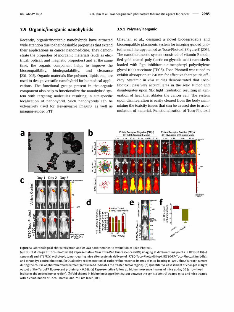
Chauhan et al., designed a novel biodegradable andbiocompatible plasmonic system for imaging guided pho-tothermal therapy named as Toco-Photoxil (Figure 5) [203].The nanotheranostic system consisted of vitamin E modi-fied gold-coated poly (lactic-co-glycolic acid) nanoshellsloaded with Pgp inhibitor D-α-tocopheryl polyethyleneglycol 1000 succinate (TPGS). Toco-Photoxil was tuned toexhibit absorption at 750 nm for effective therapeutic effi-cacy. Systemic in vivo studies demonstrated that Toco-Photoxil passively accumulates in the solid tumor anddisintegrates upon NIR light irradiation resulting in gen-eration of heat that ablates the cancer cell. The systemupon disintegration is easily cleared from the body mini-mizing the toxicity issues that can be caused due to accu-mulation of material. Functionalization of Toco-Photoxil
Figure 5: Morphological characterization and in vivo nanotheranostic evaluation of Toco-Photoxil.(a) FEG-TEM image of Toco-Photoxil. (b) Representative Near Infra-Red Fluorescence (NIRF) imaging at different time points in HT1080 FR(−)xenograft and 4T1 FR(+) orthotopic tumor-bearing mice after systemic delivery of IR780-Toco-Photoxil (top), IR780-FA-Toco-Photoxil (middle),and IR780 dye control (bottom). (c) Qualitative representation of TurboFP fluorescence images of mice bearing HT1080-fluc2-turboFP tumorsduring the course of photothermal treatment (arrow head indicates the treated tumor region). (d) Quantitative assessment of changes in lightoutput of the TurboFP fluorescent protein (p < 0.01). (e) Representative follow up bioluminescence images of mice at day 10 (arrow headindicates the treated tumor region). (f) Fold change in bioluminescence light output between the vehicle control treatedmice andmice treatedwith a combination of Toco-Photoxil and 750 nm laser [203].
N.K. Jain et al.: Nanoengineered photoactive theranostic agents for cancer 2985
with folic acid (FA) and IR780 reduced the stability leadingto aggregation and decreased the photothermal trans-duction potential due to disturbance in plasmon reso-nance. Computed tomography (CT) imaging studies withToco-Photoxil revealed comparable contrast to that ofiodine based contrast agent Omnipaque at five times lesserconcentration due to high X-ray attenuation power. Hence,Toco-Photoxil with high photothermal conversion andlocalized accumulation can serve as an effective and safematerial that can be clinically translated for cancer nano-theranostic applications in future.
Chauhan et al. [204], has also reported a facile andgreen synthesis of gold deposited zein nanoshells (AuZNS)for image guided plasmonic photothermal therapy. Thezein nanoparticles were surface functionalized withcationic glycol chitosan that helps in stabilizing the systemand ex-situ coating of gold was done over the zein nano-particles. The designed system demonstrated highbiocompatibility and effectively killed cancer cells underNIR light (808 nm) irradiation. Further, it also assisted indiagnosis of tumor through CT imaging and hence thesystem functions as a nanotheranostic agent in cancer.
Similarly, they have also reported NIR light-triggeredthermoresponsive nanoshell for plasmonic PTT basedcancer theranostic application [205]. The hybrid nano-shell (Au PNVCL NS) was formed by ascorbic acid-drivenin situ gold coating over thermoresponsive chitosan-grafted poly(N-vinyl caprolactam) nanoparticles. Theplasmonic absorption peak was tuned in NIR region(750 nm) for its application in PTT and loading of drug inthe polymeric core results in controlled release due tohyperthermia triggered shrinkage of polymer. Grafting ofchitosan was found to increase the biocompatibility,biodegradability, and elevates the lower critical solutiontemperature (LCST) to desired value. Au PNVCL NSdemonstrated superior contrast over Omnipaque in X-rayimaging. In vitro studies in mouse normal fibroblast L929cells confirmed the biocompatible nature of the nano-hybrid and studies in breast cancer cells MCF-7 revealedthe therapeutic potential of Au PNVCL NS. Based on theresults, Au PNVCL NS could be considered as a promisingmultifunctional theranostic agent for image-guided PTTand can be explored for in future for combinatorialchemo-photothermal therapy.
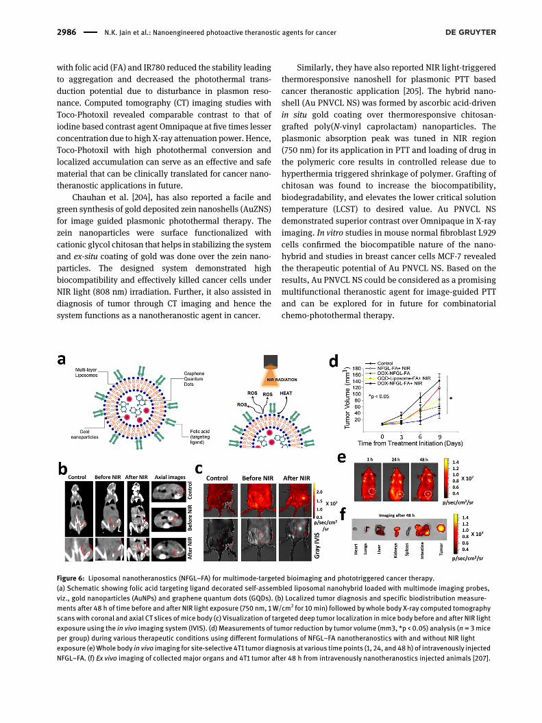
Figure 6: Liposomal nanotheranostics (NFGL–FA) for multimode-targeted bioimaging and phototriggered cancer therapy.(a) Schematic showing folic acid targeting ligand decorated self-assembled liposomal nanohybrid loaded with multimode imaging probes,viz., gold nanoparticles (AuNPs) and graphene quantum dots (GQDs). (b) Localized tumor diagnosis and specific biodistribution measure-ments after 48 h of time before and after NIR light exposure (750 nm, 1 W/cm2 for 10min) followed by whole body X-ray computed tomographyscanswith coronal and axial CT slices of mice body (c) Visualization of targeted deep tumor localization inmice body before and after NIR lightexposure using the in vivo imaging system (IVIS). (d) Measurements of tumor reduction by tumor volume (mm3, *p < 0.05) analysis (n = 3miceper group) during various therapeutic conditions using different formulations of NFGL–FA nanotheranostics with and without NIR lightexposure (e)Whole body in vivo imaging for site-selective 4T1 tumor diagnosis at various time points (1, 24, and 48 h) of intravenously injectedNFGL–FA. (f) Ex vivo imaging of collected major organs and 4T1 tumor after 48 h from intravenously nanotheranostics injected animals [207].
2986 N.K. Jain et al.: Nanoengineered photoactive theranostic agents for cancer
3.9.2 Lipid/inorganic
Rengan et al. [206], has designed multifunctional andbiodegradable gold coated thermosensitive liposomes formultimodal imaging and photothermal therapy. In situreduction of chloro-auric acid was used to coat gold overthe liposomal surface. Upon NIR laser irradiation, thesesystems degrade into small sized gold nanoparticles thatcan under renal clearance easily. Also, the nanohybriddemonstrated excellent biocompatibility and high thera-peutic efficacy by photo ablation of cancer cells. Besides,they were useful in imaging establishing their potential asa promising nanotheranostic agent for cancer.
Prasad et al. [207], have developed a multifunctionalliposomal based nanotheranostic agent for phototriggeredchemotherapy. Herein, the liposomal systemwas used as acarrier to encapsulate doxorubicin (DOX) drug, goldnanoparticles (AuNPs), and emissive graphene quantumdots (GQDs) which was subsequently functionalized withfolic acid as targeting ligand (NFGL-FA). The designedsystem presented imaging bimodality in vivo due to thehigh contrast and emissive nature of encapsulated agentsaiding in diagnosis of tumor (Figure 6). Further, in vivostudies under NIR light (750 nm) irradiation revealed tumorregression due to generated heat and reactive oxygenspecies (ROS) production. Additionally, synergistic effectin tumor regression was noticed with combined chemo-phototherapy when compared to stand-alone therapies(chemotherapy and PTT). Such biodegradable andbiocompatible multifunctional-targeted nanotheranosticsystem results in selective uptake with high therapeuticefficacy can serve as potential platform for image guidedsynergistic treatment approach by PPT.
3.10 Biological agents for photoactivity
Photosensitizers in nature’s list are quite few and is inter-esting due to their lower toxicity profile in normal cells andthe toxic effects shown towards abnormal cells [208, 209].Besides, the biological compatibility also makes phyto-compounds apt as photosensitizers (absorption maxima400–700 nm) [210]. Discovery of natural photoactiveagents should be supported, as it can contribute photo-sensitizers of minimal toxicity and adverse effects thansynthetic agents. The clinically approved PS of biologicalorigin is Foscan, Levulan, and Photofrin [211, 212].Describing here are some common phytocompoundscapable of producing photoactivity.
3.10.1 Alkaloids
Alkaloids are nitrogen group containing heterocycliccompounds [213]. Few compounds are shown to exhibitphotoactivity among the alkaloid group. Harmine andBerberine are the ones used in PDT till date [214, 215].Harmine’s activity was confirmed upon longer irradiationto ultra violet rays. While, Berberine is extensively usedand proven photosensitizer in PDT [216].
3.10.2 Thiophenes and polyacetylene
The compounds are said to get excited at 314–350 nm(nearing the biological absorption) and is capable of pro-ducing photoactive effects [217]. Basically, these are un-saturated carbon molecules containing thiophene groups[218]. The produced UV irradiation by these compounds iscapable of producing free radicals and has ability to induceapoptosis [212].
3.10.3 Curcumin
Curcumins are one of the most important compounds usedin cancer treatment. The absorption peak of curcumin is at300–500 nm favoring phototoxic reaction at reducedtemperatures and therefore can be used as good phytophotosensitizer [219]. Notable, studies havewidely used forcancer studies due to its immense ability in producingreactive oxygen species [220]. Study performed by Wuet al., confirmed the photoactivity on human epidermalA431 carcinoma cell. The study also revealed that the moreconcentration of curcumin used the better is the irradiationpermeation [221]. Considering these studies with no doubtcould be concluded that curcumin can beused as one of thebest photosensitizer, and could reap tremendous resultswith PDT [222].
3.10.4 Coumarins
Coumarins are present mainly in higher plant species [223].Photoactivity was evidenced in furanocoumarins whenKim et al., studied the reaction of Grapefruit extract fur-anocoumarins in eradicating multiple myeloma. Thecompound successfully inhibited STAT 3 signalingpathway further resulting in chemo sensitization andapoptosis [224].
Among the phyto derivatives, anthraquinone are alsofit as photoactive agents. Among this class, soranjidiol andrubradin are the most used in studies [225]. Research is on
N.K. Jain et al.: Nanoengineered photoactive theranostic agents for cancer 2987
its way to explore more anthraquinones suitable to use asphotoactive agents [226].
4 Emerging ablation modality
The combination of chemo-photo therapies is yet anotheroption, which can result in synergistic anticancer effects.Photothermal agents upon NIR irradiation aids in killingtumor cells and the heat produced can act as stimuli forrelease of drug in systematic manner [227]. Optimization ofchemotherapeutic system could help eradicate tumorswith the support of photothermal therapy [228]. Combinedtreatment methods could to a vast extend improvisemultidrug resistance and minimize metastasis of the lungs[104]. Zhang et al. demonstrated mesoporous silica deco-rated AuNPs to support light controlled drug release withlow intensity NIR radiation chemo-phototherapy foreffective destruction of cancer cells [229]. Zheng et al.Through his work has showcased the efficacy of smallmolecular NIR dyes via chemo-phototherapy. The authorsuccessfully incorporated chemotherapeutic drug and NIRdye into nanoparticle by physical interactions. Indoc-yanine green being the stereotypical NIR dye was coen-capsulated with doxorubicin in poly(lactic-co-glycolic acidlecithin poly ethylene glycol(PLGA lecithin PEG NP), uponhike in localized temperature than free ICG, facilitatedcellular uptake and drug release [230]. Interestingly,chemo-phototherapy hampered tumor cells and furtherprevented its reoccurrence. Similar way attempts weremade to coencapsulate drugs with NIR dyes in protein,polymeric, metallic, nanogels, and liposomes whichlanded up in remarkable results [231–233]. Thoughcombinatorial therapy holds promise in future oncoclinics, premature leak of drugs and NIR dyes from NPswhich could worsen tumor accumulation should be offoremost concern. Covalent conjugation of drugs/dyes toNPs, or surface tailoring can be of solution to a certainextend [233]. Scientists should be vigilant in choosingphotothermal agents and NIR dyes for exhibiting goodconversion efficiency. Further attention should be given indesigning newer nanocarriers with optimized photo-thermal conversion efficiency and minimal cytotoxicity.Also, the use of FDA approved biomaterials is highly rec-ommended for better biocompatibility and degradation.All the aforementioned points taken into consideration;nanomedicine can create transformation in oncologyclinics.
5 Challenges and possiblesolutions
One of the main challenges faced in photoactive therapy isthe thermal damage caused to normal tissues. Scientistsare trying their best to eliminate this issue and have comeup with more specifically targeted use of photosensitizers.Photoactive materials being dependent on enhancedpermeability and retention effect, is largely dependent onparticle size that has the ability to penetrate into tumortissues compared to normal cells. Application of low tem-perature PTT will be the other solution in surpassing theproblem. The other limitation of PTT being unevendispersion of photosensitizers and is solved by designingmesoporous nanoenzymes by aggregation effect. In addi-tion, the issues associated with ROS were proposed viatargetingmitochondria and interveningwith its function toattain tumor death [234]. In doing so there will betremendous reduction in heat resistance of abnormal cellscaused due to inhibition of heat shock proteins, while thelow temperature will not hamper normal cell physiology.Yet another approach put forward by Du et al. [77], loadingof tumor targeting ligands onto NPs with the aim of NPsbinding the tumor cells. While Quian et al., tailored Lyp-1ligand on NPs with intention to target p32 (overexpressedin breast cancer). The authors were successful in vitro andin vivo in producing anti-tumor effect in mouse withcombinatorial PDT/PTT. To brief, targeting photoactivematerials can be achieved by tailoring size, properties onNPs and enhancing surface ligands associated with NPs[235, 236].
The next challenge faced is the use of single photo-active therapy. Often the tumor site receives inadequatelight penetration leading to recurrence of tumor. This canonly be corrected with combinatorial therapeutic ap-proaches, like PTT with chemotherapy, PDT with PTT, oractivating body’s antitumor immune response. Lam et al.,through self-assembled approach synthesized doxorubicinloaded nanoparticulate system delivering photothermalconversion of high efficiency. Nanoparticulate system incombination with PTT-chemo showed good therapeuticefficacy in mouse breast cancer model (xeno-transplantation model) treated xenotransplantation can-cer. The model was also successful in activating immuneactivity against tumor [237].
Although PDT under preclinical settings is appealing,whenpracticed clinically faces somedrawbacks,which is oneof the unmet needs to be sought out quickly. These include
2988 N.K. Jain et al.: Nanoengineered photoactive theranostic agents for cancer

(1) The observed side effects for PDT till date is increasedrisk of dermal toxicity on exposure to sun, which couldbe accounted due to the complicated composition andthe limited absorption to red light [103, 238]. Generally,NIR-II ranging from 1000 to 1700 nm is recommendedas light source than the ones with shorter wavelengthand the visible light [22].
(2) The abnormal narrowing of canal or duct was reportedusing PDT, upon higher dose and light applicationbeyond a certain limit. Due to this, the PS accumulatedin organs may likely result in harmful side effects tounintended incidental damage [103, 239].
(3) This technology seems inept for deeply rooted tumorsowing to their poor specificity. Effective penetrationdepth recommended is not more than 1 cm for the lightsource used. In certain cases (oral and topical skincancer) optical fibers can be of help to achieve bettertherapeutic outcomes [239].
(4) Besides, the advent of endoscopic techniques, ablationwith radiofrequency has gained more efficacy overPDT, which can be advocated due to lack of lightreaching the tumor site [240]. This condition could beaddressed by designing a site-specific targetednanoparticle.
(5) Also, another drawback includes PDT’s ineffectivenessin hypoxic tumors as it demands oxygen for therapy. Inother terms, molecular oxygen consumption by PSleads to resistance of PDT under hypoxic environment[241]. Hypoxic condition in PDT could be bridled only ifnewer paradigm like designing oxygen vesicles, hyp-oxia activated drugs/prodrugs, water splitting nano-composites, self-oxygen supplementation (to use redblood cells, hemoglobin as oxygen carrier) modifica-tion of tumor environment, hypoxia dependent orfractional PDT are brought in as a modification inexisting therapeutic regimen [242–244].
PTT/PDT therapy was successfully demonstrated usingcationic polyethylenimine to electrostatically adsorbedICG to Prussian blue NPs. ICG loading with NP helpedimproving systemic circulation time. Solid tumor in nudemice was efficiently inhibited using PDT/PTT. The workwas proceeded by Dong et al., connected monoaminesubstituted porphyrin photosensitizer to covalent frame-work, further, loaded naphthalocyanine for light stability.The outcome of PDT/PTT was in lysis of lysosomes andmitochondria demonstrating synergistic effect using PDT/PTT [245, 246].
Nevertheless, PDT/PTT based nanomedicine whencombined could revolutionize the field of cancer treatmentusing tumor specific photosensitizers via localized/
targeted activation. In addition, it also results in minimaladverse effects, which could also be preferred forcombining chemo or immunotherapies. Also, as a part ofmultifaceted nanomedicine, abetment of newer immuneadjuncts could be of patient beneficence.
6 Conclusions and perspectives
Various examples of cancer theranostics with their majorchallenges have been addressed in this review article.Moreover, the importance of integrated nanotheranosticsplatform has been discussed thoroughly. Among varioushybrids nanomedicines, liposomal systems have beenrecognized as versatile systems for localized diagnosticsand therapeutics agents due to their good biocompati-bility and easy preparation, and several others asaddressed in this article. The clinical aspect of PTT andPDT with advanced development has been highlightedhere. Overall, cancer should no longer be contemplated adeath sentence. With huge understanding in this field,scientists have brought in new techniques to combat thislife intimidating disorder; among them one of the prom-ising therapies being the use of photoactive agents. Pho-toactive therapy assists people in exploring life’spanorama to a complete extend, making “evolve andresistant”, theory of Darwinian to meet reality. Therefore,clinically the thought of binary cure needs to becompletely erased and concepts of newer techniques tocombat cancer should be relied for better care and qualitylife style of patients. However, modernized photoactiveassisted therapies have gained much attention due to itsnon-invasiveness, spatial control, tumor senescence/assassination capacity, and high specificity. Additionally,the real time monitoring has led photoactive therapiessuperior when compared with traditional treatment pro-cedures. Many efforts are required to translate photo-active therapies from lab to clinical setting. However,more clinical trials need to be conducted for under-standing the exact effectiveness and survival rates when itcomes to human subjects. Laser selection, illumination,bowel complications, and security of clinicians are theplausible hurdles to overcome. While, using NPs in therapy,attention is needed for their reproducibility, degradationproperties, and toxicity profiles. Alternatives like biologicalagents/liposomes can be much promising and need to beexplored further. Considering all this, photoactive therapy isin its infant stage and can be solved with cutting edgeresearchandmoregroundbreaking studies,where futurewillregard photoactive therapy as gold standard for cancertreatment.
N.K. Jain et al.: Nanoengineered photoactive theranostic agents for cancer 2989
Acknowledgements: The authors are grateful to Depart-ment of Biotechnology (DBT), Government of India for theirsupport. N.K.J. would like to thank Department of Scienceand Technology, Government of India for providing DSTINSPIRE fellowship and Indian Institute of TechnologyBombay (IITB) for IPD fellowship.Author contributions: All the authors have acceptedresponsibility for the entire content of this submittedmanuscript and approved submission.Research funding: None declared.Conflict of interest statement: The authors declare noconflicts of interest regarding this article.
References
[1] D. M. Parkin, “Global cancer statistics in the year 2000,” LancetOncol., vol. 2, no. 9, pp. 533–543, 2001.
[2] M. Heron and R. N. Anderson, “Changes in the leading cause ofdeath: recent patterns in heart disease and cancer mortality,”NCHS Data Brief, vol. 254, pp. 1–8, 2016.
[3] A. Sirokman, “AMA policies and code ofmedical ethics’ opinionsrelated to cancer prevention in low-and middle-incomecountries,” AMA J. Ethics, vol. 22, no. 2, pp. 112–115, 2020.
[4] B. Xiao, L. Ma, and D. Merlin, “Nanoparticle-mediated co-delivery of chemotherapeutic agent and siRNA for combinationcancer therapy,” Expert Opin. Drug Deliv., vol. 14, no. 1,pp. 65–73, 2017.
[5] R. Misra, S. Acharya, and S. K. Sahoo, “Cancer nanotechnology:application of nanotechnology in cancer therapy,” Drug Discov.Today, vol. 15, nos 19–20, pp. 842–850, 2010.
[6] X. Wang, L. Yang, Z. Chen, and D. M. Shin, “Application ofnanotechnology in cancer therapy and imaging,” CA Canc.J. Clin., vol. 58, no. 2, pp. 97–110, 2008.
[7] B. Aslan, B. Ozpolat, A. K. Sood, and G. Lopez-Berestein,“Nanotechnology in cancer therapy,” J. Drug Target., vol. 21, no.10, pp. 904–913, 2013.
[8] S. Jiang, K. Y. Win, S. Liu, C. P. Teng, Y. Zheng, and M.-Y. Han,“Surface-functionalized nanoparticles for biosensing andimaging-guided therapeutics,” Nanoscale, vol. 5, no. 8,pp. 3127–3148, 2013.
[9] W. Yu and N. Zhang, “Surface modification of nanocarriers forcancer therapy,” Curr. Nanosci., vol. 5, no. 2, pp. 123–134, 2009.
[10] K. Saravanakumar, X. Hu, D. M. Ali, and M.-H. Wang, “Emergingstrategies in stimuli-responsive nanocarriers as the drugdelivery system for enhanced cancer therapy,” Curr.Pharmaceut. Des., vol. 25, no. 24, pp. 2609–2625, 2019.
[11] S. Mura, J. Nicolas, and P. Couvreur, “Stimuli-responsivenanocarriers for drug delivery,” Nat. Mater., vol. 12, no. 11,pp. 991–1003, 2013.
[12] A. C. V. Doughty, A. Hoover, E. Layton, C. Murray, E. Howard, andW. Chen, “Nanomaterial applications in photothermal therapyfor cancer,” Materials (Basel), vol. 12, no. 5, p. 779, 2019.
[13] C.-F. He, C.-F. He, S.-H. Wang, et al., “Advances in biodegradablenanomaterials for photothermal therapy of cancer,” Canc. Biol.Med., vol. 13, no. 3, p. 299, 2016.
[14] D. Zhi, T. Yang, J. O’Hagan, et al., “Photothermal therapy,”J. Contr. Release, vol. 325, pp. 52–71, 2020.
[15] C. Murugan, V. Sharma, R. K. Murugan, G. Malaimegu, andA. Sundaramurthy, “Two-dimensional cancer theranosticnanomaterials: synthesis, surface functionalization andapplications in photothermal therapy,” J. Contr. Release, vol.299, pp. 1–20, 2019.
[16] X. Song, Q. Chen, and Z. Liu, “Recent advances in thedevelopment of organic photothermal nano-agents,” Nano Res.,vol. 8, no. 2, pp. 340–354, 2015.
[17] J. Cao, B. Zhu, K. Zheng, et al., “Recent progress in NIR-II contrastagent for biological imaging,” Front. Bioeng. Biotechnol., vol. 7,p. 487, 2020.
[18] Y. Yi, H. Wang, X. Wang, Q. Liu, M. Ye, and W. Tan, “A smart,photocontrollable drug release nanosystem for multifunctionalsynergistic cancer therapy,” ACS Appl. Mater. Interfaces, vol. 9,no. 7, pp. 5847–5854, 2017.
[19] Q. Sun, Z. Yang, M. Lin, et al., “Phototherapy and anti-GITRantibody-based therapy synergistically reinvigorateimmunogenic cell death and reject established cancers,”Biomaterials, vol. 269, p. 120648, 2021.
[20] W. Sang, Z. Zhang, Y. Dai, and X. Chen, “Recent advances innanomaterial-based synergistic combination cancerimmunotherapy,” Chem. Soc. Rev., vol. 48, no. 14,pp. 3771–3810, 2019.
[21] P. Mi, “Stimuli-responsive nanocarriers for drug delivery, tumorimaging, therapy and theranostics,” Theranostics, vol. 10, no.10, p. 4557, 2020.
[22] D. Gao, X. Guo, X. Zhang, et al., “Multifunctionalphototheranostic nanomedicine for cancer imaging andtreatment,” Mater. Today Bio, vol. 5, p. 100035, 2020.
[23] R. J. E. van den Bijgaart, D. C. Eikelenboom, M. Hoogenboom,J. J. Fütterer, M. H. den Brok, and G. J. Adema, “Thermal andmechanical high-intensity focused ultrasound: perspectiveson tumor ablation, immune effects and combinationstrategies,” Canc. Immunol. Immunother., vol. 66, no. 2,pp. 247–258, 2017.
[24] Y. Keisari, Ed. Tumor Ablation: Effects onSystemic and Local Anti-tumor Immunity and on Other Tumor-MicroenvironmentInteractions, vol. 5, Netherlands, Springer Science & BusinessMedia, 2012. Available at: https://doi.org/10.1007/978-94-007-4694-7.
[25] L. Xu, W. Zhang, H.-B. Park, et al., “Indocyanine green and poly I:C containing thermo-responsive liposomes used in immune-photothermal therapy prevent cancer growth and metastasis,”J. Immunother. Canc., vol. 7, no. 1, pp. 1–14, 2019.
[26] J. Li, C. Xie, J. Huang, Y. Jiang, Q. Miao, and K. Pu,“Semiconducting polymer nanoenzymes with photothermicactivity for enhanced cancer therapy,” Angew. Chem., vol. 130,no. 15, pp. 4059–4062, 2018.
[27] N. Y. Kim, S. Blake, D. De, J. Ouyang, J. Shi, and N. Kong, “Two-dimensional nanosheet-based photonic nanomedicine forcombined gene and photothermal therapy,” Front. Pharmacol.,vol. 10, p. 1573, 2020.
[28] F. E. Bair, Ed. Cancer Sourcebook: Basic Information on CancerTypes, Symptoms, Diagnostic Methods, and Treatments,Including Statistics on Cancer Occurrences Worldwide and theRisks Associated with Known Carcinogens and Activities, vol. 1,Omnigraphics Incorporated, 1990.
2990 N.K. Jain et al.: Nanoengineered photoactive theranostic agents for cancer
[29] M. Arruebo, N. Vilaboa, B. Sáez-Gutierrez, et al., “Assessment ofthe evolution of cancer treatment therapies,” Cancers, vol. 3, no.3, 2011, https://doi.org/10.3390/cancers3033279.
[30] Q. Peng, J. Moan, and J. M. Nesland, “Correlation of subcellularand intratumoral photosensitizer localization withultrastructural features after photodynamic therapy,”Ultrastruct. Pathol., vol. 20, no. 2, pp. 109–129, 1996.
[31] J. Moan and K. Berg, “The photodegradation of porphyrins incells can be used to estimate the lifetime of singlet oxygen,”Photochem. Photobiol., vol. 53, no. 4, pp. 549–553, 1991.
[32] T. J. Dougherty, C. J. Gomer, B. W. Henderson, et al., “Photodynamictherapy,” JNCI J. Natl. Canc. Inst., vol. 90, no. 12, pp. 889–905, 1998.
[33] D. Jaque, L.MartínezMaestro, B. del Rosal, et al., “Nanoparticlesfor photothermal therapies,” Nanoscale, vol. 6, no. 16,pp. 9494–9530, 2014.
[34] X. Huang, P. K. Jain, I. H. El-Sayed, and M. A. El-Sayed, “Plasmonicphotothermal therapy (PPTT) usinggoldnanoparticles,” LaserMed.Sci., vol. 23, no. 3, pp. 217–228, 2008.
[35] H. Huang, W. Song, J. Rieffel, and J. F. Lovell, “Emergingapplications of porphyrins in photomedicine,” Front. Physiol.,vol. 3, no. 23, pp. 1–15, 2015.
[36] S. Parker, “The use of diffuse laser photonic energy andindocyanine green photosensitiser as an adjunct to periodontaltherapy,” Br. Dent. J., vol. 215, no. 4, pp. 167–171, 2013.
[37] Z. Zha, X. Yue, Q. Ren, and Z. Dai, “Uniform polypyrrolenanoparticles with high photothermal conversion efficiency forphotothermal ablation of cancer cells,” Adv. Mater., vol. 25, no.5, pp. 777–782, 2013.
[38] K. Urbanska, B. Romanowska-Dixon, Z. Matuszak, J. Oszajca,P. Nowak-Sliwinska, and G. Stochel, “Indocyanine green as aprospective sensitizer for photodynamic therapy of melanomas,”Acta Biochim. Pol., vol. 49, no. 2, pp. 387–391, 2002.
[39] H. Abrahamse and M. R. Hamblin, “New photosensitizers forphotodynamic therapy,” Biochem. J., vol. 473, no. 4,pp. 347–364, 2016.
[40] R. Bhavane, Z. Starosolski, I. Stupin, K. B. Ghaghada, andA. Annapragada, “NIR-IIfluorescence imaging using indocyaninegreen nanoparticles,” Sci. Rep., vol. 8, no. 1, pp. 1–10, 2018.
[41] T. Desmettre, J. M. Devoisselle, and S. Mordon, “Fluorescenceproperties and metabolic features of indocyanine green (ICG) asrelated to angiography,” Surv. Ophthalmol., vol. 45, no. 1,pp. 15–27, 2000.
[42] R. Manchanda, A. Fernandez-Fernandez, A. Nagesetti, andA. J. McGoron, “Preparation and characterization of a polymeric(PLGA) nanoparticulate drug delivery system with simultaneousincorporation of chemotherapeutic and thermo-optical agents,”Colloids Surf. B Biointerfaces, vol. 75, no. 1, pp. 260–267, 2010.
[43] J. Zweck and A. Penzkofer, “Microstructure of indocyanine greenJ-aggregates in aqueous solution,” Chem. Phys., vol. 269, nos 1–3, pp. 399–409, 2001.
[44] G. R. Cherrick, S. W. Stein, C. M. Leevy, and C. S. Davidson,“Indocyanine green: observations on its physical properties,plasma decay, and hepatic extraction,” J. Clin. Invest., vol. 39,no. 4, pp. 592–600, 1960.
[45] K. J. Baker, “Binding of sulfobromophthalein (BSP) sodium andindocyanine green (ICG) by plasma 1 lipoproteins,” Proc. Soc.Exp. Biol. Med., vol. 122, no. 4, pp. 957–963, 1966.
[46] S. Yoneya, T. Saito, Y. Komatsu, I. Koyama, K. Takahashi, andJ. Duvoll-Young, “Binding properties of indocyanine green in
human blood,” Invest. Ophthalmol. Vis. Sci., vol. 39, no. 7,pp. 1286–1290, 1998.
[47] F. Chen and W. Cai, “Nanomedicine for targeted photothermalcancer therapy: where are we now?” Nanomedicine, vol. 10, no.1, pp. 1–3, 2015.
[48] D. P. O’Neal, L. R. Hirsch, N. J. Halas, et al., “Photo-thermal tumorablation in mice using near infrared-absorbing nanoparticles,”Canc. Lett., vol. 209, no. 2, pp. 171–176, 2004.
[49] Y. Omidi and J. Barar, “Targeting tumor microenvironment:crossing tumor interstitial fluid by multifunctionalnanomedicines,” Bioimpacts, vol. 4, no. 2, pp. 55–67, 2014.
[50] B. Tunc and M. Gulsoy, “Tm: Fiber laser ablation with real-timetemperature monitoring for minimizing collateral thermaldamage: ex vivo dosimetry for ovine brain,” Laser Surg. Med.,vol. 45, no. 1, pp. 48–56, 2013.
[51] C. L. West, A. C. V. Doughty, K. Liu, and W. R. Chen, “Monitoringtissue temperature during photothermal therapy for cancer,”J. bio-X Res., vol. 2, no. 4, pp. 159–168, 2019.
[52] Y. Zhang, X. Zhan, J. Xiong, et al., “Temperature-dependent celldeath patterns induced by functionalized gold nanoparticlephotothermal therapy inmelanoma cells,” Sci. Rep., vol. 8, no. 1,pp. 1–9, 2018.
[53] R. M. Fratila and J. M. De La Fuente, Nanomaterials for Magneticand Optical Hyperthermia Applications, Amsterdam,Netherlands, Elsevier, 2018. Available at: https://doi.org/10.1016/C2017-0-00855-8.
[54] X.Wu,Y.Suo,H.Shi, et al.,“Deep-tissuephotothermal therapyusinglaser illumination at NIR-iiawindow,”Nano-Micro Lett., vol. 12, no. 1,pp. 1–13, 2020.
[55] A. Pinto and M. Pocard, “Photodynamic therapy andphotothermal therapy for the treatment of peritonealmetastasis: a systematic review,” Pleura and Peritoneum, vol. 3,no. 4, p. 20180124, 2018.
[56] G. Stüttgen, “The risk of photochemotherapy,” Int. J. Dermatol.,vol. 21, no. 4, pp. 198–202, 1982.
[57] Y. Liu, J. R. Ashton, E. J. Moding, et al., “A plasmonic goldnanostar theranostic probe for in vivo tumor imaging andphotothermal therapy,” Theranostics, vol. 5, no. 9, pp. 946–960,2015.
[58] Z. M.Markovic, L. M. Harhaji-Trajkovic, B. M. Todorovic-Markovic,et al., “In vitro comparison of the photothermal anticancer activityof graphene nanoparticles and carbon nanotubes,” Biomaterials,vol. 32, no. 4, pp. 1121–1129, 2011.
[59] Y. Yong, X. Cheng, T. Bao, et al., “Tungsten sulfide quantum dotsas multifunctional nanotheranostics for in vivo dual-modalimage-guided photothermal/radiotherapy synergistic therapy,”ACS Nano, vol. 9, no. 12, pp. 12451–12463, 2015.
[60] U. Chitgupi, Y. Qin, and J. F. Lovell, “Targeted nanomaterials forphototherapy,” Nanotheranostics, vol. 1, no. 1, pp. 38–58, 2017.
[61] Y. He, K. Laugesen, D. Kamp, et al., “Effects and side effects ofplasmonic photothermal therapy in brain tissue,” Canc.Nanotechnol., vol. 10, no. 1, pp. 1–11, 2019.
[62] S. Nomura, Y. Morimoto, H. Tsujimoto, et al., “Highly reliable,targeted photothermal cancer therapy combined with thermaldosimetry using a near-infrared absorbent,” Sci. Rep., vol. 10,no. 1, pp. 1–7, 2020.
[63] W.Sheng,S.He,W. J.Seare,andA.Almutairi,“Reviewof theprogresstoward achieving heat confinement-the holy grail of photothermaltherapy,” J. Biomed. Opt., vol. 22, no. 8, p. 080901, 2017.
N.K. Jain et al.: Nanoengineered photoactive theranostic agents for cancer 2991
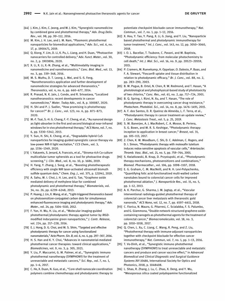
[64] J. Kim, J. Kim, C. Jeong, andW. J. Kim, “Synergistic nanomedicineby combined gene and photothermal therapy,” Adv. Drug Deliv.Rev., vol. 98, pp. 99–112, 2016.
[65] M. Kim, J. H. Lee, and J. M. Nam, “Plasmonic photothermalnanoparticles for biomedical applications,” Adv. Sci., vol. 6, no.17, p. 1900471, 2019.
[66] Q. Xiong, Y. Lim, D. Li, K. Pu, L. Liang, and H. Duan, “Photoactivenanocarriers for controlled delivery,” Adv. Funct. Mater., vol. 30,no. 2, p. 1903896, 2020.
[67] X. Li, X. Li, X.-N. Zhang, et al., “Multimodality imaging innanomedicine and nanotheranostics,” Canc. Biol. Med., vol. 13,no. 3, pp. 339–348, 2016.
[68] M. S. Muthu, D. T. Leong, L. Mei, and S.-S. Feng,“Nanotheranostics application and further development ofnanomedicine strategies for advanced theranostics,”Theranostics, vol. 4, no. 6, pp. 660–677, 2014.
[69] R. Prasad, N. K. Jain, J. Conde, and R. Srivastava, “Localizednanotheranostics: recent developments in cancernanomedicine,” Mater. Today Adv., vol. 8, p. 100087, 2020.
[70] H. Shi and P. J. Sadler, “How promising is phototherapyfor cancer?” Br. J. Canc., vol. 123, no. 6, pp. 871–873,2020.
[71] M.-F. Tsai, S.-H. G. Chang, F.-Y. Cheng, et al., “Au nanorod designas light-absorber in the first and second biological near-infraredwindows for in vivo photothermal therapy,” ACS Nano, vol. 7, no.6, pp. 5330–5342, 2013.
[72] Y. Sun, H. Shi, X. Cheng, et al., “Degradable hybrid CuSnanoparticles for imaging-guided synergistic cancer therapy vialow-power NIR-II light excitation,” CCS Chem., vol. 3,pp. 1336–1349, 2020.
[73] I. Yakavets, S. Jenard, A. Francois, et al., “Stroma-rich Co-culturemulticellular tumor spheroids as a tool for photoactive drugsscreening,” J. Clin. Med., vol. 8, no. 10, p. 1686, 2019.
[74] H. Yang, Y. Zhang, J. Song, et al., “Boosting phototherapeuticefficiency with single NIR laser-activated ultrasmall bismuthsulfide quantum dots,” Chem. Eng. J., vol. 375, p. 121941, 2019.
[75] A. Sahu, W. I. Choi, J. H. Lee, and G. Tae, “Graphene oxidemediated delivery of methylene blue for combinedphotodynamic and photothermal therapy,” Biomaterials, vol.34, no. 26, pp. 6239–6248, 2013.
[76] P. Huang, J. Lin, X.Wang, et al., “Light-triggered theranostics basedon photosensitizer-conjugated carbon dots for simultaneousenhanced-fluorescence imaging and photodynamic therapy,” Adv.Mater., vol. 24, pp. 5104–5110, 2012.
[77] F. Yan, H. Wu, H. Liu, et al., “Molecular imaging-guidedphotothermal/photodynamic therapy against tumor by iRGD-modified indocyanine green nanoparticles,” J. Contr. Release,vol. 224, pp. 217–228, 2016.
[78] E. J. Hong, D. G. Choi, and M. S. Shim, “Targeted and effectivephotodynamic therapy for cancer using functionalizednanomaterials,”ActaPharm.Sin. B, vol. 6, no.4, pp. 297–307, 2016.
[79] H. S. Han and K. Y. Choi, “Advances in nanomaterial-mediatedphotothermal cancer therapies: toward clinical applications,”Biomedicines, vol. 9, no. 3, p. 305, 2021.
[80] Y. Liu, P. Maccarini, G. M. Palmer, et al., “Synergistic immunophotothermal nanotherapy (SYMPHONY) for the treatment ofunresectable and metastatic cancers,” Sci. Rep., vol. 7, no. 1,pp. 1–6, 2017.
[81] C. He, X. Duan, N. Guo, et al., “Core-shell nanoscale coordinationpolymers combine chemotherapy and photodynamic therapy to
potentiate checkpoint blockade cancer immunotherapy,” Nat.Commun., vol. 7, no. 1, pp. 1–12, 2016.
[82] X. Hou, Y. Tao, Y. Pang, X. Li, G. Jiang, and Y. Liu, “Nanoparticlebased photothermal and photodynamic immunotherapy fortumor treatment,” Int. J. Canc., vol. 143, no. 12, pp. 3050–3060,2018.
[83] I. O. L. Bacellar, T. Tsubone, C. Pavani, and M. Baptista,“Photodynamic efficiency: from molecular photochemistry tocell death,” Int. J. Mol. Sci., vol. 16, no. 9, pp. 20523–20559,2015.
[84] P. Cramers,M. Ruevekamp,H. Oppelaar, O. Dalesio, P. Baas, andF. A. Stewart, “Foscan® uptake and tissue distribution inrelation to photodynamic efficacy,” Br. J. Canc., vol. 88, no. 2,pp. 283–290, 2003.
[85] B. W. Pogue, B. Ortel, N. Chen, R. W. Redmond, and T. Hasan, “Aphotobiological and photophysical-based study of phototoxicityof two chlorins,” Canc. Res., vol. 61, no. 2, pp. 717–724, 2001.
[86] B. Q. Spring, I. Rizvi, N. Xu, and T. Hasan, “The role ofphotodynamic therapy in overcoming cancer drug resistance,”Photochem. Photobiol. Sci., vol. 14, no. 8, pp. 1476–1491, 2015.
[87] A. F. dos Santos, D. R. Queiroz de Almeida, L. F. Terra, et al.,“Photodynamic therapy in cancer treatment-an update review,”J. Canc. Metastasis Treat., vol. 5, p. 25, 2019.
[88] S. M. Banerjee, A. J. MacRobert, C. A. Mosse, B. Periera,S. G. Bown, and M. R. S. Keshtgar, “Photodynamic therapy:inception to application in breast cancer,” Breast, vol. 31,pp. 105–113, 2017.
[89] Z. Chen, K. W. Woodburn, C. Shi, D. C. Adelman, C. Rogers, andD. I. Simon, “Photodynamic therapy with motexafin lutetiuminduces redox-sensitive apoptosis of vascular cells,” Arterioscler.Thromb. Vasc. Biol., vol. 21, no. 5, pp. 759–764, 2001.
[90] S. Kwiatkowski, B. Knap, D. Przystupski, et al., “Photodynamictherapy-mechanisms, photosensitizers and combinations,”Biomed. Pharmacother., vol. 106, pp. 1098–1107, 2018.
[91] E. G. Graham, C. M. MacNeill, and N. H. Levi-Polyachenko,“Quantifying folic acid-functionalized multi-walled carbonnanotubes bound to colorectal cancer cells for improvedphotothermal ablation,” J. Nanoparticle Res., vol. 15, no. 5,pp. 1–12, 2013.
[92] A. K. Parchur, G. Sharma, J. M. Jagtap, et al., “Vascularinterventional radiology-guided photothermal therapy ofcolorectal cancer liver metastasis with theranostic goldnanorods,” ACS Nano, vol. 12, no. 7, pp. 6597–6611, 2018.
[93] C. Fiorica, N. Mauro, G. Pitarresi, C. Scialabba, F. S. Palumbo,and G. Giammona, “Double-network-structured graphene oxide-containing nanogels as photothermal agents for the treatment ofcolorectal cancer,” Biomacromolecules, vol. 18, no. 3,pp. 1010–1018, 2017.
[94] Q. Chen, L. Xu, C. Liang, C. Wang, R. Peng, and Z. Liu,“Photothermal therapy with immune-adjuvant nanoparticlestogether with checkpoint blockade for effective cancerimmunotherapy,” Nat. Commun, vol. 7, no. 1, pp. 1–13, 2016.
[95] T. Vo-Dinh, et al., “Synergistic immuno photothermalnanotherapy (SYMPHONY) to treat unresectable and metastaticcancers and produce and cancer vaccine effect,” in AdvancedBiomedical and Clinical Diagnostic and Surgical GuidanceSystems XVI 10484, International Society for Optics andPhotonics, 2018, p. 1048408.
[96] L. Shao, R. Zhang, J. Lu, C. Zhao, X. Deng, and Y. Wu,“Mesoporous silica coated polydopamine functionalized
2992 N.K. Jain et al.: Nanoengineered photoactive theranostic agents for cancer

reduced graphene oxide for synergistic targeted chemo-photothermal therapy,”ACSAppl.Mater. Interfaces, vol. 9, no. 2,pp. 1226–1236, 2017.
[97] H. Sun, J. Su, Q. Meng, et al., “Cancer cell membrane coatedgold nanocages with hyperthermia triggered drug releaseand homotypic target inhibit growth and metastasis ofbreast cancer,” Adv. Funct. Mater., vol. 27, no. 3,p. 1604300, 2017.
[98] Y.-D. Zhu, S.-P. Chen, H. Zhao, et al., “PPy@ MIL-100nanoparticles as a pH-and near-IR-irradiation-responsive drugcarrier for simultaneous photothermal therapy andchemotherapy of cancer cells,” ACSAppl.Mater. Interfaces, vol.8, no. 50, pp. 34209–34217, 2016.
[99] T. Yang, H. Ke, Q.Wang, et al., “Bifunctional tellurium nanodotsfor photo-induced synergistic cancer therapy,” ACS Nano, vol.11, no. 10, pp. 10012–10024, 2017.
[100] H. S. Han, K. Y. Choi, H. Lee, et al., “Gold-nanoclusteredhyaluronan nano-assemblies for photothermally maneuveredphotodynamic tumor ablation,” ACS Nano, vol. 10, no. 12,pp. 10858–10868, 2016.
[101] Y. Wang, H. Wang, D. Liu, S. Song, X. Wang, and H. Zhang,“Graphene oxide covalently grafted upconversionnanoparticles for combined NIR mediated imaging andphotothermal/photodynamic cancer therapy,” Biomaterials,vol. 34, no. 31, pp. 7715–7724, 2013.
[102] X. Wei, H. Chen, H. P. Tham, et al., “Combined photodynamicand photothermal therapy using cross-linkedpolyphosphazene nanospheres decorated with goldnanoparticles,” ACS Appl. Nano Mater., vol. 1, no. 7,pp. 3663–3672, 2018.
[103] R. Baskaran, J. Lee, and S.-G. Yang, “Clinical development ofphotodynamic agents and therapeutic applications,”Biomater.Res., vol. 22, no. 1, pp. 1–8, 2018.
[104] D. E. J. G. J. Dolmans, D. Fukumura, and R. K. Jain,“Photodynamic therapy for cancer,” Nat. Rev. Canc., vol. 3, no.5, pp. 380–387, 2003.
[105] A. Juarranz, P. Jaén, F. Sanz-Rodríguez, J. Cuevas, andS. González, “Photodynamic therapy of cancer. Basic principlesand applications,” Clin. Transl. Oncol., vol. 10, no. 3,pp. 148–154, 2008.
[106] J. P. Celli, B. Q. Spring, I. Rizvi, et al., “Imaging and photodynamictherapy: mechanisms, monitoring, and optimization,” Chem.Rev., vol. 110, no. 5, pp. 2795–2838, 2010.
[107] Y. Liu, P. Bhattarai, Z. Dai, and X. Chen, “Photothermal therapyand photoacoustic imaging via nanotheranostics in fightingcancer,” Chem. Soc. Rev., vol. 48, no. 7, pp. 2053–2108, 2019.
[108] A. Kamkaew, S. H. Lim, H. B. Lee, L. V. Kiew, L. Y. Chung, andK. Burgess, “BODIPY dyes in photodynamic therapy,” Chem.Soc. Rev., vol. 42, no. 1, pp. 77–88, 2013.
[109] B. W. Pogue, J. T. Elliott, S. C. Kanick, et al., “Revisitingphotodynamic therapy dosimetry: reductionist & surrogateapproaches to facilitate clinical success,” Phys. Med. Biol., vol.61, no. 7, p. R57, 2016.
[110] E. M. Knavel and C. L. Brace, “Tumor ablation: commonmodalities and general practices,” Tech. Vasc. Intervent.Radiol., vol. 16, no. 4, pp. 192–200, 2013.
[111] K. Richter, M. Haslbeck, and J. Buchner, “The heat shockresponse: life on the verge of death,” Mol. Cell, vol. 40, no. 2,pp. 253–266, 2010.
[112] R. R. Allison, C. H. Sibata, G. H. Downie, and R. E. Cuenca, “Aclinical review of PDT for cutaneous malignancies,”Photodiagnosis Photodyn. Ther., vol. 3, no. 4, pp. 214–226, 2006.
[113] A. Casas, G. Di Venosa, T. Hasan, and A. Batlle, “Mechanisms ofresistance to photodynamic therapy,” Curr. Med. Chem., vol.18, no. 16, pp. 2486–2515, 2011.
[114] M. F. Tartaglione, M. E. Zabaleta, R. Lazzarini, et al., “Apoptoticmechanism activated by blue light and cisplatinum incutaneous squamous cell carcinoma cells,” Int. J. Mol. Med.,vol. 47, no. 4, pp. 1–13, 2021.
[115] J.-T. Lin, “Progress of medical lasers: fundamentals andapplications,” Med. Devices Diagn. Eng., vol. 1, no. 2,pp. 36–41, 2016.
[116] H. Van den Bergh, “On the evolution of some endoscopic lightdelivery systems for photodynamic therapy,” Endoscopy, vol.30, no. 04, pp. 392–407, 1998.
[117] G. Shafirstein, D. Bellnier, E. Oakley, et al., “Interstitialphotodynamic therapy-a focused review,” Cancers (Basel), vol.9, no. 2, p. 12, 2017.
[118] M. Panjehpour, B. F. Overholt, R. C. Denovo, R. E. Sneed, andM. G. Petersen, “Centering balloon to improve esophagealphotodynamic therapy,” Laser Surg. Med., vol. 12, no. 6,pp. 631–638, 1992.
[119] H. Moseley, “Light distribution and calibration of commercialPDT LED arrays,” Photochem. Photobiol. Sci., vol. 4, no. 11,pp. 911–914, 2005.
[120] S. Fitzmaurice and D. B. Eisen, “Daylight photodynamictherapy: what is known and what is yet to be determined,”Dermatol. Surg., vol. 42, no. 3, pp. 286–295, 2016.
[121] Z. Yang, Z. Sun, Y. Ren, et al., “Advances in nanomaterials foruse in photothermal andphotodynamic therapeutics (Review),”Mol. Med. Rep., vol. 20, no. 1, pp. 5–15, 2019.
[122] L. R. Hirsch, J. L.West, R. J. Stafford, et al., “Nanoshell-mediatednear infrared photothermal tumor therapy,” in AnnualInternational Conference of the IEEE Engineering in Medicineand Biology – Proceedings 2, 2003.
[123] S. J. Norton and T. Vo-Dinh, “Photothermal effects of plasmonicmetal nanoparticles in a fluid,” J. Appl. Phys., vol. 119, no. 8,p. 083105, 2016.
[124] H. Samadian, S. Hosseini-Nami, S. K. Kamrava, H. Ghaznavi,and A. Shakeri-Zadeh, “Folate-conjugated gold nanoparticle asa new nanoplatform for targeted cancer therapy,” J. Canc. Res.Clin. Oncol., vol. 142, no. 11, pp. 2217–2229, 2016.
[125] A. Dadwal, A. Baldi, and R. Kumar Narang, “Nanoparticles ascarriers for drug delivery in cancer,” Artif. Cells Nanomed.Biotechnol., vol. 46, no. sup2, pp. 295–305, 2018.
[126] J. Zhang, C. Yang, R. Zhang, et al., “Biocompatible D-Asemiconducting polymer nanoparticle with light harvestingunit for highly effective photoacoustic imaging guidedphotothermal therapy,” Adv. Funct. Mater., vol. 27, no. 13,p. 1605094, 2017.
[127] D. Zhang, H. Xu, X. Zhang, et al., “Self-quenched metal-organicparticles as dual-mode therapeutic agents for photoacousticimaging-guided second near-infrared windowphotochemotherapy,” ACS Appl. Mater. Interfaces, vol. 10, no.30, pp. 25203–25212, 2018.
[128] X. Liao, Y. Zheng, Z. Lin, et al., “Self-assembled metallo-supramolecular nanoflowers for NIR/acidic-triggeredmultidrugrelease, long-term tumor retention and NIR-II fluorescence
N.K. Jain et al.: Nanoengineered photoactive theranostic agents for cancer 2993
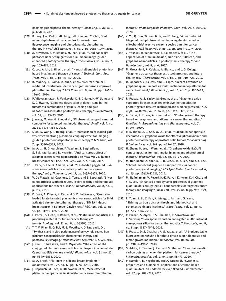
imaging-guided photo-chemotherapy,” Chem. Eng. J., vol. 400,p. 125882, 2020.
[129] B. Jang, J.-Y. Park, C.-H. Tung, I.-H. Kim, and Y. Choi, “Goldnanorod-photosensitizer complex for near-infraredfluorescence imaging and photodynamic/photothermaltherapy in vivo,” ACS Nano, vol. 5, no. 2, pp. 1086–1094, 2011.
[130] A. Srivatsan, S. V. Jenkins, M. Jeon, et al., “Gold nanocage-photosensitizer conjugates for dual-modal image-guidedenhanced photodynamic therapy,” Theranostics, vol. 4, no. 2,pp. 163–174, 2014.
[131] C. Loo, A. Lin, L. Hirsch, et al., “Nanoshell-enabled photonics-based imaging and therapy of cancer,” Technol. Canc. Res.Treat., vol. 3, no. 1, pp. 33–40, 2004.
[132] R. Mooney, L. Roma, D. Zhao, et al., “Neural stem cell-mediated intratumoral delivery of gold nanorods improvesphotothermal therapy,” ACS Nano, vol. 8, no. 12, pp. 12450–12460, 2014.
[133] P. Vijayaraghavan, R. Vankayala, C.-S. Chiang, H.-W. Sung, andK. C. Hwang, “Complete destruction of deep-tissue buriedtumors via combination of gene silencing and goldnanoechinus-mediated photodynamic therapy,” Biomaterials,vol. 62, pp. 13–23, 2015.
[134] J. Wang, M. You, G. Zhu, et al., “Photosensitizer-gold nanorodcomposite for targeted multimodal therapy,” Small, vol. 9, no.21, pp. 3678–3684, 2013.
[135] J. Lin, S. Wang, P. Huang, et al., “Photosensitizer-loaded goldvesicles with strong plasmonic coupling effect for imaging-guided photothermal/photodynamic therapy,” ACS Nano, vol.7, pp. 5320–5329, 2013.
[136] M. Azizi, H. Ghourchian, F. Yazdian, S. Bagherifam,S. Bekhradnia, and B. Nyström, “Anti-cancerous effect ofalbumin coated silver nanoparticles on MDA-MB 231 humanbreast cancer cell line,” Sci. Rep., vol. 7, p. 5178, 2017.
[137] T. Park, S. Lee, R. Amatya, et al., “ICG-loaded pegylated BSA-silver nanoparticles for effective photothermal cancertherapy,” Int. J. Nanomed., vol. 15, pp. 5459–5471, 2020.
[138] V. De Matteis, M. Cascione, C. Toma, and S. Leporatti, “Silvernanoparticles: synthetic routes, in vitro toxicity and theranosticapplications for cancer disease,” Nanomaterials, vol. 8, no. 5,p. 319, 2018.
[139] P. Bose, A. Priyam, R. Kar, and S. P. Pattanayak, “Quercetinloaded folate targeted plasmonic silver nanoparticles for lightactivated chemo-photothermal therapy of DMBA inducedbreast cancer in Sprague–Dawley rats,” RSC Adv., vol. 10, no.53, pp. 31961–31978, 2020.
[140] E. Porcel, S. Liehn, H. Remita, et al., “Platinum nanoparticles: apromising material for future cancer therapy?”Nanotechnology, vol. 21, no. 8, p. 085103, 2010.
[141] T. T. V. Phan, N. Q. Bui, M. S. Moorthy, K. D. Lee, and J. Oh,“Synthesis and in vitro performance of polypyrrole-coated iron–platinum nanoparticles for photothermal therapy andphotoacoustic imaging,”Nanoscale Res. Lett., vol. 12, p. 570, 2017.
[142] J. Kim, T. Shirasawa, and Y. Miyamoto, “The effect of TATconjugated platinum nanoparticles on lifespan in a nematodeCaenorhabditis elegans model,” Biomaterials, vol. 31, no. 22,pp. 5849–5854, 2010.
[143] M. A. Brook, “Platinum in silicone breast implants,”Biomaterials, vol. 27, no. 17, pp. 3274–3286, 2006.
[144] J. Depciuch, M. Stec, B. Klebowski, et al., “Size effect ofplatinum nanoparticles in simulated anticancer photothermal
therapy,” Photodiagnosis Photodyn. Ther., vol. 29, p. 101594,2020.
[145] Z. Yu, Q. Sun, W. Pan, N. Li, and B. Tang, “A near-infraredtriggered nanophotosensitizer inducing domino effect onmitochondrial reactive oxygen species burst for cancertherapy,” ACS Nano, vol. 9, no. 11, pp. 11064–11074, 2015.
[146] Z. Youssef, R. Vanderesse, L. Colombeau, et al., “Theapplication of titanium dioxide, zinc oxide, fullerene, andgraphene nanoparticles in photodynamic therapy,” Canc.Nanotechnol., vol. 8, p. 6, 2017.
[147] M. Orecchioni, R. Cabizza, A. Bianco, and L. G. Delogu,“Graphene as cancer theranostic tool: progress and futurechallenges,” Theranostics, vol. 5, no. 7, pp. 710–723, 2015.
[148] D. Iannazzo, C. Celesti, and C. Espro, “Recent advances ongraphene quantum dots as multifunctional nanoplatforms forcancer treatment,” Biotechnol. J., vol. 16, no. 2, p. 1900422,2021.
[149] R. Prasad, A. S. Yadav, M. Gorain, et al., “Graphene oxidesupported liposomes as red emissive theranostics forphototriggered tissue visualization and tumor regression,” ACSAppl. Bio Mater., vol. 2, no. 8, pp. 3312–3320, 2019.
[150] A. Gazzi, L. Fusco, A. Khan, et al., “Photodynamic therapybased on graphene and MXene in cancer theranostics,”Frontiers in Bioengineering and Biotechnology, vol. 7,p. 295, 2019.
[151] R. K. Thapa, Z. C. Soe, W. Ou, et al., “Palladium nanoparticle-decorated 2-D graphene oxide for effective photodynamic andphotothermal therapy of prostate solid tumors,” Colloids Surf.B Biointerfaces, vol. 169, pp. 429–437, 2018.
[152] H. Zhang, H. Wu, J. Wang, et al., “Graphene oxide-BaGdF5nanocomposites for multi-modal imaging and photothermaltherapy,” Biomaterials, vol. 42, pp. 66–77, 2015.
[153] M. Nurunnabi, Z. Khatun, G. R. Reeck, D. Y. Lee, and Y.-K. Lee,“Photoluminescent graphene nanoparticles for cancerphototherapy and imaging,” ACS Appl.Mater. Interfaces, vol. 6,no. 15, pp. 12413–12421, 2014.
[154] M. Nafiujjaman, V. Revuri, H.-K. Park, I. K. Kwon, K.-J. Cho, andY.-K. Lee, “Enhanced photodynamic properties of graphenequantum dot conjugated Ce6 nanoparticles for targeted cancertherapy and imaging,” Chem. Lett., vol. 45, no. 8, pp. 997–999,2016.
[155] F. Yuan, S. Li, Z. Fan, X. Meng, L. Fan, and S. Yang,“Shining carbon dots: synthesis and biomedical andoptoelectronic applications,” Nano Today, vol. 11, no. 5,pp. 565–586, 2016.
[156] R. Prasad, S. Aiyer, D. S. Chauhan, R. Srivastava, andK. Selvaraj, “Bioresponsive carbon nano-gated multifunctionalmesoporous silica for cancer theranostics,” Nanoscale, vol. 8,no. 8, pp. 4537–4546, 2016.
[157] R. Prasad, D. S. Chauhan, A. S. Yadav, et al., “A biodegradablefluorescent nanohybrid for photo-driven tumor diagnosis andtumor growth inhibition,” Nanoscale, vol. 10, no. 40,pp. 19082–19091, 2018.
[158] S. Adrita, K. Tasnim, J. Ryu, and S. Sharker, “Nanotheranosticcarbon dots as an emerging platform for cancer therapy,”J. Nanotheranostics, vol. 1, no. 1, pp. 58–77, 2020.
[159] P. Namdari, B. Negahdari, and A. Eatemadi, “Synthesis,properties and biomedical applications of carbon-basedquantum dots: an updated review,” Biomed. Pharmacother.,vol. 87, pp. 209–222, 2017.
2994 N.K. Jain et al.: Nanoengineered photoactive theranostic agents for cancer
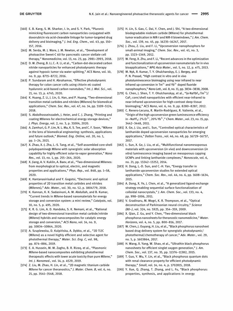
[160] E. B. Kang, S. M. Sharker, I. In, and S. Y. Park, “Pluronicmimicking fluorescent carbon nanoparticles conjugated withdoxorubicin via acid-cleavable linkage for tumor-targeted drugdelivery and bioimaging,” J. Ind. Eng. Chem., vol. 43, pp. 150–157, 2016.
[161] M. Serda, M. J. Ware, J. M. Newton, et al., “Development ofphotoactive Sweet-C 60 for pancreatic cancer stellate celltherapy,” Nanomedicine, vol. 13, no. 23, pp. 2981–2993, 2018.
[162] D.W. Zheng, B. Li, C.-X. Li, et al., “Carbon-dot-decorated carbonnitride nanoparticles for enhanced photodynamic therapyagainst hypoxic tumor via water splitting,” ACS Nano, vol. 10,no. 9, pp. 8715–8722, 2016.
[163] P. Sundaram and H. Abrahamse, “Effective photodynamictherapy for colon cancer cells using chlorin e6 coatedhyaluronic acid-based carbon nanotubes,” Int. J. Mol. Sci., vol.21, no. 13, p. 4745, 2020.
[164] K. Huang, Z. Li, J. Lin, G. Han, and P. Huang, “Two-dimensionaltransition metal carbides and nitrides (MXenes) for biomedicalapplications,” Chem. Soc. Rev., vol. 47, no. 14, pp. 5109–5124,2018.
[165] S. Abdolhosseinzadeh, J. Heier, and C. J. Zhang, “Printing andcoating MXenes for electrochemical energy storage devices,”J. Phys. Energy, vol. 2, no. 3, p. 31004, 2020.
[166] A. Zamhuri, G. P. Lim, N. L.Ma, K. S. Tee, andC. F. Soon, “MXenein the lens of biomedical engineering: synthesis, applicationsand future outlook,” Biomed. Eng. Online, vol. 20, no. 1,pp. 1–24, 2021.
[167] X. Zhao, X.-J. Zha, L.-S. Tang, et al., “Self-assembled core-shellpolydopamine@ MXene with synergistic solar absorptioncapability for highly efficient solar-to-vapor generation,” NanoRes., vol. 13, no. 1, pp. 255–264, 2020.
[168] X. Jiang, A. V. Kuklin, A. Baev, et al., “Two-dimensionalMXenes:from morphological to optical, electric, and magneticproperties and applications,” Phys. Rep., vol. 848, pp. 1–58,2020.
[169] K. Hantanasirisakul and Y. Gogotsi, “Electronic and opticalproperties of 2D transition metal carbides and nitrides(MXenes),” Adv. Mater., vol. 30, no. 52, p. 1804779, 2018.
[170] K. Kannan, K. K. Sadasivuni, A. M. Abdullah, and B. Kumar,“Current trends in MXene-based nanomaterials for energystorage and conversion system: a mini review,” Catalysts, vol.10, no. 5, p. 495, 2020.
[171] K. R. G. Lim, A. D. Handoko, S. K. Nemani, et al., “Rationaldesign of two-dimensional transition metal carbide/nitride(MXene) hybrids and nanocomposites for catalytic energystorage and conversion,” ACS Nano, vol. 14, no. 9,pp. 10834–10864, 2020.
[172] A. Szuplewska, D. Kulpińska, A. Dybko, et al., “2D Ti2C(MXene) as a novel highly efficient and selective agent forphotothermal therapy,” Mater. Sci. Eng. C, vol. 98,pp. 874–886, 2019.
[173] E. A. Hussein, M. M. Zagho, B. R. Rizeq, et al., “PlasmonicMXene-based nanocomposites exhibiting photothermaltherapeutic effects with lower acute toxicity than pure MXene,”Int. J. Nanomed., vol. 14, p. 4529, 2019.
[174] Z. Liu, M. Zhao, H. Lin, et al., “2D magnetic titanium carbideMXene for cancer theranostics,” J. Mater. Chem. B, vol. 6, no.21, pp. 3541–3548, 2018.
[175] H. Lin, S. Gao, C. Dai, Y. Chen, and J. Shi, “A two-dimensionalbiodegradable niobium carbide (MXene) for photothermaltumor eradication in NIR-I and NIR-II biowindows,” J. Am. Chem.Soc., vol. 139, no. 45, pp. 16235–16247, 2017.
[176] J. Zhou, Z. Liu, and F. Li, “Upconversion nanophosphors forsmall-animal imaging,” Chem. Soc. Rev., vol. 41, no. 3,pp. 1323–1349, 2012.
[177] W. Feng, X. Zhu, and F. Li, “Recent advances in the optimizationand functionalization of upconversion nanomaterials for in vivobioapplications,” NPG Asia Mater., vol. 5, no. 12, p. e75, 2013.
[178] M. Nyk, R. Kumar, T. Y. Ohulchanskyy, E. J. Bergey, andP. N. Prasad, “High contrast in vitro and in vivophotoluminescence bioimaging using near infrared to nearinfrared up-conversion in Tm3+ and Yb3+ doped fluoridenanophosphors,”Nano Lett., vol. 8, no. 11, pp. 3834–3838, 2008.
[179] G. Chen, J. Shen, T. Y. Ohulchanskyy, et al., “(α-NaYbF4:Tm3+)/CaF2 core/shell nanoparticles with efficient near-infrared tonear-infrared upconversion for high-contrast deep tissuebioimaging,” ACS Nano, vol. 6, no. 9, pp. 8280–8287, 2012.
[180] C. Renero-Lecuna, R. Martín-Rodríguez, R. Valiente, et al.,“Origin of the high upconversion green luminescence efficiencyin–NaYF4: 2%Er3+, 20%Yb3+,”Chem.Mater., vol. 23, no. 15, pp.3442–3448, 2011.
[181] X. Ge, J. Liu, and L. Sun, “Controlled optical characteristics oflanthanide doped upconversion nanoparticles for emergingapplications,” Dalton Trans., vol. 46, no. 48, pp. 16729–16737,2017.
[182] L. Sun, X. Ge, J. Liu, et al., “Multifunctional nanomesoporousmaterials with upconversion (in vivo) and downconversion (invitro) luminescence imaging based on mesoporous cappingUCNPs and linking lanthanide complexes,” Nanoscale, vol. 6,no. 21, pp. 13242–13252, 2014.
[183] H. Dong, L.-D. Sun, and C.-H. Yan, “Energy transfer inlanthanide upconversion studies for extended opticalapplications,” Chem. Soc. Rev., vol. 44, no. 6, pp. 1608–1634,2015.
[184] A. Dong, X. Ye, J. Chen, et al., “A generalized ligand-exchangestrategy enabling sequential surface functionalization ofcolloidal nanocrystals,” J. Am. Chem. Soc., vol. 133, no. 4,pp. 998–1006, 2011.
[185] V. Gradinaru, M. Mogri, K. R. Thompson, et al., “Opticaldeconstruction of Parkinsonian neural circuitry,” Science(80–), vol. 324, no. 5925, pp. 354–359, 2009.
[186] X. Qian, Z. Gu, and Y. Chen, “Two-dimensional blackphosphorusnanosheets for theranostic nanomedicine,”Mater.Horizons, vol. 4, no. 5, pp. 800–816, 2017.
[187] W. Chen, J. Ouyang, H. Liu, et al., “Black phosphorus nanosheetbased drug delivery system for synergistic photodynamic/photothermal/chemotherapy of cancer,” Adv. Mater., vol. 29,no. 5, p. 1603864, 2017.
[188] H. Wang, X. Yang, W. Shao, et al., “Ultrathin black phosphorusnanosheets for efficient singlet oxygen generation,” J. Am.Chem. Soc., vol. 137, no. 35, pp. 11376–11382, 2015.
[189] T. Guo, Y. Wu, Y. Lin, et al., “Black phosphorus quantum dotswith renal clearance property for efficient photodynamictherapy,” Small, vol. 14, no. 4, p. 1702815, 2018.
[190] Y. Xue, Q. Zhang, T. Zhang, and L. Fu, “Black phosphorus:properties, synthesis, and applications in energy
N.K. Jain et al.: Nanoengineered photoactive theranostic agents for cancer 2995
conversion and storage,” ChemNanoMat, vol. 3, no. 6,pp. 352–361, 2017.
[191] M. Luo, T. Fan, Y. Zhou, H. Zhang, and L. Mei, “2D blackphosphorus-based biomedical applications,” Adv. Funct.Mater., vol. 29, no. 13, p. 1808306, 2019.
[192] W. Tao, X. Zhu, X. Yu, et al., “Black phosphorus nanosheets as arobust delivery platform for cancer theranostics,” Adv. Mater.,vol. 29, no. 1, p. 1603276, 2017.
[193] Z. Li, H. Xu, J. Shao, et al., “Polydopamine-functionalized blackphosphorus quantum dots for cancer theranostics,” Appl.Mater. Today, vol. 15, pp. 297–304, 2019.
[194] M. Luo, W. Cheng, X. Zeng, L. Mei, G. Liu, and W. Deng, “Folicacid-functionalized black phosphorus quantum dots fortargeted chemo-photothermal combination cancer therapy,”Pharmaceutics, vol. 11, no. 5, p. 242, 2019.
[195] L. Deng, Y. Xu, C. Sun, et al., “Functionalization of small blackphosphorus nanoparticles for targeted imaging andphotothermal therapy of cancer,” Sci. Bull., vol. 63, no. 14,pp. 917–924, 2018.
[196] X. Yang, R. Liu, Z. Zhong, et al., “Platinum nanoenzymefunctionalized black phosphorus nanosheets for photothermaland enhanced-photodynamic therapy,” Chem. Eng. J., vol. 409,p. 127381, 2021.
[197] Z. Wang, Z. Liu, C. Su, et al., “Biodegradable black phosphorus-based nanomaterials in biomedicine: theranosticapplications,” Curr. Med. Chem., vol. 26, no. 10,pp. 1788–1805, 2019.
[198] M. Luo, Y. Zhou, N. Gao, et al., “Mesenchymal stem cellstransporting black phosphorus-based biocompatiblenanospheres: active Trojan horse for enhanced photothermalcancer therapy,” Chem. Eng. J., vol. 385, p. 123942, 2020.
[199] J. Wang, D. Liang, Z. Qu, I. M. Kislyakov, V.M. Kiselev, and J. Liu,“PEGylated-folic acid-modified black phosphorus quantumdots as near-infrared agents for dual-modality imaging-guidedselective cancer cell destruction,”Nanophotonics, vol. 9, no. 8,pp. 2425–2435, 2020.
[200] W. Liang, D.Wang, X. Ren, et al., “Facile sonochemical-assistedsynthesis of orthorhombic phase black phosphorus/rGOhybrids for effective photothermal therapy,” Nanophotonics,vol. 9, no. 9, pp. 3023–3034, 2020.
[201] N. Zhao, L. Yan, X. Zhao, et al., “Versatile types of organic/inorganic nanohybrids: from strategic design to biomedicalapplications,” Chem. Rev., vol. 119, no. 3, pp. 1666–1762, 2019.
[202] Y. Sasaki and K. Akiyoshi, “Design and function of smartbiomembrane nanohybrids for biomedical applications,”Polym. J., vol. 53, no. 5, pp. 587–592, 2021.
[203] D. S. Chauhan, A. B. Bukhari, G. Ravichandran, et al.,“Enhanced EPR directed and imaging guided photothermaltherapy using vitamin E modified toco-photoxil,” Sci. Rep., vol.8, no. 1, p. 16673, 2018.
[204] D. S. Chauhan, P. Arunkumar, R. Prasad, et al., “Facilesynthesis of plasmonic zein nanoshells for imaging-guidedphotothermal cancer therapy,” Mater. Sci. Eng. C, vol. 90,pp. 539–548, 2018.
[205] D. S. Chauhan, S. Indulekha, R. Gottipalli, et al., “NIR light-triggered shrinkable thermoresponsive PNVCL nanoshells forcancer theranostics,” RSC Adv., vol. 7, no. 70,pp. 44026–44034, 2017.
[206] A. K. Rengan, M. Jagtap, A. De, R. Banerjee, and R. Srivastava,“Multifunctional gold coated thermo-sensitive liposomes for
multimodal imaging and photo-thermal therapy of breastcancer cells,” Nanoscale, vol. 6, no. 2, pp. 916–923, 2014.
[207] R. Prasad, N. K. Jain, A. S. Yadav, et al., “Liposomalnanotheranostics for multimode targeted in vivo bioimagingand near‐infrared light mediated cancer therapy,” Commun.Biol., vol. 3, no. 1, pp. 1–14, 2020.
[208] M. Marrelli, G. Menichini, E. Provenzano, and F. Conforti,“Applications of natural compounds in the photodynamictherapy of skin cancer,” Curr. Med. Chem., vol. 21, no. 12,pp. 1371–1390, 2014.
[209] B. Siewert and H. Stuppner, “The photoactivity of naturalproducts – an overlooked potential of phytomedicines?”Phytomedicine, vol. 60, p. 152985, 2019.
[210] N. B. R. Vittar, J. Awruch, K. Azizuddin, and V. Rivarola,“Caspase-independent apoptosis, in human MCF-7c3 breastcancer cells, following photodynamic therapy, with a novelwater-soluble phthalocyanine,” Int. J. Biochem. Cell Biol., vol.42, no. 7, pp. 1123–1131, 2010.
[211] E. W. Jeffes, J. L. McCullough, G. D. Weinstein, R. Kaplan,S. D. Glazer, and J. R. Taylor, “Photodynamic therapy of actinickeratoses with topical aminolevulinic acid hydrochloride andfluorescent blue light,” J. Am. Acad. Dermatol., vol. 45, no. 1,pp. 96–104, 2001.
[212] K. Muniyandi, B. George, T. Parimelazhagan, and H. Abrahamse,“Role of photoactive phytocompounds in photodynamic therapyof cancer,” Molecules, vol. 25, no. 18, p. 4102, 2020.
[213] M. He, C. Qu, O. Gao, X. Hu, and X. Hong, “Biological andpharmacological activities of amaryllidaceae alkaloids,” RSCAdv., vol. 5, no. 21, pp. 16562–16574, 2015.
[214] J. M. PérezMartín, V. Labrador, P. Fernández Freire,M. L.Molero,and M. J. Hazen, “Ultrastructural changes induced in HeLa Cellsafter phototoxic treatment with harmine,” J. Appl. Toxicol., vol.24, no. 3, pp. 197–201, 2004.
[215] D. Skalkos, E. Gioti, C. D. Stalikas, et al., “Photophysicalproperties of Hypericum perforatum L. extracts – novelphotosensitizers for PDT,” J. Photochem. Photobiol. B Biol., vol.82, no. 2, pp. 146–151, 2006.
[216] S. Jantová, S. Letašiová, V. Brezová, L. U. Čipák, and J. Lábaj,“Photochemical and phototoxic activity of berberine on murinefibroblast NIH-3T3 and Ehrlich ascites carcinoma cells,”J. Photochem. Photobiol. B Biol., vol. 85, no. 3, pp. 163–176,2006.
[217] K. Park and J. H. Lee, “Photosensitizer effect of curcumin onUVB-irradiated HaCaT cells through activation of caspasepathways,” Oncol. Rep., vol. 17, no. 3, pp. 537–540, 2007.
[218] W. Jin, Q. Shi, C. Hong, Y. Cheng, Z. Ma, and H. Qu, “Cytotoxicproperties of thiophenes from Echinops grijissi Hance,”Phytomedicine, vol. 15, no. 9, pp. 768–774, 2008.
[219] K. I. Priyadarsini, “Photophysics, photochemistry andphotobiology of curcumin: studies from organic solutions, bio-mimetics and living cells,” J. Photochem. Photobiol. CPhotochem. Rev., vol. 10, no. 2, pp. 81–95, 2009.
[220] Y. Li, P. Xiao, Z. Huang, et al., “Evaluation of curcumin-mediatedphotodynamic therapy on the reverse ofmultidrug resistance intumor cells,” RSC Adv., vol. 10, no. 1, pp. 298–306, 2019.
[221] W. H. Chan and H. J. Wu, “Anti-apoptotic effects of curcumin onphotosensitized human epidermal carcinoma A431 cells,”J. Cell. Biochem., vol. 92, no. 1, pp. 200–212, 2004.
[222] S. Banerjee and A. R. Chakravarty, “Metal complexes ofcurcumin for cellular imaging, targeting, and photoinduced
2996 N.K. Jain et al.: Nanoengineered photoactive theranostic agents for cancer
anticancer activity,” Acc. Chem. Res., vol. 48, no. 7, pp. 2075–2083, 2015.
[223] N. K. Gibbs, E. Quanten, S. Baydoun, et al., “Photophysical,photochemical and photobiological properties ofpyrrolocoumarins; A new class of photoactive compounds,”J. Photochem. Photobiol. B Biol., vol. 2, no. 1, pp. 109–122, 1988.
[224] S. M. Kim, J. H. Lee, G. Sethi, et al., “Bergamottin, a naturalfuranocoumarin obtained from grapefruit juice induceschemosensitization and apoptosis through the inhibition ofSTAT3 signaling pathway in tumor cells,” Canc. Lett., vol. 354,no. 1, pp. 153–163, 2014.
[225] S. C. Núñez Montoya, L. R. Comini, B. Rumie Vittar,I. M. Fernández, V. A. Rivarola, and J. L. Cabrera, “Phototoxiceffects of heterophyllaea pustulata (rubiaceae),” Toxicon,vol. 51, no. 8, pp. 1409–1415, 2008.
[226] I. Gutiérrez, S. G. Bertolotti, M. A. Biasutti, A. T. Soltermann,and N. A. García, “Quinones and hydroxyquinones asgenerators and quenchers of singlet molecular oxygen,” Can.J. Chem., vol. 75, no. 4, pp. 423–428, 1997.
[227] D. Luo, K. A. Carter, D. Miranda, et al., “Chemophototherapy: anemerging treatment option for solid tumors,” Adv. Sci., vol. 4, no.1, p. 1600106, 2017.
[228] P. Agostinis, K. Berg, K. A. Cengel, et al., “Photodynamictherapy of cancer: an update,” CA Canc. J. Clin., vol. 61, no. 4,pp. 250–281, 2011.
[229] Z. Zhang, L. Wang, J. Wang, et al., “Mesoporous silica-coatedgold nanorods as a light-mediated multifunctional theranosticplatform for cancer treatment,” Adv. Mater., vol. 24, no. 11,pp. 1418–1423, 2012.
[230] M. Zheng, C. Yue, Y. Ma, et al., “Single-step assembly of DOX/ICG loaded lipid-polymer nanoparticles for highly effectivechemo-photothermal combination therapy,” ACS Nano, vol. 7,no. 3, pp. 2056–2067, 2013.
[231] Y. Deng, F. Käfer, T. Chen, et al., “Let there be light: polymericmicelles with upper critical solution temperature as lighttriggered heat nanogenerators for combating drug resistantcancer,” Small, vol. 14, no. 37, p. 1802420, 2018.
[232] Y. Tan, Y. Zhu, L. Wen, et al., “Mitochondria-responsive drugrelease along with heat shock mediated by multifunctionalglycolipid micelles for precise cancer chemo-phototherapy,”Theranostics, vol. 9, no. 3, pp. 691–707, 2019.
[233] A. Zhang, A. Li, W. Zhao, and J. Liu, “Recent advances infunctional polymer decorated two dimensional transitionmetal dichalcogenides nanomaterials for chemophotothermal therapy,” Chem. Eur J., vol. 24, no. 17,pp. 4215–4227, 2018.
[234] X. Deng, Z. Shao, and Y. Zhao, “Solutions to the drawbacks ofphotothermal and photodynamic cancer therapy,” Adv. Sci.,vol. 8, no. 3, p. 2002504, 2021.
[235] W. Li, J. Peng, L. Tan, et al., “Mild photothermal therapy/photodynamic therapy/chemotherapy of breast cancer by Lyp-1modified Docetaxel/IR820 Co-loaded micelles,” Biomaterials,vol. 106, pp. 119–133, 2016.
[236] Y. Chen, Q. Chen, Q. Zhu, et al., “Small molecular theranosticassemblies functionalized by doxorubicin-hyaluronic acid-methotrexate prodrug for multiple tumor targeting andimaging-guided combined chemo-photothermal therapy,”Mol.Pharm., vol. 16, no. 6, pp. 2470–2480, 2019.
[237] L. Zhang, D. Jing, L.Wang, et al., “Uniquephotochemo-immuno-nanoplatform against orthotopic xenograft oral cancer andmetastatic syngeneic breast cancer,” Nano Lett., vol. 18, no. 11,pp. 7092–7103, 2018.
[238] T. J. Dougherty, M. T. Cooper, and T. S. Mang, “Cutaneousphototoxic occurrences in patients receiving Photofrin®,”Laser Surg. Med., vol. 10, no. 5, pp. 485–488, 1990.
[239] X. Li, J. F. Lovell, J. Yoon, andX. Chen, “Clinical development andpotential of photothermal and photodynamic therapies forcancer,” Nat. Rev. Clin. Oncol., vol. 17, no. 11, pp. 657–674,2020.
[240] H. Wu, T. Minamide, and T. Yano, “Role of photodynamictherapy in the treatment of esophageal cancer,” Dig.Endoscopy, vol. 31, no. 5, pp. 508–516, 2019.
[241] I. Freitas, “Facing hypoxia: a must for photodynamic therapy,”J. Photochem. Photobiol. B, vol. 2, no. 2, pp. 281–282, 1988.
[242] J. Dang, H. He, D. Chen, and L. Yin, “Manipulating tumor hypoxiatoward enhancedphotodynamic therapy (PDT),”Biomater. Sci.,vol. 5, no. 8, pp. 1500–1511, 2017.
[243] M.Wang, J. Shen, H. Jin, H.-J. Im, J. Sandy, and D. Chen, “Recentprogress in understanding molecular mechanisms of cartilagedegeneration during osteoarthritis,” Ann. N. Y. Acad. Sci., vol.1240, pp. 61–69, 2011.
[244] L. Larue, B. Myrzakhmetov, A. Ben-Mihoub, et al., “Fightinghypoxia to improve PDT,” Pharmaceuticals, vol. 12, no. 4,p. 163, 2019.
[245] P. Xue, R. Yang, L. Sun, et al., “Indocyanine green-conjugatedmagnetic prussian blue nanoparticles for synchronousphotothermal/photodynamic tumor therapy,” Nano-MicroLett., vol. 10, p. 74, 2018.
[246] Q. Guan, L.-L. Zhou, Y.-A. Li, et al., “Nanoscale covalent organicframework for combinatorial antitumor photodynamic andphotothermal therapy,” ACS Nano, vol. 13, no. 11, pp. 13304–13316, 2019.
N.K. Jain et al.: Nanoengineered photoactive theranostic agents for cancer 2997