nano-based theranostic tools for the detection and ... - mdpi
TRANSCRIPT

cells
Review
Nano-Based Theranostic Tools for the Detection andElimination of Senescent Cells
Jagoda Adamczyk-Grochala * and Anna Lewinska *
Department of Biotechnology, Institute of Biology and Biotechnology, University of Rzeszow, Pigonia 1,35-310 Rzeszow, Poland* Correspondence: [email protected] (J.A.-G.); [email protected] (A.L.); Tel.: +48-17-851-85-20 (J.A.-G.);
+48-17-851-86-09 (A.L.)
Received: 18 November 2020; Accepted: 7 December 2020; Published: 10 December 2020 �����������������
Abstract: The progressive accumulation of apoptosis-resistant and secretory active senescent cells(SCs) in animal and human aged tissues may limit lifespan and healthspan and lead to age-relateddiseases such as cancer, neurodegenerative disorders, and metabolic syndrome. Thus, SCs aresuggested targets in anti-aging therapy. In the last two decades, a number of nanomaterialshave gained much attention as innovative tools in theranostic applications due to their uniqueproperties improving target visualization, drug and gene delivery, controlled drug release, effectivediagnosis, and successful therapy. Although the healthcare industry has focused on a plethora ofapplications of nanomaterials, it remains elusive how nanomaterials may modulate cellular senescence,a hallmark of aging. In this review paper, we consider novel nanotechnology-based strategies forhealthspan promotion and the prevention of age-related dysfunctions that are based on the deliveryof therapeutic compounds capable to preferentially killing SCs (nano-senolytics) and/or modulating aproinflammatory secretome (nano-senomorphics/nano-senostatics). Recent examples of SC-targetednanomaterials and the mechanisms underlying different aspects of the nanomaterial-mediatedsenolysis are presented and discussed.
Keywords: nanomaterials; cellular senescence; senotherapy; senolytics; nanotherapeutic-mediated senolysis
1. Cellular Senescence
Cellular senescence (CS) is characterized by a state of permanent cell growth arrest with alteredmetabolic features (e.g., affected glycolysis, mitochondrial function, and autophagic flux), modifiedprotein secretion, and biomolecular damage that occurs in response to stressful stimuli [1,2]. CS wasoriginally described in 1961 by Hayflick and colleagues, who observed that normal diploid embryonicfibroblasts had a limited ability to proliferate in an in vitro culture [3]. Based on these results, Hayflickcoined the term replicative senescence to describe this phenomenon. Subsequently, progressivetelomere shortening promoting DNA damage response (DDR) has been recognized as one of themajor determinants of replicative senescence [4,5]. Later, the presence of senescent cells (SCs) inaging tissues that linked cellular senescence with organismal aging was documented [6]. Cells canalso prematurely activate the senescence program upon treatment with stress stimuli such as UVradiation, oxidants, or DNA-damaging agents [7,8]. This process is known as stress-induced prematuresenescence (SIPS) [9]. Moreover, the expression of oncogenes could trigger senescence prematurely, in aprocess called oncogene-induced senescence (OIS) [10]. Cellular senescence may be considered as atumor suppression mechanism by blocking the proliferation and division of cells with unrepaired DNAdamage to prevent their stepwise malignant propagation [11]. However, it has also been demonstratedthat cellular senescence may participate in other physiological processes such as specific tissue
Cells 2020, 9, 2659; doi:10.3390/cells9122659 www.mdpi.com/journal/cells

Cells 2020, 9, 2659 2 of 19
remodeling (so-called developmentally-programmed senescence) that acts in a damage-independentmanner during embryonic development and wound healing in adulthood [12,13]. Despite the protectiverole of cellular senescence during stress-induced cellular responses, it is commonly accepted thatCS may promote aging and contribute to the development of age-related diseases. Namely, it hasbeen revealed that diverse pathological conditions associated with accelerated aging such as cancer,neurodegenerative diseases, cardiovascular disorders, and progeroid and metabolic syndromes areaccompanied by the accumulation of SCs and cellular senescence may be a causative factor for theprogression of age-related pathologies [14–19].
Many different molecular pathways have been proven to contribute to cellular senescence [20,21].However, one of common features of all SCs is irreversible cell-cycle arrest regulated by p53/p21WAF1/Cip1
and p16INK4A/RB (retinoblastoma) tumor-suppressor pathways, which can interact with each other oract independently [2]. Unlike quiescent cells (the cells at the G0 phase) or terminally-differentiatedcells, SCs do not respond to growth and mitogenic stimuli, thus they are irreversibly withdrawn fromthe cell cycle [17,22]. Two cell-cycle inhibitors that are often overexpressed in SCs, namely p21WAF1/Cip1
(encoded by CDKN1a gene) and p16INK4A (encoded by CDKN2a gene) are crucial mediators ofsenescence. Accumulation of these cyclin-dependent kinase inhibitors (CDKI) resulted in permanentactivation of RB family proteins, inhibition of E2F family transcription factor activity, and finallycell-cycle arrest [20].
SCs display common features such as granularity, increased activity of lysosomalsenescence-associated β-galactosidase (SA-β-gal), lipofuscin accumulation, exclusion of proliferativemarkers, formation of senescence-associated heterochromatin foci (SAHF), and persistent expressionof proteins that participate in DDR (Figure 1) [23–25]. Moreover, SCs are characterized bysenescence-associated secretory phenotype (SASP) [26,27]. The SASP entails secretion of numerousmolecules, such as inflammatory chemokines (e.g., MCP-1, MIP 1α, and CCL-16) and cytokines (e.g., IL-6and IL-8), growth factors (e.g., IGF-1 and EGF) and angiogenic factors/regulators, miRNAs, proteases,and damage-associated molecular patterns (DAMPs), which can induce changes in neighboring cellsvia both paracrine and autocrine mechanisms [28–31]. SASP may have both beneficial and pathologicalfunctions. First, these factors are suggested to regulate immune clearance of SCs to prevent fibrosisand promote tissue regeneration [1,32,33]. Contrariwise, SASP factors can induce the development ofsecondary senescence within non-senescent nearby cells [34]. In addition, the SASP is responsible forpromotion of low-grade chronic inflammation. This malfunction can alter the tissue microenvironment,which can also stimulate neoplastic cell growth, tumor metastasis, and angiogenesis [35].
A recently-proposed strategy to delay symptoms of aging is the suppression of SASPcomponents [36]. Notably, compounds that interfere with the pathways involved in SASP regulationare the most effective. These include nuclear factor (NF)-κB, Janus kinase (JAK)/signal transducer andactivator of transcription (STAT), mitogen-activated protein kinase (MAPK), and mammalian target ofrapamycin (mTOR) pathways [37]. For example, various drugs have been shown to lower productionor secretion of SASP factors. Simvastatin decreases production of interleukins IL-6 and IL-8 in vitro [38].Natural compounds such as flavonoids have also been documented to impair the secretory phenotypein SCs [39]. Importantly, SASP is not the only mechanism by which SCs modulate their neighboringcells. For example, it has been shown that OIS is accompanied by a dynamic fluctuation of NOTCH1activity [40], reactive oxygen species (ROS) production [41], or by release of exosomes [42]. Apart fromirreversible withdrawal from the cell cycle and SASP, another feature of SCs is their resistance toapoptosis through the multilevel regulated pro-survival senescent cell anti-apoptotic pathways (SCAPs)such as p53/p21/serpins, BCL-2/Bcl-XL, PI3K/AKT/ceramide signaling, the hypoxia-inducible factor(HIF-1α) pathway, or HSP90-dependent networks [24,30,43].

Cells 2020, 9, 2659 3 of 19Cells 2020, 9, x FOR PEER REVIEW 3 of 19
Figure 1. Biomarkers of cellular senescence. Senescent cell (right) is characterized by irreversible cell cycle arrest induced by various stressors (e.g., DNA damage or telomere shortening). Classical features of senescent cells include flat and enlarged cell size, elevated expression of cell cycle inhibitors (such as p16INK4A or p21Cip1), increased nucleus size and multiple nucleoli, lysosomes overexpressing β-galactosidase, α-fucosidase, and lipofuscin, chromatin reorganization based on senescence-associated heterochromatic foci (SAHF) and loss of lamin B1, senescence-associated ribosome biogenesis defects (e.g., “free” cytoplasmic polysomes with ribosomes), the overexpression of anti-apoptotic Bcl-2 family members, reactive oxygen species (ROS) production in mitochondria, and expression of cell surface markers such as CD9 receptor, urokinase-type plasminogen activator receptor (uPAR), and epitope of β2 microglobulin (B2M). A characteristic key feature of cellular senescence is also the manifestation of the senescence-associated secretory phenotype (SASP) via the Golgi apparatus. SASP mediates the autocrine/paracrine activities of senescent cells by the secretion of matrix metalloproteinases (MMPs) such as MMP-3 and MMP-9, growth factors (e.g., epidermal growth factor (EGF)), cytokines and chemokines (e.g., IL-6 and IL-8), miRNAs, activins, and inhibins, lipids (e.g., ceramides), as well as exosome-like small extracellular vesicles (EVs). SASP can modify the microenvironment of senescent cells (SCs) and directly affects neighboring cells. A non-senescent cell is also presented (left).
2. Nano-Based Delivery Systems for Diagnostic and Therapeutic Purposes
The applications of nanotechnology to medicine provide an opportunity to improve the safety, efficiency, and sensitivity of conventional medical therapeutics [44]. A nano-based drug delivery system relies on the use of nanostructures and nanomaterials for targeted transport of a therapeutic or diagnostic molecule and for its release in controlled manner [45,46]. In addition, the use of large-size structures in drug delivery is challenging, because of their poor bioavailability and stability, undesirable effects, limited targeted drug delivery, and therapy efficiency [47]. Recently, several approaches to drug delivery have been proposed to minimize these limitations by the development and production of smart nanomaterials. Nanomaterials have attracted widespread attention due to their unique properties such as size (100 nanometers or smaller in at least one dimension), shape, surface area, permeability, and various mechanical, magnetic, optical, and electronic properties [48–51]. Nanomaterial properties are significant in determining their important pharmacokinetics criteria, such as adsorption, distribution, accumulation, cellular uptake, and excretion mechanisms [52].
Figure 1. Biomarkers of cellular senescence. Senescent cell (right) is characterized by irreversible cellcycle arrest induced by various stressors (e.g., DNA damage or telomere shortening). Classical featuresof senescent cells include flat and enlarged cell size, elevated expression of cell cycle inhibitors (suchas p16INK4A or p21Cip1), increased nucleus size and multiple nucleoli, lysosomes overexpressingβ-galactosidase, α-fucosidase, and lipofuscin, chromatin reorganization based on senescence-associatedheterochromatic foci (SAHF) and loss of lamin B1, senescence-associated ribosome biogenesis defects(e.g., “free” cytoplasmic polysomes with ribosomes), the overexpression of anti-apoptotic Bcl-2 familymembers, reactive oxygen species (ROS) production in mitochondria, and expression of cell surfacemarkers such as CD9 receptor, urokinase-type plasminogen activator receptor (uPAR), and epitope ofβ2 microglobulin (B2M). A characteristic key feature of cellular senescence is also the manifestation ofthe senescence-associated secretory phenotype (SASP) via the Golgi apparatus. SASP mediates theautocrine/paracrine activities of senescent cells by the secretion of matrix metalloproteinases (MMPs)such as MMP-3 and MMP-9, growth factors (e.g., epidermal growth factor (EGF)), cytokines andchemokines (e.g., IL-6 and IL-8), miRNAs, activins, and inhibins, lipids (e.g., ceramides), as well asexosome-like small extracellular vesicles (EVs). SASP can modify the microenvironment of senescentcells (SCs) and directly affects neighboring cells. A non-senescent cell is also presented (left).
2. Nano-Based Delivery Systems for Diagnostic and Therapeutic Purposes
The applications of nanotechnology to medicine provide an opportunity to improve the safety,efficiency, and sensitivity of conventional medical therapeutics [44]. A nano-based drug deliverysystem relies on the use of nanostructures and nanomaterials for targeted transport of a therapeutic ordiagnostic molecule and for its release in controlled manner [45,46]. In addition, the use of large-sizestructures in drug delivery is challenging, because of their poor bioavailability and stability, undesirableeffects, limited targeted drug delivery, and therapy efficiency [47]. Recently, several approaches to drugdelivery have been proposed to minimize these limitations by the development and production of smartnanomaterials. Nanomaterials have attracted widespread attention due to their unique propertiessuch as size (100 nanometers or smaller in at least one dimension), shape, surface area, permeability,and various mechanical, magnetic, optical, and electronic properties [48–51]. Nanomaterial propertiesare significant in determining their important pharmacokinetics criteria, such as adsorption, distribution,accumulation, cellular uptake, and excretion mechanisms [52]. Uptake of nano-sized materials by

Cells 2020, 9, 2659 4 of 19
cellular systems may occur via four different basic endocytic mechanisms, namely phagocytosis,macropinocytosis, clathrin-mediated endocytosis, and caveolae-mediated endocytosis [53]. Among themost-often-used nano-sized materials are nanoparticles (NPs), polymers, dendrimers, micelles,liposomes, carbon nanotubes, and fullerenes [52,54]. Moreover, an interesting idea in nanomedicalapproaches may be the design of multifunctional nanomaterial complexes able to carry out intracellulardelivery of one or several cargos, such as diagnostic, imaging, or therapeutic molecules to specificlocations in the body (e.g., cells, tissues, or organs) (Figure 2). Such conjugates, upon synthesis, coating,and functionalization, can integrate various functionalities [52,55]. Of note, surface functionalizationand coating of nanostructures with various substances such as polymers [56,57], antibodies [58],peptides [59], or surfactants [60] may improve their biocompatibility and limit their immunogenicity.Polyethylene glycol (PEG)-coating of nanostructures (PEGylation) is one of the commonly-used chemicalmodifications, which may also affect the bioavailability of nano-sized materials [61]. The combinationof imaging and therapeutic compounds within a single nanostructure complex may yield a theranosticnanodevice for improved diagnosis and therapy of various diseases such as cancer [62,63]. In addition,nanoformulations for cancer theranostics have been used for targeted drug delivery to solid tumors atrelatively high concentrations with minimal toxic effects on surrounding normal cells or tissues [64,65].
Cells 2020, 9, x FOR PEER REVIEW 4 of 19
Uptake of nano-sized materials by cellular systems may occur via four different basic endocytic mechanisms, namely phagocytosis, macropinocytosis, clathrin-mediated endocytosis, and caveolae-mediated endocytosis [53]. Among the most-often-used nano-sized materials are nanoparticles (NPs), polymers, dendrimers, micelles, liposomes, carbon nanotubes, and fullerenes [52,54]. Moreover, an interesting idea in nanomedical approaches may be the design of multifunctional nanomaterial complexes able to carry out intracellular delivery of one or several cargos, such as diagnostic, imaging, or therapeutic molecules to specific locations in the body (e.g., cells, tissues, or organs) (Figure 2). Such conjugates, upon synthesis, coating, and functionalization, can integrate various functionalities [52,55]. Of note, surface functionalization and coating of nanostructures with various substances such as polymers [56,57], antibodies [58], peptides [59], or surfactants [60] may improve their biocompatibility and limit their immunogenicity. Polyethylene glycol (PEG)-coating of nanostructures (PEGylation) is one of the commonly-used chemical modifications, which may also affect the bioavailability of nano-sized materials [61]. The combination of imaging and therapeutic compounds within a single nanostructure complex may yield a theranostic nanodevice for improved diagnosis and therapy of various diseases such as cancer [62,63]. In addition, nanoformulations for cancer theranostics have been used for targeted drug delivery to solid tumors at relatively high concentrations with minimal toxic effects on surrounding normal cells or tissues [64,65].
Figure 2. Schematic illustration of nanostructure-based multifunctional complexes. Cargos such as diagnostic probes (e.g., fluorescent or chromogenic dyes, radiotracers, or contrast agents), therapeutic agents (e.g., drugs, siRNA, natural compounds, or chemotherapeutics), surface coating (e.g., PEG, Gal, or mesoporous silica) and targeting ligands (e.g., antibodies or aptamers) can be attached/encapsulated to nanostructures using chemistry and surface modification methods. PEG, polyethylene glycol; Gal, galacto-oligosaccharides.
The use of a nano-based drug delivery system is a rapidly-developing branch of nanomedicine where materials in the nanoscale range are employed to serve as diagnostic and therapeutic tools that may offer multiple benefits in treating human diseases [66]. One of the uses of nanocarriers as drug delivery systems is nano-encapsulation, conjugation, and site-specific transport of therapeutic agents or natural substances for therapeutic purposes [46]. For example, the nucleolin-targeting AS1411 aptamer-decorated niosomes can both effectively recognize cancer cells and deliver the nucleolipidic anticancer drugs that in turn promote antiproliferative effects against human cervical cancer cells [67]. Moreover, the efficiency of natural bioactive compounds, such as quercetin, curcumin, resveratrol, and berberine has been significantly improved by the use of nano-based drug delivery systems [46,68–72]. The properties of nanoscale-sized materials also often differ significantly from those of the same materials at large scales. Hence, nanotechnology has been used to improve the uptake of small agents [46,52]. Moreover, the small size of these agents may enable them to overcome
Figure 2. Schematic illustration of nanostructure-based multifunctional complexes. Cargos such asdiagnostic probes (e.g., fluorescent or chromogenic dyes, radiotracers, or contrast agents), therapeuticagents (e.g., drugs, siRNA, natural compounds, or chemotherapeutics), surface coating (e.g., PEG, Gal,or mesoporous silica) and targeting ligands (e.g., antibodies or aptamers) can be attached/encapsulatedto nanostructures using chemistry and surface modification methods. PEG, polyethylene glycol;Gal, galacto-oligosaccharides.
The use of a nano-based drug delivery system is a rapidly-developing branch of nanomedicinewhere materials in the nanoscale range are employed to serve as diagnostic and therapeutic toolsthat may offer multiple benefits in treating human diseases [66]. One of the uses of nanocarriers asdrug delivery systems is nano-encapsulation, conjugation, and site-specific transport of therapeuticagents or natural substances for therapeutic purposes [46]. For example, the nucleolin-targetingAS1411 aptamer-decorated niosomes can both effectively recognize cancer cells and deliver thenucleolipidic anticancer drugs that in turn promote antiproliferative effects against human cervicalcancer cells [67]. Moreover, the efficiency of natural bioactive compounds, such as quercetin, curcumin,resveratrol, and berberine has been significantly improved by the use of nano-based drug deliverysystems [46,68–72]. The properties of nanoscale-sized materials also often differ significantly fromthose of the same materials at large scales. Hence, nanotechnology has been used to improve the uptakeof small agents [46,52]. Moreover, the small size of these agents may enable them to overcome various

Cells 2020, 9, 2659 5 of 19
anatomical and physiological barriers which may improve nanodrug mobility and diffusivity to specificbody parts [46,73]. For example, it has been shown that transferrin-containing gold nanoparticleshave increased ability to cross the blood–brain barrier through transcytosis in vivo [74]. In addition,nanosized materials loaded with poorly-water-soluble and chemically-unstable compounds such asquercetin may improve their bioavailability and promote targeted delivery [75]. For example, a six-foldincrease in bioavailability of thymoquinone (bioactive compound isolated from Nigella sativa) has beenshown after its encapsulation in a lipid nanocarrier in comparison with the unmodified plant-derivedcompound. The combined use of lipid nanocarrier with thymoquinone increased the pharmacokineticproperties of the bioactive product resulting in better therapeutic effects [76]. Nanotechnology-baseddrug delivery systems may also protect drugs from the degradation in the gastrointestinal tract andbypass the liver first-pass metabolism, increasing lymphatic absorption and improving drug solubility.Moreover, drugs encapsulated in nano-structures have prolonged circulation time, reduced toxicity,decreased immunogenicity, and increased efficacy [46,77,78].
It is important to mention that stimuli-responsive nanocarriers (also called stimuli-triggereddrug delivery) that provide a drug in a time-dependent and concentration-controlled manner canbe sensitive to microenvironmental changes and various parameters, namely enzyme concentration,hypoxia, pH, heat, light or electronic pulses, magnetism, temperature and others [79]. For instance,stable at physiological pH, pH-responsive nanocarriers are promising agents for specific drug deliveryto acidic diseased tissues, such as cancer [80]. pH-sensitive controlled release of doxorubicin fromdoxorubicin-encapsulated nanoparticles has been revealed to result in apoptosis in MCF-7 breast-cancercells [81]. pH-sensitive nanoparticles loaded with the chemotherapeutic agent are also consideredas non-toxic to normal tissues [82]. Recently, several nanocarriers have been proposed for efficienttargeting of SCs in vitro and in vivo [83]. The most frequently-used method to detect SCs in vitroand in vivo is the evaluation of the senescence-associated β-galactosidase activity [84]. It was shownthat nanoparticles coated with a substrate for SA-β-gal and encapsulated with cytotoxic drugs,small molecules, or probes were able to target cargo release mediated by increased SA-β-gal activity inSCs (Figure 3) [83,85]. Molecules that have been developed and designed to detect and visualize SCsare called senoprobes [55]. Importantly, the coated nanostructures are activated only in SCs, where thecoating is digested and the cytotoxic drugs/senolytics can be released into the cytoplasm to induceapoptotic cell death that promotes healthspan and extends lifespan in mouse aging models [83,86].
Cells 2020, 9, x FOR PEER REVIEW 5 of 19
various anatomical and physiological barriers which may improve nanodrug mobility and diffusivity to specific body parts [46,73]. For example, it has been shown that transferrin-containing gold nanoparticles have increased ability to cross the blood–brain barrier through transcytosis in vivo [74]. In addition, nanosized materials loaded with poorly-water-soluble and chemically-unstable compounds such as quercetin may improve their bioavailability and promote targeted delivery [75]. For example, a six-fold increase in bioavailability of thymoquinone (bioactive compound isolated from Nigella sativa) has been shown after its encapsulation in a lipid nanocarrier in comparison with the unmodified plant-derived compound. The combined use of lipid nanocarrier with thymoquinone increased the pharmacokinetic properties of the bioactive product resulting in better therapeutic effects [76]. Nanotechnology-based drug delivery systems may also protect drugs from the degradation in the gastrointestinal tract and bypass the liver first-pass metabolism, increasing lymphatic absorption and improving drug solubility. Moreover, drugs encapsulated in nano-structures have prolonged circulation time, reduced toxicity, decreased immunogenicity, and increased efficacy [46,77,78].
It is important to mention that stimuli-responsive nanocarriers (also called stimuli-triggered drug delivery) that provide a drug in a time-dependent and concentration-controlled manner can be sensitive to microenvironmental changes and various parameters, namely enzyme concentration, hypoxia, pH, heat, light or electronic pulses, magnetism, temperature and others [79]. For instance, stable at physiological pH, pH-responsive nanocarriers are promising agents for specific drug delivery to acidic diseased tissues, such as cancer [80]. pH-sensitive controlled release of doxorubicin from doxorubicin-encapsulated nanoparticles has been revealed to result in apoptosis in MCF-7 breast-cancer cells [81]. pH-sensitive nanoparticles loaded with the chemotherapeutic agent are also considered as non-toxic to normal tissues [82]. Recently, several nanocarriers have been proposed for efficient targeting of SCs in vitro and in vivo [83]. The most frequently-used method to detect SCs in vitro and in vivo is the evaluation of the senescence-associated β-galactosidase activity [84]. It was shown that nanoparticles coated with a substrate for SA-β-gal and encapsulated with cytotoxic drugs, small molecules, or probes were able to target cargo release mediated by increased SA-β-gal activity in SCs (Figure 3) [83,85]. Molecules that have been developed and designed to detect and visualize SCs are called senoprobes [55]. Importantly, the coated nanostructures are activated only in SCs, where the coating is digested and the cytotoxic drugs/senolytics can be released into the cytoplasm to induce apoptotic cell death that promotes healthspan and extends lifespan in mouse aging models [83,86].
Figure 3. Nanoparticles capped with galacto-oligosaccharide and loaded with senoprobes, senolytics, and/or senomorphics. Cargos can be preferentially released and activated in senescent cells with high activity of SA-β-galactosidase that is based on SA-β-galactosidase-mediated hydrolysis of an oligosaccharide coat.
Figure 3. Nanoparticles capped with galacto-oligosaccharide and loaded with senoprobes, senolytics,and/or senomorphics. Cargos can be preferentially released and activated in senescent cells withhigh activity of SA-β-galactosidase that is based on SA-β-galactosidase-mediated hydrolysis of anoligosaccharide coat.

Cells 2020, 9, 2659 6 of 19
3. Senolytics and Senotherapy
As cellular senescence is an important contributor to aging and age-related disorders,new approaches are required to postpone/prevent detrimental effects mediated by dysfunctionalSCs. It has been shown that the selective elimination of SCs is able to attenuate numerous aging-relateddisorders and extend mouse longevity [87,88]. For instance, in progeroid BubR1 (BubR1H/H) mousemodel, the selective eradication of SCs reduces age-related disorders, in a process termed senolysis [88].It has been shown that removing senescent cells from tissues might inhibit cancer formation in micesuggesting that senolysis in older patients may have healthspan benefits [87]. Senolytic drugs andnovel galacto-oligosaccharide-coated nanomaterials with toxic cargos can be considered to target SCs.Senolytic compounds are able to selectively kill SCs, whereas senomorphic compounds are able todiminish SASP [15,43,88,89]. It should be pointed out that senolytics must be precisely targeted toeliminate SCs without affecting normal neighboring cells [30].
Bioinformatics approaches based on RNA and protein expression profiles of senescent versusnon-senescent cells have been used for identification of SCAPs. Key pro-survival proteins wererecognized as potential targets for senotherapy. It is important to note that inhibition of pro-survivalpathways and a decrease in the expression of SCAPs may promote death of SCs [43,90–92]. Selecteddrugs and small molecules from chemical libraries were found to interfere with key componentsof pro-survival pathways in senescent cells and were considered as first-generation senolytics [24].Until now, several classes of first-generation senolytics such as natural compounds (e.g., fisetin,quercetin, and piperlongumine), a pan-receptor tyrosine kinase inhibitor dasatinib, HSP90 inhibitors(e.g., geldanamycin, 17-AAG, and 17-DMAG), and Bcl-2 family inhibitors (e.g., navitoclax, ABT-737,A1331852, and UBX1967) have been described [24,43,86,90–99]. Targeting SCAPs in culture in vitrousing senolytics may result in apoptosis of some SCs. For example, it has been documented that acombination of drugs with senolytic activity, namely dasatinib (a tyrosine kinase inhibitor) and quercetin(a flavonoid with estrogenic activity) eliminates SCs in human and murine cultured cells in vitro andin vivo [90,93]. In addition, this combined treatment significantly improves physiological functionand increases lifespan in prematurely-aged mice [90,94–96]. Another agent having senotherapeuticactivity is a naturally-occurring flavonoid with low toxicity, fisetin. It has been shown that treatmentof old and progeroid mice with fisetin reduces some senescence markers in multiple tissues andextends animal lifespan [97,98]. Moreover, fisetin preferentially induces apoptosis in senescent humanumbilical vein endothelial cells [97]. It has been shown that a natural product, piperlongumine isalso a senolytic agent and preferentially induces apoptosis in oncogene-induced senescent humanfibroblast cells [99]. In addition, the senolytic potential of curcumin analog EF24 (a synthetic analog ofnatural anti-aging compound) in vitro has been reported that was achieved by apoptosis induction inSCs in an ROS-independent manner, and enhanced proteasome mediated-degradation of the Bcl-2anti-apoptotic protein [100]. Panobinostat, a histone deacetylase inhibitor, also has post-chemotherapysenolytic activity in senescent cancer cells [101]. It has been shown that panobinostat is able to increasecaspase 3/7 activity and decrease the expression of anti-apoptotic proteins leading to clearance of SCs,which accumulate after anticancer drug treatment. Another approach to removing SCs from agedmice is targeting the pathways involved in senescence such as the FOXO–p53 axis. Activated FOXO4transcription factor can induce cellular senescence by interacting with p53, which leads to upregulationof the transcription of a major regulator of senescence, p21. A novel FOXO4-interacting peptidedisturbs the FOXO4 communication with p53 and also can induce apoptosis in both prematurely-and naturally-aging mice. Treatment with FOXO4-peptide can enhance health and lifespan in oldmice [102]. Recently, several compounds targeting the chaperone protein HSP90, namely 17-AAG(tanespimycin), geldanamycin, and 17-DMAG (alvespimycin) have been demonstrated to be effectivein inducing apoptosis in senescent murine and human cells [43,103]. Interestingly, the first in-humanpilot study (14 patients) demonstrated that dasatinib and quercetin can eliminate SCs in patientswith age-related chronic lung disease (idiopathic pulmonary fibrosis), reduce inflammation, and limitfrailty [93]. More recently, senolytic chimeric antigen receptor (CAR) T cells were designed and

Cells 2020, 9, 2659 7 of 19
their efficacy in eliminating of SCs expressing urokinase-type plasminogen activator receptor (uPAR)in vitro and in vivo systems was documented [104]. It has been shown that uPAR-specific CAR T cellsmay preferentially eradicate SCs in a number of experimental models of senescence [104]. Moreover,BET-family protein degrader (BETd) has been tested for a potential senolytic activity in obesity-inducedhepatocellular carcinoma mouse model [105]. It has been reported that BETd may induce senolysisby targeting non-homologous end joining (NHEJ) and autophagy [105]. Autophagy is generallyconsidered as a protective and adaptive stress response, but in some cellular settings, autophagy mayalso contribute to cell death [106]. Indeed, BETd provoked autophagy-induced apoptosis in senescentcells that was achieved, at least in part, by up-regulation of autophagic gene expression [105].
Several other strategies have been recently proposed to provide more accurate and targetedsenolytic-based interventions. These include nanotechnology-based approaches for detection,site-specific delivery of senolytics and/or senoprobes, and successful elimination of SCs [83,107–110].As cellular senescence may be implicated in the pathogenesis of age-related neurodegenerative diseasessuch as Alzheimer’s disease and Parkinson’s disease [111,112], the use of nano-senolytics in the centralnervous system (CNS) seems promising. One should remember that nano-based systems are able toovercome the blood–brain barrier and improve the delivery of encapsulated therapeutic agents anddietary polyphenols at lower systemic doses [68,74].
4. Nanomaterials for the Clearance of Senescent Cells
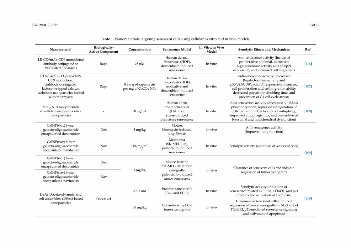
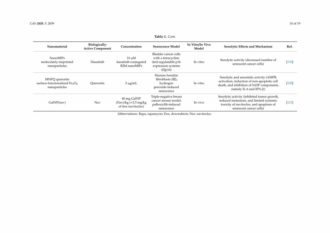
As the main purpose of senotherapy is to kill SCs, safe and effective detection and targetingof these cells is crucial to improving human health and prolonging lifespan [113]. Nano-basedsystems developed to identify and kill senescent cells can be considered as second-generationtargeted and selective senolytics that are able to efficiently eliminate senescent cells upon systemicadministration without causing adverse side effects. One of the best-explored groups of nano-senolyticsis smart nanodevices that are based on porous calcium carbonate nanoparticles, mesoporous silicananoparticles, carbon quantum dots, and molecularly-imprinted polymer nanoparticles (nanoMIPs)(Figure 4) [114]. Targeted delivery and detection/elimination of SCs can be achieved by encapsulation ofsenolytics/senomorphics/fluorophores using a number of nanomaterials. For example, cargo release inthe presence of β-galactosidase (β-gal) was due to the hydrolysis of the capping galacto-oligosaccharide(Gal) polymer [109]. In vitro studies demonstrated that nanomaterials covered with Gal and loadedwith fluorophores (e.g., rhodamine B, indocyanine green, coumarin-6, or Nile blue) were preferentiallyactivated in β-galactosidase-overexpressing SCs, which were able to lyse the galacto-oligosaccharidecoat (Figure 3) [85,114]. Moreover, β-galactosidase-instructed supramolecular assemblies can alsolead to the formulation of hydrogels and nanofibers in SCs, which decreases the expression ofsenescence-driving proteins [115]. Apart fromβ-gal, increased expression of other lysosomal hydrolases(e.g., α-L-fucosidase) has been used for detection of senescent cells [116]. To date, a collection ofsenoprobes has been described [114]. Nano-based senoprobes could be utilized to monitor the responseof tumors to the administration of senescence-inducing chemotherapeutic drugs. More recently, anothermethod for the real-time in vivo detection of senescent cells based on mesoporous silica nanoparticlesloaded with Nile blue and coated with a galacto-hexasaccharide was proposed [117]. Functionalizednanomaterials appear to have a promising potential as nanocarriers and can be used for improvingSC clearance. Table 1 provides published examples of in vitro and in vivo senotherapeutic action ofnano-based drug delivery systems [83,107–110,114,118–121].

Cells 2020, 9, 2659 8 of 19
Cells 2020, 9, x FOR PEER REVIEW 11 of 19
optimal nanocarrier type for senolytic drug delivery as well as optimal biocompatible concentration(s), improved drug encapsulation, and ligand conjugation efficiency. The parameters of nanocarrier absorption, interactions with human biological fluids, distribution, metabolism, and excretion are also important for future anti-aging applications of senotherapeutic nanocarriers. In conclusion, a nano-based drug delivery system constitutes a novel strategy to target senescent cells for potential therapeutic interventions. While few initial studies are promising, additional further studies involving the development of second-generation targeted and selective senolytics, namely senescence-targeted nanocarriers and validation of nano-senolytics using preclinical aging model systems and clinical trials are needed to determine whether these nanodevices might be effective against age-related diseases.
Figure 4. Different examples of nanocarriers used for targeted delivery of senolytics/senomorphics to senescent cells. Chemical structures of the loaded cargos are presented. LR, rapamycin-loaded PEGylated liposomes; NPs, nanoparticles; PEG, polyethylene glycol; Lac-PEG, lactose-polyethylene
Figure 4. Different examples of nanocarriers used for targeted delivery of senolytics/senomorphicsto senescent cells. Chemical structures of the loaded cargos are presented. LR, rapamycin-loadedPEGylated liposomes; NPs, nanoparticles; PEG, polyethylene glycol; Lac-PEG, lactose-polyethyleneglycol; Gal, galacto-oligosaccharide; DSAs, docetaxel-tannic acid self-assemblies; NanoMIPs,molecularly imprinted nanoparticles; B2M, beta-2 microglobulin; Gal6, 6-mer galacto-oligosaccharide;Nav, navitoclax; MNPQ, quercetin surface-functionalized Fe3O4 nanoparticles.
Cells 2020, 9, 2659 9 of 19
Table 1. Nanomaterials targeting senescent cells using cellular in vitro and in vivo models.
Nanomaterial Biologically-Active Component Concentration Senescence Model In Vitro/In Vivo
Model Senolytic Effects and Mechanism Ref.
LR-CD9mAb CD9 monoclonalantibody conjugated toPEGylated liposomes
Rapa 25 nM
Human dermalfibroblasts (HDF),
doxorubicin-inducedsenescence
In vitro
Anti-senescence activity (increasedproliferative potential, decreased
β-galactosidase activity and p53/p21expression, and increased cell migration)
[118]
CD9-Lac/CaCO3/Rapa NPsCD9 monoclonal
antibody-conjugatedlactose-wrapped calcium
carbonate nanoparticles loadedwith rapamycin
Rapa 0.2 mg of rapamycinper mg of CaCO3 NPs
Human dermalfibroblasts (HDF),
replicative anddoxorubicin-induced
senescence
In vitro
Anti-senescence activity (decreasedβ-galactosidase activity and
p53/p21/CD9/cyclin D1 expression, increasedcell proliferation and cell migration ability,decreased population doubling time, and
prevention of G1 cell cycle arrest)
[107]
MoS2 NPs molybdenumdisulfide mesoporous silica
nanoparticles- 50 µg/mL
Human aorticendothelial cells
(HAECs),stress-induced
premature senescence
In vitro
Anti-senescence activity (decreased γ−H2AXphosphorylation, repressed upregulation ofp16, p21 and p53, activation of autophagy,
improved autophagic flux, and prevention oflysosomal and mitochondrial dysfunction)
[108]
GalNP(dox) 6-mergalacto-oligosaccharide
encapsulated doxorubicinDox 1 mg/kg
Mouse,bleomycin-induced
lung fibrosisIn vivo Anti-senescence activity
(improved lung function)
[109]
GalNP(nav) 6-mergalacto-oligosaccharideencapsulated navitoclax
Nav 0.06 mg/mL
Melanoma(SK-MEL-103),
palbociclib-inducedsenescence
In vitro Senolytic activity (apoptosis of senescent cells)
GalNP(dox) 6-mergalacto-oligosaccharide
encapsulated doxorubicinDox
1 mg/kg
Mouse-bearingSK-MEL-103 tumor
xenografts,palbociclib-inducedtumor senescence
In vivoClearance of senescent cells and induced
regression of tumor xenograftsGalNP(nav) 6-mergalacto-oligosaccharideencapsulated navitoclax
Nav
DSAs Docetaxel-tannic acidself-assemblies (DSAs)-based
nanoparticlesDocetaxel
2.5-5 nM Prostate cancer cells(C4-2 and PC- 3) In vitro
Senolytic activity (inhibition ofsenescence-related TGFβR1, FOXO1, and p21
proteins and activation of apoptosis)[119]
30 mg/kg Mouse-bearing PC-3tumor xenografts In vivo
Clearance of senescent cells (inducedregression of tumor xenografts by blockade ofTGFβR1/p21-mediated senescence signaling
and activation of apoptosis)
Cells 2020, 9, 2659 10 of 19
Table 1. Cont.
Nanomaterial Biologically-Active Component Concentration Senescence Model In Vitro/In Vivo
Model Senolytic Effects and Mechanism Ref.
NanoMIPsmolecularly-imprinted
nanoparticlesDasatinib
10 µMdasatinib-conjugated
B2M nanoMIPs
Bladder cancer cellswith a tetracycline
(tet)-regulatable p16expression systems
(EJp16)
In vitro Senolytic activity (decreased number ofsenescent cancer cells) [110]
MNPQ quercetinsurface-functionalized Fe3O4
nanoparticlesQuercetin 5 µg/mL
Human foreskinfibroblasts (BJ),
hydrogenperoxide-induced
senescence
In vitro
Senolytic and senostatic activity (AMPKactivation, induction of non-apoptotic celldeath, and inhibition of SASP components,
namely IL-6 and IFN-β)
[120]
GalNP(nav) Nav40 mg GalNP
(Nav)/kg (≈2.5 mg/kgof free navitoclax)
Triple-negative breastcancer mouse model,palbociclib-induced
senescence
In vivo
Senolytic activity (inhibited tumor growth,reduced metastasis, and limited systemic
toxicity of navitoclax, and apoptosis ofsenescent cancer cells)
[121]
Abbreviations: Rapa, rapamycin; Dox, doxorubicin; Nav, navitoclax.
Cells 2020, 9, 2659 11 of 19
The first-reported SC-targeted cargo delivery nano-system is a nano-structure based on mesoporoussilica nanoparticles (MSN S1) coated with galacto-oligosaccharides and loaded with rhodamine B [85].MSN S1 are engulfed by human SCs and activated by SA-β-gal. There are also studies showingthat encapsulation of a senolytic agent, namely navitoclax with β(1,4)-galacto-oligosaccharides iseffective in clearing SCs in models of damage-induced and chemotherapy-induced senescence [109].Nanostructures conjugated with drugs may also exert a senomorphic effect by inhibiting theSASP [107,120]. A recent in vitro study demonstrated that nanoparticles functionalized with monoclonalantibody against CD9 receptor (overexpressed in old cells) and loaded with rapamycin (an mTORinhibitor with well-recognized anti-aging activity) (CD9-Lac/CaCO3/Rapa NPs) have anti-senescenceeffect [107]. CD9-Lac/CaCO3/Rapa NPs significantly improved the proliferative capacity of SCs,which was accompanied by lower expression of IL-6 and IL-1β, the SASP components [107]. Moreover,CD9-targeted PEGylated liposomes have been documented as a promising drug delivery platform totarget senescent cells [118]. The uptake of CD9-targeted liposomes by premature senescent humandermal fibroblasts (HDFs) was revealed to be higher than that by young HDFs [118]. Targeteddelivery of rapamycin (LR-CD9mAb) to diminish senescence of CD9-receptor-overexpressing cellswas also explored and rapamycin was found to promote cell proliferation and reduce the levels ofSA-β-gal-positive cells [118]. Another study performed by Ke et al. [108] showed that MoS2 NPssuppressed hydrogen-peroxide-induced senescence in endothelial cells. In addition, MoS2 NP treatmentleads to autophagy activation, which suppressed SIPS in vitro [108]. More recently, the docetaxel–tannicacid self-assembly (DSA)-based nanoparticle strategy has been developed and documented to be anefficient delivery system to provide docetaxel to prostate cancer cells and xenograft tumors and diminishcellular senescence [119]. DSAs-induced anti-senescence and anti-cancer effects were mediated bythe inhibition of TGFβR1/FOXO1/p21-associated senescence and apoptosis induction in vitro andin vivo [119]. The authors concluded that DSAs can be considered as a useful nano-platform todeliver anticancer molecules (here docetaxel) to cancer cells that would result in improved therapeuticbenefits and limited chemotherapy-induced senescence and drug resistance [119]. Targeted clearanceof SCs using B2M (β2 microglobulin) nanoMIPs (molecularly imprinted nanoparticles) has been alsodescribed [110]. B2M nanoMIPs specifically recognize one of the previously-identified members of acell membrane surface protein that is overexpressed in SCs, namely B2M [110]. In addition, it has beenshown that B2M nanoMIPs loaded with a senolytic drug (dasatinib) can kill senescent bladder cancercells [110]. Moreover, fluorescently-tagged nanoMIPs detected senescent cells in vivo and were nottoxic when injected at single doses [110]. The authors concluded that nanoMIPs may have diagnostic,prognostic, and therapeutic potential when considering the pathological conditions mediated by theaccumulation of senescent cells [110]. We have also documented a senolytic and senostatic activity ofquercetin surface-functionalized magnetite nanoparticles (MNPQ) in prematurely-senescent humanfibroblasts (hydrogen peroxide treatment in vitro) [120]. It has been shown that MNPQ are ableto eliminate senescent human fibroblast cells in vitro [120]. MNPQ diminish senescence-mediatedproinflammatory response as judged by decreased secretion of IL-8 and IFN-β that was accompaniedby the activation of AMP-activated protein kinase (AMPK) [120].
5. Conclusions
The nano-based drug delivery system and its applications in the field of aging research havedeveloped and evolved over the last several years. Because of their nano-sized nature and their abilityto meet diverse functionalities, senescent-cell-targeted nanocarriers may have potential for eliminationof senescent cells from the human body, which may improve the treatment of age-related disorders.However, many challenges restrain their successful clinical translation. Firstly, our current knowledgeabout molecular mechanisms underlying cellular senescence and modulating senescence-associatedsecretory phenotype in the context of age-associated disorders is not yet complete. In addition,successful application of senotherapeutic nanocarriers may depend on the development of best-suitedmethods for minimizing potential off-target effects and maximizing on-target effects. Next, for those

Cells 2020, 9, 2659 12 of 19
who study senescent-cell-targeted nanocarriers, it may be challenging to find an optimal nanocarriertype for senolytic drug delivery as well as optimal biocompatible concentration(s), improved drugencapsulation, and ligand conjugation efficiency. The parameters of nanocarrier absorption, interactionswith human biological fluids, distribution, metabolism, and excretion are also important for futureanti-aging applications of senotherapeutic nanocarriers. In conclusion, a nano-based drug deliverysystem constitutes a novel strategy to target senescent cells for potential therapeutic interventions.While few initial studies are promising, additional further studies involving the development ofsecond-generation targeted and selective senolytics, namely senescence-targeted nanocarriers andvalidation of nano-senolytics using preclinical aging model systems and clinical trials are needed todetermine whether these nanodevices might be effective against age-related diseases.
Author Contributions: Conceptualization, J.A.-G. and A.L.; writing—original draft preparation, J.A.-G. and A.L.;writing—review and editing, J.A.-G. and A.L.; visualization, J.A.-G.; supervision, J.A.-G. and A.L. All authorshave read and agreed to the published version of the manuscript.
Funding: This research received no external funding.
Acknowledgments: Figures were created using Biorender, https://biorender.com/.
Conflicts of Interest: The authors declare no conflict of interest.
Abbreviations
17-AAG tanespimycin17-DMAG alvespimycinAKT protein kinase BAMPK AMP-activated protein kinaseB2M Beta-2 microglobulinBcl-2 B cell lymphoma 2 familyBETd BET family protein degraderCAR Chimeric antigen receptorCCL-16 Monotactin-1CDKI Cyclin-dependent kinase inhibitorCS Cellular senescenceDAMPs Damage-associated molecular patternsDDR DNA damage responseDNA Deoxyribonucleic acidDox DoxorubicinDSAs Docetaxel-tannic acid self-assembliesEGF Endothelial growth factorEGF Epidermal growth factorEVs Extracellular vesiclesFOXO4 Forkhead box protein O4Gal Galacto-oligosaccharideGal6 6-mer galacto-oligosaccharideHAECs Human aortic endothelial cellsHDF Human dermal fibroblastsHIF-1α Hypoxia inducible factor 1αHSP90 Chaperone heat shock protein 90IFN-β Interferon betaIGF-1 Insulin-like growth factor 1IL-6 Interleukin 6IL-8 Interleukin 8IL-1β Interleukin 1βJAK Janus kinaseLac-PEG Lactose-polyethylene glycolLR Rapamycin-loaded PEGylated liposomes

Cells 2020, 9, 2659 13 of 19
MAPK Mitogen-activated protein kinaseMCP-1 Monocyte chemoattractant protein 1MIP-1α Macrophage inflammatory protein 1αmiRNA MicroRNAMMPs MetalloproteinasesMoS2 Molybdenum disulfideMNPQ Quercetin surface-functionalized Fe3O4 nanoparticlesMSN Mesoporous silica nanoparticlesmTOR Mammalian target of rapamycinnanoMIPs Molecularly-imprinted polymer nanoparticlesNav NavitoclaxNF Nuclear factorNHEJ Non-homologous end joiningNPs NanoparticlesOIS Oncogene-induced senescencep16 Cyclin-dependent kinase inhibitor 2Ap21 Cyclin-dependent kinase inhibitor 1Ap53 Tumor suppressor protein p53PEG Polyethylene glycolPI3K Phosphatidylinositol-3-kinaseRapa RapamycinRb RetinoblastomaRNA Ribonucleic acidROS Reactive oxygen speciesSAHF Senescence-associated heterochromatin fociSASP Senescence-associated secretory phenotypeSA-β-gal Senescence associated β-galactosidaseSCAPs Senescent cell anti-apoptotic pathwaysSCs Senescent cellsSIPS Stress-induced premature senescencesiRNA Small interfering RNASTAT Signal transducer and activator of transcriptionuPAR Urokinase-type plasminogen activator receptor
References
1. Rodier, F.; Campisi, J. Four faces of cellular senescence. J. Cell Biol. 2011, 192, 547–556. [CrossRef] [PubMed]2. Herranz, N.; Gil, J. Mechanisms and functions of cellular senescence. J. Clin. Investig. 2018, 128, 1238–1246.
[CrossRef] [PubMed]3. Hayflick, L.; Moorhead, P.S. The serial cultivation of human diploid cell strains. Exp. Cell Res. 1961, 25,
585–621. [CrossRef]4. Shay, J.W.; Wright, W.E. Telomeres and telomerase: Three decades of progress. Nat. Rev. Genet. 2019, 20,
299–309. [CrossRef]5. Bodnar, A.G. Extension of Life-Span by Introduction of Telomerase into Normal Human Cells. Science 1998,
279, 349–352. [CrossRef]6. Dimri, G.P.; Lee, X.; Basile, G.; Acosta, M.; Scott, G.; Roskelley, C.; Medrano, E.E.; Linskens, M.; Rubelj, I.;
Pereira-Smith, O. A biomarker that identifies senescent human cells in culture and in aging skin in vivo.Proc. Natl. Acad. Sci. USA 1995, 92, 9363–9367. [CrossRef]
7. Chen, Q.; Fischer, A.; Reagan, J.D.; Yan, L.J.; Ames, B.N. Oxidative DNA damage and senescence of humandiploid fibroblast cells. Proc. Natl. Acad. Sci. USA 1995, 92, 4337–4341. [CrossRef]
8. Kuilman, T.; Michaloglou, C.; Mooi, W.J.; Peeper, D.S. The essence of senescence. Genes Dev. 2010, 24,2463–2479. [CrossRef]

Cells 2020, 9, 2659 14 of 19
9. Toussaint, O.; Medrano, E.E.; von Zglinicki, T. Cellular and molecular mechanisms of stress-inducedpremature senescence (SIPS) of human diploid fibroblasts and melanocytes. Exp. Gerontol. 2000, 35, 927–945.[CrossRef]
10. Serrano, M.; Lin, A.W.; McCurrach, M.E.; Beach, D.; Lowe, S.W. Oncogenic ras Provokes Premature CellSenescence Associated with Accumulation of p53 and p16INK4a. Cell 1997, 88, 593–602. [CrossRef]
11. Collado, M.; Serrano, M. Senescence in tumours: Evidence from mice and humans. Nat. Rev. Cancer 2010, 10,51–57. [CrossRef] [PubMed]
12. Storer, M.; Mas, A.; Robert-Moreno, A.; Pecoraro, M.; Ortells, M.C.; Di Giacomo, V.; Yosef, R.; Pilpel, N.;Krizhanovsky, V.; Sharpe, J.; et al. Senescence Is a Developmental Mechanism that Contributes to EmbryonicGrowth and Patterning. Cell 2013, 155, 1119–1130. [CrossRef] [PubMed]
13. Muñoz-Espín, D.; Serrano, M. Cellular senescence: From physiology to pathology. Nat. Rev. Mol. Cell Biol.2014, 15, 482–496. [CrossRef] [PubMed]
14. Tchkonia, T.; Morbeck, D.E.; Von Zglinicki, T.; Van Deursen, J.; Lustgarten, J.; Scrable, H.; Khosla, S.;Jensen, M.D.; Kirkland, J.L. Fat tissue, aging, and cellular senescence: Fat tissue and aging. Aging Cell 2010,9, 667–684. [CrossRef] [PubMed]
15. Naylor, R.M.; Baker, D.J.; van Deursen, J.M. Senescent cells: A novel therapeutic target for aging andage-related diseases. Clin. Pharmacol. Ther. 2013, 93, 105–116. [CrossRef] [PubMed]
16. van Deursen, J.M. The role of senescent cells in ageing. Nature 2014, 509, 439–446. [CrossRef] [PubMed]17. He, S.; Sharpless, N.E. Senescence in Health and Disease. Cell 2017, 169, 1000–1011. [CrossRef] [PubMed]18. Kritsilis, M.; Rizou, V.S.; Koutsoudaki, P.N.; Evangelou, K.; Gorgoulis, V.G.; Papadopoulos, D. Ageing,
Cellular Senescence and Neurodegenerative Disease. Int. J. Mol. Sci. 2018, 19, 2937. [CrossRef] [PubMed]19. Palmer, A.K.; Gustafson, B.; Kirkland, J.L.; Smith, U. Cellular senescence: At the nexus between ageing and
diabetes. Diabetologia 2019, 62, 1835–1841. [CrossRef] [PubMed]20. Wei, W.; Ji, S. Cellular senescence: Molecular mechanisms and pathogenicity. J. Cell. Physiol. 2018, 233,
9121–9135. [CrossRef] [PubMed]21. Sikora, E.; Bielak-Zmijewska, A.; Mosieniak, G. Targeting normal and cancer senescent cells as a strategy of
senotherapy. Ageing Res. Rev. 2019, 55, 100941. [CrossRef] [PubMed]22. Terzi, M.Y.; Izmirli, M.; Gogebakan, B. The cell fate: Senescence or quiescence. Mol. Biol. Rep. 2016, 43,
1213–1220. [CrossRef] [PubMed]23. Sharpless, N.E.; Sherr, C.J. Forging a signature of in vivo senescence. Nat. Rev. Cancer 2015, 15, 397–408.
[CrossRef] [PubMed]24. Childs, B.G.; Gluscevic, M.; Baker, D.J.; Laberge, R.M.; Marquess, D.; Dananberg, J.; van Deursen, J.M.
Senescent cells: An emerging target for diseases of ageing. Nat Rev Drug Discov. 2017, 16, 718–735. [CrossRef][PubMed]
25. von Kobbe, C. Targeting senescent cells: Approaches, opportunities, challenges. Aging 2019, 11, 12844–12861.[CrossRef] [PubMed]
26. Coppé, J.-P.; Patil, C.K.; Rodier, F.; Sun, Y.; Muñoz, D.P.; Goldstein, J.; Nelson, P.S.; Desprez, P.-Y.; Campisi, J.Senescence-associated secretory phenotypes reveal cell-nonautonomous functions of oncogenic RAS and thep53 tumor suppressor. PLoS Biol. 2008, 6, 2853–2868. [CrossRef]
27. Kuilman, T.; Peeper, D.S. Senescence-messaging secretome: SMS-ing cellular stress. Nat. Rev. Cancer 2009, 9,81–94. [CrossRef]
28. Rodier, F.; Coppé, J.-P.; Patil, C.K.; Hoeijmakers, W.A.M.; Muñoz, D.P.; Raza, S.R.; Freund, A.; Campeau, E.;Davalos, A.R.; Campisi, J. Persistent DNA damage signalling triggers senescence-associated inflammatorycytokine secretion. Nat. Cell Biol. 2009, 11, 973–979. [CrossRef]
29. Coppé, J.-P.; Desprez, P.-Y.; Krtolica, A.; Campisi, J. The senescence-associated secretory phenotype: The darkside of tumor suppression. Annu. Rev. Pathol. 2010, 5, 99–118. [CrossRef]
30. Kirkland, J.L.; Tchkonia, T. Cellular Senescence: A Translational Perspective. EBioMedicine 2017, 21, 21–28.[CrossRef]
31. Panda, A.C.; Abdelmohsen, K.; Gorospe, M. SASP regulation by noncoding RNA. Mech. Ageing Dev. 2017,168, 37–43. [CrossRef] [PubMed]
32. Kang, T.-W.; Yevsa, T.; Woller, N.; Hoenicke, L.; Wuestefeld, T.; Dauch, D.; Hohmeyer, A.; Gereke, M.;Rudalska, R.; Potapova, A.; et al. Senescence surveillance of pre-malignant hepatocytes limits liver cancerdevelopment. Nature 2011, 479, 547–551. [CrossRef] [PubMed]

Cells 2020, 9, 2659 15 of 19
33. Yun, M.H. Cellular senescence in tissue repair: Every cloud has a silver lining. Int. J. Dev. Biol. 2018, 62,591–604. [CrossRef] [PubMed]
34. Acosta, J.C.; O’Loghlen, A.; Banito, A.; Guijarro, M.V.; Augert, A.; Raguz, S.; Fumagalli, M.; Da Costa, M.;Brown, C.; Popov, N.; et al. Chemokine Signaling via the CXCR2 Receptor Reinforces Senescence. Cell 2008,133, 1006–1018. [CrossRef]
35. Krtolica, A.; Parrinello, S.; Lockett, S.; Desprez, P.-Y.; Campisi, J. Senescent fibroblasts promote epithelial cellgrowth and tumorigenesis: A link between cancer and aging. Proc. Natl. Acad. Sci. USA 2001, 98, 12072.[CrossRef]
36. Vizioli, M.G.; Adams, P.D. Senescence Can Be BETter without the SASP? Cancer Discov. 2016, 6, 576–578.[CrossRef]
37. Soto-Gamez, A.; Demaria, M. Therapeutic interventions for aging: The case of cellular senescence. DrugDiscov. Today 2017, 22, 786–795. [CrossRef]
38. Sakoda, K.; Yamamoto, M.; Negishi, Y.; Liao, J.K.; Node, K.; Izumi, Y. Simvastatin Decreases IL-6 and IL-8Production in Epithelial Cells. J. Dent. Res. 2006, 85, 520–523. [CrossRef]
39. Lim, H.; Park, H.; Kim, H.P. Effects of flavonoids on senescence-associated secretory phenotype formationfrom bleomycin-induced senescence in BJ fibroblasts. Biochem. Pharmacol. 2015, 96, 337–348. [CrossRef]
40. Hoare, M.; Ito, Y.; Kang, T.-W.; Weekes, M.P.; Matheson, N.J.; Patten, D.A.; Shetty, S.; Parry, A.J.; Menon, S.;Salama, R.; et al. NOTCH1 mediates a switch between two distinct secretomes during senescence. Nat. CellBiol. 2016, 18, 979–992. [CrossRef]
41. Nelson, G.; Wordsworth, J.; Wang, C.; Jurk, D.; Lawless, C.; Martin-Ruiz, C.; von Zglinicki, T. A senescentcell bystander effect: Senescence-induced senescence. Aging Cell 2012, 11, 345–349. [CrossRef] [PubMed]
42. Lehmann, B.D.; Paine, M.S.; Brooks, A.M.; McCubrey, J.A.; Renegar, R.H.; Wang, R.; Terrian, D.M.Senescence-Associated Exosome Release from Human Prostate Cancer Cells. Cancer Res. 2008, 68, 7864–7871.[CrossRef] [PubMed]
43. Fuhrmann-Stroissnigg, H.; Niedernhofer, L.J.; Robbins, P.D. Hsp90 inhibitors as senolytic drugs to extendhealthy aging. Cell Cycle Georget. Tex 2018, 17, 1048–1055. [CrossRef] [PubMed]
44. Sajja, H.; East, M.; Mao, H.; Wang, Y.; Nie, S.; Yang, L. Development of Multifunctional Nanoparticles forTargeted Drug Delivery and Noninvasive Imaging of Therapeutic Effect. Curr. Drug Discov. Technol. 2009, 6,43–51. [CrossRef]
45. Obeid, M.A.; Al Qaraghuli, M.M.; Alsaadi, M.; Alzahrani, A.R.; Niwasabutra, K.; Ferro, V.A. Deliveringnatural products and biotherapeutics to improve drug efficacy. Ther. Deliv. 2017, 8, 947–956. [CrossRef]
46. Patra, J.K.; Das, G.; Fraceto, L.F.; Campos, E.V.R.; Rodriguez-Torres, M.P.; Acosta-Torres, L.S.; Diaz-Torres, L.A.;Grillo, R.; Swamy, M.K.; Sharma, S.; et al. Nano based drug delivery systems: Recent developments andfuture prospects. J. Nanobiotechnol. 2018, 16, 71. [CrossRef]
47. Chenthamara, D.; Subramaniam, S.; Ramakrishnan, S.G.; Krishnaswamy, S.; Essa, M.M.; Lin, F.-H.;Qoronfleh, M.W. Therapeutic efficacy of nanoparticles and routes of administration. Biomater. Res. 2019, 23,20. [CrossRef]
48. Farokhzad, O.; Langer, R. Nanomedicine: Developing smarter therapeutic and diagnostic modalities. Adv.Drug Deliv. Rev. 2006, 58, 1456–1459. [CrossRef]
49. Nie, S.; Xing, Y.; Kim, G.J.; Simons, J.W. Nanotechnology Applications in Cancer. Annu. Rev. Biomed. Eng.2007, 9, 257–288. [CrossRef]
50. Seigneuric, R.; Markey, L.; Nuyten, S.A.D.; Dubernet, C.; Evelo, T.A.C.; Finot, E.; Garrido, C. FromNanotechnology to Nanomedicine: Applications to Cancer Research. Curr. Mol. Med. 2010, 10, 640–652.[CrossRef]
51. Kolahalam, L.A.; Kasi Viswanath, I.V.; Diwakar, B.S.; Govindh, B.; Reddy, V.; Murthy, Y.L.N. Review onnanomaterials: Synthesis and applications. Mater. Today Proc. 2019, 18, 2182–2190. [CrossRef]
52. Ventola, C.L. The nanomedicine revolution: Part 1: Emerging concepts. P T Peer-Rev. J. Formul. Manag. 2012,37, 512–525.
53. Salatin, S.; Yari Khosroushahi, A. Overviews on the cellular uptake mechanism of polysaccharide colloidalnanoparticles. J. Cell. Mol. Med. 2017, 21, 1668–1686. [CrossRef] [PubMed]
54. Kargozar, S.; Mozafari, M. Nanotechnology and Nanomedicine: Start small, think big. Mater. Today Proc.2018, 5, 15492–15500. [CrossRef]

Cells 2020, 9, 2659 16 of 19
55. Paez-Ribes, M.; González-Gualda, E.; Doherty, G.J.; Muñoz-Espín, D. Targeting senescent cells in translationalmedicine. EMBO Mol. Med. 2019, 11. [CrossRef]
56. Pelaz, B.; del Pino, P.; Maffre, P.; Hartmann, R.; Gallego, M.; Rivera-Fernández, S.; de la Fuente, J.M.;Nienhaus, G.U.; Parak, W.J. Surface Functionalization of Nanoparticles with Polyethylene Glycol: Effects onProtein Adsorption and Cellular Uptake. ACS Nano 2015, 9, 6996–7008. [CrossRef]
57. Schöttler, S.; Becker, G.; Winzen, S.; Steinbach, T.; Mohr, K.; Landfester, K.; Mailänder, V.; Wurm, F.R. Proteinadsorption is required for stealth effect of poly(ethylene glycol)- and poly(phosphoester)-coated nanocarriers.Nat. Nanotechnol. 2016, 11, 372–377. [CrossRef]
58. Kolhar, P.; Anselmo, A.C.; Gupta, V.; Pant, K.; Prabhakarpandian, B.; Ruoslahti, E.; Mitragotri, S. Using shapeeffects to target antibody-coated nanoparticles to lung and brain endothelium. Proc. Natl. Acad. Sci. USA2013, 110, 10753–10758. [CrossRef]
59. Gao, H.; Yang, Z.; Zhang, S.; Cao, S.; Shen, S.; Pang, Z.; Jiang, X. Ligand modified nanoparticles increasescell uptake, alters endocytosis and elevates glioma distribution and internalization. Sci. Rep. 2013, 3, 2534.[CrossRef]
60. Gao, W.; Zhang, L. Coating nanoparticles with cell membranes for targeted drug delivery. J. Drug Target.2015, 23, 619–626. [CrossRef]
61. Suk, J.S.; Xu, Q.; Kim, N.; Hanes, J.; Ensign, L.M. PEGylation as a strategy for improving nanoparticle-baseddrug and gene delivery. Adv. Drug Deliv. Rev. 2016, 99, 28–51. [CrossRef] [PubMed]
62. Zavaleta, C.; Ho, D.; Chung, E.J. Theranostic Nanoparticles for Tracking and Monitoring Disease State. SLASTechnol. Transl. Life Sci. Innov. 2018, 23, 281–293. [CrossRef] [PubMed]
63. Ramanathan, S.; Archunan, G.; Sivakumar, M.; Tamil Selvan, S.; Fred, A.L.; Kumar, S.; Gulyás, B.;Padmanabhan, P. Theranostic applications of nanoparticles in neurodegenerative disorders. Int. J. Nanomed.2018, 13, 5561–5576. [CrossRef] [PubMed]
64. Haley, B.; Frenkel, E. Nanoparticles for drug delivery in cancer treatment. Urol. Oncol. Semin. Orig. Investig.2008, 26, 57–64. [CrossRef] [PubMed]
65. Chen, F.; Ehlerding, E.B.; Cai, W. Theranostic nanoparticles. J. Nucl. Med. 2014, 55, 1919–1922. [CrossRef][PubMed]
66. Chen, G.; Roy, I.; Yang, C.; Prasad, P.N. Nanochemistry and Nanomedicine for Nanoparticle-based Diagnosticsand Therapy. Chem. Rev. 2016, 116, 2826–2885. [CrossRef] [PubMed]
67. Riccardi, C.; Fàbrega, C.; Grijalvo, S.; Vitiello, G.; D’Errico, G.; Eritja, R.; Montesarchio, D. AS1411-decoratedniosomes as effective nanocarriers for Ru(iii)-based drugs in anticancer strategies. J. Mater. Chem. B 2018, 6,5368–5384. [CrossRef] [PubMed]
68. Squillaro, T.; Cimini, A.; Peluso, G.; Giordano, A.; Melone, M.A.B. Nano-delivery systems for encapsulationof dietary polyphenols: An experimental approach for neurodegenerative diseases and brain tumors.Biochem. Pharmacol. 2018, 154, 303–317. [CrossRef]
69. Di Martino, P.; Censi, R.; Gigliobianco, M.R.; Zerrillo, L.; Magnoni, F.; Agas, D.; Quaglia, W.; Lupidi, G.Nano-medicine Improving the Bioavailability of Small Molecules for the Prevention of NeurodegenerativeDiseases. Curr. Pharm. Des. 2017, 23, 1897–1908. [CrossRef]
70. McClements, D.J.; Xiao, H. Designing food structure and composition to enhance nutraceutical bioactivity tosupport cancer inhibition. Semin. Cancer Biol. 2017, 46, 215–226. [CrossRef]
71. Squillaro, T.; Peluso, G.; Melone, M.A.B. Nanotechnology-Based Polyphenol Delivery: A Novel TherapeuticStrategy for the Treatment of Age-Related Neurodegenerative Disorder. Austin Aging Res. 2017, 1, 1004.
72. Li, C.; Zhang, J.; Zu, Y.J.; Nie, S.F.; Cao, J.; Wang, Q.; Nie, S.P.; Deng, Z.Y.; Xie, M.Y.; Wang, S. Biocompatibleand biodegradable nanoparticles for enhancement of anti-cancer activities of phytochemicals. Chin. J. Nat.Med. 2015, 13, 641–652. [CrossRef]
73. Thomas, O.S.; Weber, W. Overcoming Physiological Barriers to Nanoparticle Delivery-Are We There Yet?Front. Bioeng. Biotechnol. 2019, 7, 415. [CrossRef] [PubMed]
74. Clark, A.J.; Davis, M.E. Increased brain uptake of targeted nanoparticles by adding an acid-cleavable linkagebetween transferrin and the nanoparticle core. Proc. Natl. Acad. Sci. USA 2015, 112, 12486–12491. [CrossRef][PubMed]
75. Wang, W.; Sun, C.; Mao, L.; Ma, P.; Liu, F.; Yang, J.; Gao, Y. The biological activities, chemical stability,metabolism and delivery systems of quercetin: A review. Trends Food Sci. Technol. 2016, 56, 21–38. [CrossRef]
Cells 2020, 9, 2659 17 of 19
76. Abdelwahab, S.I.; Sheikh, B.Y.; Taha, M.M.E.; How, C.W.; Abdullah, R.; Yagoub, U.; El-Sunousi, R.; Eid, E.E.M.Thymoquinone-loaded nanostructured lipid carriers: Preparation, gastroprotection, in vitro toxicity, andpharmacokinetic properties after extravascular administration. Int. J. Nanomed. 2013, 8, 2163–2172. [CrossRef]
77. Sharma, M.; Sharma, R.; Jain, D.K. Nanotechnology Based Approaches for Enhancing Oral Bioavailability ofPoorly Water Soluble Antihypertensive Drugs. Scientifica 2016, 2016, 8525679. [CrossRef]
78. Caster, J.M.; Patel, A.N.; Zhang, T.; Wang, A. Investigational nanomedicines in 2016: A review ofnanotherapeutics currently undergoing clinical trials: Investigational nanomedicines in 2016. WileyInterdiscip. Rev. Nanomed. Nanobiotechnol. 2017, 9, e1416. [CrossRef]
79. Mura, S.; Nicolas, J.; Couvreur, P. Stimuli-responsive nanocarriers for drug delivery. Nat. Mater. 2013, 12,991–1003. [CrossRef]
80. Liu, J.; Huang, Y.; Kumar, A.; Tan, A.; Jin, S.; Mozhi, A.; Liang, X.-J. pH-Sensitive nano-systems for drugdelivery in cancer therapy. Biotechnol. Adv. 2014, 32, 693–710. [CrossRef]
81. Wang, X.; Teng, Z.; Wang, H.; Wang, C.; Liu, Y.; Tang, Y.; Wu, J.; Sun, J.; Wang, H.; Wang, J.; et al. Increasingthe cytotoxicity of doxorubicin in breast cancer MCF-7 cells with multidrug resistance using a mesoporoussilica nanoparticle drug delivery system. Int. J. Clin. Exp. Pathol. 2014, 7, 1337–1347. [PubMed]
82. Hamidu, A.; Mokrish, A.; Mansor, R.; Razak, I.S.A.; Danmaigoro, A.; Jaji, A.Z.; Bakar, Z.A. Modified methodsof nanoparticles synthesis in pH-sensitive nano-carriers production for doxorubicin delivery on MCF-7breast cancer cell line. Int. J. Nanomed. 2019, 14, 3615–3627. [CrossRef]
83. Muñoz-Espín, D. Nanocarriers targeting senescent cells. Transl. Med. Aging 2019, 3, 1–5. [CrossRef]84. Lee, B.Y.; Han, J.A.; Im, J.S.; Morrone, A.; Johung, K.; Goodwin, E.C.; Kleijer, W.J.; DiMaio, D.; Hwang, E.S.
Senescence-associated β-galactosidase is lysosomal β-galactosidase. Aging Cell 2006, 5, 187–195. [CrossRef]85. Agostini, A.; Mondragón, L.; Bernardos, A.; Martínez-Máñez, R.; Marcos, M.D.; Sancenón, F.; Soto, J.;
Costero, A.; Manguan-García, C.; Perona, R.; et al. Targeted Cargo Delivery in Senescent Cells Using CappedMesoporous Silica Nanoparticles. Angew. Chem. Int. Ed. 2012, 51, 10556–10560. [CrossRef]
86. Ovadya, Y.; Krizhanovsky, V. Strategies targeting cellular senescence. J. Clin. Investig. 2018, 128, 1247–1254.[CrossRef]
87. Baker, D.J.; Childs, B.G.; Durik, M.; Wijers, M.E.; Sieben, C.J.; Zhong, J.; Saltness, R.A.; Jeganathan, K.B.;Verzosa, G.C.; Pezeshki, A.; et al. Naturally occurring p16(Ink4a)-positive cells shorten healthy lifespan.Nature 2016, 530, 184–189. [CrossRef]
88. Baker, D.J.; Wijshake, T.; Tchkonia, T.; LeBrasseur, N.K.; Childs, B.G.; van de Sluis, B.; Kirkland, J.L.; vanDeursen, J.M. Clearance of p16Ink4a-positive senescent cells delays ageing-associated disorders. Nature2011, 479, 232–236. [CrossRef]
89. Kirkland, J.L.; Tchkonia, T. Clinical strategies and animal models for developing senolytic agents. Exp.Gerontol. 2015, 68, 19–25. [CrossRef]
90. Zhu, Y.; Tchkonia, T.; Pirtskhalava, T.; Gower, A.C.; Ding, H.; Giorgadze, N.; Palmer, A.K.; Ikeno, Y.;Hubbard, G.B.; Lenburg, M.; et al. The Achilles’ heel of senescent cells: From transcriptome to senolyticdrugs. Aging Cell 2015, 14, 644–658. [CrossRef]
91. Zhu, Y.; Tchkonia, T.; Fuhrmann-Stroissnigg, H.; Dai, H.M.; Ling, Y.Y.; Stout, M.B.; Pirtskhalava, T.;Giorgadze, N.; Johnson, K.O.; Giles, C.B.; et al. Identification of a novel senolytic agent, navitoclax, targetingthe Bcl-2 family of anti-apoptotic factors. Aging Cell 2016, 15, 428–435. [CrossRef] [PubMed]
92. González-Gualda, E.; Pàez-Ribes, M.; Lozano-Torres, B.; Macias, D.; Wilson, J.R.; González-López, C.;Ou, H.-L.; Mirón-Barroso, S.; Zhang, Z.; Lérida-Viso, A.; et al. Galacto-conjugation of Navitoclax as anefficient strategy to increase senolytic specificity and reduce platelet toxicity. Aging Cell 2020, e13142.[CrossRef] [PubMed]
93. Justice, J.N.; Nambiar, A.M.; Tchkonia, T.; LeBrasseur, N.K.; Pascual, R.; Hashmi, S.K.; Prata, L.;Masternak, M.M.; Kritchevsky, S.B.; Musi, N.; et al. Senolytics in idiopathic pulmonary fibrosis: Results froma first-in-human, open-label, pilot study. EBioMedicine 2019, 40, 554–563. [CrossRef] [PubMed]
94. Schafer, M.J.; White, T.A.; Iijima, K.; Haak, A.J.; Ligresti, G.; Atkinson, E.J.; Oberg, A.L.; Birch, J.;Salmonowicz, H.; Zhu, Y.; et al. Cellular senescence mediates fibrotic pulmonary disease. Nat. Commun.2017, 8, 14532. [CrossRef] [PubMed]
95. Ogrodnik, M.; Miwa, S.; Tchkonia, T.; Tiniakos, D.; Wilson, C.L.; Lahat, A.; Day, C.P.; Burt, A.; Palmer, A.;Anstee, Q.M.; et al. Cellular senescence drives age-dependent hepatic steatosis. Nat. Commun. 2017, 8, 15691.[CrossRef] [PubMed]

Cells 2020, 9, 2659 18 of 19
96. Farr, J.N.; Xu, M.; Weivoda, M.M.; Monroe, D.G.; Fraser, D.G.; Onken, J.L.; Negley, B.A.; Sfeir, J.G.;Ogrodnik, M.B.; Hachfeld, C.M.; et al. Targeting cellular senescence prevents age-related bone loss in mice.Nat. Med. 2017, 23, 1072–1079. [CrossRef] [PubMed]
97. Zhu, Y.; Doornebal, E.J.; Pirtskhalava, T.; Giorgadze, N.; Wentworth, M.; Fuhrmann-Stroissnigg, H.;Niedernhofer, L.J.; Robbins, P.D.; Tchkonia, T.; Kirkland, J.L. New agents that target senescent cells: Theflavone, fisetin, and the BCL-XL inhibitors, A1331852 and A1155463. Aging 2017, 9, 955–963. [CrossRef]
98. Yousefzadeh, M.J.; Zhu, Y.; McGowan, S.J.; Angelini, L.; Fuhrmann-Stroissnigg, H.; Xu, M.; Ling, Y.Y.;Melos, K.I.; Pirtskhalava, T.; Inman, C.L.; et al. Fisetin is a senotherapeutic that extends health and lifespan.EBioMedicine 2018, 36, 18–28. [CrossRef]
99. Wang, Y.; Chang, J.; Liu, X.; Zhang, X.; Zhang, S.; Zhang, X.; Zhou, D.; Zheng, G. Discovery of piperlongumineas a potential novel lead for the development of senolytic agents. Aging 2016, 8, 2915–2926. [CrossRef]
100. Li, W.; He, Y.; Zhang, R.; Zheng, G.; Zhou, D. The curcumin analog EF24 is a novel senolytic agent. Aging2019, 11, 771–782. [CrossRef]
101. Samaraweera, L.; Adomako, A.; Rodriguez-Gabin, A.; McDaid, H.M. A Novel Indication for Panobinostat asa Senolytic Drug in NSCLC and HNSCC. Sci. Rep. 2017, 7, 1900. [CrossRef] [PubMed]
102. Baar, M.P.; Brandt, R.M.C.; Putavet, D.A.; Klein, J.D.D.; Derks, K.W.J.; Bourgeois, B.R.M.; Stryeck, S.;Rijksen, Y.; van Willigenburg, H.; Feijtel, D.A.; et al. Targeted Apoptosis of Senescent Cells Restores TissueHomeostasis in Response to Chemotoxicity and Aging. Cell 2017, 169, 132–147.e16. [CrossRef] [PubMed]
103. Kirkland, J.L.; Tchkonia, T.; Zhu, Y.; Niedernhofer, L.J.; Robbins, P.D. The Clinical Potential of SenolyticDrugs. J. Am. Geriatr. Soc. 2017, 65, 2297–2301. [CrossRef] [PubMed]
104. Amor, C.; Feucht, J.; Leibold, J.; Ho, Y.-J.; Zhu, C.; Alonso-Curbelo, D.; Mansilla-Soto, J.; Boyer, J.A.; Li, X.;Giavridis, T.; et al. Senolytic CAR T cells reverse senescence-associated pathologies. Nature 2020, 583,127–132. [CrossRef] [PubMed]
105. Wakita, M.; Takahashi, A.; Sano, O.; Loo, T.M.; Imai, Y.; Narukawa, M.; Iwata, H.; Matsudaira, T.; Kawamoto, S.;Ohtani, N.; et al. A BET family protein degrader provokes senolysis by targeting NHEJ and autophagy insenescent cells. Nat. Commun. 2020, 11, 1935. [CrossRef] [PubMed]
106. Mariño, G.; Niso-Santano, M.; Baehrecke, E.H.; Kroemer, G. Self-consumption: The interplay of autophagyand apoptosis. Nat. Rev. Mol. Cell Biol. 2014, 15, 81–94. [CrossRef]
107. Thapa, R.K.; Nguyen, H.T.; Jeong, J.-H.; Kim, J.R.; Choi, H.-G.; Yong, C.S.; Kim, J.O. Progressiveslowdown/prevention of cellular senescence by CD9-targeted delivery of rapamycin using lactose-wrappedcalcium carbonate nanoparticles. Sci. Rep. 2017, 7, 43299. [CrossRef]
108. Ke, S.; Lai, Y.; Zhou, T.; Li, L.; Wang, Y.; Ren, L.; Ye, S. Molybdenum Disulfide Nanoparticles Resist OxidativeStress-Mediated Impairment of Autophagic Flux and Mitigate Endothelial Cell Senescence and AngiogenicDysfunctions. ACS Biomater. Sci. Eng. 2018, 4, 663–674. [CrossRef]
109. Muñoz-Espín, D.; Rovira, M.; Galiana, I.; Giménez, C.; Lozano-Torres, B.; Paez-Ribes, M.; Llanos, S.; Chaib, S.;Muñoz-Martín, M.; Ucero, A.C.; et al. A versatile drug delivery system targeting senescent cells. EMBO Mol.Med. 2018, 10, e9355. [CrossRef]
110. Ekpenyong-Akiba, A.E.; Canfarotta, F.; Abd, H.B.; Poblocka, M.; Casulleras, M.; Castilla-Vallmanya, L.;Kocsis-Fodor, G.; Kelly, M.E.; Janus, J.; Althubiti, M.; et al. Detecting and targeting senescent cells usingmolecularly imprinted nanoparticles. Nanoscale Horiz. 2019, 4, 757–768. [CrossRef]
111. Musi, N.; Valentine, J.M.; Sickora, K.R.; Baeuerle, E.; Thompson, C.S.; Shen, Q.; Orr, M.E. Tau proteinaggregation is associated with cellular senescence in the brain. Aging Cell 2018, 17, e12840. [CrossRef][PubMed]
112. Riessland, M.; Kolisnyk, B.; Kim, T.W.; Cheng, J.; Ni, J.; Pearson, J.A.; Park, E.J.; Dam, K.; Acehan, D.;Ramos-Espiritu, L.S.; et al. Loss of SATB1 Induces p21-Dependent Cellular Senescence in Post-mitoticDopaminergic Neurons. Cell Stem. Cell 2019, 25, 514–530.e8. [CrossRef] [PubMed]
113. Salmonowicz, H.; Passos, J.F. Detecting senescence: A new method for an old pigment. Aging Cell 2017, 16,432–434. [CrossRef] [PubMed]
114. Muñoz-Espin, D.; Demaria, M. Senolytics in Disease, Ageing and Longevity, 1st ed.; Springer Nature Switzerland:Cham, Switzerland, 2020; pp. 163–180.
115. Xu, T.; Cai, Y.; Zhong, X.; Zhang, L.; Zheng, D.; Gao, Z.; Pan, X.; Wang, F.; Chen, M.; Yang, Z.β-Galactosidase instructed supramolecular hydrogelation for selective identification and removal of senescentcells. Chem. Commun. 2019, 55, 7175–7178. [CrossRef]

Cells 2020, 9, 2659 19 of 19
116. Hildebrand, D.; Lehle, S.; Borst, A.; Haferkamp, S.; Essmann, F.; Schulze-Osthoff, K. α-Fucosidase as a novelconvenient biomarker for cellular senescence. Cell Cycle 2013, 12, 1922–1927. [CrossRef]
117. Lozano-Torres, B.; Blandez, J.F.; Galiana, I.; García-Fernández, A.; Alfonso, M.; Marcos, M.D.; Orzáez, M.;Sancenón, F.; Martínez-Máñez, R. Real-Time In Vivo Detection of Cellular Senescence through the ControlledRelease of the NIR Fluorescent Dye Nile Blue. Angew. Chem. Int. Ed. 2020, 59, 15152–15156. [CrossRef]
118. Nguyen, H.T.; Thapa, R.K.; Shin, B.S.; Jeong, J.-H.; Kim, J.-R.; Yong, C.S.; Kim, J.O. CD9 monoclonalantibody-conjugated PEGylated liposomes for targeted delivery of rapamycin in the treatment of cellularsenescence. Nanotechnology 2017, 28, 095101. [CrossRef]
119. Nagesh, P.K.B.; Chowdhury, P.; Hatami, E.; Kumari, S.; Kashyap, V.K.; Tripathi, M.K.; Wagh, S.; Meibohm, B.;Chauhan, S.C.; Jaggi, M.; et al. Cross-Linked Polyphenol-Based Drug Nano-Self-Assemblies Engineered toBlockade Prostate Cancer Senescence. ACS Appl. Mater. Interfaces 2019, 11, 38537–38554. [CrossRef]
120. Lewinska, A.; Adamczyk-Grochala, J.; Bloniarz, D.; Olszowka, J.; Kulpa-Greszta, M.; Litwinienko, G.;Tomaszewska, A.; Wnuk, M.; Pazik, R. AMPK-mediated senolytic and senostatic activity of quercetinsurface-functionalized Fe3O4 nanoparticles during oxidant-induced senescence in human fibroblasts.Redox Biol. 2020, 28, 101337. [CrossRef]
121. Galiana, I.; Lozano-Torres, B.; Sancho, M.; Alfonso, M.; Bernardos, A.; Bisbal, V.; Serrano, M.;Martínez-Máñez, R.; Orzáez, M. Preclinical antitumor efficacy of senescence-inducing chemotherapycombined with a nanoSenolytic. J. Control. Release 2020, 323, 624–634. [CrossRef]
Publisher’s Note: MDPI stays neutral with regard to jurisdictional claims in published maps and institutionalaffiliations.
© 2020 by the authors. Licensee MDPI, Basel, Switzerland. This article is an open accessarticle distributed under the terms and conditions of the Creative Commons Attribution(CC BY) license (http://creativecommons.org/licenses/by/4.0/).