association analysis of brain-derived neurotrophic factor (bdnf) gene val66met polymorphism in...
TRANSCRIPT
The World Journal of Biological Psychiatry
The Official Journal of the World Federation of Societies of Biological Psychiatry
VOLUME 5
Number 4
October 2004
The World Journal of Biological PsychiatryISSN print edition 1562-2975
Chief EditorHans-Jürgen MöllerDepartment of PsychiatryLudwig-Maximilians-UniversityNussbaumstrasse 7 80336 MunichGermanyTel: + 49 89 5160 5501Fax: + 49 89 5160 5522E-mail: [email protected]
Assistant Chief EditorRainer RupprechtDepartment of PsychiatryLudwig-Maximilians-UniversityNussbaumstrasse 780336 MunichGermanyTel: + 49 89 5160 2770Fax: + 49 89 5160 5524E-mail: [email protected]
Associate EditorsCarlos Roberto HojaijThe Melbourne Institute of BiologicalPsychiatry511 Whitehorse RoadSurrey Hills 3127MelbourneAustraliaTel: + 61 3 9836 0088Fax: + 61 3 9836 0644
Joseph ZoharChaim Sheba Medical CenterDivision of PsychiatryTel-Hashomer, 52621IsraelTel: + 972 3 530 3300Fax: + 972 3 535 2788
Regional EditorsAfrica, Driss Moussaoui (Morocco)Asia, Takuya Kojima (Japan)Europe, Birte Glenthøj (Denmark)
Siegfried Kasper (Austria)Latin-America, Wagner Gattaz (Brazil)North America, Charles Nemeroff (USA)
Owen M. Wolkowitz (USA)Oceania, Isaac Schweitzer (Australia)
Editorial BoardHagop Akiskal (USA)Helmut Beckmann (Germany)Robert H. Belmaker (Israel)Graham Burrows (Australia)Arvid Carlsson (Sweden)Giovanni B Cassano (Italy)Marcelo Cetkovich-Bakmas (Argentina)Delcir da Costa (Brazil)Frederick Goodwin (USA)Jose Luis Ayuso Gutierrez (Spain)Ralf P Hemmingsen (Denmark)Eric Hollander (USA)Florian Holsboer (Germany)Lewis L Judd (USA)Nobumasa Kato (Japan)Martin B Keller (USA)Yves Lecrubier (France)Brian Leonard (Ireland)Odd Lingjaerde (Norway)Henri Loo (France)Juan J Lopez-Ibor (Spain)Mario Maj (Italy)Herbert Y Meltzer (USA)Julien Mendlewicz (Belgium)Philip Mitchell (Australia)Stuart Montgomery (UK)David Nutt (UK)Tatsuro Ohta (Japan)Ahmed Okasha (Egypt)Antonio Pacheco Palha (Portugal)Stanislaw Puzynski (Poland)Giorgio Racagni (Italy)Americo Reyes-Tucas (Honduras)Philippe H Robert (France)Bernd Saletu (Austria)Norman Sartorius (Switzerland)Jan Sikora (Czech Republic)Hernan Silva-Ibarra (Chile)Constantin Soldatos (Greece)Costas Stefanis (Greece)Dan J Stein (South Africa)Saburo Takahashi (Japan)Marcio Versiani (Brazil)Jerzy Vetulani (Poland)Daniel Weinberger (USA)
Subscription Information• Volume 5 of The World Journal of Biological Psychiatry (ISSN print
edition 1562-2975) is printed in 4 issues.
• The subscription price of Volume 5 (which excludes postage) is €200/£140 (USA, Canada and Mexico US$240) for institutions; €100/£70 (USA, Canada and Mexico US$120) for individuals.Single parts cost €60/£38 (USA, Canada and Mexico US$64) pluspostage.
• Orders may be sent to the Editorial Assistant at the address givenabove.
• All subscriptions are entered on a December to December year basisand must be pre-paid. Missing issues must be claimed within threemonths of non-receipt or upon receipt of the subsequent issues,whichever is longer. No cancellations will be accepted after the firstissue has been mailed.
IndexingThe World Journal of Biological Psychiatry is included in the follow-ing:Index Medicus/MEDLINEScience Citation Index Expanded®
ISI Alerting Services®
Current Contents®/Clinical Medicine®
Current Contents®/Life Sciences®
NeuroScience Citation Index®
Copyright• Submission of a manuscript implies that if and when the
manuscript is accepted for publication, the authors agree toautomatic transfer of the copyright to the publisher; and that themanuscript will not be published elsewhere in any languagewithout the consent of the copyright holders.
• All articles published in this journal are protected by copyright,which covers the exclusive rights to reproduce and distribute thearticle (e.g. as offprints), as well as all translation rights.
• The use of general descriptive names, trade names, trademarks etc.,in this publication, even if not specifically identified, does notimply that these names are not protected by the relevant laws andregulations.
• While the advice and information in this journal is believed to betrue and accurate at the date of its going to press, neither theauthors, editors, nor the publisher can accept any legal responsi-bility for any errors or omissions that may be made. The publishermakes no warranty, expressed or implied, with respect to thematerial contained herein.
Editorial AssistantJacqueline KlesingDepartment of PsychiatryLudwig-Maximilians-UniversityNussbaumstrasse 780336 MunichGermanyTel: + 49 89 5160 5531Fax: + 49 89 5160 5530E-mail: [email protected]
Manuscripts should be addressed to:Dorothea BodeEditorial AdministratorDepartment of PsychiatryLudwig-Maximilians-UniversityNussbaumstrasse 780336 MunichGermanyTel: + 49 89 5160 5531Fax: + 49 89 5160 5530E-mail: [email protected]
PublisherWorld Federation of Societies of Biological Psychiatryc/o Professor Hans-Jürgen MöllerDepartment of PsychiatryLudwig-Maximilians-UniversityNussbaumstrasse 780336 MunichGermanyTel: + 49 89 5160 5501Fax: + 49 89 5160 5522E-mail: [email protected]
PrintersPrinted in Belgium by the World Federation of Societies of Biological Psychiatry.
The World Journal of Biological PsychiatryOctober 2004Volume Five, Number Four
ContentsEditorialSSRIs: Are the Accusations Justified?
Hans-Jürgen Möller ............................................................................................................................................174
Reviews/Mini-ReviewsThe Insular Lobe of Reil-its Anatamico-Functional, Behavioural andNeuropsychiatric Attributes in Humans-A Review
Bhaskara P Shelley, Michael R Trimble ..................................................................................176
Toward a New Prevention of Suicide in SchizophreniaMaurizio Pompili, Paolo Girardi, Amedeo Ruberto, Roberto Tatarelli ..............................................................................................................................................201
Original Investigations/Summaries of Original Research
Short-Term Efficacy and Safety of Risperidone in Young Children with AutisticDisorder (AD)
Nahit Motavalli Mukaddes, Osman Abali, Kagan Gurkan................................211
Association Analysis of Brain-Derived Neurotrophic Factor (BDNF) Gene Val66Met Polymorphism in Schizophrenia and Bipolar Affective Disorder
Maria Skibinska, Joanna Hauser, Piotr M. Czerski,Anna Leszczynska-Rodziewicz, Magdalena Kosmowska,Pawel Kapelski, Agnieszka Slopien, Marzena Zakrzewska,Janusz K Rybakowski..................................................................................................................................215
Case Reports/Case Series
Child and Adolescent Electroconvulsive Therapy: A Case ReportJose Segal, Christopher Paul Szabo, Jaco du Toit ........................................................221
Reversible Neutropenia during Treatment with Olanzapine: Three Case ReportsJoachim Cordes, Marcus Streit, Stefan Loeffler,Martina von Wilmsdorff, Markus Agelink, Ansgar Klimke ..............................230
Letters to the Editors
Sexual Dimorphism in Obsessive-Compulsive DisorderRuth Gross-Isseroff, Haggai Hermesh, Abraham Weizman,Joseph Zohar ......................................................................................................................................................235
Instructions to Authors
Author Disclosure Declaration
174
A wave of uncertainty is currently rolling through child and adolescent psychiatry. It was triggeredby information and warnings from various national licensing and regulatory authorities such as theBritish Medicines and Healthcare Products Regulatory Agency (MHRA) and the American Food andDrug Administration (FDA). They claim that induction of suicidality should be seen as a serious sideeffect of SSRIs in children and adolescents, and that extreme care should therefore be taken inconsidering whether SSRIs or other treatment approaches are indicated in depressive children or inthose suffering from compulsive disorders. The licensing authorities mentioned above, whosewarnings were followed by other licensing authorities, formulated their statements somewhatdifferently and also specified somewhat different conditions. However, the principal messageremains the same. This information was taken up not only by professional circles but also by thegeneral media, who often presented it in their usual distorted manner. This led to a great deal ofconcern and uncertainty, particularly by treating physicians and their patients.
These events are an echo of something that occurred over 10 years ago, shortly after theintroduction to adult psychiatry of the first widely applied SSRI, fluoxetine. At that time thediscussion was initiated in particular by Teicher et al. (1990) whose publication presented thesuicide-inducing potential of fluoxetine in a series of case reports. Further case reports followed. Thecase reports were far from evidentiary and it was questionable how Teicher found out that fluoxetineshould be implicated when most of the patients were being treated with several drugssimultaneously. Various pooled analyses of all data from placebo-controlled clinical studies availablefrom pharmaceutical companies, e.g. for fluoxetine and paroxetine, could not confirm the casereports-based hypothesis that SSRIs induce suicidality. Pooled analyses of a large number of licensingstudies performed over the past few years and submitted to the FDA or European Medicines Agency(EMEA) have also not delivered any statistical confirmation of this hypothesis (Kahn et al. 2000;Storosum et al. 2001).
It is noteworthy that the data which formed the basis for the registration of various SSRIs and whichwere obtained from controlled studies performed in child and adolescent psychiatry do not allowany significant conclusions to be drawn, either for an individual drug or overall. At the most anumeric difference can be determined, although this is on a very low level (3.7 % in the treatedgroup and 2.5 % in the placebo group). In this context it is also of interest that of the 4100 childrenand adolescents included in the SSRI studies, not one committed suicide (Vitiello et al. 2004). Thestatements of the licensing authorities may have been particularly stressed since there is anunfavourable ratio between the allegedly recognised risk and the benefit, i.e. the efficacy. The betterpart of the SSRI studies in depressive children and adolescents could not show a statisticallysignificant superiority of the medication versus placebo with respect to antidepressive efficacy(Jureidini et al. 2004). The current situation may be explained by the emotional aspect, i.e. that theuse of psychopharmacological drugs in children and adolescents is viewed particularly critically bythe general population and that certain risks which may be seen as acceptable for adults can on noaccount be accepted for children and adolescents.
Much ado about nothing? Unfortunately, the current discussion has resulted in a renewed debate,particularly in the wider medical fraternity i.e. not only among specialists, as well as in the generalpopulation, about the basic question whether psychopharmaceuticals are indicated at all in thetreatment of mental disorders in children and adolescents. Furthermore, the discussion about childand adolescent psychiatry has refuelled the earlier discussion about SSRI-induced suicidality inadults mentioned above.
In this context it is important to summarise briefly a few principal views on the basic evaluation ofthis question: clinically we basically assume that antidepressants reduce the suicidality associatedwith depression. In isolated cases an antidepressant, and also other psychopharmaceuticals such asbenzodiazepines, can induce or increase suicidality or bring about other paradoxical effects (Möller1992). It remains unclear whether drive-increasing or non-sedating antidepressants carry greaterrisks in this respect than non-drive-increasing or sedating antidepressants. In some Europeancountries e.g. Germany there is a long clinical tradition to associate drive-increasing/non-sedatingantidepressants with such risks. Based on this tradition, in such countries it is common to give asedating drug, e.g. a benzodiazepine, when administering a non-sedating antidepressant, at least atthe beginning of treatment. It is quite unclear whether certain pharmacological mechanisms of
EditorialSSRIs: Are the Accusations Justified?
World J Biol Psychiatry (2004) 5, 174 - 175

175
action, such as serotonin reuptake inhibition, for example, are associated with a particular risk ofsuicide induction. None of the meta-analytical evaluations mentioned above have produced anyclear indications in this direction. Even so, serotonergic over-stimulation, especially during the finaldosing phase of an SSRI, and the associated increased drive or even agitation (there have even beensome reports about akathesia-like agitation) may explain a possible increase of suicidality inindividual cases. However, this is obviously so seldom that it does not become apparent withstatistical significance in the large sets of data from placebo-controlled studies.
If one undertakes such explanations or interpretations of single cases, it is imperative to consider thebasic risk of drawing wrong conclusions in the face of the complex situation of the clinical singlecase. Even if there appears to be a temporal association with the introduction of the antidepressantone still has to consider the alternative hypothesis that the antidepressant was introduced at a timewhen the depressive and perhaps also the suicidal symptoms were increasing in intensity.
Despite all the critical discussions about the risk of old and newer antidepressants and the necessityto investigate consistently any signs of side effects, it should not be forgotten that antidepressantsare a central component of an effective depression treatment and that any doubts in this respect,especially by the general public, should be avoided. The discussion should therefore be conducted,alike in professional circles, with the necessary sensibility and not be transferred too quickly to thegeneral media, always on the lookout for scandals as they are.
Hans-Jürgen Möller
Correspondence: Professor Hans-Jürgen MöllerDepartment of PsychiatryLudwig-Maximilians-UniversityNussbaumstrasse 780336 MunichGermanyTel: +49 89 5160 5501Fax: +49 89 5160 5522E-mail: [email protected]
References
Jureidini JN, Doecke Ch, Mansfiedl R, Haby M, Menkes B, TonkinA (2004) Efficacy and saftey of antidepressants for children andadolescents. BMJ 328: 879-883.
Kahn A, Warner HA, Brown WA (2000) Symptom reduction andsuicide risk in patients treated with placebo in antidepressantclinical trials. Arch Gen Psychiatry 57: 311-317.
Möller HJ (1992) Antidepressants - do they decrease or increasesuicidality? Pharmacopsychiatry 25: 249-253.
Storosum JG, van Zwieten BJ, van den Brink W, Gersons GPR,Boekmans AW (2001) Suicide risk in placebo-controlled studies ofmajor depression. Am J Psychiatry 158: 1271-1275.
Teicher M, Glod C, Cole J (1990) Emergence of intense suicidalpreoccupation during fluoxetine treatment. Am J Psychiatry 147:207-210.
Vitiello B, Swedo S (2004) Antidepressant Medications in Children.N Engl J Med 350:1489-1491.
▲
SummaryThere is considerable clinical and experimentalresearch to explore the anatamico-functionalcorrelations of the limbic lobe to establish itsrelevance in modern neuroscience. The insula beinga pivotal structure in the concept of the greaterlimbic lobe, we have attempted to highlight in thisreview the topographical anatomy and development,the remarkable heterogeneity of the insular corticalarchitecture, the widespread multifaceted spectrumof functional connectivity patterns and how this istranslated to its behavioural specialisation inhumans. The insula serves as an intergration cortexfor multimodal convergence of distributed neuralnetworks such as the somesthetic-limbic, insulo-limbic, insulo-orbito-temporal and the prefrontal-striato-pallidal-basal forebrain. This provides theconceptual framework to facilitate functional andclinical considerations relevant to the variousbehavioural and neuropsychiatric disorders outlinedin this review. The functional role of the insula inthese disorders with particular reference to thecurrent functional neuroimaging data has been alsoreviewed in this article.
Key words: insula, insular cortex, insular lobe,behaviour, neuropsychiatry.
Correspondence:Prof. Michael R Trimble MD, FRCP, FRCPsychRaymond Way Neuropsychiatry Research GroupRooms 808-809Institute of NeurologyQueen SquareLondon WC1N 3BGUnited KingdomTel: +44 207 837 3611 Ext. 4273Fax: +44 207 833 8658E-mail: [email protected]
AcknowledgementDr. Shelley is a Research Fellow and is funded bythe Raymond Way Research Group, Institute ofNeurology, Queen Square, London. There are noconflicts of interest.
IntroductionThe Island of Reil, or the insular cortex, isnamed after the eminent anatomist Dr. JohannChristian Reil, who was responsible for theearliest influential treatise to focus on this‘hidden’ portion of the cerebral cortex. It formsthe base of the Sylvian fissure, and was named‘die Insel’, in 1809 (Reil 1809a, b). Since thattime, the Island of Reil has been the acceptednomenclature for this area.
An awareness of the insula has existed at leastsince 1543, when Vesalius (Saunders andO’Malley 1982) made simplistic sketches of thearea, presumably based on his dissections in his‘de Humani Corporis Fabrica Libri Septem’.However, the first unmistakable illustration ofthe insula was not published until 1641, in the‘Institutiones Anatomicae’ of Casper Bartholin(Bartholin 1641). Vicq d’Azyr was the first todeclare an interest in this area of the brain anddescribed it as ‘the convolutions situatedbetween the Sylvian fissure and the corpusstriatum’ (Vicq d’Azyr 1786).
Monro depicted the three anterior gyri brevi ina sagittal plane after removing the anteriorcerebrum, but had neither named nor describedthem (Monro 1783). During the next 50 years,the insula attracted little attention. It was in1860, as a result of studies encompassing theanatomical and topographic attempts atidentifying and localising various brainfunctions, that renewed interest was directedtoward the study of the insula.
One of the debates at this time involveddetermining the connections of the insula tothe surrounding neuronal structures. Anotherrelated to its role in speech. Broca identified themotor speech centre as the left frontaloperculum, and disproved the role of the insulaby comparing post-mortem findings in thebrains of aphasic patients with normals (Broca1861a, b).
The earliest, most complete, morphologicaloutline of the insula was by Eberstaller. Hiscontributions and terminology form the basisfor most of the subsequent anatomicaldescriptions (Eberstaller 1887). At the end of the19th century several landmark articles were
176
REVIEW/MINI-REVIEW
▲
World J Biol Psychiatry (2004) 5, 176 - 200
The Insular Lobe of Reil–its Anatamico-Functional,Behavioural and Neuropsychiatric Attributes in Humans–AReview
Bhaskara P Shelley, Michael R TrimbleRaymond Way Neuropsychiatry Research Group, Institute of Neurology, Queen Square, London
177
▲published, in which the anatomy of the insulaand surrounding areas were described in detail.Von Economo published his work on theanatomy of the brain, including the insula, withcomprehensive illustrations of its intricate gyraland sulcal patterns (von Economo 1929).
The role of the insula in behaviouralspecialisation stems from the concept that it isan integral component of an ‘insulo-orbito-temporopolar complex’, forming amultifunctional region of the paralimbic brainwhere a remarkably wide range of neuralprocesses modulate behaviours which primarilydepend on interactions between theextrapersonal world and the ‘milieu interieur’ ofBernard and Cannon (Mesulam 1985a, b). Thus,the insula has come to be seen as a pivotalstructure in the concept of the limbic lobe,which provides bridges to permit the adaptationof the organism to the external environment.
In the early 1900s several fundamentalcontributions were made with respect toarchitecture of cortical neurones(cytoarchitectonics) and the arrangement ofcortical fibres (myeloarchitectonics) (Clarke andO’Malley 1968). Brodmann, best known todayfor his classical cortical architechtonic maps,actually defined five lobes of the brain, the fifthbeing the insular lobe (Vogt and Vogt 1903;Brodmann 1909). The human insular lobeincludes Brodmann areas 13 to 16. The elegantefforts of many investigators have made itpossible to unravel the intricate organisationalcomplexity of the insula, its neuro-anatomicalcircuitry, and to propose theories regarding itsfunctional role. The electroencephalographicrecordings of Penfield and colleagues (Penfieldand Jasper 1954; Penfield and Faulk 1955)demonstrated associations between thetemporal and insular lobes in temporal lobeseizures, and mapped some of the visceral andsomesthetic networks of the insular cortex.
Although the insula is encased within thedepths of the Sylvian fissure, erecting anobstacle to experimental explorations, themodern era of neuroimaging has made itpossible to explore the morphological,anatomical and functional aspects of the insula.
Topographical anatomy–a review
Much literature on the gross anatomical featuresof the insular lobe has been derived fromexperimental comparative animal studies,especially of the cat and of primates. A completeanatomical description of the insula is beyondthe scope of this review; the reader is referred tothe articles by Ture et al. (1999), Mesulam andMufson (1985) and Augustine (1985).
In humans, the insula is a highly developedstructure, totally encased within the brain at the
depths of the Sylvian fissure, and covered by thefronto-orbital, fronto-parietal, and the temporalopercula. It becomes visible only when theSylvian fissure is widely opened. In the processof ‘telencephalisation’, which leads to massivefrontal, parietal and temporal opercularisation,the Sylvian fissure is formed, with the result thatstructures situated on the surface of thecerebrum during fetal stages become buriedfrom the time of birth onwards. While itsgrowth may not keep pace with thistelencephalisation process, the insula is by nomeans a vestigial structure: it has undergone agradual increase in the complexity of itsorganisation in the course of primate andhominid evolution. In terms of the sulcationpatterns, comparative studies of the insulareveal that the insula of the New World monkeyis entirely smooth, with no evidence ofsulci/gyri formation. There is a singleorbitoinsular groove in Old World monkeybrains and the human insula has 5-7 sulci. Theinsula of cetaceans is the most complex in theentire animal kingdom with up to 20 sulci. Thehuman insula is considerably greater in sizewhen compared to the macaque brain and it hasalso been observed that the insula is longer in itsantero-posterior length in humans, baboonsand macaques than in the orang-utan andchimpanzee.
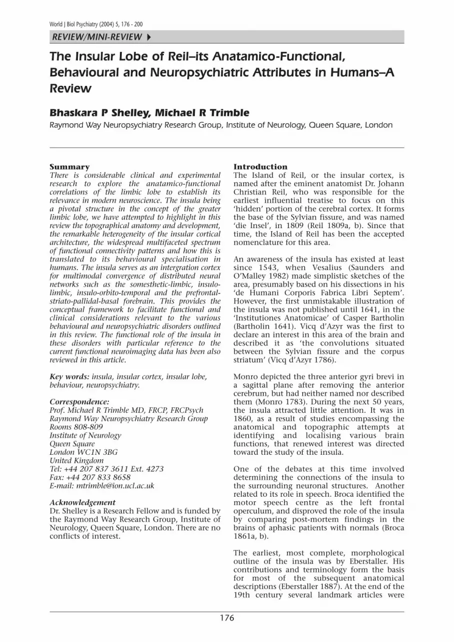
The insula, lying at the base of the Sylvianfissure, covers the claustrum and the basalganglia. The circular, periinsular sulci (anterior,superior, and inferior) of the fronto-orbital,fronto-parietal, and temporal opercula coverand enclose the insula, and the removal of theseopercula reveals the entire insula in the shape ofan inverted pyramid (Figure 1). The limeninsulae is located in the depths of the Sylvianfissure and constitutes the anterobasal portionof the insula.
Figure 1 Schematic drawing showing the rostro-caudal architectonicorganisation of the human insular lobe with Brodmannrepresentation - areas 13 to 16 (SPS = superior periinsular sulcus;IPS = inferior periinsular sulcus; APS = anterior periinsular sulcus; Ig= granular insula; Id = dysgranular insula; Ia = agranular insula)
178
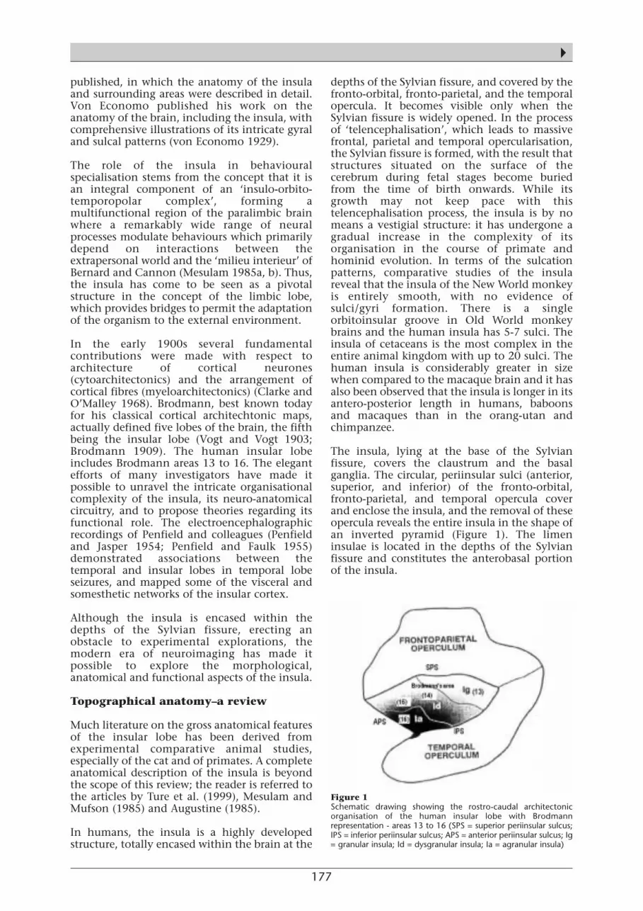
A central insular sulcus (sulcus centralis insulae)divides the surface of the insula into twoportions, the larger anterior insula and thesmaller posterior insula. The anterior insula iscomposed of three principal short insular gyri(anterior, middle and posterior) as well as theaccessory and transverse insular gyri. All fivegyri converge at the insular apex, whichrepresents the most superficial aspect of theinsula. The posterior insula is composed of theanterior and posterior long insular gyri and thepostcentral insular sulcus, which separates them(Figure 2). At the smooth, low ridge called thelimen insulae there is no definite topographicboundary between the anterior insula and theadjacent orbitofrontal cortex. The insula stem islocated in the depths of the Sylvian fissure, andconstitutes the anterobasal portion of theinsula. The limen insulae is located in theinsular stem and forms the threshold to theinsula, a place where the anterior perforatedspace continues with the anteroventralagranular insular olfactory cortex.
The anterior insula is connected exclusively tothe frontal lobe, whereas the posterior insula isconnected to both the parietal and temporallobes. Gyri and sulci of the operculainterdigitate, and also interdigitate with the gyriand sulci of the insula. The three opercula thatcover the insula are separated by the horizontaland posterior rami of the Sylvian fissure. Thehorizontal ramus interposes the fronto-orbitaland frontoparietal opercula, and the posteriorramus interposes the frontoparietal andtemporal opercula.
Extensive anatomical studies of the insula havebeen done from a neurosurgical perspective todelineate its vasculature by adult cadaverichemispheric dissections (Varnavas and Grand
1999; Ture et al. 2000). They described thearterial and venous anatomy of the insula, andits great variability. The main arterial supply ofthe insula stems from the middle cerebral artery,with predominance from the M2 segment. Theinsular arteries principally supply the insularcortex, the capsula extrema and sporadically theclaustrum and the capsula externa.
Development of the insular lobe
The human insula displays consistentasymmetries during development. The sulcalmarkings appear earlier in the right insula,although the left insula in the normal adult issaid to reach a larger size. During earlydevelopment in human fetuses, the temporallobe shifts downwards and anteriorly forming adepression, the primordium of the insula, onthe superolateral surface of the cerebralhemisphere (Lockard 1948). As the insula isfirmly attached to underlying structures itretains its position while the temporal lobepivots around it. Since the insular area developsmore slowly, its growth does not keep pace withthe massive development of the surroundingneocortex and it thus becomes covered over asthese lobes enlarge in an enclosed space, and asthe opercula of the lateral sulcus are broughttogether making the insula the ‘hidden fifthlobe’ of the brain (Cunningham 1891).
The surface of the insula remains smooth up tothe middle of the fifth month of development.At about 15 weeks, the inferior and superiorlimiting sulci appear, joining to form thecircular insular sulcus that outlines theanatomical boundaries of the insula. At about19 weeks, the insula enlarges, by now with ananterior and a posterior portion visible. By 19-20 weeks the central sulcus becomes evident, inline with the central sulcus on the superolateralsurface of the cerebral hemisphere(Cunningham 1891). The precentral insularsulcus appears before the end of the fifthmonth, whereas the postcentral insular sulcusappears by the middle of the sixth month andall insular sulci are present by 31 weeks ofdevelopment. Secondary gyri and sulci appearby 40-44 weeks of development and thenewborn insula presents an almost adultappearance (Cunningham 1891).
Structure (cyto-, myelo-, andchemoarchitectonics) of the insula
The general architectonic plan of the humaninsula is very similar to that observed in themacaque brain. The insular lobe has beenconsidered as a limbic cortex for ontogeneticand phylogenetic reasons. The interested readeris referred to the exhaustive descriptions on thecytoarchitectonics of the insular cortex and onthe cyto-chemoarcitectonics, connectivity andfunction of the primate insular cortex (Mesulam
Figure 2 Photograph showing typical five lobed human insular cortex withthe overlying frontal, parietal and temporal opercula removed (sps= superior periinsular sulcus; aps = anterior periinsular sulcus; ips= inferior periinsular sulcus; agb = anterior gyrus brevis; mgb =middle gyrus brevis; pgb = posterior gyrus brevis; agl = anteriorgyrus longus; pgl = posterior gyrus longus; sis = short insularsulcus; pcis = precentral insular sulcus; cis = central insular sulcus;pis = postcentral insular sulcus)
REVIEW/MINI-REVIEW
▲

179
▲and Mufson 1982a; Mufson et al. 1997).
Most of the human cerebral cortex isphylogenetically recent, and termed the‘neocortex/neopallium’, also referred to as‘isocortex’ (Vogt and Vogt 1903) and‘homogenetic cortex’ (Brodmann 1909). Thephylogenetically older cortices, comprising thehippocampus, parts of the amygdala, and theolfactory cortex are referred to as the‘archipallium’ or ‘allocortex’.
The Brodmann areas 13 to 16 represent thehuman insular lobe. A dorsocaudal granularfield is designated as area 13 while aventrolateral agranular field is assigned to areas14 to 16 (Brodmann 1909). The insular cortexshows a gradual transition from agranular anddysgranular cortex in the rostral two-thirds to afully-fledged granular cortex in its caudal part(Figure 1). The boundary between the agranularand the dysgranular zones of the insula is notsharp.
There is preferential interconnectivity andconcordance between the cytoarchitectonics ofregions that are connected to the insula and thearchitecture of the insular cortex (Vogt and Vogt1903). The agranular part of the orbitofrontalcortex is preferentially linked to the agranularpart of the insula and the granular orbitofrontalcortex is preferentially linked to the granularinsula. The granular sector is the most heavilymyelinated one in the insula.
In terms of architectonics, the anterior parts ofthe insula receive direct input from thegustatory and olfactory cortex, and the posteriorparts receive input from the somatosensory andauditory areas. The insula thus forms a site ofmultimodal convergence of inputs and plays apivotal role in limbic interactions, whichprovides i) a means for interrelating events ofthe extrapersonal world with relevantmotivational states and ii) an affective colouringand hedonic valence to perceptual experience.
Insular circuitry
Much of the progress in understanding theinsula and its circuitry is based on animalstudies (Chikama et al. 1997). There is aconcordance between the architecture of thebrain regions connected to the insula and thearchitecture of the insular sector that acts as afocus for that connection (Vogt and Vogt 1903).With the aid of a variety of methods, numerousinvestigators since the early 1980s havedemonstrated the abundance of the afferent andefferent connections of the insula as well as itslocal intrainsular connections. They have beenstudied in macaque cerebra (Berke 1960; Kreig1965; Mesulam and Mufson 1982b; Mettler1945), baboon, mangabey (Mettler 1945),macaca mulatta (Hurst 1959; Showers and Lauer
1961; Turner et al. 1980), macaca iris (Hurst1959), saimiri sciureus (Forbes and Moskowitz1974), and humans (Rae 1954). The differentmethods used by various investigators to revealthe essential outlines of insular connectionsinclude injections of titrated amino acids (TAA),horseradish peroxide (HRP), neuro-histochemistry (anterograde and retrogradeaxonal methods), strychnine neuronography,the Marchi method (Berke 1960) and Weil-stained (Lockard 1948) preparations. The insularconnections with the basal nuclei, theamygdaloid body, other limbic areas and thedorsal thalamus were studied by various silverpreparations: the Nauta-Gygax, Fink-Heimermethods (Forbes 1974) and the modified Fink-Heimer method (Turner et al. 1980).
The insular lobe in primates including humanshas connections with (1) cerebral cortex, (2)basal ganglia, (3) amygdaloid body, (4) otherlimbic areas, (5) the dorsal thalamus and (6)striato-pallidal basal forebrain macrosystems(Heimer et al. 1991; DeOlmos and Heimer 1999;Alheid and Heimer 1988). The extensivefunctional connections include (1)somatosensory connections, (2) auditoryconnections, (3) gustatory connections, (4)motor connections, (5) higher order associationconnections (6) olfactory and (7) paralimbicconnections. Figures 3 and 4 summarise thecircuitry of the insular lobe. Figure 5 illustratesFrontal lobeSMA area Amygdaloid bodyArea 6,12 Basolateral partFrontal operculum Corticomedial partOrbital cortexPosterior and lateral orbital areasPrefrontal cortex
Temporal lobe Other Limbic areasSuperior temporal gyrus Anterior entorhinalTemporal pole cortexTemporopolar cortex Anterior hippocampus Periamygdaloid cortex
Prepiriform olfactory cortex
Insular lobeIntrainsular connectionsLimen of the insula
Anterior cingulate gyrus Dorsal thalamus
Parietal lobeAnterior inferior parietal cortex Striatum-Basal forebrainParietal operculumSomatosensory area
Afferent insular projections
Efferent insular projections
Frontal lobe Amygdaloid bodyFrontal operculum Basolateral,medial,anterior partsLateral orbital cortexPosterior orbital cortexOrbitifrontal cortexPrefrontal cortex Other limbic areas Entorhinal cortexParietal lobe Olfactory bulbAnterior inferior parietal cortex Periamygdaloid cortexParietal operculumSomatosensory cortex
Cingulate gyrus Dorsal thalamus
Temporal lobeAuditory cortexAuditory association cortexSuperior temporal cortexTemporal poleTemporal operculum
INSULARCORTEX
INSULARCORTEX
Figure 3Afferent and efferent insular projections
180
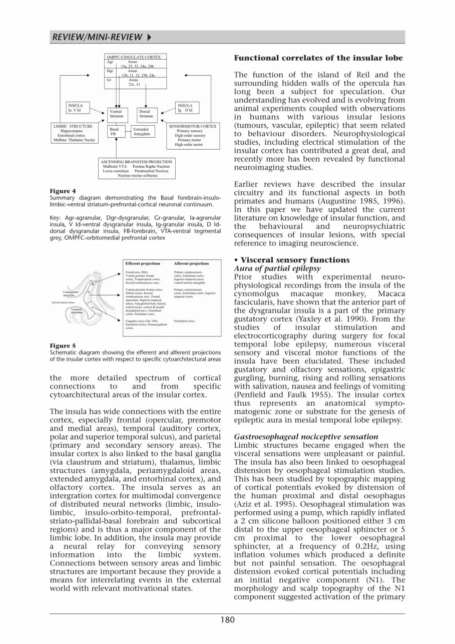
the more detailed spectrum of corticalconnections to and from specificcytoarchitectural areas of the insular cortex.
The insula has wide connections with the entirecortex, especially frontal (opercular, premotorand medial areas), temporal (auditory cortex,polar and superior temporal sulcus), and parietal(primary and secondary sensory areas). Theinsular cortex is also linked to the basal ganglia(via claustrum and striatum), thalamus, limbicstructures (amygdala, periamygdaloid areas,extended amygdala, and entorhinal cortex), andolfactory cortex. The insula serves as anintergration cortex for multimodal convergenceof distributed neural networks (limbic, insulo-limbic, insulo-orbito-temporal, prefrontal-striato-pallidal-basal forebrain and subcorticalregions) and is thus a major component of thelimbic lobe. In addition, the insula may providea neural relay for conveying sensoryinformation into the limbic system.Connections between sensory areas and limbicstructures are important because they provide ameans for interrelating events in the externalworld with relevant motivational states.
Functional correlates of the insular lobe
The function of the island of Reil and thesurrounding hidden walls of the opercula haslong been a subject for speculation. Ourunderstanding has evolved and is evolving fromanimal experiments coupled with observationsin humans with various insular lesions(tumours, vascular, epileptic) that seem relatedto behaviour disorders. Neurophysiologicalstudies, including electrical stimulation of theinsular cortex has contributed a great deal, andrecently more has been revealed by functionalneuroimaging studies.
Earlier reviews have described the insularcircuitry and its functional aspects in bothprimates and humans (Augustine 1985, 1996).In this paper we have updated the currentliterature on knowledge of insular function, andthe behavioural and neuropsychiatricconsequences of insular lesions, with specialreference to imaging neuroscience.
• Visceral sensory functionsAura of partial epilepsyPrior studies with experimental neuro-physiological recordings from the insula of thecynomolgus macaque monkey, Macacafascicularis, have shown that the anterior part ofthe dysgranular insula is a part of the primarygustatory cortex (Yaxley et al. 1990). From thestudies of insular stimulation andelectrocorticography during surgery for focaltemporal lobe epilepsy, numerous visceralsensory and visceral motor functions of theinsula have been elucidated. These includedgustatory and olfactory sensations, epigastricgurgling, burning, rising and rolling sensationswith salivation, nausea and feelings of vomiting(Penfield and Faulk 1955). The insular cortexthus represents an anatomical sympto-matogenic zone or substrate for the genesis ofepileptic aura in mesial temporal lobe epilepsy.
Gastroesophageal nociceptive sensationLimbic structures became engaged when thevisceral sensations were unpleasant or painful.The insula has also been linked to oesophagealdistension by oesophageal stimulation studies.This has been studied by topographic mappingof cortical potentials evoked by distension ofthe human proximal and distal oesophagus(Aziz et al. 1995). Oesophageal stimulation wasperformed using a pump, which rapidly inflateda 2 cm silicone balloon positioned either 3 cmdistal to the upper oesophageal sphincter or 5cm proximal to the lower oesophagealsphincter, at a frequency of 0.2Hz, usinginflation volumes which produced a definitebut not painful sensation. The oesophagealdistension evoked cortical potentials includingan initial negative component (N1). Themorphology and scalp topography of the N1component suggested activation of the primary
OMPFC/CINGULATE CORTEX
Agr Areas
13a, 25, 32, 24a, 24b
Dgr Areas
13b, 11, 12, 23b, 24c
Gr Areas
23c, 31
ASCENDING BRAINSTEM PROJECTION
Midbrain VTA Pontine Raphe Nucleus
Locus coeruleus Parabrachial Nucleus
Nucleus tractus solitarius
INSULA
Ig D Id
INSULA
Ia V Id
LIMBIC STRUCTURE
Hippocampus
Entorhinal cortex
Midline- Thalamic Nuclei
SENSORIMOTOR CORTEX
Primary sensory
High-order sensory
Primary motor
High-order motor
Ventral
Striatum
Dorsal
Striatum
Basal
FB
Extended
Amygdala
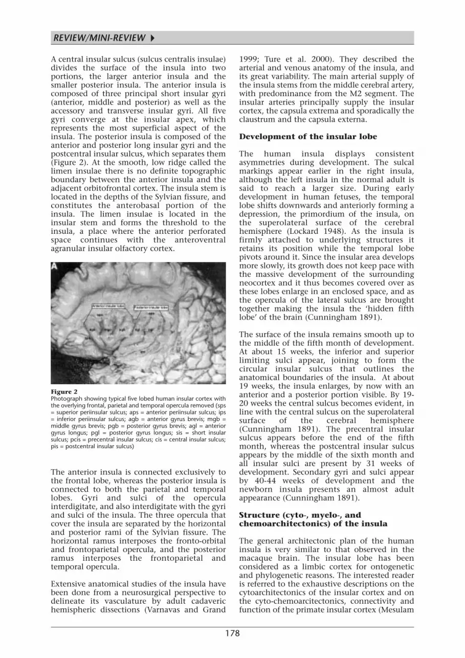
Figure 4 Summary diagram demonstrating the Basal forebrain-insulo-limbic-ventral striatum-prefrontal-cortical neuronal continuum.
Key: Agr-agranular, Dgr-dysgranular, Gr-granular, Ia-agranularinsula, V Id-ventral dysgranular insula, Ig-granular insula, D Id-dorsal dysgranular insula, FB-forebrain, VTA-ventral tegmentalgrey, OMPFC-orbitomedial prefrontal cortex
Efferent projections Afferent projections
Frontal area, SMA Primary somatosensory
Ventral granular frontal cortex, Entorhinal cortex
cortex, Temporopolar cortex, Superior temporal sulcus
Second somatosensory area, Lateral nucleus-amygdala
Ventral granular frontal cortex, Primary somatosensory
Orbital cortex, Second cortex, Entorhinal cortex, Superior
somatosensory area , Frontal temporal cortex
operculum, Superior temporal
sulcus, Amygdaloid body (lateral,
central nuclei, cortical & medial
amygdaloid area ), Entorhinal
cortex, Perirhinal cortex
Cingulate areas (24a/ 24b), Entorhinal cortex
Entorhinal cortex, Periamygdaloid
cortex
Frontoparietal
operculum
Temporal
operculum
Sylvian lateral sulcus
Figure 5 Schematic diagram showing the efferent and afferent projectionsof the insular cortex with respect to specific cytoarchitectural areas
REVIEW/MINI-REVIEW
▲

181
▲somatosensory cortex, the insular cortex, orboth.
Functional magnetic resonance imaging hasbeen used to study cerebral representations ofsomatic and limbic activation duringoesophageal distension (Binkofski et al. 1998).These data demonstrated that the secondarysomatosensory-insular cortex is the primarycortical target of visceral afferents originating inthe oesophagus.
Gustatory areaTaste perception in patients with unilateralinsular cortex lesions has been studied wheresubjects were required to identify the qualityand intensity of the gustatory stimuli appliedseparately to either side of the tongue (Pritchardet al. 1999). Damage to the right insulaproduced ipsilateral taste recognition andintensity deficits. Damage to the left insulacaused an ipsilateral deficit in taste intensity buta bilateral deficit in taste recognition, suggestingthat taste information from both sides of thetongue passes through the left insula.
The higher order projections in the humanbrain were studied by using positron emissiontomography, which showed increased cerebralblood flow in the left insular lobe during a tastediscrimination task in five normal volunteers(Fukuda et al. 1991). Increased blood flowpatterns were also noted in the left thalamusand right parietal cortex in four cases. Thisstudy confirmed that the insula is involved witha higher order gustatory circuit. Anotherfunctional imaging study demonstratedactivation of the anterior insula in humanswhile tasting salt (Kinomura et al. 1994).
• Visceral motor functionsVisceral epilepsyThe insula stimulation studies in temporal lobeepilepsy have revealed numerous visceral motorphenomena during seizures including audiblerumbling (borborgymi) or gurgling noises in thegastrointestinal tract, alteration of gastricmotility, belching, vomiting and the urge todefaecate (Penfield and Faulk 1955). Vomitingas a manifestation of seizures (visceral epilepsy)has been described in the literature (Mulder etal. 1954). The role of the insula in triggeringvomiting as in ‘ictus emeticus’ was studied usingvideo EEG and corticography (Fiol et al. 1988).It was concluded that the insula might act as atrigger to the medullary vomiting centre,probably by a pathway involving the anterior-mesial temporal structures.
The role of insular cortex in dysphagiaThe insula as a cortical substrate in themediation of dysphagia was investigated in fourunilateral stroke patients with discrete lesions ofthe insular cortex (Daniels and Foundas 1997).CT scan localisation studies, neurological
examination, bedside swallowing evaluation,videofluoroscopy and clinical oropharyngealexamination were done in these patients. Theresults indicated that dysphagia was associatedwith lesions of the anterior insula. Theypostulated that the anterior insula might beimportant in oropharyngeal deglutition becauseof its connectivity to crucial cortical, subcorticaland brainstem sites known to be important inswallowing. This was based on the connectionsof the anterior insula with the primary andsupplementary motor cortices, theventroposterior medial nucleus of the thalamus,and the nucleus of the tractus solitarius, all ofwhich are important in the act of oropharyngealswallowing. The connections the anterior insulahas with the primary and supplementary motorcortices thus facilitate coordinated interactionof tongue, face and jaw in swallowing (Figure 6).Lesions in the anterior insula may also producedysphagia by disrupting the processing ofgustatory input by disconnecting sensorimotorinformation between the NTS and the anteriorinsular cortex. This results in a delayedelicitation of the pharyngeal swallow, whichcontributes to dysphagia.
A [15O] labelled H2O PET study was conducted toidentify the cerebral loci processing humanvolitional swallowing in healthy volunteers(Hamdy et al. 1999). Submental electro-myography as well as transcranial magneticstimulation was used to map the cortical motorrepresentation of the pharynx. The transcranialmagnetic stimulation results indicatedactivation of the insula in addition tosensorimotor cortex, temporopolar cortex,cerebellum and brainstem.
Prolonged dysphagia following acute stroke isassociated with stroke severity, dysphasia andlesions of the frontal and insular cortex on brainimaging.
Primary MotorCortex
SupplementaryMotor Cortex
ANTERIOR INSULARCORTEX
Thalamus (vpm)
Nucleus tractussolitarius
Cortex
Subcortical
Brainstem
Figure 6 Diagram showing the afferent and efferent connectivity of theinsular cortex to critical swallowing regions of the brain (vpm =ventroposterior medial nucleus)
182
The role of the insular cortex inneurocardiologyAnother important visceral motor autonomicfunction ascribed to the insula relates to theconcept of neurocardiology, which is basedupon the anatomy and physiology of corticalmechanisms of cardiac control. Literature onthis concept comes from the observations ofelectrocardiographic changes andarrhythmogenicity accompanying acutestrokes and subarachnoid haemorrhages andsudden unexpected deaths in epilepsy (SUDEP).The role of the insular cortex in cardiovascularfunction was experimentally studied in theanaesthetised, paralysed and artificiallyventilated Sprague-Dawley rats (Ruggiero et al.1987). This study analysed the responses toelectrical and chemical stimulation of the ratinsular cortex with respect to the regulation ofarterial blood pressure. It was concluded thatneurones within an area of the insular cortexprojecting to multiple brainstem autonomicnuclei, including the cardiopulmonarysegments of the nucleus of the tractus solitariusand nucleus reticularis parvocellularisinnervated by baroreceptor afferents, increaseblood pressure and heart rate.
The differential left/right hemisphere heart rateresponses following unilateral hemisphericinactivation by the intracarotid amobarbital(ISA) test was studied in 25 patients undergoingpreoperative evaluation for epilepsy surgery(Zamrini et al. 1990). Heart rate increased afterleft hemisphere inactivation, but decreasedfollowing right hemisphere inactivation. Itseems that the right insula is involved incardiovascular sympathetic, and the left insulain parasympathetic control. A recent study in 73subjects using the ISA test re-investigated thedifferential cerebral involvement on cardiacdromotropic and heart-rate variability changes(Ahern et al. 2001). Contrary to the results ofthe previous study, it concluded that the righthemisphere exerted a greater parasympatheticcontrol. The study summarised that there weremore cardiac consequences of arrthymogenecityand sudden cardiac death in patients with righthemispheric infarcts, and SUDEP for patientswith right hemispheric epileptic foci.
Experimental studies in rats showed that theposterior insular cortex possesses cardiacchronotropic organisation and stimulationresulted in increasing degrees of heart blockleading to escape rhythms, ventricular ectopicsand ultimately death in asystole (Oppenheimeret al. 1991). These data suggest that thepathophysiological activation of the insularcortex by stroke or an epileptic seizure couldlead to ECG changes, cardiac arrhythmias andsudden death.
Two studies described the association of cerebral strokes of all subtypes with specific pathological
changes in the ventricular myocardium(myocytolysis) that were not attributable toconcomitant cardiac ischemic disease(Oppenheimer et al. 1991; Oppenheimer 1992).The studies noted the cardiovascular effects ofhuman insular cortex stimulation and itslateralisation in five epileptic patients prior totemporal lobectomy for seizure control. Rightmiddle cerebral artery infarction disinhibitsinsular function and causes an increase insympathetic cardivascular tone and thepotential for cardiac consequences of stroke.Studies on ECG alterations reported that thefrequency-corrected QT interval dispersion wassignificantly prolonged in patients with strokesinvolving the insular cortex (Eckardt et al.1999). This result led to the hypothesis that theinsular cortex might be involved in theregulation of myocardial repolarisation. Thishypothesis has been supported by results ofanimal studies in which stimulation of theinsular cortex was shown to lead to ventricularectopy and death in asystole in rats(Oppenheimer and Norris 1995). Neurogenic STdepression in the ECG has been reported in a48-year-old female patient that was related toleft insular infarction and who developedsudden expressive aphasia (Chua et al. 1999).
The differential effects of stroke localisation onautonomic function parameters using heart ratevariability were studied in 62 patients with age-and sex-matched controls (Tokgozoglu et al.1999). It was concluded that stroke in the rightinsula leads to decreased heart rate variability,which is an important predictor forarrhythmias, and sudden cardiac death.Experimental and clinical studies indicated thatcertain structures such as the insula, amygdalaand the lateral hypothalamus exert an influencein the autonomic control of the heart(Oppenheimer et al. 1992). Of these, the insularcortex, within the middle cerebral arteryterritory, is the most important cortical area thatcontrols both parasympathetic- andsympathetic-mediated cardiovascular regulationwhich led to its description as ‘the insula ofsudden death’ (Hachinski 1999).
• Somatosensory functionsThe granular insula has a somatosensoryfunction with somatotopic organisation and is asite of multimodal convergence among inputsfrom olfactory, gustatory and auditorymodalities. It also plays a pivotal role forrelaying somatosensory impulses into otherlimbic areas, having the function of asomatosensory-limbic integration cortex. Thisfunction of the granular insula has been wellstudied in rhesus monkeys (Schneider et al.1993).
Positron emission tomographic brain imagingstudies in humans have shown activationalcerebral blood flow patterns during
REVIEW/MINI-REVIEW
▲
183
▲somatosensory tactile-vibration stimulation ofthe hands and feet to be topographicallylocalised to primary and secondarysomatosensory areas and the insular cortex(Burton et al. 1993).
Cortical activational patterns during theprocessing of painful stimuli have been studiedby intracortical recordings of early pain-relatedCO2 laser evoked potentials in the humansecond somatosensory area during stereotacticEEG presurgical assessment of patients withdrug-resistant temporal lobe epilepsy (Frot et al.1999). This study revealed that activation of thecontralateral secondary somatosensory-insulacortex represents the first step in the corticalprocessing of peripheral A-delta fibre paininputs.
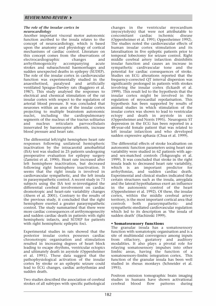
A review and meta-analysis of PET and fMRIstudies on brain responses to pain has beenperformed (Peyron et al. 2000). This studyreported functional activation of the secondarysomatosensory area and insular regions, and theanterior cingulate cortex. Figure 7 depicts someof the main anatomical components involvedin human nociceptive processing in the brain.
In a recent study, multimodality tests using PET,fMRI, dipole modelling and operculoinsularevoked potentials further confirmed the insulaas a major site for cortical pain encoding in thehuman brain (Peyron et al. 2002). Anotherstudy used fMRI combined with a thalium:yttrium-aluminium-garnate infrared laser to
investigate pain and stimulus intensity in ninehealthy human volunteers and showedactivation of the secondary sensory cortex andinsula cortex for painful trials (Bornhovd et al.2002).
The central processing of innocuous andnoxious cold stimuli was studied in humanhealthy subjects using magnetoencephalo-graphy (Maihofner et al. 2002). This studyrevealed an exclusive activation of the contra-and ipsilateral posterior insular cortex mediatedby A-delta fibres. In addition, noxious coldstimuli produced activation in the contra- andispsilateral secondary (SII) areas and cingulatecortex. This study confirmed the participationof the posterior insular cortex as the primarysomatosensory area for the cortical processingof cold sensation.
Intracortical S-EEG recordings of early pain inthe human second somatosensory area usingCO2 laser stimulation of A-delta fibre endings inthe skin of the dorsum of the hand in humanshave been studied (Frot and Mauguiere 1999).Responses were seen on the contralateral regionof the upper bank of the Sylvian fissure, that isthe upper part of the insular cortex (S II-insularcortex). The contralateral S II-insular cortexactivation occurred through directthalamocortical projections, while theipsilateral side gets stimulated via transcallosalfibres coming from the opposite S II area.
The somatosensory-limbic path, relayingsomatosensory information by means of theinsula, also subserves tactile recognition andrecall. This was studied using PET in humans,and revealed that the ipsilateral anterior insulaand orbitofrontal cortices were activated duringthermosensory stimulation, regarded as asubmodality of touch (Craig et al. 2000).Another study employed event-relatedsomatosensory stimulation paradigms in fMRIstudies using electrical finger stimulation(Deuchert et al. 2002). Results from this studyrevealed cortical activation patterns to belocalised to the contralateral primary, andbilaterally in secondary somatosensory cortex aswell as in posterior parietal cortex and insula.
In another study, painful and non-painfulsomaesthetic sensations were evoked by directelectrical stimulations of the insular cortex inpatients with drug refractory temporal lobeepilepsy using stereotactically implanted depthelectrodes (Ostrowsky et al. 2002). Both types ofstimuli elicited were evoked from the posteriorpart of the insula. However, painful sensationsevoked were lateralised to the right hemisphere.
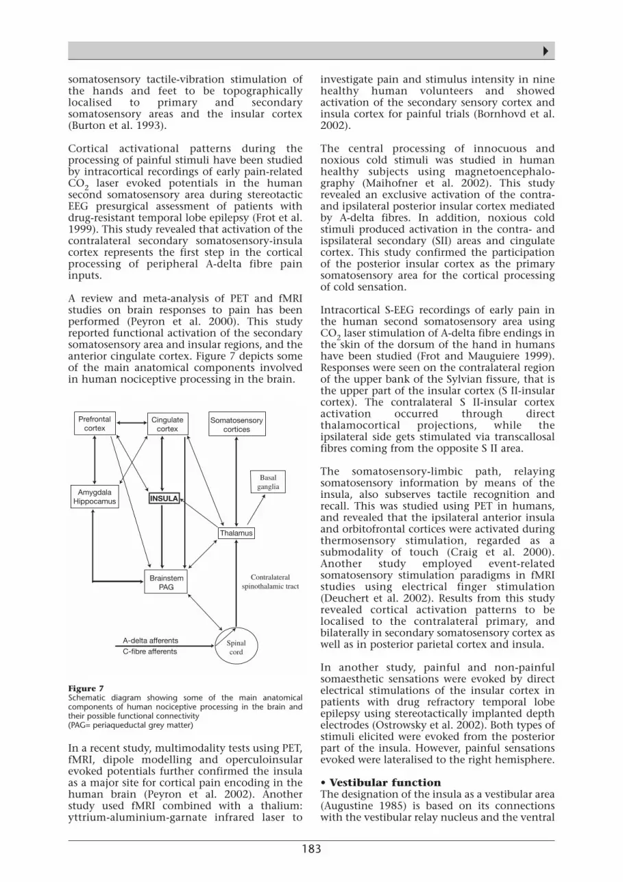
• Vestibular functionThe designation of the insula as a vestibular area(Augustine 1985) is based on its connectionswith the vestibular relay nucleus and the ventral
Prefrontalcortex
Cingulatecortex
Somatosensorycortices
AmygdalaHippocamus INSULA
Basalganglia
Thalamus
BrainstemPAG
Spinalcord
A-delta afferentsC-fibre afferents
Contralateralspinothalamic tract
Figure 7 Schematic diagram showing some of the main anatomicalcomponents of human nociceptive processing in the brain andtheir possible functional connectivity(PAG= periaqueductal grey matter)
184
posterior inferior nucleus of the thalamus(Deecke et al. 1974). Vestibular information maybe blended in the thalamus with somaticproprioceptive information (Deecke et al. 1977).In vertigo of cortical origin, the associatednausea and vomiting can be explained by theinvolvement of the insular cortex whereviscerosensory impulses are combined withvestibulo-proprioceptive information. A studyinvolving 71 patients with unilateral middlecerebral artery infarctions reported that theperception of verticality was affected in 20 of 52patients with topographic infarctions localisedto the posterior granular insula (Brandt et al.1994). The vestibular function of the insula wasstudied using PET and fMRI, revealing thatcaloric stimulation of the ears activates themultisensory parieto-insular cortex in humansand is involved in the perception of verticalityand self-motion (Brandt and Dieterich 1994;Brandt et al. 2002). The parieto-insular cortex isthe human homologue of the parieto-insularvestibular cortex in monkeys.
• Auditory processing functionThe insula receives afferent connections fromthe temporal pole, primary auditory cortex,auditory association cortex, superior temporalcortex and the temporal operculum. Auditoryfunction has been studied by mapping cerebralglucose consumption following verbal auditorystimulation that revealed activation in the leftinsular cortex, the temporoparietal junction,the inferior parietal region and the corpuscallosum (Kushner et al. 1987). Deficits inunilateral auditory processing disorder in apatient with stroke involving the right insulaand the adjacent white matter have beendescribed (Fifer 1993). With these reports it hasbeen proposed that the insula is an auditoryassociation area involved in the preprocessing ofauditory stimuli and in receptive auditoryaspects of language comprehension.
A more recent study using fMRI tasks of passivelistening and semantic categorisation providedfurther insights into the role of the humaninsula in verbal-auditory processing (Noesselt etal. 2003). The study found insular activations inthe context of different verbal or auditory-verbal tasks. Since the insula is stronglyinterconnected with the temporal and frontalstructures, they speculated that the posteriorinsula might play a role in linking together thedifferent neural networks involved in auditoryprocessing.
• Summary - insular functionThe section above summarises the variousstudies and investigations that lend evidence tothe multifunctional activity of the insula (Figure8). The insula is thus an importantsomatosensory area, as part of the humansecondary somatosensory area (S II) and a painprocessing area. The insula is also involved in
volitional swallowing, visceral motor sensoryprocessing and is a cortical gustatory area. Itplays an important role in cardiovascularfunction and cerebrogenic sudden death. Theposterior portion of the insula is probably acomponent of the human vestibular cortex.There is evidence to suggest that the insula isinvolved with aspects of speech and languageand auditory processing.
Since the insula forms a waystation on thepathway of the limbic-paralimbic system and isa major limbic intergration cortex, thebehavioural and neuropsychiatric attributes arediscussed below.
Behavioural correlates of the insularlobe
• Insula and aphasiaHistorically, language disorders were the firstdisorders of higher cortical function to becorrelated with focal brain lesions. In theliterature, there is considerable controversybetween various investigators about anylanguage functions of the insula.
Some investigators had made fundamentalobservations that a lesion of the left insula andoverlying operculum deprived a person ofspeech (Broca 1861). A theoretical framework ofthe organisation of language functions in thebrain for conduction aphasia was postulated bythe model of ‘Leitungsaphasie’ (Wernicke 1874).This postulated that conduction aphasiaresulted from an anatomical disconnection ofan intact Wernicke’s area from an intact Broca’sarea by a deep white matter lesion interruptingthe arcuate fasciculus. This ‘disconnection’hypothesis was reformulated for the modern eraby proposing that conduction aphasia is notpurely due to white matter lesions, but involves
Figure 8 Schematic representation of the behavioural specialisation in theinsula and adjacent regions
REVIEW/MINI-REVIEW
▲
185
▲at least some of the cortex of the insula, superiortemporal gyrus, and the supramarginal gyrus(Geschwind 1965). It has been concluded thatthe decisive lesion must be a destructiveinterruption of the arcuate fasciculus fibres thattravel in the extreme capsule underneath theinsular cortex, rather than the lesions of theinsular cortex per se (Damasio and Damasio1980).
Impaired speech initiation and poor verbalfluency as the result of MRI detected leftanterior insular infarct has been reported in a59-year-old woman (Shuren 1993). It washypothesised that the deficits in speechinitiation were related to the role of the insulain the limbic-reticular-cortical network formotivation and arousal-activation ofbehavioural responses in speech initiation. Thisis based on the reciprocal connections theanterior insula has with the limbic (cingulateand amygdala) and the reticular (thalamiccentromedian-parafascicular and reticularnuclei) systems, as well as the frontal opercularregions.
A study, using PET brain imaging, of memorymechanisms in the processing of single wordsand word-like symbols, learning and repetitionof words all revealed bilateral insular cortexactivation (Raichle 1991). A study, using high-speed echo-planar magnetic resonance imaging,in humans has documented activation of theanterior insula during word generation(McCarthy et al. 1993). Another study hasidentified activation of mid-dorsal insulabilaterally in auditory-vocal integration insinging and in speech, using PET with MRI(Perry et al. 1993).
The role of the insula in the auditory processingunderlying speech has been clearly exemplifiedby stroke involving the right insula. PET studieshave shown deactivation of bilateraltemporal/insular areas during a graded auditory-verbal memory task (Grasby et al. 1994).Phonological agraphia, a highly selectivedisturbance of the spelling system, has beendescribed following a focal left anterior insulo-opercular infarction (Marien et al. 2001). Theneuroanatomical and neurolinguisticcharacteristics of these phonologically mediatedaphasic manifestations suggest a furtherdelineation of the role of the insula in languagefunctions in humans. To delineate theneuroanatomical basis, four case reports of ‘theopercular-subopercular syndrome’ have beenreviewed (Bakar et al. 1998). This syndrome,also known as Foix-Chavany-Marie syndrome, isa cortical type of pseudobulbar paralysisinvolving the facio-labio-pharyngo-glosso-laryngo-brachial regions. They stress that theneuroanatomical basis involves bilateral lesionsof the cortical and subcortical frontal opercularcortex disrupting the corticobulbar tracts, rather
than the insula per se. However, in anotherstudy of brain lesions in 25 stroke patients witha disorder in the motor planning of articulatorymovements, discrete lesions in the region of theleft precentral gyrus of the insula were shown(Dronkers 1996). The left insula thus representsa region related to complex planning andcoordination of human speech articulation.
A recent study investigated 10 patients withprogressive non-fluent aphasia in an attempt toidentify a consistent neural site for the languagedisorder (Nestor et al. 2003). Seven patients hadan isolated progressive non-fluent aphasiasyndrome, while the remaining three hadprogressive dysfluency as part of a dementiaprocess. Compared with controls, the patientswith non-fluent aphasia showed significanthypometabolism in the left anterior insula andfrontal opercular region. The study concludedthe left anterior insula as the anatomical site fordysfluency resulting in a deficit in motorarticulatory planning (speech apraxia)combined with agrammatism.
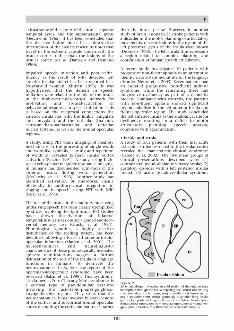
• Insula and strokeA study of four patients with their first acuteischaemic stroke restricted to the insular cortexrevealed five characteristic clinical syndromes(Cereda et al. 2002). The five main groups ofclinical presentations described were: (1)contralateral pseudothalamic sensory stroke; (2)gustatory disorder with a left posterior insularinfarct; (3) acute pseudovestibular syndrome
Insular ribbon
asg
msg
psg
alg
plg
sf
p gp
cn
th
fpo
to
Figure 9 Schematic diagram showing an axial section of the right cerebralhemisphere through the insula depicting the ‘insular ribbon’ (asg= anterior short insular gyrus; msg = middle short insular gyrus;psg = posterior short insular gyrus; alg = anterior long insulargyrus; plg = posterior long insular gyrus; sf = Sylvian fissure; fpo =frontoparietal operculum; to = temporal operculum; p = putamen;gp = globus pallidus; th = thalamus; cn = caudate nucleus)
186
with posterior insular infarct; (4) cardiovasculardisturbances with a right posterior insularinfarct and; (5) neuropsychological disorders,including aphasia (left posterior insula),dysarthria, and a transient neuropsychiatricdisorder of somatoparaphrenia in right sidedinfarcts.
The insula region has been described in theneuroradiology of acute ischaemic stroke with‘loss of insular ribbon’ sign (LIR) (Koga et al.2003). The CT scan does not usually show muchin the first 24 hours of a middle cerebral arteryinfarction; however the ‘loss of insular ribbon’represents an abnormal early reliable CT finding(Truwit et al. 1990). There is loss of definition ofthe grey-white interface in the lateral margins ofthe insula (insular ribbon) (Figure 9). Theinsular ribbon lies in an arterial watershed zone,and loss of the radiological appearance ofinsular ribbon in CT is a reflection of acuteoedema due to infarction.
• Insula and pain perceptionBased on connectional and functional data,several investigators have defined the role of thegranular insula as a modality-specific corticalarea for the processing of somatosensoryinformation. In this context, asymbolia for painsecondary to interruption of insularsomatosensory and limbic connections has beenreported (Berthier et al. 1988). Unpleasantspontaneous pain generation was described insix patients with CT detected white matterlesions deep to the caudal insula and theposterior parietal operculum (Schmahmann andLeifer 1992). This pseudo-thalamic painsyndrome was attributable to a loss of corticalinhibition of the dorsal thalamus due to thedisconnection of these cortical secondarysomatosensory areas and the dorsal thalamus.
• Insula and neglect syndromeA severe multimodal neglect syndrome in aright handed male has been reported followingan ischaemic stroke with neuropathologicalevidence of infarction involving the whole rightinsula, adjacent white matter, and the innercortical surface of the right fronto-temporo-parietal operculum (Berthier et al. 1987). Thislesion probably disrupted the posterior insula-amygdala limbic connection, which resulted inneglect and lack of appropriate response topainful stimulation presenting as asymbolia forpain. Another study also reported that neglectwas associated with right insula lesions (Maneset al. 1999a). Neglect has been commonlydescribed with lesions in the right inferiorparietal lobe (Heilman et al. 1993), thedorsolateral frontal cortex (Heilman andValenstein 1972), the thalamus and themesencephalic reticular formation (Watson andHeilman 1979). However, because of itsconnections with the limbic and sensory motorcortices, the insular cortex is believed to play a
role in affective and attentional aspects ofhuman behaviour. The right insula infarctiondescribed by Manes led to neglect inmultisensory modalities including (a) tactileinattention, (b) auditory and, (c) visual. It wastherefore suggested that neglect, anergia andapathy might all be related to insuladysfunction (Manes et al. 1999b; Roaslo et al.1997). PET activation of blood flow in theinsular cortex has been demonstrated duringselective-attention tasks related to visualdiscrimination of shape, colour, and speed of avisual stimulus has been studied (Corbetta et al.1991).
• Insula and memoryPET studies have shown an association betweenchanges in blood flow in the insular cortex andverbal memory. The verbal component ofworking or short-term memory in humans andcontrols has been studied using PET byemploying two tasks, the covert rehearsal ofvisually presented letters and rhymingjudgement for letters (Paulesu et al. 1993).Significant increases in blood flow patternsoccurred bilaterally in area 44, in areas 22 and24 of the superior temporal gyri, thesupramarginal gyri and in the insular cortexbilaterally.
The laterality of verbal memory deficits wasstudied by using PET which compared verbalmemory profiles by employing the CERAD (theconsortium to establish a registry forAlzheimer’s disease) word list memory and storyA of the WMS-R (Wechsler memory scale)logical memory subtests, between a group offour right-handed patients with right insularinfarction and a group of six right-handedpatients with left insular infarction (Manes et al.1999c). The results indicated that patients withleft insular damage had significantly poorerperformance on verbal memory tasks thanpatients with right insular lesions. These areconsistent with PET studies in normal subjects,suggesting a role for the insular cortex inlanguage and verbal memory tasks (Paulesu etal. 1993).
Although the functional neuroanatomy of recallhas not been determined, studies have shownverbal memory impairment in patients with lefttemporal lobe dysfunction (Cabezo et al. 1997).The insula sends efferents to the temporalcortex including the temporal pole and thesupratemporal plane. Afferent fibres to theinsula arise from the temporal lobe (includingthe temporal pole, primary auditory, auditoryassociation, postauditory cortex, superiortemporal cortex and the temporal operculum).It is thus suggested that insular connectionsprovide a fundamental anatomical substrate forlearning and memory functions. Left insularlesions disrupt connections with brain areasthat are necessary for executing memory tasks
REVIEW/MINI-REVIEW
▲
187
▲and, therefore, lead to verbal memory deficits.
In another study, left temporal lobehypoperfusion on SPECT was described in apatient who developed aphasia after left insularinfarction (Marshall et al. 1996). These authorssuggested that hypoperfusion in the lefttemporal lobe may have reflected a functionaldisconnection between the posterior insula andlanguage areas in the temporal lobe. The suddenremoval of areas of functioning brain, as occurswith stroke, may produce distant effects infunctionally connected neural structures, aphenomenon termed diaschisis. Thus, theverbal memory impairment in left insularpatients could be the result of diaschisis. Thestudy hypothesised that the insular cortex formsa component of a functional neuroanatomicalcircuit that mediates verbal memory.
The componential role of the right insula in thefunctional anatomy and neural networksinvolved in autobiographical memory wasdemonstrated in another study using H2
15O PET(Fink et al. 1996). This study revealed functionalneuronal activity in a network of primarily righthemispheric regions including temporomedialand temporolateral cortex, and the surroundingright hemispheric ‘satellite’ regions of theexpanded limbic system network (Nauta 1979)such as amygdala, hippocampus-parahippocampus, insula, posterior cingulatecortex, temporoparietal cortex and prefrontalcortex.
• Insula and motoric function/neuroplasticitySeveral investigators and reports havecollectively demonstrated that the anteriorinsula has a prominent role as a motorassociation area (Chollet et al. 1991; Weiller etal. 1992, 1993). An epileptic aura consisting ofcircling or rotational movements has beendescribed with an insula tumour (Fiol et al.1988) and with an aneurysm of the middlecerebral artery resting on the insula (Schneideret al. 1971). A study using PET explored thecortical/subcortical mechanisms underlying theexecution of human voluntary horizontalsaccadic eye movements in total darkness, andshowed activation of the right insula in additionto structures in the basal ganglia-thalamocortical motor loop during theexecution of voluntary saccades (Petit et al.1993). To investigate the motoric functionalaspects of the insula, regional blood flow wasmeasured in amyotrophic lateral sclerosispatients and age-matched controls (Kew et al.1993). It was observed that the anterior insulaand premotor cortices showed significantlygreater activation in amyotrophic lateralsclerosis patients than the controls during upperlimb movements. It was concluded that therecruitment of the anterior insula and premotorcortices in amyotrophic lateral sclerosis patients
reflected the brain’s adaptation to the injury tothe corticospinal tract and suggested that theanterior insula may act as a motor associationarea.
This was further corroborated using PET tostudy organisational changes in the functionalanatomy of the brain in 10 patients followingmotoric recovery of upper limb function fromstriatocapsular motor strokes (Weiller et al.1992). The study revealed that during theperformance of a motor task by the recoveredhand there was greater activation than innormal subjects in both anterior insulae, theinferior parietal cortex, the prefrontal/anteriorcingulate, the ipsilateral premotor/basal gangliaand in the contralateral cerebellum.
• Insula and human eating behaviourThe hypothalamus has a major role in thecontrol of food intake. The neuroanatomicalcorrelates of hunger and satiation in both leanand obese subjects have been studied by PETgenerated functional brain maps (Del-Parigi etal. 2002). The results in lean individualsindicated that the networks involved in hungerincluded the hypothalamus, thalamus andseveral limbic structures including the insula,the hippocampal formation and theorbitofrontal cortex. Satiation was associatedwith preferentially increased neuronal activityin the prefrontal cortex.
• Insula and sexual behaviourHuman emotions may be classified under twodistinctive systems (a) the appetitivemotivational system associated with positive orpleasant emotions and (b) the aversivemotivational system associated with negative orunpleasant emotions. Despite the brain’s centralrole in human appetitive sexual function, littleis known about the functional neuroanatomyand patterns of brain activation associated withsexual arousal. Visually evoked sexual arousalwas studied using [15O] H2O PET in healthy malesubjects to identify the activated brain areas thatwere time locked to sexual arousal (Stoleru et al.1999). The brain areas activated whileexperiencing sexual arousal from seeing sexuallyexplicit film clips were (a) bilateral inferiortemporal cortex, a visual association area; (b)right insula and right inferior frontal cortex,paralimbic areas related to processing of sensoryinformation with motivational states; and (c)left anterior cingulate cortex, a paralimbic arearelated to autonomic and neuroendocrinefunctions.
Another study using blood-oxygeneration-level-dependent (BOLD) functional MRIdemonstrated the cerebral centres controllingpenile erection while visualising erotic and non-erotic film in sexually potent male volunteersand hypogonadal impotent patients (Park et al.2001a). The sites of brain activation in response
188
to the erotic film were areas of inferior frontallobe, cingulate gyrus, insula and inferiortemporal lobe. The neuroanatomical correlatesof female sexual arousal evoked by visualstimulation were studied in healthy femalevolunteers using BOLD functional magneticresonance imaging (fMRI) (Park et al. 2001b).The activated sites associated with sexualresponse were the inferior frontal lobe,cingulate gyrus, insula and inferior temporallobe in addition to thalamus, caudate nucleusand globus pallidus. These studies reveal insularactivation amongst other cerebral centres in theneurobiological processes that underlie humansexual arousal.
• Insula and temporal lobe epilepsyThe landmark paper entitled ‘The insula: furtherobservations on its function’ (Penfield andFaulk 1955) underlined the similarity betweenthe symptoms observed during temporal lobeseizures and those evoked by insular cortexstimulation. Their electrocorticographic andintracortical electrical stimulation studies of theinsula during presurgical evaluation of temporallobe epilepsy patients made observations onvisceral sensory, visceral motor, somatic sensory,gustatory, motor, pain and speech, andauditory/vestibular sensory phenomena. Theseobservations were predictable when consideringthe dense connectivity between the insular lobeand the temporo-limbic structures.
The findings of this seminal study wereconfirmed from Penfield’s recordings in 106patients, which demonstrated that 50% showedspontaneous spikes or spike waves in the insularlobe in temporal lobe epilepsy (Silfvenius et al.1964). This was further illustrated by a report ofa partial seizure originating during the surgicalremoval of an insula tumour (Roper et al. 1993).A specific epileptic insular network, with seizuredischarge propagation pathways based on theconnections of the insula, the temporal poleand the amygdalo-hippocampal structures, hasbeen postulated by Wieser (1983).
The role of the insular cortex in the genesis oftemporal lobe epileptic seizures has been furtherexplored (Isnard et al. 2000) by using 3-Dreconstructed cerebral MRI-assisted stereotacticimplantation of transopercular depth electrodes(Talairach and Bancaud 1973) in 21 patientswith drug refractory TLE. In addition, video-EEGrecordings of the ictal symptoms of 81spontaneous electroclinical seizures werecaptured. All of the recorded seizures werefound to invade the insula, and two patientshad seizures that originated in the insular cortexitself. It was shown that scalp video-electroencephalographic monitoring did notpermit differentiation between the ictalsymptoms of temporo-mesial and insulardischarges. The authors concluded from thestudy that seizures secondarily propagating to
the insular cortex were fully controlled bysurgery, whereas those originating in the insularcortex were not influenced by temporallobectomy, and persist and may be mistaken fortemporo-limbic or opercular seizures. A highrate of pharmaco-resistant epilepsy from insularlow-grade gliomas has been reported whichstressed the relative epileptogenicity of theinsular cortex and improvement after anextended lesionectomy (Daffau et al. 2002).
The involvement of the insular cortex inpatients with mesial temporal lobe epilepsywith respect to the emotional symptoms werestudied using 18F-flurodeoxyglucose (FDG)positron emission tomography and 11C-flumazenil receptor binding (Bouilleret et al.2002). Unilateral mesial temporal lobe epilepsyis associated with insular hypometabolism andbenzodiazepine receptor loss. This study showedthat emotional symptoms correlated withhypometabolism in the anterior part ofipsilateral insular cortex, whereas somestheticsymptoms correlated with hypometabolism inthe posterior insular cortex.
Through the ‘looking glass’ of epileptic auras, asevoked by neurostimulation of epileptic insularcortices, it may be possible to understand thefunctions of the insula and its topographicallocalisation in more detail. The functionalmapping of the insula cortex in temporal lobeepilepsy (Ostrowsky et al. 2000), suggested aclear topographic specificity inside the insularcortex with respect to two different corticalnetworks. A visceral network for viscerosensitiveand visceromotor induced responses wasmapped to the anterior insula, whereas asomesthetic network was localised to theposterior insula. In this study the symptomsevoked were (a) viscerosensory sensationsincluding nausea, epigastric pressure and anunpleasant throat sensation; (b) visceromotorresponses including chewing movements, lipsmacking, gustatory sensations described as a‘bad’, salted or an acid taste; (c) an auditoryillusion; (d) olfactory pleasant sensations; (e) avisual sensation; (f) nociceptive sensations; and(g) speech disturbances. These two networkshave dense connections between more‘upstream’ and more ‘downstream’ levels of atleast five anatomically distinct networks, whichhelp to create a highly edited subjective versionof the world (Mesulam 1998).
Neuropsychiatric correlates of theinsular lobe
• Insula and emotional behaviourInsula and anxiety/phobiaSince the insula plays a pivotal role in limbicintegration, complex behaviours such as simplephobia with their distinct autonomic andvisceral components may recruit the insularcortex in the functional neuroanatomic
REVIEW/MINI-REVIEW
▲
189
▲distributed networks of emotion. This wasstudied using PET and oxygen 15 whichmeasured normalised regional cerebral bloodflow in seven subjects with simple phobiaduring control and provoked states (Rauch et al.1995). Stereotactic transformation andstatistical parametric mapping were used todetermine the locations of significantactivation. Significant increases in normalisedregional blood flow for the symptomatic statescompared with the control state were seen inthe right anterior cingulate cortex, the leftinsular cortex, the right anterior temporalcortex, the left somatosensory cortex, the leftposterior medial orbitifrontal cortex and in theleft thalamus. This study suggested that theneuronal circuitry that underlies anxietyassociated with simple phobic symptomaticstates is mediated by paralimbic structures,including the insula.
Further studies of the neural substrates ofspecific neuropsychiatric disorders such asobsessive-compulsive disorder, simple phobiaand post-traumatic stress disorder were done todetermine the shared mediating neuroanatomyof anxiety symptoms across these three anxietydisorders (Rauch et al. 1997). Relative regionalcerebral blood flow was measured using PET inthe context of provocation paradigms. Theanalysis of pooled imaging data indicatedactivation in the right inferior frontal cortex,the right posterior medial orbitofrontal cortex,bilateral insular cortex, bilateral lenticulatenuclei and bilateral brain stem foci during thesymptomatic versus control conditions. Thesefindings confirm the hypothesis that elementsof the paralimbic belt, together with rightinferior frontal cortex and subcortical nuclei,mediate symptoms across different anxietydisorders.
A study using [15O] PET investigated the brainregions that participated in externally andinternally generated human emotion (Reimanet al. 1997). Emotional responses to complexvisual (film generated exteroceptive emotion)and cognitive (recall generated interoceptiveemotion) stimuli were compared with areas ofincreased regional brain activity. Recallgenerated sadness was found to be associatedwith greater increases in activity in the anteriorinsular cortex. This suggested that the anteriorinsular cortex participates in the generation ofhuman emotional response to potentiallydistressing cognitive or interoceptive sensorystimuli.
The neural correlates of affective disorders havebeen investigated by using more advancedneuroimaging techniques such as PET andfunctional magnetic resonance imaging(Kucharska-Pietura et al. 2001). This workdemonstrated an important role of the posteriortemporal cortex, orbitofrontal cortex,
amygadaloid nucleus and the insula in theneuroanatomy of affect control.
The functional aspects of brain segregation andconnectivity for emotional operations werestudied by meta-analysis of 55 PET and fMRIactivation studies in healthy subjects (Phan etal. 2002). The purpose of the study was todetermine whether there were common orsegregated patterns of activation that existedacross the various emotional tasks. They dividedthe brain into 20 non-overlapping regions, andcharacterised each region by its responsivenessacross individual emotions (positive, negative,happiness, fear, anger, sadness, disgust), todifferent induction methods (visual, auditory,recall/imagery), and in emotional tasks withand without cognitive demand. The separatebrain regions that were involved in differentaspects of emotion were observed to be: (a)induction by emotional recall or imageryrecruited the anterior cingulate and insula, (b)emotional tasks with cognitive demand alsoinvolved the anterior cingulate and insula, (c)fear specifically engaged the amygdala, (d) themedial prefrontal cortex played a general role inemotional processing, (e) sadness was associatedwith activity in the subcallosal cingulate and (f)the occipital cortex and amygdala was recruitedin emotion induction by visual stimuli.
Insula and recognition of facial emotionalexpressionThe human brain is expert in analysing rapidlyand precisely facial features, especiallyemotional expressions, a powerful com-munication vector. Recognition of facialexpressions is critical to our appreciation of thesocial and physical environment, with separateemotions having distinct facial expressions.
A study which used fMRI examined the neuralsubstrate for perceiving facial expressions ofdisgust, in which normal volunteers werepresented with faces showing mild or strongdisgust or fear (Phillips et al. 1997). The cerebralactivation in response to these stimuli wascontrasted with neutral faces. The right anteriorinsular cortex, not the amygdala, was seen to beactivated by both strong and mild facialexpressions of disgust, whereas the leftamygdala was activated for perception of fearfulfacial expression. This study confirmed thecrucial role of the anterior insula in therecognition of disgust facial expressions, whichis critical to our appreciation of the social andphysical environment. Strong disgust alsoactivated structures linked to a limbic cortico-striatal-thalamic circuit. The anterior insula isknown to be involved in responses to offensivetastes. The neural response to facial expressionsof disgust in others is thus closely related toappraisal of distasteful stimuli.
This was further corroborated by a recent study
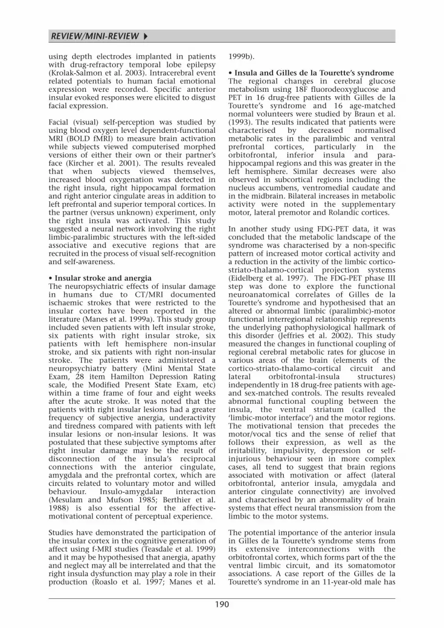
190
using depth electrodes implanted in patientswith drug-refractory temporal lobe epilepsy(Krolak-Salmon et al. 2003). Intracerebral eventrelated potentials to human facial emotionalexpression were recorded. Specific anteriorinsular evoked responses were elicited to disgustfacial expression.
Facial (visual) self-perception was studied byusing blood oxygen level dependent-functionalMRI (BOLD fMRI) to measure brain activationwhile subjects viewed computerised morphedversions of either their own or their partner’sface (Kircher et al. 2001). The results revealedthat when subjects viewed themselves,increased blood oxygenation was detected inthe right insula, right hippocampal formationand right anterior cingulate areas in addition toleft prefrontal and superior temporal cortices. Inthe partner (versus unknown) experiment, onlythe right insula was activated. This studysuggested a neural network involving the rightlimbic-paralimbic structures with the left-sidedassociative and executive regions that arerecruited in the process of visual self-recognitionand self-awareness.
• Insular stroke and anergiaThe neuropsychiatric effects of insular damagein humans due to CT/MRI documentedischaemic strokes that were restricted to theinsular cortex have been reported in theliterature (Manes et al. 1999a). This study groupincluded seven patients with left insular stroke,six patients with right insular stroke, sixpatients with left hemisphere non-insularstroke, and six patients with right non-insularstroke. The patients were administered aneuropsychiatry battery (Mini Mental StateExam, 28 item Hamilton Depression Ratingscale, the Modified Present State Exam, etc)within a time frame of four and eight weeksafter the acute stroke. It was noted that thepatients with right insular lesions had a greaterfrequency of subjective anergia, underactivityand tiredness compared with patients with leftinsular lesions or non-insular lesions. It waspostulated that these subjective symptoms afterright insular damage may be the result ofdisconnection of the insula’s reciprocalconnections with the anterior cingulate,amygdala and the prefrontal cortex, which arecircuits related to voluntary motor and willedbehaviour. Insulo-amygdalar interaction(Mesulam and Mufson 1985; Berthier et al.1988) is also essential for the affective-motivational content of perceptual experience.
Studies have demonstrated the participation ofthe insular cortex in the cognitive generation ofaffect using f-MRI studies (Teasdale et al. 1999)and it may be hypothesised that anergia, apathyand neglect may all be interrelated and that theright insula dysfunction may play a role in theirproduction (Roaslo et al. 1997; Manes et al.
1999b).
• Insula and Gilles de la Tourette’s syndromeThe regional changes in cerebral glucosemetabolism using 18F fluorodeoxyglucose andPET in 16 drug-free patients with Gilles de laTourette’s syndrome and 16 age-matchednormal volunteers were studied by Braun et al.(1993). The results indicated that patients werecharacterised by decreased normalisedmetabolic rates in the paralimbic and ventralprefrontal cortices, particularly in theorbitofrontal, inferior insula and para-hippocampal regions and this was greater in theleft hemisphere. Similar decreases were alsoobserved in subcortical regions including thenucleus accumbens, ventromedial caudate andin the midbrain. Bilateral increases in metabolicactivity were noted in the supplementarymotor, lateral premotor and Rolandic cortices.
In another study using FDG-PET data, it wasconcluded that the metabolic landscape of thesyndrome was characterised by a non-specificpattern of increased motor cortical activity anda reduction in the activity of the limbic cortico-striato-thalamo-cortical projection systems(Eidelberg et al. 1997). The FDG-PET phase IIIstep was done to explore the functionalneuroanatomical correlates of Gilles de laTourette’s syndrome and hypothesised that analtered or abnormal limbic (paralimbic)-motorfunctional interregional relationship representsthe underlying pathophysiological hallmark ofthis disorder (Jeffries et al. 2002). This studymeasured the changes in functional coupling ofregional cerebral metabolic rates for glucose invarious areas of the brain (elements of thecortico-striato-thalamo-cortical circuit andlateral orbitofrontal-insula structures)independently in 18 drug-free patients with age-and sex-matched controls. The results revealedabnormal functional coupling between theinsula, the ventral striatum (called the‘limbic-motor interface’) and the motor regions.The motivational tension that precedes themotor/vocal tics and the sense of relief thatfollows their expression, as well as theirritability, impulsivity, depression or self-injurious behaviour seen in more complexcases, all tend to suggest that brain regionsassociated with motivation or affect (lateralorbitofrontal, anterior insula, amygdala andanterior cingulate connectivity) are involvedand characterised by an abnormality of brainsystems that effect neural transmission from thelimbic to the motor systems.
The potential importance of the anterior insulain Gilles de la Tourette’s syndrome stems fromits extensive interconnections with theorbitofrontal cortex, which forms part of the theventral limbic circuit, and its somatomotorassociations. A case report of the Gilles de laTourette’s syndrome in an 11-year-old male has
REVIEW/MINI-REVIEW
▲

191
▲been associated with MRI documented leftfrontal multicystic changes predominantly inthe gyrus rectus (McAbee et al. 1999). Theyproposed that lesions remote from theneuroanatomical regions of the basal gangliacan be influential by an effect on theinterconnecting circuits involving the lateralorbitofrontal, cingulate, thalamus and the basalganglia resulting in motor and behaviouralphenomena similar to those seen in TS.
• Insula and obsessive-compulsive disorderA case report has described a 12-year-old, right-handed boy with obsessive-compulsivebehaviour and reactive depressive symp-tomatology, which developed on a two-yearbackground history of headaches, vomiting andcomplex partial seizures (Shuren et al. 1995). Hewas diagnosed with OCD using the DSM-III-Rcriteria. Multiple MRI scans demonstrated anenhancing mass in the left posterior insula withslight peri-lesional oedema, which at openbiopsy demonstrated a pleomorphicxanthoastrocytoma. The neurological, neuro-psychological and radiological studies did notdemonstrate frontal lobe or basal gangliadysfunctions commonly seen with patients withOCD (Hymas et al. 1991).
It was postulated that the OCD was the result ofthe posterior insula tumour and proposed thatthe insula is an important structure in thegeneration of OCD in the context of a frontal-basal ganglia-posterior insula loop. Others haveimplicated the lateral orbitofrontal circuit to beimportant in the pathobiology of OCD, but notdiscussed the insula (Leckman et al. 1997).
• Autism and insula-gut-brain aIChypothesisIt has been hypothesised that, in a subgroup ofautistic-spectrum children, a link exists betweenintestinal pathology, attention and language,which may be derived from sharedneuroanatomical pathways within the anteriorinsular cortex (aIC) (Binstock 2001). Severalinvestigators have associated intestinalpathologies with autism and a growing amountof anecdotal data has indicated that a subgroupof autism-spectrum children experiencedimprovements in word production andreceptive and spoken language in response torestoring intestinal health, such as using gluten-free and casein-free diets, and antifungal andantibiotic therapies (Horvath et al. 1999;Rimland 2000; Sandler et al. 2000). The anteriorinsular cortex (aIC), the ‘visceral cortex’, linksgastrointestinal tract mucosal senses with limbicinformation in the amygdala andhypothalamus.
The aIC has been implicated in the functionalneuroanatomy of social behaviour with respectto people with autistic disorder in relation tosocial and emotional perception of facial
expression processing (Critchley et al. 2000). Ina study involving 25 stroke patients witharticulatory deficits, the lesion location hasbeen identified to be in the left aIC (Dronker1996). They proposed that this neuroanatomicallocation might be an aetiologically significantsubstrate in autism-spectrum disordersinterconnected with the gastrointestinalmucosa. The mechanisms of disruption of theaIC in the brain-gut axis may be due to (a)intraneuronal migration of a neutrophic virusgaining initial access via the gastrointestinaltract and/or (b) chronic hyperstimulation of theneuronal pathways providing informationabout gastrointestinal conditions to the limbicregions and the insular cortex. The autonomicirregularity described in autism may have agastrointestinal link via the NTS and the vagusnerve (Palkovitz and Wiesenfeld 1980). Variousinvestigators have linked the aIC andgastrointestinal visceral pathways via the vagalsensory ganglia and the human nucleus oftractus solitarius (Ruggiero et al. 2000; Gesser etal. 1994).
• Insula and depressionThat the insula has been implicated as a neuralsubstrate in the genesis of apathy, anergia andneglect was highlighted earlier (Manes et al.1999a). Converging clinical, post mortem andfunctional neuroimaging evidence suggest thatdepression is unlikely to be a disorder of a singlebrain region or neurotransmitter system.Instead, it is best viewed as a multidimensional,systems level disorder affecting discrete butintegrated pathways involving select cortical,subcortical and limbic sites and their relatedneurotransmitter and molecular mediators. Thistheoretical construct led to the neural networkmodel of depression which proposed thatdepression results from a failure of thecoordinated interactions of a distributednetwork of limbic-cortical pathways (limbic-cortical dysregulation model) (Mayberg 1997).Resting state patterns of regional glucosemetabolism using FDG-PET in idiopathicdepressed patients, changes in metabolism withantidepressant treatment, and blood flowchanges with induced sadness in healthysubjects were used to test this hypothesis. Thethree main compartments of this modelinclude: (a) a dorsal compartment, thatmediates the normal cognitive aspects of mood,comprising of dorsal prefrontal, inferiorparietal, and dorsal and posterior cingulate, (b)a ventral compartment that mediates thevegetative and somatic aspects of mood,composed of paralimbic cortical (ventralorbitofrontal), subcortical and brainstemregions (ventral anterior insula, hippocampus,hypothalamic-pituitary axis and mid-brain/pons), (c) the rostral cingulate, that servesan important regulatory role in the overallnetwork by facilitating the interactions betweenthe dorsal and ventral compartments (Figure
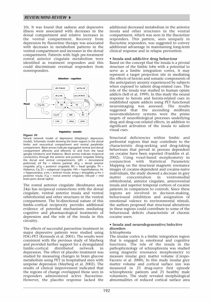
192
10). It was found that sadness and depressiveillness were associated with decreases in thedorsal compartment and relative increases inthe ventral compartment. Recovery fromdepression by fluoxetine therapy was associatedwith decreases in metabolism patterns in theventral compartment and increases in the dorsalcompartment. Patients with high pre-treatmentrostral anterior cingulate metabolism wereidentified as treatment responders and thiscould discriminate eventual responders fromnonresponders.
The rostral anterior cingulate (Brodmann area24a) has reciprocal connections with the dorsalcingulate, ventral anterior insula and ventralorbitofrontal and other structures in the ventralcompartment. The bi-directional nature of thislimbic-cortical reciprocity provides additionalevidence of potential mechanisms mediatingcognitive and pharmacological treatments ofdepression and the role of the insula in thiscircuitry.
The effects of successful paroxetine treatment inmajor depressive patients were studied usingFDG-PET (Kennedy et al. 2001). The results wereconsistent with the previous study of Maybergand provided further support for a dysregulatedlimbic-cortical distributed network indepression. The effect of placebo treatment wasstudied by measuring changes in brain glucosemetabolism using PET in hospitalised men withunipolar depression (Mayberg et al. 2002). Theresults of clinical improvement indicated thatthe regions of change overlapped those seen inresponders administered active fluoxetine.However, the placebo response lacked the
additional decreased metabolism in the anteriorinsula and other structures in the ventralcompartment, which was seen in the fluoxetineresponders. This pattern, seen uniquely influoxetine responders, was suggested to conveyadditional advantage in maintaining long-termclinical response and in relapse prevention.
• Insula and addictive drug behaviourBased on the concept that the insula is a pivotalstructure of the limbic lobe with a potential toserve as a limbic integration cortex, it mayrepresent a target projection site in mediatingthe effects of heroin and somatic components ofthe anticipatory anxiety experienced by subjectswhen exposed to salient drug-related cues. Therole of the insula was studied in human opiateaddicts (Sell et al. 1999). In this study the neuralresponse to heroin and heroin-related cues inestablished opiate addicts using PET functionalneuroimaging was assessed. The resultssuggested that the ascending midbrainneuromodulatory systems were the primetargets of neurobiological processes underlyingdrug and drug-cue-related effects, in addition tosignificant activation of the insula to salientvisual cues.
Structural deficiencies within limbic andprefrontal regions that may contribute to thecharacteristic drug-seeking and drug-takingbehaviours that prevail in persons dependenton cocaine have been reported (Franklin et al.2002). Using voxel-based morphometry inconjunction with Statistical ParametricMapping on the structural magnetic resonanceimages of cocaine-dependent and cocaine-naiveindividuals, the study showed a decrease in greymatter concentration in ventromedialorbitofrontal, anterior cingulate, anteroventralinsula and superior temporal cortices of cocainepatients in comparison to controls. Since theseregions are involved in decision-making,behavioural inhibition and assignation ofemotional valence to environmental stimuli,the authors proposed that structural alterationsin these regions could contribute to some of thebehavioural deficits characteristic of chroniccocaine users.
• Insula and neurodegenerative/infectivedisordersSchizophreniaThe insular cortex is a limbic integration regionthat is engaged in emotional and cognitivefunctions. The role of the insula in thepathophysiology of schizophrenia was studiedusing magnetic resonance morphometry tomeasure insular grey matter volume (Crespo-Facorro et al. 2000). In this study insular greymatter volume and cortical surface size wasmeasured in 25 drug-naive first episodeschizophrenic patients and 25 healthy malevolunteers. The study revealed morphologicalabnormalities of reduced cortical surface area
dFr 9/46/44 inf Par 40
dCg 24b pCg 29/30/31
rCg 24a BG Th
Cg 25 Hth Hc
p-insamyga-ins
mb-pdr
Attention - cognition
Vegetative - somatic
Mood
Figure 10 Neural network model of depression (Mayberg’s depressionmodel). Schematic model showing the brain regions in the dorsallimbic and neocortical compartment and ventral paralimbiccompartment. Black arrows indicate segregated ventral and dorsalcompartment afferents and efferents to and from the basalganglia (striatum) and thalamus. Block arrows indicate reciprocalconnections through the anterior and posterior cingulate linkingthe dorsal and ventral compartments. (dFr = dorsolateralprefrontal; inf Par = inferior parietal; dCg = dorsal anteriorcingulate; pCg = posterior cingulate; BG = basal ganglia; Th =thalamus; Cg 25 = subgenual cingulate; Hth = hypothalamus; Hc= hippocampus; a-ins = anterior insula; amyg = amygdala; p-ins =posterior insula; rCg = rostral anterior cingulate; mb-pdr = midbrain-pons dorsal raphe)
REVIEW/MINI-REVIEW
▲
193
▲and grey matter volume in the left insular cortexin schizophrenic patients. These abnormalitiescorrelated negatively with the severity of thepsychotic symptom dimension.
The neuroanatomical abnormalities docu-mented by neuroimaging include diffuseenlargement of the lateral and third ventricle byapproximately 10% and diffuse reduction ofcortical grey matter volume by approximately3%-4% (Harvey et al. 1993; Lawrie andAbukemeil 1998; Zipursky et al. 1992).Considerable evidence exists in the literature tosuggest that pathological change in patientswith schizophrenia may be expressed at thelevel of spatially distributed networks thatsubsume multiple, densely interconnectedcortical and subcortical regions (Robbins 1990;Firth et al. 1995; Wright et al. 1999). Automateddual echo MRI morphometric analysis has beendone to estimate the grey and white matterdeficits in a clinically homogenous group ofpatients who met the DSM-IV criteria forschizophrenia with negative symptoms andfrom healthy comparison subjects(Sigmundsson et al. 2001). The anatomicalabnormalities in these schizophrenic patientswith marked negative symptoms were mostevident in the left neocortical and limbicregions and related white matter tracts. Thesignificant deficits of grey matter volumes in thepatient group (14%) was localised to (a) the leftsuperior temporal gyrus and insular cortex, (b)the left medial temporal lobe, including theparahippocampal gyrus and hippocampus, and(c) the anterior cingulate and medial frontalgyri. The white matter abnormalities were foundin the left temporal lobe and left frontal lobe.
Deficits in emotional expression andcomprehension have been reported in patientswith insular lesions (Cancelliere and Kertesz1990). These features of insular damage are alsoseen in patients with schizophrenia, especiallyin patients with marked negative symptoms ofalogia and autism. Cytoarchitectonicabnormalities in the insular cortex inschizophrenia have also been reported by otherinvestigators (Jakob and Beckmann 1986;Wright et al. 1999).
Loss of capacity to experience pleasure(anhedonia) is a core clinical feature ofschizophrenia. The neural basis of emotionalprocessing with respect to their capacity toexperience pleasure within the olfactorydomain was studied in 18 schizophreniapatients matched with controls using PET and15[O] water method (Crespo-Facorro et al. 2001).Patients with schizophrenia subjectivelyexperienced unpleasant odours (negativehedonic affect) in a manner similar to healthyvolunteers. However, in schizophrenia patientsthere was impairment in the experience ofpleasant odours (positive hedonic affect). The
results of the regional cerebral blood flowrevealed that the schizophrenia patients haddecreased regional cerebral blood flow in the leftanterior insula, the right nucleus accumbens,the left superior temporal gyrus andparahippocampal gyrus, the lingual gyrus andthe cerebellar vermis. This failure in activationof the limbic/paralimbic regions during theexperience of unpleasant odours was associatedwith an abnormal increase in regional cerebralblood flow in bilateral frontal cortical regions.Thus, dysfunction in distributed neural circuitsaffecting the complex interactions andconnectivity between the limbic/paralimbic(insular cortex, nucleus accumbens,hippocampus and parahippcampal regions) andthe frontal cortex may represent the neuralsubstrates of emotional disturbances seen inpatients with schizophrenia.
Frontotemporal lobar degeneration (FTLD)Frontotemporal lobar degeneration (FTLD) is aneurodegenerative cognitive disordercharacterised by progressive early distinctivebehavioural symptoms associated withprogressive loss of cerebral tissue in the frontaland temporal lobes. A recent study has shownsignificant atrophy of the insula bilaterally inboth frontotemporal dementia (FTD) andsemantic dementia (SemD), implicating thepotential role of the insula in social behaviour(Rosen et al. 2002). They concluded that theabnormalities in the neural processing ofstimulus reward contingency and autonomicfeedback are important mechanisms by whichdamage in the orbital frontal, anterior cingulateand insular areas may produce behaviouraldeficits in FTLD.
Certain distinctive behavioural changes arespecific and more common in frontotemporaldementia that occurs early, and has relevancefor the differentiation of frontotemporaldementia from Alzheimer’s disease. Recentstudies have highlighted the high prevalence ofalterations in food preference (craving sweetfoods, food fads), appetite (increased appetite,gluttony) and eating behaviour (stereotypiceating behaviours, abnormal oral behaviours) inthe frontal variant of frontotemporal dementia(Bozeat et al. 2000). The neuroanatomicalimplications for these specific early corebehavioural features have been hypothesised toinvolve a network comprising the orbitobasalfrontal lobe, the temporal pole, the amygdalaand the anterior insular cortex (orbitofrontal-amygdala-insular network). Brain networkmechanisms underlying appetite control andsatiation have been studied by PET generatedfunctional brain maps, which indicated theinvolvement of these structures (Del-Parigi et al.2002).
A recent study looked into the prevalence andpattern of distinctive ritualistic and complex
194
stereotypic behaviours in frontotemporaldementia (Nyastsanza et al. 2003). Theyconcluded that these behaviours (foot tapping,grunting, hand rubbing, rigid adherence toroutines, verbal stereotypies, repetitively eatingthe same food, compulsive symptoms) have ahigher prevalence in FTD than in Alzheimer’sdisease, and suggested that these aberrant motorbehaviours may have discriminatory power inthe diagnosis of FTD and are a core feature ofthe dementing syndrome in FTD. It wasconcluded that these behavioural abnormalitiesreflected disruption of an integrated circuitinvolving the orbitofrontal cortex, amygdalaand the insular cortex and/or the striatalcircuitry.
Thus the insular cortex has been implicated inthe neural processing of human eatingbehaviour, satiation and aberrant motorbehaviour in FTD as a result of its intenseinterconnections with the ventromedial frontallobe, temporal pole and amygdala. Theelucidation of the pathophysiological basis forthese core neurobehavioural abnormalities byfuture neuroimaging and behaviour studiesshould clarify the componential role of theinsular cortex in the neural networks mediatingthese distinctive behavioural abnormalities inFTD.
Alzheimer’s diseaseA recent study implicated the insular cortex andthe midfrontal gyrus in the neurodegenerationthat occurs with Alzheimer’s disease. In thiscase-control study, brain tissue samples wereobtained from the insular cortex and midfrontalgyrus, which revealed DNA fragmentation inthe pyramidal neurones along with denseaccumulation of beta-amyloid deposition(Colurso et al. 2003). Beta-amyloid mediatesneuronal injury through the process ofapoptosis as evidenced by the positive assayidentifying DNA fragmentation. This studyidentified these two distinctive neuro-pathologies co-occurring in the insular cortexand midfrontal gyrus, both of which areinterconnected reciprocally, and with the otherlimbic areas.
Dementia with Lewy bodies (DLB)This non-Alzheimer degenerative dementia ischaracterised neuropathologically by thepresence of cortical Lewy bodies. Several studieshave shown the site of cortical Lewy bodydensity which has a consistent gradient asfollows: substantia nigra > entorhinal cortex >cingulate gyrus > insular cortex > frontal cortex> hippocampus > occipital cortex (McKeith2001). A recent study showed that Lewypathology starts in the deeper layers of theneocortex and paralimbic regions, including theinsular cortex, by using alpha-synucleinimmunohistochemistry (Marui et al. 2002). Inanother study using the image processing
technique of voxel based morphometry,regional grey matter volume loss was shown toaffect bilateral temporal and frontal lobes andinsular cortex of patients with DLB compared tocontrol subjects (Burton et al. 2002).
NeuroinfectionsIn an autopsy study of human herpes simplexvirus encephalitis, the distribution of viralantigen within the brain was mapped (Esiri1982). The viral antigens were mainly localisedin the medial and inferior temporal lobes inaddition to the insula, olfactory cortex,hippocampus, amygdaloid nuclei and cingulategyrus. In another study usingimmunocytochemistry, positive viral antigenimmunostaining was identified in the fronto-orbital, mediobasal temporal lobes, insula andcingulate gyrus (Charpin et al. 1985).
The insular cortex has been implicated inintracerebral Whipple’s disease in addition totypical lesions involving the hypothalamus,cingulate gyrus, basal ganglia and cerebellum(Mendel et al. 1999). Whipple’s disease resultsfrom a systemic infection by the bacteriumTropheryma whippelii and may manifest withcentral nervous system involvement alone,without typical gastrointestinal disease,comprising a dementia, personality change,supranuclear opthalmoplegia and characteristicoculomasticatory-skeletal myoarrythmia.
Insula and therapeuticneurostimulation
The neuroanatomical implication of the humaninsular cortex in epilepsy, treatment-resistantdepression and other neuropsychiatric disordershas come to light by studying the effects ofvagus nerve stimulation (VNS) in such disorders(George et al. 2000). VNS has been shown tohave antiseizure and antidepressant effects. Theother areas of potential research or clinicalneuropsychiatric implications with VNS includeanxiety disorders, obesity, addictions, sleepdisorders, dementing disorders and painsyndromes.
The vagus nerve afferents synapse in thenucleus of the tractus solitarius (NTS) whichthen projects to the locus ceruleus, dorsal raphe,hypothalamus and the thalamus (vagus-NTSpathway). The pathway to the thalamuscontinues to the anterior insular cortex andlimbic structures (amygdala, hippocampus,posterior cingulate gyrus, entorhinal cortex andanterior temporal cortex and orbitofrontalregions). The insular cortex also sendsprojections back to the nucleus of the tractussolitarius. Higher-order projections of theanterior insula are particularly dense in inferiorand inferolateral frontal cortex of the limbicsystem.
REVIEW/MINI-REVIEW
▲
195
▲The cerebral blood flow alterations inducedduring VNS revealed increased blood flow inrostral, dorsal-central medulla; bilateralhypothalami, thalami, insular cortices andinferior cerebellar hemispheres; and decreasedbilaterally in hippocampus, amygdala andposterior cingulate gyri (Henry et al. 1998).
Transcranial magnetic stimulation (TMS) isanother new, non-invasive technique fordirectly stimulating cortical neurones which hasbeen used in treatment of mood in depression.Using 15 [O] water and PET, the changes inregional cerebral blood flow were studied afterhigh and low frequency r-TMS over the leftprefrontal cortex in ten medication-free patientswith major depression (Speer et al. 2000). Highfrequency r-TMS was associated with significantincreases in regional cerebral blood flow in theprefrontal cortex and cingulate gyrus (left >right), left amygdala and bilateral insula, basalganglia, uncus, hippocampus, para-hippocampus, thalamus and cerebellum.
Thus functional neuroimaging tools, when usedin combination with VNS and TMS, havethrown light on the role of the anterior insularcortex within the distributed neural networksthat may underlie many neuropsychiatricdisorders.
Conclusions
Over the course of the last two centuries, sinceits initial description, a great deal of work hasfocused on the anatomical and functionalaspects of this ‘hidden lobe’ called ‘the island ofReil’. It can be concluded that the insula is amultifaceted functional area, which has acomponential role in various ‘distributed neuralnetworks’ subserving human behaviour and inthe causation of disorders affecting the brainand the mind. The human insular cortex ischaracterised by the greatest conceivablespectrum of cortical and subcorticalconnections, which would explain the panoplyof its complex behavioural specialisations. Theinsula is a pivotal functional structure of thelimbic lobe and plays a vital role as a limbicintegration cortex. Like the extended amygdala,the insula serves as a neural relay ‘satellite’station and forms an integrative anatomical,functional and behavioural unit of thetelencephalon.
With the advent of state of the art neuroimagingtools for research to map brain functions it hasbeen possible to gain further insights regardingthe neural substrates and neurobiologicalfoundations of human behaviour, and on therole of the insula. Imaging neuroscience has ledto a conceptual shift in understanding of brainfunctional architecture from the traditional‘functional segregation approach’ (segregatedcircuitry) to a ‘dynamic network approach’
mediated by multidimensional distributedneural network connectivity (distributedcircuitry). Brain functions are viewed as aworking mosaic model mediated by large-scaleinterconnected neural networks incorporatingmultiple cortical regions (multimodularnetworks) with collaborative and overlappingfunctions (functional integration approach).The blueprint of complex human behaviour isnow mapped at the level of multifocal neuralsystems rather than specific anatomical sitescharacterised by almost infinite richness andflexibility. It is the abnormal maladaptedfunctional relationship between elements in aseries of neural network that underlies thegenesis of neuropsychiatric or behaviouraldisorders (network approach to behaviouralneurology).
Thus the insula does not operate in isolationand functions as albeit one important elementin a series of neural networks distributedthroughout the central nervous system, and it isthe abnormal functional relationships betweenthe various regions that dictate the behaviouraland neuropsychiatric afflictions that we relateto the insula. Further research into the inter-regional connectivity and functional aspects ofthe insula will be made possible by robustparadigms of computational neuroscience; thecombined use of non-invasive functionalneuroimaging tools (PET, f-MRI, MEG) and non-invasive cortical stimulation using TMS both inanimal and human studies.
References
Ahern GL, Sollers JJ, Lane RD, Labiner DM, Herring AM, WeinandME, Hutzler R, Thayer JF (2001) Heart rate and heart ratevariability changes in the intracarotid sodium amobarbital test.Epilepsia 42: 912-921.
Alheid GF, Heimer L (1988) New perspectives in basal forebrainorganisation of special reference for neuropsychiatric disorders;the striatopallidal, amygdaloid, and corticopetal components ofsubstantia innominata. Neuroscience 27: 1-39.
Augustine JR (1985) The insular lobe in primates includinghumans. Neurol Res 7: 2-10.
Augustine JR (1996) Circuitry and functional aspects of the insularlobe in primates including humans. Brain Res Rev 22: 229-244.
Aziz Q, Furlong PL, Barlow J, Hobson A, Alani S, Bancewicz J,Ribbands M, Harding GFA, Thompson DG (1995) Topographicmapping of cortical potentials evoked by distension of the humanproximal and distal oesophagus. Electroencephalogr ClinNeurophysiol 96: 219-228.
Bakar M, Kirshner HS, Niaz F (1998) The opercular-subopercularsyndrome: four cases with review of the literature. Behav Neurol11: 97-103.
Bartholin C (1641) Institutiones Anatomicae ab auctoris filioThoma Bartholino. Hack, Leiden.
Berke JJ (1960) The claustrum, the external capsule and theextreme capsule of Macaca mulatta. J Comp Neurol 115: 297-331.
196
Berthier M, Starkstein S, Leiguarda R (1987) Behavioural effects ofdamage to the right insula and surrounding regions. Cortex 23:673-678.
Berthier M, Starkstein S, Leiguarda R (1988) Asymbolia for pain: asensory-limbic disconnection syndrome. Ann Neurol 24: 41-49.
Binkofski F, Schnitzler A, Enck P, Frieling T, Posse S, Seitz RJ, FreundHJ (1998) Somatic and limbic cortex activation in esophagealdistension: a functional magnetic resonance imaging study. AnnNeurol 44: 811-815.
Binstock T (2001) Anterior insular cortex: linking intestinalpathology and brain function in autism-spectrum subgroups. MedHypotheses 57: 714-717.
Bornhovd K, Quante M, Glauche V, Bromm B, Weiller C, Buchel C(2002) Painful stimuli evoke different stimulus-response functionsin the amygdale, prefrontal, insula and somatosensory cortex: asingle trial fMRI study. Brain 125: 1326-1336.
Bouilleret V, Dupont S, Spelle L, Baulac M, Samson Y, Semah F(2002) Insular cortex involvement in mesiotemporal lobe epilepsy:a positron emission tomography study. Ann Neurol 51: 202-208.
Bozeat S, Gregory CA, Lambon RMA, Hodges JR (2000) Whichneuropsychiatric and behavioural features distinguish frontal andtemporal variants of frontotemporal dementia from Alzheimer’sdisease? J Neurol Neurosurg Psychiatry 69: 178-186.
Brandt T, Dieterich M (1999) The vestibular cortex. Its locations,functions, and disorders. Ann N Y Acad Sci 871: 293-312.
Brandt T, Dieterich M, Danek A (1994) Vestibular cortex lesionsaffect the perception of verticality. Ann Neurol 35: 403-412.
Brandt T, Glasauer S, Stephan T, Bense S, Yousry TA,Deutschlander A (2002) Visual-vestibular and visuovisual corticalinteraction: new insights from fMRI and PET. Ann N Y Acad Sci956: 230-241.
Braun AR, Stoetter B, Randolph C, Hsiao JK, Vladar K, Gernert J,Carson RE, Herscovitch P, Chase TN (1993) The functionalneuroanatomy of Tourette’s syndrome: An FDG-PET Study I:Regional changes in cerebral glucose metabolism differentiatingpatients and controls. Neuropsychopharmacology 9: 277-291.
Broca P (1861a) Perte de la parole: Ramollisement chronique etdestruction partielle du lobe antérieur gauche du cerveau. ParisBull Soc Anthropol 2: 235-238.
Broca P (1861b) Remarques sur le siége de la faculté au langagearticulé, suivies d’une observation d’aphémie (perte de la parole).Bull Soc Anat 36: 330-357.
Brodmann K (1909) Vergleichende Localisationslehre derGrosshirnrinde in ihren Prinzipien dargestellt auf Grund desZellenbaues. JA Barth, Leipzig.
Burton EJ, Karas G, Paling SM, Barber R, Williams ED, Ballard CG,McKeith IG, Scheltens P, Barkhof F, O’Brien JT (2002) Patterns ofcerebral atrophy in dementia with Lewy bodies using voxel-basedmorphometry. Neuroimage 17: 618-630.
Burton H, Videen TO, Raichle ME (1993) Tactile vibration activatedfoci in insular and parietal-opercular cortex studied with positronemission tomography: mapping the second somatosensory areain humans. Somatosens Motil Res 10: 297-308.
Cabezo R, Kapur S, Craik F (1997) Functional neuroanatomy ofrecall and recognition: a PET study of episodic memory. J CogNeurosci 9: 254-265.
Cancelliere AE, Kertesz A (1990) Lesion localisation in acquireddeficits of emotional expression and comprehension. Brain Cogn13: 133-147.Cereda C, Ghika J, Maeder P, Bogousslavsky J (2002) Strokesrestricted to the insular cortex. Neurology 59: 1950-1955.
Charpin C, Gambarelli D, Lavaut MN, Seigneurin JM, Raphael M,Berard M, Toga M (1985) Herpes simplex virus antigen detectionin human acute encephalitis: an immunohistochemical studyusing avidin-biotin-peroxidase complex method. Acta
Neuropathol (Berl) 68: 245-252.
Chikama M, McFarland NR, Amaral DG, Haber SN (1997) Insularcortical projections to functional regions of the striatum correlatewith cortical cytoarchitectonic organization in the primate. JNeurosci 17: 9686-9705.
Chollet F, DiPiero V, Wise RJS, Brooks DJ, Dolan RJ, Frackowiak RSJ(1991) The functional anatomy of motor recovery after stroke inhumans: a study with positron emission tomography. Ann Neurol29: 63-71.
Chua HC, Sen S, Cosgriff RF, Gerstenblith G, Beauchamp NJ Jr,Oppenheimer SM (1999) Neurogenic ST depression in stroke. ClinNeurol Neurosurg 101: 44-48.
Clarke E, O’Malley CD (1968) The human brain and the spinalcord. A historical study illustrated by writings from antiquity to thetwentieth century. University of California Press, Berkeley, p 926.
Colurso GJ, Nilson JE, Vervoort LG (2003) Quantitative assessmentof DNA fragmentation and beta-amyloid deposition in insularcortex and midfrontal gyrus from patients with Alzheimer’sdisease. Life Sci 73: 1795-1803.
Corbetta M, Miezin FM, Dobmeyer S, Shulman GL, Petersen SE(1991) Selective and divided attention during visualdiscrimination of shape, colour, and speed: functional anatomy bypositron emission tomography. J Neurosci 11: 2383-2402.
Craig AD, Chen K, Bandy D, Reiman EM (2000) Thermosensoryactivation of insular cortex. Nat Neurosci 3: 184-190.
Crespo-Facorro B, Kim J, Andreasen NC, O’Leary DS, Bockholt HJ,Magnotta V (2000) Insular cortex abnormalities in schizophrenia:a structural magnetic resonance imaging study of first episodepatients. Schizophr Res 46: 35-43.
Crespo-Facorro B, Paradiso S, Andreasen NC, O’Leary DS, WatkinsGL, Ponto LLB, Hichwa RD (2001) Neural mechanisms ofanhedonia in schizophrenia: A PET study of response tounpleasant and pleasant odors. JAMA 286: 427-435.
Critchley HD, Daly EM, Bullmore ET (2000) The functionalneuroanatomy of social behaviour: changes in cerebral blood flowwhen people with autistic disorder process facial expressions.Brain 123: 2203-2212.
Cunningham DJ (1891) The development of the gyri and sulci onthe surface of island of Reil of the human brain. J Anat Physiol 25:338-348.
Daffau H, Capelle L, Lopes M, Bitar A, Sichez JP, Van Effenterre R(2002) Medically intractable epilepsy from insular low-gradegliomas: improvement after an extended lesionectomy. ActaNeurochir 144: 563-573.
Damasio H, Damasio AR (1980) The anatomical basis ofconduction aphasia. Brain 103: 337.
Daniels SK, Foundas AL (1997) The role of the insular cortex indysphagia. Dysphagia 12: 146-156.
De Olmos JS, Heimer L (1999) The concepts of the ventralstriatopallidal system and extended amygdala. In: Advancing fromthe ventral striatum to the extended amygdala: implications forneuropsychiatry and drug abuse. McGinty JF (ed) Ann NY AcadSci 877: 1-32.
Deecke L, Schwarz DWF, Fredrickson JM (1974) Nucleusventroposterior inferior (VPI) as the vestibular thalamic relay in therhesus monkey. I. Field potential investigation. Exp Brain Res 20:88-100.
Deecke L, Schwarz DWF, Fredrickson JM (1977) Vestibularresponses in the rhesus monkey ventroposterior thalamus. II.Vestibulo-proprioceptive convergence at thalamic neurons. ExpBrain Res 30: 219-232.
Del Parigi A, Gautier JF, Chen K, Salbe AD, Ravussin E, Reiman E,Tataranni PA (2002) Neuroimaging and obesity: mapping thebrain responses to hunger and satiation in humans using positronemission tomography. Ann N Y Acad Sci 967: 389-397.
REVIEW/MINI-REVIEW
▲
197
▲Deuchert M, Ruben J, Schwiemann J, Meyer R, Thees S, Krause T,Blankenburg F, Villringer K, Kurth R, Curio G, Villringer A (2002)Event-related fMRI of the somatosensory system using electricalfinger stimulation. Neuroreport 13: 365-369.
Dronkers NF (19960 A new brain region for coordinating speecharticulation. Nature 384: 159-161.
Eberstaller O (1887) Zur Anatomie und Morphologie Der InsulaReil. Anat Anz 2: 739-750.
Eckardt M, Gerlach L, Welter FL (1999) Prolongation of thefrequency-corrected dispersion following cerebral strokes withinvolvement of the insula of Reil. Eur Neurol 42: 190-193.
Eidelberg D, Moeller JR, Antonini A, Kazumata K, Dhawan V,Budman C, Feigin A (1997) The metabolic anatomy of Tourette’ssyndrome. Neurology 48: 927-934.
Esiri MM (1982) Herpes simplex encephalitis. Animmunohistological study of the distribution of viral antigenwithin the brain. J Neurol Sci 54: 209-226.
Fifer RC (1993) Insular stroke causing unilateral auditoryprocessing disorder: case report. J Am Acad Audiol 4: 364-369.
Fink GR, Markowitsch HJ, Reinkemeier T, Bruckbauer T, Kesslar J,Heiss WD (1996) Cerebral representation of one’s own past:Neural networks involved in autobiographical memory. J Neurosci16: 4275-4282.
Fiol ME, Leppick IE, Mireles R, Maxwell R (1988) Ictus emeticusand the insular cortex. Epilepsy Res 2: 127-131.
Firth CD, Friston KJ, Herold S, Silbersweig D, Fletcher P, Cahill C,Dolan RJ, Frackowiak RS, Liddle PF (1974) Regional brain activityin chronic schizophrenic patients during the performance of averbal fluency task. Br J Psychiatry 167: 343-349.
Forbes BF, Moskowitz N (1974) Projections of auditory responsivecortex in squirrel monkey. Brain Res 67: 239-254.
Franklin TR, Acton PD, Maldjian JA, Gray JD, Croft JR, Dackis CA,O’Brien CP, Childress AR (2002) Decreased gray matterconcentration in the insular, orbitofrontal, cingulate, andtemporal cortices of cocaine patients. Biol Psychiatry 51: 134-142.
Frot M, Mauguiere F (1999) Operculo-insular responses tonociceptive skin stimulation in humans. A review of the literature.Neurophysiol Clin 29: 401-410.
Frot M, Rambaud L, Guenot M, Mauguiere F (1999) Intracorticalrecordings of early pain-related CO2-laser evoked potentials in thehuman second somatosensory (SII) area. Clin Neurophysiol 110:133-145.
Fukuda H, Yamada K, Kinomura S, Kawashima R, Ono S, YoshiokaS, Yamaguchi T, Matui H, Itoh M, Seo S (1991) High ordergustatory projections in the human brain studied with positronemission tomography. The Salt Science Annual Research Report,Section of Physiology and Food Science, pp 337-347.
George MS, Sackeim HA, Rush AJ, Marangell LB, Nahas Z, HusainMM, Lisanby S, Burt T, Goldman J, Ballenger JC (2000) Vagusnerve stimulation: A new tool for brain research and therapy. BiolPsychiatry 47: 287-295.
Geschwind N (1965) Disconnection syndromes in animals andman. Brain 88: 237-585.
Gesser RM, Valyi-Nagy T, Altschuler SM, Fraser NW (1994) Oral-oesopahgeal inoculation of mice with herpes simplex virus type 1causes latent infection of the vagal sensory ganglia (nodoseganglia). J Gen Virol 75: 2379-2386.
Grasby PM, Firth CD, Friston KJ, Simpson J, Fletcher PC,Frackowiak RSJ, Dolan RJ (1994) A graded task approach to thefunctional mapping of brain areas implicated in auditory-verbalmemory. Brain 117: 1271-1282.
Hachinski V (1999) The insula of sudden death. J W Neurol 1001:1.
Hamdy S, Rothwell JC, Brooks DJ, Bailey D, Aziz Q, Thompson DG(1999) Identification of the cerebral loci processing humanswallowing with H2(15)O PET activation. J Neurophysiol 81:1917-1926.
Harvey I, Ron MA, Du Boulay G, Wicks D, Lewis SW, Murray RM(1993) Reduction of cortical volume in schizophrenia on magneticresonance imaging. Psychol Med 23: 591-604.
Heilman KM, Valenstein E (1972) Frontal lobe neglect in man.Neurology 22: 660-664.
Heilman KM, Watson RT, Valenstein E (1993) Neglect and relateddisorders. In: Heilman KM, Valenstein E (eds) ClinicalNeuropsychology. 3rd ed. Oxford University Press, New York, pp279-336.
Heimer L, De Olmos JS, Alheid GF, Zaborszky L (1991)"Perestroika" in the basal forebrain: opening the border betweenneurology and psychiatry. Prog Brain Res 87: 109-165.
Henry TR, Votaw JR, Pennell PB, Epstein CM, Bakay RAE, Faber TL,Grafton ST, Hoffman JM (1999) Acute blood flow changes andefficacy of vagus nerve stimulation in partial epilepsy. Neurology52: 1166-1173.
Horvath K, Papadimitriou JC, Rabsztyn A, Drachenberg C, TildonJT (1999) Gastrointestinal abnormalities in children with autisticdisorder. J Pediatr 135: 559-563.
Hurst EM (1959) Some cortical association systems related toauditory functions. J Comp Neurol 112: 103-117.
Hymas N, Lees A, Bolton K (1991) The neurology of obsessionalslowness. Brain 114: 2203-2233.
Isnard J, Guenot M, Ostrowsky K, Sindou M, Mauguiere F (2000)The role of the insular cortex in temporal lobe epilepsy. AnnNeurol 48: 614-623.
Jakob H, Beckmann H 1986) Prenatal developmental disturbancesin the limbic allocortex in schizophrenia. J Neural Transm 65: 303-326.
Jeffries KJ, Schooler C, Schoenbach C, Herscovitch P, Chase TN,Braun AR (2002) The functional neuroanatomy of Tourette’ssyndrome: An FDG-PET Study III: Functional coupling of regionalcerebral metabolic rates. Neuropsychopharmacology 27: 92-104.
Kennedy SH, Evans KR, Kruger S, Mayberg HS, Meyer JH, McCannS, Arifuzzman AI, Houle S, Vaccarino FJ (2001) Changes in regionalbrain glucose metabolism measured with positron emissiontomography after paroxetine treatment of major depression. Am JPsychiatry 158: 899-905.
Kew JJ, Leigh PN, Playford ED, Passingham RE, Goldstein LH,Frackowiak RSJ, Brooks DJ (1993) Cortical function in amyotrophiclateral sclerosis. Brain 116: 655-680.
Kinomura S, Kawashima R, Yamada K, Ono S, Itoh M, Yoshioka S,Yamaguchi T, Matsui H, Miyazawa H, Itoh H, Goto R, Fujiwara T,Satoh K, Fukuda H (1994) Functional anatomy of taste perceptionin the human brain studied with positron emission tomography.Brain Res 659: 263-266.
Kircher TT, Senior C, Phillips ML, Rabe-Hesketh S, Benson PJ,Bullmore ET, Brammer M, Simmons A, Bartels M, David AS (2001)Recognizing one’s own face. Cognition 78: B1-B15.
Koga M, Saku Y, Toyoda K, Takaba H, Ibayashi S, Iida M (2003)Reappraisal of early CT signs to predict the arterial occlusion sitein acute embolic stroke. J Neurol Neurosurg Psychiatry 74: 649-653.
Krieg WJS (1965) Connections of the cerebral cortex of themonkey. Symp Biol Hung 5: 177-217.
Krolak-Salmon P, Henaff MA, Isnard J, Tallon-Baudry C, Guenot M,Vighetto A, Bertrand O, Mauguiere F (2003) An attentionmodulated response to disgust in human ventral anterior insula.Ann Neurol 53: 446-453.
198
Kucharska-Pietura K, Pietura R, Masiak M (2001) Neural correlatesof emotions in psychiatric patients in the light of functionalneuroimaging findings. Ann Univ Mariae Curie Sklodowska 56:343-348.
Kushner MJ, Schwartz R, Alavi A, Dann R, Rosen M, Silver R,Reivich M (1987) Cerebral glucose consumption following verbalauditory stimulation. Brain Res 409: 79-87.
Lawrie SM, Abukmeil SS (1998) Brain abnormality inschizophrenia. Br J Psychiatry 172: 110-120.
Leckman JF, Peterson BS, Anderson GM, Arnsten AF, Pauls DL,Cohen DJ (1997) Pathogenesis of Tourette’s syndrome. J ChildPsychol Psychiatry 38: 119-142.
Lockard I (1948) Certain developmental relations and fiberconnections of the triangular gyrus in primates. J Comp Neurol89: 349-386.
Maihofner C, Kaltenhauser M, Neundorfer B, Lang E (2002)Temporo-spatial analysis of cortical activation by phasic innocuousand noxious cold stimuli - a magnetoencephalographic study.Pain 100: 281-290.
Manes F, Paradiso S, Robinson RG (1999a) Neuropsychiatriceffects of insular stroke. J Nerv Ment Dis 187: 707-712.
Manes F, Paradiso S, Springer JA, Lamberty GJ, Robinson RG(1999b) Neglect after right insular cortex infarction. Stroke 30:946-948.
Manes F, Springer J, Jorge R, Robinson RG (1999c) Verbal memoryimpairment after left insular cortex infarction. J Neurol NeurosurgPsychiatry 67: 532-534.
Marien P, Pickut BA, Engelborghs S, Martin JJ, De Deyn PP (2001)Phonological agraphia following a focal anterior insulo-opercularinfarction. Neuropsychologia 39: 845-855.
Marshall R, Lazar R, Mohr JP (1996) Semantic conduction aphasiafrom a posterior insular cortex infarction. J Neuroimaging 362:342-345.
Marui W, Iseki E, Nakai T, Miura S, Kato M, Ueda K, Kosaka K(2002) Progression and staging of Lewy pathology in brains frompatients with dementia with Lewy bodies. J Neurol Sci 195: 153-159.
Mayberg HS (1997) Limbic-cortical dysregulation: a proposedmodel of depression. J Neuropsychiatry Clin Neurosci 9: 471-481.
Mayberg HS, Silva JA, Brannan SK, Tekell JL, Mahurin RK, McGinnisS, Jerabek PA (2002) The functional neuroanatomy of the placeboeffect. Am J Psychiatry 159: 728-737.
McAbee GN, Wark JE, Manning A (1999) Tourette syndromeassociated with unilateral cystic changes in the gyrus rectus.Pediatr Neurol 20: 322-324.
McCarthy G, Blamire AM, Rothman DL, Gruetter R, Shulman RG(1993) Echo-planar magnetic resonance imaging studies of frontalcortex activation during word generation in humans. Proc NatlAcad Sci USA 90: 4952-4956.
McKeith IG (2001) Dementia with Lewy bodies. In: Miyoshi K,Shapiro CM, Gaviria M, Morita Y (eds) Contemporaryneuropsychiatry. Springer-Verlag, Tokyo, pp 180-186.
Mendel E, Khoo LT, Go JL, Hinton D, Zee CS, Apuzzo ML (1999)Intracerebral Whipple’s disease diagnosed by stereotactic biopsy:a case report and review of the literature. Neurosurgery 44: 203-209.
Mesulam MM (1985) Principles of Behavioural Neurology. FADavis, Philadelphia, pp 1-70.
Mesulam MM (1998) From sensation to cognition. Brain 121:1013-1052.
Mesulam MM, Mufson EJ (1982a) Insula of the Old Worldmonkey. Architectonics in the insulo-orbito-temporal component
of the paralimbic brain. J Comp Neurol 212: 1-22.
Mesulam MM, Mufson EJ (1982b) Insula of the old world monkey.III: Efferent cortical output and comments on function. J CompNeurol 212: 38-52.
Mesulam MM, Mufson EJ (1985) The insula of Reil in man andmonkeys. Architectonics, connectivity, and function. In: Peters A,Jones EG (eds) Cerebral Cortex Vol 4: Association and Auditorycortices. Plenum Press, New York, pp 179-226.
Mettler FA (1945) Fiber connections of the corpus striatum of themonkeys and baboon. J Comp Neurol 82: 169-204.
Monro A (1783) Observations on the structure and function of thenervous system. Edinburgh.
Mufson EJ, Sobreviela T, Kordower JH (1997) Chemicalneuroanatomy of the primate insula cortex: relationship tocytoarchitectonics, connectivity, function and neurodegeneration.In: Bloom FE, Bjorklund A, Hokfelt T (eds) Handbook of ChemicalNeuroanatomy, Vol 13. Elsevier, Amsterdam, pp 377-454.
Mulder DW, Daly D, Bailey AA (1954) Visceral epilepsy. Arch InternMed 4: 93.
Nauta WJH (1979) Expanding borders of the limbic systemconcept. In: Rasmussen T, Marino R (eds) Functional neurosurgery.Raven, New York, p 7.
Nestor PJ, Graham NL, Fryer TD, Williams GB, Patterson K, HodgesJR (2003) Progressive non-fluent aphasia is associated withhypometabolism centered on the left anterior insula. Brain 126:2406-2418.
Noesselt T, Shah NJ, Jancke L (2003) Top-down and bottom-upmodulation of language related areas - an fMRI study. Neurosci 4:13-25.
Nyatsnaza S, Shetty T, Gregory C, Lough S, Dawson K, Hodges JR(2003) A study of stereotypic behaviours in Alzheimer’s diseaseand frontal and temporal variant frontotemporal dementia. JNeurol Neurosurg Psychiatry 74: 1398-1402.
Oppenheimer S (1992) The insular cortex and thepathophysiology of stroke-induced cardiac changes. Can J NeurolSci 19: 208-211.
Oppenheimer S (1993) The anatomy and physiology of corticalmechanisms of cardiac control. Stroke 24 (Suppl 12): 13-15.
Oppenheimer S, Norris JW (1995) Cardiac manifestation of acuteneurological lesions. In: Aminoff MJ (ed) Neurology and GeneralMedicine: The neurological aspects of medical disorder. 2nd edChurchill-Livingstone, New York, pp 183-200.
Oppenheimer SM, Wilson JX, Guiraudon C, Cechetto DF (1991)Insular cortex stimulation produces lethal cardiac arrthymias: amechanism of sudden death? Brain Res 550: 115-121.
Oppenheimer SM, Gelb A, Girvin JP, Hachinski VC (1992)Cardiovascular effects of human insular cortex stimulation.Neurology 42: 1727-1732.
Ostrowsky K, Isnard J, Ryvlin P, Guenot M, Fischer C, Mauguiere F(2000) Functional mapping of the insular cortex: clinicalimplication in temporal lobe epilepsy. Epilepsia 41: 681-686.
Ostrowsky K, Magnin M, Ryvlin P, Isnard J, Guenot M, MauguiereF (2002) Representation of pain and somatic sensation in thehuman insula: a study of responses to direct electrical corticalstimulation. Cereb Cortex 12: 376-385.
Palkovitz RJ, Wiesenfeld AR (1980) Differential autonomicresponses of autistic and normal children. J Autism Dev Disord 10:347-360.
Park K, Seo JJ, Kang HK, Ryu SB, Kim HJ, Jeong GW (2001a) A newpotential of blood oxygenation level dependent (BOLD)functional MRI for evaluating cerebral centers of penile errection.Int J Impot Res 13: 73-81.
Park K, Kang HK, Seo JJ, Kim HJ, Ryu SB, Jeong GW (2001b) Blood-
REVIEW/MINI-REVIEW
▲
199
▲oxygenation-level-dependant functional magnetic resonanceimaging for evaluating cerebral regions of female sexual arousalresponse. Urology 57: 1189-1194.
Paulesu E, Firth CD, Frackowiak RSJ (1993). The neural correlatesof the verbal component of working memory. Nature 362: 342-344.
Penfield W, Faulk ME (1955) The Insula: further observations on itsfunction. Brain 78: 445-470.
Penfield W, Jasper WW (1954) Epilepsy and the functionalanatomy of the human brain. Little, Brown, Boston.
Perry DW, Alivisatos B, Evans AC, Meyer E, Petrides M, Zatorre RJ(1993) Neural network supporting auditory-vocal integration insinging. J Acoust Soc Am 93: 2403-2404.
Petit L, Orssaud C, Tzourio N, Salamon G, Mazoyer B, Berthoz A(1993) PET study of voluntary saccadic eye movements inhumans: basal ganglia-thalamocortical system and cingulatecortex involvement. J Neurophysiol 69: 1009-1017.
Peyron R, Laurent B, Garcia-Larrea L (2000) Functional imaging ofbrain responses to pain. A meta-analysis. Neurophysiol Clin 30:263-288.
Peyron R, Frot M, Schneider F, Gracia-Larrea L, Mertens P, BarralFG, Sindou M, Laurent B, Mauguiere F (2002) Role ofoperculoinsular cortices in human pain processing: convergingevidence from PET, fMRI, dipole modelling, and intracerebralrecordings of evoked potentials. Neuroimage 17: 1336-1346.
Phan KL, Wager T, Taylor SF, Liberzon I (2002) Functionalneuroanatomy of emotion: a meta-analysis of emotion activationstudies in PET and fMRI. Neuroimage 16: 331-348.
Phillips ML, Young AW, Senior C, Brammer M, Andrew C, CalderAJ, Bullmore ET, Perrett DI, Rowland D, Williams SC, Gray JA, DavidAS (1997) A facial neural substrate for perceiving facial expressionsof disgust. Nature 389: 495-498.
Pritchard TC, Macaluso DA, Eslinger PJ (1999) Taste perception inpatients with insular cortex lesions. Behav Neurosci 113: 663-671.
Rae ASL (1954) The form and structure of the human claustrum. JComp Neurol 100: 15-39.
Raichle ME (1991) Memory mechanisms in the processing ofwords and word-like symbols. In: Exploring brain functionalanatomy with positron emission tomography. Ciba Found Symp163: 198-204.
Rauch SL, Savage CR, Alpert NM, Miguel EC, Baer L, Breiter HC,Fischman AJ, Manzo PA, Moretti C, Jenike MA (1995) A positronemission tomographic study of simple phobic symptomprovocation. Arch Gen Psychiatry 52: 20-28.
Rauch SL, Savage CR, Alpert NM, Fischman AJ, Jenike MA (1997)The functional neuroanatomy of anxiety: a study of threedisorders using positron emission tomography and symptomprovocation. Biol Psychiatry 42: 446-452.
Reil JC (1809a) Die sylvische Grube. Arch Physiol. (Halle) 9: 195-208.
Reil JC (1809b) Untersuchungen über den Bau des grossenGehirns im Menschen… Vierte Fortsetzung VIII. Arch Physiol.(Halle) 9: 136-146.
Reiman EM, Lane RD, Ahern GL, Schwartz GE, Davidson RJ, FristonKJ, Yun LS, Chen K (1997) Neuroanatomical correlates ofexternally and internally generated emotion. Am J Psychiatry 154:918-925.
Rimland B (2000) Parent ratings of behavioural effects of drugs,nutrients and diets. Autism Research Publication 34.
Roaslo A, Benediktsson M, Finset A, Syvertsen A (1997) Apathyand neglect: Aspects of the same phenomenon? (Abstract) JNeuropsychiatry Clin Neurosci 9: 657.
Robbins TW (1990) The case for frontostriatal dysfunction in
schizophrenia. Schizophr Bull 16: 391-402.
Roper SN, Levesque MF, Sutherling WW, Engel J (1993) Surgicaltreatment of partial epilepsy arising from the insular cortex. JNeurosurg 79: 266-269.
Rosen HJ, Gorno-Tempini ML, Goldman WP, Perry RJ, Schuff N,Weiner M, Feiwell R, Kramer JH, Miller BL (2002) Patterns of brainatrophy in frontotemporal dementia and semantic dementia.Neurology 58: 198-208.
Ruggiero DA, Mraovitch S, Granata AR, Anwar M, Reis DJ (1987)A role of insular cortex in cardiovascular function. J Comp Neurol257: 189-207.
Ruggiero DA, Underwood MD, Mann JJ, Anwar M, Arango V(2000) The human nucleus of the solitary tract: visceral pathwaysrevealed with an ‘in vitro’ post-mortem tracing method. JAutonom Nerv System 79: 181-190.
Sandler RH, Finegold SM, Bolte ER (2000) Short-term benefit fromoral vancomycin treatment of regressive-onset autism. J ChildNeurol 15: 429-435.
Saunders JB de CM, O’Malley CD (1982) The anatomical drawingsof Andreas Vesalius: De Humani Corpus Fabrica, Epitome, TabulaeSex. Bonanza Books, New York, pp 190-195.
Schmahmann JD, Leifer D (1992) Parietal pseudothalamic painsyndrome: clinical features and anatomic correlates. Arch Neurol49: 1032-1037.
Schneider RC, Calhoun HD, Kooi KA (1971) Circling and rotationalautomatisms in patients with frontotemporal cortical andsubcortical lesions. J Neurosurg 35: 554-563.
Schneider RJ, Friedman DP, Mishkin M (1993) A modality specificsensory area within the insula of the rhesus monkey. Brain Res621: 116-120.
Sell LA, Morris J, Bearn J, Frackowiak RSJ, Friston KJ, Dolan RJ(1999) Activation of reward circuitry in human opiate addicts. EurJ Neurosci 11: 1042-1048.
Showers MJC, Lauer EW (1961) Somatovisceral motor patterns inthe insula. J Comp Neurol 117: 107-115.
Shuren J (1993) Insula and aphasia. J Neurol 240: 216-218.
Shuren JE, Flynn T, Fennell E, Crosson B (1995) Insula andobsessive-compulsive disorder. Can J Psychiatry 40: 112.
Sigmundsson T, Suckling J, Maier M, Williams SCR, Bullmore ET,Greenwood KE, Fukuda R, Ron MA, Toone BK (2001) Structuralabnormalities in frontal, temporal, and limbic regions andinterconnecting white matter tracts in schizophrenia patients withprominent negative symptoms. Am J Psychiatry 158: 234-243.
Silfvenius H, Gloor P, Rasmussen T (1964) Evaluation of insularablation in surgical treatment of temporal lobe epilepsy. Epilepsia5: 307-320.
Speer AM, Kimbrell TA, Wassermann EM, Repella DJ, Willis MW,Herscovitch P, Post RM (2000) Opposite effect of high and lowfrequency r TMS on regional brain activity in depressed patients.Biol Psychiatry 48: 1133-1141.
Stoleru S, Gregoire MC, Gerard D, Decety J, Lafarge E, Cinotti L,Lavenne MG, Le Bars D, Vernet-Maury E, Rada H, Collet C,Mazoyer B, Forest MG, Magnin F, Spira A, Comar D (1999)Neuroanatomical correlates of visually evoked sexual arousal inhuman males. Arch Sex Behav 28: 1-21.
Talairach J, Bancaud J (1973) Stereotaxic approach to epilepsy:methodology of anatomofunctional stereotaxic investigations.Prog Neurol Surg 5: 297-354.
Teasdale JD, Howard RJ, Cox SG, Ha Y, Brammer MJ, Williams SC,Checkley SA (1999) Functional MRI study of the cognitivegeneration of affect. Am J Psychiatry 156: 209-215.
Tokgozoglu SL, Batur MK, Topcuoglu MA, Saribas O, Kes S, Oto A(1999) Effects of stroke localisation on cardiac autonomic balance
200
and sudden death. Stroke 30: 1307-1311.
Truwit CL, Barkovich AJ, Gean-Marton A, Hibri N, Norman D(1990) Loss of the insular ribbon: another early CT sign of acutemiddle cerebral artery infarction. Radiology 176: 801-806.
Ture U, Yasargil DCH, Al-Mefty O, Yasargil MG (1999)Topographic anatomy of the insular region. J Neurosurg 90: 720-733.
Ture U, Yasargil MG, Al-Mefty O, Yasargil DCH (2000) Arteries ofthe insula. J Neurosurg 92: 676-687.
Turner BH, Mishkin M, Knapp M (1980) Organisation of theamygdalopetal projections from modality-specific corticalassociation areas in the monkey. J Comp Neurol 191: 515-543.
Varnavas GG, Grand W (1999) The insular cortex: morphologicaland vascular anatomic characteristics. Neurosurg 44: 127-136
Vicq d’ Azyr F (1786) Traite d’anatomie et de physiologie. Paris.
Vogt O, Vogt C (1903) Zur anatomischen Gliederung des Cortexcerebri. J Psychol Neurol 2: 160-180.
Von Economo C (1929) The cytoarchitectonics of the humancerebral cortex. Translated Parker S. Oxford University Press,London.
Watson RT, Heilman KM. Thalamic neglect (1979). Neurology 29:690-694.
Weiller C, Chollet F, Friston KJ, Wise RJS, Frackowiak RSJ (1992)Functional reorganisation of the brain in recovery fromstriatocapsular infarction in man. Ann Neurol 31: 463-472.
Weiller C, Ramsay SC, Wise RJS, Friston KJ, Frackowiak RSJ (1993)Individual patterns of functional reorganisation in the humancerebral cortex after capsular infarction. Ann Neurol 33: 181-189.
Wernicke K (1874). Der aphasische Symptomkomplex. Kohn &Weigert, Breslau. English translation by Eggert GH (1977)Wernicke works on aphasia: A source book and review. Mouton,The Hague.
Wieser HG (1983) Electroclinical features of the psychomotorseizure. Gustav Fisher, Stuttgart.
Wright IC, Ellison ZR, Sharma T, Friston KJ, Murray RM, McGuirePK (1999) Mapping of grey matter changes in schizophrenia.Schizophr Res 35: 1-14.
Yaxley S, Rolls ET, Sienkiewicz ZJ (1990) Gustatory responses ofsingle neurons in the insula of the macaque monkey. JNeurophysiol 63: 689-700.
Zamrini EY, Meador KJ, Loring DW, Nichols FT, Lee GP, FigueroaRE, Thompson WO (1990) Unilateral cerebral inactivationproduces differential left/right heart rate responses. Neurology 40:1408-1411.
Zipursky RB, Lim KO, Sullivan EV, Brown BW, Pfefferbaum A (1992)Widespread cerebral grey matter volume deficits in schizophrenia.Arch Gen Psychiatry 49: 195-205.
▲REVIEW/MINI-REVIEW

201
Toward a New Prevention of Suicide in Schizophrenia
Maurizio Pompili, Paolo Girardi, Amedeo Ruberto, Roberto TatarelliDepartment of Psychiatry, Sant’Andrea Hospital, University of Rome "La Sapienza", Italy
SummarySuicide is the primary cause of death amongschizophrenic patients; follow-up studies suggestedthat 10-13% of schizophrenic patients die bysuicide. Preventive measures based on earlyrecognition of risk factors and the establishment ofdrug treatment protocols are no doubt of great helpbut have not resulted in a significant reduction ofthe number of suicides among these patients.Schizophrenia is a chronic disorder affecting allaspects of the individual’s life. Prevention shouldtherefore be addressed to various areas. This paperoverviews studies dealing with major fields ofinterest in the prevention of suicide among patientswith schizophrenia. The authors focus on the role ofpharmacological treatment, psychosocialinterventions and psychotherapy, the struggleagainst stigmatisation and the role of GPs.Prevention of suicide among inpatients withschizophrenia is also analysed. It is concluded thatthose integrated strategies already in use and theimplementation of less known interventions shouldconstitute a more effective prevention of self-inflicteddeaths among these patients.
Key words: suicide, schizophrenia, antipsychotics,psychosocial interventions, stigma.
Correspondence:Maurizio Pompili, MDOspedale Sant’AndreaPsichiatriaVia di Grottarossa, 1035-103900189 RomeItalyTel: +39 680345687Fax: +39 680345001E-mail: [email protected]
IntroductionAccording to WHO estimates, approximatelyone million people worldwide took their lives inthe year 2000, and 10 to 20 times moreattempted to do so. This represents one deathevery 40 seconds and one attempt every threeseconds, on average. No doubt, individualsaffected by psychiatric illness are morerepresented among those who manifest suicidalbehaviour. Recently, Althaus and Hegerl (2003)brilliantly investigated different approaches toprevent suicide. They thoughtfully reviewedmain strategies that should be implemented inthe struggle against self-inflicted death. Yet,prevention of suicide in schizophrenic patientsis often a neglected issue in the literature, whichsomehow contrasts with the great numbers ofstudies devoted to the investigation of self-killing among these individuals.
Miles (1977) reviewed 34 studies of suicideamong schizophrenics and estimated that 10%of schizophrenic patients kill themselves.Follow-up studies have estimated that 10-13%of individuals with schizophrenia die by suicide,which is the main cause of death among thesepatients (Caldwell and Gottesman 1990).Suicide attempts, which often result incompleted suicide, are a burning issue amongpatients with schizophrenia; 20-40% of thesepatients do make suicide attempts (Landmark etal. 1987; Planansky and Johnston 1971; Roy etal. 1984). Compared with suicide attemptsamong persons without schizophrenia, attemptsamong those with schizophrenia are serious andtypically require medical attention. Intent isgenerally strong, and the majority of those whoattempt suicide have made multiple attempts.In addition, the methods used to attemptsuicide are considered more likely to be lethalthan those used by suicidal persons in thegeneral population. Up to half the suicidesamong patients with schizophrenia occurduring inpatient treatment. Inpatient suicideswere mostly found among those of a young agegroup who were predominantly single, childlessand socially isolated. The vast majorityexperienced an illness characterised by longduration and prolonged psychiatrichospitalisations or multiple admissions anddischarges. Up to 50 per cent of the suicidesoccurred in the first few weeks and monthsfollowing discharge from hospital (Roy 1986).The paranoid subtype of schizophrenia, whichemphasises positive symptoms and the absenceof negative symptoms, is associated with a
World J Biol Psychiatry (2004) 5, 201 - 210
REVIEW/MINI-REVIEW
▲
202
suicide risk that is three times greater than thatassociated with non-paranoid subtypes andeight times greater than the risk associated withthe deficit subtypes (Fenton et al. 1997). Thelifetime incidence of suicide in the generalpopulation is about 1% (Fremouv et al. 1990). Ithas been estimated that the life expectancyamong schizophrenic persons, as a group, isshortened by 9 to 10 years, and that the excessin mortality is chiefly accounted for by suicideand accidental deaths (Tsuang et al. 1980;Tsuang and Woolson 1978).
There is evidence that those affected byschizophrenia who are more likely to commitsuicide are young, male, white, unmarried, havegood pre-morbid function, have post-psychoticdepression, and have a history of substanceabuse and suicide attempts. Hopelessness, socialisolation, awareness of illness andhospitalisation are also very important riskfactors in schizophrenics who commit suicide.Deteriorating health with a high level of pre-morbid functioning, recent loss or rejection,limited external support and family stress orinstability are other risk factors traceable inpatients with schizophrenia who commitsuicide. These patients usually fear furthermental deterioration and experience excessivetreatment dependence or loss of faith intreatment. Suicides as a result of commandhallucinations are rare, but have been reportedin the literature (Zisook et al. 1995). Theliterature abounds with descriptions of riskfactors for suicide in individuals withschizophrenia; less investigated are possibleprotective factors for suicide in schizophrenia(Table 1).
Despite great efforts, both on the side of drug
treatment and psychosocial strategies, thenumber of suicides among schizophrenicpatients has remained unchanged (Meltzer et al.2003).
Background analysis
In order to address, discuss and overview theproblem of prevention of suicide amongindividuals with schizophrenia, we performedcareful MedLine, Excerpta Medica and PsycLitsearches to identify papers and book chapters inEnglish during the period 1966-2004 and theIndex Medicus and Excerpta Medica prior to1966. Search terms were "suicid*", (whichcomprises suicide, suicidal, suicidality, andother suicide-related terms), "parasuicid*","schizophreni*" "inpatient or in-patient","outpatient", "psychosocial treatment orrehabilitation", "social skill training", "cognitivetechniques", "social support or socialadjustment", "rehabilitation counseling or socialsupport network", "prevention". Each term wasalso cross-referenced with the others using theMeSH method (Medical Subjects Headings).Also, using the same databases and methods, wecrossed-referenced "schizophrenia" with keywords such as "atypical antipsychotics" any of"clozapine", "olanzapine", "risperidon*","quetiapine" or "seroquel" or "ICI 204 636","ziprasidone", "sertindol*", aripirazol* or "OPC-14597", "zotepine" as these new drugs aregenerally associated with some impact onsuicide.
In such a way the entire literature on suicide inschizophrenia was carefully reviewed. Byreviewing selected articles we identified somespecific fields of interest. We also consulted anumber of international experts in the field todetermine whether studies selected wererelevant for discussing preventive measures forsuicide in schizophrenia. The authors andexperts consulted performed a careful analysisof the literature data and agreed on a number ofkey subjects relevant to the aim of this paper.We will therefore overview studies dealing withpharmacological treatment, psychosocialintervention and psychotherapy, stigmatisation,GPs’ role in preventive suicide and preventionof suicide among inpatients with schizophrenia.The aim of this study is to go over a number ofsuicide preventive strategies and stimulatefurther discussion on the field of prevention ofsuicidal behaviour in schizophrenic patients.
Where does prevention begin?
In normal clinical practice the doctor interactswith a subject who desires passionately themaintenance of health. On the contrary, thesuicidal patient struggles to defeat the doctorand tries the self-annihilation process. Mentalhealth professionals, or doctors as a whole, areoften disorientated in relation to suicidal
Compliance to therapy (Roy 2001)Therapy with atypical antipsychotics (Meltzer 1998)Family support for the illness and for the stigma that arises from itRegular sessions of family therapy that is able contribute to reduce thenumber and the duration of hospitalizations, the number of the relapsesand increases compliance to therapy (Roy 2001)Suitable antidepressant therapy (Roy 2001)Possibility to speak of the intention to commit suicide (Harkavy-Friedmanand Nelson 1997a)Family history negative for suicide (Roy 1983)Support and programmes of aftercare at discharge (Farberow et al. 1971)Programmes of prevention about substance abuse (Allebeck et al. 1987)Possibility of working and carrying out pleasant tasksSubtypes of schizophrenia as simplex and hebephrenicTraining in the development of social and cognitive skillsLimitations to the more common methods of suicideNot being stigmatizedLive in an environment adjusted to patient’s needsPsychological well-being: given by the mastery of choices and by therelationships with others
Table 1 Protective factors for suicide in individuals with schizophrenia
REVIEW/MINI-REVIEW
▲
203
▲patients, especially because suicide is the eventmost alien to the nature of medicine. Yet, manyof those who decide to commit suicide contacttheir doctors days or weeks before the act(Blumenthal 1988; Robins et al. 1959; Andersenet al. 2000). This is also true for theschizophrenic patient (Saarinen et al. 1999).Doctors are therefore in a privileged position fordelivering preventive measures. Since Bleuler(1911), suicide has been recognised as animportant complication of schizophrenia.Kraepelin (1971) stated as well that "suicide,especially in the first period of the malady, isnot infrequent and occurs, sometimes withoutrecognisable cause, also in patients who for longhave been weak-minded and apparently quiet."
If we consider suicide as a complication ofschizophrenia we should emphasise the need toprevent complications of the illness, but takenas an event belonging to the complexity ofschizophrenia, suicide may be prevented withthe help of concepts referred to as primary,secondary and tertiary prevention.
• Primary preventionPrimary prevention represents the search for theprevention and elimination of risk factors.These factors include developing socialisolation, substance abuse, depression,hopelessness and disappointment for lostexpectations toward the future. Also, insightinto the illness should be monitored verycarefully, as it has become apparent that theawareness of one’s illness leads todiscouragement and suicide risk. Appropriatepharmacotherapy and psychotherapy shouldprevent the emergence of risk factors for suicideand the reduction of those factors alreadydetected in the patient.
Patients should always be asked about theirintention to commit suicide. There are nocontraindications to the investigation ofsuicidality in schizophrenic patients. They areinstead relieved by an explicit investigation, asthey have the opportunity to share their innerfeelings (Harkavy-Friedman and Nelson 1997b).
Care-givers should be particularly alert whensomething new happens, such as:1) Meaningful changes: ward, therapist ortherapy, family structure, relationship,residence;2) Meaningful losses: family, medical staff,therapist;3) Discrimination and abuse: social, emotionaland/or psychological, sexual.
• Secondary preventionSecondary prevention is identifiable as anoperation that aims to check the phenomena inthose subjects who have already developed riskfactors for suicide. State-dependent risk factorsare those that can potentially be modified (such
as depression, substance abuse, hopelessness,etc.); on the contrary, trait-dependent riskfactors are unchangeable (gender, age, pre-morbid functioning, etc.). No doubt, a promptrecognition of individuals who are at risk is akey element in the prevention of suicide.Screening procedures taking into accountsuicidal indicators should be implemented.Patients who are depressed, substance abusersand hopeless should be monitored carefully.Also, those who have experienced multiplehospitalisations and threatened or attemptedsuicide should be treated according to adequateprocedures, such as programmes of aftercare andpsychosocial intervention.
• Tertiary preventionTertiary prevention is addressed to thoseindividuals who have attempted suicide or aresuicidal. Not only risk factors for suicide areidentifiable in these patients, but also suicidespectrum activities are easily detected.Pharmacological interventions are no doubt ofparamount importance, but psychosocialinterventions also play a central role.Psychotherapy with suicidal schizophrenicpatients should also be considered.
Psychopharmacology
The positive impact of atypical antipsychoticson suicidality in patients with schizophreniahas been reviewed (Keck et al. 2000). Carone etal. (1991) reported the results of treatment withtypical antipsychotics and the impact on suicidein individuals with schizophrenia. Theseauthors followed 80 young people withschizophrenia who were receiving typicalantipsychotics for up to five years. After two anda half and five years there was an overall 10%suicide incidence, and as many peoplecommitted suicide as had a good outcome.
Clozapine, olanzapine, risperidone andquetiapine have shown some power in reducingsuicidality among schizophrenic patients (Kecket al. 2000; Meltzer 2001) Clozapine was shownto reduce suicidality in schizophrenia, especiallyin treatment-resistant patients (Meltzer 1998;Meltzer and Okayli 1995; Reid et al. 1998;Walker et al. 1997; Munro et al. 1999). Meltzerand Okayli’s study offers interesting elementsfor the analysis of the impact of clozapine onsuicidal behaviour in schizophrenia. The dataon suicidality during clozapine treatment werecollected prospectively throughout a follow-upperiod. At the end of the study, in order toensure as accurate an analysis as possible, thepatients were re-interviewed and these datawere compared with all available data toconfirm that the authors had not missed anypertinent information (Meltzer and Okayli1995). According to these authors the potentialdecrease in suicide mortality with clozapinetreatment is estimated to be as high as 85%. In

204
terms of benefit versus risk, while 1.5 of every10,000 patients with schizophrenia who weretreated with clozapine would be expected to diefrom agranulocytosis (evidence suggests apercentage even lower), 1000 to 1300 would beexpected to commit suicide with standardtreatment (Meltzer and Fatemi, 1995). In fact,the US Food and Drug Administration recentlyapproved clozapine for the treatment of suicidalbehaviour in patients with schizophrenia orschizoaffective disorder (Meltzer et al. 2003).Yet, according to Sernyak et al. (2001) clozapinetreatment was not associated with significantlyfewer deaths from suicide. These authors usedfor the first time a matched control group toexamine the effect of clozapine on the rate ofsuicide in patients with schizophrenia. In theirsample they did not observe a significantreduction of suicides due to clozapine.According to these authors, the comparison thatbest reflects clinical practice and is mostinformative compares all patients who receivedclozapine with a carefully matched group ofpatients who were never exposed to clozapine,yielding a nonsignificant (P=0.76) difference inthe rates of suicide. However, one third of thesample received clozapine for less than sixmonths even though the follow-up period wasfive to six years. Both studies about clozapine(Meltzer and Okayli 1995; Sernyak et al. 2001)have a number of limitations that prevent adefinite conclusion. Meltzer and Okayli did notmatch cases with a controlled group, thus eachpatient acted as his or her own comparisonsubject. This design is not as robust as arandomised, parallel-group, double-blind study(Ertugrul 2002). The study by Sernyak et al. alsohas several limitations: the effect of clozapineon all causes of mortality, including suicide, wascompared in a study with a group chosen by theuse of "propensity scaling", a potentiallyproblematic method that has severe limitationsin this context (Meltzer 2002). The authorsfailed to consider the variables available forsubject matching and did not include the fourmost important characteristics necessary formatching for suicide (the number, timing andlethality of prior suicide attempts and theseverity of depression at index admission). Allthe variables used to create a comparison grouphave no connection with suicidality. Despite thefact that various studies suggested thattreatment with clozapine might reducesuicidality among patients with schizophrenia,future studies are needed to fill themethodological gaps mentioned above.
Atypical antipsychotics also have the advantageof improving adherence to treatment, as theyare not generally associated with extrapyramidalsymptoms or tardive dyskinesia. Treatmentresults in a better quality of life for patients andreduced risk of suicide as intolerableextrapyramidal symptoms may be likewiseassociated with an increased risk of suicide.
Psychosocial interventions andpsychotherapy
The international literature presents variouspsychosocial interventions for schizophrenicpatients, often requiring the integration ofpharmacological, psychosocial andrehabilitative strategies in treatment.Nevertheless, the impact of these strategies onsuicide has only rarely been investigated. Drakeet al. (1989) pointed to the need for empathicsupport in reducing suicide risk. These authorssuggested that clinicians should acknowledgethe patient’s despair, discuss losses and dailydifficulties, and help to establish new andaccessible goals. Social isolation and workimpairment have been reported as risk factorsfor suicide in individuals with schizophrenia(Roy 1982; Drake et al. 1985; Nyman andJonsson 1986). Individuals with good pre-morbid functioning are those more at risk ofsuicide. Interventions such as social skilltraining, vocational rehabilitation andsupportive employment are therefore veryimportant in the prevention of suicide ofschizophrenic patients. Broadly speaking, thesekinds of therapies focus on working out dailyproblems rather than achieving psychologicalinsight. It has become increasingly clear thatsupportive, reality-orientated therapies aregenerally of great value in the treatment ofpatients with schizophrenia. In particular,supportive psychotherapy aims at offering thepatient the opportunity to meet with thetherapist and discuss the difficultiesencountered in daily activities. Patients aretherefore encouraged to discuss concerns aboutmedications and side effects as well as socialisolation, money, stigma, etc. The therapist hasan active role as he gives suggestions and sharesgood and bad periods empathetically. Thenature of these treatments and their availabilityvary greatly from place to place (Figures 1 and2). Psychosocial approaches have limited valuefor acutely psychotic patients.
Psychosocial
treatments
rehabilitation
Individual
psychotherapy
Family therapy
Group
therapySelf-help
groups
Community
treatments
Figure 1 Psychosocial treatments for patients with schizophrenia
REVIEW/MINI-REVIEW
▲
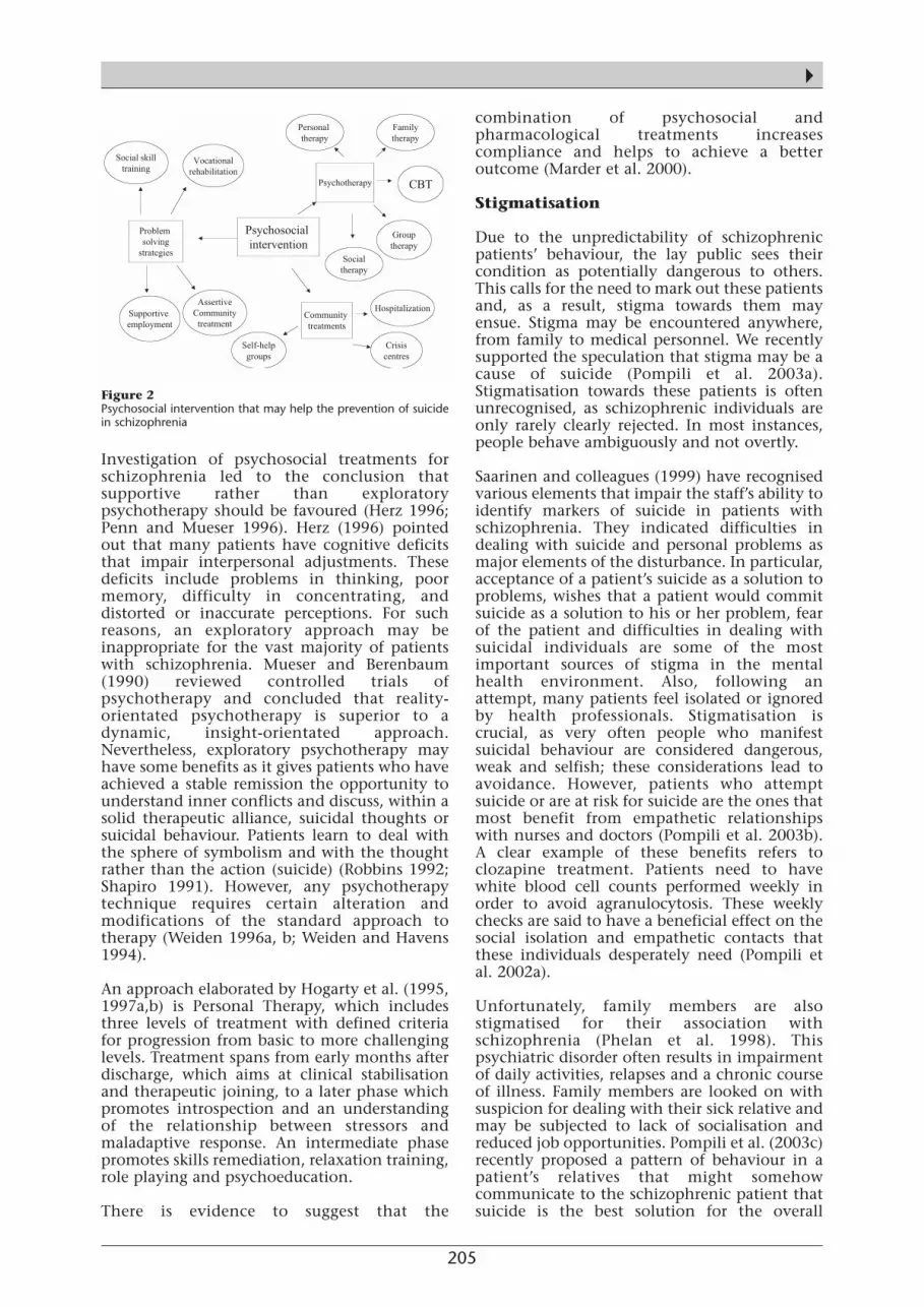
205
▲
Investigation of psychosocial treatments forschizophrenia led to the conclusion thatsupportive rather than exploratorypsychotherapy should be favoured (Herz 1996;Penn and Mueser 1996). Herz (1996) pointedout that many patients have cognitive deficitsthat impair interpersonal adjustments. Thesedeficits include problems in thinking, poormemory, difficulty in concentrating, anddistorted or inaccurate perceptions. For suchreasons, an exploratory approach may beinappropriate for the vast majority of patientswith schizophrenia. Mueser and Berenbaum(1990) reviewed controlled trials ofpsychotherapy and concluded that reality-orientated psychotherapy is superior to adynamic, insight-orientated approach.Nevertheless, exploratory psychotherapy mayhave some benefits as it gives patients who haveachieved a stable remission the opportunity tounderstand inner conflicts and discuss, within asolid therapeutic alliance, suicidal thoughts orsuicidal behaviour. Patients learn to deal withthe sphere of symbolism and with the thoughtrather than the action (suicide) (Robbins 1992;Shapiro 1991). However, any psychotherapytechnique requires certain alteration andmodifications of the standard approach totherapy (Weiden 1996a, b; Weiden and Havens1994).
An approach elaborated by Hogarty et al. (1995,1997a,b) is Personal Therapy, which includesthree levels of treatment with defined criteriafor progression from basic to more challenginglevels. Treatment spans from early months afterdischarge, which aims at clinical stabilisationand therapeutic joining, to a later phase whichpromotes introspection and an understandingof the relationship between stressors andmaladaptive response. An intermediate phasepromotes skills remediation, relaxation training,role playing and psychoeducation.
There is evidence to suggest that the
combination of psychosocial andpharmacological treatments increasescompliance and helps to achieve a betteroutcome (Marder et al. 2000).
Stigmatisation
Due to the unpredictability of schizophrenicpatients’ behaviour, the lay public sees theircondition as potentially dangerous to others.This calls for the need to mark out these patientsand, as a result, stigma towards them mayensue. Stigma may be encountered anywhere,from family to medical personnel. We recentlysupported the speculation that stigma may be acause of suicide (Pompili et al. 2003a).Stigmatisation towards these patients is oftenunrecognised, as schizophrenic individuals areonly rarely clearly rejected. In most instances,people behave ambiguously and not overtly.
Saarinen and colleagues (1999) have recognisedvarious elements that impair the staff’s ability toidentify markers of suicide in patients withschizophrenia. They indicated difficulties indealing with suicide and personal problems asmajor elements of the disturbance. In particular,acceptance of a patient’s suicide as a solution toproblems, wishes that a patient would commitsuicide as a solution to his or her problem, fearof the patient and difficulties in dealing withsuicidal individuals are some of the mostimportant sources of stigma in the mentalhealth environment. Also, following anattempt, many patients feel isolated or ignoredby health professionals. Stigmatisation iscrucial, as very often people who manifestsuicidal behaviour are considered dangerous,weak and selfish; these considerations lead toavoidance. However, patients who attemptsuicide or are at risk for suicide are the ones thatmost benefit from empathetic relationshipswith nurses and doctors (Pompili et al. 2003b).A clear example of these benefits refers toclozapine treatment. Patients need to havewhite blood cell counts performed weekly inorder to avoid agranulocytosis. These weeklychecks are said to have a beneficial effect on thesocial isolation and empathetic contacts thatthese individuals desperately need (Pompili etal. 2002a).
Unfortunately, family members are alsostigmatised for their association withschizophrenia (Phelan et al. 1998). Thispsychiatric disorder often results in impairmentof daily activities, relapses and a chronic courseof illness. Family members are looked on withsuspicion for dealing with their sick relative andmay be subjected to lack of socialisation andreduced job opportunities. Pompili et al. (2003c)recently proposed a pattern of behaviour in apatient’s relatives that might somehowcommunicate to the schizophrenic patient thatsuicide is the best solution for the overall
Figure 2 Psychosocial intervention that may help the prevention of suicidein schizophrenia
Psychosocial intervention
Social skilltraining
Vocationalrehabilitation
Supportive employment
AssertiveCommunitytreatment
Psychotherapy
Grouptherapy
Familytherapy
Personal therapy
CBT
Socialtherapy
Communitytreatments
Hospitalization
Crisiscentres
Self-helpgroups
ProblemSolvingstrategies
Problem solving
strategies
schizophrenia
206
system. Langs’ (1986) unconsciouscommunication in everyday life might be usedto support the concept that through a networkof unconscious messages, people around thepatient may lead him or her to conclude thatsuicide is the best solution for an exhaustingillness. Patients may commit suicide not onlywhen they become aware of negativeexpectations for the future and from anondelusional but acutely painful awareness ofthe illness process, depleted self-esteem andhopelessness for the future (Fenton, 2000) butalso when close to people trying to depict theirsituation.
GPs’ role in the prevention of suicide inschizophrenia
Schizophrenic patients, who are generally proneto develop suicidal ideation, need a warmenvironment where they feel protected andaccepted. Unfortunately, schizophrenia oftenevokes the idea of a treatment resistant disorderthat may damage people around the patient.Strangely enough, this is true even amongdoctors, who may feel uncomfortable with thesepatients. This behaviour is even recognisableamong psychiatrists and mental healthprofessionals. Also, schizophrenic patients veryoften meet GPs for drug prescriptions or torequest psychiatric consultation. Generalpractitioners often do not take intoconsideration their role in the prevention ofsuicidality. According to the databasesmentioned above, in the entire Englishscientific literature only one paper (an editorial)was published stressing plainly the need forsuicide prevention in schizophrenia in generalpractice (Pompili et al. 2002b). Barraclough etal. (1974) found in their sample that 63% ofsuicides had seen their general practitioners inthe month before death and 36% in the weekbefore death. A later report found fewer patientshaving seen general practitioners shortly beforedeath, which may be due to improved detectionand treatment of patients at risk (Vassilas andNorgan 1993). Thus, the recognition of riskfactors is an element of prevention andprediction. In fact, not only people whoconsider suicide often contact their doctorsimmediately before action, but also people whoare at risk should be promptly recognised.Moreover, rejection by these figures may be theultimate insult to very weak self-esteem.Although general practitioners may have anancillary role in the treatment of schizophrenicpatients, they may be in a strategic position todetect early risk factors or to contribute to thechanging of state-dependent risk factors.
Prevention of suicide among inpatientswith schizophrenia
Prevention of suicide by inpatients withschizophrenia is a daily challenge, which has to
be met with various modalities. Farberow et al.(1971) emphasise the need for careful screeningof patients when they are considered fordischarge or leave on pass. Such a processinvolves careful evaluation of theenvironmental factors to which the patients arereturning as well as contact with their family.
Crammer (1984) highlighted the importance oftaking environmental factors into accountwhen thinking proactively about suicide amonginpatients. He pointed to the potentiallydisruptive effects of transitions – for example,initial acclimatisation to ward life or plans fordischarge or rehabilitation. He also emphasisedthe environmental impact of staff variables,such as low morale or the absence of keypersonnel, as well as the need for effectivecommunication among relevant staff aboutpatients judged at increased risk of suicide.
Yarden (1974) drew attention to the importanceof suitable discharge plans and aftercareprogrammes. Supportive, supervised livingarrangements are ideal. Adverse circumstancessuch as single-occupancy rooms or the return toa family in which the patient’s presencerepresents a severe emotional or financial strain,most probably add to the suicide risk for aschizophrenic patient (Vaughn and Leff 1976).
With chronic, incapacitated patients,surveillance should be increased in times ofpersonal crisis and impeding environmentalchange, including staff, therapist or contactperson changes, hospitalisation, discharge orrehospitalisation (Caldwell and Gottesman1990).
Some authors have suggested that variations inthe ward environment because of the staffingchanges might be a factor in suicidal deaths.Salmons (1984) showed that many of the deathsin one unit were associated with periods atwhich there were lower levels of staffing thanusual. Hesso (1977) drew attention to theapparent rise in suicide rates in Scandinavianhospitals after 1955. He suggested that mentallyill patients were more likely to be admitted thanpreviously, and thus there were more socialpressures upon those who are seriously ill toconform and lead more "normal" lives. He alsothought that the use of neuroleptics and open-door policies led to shorter stays for those whowere ill, perhaps increasing pressure in this way.Another factor he considered was the increasedpersonnel turnover in psychiatric hospitals,leading to less experienced staff.
An anti-suicidal ward is one with a calm routine,carried out daily by staff who are themselvesunworried and confident of the immediatefuture. Morgan (1979) gave case histories ofeight inpatient suicides who were soprovocative, difficult and unreasonable that the
REVIEW/MINI-REVIEW
▲

207
▲staff ultimately felt hostile towards them beforetheir suicides.
Farberow et al. (1966) described the "dependent-dissatisfied" person who is able to provokerejection and thus bring about the state theydread most: loneliness and the feeling that noone, not even the hospital nor its staff, cares.
Among long-stay patients, plans forrehabilitation or discharge may createuncertainty and disruption of a routine, leadingto the death. Among the newly admitted, fear ofthe future may be aggravated by contact withpsychiatrists or with disordered fellow patients.Where a patient is recognised to be at risk ofsuicide, not all the relevant staff may beproperly informed. Also, low staff morale, theemployment of locums, the absence on leave ofconsultants may all have an influence ontendency to suicide (Crammer 1984).
Bleuler (1911) attributed a detrimental effect tothe then prevailing humiliating surveillanceand restraint of schizophrenics, and was of theopinion that these very methods increase andmaintain the suicidal drive. During recent years,modern principles of community psychiatryhave been applied in the large public mentalhospitals. This trend, which emphasises opendoors, the abolition of involuntary restrictions,therapeutic community, early discharge andmaintenance of schizophrenics within theirfamilies and communities, has brought aboutwelcome changes in the life patterns of mostmental hospital patients (Yarden 1974).
Medical staff behaviour is a potential cause ofsuicide when they fail to recognise suicide riskbecause of personal problems or difficulties withthe topic of suicide. Pompili et al. (2003b, 2004)recently pointed to the role of nurses in theprevention of suicide in schizophrenia. Theseauthors outlined the many difficulties in thecare of a schizophrenic patient who is at risk ofsuicide and paid particular attention to staff"countertransference" reactions to thesepatients. We believe that having theopportunity to explore part of the psychicprocesses of suicidal schizophrenic patients mayhelp mental health professionals to understandtheir patients better. Nursing a schizophrenicpatient who is at risk of suicide involves theestablishment of a very uncommonrelationship. A very interesting topic is theconcept of "terminal malignant alienation"(Morgan and Priest 1984, 1991). Some patients,particularly those with recurrent relapses andresistance to treatment, may be perceived bystaff as manipulative, provocative,unreasonable, over-dependent and feigningdisability (Schwartz et al. 1975; Morgan andPriest 1984, 1991; Kullgren 1988). Patients withfluctuating suicidal ideation are particularlylikely to fall into these categories; this may lead
to under-reporting of suicidal ideation bynursing staff. This may result in criticism and alower level of support leading to alienation. Thecombination of such alienation and fluctuatingsuicidal ideation can lead to failure in therecognition of seriousness of suicidal risk(Morgan and Priest 1984, 1991).
Various methods of suicide have beenrecognised among inpatients withschizophrenia. Shah and Ganesvaran (1999)found that suicide was most often performedwith methods such as jumping in front oftrains, trams or road traffic, jumping offbuildings, hanging and drowning. Thesemethods are consistent with previous studies(Roy 1982; Modestin at al. 1992).
Emmerson and Cantor (1993) underline the factthat almost half of the suicides in their sampleoccurred on a railway track close to the regionalpsychiatric hospital which contrasts withSymonds’ (1985) suggestion that there is norelation with the proximity of suicide topsychiatric hospitals. It should be noted that theBrisbane psychiatric hospital (1993) is close toan unfenced railway line and an open accessstation.
Farberow et al. (1971) pointed out that while inthe hospital, hanging is quite a commonmethod of suicide, comprising 53%; jumping isthe next most common method with 17%.Again, however, when the patient is out of thehospital the use of guns dominates the rest ofthe methods with 43%, followed by hanging15%, poison 11% and jumping 10%. Thepercentages show that patients predominantlytended to use guns whereas poison, hangingand drowning were much less frequent.
Suicide precautions must be carefully adhered toby staff in order to maintain constantobservation while the patient remains acutelypsychotic, especially during the first week ofhospitalisation. Lapses in observation shouldnot be permitted while the patient is in thebathroom or in his/her room. As hanging is themost common method, bathroom stall bars androom and closet fixtures should be of abreakaway variety. Farberow et al. (1971)suggested the use of physical safeguards such assafety screens, or stops on the windows and theenclosure of stairwells, or installation of higherguardrails difficult to climb over, to be useful.
Shah and Ganesvaran (1999) reported that a six-lane road, a tram route and a railway stationwere all very close to the hospital, thus allowingfor easily accessible violent methods. Reducingthe accessibility of possible suicide methods canreduce suicide rates (Morgan and Priest 1984;Surtees and Duffy 1989).
208
Conclusions
Despite considerable research on suicide inschizophrenia, this event remains a majorhealth problem. Antipsychotic medicationscertainly help to reduce suicidality, but thenumber of deaths due to suicide amongschizophrenic patients is still much highercompared with the general population.Psychosocial interventions are of great help butlack scientific validation. Patients are rarelyinvestigated for suicidality and different helpfultreatments are often kept apart, such asreduction of stigmatisation, improvedrelationship with GPs and instructions given tostaff to tolerate difficult patients. Families arenot supported properly and prevention ofsuicide inside the family environment is, for thetime being, missing. GPs’ role is a neglectedtopic and it has become increasingly apparentthat they may represent a key element in theimplementation of preventive measures.Stigmatisation is another great problem in theprevention of suicide of schizophrenic patients,especially because stigma toward these patientscan be perceived even inside closed circuits.Suicide among inpatients with schizophrenia isalso a major issue and, despite the introductionof protocols in the hospital environment aimedat preventing suicide, our feeling is that we stillneed to work very hard to reduce suicide and tobe able to handle practical tools capable ofpreventing suicide among these patients.
This review has a number of limitations. Firstly,no meta-analytic technique has been used toevaluate results of the various preventivestrategies. Secondly, the authors chose to reportthose studies available in the literature thatcould support a broad analysis of the topic so asto offer a tutorial paper. Despite careful andsystematic search, we extrapolated those studiesthat presented original data; however, a numberof additional papers could have been added asuseful sources of information.
A new prevention of suicide in schizophreniashould include the integration of strategiesalready in use and the implementation of lesswell known interventions. Proper informationshould be addressed to the family and,hopefully, hostility of family members towardthe patient should be investigated. Butinformation should constitute a key element forpromoting changes in people’s attitude towardthese patients. Mass media portrayal of violentschizophrenic patients should becounterbalanced by delivering reality-basedmessages of their struggle for socialisation andacceptance. Last but not least, properinformation should be the guide for a jointprevention between psychiatrists and GPs. Theimportance of GPs’ role in the prevention ofsuicide in schizophrenia is a neglected topicwith no articles published on this subject apart
from an editorial (Pompili et al. 2002b), whichstressed plainly the concept mentioned above.
Suicide risk continues throughout the lifespanof the individual with schizophrenia. Mentalhealth professionals should join forces for abetter definition of guidelines specificallydesigned to prevent suicide among patientswith schizophrenia.
References
Allebeck P, Varla E, Kristjamsson E, Westedt B (1987) Risk factorsfor suicide among patients with schizophrenia. Acta PsychiatrScand 76: 414-419.
Althaus D, Hegerl U (2003) The evaluation of suicide preventionactivities: state of the art. World J Biol Psychiatry 4: 156-165.
Andersen UA, Andersen M, Rosholm JU, Gram LF (2000) Contactsto the health care system prior to suicide: a comprehensiveanalysis using registers for general and psychiatric hospitaladmissions, contacts to general practitioners and practisingspecialists and drug prescriptions. Acta Psychiatr Scand 102: 126-134.
Barraclough BM, Bunch J, Nelson B, Sainsbury P (1974) A hundredcases of suicide: clinical aspects. Br J Psychiatry 12: 355-373.
Bleuler E (1950) Dementia praecox or the group of schizophrenias(1911). International Universities Press, New York.
Blumenthal SJ (1988) Suicide: a guide to risk factors, assessment,and treatment of suicidal patients. Med Clin North Am 72: 937-971.
Caldwell CB, Gottesman II (1990) Schizophrenics kill themselvestoo: a review of risk factors for suicide. Schizophr Bull 16: 571-589.
Crammer JL (1984) The special characteristics of suicide inhospital inpatients. Br J Psychiatry 145: 460-463.
Drake RE, Gates C, Cotton PG, Whitaker A (1984) Suicide amongschizophrenics: who is at risk? J Nerv Ment Dis 172: 613-617.
Drake RE, Bartels SJ, Torrey WC (1989) Suicide in schizophrenia:clinical approaches. In: Williams R, Dalby JT (eds) Depression inschizophrenia. Plenum Press, New York, pp 153-169.
Emmerson B, Cantor C (1993) Train suicides in Brisbane,Australia, 1980-1986. Crisis 14: 90-94.
Ertugrul A (2002) Clozapine and suicide. Am J Psychiatry 159:323.
Farberow NL, Shneidman ES, Neuringer C (1966) Case historyand hospitalization factors in suicides of neuropsychiatric hospitalpatients. J Nerv Ment Dis 142: 32-44.
Farberow NL, Ganzler S, Cutter F, Reynolds D (1971) An eight-yearsurvey of hospital suicides. Life Threat Behav 1: 184-202.
Fenton WS (2000) Depression, suicide, and suicide prevention inschizophrenia. Suicide Life Threat Behav 31: 34-49.
Fenton WS, McGlashan TS, Victor BJ, Blyler CR (1997) Symptoms,subtype and suicidality in patients with schizophrenia spectrumdisorder. Am J Psychiatry 154: 199-204.
Fremouv WJ, de Perczel M, Ellis TE (1980) Suicide risk: assessmentand response guidelines. Pergamon, New York.
Harkavy-Friedman JM, Nelson EA (1997a) Assessment andintervention for the suicidal patient with schizophrenia. PsychiatricQ 68: 361-375.
REVIEW/MINI-REVIEW
▲
209
▲Harkavy-Friedman JM, Nelson EA (1997b) Management of thesuicidal patient with schizophrenia. Psychiatr Clin North Am20: 625-640.
Herz MI (1996) Psychosocial treatment. Psychiatr Ann 26: 531-535.
Hësso R (1977) Suicide in Norwegian, Finnish and Swedishpsychiatric hospitals. Arch Psychiatr Nervenkr 224: 119-127.
Hogarty GE, Kornblith SJ, Greenwald D, DiBarry AL, Cooley S,Flesher S, Reiss D, Carter M, Ulrich R (1995) Personal therapy: adisorder-relevant psychotherapy for schizophrenia. Schizophr Bull21: 379-393.
Hogarty GE, Kornblith SJ, Greenwald D, DiBarry AL, Cooley S,Ulrich RF, Carter M, Flesher S (1997a) Three-year trials of personaltherapy among schizophrenic patients living with or independentof family. I: description of study and effects on relapse rates. Am JPsychiatry 154: 1504-1513.
Hogarty GE, Greenwald D, Ulrich RF, Kornblith SJ, DiBarry AL,Cooley S, Carter M, Flesher S (1997b) Three-year trials of personaltherapy among schizophrenic patients living with or independentof family. II: effects on adjustment of patients. Am J Psychiatry 154:1514-1524.
Keck PE, Strakowski SM, McElroy SL (2000) The efficacy of atypicalantipsychotics in the treatment of depressive symptoms, hostility,and suicidality in patients with schizophrenia. J Clin Psychiatry61(Suppl 3): s4-s9.
Kraepelin E (1971) Dementia praecox and paraphrenia. Krieger,New York.
Kullgren G (1988) Factors associated with completed suicides inborderline personality disorders. J Nerv Ment Dis 176: 40-44.
Langs R (1986) Unconscious communication in everyday life.Jason Aronson, New York.
Marder AR, Wirshing WC, Wirshing DA (2000) New strategies withconventional antipsychotics. In: Bentliff S, de Souza, Rabson J(eds) Strategies for managing acute schizophrenia. Arnold,London, pp 211-214.
Meltzer HY (1998) Suicide in schizophrenia: risk factors andclozapine treatment. J Clin Psychiatry 59 (Suppl 3): s15-s20.
Meltzer HY (2001) Decreasing suicide in schizophrenia. PsychiatricTimes 18: 1-5.
Meltzer HY (2002) Suicide and clozapine. Am J Psychiatry 159:323-324.
Meltzer HY, Okayli G (1995) Reduction of suicidality duringclozapine treatment of neuroleptic-resistant schizophrenia: impacton risk-benefit assessment. Am J Psychiatry 152: 183-190.
Meltzer HY, Conley RR, De Leo D, Green AI, Kane JM, KnesevichMA, Lieberman JA, Lindenmayer JP, Potkin SG (2003) Interventionstrategies for suicidality. J Clin Psychiatry 6: 1-16 [audiographseries].
Miles CP (1977) Conditions predisposing to suicide: a review. JNerv Ment Dis 164: 231-246.
Modestin J, Zarro I, Waldvogel D (1992) A study of suicide inschizophrenic inpatients. Br J Psychiatry 160: 398-401.
Morgan HG, Priest P (1984) Assessment of suicide risk inpsychiatric in-patients. Br J Psychiatry 145: 467-469.
Morgan HG, Priest P (1991) Suicide and other unexpected deathsamong psychiatric in-patients. The Bristol confidential enquiry. BrJ Psychiatry 158: 368-374.
Munro J, O’Sullivan D, Andrews C, Arana A, Mortimer A, Kerwin R(1999) Active monitoring of 12,760 clozapine recipients in the UKand Ireland: beyond pharmacovigilance. Br J Psychiatry 175: 576-580.
Nyman AK, Jonsson H (1986) Patterns of self-destructive
behaviour in schizophrenia. Acta Psychiatr Scand 73: 252-262.
Penn DL, Mueser KT (1996) Research update on the psychosocialtreatment of schizophrenia. Am J Psychiatry 153: 607-617.
Phelan JC, Bromet EJ, Link BG (1998) Psychiatric illness and familystigma. Schizophr Bull 24: 115-126.
Pompili M, Mancinelli I, Tatarelli R (2002a) Schizophrenia, suicide,and blood count during treatment with clozapine. Can JPsychiatry 47: 977.
Pompili M, Mancinelli I, Tatarelli R (2002b) GPs’ role in theprevention of suicide in schizophrenia. Fam Pract 19: 221.
Pompili M, Mancinelli I, Tatarelli R (2003a) Stigma as a cause ofsuicide. Br J Psychiatry 183: 173-174.
Pompili M, Mancinelli I, Girardi P, Tatarelli R (2003b) Nursingschizophrenic patients who are at risk of suicide. J Psychiat MentHealth Nurs 10: 622-624.
Pompili M, Mancinelli I, Girardi P, Tatarelli R. (2003c) Preventingsuicide in schizophrenia inside family environment. Crisis 24: 181-182.
Pompili M, Mancinelli I, Girardi P, Tatarelli R (2004) Making senseof nurses’ role in the prevention of suicide in schizophrenia. IssuesMent Health Nurs 25: 5-7.
Reid WH, Mason H, Hogan T (1998) Suicide prevention effectsassociated with clozapine therapy in schizophrenia andschizoaffective disorder. Psychiatr Serv 49: 1029-1033.
Robbins M (1992) Psychoanalytic and biological approaches tomental illness: schizophrenia. J Am Psychoanal Ass 40: 425-454.
Robins E, Murphy GE, Wilkinson RH Jr, Gassner S, Kayes J (1959)Some clinical considerations in the prevention of suicide based ona study of 134 successful suicides. Am J Public Health 49: 888-99.
Roy A (1982) Suicide in chronic schizophrenia. Br J Psychiatry 141:171-177.
Roy A (1983) Family history of suicide. Arch General Psychiatry 40:971-974.
Roy A (1986) Depression, attempted suicide, and suicide inpatients with chronic schizophrenia. Psychiatr Clin North Am 9:193-206.
Roy A (2001) Consumers of mental health services. Suicide LifeThreat Behav 31 (Suppl): s960-s983.
Saarinen PI, Lehtonen J, Lonnqvist J (1999) Suicide risk inschizophrenia: an analysis of 17 consecutive suicides. SchizophrBull 25: 533-542.
Schwartz DA, Flinn DE, Slawson PF (1975) Suicide in Psychiatrichospitals. Am J Psychiatry 132: 150-153.
Sernyak MJ, Desai R, Stolar M, Rosenheck R (2001) Impact ofclozapine on completed suicide. Am J Psychiatry 158: 931-937.
Shah AK, Ganesvaran T (1999) Suicide among psychiatricinpatients with schizophrenia in an Australian mental hospital.Med Sci Law 39: 251-259.
Shapiro S (1991) Affect integration in psychoanalysis: a clinicalapproach to self-destructive behavior. Bull Menninger Clin 55:363-374.
Surtees PG, Duffy JC (1989) Suicide in England and Wales 1946-1985: an age-period-cohort analysis. Acta Psychiatr Scand 79:216-223.
Symonds RL (1985) Psychiatric aspects of railway fatalities. PsycholMed 15: 609-621.
Tsuang MA, Woolson RF (1978) Excess mortality in schizophreniaand affective disorders: do suicides and accidental deaths solelyaccount for this excess? Acta Psychiatr Scand 35: 1181-1185.
210
Tsuang MA, Woolson RF, Fleming JA (1980) Premature deaths inschizophrenia and affective disorders: an analysis of survival curvesand variables affecting the shortened survival. Arch Gen Psychiatry37: 979-983.
Vassilas CA, Morgan HG (1993) General practitioners’ contactwith victims of suicide. Br Med J 307: 300-301.
Vaughn CE, Leff JP (1976) The influence of family and socialfactors on the course of psychiatric illness. A comparison ofschizophrenic and depressed neurotic patients. Br J Psychiatry129: 125-37.
Walker AM, Lanza LL, Arellano F, Rothman KJ (1997) Mortality incurrent and former users of clozapine. Epidemiology 8: 671-677.
Weiden PJ (1996a) Communicating with acutely psychoticpatients: the initial evaluation. J Prac Psychiatry Behav Health 2:47-50.
Weiden PJ (1996b) Communicating with psychotic patientsduring the course of acute treatment. J Prac Psychiatry BehavHealth 2: 122-124.
Weiden P, Havens L (1994) Psychotharapeutic managementtechniques in the treatment of outpatients with schizophrenia.Hosp Comm Psychiatry 45:549-555.
Yarden PE (1974) Observation on suicide in chronicschizophrenics. Compreh Psychiatry 15: 325-333.
Zisook S, Byrd D, Kuck J, Seste DV (1995) CommandHallucinations in outpatients with schizophrenia. J Clin Psychiatry56: 462-5.
▲REVIEW/MINI-REVIEW
211
Short-Term Efficacy and Safety of Risperidone in YoungChildren with Autistic Disorder (AD)
Nahit Motavalli Mukaddes, Osman Abali, Kagan GurkanDepartment of Child Psychiatry, Istanbul Medical Faculty, Istanbul University, Istanbul, Turkey
ORIGINAL INVESTIGATION/SUMMARY OF ORIGINAL RESEARCH
▲SummaryRecently, atypical antipsychotic treatments havebeen used in children with autistic disorder (AD).However, data on safety and efficacy of atypicalantipsychotic agents in autistic children are limited.In this open, prospective trial, subjects were treatedwith risperidone for six weeks. Nineteen children (12male, 7 female) aged 4 to 8 years were started on 0.5mg daily with individual titration to a maximum of1.5 mg daily. Behavioural assessments werecompleted by Conner’s parent 10-item index, ADsymptom checklist and CGI-Global improvement.Statistically significant improvement was observedin mean total scores of Conner’s parent 10-itemindex from baseline to the end of study (p< 0.001).On the basis of the CGI-Global improvement item,15 children were considered responders. Statisticallysignificant improvement was also found in someaspects of social contact, impulsive-aggression andrepetitive, ritualistic behaviour based on assessmentwith the AD-symptom checklist. Weight gain andincrease in night-time sleep were the most frequentside effects.
Key words: autistic disorder, risperidone,treatment, children, adolescents.
Correspondence:Nahit Motavalli Mukaddes, MD Istanbul Tıp Fakultesi PTT’si PK53 IstanbulTurkey 34272Fax: +90 212 234 92 08E-mail: [email protected]
IntroductionAutistic disorder is a type of pervasivedevelopmental disorder characterised by severeand pervasive impairment in several areas ofdevelopment, such as reciprocal socialinteraction skills, communication skills or thepresence of stereotyped behaviours, interestsand activities (DSM IV; American PsychiatricAssociation 1994).
Despite the use of a wide variety of treatmentmethods in subjects with pervasivedevelopmental disorders (PDD), no singletreatment method has produced marked andspecific effects. Nevertheless, pharmacotherapymay play an important role in the developmentof an individualised treatment programme(Martin et al. 1999; Herzig 1997).
Many studies report the positive effect ofmedication on symptoms such as hyperactivity,stereotypes and aggressive behaviours. Clinicalexperiences show that medication with primaryeffects on the dopaminergic and serotonergicsystems decreases hyperactivity, aggressivity andstereotypies. Haloperidol, a dopamine (DA)receptor antagonist, has been shown to besuperior to placebo in the treatment of autisticdisorder, as have the serotonin reuptakeinhibitors clomipramine and fluvoxamine.Unfortunately, these medications are associatedwith a significant risk of side effects, particularlyin young children (Nicholson et al. 1998).
Risperidone is a very potent dopamine D2 andserotonin 5HT2a receptor antagonist. Thetherapeutic effect of this drug in people withPDD has been described in several open studies(Nicolson et al. 1998; McDougle et al. 1997;Hardan et al. 1996; Finding et al. 1997; Horriganand Barnbill 1997). Improvements were oftenreported in hyperactivity, withdrawal,stereotypies and aggressive behaviour, which areoften target symptoms for pharmacologicalintervention in PDD (Nicolson et al. 1998;McDougle et al. 1997; Hardan et al. 1996;Finding et al. 1997; Horrigan and Barnbill1997). Recently, some case reports includingyoung children with AD state the improvementin social relatedness after treatment withrisperidone (Posey et al. 1999; Boon-Yosidhi etal. 2002). The existence of these reportsmotivated us to design an open-label study inyoung children with AD. We aimed to evaluatethe effect of risperidone treatment onhyperactivity, impulsivity, stereotypical
World J Biol Psychiatry (2004) 5, 211 - 214
212
behaviours and social relatedness.
Methods
• SubjectsSubjects were 19 children (12 boys, 7 girls), aged4 to 8.
The subjects were all referred to the PervasiveDevelopmental Disorders Unit in the ChildPsychiatry Department of the Istanbul MedicalFaculty for evaluation and treatment. All of theparticipants met the criteria for autistic disorderaccording to DSM-IV. The diagnosis was madeby consensus of two investigators for each child.
Exclusion criteria included a seizure disorder orhistory of seizures and history or evidence of asystemic disease. All participants were free ofother psychoactive medication for at least twoweeks before enrolment and for the duration ofthe study.
• Risperidone treatmentAfter two baseline visits in which behaviouralratings were obtained, subjects began to takerisperidone. To ensure compliance, medicationwas administered by parents. Risperidone wasstarted at 0.5 mg/day every night and wasregulated individually until therapeutic effectsor untoward effects were observed. Themaximum dose of 1.5 mg/day was notexceeded.
• AssessmentsThe children were rated by:
a) Conner’s Parent 10-item index, which hasbeen shown to be sensitive to changes inbehaviour in many drug studies. Thisquestionnaire was rated twice during the study,at baseline and after six weeks (Goyette et al.1978).b) An autistic disorder symptom checklist,which includes 27 items, was developed by theauthors according to the DSM–IV diagnosticcriteria for autistic disorder. This checklist wasrated by clinicians taking detailed informationfrom parents (caregivers) and observation of thechild in a semi-structured play situation duringeach interview. Each item on the AD-symptomchecklist is scored on a 4-point scale (0 = never,1 = rarely, 2 = frequently, 3 = almost always).The first 11 items of this questionnaire aredesigned to assess the severity of affective-socialdeficit, the next six items assess languagedevelopment, and the last 10 items determinethe severity of different behavioural problemsthat are expected in this group of children. Thisinstrument was administered twice, at baselineand at the end of six weeks of treatment.c) Clinical Global Impression Scale: Globalimprovement item was recorded at the end ofsix weeks of risperidone treatment.
• Statistical analysisAll subjects completed the six-week study andwere thus included in the efficacy analysis.Wilcoxon signed ranks test and paired t-testwere used for evaluation of efficacy oftreatment.
Results
• Subject characteristicsAll subjects completed the trial. Subjects were 12males and 7 females, aged 4 to 8. Twelve of thesubjects had participated in previousmedication trials, but discontinued themprematurely because of intolerable side effectsor lack of efficacy.
• Dosage and adverse effects of drugtreatmentThe optimal dose of risperidone for the 19subjects ranged from 0.5 mg/day to 1.5 mg/day(mean 1.1 mg/day ± 0.3). The most frequentside effects were weight gain (n=15, mean 2 kg ±1.8) and an increase in night-time sleep (n=7).
No clinically significant changes in bloodpressure, heart rate, respiratory rate ortemperature were recorded and no acute extra-pyramidal side effects, seizures or cardiac eventsoccurred. In general, risperidone was welltolerated with the most prominent adverseeffect being weight gain. Other side effectsdisappeared after the second week of treatment.
• Behavioural assessmentsConner’s parent 10-item index: Statisticallysignificant improvement was shown in all itemsof this index except items number 8, 9 and 10(Table 1). Also, a statistically significant changewas observed in mean total scores of this scalefrom baseline to the end of the study (p <0.001).
Most of the parents reported that their childrenseemed happier and life at home was easier foreveryone after treatment.
AD-Symptoms Checklist: Assessment oftreatment efficacy in all items of this scale isshown in Table 2. Statistically significantimprovements were found in affective-socialdevelopmental items such as meaningful facialexpression (p=0.046), eye contact (p=0.005),response to verbal stimulus (p=0.008), interestin peers (p=0.025), meaningful gesture andmimicking (p=0.014).
There was a statistically significant increase inthe use of meaningful words (p=0.008).Improvement in stereotypical behaviouralproblems were observed in behaviours such asturning around on his axis (p=0.008), rocking(p=0.02), jumping (p=0.01), arm and handflapping (p=0.01), odd hand movements(p=0.025). Also, significant reductions ofaggressiveness and irritability were observed (p=0.001) (Table 2).
ORIGINAL INVESTIGATION/SUMMARY OF ORIGINAL RESEARCH
▲
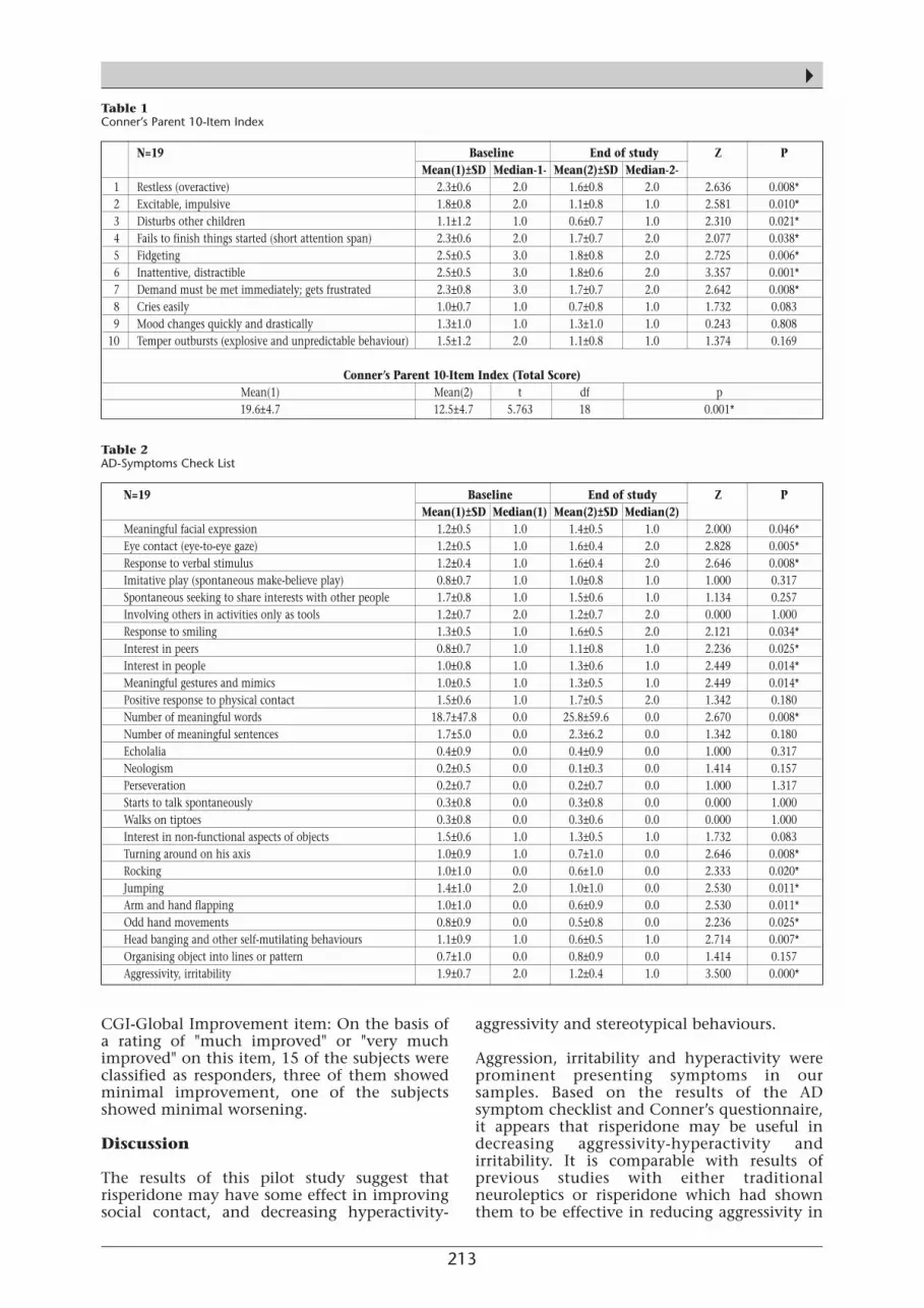
213
▲
CGI-Global Improvement item: On the basis ofa rating of "much improved" or "very muchimproved" on this item, 15 of the subjects wereclassified as responders, three of them showedminimal improvement, one of the subjectsshowed minimal worsening.
Discussion
The results of this pilot study suggest thatrisperidone may have some effect in improvingsocial contact, and decreasing hyperactivity-
aggressivity and stereotypical behaviours.
Aggression, irritability and hyperactivity wereprominent presenting symptoms in oursamples. Based on the results of the ADsymptom checklist and Conner’s questionnaire,it appears that risperidone may be useful indecreasing aggressivity-hyperactivity andirritability. It is comparable with results ofprevious studies with either traditionalneuroleptics or risperidone which had shownthem to be effective in reducing aggressivity in
N=19 Baseline End of study Z P Mean(1)±SD Median-1- Mean(2)±SD Median-2-
1 Restless (overactive) 2.3±0.6 2.0 1.6±0.8 2.0 2.636 0.008*2 Excitable, impulsive 1.8±0.8 2.0 1.1±0.8 1.0 2.581 0.010*3 Disturbs other children 1.1±1.2 1.0 0.6±0.7 1.0 2.310 0.021*4 Fails to finish things started (short attention span) 2.3±0.6 2.0 1.7±0.7 2.0 2.077 0.038*5 Fidgeting 2.5±0.5 3.0 1.8±0.8 2.0 2.725 0.006*6 Inattentive, distractible 2.5±0.5 3.0 1.8±0.6 2.0 3.357 0.001*7 Demand must be met immediately; gets frustrated 2.3±0.8 3.0 1.7±0.7 2.0 2.642 0.008*8 Cries easily 1.0±0.7 1.0 0.7±0.8 1.0 1.732 0.0839 Mood changes quickly and drastically 1.3±1.0 1.0 1.3±1.0 1.0 0.243 0.808
10 Temper outbursts (explosive and unpredictable behaviour) 1.5±1.2 2.0 1.1±0.8 1.0 1.374 0.169
Conner’s Parent 10-Item Index (Total Score)Mean(1) Mean(2) t df p19.6±4.7 12.5±4.7 5.763 18 0.001*
Table 1 Conner’s Parent 10-Item Index
N=19 Baseline End of study Z P Mean(1)±SD Median(1) Mean(2)±SD Median(2)
Meaningful facial expression 1.2±0.5 1.0 1.4±0.5 1.0 2.000 0.046*Eye contact (eye-to-eye gaze) 1.2±0.5 1.0 1.6±0.4 2.0 2.828 0.005*Response to verbal stimulus 1.2±0.4 1.0 1.6±0.4 2.0 2.646 0.008*Imitative play (spontaneous make-believe play) 0.8±0.7 1.0 1.0±0.8 1.0 1.000 0.317Spontaneous seeking to share interests with other people 1.7±0.8 1.0 1.5±0.6 1.0 1.134 0.257Involving others in activities only as tools 1.2±0.7 2.0 1.2±0.7 2.0 0.000 1.000Response to smiling 1.3±0.5 1.0 1.6±0.5 2.0 2.121 0.034*Interest in peers 0.8±0.7 1.0 1.1±0.8 1.0 2.236 0.025*Interest in people 1.0±0.8 1.0 1.3±0.6 1.0 2.449 0.014*Meaningful gestures and mimics 1.0±0.5 1.0 1.3±0.5 1.0 2.449 0.014*Positive response to physical contact 1.5±0.6 1.0 1.7±0.5 2.0 1.342 0.180Number of meaningful words 18.7±47.8 0.0 25.8±59.6 0.0 2.670 0.008*Number of meaningful sentences 1.7±5.0 0.0 2.3±6.2 0.0 1.342 0.180Echolalia 0.4±0.9 0.0 0.4±0.9 0.0 1.000 0.317Neologism 0.2±0.5 0.0 0.1±0.3 0.0 1.414 0.157Perseveration 0.2±0.7 0.0 0.2±0.7 0.0 1.000 1.317Starts to talk spontaneously 0.3±0.8 0.0 0.3±0.8 0.0 0.000 1.000Walks on tiptoes 0.3±0.8 0.0 0.3±0.6 0.0 0.000 1.000Interest in non-functional aspects of objects 1.5±0.6 1.0 1.3±0.5 1.0 1.732 0.083Turning around on his axis 1.0±0.9 1.0 0.7±1.0 0.0 2.646 0.008*Rocking 1.0±1.0 0.0 0.6±1.0 0.0 2.333 0.020*Jumping 1.4±1.0 2.0 1.0±1.0 0.0 2.530 0.011*Arm and hand flapping 1.0±1.0 0.0 0.6±0.9 0.0 2.530 0.011*Odd hand movements 0.8±0.9 0.0 0.5±0.8 0.0 2.236 0.025*Head banging and other self-mutilating behaviours 1.1±0.9 1.0 0.6±0.5 1.0 2.714 0.007*Organising object into lines or pattern 0.7±1.0 0.0 0.8±0.9 0.0 1.414 0.157Aggressivity, irritability 1.9±0.7 2.0 1.2±0.4 1.0 3.500 0.000*
Table 2 AD-Symptoms Check List
214
this group of children (Nicolson et al. 1998;McDougle 1997).
In our study, as in some case reports (Posey et al.1999; Boon Yasidhi et al. 2002), risperidone wasobserved to be effective in social-affectiveimprovement. This finding is similar to theeffect of risperidone in children and adolescentswith schizophrenia. Risperidone had beenfound to improve the negative symptoms ofschizophrenia including blunted affect,emotional withdrawal, passive/apathetic socialwithdrawal, lack of spontaneity and flow ofconversation in children (Cozza and Edison1994). It has been hypothesised that theuniquely high ratio of 5HT2a to D2 receptorantagonism of risperidone may account for itsbeneficial effect on negative symptoms ofschizophrenia. Similar considerations may berelevant to the improvement in measures ofsocial function observed in children andadolescents with AD. In addition, preclinicalstudies with risperidone have shown significantincrease in social interaction behaviour betweenunfamiliar, but not familiar rats, whencompared with conventional neurolepticshaloperidol and chlorpromazine (Nicolson et al.1998).
One of the striking results of this study is theimprovement in verbal-communicativeabilities. The increase of meaningful wordsduring treatment is considered to be a result ofimprovement in social responsiveness. Thedecrease of stereotypical behaviours haspreviously been reported to improve withrisperidone in many studies with this group(Quintana and Keshavan 1995).
The low incidence of untoward side effects inthis sample is considered to be due to the use ofrelatively low doses of risperidone and very slowincrease over a period of several weeks. Themost frequent untoward effect was weight gain.This underlines the need for nutritionalcounselling for children and adolescents whenrisperidone is prescribed. Transient adverseeffects such as increase in night-time sleep,drowsiness and nocturnal enuresis disappearedafter some weeks of treatment.
In summary, these preliminary data suggest theefficacy of risperidone in improving someaspects of social dysfunction and reducingimpulsive aggression and repetitive, ritualisticbehaviour in this sample. The relatively lowincidence of untoward effects and theirtransient nature, other than weight gain, couldresult from lower dose and gradual titration.Despite these favourable results, we prefer tointerpret our findings with caution due tomethodological limitations.
Some of the methodological limitations of thisreport include the uncontrolled nature of the
study and the lack of internationallystandardised measures. A critical assessment ofthe efficacy and safety of risperidone underdouble-blind, placebo-controlled conditionsseems justified.
References
American Psychiatric Association (1994) Diagnostic and StatisticalManual of Mental Disorders. American Psychiatric Associationfourth ed. Washington DC.
Boon-Yasidhi V, Tarugsa J, Suwan Wattana C, Soising L (2002)Risperidone in the treatment of autistic Thai children under 4 yearsage. J Med Assoc Thai 85 (Suppl 2): 784-789.
Cozza SJ, Edison D (1994) Risperidone in adolescents (letter). J AmAcad Child Adolesc Psychiatry 33: 1211.
Dodt JE, Byerly MJ, Cuadros C, Christensen RE (1997) Treatmentof risperidone induced obsessive-compulsive symptoms withsertralin. Am J Psychiatry 154: 582.
Goyette CH, Ulrich RF (1978) Normative data on Revised Conner’sParent and Teacher Ratings scale. J Abnormal Child Psychol 6:221-236.
Finding RL, Maxwell K, Wiznitzer M (1997) An open trial ofrisperidone monotherapy in young autistic children.Psychopharmacol Bull 33: 155-159.
Hardan A, Johnson K, Johnson C, Hrecznyj B (1996) Case study:Risperidone treatment of children and adolescents withdevelopmental disorders. J Am Acad Child Adolesc Psychiatry 35:1551-1556.
Herzig ME (1997) Pervasive developmental disorders. In:Handbook of Child and Adolescent Psychiatry, Vol 2. Noshpitz JD(ed) John Wiley & Son Inc, New York, pp 221-235.
Horrigan JP, Barnbill LJ (1997) Risperidone in impulsive aggressivebehavior in autism. J Autism Dev Disorder 27: 313-323.
Martin A, Scahill L, Klim A, Volkmar F (1999) High-functioningpervasive developmental disorders: rates and patterns ofpsychotropic drug use. J Am Acad Child Adolesc Psychiatry 38:923-930.
McDougle CJ, Holmes JP, Bronson MR, Anderson GM, Volkmar FR,Price LH, Cohen DJ (1997) Risperidone treatment of children andadolescents with pervasive developmental disorders: Aprospective open-label study. J Am Acad Child Adolesc Psychiatry36: 685-693.
Nicolson R, Awad G, Slomon L (1998) An open trial of risperidonein young autistic children. J Am Acad Child Adolesc Psychiatry 37:372-377.
Posey DJ, Walsh KH, Wilson GA, McDougle CJ (1999) Risperidonein the treatment of two very young children children with autism.J Child Adolesc Psychopharmacol 9: 273-276.
Quintana H, Keshavan M (1995) Case study: Risperidone inchildren and adolescents with schizophrenia. J Am Acad ChildAdolesc Psychiatry 34: 1292-1296.
▲ORIGINAL INVESTIGATION/SUMMARY OF ORIGINAL RESEARCH

215
Association Analysis of Brain-Derived Neurotrophic Factor(BDNF) Gene Val66Met Polymorphism in Schizophrenia andBipolar Affective Disorder
Maria Skibinska1, Joanna Hauser1,2, Piotr M. Czerski1, Anna Leszczynska-Rodziewicz2, Magdalena Kosmowska2, Pawel Kapelski2, Agnieszka Slopien3, Marzena Zakrzewska4,Janusz K Rybakowski2
1 Laboratory of Psychiatric Genetics, Department of Psychiatry, University of Medical Sciences,Poznan, Poland
2 Department of Adult Psychiatry, University of Medical Sciences, Poznan, Poland 3 Department of Child and Adolescent Psychiatry, University of Medical Sciences, Poznan, Poland4 Institute of Psychology, Adam Mickiewicz University, Poznan, Poland
SummaryBrain-derived neurotrophic factor (BDNF) has beenimplicated in the pathogenesis of schizophrenia andbipolar disorder. A functional polymorphismVal66Met of BDNF gene was studied in patientswith schizophrenia (n=336), bipolar affectivedisorder (n=352) and healthy controls (n=375).Consensus diagnosis by at least two psychiatrists,according to DSM-IV and ICD-10 criteria, wasmade for each patient using a structured clinicalinterview for DSM-IV Axis I disorders (SCID). Noassociation was found between the studiedpolymorphism and schizophrenia or bipolaraffective disorder either for genotype or alleledistribution (for genotype: p=0.210 inschizophrenia, p=0.400 in bipolar disorder; foralleles: p=0.260 in schizophrenia, p=0.406 inbipolar disorder). Results were also not significantwhen analysed by gender. For males genotypedistribution and allele frequency were (respectively):p=0.480 and p=0.312 in schizophrenia, p=0.819and p=0.673 in bipolar affective disorder. Genotypedistribution and allele frequency observed in thefemale group were: p=0.258 for genotypes, p=0.482for alleles in schizophrenia; p=0.432 for genotypes,p=0.464 for alleles in bipolar affective disorder. Asubgroup of schizophrenic (n=62) and bipolaraffective patients (n=28) with early age at onset (18years or younger) was analysed (p=0.328 forgenotypes, p=0.253 for alleles in schizophrenia;p=0.032 for genotypes, p=0.858 for alleles inbipolar affective disorder).
Key words: schizophrenia, bipolar affectivedisorder, brain-derived neurotrophic factor (BDNF),genetics, association.
Correspondence:Maria SkibinskaDepartment of Adult Psychiatry University ofMedical Sciencesul. Szpitalna 27/33 60-572 PoznanPolandTel: +48 61 8491311Fax: +48 61 8480392E-mail: [email protected]
AcknowledgementThis work was supported by the Polish StateCommittee for Scientific Research (KBN) grantNo.4PO5B05320. Piotr M. Czerski is the recipientof a 2004 Annual Stipend for Young Scientists fromthe Foundation for Polish Science (FNP).
IntroductionThe neurodevelopmental hypothesis ofpsychiatric disorders postulates alterations inembryonal neurogenesis and explains thesechanges as the result of disturbances ofprocesses involving the trophic factors (Thomeet al. 1998; Nawa et al. 2000). Disturbances ofbrain-derived neurotrophic factor (BDNF) havebeen postulated as a risk factor for psychiatricdisorders and neurological diseases (Thome etal. 1998; Siegel and Chauhan 2000).
Brain-derived neurotrophic factor (BDNF) is amember of the neurotrophin family of growthfactors (Barde et al. 1982; Leibrock et al. 1989),interacting with the trkB receptor tyrosinekinase (Squinto et al. 1991). It has trophiceffects on dopaminergic (Altar et al. 1992; Shenet al. 1994; Shults et al. 1994), cholinergic(Lindsay 1995; Lindvall et al. 1994) andserotonergic (White et al. 1994; Mamounas et al.1995, 2000) neurons. It is essential fordevelopment of sensory ganglia, cerebral cortex,hippocampus and striatum (Liu et al. 1995).
World J Biol Psychiatry (2004) 5, 215 - 220
ORIGINAL INVESTIGATION/SUMMARY OF ORIGINAL RESEARCH
▲
216
BDNF affects neuronal proliferation, survivaland plasticity and is involved in hippocampallong-term potentiation (LPT), learning andmemory (Lu and Gottschalk 2000; Poo 2001;Egan et al. 2003; Lu 2003; Mizuno et al. 2003).
The BDNF gene was first reported byMaisonpierre et al. (1991) to be localised on theshort arm of chromosome 11 (11p13). The geneconsists of four short 5’exons with separatepromoters and one 3’exon encoding the matureBDNF protein (Timmusk et al. 1993; Metsis et al.1993).
Dinucleotide repeat polymorphism (GT)n in thepromoter region 1.4 kb from the transcriptionstart site (Proschel et al. 1992), Val66Met(196G/A) polymorphism (Cargill et al. 1999), -270C/T substitution (Kunugi et al. 2001), -374A/T and -256G/A polymorphisms (Ribases etal. 2003) have been reported in the BDNF geneand studied in psychiatric disorders. Anassociation of the Val variant of Val66Metpolymorphism and affective disorders has beenreported (Neves-Pereira et al. 2002; Sklar et al.2002), but analysis performed by Nakata et al.(2003) did not confirm such association. Metallele is associated with impairments of thehippocampal functions and episodic memory inhumans (Egan et al. 2003a) and anorexianervosa restrictive type (Ribases et al. 2003). Onthe other hand, Met allele has been found to beprotective against depression (Sen et al. 2003)and obsessive-compulsive disorder (Hall et al.2003). An association of the dinucleotide repeatpolymorphism (GT)n ("long" alleles 172-176 bp)with late age at onset and better response toneuroleptic treatment in schizophrenic patientshave been reported by Krebs et al. (2000). Anassociation of the A3 allele (170 bp) withschizophrenia has been described by Muglia etal. (2003). Other studies did not confirmassociation of this polymorphism withschizophrenia (Sasaki et al. 1997; Hawi et al.1998; Wassink et al. 1999; Virgos et al. 2001).Polymorphism -270C/T has been investigated inbipolar disorder with negative results (Nakata etal. 2003). Two novel polymorphisms, -374A/Tand -256G/A, have been reported by Ribases etal. (2003) in patients with anorexia nervosa.
In our study, we investigated allelic distributionof G196A single nucleotide polymorphism(SNP) responsible for valine to methioninesubstitution at amino acid position 66 in theprodomain of BDNF in the groups of patientswith schizophrenia, affective disorder andcontrols.
Subjects and methods
• SubjectsThe study was performed on 336 patients withschizophrenia (189 males with a mean age of30.6 years, SD=11.3, 147 females with a mean
age of 33.2 years, SD=12.1), and 352 patientswith bipolar affective disorder (bipolar Iaffective disorder: 128 males with a mean age of45.1 years, SD=14.5; 172 females with a meanage of 47.5 years, SD=13.6; bipolar II affectivedisorder: 18 males with a mean age of 44.0years, SD=12.8; 34 females with a mean age of43.5 years, SD=14.3). Subgroups ofschizophrenic (n=62) and bipolar affective(n=28) patients with early age at onset (18 yearsor younger) were distinguished. Patients wererecruited from inpatients being treated at theDepartment of Psychiatry, University of MedicalSciences in Poznan, Department of Psychiatry,University School of Medicine in Bydgoszcz andPsychiatric Hospital in Koscian. All subjectswere Caucasians from the Wielkopolska region.Consensus diagnosis by at least twopsychiatrists, according to DSM-IV and ICD-10criteria, was made for each patient using astructured clinical interview for DSM-IV Axis Idisorders (SCID) (First et al. 1996).
The control group consisted of 375 subjects (149males with a mean age of 41.2 years, SD=120;226 females with a mean age of 40.4 years,SD=10.8). Control subjects were recruited fromthe group of blood donors, hospital staff andstudents of the University of Medical Sciences inPoznan. They were not psychiatrically screened.The local ethics committee approved theproject.
• GenotypingDNA was extracted from 10 ml of EDTAanticoagulated whole blood, using the saltingout method (Miller et al. 1988). A 113-basepairfragment of the BDNF gene was amplified byPCR with primer pair described by Neves-Pereiraet al. (2002) in PTC-200 (MJ Research) thermalcycler. A 20 ml amplification mixture contained150-300 ng of genomic DNA, 0.3 mM of eachprimer, 0.17 mM of each dNTP, 1.5 mM MgCl2,75 mM Tris-HCl, 20 mM (NH4)2SO4, 0.01%Tween 20 and 0.4 U of Taq DNA polymerase(MBI Fermentas). Cycling conditions were:initial denaturation at 95°C for 2 minutesfollowed by 35 cycles, with a profile of 94°C for30 seconds, 60°C for 30 seconds, 72°C for 30seconds, and final elongation at 72°C for 5minutes. A volume of 6.5 ml of each PCRproduct was then digested overnight in a totalvolume of 10 ml at 37°C with 0.7 U of Eco72Irestriction endonuclease (MBI Fermentas).Digestion products were then separated on 2.5%basica LE agarose gel (Prona, Spain) with 90Vand visualised by ethidium bromide staining.Band sizes were compared withpUC19DNA/MspI DNA ladder (MBI Fermentas).
The uncut product size was 113 bp (allele A).Allele G comprised the cut bands of 78 and 35bp.
• Statistical analyses
ORIGINAL INVESTIGATION/SUMMARY OF ORIGINAL RESEARCH
▲
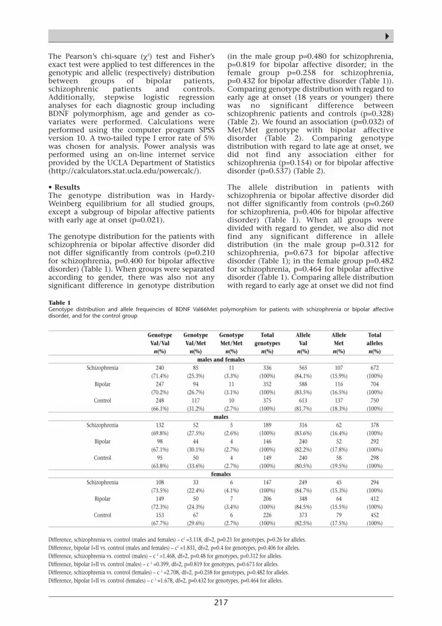
217
▲The Pearson’s chi-square (χ2) test and Fisher’sexact test were applied to test differences in thegenotypic and allelic (respectively) distributionbetween groups of bipolar patients,schizophrenic patients and controls.Additionally, stepwise logistic regressionanalyses for each diagnostic group includingBDNF polymorphism, age and gender as co-variates were performed. Calculations wereperformed using the computer program SPSSversion 10. A two-tailed type I error rate of 5%was chosen for analysis. Power analysis wasperformed using an on-line internet serviceprovided by the UCLA Department of Statistics(http://calculators.stat.ucla.edu/powercalc/).
• ResultsThe genotype distribution was in Hardy-Weinberg equilibrium for all studied groups,except a subgroup of bipolar affective patientswith early age at onset (p=0.021).
The genotype distribution for the patients withschizophrenia or bipolar affective disorder didnot differ significantly from controls (p=0.210for schizophrenia, p=0.400 for bipolar affectivedisorder) (Table 1). When groups were separatedaccording to gender, there was also not anysignificant difference in genotype distribution
(in the male group p=0.480 for schizophrenia,p=0.819 for bipolar affective disorder; in thefemale group p=0.258 for schizophrenia,p=0.432 for bipolar affective disorder (Table 1)).Comparing genotype distribution with regard toearly age at onset (18 years or younger) therewas no significant difference betweenschizophrenic patients and controls (p=0.328)(Table 2). We found an association (p=0.032) ofMet/Met genotype with bipolar affectivedisorder (Table 2). Comparing genotypedistribution with regard to late age at onset, wedid not find any association either forschizophrenia (p=0.154) or for bipolar affectivedisorder (p=0.537) (Table 2).
The allele distribution in patients withschizophrenia or bipolar affective disorder didnot differ significantly from controls (p=0.260for schizophrenia, p=0.406 for bipolar affectivedisorder) (Table 1). When all groups weredivided with regard to gender, we also did notfind any significant difference in alleledistribution (in the male group p=0.312 forschizophrenia, p=0.673 for bipolar affectivedisorder (Table 1); in the female group p=0.482for schizophrenia, p=0.464 for bipolar affectivedisorder (Table 1). Comparing allele distributionwith regard to early age at onset we did not find
Genotype Genotype Genotype Total Allele Allele TotalVal/Val Val/Met Met/Met genotypes Val Met alleles
n(%) n(%) n(%) n(%) n(%) n(%) n(%)males and females
Schizophrenia 240 85 11 336 565 107 672(71.4%) (25.3%) (3.3%) (100%) (84.1%) (15.9%) (100%)
Bipolar 247 94 11 352 588 116 704(70.2%) (26.7%) (3.1%) (100%) (83.5%) (16.5%) (100%)
Control 248 117 10 375 613 137 750(66.1%) (31.2%) (2.7%) (100%) (81.7%) (18.3%) (100%)
males Schizophrenia 132 52 5 189 316 62 378
(69.8%) (27.5%) (2.6%) (100%) (83.6%) (16.4%) (100%)Bipolar 98 44 4 146 240 52 292
(67.1%) (30.1%) (2.7%) (100%) (82.2%) (17.8%) (100%)Control 95 50 4 149 240 58 298
(63.8%) (33.6%) (2.7%) (100%) (80.5%) (19.5%) (100%)females
Schizophrenia 108 33 6 147 249 45 294(73.5%) (22.4%) (4.1%) (100%) (84.7%) (15.3%) (100%)
Bipolar 149 50 7 206 348 64 412(72.3%) (24.3%) (3.4%) (100%) (84.5%) (15.5%) (100%)
Control 153 67 6 226 373 79 452(67.7%) (29.6%) (2.7%) (100%) (82.5%) (17.5%) (100%)
Difference, schizophrenia vs. control (males and females) – c2 =3.118, df=2, p=0.21 for genotypes, p=0.26 for alleles.Difference, bipolar I+II vs. control (males and females) – c2 =1.831, df=2, p=0.4 for genotypes, p=0.406 for alleles.Difference, schizophrenia vs. control (males) – c 2 =1.468, df=2, p=0.48 for genotypes, p=0.312 for alleles.Difference, bipolar I+II vs. control (males) – c 2 =0.399, df=2, p=0.819 for genotypes, p=0.673 for alleles.Difference, schizophrenia vs. control (females) – c 2 =2.708, df=2, p=0.258 for genotypes, p=0.482 for alleles.Difference, bipolar I+II vs. control (females) – c 2 =1.678, df=2, p=0.432 for genotypes, p=0.464 for alleles.
Table 1 Genotype distribution and allele frequencies of BDNF Val66Met polymorphism for patients with schizophrenia or bipolar affectivedisorder, and for the control group

218
any significant difference between eitherschizophrenic patients and controls (p=0.253)or bipolar affective patients and controls(p=0.858) (Table 2). Comparing alleledistribution with regard to late age at onset, wedid not find any association for eitherschizophrenia (p=0.416) or bipolar affectivedisorder (p=0.322) (Table 2).
In logistic regression analyses, after controllingfor age, gender and BDNF polymorphism, therewas no association between BDNFpolymorphism and either schizophrenia orbipolar disorder (comparing: schizophrenicpatients with bipolar patients: Wald test=0.171,df=2, p=0.918; schizophrenic patients withcontrols: Wald test=5.416, df=2, p=0.067;bipolar patients with controls: Wald test=1.949,df=2, p=0.377).
The power to detect an association wasestablished to be 47.2% in bipolar affectivedisorder and 47.1% in schizophrenia with arelative risk of 1.5, and 71.8% in bipolaraffective disorder and 69.3% in schizophreniawith a relative risk of 1.75.
Discussion
In our study we did not find an associationbetween Val66Met polymorphism of the BDNFgene and schizophrenia or bipolar affectivedisorder in the whole group of patients. Whenwe divided the sample into subgroups withregard to gender we still did not find anyassociation.
We did find an association of Met/Met genotypewith bipolar affective disorder and early age at
onset (p=0.032). Genotype distribution in thissubgroup of patients was not in Hardy-Weinbergequilibrium and association of the Met/Metgenotype in this subgroup might be a falsepositive, however further studies on a largergroup are required.
This association differs from the results ofNeves-Pereira et al. (2002) and Sklar et al.(2002), where an association of bipolar affectivedisorder with the Val allele has been found. TheMet allele has been found to be protectiveagainst depression (Sen et al. 2003) andobsessive-compulsive disorder (Hall et al. 2003).On the contrary, results obtained by Egan et al.(2003a) show an association of the Met allelewith impairments in hippocampal functionsand episodic memory in humans. However, theMet allele did not affect risk for schizophrenia(Egan et al. 2003b). In our own study, patientswith bipolar affective disorder and Val/Valgenotype had better results in the WisconsinCard Sorting Test (WCST), the test measuringfunctions of prefrontal cortex, compared withVal/Met genotype patients (Rybakowski et al. Inpress).
The functional significance of the Val66Metsubstitution in the prodomain of BDNF is notclear. The Met allele is thought to be novel inevolutionary history, while Val allele isconserved among >70 species includingmammals, birds and fish (Murphy et al. 2001).Egan et al. (2003b) showed that the Met allele ofBDNF impairs depolarisation-dependentsecretion of the protein. This might be due tothe inability of Met allele to be sorted from theGolgi apparatus to appropriate secretorygranules. The Met variant of BDNF protein can
Genotype Genotype Genotype Total Allele Allele TotalVal/Val Val/Met Met/Met genotypes Val Met alleles
n(%) n(%) n(%) n(%) n(%) n(%) n(%)Early age on onset
Schizophrenia 45 17 0 62 107 17 124(72.6%) (27.4%) (0%) (100%) (86.3%) (13.7%) (100%)
Bipolar 20 5 3 28 45 11 56(71.4%) (17.9%) (10.7%) (100%) (80.4%) (19.6%) (100%)
Control 248 117 10 375 613 137 750(66.1%) (31.2%) (2.7%) (100%) (81.7%) (18.3%) (100%)
Late age at onset Schizophrenia 195 68 11 274 458 90 548
(71.2%) (24.8%) (4.0%) (100%) (83.6%) (16.4%) (100%)Bipolar 227 89 8 324 543 105 648
(70.1%) (27.5%) (2.5%) (100%) (83.8%) (16.2%) (100%)Control 248 117 10 375 613 137 750
(66.1%) (31.2%) (2.7%) (100%) (81.7%) (18.3%) (100%)
Difference, schizophrenia vs. control (early age at onset) – c 2 =2.231, df=2, p=0.328 for genotypes, p=0.253 for alleles. Difference, bipolar I+II vs. control (early age at onset) – c 2 =6.873, df=2, p=0.032 for genotypes, p=0.858 for alleles.Difference, schizophrenia vs. control (late age at onset) – c 2 =3.739, df=2, p=0.154 for genotypes, p=0.416 for alleles.Difference, bipolar I+II vs. control (late age at onset) – c 2 =1.242, df=2, p=0.537 for genotypes, p=0.322 for alleles.
Table 2 Genotype distribution and allele frequencies of BDNF Val66Met polymorphism with regard to the onset of illness of subjects withschizophrenia or bipolar affective disorder and to the control group
ORIGINAL INVESTIGATION/SUMMARY OF ORIGINAL RESEARCH
▲
219
▲be secreted in small amounts near the cell bodythrough the constitutive pathway, but cannotbe secreted at synapses.
Changes in BDNF protein level in brains orserum of schizophrenic (Takahashi et al. 2000;Durany et al. 2002; Weickert et al. 2003) andbipolar affective (Chen et al. 2001; Karege et al.2002; Shimizu et al. 2003) patients have beenreported, but there is no evidence that BDNFVal66Met polymorphism is responsible for suchalterations in protein expression. Changes inBDNF expression have been reported inresponse to antipsychotic (Angelucci et al. 2000;Linden et al. 2000; Chlan-Fourney et al. 2002)or antidepressant (Popoli et al. 2002; Coppell etal. 2003; Ivy et al. 2003) treatment. Anassociation of the Val/Val genotype andclozapine response in schizophrenic patientshas been reported recently (Hong et al. 2003).All these data make BDNF a novel andpromising target in pharmacogenetic studies.
ReferencesAltar CA, Boylan CB, Jackson C, Hershenson S, Miller J, WiegandSJ, Lindsay RM, Hyman C (1992) Brain-derived neurotrophicfactor augments rotational behavior and nigrostriatal dopamineturnover in vivo. Proc Natl Acad Sci USA 89: 11347-11351.
Angelucci F, Mathe AA, Aloe L (2000) Brain-derived neurotrophicfactor and tyrosine kinase receptor TrkB in rat brain aresignificantly altered after haloperidol and risperidoneadministration. J Neurosci Res 60: 783-794.
Barde Y-A, Edgar D, Thoenen H (1982) Purification of a newneurotrophic factor from mammalian brain. EMBO J 1: 549-553.
Cargill M, Altshuler D, Ireland J, Sklar P, Ardlie K, Patil N, Shaw N,Lane CR, Lim EP, Kalyanaraman N, Nemesh J, Ziaugra L, FriedlandL, Rolfe A, Warrington J, Lipshutz R, Daley GQ, Lander ES (1999)Characterization of single-nucleotide polymorphisms in codingregions of human genes. Nat Genet 22: 231-238.
Chen B, Dowlatshahi D, MacQueen GM, Wang JF, Young LT(2001) Increased hippocampal BDNF immunoreactivity insubjects treated with antidepressant medication. Biol Psychiatry50: 260-265.
Chlan-Fourney J, Ashe P, Nylen K, Juorio AV, Li XM (2002)Differential regulation of hippocampal BDNF mRNA by typical andatypical antipsychotic administration. Brain Res 954: 11-20.
Coppell AL, Pei Q, Zetterstrom TS (2003) Bi-phasic change inBDNF gene expression following antidepressant drug treatment.Neuropharmacology 44: 903-910.
Durany N, Michel T, Zochling R, Boissl KW, Cruz-Sanchez FF,Riederer P, Thome J (2001) Brain-derived neurotrophic factor andneurotrophin 3 in schizophrenic psychoses. Schizophrenia Res 52:79-86.
Egan MF, Kojima M, Callicott JH, Goldberg TE, Kolachana BS,Bertolino A, Zaitsev E, Gold B, Goldman D, Dean M, Lu B (2003a)The BDNF val66met polymorphism affects activity-dependentsecretion of BDNF and human memory and hippocampalfunction. Cell 112: 257-269.
Egan MF, Weinberger DR, Lu B (2003b) Schizophrenia, III. Am JPsychiatry 160: 1242.
First MB, Spitzer RL, Gibbon M, Williams J (1996) StructuredClinical Interview for DSM-IV Axis I Disorders, Clinician Version(SCID-CV). American Psychiatric Press, Washington, DC.
Hawi Z, Straub RE, O’Neil A, Kendler KS, Walsh D, Gill M (1998)No linkage or linkage disequilibrium between brain-derivedneurotrophic factor (BDNF) dinucleotide repeat polymorphismand schizophrenia in Irish families. Psychiatry Res 81: 111-116.
Hong C-J, Yu Y W-Y, Lin C-H, Tsai SJ (2003) An association studyof a brain-derived neurotrophic factor Val66Met polymorphismand clozapine response of schizophrenic patients. Neurosci Lett349: 206-208.
Ivy AS, Rodriguez FG, Garcia C, Chen MJ, Russo-Neustadt AA(2003) Noradrenergic and serotonergic blockade inhibits BDNFmRNA activation following exercise and antidepressant.Pharmacol Biochem Behav 75: 81-88.
Karege F, Perret G, Bondolfi G, Schwald M, Bertschy G, Aubry JM(2002) Decreased serum brain-derived neurotrophic factor levelsin major depressed patients. Psychiatry Res 109: 143-148.
Krebs MO, Guillin O, Bourdell MC, Schwartz JC, Olie JP, Poirer MF,Sokoloff P (2000) Brain-derived neurotrophic factor (BDNF) genevariants association with age at onset and therapeutic response inschizophrenia. Mol Psychiatry 5: 558-562.
Kunugi H, Ueki M, Otsuga M, Isse K, Hirasawa H, Kato N, NabikaT, Kobayashi S, Nanko S (2001) A novel polymorphism of thebrain-derived neurotrophic factor (BDNF) gene associated withlate-onset Alzheimer’s disease. Mol Psychiatry 6: 83-86.
Leibrock J, Lottspeich F, Hohn A, Hofer M, Gengerer B,Masiakowski P, Thoenen H, Barde Y (1989) Molecular cloning andexpression of brain-derived neurotrophic factor. Nature 341: 149-152.
Linden AM, Vaisanen J, Lakso M, Nawa H, Wong G, Castren E(2000) Expression of neurotrophins BDNF and NT-3, and theirreceptors in rat brain after administration of antipsychotic andpsychotrophic agents. J Mol Neurosci 14: 27-37.
Lindsay RM (1995) Neuron saving schemes. Nature 373: 289-290.
Lindvall O, Kokaia Z, Bengzon J, Elmer E, Kokaia M (1994)Neurotrophins and brain insults. Trends Neurosci 17: 490-496.
Liu X, Ernfors P, Wu H, Jaenisch R (1995) Sensory but not motorneuron deficits in mice lacking NT4 and BDNF. Nature 375: 238-241.
Lu B (2003) BDNF and activity-dependent synaptic modulation.Learning and Memory 10: 86-98.
Lu B, Gottschalk W (2000) Modulation of hippocampal synaptictransmission and plasticity by neurotrophins. Prog Brain Res 128:231-241.
Maisonpierre PC, Le Beau MM, Espinosa RIII, Ip NY, Belluscio L, dela Monte SM, Squinto S, Furth ME, Yancopoulos GD (1991)Human and rat brain-derived neurotrophic factor andneurotrophin-3: gene structures, distributions and chromosomallocalizations. Genomics 10: 558-568.
Mamounas LA, Blue ME, Siuciak JA, Altar CA (1995) Brain-derivedneurotrophic factor promotes the survival and sprouting ofserotonergic axons in rat brain. J Neurosci 15: 7929-7939.
Mamounas LA, Altar CA, Blue ME, Kaplan DR, Tessarollo L, LyonsWE (2000) BDNF promotes the regenerative sprouting, but notsurvival, of injured serotonergic axons in the adult rat brain. JNeurosci 20: 771-782.
Metsis M, Timmusk T, Arenas E, Persson H (1993) Differentialusage of multiple brain-derived neurotrophic factor promoters inthe rat brain following neuronal activation. Proc Natl Acad Sci USA90: 8802-8806.
Miller SA, Dykaes DD, Polesky HF (1988) A simple salting outprocedure for extracting DNA from human nucleated cells.Nucleic Acid Res 16: 1215.
Mizuno M, Yamada K, He J, Nakijama A, Nabeshima T (2003)Involvement of BDNF receptor TrkB in spatial memory formation.Learning and Memory 10: 108-115.
220
Muglia P, Vincente AM, Verga M, King N, Macciardi F, Kennedy JL(2003) Association between the BDNF gene and schizophrenia.Mol Psychiatry 8: 147-148.
Murphy WJ, Eizirik E, Johnson WE, Zhang YP, Ryder OA, O’Brien SJ(2001) Molecular phylogenetics and the origins of placentalmammals. Nature 409: 614-618.
Nakata K, Ujike H, Sakai A, Uchida N, Nomura A, Imamura T, KatsuT, Tanaka Y, Hamamura T, Kuroda S (2003) Association study ofthe brain-derived neurotrophic factor (BDNF) gene with bipolardisorder. Neurosci Lett 337: 17-20.
Nawa H, Takahashi M, Patterson PH (2000) Cytokine and growthfactor involvement in schizophrenia-support for thedevelopmental model. Mol Psychiatry 5: 594-603.
Neves-Pereira M, Mundo E, Muglia P, King N, Macciardi F,Kennedy JL (2002) The brain-derived neurotrophic factor geneconfers susceptibility to bipolar disorder: evidence from a family-based association study. Am J Hum Genet 71: 651-655.
Poo MM (2001) Neurotrophins as synaptic modulators. Nat RevNeurosci 2: 24-32.
Popoli M, Gennarelli M, Racagni G (2002) Modulation of synapticplasticity by stress and antidepressants. Bipolar Disorders 4: 166-182.
Proschel M, Saunders A, Roses AD, Muller CR (1992) Dinucleotiderepeat polymorphism at the human gene for brain-derivedneurotrophic factor (BDNF). Hum Mol Genet 1: 353.
Ribases M, Gratacos M, Armengol L, De Cid R, Badia A, Jimenez L,Solano R, Vallejo J, Fernandez F, Estivill X (2003) Met66 in thebrain-derived neurotrophic factor (BDNF) precursor is associatedwith anorexia nervosa restrictive type. Mol Psychiatry 8: 745-751.
Rybakowski JK, Borkowska A, Czerski PM, Skibinska M, Hauser J (Inpress) Polymorphism of the brain-derived neurotrophic factorgene and performance on a cognitive prefrontal test in bipolarpatients. Bipolar Disorders.
Sasaki T, Dai XY, Kuwata S, Fukuda R, Kunugi H, Hattori M, NankoS (1997) Brain-derived neurotrophic factor gene andschizophrenia in Japanese subjects. Am J Med Genet 74: 443-444.
Sen S, Nesse RM, Stoltenberg SF, Li S, Gleiberman L, ChakravartiA, Weder AB, Burmeister M (2003) A BDNF coding variant isassociated with the NEO personality inventory domainneuroticism, a risk factor for depression. Neuropsychopharmacol28: 397-401.
Shen R-Y, Altar CA, Chiodo LA (1994) Brain-derived neurotrophicfactor increases the electrical activity of pars compacta dopamineneurons in vivo. Proc Natl Acad Sci USA 91: 8920-8924.
Shimizu E, Hashimoto K, Okamura N, Koike K, Komatsu N,Kumakiri C, Nakazato M, Watanabe H, Shinoda N, Okada S, Iyo M(2003) Alterations of serum levels of brain-derived neurotrophicfactor (BDNF) in depressed patients with or withoutantidepressants. Biol Psychiatry 54: 70-75.
Shults CW, Matthews RT, Altar CA, Hill LR, Langlais PJ (1994) Asingle intramesencephalic injection of brain-derived neurotrophicfactor induces persistent rotational asymmetry in rats. Exp Neurol125: 183-194.
Siegel GJ, Chauhan NB (2000) Neurotrophic factors in Alzheimer’sand Parkinson’s disease brain. Brain Res Rev 33: 199-227.
Sklar P, Gabriel SB, McInnis MG, Bennett P, Lim Y-M, Tsan G,Schaffner S, Kirov G, Jones I, Owen M, Craddock N, DePaulo JR,Lander ES (2002) Family-based association study of 76 candidategenes in bipolar disorder: BDNF is a potential risk locus. MolPsychiatry 7: 579-593.
Squinto SP, Stitt TN, Aldrich TH, Davis S, Bianco SM, RadziejewskiC, Glass DJ, Masiakowski P, Furth ME, Valenzuela DM (1991) TrkBencodes a functional receptor for brain-derived neurotrophicfactor and neurotrophin-3 but not nerve growth factor. Cell 65:558-568.
Takahashi M, Shirakawa O, Toyooka K, Kitamura N, Hashimoto T,Maeda K, Koizumi S, Wakabayashi K, Takahashi H, Someya T,Nawa H (2000) Abnormal expression of brain-derivedneurotrophic factor and its receptor in the corticolimbic system ofschizophrenic patients. Mol Psychiatry 5: 293-300.
Thome J, Foley P, Riederer P (1998) Neurotrophic factors andmaldevelopmental hypothesis of schizophrenia psychoses. JNeural Transm 105: 85-100.
Timmusk T, Palm K, Metsis M., Reintam T, Paalme V, Saarma M,Persson H (1993) Multiple promoters direct tissue-specificexpression of the rat BDNF gene. Neuron 10: 475-489.
Virgos C, Martorell L, Valero J, Figuera L, Civieira F, Joven J, LabadA, Vilella E (2001) Association study of schizophrenia withpolymorphisms at six candidate genes. Schizoprenia Res 49: 65-71.
Wassink TH, Nelson JJ, Crowe RR, Andreasen NC (1999)Heritability of BDNF alleles and their effect on brain morphologyin schizophrenia. Am J Med Genet 88: 724-728.
Weickert CS, Hyde TM, Lipska BK, Herman MM, Weinberger DR,Kleinman JE (2003) Reduced brain-derived neurotrophic factor inprefrontal cortex of patients with schizophrenia. Mol Psychiatry 8:592-610.
White LA, Eaton MJ, Castro MC, Klose KJ, Globus M, Shaw G,Whittemore SR (1994) Distinct regulatory pathways controlneurofilament expression and neurotransmitter synthesis inimmortalized serotoninergic neurons. J Neurosci 14: 6744-6753.
▲ORIGINAL INVESTIGATION/SUMMARY OF ORIGINAL RESEARCH
221
Child and Adolescent Electroconvulsive Therapy:A Case Report
Jose Segal, Christopher Paul Szabo, Jaco du ToitDivision of Psychiatry, Department of Neurosciences, Faculty of Health Sciences,University of the Witwatersrand, Johannesburg, South Africa
CASE REPORT/CASE SERIES
▲
SummaryElectroconvulsive therapy (ECT) is a controversialtreatment modality that is only rarely performed onchildren and adolescents. There is a marked paucityof published data relating to the indications, use,clinical outcome and complications of ECT in thisage group. The ethical and moral issues surroundingthe use and in particular the research of ECT in thisgroup of patients makes controlled data very difficultto come by. The following case report and review ofthe literature will address some of these issues.
Key words: electroconvulsive therapy, children,adolescents.
Correspondence:Dr. Jose SegalTara The H. Moross CentrePrivate Bag X7Randburg 2125South AfricaTel: +27 11 5353000Fax: +27 11 5353184E-mail: [email protected]
AcknowledgementThe authors wish to thank the staff of TaraHospital for their unswerving dedication andcommitment to their patients.
IntroductionBertagnoli and Borchardt (1990) posed thequestion, "Is electroconvulsive therapy (ECT) atreatment option for children and adolescents?"Unfortunately this question, for the most part,remains unanswered due to the lack ofcontrolled data. ECT remains one of the mostcontroversial treatments in medicine, with anactive and energetic campaign by variousadvocacy groups for it to be banned (Baker1995; Smith 2001; Carney and Geddes 2003).Combined with this is an ever increasing rangeof alternative interventions, together withprogressively restrictive legislation andincreasing litigation risks associated with itsusage. Consequently the applicability of ECT isgradually but successfully being diminished(Baldwin and Jones 1998; Taieb et al. 2001;Carney and Geddes 2003).
The situation is further complicated when theuse of ECT involves children and adolescents.Due to the ongoing problem of paucity of gooddata, a recent review article on appropriatepatient selection for ECT does not mentionchildren and adolescents. Perhaps ratheralarmingly, what is highlighted is the fact thatthere are no "universally agreed-upon guidelinesfor the ECT clinician" (Rasmussen 2003).Amongst child and adolescent psychiatrists theclinical experience with ECT in their specificpatient populations is extremely limited. Asrecently demonstrated, only about 20% or so ofthese specialists have had patients treated withor have administered ECT (Parmar 1993; Reyand Walter 1997; Walter et al. 1997; Baldwinand Jones 1998). Further problems relate to thefact that many mental health professionals arenever exposed to the administration of ECT inthe young (Walter et al. 1999a). Also, it wouldseem that little attention is given to ECT inchild psychiatry texts and in the training ofchild and adolescent mental healthprofessionals (Rey and Walter 1997; Walter et al.1999a). The situation reflected in the publishedliterature for ECT use in adults is not muchdifferent. In the UK, for example, ECT is aneglected service with widespread unexplainedvariations in practice, with many hospitalsfailing to adhere to college standards, onlyabout one third meeting the college standards,very poor consultant attendance at ECT clinics,and the ongoing use of outdated machines(Duffett and Lelliott 1998; Carney and Geddes2003). In the USA, wide variations in the use ofECT exist and it is considered one of the
World J Biol Psychiatry (2004) 5, 221 - 229
222
procedures in medicine with the highestvariation in its application (Herman et al. 1995).Some metropolitan areas reported the use of noECT at all. Worst of all, it seems likely thatpsychiatry residents in some programmes maynever even have seen an ECT treatment, letalone administered one (Salzman 1998). Finally,ever increasing interprofessional contact anddivergent opinions within the variousmultidisciplinary team members has invariablyincreased the probability of disagreement aboutoptimum treatment strategies. These sources ofdisagreement are often entrenched in thevarious team members’ codes of professionalconduct (Baldwin and Jones 1998).
Ethical issues regarding the use of ECT, inparticular in child and adolescent populations,have been the source of much debate over thedecades (Baldwin and Jones 1998; Knapp 2001;Rudnick 2001). Such issues, among others,involve the problems of risk versus benefitanalysis with ECT, decisional capacity andsurrogate decision making, fully-informedconsent and assent, and the arguments ofcommission versus omission of treatment.Included in this is the spreading of the burdenof responsibility by enforcing case presentationto various review boards, and obtaining secondor more opinions prior to commencing ECT(Baldwin and Jones 1998; Duffett and Lelliott1998; Walter and Rey 1999a; Knapp 2001;Rudnick 2001). As has been shown, restrictivelegislation does decrease ECT use (Hermann etal. 1995). Given the issues associated with ECT,psychiatrists might well be overwhelmed byambivalence when it comes to prescribing ECT(Salzman 1998).
Against this background we now have toconsider whether ECT is a treatment option forchildren and adolescents. With recent reviews ofthe available data in this regard, including therecent advances in the practice of ECT, thereseems to be a common finding and answer(Frukacz and Mitchel 1995; Rey and Walter1997; Walter and Rey 1997, 1999a; Walter et al.1999b; Cohen et al. 2000; Bloch et al. 2001;Rabheru 2001; Taieb et al. 2001). ECT appears tobe an effective and therapeutically sound andsafe treatment option for adolescents. Howeverthe issue of ECT use in pre-pubertal childrenremains controversial.
Repetitive transcranial magnetic stimulation(rTMS) is a mode of treatment beinginvestigated as a potentially non-invasivetreatment for a number of neuropsychiatricconditions (George 1999). Some authorsconsider rTMS to be an experimental mode oftreatment that requires much further research(Hasey 2001). More recently, other authors haverecommended that rTMS should be made moreavailable, and not only be limited to clinicalresearch trials (Fitzgerald 2003). However, it is a
sobering thought to consider that the presenceof psychosis is a poor prognostic sign for rTMSresponse (Kapstan et al. 2003). Also, maniainduction has been reported in some depressedpatients receiving rTMS (Dolberg et al. 2001;Sakkas et al. 2003). In a recent controlled study,right prefrontal TMS was found to be no moreeffective than sham TMS for manic patients(Kapstan et al. 2003). The role of rTMS inchildren and adolescents with psychiatric illnessis less well studied than in adults. The dataavailable have recently been reviewed, with theconclusion that rTMS should be considered anexperimental mode of treatment, and thatfurther studies are needed in this populationgroup, in particular as the effects of rTMS on thedeveloping brain are unknown (Walter et al.2001). In support of this finding, it was recentlyhighlighted that the safety and methodology ofrTMS in children and adolescents does requirefurther study (Lin and Pascual-Leone 2002). It isthus very difficult to make a definitivestatement regarding the present efficacy andapplicability of rTMS to children andadolescents. Results of further controlled trailsare eagerly awaited to elucidate the role, if any,of rTMS to this population group.
The following case report aims to illustrate someof the points outlined above. Also based on theclinical observations in this case, we would liketo highlight some of the common clinicalchallenges facing the multidisciplinary teamtreating children and adolescents with ECT.
Case report
• PresentationPatient AB, a 13-year-old black female, wasreferred to our unit from an outlying child andadolescent unit for further management, as shewas not responding adequately to theinterventions being used at the time. Thereferring team was treating her with risperidone4 mg nocte, and lorazepam on an as neededbasis. Risperidone had been used because of herprevious good response to the agent, as will beoutlined in her past psychiatric history below.She had presented initially to them with a one-month history of a rapid onset of beingmarkedly irritable, extremely restless anddriven, with rapid speech, a pronounceddecreased need for sleep and increased energy.These symptoms progressively worsenedeventually to include the development ofnonsensical speech, feelings of being poisonedby her family, bizarre and disorganisedbehaviour with admissions to hearing voicesand seeing deceased family members talking toher.
• HistoryHer index episode was one year prior to thecurrent presentation at the age of 12 years. Atthat point she presented with what was
CASE REPORT/CASE SERIES
▲

223
▲eventually given as a final diagnosis, accordingto the Diagnostic and Statistical Manual–TextRevised (DSM IV-TR; American PsychiatricAssociation 2000), as a major depressive episodewith mood congruent psychosis as well as co-morbid PTSD features. This diagnosis wasreached after a two-month inpatient stay at thespecialist child and adolescent psychiatric unitof the hospital. The apparent precipitant for thisepisode was reported severe sexual abuse. Sheresponded well to citalopram 20 mg mane,risperidone 1 mg nocte and orphenadrine 50mgtwice daily by mouth. On discharge she wasreferred to a child, adolescent and familyspecialist academic unit near her home, wherethe above biological treatment as well asintensive individual psychotherapy wascontinued. However, she unfortunatelydefaulted on both medication and all out-patient visits and was lost to follow up. Thisnoncompliance seemed to be the precipitant forthe current presentation mentioned above.
Other relevant information regarding this caseincludes an absence of family psychiatrichistory or neuro-developmental difficulties ofany type. There was no evidence of substanceuse or abuse nor was there any evidence oforganic illness of any type.
• Mental statusUpon admission to our unit, mental statusexamination showed her to be a fully alert,relatively unkempt, early pubescent child ofvery slight build. She was extremely restless andeasily distractible and uncooperative. Herspeech was rapid and very pressurised. She wasnot orientated to time and place. She was sounwell as to render the mini-mental statusexamination (MMSE) impossible to perform onher. Her affect was extremely labile, alternatingbetween expansive fatuousness and fearfullydistressed tearful wailing episodes. Her moodappeared dysphoric and profoundly irritablewhich was aggravated by extremes ofpsychomotor agitation. A severe formal thoughtdisorder was noted with tangentiality andloosening of associations and at timesdegenerating into a near word salad. She wasable to express various paranoid andpersecutory delusions. Objectively she wasnoted to respond to both auditory and visualhallucinations. These psychotic phenomenaappeared to be extremely frightening for herand resulted in disruptive behaviours andagitated screaming.
No abnormalities were found on physicalexamination. Her weight was 34 kg and herheight 1.43 m with a BMI of 16.6 kg/m2. Thiswas not considered to be indicative of an eatingdisorder due to the severity of her current mooddisorder and the influence this had on herneurovegetative functions. Full blood work-upalso proved to be normal. Both syphilis serology
and retroviral studies were non-reactive. Therewas no evidence to support a co-morbiddiagnosis. No formal psychiatric rating scaleswere used. A working diagnosis according to theDSM IV-TR (American Psychiatric Association2000) was as follows: Axis I - Bipolar mooddisorder, severe, mixed episode with moodincongruent psychotic features, together withthe V-Code Noncompliance with treatment;Axis II - no diagnosis; Axis III – none; Axis IV -Problems with primary support group: inparticular past history of sexual abuse; Axis V -Global Assessment of Function Scale score of 1.She was considered to be too ill for a formalneuropsychiatric evaluation.
• TreatmentShe was maintained on risperidone 4 mg dailyas an optimal dose as suggested in the literature(Nyberg et al. 1999). Lorazepam was continuedon an as needed basis and orphenadrine 50 mgthree times daily commenced. Unfortunately,her condition deteriorated further during thenext two days. The risperidone wasdiscontinued at this point. In total, she hadalmost six weeks of risperidone at 4 mg daily. Itwas considered inappropriate to increase thedose of her risperidone as she had not shownany evidence of clinical improvement at thispoint, and also there was already evidence ofsome Parkinsonian side effects. There was alsothe nagging possibility that the risperidone wasin fact aggravating the manic-like clinicalpresentation (Segal et al. 2000).
In terms of adequacy of treatment duration anddose, at this point the patient was considered tobe resistant, in terms of defined criteria in theliterature (Marder 1996), to the treatment withrisperidone, i.e. six weeks at 4 mg per day(Nyberg et al. 1999).
In an attempt to control her severepsychomotor agitation, a trial of a moresedating antipsychotic regimen was consideredworthwhile. She was given intramuscularzuclopenthixole acetate (50 mg). This dose wasrepeated twice during the next two days (total150 mg). At this stage she was also commencedon oral chlorpromazine at 400 mg per day, withlorazepam (up to 3 mg/24 hours) andclothiapine (up to 60 mg/24 hours) on an asneeded basis. After 48 hours of observation todocument response to the intramuscularneuroleptic medications it became obvious thatdespite all these efforts to control her symptomsshe remained uncontrollable in the ward. Shewas not sleeping, displaying markedlydisruptive behaviour and severe psychomotoragitation. She continued to respond objectivelyto both visual and auditory hallucinations. Atthis stage sodium valproate was introduced at adose of 20 mg/kg/day. Difficulties wereencountered with getting her to accept the oralmedications as she would periodically refuse the

224
treatment. All attempts to persevere with thevalproate resulted in a total of only four doses of400 mg being successfully dispensed over thenext four days. Unfortunately, 10 days afterbeing admitted to our unit the patient refused totake any further oral medication. In an attemptto sedate her, she was given lorazepamintravenously. A total dose of 6 mg had no effectwhatsoever. This was followed by clothiapine 40mg intravenously, which did settle her partially,however she remained fully conscious and ableto respond to stimuli. Two days later thesituation deteriorated to a point where sherefused all food and drink. It was subsequentlyestablished that this was because she believedthe professional staff were trying to poison her.Intravenous fluid administration was initiated,but because the patient believed this was anattempt to kill her, she repeatedly pulled theintravenous cannula from her arm. In order totry to prevent this situation she was periodicallygiven intramuscular lorazepam (up to 12 mg perday in divided doses) over the next three to fourdays. Only one dose of haloperidol 5 mgintravenously was successfully administered.
At this stage in her management it becamepatently obvious that the significantpolypharmacy that had been initiated washaving a negligible therapeutic effect. There wasa significant fear of inducing an episode ofneuroleptic malignant syndrome. Indeed, at onepoint her plasma creatine kinase levels reached812µ/l (2-143µ/l laboratory reference range) butthis was felt to be due to the intramuscularinjections received. At no point was the patientpyrexial. Although catatonic excitement wasconsidered a possible differential diagnosis therewere insufficient criteria to warrant it at anytime during the admission. The multi-disciplinary team decided at this stage to initiateECT, as 14 days of intensive psychiatric nursingcare and pharmacotherapy had clearly failed tooffer any symptom control. The clinicalsituation dictated urgent management witheffective intervention, especially since thepatient was refusing to eat and drink as a directresult of her ongoing psychotic symptoms. Afteran intensive discussion with the patient’smother, which included education, ECTindication, procedure and protocol includingpossible side effects and other problemsassociated with the treatment, consent wasgiven for us to proceed. Attempts at explainingthe situation to the patient proved fruitless dueto the extreme severity of her illness, so assentfor the procedure was impossible to obtain. Thefirst ECT treatment was finally given 17 daysafter admission to our unit.
• Electroconvulsive therapyECT is conducted twice a week in our unit, withall anaesthesia performed by a specialistanaesthetist who has many years of experiencewith ECT. The anaesthetist performed a detailed
physical and neurological examination prior tocommencing the ECT on this patient. Apaediatrician had previously assessed thepatient physically before her referral to us.Electroencephalogram (EEG) and electro-cardiograms (ECG) are not routinely conductedprior to ECT in our unit unless they areclinically indicated, and as such they were notperformed on this patient. The patient receivedstimulus-dosing, bifrontal modified ECT with aThymatron machine as described in theliterature (Sackeim et al. 1987; Frukacz andMitchell 1995; Beale 1998; Rasmussen 2003).Bilateral EEG with ECG and pulse oxymetrymonitoring were initiated during the treatment.Limb isolation for motor seizure monitoring wasconducted. Propofol was used as inductionagent with succinylcholine as the musclerelaxant. Bifrontal ECT was chosen in this casein an effort to maximise therapeutic clinicalresponse. This is discussed in more detail later inthe text.
A relatively large dose of 200 mg of propofol(5.8 mg/kg) was required to achieve a minimallyacceptable, shallow depth of anaestheticinduction. The recommended dose is 1.5-2.5mg/kg (Fresenius Kabi 1998). Succinylcholine20 mg was given. Due to the unfortunatenecessity for the ongoing use of high doses ofbenzodiazepines as well as the dose of propofolrequired, the stimulus dosing was commencedat level 2, equating to a charge of 50.4 mC, at 1msec pulse width at 30 Hz as the anticipationwas to find an artificially elevated seizurethreshold. However this resulted in a thresholdseizure of 82 seconds with a bilateral tonic-clonic seizure clinically. The corresponding ictalactivity on EEG showed low amplitude mixedwaveforms with poor post-ictal suppression. Asa result, the patient was then re-stimulated attwice seizure threshold, corresponding to acharge of 100.8 mC, 1 msec pulse width at 30Hz. This second stimulus resulted in a 39 secondbilateral tonic-clonic seizure clinically. OngoingEEG recording, however, showed persistent ictalactivity, which was pharmacologically abortedafter 120 seconds for fear of inducing statusepilepticus. She needed 20 mg of diazepam aswell as 2 mg of clonazepam intravenously toachieve this. Her recovery thereafter wasuneventful. Once she was fully awake, whichoccurred about one hour after her treatment,she was found to be disorientated to time andplace, but not excessively sedated as would havebeen expected. There was no clinical evidence ofongoing ictal activity at that point. As early asthe following day, therapeutic benefits of theECT were noted. She appeared less driven andmade better contact with nursing staff. To oursurprise, she also agreed to take oral medicationand was commenced on haloperidol 2.5 mgnocte, biperidine 2 mg thrice daily andlorazepam on an as needed basis for behaviouralcontrol. Despite the clinical improvements
CASE REPORT/CASE SERIES
▲
225
▲indicated above, the decision to continue withECT was made based on the fact that the patientwas still significantly unwell and the team washoping to achieve rapid symptom resolution.
For her second ECT she needed a total of 150 mgpropofol (4.41 mg/kg) as the induction agentwith 20 mg of succinylcholine. A bifrontalstimulus at a charge of 100.8 mC, 1 msec pulsewidth at 30 Hz did not induce any ictal activityat all, clinically or on EEG. Re-stimulation at acharge of 201.6 mC, 1 msec pulse width at 50 Hzresulted in a 16 second bilateral tonic-clonicseizure, with good post-ictal suppression and noevidence of ongoing ictal activity on EEG.Recovery was uneventful. Once again it wasnoted that she was now more settled in theward, making good contact with nursing staffand only occasionally appeared to beresponding to hallucinations, which weresubjectively far less frightening to her andresulted in much less disruptive behaviour. Atthis time sodium valproate 400 mg and lithium200 mg, both twice a day, were added to hertreatment regimen. The addition of lithium andsodium valproate at this point could probablybe considered to be controversial. Purists wouldinsist on using ECT with as little concomitantmedication as possible, and certainly this is aprinciple that one does try to follow wheneverpossible. However, in cases such as the onedescribed, the clinical situation is one of suchurgency that withdrawing medication duringthe course of ECT in a patient receiving onlytwo ECT treatments a week is often notclinically feasible. In units where emergencyECT can be given more frequently or even daily,withdrawing all treatments is an ideal that canbe achieved on occasion. In our unit lithium isused concomitantly with ECT from time totime, and the patients are run at plasma levels ator near 0.6 mmol per litre without any problemsbeing encountered. As pointed out by Schou inhis review (1991) the data supporting theabsolute necessity for stopping lithium prior toECT is not controlled and is mainly based onretrospective reports. He concludes that lithiumcan be used with ECT providing the clinician iscautious and the patient is closely monitored(Schou 1991). Indeed, current ECT Guidelines(Victorian Government 2000) do not considerlithium as an absolute contraindication withECT. Similarly, the anticonvulsant moodstabilising agents, in this case sodium valproate,are not considered to be contraindicated withECT even though it is recognised that theseagents increase the seizure threshold (VictorianGovernment 2000). Once again the risk tobenefit ratio of artificially elevating seizurethreshold versus therapeutic behavioural andillness control between ECT treatments must beweighed up and carefully evaluated by thetreating team. In our case it was considered to bea fair trade off at this time, especially as therewas an episode of prolonged ictal activity with
one treatment. Given the failure to achieveadequate behavioural control with high doseneuroleptics and benzodiazepines in this case,the mood stabilisers were a very attractiveoption.
The third ECT treatment required only 80 mg ofpropofol (2.3 mg/kg) with 20 mg ofsuccinylcholine to achieve good depth ofanaesthetic induction. Due to what appeared tobe a rapidly escalating seizure threshold abifrontal stimulus at a charge of 302.5 mC, 1msec pulse width at 60 Hz was delivered. Thisresulted in a bilateral tonic-clonic seizure of 14seconds with a corresponding EEG duration.Due to the short duration of the seizure and theongoing severity of her psychiatric illness it wasdecided to re-stimulate her at a charge of 403.4mC, 1 msec pulse width at 70 Hz which resultedin a 21 second bilateral tonic-clonic seizure withcorresponding EEG duration. No post-ictalcomplications were noted and her recovery wasuneventful.
The patient continued to show clinicalimprovement, but was not yet euthymic orapsychotic and as such it was decided tocontinue with the ECT in order to achieve fullsymptom resolution prior to stopping. The teamwas also fearful that premature termination ofthe ECT treatment would result in a flare up ofher psychotic and mood symptoms. With thefourth to the sixth treatments she required 80mg of propofol (2.3 mg/kg) and 20 mg ofsuccinylcholine. Her seizure thresholdcontinued to increase. She was subsequentlygiven 402.3 mC for the fourth treatment andtwo treatments at 504 mC bifrontal stimuli forthe fifth and sixth treatments, which resulted inadequate therapeutic seizures lasting 13, 14 and14 seconds respectively. All lorazepam wasstopped after the fourth ECT treatment, as it wasno longer needed for behavioural control.
At this stage, the patient was clinically euthymicand apsychotic. Full symptom resolution hadtaken 21 days since the commencement of theECT. By the end of the course of ECT her plasmalevels of lithium were 0.79 mmol/l and that ofvalproate 578 µmol/l (350-700 µmol/l).Unfortunately, however, she had graduallydeveloped a dense retrograde and anterogradeamnesia with almost no recollection at all of theevents surrounding her admission to hospital,her symptoms during the course of her stay orthe treatment she had received. Indeed, itappeared that she had forgotten about havingthe ECT as well. At this time she scored 20/30 onMMSE, losing points on registration, attentionand recall.
Her medication was adjusted for maintenancetreatment, to haloperidol 1 mg nocte, sodiumvalproate 400 mg twice a day, lithium 800mg/day (serum level = 1.01 mmol/l) and
226
orphenadrine 1 mg thrice daily, as needed. Shewas referred to daily occupational therapy andwas also enrolled to attend the hospital schoolto assist with her rehabilitation.
Two weeks after her last ECT, despite havingbeen on therapeutic doses of two moodstabilising agents, she suddenly developed anelated mood and became irritable and intrusiveand was diagnosed as having a relapse of mania.Haloperidol was increased to 1 mg twice a dayby mouth. Her mood settled again within oneweek without the need for repeating the ECT.
Her mood state remained stable and hercognition gradually improved. Repeated MMSEscores never exceeded 27/30 for a follow upperiod of 10 weeks after the last ECT. There werepersistent attention and recall deficits. She didhowever remain euthymic and apsychotic andwas eventually discharged nine weeks after heradmission. Her discharge medication wassodium valproate 400 mg twice daily, lithium400mg twice daily, haloperidol 0.5 mg maneand 1 mg nocte and orphenadrine 1mg thricedaily as needed. There were no side effects ofnote. She was to attend follow-up at acommunity clinic near to her home. Telephonicconsultation with the mother confirmed thatthe patient remained well and stable threemonths post discharge, was compliant onmedication and was coping with herschoolwork.
Discussion
Some of the points we wish to highlight fromthe case report are as follows:1. The initial need for significant doses of
sedative hypnotic and induction agentswhich changed once the ECT wascommenced.
2. The unequivocal and dramatic clinicalresponse to the ECT.
3. The attention and memory deficits notedpost ECT and the protracted nature of thesesymptoms in an attenuated form.
Point 1. The rapid dose reduction in sedativehypnotic and anaesthetic induction agentsseemed to be due to the altered sensitivity thepatient displayed to the therapeutic effects ofthese agents once the ECT was commenced. Weare unaware of any literature that can explainsuch a clinical finding over such a brief periodof time, especially regarding the anaestheticinduction agents. One could speculate that theECT rendered the patient’s central nervoussystem more sensitive to the drugs concerned. Ithas recently been demonstrated that ECT doestemporarily disrupt the blood-brain-barrier(BBB), and increases cerebral blood flow andcerebrovascular permeability (Devanand et al.1994). It may be possible to explain the decreasein dosage of these agents administered on the
basis that far more of the products werereaching the CNS receptor targets than beforethe commencement of the ECT. Anotherpossibility is that the ECT influenced thereceptors themselves in such a way as to renderthem more sensitive to the effects of the agentsadministered. We find this unlikely as thechanges in sensitivity were noted after a singleECT. Another possibility is the influence of ECTon the primary psychotic illness itself. Byattenuating this condition the patient becomesmore responsive to the therapeuticpharmacological agents. Although the precisemechanism by which this clinical finding isachieved is unknown, it should be subjected tofurther controlled research. One possibilitycould including pre- and post-ECT imagingstudies like SPECT to investigate receptor drugoccupancy levels.
Point 2. As highlighted in the case report, thisyoung lady can be regarded as having beenseverely ill, to a life-threatening degree.Significant doses of oral and parenteralmedications, from numerous different classes ofagent, had failed to improve or control hersymptoms. Pressures to achieve symptomcontrol were significant, yet the treating teamfound itself being increasingly fearful of therisks associated with vigorous polypharmacy.The decision to use ECT in this case was stronglyinfluenced by the fact that the patient stoppedeating and drinking in the unit. However, in ouropinion, ECT should not be reserved for cases insuch extremes. The patient had been in our unit17 days prior to the first ECT. Some may arguethat we should have waited for three to fiveweeks before commencing ECT to be certain ofthe efficacy (or lack thereof) of the prescribedmedication. On the other hand, one could arguethat perhaps ECT should even have beeninitiated sooner, prior to the patient refusingoral treatments, once it became clear the verylarge doses of intramuscular antipsychoticmedications were not producing the desiredclinical effect. Given the severity of thesymptoms experienced, and our inability tocommence oral mood stabilising agents, we feltthat ECT was justified at such an early stage.
The consequences of denying a young patient apotentially therapeutic intervention (ECT) dueto limited controlled data versus subjecting herto pharmacological regimens that do notproduce desired clinical effects or which exposethe patient to very real side effect problems,have to be carefully considered. This risk tobenefit analysis is extremely important,especially in the light of the fact that there areno available data or accepted theoreticalconsiderations suggesting that ECT is unsafe inthis population group (Rudnick 2001). Theethical arguments as outlined in the literature(Rudnick 2001) of "playing it safe" by not givingECT is founded on the premise that to give the
CASE REPORT/CASE SERIES
▲
227
▲treatment (commission) would be more harmfulthan not to give the treatment (omission). As wehave attempted to highlight in this case, it is ourexperience that ECT is indeed a highly effectivetreatment and should not be withheld fromyoung patients unless clinically contra-indicated.
One of the side effects noted with ECT in youngpatients is an increased incidence of prolongedseizures. As indicated in a recent review (Reyand Walter 1997) this has been a concern inadolescent ECT, and it was documented on oneoccasion in this case. However, it was easilybrought under control and did not appear toinduce any further tardive seizures or otherepisodes of prolonged seizures. Indeed a numberof the treatments would have been consideredof "short" duration, lasting only 14 seconds, andyet being clinically efficacious. This may havealso been a complicating factor with the use ofpropofol as the induction agent. Interestingly,the prolonged seizure occurred with the firsttreatment when a large dose of propofol (200mg, 5.8 mg/kg) was administered. The "short"duration seizures occurred with much moremodest doses of propofol (80 mg, 2.3 mg/kg).This situation implies that the propofol was notdirectly influencing the seizure threshold, andthat the short duration of subsequenttreatments were almost certainly due to therapidly elevating seizure threshold as outlinedpreviously. Given the afore mentioned, wewould certainly recommend that our colleaguesbe vigilant for prolonged seizures as a potentialside effect, but it is certainly not a reason towithhold ECT in this population group.
The determination of the seizure threshold inthis case is of particular interest. Despite thepatient having been on large doses ofintramuscular lorazepam the day prior to theinitial treatment (12 mg/24 hrs), and havingreceived 200 mg of propofol as an inductionagent, her threshold was still found to be at level2 (Sackeim et al. 1987). This rather surprisingfinding is hard to explain, given theconcomitant treatments that she was on. Theimplication may be that her threshold wasintrinsically very low, and elevated to this"normal" level by the anticonvulsantmedications she was receiving. Clearly muchfurther research data is needed to investigate thenature of seizure thresholds in young patients.
Point 3. Difficulty in separating out the sideeffects observed in patients treated with ECTwhile on concomitant lithium and valproatetreatment is a situation often encountered byECT practitioners. In an ideal situation, ECTshould be conducted without concomitantpharmacotherapy, as highlighted in theliterature (Rey and Walter 1997). In reality, thisis often not possible, as outlined by the caseabove and acknowledged by respected
guidelines (Victorian Government 2000) andliterature reviews (Rey and Walter 1997).Although the valproate and lithium were onlycommenced after the second ECT treatment,and would only have reached significant plasmalevels well after the first three ECT treatmentshad been given, they may have influenced theseizure threshold in the later stages of thetreatment course. These agents may well haveimpacted on the degree of cognitive side effectsobserved, but one needs to consider theprofound memory and cognitive side effectsthat are well documented to occur with highdose benzodiazepine treatments, as was the casewith this patient. As has been clearlydemonstrated, however, these agents certainlycould not have contributed to the dramaticinitial clinical improvements observed, whichmust have been entirely due to the ECT.
Another factor influencing the side effect profileof ECT is the bilateral versus unilateral electrodeplacement. Bifrontal ECT treatment was thechoice in this case, as outlined earlier, in orderto maximise clinical response and hopefullyminimise adverse events. Other reasons for thischoice included the following. Given thepotentially life-threatening severity of herillness, therapeutic efficacy of the ECT wasconsidered crucially important. Consequently abitemporal approach would have beenconsidered, as this placement remains the "goldstandard for efficacy" (Rassmussen 2003).However, given the paucity of data pertaining tothe neurocognitive side effects of ECT in youngadolescents, we attempted to attenuate thisproblem by opting for bifrontal electrodeplacements as opposed to bitemporal. Asindicated in the literature (Rasmussen 2003)bifrontal ECT has comparable efficacy to, andmay cause less cognitive side effects than,bitemporal ECT. Rasmussen also points out thatfor particularly severely ill patients bifrontalplacements are appropriate (Rasmussen 2003).As such, we felt it would perhaps unfairlydisadvantage the patient to commence rightunilateral ECT at this point. However, given ourmost recent experiences with psychoticallymanic adolescents (unpublished data) high doseright unilateral ECT certainly is effective and isnow the preferred placement in our ECT unit,even in the severely ill patient.
Another critical issue in severe manic conditionstreated with ECT is at what point it isappropriate to commence treatment withmaintenance mood stabilising agents. It is welldocumented that post-ECT relapse ratesapproach 90% within six months in depressivedisorders (Sackeim et al. 2001). We are unawareof any controlled data for manic conditions.However it is our experience, as indicated in thiscase, that symptom relapse often appearsshortly after discontinuing the ECT. In order totry to minimise the risk of this occurrence,
228
prophylactic medications are often commencedat some point during the later part of the ECTcourse in order to have therapeutic levels ofmaintenance agents at the time of treatmenttermination. As shown in this case, despite ourefforts the patient had a manic relapse twoweeks after the last ECT. Fortunately thisresponded to a slightly higher dose ofhaloperidol without the need for further ECTtreatment. On the other hand one could arguethat in severe conditions, as outlined in thiscase, perhaps one should opt for continuationECT (C-ECT) once symptom resolution hasoccurred, as opposed to aggressivepolypharmacy. As highlighted recently, C-ECT isunderused and insufficiently studied (Petrides1998) making the decision to utilise thismodality very difficult, particularly in theadolescent population. We are unaware of anyliterature providing guidelines for C-ECT inadolescents. In this particular case the patienthad never had a trial of mood stabilising agentsfor symptom prophylaxis and as such it wouldhave made the decision to proceed with C-ECTvery difficult to justify.
Clarification of the basic mechanisms of theresponse noted is worthy of specific research.The enduring impression is one of efficacy andtreatment response to the ECT, with a reductionin required medication. While the cognitiveimpairment was noticeable, there wasattenuation over time with ultimately a returnto functionality. Regarding the ECT procedure,the case illustrates specific treatment challengestogether with both therapeutic outcome andcomplicating side effects.
Conclusion
Given the paucity of literature in this patientpopulation, the case described contributesspecifically to the ECT knowledge base in termsof outcome, side effects and procedure.Optimisation of approach will require furtherstudy of ECT in this population group, whichwe hope will be judiciously applied inappropriate settings. The concern for the risks,while justified, needs to be balanced with theunderstanding of the benefits, so that apotentially life saving modality of treatment isnot inappropriately withheld from deservingcases.
References
American Psychiatric Association (2000) Diagnostic and StatisticalManual of Mental Disorders, 4th Edition, Text Revision, DSM-IV-TR, American Psychiatric Association, Washington DC.
Baker T (1995) ECT and young minds. Lancet 345: 65.
Baldwin S, Jones Y (1998) Is electroconvulsive therapy unsuitablefor children and adolescents? Adolescence 33: 645-655.
Beale MD (1998) Stimulus dosing methods in electroconvulsive
therapy. Psychiatric Annals 28: 510-512.
Bertagnoli MW, Borchardt CM (1990) A review of ECT for Childrenand Adolescents. J Am Acad Child Adolesc Psychiatry 29: 302-307.
Bloch Y, Levcovitch Y, Bloch AM, Mendlovic S, Ratzoni G (2001)Electroconvulsive therapy in adolescent: similarities anddifferences from adults. J Am Acad Child Adolesc Psychiatry 40:1332-1336.
Carney S, Geddes J (2003) Electroconvulsive therapy. Editorial.BMJ 326: 1343-1344.
Cohen D, Taieb O, Flament M, Benoit N, Chevret S, Corcos M,Fossati P, Jeammet P, Allilaire J-F, Basquin M (2000) Absence ofcognitive impairment at long term follow-up in adolescentstreated with ECT for severe mood disorder. Am J Psychiatry 157:460-462.
Devanand DP, Dwork AJ, Hutchinson ER, Bolwig TG, Sackeim HA(1994) Does ECT alter brain structure? Am J Psychiatry 151: 957-970.
Dolberg OT, Schreiber S, Grunhaus L (2001) Transcranialmagnetic stimulation-induced switch into mania: a report of twocases. Biol Psychiatry 49: 468-470.
Duffett R, Lelliott P (1998) Auditing electroconvulsive therapy. Thethird cycle. B J Psychiatry 172: 401-405.
Fitzgerald P (2003) Is it time to introduce transcranial magneticstimulation into standard clinical practice for the treatment ofdepressive disorders? Aust N Z J Psychiatry 37: 5-11.
Fresenius Kabi (1998) Propofol 1% Fresenius. Package Insert, 16thOctober 1998.
Frukacz A, Mitchell P (1995) Fitting the treatment to the patient:recent advances in the practice of electroconvulsive therapy. AustN Z J Psychiatry 29: 484-491.
George MS, Lisanby SH, Sackeim HA (1999) Transcranial MagneticStimulation. Applications in neuropsychiatry. Arch Gen Psychiatry56: 300-311.
Hasey G (2001) Trasnscranial magnetic stimulation in thetreatment of mood disorders: a review and comparison withelectroconvulsive therapy. Can J Psychiatry 46: 720-727.
Hermann RC, Dorwart RA, Hoover CW, Brody J (1995) Variation inECT use in the United States. Am J Psychiatry 152: 869-875.
Kapstan A, Yarovslavsky Y, Applebaum J, Belmaker RH, Grisaru N(2003) Right prefrontal TMS versus sham treatment of mania: acontrolled study. Bipolar Disord 5: 36-39.
Knapp P (2001) Ethics of ECT for children: In reply. J Am AcadChild Adolesc Psychiatry 40: 387-388.
Lin KL, Pascual-Leone A (2002) Transcranial magnetic stimulationand its applications in children. Chang Gung Med J 25: 424-436.
Marder SR (1996) Management of treatment-resistant patientswith schizophrenia. J Clin Psychiatry 57 (Suppl 11): 26-30.
Nyberg S, Eriksson B, Oxenstierna G, Halldin C, Farde L (1999)Suggested minimal effective dose of Risperidone based on PET-measured D2 and 5-HT2a receptor occupancy in schizophrenicpatients. Am J Psychiatry 156: 869-875.
Parmar R (1993) Attitudes of child psychiatrists toelectroconvulsive therapy. Psychiatric Bulletin 17: 12-13.
Petrides G (1998) Continuation ECT: A review. Psychiatric Annals28: 517-522.
Rabheru K (2001) The use of electroconvulsive therapy in specialpatient populations. Can J Psychiatry 46: 710-719.
Rasmussen KG (2003) Clinical applications of recent research onelectroconvulsive therapy. Bulletin of the Menninger Clinic 67: 18-31.
CASE REPORT/CASE SERIES
▲

229
▲
Rey JM, Walter G (1997) Half a century of ECT use in youngpeople. Am J Psychiatry 154: 595-602.
Rudnick A (2001) Ethics of ECT for children. J Am Acad ChildAdolesc Psychiatry 40: 387-388.
Sackeim HA, Decina P, Prohovnik I, Malitz S (1987) Seizurethreshold in electroconvulsive therapy. Arch Gen Psychiatry 44:355-360.
Sackeim HA, Haskett RF, Mulsant BH, Thase ME, Mann JJ, PettinatiHM, Greenberg RM, Crowe RR, Cooper TB, Prudic J (2001)Continuation pharmacotherapy in the prevention of relapsefollowing electroconvulsive therapy: a randomised controlled trial.JAMA 285: 1299-1307.
Sakkas P, Mihalopoulou P, Mourtzouhou P, Psarros C, Masdrakis V,Politis A, Christodoulou GN (2003) Induction of mania by rTMS;report of two cases. Eur Psychiatry 18: 196-198.
Salzman C (1998) ECT, research and professional ambivalence.Am J Psychiatry 155: 1-2.
Schou M (1991) Lithium and electroconvulsive therapy:adversaries, competitors, allies? Acta Psychiatr Scand 84: 435-438.
Segal J, Walton S, Clark H, Berk M (2000) Pharmacotherapy ofmania: A review. Int J Psych Clin Pract: 171-193.
Smith D (2001) Shock and Disbelief. The Atlantic MonthlyFebruary: 79-90.
Taieb O, Flament MF, Corcos M, Jeammet P, Basquin M, Mazet P,Cohen D (2001) Electroconvulsive therapy in adolescents withmood disorder: patients’ and parents’ attitudes. Psychiatry Res104: 183-190.
Victorian Government Department of Human Services (2000) ECTManual. Licensing, Legal Requirements and Clinical PracticeGuidelines. Mental Health Branch, Aged, Community and MentalHealth Division, Victorian Government Department of HumanServices, Melbourne Victoria.
Walter G, Rey JM (1997) An epidemiological study of the use ofECT in adolescents. J Am Acad Child Adolesc Psychiatry 36: 809-805.
Walter G, Rey JM, Starling J (1997) Experience, knowledge andattitudes of child psychiatrists regarding ECT in the young. Aust NZ J Psychiatry 31: 676-681.
Walter G, Rey JM, Mitchell PB (1999a) Practitioner Review:Electroconvulsive therapy in adolescents. J Chil Psychol Psychiatry40: 325-334.
Walter G, Koster K, Rey JM (1999b) Electroconvulsive therapy inadolescents: experience, knowledge and attitudes of recipients. JAm Acad Child Adolesc Psychiatry 40: 1332-1336.
Walter G, Tormos JM, Israel JA, Pascual-Leone A (2001)Transcranial magnetic stimulation in young persons: a review ofknown cases. J Child Adolesc Psychopharmacol 11: 69-75.
230
Reversible Neutropenia during Treatment with Olanzapine:Three Case Reports
Joachim Cordes1, Marcus Streit1, Stefan Loeffler1, Martina vonWilmsdorff1, Marcus Agelink2, Ansgar Klimke1
1 Department of Psychiatry, Heinrich-Heine-University Duesseldorf, Germany2 Department of Psychiatry, Ruhr-University Bochum, Germany
SummaryOlanzapine is an atypical antipsychotic with a lowincidence of extrapyramidal-motoric side effects. Itschemical structure is related to clozapine, which isknown to induce neutropenia in up to 3% andagranulocytosis in approximately 1% of patients. Ithas been discussed controversially whetherolanzapine also has a potential to induceneutropenia and agranulocytosis. Up to now, sevencase reports of haematopoetic disturbances duringolanzapine treatment have been published,including one case of olanzapine-inducedagranulocytosis (Naumann et al. 1999), two casesof neutropenia (Steinwachs et al. 1999) and oneleucopenia (Meissner et al. 1999). We report threesubjects with reversible neutropenia underolanzapine, with rapid normalisation of neutrophilcell counts after discontinuation of olanzapine. Inone case neutropenia occurred after administrationof a single dose of olanzapine, in another case after6 weeks of treatment. In both cases, patients had noclinical complications. In the third case, neutropeniaappeared after 1.5 years of treatment followed bydevelopment of pneumonia. Two cases were recordedwithin the German drug surveillance project(AMSP); the third case was observed in arandomised, double-blind, multicentre studycomparing olanzapine with clozapine.
Key words: olanzapine, neutropenia, side effects.
Correspondence:Dr. Joachim CordesPsychiatric Department Heinrich-Heine-University Duesseldorf40629 GermanyTel: +49 211 9223402Fax: +49 211 922-3615E-mail: [email protected]
Prior presentationThis paper was presented in part as a poster at theCongress of the ArbeitsgemeinschaftNeuropsychopharmakologie (AGNP), Nuremberg2001.
IntroductionOlanzapine is a neuroleptic agent belonging tothe class of thienobenzodiazepines. It ischemically and pharmacologically related toclozapine. Olanzapine was clinically introducedin Germany in 1996. In the literature, on theone hand, there are five case reports of patientswho initially developed neutropenia orleucopenia under clozapine and wereconsequently changed to olanzapine. In threecases, the haematological abnormalitiesresolved after changing to olanzapine, but intwo cases neutropenia persisted (Benedetti et al.1999; Lambert et al. 1998; Swartz et al. 1999).On the other hand, a meta-analysis of differentstudies showed that 3500 patients treated witholanzapine did not develop agranulocytosis(Beasley 1997). Neutropenia (defined by aneutrophil cell count between 1000 and 1500neutrophils/µl) was found in 4.6 % of thesepatients, but in no case necessitateddiscontinuation (Tran et al. 1997). Theolanzapine summary of product characteristics(SPC) states that haematological abnormalitieslike leucopenia and thrombopenia werereported rarely.
The large official database of the US Food andDrug Administration (FDA), which collectsspontaneous reports, does not give evidence foran increased incidence of agranulocytosis undertreatment with olanzapine compared withrisperidone and quetiapine (E.Lilly Germany,personal communication). However, 74 (19.3%)of 384 case reports up to July 2000 regardingolanzapine-induced side effects registered at theGerman federal drug administration(Bundesinstitut für Arzneimittel undMedizinprodukte, BfArM) reportedabnormalities of leucocyte counts. A causalrelation between haematological abnormalitiesand olanzapine treatment could not be provedin all these cases (Bundesärztekammer 2001).Therefore, we would like to report in more detailthree further patients from our clinic whodeveloped neutropenia while on olanzapinetreatment, including a discussion of therelationship between side effect andmedication.
World J Biol Psychiatry (2004) 5, 230-234
CASE REPORT/CASE SERIES
▲
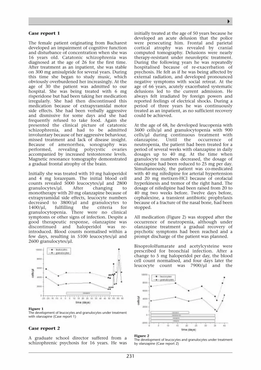
231
▲Case report 1
The female patient originating from Bucharestdeveloped an impairment of cognitive functionand disturbance of concentration when she was16 years old. Catatonic schizophrenia wasdiagnosed at the age of 26 for the first time.After treatment as an inpatient, she was stableon 300 mg amisulpride for several years. Duringthis time she began to study music, whichobviously overburdened her increasingly. At theage of 30 the patient was admitted to ourhospital. She was being treated with 6 mgrisperidone but had been taking her medicationirregularly. She had then discontinued thismedication because of extrapyramidal motorside effects. She had been verbally aggressiveand dismissive for some days and she hadfrequently refused to take food. Again shepresented the clinical picture of catatonicschizophrenia, and had to be admittedinvoluntary because of her aggressive behaviour,missed treatment and lack of disease insight.Because of amenorrhea, sonography wasperformed, revealing polycystic ovariesaccompanied by increased testosterone levels.Magnetic resonance tomography demonstrateda gradual frontal atrophy of the brain.
Initially she was treated with 10 mg haloperidoland 4 mg lorazepam. The initial blood cellcounts revealed 5000 leucocytes/µl and 2800granulocytes/µl. After changing tomonotherapy with 20 mg olanzapine because ofextrapyramidal side effects, leucocyte numbersdecreased to 3800/µl and granulocytes to1400/µl, fulfilling the criteria forgranulocytopenia. There were no clinicalsymptoms or other signs of infection. Despite agood therapeutic response, olanzapine wasdiscontinued and haloperidol was re-introduced. Blood counts normalised within afew days, resulting in 5100 leucocytes/µl and2600 granulocytes/µl.
Case report 2
A graduate school director suffered from aschizophrenic psychosis for 16 years. He was
initially treated at the age of 50 years because hedeveloped an acute delusion that the policewere persecuting him. Frontal and parietalcortical atrophy was revealed by cranialcomputed tomography. Delusions were nearlytherapy-resistant under neuroleptic treatment.During the following years he was repeatedlyhospitalised because of re-exacerbation ofpsychosis. He felt as if he was being affected byexternal radiation, and developed pronouncednegative symptoms with social retreat. At theage of 66 years, acutely exacerbated systematicdelusions led to the current admission. Healways felt irradiated by foreign powers andreported feelings of electrical shocks. During aperiod of three years he was continuouslytreated as an inpatient, as no sufficient recoverycould be achieved.
At the age of 68, he developed leucopenia with3600 cells/µl and granulocytopenia with 900cells/µl during continuous treatment witholanzapine. Until the occurrence ofneutropenia, the patient had been treated for aperiod of several weeks with olanzapine in dailydosages up to 40 mg. At the time whengranulocyte numbers decreased, the dosage ofolanzapine had been reduced to 25 mg per day.Simultaneously, the patient was co-medicatedwith 40 mg nifedipine for arterial hypertensionand 20 mg metixen-HCl because of orofacialhyperkinesis and tremor of the right hand. Thedosage of nifedipine had been raised from 20 to40 mg two weeks before. Twelve days before,cephalexine, a transient antibiotic prophylaxisbecause of a fracture of the nasal bone, had beenstopped.
All medication (Figure 2) was stopped after theoccurrence of neutropenia, although underolanzapine treatment a gradual recovery ofpsychotic symptoms had been reached and aprompt discharge of the patient was planned.
Bisoprololfumarate and acetylcysteine wereprescribed for bronchial infection. After achange to 5 mg haloperidol per day, the bloodcell count normalised, and four days later theleucocyte count was 7900/µl and the
Figure 1 The development of leucocytes and granulocytes under treatmentwith olanzapine (Case report 1)
Figure 2 The development of leucocytes and granulocytes under treatmentby olanzapine (Case report 2)

232
granulocyte count 3500/µl. The patientdeveloped serious parkinsonism andpneumonia with respiratory insufficiency afterfour days of haloperidol treatment, whichresulted in transfer to an internal medical unit.
In the medical history of this patient, aneutropenia with 400 cells/µl was reportedwhen he was on a combination of haloperidoland lorazepam. However, a two-year course oftreatment with 150 mg clozapine led to nochange in blood count.
Case report 3
This patient, who applied for asylum inGermany because of political prosecution in SriLanka, was transferred to our department for thefirst time at the age of 29 because of catatonicschizophrenia. CCT demonstrated roundedfrontal horns, which could be interpreted as aresult of brain damage during early infancy, butno other abnormalities. Because neurolepticmedication was ineffective, he was treated withelectroconvulsive therapy, resulting in clearclinical improvement of the catatonicsymptoms. However, with delusions persisting,he travelled throughout France and theNetherlands in the ensuing years. During acuteexacerbations repeated admissions thereforetook place to several psychiatric hospitals.
At the age of 35, the patient was actually againreferred to our department because of an acuteexacerbation of schizophrenia, dyskinesia,akathisia and torticollis spasticus, probablyinduced by treatment with typical neuroleptics.He reported that he was the sun and the light ofthe world. His persecutors must have kidnappedhis girlfriend, so that he was seeking herkidnappers in Europe.
Leucocyte and granulocyte counts were withinthe normal range. Two months earlier thepatient had discontinued his bi-weekly depotmedication with flupentixol 20 mg. Six yearsbefore, during a previous treatment with 250mg zotepine and 40 mg fluphenazine,haematological side effects had not appeared.After taking 2 mg lorazepam with no othermedication a granulocyte count of 1700/µl wasobserved; however, by the following day thecount was again within normal range.
After inclusion in a double-blind multicentrerandomised study comparing olanzapine withclozapine, the patient took one single dose of 10mg olanzapine. Again the granulocyte countdecreased to 1500/µl for the following two days(Figure 3). After excluding the patient from thestudy and discontinuation of the medication,the granulocyte count increased again andnormalised spontaneously to 2000/µl thefollowing day. During subsequent monotherapywith 12 mg sertindole, again low and
fluctuating granulocyte counts were observedwith a minimum count of 1700/ µl.
Discussion
In the first case, a reversible granulocytopeniaoccurred during olanzapine treatment,producing neither clinical symptoms nor theneed for specific treatment. A relationship witholanzapine is probable, because the patient wastreated in monotherapy; there were nohaematological abnormalities in the history andthe granulocyte count normalised within 10days after discontinuation of olanzapine.
In the second case, the granulocyte countdecreased to 900/µl, which demandedimmediate discontinuation of olanzapine. Inthis case, a bronchial infection occurredfollowed by pneumonia with respiratoryinsufficiency. The high-dose treatment up to 40mg olanzapine given for a few days might be thecause of the granulocyte decrease. Such a causalrelationship with olanzapine is possible in thiscase, but alternatively an influence of the co-medication nifedipine (Voth and Turner 1983)or an association with a bacterial infection cannot be excluded. An unfavourable additivehaematotoxic effect of both drugs, olanzapineand nifedipine, is also possible. Because thebronchial infection developed four days afterthe beginning of neutropenia, we suppose thatthere was no causal relationship betweeninfection and the development of neutropenia.
On the one hand, in the medical history of thispatient neutropenia as low as 600granulocytes/µl had been observed during acombined treatment with 15 mg haloperidol,0.5 mg lorazepam and 25 mg levomepromazinesix years before. On the other hand, a two-yeartreatment with 150 mg clozapine did not lead toa decrease of granulocyte counts.
In the third case report, an idiopathicvulnerability for the development ofgranulocytopenia during treatment withpsychoactive drugs may have pre-existed, as
Figure 3 The development of leucocytes and granulocytes under treatmentby olanzapine (Case report 3)
CASE REPORT/CASE SERIES
▲
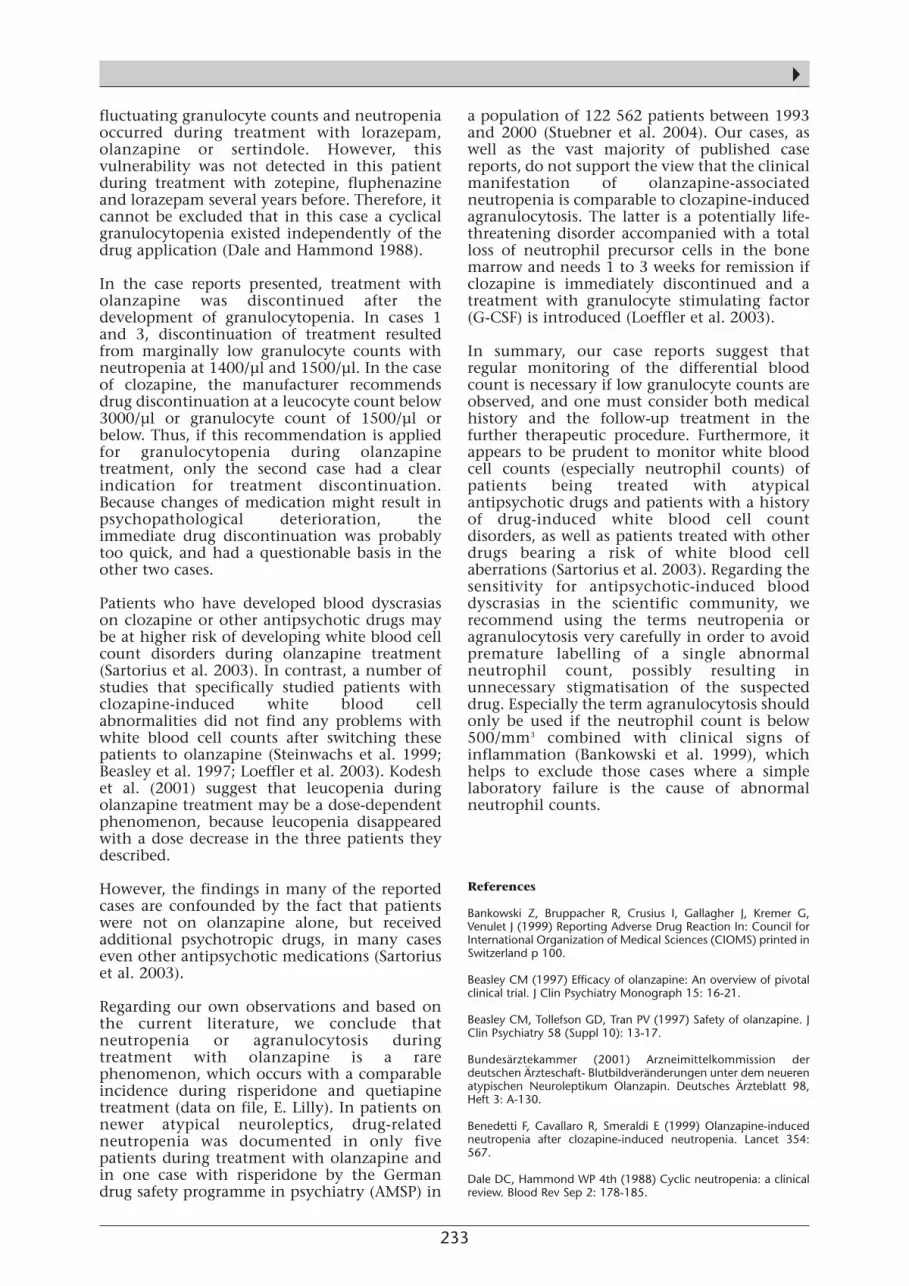
233
▲fluctuating granulocyte counts and neutropeniaoccurred during treatment with lorazepam,olanzapine or sertindole. However, thisvulnerability was not detected in this patientduring treatment with zotepine, fluphenazineand lorazepam several years before. Therefore, itcannot be excluded that in this case a cyclicalgranulocytopenia existed independently of thedrug application (Dale and Hammond 1988).
In the case reports presented, treatment witholanzapine was discontinued after thedevelopment of granulocytopenia. In cases 1and 3, discontinuation of treatment resultedfrom marginally low granulocyte counts withneutropenia at 1400/µl and 1500/µl. In the caseof clozapine, the manufacturer recommendsdrug discontinuation at a leucocyte count below3000/µl or granulocyte count of 1500/µl orbelow. Thus, if this recommendation is appliedfor granulocytopenia during olanzapinetreatment, only the second case had a clearindication for treatment discontinuation.Because changes of medication might result inpsychopathological deterioration, theimmediate drug discontinuation was probablytoo quick, and had a questionable basis in theother two cases.
Patients who have developed blood dyscrasiason clozapine or other antipsychotic drugs maybe at higher risk of developing white blood cellcount disorders during olanzapine treatment(Sartorius et al. 2003). In contrast, a number ofstudies that specifically studied patients withclozapine-induced white blood cellabnormalities did not find any problems withwhite blood cell counts after switching thesepatients to olanzapine (Steinwachs et al. 1999;Beasley et al. 1997; Loeffler et al. 2003). Kodeshet al. (2001) suggest that leucopenia duringolanzapine treatment may be a dose-dependentphenomenon, because leucopenia disappearedwith a dose decrease in the three patients theydescribed.
However, the findings in many of the reportedcases are confounded by the fact that patientswere not on olanzapine alone, but receivedadditional psychotropic drugs, in many caseseven other antipsychotic medications (Sartoriuset al. 2003).
Regarding our own observations and based onthe current literature, we conclude thatneutropenia or agranulocytosis duringtreatment with olanzapine is a rarephenomenon, which occurs with a comparableincidence during risperidone and quetiapinetreatment (data on file, E. Lilly). In patients onnewer atypical neuroleptics, drug-relatedneutropenia was documented in only fivepatients during treatment with olanzapine andin one case with risperidone by the Germandrug safety programme in psychiatry (AMSP) in
a population of 122 562 patients between 1993and 2000 (Stuebner et al. 2004). Our cases, aswell as the vast majority of published casereports, do not support the view that the clinicalmanifestation of olanzapine-associatedneutropenia is comparable to clozapine-inducedagranulocytosis. The latter is a potentially life-threatening disorder accompanied with a totalloss of neutrophil precursor cells in the bonemarrow and needs 1 to 3 weeks for remission ifclozapine is immediately discontinued and atreatment with granulocyte stimulating factor(G-CSF) is introduced (Loeffler et al. 2003).
In summary, our case reports suggest thatregular monitoring of the differential bloodcount is necessary if low granulocyte counts areobserved, and one must consider both medicalhistory and the follow-up treatment in thefurther therapeutic procedure. Furthermore, itappears to be prudent to monitor white bloodcell counts (especially neutrophil counts) ofpatients being treated with atypicalantipsychotic drugs and patients with a historyof drug-induced white blood cell countdisorders, as well as patients treated with otherdrugs bearing a risk of white blood cellaberrations (Sartorius et al. 2003). Regarding thesensitivity for antipsychotic-induced blooddyscrasias in the scientific community, werecommend using the terms neutropenia oragranulocytosis very carefully in order to avoidpremature labelling of a single abnormalneutrophil count, possibly resulting inunnecessary stigmatisation of the suspecteddrug. Especially the term agranulocytosis shouldonly be used if the neutrophil count is below500/mm3 combined with clinical signs ofinflammation (Bankowski et al. 1999), whichhelps to exclude those cases where a simplelaboratory failure is the cause of abnormalneutrophil counts.
References
Bankowski Z, Bruppacher R, Crusius I, Gallagher J, Kremer G,Venulet J (1999) Reporting Adverse Drug Reaction In: Council forInternational Organization of Medical Sciences (CIOMS) printed inSwitzerland p 100.
Beasley CM (1997) Efficacy of olanzapine: An overview of pivotalclinical trial. J Clin Psychiatry Monograph 15: 16-21.
Beasley CM, Tollefson GD, Tran PV (1997) Safety of olanzapine. JClin Psychiatry 58 (Suppl 10): 13-17.
Bundesärztekammer (2001) Arzneimittelkommission derdeutschen Ärzteschaft- Blutbildveränderungen unter dem neuerenatypischen Neuroleptikum Olanzapin. Deutsches Ärzteblatt 98,Heft 3: A-130.
Benedetti F, Cavallaro R, Smeraldi E (1999) Olanzapine-inducedneutropenia after clozapine-induced neutropenia. Lancet 354:567.
Dale DC, Hammond WP 4th (1988) Cyclic neutropenia: a clinicalreview. Blood Rev Sep 2: 178-185.

234
Kodesh A, Finkel B, Lerner AG, Kretzmer G, Sigal M (2001) Dose-dependent olanzapine-associated leukopenia: three case reports.Int Clin Psychopharmacol 16: 117-119.
Lambert T (1998) Olanzapine after clozapine: the rare case ofprolongation of granulocytopenia. Aust N Z J Psychiatry 32: 591-592.
Loeffler S, Fehsel K, Henning U, Fischer J, Agelink M, Kolb-Bachofen V, Klimke A (2003) Increased apoptosis of neutrophils ina case of clozapine-induced agranulocytosis. Pharmacopsychiatry36: 37-41.
Meissner W, Schmidt, Kupsch A, Trottenberg T, Lempert T (1999)Reversible leucopenia related to olanzapine. Mov Disord 14: 872-873.
Naumann R, Felber W, Heilemann H, Reuster T (1999) Olanzapin-induced agranulocytosis. Lancet 354: 566-567.
Oyewumi LK, Semaan Y (2000) Olanzapine: safe during clozapine-induced agranulocytosis. J Clin Psychopharmacol 20: 279-281.
Sartorius N, Fleischhacker WW, Gjerris A, Kern U, Knapp M,Leonard BE, Liebermann JA, Lopez-Ibor JJ, Raay B, Tornquist E,Twomey E (2003) The usefulness and use of second-generationantipsychotic medications–an update. Current Opinion inPsychiatry 16 (Suppl 1): 7-17.
Stuebner S, Grohmann R, Engel R, Bandelow B, Ludwig WD,Wagner G, Mueller-Oerlinghausen B, Moeller HJ, Hippius H,Ruether E (2004) Blood dyscrasias induced by psychotropic drugs.Pharmacopsychiatry 37 (Suppl 1): 70-78.
Steinwachs A, Grohmann R, Petrosa F, Ruether E, Schwerdtner I(1999) Two cases of olanzapine-induced reversible neutropenia.Pharmacopsychiatry 32: 154-156.
Swartz RJ, Ananth J, Smith MW, Burgoyne KS, Gadasally R, Arai Y(1999) Olanzapine treatment after clozapine-inducedgranulocytopenia in 3 patients. J Clin Psychiatry 60: 119-121.
Tran P, Hamilton SH, Kuntz AJ, Potvin JH, Andersen SW, Beasley CJr, Tollefson GD (1997) Double-blind comparison of olanzapineversus risperidone in the treatment of schizophrenia and otherpsychotic disorders. J Clin Psychopharmacol 17: 407-418.
Voth AJ, Turner RH (1983) Nifedipine and agranulocytosis. AnnIntern Med 99: 882.
▲CASE REPORT/CASE SERIES

235
REVIEW/MINI-REVIEW
▲
LETTER TO THE EDITORS
Sexual Dimorphism in Obsessive-Compulsive Disorder
Ruth Gross-Isseroff Geha Mental Health CentrePO Box 102Petach Tikva 49100IsraelTel: +972 3 9258439Fax: +972 3 9258388E-mail: [email protected]
Haggai Hermesh Geha Mental Health CentrePetach Tikva and Sackler Faculty of MedicineTel Aviv University, Israel
Abraham Weizman Geha Mental Health CentrePetach TikvaFelsenstein Medical Research CentrePetach Tikva and Sackler Faculty of MedicineTel Aviv University, Israel
Joseph Zohar Division of PsychiatryThe Chaim Sheba Medical Centre and SacklerFaculty of MedicineTel Aviv University, Israel
About 10 years ago we noticed a sex difference in theperformance of a neuropsychological test inobsessive-compulsive disorder (OCD) patients(Gross-Isseroff et al. 1996). Subsequently, we furtherdocumented this finding (Zohar et al. 1999a) and,in 1999, we published a review on the possibilitythat a sexual dimorphism in OCD existed (Zohar etal. 1999b).
Since then, several publications have presented datalending further support to the sexual dimorphismhypothesis. Thus, two papers (Mundo et al. 1999,2002) have claimed that there is a differentialbehavioural and biochemical response toclomipramine among OCD patients. Other studieshave shown that association and/or linkage togenetic markers of OCD are different in male andfemale patients (Nestadt et al. 2000; Alsobrook etal. 2002; Lochner et al. 2004). Lately, a basicanimal study has shown sex differences in theperformance of an alternation task, supposedly ananimal model of OCD (Ulloa et al. 2004).
In view of this bourgeoning information, whichsustains the hypothesis of sexual dimorphism inOCD, we would like to urge investigators in the fieldto include sex comparisons, even if these are not aninherent part of their studies.
References
Alsobrook JP 2nd, Zohar AH, Leboyer M, Chabane N, Ebstein RP,Pauls DL (2002) Association between the COMT locus andobsessive-compulsive disorder in females but not males. Am J MedGenet 114: 116-120.
Gross-Isseroff R, Voet H, Sasson Y, Luca-Haimovici K, Kandel-Sussman H, Hendler T, Zohar J (1996) Alternation learning inobsessive-compulsive disorder. Biol Psychiat 39: 733-738.
Lochner C, Hemmings SM, Kinnear CJ, Moolman-Smook JC,Corfield VA, Knowles JA, Niehuas DJ, Stein DJ (2004) Gender inobsessive-compulsive disorder: clinical and genetic findings. EurNeuropsychpharmacol 14: 105-113.
Mundo E, Bareggi SR, Pirola R, Bellodi L (1999) Effects of acuteintravenous clomipramine and antiobsessional response toproserotonergic drugs: is gender a predictive variable? BiolPsychiatry 45: 290-294.
Mundo E, Pirola R, Bellodi L, Smeraldi E, Bareggi SR (2002) Aregender differences in antiobsessional response related to differentclomipramine metabolism? J Clin Psychopharmacol 22: 341-342.
Nestadt G, Lan T, Samuels J, Riddle M, Bienvenu OJ 3rd, Liang KY,Hoehn-Saric R, Cullen B, Grados M, Beaty TH, Shugart YY (2000)Complex segregation analysis provides compelling evidence for amajor gene underlying obsessive-compulsive disorder and forheterogeneity by sex. Am J Hum Genet 67: 1611-1616.
Ulloa R-E, Nicolini H, Fernandez-Guasti A (2004) Sex differenceson spontaneous alternation in prepubertal rats: implications for ananimal model of obsessive-compulsive disorder. ProgNeuropsychopharmacol Biol Psychiatry 28: 687-692.
Zohar J, Hermesh H, Weizman A, Voet H, Gross-Isseroff R (1999a)Orbitofrontal cortex dysfunction in obsessive compulsivedisorder? I. Alternation learning in obsessive compulsive disorder:male-female comparisons. Eur Neuropsychopharmacol 9: 407-413.
Zohar J, Gross-Isseroff R, Hermesh H, Weizman A (1999b) Is theresexual dimorphism in obsessive compulsive disorder? NeurosciBiobehav Rev 23: 845-849.
Letters published in this Journal do not necessarilyreflect the opinions of the Editors or the EditorialBoard.
▲
World J Biol Psychiatry (2004) 5, 235
Instructions to Authors
Papers must be written in standard and grammatical English andshould present new results as well as be of scientific value. Contri-butions will be considered for the following categories:Reviews/Mini-reviewsOriginal investigations/Summaries of original researchBrief reportsViewpointsCase reports/Case seriesLetters to the editorsBook reviewsInvitations/Announcements
Please submit one original manuscript and two copies typed on oneside of the sheet. They must be typed in double-line spacing withwide margins. Submit your text on disk using Microsoft Word. Disksmust be clearly marked with the title of the paper and the author'sname. In a cover letter, the category under which the manuscript issubmitted should be indicated. Also, authors must disclose in theircover letter any commercial or financial arrangements that they havewith companies who either have products featured in the manuscriptor with companies that produce competing products. Any specialcircumstance, vested interests or sources of bias that might affect theintegrity of the reported information should also be disclosed in thecover letter. Furthermore, a copy of the Author Disclosure Decleration(see next page) should be completed, signed by all authors andsubmitted.
Submission of a manuscript implies: that the work described has notbeen published before (except in the form of an abstract or as part ofa published lecture, review or thesis); that it is not under consider-ation for publication anywhere else; that its publication has beenapproved by all co-authors, if any, as well as by responsible authorities- tacitly or explicitly - at the institute where the work has been carriedout.
Manuscripts submitted for publication must contain a statement tothe effect that all human studies have been approved by theappropriate ethics committee and have therefore been performed inaccordance with the ethical standards laid down in the 1964Declaration of Helsinki. It should also be stated clearly in the text thatall persons gave their informed consent prior to their inclusion in thestudy. Details that might disclose the identity of the subjects understudy should be omitted.
Reports of animal experiments must state that the "Principles oflaboratory animal care" (NIH publication No. 86-23, revised 1985)were followed, as well as specific national laws (e.g. the current versionof the German Law on the Protection of Animals) where applicable.
The Editorial Office will acknowledge receipt of the manuscript andprovide it with a manuscript reference number. Each manuscript willbe assigned to at least two reviewers. The reference number of themanuscript should be quoted in all correspondence with the ChiefEditor and Editorial Office.
Papers should be written in clear and simple language avoidingrepetition. The paper must be in its final form so as to exclude theneed for corrections in proof.
The subject matter should be defined in a few sentences and earlierwork on the subject referred to by citing recent bibliographies (inmonographs, handbooks, etc). The reader should be able to follow themethod by which the results were obtained, but there is no value indetailed descriptions unless they constitute a genuine innovation.
Mere confirmation of established findings is to be strictly avoided.Experiments with no significant results, if included at all, should bedealt with only briefly. Case histories and examination reports shouldbe included only if necessary for defining the subject matter and areto be presented as concisely as possible.
The editors reserve the right to reject manuscripts that do not complywith the above-mentioned requirements. The author will be heldresponsible for false statements or for failure to fulfil the above-mentioned requirements.
1. The title page should include the title of the work, the first and lastname(s) of author(s), the name of the institution, any footnotesreferring to the title (marked with an asterisk) and the address ofthe author to whom the proofs are to be sent. The author should
also provide a fax number and e-mail address. The title pageshould also include the no. of words in text/abstract/title, the no.of references and the no. of figures and tables.
2. Each article must include a short Summary of the essential results(not more than 200 words).
3. Key words. Following the summary a maximum of 5 key wordsshould be supplied. They should be representative of the contentand be characteristic of the terminology used within the particularfield of study. Index Medicus (Medical Subject Headings) isrecommended for terms and phrases.
4. References. All references cited in the text, but only those, are tobe listed at the end of the paper in alphabetical order under thename of the first author. They should be typed in double-linespacing. Each reference should be followed by a blank line.Chronological order is used for more than one work by the sameauthor or team of authors. Works by two authors should also belisted alphabetically according to the second author’s name. Thenames of all authors should be given. Journal titles should beabbreviated according to Index Medicus.
Citations in the text should be by author and date in parentheses.If there are two authors, both should be named (eg Agar andDouglas 1955); if an article with more than two authors is cited,only the first author’s name plus ‘et al.’ need be given (eg Komoret al. 1979); if there is more than one reference by the same authoror team of authors in the same year, then a, b, c, etc should beadded to the year both in the text and the list of references. Whenstyling references, the following examples should be followed:
Brain WR (1958) The physiological basis of consciousness. A critical review. Brain 81: 426-455.
Kuhlenbeck H (1954) The human diencephalon. A summaryof development, structure, function and pathology. Karger,Basel.
Teuber HL (1964) The riddle of frontal lobe function in man.In: Warren T, Akert CH (eds) The frontal and granular cortexand behavior. McGraw-Hill, New York, pp 252-271.
5. Tables should be submitted on separate sheets. Numerical datagiven in graphs must not be duplicated in tables and vice versa.
6. Figures. The number and size of the illustrations must be kept tothe minimum required for clarification of the text. Previouslypublished figures are not accepted. Explanations of figuresfurnished as legends should not be repeated in the text. All figures,whether photographs, graphs or diagrams, should be numberedconsecutively throughout and submitted on separate sheets.
7. Legends. Figure legends should be sufficient to allow each figureto be understood without reference to the text. Each figure shouldhave a short title followed by a concise description. Allabbreviations and symbols appearing in the figure must beexplained in the legend. Remarks such as “for explanation (ordetails) see text” should be avoided.
8. Offprints may be ordered at cost price when the page proofs arereturned. Corresponding authors will be supplied with a free copyof the relevant issue.
9. All papers and disks must be sent to the Editorial Office at thefollowing address:
Dorothea BodeEditorial AdministratorDepartment of PsychiatryLudwig-Maximilians-UniversityNussbaumstrasse 780336 MunichGermanyTel: +49 89 5160 5531Fax: +49 89 5160 5530E-mail: [email protected]
Manuscripts and disks will not be returned.
Please fill in and submit together with your manuscript to the Editorial Office:
Dorothea BodeEditorial AdministratorDepartment of Psychiatry, Ludwig - Maximilians - UniversityNussbaumstrasse 780336 Munich, Germany
Title of the Article:
Author / Co-Author Names:
" I do not have a financial interest/arrangement or affiliation with any organizations that could be perceived as a real or apparent conflict of interest in the context of the subject of my article(s).
" I have a financial interest/arrangement or affiliation with one or more organization(s) that could be perceived as a real or apparent conflict of interest in the context of the subject of my article(s). I am fully aware that I must disclose to the readers any real or apparent conflict(s) of interest that may have a direct bearing on the subject matter of my articles.
Please tick the box(es) which correspond to your situation, and provide details if needed:
" Ownership of stock options or other financial instruments of companies whose products are mentioned in the article or who manufacture competing products
" Ongoing paid consultancy with company or competitor (actual or within last 2 years)" Employment with company or competitor (actual or within last 2 years)" Honorarium or compensation for conducting research related to the material contained in
the article" Research and/or educational grant
I verify that the information above is accurate. I acknowledge that my article(s) must give abalanced view of the therapeutic options; that the use of generic names of products contributes toimpartiality; and that if trade names are mentioned, those of several companies should be used.
Signatures of Author / Co-Authors Date
The World Journal of Biological PsychiatryAuthor Disclosure Declaration
It is the policy of the World Federation of Societies of Biological Psychiatry (WFSBP) to ensurebalance, independence, objectivity and scientific rigor in The World Journal of Biological Psychiatry.
All authors are expected to disclose to the audience any real or apparent conflict(s) of interest thatmay have a direct bearing on the subject matter of the article. This includes relationships withpharmaceutical companies, biomedical device manufacturers, or other corporations whose productsor services are related to the subject matter of the article.
The intent of this policy is not to prevent authors with a potential conflict of interest frompublication. The Federation does not view the existence of these interests or uses as implying biasor decreasing value to the readership.The Federation feels that this disclosure is important to identify any potential conflict openly, withthe intention that the readers may form their own judgments about each article with the fulldisclosure of the facts.It remains for the readers to determine whether the author’s outside interests may reflect a possiblepartiality in either the exposition or the conclusions presented.
