brain-derived neurotrophic factor (bdnf) induces polarized signaling of small gtpase (rac1) protein...
TRANSCRIPT

Brain-derived Neurotrophic Factor (BDNF) Induces PolarizedSignaling of Small GTPase (Rac1) Protein at the Onset ofSchwann Cell Myelination through Partitioning-defective 3(Par3) Protein*
Received for publication, October 12, 2011, and in revised form, November 28, 2011 Published, JBC Papers in Press, November 29, 2011, DOI 10.1074/jbc.M111.312736
Chhavy Tep‡§1, Mi Lyang Kim§¶1, Laura I. Opincariu�, Allison S. Limpert**, Jonah R. Chan‡‡, Bruce Appel�,Bruce D. Carter**, and Sung Ok Yoon§2
From the ‡Biochemistry Program, §Department of Molecular and Cellular Biochemistry, and ¶Molecular, Cellular, andDevelopmental Biology Program, The Ohio State University, Columbus, Ohio 43210, the �Department of Pediatrics, School ofMedicine, University of Colorado, Aurora, Colorado 80045, the **Department of Biochemistry, Vanderbilt Medical School,Nashville, Tennessee 90033, and the ‡‡Department of Neurology and Program in Neuroscience, University of California,San Francisco, California 94143
Background: Both p75 and Par3 become localized in a polarized manner in Schwann cells.Results: BDNF activates Rac1 at the axon-glial interface, regulated by Par3, thereby facilitating proper alignment between theaxon and Schwann cells.Conclusion: Polarized localization of Rac1 activation is critical for myelination.Significance: Par3 is involved in Rac1 activation by BDNF-p75 at the axon-glial interface, thereby promoting myelination.
Brain-derived neurotrophic factor (BDNF) was shown to playa role in Schwann cell myelination by recruiting Par3 to theaxon-glial interface, but the underlying mechanism hasremained unclear. Here we report that Par3 regulates Rac1 acti-vation by BDNF but not by NRG1-Type III in Schwann cells,although both ligands activate Rac1 in vivo. During develop-ment, active Rac1 signaling is localized to the axon-glial inter-face in Schwann cells by a Par3-dependent polarization mecha-nism. Knockdown of p75 and Par3 individually inhibits Rac1activation, whereas constitutive activation of Rac1 disturbs thepolarized activation of Rac1 in vivo. Polarized Rac1 activation isnecessary formyelination as Par3 knockdown attenuatesmyeli-nation in mouse sciatic nerves as well as in zebrafish. Specifi-cally, Par3 knockdown in zebrafish disrupts proper alignmentbetween the axon and Schwann cells without perturbingSchwann cell migration, suggesting that localized Rac1 activa-tion at the axon-glial interface helps identify the initial wrap-ping sites.We therefore conclude that polarization of Rac1 acti-vation is critical for myelination.
During development, cell-to-cell contact plays a critical rolein facilitating the exchange of signals between neighboringcells. Myelination by Schwann cells is perhaps one of the bestexamples, where myelinating glia are in intimate, continuous
contact with the axons that they myelinate, forming a recipro-cal signaling network between the two cell types. This contactand bidirectional communication begins from the timeSchwann cells migrate along the axonal surface and continuesuntil they begin ensheathing the axons. The process of wrap-ping axons is particularly polarized, being initiated at the axon-glial interface and continuing until mature myelin is formed.Partitioning-Defective 3 (Par3), amember of the polarization
complex that includes Par6 and protein kinase C (1), wasrecently shown to play a critical role in this process. Par3 waslocalized at the axon-glial interface, recruiting p75 in responseto BDNF3 (2), which is secreted by the axons (3). In particular,Par3 knockdown markedly inhibited myelination in vitro (2).Par3 contains three PDZdomains, rendering it capable of inter-acting with a large number of proteins aside from Par6 andaPKC in a polarization complex. They include Tiam1 (4),KIF3A (5), Inscrutable (6), Nectins (7), 14-3-3 (8), JAM (9),Ku70 (10), and dynein (11). Considering the ability of Par3 tointeract with such a variety of proteins, it is possible that thepreviously reported effect of Par3 knockdown in myelinationcultures (2) could have been due to disruption of its associationwith a protein critical for myelination.Here, we report that Par3 is responsible for localized small
GTP binding protein, Rac1, activation in response to BDNF butnot by NRG1-Type III in Schwann cells. Par3, therefore, plays acritical role in distinguishing two different axonal signals,BDNF and NRG1- Type III, thereby regulating myelination.* This work was supported, in whole or in part, by National Institute of Health
grants NS039472 (to S. O. Y.), P30NS045758 (to the Ohio State Neurosci-ence Center Core), NS062796 (to J. R. C.), NS048249 (to B. D. C.), andNS062717 (to B. A.). This work was also supported by Muscular DistrophyAssociation Grant MDA115761 (to A. S. L.).
1 Both authors contributed equally to this work.2 To whom correspondence should be addressed: Dept. of Molecular and
Cellular Biochemistry, 184 Rightmire Hall, Ohio State University, 1060 Car-mack Rd., Columbus, OH 43210. Tel.: 614-292-8542; Fax: 614-292-5379;E-mail: [email protected].
3 The abbreviations used are: BDNF, brain-derived neurotrophic factor; MBP,myelin basic protein; MO, morpholino; dpf, days post-fertilization; E19.5,embryonic day 19.5; DRG, dorsal root ganglion; P3, postnatal day 3; NRG,neuregulin; PKC, protein kinase C; PDZ, postsynaptic density protein, Dro-sophila disc large tumor suppressor and Zonula Occuludens-1 protein;JAM, junctional adhesion molecule; TEM, transmission electron microscopy;PAK, p-21 activated kinase.
THE JOURNAL OF BIOLOGICAL CHEMISTRY VOL. 287, NO. 2, pp. 1600 –1608, January 6, 2012© 2012 by The American Society for Biochemistry and Molecular Biology, Inc. Published in the U.S.A.
1600 JOURNAL OF BIOLOGICAL CHEMISTRY VOLUME 287 • NUMBER 2 • JANUARY 6, 2012
by guest on June 18, 2016http://w
ww
.jbc.org/D
ownloaded from

EXPERIMENTAL PROCEDURES
Reagents—The Fc recombinant proteins were purchasedfrom R&D Systems and BDNF from Promega. The antibodiesused in the study include p-PAK (Cell Signaling Technology,Inc.), actin, and fyn (Santa Cruz Biotechnology, Inc.), Rac1,Par3, and neurofilament (Millipore), p75 (Promega), and myc(Covance). PKI-166 was a gift from Novartis.Constructs—The Par3 construct was a gift fromDr. IanMac-
ara. The PDZ 1, 2, and 3 constructs were described in Ref. 2.RNAi Sequences—The Par3 and its control RNAi sequences
were published (2), as were the p75 and its control RNAisequences (12). The oligonucleotides containing the shRNA ofthe individual RNAi sequences were placed into pSIREN-Ret-roQ-ZsGreen1 vector (Clontech) using the BamHI and XbaIsites as directed by the vendor. Retroviruses were generatedfollowing transient transfection of the shRNA constructs inPlatE cells (Cell Biolabs), and the viral supernatants were con-centrated by centrifugation at 20,000 rpm for 4 h at 15 °C usingan SW28 rotor (Beckman). The viral pellet was resuspended ina small volume of media and frozen at �80 °C until used. Forinfection of Schwann cells in vitro and in vivo, a combinedmix-ture of three different RNAi viruses were used for efficientknockdown.Primary Schwann Cell Culture—Primary rat Schwann cells
were isolated from P0-P1 rats andmaintained according to Ref.13. For RNAi knockdown experiments, Schwann cells wereinfected with the retroviruses that express the shRNAsequences, and the lysates were harvested 3 days later.RacGTP Assays and Western Blotting—RacGTP assays were
performed as described (14). For quantification, RacGTP sig-nals were adjusted to total Rac1 to obtain “relative RacGTPlevels.” The lysates were prepared and processed as described(14).Preparation of Neuregulin 1 Type III—Neuregulin 1 type III
cDNA was transfected into 293T cells, and the membraneswere prepared according to Ref. 15. As a control, the mem-branes from untransfected 293T cells were prepared in parallel.To add the membranes to Schwann cells, 8 �g of membranepreparations was placed onto Schwann cell monolayers by a3-min spin in a dish at 3000 rpm as described (15, 16). ThecDNA for NRG 1-Type III was a generous gift from Dr. DougFalls.Injection of Fc Fusion Proteins and Retroviruses into Neonate
Mice—Injection of Fc proteins or retroviruses into P0 mousesciatic nerves were performed as described (2). Briefly, 2 �l ofFc solutions (1 �g/�l) or 5 �l of concentrated retroviruses wasinjected using a glass-pulled pipette over the sciatic nerve mid-way between the knee and hip by penetrating through the outerskin. For every injection, one side was injected with experimen-tal reagents, whereas the other side was injected with appropri-ate controls in a single mouse. Four days after the injection, the1-cm sciatic nerve segment surrounding the injection site wasisolated for immunohistochemistry and biochemical analyses.For statistical analyses, Student’s t test was used.P75 Knockout andWild-type Mice—The p75 knockout mice
that carried themutation in exon 3 of the p75 gene (17) and thewild type mice were obtained from heterozygote mating as lit-
termates. Themice were backcrossed to C57/BL6 for 10 gener-ations to make them congenic. Their genotype was determinedby PCR analyses of tail DNA according to Bentley and Lee (18).For experiments, both sexes were used.Myelination Cultures with p75 Knockdown—Rat Schwann
cells were transfected with the control-shRNA or p75-shRNAas described (19). Cells were plated in Ultraculture media (Bio-Whittaker) supplemented with 10% FBS, 2 mM L-glutamine,and 50 ng/ml NGF at a density of 80,000 cells/2.2 cm2 per col-lagen-coated coverslip.Myelinationwas induced 5 days later byadding 50 �g/ml of ascorbic acid in the growth media. Growthmedia and ascorbic acid were replaced every 2 days. Following10 days of treatment, cells were fixed and immunostained forMBP. For quantification,MBP� internodeswere quantified in ablinded manner. For statistical analysis, a Student’s t test wasused.TEM Analyses and Quantification—For TEM of the tissues,
the sciatic nerves encompassing the segment from the hip tothe knee were dissected (�0.5 cm) 4 days after retrovirus in-jections and divided further into three equal parts using a sharprazor blade. The tissues were fixed for 2 h at room temperaturewith 2%paraformaldehyde/2% glutaraldehyde in 0.1MNa caco-dylate buffer (pH 7.2), rinsed in 0.1 M Na cacodylate buffer, andplaced in 1% osmium/0.1 M Na cacodylate for 60–90 min atroom temperature. The tissues were stained en bloc for 1 h in2% uranyl acetate and embedded in Spurr resin following dehy-dration procedures. Sections were cut on a coronal plane at 80nm using a Reichert Ultracut E ultramicrotome and collectedon 300 mesh grids. Sections were stained in 2% uranyl acetateandReynolds lead citrate before observation in Field Electron&Ion Source Company Technai G2 Spirit TEM at 60kV (OhioState University Campus Microscopy and Imaging Facility).For quantification ofmyelinated axons, three random images
were obtained from each cross-section of the sciatic nerve,from three cross-sections per mouse (the number of images �36 from 4 mice). The electron photomicrographs were pre-pared at �2550 magnification, and the number of myelinatedaxons, myelin thickness, and g ratio were counted using ImageJsoftware. For statistical analyses, Student’s t test was used.Morpholinos—pard3 antisense morpholino (MORPH1404,
sequence 5�-TCCAACACTCCTTCCCGAATCCAAG-3�) wasobtained from Open Biosystems. MO2 (5�-TCAAAGG-CTCCCGTGCTCTGGTGTC-3�) and a random sequence con-trol MO3 were obtained from Gene Tool LLC. Both MOs wereresuspended in H2O at a concentration of 1.0 mM. Each MOwas diluted to a working concentration of 0.25 mM in H2O andphenyl red and injected (1–2 nl) into Tg(sox10(7.2):mRFP);Tg(olig2:EGFP) zebrafish embryos (20, 21) at one cell stage. Theembryos were raised at 28.5 °C until analysis at 4 dpf.In Situ RNA Hybridization in Zebrafish—Zebrafish larvae
were fixed in 4% paraformaldehyde for 24 h and stored in 100%methanol at �20 °C. The mbp RNA probe was synthesizedusing a digoxigenin labeling kit (Roche) and T7 RNA polymer-ase (NEB). After in situ RNA hybridization and staining,embryos were dissected from the yolk and mounted in 75%glycerol on bridged microscope slides. Images were obtainedusing Volocity software (PerkinElmer Life Sciences) and a ZeissAxioObserver inverted microscope equipped with differential
Par3 Is Necessary for Polarized Rac1 Activation in Schwann Cells
JANUARY 6, 2012 • VOLUME 287 • NUMBER 2 JOURNAL OF BIOLOGICAL CHEMISTRY 1601
by guest on June 18, 2016http://w
ww
.jbc.org/D
ownloaded from

interference contrast optics and a Retiga Exi color digital cam-era. Images were exported to Photoshop (Adobe). Imagemanipulations were limited to cropping, contrast, brightness,and color matching settings.In Vivo Imaging of Zebrafish—For imaging, larvae were
lightly anesthetized using ethyl 3-aminobenzoate methanesul-fonic acid (Tricaine), immersed in 0.8% low-melting tempera-ture agarose, and mounted on their sides in glass-bottomed35-mm dishes (World Precision Instruments). Images werecaptured using a �40 oil immersion objective mounted on amotorized Zeiss AxioObserver microscope equipped with aPerkinElmer Life Sciences UltraVIEW VoX spinning disk con-focal system. Z image stacks were collected and compiled usingVolocity software and exported to Photoshop. Image manipu-lations were limited to brightness settings and cropping.
RESULTS
BDNF andNRG1-Type III Activate Rac1 during Sciatic NerveDevelopment—During development in rat sciatic nerves,RacGTP levels peaked before the onset of myelination atembryonic age 19.5 (E19.5) and then gradually declined untilreaching the adult level (Fig. 1,A andB). Because Schwann cellsare in constant contact with axons in addition to forming asignaling junction with the extracellular matrix, we located thesites of Rac1 activation by phospho-PAK (p-PAK) immuno-staining. Although PAK is known to undergo autophosphoryl-ation when bound to Rac1- and Cdc42-GTP (22), p-PAK stain-ing represents mostly Rac1 activation at the premyelinatingstage because Cdc42 is not activated until myelination begins(data not shown). In dorsal root ganglion (DRG) neuronSchwann cell cocultures inwhichmyelinationwas not initiated,p-PAK immunoreactivity was detectedwithin the Schwann cellon the side that was in contact with DRG fibers (Fig. 1C, leftpanel). Also at postnatal day 3 (P3) sciatic nerves,p-PAK immu-noreactivity was asymmetrically localized toward the axon (Fig.1C, right panel and inset). These results suggest that at least atthe premyelinating stage, signals from the axon induce Rac1activation in Schwann cells. In further support, we failed todetect any change in RacGTP levels in 2- to 3-week-old pureDRG cultures after treating them with BDNF, NRG1-Type III,and laminin (data not shown).We hypothesized that BDNF and/or neuregulin (NRG) 1, the
two well known axonal signals (3, 15, 23), are responsible foractivating Rac1 in vivo. To test the hypothesis, TrkB-Fc orErbB3-Fc was injected into sciatic nerves of P0 mice, whereasthe control-Fc was injected into sciatic nerves on the contralat-eral side. Although the RacGTP levels were readily detectedfrom the control-Fc-injected sciatic nerves at P4, they werereduced by 80%withTrkB-Fc injections (Fig. 1D), and 70%withErbB3-Fc injections (E). These results suggest that both BDNFandNRG1 are involved in activating Rac1 at P0-P4. It should benoted that myelin protein P0 levels were also reduced withTrkB-Fc and ErbB3-Fc, suggesting that the mechanisms bywhich NRG1 and BDNF promote myelination are likely toinclude Rac1 activation. In line with the in vivo data, bothBDNF and NRG1-Type III increased RacGTP levels inSchwann cell cultures (Fig. 1, F and G).
Par3 Is Necessary for BDNF-mediated but not NRG1-TypeIII-mediated Rac1 Activation—Par3, partitioning-defectiveprotein 3, was shown to be asymmetrically localized on theaxon-glial interface during the premyelination stage, recruitingp75uponBDNF stimulation (2). Par3 can also bindRacGEF andTiam1, thereby regulating Rac1 activation in epithelial cells (4).Because the p-PAK expression pattern mimics that of Par3 (2)and Par3 interacted with Rac1 in P3 sciatic nerves (Fig. 2E), wetested whether Par3 plays a role in BDNF- and/or NRG1-TypeIII-dependent Rac1 activation in Schwann cells. Par3 wasknocked down in Schwann cells using retroviruses carryingPar3 functional RNAi or control nonfunctional Par3 RNAi (2),and the changes in RacGTP levels were measured at varioustimes after treatment with BDNF or NRG1-Type III. Par3knockdown reduced RacGTP levels to the basal level withBDNF (Fig. 2, A and B) but not with NRG1-Type III (C and D).The reason for the difference was not clear because ErbB2 canbind Par3 (Fig. 2F). These results nonetheless suggest that themechanism by which BDNF activates Rac1 is distinct from thatof NRG1-Type III in Schwann cells. More importantly, thesedata indicate that Par3 plays a role in distinguishing the twodifferent axonal signals.Selective Knockdown of Par3 in Schwann Cells in Vivo Inhib-
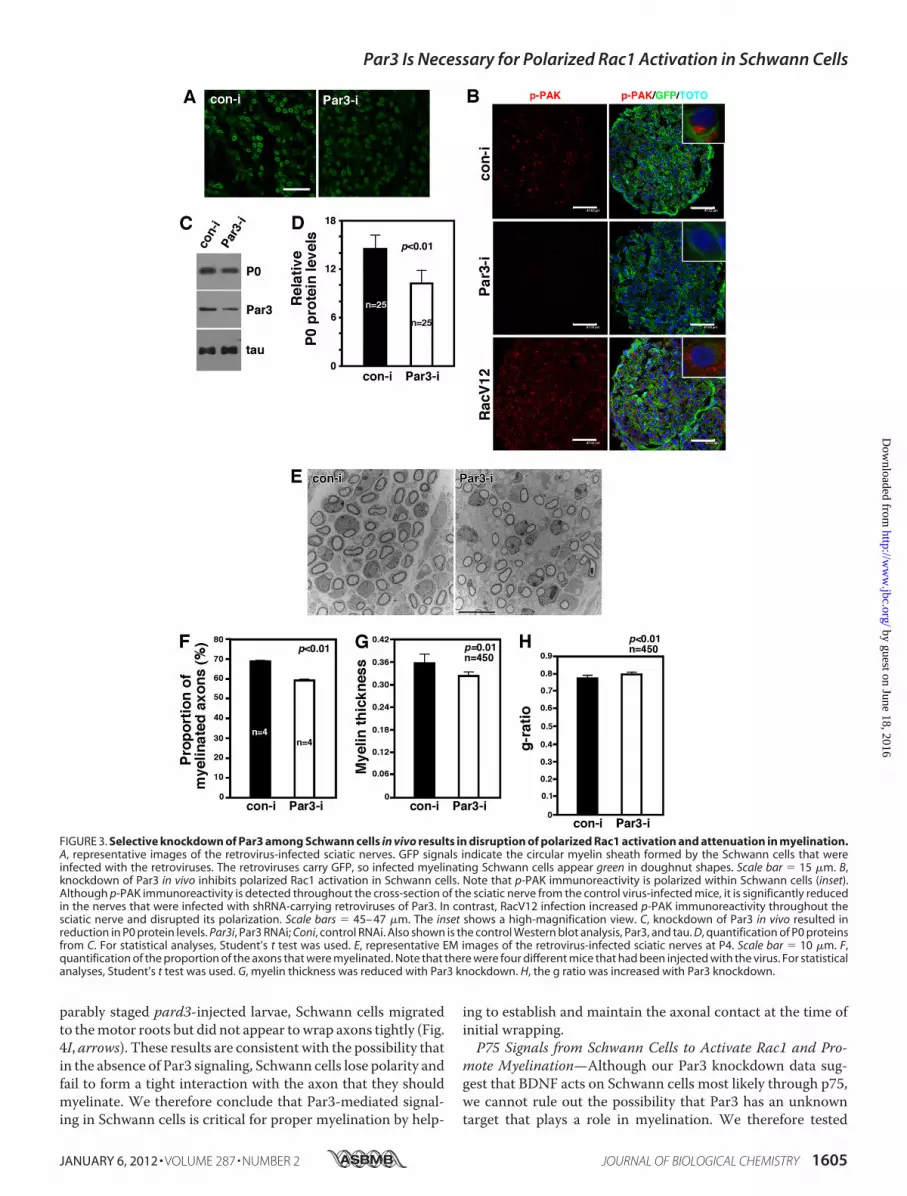
its Polarized Rac1 Activation and Myelination—P75 isexpressed both in DRG neurons and in Schwann cells, tendingto obscure whether p75 plays a positive role in myelinationfrom the neuronal side or the Schwann cell side. Our discoveryof Par3-mediated selectivity for BDNF overNRG1-Type III sig-naling presents an opportunity for testing whether it is the p75in Schwann cells that is responsible for its effect in myelina-tion. Accordingly, we knocked down Par3 only in Schwanncells by injecting the retrovirus for Par3-RNAi and its con-trol RNAi to the P0 sciatic nerve and examined whetherpolarized activation of Rac1 as well as myelination areaffected. The effect of Par3 knockdown on Rac1 activationwas assessed via p-PAK immunoreactivity. The extent ofinfection is shown in Fig. 3A.In the control virus-infected Schwann cells, red p-PAK
immunoreactivity was polarized to one side of GFP� Schwanncells (Fig. 3B, inset). Par3 knockdown resulted in a significantreduction in Rac1 activation (Fig. 3B), as it did with p75 knock-down (Fig. 5C). These results provide additional evidence thatp75 and Par3 regulate Rac1 activation in Schwann cells. Whenwe activated Rac1 constitutively using a RacV12 retrovirus,however, Rac1 activity was no longer polarized but distributedthroughout Schwann cells (Fig. 3B, inset).We found that loss ofpolarized Rac1 activation by Par3 knockdown impacts myeli-nation. P0 protein levels were reduced by 30% in Par3 knock-down nerves compared with that in the control nerves. Theextent of Par3 knockdown was 57% (Fig. 3,C andD). The effectonmyelination is also supported by EM analyses of the infectedsciatic nerves. Not only was the proportion of the axons thatweremyelinated reduced by 15%byPar3 knockdowncomparedwith the control (Fig. 3, E–G), but themyelin thickness was alsoreduced by 9.2%. In line with thinner myelin, the g ratio wasincreased by 2.9% by Par3 knockdown (Fig. 3H). It should benoted that the observed extent of changes in the myelin profileis likely to have been an underestimate because not all Schwann
Par3 Is Necessary for Polarized Rac1 Activation in Schwann Cells
1602 JOURNAL OF BIOLOGICAL CHEMISTRY VOLUME 287 • NUMBER 2 • JANUARY 6, 2012
by guest on June 18, 2016http://w
ww
.jbc.org/D
ownloaded from

FIGURE 1. BDNF and NRG1-Type III activate Rac1 in sciatic nerves. A, RacGTP levels are their highest at E19.5 in rats and gradually decline during develop-ment. B, quantification of RacGTP levels in A. n � 6 – 8. C, asymmetric localization of phospho-PAK immunoreactivity in premyelinating Schwann cells in cultureand in vivo. Note that phospho-PAK staining is along the side of Schwann cells that were in contact with DRG axons in culture. NF, neurofilament. Scale bar �5 �m (left panels). Similarly, p-PAK immunoreactivity is found predominantly adjacent to axons in P3 sciatic nerves (right panel). Scale bar � 12 �m). The insetshows a high-magnification view. D, injection of TrkB-Fc and not control Fc into the sciatic nerves of P0 mice resulted in inhibition of Rac1 activation and P0protein levels. Quantification is shown next to the figure. E, injection of ErbB3-Fc, and not control Fc, into the sciatic nerves of P0 mice resulted in inhibition ofRac1 activation and P0 protein levels. Quantification is shown next to the figure. F, BDNF activates Rac1 in primary Schwann cells. BDNF was added at 50 ng/mlfor 1 h. Quantification is shown below the figures. G, NRG1-Type III activates Rac1 in primary Schwann cells. Membrane fractions (8 �g) from 293 cells expressinga vector or NRG1-Type III cDNA were added onto Schwann cells for 1 h. Quantification is shown below the figures. Relative RacGTP levels represent the RacGTPlevels adjusted to total Rac1 level in each sample. For statistical analyses, Student’s t test was used.
Par3 Is Necessary for Polarized Rac1 Activation in Schwann Cells
JANUARY 6, 2012 • VOLUME 287 • NUMBER 2 JOURNAL OF BIOLOGICAL CHEMISTRY 1603
by guest on June 18, 2016http://w
ww
.jbc.org/D
ownloaded from

cells were infected with the virus. We therefore interpret thesedata as suggesting that the p75-ErbB2-Par3 complex regulatespolarized activation of Rac1, thereby regulating certain aspectsof myelination in vivo.Knockdown of pard3 in Zebrafish Larvae Inhibits MBP
Expression by Disrupting Normal Wrapping of Axons—Al-though the EM data suggest that a smaller number of axons ismyelinated with Par3 knockdown in mouse sciatic nerves, it isnot clear at which step Par3 regulates myelination. To deter-mine the precise step at which Par3 plays its role during myeli-nation, we utilized fluorescentlymarked transgenic zebrafish inwhich Par3 expression was knocked down by injecting into sin-gle-cell embryos one of two different antisensemorpholino oli-gonucleotides (MO1 andMO2) designed to block translation ofpard3 mRNA. At 4 dpf, larvae injected with pard3 MO had asignificant reduction in endogenous RacGTP levels as well as inPard3 protein (Fig. 4A). This result indicates that Par3 regulatesRac1 activation in zebrafish as it does in rodent Schwann cells.We next investigated whether pard3 knockdown also attenu-
ates the extent of myelination by performing in situ RNAhybridization using mbp as a marker. In all the larvae thatreceived no MO (n � 31) or a control MO (n � 19),mbp RNAwas readily detected at motor nerves (Fig. 4B) and the posteriorlateral line nerve (Fig. 4C). In larvae that were injected witheither MO1 (n � 20, Fig. 4, D and E) or MO2 (n � 65, Fig. 4, Fand G),mbp RNA levels were significantly reduced along bothmotor nerves and the posterior lateral line nerves. These resultssuggest that Par3 regulates Schwann cell myelination both inzebrafish and rodents.To investigate the steps at which pard3 regulates Schwann
cell myelination, we injected MO into embryos at the one-cellstage in Tg(sox10(7.2):mRFP);Tg(olig2:EGFP) embryos, grewthem until 4 dpf, and performed live cell imaging. This trans-genic combination marks motor axons with enhanced greenfluorescent protein driven by olig2 regulatory DNA (24) andSchwann cells with membrane-tethered RFP driven by sox10regulatory DNA (20). In control larvae, Schwann cell wrappingof motor axons is initiated by 4 dpf (Fig. 4H, arrows). In com-
FIGURE 2. Par3 is necessary for BDNF-mediated but not for NRG1-Type III-mediated Rac1 activation in Schwann cell cultures. A, Par3 is necessary forBDNF-mediated Rac1 activation in Schwann cell cultures. Following infection with the retrovirus carrying Par3 RNAi (Par3i) or control RNAi (Coni), Schwann cellswere treated with 50 ng/ml BDNF for the indicated period of time. Control Par3 Western blot analysis demonstrates the extent of Par3 knockdown. B,quantification of A. Relative RacGTP levels represent the RacGTP levels adjusted to total Rac1 level in each sample. For statistical analyses, Student’s t test wasused. C, knocking down Par3 in Schwann cells failed to inhibit Rac1 activation by NRG1-Type III. Following infection with the retrovirus carrying Par3 RNAi,Schwann cells were treated with 8 �g/ml NRG1-Type III for the indicated period of time. D, quantification of C. E, Par3 interacts with Rac1 in P3 sciatic nerves.Par3 was immunoprecipitated and blotted for Rac1. F, Par3 binds ErbB2 in 293T cells. Full-length Par3 and ErbB2 were transfected to 293T cells, and theresulting lysates were subjected to immunoprecipitation with ErbB2 and Western blot analysis with Par3. Control inputs are shown (5% of the lysates used forimmunoprecipitation).
Par3 Is Necessary for Polarized Rac1 Activation in Schwann Cells
1604 JOURNAL OF BIOLOGICAL CHEMISTRY VOLUME 287 • NUMBER 2 • JANUARY 6, 2012
by guest on June 18, 2016http://w
ww
.jbc.org/D
ownloaded from
parably staged pard3-injected larvae, Schwann cells migratedto themotor roots but did not appear towrap axons tightly (Fig.4I, arrows). These results are consistent with the possibility thatin the absence of Par3 signaling, Schwann cells lose polarity andfail to form a tight interaction with the axon that they shouldmyelinate. We therefore conclude that Par3-mediated signal-ing in Schwann cells is critical for proper myelination by help-
ing to establish and maintain the axonal contact at the time ofinitial wrapping.P75 Signals from Schwann Cells to Activate Rac1 and Pro-
mote Myelination—Although our Par3 knockdown data sug-gest that BDNF acts on Schwann cells most likely through p75,we cannot rule out the possibility that Par3 has an unknowntarget that plays a role in myelination. We therefore tested
FIGURE 3. Selective knockdown of Par3 among Schwann cells in vivo results in disruption of polarized Rac1 activation and attenuation in myelination.A, representative images of the retrovirus-infected sciatic nerves. GFP signals indicate the circular myelin sheath formed by the Schwann cells that wereinfected with the retroviruses. The retroviruses carry GFP, so infected myelinating Schwann cells appear green in doughnut shapes. Scale bar � 15 �m. B,knockdown of Par3 in vivo inhibits polarized Rac1 activation in Schwann cells. Note that p-PAK immunoreactivity is polarized within Schwann cells (inset).Although p-PAK immunoreactivity is detected throughout the cross-section of the sciatic nerve from the control virus-infected mice, it is significantly reducedin the nerves that were infected with shRNA-carrying retroviruses of Par3. In contrast, RacV12 infection increased p-PAK immunoreactivity throughout thesciatic nerve and disrupted its polarization. Scale bars � 45– 47 �m. The inset shows a high-magnification view. C, knockdown of Par3 in vivo resulted inreduction in P0 protein levels. Par3i, Par3 RNAi; Coni, control RNAi. Also shown is the control Western blot analysis, Par3, and tau. D, quantification of P0 proteinsfrom C. For statistical analyses, Student’s t test was used. E, representative EM images of the retrovirus-infected sciatic nerves at P4. Scale bar � 10 �m. F,quantification of the proportion of the axons that were myelinated. Note that there were four different mice that had been injected with the virus. For statisticalanalyses, Student’s t test was used. G, myelin thickness was reduced with Par3 knockdown. H, the g ratio was increased with Par3 knockdown.
Par3 Is Necessary for Polarized Rac1 Activation in Schwann Cells
JANUARY 6, 2012 • VOLUME 287 • NUMBER 2 JOURNAL OF BIOLOGICAL CHEMISTRY 1605
by guest on June 18, 2016http://w
ww
.jbc.org/D
ownloaded from

whether knocking down p75 itself in Schwann cells affectsmyelination in vivo and in vitro. First, in an in vitro study, weutilized DRG-Schwann cell cocultures where p75 expressionwas knocked down by transfecting Schwann cells with the p75-RNAi prior to seeding them onto pure DRG neurons asdescribed (19).When the extent ofmyelination was assessed bystaining for MBP on days 10–14 day following ascorbic acidtreatment, the number of MBP� fibers was reduced by 62% inthe p75 knockdown Schwann cells compared with the controlcultures (Fig. 5,A and B). These results suggest that it is the p75in Schwann cells that plays a signaling role in myelination invitro.To study the effect of p75 knockdown in vivo, we opted to
inject a retrovirus that carries the same p75 shRNA into mousesciatic nerves. As with Par3, knockdown of p75 resulted in asignificant reduction in p-PAK immunoreactivity as well asMBP staining (Fig. 5C). In correspondence with the p-PAKresults, RacGTP levels in p75�/� mice were reduced by 40%comparedwith that of the wild-typemice at E18.5 (Fig. 5,D andE). The RacGTP levels at P3 and adult stages did not differbetween the genotypes. We therefore conclude that p75 pro-motes myelination from the Schwann cell side, in part by regu-lating Rac1 activation.
DISCUSSION
Here, we identified two signals that can activate Rac1 inSchwann cells: BDNF and NRG1-Type III, in addition to �1integrin (25). Of these three different inputs to activate Rac1 inSchwann cells, it is the BDNF-dependent Rac-GTP signal thatis located at the axon-glial interface where myelination is likelyto occur. It is our contention that the location where Rac1 isactivated inside the cell is likely to affect which downstreamsignaling pathways are to be activated, thereby impacting a par-ticular step that is involved in myelination.On the basis of Par3 knockdown results from zebrafish, we
propose that Par3-dependent Rac1 activation regulates whereaxon wrapping should commence on the axon surface,although it has little effect on Schwann cell migration along theaxon. These data are in agreementwith a report inwhich BDNFwas shown to inhibit Schwann cell migration in rat Schwanncells (26). Although it remains to be tested whether Par3 inzebrafish is also linked to BDNF and p75 signaling as it is inrodents, we surmise that BDNF signaling in rodents is likely toplay a role in establishing the initial wrapping points along theaxon.A recent study by Xiao et al. (27) demonstrated that BDNF
promoted myelination through p75 from the neuron side and
FIGURE 4. Knockdown of pard3 in zebrafish larvae resulted in abnormal Schwann cell wrapping and a significant reduction in mbp expression. A, pard3MO1 reduces Pard3 protein levels and inhibits RacGTP levels in zebrafish larvae. Embryos at 4 dpf were deyolked, and proteins were extracted for RacGTP assaysand control Western blot analyses. B–G, lateral views at the level of the trunk of 4 dpf larvae hybridized for mbp. In control larvae (B and C), mbp is expressed instripes associated with ventral motor roots (B, asterisks) and along the posterior lateral line nerve (C, arrow). In pard3 MO1- (D and E) and MO2 (F and G)-injectedlarvae, mbp expression is lost at the ventral roots (asterisks) and the posterior lateral line nerve (arrows), whereas it is still evident in the spinal cord (sc, brackets).H and I, lateral views at the level of the trunk of 4 dpf Tg(olig2:EGFP);Tg(sox10:mRFP) larvae. Motor axons are green, and Schwann cells are red. In a control larva(H), Schwann cells wrap motor axons (arrows), whereas in a MO1 injected larva (I), Schwann cells failed to align along the axon, forming a loose association withthe axon. The MO-injected larva also has a deficit of axon wrapping by oligodendrocytes, which are marked by RFP expression, in the spinal cord.
Par3 Is Necessary for Polarized Rac1 Activation in Schwann Cells
1606 JOURNAL OF BIOLOGICAL CHEMISTRY VOLUME 287 • NUMBER 2 • JANUARY 6, 2012
by guest on June 18, 2016http://w
ww
.jbc.org/D
ownloaded from

that knocking down the receptor in Schwann cells hadno effect.This is in direct contrast to our results presented here. Thereason for the difference is not clear, but it should be noted thatthe age of theDRGneurons used in the two studies differ. In ourstudy, DRG neurons were isolated at E15 and cultured for aweek in the presence of NGF before Schwann cells were added.This procedure approximates the neuronal stage when myeli-nation occurs during normal development. On the other hand,Xiao et al. isolated DRG neurons at P2 and cultured them for2–3 weeks until they became independent of neurotrophinsbefore adding Schwann cells. The DRG neurons would essen-tially be adult neurons. Hence, it is possible that p75 plays acritical role in Schwann cells duringmyelin formation in devel-opment but that in the adult when myelin forms, such as afteran injury, the receptor has amore critical role in the neurons. A
conditional knockout strategywill be important to better clarifythe role of p75 in Schwann cells pertaining tomyelination in thefuture.
REFERENCES1. Goldstein, B., and Macara, I. G. (2007) The PAR proteins. Fundamental
players in animal cell polarization. Dev. Cell 13, 609–6222. Chan, J. R., Jolicoeur, C., Yamauchi, J., Elliott, J., Fawcett, J. P., Ng, B. K.,
and Cayouette, M. (2006) The polarity protein Par-3 directly interactswith p75NTR to regulate myelination. Science 314, 832–836
3. Ng, B. K., Chen, L., Mandemakers, W., Cosgaya, J. M., and Chan, J. R.(2007) Anterograde transport and secretion of brain-derived neu-rotrophic factor along sensory axons promote Schwann cell myelination.J. Neurosci. 27, 7597–7603
4. Chen, X., and Macara, I. G. (2005) Par-3 controls tight junction assemblythrough the Rac exchange factor Tiam1. Nat. Cell Biol. 7, 262–269
5. Nishimura, T., Kato, K., Yamaguchi, T., Fukata, Y.,Ohno, S., andKaibuchi,
FIGURE 5. P75 signals from Schwann cells activate Rac1 and promote myelination. A, knockdown of p75 in rat Schwann cells inhibited the extent ofmyelination in culture. Also shown are the control Westerns for the p75 knockdown with RNAi. For statistical analyses, Student’s t test was used. B, represent-ative images of MBP staining of the cocultures. Scale bar � 10 �m. C, knockdown of p75 among Schwann cells reduced the extent of p-PAK and MBPimmunoreactivity. Note that we are using the same image from Fig. 3B for the control shRNA-infected nerves. Scale bars � 45– 47 �m. D, P75 is necessary forRac1 activation in vivo. RacGTP levels are reduced in p75�/� mice compared with those in p75�/� at E18.5. As controls, the protein levels of p75, tau, and P0 arealso shown. Note that there is a reduction in tau protein levels, reflecting a reduction in DRG fibers. E, quantification of RacGTP from D.
Par3 Is Necessary for Polarized Rac1 Activation in Schwann Cells
JANUARY 6, 2012 • VOLUME 287 • NUMBER 2 JOURNAL OF BIOLOGICAL CHEMISTRY 1607
by guest on June 18, 2016http://w
ww
.jbc.org/D
ownloaded from

K. (2004) Role of the PAR-3-KIF3 complex in the establishment of neuro-nal polarity. Nat. Cell Biol. 6, 328–334
6. Schober, M., Schaefer, M., and Knoblich, J. A. (1999) Bazooka recruitsInscuteable to orient asymmetric cell divisions inDrosophila neuroblasts.Nature 402, 548–551
7. Takekuni, K., Ikeda, W., Fujito, T., Morimoto, K., Takeuchi, M., Monden,M., and Takai, Y. (2003) Direct binding of cell polarity protein PAR-3 tocell-cell adhesion molecule nectin at neuroepithelial cells of developingmouse. J. Biol. Chem. 278, 5497–5500
8. Hurd, T.W., Fan, S., Liu, C. J., Kweon, H. K., Hakansson, K., andMargolis,B. (2003) Phosphorylation-dependent binding of 14-3-3 to the polarityprotein Par3 regulates cell polarity inmammalian epithelia.Curr. Biol. 13,2082–2090
9. Ebnet, K., Suzuki, A., Horikoshi, Y., Hirose, T., Meyer Zu Brickwedde,M. K., Ohno, S., and Vestweber, D. (2001) The cell polarity protein ASIP/PAR-3 directly associates with junctional adhesion molecule (JAM).EMBO J. 20, 3738–3748
10. Fang, L., Wang, Y., Du, D., Yang, G., Tak Kwok, T., Kai Kong, S., Chen, B.,Chen, D. J., and Chen, Z. (2007) Cell polarity protein Par3 complexes withDNA-PK via Ku70 and regulates DNA double-strand break repair. CellRes. 17, 100–116
11. Schmoranzer, J., Fawcett, J. P., Segura,M., Tan, S., Vallee, R. B., Pawson, T.,and Gundersen, G. G. (2009) Par3 and dynein associate to regulate localmicrotubule dynamics and centrosome orientation during migration.Curr. Biol. 19, 1065–1074
12. Vilar, M., Charalampopoulos, I., Kenchappa, R. S., Simi, A., Karaca, E.,Reversi, A., Choi, S., Bothwell, M., Mingarro, I., Friedman, W. J., Schiavo,G., Bastiaens, P. I., Verveer, P. J., Carter, B. D., and Ibáñez, C. F. (2009)Activation of the p75 neurotrophin receptor through conformational re-arrangement of disulphide-linked receptor dimers. Neuron 62, 72–83
13. Chan, J. R., Watkins, T. A., Cosgaya, J. M., Zhang, C., Chen, L., Reichardt,L. F., Shooter, E. M., and Barres, B. A. (2004) NGF controls axonal recep-tivity to myelination by Schwann cells or oligodendrocytes. Neuron 43,183–191
14. Harrington, A. W., Kim, J. Y., and Yoon, S. O. (2002) Activation of RacGTPase by p75 is necessary for c-junN-terminal kinase-mediated apopto-sis. J. Neurosci. 22, 156–166
15. Taveggia, C., Zanazzi, G., Petrylak, A., Yano, H., Rosenbluth, J., Einheber,S., Xu, X., Esper, R. M., Loeb, J. A., Shrager, P., Chao, M. V., Falls, D. L.,Role, L., and Salzer, J. L. (2005) Neuregulin-1 type III determines theensheathment fate of axons. Neuron 47, 681–694
16. Maurel, P., and Salzer, J. L. (2000) Axonal regulation of Schwann cellproliferation and survival and the initial events of myelination requiresPI3-kinase activity. J. Neurosci. 20, 4635–4645
17. Lee, K. F., Li, E., Huber, L. J., Landis, S. C., Sharpe, A. H., Chao, M. V., andJaenisch, R. (1992) Targeted mutation of the gene encoding the low-affin-ity NGF receptor p75 leads to deficits in the peripheral sensory nervoussystem. Cell 69, 737–749
18. Bentley, C. A., and Lee, K. F. (2000) p75 is important for axon growth andSchwann cell migration during development. J. Neurosci. 20, 7706–7715
19. Yoon, C., Korade, Z., and Carter, B. D. (2008) Protein kinase A-inducedphosphorylation of the p65 subunit of nuclear factor �B promotesSchwann cell differentiation into amyelinating phenotype. J. Neurosci. 28,3738–3746
20. Kucenas, S., Takada, N., Park, H. C., Woodruff, E., Broadie, K., and Appel,B. (2008) CNS-derived glia ensheath peripheral nerves andmediatemotorroot development. Nat. Neurosci. 11, 143–151
21. Flanagan-Steet, H., Fox, M. A., Meyer, D., and Sanes, J. R. (2005) Neuro-muscular synapses can form in vivo by incorporation of initially aneuralpostsynaptic specializations. Development 132, 4471–4481
22. Manser, E., Huang, H. Y., Loo, T. H., Chen, X. Q., Dong, J. M., Leung, T.,and Lim, L. (1997) Expression of constitutively active �-PAK reveals ef-fects of the kinase on actin and focal complexes. Mol. Cell Biol. 17,1129–1143
23. Michailov, G. V., Sereda, M. W., Brinkmann, B. G., Fischer, T. M., Haug,B., Birchmeier, C., Role, L., Lai, C., Schwab, M. H., and Nave, K. A. (2004)Axonal neuregulin-1 regulates myelin sheath thickness. Science 304,700–703
24. Shin, J., Park, H. C., Topczewska, J. M., Mawdsley, D. J., and Appel, B.(2003) Neural cell fate analysis in zebrafish using olig2 BAC transgenics.Methods Cell Sci. 25, 7–14
25. Nodari, A., Zambroni, D., Quattrini, A., Court, F. A., D’Urso, A., Recchia,A., Tybulewicz, V. L., Wrabetz, L., and Feltri, M. L. (2007) �1 integrinactivates Rac1 in Schwann cells to generate radial lamellae during axonalsorting and myelination. J. Cell Biol. 177, 1063–1075
26. Yamauchi, J., Chan, J. R., and Shooter, E. M. (2004) Neurotrophins regu-late Schwann cell migration by activating divergent signaling pathwaysdependent on RhoGTPases. Proc. Natl. Acad. Sci. U.S.A. 101, 8774–8779
27. Xiao, J., Wong, A. W., Willingham, M. M., Kaasinen, S. K., Hendry, I. A.,Howitt, J., Putz, U., Barrett, G. L., Kilpatrick, T. J., andMurray, S. S. (2009)BDNF exerts contrasting effects on peripheral myelination of NGF-de-pendent and BDNF-dependent DRGneurons. J. Neurosci. 29, 4016–4022
Par3 Is Necessary for Polarized Rac1 Activation in Schwann Cells
1608 JOURNAL OF BIOLOGICAL CHEMISTRY VOLUME 287 • NUMBER 2 • JANUARY 6, 2012
by guest on June 18, 2016http://w
ww
.jbc.org/D
ownloaded from
Bruce Appel, Bruce D. Carter and Sung Ok YoonChhavy Tep, Mi Lyang Kim, Laura I. Opincariu, Allison S. Limpert, Jonah R. Chan,
Partitioning-defective 3 (Par3) ProteinGTPase (Rac1) Protein at the Onset of Schwann Cell Myelination through
Brain-derived Neurotrophic Factor (BDNF) Induces Polarized Signaling of Small
doi: 10.1074/jbc.M111.312736 originally published online November 29, 20112012, 287:1600-1608.J. Biol. Chem.
10.1074/jbc.M111.312736Access the most updated version of this article at doi:
Alerts:
When a correction for this article is posted•
When this article is cited•
to choose from all of JBC's e-mail alertsClick here
http://www.jbc.org/content/287/2/1600.full.html#ref-list-1
This article cites 27 references, 14 of which can be accessed free at
by guest on June 18, 2016http://w
ww
.jbc.org/D
ownloaded from