assessment of canine best1 variations identifies
TRANSCRIPT
University of Pennsylvania University of Pennsylvania
ScholarlyCommons ScholarlyCommons
Departmental Papers (Vet) School of Veterinary Medicine
12-2010
Assessment of Canine Assessment of Canine BEST1 Variations Identifies New Variations Identifies New
Mutations and Establishes an Independent Bestrophinopathy Mutations and Establishes an Independent Bestrophinopathy
Model (Model (cmr3) )
Barbara Zangerl University of Pennsylvania, [email protected]
Kaisa Wickström
Julianna Slavik University of Pennsylvania
Sarah J. Lindauer University of Pennsylvania
Saija Ahonen
See next page for additional authors
Follow this and additional works at: https://repository.upenn.edu/vet_papers
Part of the Veterinary Medicine Commons
Recommended Citation Recommended Citation Zangerl, B., Wickström, K., Slavik, J., Lindauer, S. J., Ahonen, S., Schelling, C., Lohi, H., Guziewicz, K. E., & Aguirre, G. D. (2010). Assessment of Canine BEST1 Variations Identifies New Mutations and Establishes an Independent Bestrophinopathy Model (cmr3). Molecular Vision, 16 2791-2804. Retrieved from https://repository.upenn.edu/vet_papers/76
This paper is posted at ScholarlyCommons. https://repository.upenn.edu/vet_papers/76 For more information, please contact [email protected].
Assessment of Canine Assessment of Canine BEST1 Variations Identifies New Mutations and Variations Identifies New Mutations and Establishes an Independent Bestrophinopathy Model (Establishes an Independent Bestrophinopathy Model (cmr3) )
Abstract Abstract Purpose: Mutations in bestrophin 1 (BEST1) are associated with a group of retinal disorders known as bestrophinopathies in man and canine multifocal retinopathies (cmr) in the dog. To date, the dog is the only large animal model suitable for the complex characterization and in-depth studies of Best-related disorders. In the first report of cmr, the disease was described in a group of mastiff-related breeds (cmr1) and the Coton de Tulear (cmr2). Additional breeds, e.g., the Lapponian herder (LH) and others, subsequently were recognized with similar phenotypes, but linked loci are unknown. Analysis of the BEST1 gene aimed to identify mutations in these additional populations and extend our understanding of genotype–phenotype associations.
Methods: Animals were subjected to routine eye exams, phenotypically characterized, and samples were collected for molecular studies. Known BEST1 mutations were assessed, and the canine BEST1 coding exons were amplified and sequenced in selected individuals that exhibited a cmr compatible phenotype but that did not carry known mutations. Resulting sequence changes were genotyped in several different breeds and evaluated in the context of the phenotype.
Results: Seven novel coding variants were identified in exon 10 of cBEST1. Two linked mutations were associated with cmr exclusive to the LH breed (cmr3). Two individuals of Jämthund and Norfolk terrier breeds were heterozygous for two conservative changes, but these were unlikely to have disease-causing potential. Another three substitutions were found in the Bernese mountain dog that were predicted to have a deleterious effect on protein function. Previously reported mutations were excluded from segregation in these populations, but cmr1 was confirmed in another mastiff-related breed, the Italian cane corso.
Conclusions: A third independent canine model for human bestrophinopathies has been established in the LH breed. While exhibiting a phenotype comparable to cmr1 and cmr2, the novel cmr3 mutation is predicted to be based on a distinctly different molecular mechanism. So far cmr2 and cmr3 are exclusive to a single dog breed each. In contrast, cmr1 is found in multiple related breeds. Additional sequence alterations identified in exon 10 of cBEST1 in other breeds exhibit potential disease-causing features. The inherent genetic and phenotypic variation observed with retinal disorders in canines is complicated further by cmr3 being one of four distinct genetic retinal traits found to segregate in LH. Thus, a combination of phenotypic, molecular, and population analysis is required to establish a strong phenotype–genotype association. These results indicate that cmr has a larger impact on the general dog population than was initially suspected. The complexity of these models further confirms the similarity to human bestrophinopathies. Moreover, analyses of multiple canine models will provide additional insight into the molecular basis underlying diseases caused by mutations in BEST1.
Keywords Keywords Mutations, bestrophin 1 (BEST1), retinal disorders, dog, canine multifocal retinopathies (cmr)
Disciplines Disciplines Medicine and Health Sciences | Veterinary Medicine
Author(s) Author(s) Barbara Zangerl, Kaisa Wickström, Julianna Slavik, Sarah J. Lindauer, Saija Ahonen, Claude Schelling, Hannes Lohi, Karina E. Guziewicz, and Gustavo D. Aguirre
This journal article is available at ScholarlyCommons: https://repository.upenn.edu/vet_papers/76
Assessment of canine BEST1 variations identifies new mutationsand establishes an independent bestrophinopathy model (cmr3)
Barbara Zangerl,1 Kaisa Wickström,2 Julianna Slavik,1 Sarah J. Lindauer,1 Saija Ahonen,3 Claude Schelling,4Hannes Lohi,3 Karina E. Guziewicz,1 Gustavo D. Aguirre1
1Section of Ophthalmology, School of Veterinary Medicine, University of Pennsylvania, Philadelphia, PA; 2Veterinary Clinic,Akuutti, Finland; 3Department of Veterinary Biosciences, Department of Medical Genetics, Program in Molecular Medicine,University of Helsinki and Folkhälsan Research Institute, Helsinki, Finland; 4Department of Animal Sciences, Swiss Federal Instituteof Technology Zurich and Vetsuisse Faculty Zurich, University of Zurich, Zurich, Switzerland
Purpose: Mutations in bestrophin 1 (BEST1) are associated with a group of retinal disorders known as bestrophinopathiesin man and canine multifocal retinopathies (cmr) in the dog. To date, the dog is the only large animal model suitable forthe complex characterization and in-depth studies of Best-related disorders. In the first report of cmr, the disease wasdescribed in a group of mastiff-related breeds (cmr1) and the Coton de Tulear (cmr2). Additional breeds, e.g., theLapponian herder (LH) and others, subsequently were recognized with similar phenotypes, but linked loci are unknown.Analysis of the BEST1 gene aimed to identify mutations in these additional populations and extend our understanding ofgenotype–phenotype associations.Methods: Animals were subjected to routine eye exams, phenotypically characterized, and samples were collected formolecular studies. Known BEST1 mutations were assessed, and the canine BEST1 coding exons were amplified andsequenced in selected individuals that exhibited a cmr compatible phenotype but that did not carry known mutations.Resulting sequence changes were genotyped in several different breeds and evaluated in the context of the phenotype.Results: Seven novel coding variants were identified in exon 10 of cBEST1. Two linked mutations were associated withcmr exclusive to the LH breed (cmr3). Two individuals of Jämthund and Norfolk terrier breeds were heterozygous fortwo conservative changes, but these were unlikely to have disease-causing potential. Another three substitutions werefound in the Bernese mountain dog that were predicted to have a deleterious effect on protein function. Previously reportedmutations were excluded from segregation in these populations, but cmr1 was confirmed in another mastiff-related breed,the Italian cane corso.Conclusions: A third independent canine model for human bestrophinopathies has been established in the LH breed.While exhibiting a phenotype comparable to cmr1 and cmr2, the novel cmr3 mutation is predicted to be based on a distinctlydifferent molecular mechanism. So far cmr2 and cmr3 are exclusive to a single dog breed each. In contrast, cmr1 is foundin multiple related breeds. Additional sequence alterations identified in exon 10 of cBEST1 in other breeds exhibit potentialdisease-causing features. The inherent genetic and phenotypic variation observed with retinal disorders in canines iscomplicated further by cmr3 being one of four distinct genetic retinal traits found to segregate in LH. Thus, a combinationof phenotypic, molecular, and population analysis is required to establish a strong phenotype–genotype association. Theseresults indicate that cmr has a larger impact on the general dog population than was initially suspected. The complexityof these models further confirms the similarity to human bestrophinopathies. Moreover, analyses of multiple canine modelswill provide additional insight into the molecular basis underlying diseases caused by mutations in BEST1.
A major challenge in the description, interpretation, andtherapy for genetically caused diseases is identifying uniquephenotypic disease characteristics and discerning theirunderlying genotype. On one hand, genetic heterogeneity canresult in the same phenotype in a single population, which iscaused by mutations in one (allelic heterogeneity) or more(nonallelic heterogeneity) loci. On the other hand, individualloci or genes can be associated with several distinctly differentdisorders, commonly known as a pleiotropic effect. Both
Correspondence to: Barbara Zangerl, University of Pennsylvania,School of Veterinary Medicine Ryan Veterinary Hospital, #2050,3900 Delancey Street Philadelphia, PA, 19104; Phone: (215)898-6068; FAX: (215) 573-6050; email: [email protected]
phenomena are common to blinding disorders. For example,a single retinal phenotype, such as retinitis pigmentosa, islinked to several genes and loci (>40 loci, RetNet).Conversely, alterations in frequently mutated genes inhumans, e.g., retinal-specific ATP-binding cassettetransporter (ABCA4) or bestrophin 1 (BEST1), result inpleiotropic effects and are associated with large numbers ofoverlapping phenotypes [1-4]. Diagnoses are furthercomplicated as, in many cases, the disease-causing mutationand modifying alleles are not yet known. Mutations in theBEST1 (also known as VMD2) gene cause several humanretinal disorders grouped as bestrophinopathies [5-7]. Despitedifferences in the clinical appearance and mode of inheritancebetween individual patients, these disorders predominantly
Molecular Vision 2010; 16:2791-2804 <http://www.molvis.org/molvis/v16/a299>Received 8 July 2010 | Accepted 11 December 2010 | Published 16 December 2010
© 2010 Molecular Vision
2791
affect the macula and fovea area, with varying involvementof the peripheral retina having been reported [8-10].
Autosomal dominant Best vitelliform macular dystrophy(BVMD), the most prominent bestrophinopathy phenotype,typically presents in childhood with the appearance of asingle, yolk-like lesion of the macula. However, variationoccurs in the disease manifestation, which can extend tomultifocal vitelliform, atrophic lesions, or chorioretinal scars.Additionally, the age of onset and progression withinindividual pedigrees is not consistent. As a consequence,visual impairment in patients ranges from minor dropout ofcentral vision to complete central blindness following atrophyof the retinal pigment epithelium and photoreceptordegeneration [10-12]. Overlapping clinical presentationbetween BVMD and other bestrophinopathies [4], caused bywell over 100 different mutations in BEST1, demonstrates thecomplexity of genotype–phenotype associations. Theseintricacies hamper development of allele- and phenotype-independent therapy options. The lack of a clear prognosis andof treatment options adds to the emotional strain for childrenand young adults with early onset bestrophinopathies.
The development of large animal models complementsongoing research in rodents, the latter of which do notnecessarily recapitulate the complexity of the humanphenotype [13]. The particular population structure of the dogis a direct result of separating isolated breeds for manygenerations and proves advantageous for tracking phenotypicand disease traits [14,15]. Identification of mutations causingcanine blinding disorders has not only improved breedingstrategies but has also significantly advanced theunderstanding of disease mechanisms and development oftherapies [16]. To this end, several experimental studies indogs have formed the basis for human clinical trials, and manymore are expected to follow suit [17-19]. As the molecularbases of inherited retinal disorders are being discovered, itappears that simple mutation–breed associations are notnecessarily the rule. While these do occur [20,21], one cannotpredict the number of breeds associated with either allelic[22] or nonallelic [23,24] forms of disease.
Two mutations in canine BEST1 (cBEST1) havepreviously been described in different dog breeds andestablished the canine bestrophinopathy models [24]. Both theC73T premature stop mutation (R25X) in Great Pyrenees andmastiff-related breeds, and the G482A missense mutation(G161D) in the Coton de Tulear present with a characteristicclinical manifestation termed canine multifocal retinopathy(cmr). The autosomal recessive, post-developmental diseasephenotype results in early onset of retinal elevations, withareas containing subretinal pink-tan fluid that develop intofocal to multifocal outer retinal atrophy. Similar lesions havebeen recognized in other canine populations, but thus far theresponsible genetic defects are unknown. One of these breeds,the Lapponian herder (LH), native to Finland, is known to also
be affected by progressive rod–cone degeneration (prcd)[22,25], the most common retinal degeneration in dogsdistributed in >20 distinct breeds. However, after eliminatingprcd-affected animals, phenotypic variation in LH with retinalabnormalities remained, with pathologies including lesionsmimicking cmr, multifocal retinal dysplasia (MRD), post-inflammatory changes, and generalized progressive retinalatrophy (PRA). Based on the onset of the multifocalretinopathy at approximately 1 year of age, and similarities inthe disease manifestation to cmr, we hypothesized that at leasta subset of the spectrum of retinal diseases in LH could beexplained by mutations in cBEST1.
To examine the association of known and new cBEST1mutations in LH and other breeds, a total of 614 dogs from 38different breeds were assessed for cmr1 and cmr2 mutations(Table 1). The presence of the cmr1 mutation was confirmedin additional mastiff-related dog populations, including theItalian cane corso (also known as Italian mastiff), but was notpresent in a large LH pedigree or isolated cases of otherbreeds. Notably, seven novel coding changes in cBEST1 exon10 were recognized in those samples. Two of these wereassociated with one (cmr3) of four different retinal diseasesin the LH. The presented variation among and withinindividual cmr genotypes appears comparable to humanBVMD [5]. Thus, the different molecular consequences andindependent breed backgrounds of the caninebestrophinopathy models will provide a solid basis forinvestigating genotype–phenotype correlations with regard toBEST1 mutations and, most importantly, present insights forpotential therapeutic intervention.
METHODSSample collection: Blood in anticoagulant or buccal swabswere obtained from privately owned dogs in accordance withstandard clinical veterinary care and forwarded to researchlaboratories as part of ongoing research at the University ofPennsylvania, the University of Helsinki and FolkhälsanResearch Center, or the University of Zurich. DNA wasextracted using the DNeasy Blood & Tissue Kit (Qiagen Inc.,Valencia, CA) following the manufacturer’s protocol andstored at −80 °C until further use. After exclusion of otherknown retinal disorders in the respective breeds (prcd [22];X-linked PRA [23]; Collie Eye Anomaly [26]), 614 samplesfrom 38 individual breeds were enrolled in the current studyas normal controls and dogs with retinal disease of unknowncause (Table 1).Phenotype evaluation: Except for most Bernese mountaindogs (BMD) and mastiffs, all dogs received routineophthalmic examination following the guidelines of theCanine Eye Registration Foundation, American College ofVeterinary Ophthalmologists, and the European College ofVeterinary Ophthalmology hereditary eye disease scheme.All LH in the study were phenotypically evaluated by one of
Molecular Vision 2010; 16:2791-2804 <http://www.molvis.org/molvis/v16/a299> © 2010 Molecular Vision
2792
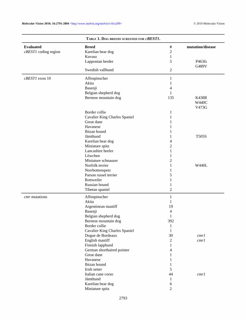
TABLE 1. DOG BREEDS SCREENED FOR CBEST1.
Evaluated Breed # mutation/diseasecBEST1 coding region Karelian bear dog 2 Kuvasz 1 Lapponian herder 5 P463fs G489V Swedish vallhund 2
cBEST1 exon 10 Affenpinscher 1 Akita 1 Basenji 4 Belgian shepherd dog 1 Bernese mountain dog 135 K438R W440C V473G Border collie 1 Cavalier King Charles Spaniel 1 Great dane 1 Havanese 1 Ibizan hound 1 Jämthund 1 T505S Karelian bear dog 4 Miniature spitz 2 Lancashire heeler 1 Löwchen 1 Miniature schnauzer 2 Norfolk terrier 1 W440L Norrbottenspetz 1 Parson russel terrier 5 Rottweiler 1 Russian hound 1 Tibetan spaniel 2
cmr mutations Affenpinscher 1 Akita 1 Argentinean mastiff 19 Basenji 4 Belgian shepherd dog 1 Bernese mountain dog 392 Border collie 1 Cavalier King Charles Spaniel 1 Dogue de Bordeaux 30 cmr1 English mastiff 2 cmr1 Finnish lapphund 1 German shorthaired pointer 4 Great dane 1 Havanese 1 Ibizan hound 1 Irish setter 5 Italian cane corso 44 cmr1 Jämthund 1 Karelian bear dog 6 Miniature spitz 2
Molecular Vision 2010; 16:2791-2804 <http://www.molvis.org/molvis/v16/a299> © 2010 Molecular Vision
2793
the co-authors (KW); this represented a total of 70 animalsbased on data covering more than 10 years. However, DNAwas available from only 54 of these dogs. Dogs included inthe genetic study were examined between 1 and 2 years of agein most cases; follow-up exams were scheduled every 1–2years depending on findings. Although cmr lesions developbefore 2 years of age, only dogs with no changes after 3 yearsof age were considered nonaffected. These stringent criteriaidentified a total of eight cmr-affected LH and 46 that wereeither normal or had other fundus changes.BEST1 sequence comparison: All coding exons and flankingsplice junctions of cBEST1 (exons 2 through 11) wereamplified individually from dogs with clinical disease similarto cmr and three obligate carrier LH dogs (Table 1), usinggene-specific primers and conditions previously published[24]. Amplification products were visualized on 1% agarosegels and purified using the QIAquick Gel Extraction Kit(Qiagen Inc., Valencia, CA). Sequences were obtained bydirect sequencing of the direct and indirect strand (ABI 3730sequencer; Applied Biosystems, Foster City, CA) at the DNASequencing Facility of the University of Pennsylvania andevaluated with the Sequencher 4.2.2 software package (GeneCodes Corporation, Ann Arbor, MI).
Additional breeds harboring molecularly undefinedretinal changes were selected based on a potentially shared
ancestral or geographic origin with LH (Table 1). From these,cBEST1 Exon 10 was amplified under the same conditions;forward primer 5′- AAG GAG GGA AAA GAT AGG GT−3′, reverse primer 5′- AGG TGG AAG GAG GGT AGA AT−3′), and sequenced in both directions (forward primer 5′-CTC ACC CAG GTG TGT GTT TG −3′, reverse primer 5′-TCA AGT CCT GCT TTG GTC CT −3′). All obtainedsequences were aligned against the canine genome sequencedraft [27] and cBEST1 reference sequence(NM_001097545). Observed sequence alterations wereanalyzed for their potential to affect protein function using theSorting Intolerant From Tolerant (SIFT) algorithm [28,29].
Mutation screening and testing:Allele-specific PCR amplification—Confirmed cmr
mutations were tested in all enrolled dogs (Table 1). Tests forcmr1 (C73T/R25X) and cmr2 (G482A/G161D) mutationsfollowed published protocols [24]; for the cmr3 sequencevariations, tests were established for amplification of the wildtype (WT) or mutation (LH)-specific allele. Two allele-specific primers for C1388del (WT: 5′-AGG CTA CCA CAGTGC CCC A-3′, LH: 5′-CAG GCT ACC ACA GTG CCCA-3′) were amplified against an anchor primer (5′-CTC ACCCAG GTG TGT GTT TG-3′), using 1.5 mM MgCl2, 0.4 µMof each primer, 0.2 µM of each dNTP, and 0.875 U Taqpolymerase (Invitrogen, Carlsbad, CA) on 50 ng of genomic
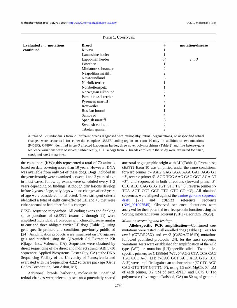
TABLE 1. CONTINUED.
Evaluated cmr mutationscontinued
Breed # mutation/disease Kuvasz 1 Lancashire heeler 1 Lapponian herder 54 cmr3 Löwchen 1 Miniature schnauzer 2 Neapolitan mastiff 2 Newfoundland 3 Norfolk terrier 1 Norrbottenspetz 1 Norwegian elkhound 2 Parson russel terrier 5 Pyrenean mastiff 7 Rottweiler 1 Russian hound 1 Samoyed 4 Spanish mastiff 6 Swedish vallhund 2 Tibetan spaniel 2
A total of 179 individuals from 25 different breeds diagnosed with retinopathy, retinal degenerations, or unspecified retinal changes were sequenced for either the complete cBEST1 coding region or exon 10 only. In addition to two mutations (P463FS, G489V) identified in cmr3 affected Lapponian herder, three novel polymorphisms (Table 2) and five heterozygote sequence variations were observed. Subsequently, all 614 dogs from 38 breeds enrolled in the study were evaluated for cmr1, cmr2, and cmr3 mutations.
Molecular Vision 2010; 16:2791-2804 <http://www.molvis.org/molvis/v16/a299> © 2010 Molecular Vision
2794
DNA in 35 cycles at 94 °C for 45 s, 69 °C for 30 s, and 72 °Cfor 30 s, after an initial denaturation at 94 °C for 1 min andfollowed by a final extension at 72 °C for 10 min. The 420-bp PCR product was separated on 6% polyacrylamide gels andscored based on the presence or absence of DNA product. TheG1466T test used the same anchor primer combined withprimers specific to the respective alleles (WT: 5′-CCT ACGCAG AGT CTC AGG G-3′, LH: 5′-CCT ACG CAG AGTCTC AGT G-3′). Amplification and genotyping of the 342-bpPCR product was obtained and evaluated under the sameconditions as cmr3, substituting the annealing temperature for68 °C (WT) and 67 °C (LH), respectively.
Restriction enzyme digest—Coding changes identifiedin the BMD were screened in all individuals of the breed byeither direct sequencing, as described above (BEST1 sequencecomparison), or restriction enzyme tests. Thus, cBEST1 exon10 was amplified and subsequently digested at 37 °Covernight with StuI, BstNI, and BbsI (New England Biolabs,Ipswich, MA) to assess the Lys438Arg, Trp440Cys, andVal473Gly substitutions, respectively. Resulting DNApatterns were evaluated on 1% agarose gels reflecting the a)Lys438Arg WT allele (A) by 304- and 521-bp bands versusthe 825-bp mutant allele (G); b) Trp440Cys WT allele (G) by36- and 185-bp bands compared to a 121-bp allele for themutant allele (T) in addition to 38-, 54-, 84-, 186-, and 242-bp bands present with both genotypes; c) Val473Gly WTallele (T) by 100- and 426-bp bands in contrast to a 526-bpband indicating the mutant allele (G) next to 107- and 192-bpproducts being present with both genotypes.
Intron 2 and 3 polymorphism screening—Twopolymorphisms in introns 2 and 3 (Table 2, #2 and #3) wereassessed in LH by restriction enzyme digest. Single nucleotidepolymorphism (SNP) 57,507,851 (C/T) was located within a346-bp PCR product (primers 5′-ACT TAT GAG GCC CAGACA AGC-3′ and 5′-TGA ATG GCT GGC TAT TTGTTC-3′) obtained from 50 ng genomic DNA with 1.5 mMMgCl2, 0.625 µM of each primer, 0.2 µM of each dNTP, and0.875 U Qiagen Taq polymerase in 35 cycles at 60 °Cannealing temperature. The PCR product was digested with20 U of Eco01091 (New England Biolabs) at 37 °C for 1 h,resulting in a diagnostic product of either 213 bp (C) or 259bp (T) in size. Similarly, SNP 57,506,423 (C/T) was includedin a 561-bp PCR product (primers 5′-GTG TGC TCC CAGTGT CTA CAT C-3′ and 5′-CAC GAC CAG AGT CAC GTAGAA G-3′), using the same conditions as described above at65 °C annealing temperature. Digestion was achieved with 20U of AatII (New England Biolabs) at 37 °C for 2 h. Thepresence of 237- and 314-bp bands distinguished the commonallele (C) from the minor allele (T) which had 259- and 302-bp products.
RESULTSRetinal phenotypes: Retinal fundus phenotypes observed inLH and other dog breeds can be related to several different
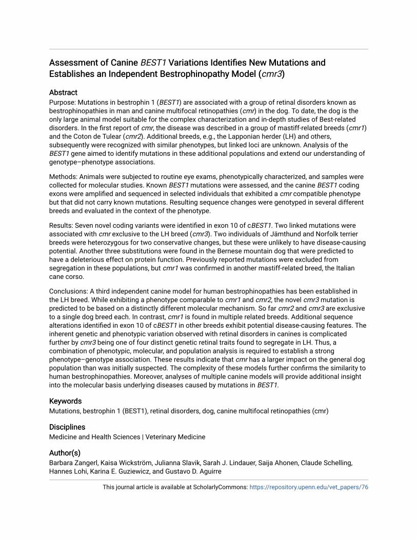
recognized disease variations. The LH breed is known tosegregate autosomal recessive prcd and a non-prcd form ofgeneralized retinal degeneration. Several additional retinalabnormalities also were observed, including acquired post-inflammatory changes, and more commonly a spectrum offundus changes referred to as multifocal retinal dysplasia(MRD [30]). This latter phenotype is characterized generallyby retinal folds and small hyporeflective areas in one or botheyes (Figure 1A,B) that are more frequent in younger animalsand become less prominent or disappear in older adults. Asubset of LH dogs examined presented with multiple elevatedsubretinal brown-gray lesions (Figure 1C,D) similar topreviously described cmr ( [24], Figure 1E). The particularhallmarks of this fundus appearance were bilateral expression,larger lesions as compared to MRD, and clear indication ofretinal elevation, often in combination with lighter areas offluid accumulation next to or around the darker center of thelesions (Figure 1D). Pedigree analysis revealed autosomalrecessive inheritance for the cmr phenotype (Figure 2) but didnot definitively determine if MRD and cmr comprise the sameor independent genetic traits. To further evaluate the geneticbasis for these phenotypes, BEST1 was considered as acandidate gene for retinal disease in LH and isolated casesfrom three additional breeds, Karelian beardog, Kuvasz, andSwedish vallhund, diagnosed with MRD.
BEST1 mutations: A total of 614 animals from 38 individualbreeds (Table 1), including LH, were selected based on (a)results of the above described phenotype evaluation andsubsequent outcomes of presented screens (BMD); (b) knowninvolvement with cmr1 but originating from Europeanpopulations not previously screened (Dogue de Bordeaux andEnglish mastiff); (c) known mastiff origin (e.g., Italian canecorso and Neapolitan mastiff); (d) control breeds (e.g.,Norwegian elkhound and Samoyed).
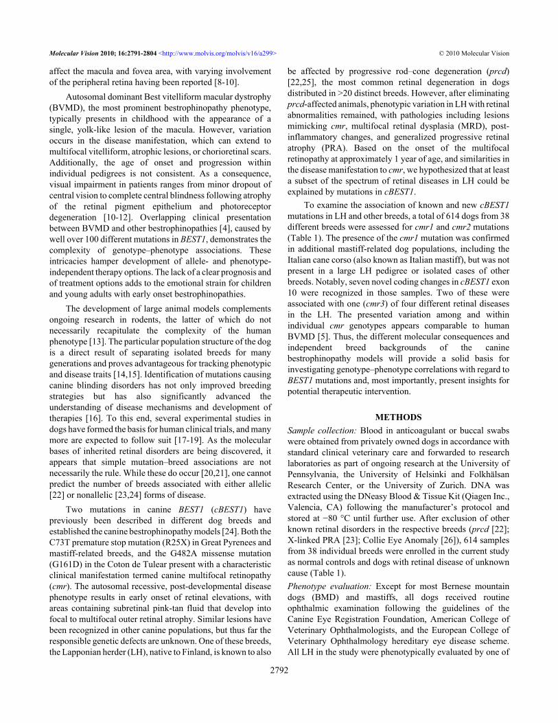
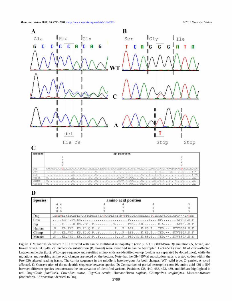
Initially, affected animals from selected breeds withtypical cmr changes and three LH that were obligate cmrcarriers (Table 1) had the complete cBEST1 coding region andcorresponding exon–intron boundaries sequenced. Severalknown and novel (NCBI ss250608388-ss250608389,ss250608394-ss250608401) polymorphisms were identifiedin these samples (Table 2). Of these, two coding changes thatdiffer from the WT cBEST1 sequence were foundhomozygous in affected LH; a deletion at nucleotide position1,388 of the open reading frame (Figure 3A; NCBIss250608397), and a substitution at nucleotide position 1,466(Figure 3B; NCBI ss250608399). The C1388del results in aframe shift (Pro463fs) introducing a new stop codon at aminoacid 490. The G1466T substitution by itself leads to aconservative change in the amino acid sequence (Gly489Val),which is predicted to change the protein function with onlymarginal significance (SIFT p=0.05; Table 3). In combinationwith the C1388del, however, the G1466T substitutions resultsin an additional stop codon at amino acid position 489 within
Molecular Vision 2010; 16:2791-2804 <http://www.molvis.org/molvis/v16/a299> © 2010 Molecular Vision
2795
the shifted reading frame (Gly489X). Since the mutationshave only been found in complete linkage disequilibrium, weconclude that the combination of changes results in the diseasewe now refer to as cmr3. Notably, both positions appear highlyconserved in the BEST1 gene of different species at thenucleotide (Figure 3C) and amino acid level (Figure 3D).
The three sequenced obligate LH carrier animals werehomozygous for the complete cBEST1 coding region, with theexception of the above-mentioned mutations and twopolymorphisms in introns 2 and 3 (Table 2, #2 and #3; SNP57,507,851 [BICF2P299043] [27]; SNP 57,506,423 [24]).Although both mutations in exon 10 were found to be linkedto each other and the cmr phenotype, the intronicpolymorphisms were dissociated with the mutations ordisease status (data not shown) and therefore cannot predictdisease status in the breed.
Focusing on cBEST1 exon 10, sequence of this exon wasobtained from samples of an additional 21 breeds reported tosegregate retinopathies and/or unspecified retinal changes(Table 1). None of the investigated animals carried the cmr3mutation alleles, but five additional sequence alterations wereidentified (Table 2 and Table 3).These were analyzed for theirpotential deleterious effects using SIFT. Two conservative
changes in one individual each of the Norfolk terrier andJämthund breeds were not predicted to impact proteinfunction (p=0.67 and 0.75). Similarly, a Lys438Argsubstitution identified in two heterozygous BMD does notaffect a critical position in the bestrophin protein (p=0.44)even though this amino acid is highly conserved betweenspecies (Figure 3D). The remaining two mutations,Trp440Cys and Val473Gly, have high potential to altermolecular properties (p=0.02 in each case). Both were foundin the heterozygous state in BMD at an allele frequency of lessthan 1% (0.5 and 0.1%, respectively, based on 392 individualdogs), and no homozygous-affected animal has beenidentified thus far.
Subsequent genotyping of the cmr1, cmr2, and cmr3mutations confirmed segregation of cmr1 in three mastiff-related breeds from Italian kennels: the Dogue de Bordeaux,English mastiff, and Italian cane corso (Table 3).Genotype–phenotype correlation: The two novel cmr3mutations found exclusively in the LH breed were in completelinkage equilibrium and homozygous in all animals diagnosedwith the cmr phenotype (Table 4). Pedigree analysis furthersupported segregation of the mutations with the cmrphenotype (Figure 2). None of the individuals with either post-
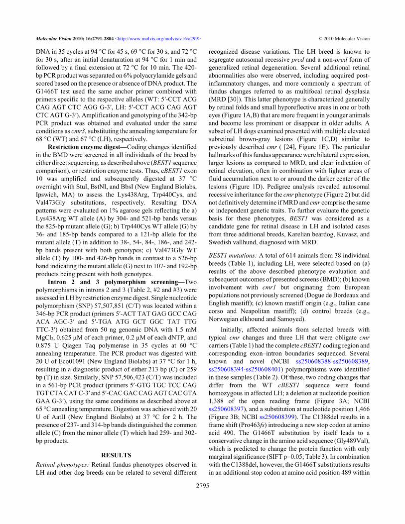
TABLE 2. OBSERVED GENOMIC VARIATION IN THE CBEST1 GENE.
Number CFA18 bp Geneposition
Variation Consequence Observed in Reference
1 57507965 Exon 2 G>A silent Karelian beardog (KBD) [24]2 58507851 Intron 2 C>T non-coding Lapponian herder BICF2P29904323 57506423 Intron 3 C>T non-coding three or more breeds [24]4 57506082 Exon 4 C>A silent three or more breeds [24]5 57505871 Intron 4 G>A non-coding Swedish vallhund (SV) [24]6 57505579 Intron 4 A>G non-coding Kuvasz [24]7 57505571 Intron 4 A>G non-coding KBD, SV BICF2G63068928128 57505345 Intron 5 C>T non-coding Kuvasz [24]9 57505321 Intron 5 T>C non-coding Kuvasz [24]10 57505315 Intron 5 C>A non-coding Kuvasz [24]11 57504881 Intron 5 C>G non-coding Swedish vallhund [24]12 57504859 Intron 5 A>G non-coding KBD, SV NCBI ss25060838813 57504620 Intron 6 G>A non-coding three or more breeds BICF2G630689283214 57504059 Intron 6 T>A non-coding Swedish vallhund BICF2G630689286215 57503846 Exon 7 G>A silent three or more breeds BICF2G630689287216 57503789 Intron 7 A>G non-coding three or more breeds BICF2G630689288217 57502488 Intron 7 A>G non-coding Swedish vallhund BICFPJ1456205118 57502387 Exon 8 T>C silent Karelian beardog NCBI ss25060838919 57502384 Exon 8 T>C silent KBD, SV BICFPJ1456206120 57500034 Exon 10 A>G Lys438Arg Bernese mountain dog NCBI ss25060839421 57500028 Exon 10 G>T Trp440Leu Norfolk terrier NCBI ss25060839522 57500027 Exon 10 G>T Trp440Cys Bernese mountain dog NCBI ss25060839623 57499959 Exon 10 C>del Pro463fs Lapponian herder NCBI ss25060839724 57499929 Exon 10 T>G Val473Gly Bernese mountain dog NCBI ss25060839825 57499881 Exon 10 G>T Gly489Val Lapponian herder NCBI ss25060839926 57499834 Exon 10 A>T Thr505Ser Jämthund NCBI ss25060840027 57449754 Exon 10 C>T silent three or more breeds NCBI ss25060840128 57498425 3′UTR A>T non-coding Swedish vallhund [24]
The majority of polymorphisms identified had previously been reported. Three new polymorphisms (row #12, 18, and 27), however, were observed in addition to seven coding changes in exon 10 (row #20–26); two of these segregate with retinal disease in the Lapponian herder breed (row #23 and 25, bold, cmr3). *CFA18=Canis familiaris chromosome 18. 1Based on CanFam1; Based on Can Fam2.
Molecular Vision 2010; 16:2791-2804 <http://www.molvis.org/molvis/v16/a299> © 2010 Molecular Vision
2796
2
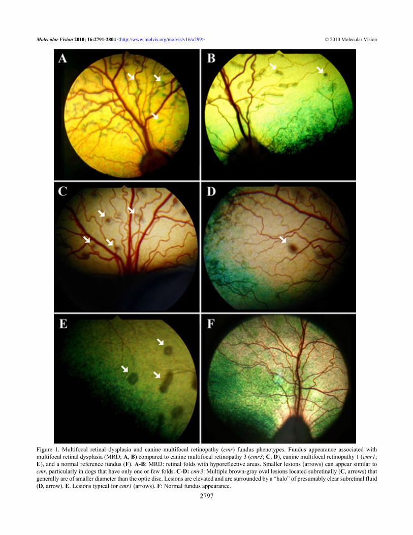
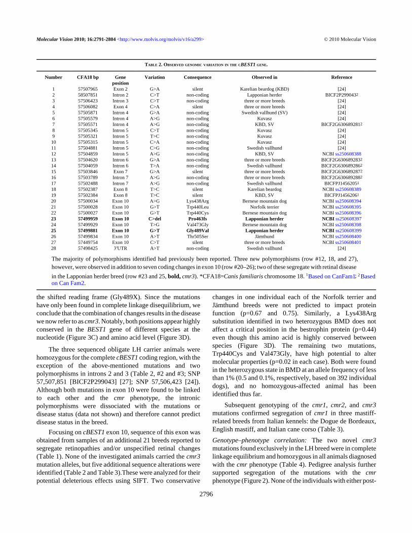
Figure 1. Multifocal retinal dysplasia and canine multifocal retinopathy (cmr) fundus phenotypes. Fundus appearance associated withmultifocal retinal dysplasia (MRD; A, B) compared to canine multifocal retinopathy 3 (cmr3; C, D), canine multifocal retinopathy 1 (cmr1;E), and a normal reference fundus (F). A-B: MRD: retinal folds with hyporeflective areas. Smaller lesions (arrows) can appear similar tocmr, particularly in dogs that have only one or few folds. C-D: cmr3: Multiple brown-gray oval lesions located subretinally (C, arrows) thatgenerally are of smaller diameter than the optic disc. Lesions are elevated and are surrounded by a “halo” of presumably clear subretinal fluid(D, arrow). E. Lesions typical for cmr1 (arrows). F: Normal fundus appearance.
Molecular Vision 2010; 16:2791-2804 <http://www.molvis.org/molvis/v16/a299> © 2010 Molecular Vision
2797
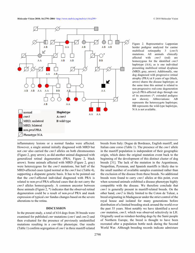
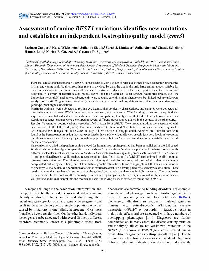
inflammatory lesions or a normal fundus were affected.However, a single animal initially diagnosed with MRD butnot cmr also carried the cmr3 alleles on both chromosomes(Figure 2, gray arrow), as did another animal diagnosed withgeneralized retinal degeneration (PRA; Figure 2, blackarrow). Some animals affected with MRD (Figure 2, gray)were heterozygous for the cmr3 mutations, but half of theMRD-affected cases typed normal at the cmr3 loci (Table 4),supporting a disparate genetic basis. It has to be pointed outthat the cmr3-affected individual diagnosed with PRA isrelated to non-prcd PRA-affected cases that do not carry thecmr3 alleles homozygously. A common ancestor betweenthese animals (Figure 2, *) indicates that the observed retinaldegeneration could be a result of non-prcd PRA and maskexpression of typical cmr fundus changes based on the severealterations to the retina.
DISCUSSIONIn the present study, a total of 614 dogs from 38 breeds wereexamined for published cmr mutations (cmr1 and cmr2) andthen evaluated for the presence of previously unidentifiedmutations resulting in a cmr-like phenotype. Our results(Table 1) confirm segregation of cmr1 in three mastiff-related
breeds from Italy: Dogue de Bordeaux, English mastiff, andItalian cane corso (Table 1). The presence of the cmr1 allelein the mastiff population is independent of their geographicorigin, which dates the original mutation event back to thebeginning of the development of this distinct cluster of dogbreeds [31]. The lack of the mutation in the Argentinean,Neapolitan, Pyrenean, and Spanish mastiffs is likely due tothe small number of available samples examined rather thanthe exclusion of the disease from these breeds. No additionalbreeds were found to carry cmr1 alleles at this point, evenwhen screened animals exhibited a disease phenotype highlycompatible with the disease. We therefore conclude thatcmr1 is generally present in mastiff-related breeds. On theother hand, cmr2 is likely limited to the Coton de Tulear, abreed originating in Madagascar under the strict control of theroyal house and isolated for many generations beforedistribution of a limited breeding stock around the world overthe past 35 years. Most notably we have identified a novelcmr mutation, cmr3, which was observed selectively in LH.Originally used as reindeer-herding dogs by the Sami peopleof Northern Europe, the breed is thought to have beenrecreated after a population bottle neck during the SecondWorld War. Although breeding records indicate admixture
Figure 2. Representative Lapponianherder pedigree analyzed for caninemultifocal retinopathy 3 (cmr3)mutations. All animals clinicallyaffected with cmr3 (red) arehomozygous for the identified cmr3haplotype (AA), as is one individualpresenting multifocal retinal dysplasia(MRD; gray, arrow). Additionally, onedog diagnosed with progressive retinalatrophy (PRA) at 8 years of age (black,arrow) shares the disease haplotype; atthe same time this animal is related tonon-progressive rod-cone degeneration(prcd) PRA-affected dogs through oneof its ancestors (*; extended pedigreenot shown). Abbreviations: ABrepresents the heterozygote haplotype;BB represents the wild-type haplotype,N/A is not available.
Molecular Vision 2010; 16:2791-2804 <http://www.molvis.org/molvis/v16/a299> © 2010 Molecular Vision
2798
Molecular Vision 2010; 16:2791-2804 <http://www.molvis.org/molvis/v16/a299> © 2010 Molecular Vision
2799
Figure 3. Mutations identified in LH affected with canine multifocal retinopathy 3 (cmr3). A C1388del/Pro463fs mutation (A, boxed) andlinked G1466T/Gly489Val nucleotide substitution (B, boxed) were identified in canine bestrophin 1 (cBEST1) exon 10 of cmr3-affectedLapponian herder (LH). Wild-type sequence and resulting amino acids are identified on top (codons are separated by dotted lines), while themutations and resulting amino acid changes are noted on the bottom. Note that the Gly489Val substitution leads to a stop codon within thePro463fs altered reading frame. The carrier sequence in the middle is heterozygous for both changes. WT=wild type, C=carrier, A=cmr3affected. C: Conservation of the nucleotide sequence between species. D: Comparison of partial bestrophin exon 10, amino acid 436 to 507between different species demonstrates the conservation of identified variants. Positions 438, 440, 463, 473, 489, and 505 are highlighted inred. Dog=Canis familiaris, Cow=Bos taurus, Pig=Sus scrofa, Human=Homo sapiens, Chimp=Pan troglodytes, Macaca=Macacafascicularis. “.”=position identical to Dog.
with other local breeds, including the Karelian beardog, themutation may either have occurred or been preserved only inthose lines leading to the modern dog breed now constitutingthe LH.
Even though cmr and MRD are distinct and separableretinal disorders in dogs, they share enough clinicalsimilarities that could make specific diagnosis difficult. Tothis end, individuals from four breeds (Table 1), diagnosedwith either MRD (Karelian beardog, Kuvasz, Swedishvallhund) or cmr-compatible fundus changes (LH), werescreened for mutations in the cBEST1 coding region. Twonovel sequence alterations (C1388del/Pro463fs, G1466T/Gly489Val) in exon 10 segregated only with the LH cmr3(Figure 2). The C1388del microdeletion (Figure 3A) initiatesa Pro463fs frame shift that results in a stop codon at aminoacid 490. To date, the G1466T/Gly489Val substitution has notbeen observed without the upstream C1388del microdeletion,
and the resulting inherent pathogenic effect has therefore notyet been established. Within the context of the upstreammutated reading frame, the G1466T mutation coded for anadditional Gly489X stop codon (Figure 3B). These findingsstrongly indicate that the identified mutations shorten the 585-amino acid bestrophin to a 488-amino acid protein with analtered C-terminus.
The amino acid position and translational consequence ofthe cmr3 mutations are distinctly different from the cmr1 stopmutation (no protein) and the cmr2 missense mutation(mislocalization; Guziewicz et al., submitted), all resulting inhighly similar phenotypes (Figure 1). The cmr3 mutationslikely will not affect bestrophin targeting to the basolateralmembrane of the retinal pigment epithelium, but proteinfunction might be impacted by the loss of interaction betweenthe bestrophin C and N-terminus [32]. The bestrophin C-terminus has also been implicated in the activation and
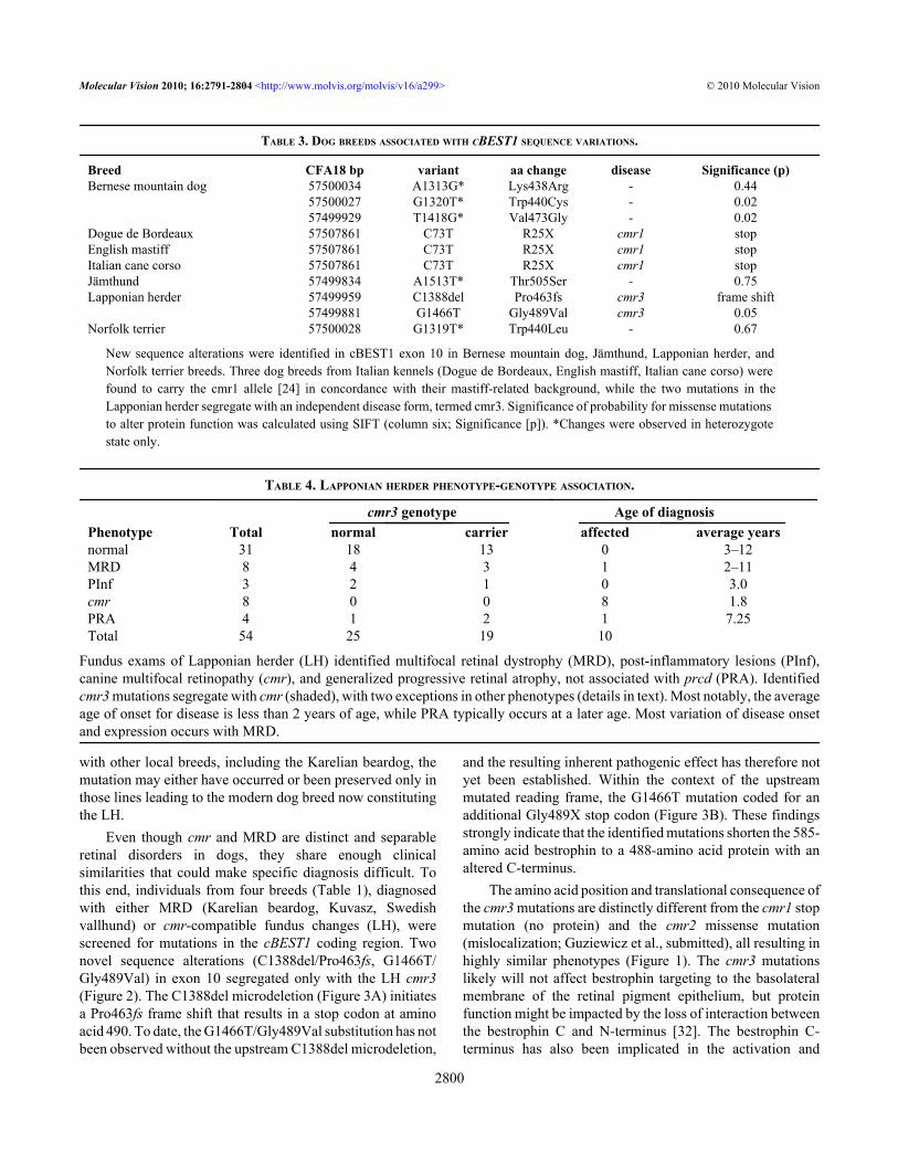
TABLE 3. DOG BREEDS ASSOCIATED WITH CBEST1 SEQUENCE VARIATIONS.
Breed CFA18 bp variant aa change disease Significance (p)Bernese mountain dog 57500034 A1313G* Lys438Arg - 0.44 57500027 G1320T* Trp440Cys - 0.02 57499929 T1418G* Val473Gly - 0.02Dogue de Bordeaux 57507861 C73T R25X cmr1 stopEnglish mastiff 57507861 C73T R25X cmr1 stopItalian cane corso 57507861 C73T R25X cmr1 stopJämthund 57499834 A1513T* Thr505Ser - 0.75Lapponian herder 57499959 C1388del Pro463fs cmr3 frame shift 57499881 G1466T Gly489Val cmr3 0.05Norfolk terrier 57500028 G1319T* Trp440Leu - 0.67
New sequence alterations were identified in cBEST1 exon 10 in Bernese mountain dog, Jämthund, Lapponian herder, and Norfolk terrier breeds. Three dog breeds from Italian kennels (Dogue de Bordeaux, English mastiff, Italian cane corso) were found to carry the cmr1 allele [24] in concordance with their mastiff-related background, while the two mutations in the Lapponian herder segregate with an independent disease form, termed cmr3. Significance of probability for missense mutations to alter protein function was calculated using SIFT (column six; Significance [p]). *Changes were observed in heterozygote state only.
TABLE 4. LAPPONIAN HERDER PHENOTYPE-GENOTYPE ASSOCIATION.
cmr3 genotype Age of diagnosisPhenotype Total normal carrier affected average yearsnormal 31 18 13 0 3–12MRD 8 4 3 1 2–11PInf 3 2 1 0 3.0cmr 8 0 0 8 1.8PRA 4 1 2 1 7.25Total 54 25 19 10
Fundus exams of Lapponian herder (LH) identified multifocal retinal dystrophy (MRD), post-inflammatory lesions (PInf),canine multifocal retinopathy (cmr), and generalized progressive retinal atrophy, not associated with prcd (PRA). Identifiedcmr3 mutations segregate with cmr (shaded), with two exceptions in other phenotypes (details in text). Most notably, the averageage of onset for disease is less than 2 years of age, while PRA typically occurs at a later age. Most variation of disease onsetand expression occurs with MRD.
Molecular Vision 2010; 16:2791-2804 <http://www.molvis.org/molvis/v16/a299> © 2010 Molecular Vision
2800
regulation of the channel function [33,34], even though mostpredicted domains are located upstream of the cmr3mutations. Additionally, participation in channelmultimerization [35] and direct involvement of the C-terminus in the activation or regulation by Ca2+ and otherphysiologic processes has been proposed [36]. However,these predictions are mainly based on hypothesizedlocalization of phosphorylation sites or binding sites forkinases, such as protein kinase A or extracellular signal-regulated kinases (Erk) [37]. Since few human BEST1mutations are located within the C-terminal part of bestrophin[7], the cmr3 model provides initial clinical evidence for afunctional role of the BEST1 C-terminus. Thus, theidentification of a third nonallelic form of cmr will furthercontribute to the investigation of BEST1 disease mechanismsrelevant to canines and humans.
BEST1 exon 10 was sequenced in a larger number ofbreeds (Table 1) and presented an additional five newsequence alterations (Table 3). Two of these, Thr505Ser andTrp440Leu, were found in the heterozygous state in a singleJämthund and Norfolk terrier, respectively. Clinical reportsfor these animals did not indicate any retinal changes, and noother individuals from the breeds were available forgenotyping. More importantly, both sequence changes areconservative and are not expected to impact protein function[28,29]. Thus, they are not considered to be associated withretinal disease and were not investigated further. Surprisingly,three independent substitutions were present in the BMDbreed. Two of these (Trp440Cys and Val473Gly) are likelydeleterious (Table 3) and located in a region that is con-served between species (Figure 3D), but neither was foundhomozygous in an animal with retinal disease. Because of thelow allele frequency (< 1%) of these two potential mutations,it is likely that very few if any affected animals will occur inthe breed. Hence, the contribution of these mutations todisease risk cannot be evaluated without further elucidatingthe functional properties of the BEST1 C-terminus.
Pedigree analysis of cmr3 supports autosomal recessiveinheritance with the predominant clinical appearance similarto cmr (Table 4, Figure 2). The partial overlap in phenotypewith prcd [22], MRD, and non-prcd PRA, however,complicates the characterization of the potential phenotypicvariation for each individual trait. Above all, some similaritiesin the appearance of the fundus lesions in MRD and cmr(Figure 1) and lack of a clear definition of the MRD phenotype[30] can raise questions with cases that develop either atypicalor only few lesions. In strict contrast to MRD, the cmrphenotype always presents bilaterally. Nevertheless, severalcmr3 heterozygote animals exhibit a bilateral MRDphenotype, as does one animals homozygous for the mutations(Figure 2, gray arrow). The latter was diagnosed at 7 years ofage with minor changes in both eyes consistent with MRD.Earlier exams list no retinal abnormalities at 1.5 years of age,and multiple “rosettes” in the right eye at 5 years of age.
Another cmr3-affected animal (Figure 2, black arrow) wasdiagnosed with generalized retinal degeneration during thefirst available fundus exam at 8 years of age. Three additionalanimals with non-prcd PRA shared common ancestry withthis individual; two of these were carriers and one was normalfor the cmr3 mutations. Thus, the observed phenotype couldbe the consequence of non-prcd PRA segregating in the breed.Without early clinical data, we cannot determine whethercmr-related retinal changes were present at an earlier timepoint.
Longitudinal studies of several cmr3-affected animalssuggest that fluctuations in the appearance and presence ofmultifocal lesions can occur. This includes the age of onset(between 9 months and 2 years) as well as extent andprogression of lesions; in fact some of the individual cmrlesions can disappear over time. The age of onset wasobserved as early as 11 weeks for cmr1 [38,39] and 15 weeksfor cmr2 [40], with little or no progression of the lesionsbeyond 1 year of age. However, diminishedelectroretinograms suggest that Cotons affected with cmr2develop retinal degeneration that can be more extensive thanthe focal lesion [40]. This is caused by the resolution of thedetachment resulting in hypertrophic- and hyperplastic-pigmented retinal epithelium and focal outer retinal thinning.In some older animals with extensive lesions, confluence ofthese areas can lead to a generalized retinal degeneration. Thedegenerative process occurs earlier in Great Pyrenees (cmr1)and is more likely to result in focal rather than generalizedretinal degeneration [38,39]. Additionally, the lower lifeexpectancy of this breed could prevent observing advanceddisease stages. However, at least one cmr1 case with resultinggeneralized retinal degeneration has been observed clinicallyand confirmed histopathologically (Bruce Grahn, DVM,Professor of Veterinary Ophthalmology and Associate Dean,Western College of Veterinary Medicine, University ofSaskatchewan, Saskatoon, Canada). Association of BEST1missense mutations with the human PRA equivalent RetinitisPigmentosa has recently been established [41]. Although thereis no current evidence that cmr causes PRA in the investigateddog models, we suggest that generalized retinal degenerationis a potential rare outcome of the disorder and can mimic aPRA phenotype in older animals.
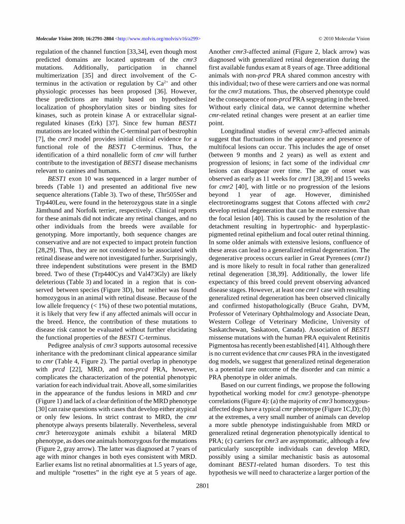
Based on our current findings, we propose the followinghypothetical working model for cmr3 genotype–phenotypecorrelations (Figure 4): (a) the majority of cmr3 homozygous-affected dogs have a typical cmr phenotype (Figure 1C,D); (b)at the extremes, a very small number of animals can developa more subtle phenotype indistinguishable from MRD orgeneralized retinal degeneration phenotypically identical toPRA; (c) carriers for cmr3 are asymptomatic, although a fewparticularly susceptible individuals can develop MRD,possibly using a similar mechanistic basis as autosomaldominant BEST1-related human disorders. To test thishypothesis we will need to characterize a larger portion of the
Molecular Vision 2010; 16:2791-2804 <http://www.molvis.org/molvis/v16/a299> © 2010 Molecular Vision
2801
population at risk in long-term molecular and clinical studies.Indeed, the establishment of an accurate classification systemwill aid in the development of categorized information fromdiagnoses of less defined retinal changes or MRD [30] andwould be further simplified by the identification of other lociresponsible for MRD or PRA. For this it seems critical toclinically examine dogs at risk between 1 and 2 years of ageand provide yearly follow-up data to assess phenotypicvariation. Along with the examination, we recommend thatphotographic documentation be performed using acombination of wide-field and higher resolution images ofboth eyes; the exact methods have previously been published[42]. Detailed descriptions for canine retinal disorders wouldenhance our ability to correctly diagnose inherited andacquired diseases, to separate multiple genetic traits inindividual breeds, and ultimately to scrutinize cmr genotype–phenotype associations.
In summary, the currently identified mutations incBEST1 indicate a significant contribution of these variationsto retinal disease phenotypes, comparable to the humanortholog. Continued efforts to follow and characterize cmrmutations will certainly lay the foundation for understandingphenotype variation and modification of bestrophinopathiesas well as promoting the development of new therapies.
ACKNOWLEDGMENTSThe authors thank Amari T. Simpson, a student in thePhysician Scientist Training Program supported in part byJohnson & Johnson, for technical support and illuminating
discussions. Particular gratitude goes to Guilia Pertica, DVM,and Maria Longeri, DVM, PhD of the University of Milan,Milan, Italy, Kate Earle, and Sue Pearce-Kelling of OptiGen,LLC., and Sandy Novocin for sample submissions, as well asLydia Melnyk for organizational and editorial help. Researchwas supported by The Foundation Fighting Blindness, NEI/NIH individual grants R01EY06855 and R01EY17549, NEI/NIH core grant P30EY001583, ONCE International Prize, theVan Slound Fund for Canine Genetic Research, Hope forVision, the Bernese Mountain Dog Club of America, Inc., thePotomac Valley Bernese Mountain Dog Club, the Academyof Finland, the Jane and Aatos Erkko Foundation, TheUniversity of Helsinki Research Funds, and the Sigrid JuseliusFoundation.
REFERENCES1. Webster AR, Héon E, Lotery AJ, Vandenburgh K, Casavant TL,
Oh KT, Beck G, Fishman GA, Lam BL, Levin A,Heckenlively JR, Jacobson SG, Weleber RG, Sheffield VC,Stone EM. An analysis of allelic variation in the ABCA4gene. Invest Ophthalmol Vis Sci 2001; 42:1179-89. [PMID:11328725]
2. Molday RS, Zhong M, Quazi F. The role of the photoreceptorABC transporter ABCA4 in lipid transport and Stargardtmacular degeneration. Biochim Biophys Acta 2009;1791:573-83. [PMID: 19230850]
3. Kitiratschky VB, Grau T, Bernd A, Zrenner E, Jägle H, RennerAB, Kellner U, Rudolph G, Jacobson SG, Cideciyan AV,Schaich S, Kohl S, Wissinger B. ABCA4 gene analysis inpatients with autosomal recessive cone and cone rod
Figure 4. A proposed working hypothesis for canine multifocal retinopathy (cmr) genotype–phenotype correlation. cmr-affected animalstypically develop multifocal lesions, but generalized retinal degeneration (RetDegen) may occasionally be seen in a small number of olderdogs as a consequence of the disease. In short, only one animal affected with canine multifocal retinopathy 3 (cmr3) was observed in thecurrent study with retinal degeneration; we cannot exclude that this case may have resulted from a mutation at a non-cmr3 locus becauserelated dogs, normal or carrier for the cmr3 mutations, were also diagnosed with RetDegen. However, similar consequences have been observedfor cmr1 and cmr2 (see discussion for details). Few or no animals heterozygous for cmr3 are expected to exhibit clinical disease, while itcannot be excluded at this point that more susceptible individuals may present a mild disease equivalent to multifocal retinal dysplasia (MRD).The later phenotype could also be observed in some homozygous-affected dogs, although it is not clear whether this is a consequence ofincorrectly interpreted fundus appearance or truly signifies the potential of the phenotypic spectrum related to cmr. Follow-up long-termstudies will further elucidate the validity of the working model.
Molecular Vision 2010; 16:2791-2804 <http://www.molvis.org/molvis/v16/a299> © 2010 Molecular Vision
2802
dystrophies. Eur J Hum Genet 2008; 16:812-9. [PMID:18285826]
4. Xiao Q, Hartzell HC, Yu K. Bestrophins and retinopathies.Pflugers Arch 2010; 460:559-69. [PMID: 18285826]
5. Schatz P, Klar J, Andreasson S, Ponjavic V, Dahl N. VariantPhenotype of Best Vitelliform Macular Dystrophy Associatedwith Compound Heterozygous Mutations in VMD2.Ophthalmic Genet 2006; 27:51-6. [PMID: 16754206]
6. Seddon JM, Sharma S, Chong S, Hutchinson A, Allikmets R,Adelman RA. Phenotype and genotype correlations in twoBest families. Ophthalmology 2003; 110:1724-31. [PMID:13129869]
7. Burgess R, Millar ID, Leroy BP, Urquhart JE, Fearon IM, DeBaere E, Brown PD, Robson AG, Wright GA, Kestelyn P,Holder GE, Webster AR, Manson FD, Black GC. Biallelicmutation of BEST1 causes a distinct retinopathy in humans.Am J Hum Genet 2008; 82:19-31. [PMID: 18179881]
8. Boon CJ, Klevering BJ, den Hollander AI, Zonneveld MN,Theelen T, Cremers FP, Hoyng CB. Clinical and geneticheterogeneity in multifocal vitelliform dystrophy. ArchOphthalmol 2007; 125:1100-6. [PMID: 17698758]
9. Renner AB, Tillack H, Kraus H, Krämer F, Mohr N, Weber BH,Foerster MH, Kellner U. Late onset is common in bestmacular dystrophy associated with VMD2 gene mutations.Ophthalmology 2005; 112:586-92. [PMID: 15808248]
10. Mullins RF, Oh KT, Heffron E, Hageman GS, Stone EM. Latedevelopment of vitelliform lesions and flecks in a patient withbest disease: clinicopathologic correlation. Arch Ophthalmol2005; 123:1588-94. [PMID: 16286623]
11. Lorenz B, Preising MN. Best's disease. Overview of pathologyand its causes. Ophthalmologe 2005; 102:111-5. [PMID:15657691]
12. Maruko I, Iida T, Spaide RF, Kishi S. Indocyanine greenangiography abnormality of the periphery in vitelliformmacular dystrophy. Am J Ophthalmol 2006; 141:976-8.[PMID: 16678528]
13. Zhang Y, Stanton JB, Wu J, Yu K, Hartzell HC, Peachey NS,Marmorstein LY, Marmorstein AD. Suppression of Ca2+signaling in a mouse model of Best disease. Hum Mol Genet2010; 19:1108-18. [PMID: 20053664]
14. Parker HG, Kim LV, Sutter NB, Carlson S, Lorentzen TD,Malek TB, Johnson GS, DeFrance HB, Ostrander EA,Kruglyak L. Genetic structure of the purebred domestic dog.Science 2004; 304:1160-4. [PMID: 15155949]
15. Sutter NB, Ostrander EA. Dog star rising: the canine geneticsystem. Nat Rev Genet 2004; 5:900-10. [PMID: 15573122]
16. Aguirre GD, Acland GM. Models, Mutants and Man: Searchingfor Unique Phenotypes and Genes in the Dog Model ofInherited Retinal Degeneration. In: Ostrander EA, Giger U,Lindblad-Toh K, editors. The Dog and its Genome. ColdSpring Harbor, NY: Cold Spring Harbor Laboratory Press;2006. p. 291–325.
17. Acland GM, Aguirre GD, Ray J, Zhang Q, Aleman TS,Cideciyan AV, Pearce-Kelling SE, Anand V, Zeng Y,Maguire AM, Jacobson SG, Hauswirth WW, Bennett J. Genetherapy restores vision in a canine model of childhoodblindness. Nat Genet 2001; 28:92-5. [PMID: 11326284]
18. Buch PK, Bainbridge JW, Ali RR. AAV-mediated gene therapyfor retinal disorders: from mouse to man. Gene Ther 2008;15:849-57. [PMID: 18418417]
19. Komáromy AM, Alexander JJ, Rowlan JS, Garcia MM, ChiodoVA, Kaya A, Tanaka JC, Acland GM, Hauswirth WW,Aguirre GD. Gene therapy rescues cone function incongenital achromatopsia. Hum Mol Genet 2010;19:2581-93. [PMID: 20378608]
20. Kukekova AV, Goldstein O, Johnson JL, Richardson MA,Pearce-Kelling SE, Swaroop A, Friedman JS, Aguirre GD,Acland GM. Canine RD3 mutation establishes rod-conedysplasia type 2 (rcd2) as ortholog of human and murine rd3.Mamm Genome 2009; 20:109-23. [PMID: 19130129]
21. Sidjanin DJ, Lowe JK, McElwee JL, Milne BS, Phippen TM,Sargan DR, Aguirre GD, Acland GM, Ostrander EA. CanineCNGB3 mutations establish cone degeneration asorthologous to the human achromatopsia locus ACHM3.Hum Mol Genet 2002; 11:1823-33. [PMID: 12140185]
22. Zangerl B, Goldstein O, Philp AR, Lindauer SJ, Pearce-KellingSE, Mullins RF, Graphodatsky AS, Ripoll D, Felix JS, StoneEM, Acland GM, Aguirre GD. Identical mutation in a novelretinal gene causes progressive rod-cone degeneration in dogsand retinitis pigmentosa in humans. Genomics 2006;88:551-63. [PMID: 16938425]
23. Zhang Q, Acland GM, Wu WX, Johnson JL, Pearce-Kelling S,Tulloch B, Vervoort R, Wright AF, Aguirre GD. DifferentRPGR exon ORF15 mutations in Canids provide insights intophotoreceptor cell degeneration. Hum Mol Genet 2002;11:993-1003. [PMID: 11978759]
24. Guziewicz KE, Zangerl B, Lindauer SJ, Mullins RF, SandmeyerLS, Grahn BH, Stone EM, Acland GM, Aguirre GD.Bestrophin gene mutations cause canine multifocalretinopathy: a novel animal model for best disease. InvestOphthalmol Vis Sci 2007; 48:1959-67. [PMID: 17460247]
25. Goldstein O, Zangerl B, Pearce-Kelling S, Sidjanin DJ, KijasJW, Felix J, Acland GM, Aguirre GD. Linkage disequilibriummapping in domestic dog breeds narrows the progressive rod-cone degeneration interval and identifies ancestral disease-transmitting chromosome. Genomics 2006; 88:541-50.[PMID: 16859891]
26. Lowe JK, Kukekova AV, Kirkness EF, Langlois MC, AguirreGD, Acland GM, Ostrander EA. Linkage mapping of theprimary disease locus for collie eye anomaly. Genomics 2003;82:86-95. [PMID: 12809679]
27. Lindblad-Toh K, Wade CM, Mikkelsen TS, Karlsson EK, JaffeDB, Kamal M, Clamp M, Chang JL, Kulbokas EJ 3rd, ZodyMC, Mauceli E, Xie X, Breen M, Wayne RK, Ostrander EA,Ponting CP, Galibert F, Smith DR, DeJong PJ, Kirkness E,Alvarez P, Biagi T, Brockman W, Butler J, Chin CW, CookA, Cuff J, Daly MJ, DeCaprio D, Gnerre S, Grabherr M, KellisM, Kleber M, Bardeleben C, Goodstadt L, Heger A, Hitte C,Kim L, Koepfli KP, Parker HG, Pollinger JP, Searle SM,Sutter NB, Thomas R, Webber C, Baldwin J, Abebe A,Abouelleil A, Aftuck L, Ait-Zahra M, Aldredge T, Allen N,An P, Anderson S, Antoine C, Arachchi H, Aslam A, AyotteL, Bachantsang P, Barry A, Bayul T, Benamara M, Berlin A,Bessette D, Blitshteyn B, Bloom T, Blye J, Boguslavskiy L,Bonnet C, Boukhgalter B, Brown A, Cahill P, Calixte N,Camarata J, Cheshatsang Y, Chu J, Citroen M, Collymore A,Cooke P, Dawoe T, Daza R, Decktor K, DeGray S, DhargayN, Dooley K, Dooley K, Dorje P, Dorjee K, Dorris L, DuffeyN, Dupes A, Egbiremolen O, Elong R, Falk J, Farina A, FaroS, Ferguson D, Ferreira P, Fisher S, FitzGerald M, Foley K,
Molecular Vision 2010; 16:2791-2804 <http://www.molvis.org/molvis/v16/a299> © 2010 Molecular Vision
2803
Foley C, Franke A, Friedrich D, Gage D, Garber M, GearinG, Giannoukos G, Goode T, Goyette A, Graham J, GrandboisE, Gyaltsen K, Hafez N, Hagopian D, Hagos B, Hall J, HealyC, Hegarty R, Honan T, Horn A, Houde N, Hughes L,Hunnicutt L, Husby M, Jester B, Jones C, Kamat A, KangaB, Kells C, Khazanovich D, Kieu AC, Kisner P, Kumar M,Lance K, Landers T, Lara M, Lee W, Leger JP, Lennon N,Leuper L, LeVine S, Liu J, Liu X, Lokyitsang Y, LokyitsangT, Lui A, Macdonald J, Major J, Marabella R, Maru K,Matthews C, McDonough S, Mehta T, Meldrim J, MelnikovA, Meneus L, Mihalev A, Mihova T, Miller K, Mittelman R,Mlenga V, Mulrain L, Munson G, Navidi A, Naylor J, NguyenT, Nguyen N, Nguyen C, Nguyen T, Nicol R, Norbu N, NorbuC, Novod N, Nyima T, Olandt P, O'Neill B, O'Neill K, OsmanS, Oyono L, Patti C, Perrin D, Phunkhang P, Pierre F, PriestM, Rachupka A, Raghuraman S, Rameau R, Ray V, RaymondC, Rege F, Rise C, Rogers J, Rogov P, Sahalie J, Settipalli S,Sharpe T, Shea T, Sheehan M, Sherpa N, Shi J, Shih D, SloanJ, Smith C, Sparrow T, Stalker J, Stange-Thomann N,Stavropoulos S, Stone C, Stone S, Sykes S, Tchuinga P,Tenzing P, Tesfaye S, Thoulutsang D, Thoulutsang Y,Topham K, Topping I, Tsamla T, Vassiliev H, VenkataramanV, Vo A, Wangchuk T, Wangdi T, Weiand M, Wilkinson J,Wilson A, Yadav S, Yang S, Yang X, Young G, Yu Q,Zainoun J, Zembek L, Zimmer A, Lander ES. Genomesequence, comparative analysis and haplotype structure of thedomestic dog. Nature 2005; 438:803-19. [PMID: 16341006]
28. Kumar P, Henikoff S, Ng PC. Predicting the effects of codingnon-synonymous variants on protein function using the SIFTalgorithm. Nat Protoc 2009; 4:1073-81. [PMID: 19561590]
29. Ng PC, Henikoff S. SIFT: Predicting amino acid changes thataffect protein function. Nucleic Acids Res 2003; 31:3812-4.[PMID: 12824425]
30. Grahn BH, Storey ES, Cullen CL. Diagnostic ophthalmology.Can Vet J 2002; 43:889-90. [PMID: 12497969]
31. Vonholdt BM, Pollinger JP, Lohmueller KE, Han E, Parker HG,Quignon P, Degenhardt JD, Boyko AR, Earl DA, Auton A,Reynolds A, Bryc K, Brisbin A, Knowles JC, Mosher DS,Spady TC, Elkahloun A, Geffen E, Pilot M, Jedrzejewski W,Greco C, Randi E, Bannasch D, Wilton A, Shearman J,Musiani M, Cargill M, Jones PG, Qian Z, Huang W, Ding ZL,Zhang YP, Bustamante CD, Ostrander EA, Novembre J,Wayne RK. Genome-wide SNP and haplotype analysesreveal a rich history underlying dog domestication. Nature2010; 464:898-902. [PMID: 20237475]
32. Qu Z, Cheng W, Cui Y, Zheng J. Human disease-causingmutations disrupt an N-C-terminal interaction and channelfunction of bestrophin 1. J Biol Chem 2009; 284:16473-81.[PMID: 19372599]
33. Qu ZQ, Yu K, Cui YY, Ying C, Hartzell C. Activation ofbestrophin Cl- channels is regulated by C-terminal domains.J Biol Chem 2007; 282:17460-7. [PMID: 17442670]
34. Xiao Q, Prussia A, Yu K, Cui YY, Hartzell HC. Regulation ofbestrophin Cl channels by calcium: role of the C-terminus. JGen Physiol 2008; 132:681-92. [PMID: 19029375]
35. Xiao Q, Hartzell HC, Yu K. Bestrophins and retinopathies.Pflugers Arch 2010; 460:559-69. [PMID: 20349192]
36. Kranjc A, Grillo FW, Rievaj J, Boccaccio A, Pietrucci F, MeniniA, Carloni P, Anselmi C. Regulation of bestrophins byCa2+: a theoretical and experimental study. PLoS ONE 2009;4:e4672. [PMID: 19262692]
37. Hartzell HC, Qu Z, Yu K, Xiao Q, Chien LT. Molecularphysiology of bestrophins: multifunctional membraneproteins linked to best disease and other retinopathies. PhysiolRev 2008; 88:639-72. [PMID: 18391176]
38. Grahn BH, Philibert H, Cullen CL, Houston DM, Semple HA,Schmutz SM. Multifocal retinopathy of Great Pyrenees dogs.Vet Ophthalmol 1998; 1:211-21. [PMID: 11397233]
39. Grahn BH, Cullen CL. Retinopathy of Great Pyrenees dogs:fluorescein angiography, light microscopy and transmittingand scanning electron microscopy. Vet Ophthalmol 2001;4:191-9. [PMID: 11722783]
40. Grahn BH, Sandmeyer LL, Breaux C. Retinopathy of Coton deTulear dogs: clinical manifestations, electroretinographic,ultrasonographic, fluorescein and indocyanine greenangiographic, and optical coherence tomographic findings.Vet Ophthalmol 2008; 11:242-9. [PMID: 18638350]
41. Davidson AE, Millar ID, Urquhart JE, Burgess-Mullan R,Shweikh Y, Parry N, O'Sullivan J, Maher GJ, McKibbin M,Downes SM, Lotery AJ, Jacobson SG, Brown PD, Black GC,Manson FD. Missense mutations in a retinal pigmentepithelium protein, bestrophin-1, cause retinitis pigmentosa.Am J Hum Genet 2009; 85:581-92. [PMID: 19853238]
42. Holle DM, Stankovics ME, Sarna CS, Aguirre GD. Thegeographic form of retinal dysplasia in dogs is not always acongenital abnormality. Vet Ophthalmol 1999; 2:61-6.[PMID: 11397243]
Molecular Vision 2010; 16:2791-2804 <http://www.molvis.org/molvis/v16/a299> © 2010 Molecular Vision
The print version of this article was created on 12 December 2010. This reflects all typographical corrections and errata to thearticle through that date. Details of any changes may be found in the online version of the article.
2804