high dose antimicrobial protocols for canine urinary tract
TRANSCRIPT

High Dose Antimicrobial Protocols for Canine Urinary Tract Infections
Thesis
Presented in Partial Fulfillment of the Requirements for the Masters of Science in the
Graduate School of The Ohio State University
By
Sara J. Irom, D.V.M.
Graduate Program in Veterinary Clinical Sciences
The Ohio State University
2010
Thesis Committee:
Joshua Daniels, Advisor
Dennis Chew
Andrew Hillier

Copyright by
Sara J. Irom
2010
ii
Abstract
Background: Treatment for canine urinary tract infections (UTI) typically consists of 7-
14 days of empirically chosen antimicrobial drugs. Enrofloxacin is a veterinary approved
fluoroquinolone (FQ) antimicrobial and is useful for treatment of canine UTI.
Ciprofloxacin, the primary metabolite of enrofloxacin, contributes additive antimicrobial
activity. Higher doses of FQs may inhibit the emergence of antimicrobial resistance.
Objectives: 1) Determine if dogs with naturally occurring uncomplicated UTI have
equivalent microbiologic cure with a high dose short duration protocol of enrofloxacin,
compared to a standard antimicrobial protocol. 2) Measure urine concentrations of
enrofloxacin, and ciprofloxacin, following a 20mg/kg single oral dose in healthy dogs
(n=6).
Animals: Client-owned dogs with naturally occurring, uncomplicated UTI (n=38), and
healthy dogs owned by students and staff of OSU-VMC (n=6).
Methods: A multi-center clinical trial was conducted. Dogs were assigned to 1 of 2
groups in a randomized blinded manner. Dogs in group 1 received treatment with 18-
20mg/kg oral enrofloxacin (Baytril®) once daily for 3 consecutive days. Dogs in Group
2 were treated with 13.75-25mg/kg oral amoxicillin-clavulante (Clavamox®) twice daily
iii
for 14 days. Urine and plasma concentrations of enrofloxacin and ciprofloxacin were
measured following a single dose of 20mg/kg oral enrofloxacin in 6 healthy dogs.
Results: Thirty-eight dogs completed the clinical trial. No difference in microbiologic
cure was found between the enrofloxacin or the amoxicillin-clavulanate groups (P= 1.0).
In the 6 healthy dogs, mean peak urine concentrations were 138.7 µg/mL (range 73.0
µg/mL – 226.0 µg/mL) for enrofloxacin and 370.9 µg/mL (range 200.5 µg/mL – 638.9
µg/mL) for ciprofloxacin. Two-hour mean plasma levels were 3.4 µg/mL (range 0.7
µg/mL – 8.9 µg/mL) for enrofloxacin and 0.5 µg/mL (range 0.18 µg/mL – 0.96 µg/mL)
for ciprofloxacin.
Conclusions and Clinical Relevence: The high-dose, short-duration enrofloxacin protocol
was equivocally effective to the standard protocol in treating uncomplicated canine UTI
in the sample patient population, and may represent a viable alternative therapeutic
regime for similar patients. Ciprofloxacin contributes the majority of the antimicrobial
activity in the urine after high dose enrofloxacin.

iv
Dedication
Dedicated to my family: Mark, Janet, and Daniel Irom for all their years of support.

v
Acknowledgments
I would like to acknowledge my advisor, Dr. Josh Daniels, as well as other member of
my thesis committee Dr. Dennis Chew and Dr. Andrew Hillier for all their guidance and
expertise. I would also like to thank the Clinical Trials Office, namely Dr. Cheryl
London, Nicole Stingle, and Tamra Mathie for all their help with my thesis. I would also
like to acknowledge my funding sources: The Bayer Corporation, and Intramural Canine
Grants.
vi
Vita
1998 ...............................................................Coral Springs High
2002................................................................B.S. Biochemistry, Dartmouth College
2006................................................................D.V.M., The Ohio State University
2007 ...............................................................Intern, Small Animal Medicine and Surgery,
The Animal Medical Center
2007 to present .................................................Resident, Small Animal Internal Medicine,
The Ohio State University Veterinary Medical Center
Fields of Study
Major Field: Veterinary Clinical Sciences

vii
Table of Contents
Abstract .............................................................................................................................. ii
Dedication ......................................................................................................................... iv
Acknowledgments................................................................................................................v
Vita .................................................................................................................................... vi
List of Tables ...................................................................................................................viii
List of Figures ....................................................................................................................ix
Chapter 1: High Dose Antimicrobial Protocols for Canine Urinary Tract
Infections……………………………..................................................................................1
Chapter 2: Evaluation of the Efficacy of a High Dose Short Duration Baytril® Treatment
Regimen for Uncomplicated Urinary Tract Infection in Dogs..........................................25
Chapter 3: Urine Concentrations of Enrofloxacin and Ciprofloxacin in Healthy
Dogs…………………………………………………………...........................................38
References .........................................................................................................................56
viii
List of Tables
Table 1. Significant Bacterial Growth on Quantitative Urine Culture in Dogs...................4

ix
List of Figures
Figure 1. Mutant Prevention Concentration.......................................................................22
Figure 2. Canine UTI Isolates............................................................................................33
Figure 3. Microbiologic Cure Rate....................................................................................34
Figure 4. Enrofloxacin Urine Concentrations in 6 Healthy Dogs After 20mg/kg Single
Oral Dose Enrofloxacin.................................................................................................... 47
Figure 5. Ciprofloxacin Urine Concentrations in 6 Healthy Dogs After 20mg/kg Single
Oral Dose Enrofloxacin.....................................................................................................48
1
Chapter 1: High Dose Antimicrobial Protocols for Canine Urinary Tract Infections
1.1 Canine Urinary Tract Infections - General introduction
Canine lower urinary tract infections (UTIs) are a common reason for
presentation to a veterinary hospital. UTI occurs when there is adherence, multiplication,
and persistence of an infectious agent in the urinary system. 1 Normal host defenses that
are important in prevention of UTI include normal micturition, mucosal defense barriers,
antibacterial properties of urine, specific anatomic structures, and systemic
immunocompetence. 2 The urinary tract from the kidney, ureter, bladder, and proximal
urethra is normally sterile; there is a normal microflora in the canine distal urethra,
prepuce, vagina, and vestibule. 3 Approximately 14% of dogs will be affected with UTI
in their lifetime. 1
In the largest retrospective study of canine UTI, which included 8,354 Canine
Urinary Tract Infections, 3.9% of female dogs and 2.9% of male dogs examined at a
teaching hospital had positive urine culture results during the 1969 to 1995 study period.
In the initial study period prior to 1980, urine was only cultured if the urinalysis was
abnormal. This practice likely lowered the detection rate. In this study, 37% of urine
cultures from female dogs and 29% from male dogs were positive for UTI. 4 Female
dogs are more commonly affected than males, presumably due to the shorter length of the
2
urethra. 4 Nevertheless, both sexes are represented in the literature, and two retrospective
studies reported similar rates of occurrence between female and male dogs with recurrent
or complicated UTI. 2,
5 UTIs are caused by similar bacterial organisms in both male and
female dogs. 4 However, female dogs were more likely to have polymicrobial infections.
2, 5 Distributions of ages at UTI diagnosis tended to be similar between genders.
Incidence of UTI increases with age, and peaks at 8 years of age, in both males and
females. Some breeds may be predisposed to UTI at younger ages (0-4 years old); this is
especially prevalent in large and giant breed dogs. 2,
4
Clinical signs of UTI include pollakiuria (frequent urination), stranguria (straining
to urinate), hematuria, and inappropriate urination. Dogs will occasionally show no
clinical signs (i.e. ”occult UTI”). This is more common in immunocompromised patients.
For example, <5% of dogs with UTI and hyperadrenocorticism, diabetes mellitus, or
both, have clinical signs of UTI. 6 Healthy dogs with UTI will commonly have mild
clinical signs localizing to the lower urinary tract but systemic signs of fever,
inappetence, or abdominal pain are uncommon. 7
1.2 Canine Urinary Tract Infections - Diagnosis
Antepubic cystocentesis is the preferred urine collection method since it is least
likely to cause sample contamination. 8 Contamination during urine collection varies with
the sampling method used, and urinary catheterization and collection of mid-stream
voided samples both are to different degrees, contaminated by the normal flora of the
3
lower urinary tract. 9 Sample contamination is more common with free catch samples
compared with sterile catheterization. 10
Urine specimens collected for bacterial culture
should be transported and stored in sealed and sterilized containers, and processing
should begin as soon as possible since bacterial counts multiply quickly at room
temperature. If laboratory processing is delayed by more than 30 minutes, it is
recommended to refrigerate the specimen at 4 degrees C for up to 6-8 hours. 7
Common urinalysis findings include: pyuria, hematuria, and/or bacteriuria.
Urinalysis may also be within normal limits in dogs meeting bacteriological criteria for
UTI. 4 In dogs with UTI and diabetes mellitus, hyperadrenocorticism, or both, urinalysis
was normal in 20% of cases. 6 Pyuria and hematuria indicate inflammation and
hemorrhage, respectively, within the urinary tract, but are not diagnostic for UTI alone. 10
Visible bacteria in the urine sediment may occur in the absence of UTI (aymptomatic
bacteriuria) or from contamination during sampling. 11
Furthermore, the presence of
bacteria in urine sediment may not correlate with culture results; varying amounts of
amorphous debris in the sediment that can mask or that may be mistaken for bacteria may
explain this. 10
The diagnosis of UTI is based on quantitative urine culture. Quantitative cultures
should be performed on all urine specimens so that the number of bacteria per milliliter
of urine can be determined and expressed as colony-forming units per milliliter (cfu/ml).
10 The most common method uses a calibrated bacteriologic platinum loop that delivers
0.001 ml or 0.01 ml of urine onto culture plates. Suggested reference ranges are available
4
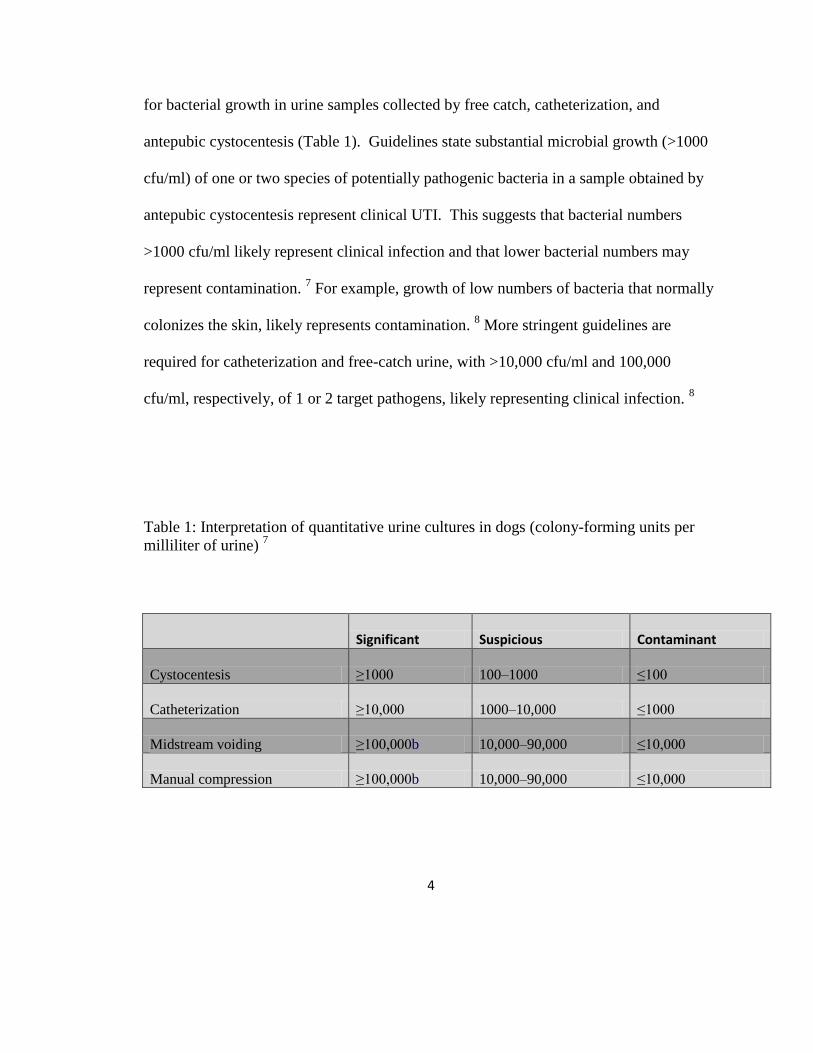
for bacterial growth in urine samples collected by free catch, catheterization, and
antepubic cystocentesis (Table 1). Guidelines state substantial microbial growth (>1000
cfu/ml) of one or two species of potentially pathogenic bacteria in a sample obtained by
antepubic cystocentesis represent clinical UTI. This suggests that bacterial numbers
>1000 cfu/ml likely represent clinical infection and that lower bacterial numbers may
represent contamination. 7 For example, growth of low numbers of bacteria that normally
colonizes the skin, likely represents contamination. 8 More stringent guidelines are
required for catheterization and free-catch urine, with >10,000 cfu/ml and 100,000
cfu/ml, respectively, of 1 or 2 target pathogens, likely representing clinical infection. 8
Table 1: Interpretation of quantitative urine cultures in dogs (colony-forming units per
milliliter of urine) 7
Significant Suspicious Contaminant
Cystocentesis ≥1000 100–1000 ≤100
Catheterization ≥10,000 1000–10,000 ≤1000
Midstream voiding ≥100,000b 10,000–90,000 ≤10,000
Manual compression ≥100,000b 10,000–90,000 ≤10,000
5
Contamination of midstream voided samples may cause growth of >10,000
colonyforming units per milliliter (ie, false-positive results); therefore, these samples
should not be used routinely for diagnostic culture. 7 Growth of low numbers of multiple
enteric bacteria in a urine sample may represent inadvertent puncture of the colon by
cystocentesis. 8
If there is bacterial growth present on culture plates (typically within 24 hours for
common urinary pathogens), organism identification and antimicrobial susceptibility is
performed. 7 Antimicrobial susceptibility can be performed by measuring minimum
inhibitory concentrations (MIC), or the lowest drug concentration used to inhibit bacterial
growth. Susceptibility can also be performed by agar disk diffusion (Kirby-Bauer). The
agar disk diffusion method uses Mueller-Hinton agar plates that have been inoculated
with a standardized suspension of the target bacteria. Paper disks impregnated with
different antimicrobial drugs are placed on the plate. Eighteen to 24 hours after
inoculation and incubation at 38 degrees C, antimicrobial susceptibility is estimated by
measuring zones of inhibition of bacterial growth surrounding each disk. Zones of
inhibition are then interpreted in light of established breakpoint standards that correlate to
organisms MIC, and susceptibility is recorded as resistant, susceptible, or intermediate. 7
Interpretive categories, termed clinical breakpoints, were created to interpret
antimicrobial susceptibility as resistant, susceptible, or intermediate for a specific
bacterial isolate. 13
A “susceptible” result implies that this antibiotic used for this
infection at approved dosages is likely to achieve clinical cure. A “resistant” result
6
implies that the isolate is not inhibited by clinically achievable drug levels, development
of antimicrobial resistance is likely, or that clinical cure was not reliable in clinical
studies. An “intermediate” result implies that the isolate may be treated in body sites
where the drug concentrates or when a high dosage of the drug can be used. 13
Clinical
breakpoints are determined by The Clinical and Laboratory Standards Institute (CLSI)
committee using information specific to bacterial organism and antimicrobial drug or
class of drug. 12
This information includes: data from pharmacokinetic,
pharmacodynamic, microbiologic, and animal modeling studies, as well as clinical trials
in humans and animals, if available. Most breakpoints are set based on drug levels
achieved in plasma, but breakpoints are set for different sites of infection if supporting
data is available. 13
1.3 Canine Urinary Tract Infection – Predisposing Factors
UTIs are considered uncomplicated if the patient has no identifiable concurrent
urinary or systemic diseases. Host factors that may predispose dogs to UTI include
structural, functional, and metabolic abnormalities; UTI in the presence of these factors is
considered complicated. Structural abnormalities such as recessed vulva, vestibulovaginal
stenosis, ectopic ureters, bladder diverticuli, perivulvar dermatitis, and persistent urachus
can all predispose dogs to UTI. In multiple retrospective studies, 64-68% of dogs with
ectopic ureters have urinary tract infections at initial diagnosis. 14,
15
Pelvic bladder can
be associated with urinary incontinence and UTI, with 65% of dogs with pelvic bladder
7
diagnosed with UTI. 16
Diabetes mellitus and hyperadrenocorticism predispose dogs to
UTI due to local and systemic immunosuppression, respectively. 6
Glucocorticoids inhibit some aspects of immune function, and 39% of dogs
receiving chronic corticosteroid treatment were diagnosed with UTI. 17
In diabetic dogs,
hyperglycemia and glucosuria inhibit leukocyte function and migration. 17
In one
retrospective, 42% of dogs with diabetes mellitus, hyperadrenocorticism, or both, were
diagnosed with UTI. 6
Poorly concentrated urine, such as that found in patients with chronic kidney
disease, supports the growth of bacteria. 18
There are no studies reporting the incidence of
UTI in dogs with chronic kidney disease (CKD). Nevertheless, this is likely higher than
the general population. Twenty percent of feline patients with stable CKD have UTI and
CKD is thought to be a risk factor for UTI in humans. 19,
20
UTI was present in 30% of
Boxer dogs at the time of diagnosis with juvenile nephropathy. 21
Urine acidity, normal
urine flow, and urine concentration are inherent defenses against ascending infection in
addition to organic acids, and urea concentration. Urinary tract infection is also more
likely to occur in conjunction with diseases such as urolithiasis and bladder neoplasia,
which damage the normal uroepithelium. 22,
23
Chemical irritants, such as
cyclophosphamide may inflame the bladder wall and predispose it to bacterial infection.
24 Neurologic disease, causing retained volumes of urine or obstruction to flow, can also
predispose dogs to UTI. 25
Indwelling urinary catheters also predispose dogs to UTI. The
prevalence of UTI was 48% in dogs with indwelling urinary catheters in one study and
8
the incidence of UTI increased by 27% per day increase in duration of catheterization. 25
Factors that predispose catheterized dogs to UTI include mucosal damage during catheter
insertion and manipulation, and contamination of the catheter by fecal microorganisms.
Retrograde flow of urine from the collection system to the urinary bladder, residual urine,
and urinary bladder dysfunction also contribute to the predisposition to UTI. 26
The risk
of UTI in dogs with indwelling urinary catheters also increased with each year increase in
patient age and also with concurrent administration of antimicrobials. 25
Complicated UTIs occur with conditions that increase the risk of serious
complications or treatment failure, such as with immunosuppression, lack of normal
micturition, concurrent pyelonephritis, or damaged uroepithelium. 27
For dogs with
persistent UTI, the prognosis was better in dogs where an underlying disorder was
identified and corrected. Clinical resolution was achieved with treatment of the
underlying disorder in addition to treatment with antibiotics. 2 Dogs with UTI that are
younger than 3 years may be less likely to respond to antibiotics alone since anatomic
abnormalities are more common in this age group. Dogs with congenital anatomic
abnormalities may have the highest risk of persistent UTI. 2,
5 Routine screening for UTI
is therefore recommended for dogs with structural, functional and metabolic
abnormalities. 28
1.4 Canine Urinary Tract Infection - Pathogens
9
Most UTIs are associated with a single bacterial pathogen, which usually
originates from the gastrointestinal tract or the distal urinary tract. 29
UTIs typically
involve bacterial pathogens, but rarely fungi or viruses can cause infections of the urinary
tract. 4 Approximately 75% of bacterial UTIs in dogs are caused by a single species of
pathogen, approximately 20% are caused by two species, and approximately 5% are
caused by three species. 4 E. coli is the most common canine uropathogen, and comprised
greater than 40% of all laboratory isolates in the largest retrospective series on canine
UTI. No other single pathogen represented more than 11% of the laboratory isolates. 4
The available retrospective studies include urinary isolates both from healthy dogs and
those with underlying predisposing disease. There are currently no large studies on UTI
solely in healthy dogs. Healthy young women have a high incidence of UTI; a large
prospective study identified an incidence of 0.5 per person per year. In this population,
80% of UTIs are caused by E. coli. 30
While healthy dogs often present to primary care
veterinarians for acute onset of lower urinary signs and are suspected of UTI, empiric
antibiotics are often prescribed without a urine culture. A urine culture is often
recommended only after a treatment failure or recurrent infection occurs. Other
commonly isolated bacteria from canine UTI include Proteus, Staphylococcus,
Klebsiella, Enterococcus, and Streptococcus. Less common bacterial isolates include
Mycoplasma, Pseudomonas, and Pasteurella. 4 Corynebacterium urealyticum has also
been reported as an uncommon cause of canine UTI and a cause of encrusting cystitis. 31
C. urealyticum is reported most often in conjunction with other abnormalities of the
10
urinary tract such as repeated catheterization, recent urogenital surgery, or neurologic
defecits. 31
Fungal UTI are rarely reported in small animals but Candida spp,
Cryptococcus spp, Blastomyces spp., Aspergillus spp., Histoplasma spp., and
Rhodotorula have been reported in the dog. 32,
33
Candida albicans was the most common
isolate (48%) in a recent retrospective on fungal UTI in cats and dogs. 32
Antimicrobial agents are necessary for treatment of bacterial UTI, but functional
host resistance mechanisms are required for successful treatment and prevention. 2
Experimental models of UTI were conducted in healthy dogs, during which E. coli was
directly inoculated into the bladder. These dogs were able to clear the infection without
administration of antimicrobial drugs. 34
The same phenomenon occurred in rats when E.
coli was experimentally inoculated into their bloodstream or bladder; all normal rats
cleared the bacteria without administration of antibiotics. 18
Consequences of untreated UTI include pyelonephritis, prostatitis, and infertility.
1 Infection with coagulase positive staphylococci is the main causative factor for struvite
urolithiasis in dogs. 23
Additionally, septicemia and discospondylitis can be consequences
of untreated UTI in dogs. 1
1.4.1 Uropathogenic E Coli
The risk of UTI increases with exposure to virulent E. coli. 35
Specific virulence
factors appear to influence the ability of E. coli to overcome host defenses and
successfully colonize the host urinary tract, causing UTI. 36
A high frequency of specific
11
virulence factors are present in E. coli strains isolated in canine UTI. 37
Strains carrying
these specific urovirulence factors are referred to as uropathogenic E. coli (UPEC).
UPEC were included in the recently defined pathotype ExPEC (extraintestinal pathogenic
E. coli) that classifies a variety of E. coli pathogenic to specific extraintestinal body sites.
38 The urinary tract is the most common site for colonization and infection with ExPEC.
39 Although UPEC typically originate from the gut flora, they do not appear to cause
disease in the gastrointestinal tract. 40
Dogs spontaneously develop UTIs with strains of E. coli carrying urovirulence factors
genetically similar to UPEC in humans. 37
Like human UPEC, canine UTI isolates are in
phylogenetic group B2 and express O antigens on the outer surface of cell surface LPS,
which are important for host evasion. These strains also possess numerous extraintestinal
virulence-associated factors (VFs). Strains lacking these phylogenetic and urovirulence
characteristics are more likely to behave as commensals. 37
In both dogs and humans with
UTI, the UTI strain is often isolated from the feces concurrently, consistent with host
fecal strains as the primary source for UTI pathogens. 41
Nevertheless, in a study of dogs
with naturally occurring E. coli UTI, concurrent fecal cultures were performed to
determine the predominant fecal strain of E. coli in each dog. In the majority of dogs, the
predominant fecal E. coli was not the infecting strain causing the UTI. Instead, the UTI
strain contained particular urovirulence factors by DNA analysis, while the fecal E. coli
strain did not. In the cases where the predominant fecal strain was identical to the UTI
strain, both contained urovirulence factors. 37
This supports the hypothesis that UTIs
12
occur from microbial pathogens with enhanced intrinsic virulence capability rather than
quantitative presence of organisms (the special pathogenicity hypothesis). 42
Specific UPEC virulence factors include fimbriae to enhance epithelial adherence,
iron uptake systems, and cytotoxins. 36
Bacterial adherence structures require a complex
organization of multiple proteins for assembly of fimbriae. The adhesive organelles, type
1, P, S, and F1C fimbriae promote adherence to host cells and tissues. UPEC also
characteristically carry siderophores that sequester iron from the host. Secreted toxins
include alpha haemolysin, cytotoxic necrotizing factor-1 (CNF1), and secreted
autotransporter cytotoxin (Sat). These toxins alter host cell signaling cascades, modulate
inflammatory responses, and stimulate apoptosis. 38
Experimental and epidemiological
data strongly suggest that no single virulence factor is sufficient for UPEC to cause
disease. Rather, expression of multiple, putative virulence factors, in concert with host
factors, contributes to establishment of UTIs. 36,
43 Some virulence factors frequently
occur together in the same strains due to their association with pathogenicity-associated
islands (PAIs). PAIs are discrete genetic units incorporated into the genome of
pathogenic microorganisms. PAIs are likely horizontally acquired due to flanking sites
that allow for direct insertion into DNA, and may allow for simultaneous spread of
multiple virulence factors among bacterial populations. 41
UPEC also invade epithelial cells of the urinary tract; this has been demonstrated
both in vitro and in vivo. 36
This may provide a survival advantage within the urinary tract
and allow evasion of some host immune mechanisms, as well as contribute to treatment
13
failure when antimicrobial agents do not reach therapeutic levels in cytoplasm. 36
Recent
studies of uropathogenic E. coli in superficial epithelial cells of the mouse bladder have
identified intracellular bacterial biofilms. 44
Persistence of E. coli in biofilms is
conjectured to be one source of recurrent E. coli UTI. Recurrent E. coli UTI in dogs is
often due to genetically identical strains (confirmed by PFGE), suggesting that some
cases of chronic E. coli UTI in dogs occur secondary to relapsing infections. 45
Antibiotic
susceptibility profiles, often used as a proxy for DNA fingerprinting methods do not
provide sufficient resolution to reliably distinguish relapsing or persisting infections from
reinfections. 45
E. coli strains genetically identical to canine UPEC have been isolated in human
UTI. 42
One study showed that 26% of healthy dogs sampled shed E. coli containing
urovirulence factors in their feces. 42
Thus, healthy dogs may serve as potential reservoirs
for such pathogenic E. coli. 37
1.5 Canine Urinary Tract Infection – Therapy
There are a multitude of antimicrobial therapies available for canine UTI.
Antimicrobials appropriate for treatment of UTI include those that are able to attain high
urine drug concentrations, have oral formulations, are easy to administer with few side
effects, are inexpensive for owners and have minimal effects on intestinal flora. 46
Cure
of urinary tract infections depends on antimicrobial concentrations in the urine rather than
in the serum, and therapy based on urinary drug levels is recommended. 47
Higher drug
14
levels in the tissues are essential for infections that are deep seated in the bladder wall or
have ascended to the kidneys (pyelonephritis). Longer duration of therapy for these
infections may also be necessary. For uncomplicated canine UTIs, antimicrobial therapy
has traditionally been recommended for ten to fourteen days. 7,
48 This is based on
experimental studies in dogs using a UTI model created by instilling sulfosalicylic acid
into the bladder of healthy dogs (12 males and 12 females) and then innoculating a single
E. coli isolate (1010
bacteria). 49
In this study, single and 3 consecutive daily doses of
amikacin (20mg/kg) and trimethoprim-sulfadiazine (TMS, 30mg/kg) were given to 12
dogs each. Only the 3-day regimen used in female dogs was successful in clearing UTI
in 4/4 dogs, and no dosage was successful in male dogs. 49
Nevertheless, treatment of
naturally occurring UTI in dogs may not be equivalent to that for induced UTI. Standard
of care for women with acute, uncomplicated cystitis, is 3 days of empiric antimicrobial
treatment. Empiric antibiotic therapy for 3 days is equivalent in efficacy to longer
durations of therapy, while therapy for 1 or 2 days is associated with a lower cure rate. 27
Many antimicrobials chosen for the treatment of UTIs have relatively short half-
lives or time-dependent dosing; therefore, repeated dosing and client compliance during
treatment is necessary for treatment outcomes to be successful. For example,
Amoxicillin (Amoxi-tabs®, Pfizer Animal Health) is a veterinary approved antimicrobial
appropriate for canine UTI. This is a time-dependent antimicrobial and recommended
dosage frequency is every 8-12 hours. 50
Cephalexin is another antimicrobial commonly
used for uncomplicated UTI in dogs; there are no veterinary approved products available
15
but dosage frequency recommended for use in dogs is every 8-12 hours (Keflex®, Dista).
51 Evidence from clinical practice suggests that in many cases, owners fail to comply with
dosing regimens. Poor owner compliance and inadequate antimicrobial dosing can
contribute to persistence of UTIs. 1 Several authors have attempted to assess veterinary
owner compliance with short and long term medication dosing. In one study of
veterinary compliance, 14% of enrolled dogs were excluded when their owners failed to
fill the initial prescription. Of those clients that remained in the study, only 44% had
100% compliance with the short term of antimicrobial drug prescribed. 52
Compliance
was higher in a recent study in which owners enrolled in a program to monitor
compliance with short-term antimicrobials. 53
In this study, the dosage regimen was
found to significantly influence compliance. Compliance was lower with TID dosing
compared to SID or BID dosing. 53
Additional studies are recommended to assess
whether owner compliance increases with shorter duration antimicrobial therapy.
1.6 Fluoroquinolone Antibiotics – Enrofloxacin
Fluoroquinolone (FQ) antimicrobial drugs have a broad spectrum of activity, and
are used to treat a variety of bacterial infections, including urinary tract infections.
Fluoroquinolone antimicrobial agents block bacterial DNA replication by inhibiting DNA
gyrase. DNA gyrase is responsible for relaxing torsional strain on DNA supercoils; this is
necessary for DNA replication, transcription, and recombination. 54

16
Enrofloxacin (Baytril®, Bayer Animal Health) is a synthetic antibacterial agent,
developed for veterinary use, and was approved by the Food and Drug Administration in
1989 for use in dogs. 55
Enrofloxacin has nearly 100% bioavailability after oral
administration and reaches high tissue concentrations. 56
Enrofloxacin has activity against
many gram-negative and gram-positive bacteria that are associated with canine
infections. Its spectrum includes the majority of bacterial genera isolated from UTI in
dogs. Enrofloxacin has a labeled dosage range of 5-20mg/kg/day, and may be
administered once daily or divided into two doses. Antimicrobial cure after
fluoroquinolone therapy is associated with a Cmax/MIC ratio of 8 to 10 or AUC relative
to MIC (AUC/MIC) ratio of > 100, where Cmax is the maximum concentration of the
drug, AUC is the area-under-the-curve, and the MIC is the minimum inhibitory
concentration required to prevent growth of the bacteria. 57
In dogs, enrofloxacin is
metabolized by the liver and deethylated to ciprofloxacin, which also has bactericidal
activity. 58
A study of enrofloxacin plasma pharmacokinetics in 4 dogs following a
5mg/kg oral or intravenous dose, suggested that a significant portion of antimicrobial
activity was due to ciprofloxacin since antimicrobial levels of ciprofloxacin were reached
in the plasma as well. 59
Enrofloxacin is eliminated by the kidneys and high
concentrations of both enrofloxacin and ciprofloxacin are reached in urine compared to
plasma following oral dosing. 50
Urine concentrations of enrofloxacin and ciprofloxacin
were measured 2 hours following 20mg/kg IV dose of enrofloxacin to anesthetized dogs.
58 High concentrations of both enrofloxacin and ciprofloxacin were present in the urine
17
compared to plasma, with ciprofloxacin contributing 50% to total antimicrobial
concentration. 58
Ciprofloxacin is available and approved for use in humans but is not
approved for use in animals. Ciprofloxacin can be used off-label by veterinary
prescription in dogs, but has inconsistent bioavailability in dogs. Bioavailability of
ciprofloxacin after oral administration in dogs ranges from 42-97% depending on the
report. 60,
61
1.6.1 Fluoroquinolone Antibiotics - Mechanisms of Antimicrobial Resistance
Antimicrobial resistance to fluoroquinolones occurs primarily from the
accumulation of specific point mutations in the FQ target genes leading to inhibition of
bacterial DNA topoisomerases (DNA gyrase and topoisomerase IV) or decreased drug
uptake by changes in the outer membrane and increased efflux. 62
The DNA
topoisomerases II (DNA gyrase) and IV are essential for bacterial replication by
controlling DNA supercoiling and chromosome partitioning. They consist of two
subunits, A and B, which are encoded by gyrA and gyrB for DNA gyrase, and parC and
parE for topoisomerase IV, respectively. 63
The primary mechanism of bacterial
resistance to FQ antimicrobials has been elucidated in Escherichia coli due to a mutation
in gyrA, the gene encoding the A subunit of the enzyme DNA gyrase. 64
This is
consequently designated the quinolone resistance-determining region (QRDR). Mutations
in gyrB are of less significance, whereas mutations in parC are the predominant cause of
FQ resistance in Gram-positive bacteria. 54,
65 Single mutations in gyrA confer low-level
18
FQ resistance; several mutations are necessary for the development of high level FQ
resistance. Most isolates with high level FQ resistance have an additional mutation in
parC. Topoisomerase IV (encoded by the parC and parE genes) is a secondary target of
the fluoroquinolone antimicrobials, and mutations in parC and parE contribute to
quinolone resistance. 54
Decreased bacterial cell wall permeability to FQ antimicrobials
causing decreased cytoplasmic drug accumulation also contributes to resistance. 54
Efflux-related mutations are also common in highly resistant strains. 63
These efflux-
related mutations inactivate marR or acrR, thus up-regulating the activity of the drug
efflux pump, and reducing the antimicrobial concentration in bacterial cytoplasm. In
addition to this mechanism, a reduction of the OmpF porin channels that can reduce
intracellular FQ concentrations has been described. 62
E. coli is one of several pathogens for which elevated mutation frequencies have
been described among natural isolates. These hypermutator strains have up to 1000-fold
higher mutation frequencies compared to normal strains. 63
When bacteria acquire two or
more of these mutations, they will survive despite the presence of antimicrobial drugs at
concentrations many times higher than what is achievable in a patient. These point
mutations occur spontaneously in bacterial populations at a rate of 1 in 106
to 1 in 108
cells, and mutants can be further amplified under selective pressure during antimicrobial
therapy. 66
Furthermore, mutations in bacterial DNA are essentially irreversible and
resistance is transmitted to all daughter cells of the resistant isolate, potentially leading to
large populations of resistant bacteria. 5
19
Plasmid mediated mechanisms of resistance to FQ antimicrobials have also been
described. QepA, a plasmid-associated gene responsible for reduced FQ susceptibility,
has been isolated from multiple individuals with E coli infection worldwide. QepA
encodes an efflux pump that confers FQ resistance. 67
MICs of norfloxacin, enrofloxacin,
and ciprofloxacin are 32- to 64-fold higher in experimentally tranformed strains
expressing QepA than nontransformed isogenic controls. 68
Multidrug resistant E. coli
occur in both human and veterinary medicine, and minimize available therapeutic
options. 63
The production of extended-spectrum Beta-lactamases (ESBL) by multidrug
resistant E. coli strains has caused increasing concern over the last decade. Plasmids
encoding extended-spectrum β-lactamases that additionally encode plasmid-mediated
quinolone resistance (PMQR) genes have been isolated from clinical isolates of E. coli in
humans and dogs. 68
Multi-drug resistant E. coli with extended-spectrum beta-lactamase
activity and fluoroquinolone resistance are reported in clinical infections in dogs. 64
1.6.2 Fluoroquinolone Antibiotics – Increasing Antimicrobial Resistance
Bacterial resistance to FQ antimicrobials in human and veterinary medicine is
increasing, and parallels increased antimicrobial use. 62,
69 During routine administration,
antimicrobial agents apply selective pressure on populations of bacteria, leading to an
increase in bacterial populations with antimicrobial-resistance. This occurs with FQ
antimicrobials both in vitro, and in animal models, demonstrating that resistant mutant
bacterial populations are selectively enriched after exposure to FQs. 62
Widespread use of
20
fluoroquinolones in humans has selected for bacterial resistance to fluoroquinolones. 70
Several investigators have associated increased antibiotic usage in practice with increased
antibiotic resistance. There is a significant increase in the incidence of enrofloxacin-
resistant E coli isolated in urine from dogs with UTI in recent years. This corresponds to
increased use of enrofloxacin, as documented by veterinary pharmacy records. 54
A
retrospective study of bacterial isolates from canine patients with urinary tract infections
documented an increase in the prevalence of resistance to commonly used FQ
antimicrobials from 1992-2001. 71
An increase in the incidence of FQ resistant canine
UTI E. coli isolates was documented from 1996-1998 in a veterinary teaching hospital.
This was shown by pulsed field gel electrophoresis to not be associated with clonal
populations of E. coli. 54
An increase in the resistance to ciprofloxacin in hospitalized
human patients over time was also seen from 1990-1996, and this increase in the
prevalence of FQ resistance was parallel to an increase in ciprofloxacin usage. 69
Additionally, FQ resistance in E. coli is not restricted to clinical isolates, but is
commonly present in surveys of the healthy human and animal feces. These may serve as
reservoirs for pathogenic E.coli and contribute to spread between animals and humans. 72
These findings question whether the FQ-resistant E. coli encountered in dogs arise
through mutation of FQ-susceptible canine resident strains to resistance, or instead
originate from an external source.
1.7 Mutant Prevention Concentration
21
To combat the increasing prevalence of antimicrobial resistance, recent studies
have highlighted the need for optimal dosing regimens of antimicrobials that consider
bactericidal efficacy, as well as the potential for selection of resistant mutants. Standard
dosing regimens relying on interpretations of minimum inhibitory concentration (MIC)
take into account only the clinical laboratory definition of in vitro inhibition of growth,
and are not relevant to prevention of mutant selection. New dosing strategies using
Mutant Prevention Concentration (MPC) guidelines consider both goals of curing
bacterial infections as well as preventing formation and selection of resistant mutant
bacteria. These dosing regimens may help to combat the emergence of resistant mutant
bacterial strains, and are most applicable to FQ antimicrobials. 73
Mutations arise
spontaneously in a microbial population, and some of these mutations confer
antimicrobial resistance. These mutant subpopulations may be present in an infection
site, or in other sites in the host, prior to initiation of antimicrobial treatment. Certain
dosing strategies may select for mutant subpopulations when antimicrobial
concentrations are high enough to kill susceptible bacteria in the population, but are not
high enough to kill some mutants. This is referred to as the mutant selection window
hypothesis, and the dose range in which mutants may be selected is referred to as the
mutant selection window. 74
The mutant selection window hypothesis contends that ideally, drug levels should
be kept above the mutant prevention concentration (MPC) to restrict selection for
resistant mutants. (Figure 1) MPC is defined as the minimum inhibitory concentration
Figure 1
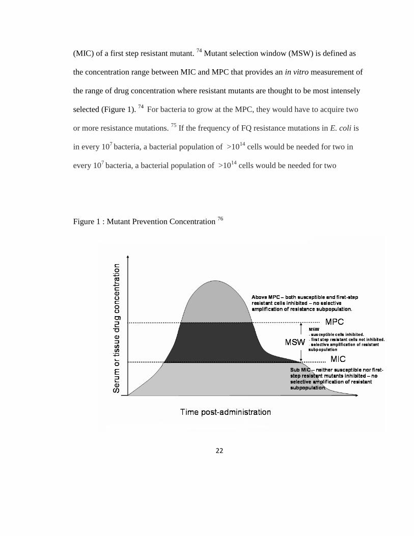
22
(MIC) of a first step resistant mutant. 74
Mutant selection window (MSW) is defined as
the concentration range between MIC and MPC that provides an in vitro measurement of
the range of drug concentration where resistant mutants are thought to be most intensely
selected (Figure 1). 74
For bacteria to grow at the MPC, they would have to acquire two
or more resistance mutations. 75
If the frequency of FQ resistance mutations in E. coli is
in every 107
bacteria, a bacterial population of >1014
cells would be needed for two in
every 107
bacteria, a bacterial population of >1014
cells would be needed for two
Figure 1 : Mutant Prevention Concentration 76
23
in every 107
bacteria, a bacterial population of >1014
cells would be needed for two
concurrent resistance mutations to arise. 71
Such large bacterial population sizes are
considered unlikely in clinical infections. 75
Mutant subpopulations have been isolated from patients treated with antimicrobial
doses that fall within the MSW. 73
Although MPC is a laboratory assessment, it may
become an effective tool when applied in vivo. Notably, the selection of FQ resistant
strains of Streptococcus pneumoniae was effectively prevented in rabbits when MPC
criteria were used. 77
In a different study, doses using only MIC criteria were insufficient
to prevent the selection of resistant mutants of E. coli in vitro when serum concentrations
of some commonly used FQs at standard doses were simulated. Mutant growth was
prevented only using in vitro ciprofloxacin concentrations that simulated the highest
labeled dose of ciprofloxacin approved in humans. 73
MPC appears to be valid for
evaluation of enrofloxacin against E. coli isolated from chickens. All single-step mutants
that occurred for E. coli during in vitro incubation with enrofloxacin had MICs equal to
or lower than the MPC for enrofloxacin. 74
Ciprofloxacin has better efficacy in vitro
using MPC criteria, meaning that MPC values were lower for all isolates when using
ciprofloxacin compared to enrofloxacin. 73,
74 This may be an advantage in vivo since
ciprofloxacin is the main metabolite of enrofloxacin and has antimicrobial activity. 74
Another method, the agar-plate mutant accumulation assay is an in vitro assessment of
propensity for clinical isolates to acquire mutations that confer fluoroquinolone
resistance. 78
This also may be useful when designing antimicrobial protocols.
24
The MPC method has not yet been validated in clinical patients. 79
MPC data for
common veterinary pathogens are necessary before doses can be adjusted to target MPC
guidelines in clinical patients. 80
This has been explored in E. coli isolates from chickens,
and in E. coli isolated from human UTI, but not in dogs or cats. 66, 81
Furthermore,
pharmacokinetic parameters that correlate with clinical efficacy using MPC criteria have
been suggested in vitro but not tested in vivo. 79
The AUC24/MPC and t>MPC may be
useful pharmacokinetic parameter for predicting prevention of the emergence of resistant
bacteria in vivo. 74,
80 From initial studies in non-neutropenic mice, it appears that
antimicrobial concentrations would not need to remain above MPC levels for the duration
of treatment in animals with an intact immune response, but the duration antimicrobial
concentrations would need to remain above MPC for clinical success is unknown. 82
For
example, with a local Staphylococcus aureus infection of rabbits, levofloxacin
concentrations needed to be above MPC for only 20% of the dosing interval to restrict
mutant amplification. 80
MPC is not predictable based on established susceptibility
points, such as MIC. 81
High dose antimicrobial therapy protocols may be more
efficacious in prevention of antimicrobial resistant populations by targeting MPC
guidelines. MPC is an in vitro laboratory tool and is time-consuming and technically
demanding. New ways to estimate MPC for clinical isolates are necessary before
application to clinical practice.
25
Chapter 2: Evaluation of the Efficacy of a High Dose Short Duration Baytril® Treatment
Regimen for Uncomplicated Urinary Tract Infection in Dogs.
2.1 Introduction
The urinary tract is a common site for bacterial infection in dogs, and it is
estimated that 14% of dogs will be affected by a urinary tract infection (UTI) in their
lifetime. 1 It is important to differentiate between uncomplicated and complicated UTI for
diagnostic and treatment purposed. UTIs are considered uncomplicated if the patient has
neither 1) concurrent disease affecting the urinary tract nor 2) systemic disease with
known comorbidity with bacterial cystitis. Uncomplicated canine UTI is routinely treated
with a 7-14 day course of orally administered antimicrobial agents. 7,
48 Poor owner
compliance and inadequate dosing can contribute to treatment failure in canine UTI. A
shorter duration of therapy may increase client compliance and decrease cost. 53
Furthermore, therapy using high doses of antimicrobial agents with concentration-
dependent pharmacodynamic targets will increase bactericidal activity; this may decrease
the development of antimicrobial resistance due to high drug concentrations achieved. 69
Fluoroquinolones are frequently used to treat canine bacterial infections because
of their broad antimicrobial spectrum and relatively wide therapeutic margin. Baytril®
26
(enrofloxacin) is a fluoroquinolone antimicrobial drug developed specifically for
veterinary use and approved by the FDA for use in dogs at 5-20mg/kg orally.
Enrofloxacin is active against most major pathogens that cause UTI in dogs, including E.
coli, other coliform bacteria, and staphylococci spp. The bactericidal effect of
fluoroquinolones is concentration dependent. Fluoroquinolones also exert a post-
antibiotic effect on Gram positive and Gram negative bacteria. It is conceivable that
high-doses of FQs used once daily for fewer than 7 days may be an efficacious regimen
for treatment of UTIs in dogs.
The objective of this study is to evaluate the effectiveness of a high dose - short
duration enrofloxacin (Baytril®) dose regimen in the treatment of naturally-occuring
lower urinary tract infections in dogs. We hypothesized that in naturally occurring canine
uncomplicated urinary tract infections, neither microbiologic cure rate nor clinical
resolution rate will be significantly different between two treatment conditions: 1)
enrofloxacin administered at the highest FDA-labelled dosage for 3 days and 2) A
standard and common treatment regimen of antimicrobial therapy (amoxicilln-
clavulanate 13.75-25mg/kg for 14 days).
2.2 Materials and Methods
2.2.1 Clinical Drug Trial Design
A multicenter clinical drug trial was designed to evaluate a high dose - short
duration enrofloxacin (Baytril®) dose regimen for the treatment of UTI in dogs using

27
Baytril® (enrofloxacin) tablets. This study was conducted as a prospective, controlled,
randomized, and blinded clinical trial. The study was conducted starting February 2009
and aimed to enroll 100 dogs between 3 study sites. A midpoint analysis was conducted
in March 2010 for Masters thesis analysis.
2.2.2 Planned Enrollment
Client owned dogs presented to study site veterinary clinics by their owners for
lower urinary tract signs and considered for enrollment. Study sites included: The Ohio
State University Veterinary Medical Center (Columbus, OH), The University of
California-Davis Veterinary Teaching Hospital (Davis, CA), and Bradford Park
Veterinary Hospital, a private referral practice (Springfield, MO). Adult dogs, 5-50kg
were enrolled to ensure appropriate dosing based on tablet sizes used in the study. The
dogs were classified as adults based on the following criteria: >9 months in small or
medium sized dogs 5 - 25.9kg, >1 year in large breed dogs 26 -45kg or >18 months in
giant breeds >45kg. The dogs were deemed otherwise healthy based on owner history,
physical exam performed by one of the investigator veterinarians, a complete blood
count, and serum biochemical analysis. Dogs were enrolled into the study when
provisional enrollment criteria were met and when target pathogens were identified on
urine culture.
For definitive eligibility, diagnosis of bacterial UTI was based on quantitative
urine culture obtained by antepubic cystocentesis on the study Day 0. UTI was
28
diagnosed when ≥ 1000 cfu/ml of 1 or a mixture of up to 2 target pathogenic bacteria was
grown. Target pathogenic organisms included E. coli, Proteus mirabilis, Klebsiella
pneumonia, Pseudomonas aeruginosa, Enterobacter spp., Staphylococcus aureus,
Staphylococcus pseudointermedius, Streptococcus spp. (alpha hemolytic), and
Enterococcus spp.
We aimed to enroll a total of 150 dogs over 12 months, estimating that 1/3 of
dogs initially enrolled would be excluded due to study criteria and lost to follow-up. This
would leave 100 dogs followed to study endpoint. After the first 3 months, only 3 dogs
had been followed to study endpoint. Due to slow enrollment of clinical cases,
recruitment of dogs with uncomplicated UTI from primary care veterinarians was
performed. The clinical trial was advertised to primary care clinics in areas surrounding
the study cite centers. Advertisements consisted of posters, mailings, phone calls, and
personal visits by investigators. A stipend was given to primary care veterinarians for
referral of adult, otherwise healthy dogs with lower urinary tract signs to study site
centers without initiating treatment.
2.2.3 Exclusion Criteria
Dogs with persistent or recurrent UTI were excluded. Persistent UTI was defined
as more than 3 positive urine cultures in succession with no intervening sterile cultures
and an elapsed time period of 1-8 weeks between cultures. 83
Recurrent UTI was defined
as more than 3 successfully treated UTI episodes within a single year in which each
29
episode is followed by a period of sterile urine. 83
Dogs with suspected pyelnonephritis,
prostatitits, urinary tract neoplasia, or urinary calculi were excluded as well. Animals
with uncontrolled systemic disorders and animals with spinal cord injuries were also
excluded. Dogs treated within 7 days with a short duration antimicrobial, or within the
last 14 days with a long-term antimicrobial were excluded. Dogs treated within the last
14 days with a systemic short-acting steroid or within the last 28 days with a sustained
release steroid were also excluded. Dogs administered any medication that could
interfere with systemic absorption, such as antacids and other compounds containing
metal cations, were also excluded.
Exclusion criteria also included animals with known or suspected central nervous
system (CNS) disorders (quinolones, in rare instances, have been associated with CNS
stimulation that may lead to convulsive seizures). Young dogs, based on the
aforementioned criteria, were also excluded since quinolone-class drugs have been
associated with cartilage erosions in weight bearing joints of immature animals.
2.2.4 Treatment Groups
Amoxicillin-clavulante (Clavamox®) was chosen as the positive control because
it is a suitable comparative antimicrobial indicated for use in canine UTIs, and is
considered to be highly efficacious for UTI in dogs. Dogs were randomized to group 1 or
group 2 by randomly generated numbers to ensure 1:1 ratio of dogs between groups. A
veterinary technician was assigned to enroll dogs and dispense medications. The
30
dispenser instructed all pet owners not to discuss the treatment regimen with anyone
other than the dispenser. All veterinarians, technicians, and staff responsible for clinical
observations were blinded to the treatment. Dogs in Group 1were treated with 18-
20mg/kg oral enrofloxacin (Baytril®) once daily for 3 consecutive days. Dogs in Group
2 were treated with 13.75-25mg/kg oral Amoxicillin-clavulante (Clavamox®) twice daily
for 14 days.
2.2.5 Timeline and Assessment Criteria
All dogs were examined on Day 0 (initial day of presentation and treatment), Day
10 (+/- 2 days) and Day 21 (+/- 2 days). At Day 0, clinical signs were recorded (presence
or absence of pollakiuria, stranguria, hematuria), and a physical exam, including bladder
palpation, was performed by one of the study investigators. A complete blood count,
serum biochemical analysis, urinalysis (by cystocentesis), and quantitative urine culture
(by cystocentesis) with susceptibility by MIC were also performed at Day 0. Treatment
was initiated on Day 0. Quantitative culture and susceptibility results were available 3-5
days after Day 0. At Day 10 and Day 21, clinical signs were again recorded (presence or
absence of pollakiuria, stranguria, hematuria), and a physical exam, including bladder
palpation, was performed by one of the study investigators. A urinalysis and quantitative
urine culture +/- susceptibility by MIC were also performed.

31
Due to slow enrollment, an amendment was made to the study protocol allowing
dogs to be enrolled up to 3 days after initial presentation to a study site if a positive urine
culture was obtained. Day 0 was defined as the day of initial treatment for these dogs.
For definitive enrollment, ≥ 1000 cfu/ml of 1 or a mixture of up to 2 target
pathogenic bacteria was required, isolated from a urine sample collected by cystocentesis.
All enrolled dogs were followed to study endpoints 1) microbiologic cure 2)
microbiologic failure 3) withdrawal due to adverse effects. The clinical cure rate (based
on resolution, if present, of pollakiuria, stanguria, hematuria, and pain on bladder
palpation) was also recorded. A treatment failure was defined as post-treatment culture
positive for the same pathogen as the pre-treatment urine sample. Isolation of colonies of
any pathogen different from the pre-treatment urine sample was considered a
contaminant or a re-infection.
2.2.6 Statistical Analysis
An unpaired t-test was used to determine difference in treatment response
between the two groups. A necessary sample size of 17 in each group was determined by
power analysis using an alpha error 0.05 and a desired power of 0.80. This analysis was
powered to detect a 10% difference between groups.
2.3 Results
32
Due to the unanticipated slow enrollment of clinical cases of uncomplicated UTI,
midpoint study analysis was conducted for MS thesis publication. At midpoint, 77 dogs
were preliminarily enrolled in the study. 34 dogs were excluded based on negative
culture results at D0, and 3 dogs were lost to follow-up after D0. 40 dogs were
definitively enrolled and followed to study endpoint. Of these, 2 (5%) were intact
females, 5 (12%) were neutered males, and 33 (83%) were spayed females. No intact
males were enrolled. The most common breeds (and number) included Labrador
retriever (5), Mixed (5), Dachshund (4), Boxer (2), Pug (2), and Golden retriever (2).
Mean and median weights were 21kg and 22 kg, respectively (range 5-50kg). Mean and
median ages were both 7 years (range, 1-13 years). Mean and median duration of clinical
signs prior to presentation were 9.6 days and 4 days, respectively (range, 1-28 days).
Presenting complaints were: pollakiuria in 38 dogs (95%), dysuria in 22 dogs
(55%), and hematuria in 21 dogs (53%). 2 (5%) dogs presented with no clinical signs but
were enrolled based on positive urine cultures. Common physical examination
abnormalities included pain on bladder palpation (24%), vulvar hooding or recession
(14%), obesity (5%) and concurrent pyoderma (5%). Other physical exam abnormalities
were considered incidental to the study analysis. Complete blood count and biochemical
profile abnormalities were considered mild and clinically insignificant to study analysis.
Upon urinalysis, 25 dogs (60%) had clinically significant pyuria (>3WBC/hpf) and 34
dogs (81%) had bacteriuria; 24 dogs had both pyuria and bacteriuria (57%). Mean urine
specific gravity (USG) was 1.027, median 1.030 (range 1.002-1.052). Mean urine pH was
33
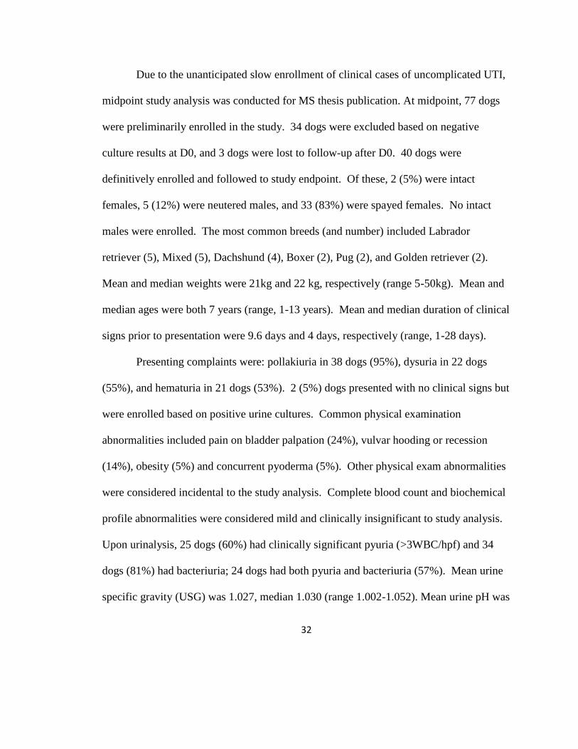
7.2, median 7.0 (range 5.0-9.0). Upon initial culture (D0) 34 dogs yielded one bacterial
isolate and 6 dogs yielded two bacterial isolates. E. coli was the most frequently isolated
urinary tract pathogen in the dogs enrolled (42%) followed by Proteus spp. (23%),
Staphylococci (21%), and Streptococcus (8%). Enterococcus spp. and Citrobacter spp.
were isolated in 1 dog each. Culture results are shown in Figure 2.
Figure 2: Canine UTI Isolates
34
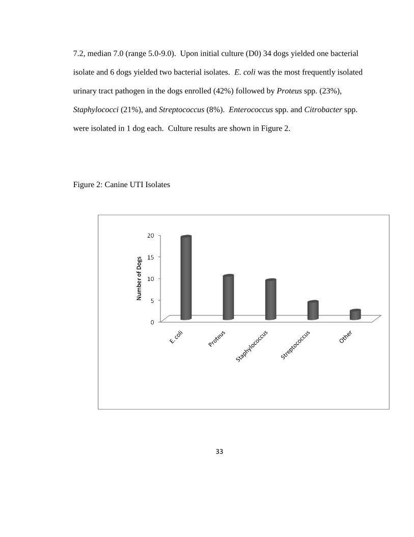
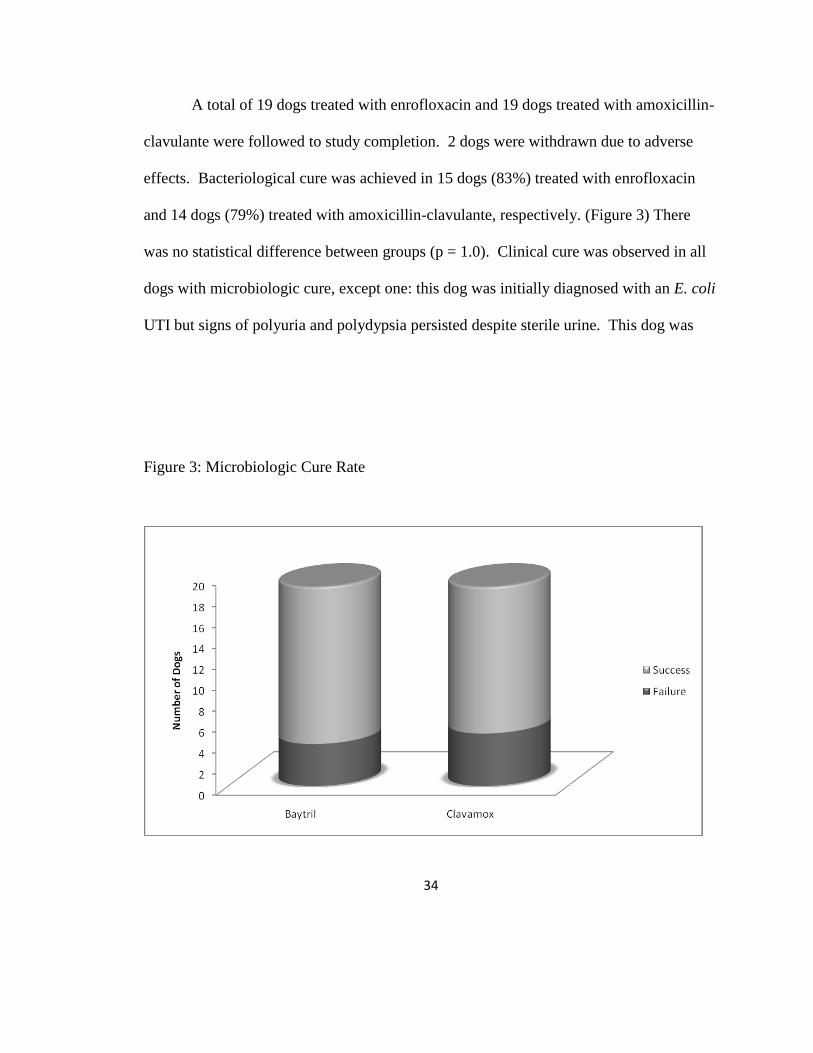
A total of 19 dogs treated with enrofloxacin and 19 dogs treated with amoxicillin-
clavulante were followed to study completion. 2 dogs were withdrawn due to adverse
effects. Bacteriological cure was achieved in 15 dogs (83%) treated with enrofloxacin
and 14 dogs (79%) treated with amoxicillin-clavulante, respectively. (Figure 3) There
was no statistical difference between groups (p = 1.0). Clinical cure was observed in all
dogs with microbiologic cure, except one: this dog was initially diagnosed with an E. coli
UTI but signs of polyuria and polydypsia persisted despite sterile urine. This dog was
Figure 3: Microbiologic Cure Rate
35
subsequently diagnosed with pyelonephritis. One dog with an initial Proteus spp. UTI
had a positive culture (Enterococcus spp.) from enrichment broth only on Day 10. No
treatment was initiated since the dog was asymptomatic and D21 urine culture was
negative.
2.4 Discussion:
This study reports a high microbiologic and clinical cure for canine
uncomplicated UTI using a novel high-dose, short-duration protocol for enrofloxacin
(Baytril®). Overall bacterial elimination rate in enrofloxacin treated dogs (83 %) was
comparable to rates reported recently for other relatively new antimicrobial agents,
including the third generation injectable cephalosporin, cefovocin, given once SQ and
pradofloxacin given orally for at least 10 days. 84,
85
The clinical cure rate for HDSD
Baytril® was also similar to that reported for marbofloxacin given orally for 10 days for
UTI. 86
Amoxicillin/clavulanate given orally for 10 days also had a similar cure rate in a
previous study (85%). 86
Resolution of clinical signs in all treated dogs mirrored microbiologic cure except
for the 1 dog with persistent polyuria and polydypsia who was considered a clinical
failure and withdrawn from the study. This dog was subsequently diagnosed with
pyelonephritis. Pyelonephritis can be difficult to diagnose due to intermittent shedding of
bacteria into the bladder. A longer course (4-6 weeks) of antimicrobial therapy is
recommended for treatment of pyelonephritis due to sequestration of bacteria within the
36
kidneys. 2 One dog with an initial Proteus spp. UTI had a positive culture (Enterococcus
spp.) from enrichment broth only on Day 10. No treatment was initiated since the dog
was asymptomatic. Treatment of asymptomatic UTI in dogs due to Enterococcus spp. is
controversial. The urine culture on Day 21 was negative in this dog even after no
antimicrobial treatment was given.
Demographics of dogs enrolled showed a wide distribution of ages, weight, and
breed. This is a small analysis compared to previous retrospectives, such as the largest
which enrolled 8,354 dogs. Female spayed dogs were over-represented, consistent with
previous reports. The bacterial species identified during this study were also typical of
those described previously for canine UTI. 2,
4,
5 All dogs in this study were otherwise
healthy. Complete blood count and biochemical profile were overall unremarkable. This
is consistent with previous data, supporting the idea that uncomplicated lower UTI does
not usually perturb routine hematologic and biochemical parameters. 7
The Clinical and Laboratory Standards Institute (CLSI) set MIC breakpoints for
enrofloxacin as ≤ 0.5 ug/ml (susceptible), 1–2 ug/ml (intermediate) and ≥ 4 ug/ml
(resistant). 12
No antimicrobial resistance to enrofloxacin was observed in any dogs
treated with enrofloxacin our study. Thus, empiric use of high dose, short duration
enrofloxacin appears to be a rational therapy for uncomplicated UTI in dogs. Efficacy
for FQ antimicrobials is dependent on the maximum drug concentration at the site of
infection – often described as the Cmax (maximum drug concentration) or AUC (area
under the time concentration curve). The duration of time the drug remains above the
37
minimum inhibitory concentration (MIC) seems less important. The dogma for FQ
pharmacodynamics is that clinical cure is based on achieving a Cmax/MIC ratio of at
least 8-10 or an AUC/MIC ration of at least 100. This is based solely on achieving
clinical cures in a neutropenic mouse model. 57
Enrofloxacin is nearly 100% bioavailable, when administered orally to dogs. It
has a relatively high volume of distribution, and is mainly eliminated by the kidneys,
allowing high levels of active drug to be reached in urine. 58
In addition, its primary
metabolite, ciprofloxacin also exerts bactericidal effects, and is also renally eliminated.
Efficacy of treatment of UTI is logically correlated with urine drug concentrations.
Applying MIC interpretive breakpoints for antimicrobial susceptibility, as defined by the
Clinical Laboratory Standards Institute, which are based on plasma drug concentrations
may be too conservative in UTIs. 47
Furthermore, higher doses of antimicrobials limit the
growth of mutant subpopulations in vitro and may decrease emergence of antimicrobial
resistant bacteria in vivo. 57
Future studies are necessary to test this hypothesis in clinical
patients.
In conclusion, 20mg/kg Baytril administered orally once daily for 3 consecutive
days, was as effective as amoxicillin-clavulanate administered at 13.75-25mg/kg twice
daily for 14 consecultive days, in the treatment of uncomplicated bacterial UTI in dogs.
Additionally, HDSD Baytril offers the benefit of potentially limiting selection for
antimicrobial resistance and increasing owner compliance.
38
Chapter 3: Urine Concentrations of Enrofloxacin and Ciprofloxacin in Healthy Dogs
3.1 Introduction
Enrofloxacin is a Fluoroquinolone antimicrobial drug approved for use in dogs
with a flexible dose range of 5 to 20 mg/kg once daily for treatment of urinary, skin, and
soft-tissue infections. Enrofloxacin is one of four Food and Drug Administration (FDA)
approved fluoroquinolones in the United States for use in dogs. Enrofloxacin is rapidly
and widely distributed in the dog, and it achieves high intracellular and tissue
concentrations. 59
The volume of distribution for enrofloxacin is 3.63 ± 1.17 L/kg after
1.24 mg/kg/hour IV infusion in dogs. 55
A volume of distribution above 1.0 L/kg implies
a higher drug concentration outside of the plasma compartment. 59
Enrofloxacin is
distributed in all body fluids and tissues and reaches particularly high concentrations in
urine and bile, as well as in the liver and stomach, relative to plasma. 58
Its spectrum of
activity also makes it a useful antimicrobial in dogs because it has bactericidal activity
against many Gram-negative aerobes as well as some Gram-positive and intracellular
bacteria. Enrofloxacin is nearly 100% bio-available after oral dosing. 59,
87 The half-life
of enrofloxacin and its post-antibiotic effect, make it suitable for once-daily oral dosing.
85 Furthermore, enrofloxacin has a high safety profile with a low incidence of side effects.
39
88 Ciprofloxain is the primary active metabolite of enrofloxacin, and contributes in an
additive fashion to overall antimicrobial activity. 56
The bactericidal activity of fluoroquinolones is concentration dependent and thus
their efficacy is dependent on maximizing drug concentration at sites of infection. 57
The
pharmacodynamic parameters of maximum plasma drug concentration (Cmax) and area
under the concentration time curve (AUC0-24) is correlated with clinical efficacy in a
neutropenic mouse model. In this model, neutropenia (<100 neutrophils/µL3) was
induced in 6 mice by intraperitoneal injection of cyclophosphamide, and then 106
colony
forming units of E. coli were injected into 1 or both thigh muscles of each mouse.
Statistical analysis (Two-way analysis of variance) revealed that drug efficacy was
significantly increased with increasing enrofloxacin total dose, but not with increasing
dose frequency. 57
Plasma concentrations of enrofloxacin are available for 2.5, 5, 7.5, 10, and
20mg/kg doses of oral enrofloxacin in healthy dogs. 50,
56
Information in the drug
monograph is limited to plasma and tissue drug concentrations after 2.5mg/kg doses in 2
healthy dogs. 50
Information regarding drug concentrations in other body tissues
following higher doses is sparse. 56,
88
Therefore, treatment recommendations are often
based on plasma drug concentrations regardless of the site of infection. 58
Dosage
regimens that optimize antimicrobial concentrations at the infection site may be better for
predicting clinical success. 58
The primary route of excretion of enrofloxacin is in the
urine, making it effective for treating UTIs because of the high drug concentrations
40
attained within the urinary tract. 54
Other antimicrobial agents commonly used in the
treatment of canine UTI also attain urine drug concentrations that are exponentially
higher than in plasma. This is the reason why successful antimicrobial treatment for UTI
can occur with an infecting microbe that would be classified as resistant when
considering concentrations of drug attained in other tissues. This has been shown with
antimicrobials such as penicillin, and provided the basis for measuring urine
concentrations of other antimicrobial agents commonly used for canine UTI. 1
Despite the common use of enrofloxacin for canine urinary tract infections, there
is limited information on urine concentrations of active drug following oral enrofloxacin
dosing. Enrofloxacin, and its main active metabolite ciprofloxacin, are both found in
higher concentrations in tissues and urine relative to plasma following oral and
intravenous enrofloxacin administration. 50,
58,
87,
88
Urine levels following oral
administration of 2.5mg/kg and 5mg/kg enrofloxacin have been measured in 1 study
each. 50,
87
Following a dosage of 2.5mg/kg, mean urine enrofloxacin concentrations,
measured in 2 dogs, were 43µg/ml at 2 hours and 55µg/ml at 8 hours post oral
administration. 50
This is markedly higher than the 2 hour plasma concentration of
0.67µg/ml. 50
The time to reach peak serum levels after oral administration in fasted dogs
varies but generally occurs between 1 to 3 hours after administration. 88
In one study,
enrofloxacin and ciprofloxacin peak urine levels were over 100 times higher than in the
plasma following 50mg IV and oral administration of enrofloxacin in 8 healthy Beagles
(mean weight 8.6 ± 0.2kg). 87
Peak urine drug levels were measured 6 hours post

41
administration. In this study, both enrofloxacin and ciprofloxacin levels were measured
by HPLC, but only calculated totals of enrofloxcain + ciprofloxacin are published.
Methods used, exact measurements, and time-points chosen for urine levels were not
provided. 87
It has been suggested that urine concentrations of enrofloxacin and ciprofloxacin
increase linearly with increasing doses similar to plasma. If so, urine drug concentrations
following higher doses of enrofloxacin could be extrapolated based on the existing
studies performed using 2.5mg/kg and 5mg/kg dosages. 59
Mean urine enrofloxacin and
ciprofloxacin concentrations of two dogs were measured two hours following intravenous
enrofloxacin administration, and were approximately equal (44 µg/ml of enrofloxacin and
43 µg/ml of ciprofloxacin). 50
One study evaluated enrofloxacin and ciprofloxacin
concentrations in tissues and body fluids after 20mg/kg enrofloxacin was given
intravenously in 4 anesthetized dogs. Enrofloxacin concentration in urine was 7 times
higher than in plasma, while ciprofloxacin concentration in urine was 13 times higher
than in plasma. 58
These findings are not consistent with the hypothesis that urine drug
concentrations increase in a linear fashion according to dosage, as the levels measured
after a 20mg/kg dose were comparable to those attained following a 2.5 mg/kg dosage. 50
However, the quantitative methods employed by each of these studies were different; the
drug levels following the 2.5 mg/kg dosage in these earlier studies were conducted by
bioassay versus high-performance liquid chromatography after a 20mg/kg dose. Drug
levels measured by bioassay are expected to be higher since this assay utilizes a
42
quantified decrease in susceptible bacterial numbers to measure the total amount of active
drug; a bioassay would measure combined enrofloxacin + ciprofloxacin antimicrobial
killing. In contrast, HPLC measures each drug metabolite separately (ie individual
enrofloxacin and ciprofloxacin concentrations). In addition, drug concentrations
measured after the 20mg/kg dose were performed in anesthetized dogs. 58
The extent to
which the drug levels attained in anaesthetized dogs apply to a UTI patient population is
unknown. General anesthesia can lower cardiac output and blood pressure, re-distribute
blood flow from the splanchnic organs to the brain and heart, and cause tissue hypoxia
and hypercarbia. 58
This is particularly relevant to drug metabolism, urine production, and
drug clearance. Furthermore, urine concentrations were only evaluated at 1 point in time
after 20mg/kg enrofloxacin (2 hours post-administration). 58
To the author’s knowledge,
urine concentrations in dogs following 20mg/kg oral administration of enrofloxacin are
not published.
The purpose of this study was to measure the concentrations of enrofloxacin and
its main metabolite, ciprofloxacin, in the urine of 6 healthy dogs, after oral administration
of 20mg/kg Baytril® tablets (enrofloxacin, Bayer Animal Health).
3.2 Materials and Methods
3.2.1 Dogs and Treatments
Six healthy adult dogs were enrolled. These were otherwise healthy dogs owned
by faculty/staff of the OSU-VTH and enrolled voluntarily with full disclosure of the
43
project protocol. All experimental protocols were approved by the University Laboratory
Animal Care Committee. Dogs were considered clinically normal based on results of
owner history, physical exam, complete blood count, serum biochemical analysis, and
urinalysis. No concurrent medications were given during the study period.
Food was withheld for 12 hours prior to dosing to ensure an empty stomach and
optimal oral absorption. Free access to water was allowed. Feeding has been shown to
slow the absorption time but does not interfere with peak drug concentration in
enrofloxacin. 56
Each dog was weighed immediately before treatment and a single oral
dose of Baytril® flavored tablets was calculated (Bayer Animal Health) at 20mg/kg
rounded down to the nearest multiple of 22.7mg to ensure FDA dosage approval. The
dogs’ bladders were emptied by voluntary voiding and confirmed as empty with
palpation prior to dosing. Vomiting occurred after oral dosing in the first study dog, so
the protocol was amended to give the drug dose with a small amount (estimated 1
tablespoon) of canned food. The exact amount and type of food given was not
standardized. The dog that vomited was re-dosed 1 week later for study analysis
according to the new protocol.
Urine was collected by free-catch sample for measurements of enrofloxacin and
ciprofloxacin at 2, 8, and 24 hours post-administration. An additional blood sample (1
ml) was obtained 2 hours post-administration to coincide with the anticipated peak
plasma concentration of the drug, to ensure that drug was absorbed from the
gastrointestinal tract. After 2 hours, the dogs were allowed access to maintenance food
44
and water since this should no longer affect absorption; moreover we aimed to simulate
realistic conditions in canine patients and document the associated variability in urine FQ
concentrations in these animals. To minimize variability associated with clearance of the
drug from the bladder itself, each dog was allowed to urinate at hour 2, 8, 14, and 24. The
timing of urine collection was chosen based on plasma disappearance half-life for
enrofloxacin (4.6-5.2 hours) and ciprofloxacin (8.8-10.7 hours) following oral
enrofloxacin dosing since the drug is eliminated through the urine. The plasma
disappearance half-life has been shown to be constant at doses 7.5, 10, and 20mg/kg of
oral enrofloxacin. 77
The plasma and urine samples (1.0mL) were collected in heparin
anticoagulated tubes. The samples were stored prior to shipment at -80°C. The samples
were then shipped to North Carolina State University Department of Molecular
Biomedical Sciences for drug analysis by overnight mail and packaged on ice. All
samples were analyzed within 30 days of collection.
3.2.2 Determination of plasma drug concentrations
Reverse phase high-performance liquid chromatography (HPLC) was performed
to determine urine and plasma levels of enrofloxacin and ciprofloxacin. Samples were
centrifuged at 805 g for 10 min, after which the plasma was extracted and stored at –70
°C pending analysis. Plasma was assayed for enrofloxacin and ciprofloxacin
simultaneously using reverse-phase high-pressure liquid chromatography with UV
detection. 89,
90
The system consisted of a pump (Agilent series 1100 system quaternary
45
solvent delivery system, Agilent Technologies, Wilmington, DE, USA), an automated
sampling system (Agilent Series 1100 series system, Autosampler, Agilent
Technologies), and a UV light detector (Agilent Series 1100 series system variable
wavelength detector, Agilent Technologies). A 4.6-mm × 15 cm reverse-phase column
(Zorbax Rx-C8 Column, MacMod Analytical, Chadds Ford, MA, USA) was used to
separate the drugs from other plasma components. The eluate was monitored using an
UV detector at a wavelength of 279 nm. Each drug was extracted from the plasma using
solid-phase extraction cartridges (Oasis, Waters Corp., Milford, MA, USA). The
cartridges were conditioned with 1.0 mL of methanol followed by 1.0 mL of deionized
water, and then each sample was washed with a mixture of deionized water:methanol (95
: 5). Each drug was eluted from the cartridge with 1.0 mL of methanol, which was
evaporated under a flow of nitrogen at 45 °C for 25 min. The dried product was
reconstituted with 200 μL of a 15 : 85 mixture of methanol:0.1% trifluoroacetic acid
(TFA) in water. The isocratic mobile phase was 77% deionized water, 23% acetonitrile
and 0.1% TFA, and the flow rate was 1 mL min−1. Retention time for enrofloxacin and
ciprofloxacin was approximately 3.0 and 4.0 min, respectively. Chromatograms were
integrated with a computer software program (Agilent Series 1100 Chem Station
software, Agilent Technologies). Calibration curves of peak height versus concentration
were calculated by use of linear-regression analysis. New calibration graphs for the range
of 0.05 to 5.0 μg mL−1 for enrofloxacin and 0.05 to 2.0 μg mL−1 for ciprofloxacin were
made for each assay by use of pooled canine plasma. Analytical reference standards for
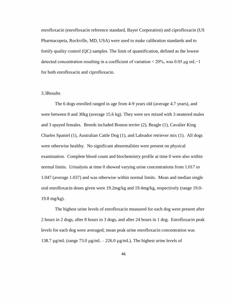
46
enrofloxacin (enrofloxacin reference standard, Bayer Corporation) and ciprofloxacin (US
Pharmacopeia, Rockville, MD, USA) were used to make calibration standards and to
fortify quality control (QC) samples. The limit of quantification, defined as the lowest
detected concentration resulting in a coefficient of variation < 20%, was 0.05 μg mL−1
for both enrofloxacin and ciprofloxacin.
3.3Results
The 6 dogs enrolled ranged in age from 4-9 years old (average 4.7 years), and
were between 8 and 30kg (average 15.6 kg). They were sex mixed with 3 neutered males
and 3 spayed females. Breeds included Boston terrier (2), Beagle (1), Cavalier King
Charles Spaniel (1), Australian Cattle Dog (1), and Labrador retriever mix (1). All dogs
were otherwise healthy. No significant abnormalities were present on physical
examination. Complete blood count and biochemistry profile at time 0 were also within
normal limits. Urinalysis at time 0 showed varying urine concentrations from 1.017 to
1.047 (average 1.037) and was otherwise within normal limits. Mean and median single
oral enrofloxacin doses given were 19.2mg/kg and 19.4mg/kg, respectively (range 19.0-
19.8 mg/kg).
The highest urine levels of enrofloxacin measured for each dog were present after
2 hours in 2 dogs, after 8 hours in 3 dogs, and after 24 hours in 1 dog. Enrofloxacin peak
levels for each dog were averaged; mean peak urine enrofloxacin concentration was
138.7 µg/mL (range 73.0 µg/mL – 226.0 µg/mL). The highest urine levels of
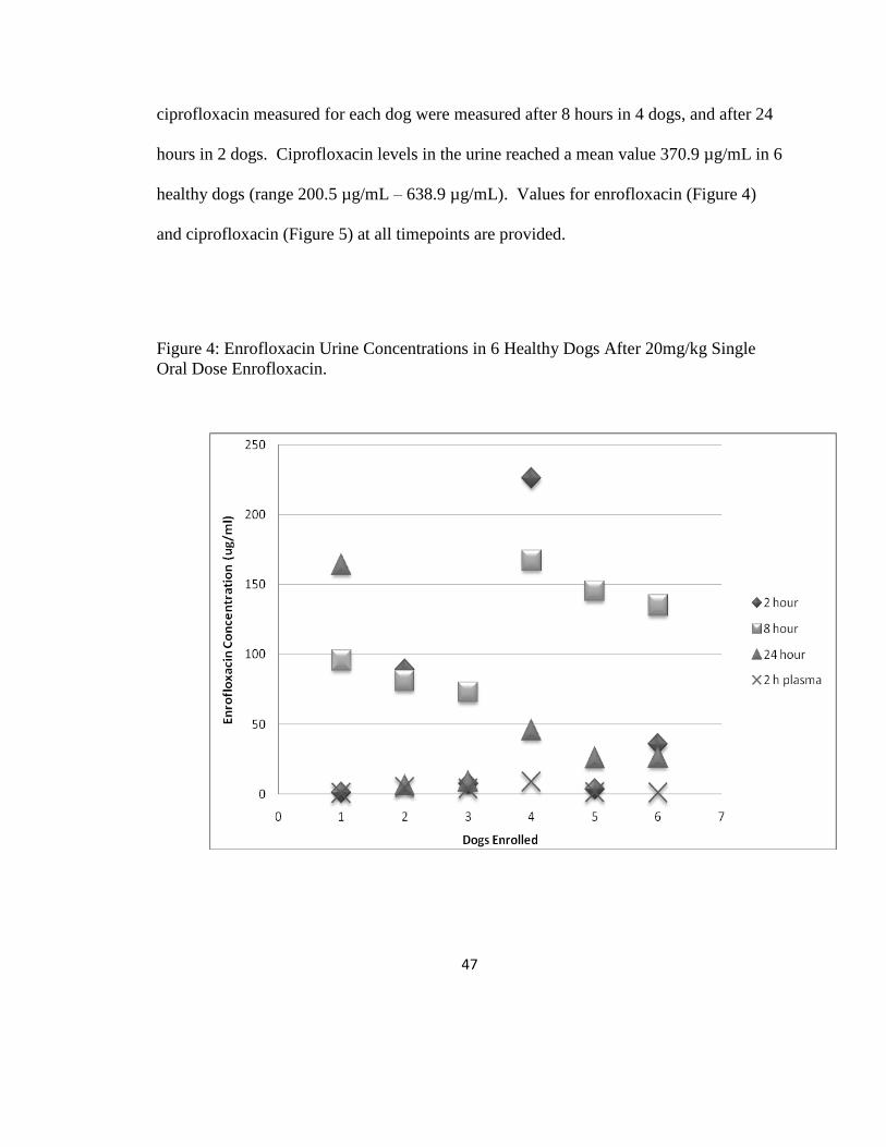
47
ciprofloxacin measured for each dog were measured after 8 hours in 4 dogs, and after 24
hours in 2 dogs. Ciprofloxacin levels in the urine reached a mean value 370.9 µg/mL in 6
healthy dogs (range 200.5 µg/mL – 638.9 µg/mL). Values for enrofloxacin (Figure 4)
and ciprofloxacin (Figure 5) at all timepoints are provided.
Figure 4: Enrofloxacin Urine Concentrations in 6 Healthy Dogs After 20mg/kg Single
Oral Dose Enrofloxacin.
48
Figure 5: Ciprofloxacin Urine Concentrations in 6 Healthy Dogs After 20mg/kg Single
Oral Dose Enrofloxacin
Average plasma level of enrofloxacin at 2 hours was 3.4 µg/mL (range 0.7 µg/mL
– 8.9 µg/mL). Average plasma level of ciprofloxacin at 2 hours after oral dosing was 0.5
µg/mL (range 0.18 µg/mL – 0.96 µg/mL). Plasma measurements were performed to
confirm systemic absorbtion post oral administration and were intended to approximate
peak plasma concentrations.
49
3.4 Discussion
Urine concentrations of antimicrobials are more relevant than serum
concentrations for predicting success of therapy for UTI in humans. 47
Efficacy of
treatment of UTI in dogs is also logically correlated with urine drug concentrations. The
data from these 6 dogs showed exponentially higher urine concentrations of enrofloxacin
and ciprofloxacin compared to plasma concentrations, 2 hours after a single 20mg/kg
dose of oral enrofloxacin was administered. The urine drug concentrations also
continued to peak at 8 and 24 hours in most of the dogs. We chose to measure
enrofloxacin and ciprofloxacin in plasma 2 hours post-administration to approximate
peak plasma levels. Peak plasma levels require continuous drug measurements due to
variability in individual dogs, and peak plasma enrofloxacin and ciprofloxacin
concentrations are reported at a mean of 2 hours with an approximate range of 1-3 hours.
We also compared our peak urine concentrations measured for each dog, to mean serum
enrofloxacin and ciprofloxacin levels previously reported in dogs following a 20 mg/kg
oral dose of enrofloxacin (previously reported maximum concentration (Cmax) mean of
4.7 µg/mL for enrofloxacin and 2.0 µg/mL for ciprofloxacin in 6 healthy dogs). 56
Our
measured urine levels were also markedly higher than serum levels in these dogs. These
results parallel previous data on urine concentrations following 5mg/kg doses of
enrofloxacin in dogs, with additive peak urine concentrations of enrofloxacin and
ciprofloxacin reaching over 100x that measured in plasma. 87
50
Ciprofloxacin was the major antimicrobial agent in the urine after oral
enrofloxacin administration during all the measured time-points. Ciprofloxacin is the
major metabolite of enrofloxacin, and is antimicrobially active with the same mechanism
of action as its prodrug; in a patient the activities of the two drugs are additive. 56
Ciprofloxacin contributes to antimicrobial activity following enrofloxacin administration,
but there are discrepancies regarding the contribution of ciprofloxacin to total
antimicrobial killing. Early studies approximated minimal contribution from
ciprofloxacin. Two more recent studies found that ciprofloxacin contributed more than
30% of drug concentration in plasma after oral dosing, and 34% in serum after 20mg/kg
was given IV to anesthetized dogs. 56,
58
In our study, ciprofloxacin levels were lower than enrofloxacin levels in plasma.
In the anaesthetized dog study, ciprofloxacin concentrations were lower than
enrofloxacin concentrations in all tissues with the exception of urine and gall bladder.
Ciprofloxacin is less lipophilic and thus cannot penetrate cell membranes as well as
enrofloxacin. This could explain the higher ciprofloxacin urine concentrations since more
ciprofloxacin would reside in extracellular fluid rather than being sequestered in
intracellular fluid. Drug measurements by reverse phase HPLC allowed us to accurately
determine concentrations of enrofloxacin and ciprofloxacin separately. Some of the
earlier studies measured enrofloxacin concentrations via bioassay, which measures all of
the active metabolites in the measurements. Such earlier studies cannot be used for
accurate comparison of drug levels. 88
51
We also noted marked variability in urine concentrations of enrofloxacin and
ciprofloxacin between individual dogs and no single time-point for peak urine
concentration was identified among the 6 dogs in the study. Therefore, we identified
peak urine concentrations for each individual dog and used these for maximum drug
levels in our analysis. Time of peak drug concentration in urine after oral administration
was variable among the six dogs in this study, and likely differs widely in the general dog
population as well as covarying with hydration status and rate of urine production. 1
Since it is not practical to conduct measurements of antimicrobial concentration of urine
in every single patient, expected urine concentrations in dogs with normal renal function
may be helpful in predicting therapeutic outcome.
Wide variation in plasma drug levels was also noted in our study. However, such
wide variability was also appreciated in earlier studies, despite having standardized
conditions and breed-standardized subjects. 59
Sources of variability likely include
efficiency of intestinal absorption (with or without food), core metabolism, and
elimination rates (how many times the patient is allowed to urinate, urine concentrating
ability). These factors likely contributed in our study. We used volunteer dogs owned by
OSU-VTH staff and students, and the study was performed using these dogs as
outpatients. Furthermore, although the dogs were fasted for 12 hours prior to drug
dosing, a non-standardized amount of dog food was given with the study drug to prevent
drug-induced emesis. After 2 hours, the dogs were allowed free access to food and water.
Although the study protocol elaborated on times allowed for urination, we relied on the
52
dog owners to comply with the study protocol. There may be differences in drug
disposition between male and female dogs which is why we chose an equal mix of male
and female dogs. 91
An additional weakness of the present study was that urine
concentrations were measured only following a single dose. It is questionable whether
enrofloxacin accumulates with repeated dosing. One study found no effect from repeated
dosing after 15 days of consecutive dosing compared to a single dose in healthy dogs. 87
This study first measured drug levels achieved with one intravenous 50mg dose. After a
drug-washout period, they administered 50mg enrofloxacin once daily for 15 consecutive
days to the same 8 dogs. They compared drug levels from a single dose to those attained
after 15 doses. Their analysis demonstrated no significant difference in drug levels after
repeated doses, demonstrating that enrofloxacin levels may not accumulate with repeated
dosing. 87
A different study found a higher Cmax when comparing drug concentrations
after the seventh dose versus the first dose when 2.75, 5, and 11mg/kg enrofloxacin was
given orally twice daily to 6 healthy dogs. 88
Although more convenient, plasma drug concentrations are less valuable than
determining active drug concentrations at the infection site. 58
Efficacy of treatment of
UTI is logically correlated with urine drug concentrations. Applying MIC interpretive
breakpoints for antimicrobial susceptibility, as defined by the Clinical Laboratory
Standards Institute, which are based on plasma drug concentrations may be too
conservative in UTIs. 47
The results from our study may be useful to guide enrofloxacin
therapy for canine UTI since current clinical breakpoints are based on plasma drug
53
concentrations. Urine concentrations of ciprofloxacin and enrofloxacin greatly exceed
clinical MICs of enrofloxacin for many specific bacteria cultured from the urinary tract of
dogs. 92
This approach is already used in the clinical microbiology laboratory at the
University of California-Davis, leading to less conservative clinical breakpoints for
urinary pathogens.
The minimum inhibitory concentration (MIC) is the lowest concentration of an
antibiotic that prevents visible growth after 18–24 hours of incubation. 47
The therapeutic
strategy for antimicrobial drugs is to achieve drug concentrations above the minimal
inhibitory concentration (MIC) during the dosing interval. This is simulated in vitro
using drug concentrations known to be achieved in plasma. For the fluoroquinolones,
studies integrating pharmacokinetics and pharmacodynamics (PK/PD) have shown that
the area under the concentration-time curve (AUC)/MIC and the maximum concentration
of drug in serum (Cmax)/MIC are the most important pharmacodynamic indices
predicting bacterial killing. Maximizing the Cmax to MIC ratio has also been linked to
decreased development of resistance, and a target Cmax of 8 to 10 times MIC was chosen
on the basis of evidence of bacterial regrowth at ratios < 8:1. 57
In addition to killing
bacteria in a concentration dependent manner, the fluoroquinolones also have a
substantial postantimicrobial effect in which the rate of bacterial growth remains
suppressed even after elimination of the antimicrobial, allowing for a longer dosing
interval. 89
54
The Clinical and Laboratory Standards Institute (CLSI) MIC breakpoints in µg/
mL for enrofloxacin are ≤0.5 (susceptible), 1–2 (intermediate) and ≥4 (resistant). 12
It is
recommended that the mean antimicrobial concentration available at the site of a urinary
tract infection should exceed 4 x MIC for bacteria to be susceptible to that agent. 93
Nevertheless, MIC breakpoints for susceptible bacteria may be different for urinary tract
infections, allowing enrofloxacin treatment for infections previously classified by CLSI
as resistant. Data showing microbiologic and clinical cure from NCCLS “resistant”
canine UTI with high doses of enrofloxacin would be necessary to support this
hypothesis. Furthermore, new MIC standards using ciprofloxacin may be beneficial for
optimal treatment of canine UTI.
3.5 Ongoing Research
Clinical and experimental studies indicate that suboptimal antibiotic dosage
regimens may be a significant risk factor for emergence of resistance. 46,
57 Hence, it is
important to optimize dosages, not only with respect to achieving a therapeutic effect, but
also with respect to minimizing resistance development. If urine enrofloxacin
concentrations achievable using the high limit of the labeled dosing range are sufficient
to prevent residual growth of common canine uropathogens, even after first-step
mutations have occurred, then this therapeutic strategy will prevent selection of resistance
during treatment.
55
Additional studies are ongoing. We are comparing the urine ciprofloxacin
concentrations attained after oral enrofloxacin administration to the minimum inhibitory
concentrations (MICs) of 50 clinical isolates from canine E. Coli UTI with varying levels
of susceptibility. We also plan to measure Mutant Prevention Concentration (MPC) for
10 of these isolates with a range of MIC values. Mutant prevention concentrations have
not been evaluated across a diverse collection of canine urinary tract infection isolates.
This information may be useful to help determine the appropriate doses of enrofloxacin
for the treatment of canine UTI while considering mutant prevention guidelines for
prevention of antimicrobial resistance. This may also change interpretation of MIC
susceptibility testing since it is based on plasma drug levels. Establishing these criteria
for canine UTI may provide guidance for veterinary practitioners using flexible dose-
range FQ drugs.
56
References
1 Ling, GV. “Therapeutic strategies involving antimicrobial treatment of the canine
urinary tract.” Journal of the American Veterinary Medical Association, v. 185 issue 10,
1984, p. 1162-4.
2 Seguin, MA., et al. “Persistent urinary tract infections and reinfections in 100 dogs
(1989-1999).” Journal of veterinary internal medicine / American College of Veterinary
Internal Medicine, v. 17 issue 5, 2003, p. 622-31.
3 Ling, GV.; Ruby, AL. “Aerobic bacterial flora of the prepuce, urethra, and vagina of
normal adult dogs.” American Journal of Veterinary Research, v. 39 issue 4, 1978, p.
695-8.
4 Ling, GV., et al. “Interrelations of organism prevalence, specimen collection method,
and host age, sex, and breed among 8,354 canine urinary tract infections (1969-1995).”
Journal of veterinary internal medicine / American College of Veterinary Internal
Medicine, v. 15 issue 4, 2001, p. 341-7.
5 Norris, CR., et al. “Recurrent and persistent urinary tract infections in dogs: 383 cases
(1969-1995).” Journal of the American Animal Hospital Association, v. 36 issue 6, 2000,
p. 484-92.
6 Forrester, SD., et al. “Retrospective evaluation of urinary tract infection in 42 dogs with
hyperadrenocorticism or diabetes mellitus or both.” Journal of veterinary internal
medicine / American College of Veterinary Internal Medicine, v. 13 issue 6, 1999, p. 557-
60.
7 Bartges, JW. “Diagnosis of urinary tract infections.” The Veterinary clinics of North
America : small animal practice, v. 34 issue 4, 2004, p. 923-33, vi.
8 Carter, JM.; Klausner, JS.; Osborne, CA.; Bates, FY. “Comparison of collection
techniques for quantitative urine culture in dogs.” Journal of the American Veterinary
Medical Association, v. 173 issue 3, 1978, p. 296-8.
57
9 Thomsen, MK.; Svane, LC.; Poulsen, PH. “Canine urinary tract infection. Detection,
prevalence and therapeutic consequences of bacteriuria.” Nordisk Veterinaermedicin,
v. 38 issue 6, 1986, p. 394-402.
10 Comer, KM.; Ling, GV. “Results of urinalysis and bacterial culture of canine urine
obtained by antepubic cystocentesis, catheterization, and the midstream voided methods.”
Journal of the American Veterinary Medical Association, v. 179 issue 9, 1981, p. 891-5.
11 Bush, BM. “The incidence of significant bacteriuria in the dog.” Tijdschrift voor
Diergeneeskunde, v. 103 issue 14, 1978, p. 750-7.
12 National Committee for Clinical Laboratory Standards (NCCLS). “Performance
Standards for Antimicrobial Disk and Dilution Susceptibility Tests for Bacteria Isolated
from Animals.” Informational Supplement NCCLS Document M31-S1, v. 24, Wayne, PA.
2004, p. 17. 13
Wikler, M. A., and P. G. Ambrose. “The breakpoint” In V. Lorian (ed.), Antibiotics in
laboratory medicine, 5th ed. Lippincott Williams & Wilkins, Philadelphia, PA. 2005, p.
1–7. 14
Mayhew, PD.; Lee, KC.; Gregory, SP.; Brockman, DJ. “Comparison of two surgical
techniques for management of intramural ureteral ectopia in dogs: 36 cases (1994-2004).”
Journal of the American Veterinary Medical Association, v. 229 issue 3, 2006, p. 389-93.
15 McLoughlin, MA.; Chew, DJ. “Diagnosis and surgical management of ectopic
ureters.” Clinical Techniques in Small Animal Practice, v. 15 issue 1, 2000, p. 17-24.
16 Adams, WM.; DiBartola, SP. “Radiographic and clinical features of pelvic bladder in
the dog.” Journal of the American Veterinary Medical Association, v. 182 issue 11, 1983,
p. 1212-7.
17 McGuire, NC.; Schulman, R.; Ridgway, MD.; Bollero, G. “Detection of occult urinary
tract infections in dogs with diabetes mellitus.” Journal of the American Animal Hospital
Association, v. 38 issue 6, 2002, p. 541-4.
18 Freedman, LR. “Experimental pyelonephritis. 13. On the ability of water diuresis to
induce susceptibility to E. coli bacteriuria in the normal rat.” The Yale journal of biology
and medicine, v. 39 issue 4, 1967, p. 255-66.
19 Mayer-Roenne, B.; Goldstein, RE.; Erb, HN. “Urinary tract infections in cats with
hyperthyroidism, diabetes mellitus and chronic kidney disease.” Journal of Feline
Medicine & Surgery, v. 9 issue 2, 2007, p. 124-32.
58
20
Gilbert, DN. “Urinary tract infections in patients with chronic renal insufficiency.”
Clinical journal of the American Society of Nephrology : CJASN, v. 1 issue 2, 2006, p.
327-31.
21 Chandler, ML., et al. “Juvenile nephropathy in 37 boxer dogs.” The Journal of small
animal practice, v. 48 issue 12, 2007, p. 690-4.
22 Mutsaers, AJ.; Widmer, WR.; Knapp, DW. “Canine transitional cell carcinoma.”
Journal of veterinary internal medicine / American College of Veterinary Internal
Medicine, v. 17 issue 2, 2003, p. 136-44.
23 Osborne, CA.; Klausner, JS.; Krawiec, DR.; Griffith, DP. “Canine struvite urolithiasis:
problems and their dissolution.” Journal of the American Veterinary Medical
Association, v. 179 issue 3, 1981, p. 239-44.
24 Francis, RS.; Shackelford, GD. “Cyclophosphamide cystitis with bladder wall
calcification.” Journal of the Canadian Association of Radiologists, v. 25 issue 4, 1974,
p. 324-6.
25 Bubenik, LJ.; Hosgood, GL.; Waldron, DR.; Snow, LA. “Frequency of urinary tract
infection in catheterized dogs and comparison of bacterial culture and susceptibility
testing results for catheterized and noncatheterized dogs with urinary tract infections.”
Journal of the American Veterinary Medical Association, v. 231 issue 6, 2007, p. 893-9.
26 Bubenik, L.; Hosgood, G. “Urinary tract infection in dogs with thoracolumbar
intervertebral disc herniation and urinary bladder dysfunction managed by manual
expression, indwelling catheterization or intermittent catheterization.” Veterinary surgery
: VS : the official journal of the American College of Veterinary Surgeons, v. 37 issue 8,
2008, p. 791-800.
27 Hooton, TM., et al. “A prospective study of risk factors for symptomatic urinary tract
infection in young women.” New England journal of medicine, v. 335 issue 7, 1996, p.
468-74.
28 Torres, SM., et al. “Frequency of urinary tract infection among dogs with pruritic
disorders receiving long-term glucocorticoid treatment.” Journal of the American
Veterinary Medical Association, v. 227 issue 2, 2005, p. 239-43.
29 Senior, DF.; deMan, P.; Svanborg, C. “Serotype, hemolysin production, and adherence
characteristics of strains of Escherichia coli causing urinary tract infection in dogs.”
American Journal of Veterinary Research, v. 53 issue 4, 1992, p. 494-8.
59
30
Anderson, GG.; Dodson, KW.; Hooton, TM.; Hultgren, SJ. “Intracellular bacterial
communities of uropathogenic Escherichia coli in urinary tract pathogenesis.” Trends in
Microbiology, v. 12 issue 9, 2004, p. 424-30.
31 Bailiff, NL.; Westropp, JL.; Jang, SS.; Ling, GV. “Corynebacterium urealyticum
urinary tract infection in dogs and cats: 7 cases (1996-2003).” Journal of the American
Veterinary Medical Association, v. 226 issue 10, 2005, p. 1676-80.
32 Jin, Y.; Lin, D. “Fungal urinary tract infections in the dog and cat: a retrospective study
(2001-2004).” Journal of the American Animal Hospital Association, v. 41 issue 6, 2005,
p. 373-81.
33 Pressler, BM., et al. “Candida spp. urinary tract infections in 13 dogs and seven cats:
predisposing factors, treatment, and outcome.” Journal of the American Animal Hospital
Association, v. 39 issue 3, 2003, p. 263-70.
34 Cox, C.; Hinman F. “Experiments with induced bacteriuria, vesical emptying and
bacterial growth on the mechanism of bladder defense to infection.” The Journal of
Urology, v. 86, 1961, p. 739-48.
35 Garrison, J.; Hooton, TM. “Fluoroquinolones in the treatment of acute uncomplicated
urinary tract infections in adult women.” Expert Opinion on Pharmacotherapy, v. 2
issue 8, 2001, p. 1227-37.
36 Dhakal, BK.; Kulesus, RR.; Mulvey, MA. “Mechanisms and consequences of bladder
cell invasion by uropathogenic Escherichia coli.” European Journal of Clinical
Investigation, v. 38 Suppl 2, 2008, p. 2-11.
37 Johnson, JR.; Kaster, N.; Kuskowski, MA.; Ling, GV. “Identification of urovirulence
traits in Escherichia coli by comparison of urinary and rectal E. coli isolates from dogs
with urinary tract infection.” Journal of Clinical Microbiology, v. 41 issue 1, 2003, p.
337-45.
38 Siqueira, AK., et al. “Virulence factors in Escherichia coli strains isolated from urinary
tract infection and pyometra cases and from feces of healthy dogs.” Research in
Veterinary Science, v. 86 issue 2, 2009, p. 206-10.
39 Oluoch, AO., et al. “Nonenteric Escherichia coli isolates from dogs: 674 cases (1990-
1998).” Journal of the American Veterinary Medical Association, v. 218 issue 3, 2001, p.
381-4.
40 Wadas, et al. “Biochemical phenotypes of Escherichia coli in dogs: comparison of
isolates isolated from bitches suffering from pyometra and urinary tract infection with
60
isolates from faeces of healthy dogs.” Veterinary Microbiology, v. 52 issue 3-4, 1996, p.
293-300.
41 Feria, et al. “Virulence genes and P fimbriae PapA subunit diversity in canine and
feline uropathogenic Escherichia coli.” Veterinary Microbiology, v. 82 issue 1, 2001, p.
81-9.
42 Stenske, KA., et al. “Prevalence of urovirulence genes cnf, hlyD, sfa/foc, and papGIII
in fecal Escherichia coli from healthy dogs and their owners.” American Journal of
Veterinary Research, v. 70 issue 11, 2009, p. 1401-6.
43 Johnson, JR., et al. “Virulence genotypes and phylogenetic background of
fluoroquinolone-resistant and susceptible Escherichia coli urine isolates from dogs with
urinary tract infection.” Veterinary Microbiology, v. 136 issue 1-2, 2009, p. 108-14.
44 Anderson, GG.; Dodson, KW.; Hooton, TM.; Hultgren, SJ. “Intracellular bacterial
communities of uropathogenic Escherichia coli in urinary tract pathogenesis.” Trends in
Microbiology, v. 12 issue 9, 2004, p. 424-30.
45 Drazenovich, N.; Ling, GV.; Foley, J. “Molecular investigation of Escherichia coli
strains associated with apparently persistent urinary tract infection in dogs.” Journal of
veterinary internal medicine / American College of Veterinary Internal Medicine, v. 18
issue 3, 2004, p. 301-6.
46 Prescott, JF.; Hanna, WJ.; Reid-Smith, R.; Drost, K. “Antimicrobial drug use and
resistance in dogs.” The Canadian veterinary journal. La revue veterinaire canadienne,
v. 43 issue 2, 2002, p. 107-16.
47 Stamey, TA., et al. “Serum versus urinary antimicrobial concentrations in cure of
urinary-tract infections.” New England journal of medicine, v. 291 issue 22, 1974, p.
1159-63.
48 Lees, GE. “Bacterial urinary tract infections.” The Veterinary clinics of North America
: small animal practice, v. 26 issue 2, 1996, p. 297-304.
49 Rogers, KS.; Lees, GE.; Simpson, RB. “Effects of single-dose and three-day
trimethoprim-sulfadiazine and amikacin treatment of induced Escherichia coli urinary
tract infections in dogs.” American Journal of Veterinary Research, v. 49 issue 3, 1988,
p. 345-9.
50 Bayley AJ. Compendium of Veterinary Products, 8th edn. Port Huron. North American
Compendiums, Inc., 2005, p. 1297.
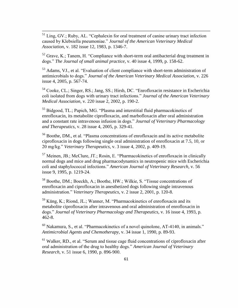
61
51
Ling, GV.; Ruby, AL. “Cephalexin for oral treatment of canine urinary tract infection
caused by Klebsiella pneumoniae.” Journal of the American Veterinary Medical
Association, v. 182 issue 12, 1983, p. 1346-7.
52 Grave, K.; Tanem, H. “Compliance with short-term oral antibacterial drug treatment in
dogs.” The Journal of small animal practice, v. 40 issue 4, 1999, p. 158-62.
53 Adams, VJ., et al. “Evaluation of client compliance with short-term administration of
antimicrobials to dogs.” Journal of the American Veterinary Medical Association, v. 226
issue 4, 2005, p. 567-74.
54 Cooke, CL.; Singer, RS.; Jang, SS.; Hirsh, DC. “Enrofloxacin resistance in Escherichia
coli isolated from dogs with urinary tract infections.” Journal of the American Veterinary
Medical Association, v. 220 issue 2, 2002, p. 190-2.
55 Bidgood, TL.; Papich, MG. “Plasma and interstitial fluid pharmacokinetics of
enrofloxacin, its metabolite ciprofloxacin, and marbofloxacin after oral administration
and a constant rate intravenous infusion in dogs.” Journal of Veterinary Pharmacology
and Therapeutics, v. 28 issue 4, 2005, p. 329-41.
56 Boothe, DM., et al. “Plasma concentrations of enrofloxacin and its active metabolite
ciprofloxacin in dogs following single oral administration of enrofloxacin at 7.5, 10, or
20 mg/kg.” Veterinary Therapeutics, v. 3 issue 4, 2002, p. 409-19.
57 Meinen, JB.; McClure, JT.; Rosin, E. “Pharmacokinetics of enrofloxacin in clinically
normal dogs and mice and drug pharmacodynamics in neutropenic mice with Escherichia
coli and staphylococcal infections.” American Journal of Veterinary Research, v. 56
issue 9, 1995, p. 1219-24.
58 Boothe, DM.; Boeckh, A.; Boothe, HW.; Wilkie, S. “Tissue concentrations of
enrofloxacin and ciprofloxacin in anesthetized dogs following single intravenous
administration.” Veterinary Therapeutics, v. 2 issue 2, 2001, p. 120-8.
59 Küng, K.; Riond, JL.; Wanner, M. “Pharmacokinetics of enrofloxacin and its
metabolite ciprofloxacin after intravenous and oral administration of enrofloxacin in
dogs.” Journal of Veterinary Pharmacology and Therapeutics, v. 16 issue 4, 1993, p.
462-8.
60 Nakamura, S., et al. “Pharmacokinetics of a novel quinolone, AT-4140, in animals.”
Antimicrobial Agents and Chemotherapy, v. 34 issue 1, 1990, p. 89-93.
61 Walker, RD., et al. “Serum and tissue cage fluid concentrations of ciprofloxacin after
oral administration of the drug to healthy dogs.” American Journal of Veterinary
Research, v. 51 issue 6, 1990, p. 896-900.
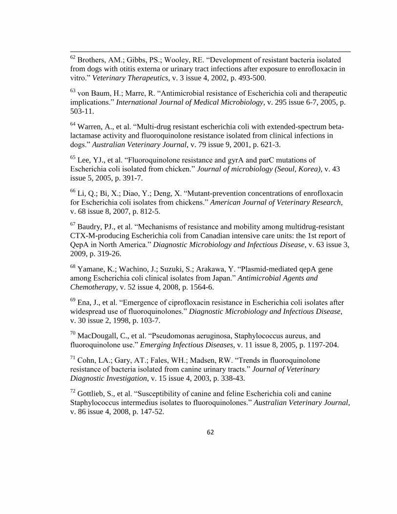
62
62
Brothers, AM.; Gibbs, PS.; Wooley, RE. “Development of resistant bacteria isolated
from dogs with otitis externa or urinary tract infections after exposure to enrofloxacin in
vitro.” Veterinary Therapeutics, v. 3 issue 4, 2002, p. 493-500.
63 von Baum, H.; Marre, R. “Antimicrobial resistance of Escherichia coli and therapeutic
implications.” International Journal of Medical Microbiology, v. 295 issue 6-7, 2005, p.
503-11.
64 Warren, A., et al. “Multi-drug resistant escherichia coli with extended-spectrum beta-
lactamase activity and fluoroquinolone resistance isolated from clinical infections in
dogs.” Australian Veterinary Journal, v. 79 issue 9, 2001, p. 621-3.
65 Lee, YJ., et al. “Fluoroquinolone resistance and gyrA and parC mutations of
Escherichia coli isolated from chicken.” Journal of microbiology (Seoul, Korea), v. 43
issue 5, 2005, p. 391-7.
66 Li, Q.; Bi, X.; Diao, Y.; Deng, X. “Mutant-prevention concentrations of enrofloxacin
for Escherichia coli isolates from chickens.” American Journal of Veterinary Research,
v. 68 issue 8, 2007, p. 812-5.
67 Baudry, PJ., et al. “Mechanisms of resistance and mobility among multidrug-resistant
CTX-M-producing Escherichia coli from Canadian intensive care units: the 1st report of
QepA in North America.” Diagnostic Microbiology and Infectious Disease, v. 63 issue 3,
2009, p. 319-26.
68 Yamane, K.; Wachino, J.; Suzuki, S.; Arakawa, Y. “Plasmid-mediated qepA gene
among Escherichia coli clinical isolates from Japan.” Antimicrobial Agents and
Chemotherapy, v. 52 issue 4, 2008, p. 1564-6.
69 Ena, J., et al. “Emergence of ciprofloxacin resistance in Escherichia coli isolates after
widespread use of fluoroquinolones.” Diagnostic Microbiology and Infectious Disease,
v. 30 issue 2, 1998, p. 103-7.
70 MacDougall, C., et al. “Pseudomonas aeruginosa, Staphylococcus aureus, and
fluoroquinolone use.” Emerging Infectious Diseases, v. 11 issue 8, 2005, p. 1197-204.
71 Cohn, LA.; Gary, AT.; Fales, WH.; Madsen, RW. “Trends in fluoroquinolone
resistance of bacteria isolated from canine urinary tracts.” Journal of Veterinary
Diagnostic Investigation, v. 15 issue 4, 2003, p. 338-43.
72 Gottlieb, S., et al. “Susceptibility of canine and feline Escherichia coli and canine
Staphylococcus intermedius isolates to fluoroquinolones.” Australian Veterinary Journal,
v. 86 issue 4, 2008, p. 147-52.
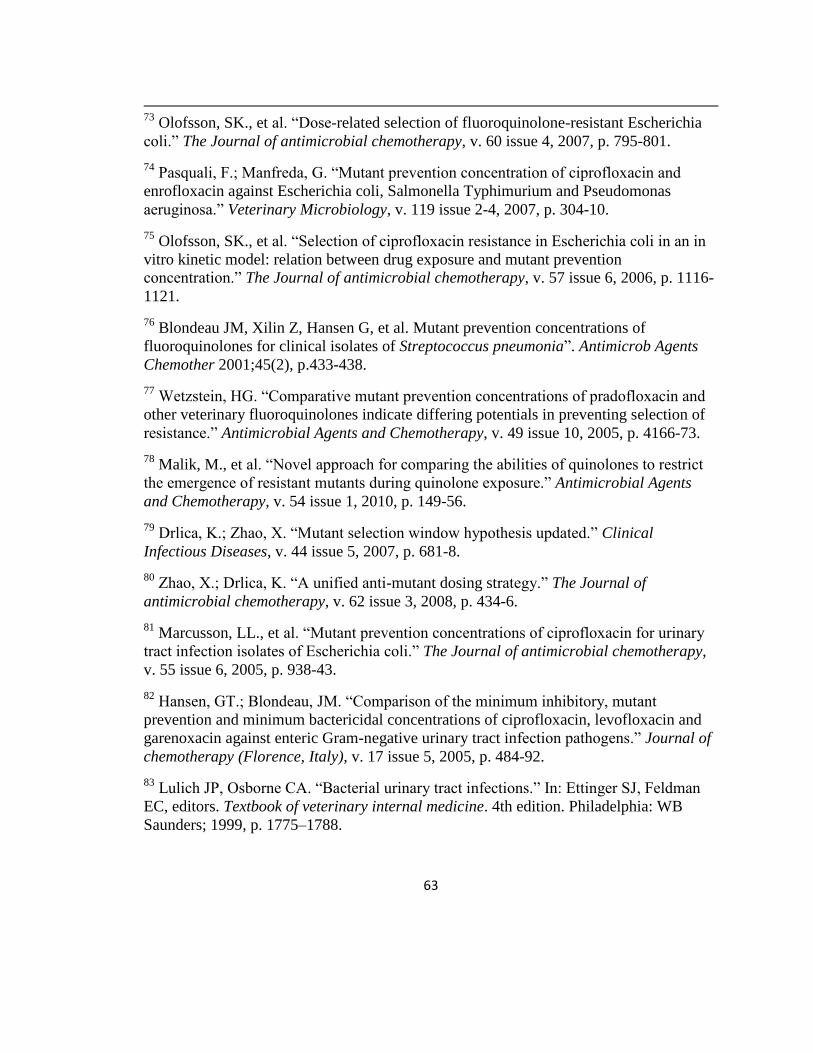
63
73
Olofsson, SK., et al. “Dose-related selection of fluoroquinolone-resistant Escherichia
coli.” The Journal of antimicrobial chemotherapy, v. 60 issue 4, 2007, p. 795-801.
74 Pasquali, F.; Manfreda, G. “Mutant prevention concentration of ciprofloxacin and
enrofloxacin against Escherichia coli, Salmonella Typhimurium and Pseudomonas
aeruginosa.” Veterinary Microbiology, v. 119 issue 2-4, 2007, p. 304-10.
75 Olofsson, SK., et al. “Selection of ciprofloxacin resistance in Escherichia coli in an in
vitro kinetic model: relation between drug exposure and mutant prevention
concentration.” The Journal of antimicrobial chemotherapy, v. 57 issue 6, 2006, p. 1116-
1121.
76 Blondeau JM, Xilin Z, Hansen G, et al. Mutant prevention concentrations of
fluoroquinolones for clinical isolates of Streptococcus pneumonia”. Antimicrob Agents
Chemother 2001;45(2), p.433-438.
77 Wetzstein, HG. “Comparative mutant prevention concentrations of pradofloxacin and
other veterinary fluoroquinolones indicate differing potentials in preventing selection of
resistance.” Antimicrobial Agents and Chemotherapy, v. 49 issue 10, 2005, p. 4166-73.
78 Malik, M., et al. “Novel approach for comparing the abilities of quinolones to restrict
the emergence of resistant mutants during quinolone exposure.” Antimicrobial Agents
and Chemotherapy, v. 54 issue 1, 2010, p. 149-56.
79 Drlica, K.; Zhao, X. “Mutant selection window hypothesis updated.” Clinical
Infectious Diseases, v. 44 issue 5, 2007, p. 681-8.
80 Zhao, X.; Drlica, K. “A unified anti-mutant dosing strategy.” The Journal of
antimicrobial chemotherapy, v. 62 issue 3, 2008, p. 434-6.
81 Marcusson, LL., et al. “Mutant prevention concentrations of ciprofloxacin for urinary
tract infection isolates of Escherichia coli.” The Journal of antimicrobial chemotherapy,
v. 55 issue 6, 2005, p. 938-43.
82 Hansen, GT.; Blondeau, JM. “Comparison of the minimum inhibitory, mutant
prevention and minimum bactericidal concentrations of ciprofloxacin, levofloxacin and
garenoxacin against enteric Gram-negative urinary tract infection pathogens.” Journal of
chemotherapy (Florence, Italy), v. 17 issue 5, 2005, p. 484-92.
83 Lulich JP, Osborne CA. “Bacterial urinary tract infections.” In: Ettinger SJ, Feldman
EC, editors. Textbook of veterinary internal medicine. 4th edition. Philadelphia: WB
Saunders; 1999, p. 1775–1788.
64
84
Passmore, CA.; Sherington, J.; Stegemann, MR. “Efficacy and safety of cefovecin
(Convenia) for the treatment of urinary tract infections in dogs.” The Journal of small
animal practice, v. 48 issue 3, 2007, p. 139-44.
85 Boothe, DM.; Boeckh, A.; Simpson, RB.; Dubose, K. “Comparison of
pharmacodynamic and pharmacokinetic indices of efficacy for 5 fluoroquinolones toward
pathogens of dogs and cats.” Journal of veterinary internal medicine / American College
of Veterinary Internal Medicine, v. 20 issue 6, 2006, p. 1297-306.
86 Cotard, JP., et al. “Comparative study of marbofloxacin and amoxicillin-clavulanic
acid in the treatment of urinary tract infections in dogs.” The Journal of small animal
practice, v. 36 issue 8, 1995, p. 349-53.
87 Monlouis, JD. “Plasma pharmacokinetics and urine concentrations after oral
administration of enrofloxacin to dogs.” Journal of veterinary pharmacology and
therapeutics, v. 20 suppl 1, 1997, p. 61-63.
88 Walker, RD.; Stein, GE.; Hauptman, JG.; MacDonald, KH. “Pharmacokinetic
evaluation of enrofloxacin administered orally to healthy dogs.” American Journal of
Veterinary Research, v. 53 issue 12, 1992, p. 2315-9.
89 Bermingham, EC.; Papich, MG. “Pharmacokinetics after intravenous and oral
administration of enrofloxacin in sheep.” American Journal of Veterinary Research, v. 63
issue 7, 2002, p. 1012-7.
90 Cole, LK., et al. “Plasma and ear tissue concentrations of enrofloxacin and its
metabolite ciprofloxacin in dogs with chronic end-stage otitis externa after intravenous
administration of enrofloxacin.” Veterinary Dermatology, v. 20 issue 1, 2009, p. 51-9.
91 Ling, GV.; Gilmore, CJ. “Penicillin G or ampicillin for oral treatment of canine urinary
tract infections.” Journal of the American Veterinary Medical Association, v. 171 issue 4,
1977, p. 358-61.
92 Lautzenhiser, SJ.; Fialkowski, JP.; Bjorling, D.; Rosin, E. “In vitro antibacterial
activity of enrofloxacin and ciprofloxacin in combination against Escherichia coli and
staphylococcal clinical isolates from dogs.” Research in Veterinary Science, v. 70 issue 3,
2001, p. 239-41.
93 Wilson, BJ., et al. “Susceptibility of bacteria from feline and canine urinary tract
infections to doxycycline and tetracycline concentrations attained in urine four hours
after oral dosage.” Australian Veterinary Journal, v. 84 issue 1-2, 2006, p. 8-11.