imaging of canine neoplastic reproductive disorders - mdpi
TRANSCRIPT
animals
Review
Imaging of Canine Neoplastic Reproductive Disorders
Marco Russo 1,* , Gary C.W. England 2, Giuseppe Catone 3 and Gabriele Marino 3,*
�����������������
Citation: Russo, M.; England,
G.C.W.; Catone, G.; Marino, G.
Imaging of Canine Neoplastic
Reproductive Disorders. Animals
2021, 11, 1213. https://doi.org/
10.3390/ani11051213
Academic Editor:
Emir Hadzijusufovic
Received: 20 March 2021
Accepted: 19 April 2021
Published: 22 April 2021
Publisher’s Note: MDPI stays neutral
with regard to jurisdictional claims in
published maps and institutional affil-
iations.
Copyright: © 2021 by the authors.
Licensee MDPI, Basel, Switzerland.
This article is an open access article
distributed under the terms and
conditions of the Creative Commons
Attribution (CC BY) license (https://
creativecommons.org/licenses/by/
4.0/).
1 Department of Veterinary Medicine and Animal Production, University of Naples, Federico II,80137 Naples, Italy
2 Sutton Bonington Campus, School of Veterinary Medicine and Science, University of Nottingham,Loughborough LE12 5RD, UK; [email protected]
3 Department of Veterinary Sciences, University of Messina, 98168 Messina, Italy; [email protected]* Correspondence: [email protected] (M.R.); [email protected] or [email protected] (G.M.)
Simple Summary: The diagnosis of canine reproductive neoplasia remains challenging as noneof the routinely performed diagnostic methods appear to have sufficient sensitivity or specificity.In recent years, advanced imaging techniques have been successfully performed in small animals;however, even though the incidence of reproductive neoplasia is high, no data are available onthe performance of these techniques. This review evaluates the applicability of various diagnosticimaging modalities in dogs and describes the findings and specific patterns that may characterisedifferent tumour types. Lamentably, some of the advanced imaging techniques have not yet beenadopted as first-line diagnostic tools, although it is clear that in the future they will become importantmethods for the detection of male and female reproductive neoplasia.
Abstract: Diagnostic imaging plays an essential role in the diagnosis and management of reproductiveneoplasia in dogs and cats. The initial diagnosis, staging, and planning of surgical and radiationtreatment and the response to therapy all involve imaging to varying degrees. Routine radiographs,ultrasound, nuclear medicine, and cross-sectional imaging in the form of computed tomography(CT) and magnetic resonance imaging (MRI) are routinely used in canine reproductive disorders.The choice of imaging modality depends on many factors, including the level of referral and thepathological information required. The biological behaviour of the tumour also guides the choice ofimaging in cancer staging, and imaging may play an important role in guiding serial tumour biopsyduring the course of therapy. The sophistication of imaging modalities is increasing exponentially.Each modality has advantages and disadvantages in terms of cost, availability, sensitivity, specificity,and qualities of anatomic versus functional imaging.
Keywords: dog; ovaries; uterus; vagina; testes; prostate; penis; radiography; ultrasound; computedtomography; magnetic resonance
1. Introduction
Several imaging modalities are now available in small animal oncology and in thestudy of neoplasia involving the female and male reproductive tracts. Each modalityhas advantages and disadvantages that may relate to cost, availability, sensitivity, andspecificity; some provide ‘anatomic imaging’ whilst others provide ‘functional imaging’.Each method may have different roles in diagnosis, staging, treatment selection and follow-up. The use of diagnostic imaging as an aid in canine and feline reproduction has evolvedover the past twenty years from its initial role in early pregnancy diagnosis to its currentuse as an integral component in the management of reproductive cases. Ultrasonographyis sensitive for the detection of lesions, but it is not specific for the aetiology of a disease.Therefore, biopsy or fine-needle aspiration of the lesion may be necessary. Ultrasound-guided sampling of tissue can be performed quickly, accurately, and safely. Advancesin ultrasound equipment and the development of ultrasonographic contrast agents are
Animals 2021, 11, 1213. https://doi.org/10.3390/ani11051213 https://www.mdpi.com/journal/animals
Animals 2021, 11, 1213 2 of 12
increasing the diagnostic specificity of ultrasound. For example, Doppler techniques areused to detect tumour vessels, which are usually seen to be tortuous and at high velocitycompared with vessels present within normal tissue.
This review describes both standard and innovative approaches to imaging in dogswith suspected reproductive malignancy and highlights the important contribution ofimaging to the management of these patients.
2. Female2.1. Reproductive Anatomy
The ovaries are located immediately caudal to the kidneys and are positioned close tothe abdominal wall. The uterus of the bitch is roughly ‘Y’-shaped and has relatively longuterine horns which are positioned proximally, close to the abdominal wall, convergingdistally in the midline at the uterine body.
2.2. Imaging of the Ovaries
The ovaries are located in the dorsal abdomen slightly lateral to the caudal poles of thekidneys. Normal-sized or mildly enlarged ovaries are not visible radiographically, limitingthe usefulness of this technique and making ultrasound the first-choice in the evaluationof ovarian disorders. Multifrequency curvilinear or linear 5–11 MHz transducers are thestandard equipment for ovarian examination. High-resolution probes (i.e., 18 MHz ormore) are currently available and may maximise the ability to visualise subtle changes inthe ovary. The caudal pole of the kidneys and the adjacent area are examined in sagittaland transverse planes to localise the ovaries. Their identification may be facilitated by theappearance of marginal artefacts dorsal to each ovary. The changes seen in the bitch ovaryduring the reproductive cycle have been described [1]. The appearance varies according tothe stage of the oestrous cycle when follicular growth can be readily detected. Multipleanechoic structures can be observed during proestrus and oestrus; after ovulation, thicker-walled corpora lutea are present during late oestrus and the first phase of dioestrus. Roundand hypoechoic corpora lutea are easily detectable in mid-dioestrus when they deform theotherwise oval profile of the ovary.
Imaging of Ovarian Neoplasia
Survey radiography was the first technique described for imaging canine ovariantumours; however, radiography provides little information regarding ovarian mass archi-tecture. When the ovary is significantly enlarged, it results in a ventrally located massthat causes medial, but not ventral displacement of adjacent organs on radiographs. Rightovarian masses displace the descending duodenum and ascending colon medially, whereasleft ovarian masses displace the descending colon medially [2–13]. Peritoneal or pleuraleffusion may be present when there are either benign or malignant masses [2,13]. Distantmetastases may be detected in thoracic projections [6]. Radiographic examination is notsensitive and other tumours, atypical pyometra, and retroperitoneal abscesses cannotbe excluded. Occasionally, ovarian teratomas and teratocarcinomas contain mineralisedareas, which are visible radiographically [4]. The easy accessibility and relative low costof ultrasound have made it the study of choice in the initial evaluation of a patient with asuspected ovarian neoplasia.
Confirmation that a mass is ovarian in origin is based on its appropriate location, beingcaudal to the kidneys, and ideally having an association with an adjacent uterine horn.The adjacent uterus can be confirmed by imaging the ovarian veins with Colour Doppler.Ultrasonographically, ovaries in bitches with ovarian tumours may appear unilaterallyenlarged, with regional or focal lesions that may be solid or cystic. Frequently there isa significant disruption of the normal appearance with an inhomogeneous echotexture.Tumours may be small or large and may be solid, contain small cysts, or be primarily cysticin appearance. A literature search of ultrasound imaging of ovarian masses identified onlytwo case series [2,13] and numerous case reports. Ultrasonography appears to be sensitive
Animals 2021, 11, 1213 3 of 12
for the detection of ovarian masses, but there are no exclusive patterns that confirm thediagnosis of tumour type. Unfortunately, no morphological scoring systems is available inveterinary medicine to standardise the interpretation of ultrasound images.
An attempt to classify ovarian masses based on ultrasonographic appearance has beenproposed [2] and includes three groups: solid masses (less than 10% anechoic cavities),solid masses with a cystic component (from 10% to 50%), or cystic masses (greater than50%) [2]. Ultrasound images of the ovarian neoplasia were described on their location,size, outer margins, and echogenicity presence of free abdominal fluid, evidence of uterineabnormalities, and signs of metastatic disease. The tumours were ultrasonographicallyclassified as solid (adenocarcinoma, thecoma) (solid with cystic component (adenocarcino-mas, granulosa cell tumour, dysgerminoma)), and cystic (adenoma, teratoma). The size ofthe anechoic cavities ranged from 0.2 to 3.5 cm in diameter. The solid parenchyma had afairly uniform appearance in all [5,7,8]. Teratomas are often cystic and may show partialmineralisation with distal shadowing, due to structures such as hair, skin, sweat glands,cartilage, bone, and teeth, which might help to distinguish these tumours from other ovar-ian masses. Finally, other rare tumours within the ovary have been described and imaged,including leiomyoma, which typically has a solid appearance [12] and hemangiosarcoma,which has been reported as solid with a heterogeneous echotexture. Uterine changes (cysticendometrial hyperplasia or pyometra) and/or ascites are commonly seen in hormonallyactive (sex-cord stromal) tumours [2] but are not exclusive to such tumours [7]; the presenceof free fluid may be associated with peritoneal dissemination, and careful evaluation formetastatic disease should be performed. Colour Doppler ultrasound may help identifysolid, vascularised components in an ovarian mass [9]. In humans, spectral Doppler wave-form characteristics were shown to correlate well with malignancy [13]. Furthermore,contrast-enhanced ultrasound (CEUS) has been used for the pre-operative evaluation ofearly benign or malignant masses by imaging tumour microvascularity [14]. Unfortunately,there are no studies describing the use of these techniques in canine ovarian tumours.
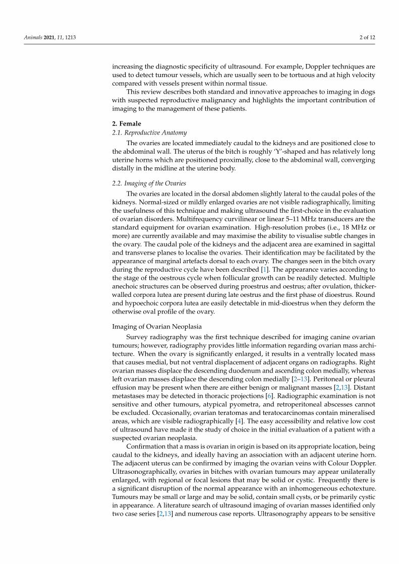
When imaged with computed tomography (CT), ovarian tumours in dogs appear aslarge soft-tissue attenuated masses located in the mid-ventral abdomen, with moderate orsevere contrast enhancement [9]. CT may help to assess the extent of disease in patientsbefore and after primary surgery. The separation of abdominal organs from the tumourmay be easily visualised (Figure 1A,B).
Animals 2021, 11, 1213 3 of 12
masses identified only two case series [2,13] and numerous case reports. Ultrasonogra-phy appears to be sensitive for the detection of ovarian masses, but there are no exclusive patterns that confirm the diagnosis of tumour type. Unfortunately, no morphological scoring systems is available in veterinary medicine to standardise the interpretation of ultrasound images.
An attempt to classify ovarian masses based on ultrasonographic appearance has been proposed [2] and includes three groups: solid masses (less than 10% anechoic cavi-ties), solid masses with a cystic component (from 10% to 50%), or cystic masses (greater than 50%) [2]. Ultrasound images of the ovarian neoplasia were described on their loca-tion, size, outer margins, and echogenicity presence of free abdominal fluid, evidence of uterine abnormalities, and signs of metastatic disease. The tumours were ultraso-nographically classified as solid (adenocarcinoma, thecoma) (solid with cystic compo-nent (adenocarcinomas, granulosa cell tumour, dysgerminoma)), and cystic (adenoma, teratoma). The size of the anechoic cavities ranged from 0.2 to 3.5 cm in diameter. The solid parenchyma had a fairly uniform appearance in all [5,7,8]. Teratomas are often cystic and may show partial mineralisation with distal shadowing, due to structures such as hair, skin, sweat glands, cartilage, bone, and teeth, which might help to distinguish these tumours from other ovarian masses. Finally, other rare tumours within the ovary have been described and imaged, including leiomyoma, which typically has a solid ap-pearance [12] and hemangiosarcoma, which has been reported as solid with a heteroge-neous echotexture. Uterine changes (cystic endometrial hyperplasia or pyometra) and/or ascites are commonly seen in hormonally active (sex-cord stromal) tumours [2] but are not exclusive to such tumours [7]; the presence of free fluid may be associated with per-itoneal dissemination, and careful evaluation for metastatic disease should be performed. Colour Doppler ultrasound may help identify solid, vascularised components in an ovarian mass [9]. In humans, spectral Doppler waveform characteristics were shown to correlate well with malignancy [13]. Furthermore, contrast-enhanced ultrasound (CEUS) has been used for the pre-operative evaluation of early benign or malignant masses by imaging tumour microvascularity [14]. Unfortunately, there are no studies describing the use of these techniques in canine ovarian tumours.
When imaged with computed tomography (CT), ovarian tumours in dogs appear as large soft-tissue attenuated masses located in the mid-ventral abdomen, with moderate or severe contrast enhancement [9]. CT may help to assess the extent of disease in pa-tients before and after primary surgery. The separation of abdominal organs from the tumour may be easily visualised (Figure 1A,B).
Figure 1. The computed tomography (CT) appearance of a granulosa cell tumour that appears as a large, well-defined low-attenuation ovarian mass. (A) Non-enhanced CT scan shows multi cystic soft-tissue mass. (B) After contrast admin-istration, CT scan shows mass as mildly and non-homogeneously enhanced. (C) Ultrasound longitudinal right ovarian mass with heterogeneous echotexture and multiloculated solid and cystic mass. (D) Sagittal cut of the right ovary con-taining polycystic structures.
With CT, the origin of the tumour may be confirmed, and the assessment of surgical options may be significantly improved compared with other imaging modalities. Similar to radiography, CT may identify mineralised areas in some ovarian masses and tooth-like
Figure 1. The computed tomography (CT) appearance of a granulosa cell tumour that appears as a large, well-defined low-attenuation ovarian mass. (A) Non-enhanced CT scan shows multi cystic soft-tissue mass. (B) After contrast administration,CT scan shows mass as mildly and non-homogeneously enhanced. (C) Ultrasound longitudinal right ovarian masswith heterogeneous echotexture and multiloculated solid and cystic mass. (D) Sagittal cut of the right ovary containingpolycystic structures.
With CT, the origin of the tumour may be confirmed, and the assessment of surgicaloptions may be significantly improved compared with other imaging modalities. Similarto radiography, CT may identify mineralised areas in some ovarian masses and tooth-likestructures in teratomas. The CT appearance of an ovarian papillary adenocarcinoma hasbeen described, showing an obvious internal structure, areas of contrast enhancement. andconnection with the ovarian vein [9]. An ovarian dysgerminoma associated with pleural
Animals 2021, 11, 1213 4 of 12
and abdominal effusions was also detected by CT and followed during chemotherapy [5].Ovarian leiomyoma has been described as a heterogeneous mass with intense contrastenhancement [12]. Magnetic resonance imaging (MRI) imaging is another advancedimaging technique that may be used when ultrasound findings are non-diagnostic orequivocal. It is especially useful to investigate distant metastasis in specific organs.
2.3. Imaging of the Uterine Tube and Uterine Tube Neoplasia
The function of the canine uterine tube, also called the oviduct (or the Fallopian tube),is to carry the ova from the ovary to the uterine horn. Each Fallopian tube is a narrowstructure that lies near the ovary and passes over it into the uterine horn. They are notvisible with modern imaging techniques unless they enlarge due to fluid accumulationor neoplasia. Tumours are extremely rare and are unlikely to become symptomatic untilthey reach a large size. At that stage, uterine tube masses may be seen on radiographs as ageneric soft tissue mass. Ultrasound is a more sensitive technique than radiography, andit is possible to speculate that mass lesions adjacent to the ovary may originate from theuterine tube. Leiomyoma of the mesosalpinx has been described as a solid mass [15]. Largeadenomas have been described as unilocular cystic or cavernous masses [16,17]. In onecase, an adenocarcinoma was described as an ovoid mass with a moderately vascularisedheterogenous parenchyma and hypoechoic cystic areas. Two of the cited case reports werealso studied by CT to reveal their internal structure and topography [17].
2.4. Imaging of the Uterus
Methods of uterine imaging include survey radiography, ultrasound, CT, and MRI.As described under ovarian mass lesions, the main limitations of survey radiography aresubject density and size. The uterus has the same subject density as adjacent soft tissuestructures, and, therefore, it cannot be identified radiographically. Ultrasound imagingof the uterus is best performed with the bitch in a standing position after clipping thehair of the ventral abdomen. Partial filling of the bladder helps identify the body of theuterus and the larger diameter of the cervix. The uterine bifurcation can be detected inabout 40% of cases. The uterus is surprisingly tortuous and may coil and position itself inunexpected directions, but the proximal portions of the two uterine horns can be imagedand traced laterally to the abdominal wall. They often lie along the length of the body wall.The uterine body and horns are composed of two distinct layers: a central homogeneous,relatively hypoechoic region surrounded by a peripheral hyperechoic layer. The ability todifferentiate these layers depends upon the stage of the cycle. During oestrus, the uterusbecomes increasingly hypoechoic and is much larger in diameter. The lumen is generallynot seen but may be visible as a bright echogenic central line, which represents the mucosal–luminal interfaces. During proestrus and oestrus, there may be minimal anechoic content(fluid) 1 mm wide in the lumen [1].
Imaging of Uterine Neoplasia
Uterine neoplasia is rare in the bitch, and since imaging techniques cannot readily dis-tinguish neoplastic from granulomatous diseases, fine-needle aspiration is often required tomake a definitive diagnosis. Diagnostic imaging is an important step in identifying uterineabdominal masses and subsequent treatment planning. However, the size and nature oflarge uterine masses can make interpretation of abdominal radiographs and ultrasoundchallenging. Two retrospective studies [18,19] and many case reports are available in theveterinary literature. Large uterine masses, despite their nature, have been describedradiographically as homogeneous mid-ventral abdominal soft-tissue mass lesions thatmay displace the small bowel, stomach, and liver [19–22]. Foci of calcification have beenseen in leiomyosarcoma and leiomyoma with dystrophic calcification and osseous meta-plasia [19,21]. Soft-tissue opacities greater than 4 cm in diameter were noted between theurinary bladder and colon in six cases with tumours of the uterine body [19]. In half of thesedogs, the outer margin of the lesion was well-defined. Cranial displacement of the urinary
Animals 2021, 11, 1213 5 of 12
bladder was observed in four dogs and dorsal displacement of the descending colon in six.The same appearance has also been documented in uterine stump adenocarcinoma [23].Despite this useful information, radiography has poor sensitivity and segmental pyometra,stump pyometra, early pregnancy, other tumours, cysts, or granuloma may appear similaron survey radiographs and should be included in the differential diagnosis.
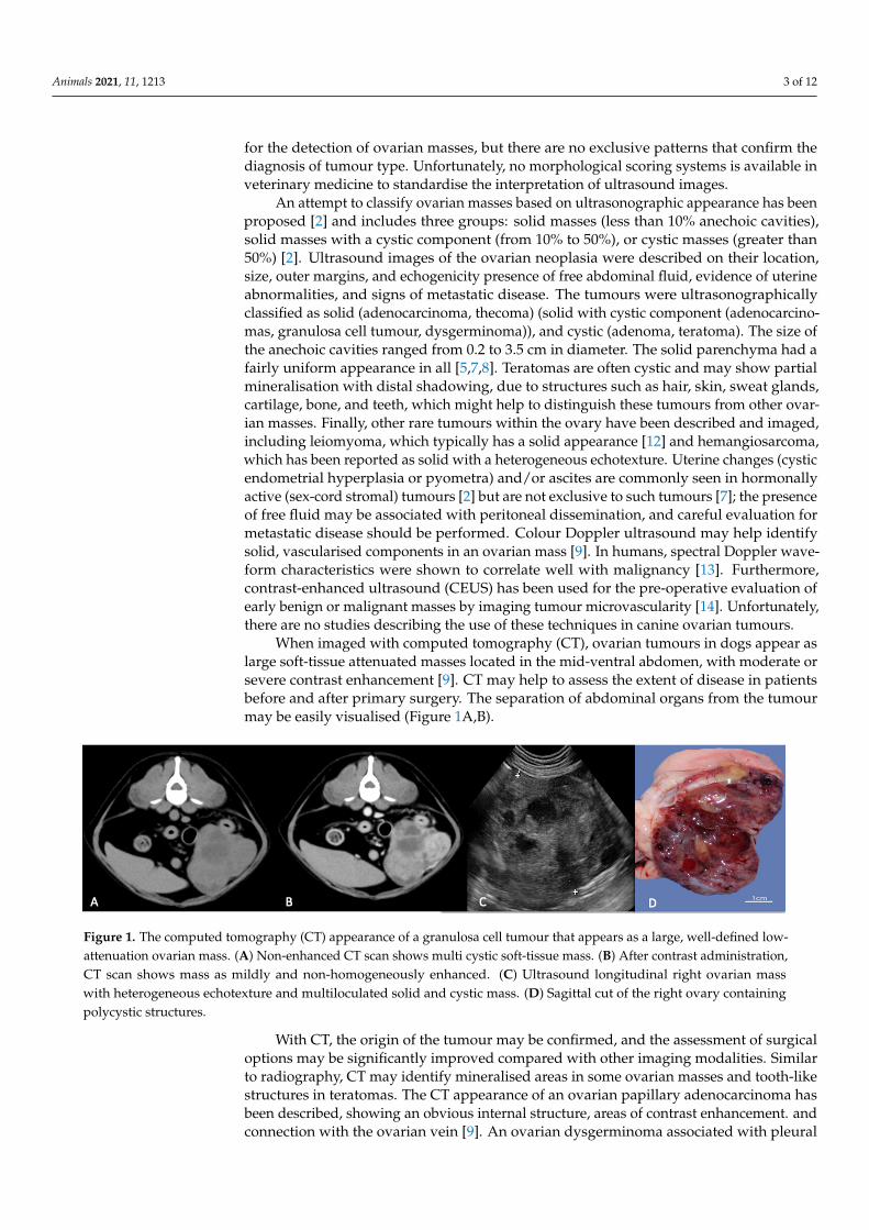
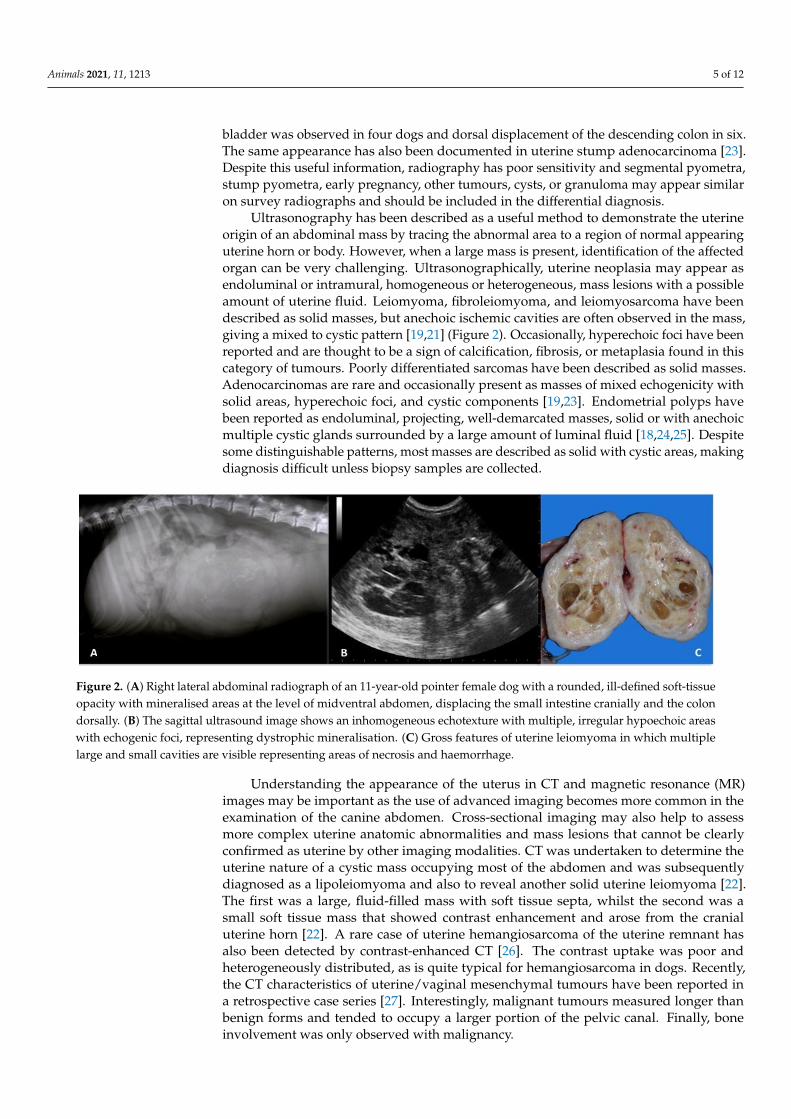
Ultrasonography has been described as a useful method to demonstrate the uterineorigin of an abdominal mass by tracing the abnormal area to a region of normal appearinguterine horn or body. However, when a large mass is present, identification of the affectedorgan can be very challenging. Ultrasonographically, uterine neoplasia may appear asendoluminal or intramural, homogeneous or heterogeneous, mass lesions with a possibleamount of uterine fluid. Leiomyoma, fibroleiomyoma, and leiomyosarcoma have beendescribed as solid masses, but anechoic ischemic cavities are often observed in the mass,giving a mixed to cystic pattern [19,21] (Figure 2). Occasionally, hyperechoic foci have beenreported and are thought to be a sign of calcification, fibrosis, or metaplasia found in thiscategory of tumours. Poorly differentiated sarcomas have been described as solid masses.Adenocarcinomas are rare and occasionally present as masses of mixed echogenicity withsolid areas, hyperechoic foci, and cystic components [19,23]. Endometrial polyps havebeen reported as endoluminal, projecting, well-demarcated masses, solid or with anechoicmultiple cystic glands surrounded by a large amount of luminal fluid [18,24,25]. Despitesome distinguishable patterns, most masses are described as solid with cystic areas, makingdiagnosis difficult unless biopsy samples are collected.
Animals 2021, 11, 1213 5 of 12
been described radiographically as homogeneous mid-ventral abdominal soft-tissue mass lesions that may displace the small bowel, stomach, and liver [19,20–22]. Foci of calcification have been seen in leiomyosarcoma and leiomyoma with dystrophic calcifi-cation and osseous metaplasia [19,21]. Soft-tissue opacities greater than 4 cm in diameter were noted between the urinary bladder and colon in six cases with tumours of the uterine body [19]. In half of these dogs, the outer margin of the lesion was well-defined. Cranial displacement of the urinary bladder was observed in four dogs and dorsal dis-placement of the descending colon in six. The same appearance has also been docu-mented in uterine stump adenocarcinoma [23]. Despite this useful information, radiog-raphy has poor sensitivity and segmental pyometra, stump pyometra, early pregnancy, other tumours, cysts, or granuloma may appear similar on survey radiographs and should be included in the differential diagnosis.
Ultrasonography has been described as a useful method to demonstrate the uterine origin of an abdominal mass by tracing the abnormal area to a region of normal appear-ing uterine horn or body. However, when a large mass is present, identification of the affected organ can be very challenging. Ultrasonographically, uterine neoplasia may appear as endoluminal or intramural, homogeneous or heterogeneous, mass lesions with a possible amount of uterine fluid. Leiomyoma, fibroleiomyoma, and leiomyosarcoma have been described as solid masses, but anechoic ischemic cavities are often observed in the mass, giving a mixed to cystic pattern [19,21] (Figure 2). Occasionally, hyperechoic foci have been reported and are thought to be a sign of calcification, fibrosis, or metapla-sia found in this category of tumours. Poorly differentiated sarcomas have been de-scribed as solid masses. Adenocarcinomas are rare and occasionally present as masses of mixed echogenicity with solid areas, hyperechoic foci, and cystic components [19,23]. Endometrial polyps have been reported as endoluminal, projecting, well-demarcated masses, solid or with anechoic multiple cystic glands surrounded by a large amount of luminal fluid [18,24,25]. Despite some distinguishable patterns, most masses are de-scribed as solid with cystic areas, making diagnosis difficult unless biopsy samples are collected.
Figure 2. (A) Right lateral abdominal radiograph of an 11-year-old pointer female dog with a rounded, ill-defined soft-tissue opacity with mineralised areas at the level of midventral abdomen, displacing the small intestine cranially and the colon dorsally. (B) The sagittal ultrasound image shows an inhomogeneous echotexture with multiple, irregular hy-poechoic areas with echogenic foci, representing dystrophic mineralisation. (C) Gross features of uterine leiomyoma in which multiple large and small cavities are visible representing areas of necrosis and haemorrhage.
Understanding the appearance of the uterus in CT and magnetic resonance (MR) images may be important as the use of advanced imaging becomes more common in the examination of the canine abdomen. Cross-sectional imaging may also help to assess more complex uterine anatomic abnormalities and mass lesions that cannot be clearly confirmed as uterine by other imaging modalities. CT was undertaken to determine the uterine nature of a cystic mass occupying most of the abdomen and was subsequently diagnosed as a lipoleiomyoma and also to reveal another solid uterine leiomyoma [22}. The first was a large, fluid-filled mass with soft tissue septa, whilst the second was a
Figure 2. (A) Right lateral abdominal radiograph of an 11-year-old pointer female dog with a rounded, ill-defined soft-tissueopacity with mineralised areas at the level of midventral abdomen, displacing the small intestine cranially and the colondorsally. (B) The sagittal ultrasound image shows an inhomogeneous echotexture with multiple, irregular hypoechoic areaswith echogenic foci, representing dystrophic mineralisation. (C) Gross features of uterine leiomyoma in which multiplelarge and small cavities are visible representing areas of necrosis and haemorrhage.
Understanding the appearance of the uterus in CT and magnetic resonance (MR)images may be important as the use of advanced imaging becomes more common in theexamination of the canine abdomen. Cross-sectional imaging may also help to assessmore complex uterine anatomic abnormalities and mass lesions that cannot be clearlyconfirmed as uterine by other imaging modalities. CT was undertaken to determine theuterine nature of a cystic mass occupying most of the abdomen and was subsequentlydiagnosed as a lipoleiomyoma and also to reveal another solid uterine leiomyoma [22].The first was a large, fluid-filled mass with soft tissue septa, whilst the second was asmall soft tissue mass that showed contrast enhancement and arose from the cranialuterine horn [22]. A rare case of uterine hemangiosarcoma of the uterine remnant hasalso been detected by contrast-enhanced CT [26]. The contrast uptake was poor andheterogeneously distributed, as is quite typical for hemangiosarcoma in dogs. Recently,the CT characteristics of uterine/vaginal mesenchymal tumours have been reported ina retrospective case series [27]. Interestingly, malignant tumours measured longer thanbenign forms and tended to occupy a larger portion of the pelvic canal. Finally, boneinvolvement was only observed with malignancy.
Animals 2021, 11, 1213 6 of 12
2.5. Imaging of the Vagina and Vaginal Neoplasia
The vagina has mainly a pelvic position. Survey radiographs and ultrasound areuseful as screening tools but have limited diagnostic value. Vaginoscopy is very useful forvisualisation of deep proliferative luminal lesions, although some large and obstructivemasses cannot be easily evaluated. On abdominal radiographs, leiomyomas may be seen asill-defined soft tissue masses in the ventral pelvic canal, causing dorsal displacement of therectum [28]. Vaginal masses may extend to the perineum and be pedunculated [29]. Nega-tive or positive vagino-urethrograms may be useful to detect lesions involving the vagina.
Ultrasonographically, it is extremely difficult to image the pelvic canal via a trans-abdominal approach. Some proximal vaginal masses may be detected by pointing theultrasound probe caudally to visualise the region immediately caudal to the cervix. It is alsofeasible to infuse saline into the vagina, thus providing anechoic contrast that can be usedto image some proximal vaginal wall lesions. It is also difficult to image the vagina with aperineal transcutaneous approach. Although not usually performed in small animals, tran-srectal ultrasound has been used to image a vaginal mass [28]. Leiomyomas, which are themost common vaginal neoplasia, may be visible as solid masses, moderately vascularised,with hyperechoic structures suggestive of necrosis or anechoic cavities [28,29].
CT and MR are important imaging modalities for the evaluation of vaginal abnormali-ties. CT allows localisation of the anatomic site of origin due to its multiplanar imagingcapabilities and improved contrast resolution. A CT of the caudal abdomen and perineumallowed assessment of the size and location of vaginal leiomyoma and fibroma [28,29].Such tumours often appear as soft-tissue masses, moderately and heterogeneously contrastenhanced. Vagino-urethrography is another imaging modality for the evaluation of thevagina and is classically performed by radiography, as mentioned above, or using CT. Airor diluted iodinated contrast agents are administered into the vestibule after Foley catheterplacement. Positive contrast may be enhanced by simultaneous intravenous administrationof the contrast agent. Vagino-urethrography has been performed in a case of vaginalleiomyoma, readily visualised as a contrast-filled defect [28]. Unfortunately, MRI has rarelybeen documented in veterinary patients to characterise vaginal masses. Only one MRIdescription of a large intrapelvic leiomyoma compressing the bladder and rectum has beenreported [30].
3. Male3.1. Imaging of the Prostate
The prostate is an ovoid-shaped bilobed gland located at the bladder neck, surround-ing the proximal urethra. The normal prostate is located within or immediately cranial tothe pelvis and is bordered dorsally by the rectum. The gland is smoothly marginated, andultrasonographically the parenchyma has a moderate homogenous echogenicity in entiredogs, with a fine to medium echotexture.
Prostate size is often easily assessed by measuring the maximum total prostate widthfrom images in the transverse plane, but is better assessed by calculating prostate volumeusing the formula for the volume of an ellipse:
Prostate volume = Length × Width × Height × 0.523. (1)
Prostatic size varies for dogs of different breeds.
Imaging of Prostatic Neoplasia
Diagnosis of malignant prostate disease is challenging because benign and malignantlesions may have similar clinical presentations and imaging appearance at the initial stages.The search for improved diagnostic techniques continues, and a variety of other imagingmodalities have been reported in human medicine, including CT, MRI, positron emissiontomography, and single-photon emission computed tomography [31]. In dogs, prostaticcarcinoma is a highly malignant neoplasia with a prevalence of 0.2–0.6% [32]. Prostate
Animals 2021, 11, 1213 7 of 12
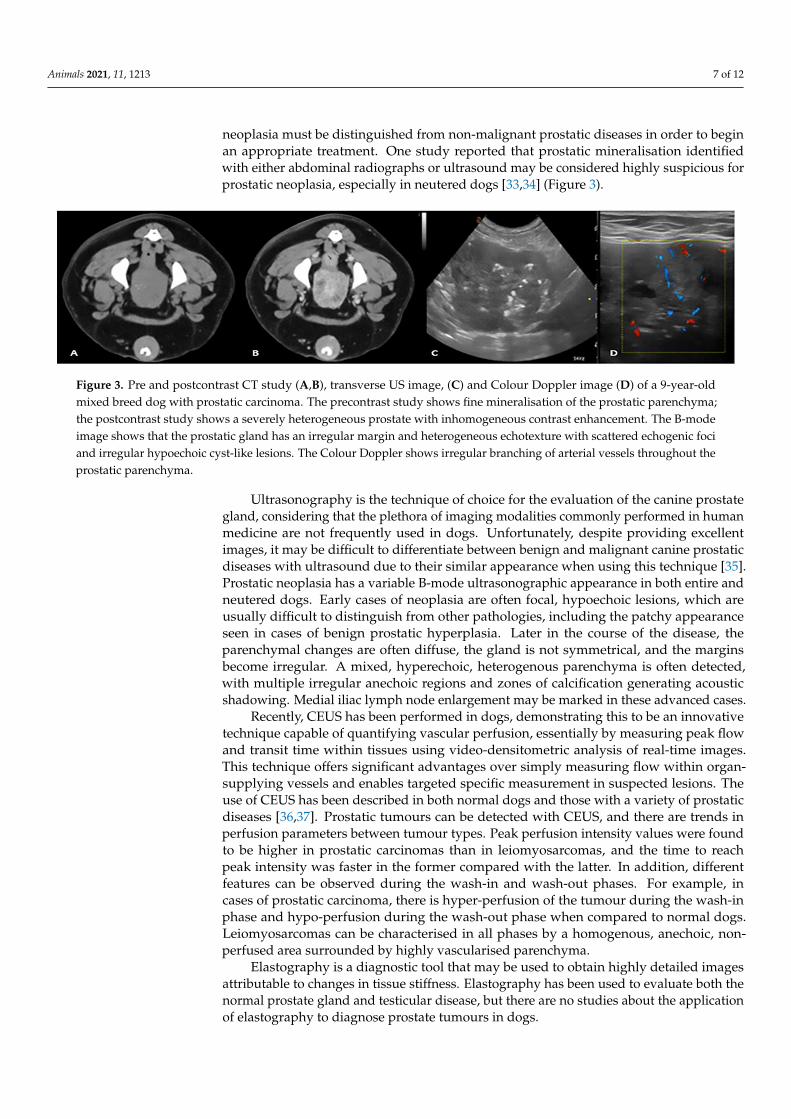
neoplasia must be distinguished from non-malignant prostatic diseases in order to beginan appropriate treatment. One study reported that prostatic mineralisation identifiedwith either abdominal radiographs or ultrasound may be considered highly suspicious forprostatic neoplasia, especially in neutered dogs [33,34] (Figure 3).
Animals 2021, 11, 1213 7 of 12
Prostate volume = Length × Width × Height × 0.523. (1)
Prostatic size varies for dogs of different breeds.
Imaging of Prostatic Neoplasia Diagnosis of malignant prostate disease is challenging because benign and malig-
nant lesions may have similar clinical presentations and imaging appearance at the initial stages. The search for improved diagnostic techniques continues, and a variety of other imaging modalities have been reported in human medicine, including CT, MRI, positron emission tomography, and single-photon emission computed tomography [31]. In dogs, prostatic carcinoma is a highly malignant neoplasia with a prevalence of 0.2–0.6% [32]. Prostate neoplasia must be distinguished from non-malignant prostatic diseases in order to begin an appropriate treatment. One study reported that prostatic mineralisation identified with either abdominal radiographs or ultrasound may be considered highly suspicious for prostatic neoplasia, especially in neutered dogs [33,34] (Figure 3).
Ultrasonography is the technique of choice for the evaluation of the canine prostate gland, considering that the plethora of imaging modalities commonly performed in hu-man medicine are not frequently used in dogs. Unfortunately, despite providing excel-lent images, it may be difficult to differentiate between benign and malignant canine prostatic diseases with ultrasound due to their similar appearance when using this tech-nique [35]. Prostatic neoplasia has a variable B-mode ultrasonographic appearance in both entire and neutered dogs. Early cases of neoplasia are often focal, hypoechoic le-sions, which are usually difficult to distinguish from other pathologies, including the patchy appearance seen in cases of benign prostatic hyperplasia. Later in the course of the disease, the parenchymal changes are often diffuse, the gland is not symmetrical, and the margins become irregular. A mixed, hyperechoic, heterogenous parenchyma is often detected, with multiple irregular anechoic regions and zones of calcification generating acoustic shadowing. Medial iliac lymph node enlargement may be marked in these ad-vanced cases.
Figure 3. Pre and postcontrast CT study (A,B), transverse US image, (C) and Colour Doppler image (D) of a 9-year-old mixed breed dog with prostatic carcinoma. The precontrast study shows fine mineralisation of the prostatic parenchyma; the postcontrast study shows a severely heterogeneous prostate with inhomogeneous contrast enhancement. The B-mode image shows that the prostatic gland has an irregular margin and heterogeneous echotexture with scattered echogenic foci and irregular hypoechoic cyst-like lesions. The Colour Doppler shows irregular branching of arterial vessels throughout the prostatic parenchyma.
Recently, CEUS has been performed in dogs, demonstrating this to be an innovative technique capable of quantifying vascular perfusion, essentially by measuring peak flow and transit time within tissues using video-densitometric analysis of real-time images. This technique offers significant advantages over simply measuring flow within or-gan-supplying vessels and enables targeted specific measurement in suspected lesions. The use of CEUS has been described in both normal dogs and those with a variety of prostatic diseases [36,37]. Prostatic tumours can be detected with CEUS, and there are trends in perfusion parameters between tumour types. Peak perfusion intensity values
Figure 3. Pre and postcontrast CT study (A,B), transverse US image, (C) and Colour Doppler image (D) of a 9-year-oldmixed breed dog with prostatic carcinoma. The precontrast study shows fine mineralisation of the prostatic parenchyma;the postcontrast study shows a severely heterogeneous prostate with inhomogeneous contrast enhancement. The B-modeimage shows that the prostatic gland has an irregular margin and heterogeneous echotexture with scattered echogenic fociand irregular hypoechoic cyst-like lesions. The Colour Doppler shows irregular branching of arterial vessels throughout theprostatic parenchyma.
Ultrasonography is the technique of choice for the evaluation of the canine prostategland, considering that the plethora of imaging modalities commonly performed in humanmedicine are not frequently used in dogs. Unfortunately, despite providing excellentimages, it may be difficult to differentiate between benign and malignant canine prostaticdiseases with ultrasound due to their similar appearance when using this technique [35].Prostatic neoplasia has a variable B-mode ultrasonographic appearance in both entire andneutered dogs. Early cases of neoplasia are often focal, hypoechoic lesions, which areusually difficult to distinguish from other pathologies, including the patchy appearanceseen in cases of benign prostatic hyperplasia. Later in the course of the disease, theparenchymal changes are often diffuse, the gland is not symmetrical, and the marginsbecome irregular. A mixed, hyperechoic, heterogenous parenchyma is often detected,with multiple irregular anechoic regions and zones of calcification generating acousticshadowing. Medial iliac lymph node enlargement may be marked in these advanced cases.
Recently, CEUS has been performed in dogs, demonstrating this to be an innovativetechnique capable of quantifying vascular perfusion, essentially by measuring peak flowand transit time within tissues using video-densitometric analysis of real-time images.This technique offers significant advantages over simply measuring flow within organ-supplying vessels and enables targeted specific measurement in suspected lesions. Theuse of CEUS has been described in both normal dogs and those with a variety of prostaticdiseases [36,37]. Prostatic tumours can be detected with CEUS, and there are trends inperfusion parameters between tumour types. Peak perfusion intensity values were foundto be higher in prostatic carcinomas than in leiomyosarcomas, and the time to reachpeak intensity was faster in the former compared with the latter. In addition, differentfeatures can be observed during the wash-in and wash-out phases. For example, incases of prostatic carcinoma, there is hyper-perfusion of the tumour during the wash-inphase and hypo-perfusion during the wash-out phase when compared to normal dogs.Leiomyosarcomas can be characterised in all phases by a homogenous, anechoic, non-perfused area surrounded by highly vascularised parenchyma.
Elastography is a diagnostic tool that may be used to obtain highly detailed imagesattributable to changes in tissue stiffness. Elastography has been used to evaluate both thenormal prostate gland and testicular disease, but there are no studies about the applicationof elastography to diagnose prostate tumours in dogs.
Animals 2021, 11, 1213 8 of 12
In general, CT imaging of abdominal organs has several advantages: organs can beimaged without superimposition; small structures can be identified due to high-resolution;organ size and shape can be assessed in multiple planes with image reconstruction. CTexamination is considered a helpful tool for the evaluation of the canine prostate gland [38].However, few studies have used CT to further investigate its diagnostic benefits for examin-ing the prostate in dogs [39], and none of them describe the features of prostatic neoplasia.
The use of MR to evaluate the prostate is still limited and only one short communica-tion describes the use of MR in canine prostatic tumours [40]. The authors described thatthe enhancement pattern of prostate lesion, relative contrast enhancement indices (RCEI),and apparent diffusion coefficient (ADC) values of prostate lesions may help to detectprostate adenocarcinoma.
3.2. Imaging of the Testes
Radiographs are rarely performed in the evaluation of intra-scrotal testicular dis-eases. Enlarged testicles or testicular masses may appear as an enlarged scrotal silhouette,but this cannot be distinguished from intra-scrotal fluid or thickening of the scrotal softtissue contents. Ultrasound examination is a sensitive method for evaluating testicularparenchymal diseases, allowing differentiation of testicular from extra-testicular causesof scrotal enlargement [41]. Improved diagnostic techniques and frequent imaging of thetestes in infertility have drawn attention to a significant number of small, solid, and oftennon-palpable tumours, whose diagnosis and management results significantly problematicfor the theriogenologists [42]. Ultrasound images can be obtained in the non-sedated dogin a lateral recumbency or standing position. Clipping of the scrotal hair should be avoidedas it often leads to excessive licking and subsequent development of scrotal dermatitis.Instead, copious amounts of ultrasound gel should be used. The testes are imaged inlongitudinal, transverse, and dorsal planes. The normal testicular parenchyma appearsrelatively hypoechoic in echotexture with regular diffuse echogenic stippling scatteredevenly throughout the organ [43]. The stippling represents an extension of the fibrous me-diastinum, which is responsible for supporting the parenchymal tissue. The mediastinumtestis, a fibrous invagination of the tunica albuginea, is located centrally within the testis.In the sagittal plane, this structure appears as an echogenic line approximately 2 mm wideextending from the cranial to the caudal pole, whereas in the transverse plane it appearsas a central echogenic circular structure. The epididymis appears hypoechoic comparedto the testicular parenchyma. Focal testicular lesions are relatively easy to identify andusually, there is a good relationship with gross pathology findings. Because of the highdiagnostic validity of ultrasound, there are no descriptions about the use of CT or MR forthe evaluation of dog testes.
Imaging of Testicular Tumours
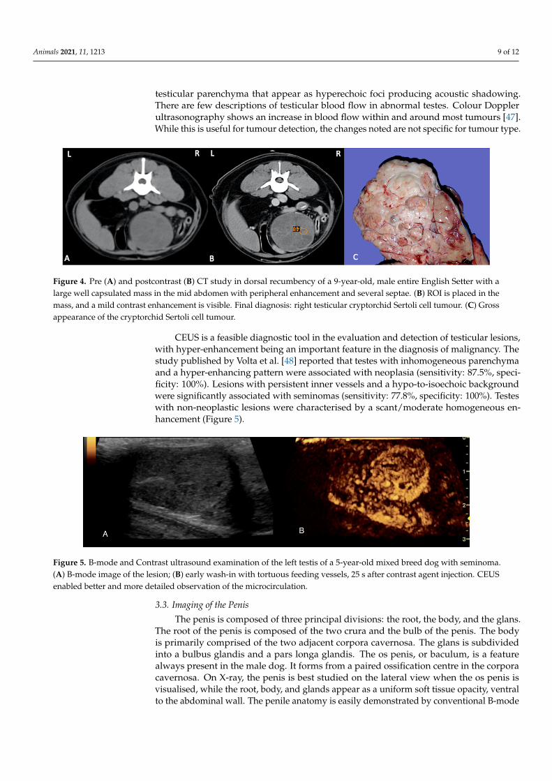
Testicular tumours are the most common tumours of the canine male genitalia andaccount for approximately 90% of all cancers of the male reproductive tract. Testiculartumours are the second most common tumour affecting male dogs. They may be benign ormalignant and may or may not be endocrinologically active. While B-mode ultrasoundis extremely useful for detecting testicular tumours, the ultrasonographic appearanceof lesions varies and is not specific for the type of tumour [41]. Testicular tumours canrange from circumscribed small nodules to large complex masses with heterogeneousecho-pattern and disruption of normal anatomy. At the time of diagnosis, Sertoli celltumours and seminomas are usually large with mixed echogenicity, resulting sometimes ingeneralised testicular enlargement. Cryptorchidism predisposes to neoplasia, and the mostcommon tumours are Sertoli cell tumours and seminomas [44–46] (Figure 4).
Interstitial cell tumours may appear as well-defined focal hypoechoic lesions. How-ever, areas of haemorrhage and necrosis may occur in all tumour types and may be seenultrasonographically as disorganised hyperechoic and hypoechoic regions. Other findingsthat may be associated with testicular neoplasia include areas of calcification within the
Animals 2021, 11, 1213 9 of 12
testicular parenchyma that appear as hyperechoic foci producing acoustic shadowing.There are few descriptions of testicular blood flow in abnormal testes. Colour Dopplerultrasonography shows an increase in blood flow within and around most tumours [47].While this is useful for tumour detection, the changes noted are not specific for tumour type.
Animals 2021, 11, 1213 9 of 12
or malignant and may or may not be endocrinologically active. While B-mode ultrasound is extremely useful for detecting testicular tumours, the ultrasonographic appearance of lesions varies and is not specific for the type of tumour [41]. Testicular tumours can range from circumscribed small nodules to large complex masses with heterogeneous echo-pattern and disruption of normal anatomy. At the time of diagnosis, Sertoli cell tumours and seminomas are usually large with mixed echogenicity, resulting sometimes in generalised testicular enlargement. Cryptorchidism predisposes to neoplasia, and the most common tumours are Sertoli cell tumours and seminomas [44–46] (Figure 4).
Figure 4. Pre (A) and postcontrast (B) CT study in dorsal recumbency of a 9-year-old, male entire English Setter with a large well capsulated mass in the mid abdomen with peripheral enhancement and several septae. (B) ROI is placed in the mass, and a mild contrast enhancement is visible. Final diagnosis: right testicular cryptorchid Sertoli cell tumour. (C) Gross appearance of the cryptorchid Sertoli cell tumour.
Interstitial cell tumours may appear as well-defined focal hypoechoic lesions. However, areas of haemorrhage and necrosis may occur in all tumour types and may be seen ultrasonographically as disorganised hyperechoic and hypoechoic regions. Other findings that may be associated with testicular neoplasia include areas of calcification within the testicular parenchyma that appear as hyperechoic foci producing acoustic shadowing. There are few descriptions of testicular blood flow in abnormal testes. Colour Doppler ultrasonography shows an increase in blood flow within and around most tu-mours [47]. While this is useful for tumour detection, the changes noted are not specific for tumour type.
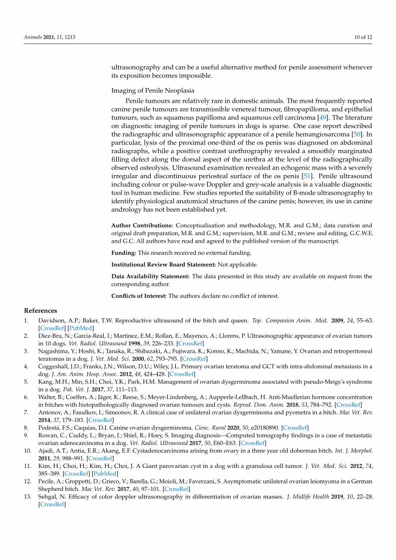
CEUS is a feasible diagnostic tool in the evaluation and detection of testicular le-sions, with hyper-enhancement being an important feature in the diagnosis of malig-nancy. The study published by Volta et al. [48] reported that testes with inhomogeneous parenchyma and a hyper-enhancing pattern were associated with neoplasia (sensitivity: 87.5%, specificity: 100%). Lesions with persistent inner vessels and a hypo-to-isoechoic background were significantly associated with seminomas (sensitivity: 77.8%, specificity: 100%). Testes with non-neoplastic lesions were characterised by a scant/moderate ho-mogeneous enhancement (Figure 5).
Figure 5. B-mode and Contrast ultrasound examination of the left testis of a 5-year-old mixed breed dog with seminoma. (A) B-mode image of the lesion; (B) early wash-in with tortuous feeding vessels, 25 s after contrast agent injection. CEUS enabled better and more detailed observation of the microcirculation.
Figure 4. Pre (A) and postcontrast (B) CT study in dorsal recumbency of a 9-year-old, male entire English Setter with alarge well capsulated mass in the mid abdomen with peripheral enhancement and several septae. (B) ROI is placed in themass, and a mild contrast enhancement is visible. Final diagnosis: right testicular cryptorchid Sertoli cell tumour. (C) Grossappearance of the cryptorchid Sertoli cell tumour.
CEUS is a feasible diagnostic tool in the evaluation and detection of testicular lesions,with hyper-enhancement being an important feature in the diagnosis of malignancy. Thestudy published by Volta et al. [48] reported that testes with inhomogeneous parenchymaand a hyper-enhancing pattern were associated with neoplasia (sensitivity: 87.5%, speci-ficity: 100%). Lesions with persistent inner vessels and a hypo-to-isoechoic backgroundwere significantly associated with seminomas (sensitivity: 77.8%, specificity: 100%). Testeswith non-neoplastic lesions were characterised by a scant/moderate homogeneous en-hancement (Figure 5).
Animals 2021, 11, 1213 9 of 12
or malignant and may or may not be endocrinologically active. While B-mode ultrasound is extremely useful for detecting testicular tumours, the ultrasonographic appearance of lesions varies and is not specific for the type of tumour [41]. Testicular tumours can range from circumscribed small nodules to large complex masses with heterogeneous echo-pattern and disruption of normal anatomy. At the time of diagnosis, Sertoli cell tumours and seminomas are usually large with mixed echogenicity, resulting sometimes in generalised testicular enlargement. Cryptorchidism predisposes to neoplasia, and the most common tumours are Sertoli cell tumours and seminomas [44–46] (Figure 4).
Figure 4. Pre (A) and postcontrast (B) CT study in dorsal recumbency of a 9-year-old, male entire English Setter with a large well capsulated mass in the mid abdomen with peripheral enhancement and several septae. (B) ROI is placed in the mass, and a mild contrast enhancement is visible. Final diagnosis: right testicular cryptorchid Sertoli cell tumour. (C) Gross appearance of the cryptorchid Sertoli cell tumour.
Interstitial cell tumours may appear as well-defined focal hypoechoic lesions. However, areas of haemorrhage and necrosis may occur in all tumour types and may be seen ultrasonographically as disorganised hyperechoic and hypoechoic regions. Other findings that may be associated with testicular neoplasia include areas of calcification within the testicular parenchyma that appear as hyperechoic foci producing acoustic shadowing. There are few descriptions of testicular blood flow in abnormal testes. Colour Doppler ultrasonography shows an increase in blood flow within and around most tu-mours [47]. While this is useful for tumour detection, the changes noted are not specific for tumour type.
CEUS is a feasible diagnostic tool in the evaluation and detection of testicular le-sions, with hyper-enhancement being an important feature in the diagnosis of malig-nancy. The study published by Volta et al. [48] reported that testes with inhomogeneous parenchyma and a hyper-enhancing pattern were associated with neoplasia (sensitivity: 87.5%, specificity: 100%). Lesions with persistent inner vessels and a hypo-to-isoechoic background were significantly associated with seminomas (sensitivity: 77.8%, specificity: 100%). Testes with non-neoplastic lesions were characterised by a scant/moderate ho-mogeneous enhancement (Figure 5).
Figure 5. B-mode and Contrast ultrasound examination of the left testis of a 5-year-old mixed breed dog with seminoma. (A) B-mode image of the lesion; (B) early wash-in with tortuous feeding vessels, 25 s after contrast agent injection. CEUS enabled better and more detailed observation of the microcirculation.
Figure 5. B-mode and Contrast ultrasound examination of the left testis of a 5-year-old mixed breed dog with seminoma.(A) B-mode image of the lesion; (B) early wash-in with tortuous feeding vessels, 25 s after contrast agent injection. CEUSenabled better and more detailed observation of the microcirculation.
3.3. Imaging of the Penis
The penis is composed of three principal divisions: the root, the body, and the glans.The root of the penis is composed of the two crura and the bulb of the penis. The bodyis primarily comprised of the two adjacent corpora cavernosa. The glans is subdividedinto a bulbus glandis and a pars longa glandis. The os penis, or baculum, is a featurealways present in the male dog. It forms from a paired ossification centre in the corporacavernosa. On X-ray, the penis is best studied on the lateral view when the os penis isvisualised, while the root, body, and glands appear as a uniform soft tissue opacity, ventralto the abdominal wall. The penile anatomy is easily demonstrated by conventional B-mode
Animals 2021, 11, 1213 10 of 12
ultrasonography and can be a useful alternative method for penile assessment wheneverits exposition becomes impossible.
Imaging of Penile Neoplasia
Penile tumours are relatively rare in domestic animals. The most frequently reportedcanine penile tumours are transmissible venereal tumour, fibropapilloma, and epithelialtumours, such as squamous papilloma and squamous cell carcinoma [49]. The literatureon diagnostic imaging of penile tumours in dogs is sparse. One case report describedthe radiographic and ultrasonographic appearance of a penile hemangiosarcoma [50]. Inparticular, lysis of the proximal one-third of the os penis was diagnosed on abdominalradiographs, while a positive contrast urethrography revealed a smoothly marginatedfilling defect along the dorsal aspect of the urethra at the level of the radiographicallyobserved osteolysis. Ultrasound examination revealed an echogenic mass with a severelyirregular and discontinuous periosteal surface of the os penis [51]. Penile ultrasoundincluding colour or pulse-wave Doppler and grey-scale analysis is a valuable diagnostictool in human medicine. Few studies reported the suitability of B-mode ultrasonography toidentify physiological anatomical structures of the canine penis; however, its use in canineandrology has not been established yet.
Author Contributions: Conceptualisation and methodology, M.R. and G.M.; data curation andoriginal draft preparation, M.R. and G.M.; supervision, M.R. and G.M.; review and editing, G.C.W.E.and G.C. All authors have read and agreed to the published version of the manuscript.
Funding: This research received no external funding.
Institutional Review Board Statement: Not applicable.
Data Availability Statement: The data presented in this study are available on request from thecorresponding author.
Conflicts of Interest: The authors declare no conflict of interest.
References1. Davidson, A.P.; Baker, T.W. Reproductive ultrasound of the bitch and queen. Top. Companion Anim. Med. 2009, 24, 55–63.
[CrossRef] [PubMed]2. Diez-Bru, N.; Garcia-Real, I.; Martinez, E.M.; Rollan, E.; Mayenco, A.; Llorens, P. Ultrasonographic appearance of ovarian tumors
in 10 dogs. Vet. Radiol. Ultrasound 1998, 39, 226–233. [CrossRef]3. Nagashima, Y.; Hoshi, K.; Tanaka, R.; Shibazaki, A.; Fujiwara, K.; Konno, K.; Machida, N.; Yamane, Y. Ovarian and retroperitoneal
teratomas in a dog. J. Vet. Med. Sci. 2000, 62, 793–795. [CrossRef]4. Coggeshall, J.D.; Franks, J.N.; Wilson, D.U.; Wiley, J.L. Primary ovarian teratoma and GCT with intra-abdominal metastasis in a
dog. J. Am. Anim. Hosp. Assoc. 2012, 48, 424–428. [CrossRef]5. Kang, M.H.; Min, S.H.; Choi, Y.K.; Park, H.M. Management of ovarian dysgerminoma associated with pseudo-Meigs’s syndrome
in a dog. Pak. Vet. J. 2017, 37, 111–113.6. Walter, B.; Coelfen, A.; Jäger, K.; Reese, S.; Meyer-Lindenberg, A.; Aupperle-Lellbach, H. Anti-Muellerian hormone concentration
in bitches with histopathologically diagnosed ovarian tumours and cysts. Reprod. Dom. Anim. 2018, 53, 784–792. [CrossRef]7. Antonov, A.; Fasulkov, I.; Simeonov, R. A clinical case of unilateral ovarian dysgerminoma and pyometra in a bitch. Mac Vet. Rev.
2014, 37, 179–183. [CrossRef]8. Podestá, F.S.; Caquias, D.I. Canine ovarian dysgerminoma. Cienc. Rural 2020, 50, e20180890. [CrossRef]9. Rowan, C.; Cuddy, L.; Bryan, J.; Shiel, R.; Hoey, S. Imaging diagnosis—Computed tomography findings in a case of metastatic
ovarian adenocarcinoma in a dog. Vet. Radiol. Ultrasound 2017, 50, E60–E63. [CrossRef]10. Ajadi, A.T.; Antia, E.R.; Akang, E.F. Cystadenocarcinoma arising from ovary in a three year old doberman bitch. Int. J. Morphol.
2011, 29, 988–991. [CrossRef]11. Kim, H.; Choi, H.; Kim, H.; Choi, J. A Giant parovarian cyst in a dog with a granulosa cell tumor. J. Vet. Med. Sci. 2012, 74,
385–389. [CrossRef] [PubMed]12. Pecile, A.; Groppetti, D.; Grieco, V.; Barella, G.; Moioli, M.; Faverzani, S. Asymptomatic unilateral ovarian leiomyoma in a German
Shepherd bitch. Mac Vet. Rev. 2017, 40, 97–101. [CrossRef]13. Sehgal, N. Efficacy of color doppler ultrasonography in differentiation of ovarian masses. J. Midlife Health 2019, 10, 22–28.
[CrossRef]
Animals 2021, 11, 1213 11 of 12
14. Qiao, J.J.; Yu, J.; Yu, Z.; Li, N.; Song, C.; Li, M. Contrast-enhanced ultrasonography in differential diagnosis of benign andmalignant ovarian tumors. PLoS ONE 2015, 10, e0118872. [CrossRef] [PubMed]
15. Eker, K.; Salmanoglu, M.R.; Vural, S.A. Unilateral leiomyoma in the mesosalpinx of a dog. J. Am. Anim. Hosp. Assoc. 2006, 42,392–394. [CrossRef]
16. Marino, G.; Quartuccio, M.; Cristarella, S.; Nicòtina, P.A.; Zanghì, A. Adenoma of the uterine tube in the bitch: Two case reports.Vet. Res. Comm. 2007, 31, 173–175. [CrossRef]
17. Plagge, J.; Bali, M. Adenoma of the fallopian tube in a 12-year-old bitch. Kleintierpraxis 2018, 63, 272–277. [CrossRef]18. Marino, G.; Barna, A.; Rizzo, S.; Zanghì, A.; Catone, G. Endometrial polyps in the bitch: A retrospective study of 21 cases. J. Comp.
Path 2013, 149, 410–416. [CrossRef]19. Patsikas, M.; Papazoglou, L.G.; Jakovljevic, S.; Papaioannou, N.G.; Papadopoulou, P.L.; Soultani, C.B.; Chryssogonidis, I.A.;
Kouskouras, K.A.; Tziris, N.E.; Charitanti, A.A. Radiographic and ultrasonographic findings of uterine neoplasms in nine dogs. J.Am. Anim. Hosp. Assoc. 2014, 50, 330–337. [CrossRef]
20. Cave, T.A.; Hine, R.; Howie, F.; Thompson, H.; Argyle, D.J. Uterine carcinoma in a 10-month-old golden retriever. J. Small Anim.Pract. 2002, 43, 133–135. [CrossRef] [PubMed]
21. Tsioli, V.G.; Gouletsou, P.G.; Loukopoulos, P.; Zavlaris, M.; Galatos, A.D. Uterine leiomyosarcoma and pyometra in a dog. J. SmallAnim. Pract. 2011, 52, 121–124. [CrossRef]
22. Percival, A.; Singh, A.; zur Linden, R.A.; Watrous, G.; Patten, S.; Valverde, A.; Ratsep, E. Massive uterine lipoleiomyoma andleiomyoma in a miniature poodle bitch. Can. Vet. J. 2018, 59, 845–850.
23. Kokkinos, P.; Ververidis, C.; Patsikas, M.; Kritsepi-Konstantinou, M.; Kazakos, G.M.; Psalla, D. Uterine stump adenocarcinoma ina bitch with an ovarian remnant: A case report. J. Hellenic. Vet. Med. Soc. 2019, 70, 1583–1588. [CrossRef]
24. Schlafer, D.H.; Yeager, A.E.; Concannon, P.W. Theriogenology question of the month. Endometrial polyp in a beagle. J. Am. Vet.Med. Assoc. 1997, 210, 759–761.
25. Chambers, B.A.; Laksito, M.A.; Long, F.; Yates, G.D. Unilateral uterine torsion secondary to an inflammatory endometrial polypin the bitch. Aust. Vet. J. 2011, 89, 380–384. [CrossRef] [PubMed]
26. Wenzlow, N.; Tivers, M.S.; Selmic, L.E.; Scurrell, E.J.; Baines, S.J.; Smith, K.C. Haemangiosarcoma in the uterine remnant of aspayed female dog. J. Small Anim. Pract. 2009, 50, 488–491. [CrossRef]
27. Barozzi, M.C.M.; Saba, C.F.; Gendron, K.P. CT characteristics of uterine and vaginal mesenchymal tumours in dogs. J. Small Anim.Pract. 2021, 62, 293–299. [CrossRef] [PubMed]
28. Weissman, A.; Jiménez, D.; Torres, B.; Cornell, K.; Holmes, S.P. Canine vaginal leiomyoma diagnosed by CT vaginourethrography.J. Am. Anim. Hosp. Assoc. 2013, 49, 394–397. [CrossRef]
29. Sathya, S.; Linn, K. Regression of a vaginal leiomyoma after ovariohysterectomy in a dog: A case report. J. Am. Anim. Hosp. Assoc.2014, 50, 424–428. [CrossRef]
30. Nelissen, P.; White, R.A.S. Subtotal vaginectomy for management of extensive vaginal disease in 11 dogs. Vet. Surg. 2012, 41,495–500. [CrossRef] [PubMed]
31. Fuchsjager, M.; Shukla-Dave, A.; Akin, O.; Barentsz, J.; Hricak, H. Prostate cancer imaging. Acta Radiol. 2008, 49, 107–120.[CrossRef]
32. Weaver, A.D. Fifteen cases of prostatic carcinoma in the dog. Vet. Rec. 1981, 109, 71–75. [CrossRef]33. Bradbury, C.A.; Westropp, J.L.; Pollard, R.E. Relationship between prostatomegaly, prostatic mineralization, and cytologic
diagnosis. Vet. Radiol. Ultrasound 2009, 50, 167–171. [CrossRef]34. Russo, M.; Vignoli, M.; England, G.C.W. B-mode and contrast-enhanced ultrasonographic findings in canine prostatic disorders.
Reprod. Domest. Anim. 2012, 47, 238–242. [CrossRef]35. Mahaffey, M.B.; Selcer, B.A.; Cartee, R.E. The reproductive system. In Practical Veterinary Ultrasound; Cartee, R.E., Selcer, B.A.,
Hudson, J.A., Eds.; Lea & Febiger: Philadelphia, PA, USA, 1995; pp. 236–265.36. Russo, M.; Vignoli, M.; Catone, G.; Rossi, F.; Attanasi, G.; England, G.C.W. Prostatic perfusion in the dog using contrast-enhanced
doppler ultrasound. Reprod. Domest. Anim. 2009, 44, 334–335. [CrossRef]37. Vignoli, M.; Russo, M.; Catone, G.; Rossi, F.; Attanasi, G.; Terragni, R.; Saunders, J.H.; England, G.C.W. Assessment of vascular
perfusion kinetics using contrast- enhanced ultrasound for the diagnosis of prostatic disease in dogs. Reprod. Domest. Anim. 2011,46, 209–213. [CrossRef]
38. Lee, K.J.; Shimizu, J.; Kishimoto, M.; Kadohira, M.; Iwasaki, T.; Miyake, Y.I.; Yamada, K. Computed tomography of the prostategland in apparently healthy entire dogs. Small Anim. Pract. 2011, 52, 146–151. [CrossRef] [PubMed]
39. Pasikowska, J.; Hebel, M.; Nizanski, W.; Nowak, M. Computed Tomography of the Prostate Gland in Healthy Intact Dogs andDogs with Benign Prostatic Hyperplasia. Reprod. Dom. Anim. 2015, 50, 776–783. [CrossRef] [PubMed]
40. Tanaka, T.; Ashida, K.; Iimori, Y.; Yamazaki, H.; Mie, K.; Nishida, H.; Akiyoshi, H. Less enhancement and low apparent diffusioncoefficient value on magnetic resonance imaging may be helpful to detect canine prostate adenocarcinoma in case series. Vet.Comp. Oncol. 2020, 18, 861–865. [CrossRef] [PubMed]
41. Pugh, C.R.; Konde, L.J. Sonographic evaluation of canine testicular and scrotal abnormalities: A review of 26 case histories. Vet.Radiol. Ultrasound 1991, 32, 243–250. [CrossRef]
42. Mantziaras, G. Imaging of the male reproductive tract: Not so easy as it looks like. Theriogenology 2020, 150, 490–497. [CrossRef]43. Pugh, C.R.; Konde, L.J.; Park, R.D. Testicular ultrasound in the normal dog. Vet. Radiol. 1990, 31, 195–199. [CrossRef]
Animals 2021, 11, 1213 12 of 12
44. England, G.C. Ultrasonographic diagnosis of non-palpable Sertoli cell tumours in infertile dogs. J. Small Anim. Pract. 1995, 36,476–480. [CrossRef] [PubMed]
45. Mostachio, G.Q.; Apparício, M.; Vicente, W.R.R.; Cardilli, D.J.; Motheo, T.F.; Toniollo, G.H. Intraabdominal torsion of a neoplastictesticle and prostatic cyst in a cryptorchid dog. Schweiz Arch. Tierheilkd 2007, 149, 408–412. [CrossRef]
46. Stokowski, S.; Ruth, J.; Lanz, O.; Ziglioli, V. Computed tomographic features in a case of bilateral neoplastic cryptorchidism withsuspected torsion in a dog. Front. Vet. Sci. 2016, 3. [CrossRef] [PubMed]
47. Bigliardi, E.; Denti, L.; de Cesaris, V.; Bertocchi, M.; di Ianni, F.; Parmigiani, E.; Bresciani, C.; Cantoni, A.M. Colour dopplerultrasound imaging of blood flows variations in neoplastic and non-neoplastic testicular lesions in dogs. Reprod. Domest. Anim.2019, 54, 63–71. [CrossRef]
48. Volta, A.; Manfredi, S.; Vignoli, M.; Russo, M.; England, G.C.; Rossi, F.; Bigliardi, E.; Di Ianni, F.; Parmigiani, E.; Bresciani, C.;et al. Use of contrast-enhanced ultrasonography in chronic pathologic canine testes. Reprod. Domest. Anim. 2014, 49, 202–209.[CrossRef] [PubMed]
49. Hall, W.C.; Nielsen, S.W.; McEntee, K. Tumours of the prostate and penis. Bull. World Health Organ. 1976, 53, 247–265. [PubMed]50. Marolf, A.; Specht, A.; Thompson, M.; Castleman, W. Imaging diagnosis: Penile hemangiosarcoma. Vet. Radiol. Ultrasound 2006,
47, 474–475. [CrossRef]51. Fry, J.K.; Burney, D.; Hottinger, H.; Fabiani, M.; Feagin, C. Pollakiuria and stranguria in a Labrador retriever with penile HSA. J.
Am. Anim. Hosp. Assoc. 2014, 50, 141–147. [CrossRef]