reproductive system - accessscience
TRANSCRIPT
Hide
Reproductive system - AccessScience from McGraw-Hill Education http://accessscience.com/content/reproductive-system/581500
(http://accessscience.com/)
Article by:
Weichert, Charles K. College of Arts and Sciences, University of Cincinnati, Cincinnati, Ohio.
Wells, Lemen J. Formerly, Professor of Anatomy, University of Minnesota, Minneapolis, Minnesota.
Jost, Alfred Laboratoire de Physiologie du Developpement, Collège de France, Paris, France.
Chang, M. C. Worcester Foundation for Experimental Biology, Shrewsbury, Massachusetts.
Harper, Michael J. K. Reproductive Biology Division, Center for Research, Department of Obstetrics and Gynecology, University of Texas-Health
Science Center, San Antonio, Texas.
Hunter, R. H. F. School of Agriculture, University of Edinburgh, Edinburgh, United Kingdom.
Callard, Ian P. Department of Biology, College of Arts and Sciences, Boston University, Boston, Massachusetts.
Last updated: 2014
DOI: https://doi.org/10.1036/1097-8542.581500 (https://doi.org/10.1036/1097-8542.581500)
Content
Comparative Anatomy
Ovaries
Oviducts and associated structures
Testes
Male ducts
Copulatory organs
Histology in Humans
Male reproductive system
Female reproductive system
Embryology
Sex determination
Role of urinary system
Development of gonads
Control of gonadal differentiation
Gonadal abnormalities
Genital tract development
Copulatory organs
Hormonal control
Abnormalities of genital tract
Physiology
Breeding season
Estrous and menstrual cycles
Mating
Endocrine function in reproduction
Nonmammalian species
Neuroendocrine function
Fertility and sterility
Additional Readings
The structures concerned with the production of sex cells (gametes) and perpetuation of the species. The
comparative anatomy, human histology, embryology, physiology, endocrinology, and biochemistry of this system are treated in
this article.
The reproductive function constitutes the only vertebrate physiological function that necessitates the existence of two
morphologically different kinds of individuals in each animal species, the males and the females (sexual dimorphism).
The purpose of the reproductive function is fertilization, that is, the fusion of a male and a female sex cell produced by two
distinct individuals. In each sex, the reproductive system comprises a sex gland or gonad, which produces sex cells, or
gametes, and ducts, which permit the passage of the gametes. In some animals, such as mammals, copulatory organs permit
the male germ cells to be introduced into the female ducts and fertilization is internal, but in many vertebrates, such as anuran
amphibians and many fishes, no copulatory organ exists and fertilization is external.
Egg cells, or ova, and sperm cells, or spermatozoa, are formed in the primary reproductive organs, which are collectively
known as gonads. Those of the male are called testes; those of the female are ovaries. Besides giving rise to reproductive
cells, both ovaries and testes give off endocrine secretions, or sex hormones, which pass into the blood or lymphatic streams
7/13/17, 5:55 AM 1 of 44
Reproductive system - AccessScience from McGraw-Hill Education http://accessscience.com/content/reproductive-system/581500
and are carried to all parts of the body, where they bring about profound effects, not only on the rest of the reproductive
system but on several other systems of the body as well. The gonads are paired structures, although in some forms what
appears to be an unpaired gonad is the result either of fusion of paired structures or of unilateral degeneration.
The reproductive elements formed in the gonads must be transported to the outside of the body. In most vertebrates, ducts
are utilized for this purpose. These ducts, together with the structures that serve to bring the gametes of both sexes together,
are known as sex organs. The structures used to transport the reproductive cells in the male are known as deferent ducts and
those of the female as oviducts. In a few forms, no ducts are present in either sex, and eggs and sperm escape from the body
cavity through genital or abdominal pores. The deferent ducts are usually the mesonephric or Wolffian ducts, which in some
cases also serve to carry urinary wastes in those vertebrates in which opistonephros or mesonephros function either during
embryonic or adult life. In vertebrates whose functional adult kidney is the metanephros, the Wolffian duct on each side
persists as the ductus deferens. See also: Kidney (/content/kidney/364000)
In most vertebrates, the reproductive ducts in both sexes open posteriorly into the cloaca. In some, modifications of the
cloacal region occur and the ducts open separately to the outside or, in the male, join the excretory ducts to emerge by a
common orifice. See also: Animal reproduction (/content/animal-reproduction/581200); Copulatory organ (/content
/copulatory-organ/161360)
The sex of an individual is dependent upon the chromosomes received from both parents at the time that the egg is fertilized.
That the balance between maleness and femaleness is a delicate one, however, is reflected in the fact that environmental
factors may assume an influential role in sexual development, and hormonal secretions may modify the extent to which
various structures and even behavioral characteristics develop and are maintained.
Ovaries
A typical mammalian ovary is a solid, irregularly shaped structure indistinctly separated into an inner medulla and an outer
cortex. The cortex contains numbers of ovarian follicles in various stages of development. See also: Ovary (/content/ovary
/479400)
In certain fishes and in amphibians, snakes, and lizards, the ovaries are hollow, saccular structures. The cavity within the
teleostean fish ovary is actually a closed-off portion of the coelom (body cavity) into which ripe ova are shed. This is not true
of the saccular ovaries of other forms.
C y c l o s t om e s
The adult female lamprey has a single ovary, representing a fusion of two, which runs the length of the body cavity,
suspended from the middorsal body wall by a single mesovarium. At the height of the breeding season, it fills the greater part
of the body cavity; ripe eggs are shed into the coelom and fertilization is external. The hagfish is hermaphroditic; the anterior
part of the single gonad is ovarian and the posterior part is testicular. Usually only one or the other region matures.
F i s h e s
The ovaries of most fishes are paired, although in some cases they have fused into a single organ. The large eggs of
elasmobranchs are discharged from the anteriorly located ovaries directly into the body cavity. In oviparous and ovoviviparous
species, following ovulation the ovarian follicles become transformed into corpora lutea, structures which presumably have an
endocrine function and may play a role in the extended retention of eggs in the ovi ducts of oviparous species and of young in
the uteri of ovoviviparous forms. Peritoneal folds form in connection with each ovary in teleosts, closing off all connection with
the coelom. The anterior end of the ovarian cavity ends blindly, but in most cases continuations of the folds at the posterior
7/13/17, 5:55 AM 2 of 44
Reproductive system - AccessScience from McGraw-Hill Education http://accessscience.com/content/reproductive-system/581500
end form an oviduct which opens directly to the outside. Ripe ova, sometimes numbered in the millions, are discharged into
the central ovarian cavity, which is actually a part of the body cavity, and thence pass down the oviducts to the outside. The
garpike Lepisosteus is the only ganoid fish with a saccular ovary. The ovaries are usually solid, flat elongate structures from
which mature ova break out into the coelom.
Amp h i b i a n s
Although the paired amphibian ovaries are saccular structures, ripe ova are libe rated into the body cavity through their
external walls. The shape of the ovaries varies with the shape of the body. They are long and narrow in caecilians, elongated
to a lesser degree in salamanders, and short and more compact in frogs and toads. Fat bodies are associated with amphibian
ovaries. They serve for the storage of nutriment and undergo profound changes during the year. A peculiar structure in the
male toad, known as Bidder's organ, may under certain conditions develop into a true ovary.
R e p t i l e s
The saccular ovaries of snakes and lizards are similar to those of amphibians, whereas turtles and crocodilians have solid
ovaries. In snakes and lizards, they are elongated but not symmetrically disposed. Only the yolk of reptilian eggs is formed in
the ovaries, and this represents the true ovum. The size of the eggs is generally in proportion to that of the animal. In certain
ovoviviparous snakes and lizards, corpora lutea form from ruptured follicles after ovulation and persist throughout pregnancy.
These probably secrete a hormone necessary for maintenance of pregnancy.
B i r d s
Although both ovaries are present during embryonic development in most birds (except many birds of prey), the right ovary
degenerates and only the left is functional. In birds, stalks extend from the surface of the ovary, and each stalk contains many
ovarian follicles in various stages of development. A mature ovum escapes from the ovarian follicle through a preformed
nonvascular band, the stigma or cicatrix, located on the surface of the follicle opposite the stalk. As in reptiles, only the yolk of
the egg represents the true ovum. Increase in the number of hours of daylight stimulates ovarian activity in many birds. Even
brief exposure to intense and bright light during hours of sleep increases production of eggs in the domestic fowl. This effect
is undoubtedly mediated through stimulation of the pituitary gland. If the functional ovary of the domestic fowl is removed, the
rudimentary right gonad will develop into a testislike organ, but without germ cells. See also: Sexual dimorphism (/content
/sexual-dimorphism/617800)
Mamma l s
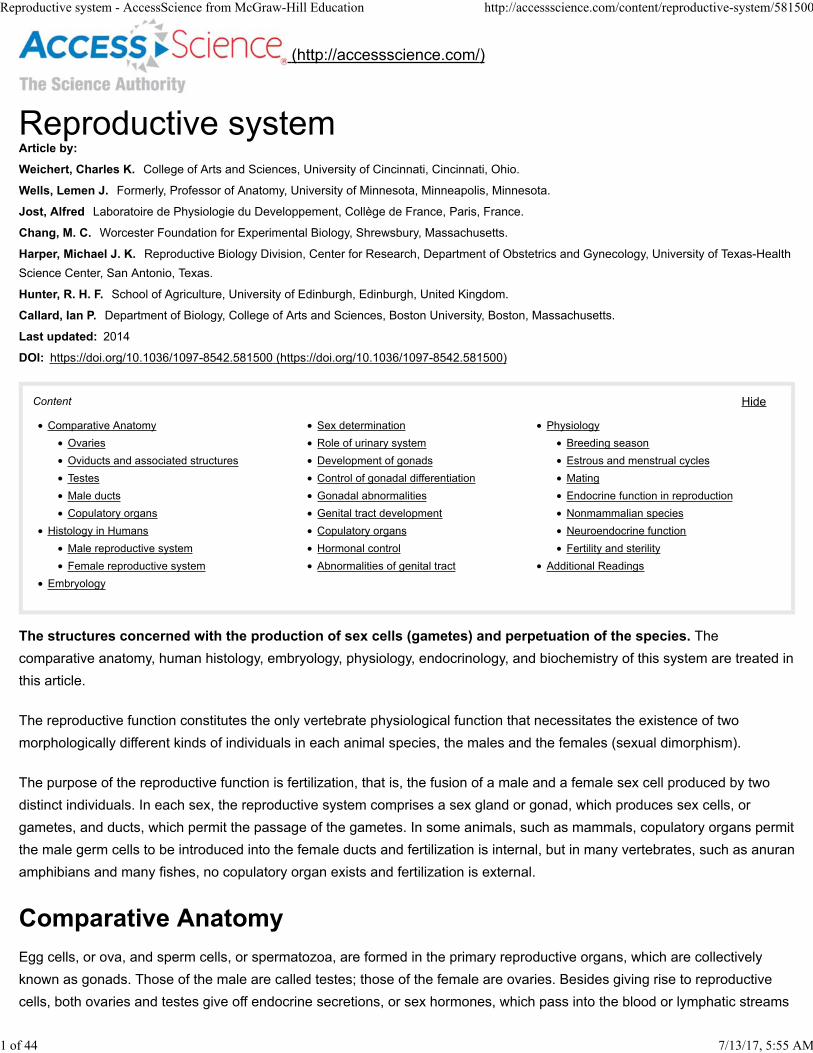
The ovaries of mammals are located in the lumbar or pelvic regions and are small in comparison to the size of the body. The
relationship of the microscopic mammalian ovum to the ovarian follicle differs somewhat from conditions in other vertebrates.
Follicles in various stages of development, with the youngest near the surface of the ovary, are depicted in Fig. 1. At periodic
intervals, one or more follicles grow to maturity, rupture, and liberate their ova into the body cavity. In such animals as the
rabbit, cat, and ferret, ovulation will not occur unless the animal copulates. Following ovulation, certain cells of the follicle
undergo a transformation and the entire structure becomes a more or less solid body, the corpus luteum.
7/13/17, 5:55 AM 3 of 44
Reproductive system - AccessScience from McGraw-Hill Education http://accessscience.com/content/reproductive-system/581500
Fig. 1 Section of portion of cortex of rat ovary, showing ovarian follicles in various stages of development, the youngestbeing near the surface of the ovary. (After C. K. Weichert, Elements of Chordate Anatomy, 3d ed., McGraw-Hill, 1967)
If pregnancy does not occur, the corpus luteum persists only for a short time. If pregnancy does ensue, the corpus luteum
usually persists throughout pregnancy. In either case, it ultimately degenerates. The corpus luteum is of primary importance
as an endocrine gland secreting a hormone called progesterone. See also: Pregnancy (/content/pregnancy/543100)
Oviducts and associated structures
Oviducts, except in teleosts and a few other fishes, are modifications of Müllerian ducts formed early during embryonic
development. In most cases, each duct is formed by an invagination of the peritoneum that covers the ventrolateral part of the
opisthonephros or mesonephros. The edges of the groove fuse to form a tube which joins the cloaca or forms a uterus
posteriorly and remains open anteriorly to become the ostium tubae. Although Müllerian ducts also form in the male, they
ordinarily degenerate except for a few vestigial remnants. In some, they persist as prominent but nonfunctional structures.
C y c l o s t om e s
Oviducts are lacking in cyclostomes. Ova pass from the coelom through genital pores into a urogenital papilla and then to the
outside.
F i s h e s
Much diversity exists in the oviducts of fishes. In some teleosts, and in a few other fish, eggs escape from the body cavity
through modified abdominal pores. In elasmobranchs, the two Müllerian ducts may fuse at their anterior ends so that only a
single ostium tubae connects with the coelom. An enlargement, known as the shell gland, is present in the upper part of each
oviduct. Beyond the shell gland, the Müllerian duct enlarges on each side to form a uterus which opens into the cloaca.
The oviducts of most teleosts are short and continuous with the cavities of the saccular ovaries. It is doubtful whether they are
true Müllerian ducts because they are formed in a different manner. A cloaca is lacking in teleosts, and the oviducts open
independently to the outside. The two oviducts usually fuse, continuing posteriorly as a single structure which may open to
the outside through a genital pore or else at the tip of a genital papilla. Most teleosts are oviparous, but many are
ovoviviparous. Although the young may develop within the cavities of the ovaries, intrauterine development is more common.
The size of the oviducts fluctuates markedly with the seasons. They are naturally largest during the breeding period.
Amp h i b i a n s
Oviducts in amphibians are paired elongated tubes, each with an ostium tubae situated well forward in the body cavity. The
7/13/17, 5:55 AM 4 of 44
Reproductive system - AccessScience from McGraw-Hill Education http://accessscience.com/content/reproductive-system/581500
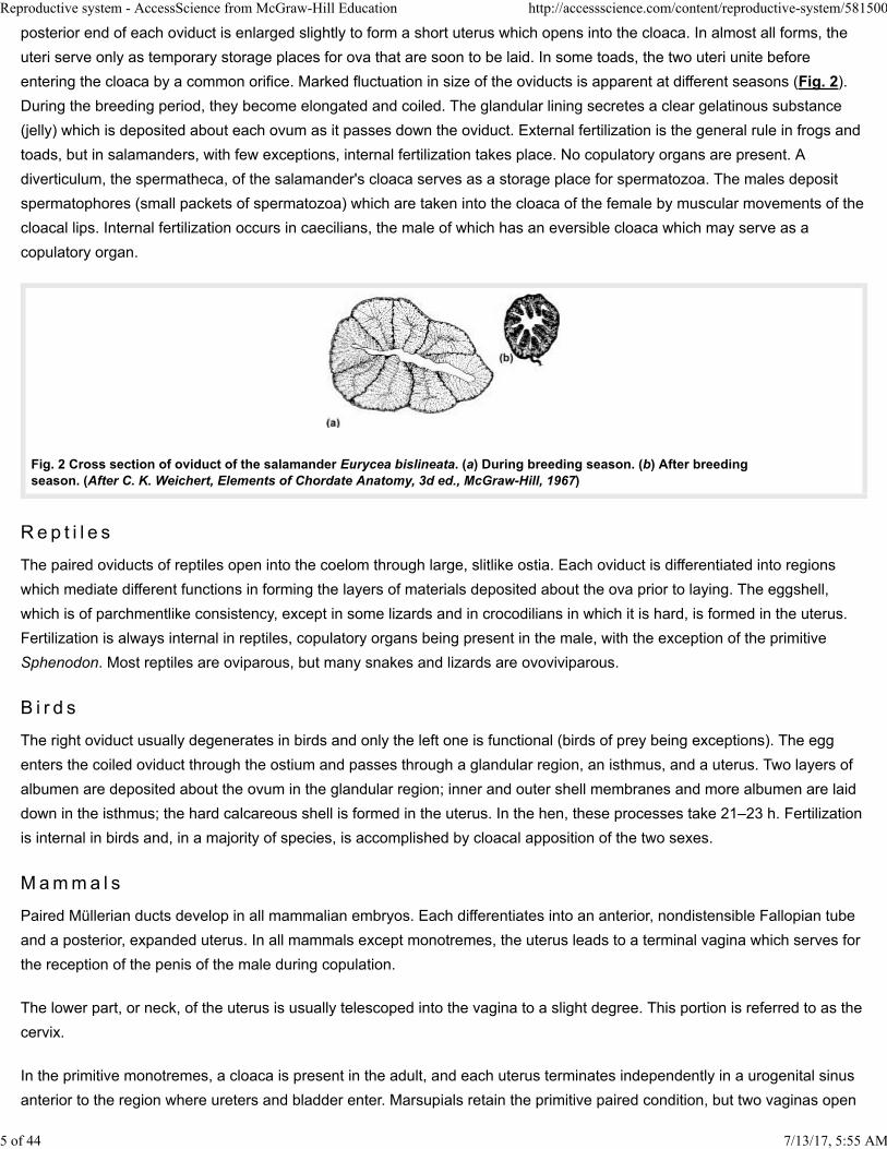
posterior end of each oviduct is enlarged slightly to form a short uterus which opens into the cloaca. In almost all forms, the
uteri serve only as temporary storage places for ova that are soon to be laid. In some toads, the two uteri unite before
entering the cloaca by a common orifice. Marked fluctuation in size of the oviducts is apparent at different seasons (Fig. 2).
During the breeding period, they become elongated and coiled. The glandular lining secretes a clear gelatinous substance
(jelly) which is deposited about each ovum as it passes down the oviduct. External fertilization is the general rule in frogs and
toads, but in salamanders, with few exceptions, internal fertilization takes place. No copulatory organs are present. A
diverticulum, the spermatheca, of the salamander's cloaca serves as a storage place for spermatozoa. The males deposit
spermatophores (small packets of spermatozoa) which are taken into the cloaca of the female by muscular movements of the
cloacal lips. Internal fertilization occurs in caecilians, the male of which has an eversible cloaca which may serve as a
copulatory organ.
Fig. 2 Cross section of oviduct of the salamander Eurycea bislineata. (a) During breeding season. (b) After breedingseason. (After C. K. Weichert, Elements of Chordate Anatomy, 3d ed., McGraw-Hill, 1967)
Rep t i l e s
The paired oviducts of reptiles open into the coelom through large, slitlike ostia. Each oviduct is differentiated into regions
which mediate different functions in forming the layers of materials deposited about the ova prior to laying. The eggshell,
which is of parchmentlike consistency, except in some lizards and in crocodilians in which it is hard, is formed in the uterus.
Fertilization is always internal in reptiles, copulatory organs being present in the male, with the exception of the primitive
Sphenodon. Most reptiles are oviparous, but many snakes and lizards are ovoviviparous.
B i r d s
The right oviduct usually degenerates in birds and only the left one is functional (birds of prey being exceptions). The egg
enters the coiled oviduct through the ostium and passes through a glandular region, an isthmus, and a uterus. Two layers of
albumen are deposited about the ovum in the glandular region; inner and outer shell membranes and more albumen are laid
down in the isthmus; the hard calcareous shell is formed in the uterus. In the hen, these processes take 21–23 h. Fertilization
is internal in birds and, in a majority of species, is accomplished by cloacal apposition of the two sexes.
M amma l s
Paired Müllerian ducts develop in all mammalian embryos. Each differentiates into an anterior, nondistensible Fallopian tube
and a posterior, expanded uterus. In all mammals except monotremes, the uterus leads to a terminal vagina which serves for
the reception of the penis of the male during copulation.
The lower part, or neck, of the uterus is usually telescoped into the vagina to a slight degree. This portion is referred to as the
cervix.
In the primitive monotremes, a cloaca is present in the adult, and each uterus terminates independently in a urogenital sinus
anterior to the region where ureters and bladder enter. Marsupials retain the primitive paired condition, but two vaginas open
7/13/17, 5:55 AM 5 of 44
Reproductive system - AccessScience from McGraw-Hill Education http://accessscience.com/content/reproductive-system/581500
into a common urogenital sinus. In some mammals, the kangaroo for example, the vaginas fuse at their upper ends to form a
vaginal sinus which extends posteriorly as a blind pocket or tube. It is sometimes referred to as a third vagina and may serve
as the birth canal. Its posterior end connects with the urogenital sinus. In the event that this pouchlike structure has no
opening, its wall ruptures at the time of delivery, thus permitting the young, lodged in it, to pass directly into the urogenital
sinus. Placental mammals have a single vagina which represents a fusion of two; only the pika, which is a lagomorph, has a
cloaca in the adult form.
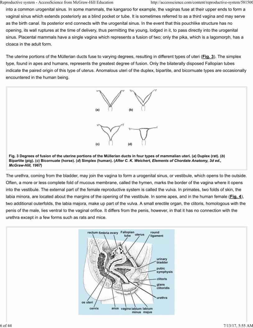
The uterine portions of the Müllerian ducts fuse to varying degrees, resulting in different types of uteri (Fig. 3). The simplex
type, found in apes and humans, represents the greatest degree of fusion. Only the bilaterally disposed Fallopian tubes
indicate the paired origin of this type of uterus. Anomalous uteri of the duplex, bipartite, and bicornuate types are occasionally
encountered in the human being.
Fig. 3 Degrees of fusion of the uterine portions of the Müllerian ducts in four types of mammalian uteri. (a) Duplex (rat). (b)Bipartite (pig). (c) Bicornuate (horse). (d) Simplex (human). (After C. K. Weichert, Elements of Chordate Anatomy, 3d ed., McGraw-Hill, 1967)
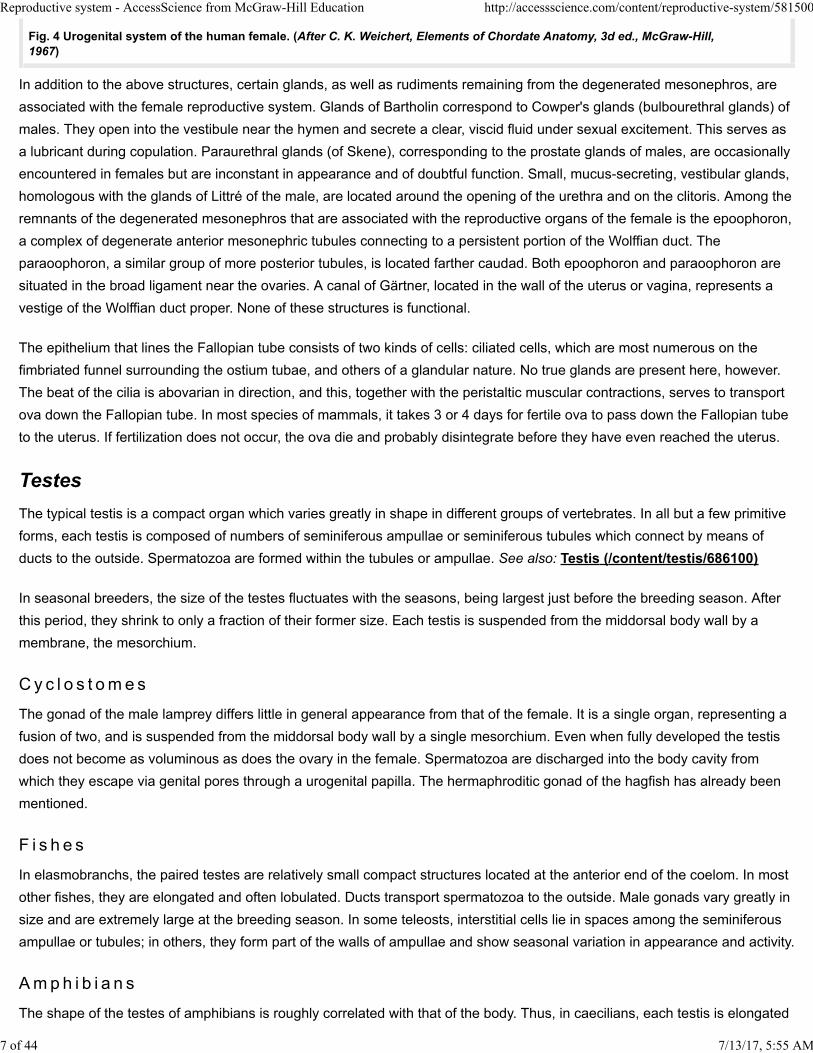
The urethra, coming from the bladder, may join the vagina to form a urogenital sinus, or vestibule, which opens to the outside.
Often, a more or less complete fold of mucous membrane, called the hymen, marks the border of the vagina where it opens
into the vestibule. The external part of the female reproductive system is called the vulva. In primates, two folds of skin, the
labia minora, are located about the margins of the opening of the vestibule. In some apes, and in the human female (Fig. 4),
two additional outerfolds, the labia majora, make up part of the vulva. A small erectile organ, the clitoris, homologous with the
penis of the male, lies ventral to the vaginal orifice. It differs from the penis, however, in that it has no connection with the
urethra except in a few forms such as rats and mice.
7/13/17, 5:55 AM 6 of 44
Reproductive system - AccessScience from McGraw-Hill Education http://accessscience.com/content/reproductive-system/581500
Fig. 4 Urogenital system of the human female. (After C. K. Weichert, Elements of Chordate Anatomy, 3d ed., McGraw-Hill, 1967)
In addition to the above structures, certain glands, as well as rudiments remaining from the degenerated mesonephros, are
associated with the female reproductive system. Glands of Bartholin correspond to Cowper's glands (bulbourethral glands) of
males. They open into the vestibule near the hymen and secrete a clear, viscid fluid under sexual excitement. This serves as
a lubricant during copulation. Paraurethral glands (of Skene), corresponding to the prostate glands of males, are occasionally
encountered in females but are inconstant in appearance and of doubtful function. Small, mucus-secreting, vestibular glands,
homologous with the glands of Littré of the male, are located around the opening of the urethra and on the clitoris. Among the
remnants of the degenerated mesonephros that are associated with the reproductive organs of the female is the epoophoron,
a complex of degenerate anterior mesonephric tubules connecting to a persistent portion of the Wolffian duct. The
paraoophoron, a similar group of more posterior tubules, is located farther caudad. Both epoophoron and paraoophoron are
situated in the broad ligament near the ovaries. A canal of Gärtner, located in the wall of the uterus or vagina, represents a
vestige of the Wolffian duct proper. None of these structures is functional.
The epithelium that lines the Fallopian tube consists of two kinds of cells: ciliated cells, which are most numerous on the
fimbriated funnel surrounding the ostium tubae, and others of a glandular nature. No true glands are present here, however.
The beat of the cilia is abovarian in direction, and this, together with the peristaltic muscular contractions, serves to transport
ova down the Fallopian tube. In most species of mammals, it takes 3 or 4 days for fertile ova to pass down the Fallopian tube
to the uterus. If fertilization does not occur, the ova die and probably disintegrate before they have even reached the uterus.
Testes
The typical testis is a compact organ which varies greatly in shape in different groups of vertebrates. In all but a few primitive
forms, each testis is composed of numbers of seminiferous ampullae or seminiferous tubules which connect by means of
ducts to the outside. Spermatozoa are formed within the tubules or ampullae. See also: Testis (/content/testis/686100)
In seasonal breeders, the size of the testes fluctuates with the seasons, being largest just before the breeding season. After
this period, they shrink to only a fraction of their former size. Each testis is suspended from the middorsal body wall by a
membrane, the mesorchium.
C y c l o s t om e s
The gonad of the male lamprey differs little in general appearance from that of the female. It is a single organ, representing a
fusion of two, and is suspended from the middorsal body wall by a single mesorchium. Even when fully developed the testis
does not become as voluminous as does the ovary in the female. Spermatozoa are discharged into the body cavity from
which they escape via genital pores through a urogenital papilla. The hermaphroditic gonad of the hagfish has already been
mentioned.
F i s h e s
In elasmobranchs, the paired testes are relatively small compact structures located at the anterior end of the coelom. In most
other fishes, they are elongated and often lobulated. Ducts transport spermatozoa to the outside. Male gonads vary greatly in
size and are extremely large at the breeding season. In some teleosts, interstitial cells lie in spaces among the seminiferous
ampullae or tubules; in others, they form part of the walls of ampullae and show seasonal variation in appearance and activity.
Amp h i b i a n s
The shape of the testes of amphibians is roughly correlated with that of the body. Thus, in caecilians, each testis is elongated
7/13/17, 5:55 AM 7 of 44
Reproductive system - AccessScience from McGraw-Hill Education http://accessscience.com/content/reproductive-system/581500
and resembles a string of beads. The enlargements consist of masses of seminiferous ampullae connected by a longitudinal
collecting duct. In salamanders, the testes are shorter and irregular in outline; in frogs and toads, they are small, oval,
compact structures. A pronounced difference in size is apparent during the breeding season. Fat bodies are associated with
the gonads of male as well as of female amphibians.
R e p t i l e s
Reptilian testes are round, oval, or pyriform in shape and contain seminiferous tubules that are long and convoluted. In
snakes and lizards, one testis usually lies farther forward in the body cavity than the other. Periodic fluctuations in size of the
testes are typical.
B i r d s
The round or oval shape of bird testes is characteristic. In the domestic fowl, the testes function throughout the year and no
periodic variations in size are obvious. Most birds, however, are seasonal breeders, the testes enlarging conspicuously at the
approach of the breeding season. Increase in the number of hours of daylight stimulates spermatogenesis in certain birds and
hence brings about testicular enlargement, this generally occurring in spring.
M amma l s
In all mammals except monotremes, the oval-shaped testes move from their place of origin to the pelvic region, where they
may remain permanently, or they may descend farther into a pouchlike scrotum. In many seasonal breeders, the testes are
located in the scrotum only during the breeding period. The scrotum serves as a temperature regulator, providing an
environment for the testes several degrees below that of the body. This seems to be a requirement for normal development of
spermatozoa. In marsupials the scrotum lies anterior to the penis but in other mammals it is posterior to that organ. In several
species, a relation between the number of hours of daylight and testicular activity has been demonstrated.
Male ducts
The ducts which in most vertebrates serve to transport spermatozoa to the outside of the body are the archinephric ducts or
Wolffian ducts formed in connection with the opisthonephros or mesonephros, respectively. Their original function is
elimination of urinary wastes. In some fishes and amphibians, certain modified kidney tubules are employed in carrying
spermatozoa from the testis to the archinephric duct. The tubules are known as efferent ductules, and the duct is termed the
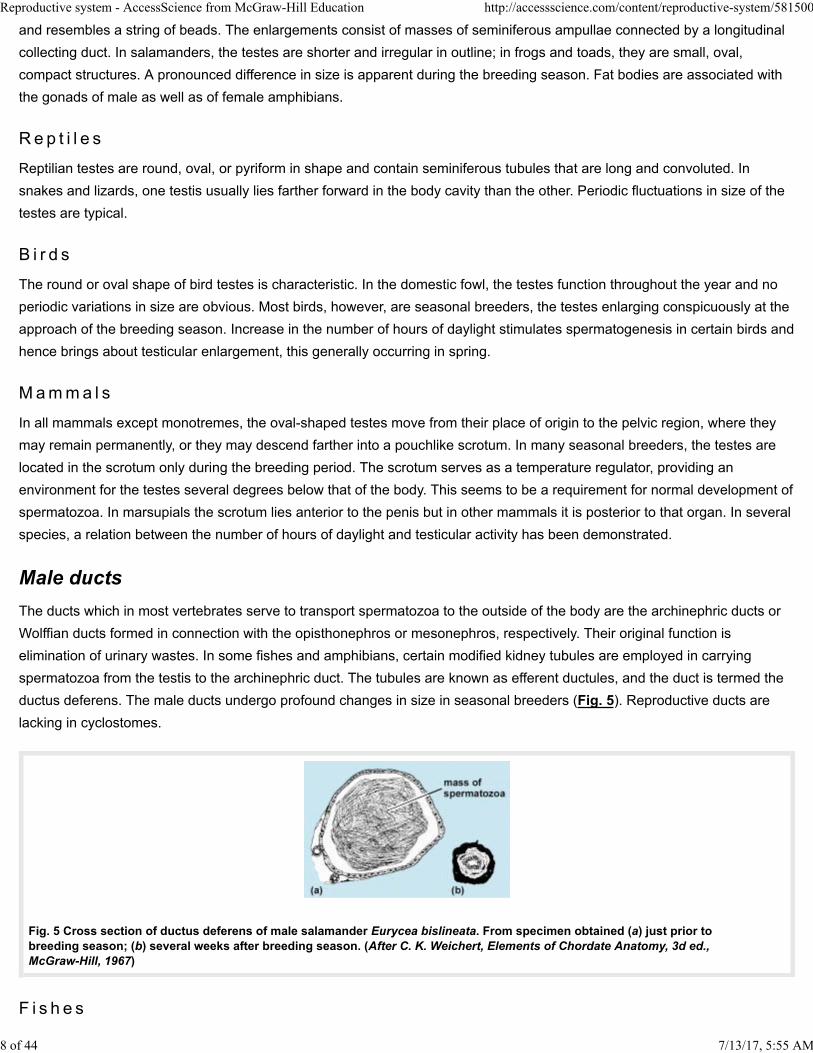
ductus deferens. The male ducts undergo profound changes in size in seasonal breeders (Fig. 5). Reproductive ducts are
lacking in cyclostomes.
7/13/17, 5:55 AM
Fig. 5 Cross section of ductus deferens of male salamander Eurycea bislineata. From specimen obtained (a) just prior to breeding season; (b) several weeks after breeding season. (After C. K. Weichert, Elements of Chordate Anatomy, 3d ed., McGraw-Hill, 1967)
F i s h e s
8 of 44
Reproductive system - AccessScience from McGraw-Hill Education http://accessscience.com/content/reproductive-system/581500
A variety of conditions is encountered in the reproductive system of male fishes. In elasmobranchs, small efferent tubules
course from the testis through the mesorchium, connecting with some anterior kidney tubules along the medial border of the
opisthonephros. The kidney tubules lead to the archinephric duct, which now serves almost entirely as a ductus deferens.
The ductus deferens courses along the ventral side of the opisthonephros. In young specimens, it is a straight tube with a
urinary function; in older individuals, it is highly convoluted. The posterior end is markedly dilated to form a seminal vesicle.
The two seminal vesicles open into a common urogenital sinus which enters the cloaca through an aperture at the tip of a
urogenital papilla. A pair of blind sperm sacs projects forward from the ventral wall of the urogenital sinus. These sacs may be
remnants of the Müllerian ducts which persist in the male.
In most other fishes, the kidney ducts serve only for the passage of urinary wastes. The sperm duct, which is not a true
ductus deferens because it is formed in a different manner, may be entirely independent of the kidney duct, although the two
may have a common opening to the outside.
Amp h i b i a n s
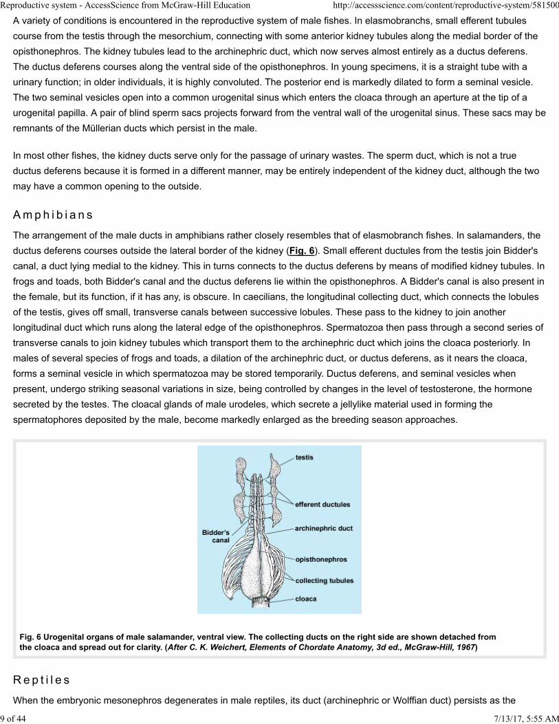
The arrangement of the male ducts in amphibians rather closely resembles that of elasmobranch fishes. In salamanders, the
ductus deferens courses outside the lateral border of the kidney (Fig. 6). Small efferent ductules from the testis join Bidder's
canal, a duct lying medial to the kidney. This in turns connects to the ductus deferens by means of modified kidney tubules. In
frogs and toads, both Bidder's canal and the ductus deferens lie within the opisthonephros. A Bidder's canal is also present in
the female, but its function, if it has any, is obscure. In caecilians, the longitudinal collecting duct, which connects the lobules
of the testis, gives off small, transverse canals between successive lobules. These pass to the kidney to join another
longitudinal duct which runs along the lateral edge of the opisthonephros. Spermatozoa then pass through a second series of
transverse canals to join kidney tubules which transport them to the archinephric duct which joins the cloaca posteriorly. In
males of several species of frogs and toads, a dilation of the archinephric duct, or ductus deferens, as it nears the cloaca,
forms a seminal vesicle in which spermatozoa may be stored temporarily. Ductus deferens, and seminal vesicles when
present, undergo striking seasonal variations in size, being controlled by changes in the level of testosterone, the hormone
secreted by the testes. The cloacal glands of male urodeles, which secrete a jellylike material used in forming the
spermatophores deposited by the male, become markedly enlarged as the breeding season approaches.
Fig. 6 Urogenital organs of male salamander, ventral view. The collecting ducts on the right side are shown detached fromthe cloaca and spread out for clarity. (After C. K. Weichert, Elements of Chordate Anatomy, 3d ed., McGraw-Hill, 1967)
Rep t i l e s
When the embryonic mesonephros degenerates in male reptiles, its duct (archinephric or Wolffian duct) persists as the
7/13/17, 5:55 AM 9 of 44
Reproductive system - AccessScience from McGraw-Hill Education http://accessscience.com/content/reproductive-system/581500
reproductive duct. The end near the testis becomes greatly convoluted and is called the epididymis. This connects with the
testes by means of a few persistent mesonephric tubules which serve as efferent ductules. The remainder is the ductus
deferens, sometimes straight and sometimes convoluted. In snakes and lizards, this joins the ureter of the metanephros
before entering the cloaca. In turtles and crocodilians, these ducts open at the proximal end of a groove which carries
spermatozoa to the free end of the penis. Seasonal variations under endocrine control are obvious in the epididymides and
deferent ducts of most reptiles. In many, some of the posterior urinary tubules of the metanephros enlarge during the
breeding season, producing an albuminous secretion which contributes to the seminal fluid. Glandular secretions from the
cloacal walls in snakes and lizards pass into the grooves of the hemipenes. Nonfunctional Müllerian ducts, usually much
reduced in size, commonly persist in male reptiles.
B i r d s
The reproductive ducts of male birds are essentially similar to those of reptiles but open independently into the cloaca. In the
few birds that possess a penis, a groove on the upper surface carries spermatozoa to the apex. In some passerine birds, a
nodule, composed of a tightly coiled portion of the ductus deferens, protrudes into the cloaca. The temperature of the nodule
is somewhat lower than body temperature. This may be of importance in the maturation of spermatozoa.
M amma l s
A few persistent mesonephric tubules connect the seminiferous tubules of each testis to a compactly coiled epididymis which
is continuous with the ductus deferens. In man, the portion of the Wolffian duct included in the epididymis averages 20 ft (6 m)
in length. Experiments have demonstrated that, during their passage through the epididymis, spermatozoa, although
remaining quiescent, acquire the capability of becoming fully motile. In mammals having scrotal testes, the ductus deferens
enters the body cavity lying between the peritoneum and the body wall. It crosses in front of the ureter, loops over that
structure, and then courses posteriorly for a short distance before joining the urethra (Fig. 7). This peculiar arrangement is the
result of the descent of testes from their original abdominal location. In many mammals, the ductus deferens enlarges at its
posterior end to form an ampulla. A glandular seminal vesicle may arise on each side as a saccular diverticulum of the ductus
deferens. It does not store spermatozoa, but its secretion contributes to the seminal fluid in which spermatozoa are
suspended. Seminal vesicles are absent in monotremes, marsupials, carnivores, and whales. The lower portion of the ductus
deferens, between seminal vesicle and urethra, is often called the ejaculatory duct. The urethra, coming from the bladder,
extends the length of the penis, opening at the tip of that organ through a small meatus. It serves both for passage of urine
and seminal fluid. Accessory glands associated with the urethra include the prostate gland, which contributes to the seminal
fluid; Cowper's glands, which secrete a clear, viscid fluid during sexual excitement; and small, mucus-secreting urethral
glands. In some mammals, as in the rat, when the secretions of the seminal vesicles and prostate gland are intermingled,
they coagulate, so that just after mating a so-called copulation plug is present in the vagina. This aids in retaining
spermatozoa in the reproductive tract of the female. The development and functioning of the accessory sex organs in
mammals are clearly under control of the hormone testosterone. A few remnants of the mesonephros may persist in male
mammals in close relation to the reproductive systems. Among these are the paradidymis, a few aberrant ductules, and the
appendix of the epididymis. Persistent homologs of the Müllerian ducts may also be present in males. The small appendix of
the testis and the prostatic utricle represent such remnants.
7/13/17, 5:55 AM 10 of 44
Reproductive system - AccessScience from McGraw-Hill Education http://accessscience.com/content/reproductive-system/581500
Fig. 7 Urogenital system of the human male. (After C. K. Weichert, Elements of Chordate Anatomy, 3d ed., McGraw-Hill, 1967)
Copulatory organs
Fertilization always takes place in a fluid medium. In many aquatic vertebrates, external fertilization takes place, with water
furnishing the medium in which sperm travel to gain access to ova. Terrestrial forms and even numerous aquatic species
have internal fertilization. Secretions supplied by both sexes furnish the necessary fluid medium. In a number of terrestrial
vertebrates, spermatozoa are transferred from male to female by cloacal apposition. In most, however, as well as in many
aquatic species, intromittent, or copulatory, organs are employed to deposit spermatozoa, suspended in seminal fluid, within
the reproductive tract of the female.
Fertilization usually takes place in the upper part of the oviduct. In forms in which the egg is surrounded by a shell, fertilization
must take place before the shell has been formed about the ovum.
F i s h e s
In those fishes having internal fertilization, the copulatory organs are modifications of fins, pelvic fins in elasmobranchs and
anal fins in teleosts. Claspers, which are modifications of pelvic fins, are used as intromittent organs by male elasmobranchs.
Each clasper is essentially a scroll-like tube through which seminal fluid may be ejected with some force into the cloaca of the
female. It is probable that only one clasper is inserted at a time. In teleosts having internal fertilization, the anterior border of
the anal fin is elongated posteriorly to form an intromittent organ, the gonopodium. In some teleosts, modifications of the
hemal spines of certain caudal vertebrae form a copulatory organ.
Amp h i b i a n s
Although internal fertilization occurs in most salamanders, copulatory organs are lacking. By muscular action of the cloacal
lips, the female is able to pick up spermatophores, or packets of spermatozoa, deposited by the male. The eversible and
protrusible cloaca of some male caecilians may be used as a copulatory organ when the cloacae of both sexes are in
apposition.
R e p t i l e s
Sphenodon is the only reptile lacking copulatory organs. In others, two types of structures are evident. Snakes and lizards
employ paired hemipenes, which are saclike structures, devoid of erectile tissue, lying under the skin adjacent to the cloaca.
7/13/17, 5:55 AM 11 of 44
Reproductive system - AccessScience from McGraw-Hill Education http://accessscience.com/content/reproductive-system/581500
Each bears a groove for the passage of spermatozoa. These organs are everted during copulation and spermatozoa are
deposited in the cloaca of the female. Hemipenes are not homologous with the single penis of turtles and crocodilians, which
is basically similar to that of mammals, and contains erectile tissue which becomes distended with blood during sexual
excitement. A homologous structure, the clitoris, is present in rudimentary form in females. Paired, thickened ridges in the
anterior and ventral walls of the cloaca are called corpora cavernosa. They are composed of connective and erectile tissue. A
groove along the dorsal surface provides for the passage of spermatozoa. During the act of mating, the corpora cavernosa
are filled and distended with blood and the penis is firm and greatly enlarged. It is then said to be erect. This property of
erectile tissue makes it possible for the penis to serve as an intromittent organ. Without erection, copulation is impossible.
During erection, the veins which normally drain blood from the erectile tissue become compressed. There is an increased
supply of arterial blood supply accompanied by an impeded venous drainage. Return of the penis to its flaccid state after
erection is termed detumescence.
B i r d s
Most birds copulate by cloacal apposition. A penis is present only in ducks, geese, swans, and ostriches. It is a single
structure of the same type as that of turtles and crocodilians. A clitoris is present in females of these species. In many other
birds, a rudimentary penis can be identified.
M amma l s
In monotremes, the single penis lies on the floor of the cloaca. It is similar to the organ in turtles, crocodilians, and birds
except that the groove on the dorsal side has become a closed tube. This is surrounded by another mass of erectile tissue
known as the corpus spongiosum. The canal in monotremes possibly carries only seminal fluid since the urethra has a
separate opening into the cloaca. The monotreme urethra thus differs from that of all other mammals. Marsupials lack a
cloaca. The marsupial penis is covered by a sheath which opens to the outside of the body just beneath the anus. It may be
protracted and retracted. The scrotum in marsupials is anterior to the penis. There are two erectile corpora cavernosa,
separated by a septum, and a single erectile corpus spongiosum surrounding the urethra. Usually in marsupials, three pairs
of Cowper's glands open into the urethra at the base of the penis.
In higher mammals, there is a tendency for the penis to be directed forward; and in all forms possessing a scrotum, the penis
is located anterior to the scrotum. In most mammals, it lies within a sheath from which it can be protracted and retracted. In
primates, the penis is permanently exserted. Its distal end bears a sensitive, swollen glans. Among mammals, there are many
variations in shape and structure of the glans, which contains erectile tissue continuous with the corpus spongiosum. In some
forms, as in the cat, the glans bears numerous horny papillae or spines on its surface; these undoubtedly function as a sexual
irritant. The glans is covered by a sheath, the prepuce, or foreskin, which may be retracted. Preputial glands secrete a
sebaceous material called smegma about the base of the glans underneath the prepuce.
A bone is present in the penis of many rodents, carnivores, bats, whales, and lower primates. It is situated in the connective
tissues between the corpora cavernosa and corpus spongiosum. In whales, there is but a single corpus cavernosum.
Anomalous development of the vertebrate penis is occasionally encountered. The most common anomalies are referred to as
hypospadias and epispadias, in which the urethra opens on the under or upper sides, respectively, of the penis, rather than
being continued to the tip of the glans.
Charles K. Weichert
The human reproductive system comprises many different organs, and therefore all the primary types of tissue are identified.
12 of 44 7/13/17, 5:55 AM
Reproductive system - AccessScience from McGraw-Hill Education http://accessscience.com/content/reproductive-system/581500
Male reproductive system
In the following sections, the histological features of the testes, scrotum, excretory ducts, and auxiliary glands are discussed.
Te s t e s
The testes have two important structural elements, the contorted seminiferous tubules and the interstitial cells which are
situated between the tubules. The contorted seminiferous tubules have two kinds of cells, nutrient cells, which also serve as
supporting cells (the cells of Sertoli), and germinal cells, which develop into spermatozoa by the process of spermatogenesis.
The interstitial cells of the testis are seemingly modified connective-tissue cells. They resemble epithelial cells and hence are
correctly designated epithelioid cells. They produce testosterone, the male sex hormone, which chemically is a steroid
compound, in mammals and perhaps in other vertebrates. See also: Spermatogenesis (/content/spermatogenesis
/643700)
S c r o t um
The scrotum consists of skin, smooth muscle, and connective tissue. The scrotal skin is rich in sweat glands; it is thus able to
act as a t hermoregulator for the testes. The human scrotal temperature is about 13°F (7°C) lower than that of the abdominal
cavity. The smooth muscle is a “skin muscle” called the dartos tunic; its relaxation lengthens the scrotum and promotes loss
of heat, whereas its contraction shortens the scrotum and reduces loss of heat. See also: Sweat gland (/content/sweat-
gland/672200); Thermoregulation (/content/thermoregulation/691600)
E x c r e t o r y d u c t s
The excretory ducts which convey spermatozoa from the contorted seminiferous tubules to the urethra are either
intratesticular (tubuli seminiferi recti and rete testis) or extratesticular (epididymis, ductus deferens, and ejaculatory duct).
From the seminiferous tubules, spermatozoa enter the epididymis, a tubular structure consisting of head, body, and tail. The
head of the epididymis has 12 or more efferent ductules (ductuli efferentes testis), the epithelium of which bears cilia, or little
hairlike projections, which assist in moving sperm into the coiled part of the epididymis. The body and tail of the epididymis
constitute the ductus epididymis, a coiled tube which, when uncoiled, is about 15–20 ft (4.5–6 m) long. The ductus deferens in
humans is about 18 in. (45 cm) when uncoiled. Its proximal end is dilated to form the ampulla ductus deferentis.
A u x i l i a r y g l a n d s
The auxiliary glands in humans include two seminal vesicles, one prostate, and two bulbourethral glands. Their secretions
together with spermatozoa and the small amount of secretion from the excretory ducts constitute the semen. In the male, one
ejection of semen, known as ejaculation, has a volume of 3–5 ml, and each milliliter contains about 6 × 10 7 spermatozoa. The
seminal vesicles, named in accordance with the erroneous belief that they are receptacles for spermatozoa (after death,
sperm may be found in the vesicles), are hollow glands which lie along the back wall of the prostate. They are about 2 i n. (5
cm) in length, and, developmentally, each gland is an outgrowth of the ampulla of the ductus deferens. The right and left
seminal vesicles open into the prostatic urethra via the ejaculatory ducts. The prostate is a solid gland which surrounds the
urethra (prostatic urethra) at its origin from the urinary bladder. It is about 1–1.5 in. (2.5–4 cm) in diameter, and has several
small ducts which open individually into the prostatic urethra. The bulbourethral glands, or glands of Cowper, are about 0.25
in. (6 mm) in diameter. Each opens into the urethra at a site slightly below the prostate.
In such species as the rat, guinea pig, and rhesus monkey, the mixture of the secretions from the auxiliary glands coagulates,
as in the case of ejaculated semen. In the mating of rats and guinea pigs, the coagulated semen produces the vaginal plug
which temporarily partly occludes the vagina of the female partner.
7/13/17, 5:55 AM 13 of 44
Reproductive system - AccessScience from McGraw-Hill Education http://accessscience.com/content/reproductive-system/581500
Female reproductive system
The h istological discussion of the female system includes the ovaries, uterine tubes, uterus, vagina, and external genitalia.
O v a r y
The ovary (ovarium), or egg producer, is largely covered by peritoneum; it has follicles with egg cells (ova), a stroma of
connective tissue, and, in maturity, corpora lutea.
Ma t u r a t i o n o f t h e o v um
The process of the origin, growth, and formation of the ovum in its preparation for fertilization is known as oogenesis. The
young sex cell is an oogonium. Growth produces the primary oocyte; only the beginning of the first maturation division occurs
before ovulation. After ovulation, the primary oocyte divides to produce two unequal elements: a secondary oocyte and a
smaller body, the first polar body. Then the secondary oocyte divides to yield the mature ovum (rarely called ootid) and the
second polar body. The mature ovum has the haploid number of chromosomes; the two polar bodies eventually disintegrate.
See also: Oogenesis (/content/oogenesis/469500)
Co r p u s l u t e um
The corpus luteum is a yellow endocrine body which originates at the site of a ruptured Graafian follicle and which produces
progesterone, one of the female sex hormones. It originates by the metamorphosis of granulosa cells and thecal cells into
lutein cells which are epithelioid. The corpus luteum of ovulation persists about 14 days, whereas the corpus luteum of
pregnancy lasts several months. The degeneration of a corpus luteum produces a white, fibrous scar in the ovary, the corpus
albicans.
F a l l o p i a n t u b e
The human Fallopian tube is a muscular structure about 4.5–5 in. (11.4–12.7 cm) long, and has three regional subdivisions: a
funnellike upper part, the infundibulum (infundibulum tubae uterinae); a dilated part below the infundibulum, the ampulla; and
a constricted part near the uterine junction, the isthmus. The mouth of the tube, or ostium, communicates with the peritoneal
cavity, and is guarded by a fringe, the fimbriae tubae. Fertilization of the ovum occurs in the tube.
U t e r u s
The uterus is covered in part by peritoneum. It has an outer layer of smooth muscle, the myometrium, and an inner layer of
connective tissue and epithelium, the endometrium. Thickness of the endometrium varies during the menstrual cycle. See
also: Menstruation (/content/menstruation/415000)
Vag i n a
The musculomembranous organ situated between the cervix uteri and the external genitalia is the vagina. During copulation,
it ensheathes the penis. Ventrally and dorsally, the anterior and posterior fornices, respectively, overlap the uterine cervix. In
youth, the lower end of the vagina is constricted a bit by a membranous shelf of mucosa, the hymen vaginae.
E x t e r n a l g e n i t a l i a
The external genitalia include the clitoris, vestibule of the vulva (fossa vestibuli vaginae), paired lips of the vulva (labium
majus pudendi and labium minus pudendi), and a pair of small vestibular glands (glandulae vestibulares minores). The large
lips of the vulva are essentially folds of skin, whereas the small lips are covered with a mucous membrane. The vestibular
glands secrete mucus, and, in cases of gonorrhea, may become inflamed and then form large cysts.
7/13/17, 5:55 AM 14 of 44
Reproductive system - AccessScience from McGraw-Hill Education http://accessscience.com/content/reproductive-system/581500
Lemen J. Wells
The embryology of the reproductive system shows great similarities in all vertebrates, with the exception of teleost fishes, in
which it is less specialized. It proceeds by three steps: sex determination; organogenesis of gonads; and later, during
development, differentiation of the genital tract.
Sex determination
At the time of fertilization, the sex of each individual is genetically determined by certain genes carried by the sex
chromosomes contained in the gametes. If both the male and the female germ cells have an identical X chromosome, the
egg, upon fertilization, receives two X chromosomes (homozygous egg). If one parent germ cell contains an X and the other
contains a Y chromosome, the egg cell receives an XY assortment (heterozygous egg). In mammals and in frogs, for
instance, the male is the heterozygous XY sex. In birds and several urodeles, the condition is reversed and the female sex is
the heterozygous XY sex.
Sex determination should be considered as the first step in sexual differentiation of each individual. Techniques have been
developed which permit direct study of the chromosomes, particularly the sex chromosomes, in dividing cells of any tissue.
Chromosomal anomalies, such as the absence of one chromosome (for example, XO) or the presence of one or more
supplementary chromosomes (for example, XXY), can be recognized in humans or in animals. Mosaicism results from the
presence of two or more types of cells, for instance, cells with XX and cells with XY chromosomes, in the same individual. In
such individuals, the genetic basis of sex is disturbed, and embryological anomalies may occur. See also: Chromosome
(/content/chromosome/134900); Sex determination (/content/sex-determination/617400)
Role of urinary system
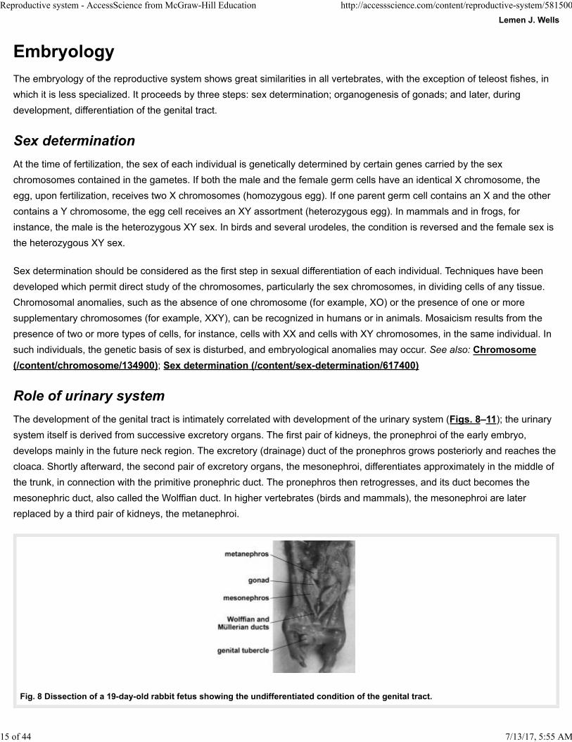
The development of the genital tract is intimately correlated with development of the urinary system (Figs. 8–11); the urinary
system itself is derived from successive excretory organs. The first pair of kidneys, the pronephroi of the early embryo,
develops mainly in the future neck region. The excretory (drainage) duct of the pronephros grows posteriorly and reaches the
cloaca. Shortly afterward, the second pair of excretory organs, the mesonephroi, differentiates approximately in the middle of
the trunk, in connection with the primitive pronephric duct. The pronephros then retrogresses, and its duct becomes the
mesonephric duct, also called the Wolffian duct. In higher vertebrates (birds and mammals), the mesonephroi are later
replaced by a third pair of kidneys, the metanephroi.
7/13/17, 5:55 AM
Fig. 8 Dissection of a 19-day-old rabbit fetus showing the undifferentiated condition of the genital tract.
15 of 44

Reproductive system - AccessScience from McGraw-Hill Education http://accessscience.com/content/reproductive-system/581500
Fig. 9 Section through the body of a 17-day-old rabbit fetus, showing the ovaries at an early stage of differentiation located on the internal side of the mesonephroi.
Fig. 10 Homologies in the development of the male and female genital tract of mammals. The gonads are shown on the inner side of the mesonephroi, and the ducts on their outer border. (After A. Jost, General outline about reproductive physiology and its developmental background, in H. Gibian and E. J. Plotz, eds., Mammalian Reproduction, pp. 4–32, Springer, 1970)
Fig. 11 Drawings of the right half of the reproductive system of the newt Triton (Triturus) cristatus. (a) Male. (b) Female. (After J. de Beaumont, Wilhelm Roux Arch. Entwicklungsmech. Organ., 129:120, 1933)
Among the vertebrates, except for the teleost fishes, the sex glands differentiate in connection with the mesonephros. They
retain these connections in male amphibians, in which the mesonephros remains the adult kidney (Fig. 11). The testicular
tubules are connected with specialized mesonephric tubules. An adult male frog displays conditions which are similar to those
7/13/17, 5:55 AM 16 of 44
Reproductive system - AccessScience from McGraw-Hill Education http://accessscience.com/content/reproductive-system/581500
found in the early bird or mammalian embryo. See also: Urinary system (/content/urinary-system/724000); Urogenital
system (/content/urogenital-system/724300)
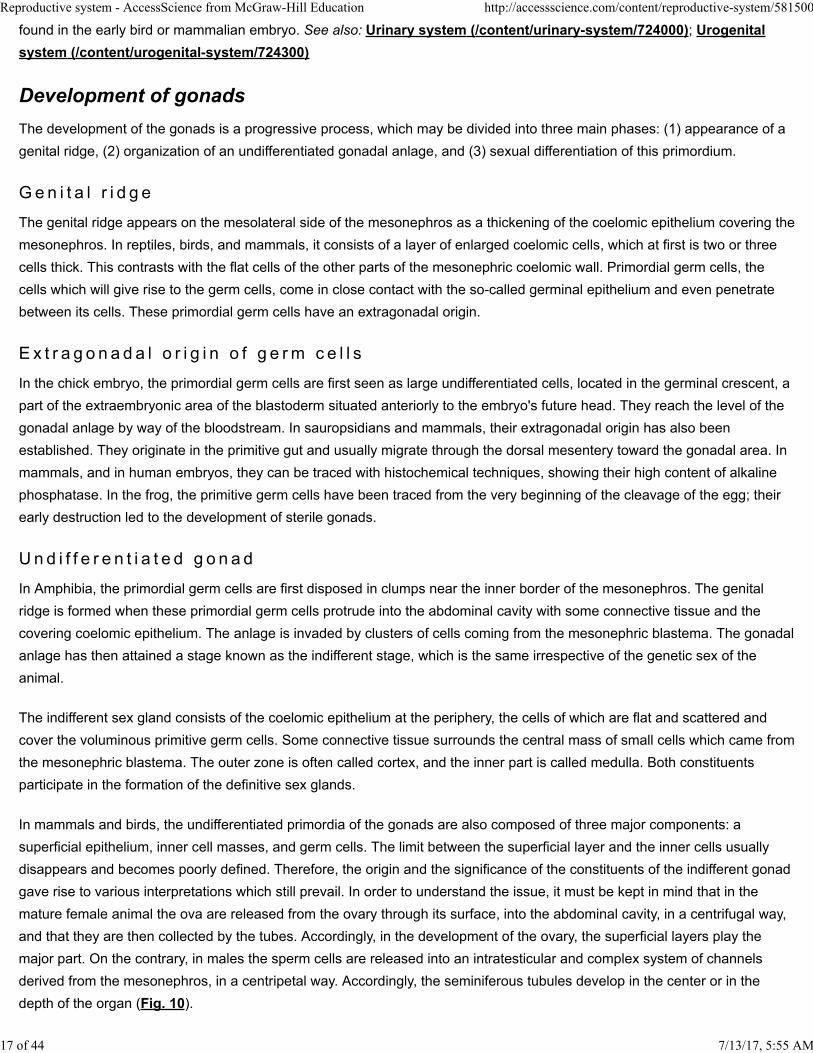
Development of gonads
The development of the gonads is a progressive process, which may be divided into three main phases: (1) appearance of a
genital ridge, (2) organization of an undifferentiated gonadal anlage, and (3) sexual differentiation of this primordium.
G e n i t a l r i d g e
The genital ridge appears on the mesolateral side of the mesonephros as a thickening of the coelomic epithelium covering the
mesonephros. In reptiles, birds, and mammals, it consists of a layer of enlarged coelomic cells, which at first is two or three
cells thick. This contrasts with the flat cells of the other parts of the mesonephric coelomic wall. Primordial germ cells, the
cells which will give rise to the germ cells, come in close contact with the so-called germinal epithelium and even penetrate
between its cells. These primordial germ cells have an extragonadal origin.
E x t r a g o n a d a l o r i g i n o f g e rm c e l l s
In the chick embryo, the primordial germ cells are first seen as large undifferentiated cells, located in the germinal crescent, a
part of the extraembryonic area of the blastoderm situated anteriorly to the embryo's future head. They reach the level of the
gonadal anlage by way of the bloodstream. In sauropsidians and mammals, their extragonadal origin has also been
established. They originate in the primitive gut and usually migrate through the dorsal mesentery toward the gonadal area. In
mammals, and in human embryos, they can be traced with histochemical techniques, showing their high content of alkaline
phosphatase. In the frog, the primitive germ cells have been traced from the very beginning of the cleavage of the egg; their
early destruction led to the development of sterile gonads.
U n d i f f e r e n t i a t e d g o n a d
In Amphibia, the primordial germ cells are first disposed in clumps near the inner border of the mesonephros. The genital
ridge is formed when these primordial germ cells protrude into the abdominal cavity with some connective tissue and the
covering coelomic epithelium. The anlage is invaded by clusters of cells coming from the mesonephric blastema. The gonadal
anlage has then attained a stage known as the indifferent stage, which is the same irrespective of the genetic sex of the
animal.
The indifferent sex gland consists of the coelomic epithelium at the periphery, the cells of which are flat and scattered and
cover the voluminous primitive germ cells. Some connective tissue surrounds the central mass of small cells which came from
the mesonephric blastema. The outer zone is often called cortex, and the inner part is called medulla. Both constituents
participate in the formation of the definitive sex glands.
In mammals and birds, the undifferentiated primordia of the gonads are also composed of three major components: a
superficial epithelium, inner cell masses, and germ cells. The limit between the superficial layer and the inner cells usually
disappears and becomes poorly defined. Therefore, the origin and the significance of the constituents of the indifferent gonad
gave rise to various interpretations which still prevail. In order to understand the issue, it must be kept in mind that in the
mature female animal the ova are released from the ovary through its surface, into the abdominal cavity, in a centrifugal way,
and that they are then collected by the tubes. Accordingly, in the development of the ovary, the superficial layers play the
major part. On the contrary, in males the sperm cells are released into an intratesticular and complex system of channels
derived from the mesonephros, in a centripetal way. Accordingly, the seminiferous tubules develop in the center or in the
depth of the organ (Fig. 10).
7/13/17, 5:55 AM 17 of 44
Reproductive system - AccessScience from McGraw-Hill Education http://accessscience.com/content/reproductive-system/581500
This explains why embryologists more than a century ago introduced the concept that the undifferentiated gonadal
primordium was hermaphroditic. It was supposed to comprise a superficial female component and a central male component,
the latter originating from the mesonephric renal structures. Only one of these sexually specialized parts was assumed to
differentiate in each sex, while the other degenerated. Another long-lived classical theory assumed that the superficial
epithelium of the genital ridge proliferated so-called sex cords into the underneath undifferentiated mesenchyme. A first set of
these cords was held to constitute the male medullary cords, while in females the superficial epithelium, the cortex,
proliferated a second set of sex cords, which developed in an ovarian cortex, after the involution of the medullary cords.
It is still difficult to assess the exact history of the various cell types involved in the development of the sex glands, since no
cellular markers are available. In any case, there is no indication of a hermaphroditic constitution of the primordium, since no
separate male or female component can be distinguished.
S e x u a l d i f f e r e n t i a t i o n o f t h e g o n a d s
Testes differentiate earlier than ovaries, and for a while female embryos can be recognized only because they do not develop
testes.
The initial step of testicular differentiation consists of the appearance of a new type of cells in the undifferentiated gonad, the
primordial Sertoli cells. These cells aggregate together, and with germ cells they delineate the early testicular seminiferous
cords. (In the adult testis, the Sertoli cells support the germ cells in the seminiferous tubules.) In the meantime, at the surface
of the testis, the cells become rarefied, flatten, and form the albuginea of the testis. A second crucial step in testicular
differentiation is the differentiation of the Leydig cells, between the seminiferous cords. These cells produce the steroid male
hormone. See also: Hormone (/content/hormone/323000); Steroid (/content/steroid/655700)
At the same time that these events occur in males, the presumptive ovaries in females maintain a morphologically
undifferentiated condition. The first clear-cut sign of ovarian differentiation is the onset of the meiotic prophase. In fetal
ovaries, the germ cells enter the preparatory phases of chromosomal reduction (or meiosis), a nuclear process which will be
completed during adulthood when oocytes are released from the ovary. A long phase of nuclear quiescence takes place
between the fetal preliminaries and their completion at the time of ovulation in adulthood. See also: Meiosis (/content
/meiosis/413500)
Meiosis seems to be a critical phase of ovarian development. Many germ cells entering meiosis degenerate; a few of them
survive and become surrounded by follicular cells. The primary ovarian follicles are constituted in that way. It seems very
likely that the follicular cells are homologous to the testicular Sertoli cells. The question as to why the ovarian germ cells enter
the meiotic prophase, whereas in males of the same age they do not, is not yet solved, though it has been suggested that a
meiosis-inhibiting substance is produced by the testis and a meiosis-inducing substance by the ovary. The reason why so
many germ cells degenerate at incipient meiosis also remains unknown.
Gonadal differentiation is well established in reptiles or birds before hatching, or in placental mammals at birth. In the
marsupials, such as the opossum, intrauterine pregnancy is very short and is followed by a period of development in the
marsupial pouch. Sexual organogenesis takes place after birth during pouch life.
E x c e p t i o n s t o g e n e r a l s c h eme
Many animals have developmental patterns that deviate from the scheme outlined above. In a great number of birds, only one
ovary becomes functional in the adult. In the female chick embryo, for instance, dissymmetry of the gonads is conspicuous at
an early stage, because the left anlage is much larger than the right one. The right gonad, composed of some vestigial
medullary tubules, remains, and is a nonfunctional rudiment in the hen. It may develop as a small testis if the left ovary is
7/13/17, 5:55 AM 18 of 44
Reproductive system - AccessScience from McGraw-Hill Education http://accessscience.com/content/reproductive-system/581500
removed.
In toads of either sex, the anterior part of the gonadal anlage acquires the structure of a persistent rudimentary ovary, Bidder's
organ, above the functional gonad. In adult animals, Bidder's organ may develop into a functional ovary when the actual
gonads are surgically removed. Males deprived of their testes undergo a slow feminization and may lay eggs.
In frogs, there are racial differences. In animals living in cold countries or at high altitude, males and females both differentiate
early. In most frogs from temperate climates, the process of sexual differentiation is different. During the months before
metamorphosis, in all individuals, whatever their genetic sex, the gonads first differentiate in a feminine way, since testicular
differentiation takes place and several germ cells increase in size and enter the meiotic prophase. However, at
metamorphosis, in half the individuals the largest germ cells degenerate and testes differentiate, while the other animals
follow the normal feminine line. Thus, males suffer from a delayed testicular organogenesis. Animal species or strains which
show such a transitory feminine phase of the male gonads are known as indifferent strains. Several amphibians, teleosts, and
cyclostomes also develop indifferent strains.
Control of gonadal differentiation
In mice and in humans, evidence that the presence of a Y chromosome is a prerequisite for morphological differentiation of
testes has accumulated. Thus, exceptional XO mice are females and XXY mice are males. The problem of how the sex
genes control gonadal organogenesis has not yet been fully explained on cellular or biochemical bases.
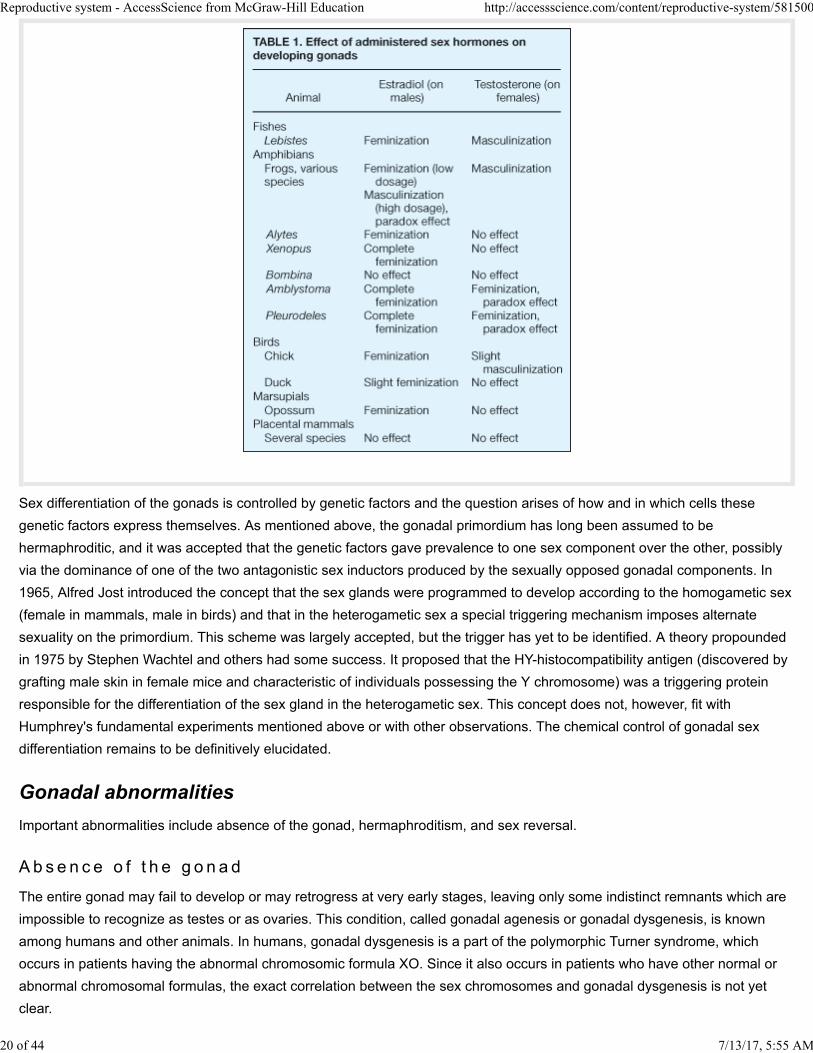
The effects of administering hormones to developing embryos are profound, but vary among organisms (Table 1). Such
experiments do not, however, warrant the generalization that sex hormones control sex differentiation. In lower vertebrates,
sex differentiation may be influenced by external factors. Experiments made on turtles, alligators, and the newt Pleurodeles
showed that breeding the eggs at different temperatures resulted in the development of young which were all of the same
sex, depending on the temperature. Genetically, male Pleurodeles which were sex-reversed into functional females by
breeding at high temperature could be mated with normal males and the progeny could be studied.
7/13/17, 5:55 AM 19 of 44
Reproductive system - AccessScience from McGraw-Hill Education http://accessscience.com/content/reproductive-system/581500
Sex differentiation of the gonads is controlled by genetic factors and the question arises of how and in which cells these
genetic factors express themselves. As mentioned above, the gonadal primordium has long been assumed to be
hermaphroditic, and it was accepted that the genetic factors gave prevalence to one sex component over the other, possibly
via the dominance of one of the two antagonistic sex inductors produced by the sexually opposed gonadal components. In
1965, Alfred Jost introduced the concept that the sex glands were programmed to develop according to the homogametic sex
(female in mammals, male in birds) and that in the heterogametic sex a special triggering mechanism imposes alternate
sexuality on the primordium. This scheme was largely accepted, but the trigger has yet to be identified. A theory propounded
in 1975 by Stephen Wachtel and others had some success. It proposed that the HY-histocompatibility antigen (discovered by
grafting male skin in female mice and characteristic of individuals possessing the Y chromosome) was a triggering protein
responsible for the differentiation of the sex gland in the heterogametic sex. This concept does not, however, fit with
Humphrey's fundamental experiments mentioned above or with other observations. The chemical control of gonadal sex
differentiation remains to be definitively elucidated.
Gonadal abnormalities
Important abnormalities include absence of the gonad, hermaphroditism, and sex reversal.
A b s e n c e o f t h e g o n a d
The entire gonad may fail to develop or may retrogress at very early stages, leaving only some indistinct remnants which are
impossible to recognize as testes or as ovaries. This condition, called gonadal agenesis or gonadal dysgenesis, is known
among humans and other animals. In humans, gonadal dysgenesis is a part of the polymorphic Turner syndrome, which
occurs in patients having the abnormal chromosomic formula XO. Since it also occurs in patients who have other normal or
abnormal chromosomal formulas, the exact correlation between the sex chromosomes and gonadal dysgenesis is not yet
clear.
7/13/17, 5:55 AM 20 of 44
Reproductive system - AccessScience from McGraw-Hill Education http://accessscience.com/content/reproductive-system/581500
He rma p h r o d i t i sm
The presence of male and female gonadal tissue in the same individual is not normal among vertebrates except in some
species of fishes. It can occur as an abnormal condition more or less frequently among other vertebrates. It is rather frequent
in some frogs and toads, and was considered to be the result of an incomplete dominance of either the cortex or the medulla
of the indifferent gonad. It might also, however, result from incomplete expression of the testicular determining system. In
humans, several cases of true hermaphroditism have been reported in sterile individuals. Several cases have been correlated
with chromosomal abnormalities such as XX/XY mosaicism, and in one case it was observed that the ovarian tissue
contained mainly XX cells and the testicular tissue XY cells.
Genital tract development
The sex ducts become sexually specialized some time after the sexual differentiation of the sex glands. The male or female
conditions develop from an indifferent condition which is identical in both sexes in early stages.
I n d i f f e r e n t s t a g e
The gonads are already recognizable as presumptive ovaries or testes and are located on the anterior part of the
mesonephros. The mesonephric or Wolffian duct is the ureter, and opens posteriorly into the cloaca in lower vertebrates or
into the urogenital sinus in mammals. Another duct, the oviduct or Müllerian duct, parallels the mesonephric duct. The oviduct
arises from a funnel which opens into the coelomic cavity. The blind end of this primordium proliferates and extends
progressively caudally.
In selachians and urodeles, the funnel from which the Müllerian duct originates corresponds to a pronephric nephrostome, the
coelomic opening of the primitive urinary tubules. Because the pronephros is located near the neck of the larva, and because
the ostium of the oviduct retains this position, the oviducts open into an anterior part of the body cavity (Fig. 11).
In birds and mammals, the origin of the oviduct from pronephric remnants is not as clear, but it is obvious that the oviduct
develops in the region of the nephric field. The early funnel is located on the top of the mesonephros, and the ostia tubae
open above the ovaries.
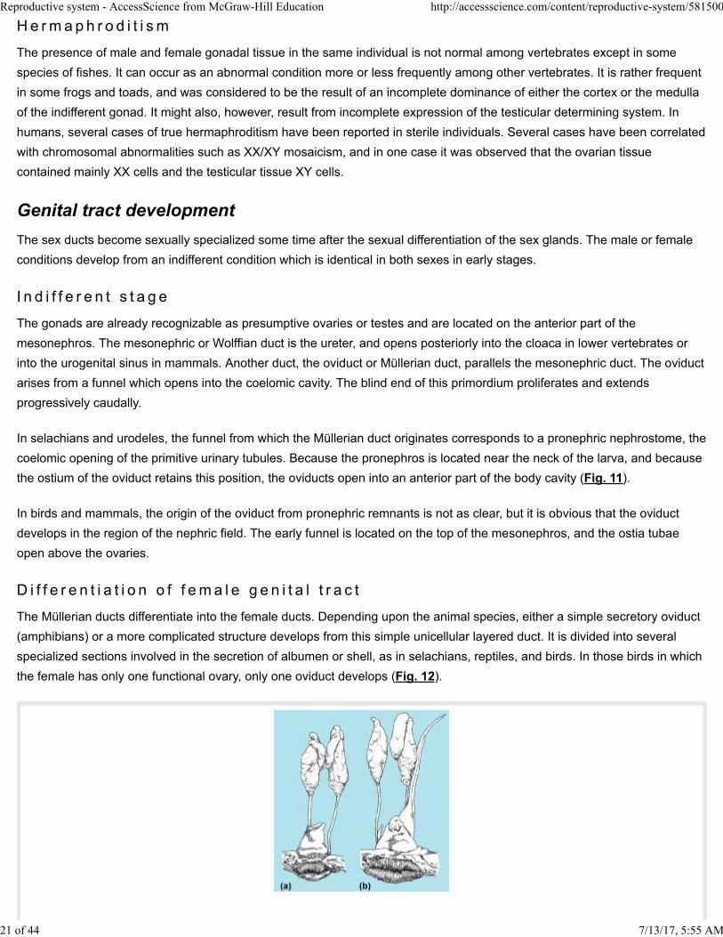
D i f f e r e n t i a t i o n o f f ema l e g e n i t a l t r a c t
The Müllerian ducts differentiate into the female ducts. Depending upon the animal species, either a simple secretory oviduct
(amphibians) or a more complicated structure develops from this simple unicellular layered duct. It is divided into several
specialized sections involved in the secretion of albumen or shell, as in selachians, reptiles, and birds. In those birds in which
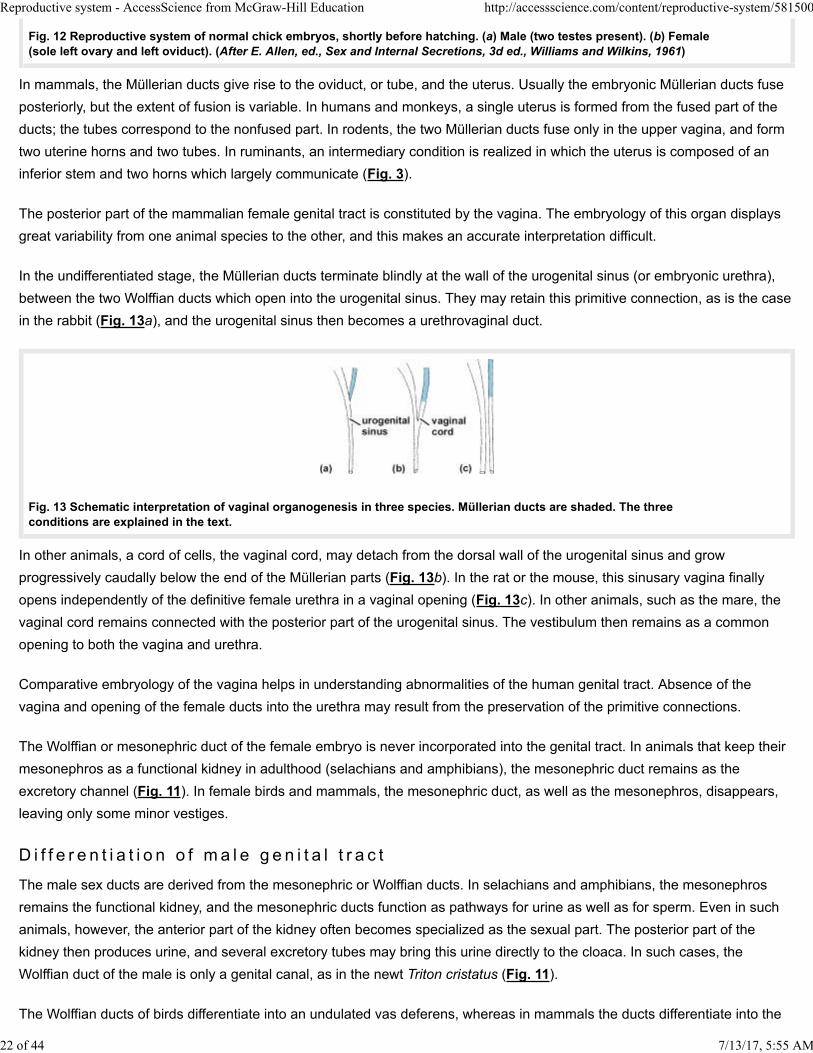
the female has only one functional ovary, only one oviduct develops (Fig. 12).
7/13/17, 5:55 AM 21 of 44
Reproductive system - AccessScience from McGraw-Hill Education http://accessscience.com/content/reproductive-system/581500
Fig. 12 Reproductive system of normal chick embryos, shortly before hatching. (a) Male (two testes present). (b) Female(sole left ovary and left oviduct). (After E. Allen, ed., Sex and Internal Secretions, 3d ed., Williams and Wilkins, 1961)
In mammals, the Müllerian ducts give rise to the oviduct, or tube, and the uterus. Usually the embryonic Müllerian ducts fuse
posteriorly, but the extent of fusion is variable. In humans and monkeys, a single uterus is formed from the fused part of the
ducts; the tubes correspond to the nonfused part. In rodents, the two Müllerian ducts fuse only in the upper vagina, and form
two uterine horns and two tubes. In ruminants, an intermediary condition is realized in which the uterus is composed of an
inferior stem and two horns which largely communicate (Fig. 3).
The posterior part of the mammalian female genital tract is constituted by the vagina. The embryology of this organ displays
great variability from one animal species to the other, and this makes an accurate interpretation difficult.
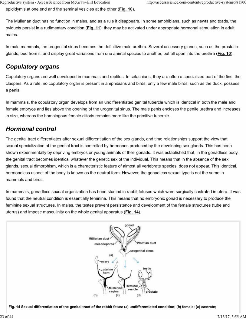
In the undifferentiated stage, the Müllerian ducts terminate blindly at the wall of the urogenital sinus (or embryonic urethra),
between the two Wolffian ducts which open into the urogenital sinus. They may retain this primitive connection, as is the case
in the rabbit (Fig. 13a), and the urogenital sinus then becomes a urethrovaginal duct.
Fig. 13 Schematic interpretation of vaginal organogenesis in three species. Müllerian ducts are shaded. The threeconditions are explained in the text.
In other animals, a cord of cells, the vaginal cord, may detach from the dorsal wall of the urogenital sinus and grow
progressively caudally below the end of the Müllerian parts (Fig. 13b). In the rat or the mouse, this sinusary vagina finally
opens independently of the definitive female urethra in a vaginal opening (Fig. 13c). In other animals, such as the mare, the
vaginal cord remains connected with the posterior part of the urogenital sinus. The vestibulum then remains as a common
opening to both the vagina and urethra.
Comparative embryology of the vagina helps in understanding abnormalities of the human genital tract. Absence of the
vagina and opening of the female ducts into the urethra may result from the preservation of the primitive connections.
The Wolffian or mesonephric duct of the female embryo is never incorporated into the genital tract. In animals that keep their
mesonephros as a functional kidney in adulthood (selachians and amphibians), the mesonephric duct remains as the
excretory channel (Fig. 11). In female birds and mammals, the mesonephric duct, as well as the mesonephros, disappears,
leaving only some minor vestiges.
D i f f e r e n t i a t i o n o f m a l e g e n i t a l t r a c t
The male sex ducts are derived from the mesonephric or Wolffian ducts. In selachians and amphibians, the mesonephros
remains the functional kidney, and the mesonephric ducts function as pathways for urine as well as for sperm. Even in such
animals, however, the anterior part of the kidney often becomes specialized as the sexual part. The posterior part of the
kidney then produces urine, and several excretory tubes may bring this urine directly to the cloaca. In such cases, the
Wolffian duct of the male is only a genital canal, as in the newt Triton cristatus (Fig. 11).
The Wolffian ducts of birds differentiate into an undulated vas deferens, whereas in mammals the ducts differentiate into the
7/13/17, 5:55 AM 22 of 44
Reproductive system - AccessScience from McGraw-Hill Education http://accessscience.com/content/reproductive-system/581500
epididymis at one end and the seminal vesicles at the other (Fig. 10).
The Müllerian duct has no function in males, and as a rule it disappears. In some amphibians, such as newts and toads, the
oviducts persist in a rudimentary condition (Fig. 11); they may be activated under appropriate hormonal stimulation in adult
males.
In male mammals, the urogenital sinus becomes the definitive male urethra. Several accessory glands, such as the prostatic
glands, bud from it, and display great variations from one animal species to another, but all open into the urethra (Fig. 10).
Copulatory organs
Copulatory organs are well developed in mammals and reptiles. In selachians, they are often a specialized part of the fins, the
claspers. As a rule, no copulatory organ is present in amphibians and birds; only a few male birds, such as the duck, possess
a penis.
In mammals, the copulatory organ develops from an undifferentiated genital tubercle which is identical in both the male and
female embryos and lies above the opening of the urogenital sinus. The male penis encloses the penile urethra and increases
in size, whereas the homologous female clitoris remains more like the primitive tubercle.
Hormonal control
The genital tract differentiates after sexual differentiation of the sex glands, and time relationships support the view that
sexual specialization of the genital tract is controlled by hormones produced by the developing sex glands. This has been
shown experimentally by depriving embryos or young animals of their gonads. It was established that, in the gonadless body,
the genital tract becomes identical whatever the genetic sex of the individual. This means that in the absence of the sex
glands, sexual dimorphism, which is a characteristic feature of almost all vertebrate species, does not appear. This identical,
hormoneless aspect of the body is known as the neutral form. However, the gonadless sexual type is not the same in
mammals and birds.
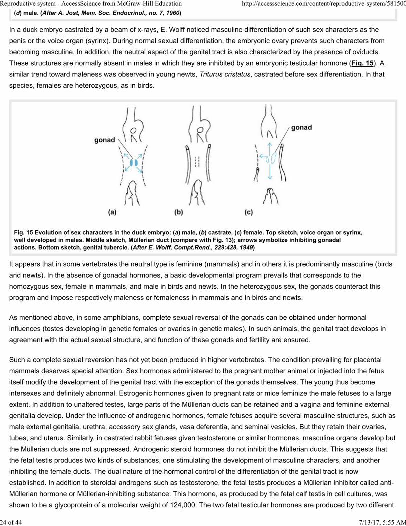
In mammals, gonadless sexual organization has been studied in rabbit fetuses which were surgically castrated in utero. It was
found that the neutral condition is essentially feminine. This means that no embryonic gonad is necessary to produce the
feminine sexual structures. In males, the testes prevent persistence and development of the female structures (tube and
uterus) and impose masculinity on the whole genital apparatus (Fig. 14).
7/13/17, 5:55 AM
Fig. 14 Sexual differentiation of the genital tract of the rabbit fetus: (a) undifferentiated condition; (b) female; (c) castrate;
23 of 44
Reproductive system - AccessScience from McGraw-Hill Education http://accessscience.com/content/reproductive-system/581500
(d) male. (After A. Jost, Mem. Soc. Endocrinol., no. 7, 1960)
In a duck embryo castrated by a beam of x-rays, E. Wolff noticed masculine differentiation of such sex characters as the
penis or the voice organ (syrinx). During normal sexual differentiation, the embryonic ovary prevents such characters from
becoming masculine. In addition, the neutral aspect of the genital tract is also characterized by the presence of oviducts.
These structures are normally absent in males in which they are inhibited by an embryonic testicular hormone (Fig. 15). A
similar trend toward maleness was observed in young newts, Triturus cristatus, castrated before sex differentiation. In that
species, females are heterozygous, as in birds.
Fig. 15 Evolution of sex characters in the duck embryo: (a) male, (b) castrate, (c) female. Top sketch, voice organ or syrinx,well developed in males. Middle sketch, Müllerian duct (compare with Fig. 13); arrows symbolize inhibiting gonadalactions. Bottom sketch, genital tubercle. (After E. Wolff, Compt.Rend., 229:428, 1949)
It appears that in some vertebrates the neutral type is feminine (mammals) and in others it is predominantly masculine (birds
and newts). In the absence of gonadal hormones, a basic developmental program prevails that corresponds to the
homozygous sex, female in mammals, and male in birds and newts. In the heterozygous sex, the gonads counteract this
program and impose respectively maleness or femaleness in mammals and in birds and newts.
As mentioned above, in some amphibians, complete sexual reversal of the gonads can be obtained under hormonal
influences (testes developing in genetic females or ovaries in genetic males). In such animals, the genital tract develops in
agreement with the actual sexual structure, and function of these gonads and fertility are ensured.
Such a complete sexual reversion has not yet been produced in higher vertebrates. The condition prevailing for placental
mammals deserves special attention. Sex hormones administered to the pregnant mother animal or injected into the fetus
itself modify the development of the genital tract with the exception of the gonads themselves. The young thus become
intersexes and definitely abnormal. Estrogenic hormones given to pregnant rats or mice feminize the male fetuses to a large
extent. In addition to unaltered testes, large parts of the Müllerian ducts can be retained and a vagina and feminine external
genitalia develop. Under the influence of androgenic hormones, female fetuses acquire several masculine structures, such as
male external genitalia, urethra, accessory sex glands, vasa deferentia, and seminal vesicles. But they retain their ovaries,
tubes, and uterus. Similarly, in castrated rabbit fetuses given testosterone or similar hormones, masculine organs develop but
the Müllerian ducts are not suppressed. Androgenic steroid hormones do not inhibit the Müllerian ducts. This suggests that
the fetal testis produces two kinds of substances, one stimulating the development of masculine characters, and another
inhibiting the female ducts. The dual nature of the hormonal control of the differentiation of the genital tract is now
established. In addition to steroidal androgens such as testosterone, the fetal testis produces a Müllerian inhibitor called anti-
Müllerian hormone or Müllerian-inhibiting substance. This hormone, as produced by the fetal calf testis in cell cultures, was
shown to be a glycoprotein of a molecular weight of 124,000. The two fetal testicular hormones are produced by two different
7/13/17, 5:55 AM 24 of 44
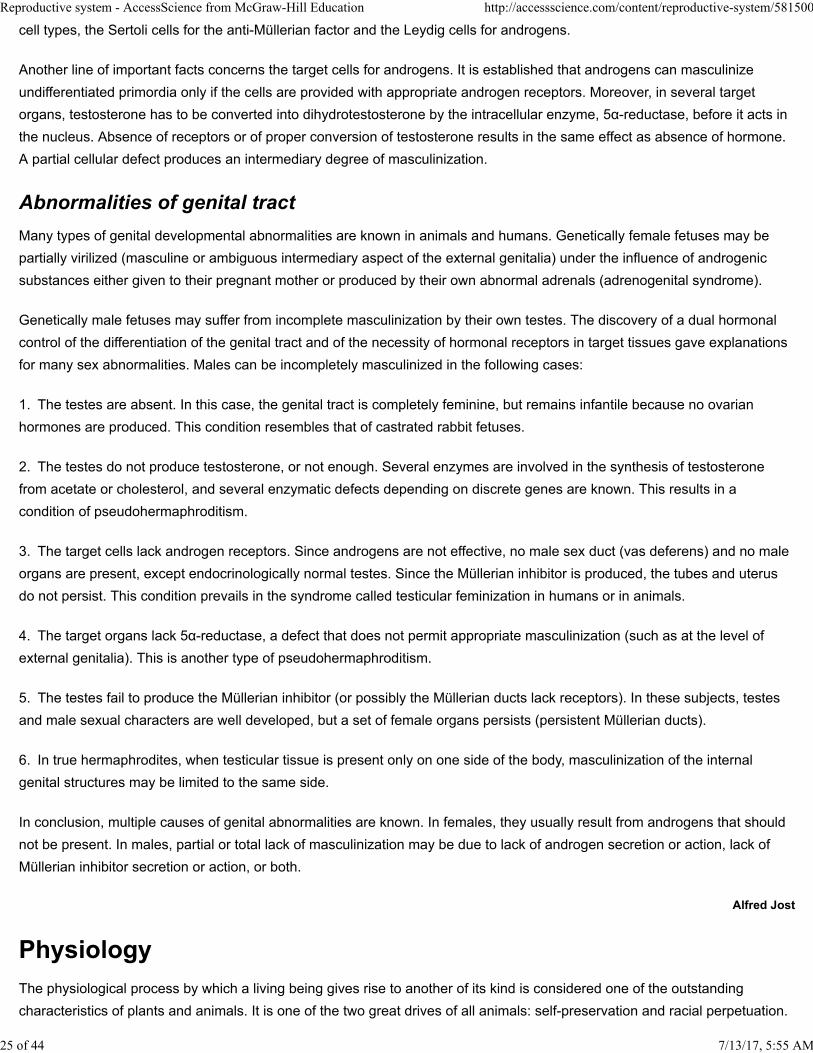
Reproductive system - AccessScience from McGraw-Hill Education http://accessscience.com/content/reproductive-system/581500
cell types, the Sertoli cells for the anti-Müllerian factor and the Leydig cells for androgens.
Another line of important facts concerns the target cells for androgens. It is established that androgens can masculinize
undifferentiated primordia only if the cells are provided with appropriate androgen receptors. Moreover, in several target
organs, testosterone has to be converted into dihydrotestosterone by the intracellular enzyme, 5α-reductase, before it acts in
the nucleus. Absence of receptors or of proper conversion of testosterone results in the same effect as absence of hormone.
A partial cellular defect produces an intermediary degree of masculinization.
Abnormalities of genital tract
Many types of genital developmental abnormalities are known in animals and humans. Genetically female fetuses may be
partially virilized (masculine or ambiguous intermediary aspect of the external genitalia) under the influence of androgenic
substances either given to their pregnant mother or produced by their own abnormal adrenals (adrenogenital syndrome).
Genetically male fetuses may suffer from incomplete masculinization by their own testes. The discovery of a dual hormonal
control of the differentiation of the genital tract and of the necessity of hormonal receptors in target tissues gave explanations
for many sex abnormalities. Males can be incompletely masculinized in the following cases:
1. The testes are absent. In this case, the genital tract is completely feminine, but remains infantile because no ovarian
hormones are produced. This condition resembles that of castrated rabbit fetuses.
2. The testes do not produce testosterone, or not enough. Several enzymes are involved in the synthesis of testosterone
from acetate or cholesterol, and several enzymatic defects depending on discrete genes are known. This results in a
condition of pseudohermaphroditism.
3. The target cells lack androgen receptors. Since androgens are not effective, no male sex duct (vas deferens) and no male
organs are present, except endocrinologically normal testes. Since the Müllerian inhibitor is produced, the tubes and uterus
do not persist. This condition prevails in the syndrome called testicular feminization in humans or in animals.
4. The target organs lack 5α-reductase, a defect that does not permit appropriate masculinization (such as at the level of
external genitalia). This is another type of pseudohermaphroditism.
The testes fail to produce the Müllerian inhibitor (or possibly the Müllerian ducts lack receptors). In these subjects, testes
and male sexual characters are well developed, but a set of female organs persists (persistent Müllerian ducts).
5.
6. In true hermaphrodites, when testicular tissue is present only on one side of the body, masculinization of the internal
genital structures may be limited to the same side.
In conclusion, multiple causes of genital abnormalities are known. In females, they usually result from androgens that should
not be present. In males, partial or total lack of masculinization may be due to lack of androgen secretion or action, lack of
Müllerian inhibitor secretion or action, or both.
Alfred Jost
The physiological process by which a living being gives rise to another of its kind is considered one of the outstanding
characteristics of plants and animals. It is one of the two great drives of all animals: self-preservation and racial perpetuation.
7/13/17, 5:55 AM 25 of 44
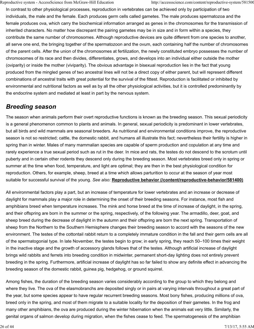
Reproductive system - AccessScience from McGraw-Hill Education http://accessscience.com/content/reproductive-system/581500
In contrast to other physiological processes, reproduction in vertebrates can be achieved only by participation of two
individuals, the male and the female. Each produces germ cells called gametes. The male produces spermatozoa and the
female produces ova, which carry the biochemical information arranged as genes in the chromosomes for the transmission of
inherited characters. No matter how discrepant the pairing gametes may be in size and in form within a species, they
contribute the same number of chromosomes. Although reproductive devices are quite different from one species to another,
all serve one end, the bringing together of the spermatozoon and the ovum, each containing half the number of chromosomes
of the parent cells. After the union of the chromosomes at fertilization, the newly constituted embryo possesses the number of
chromosomes of its race and then divides, differentiates, grows, and develops into an individual either outside the mother
(oviparity) or inside the mother (viviparity). The obvious advantage in bisexual reproduction lies in the fact that young
produced from the mingled genes of two ancestral lines will not be a direct copy of either parent, but will represent different
combinations of ancestral traits with great potential for the survival of the fittest. Reproduction is facilitated or inhibited by
environmental and nutritional factors as well as by all the other physiological activities, but it is controlled predominantly by
the endocrine system and mediated at least in part by the nervous system.
Breeding season
The season when animals perform their overt reproductive functions is known as the breeding season. This sexual periodicity
is a general phenomenon common to plants and animals. In general, sexual periodicity is predominant in lower vertebrates,
but all birds and wild mammals are seasonal breeders. As nutritional and environmental conditions improve, the reproductive
season is not so restricted; cattle, the domestic rabbit, and humans all illustrate this fact; nevertheless their fertility is higher in
spring than in winter. Males of many mammalian species are capable of sperm production and copulation at any time and
rarely experience a true sexual period such as rut in the deer. In mice and rats, the testes do not descend to the scrotum until
puberty and in certain other rodents they descend only during the breeding season. Most vertebrates breed only in spring or
summer at the time when food, temperature, and light are optimal; they are then in the best physiological condition for
reproduction. Others, for example, sheep, breed at a time which allows parturition to occur at the season of year most
suitable for successful survival of the young. See also: Reproductive behavior (/content/reproductive-behavior/581400)
All environmental factors play a part, but an increase of temperature for lower vertebrates and an increase or decrease of
daylight for mammals play a major role in determining the onset of their breeding seasons. For instance, most fish and
amphibians breed when temperature increases. The mink and horse breed at the time of increase of daylight, in the spring,
and their offspring are born in the summer or the spring, respectively, of the following year. The armadillo, deer, goat, and
sheep breed during the decrease of daylight in the autumn and their offspring are born the next spring. Transportation of
sheep from the Northern to the Southern Hemisphere changes their breeding season to accord with the seasons of the new
environment. The testes of the cottontail rabbit return to a completely immature condition in the fall and their germ cells are all
of the spermatogonial type. In late November, the testes begin to grow; in early spring, they reach 50–100 times their weight
in the inactive stage and the growth of accessory glands follows that of the testes. Although artificial increase of daylight
brings wild rabbits and ferrets into breeding condition in midwinter, permanent short-day lighting does not entirely prevent
breeding in the spring. Furthermore, artificial increase of daylight has so far failed to show any definite effect in advancing the
breeding season of the domestic rabbit, guinea pig, hedgehog, or ground squirrel.
Among fishes, the duration of the breeding season varies considerably according to the group to which they belong and
where they live. The ova of the elasmobranchs are deposited singly or in pairs at varying intervals throughout a great part of
the year, but some species appear to have regular recurrent breeding seasons. Most bony fishes, producing millions of ova,
breed only in the spring, and most of them migrate to a suitable locality for the deposition of their gametes. In the frog and
many other amphibians, the ova are produced during the winter hibernation when the animals eat very little. Similarly, the
genital organs of salmon develop during migration, when the fishes cease to feed. The spermatogenesis of the amphibian
7/13/17, 5:55 AM 26 of 44
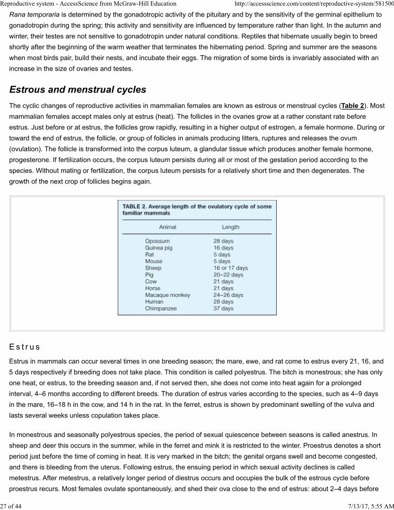
Reproductive system - AccessScience from McGraw-Hill Education http://accessscience.com/content/reproductive-system/581500
Rana temporaria is determined by the gonadotropic activity of the pituitary and by the sensitivity of the germinal epithelium to
gonadotropin during the spring; this activity and sensitivity are influenced by temperature rather than light. In the autumn and
winter, their testes are not sensitive to gonadotropin under natural conditions. Reptiles that hibernate usually begin to breed
shortly after the beginning of the warm weather that terminates the hibernating period. Spring and summer are the seasons
when most birds pair, build their nests, and incubate their eggs. The migration of some birds is invariably associated with an
increase in the size of ovaries and testes.
Estrous and menstrual cycles
The cyclic changes of reproductive activities in mammalian females are known as estrous or menstrual cycles (Table 2). Most
mammalian females accept males only at estrus (heat). The follicles in the ovaries grow at a rather constant rate before
estrus. Just before or at estrus, the follicles grow rapidly, resulting in a higher output of estrogen, a female hormone. During or
toward the end of estrus, the follicle, or group of follicles in animals producing litters, ruptures and releases the ovum
(ovulation). The follicle is transformed into the corpus luteum, a glandular tissue which produces another female hormone,
progesterone. If fertilization occurs, the corpus luteum persists during all or most of the gestation period according to the
species. Without mating or fertilization, the corpus luteum persists for a relatively short time and then degenerates. The
growth of the next crop of follicles begins again.
E s t r u s
Estrus in mammals can occur several times in one breeding season; the mare, ewe, and rat come to estrus every 21, 16, and
5 days respectively if breeding does not take place. This condition is called polyestrus. The bitch is monestrous; she has only
one heat, or estrus, to the breeding season and, if not served then, she does not come into heat again for a prolonged
interval, 4–6 months according to different breeds. The duration of estrus varies according to the species, such as 4–9 days
in the mare, 16–18 h in the cow, and 14 h in the rat. In the ferret, estrus is shown by predominant swelling of the vulva and
lasts several weeks unless copulation takes place.
In monestrous and seasonally polyestrous species, the period of sexual quiescence between seasons is called anestrus. In
sheep and deer this occurs in the summer, while in the ferret and mink it is restricted to the winter. Proestrus denotes a short
period just before the time of coming in heat. It is very marked in the bitch; the genital organs swell and become congested,
and there is bleeding from the uterus. Following estrus, the ensuing period in which sexual activity declines is called
metestrus. After metestrus, a relatively longer period of diestrus occurs and occupies the bulk of the estrous cycle before
proestrus recurs. Most females ovulate spontaneously, and shed their ova close to the end of estrus: about 2–4 days before
7/13/17, 5:55 AM 27 of 44
Reproductive system - AccessScience from McGraw-Hill Education http://accessscience.com/content/reproductive-system/581500
the end of estrus in the mare, 13 h after the end of estrus in the cow, and 10 h after the beginning of estrus in the rat. Some
species, such as the rabbit, ferret, and cat, ovulate following the stimulation of copulation. If fertilization takes place,
pregnancy occurs. If mating is unsuccessful, pseudopregnancy, a physiological condition very similar to pregnancy but which
lasts a relatively short period, may occur in some species, particularly the dog and the rabbit. See also: Estrus (/content
/estrus/242700)
Men s t r u a t i o n
The reproductive cycle of the female in the primate and human is well marked by menstruation, the period of blood flow from
the vagina. Menstruation does not correspond to estrus but occurs between the periods of ovulation at the time the corpus
luteum declines precipitously. As a result of the collapse of the superficial capillaries in the endometrium, blood is
extravasated in quantity and about two-thirds of the endometrium is desquamated, or shed. After menstruation, there is a
period in which the uterus is replenished under the influence of estrogens by the growth of the epithelium and its capillaries.
This is known as the proliferative stage and is equivalent to proestrus and estrus in other mammals. It is accompanied by
growth of the vaginal epithelium and some cornification, but these changes are gradual and are not as well defined as in
many other mammals. The Graafian follicle ruptures near the midcycle, about 15 days before the beginning of the next
menstruation. Ovulation is spontaneous and is followed by the formation of the corpus luteum, whose secretion of
progesterone causes great glandular growth of the endometrium. This is known as the luteal or secretory phase and is
equivalent to diestrus in other mammals. In New World monkeys, menstruation is confined to the appearance in the vaginal
lavage of a few red blood cells. Tissue destruction and hemorrhage in the uterus of the elephant shrew Elephantulus of South
Africa at the end of diestrus has been described, but the bleeding at proestrus in the bitch, at metestrus in the heifer 14 h after
ovulation, and at metestrus occasionally in the guinea pig is due to intensive congestion of blood vessels in the uterus or in
the upper part of the vagina and is not true menstruation.
Mating
Mating, also called copulation or coitus, is the synchronized bodily activity of the two sexes which enables them to deposit
their gametes in close contact. It is essential for successful fertilization because sperm and ovum have a very limited life
span; as examples, the fertilizing capacity of trout and salmon sperm lasts about 30 s in freshwater, and that of rabbit sperm
lasts about 30 h in the female tract. Trout and frog ova become nonfertilizable soon after contact with water, whereas rabbit
ova remain fertilizable for 6–8 h after ovulation. Human spermatozoa have a fertilizing life span of about 72 h, and ova have a
fertilizable life of about 24 h. Mating behavior is a very common phenomenon in the animal kingdom. Even in the primitive
vertebrate, the lamprey, some sort of mating takes place. The female is seized by the male, who winds his tail around her and
discharges his sperm over the ova as they extrude from her body. In the dogfish, which practices internal fertilization, contact
between male and female is necessary for the claspers of the male to convey his sperm into the cloaca of the female. In the
majority of the bony fish, physical contact may be slight, yet the male follows the female closely or presses his body to hers
and deposits his sperm over the ova either soon after they are laid or at the same time. Certain salamanders walk slowly
ahead of the female and deposit spermatophores which the female straddles and secures with the lips of her cloaca. In other
low vertebrates, a rudimentary penis exists either as a single intracloacal organ (crocodile, turtle, and duck) or as a paired
structure placed behind the cloaca (snake and lizard) and sperm are deposited into the female's cloaca at mating. In
mammals, the copulatory organs are well developed, varying a great deal in structure. Patterns of mating are different; the
time required for mating varies from 3 h (ferret) to a few seconds (sheep and rabbit), and the number of intromissions varies
from 1 (bull) to 50 (hamster).
Mating in many birds and cold-blooded vertebrates is the culminating event of a more or less complicated series of love
antics. It is quite probable that these sexual displays have a direct bearing upon ovulation and that in many species ovulation
would not occur in the absence of such sexual experiences. Sexual excitement and violent physical exertions of both partners
28 of 44 7/13/17, 5:55 AM
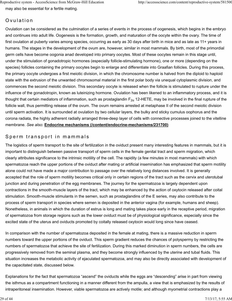
Reproductive system - AccessScience from McGraw-Hill Education http://accessscience.com/content/reproductive-system/581500
may also be essential for a fertile mating.
O v u l a t i o n
Ovulation can be considered as the culmination of a series of events in the process of oogenesis, which begins in the embryo
and continues into adult life. Oogenesis is the formation, growth, and maturation of the oocyte within the ovary. The time of
first ovulation at puberty varies among species, occurring as early as 30 days after birth in mice and as late as 11+ years in
humans. The stages in the development of the ovum are, however, similar in most mammals. By birth, most of the primordial
germ cells have become oogonia anad developed into primary oocytes. Most of these oocytes remain in this stage until,
under the stimulation of gonadotropic hormones (especially follicle-stimulating hormone), one or more (depending on the
species) follicles containing the primary oocytes begin to enlarge and differentiate into Graafian follicles. During this process,
the primary oocyte undergoes a first meiotic division, in which the chromosome number is halved from the diploid to haploid
state with the extrusion of the unwanted chromosomal material in the first polar body via unequal cytoplasmic division, and
commences the second meiotic division. This secondary oocyte is released when the follicle is stimulated to rupture under the
influence of the gonadotropin, known as luteinizing hormone. Ovulation has been likened to an inflammatory process, and it is
thought that certain mediators of inflammation, such as prostaglandin F2α 12-HETE, may be involved in the final rupture of the
follicle wall, thus permitting release of the ovum. The ovum remains arrested at metaphase II of the second meiotic division
until sperm activation. It is surrounded at ovulation by two cellular layers, the bulky and sticky cumulus oophorus and the
corona radiata, the highly adherent radially arranged three-deep layer of cells with connective processes joined to the vitelline
membrane. See also: Endocrine mechanisms (/content/endocrine-mechanisms/231700)
Sp e rm t r a n s p o r t i n m amma l s
The logistics of sperm transport to the site of fertilization in the oviduct present many interesting features in mammals, but it is
important to distinguish between passive transport of sperm cells in the female genital tract and sperm migration, which
clearly attributes significance to the intrinsic motility of the cell. The rapidity (a few minutes in most mammals) with which
spermatozoa reach the upper portions of the oviduct after mating or artificial insemination has emphasized that sperm motility
alone could not have made a major contribution to passage over the relatively long distances involved. It is generally
accepted that the role of sperm motility becomes critical only in certain regions of the tract such as the cervix and uterotubal
junction and during penetration of the egg membranes. The journey for the spermatozoa is largely dependent upon
contractions in the smooth-muscle layers of the tract, which may be enhanced by the action of oxytocin released after coital
stimulation. Smooth-muscle stimulants in the semen, such as prostaglandins of the E series, may also contribute to the
process of sperm transport in species where semen is deposited in the anterior vagina (for example, humans and sheep).
Nonetheless, in animals in which the duration of estrus is long and mating takes place early in the receptive period, migration
of spermatozoa from storage regions such as the lower oviduct must be of physiological significance, especially since the
excited state of the uterus and oviducts promoted by coitally released oxytocin would long since have ceased.
In comparison with the number of spermatozoa deposited in the female at mating, there is a massive reduction in sperm
numbers toward the upper portions of the oviduct. This sperm gradient reduces the chances of polyspermy by restricting the
numbers of spermatozoa that achieve the site of fertilization. During this marked diminution in sperm numbers, the cells are
progressively removed from the seminal plasma, and they become strongly influenced by the uterine and tubal fluids. This
situation increases the metabolic activity of ejaculated spermatozoa, and may also be directly associated with development of
the capacitated state, discussed below.
Explanations for the fact that spermatozoa “ascend” the oviducts while the eggs are “descending” arise in part from viewing
the isthmus as a compartment functioning in a manner different from the ampulla, a view that is emphasized by the results of
intraperitoneal insemination. However, viable spermatozoa are actively motile; and although myometrial contractions play a
7/13/17, 5:55 AM 29 of 44
Reproductive system - AccessScience from McGraw-Hill Education http://accessscience.com/content/reproductive-system/581500
major role in sperm transport through the uterus, progressive motility does contribute to migration into and within the oviducts.
Waves of peristaltic contraction in the isthmus proceed principally toward the ampullary-isthmic junction, and this activity
should therefore assist transport of sperm cells. These waves of contraction are propagated through the circular muscle, and
may be sufficiently powerful to occlude the lumen and, for example, to transport droplets of oil or Indian ink upward to the
peritoneal cavity, but the contractions tend to fade beyond the ampullary-isthmic junction. Whether the relatively high
concentration of 19-hydroxyprostaglandin E1 or other prostaglandins of the E series in primate semen benefit sperm transport
by means of some alteration in the myosalpinx remains to be clarified.
Cilia in the rabbit and pig isthmus may beat actively toward the ampullary-isthmic junction at the time of ovulation, thereby
creating local currents of fluid. However, such ciliary currents have not been detected in the isthmus of cat, cow, sheep,
monkey, or human.
The rate and efficiency of transport of spermatozoa are influenced by the timing of coitus relative to the moment of ovulation.
Transport of cells as far as the tubal isthmus usually follows semen deposition shortly before ovulation, but it is uncertain
whether sufficient viable spermatozoa would have reached the ampulla before the actual time of ovulation to be immediately
available for penetration of the freshly ovulated egg or eggs. Spermatozoa may be largely sequestered in the isthmus in the
preovulatory interval, only to be released for further proovarian transport when ovulation has occurred and the products of
ovulation have entered the tube. The inference here is that there may be some synchrony between release of the egg from
the Graafian follicle and a redistribution of spermatozoa from the constricted lumen of the isthmus.
Even though a specific attractant substance for spermatozoa has not yet been demonstrated to be released from mammalian
eggs or their investments, some form of chemotaxis may contribute to the final phase of sperm transport and orientation
toward the egg surface. Alternatively, maturation of the spermatozoa within the oviduct may induce intrinsic motility, which is
then responsible for further progression of the spermatozoa along the oviduct to the site of fertilization.
C a p a c i t a t i o n o f s p e rma t o z o a
Although in most mammalian species the oocyte is shed from the Graafian follicle in a condition suitable for fertilization,
ejaculated spermatozoa must undergo some form of physiological change in the female reproductive tract before they can
penetrate the egg membranes. The interval required for this change varies from about 1.5 to 6 h according to species, and
the process is referred to as capacitation. The precise changes that constitute capacitation remain unknown, although there is
strong evidence that they are—at least in part—membrane-associated phenomena, particularly in the region of the sperm
head, that permit release of lytic acrosomal enzymes with which the spermatozoon gains access to the vitelline surface of the
egg. However, increased respiratory and flagellar activities are also noted after capacitation, the changes in motility being
presumed to facilitate sperm penetration of egg membranes.
Capacitation is now understood to precede and permit a coordinated acrosome reaction, thus being an essential preliminary
to activation or release of inner acrosomal enzymes and penetration of the zona pellucida; it may involve some adjustment in
the distance separating the plasma and outer acrosomal membranes to permit the vesiculation reaction to proceed.
An understanding of the posttesticular changes occurring in spermatozoa in the male reproductive tract has provided some
insight into the possible nature of capacitation. During passage along the epididymal duct, spermatozoa undergo a series of
modifications that confer increased stability on the head, including a compaction of the nuclear chromatin and a tighter
application of the plasma and acrosomal membranes to the sperm nucleus. Secretions within the epididymal duct also
influence the maturing spermatozoa, for the antigenic nature of the sperm surface changes during the 1 to 2 weeks spent in
epididymal transit. There is good evidence that enzyme inhibitors are “added” to the spermatozoa while still in the male,
although whether the principal phase of addition occurs in the epididymis, during the preejaculatory mixing with whole seminal
7/13/17, 5:55 AM 30 of 44
Reproductive system - AccessScience from McGraw-Hill Education http://accessscience.com/content/reproductive-system/581500
plasma, or in the initial passage of spermatozoa within seminiferous and rete testis fluid remains to be clarified. Neutralization
or removal of inhibitors of acrosomal enzymes in the female tract would seem a logical stage in the capacitation process, or
an immediate sequel to it, and could explain how the enzymes of fundamental importance in fertilization are stabilized in the
sperm head until spermatozoa are within reach of the eggs.
Although capacitation seemingly involves both membranous and metabolic changes in the sperm cell, evidence suggests that
modification in the glycoprotein and other moieties of the plasma membrane is a critical step. For example, as uterine
incubation of ejaculated rabbit spermatozoa proceeds, the ability of the cell surface to bind lectins disappears progressively
from the tip of the sperm head, suggesting that carbohydrate-containing moieties are being removed progressively from the
plasma membrane. This alteration in the cell surface would be expected to prepare it for fusion with the outer acrosomal
membrane, possibly by reducing net negative charges or by increasing membrane fluidity. As a sequel to the acrosome
reaction, at least in hamster spermatozoa, there is a modification in the postnuclear cap which facilitates sperm head
incorporation by the egg plasmalemma.
Although conventional descriptions of the acrosome reaction emphasize that membrane fusion occurs as a means of
releasing proteolytic enzymes critical for sperm penetration of the egg investments, there is evidence that hyaluronidase may
escape from viable sperm cells before membrane vesiculation can be detected in the electron microscope. A second point
concerning acrosomal enzymes is that acrosin—the putative zona lysin—is apparently packaged in zymogen form and needs
to be activated from proacrosin during the capacitation process.
There is evidence from at least three mammalian species (rabbit, hamster, pig) that different compartments of the female tract
act synergistically to promote capacitation, and that sequential exposure to the uterus and then oviduct accelerates the
process of capacitation. A specific action of the oviducal environment is therefore suggested and, in the golden hamster, the
effects on stimulated respiration and achievement of the capacitated state are so distinct that they can be seen through the
thin wall of the ampulla in terms of a massive increase in sperm motility; even though hamster sperm reach the oviducts in
under 1 h, another 3 h are required for this dramatic change to be accomplished. Precise evidence for the capacitation of
primate spermatozoa is not available; if it occurs it must be rapid, since human ejaculated spermatozoa after washing, but
without any exposure to female reproductive tract fluids, can penetrate matured oocytes in culture within 4 h.
The evolutionary significance of capacitation can be viewed in two ways. Physiologically, it can be regarded as a process
whereby the sperm cell becomes competent to undergo an orderly sequence of membrane changes, collectively termed the
acrosome reaction, to permit release of the lytic enzymes. Biologically, capacitation probably evolved to meet requirements
associated with internal fertilization in mammals, and might best be regarded as an effective means of gamete selection
during sperm transport through the different regions of the female reproductive tract; only spermatozoa at the optimal stage of
their maturation process are able to penetrate the egg.
F e r t i l i z a t i o n
Fertilization is a process that begins with penetration of the ovum surface layer (zona pellucida) and binding to the vitelline
membrane by the fully matured and acrosome-reacted spermatozoon, followed by incorporation of the spermatozoon into the
ovum cytoplasm and activation of the ovum. The haploid chromosome complements of the sperm nucleus and ovum are then
transformed into male and female pronuclei, respectively. The pronuclei migrate to the center of the ovum, permitting the
joining of the two sets of chromosomes, or syngamy, which is the final stage of the fertilization process. The ovum, now
known as a zygote, begins to develop by cell division, or mitosis. Normally, after penetration of the vitelline membrane by one
spermatozoon, the fertilizing sperm, the entry of additional spermatozoa is prevented by the cortical reaction, the breakdown
of cortical granules that underline the membrane. See also: Fertilization (animal) (/content/fertilization-animal/254900)
7/13/17, 5:55 AM 31 of 44
Reproductive system - AccessScience from McGraw-Hill Education http://accessscience.com/content/reproductive-system/581500
Even if the processes of gamete transport succeed in bringing capacitated spermatozoa and recently ovulated eggs into
functional contact in the appropriate portion of the oviduct, a viable zygote may still not be formed. Anomalies of fertilization
such as digyny (retention of the second polar body) or polyspermy (penetration of the egg cytoplasm by more than one
sperm) are pathological in the higher mammals, and nearly always associated with very early embryonic death. Digyny or
polyspermy arises especially following postovulatory aging of the egg, a situation in which control of the numbers of
spermatozoa passing through the isthmus to the site of fertilization is also less effective.
R e g u l a t i o n o f e a r l y emb r y o n i c d e v e l o pme n t a n d t r a n s p o r t
As noted above, fertilization takes place in the oviducts of mammals, usually in the region of the ampullary-isthmic junction,
and the fertilized eggs or embryos do not descend to the uterus for some 3 to 4 days in most species. During this interval, the
embryo undergoes a series of mitotic divisions until it comprises a sphere of 8 or 16 cells and is termed a morula. Formation
of a blastocyst occurs when the cells of the morula rearrange themselves around a central, fluid-filled cavity, the blastocoele.
As the blastocyst develops within the uterine environment, it sheds its protective coat, the zona pellucida, and undergoes
further differentiation before developing an intimate association with the endometrium, which represents the commencement
of implantation or nidation. Early development of embryos in most species is largely under the control of the maternal
genome, with sequential activation and utilization of oocyte components. Expression of the embryonic genome does not
occur until after the two-cell stage in mice, four-cell stage in cattle, four- to eight-cell stage in humans, and eight-cell stage in
sheep zygotes.
During this preimplantation period, whose duration is approximately 4 days in small laboratory species, 7 days in humans,
and 2 to 3 weeks in pigs and sheep, respectively, metabolic support of the embryo depends partially on its own cytoplasmic
reserves of lipids and other substrates, but to a far greater extent on constituents of oviducal and uterine secretions. The
nature and volume of the secretions are known to be closely regulated by the prevailing balance of ovarian hormones,
estrogen and progesterone; and since the corpus luteum is developing during this period, it follows that the secretion of
progesterone will also be increasing. Thus, the dynamic situation in the pattern of ovarian steroid secretion will be reflected in
a changing pattern of oviducal and uterine fluids, which is critically adjusted to meet the nutritional requirements of the
developing embryo. Many experiments show that the nature of the substrate available in oviducal fluid is closely regulated by
the prevailing balance of ovarian hormones and is therefore changing to match the embryo's requirement for and ability to
utilize the substrates. Mouse one-cell zygotes require pyruvate or oxaloacetate as an energy source, but at the two-cell stage
can also utilize phosphoenolpyruvate and lactate; only after the eight-cell stage can glucose be utilized. Human zygotes
appear much less dependent on a critical composition of the surrounding medium for growth and development, since many
media, both simple and complex, have been used for culture of oocytes prior to the successful replacement of the resulting
zygotes into the oviduct or uterus of the infertile woman.
In nonprimate species, when developing zygotes are delayed in the oviduct beyond the time of normal tubal transport, they
fail to implant; similarly, when they reach the uterus too early, subsequent development can be compromised. In primates,
however, a zygote reaching the blastocyst stage in the oviduct can implant, resulting in an ectopic pregnancy. This condition
is rare in nonhuman primates but not uncommon in humans, accounting for about 1% of all pregnancies. The reason for the
occurrence of this condition, which is potentially life-threatening because of hemorrhage and shock subsequent to tubal
rupture, is unknown; in nonprimates, oviducal secretions may be hostile to continued zygote development after the normal
period of tubal transport, approximately 72 h. See also: Pregnancy disorders (/content/pregnancy-disorders/543200)
Coincident with their influence upon secretory activity, the ovarian hormones also regulate muscular activity in the oviducts,
and so control passage of the embryo into the uterus. Any interference with the ratio of estrogens to progesterone during the
free-living, preimplantation stages of the embryo will adversely influence fertility through dual effects on transport and nutrition
7/13/17, 5:55 AM 32 of 44
Reproductive system - AccessScience from McGraw-Hill Education http://accessscience.com/content/reproductive-system/581500
of the embryo. Administration of suitable preparations of ovarian steroid hormones, such as those contained in the
contraceptive pill, thereby provides potent means of regulating fertility even when taken after coitus. In fact, in some
instances, the principal mechanism whereby ovarian steroidal hormones regulate fertility is not by blocking ovulation via
steroidal feedback mechanisms, but by upsetting the delicate relationship between the egg and the reproductive tract, leading
to failure of fertilization or degeneration of the egg.
The factors that control muscular activity of the oviduct, and hence modulate the transport of the developing zygotes to the
uterus at the correct time, have not been defined. However, it is known that oviducal motility can be influenced by
prostaglandins, catecholamines, peptides (such as vasoactive intestinal peptide, neuropeptide Y, substance P, oxytocin, and
vasopressin), cyclic nucleotides, and gamma aminobutyric acid. Flow of oviducal fluid and ciliary activity of epithelial cells
lining the oviducts are also thought to be involved in the transport process.
I m p l a n t a t i o n
Association of the embryo with the uterine epithelium, by either superficial attachment or specific embedding in or beneath the
endometrium, leads in due course to the formation of a placenta and complete dependence of the differentiating embryo upon
metabolic support from the mother. Implantation and placentation exhibit a variety of forms, but in all instances the hormonal
status of the mother is of great importance in determining whether or not implantation can proceed. In several species of
rodent such as rats and mice, implantation is held in abeyance during lactation; while in other mammals such as roe deer,
badger, and certain bears, there is an obligatory period of delayed implantation known as embryonic diapause; the
inappropriate endocrine conditions associated with delayed implantation render the blastocyst metabolically dormant.
Ovariectomy shortly before the expected time of implantation can prevent implantation, whereas ovariectomy plus
replacement therapy with a suitable ratio of progesterone and estrogen permits implantation.
Despite this dominance by maternal hormonal factors, studies have indicated that the embryo itself may be modifying the
local environment between its trophoblastic surface and the endometrium by means of steroid hormone, prostaglandin, and
platelet-activating factor secretion and perhaps also by production of carbon dioxide (CO 2 ). The cells of the trophoblast
possess enzymes capable of synthesizing ovarian hormones, and embryonic membranes supplied in culture with suitable
biochemical substrates will secrete estrogens and progesterone. Such a pattern of synthetic activity in the late
preimplantation embryo, which has also been indicated by histochemical studies, may explain in part the regional changes in
the endometrium corresponding specifically to the future implantation sites, and also the local changes in capillary
permeability of the uterus as demonstrated by the pontamine-blue reaction.
The physical relationship between the implanting embryo and the uterus is best summarized according to whether the embryo
remains in the lumen of the uterus, referred to as central implantation, as in cattle, sheep, and pigs, or colonizes the glandular
crypts and maternal tissues leading to eccentric or interstitial implantation, as in rodents and humans. The process of invasion
by the embryonic trophoblast in the mouse, for example, promotes characteristic changes in the uterine stroma termed the
decidual reaction. This cellular proliferation occurs on the fourth day of pregnancy in mice in response to the presence of a
blastocyst, but the precise physiological nature of the stimulus leading to these changes is not understood.
One other feature of the implantation process that influences the form of the placenta is whether maternal cell layers are
bypassed during development of the implantation association. In species where the embryo is invasive and penetrates the
maternal tissues to reach the wall or lumen of the blood vessels, this gives rise to an endotheliochorial (carnivores) or
hemochorial (rodents, primates) form of placenta. In other words, cells of the maternal epithelium no longer interpose
between the maternal vascular supply and the developing embryo, a situation which facilitates transfer of nutrients to the
fetus.
7/13/17, 5:55 AM 33 of 44
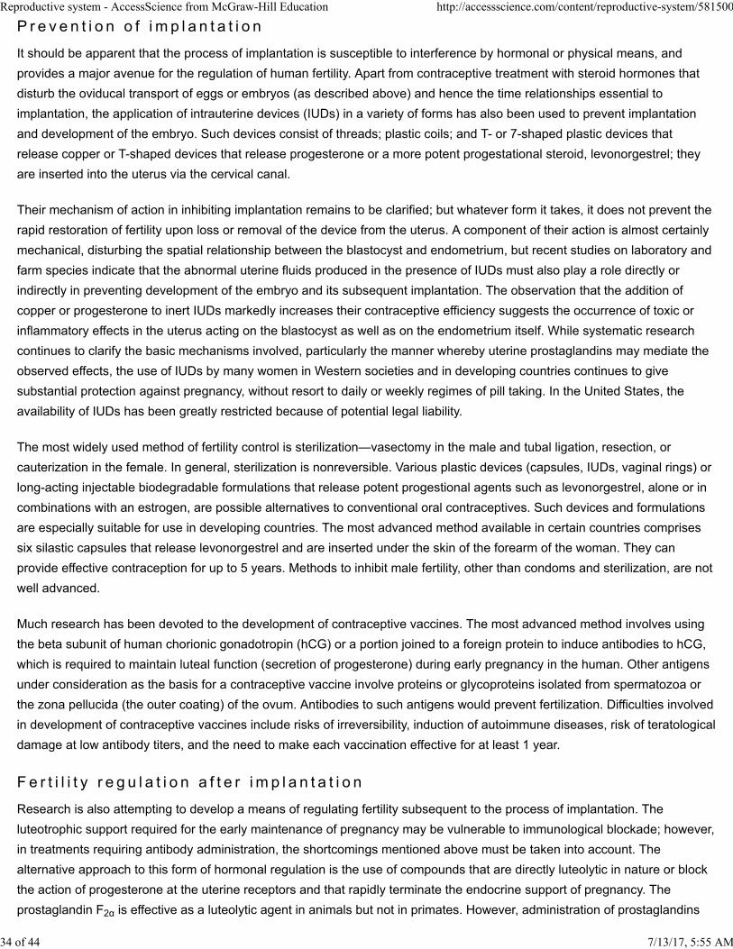
Reproductive system - AccessScience from McGraw-Hill Education http://accessscience.com/content/reproductive-system/581500
P r e v e n t i o n o f i m p l a n t a t i o n
It should be apparent that the process of implantation is susceptible to interference by hormonal or physical means, and
provides a major avenue for the regulation of human fertility. Apart from contraceptive treatment with steroid hormones that
disturb the oviducal transport of eggs or embryos (as described above) and hence the time relationships essential to
implantation, the application of intrauterine devices (IUDs) in a variety of forms has also been used to prevent implantation
and development of the embryo. Such devices consist of threads; plastic coils; and T- or 7-shaped plastic devices that
release copper or T-shaped devices that release progesterone or a more potent progestational steroid, levonorgestrel; they
are inserted into the uterus via the cervical canal.
Their mechanism of action in inhibiting implantation remains to be clarified; but whatever form it takes, it does not prevent the
rapid restoration of fertility upon loss or removal of the device from the uterus. A component of their action is almost certainly
mechanical, disturbing the spatial relationship between the blastocyst and endometrium, but recent studies on laboratory and
farm species indicate that the abnormal uterine fluids produced in the presence of IUDs must also play a role directly or
indirectly in preventing development of the embryo and its subsequent implantation. The observation that the addition of
copper or progesterone to inert IUDs markedly increases their contraceptive efficiency suggests the occurrence of toxic or
inflammatory effects in the uterus acting on the blastocyst as well as on the endometrium itself. While systematic research
continues to clarify the basic mechanisms involved, particularly the manner whereby uterine prostaglandins may mediate the
observed effects, the use of IUDs by many women in Western societies and in developing countries continues to give
substantial protection against pregnancy, without resort to daily or weekly regimes of pill taking. In the United States, the
availability of IUDs has been greatly restricted because of potential legal liability.
The most widely used method of fertility control is sterilization—vasectomy in the male and tubal ligation, resection, or
cauterization in the female. In general, sterilization is nonreversible. Various plastic devices (capsules, IUDs, vaginal rings) or
long-acting injectable biodegradable formulations that release potent progestional agents such as levonorgestrel, alone or in
combinations with an estrogen, are possible alternatives to conventional oral contraceptives. Such devices and formulations
are especially suitable for use in developing countries. The most advanced method available in certain countries comprises
six silastic capsules that release levonorgestrel and are inserted under the skin of the forearm of the woman. They can
provide effective contraception for up to 5 years. Methods to inhibit male fertility, other than condoms and sterilization, are not
well advanced.
Much research has been devoted to the development of contraceptive vaccines. The most advanced method involves using
the beta subunit of human chorionic gonadotropin (hCG) or a portion joined to a foreign protein to induce antibodies to hCG,
which is required to maintain luteal function (secretion of progesterone) during early pregnancy in the human. Other antigens
under consideration as the basis for a contraceptive vaccine involve proteins or glycoproteins isolated from spermatozoa or
the zona pellucida (the outer coating) of the ovum. Antibodies to such antigens would prevent fertilization. Difficulties involved
in development of contraceptive vaccines include risks of irreversibility, induction of autoimmune diseases, risk of teratological
damage at low antibody titers, and the need to make each vaccination effective for at least 1 year.
F e r t i l i t y r e g u l a t i o n a f t e r i m p l a n t a t i o n
Research is also attempting to develop a means of regulating fertility subsequent to the process of implantation. The
luteotrophic support required for the early maintenance of pregnancy may be vulnerable to immunological blockade; however,
in treatments requiring antibody administration, the shortcomings mentioned above must be taken into account. The
alternative approach to this form of hormonal regulation is the use of compounds that are directly luteolytic in nature or block
the action of progesterone at the uterine receptors and that rapidly terminate the endocrine support of pregnancy. The
prostaglandin F2α is effective as a luteolytic agent in animals but not in primates. However, administration of prostaglandins
7/13/17, 5:55 AM 34 of 44
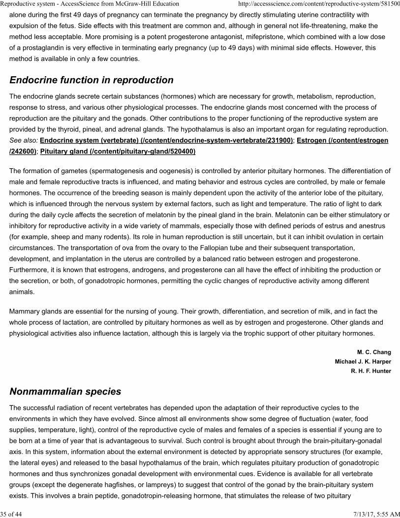
Reproductive system - AccessScience from McGraw-Hill Education http://accessscience.com/content/reproductive-system/581500
alone during the first 49 days of pregnancy can terminate the pregnancy by directly stimulating uterine contractility with
expulsion of the fetus. Side effects with this treatment are common and, although in general not life-threatening, make the
method less acceptable. More promising is a potent progesterone antagonist, mifepristone, which combined with a low dose
of a prostaglandin is very effective in terminating early pregnancy (up to 49 days) with minimal side effects. However, this
method is available in only a few countries.
Endocrine function in reproduction
The endocrine glands secrete certain substances (hormones) which are necessary for growth, metabolism, reproduction,
response to stress, and various other physiological processes. The endocrine glands most concerned with the process of
reproduction are the pituitary and the gonads. Other contributions to the proper functioning of the reproductive system are
provided by the thyroid, pineal, and adrenal glands. The hypothalamus is also an important organ for regulating reproduction.
See also: Endocrine system (vertebrate) (/content/endocrine-system-vertebrate/231900); Estrogen (/content/estrogen
/242600); Pituitary gland (/content/pituitary-gland/520400)
The formation of gametes (spermatogenesis and oogenesis) is controlled by anterior pituitary hormones. The differentiation of
male and female reproductive tracts is influenced, and mating behavior and estrous cycles are controlled, by male or female
hormones. The occurrence of the breeding season is mainly dependent upon the activity of the anterior lobe of the pituitary,
which is influenced through the nervous system by external factors, such as light and temperature. The ratio of light to dark
during the daily cycle affects the secretion of melatonin by the pineal gland in the brain. Melatonin can be either stimulatory or
inhibitory for reproductive activity in a wide variety of mammals, especially those with defined periods of estrus and anestrus
(for example, sheep and many rodents). Its role in human reproduction is still uncertain, but it can inhibit ovulation in certain
circumstances. The transportation of ova from the ovary to the Fallopian tube and their subsequent transportation,
development, and implantation in the uterus are controlled by a balanced ratio between estrogen and progesterone.
Furthermore, it is known that estrogens, androgens, and progesterone can all have the effect of inhibiting the production or
the secretion, or both, of gonadotropic hormones, permitting the cyclic changes of reproductive activity among different
animals.
Mammary glands are e ssential for the nursing of young. Their growth, differentiation, and secretion of milk, and in fact the
whole process of lactation, are controlled by pituitary hormones as well as by estrogen and progesterone. Other glands and
physiological activities also influence lactation, although this is largely via the trophic support of other pituitary hormones.
M. C. Chang
Michael J. K. Harper
R. H. F. Hunter
Nonmammalian species
The successful radiation of recent vertebrates has depended upon the adaptation of their reproductive cycles to the
environments in which they have evolved. Since almost all environments show some degree of fluctuation (water, food
supplies, temperature, light), control of the reproductive cycle of males and females of a species is essential if young are to
be born at a time of year that is advantageous to survival. Such control is brought about through the brain-pituitary-gonadal
axis. In this system, information about the external environment is detected by appropriate sensory structures (for example,
the lateral eyes) and released to the basal hypothalamus of the brain, which regulates pituitary production of gonadotropic
hormones and thus synchronizes gonadal development with environmental cues. Evidence is available for all vertebrate
groups (except the degenerate hagfishes, or lampreys) to suggest that control of the gonad by the brain-pituitary system
exists. This involves a brain peptide, gonadotropin-releasing hormone, that stimulates the release of two pituitary
7/13/17, 5:55 AM 35 of 44
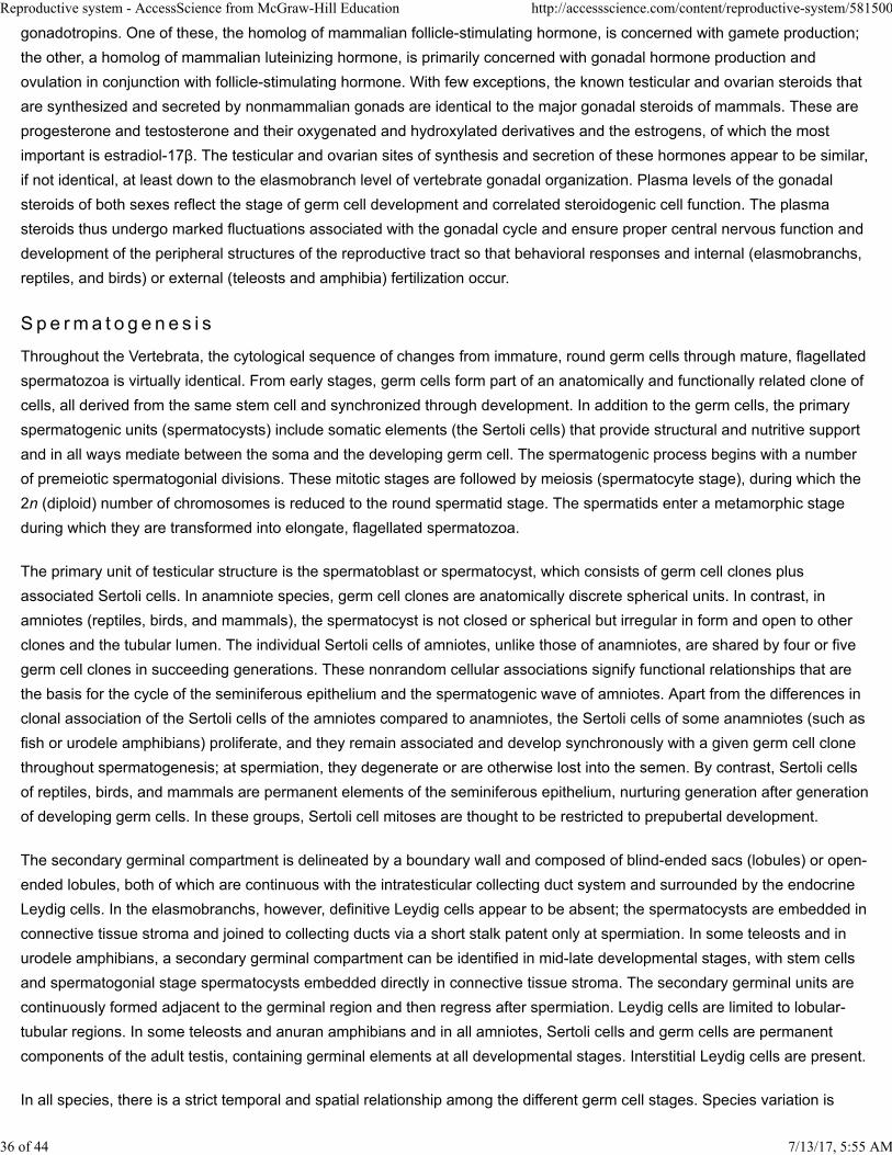
Reproductive system - AccessScience from McGraw-Hill Education http://accessscience.com/content/reproductive-system/581500
gonadotropins. One of these, the homolog of mammalian follicle-stimulating hormone, is concerned with gamete production;
the other, a homolog of mammalian luteinizing hormone, is primarily concerned with gonadal hormone production and
ovulation in conjunction with follicle-stimulating hormone. With few exceptions, the known testicular and ovarian steroids that
are synthesized and secreted by nonmammalian gonads are identical to the major gonadal steroids of mammals. These are
progesterone and testosterone and their oxygenated and hydroxylated derivatives and the estrogens, of which the most
important is estradiol-17β. The testicular and ovarian sites of synthesis and secretion of these hormones appear to be similar,
if not identical, at least down to the elasmobranch level of vertebrate gonadal organization. Plasma levels of the gonadal
steroids of both sexes reflect the stage of germ cell development and correlated steroidogenic cell function. The plasma
steroids thus undergo marked fluctuations associated with the gonadal cycle and ensure proper central nervous function and
development of the peripheral structures of the reproductive tract so that behavioral responses and internal (elasmobranchs,
reptiles, and birds) or external (teleosts and amphibia) fertilization occur.
S p e rma t o g e n e s i s
Throughout the Vertebrata, the cytological sequence of changes from immature, round germ cells through mature, flagellated
spermatozoa is virtually identical. From early stages, germ cells form part of an anatomically and functionally related clone of
cells, all derived from the same stem cell and synchronized through development. In addition to the germ cells, the primary
spermatogenic units (spermatocysts) include somatic elements (the Sertoli cells) that provide structural and nutritive support
and in all ways mediate between the soma and the developing germ cell. The spermatogenic process begins with a number
of premeiotic spermatogonial divisions. These mitotic stages are followed by meiosis (spermatocyte stage), during which the
2n (diploid) number of chromosomes is reduced to the round spermatid stage. The spermatids enter a metamorphic stage
during which they are transformed into elongate, flagellated spermatozoa.
The primary unit of testicular structure is the spermatoblast or spermatocyst, which consists of germ cell clones plus
associated Sertoli cells. In anamniote species, germ cell clones are anatomically discrete spherical units. In contrast, in
amniotes (reptiles, birds, and mammals), the spermatocyst is not closed or spherical but irregular in form and open to other
clones and the tubular lumen. The individual Sertoli cells of amniotes, unlike those of anamniotes, are shared by four or five
germ cell clones in succeeding generations. These nonrandom cellular associations signify functional relationships that are
the basis for the cycle of the seminiferous epithelium and the spermatogenic wave of amniotes. Apart from the differences in
clonal association of the Sertoli cells of the amniotes compared to anamniotes, the Sertoli cells of some anamniotes (such as
fish or urodele amphibians) proliferate, and they remain associated and develop synchronously with a given germ cell clone
throughout spermatogenesis; at spermiation, they degenerate or are otherwise lost into the semen. By contrast, Sertoli cells
of reptiles, birds, and mammals are permanent elements of the seminiferous epithelium, nurturing generation after generation
of developing germ cells. In these groups, Sertoli cell mitoses are thought to be restricted to prepubertal development.
The secondary germinal compartment is delineated by a boundary wall and composed of blind-ended sacs (lobules) or open-
ended lobules, both of which are continuous with the intratesticular collecting duct system and surrounded by the endocrine
Leydig cells. In the elasmobranchs, however, definitive Leydig cells appear to be absent; the spermatocysts are embedded in
connective tissue stroma and joined to collecting ducts via a short stalk patent only at spermiation. In some teleosts and in
urodele amphibians, a secondary germinal compartment can be identified in mid-late developmental stages, with stem cells
and spermatogonial stage spermatocysts embedded directly in connective tissue stroma. The secondary germinal units are
continuously formed adjacent to the germinal region and then regress after spermiation. Leydig cells are limited to lobular-
tubular regions. In some teleosts and anuran amphibians and in all amniotes, Sertoli cells and germ cells are permanent
components of the adult testis, containing germinal elements at all developmental stages. Interstitial Leydig cells are present.
In all species, there is a strict temporal and spatial relationship among the different germ cell stages. Species variation is
7/13/17, 5:55 AM 36 of 44
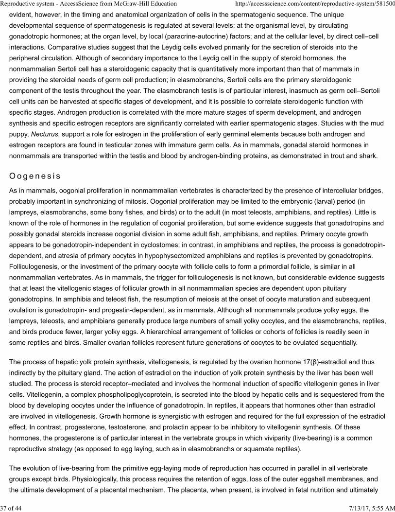
Reproductive system - AccessScience from McGraw-Hill Education http://accessscience.com/content/reproductive-system/581500
evident, however, in the timing and anatomical organization of cells in the spermatogenic sequence. The unique
developmental sequence of spermatogenesis is regulated at several levels: at the organismal level, by circulating
gonadotropic hormones; at the organ level, by local (paracrine-autocrine) factors; and at the cellular level, by direct cell–cell
interactions. Comparative studies suggest that the Leydig cells evolved primarily for the secretion of steroids into the
peripheral circulation. Although of secondary importance to the Leydig cell in the supply of steroid hormones, the
nonmammalian Sertoli cell has a steroidogenic capacity that is quantitatively more important than that of mammals in
providing the steroidal needs of germ cell production; in elasmobranchs, Sertoli cells are the primary steroidogenic
component of the testis throughout the year. The elasmobranch testis is of particular interest, inasmuch as germ cell–Sertoli
cell units can be harvested at specific stages of development, and it is possible to correlate steroidogenic function with
specific stages. Androgen production is correlated with the more mature stages of sperm development, and androgen
synthesis and specific estrogen receptors are significantly correlated with earlier spermatogenic stages. Studies with the mud
puppy, Necturus, support a role for estrogen in the proliferation of early germinal elements because both androgen and
estrogen receptors are found in testicular zones with immature germ cells. As in mammals, gonadal steroid hormones in
nonmammals are transported within the testis and blood by androgen-binding proteins, as demonstrated in trout and shark.
O o g e n e s i s
As in mammals, oogonial proliferation in nonmammalian vertebrates is characterized by the presence of intercellular bridges,
probably important in synchronizing of mitosis. Oogonial proliferation may be limited to the embryonic (larval) period (in
lampreys, elasmobranchs, some bony fishes, and birds) or to the adult (in most teleosts, amphibians, and reptiles). Little is
known of the role of hormones in the regulation of oogonial proliferation, but some evidence suggests that gonadotropins and
possibly gonadal steroids increase oogonial division in some adult fish, amphibians, and reptiles. Primary oocyte growth
appears to be gonadotropin-independent in cyclostomes; in contrast, in amphibians and reptiles, the process is gonadotropin-
dependent, and atresia of primary oocytes in hypophysectomized amphibians and reptiles is prevented by gonadotropins.
Folliculogenesis, or the investment of the primary oocyte with follicle cells to form a primordial follicle, is similar in all
nonmammalian vertebrates. As in mammals, the trigger for folliculogenesis is not known, but considerable evidence suggests
that at least the vitellogenic stages of follicular growth in all nonmammalian species are dependent upon pituitary
gonadotropins. In amphibia and teleost fish, the resumption of meiosis at the onset of oocyte maturation and subsequent
ovulation is gonadotropin- and progestin-dependent, as in mammals. Although all nonmammals produce yolky eggs, the
lampreys, teleosts, and amphibians generally produce large numbers of small yolky oocytes, and the elasmobranchs, reptiles,
and birds produce fewer, larger yolky eggs. A hierarchical arrangement of follicles or cohorts of follicles is readily seen in
some reptiles and birds. Smaller ovarian follicles represent future generations of oocytes to be ovulated sequentially.
The process of hepatic yolk protein synthesis, vitellogenesis, is regulated by the ovarian hormone 17(β)-estradiol and thus
indirectly by the pituitary gland. The action of estradiol on the induction of yolk protein synthesis by the liver has been well
studied. The process is steroid receptor–mediated and involves the hormonal induction of specific vitellogenin genes in liver
cells. Vitellogenin, a complex phospholipoglycoprotein, is secreted into the blood by hepatic cells and is sequestered from the
blood by developing oocytes under the influence of gonadotropin. In reptiles, it appears that hormones other than estradiol
are involved in vitellogenesis. Growth hormone is synergistic with estrogen and required for the full expression of the estradiol
effect. In contrast, progesterone, testosterone, and prolactin appear to be inhibitory to vitellogenin synthesis. Of these
hormones, the progesterone is of particular interest in the vertebrate groups in which viviparity (live-bearing) is a common
reproductive strategy (as opposed to egg laying, such as in elasmobranchs or squamate reptiles).
The evolution of live-bearing from the primitive egg-laying mode of reproduction has occurred in parallel in all vertebrate
groups except birds. Physiologically, this process requires the retention of eggs, loss of the outer eggshell membranes, and
the ultimate development of a placental mechanism. The placenta, when present, is involved in fetal nutrition and ultimately
7/13/17, 5:55 AM 37 of 44
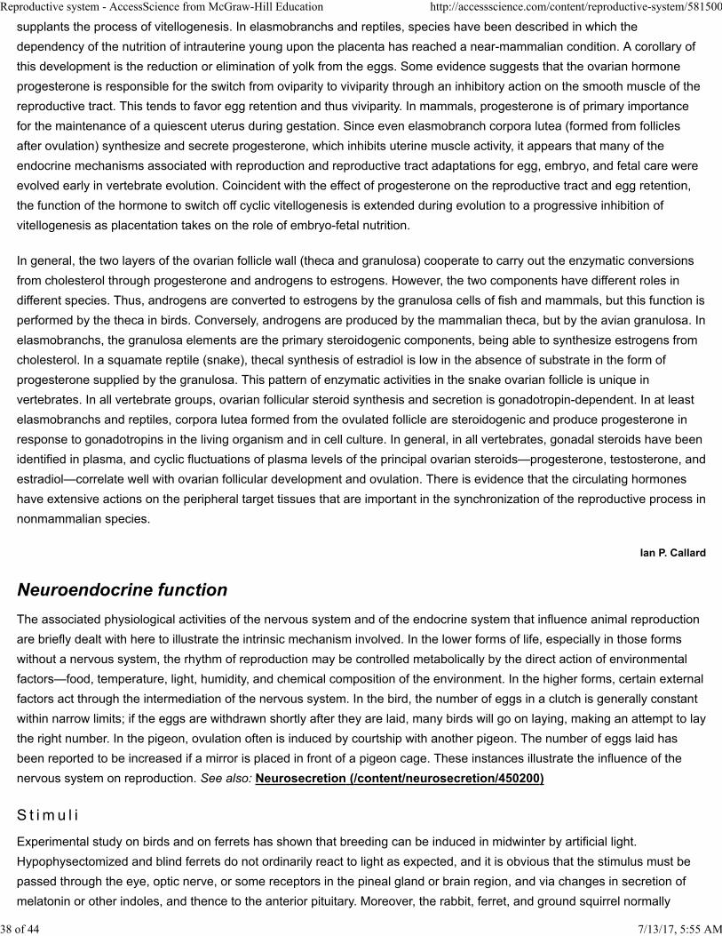
Reproductive system - AccessScience from McGraw-Hill Education http://accessscience.com/content/reproductive-system/581500
supplants the process of vitellogenesis. In elasmobranchs and reptiles, species have been described in which the
dependency of the nutrition of intrauterine young upon the placenta has reached a near-mammalian condition. A corollary of
this development is the reduction or elimination of yolk from the eggs. Some evidence suggests that the ovarian hormone
progesterone is responsible for the switch from oviparity to viviparity through an inhibitory action on the smooth muscle of the
reproductive tract. This tends to favor egg retention and thus viviparity. In mammals, progesterone is of primary importance
for the maintenance of a quiescent uterus during gestation. Since even elasmobranch corpora lutea (formed from follicles
after ovulation) synthesize and secrete progesterone, which inhibits uterine muscle activity, it appears that many of the
endocrine mechanisms associated with reproduction and reproductive tract adaptations for egg, embryo, and fetal care were
evolved early in vertebrate evolution. Coincident with the effect of progesterone on the reproductive tract and egg retention,
the function of the hormone to switch off cyclic vitellogenesis is extended during evolution to a progressive inhibition of
vitellogenesis as placentation takes on the role of embryo-fetal nutrition.
In general, the two layers of the ovarian follicle wall (theca and granulosa) cooperate to carry out the enzymatic conversions
from cholesterol through progesterone and androgens to estrogens. However, the two components have different roles in
different species. Thus, androgens are converted to estrogens by the granulosa cells of fish and mammals, but this function is
performed by the theca in birds. Conversely, androgens are produced by the mammalian theca, but by the avian granulosa. In
elasmobranchs, the granulosa elements are the primary steroidogenic components, being able to synthesize estrogens from
cholesterol. In a squamate reptile (snake), thecal synthesis of estradiol is low in the absence of substrate in the form of
progesterone supplied by the granulosa. This pattern of enzymatic activities in the snake ovarian follicle is unique in
vertebrates. In all vertebrate groups, ovarian follicular steroid synthesis and secretion is gonadotropin-dependent. In at least
elasmobranchs and reptiles, corpora lutea formed from the ovulated follicle are steroidogenic and produce progesterone in
response to gonadotropins in the living organism and in cell culture. In general, in all vertebrates, gonadal steroids have been
identified in plasma, and cyclic fluctuations of plasma levels of the principal ovarian steroids—progesterone, testosterone, and
estradiol—correlate well with ovarian follicular development and ovulation. There is evidence that the circulating hormones
have extensive actions on the peripheral target tissues that are important in the synchronization of the reproductive process in
nonmammalian species.
Ian P. Callard
Neuroendocrine function
The associated physiological activities of the nervous system and of the endocrine system that influence animal reproduction
are briefly dealt with here to illustrate the intrinsic mechanism involved. In the low er forms of life, especially in those forms
without a nervous system, the rhythm of reproduction may be controlled metabolically by the direct action of environmental
factors—food, temperature, light, humidity, and chemical composition of the environment. In the higher forms, certain external
factors act through the intermediation of the nervous system. In the bird, the number of eggs in a clutch is generally constant
within narrow limits; if the eggs are withdrawn shortly after they are laid, many birds will go on laying, making an attempt to lay
the right number. In the pigeon, ovulation often is induced by courtship with another pigeon. The number of eggs laid has
been reported to be increased if a mirror is placed in front of a pigeon cage. These instances illustrate the influence of the
nervous system on reproduction. See also: Neurosecretion (/content/neurosecretion/450200)
S t i m u l i
Experimental study on birds and on ferrets has shown that breeding can be induced in midwinter by artificial light.
Hypophysectomized and blind ferrets do not ordinarily react to light as expected, and it is obvious that the stimulus must be
passed through the eye, optic nerve, or some receptors in the pineal gland or brain region, and via changes in secretion of
melatonin or other indoles, and thence to the anterior pituitary. Moreover, the rabbit, ferret, and ground squirrel normally
7/13/17, 5:55 AM 38 of 44
Reproductive system - AccessScience from McGraw-Hill Education http://accessscience.com/content/reproductive-system/581500
ovulate in response to the stimulation of copulation. Since this stimulation to switch from the follicular phase to the luteal
phase cannot be effected in the absence of the pituitary but can be brought about by injecting pituitary extracts or pituitarylike
extracts, pregnant woman's urine (PU) or pregnant mare serum (PMS), it would seem that this stimulus is normally due to
nervous reflexes mediated through the hypothalamus and pituitary. The stimulus, however, may be carried by several nervous
paths, because local anesthesia of vagina and vulva, complete thoracosympathectomy, absence of any nerve pathway to the
ovaries, or cervical sympathectomy will not inhibit ovulation after coitus. Because stimulation of the brain, of the lumbosacral
part of the spinal cord, of the cervical sympathetic ganglion, or of the hypothalamus will induce ovulation to a certain extent, it
seems that more than one nervous path and more than one mechanism for the initiation of ovulation must be involved.
Furthermore, the rat, unlike the rabbit, ovulates spontaneously, but a prolongation of the life of the corpora lutea with
subsequent pseudopregnancy can be induced by sterile mating, mechanical stimulation of the cervix, or electrical stimulation
of the brain. Pseudopregnancy in the rat also seems to be mediated by the pituitary through nervous pathways. There is
additional evidence to show that the stimulus for luteinizing hormone release in a spontaneously ovulating animal is controlled
by nervous mechanisms employing cholinergic and adrenergic components.
L e s i o n s
It is well established that lesions in the basal tuber or median eminence induce ovarian atrophy in the cat, dog, and rabbit.
The role of the nervous system in establishing cyclic pituitary activity has also been emphasized. Localization of an erection
center and of an ejaculation center in the hypothalamus has been reported. Appropriately placed hypothalamic lesions in the
female guinea pig sometimes result in anestrus or prolonged estrous periods with sexual behavior in keeping with the gross
changes of the cycle. In the male guinea pig, similar lesions induce sexual impotence, without genital regression.
Hypothalamic production of oxytocin and the derivation of vasopressin from neurosecretory process have been postulated. As
for mating behavior, it is assumed that advancing evolutionary status is accompanied by a progressive dominance of the
nervous system and a corresponding reduction of endocrine control. To interpret the effect of sex hormones on mating
behavior, it has been suggested that their activity may increase the excitability of the central excitatory mechanism.
Fertility and sterility
The ability or inability to produce offspring is termed fertility or sterility. Fertility and sterility occur in different grades among
various species and among individuals of the same species. Absolute sterility is rare, but infertility of all degrees is very
common, especially among higher vertebrates. The rate of reproduction in any species depends upon the average number of
young born in each litter, the frequency of recurrence of breeding season, the duration of the reproductive period, and the age
at which the animal starts to breed. The age as a general rule is earlier in small species than in large ones. In general, the
number of young in a litter of mammals is inversely proportional to the size of the animal. For instance, a cow rarely produces
twins, whereas the rat occasionally bears as many as 16 young. A theory of fertility has been proposed that states that
individuation and genesis vary inversely; that is, the power to sustain individual life and the power to produce new individuals
are inversely proportional. Where there is abundant food supply and a favorable environment, and the necessary expenditure
of energy is relatively slight, the cost of individuation is much reduced and the rate of genesis is correspondingly increased.
See also: Infertility (/content/infertility/343450)
F a c t o r s c o n t r o l l i n g f e r t i l i t y
J. Hammond proposed that three factors control fertility:
1. The number of ova shed. In accordance with the genetic constitution of the species and the nutritional status, the number
of ova shed is controlled by gonadotropic hormones through the pituitary gland, but influenced by external factors. Before
puberty, at old age, and during pregnancy, pseudopregnancy, and early lactation, very few mammals ovulate. Although by
7/13/17, 5:55 AM 39 of 44
Reproductive system - AccessScience from McGraw-Hill Education http://accessscience.com/content/reproductive-system/581500
administering gonadotropic hormones the possibility of increasing the number of ova shed in mammals was demonstrated,
the actual number of young produced was rather low, probably as a result of limitations to support excessive embryos.
2. The number of ova fertilized. This depends upon the number of spermatozoa produced by the male, and the morphological
and physiological integrity of the gametes. It also depends upon the probability of meeting between gametes provided by the
male and female in the lower vertebrates, the efficiency of sperm transport to the site of fertilization, and the time of mating in
the higher vertebrates. Aging phenomena in gametes have a deleterious influence on fertility.
3. The number of embryos developing into self-sustaining individuals. The probability of normal development depends upon
where ova are deposited and the protection that the parents give to the zygotes in the lower vertebrates. In the higher
animals, it depends upon the transportation of zygotes to the prepared uterus at the right time, and the physiological activities
of embryo and of mother for proper implantation, for the maintenance of pregnancy, and for proper parturition.
C o n t r o l o f r e p r o d u c t i o n
The topic of controlling fertility, for example, the regulation of implantation in humans described above, should not be left
without portraying the considerable technology available to modify fertility in domestic animals.
While artificial breeding of cattle, and to a lesser extent pigs, sheep, and goats, by means of insemination of deep-frozen or
diluted semen samples has been extensively applied in recent years, enabling the exploitation of males of superior genetic
merit, other features of contemporary reproductive technology are less widely known; attention will be devoted to two of
these. The first can be termed controlled breeding, as seen in the various techniques of synchronization of estrus. There are
obvious advantages in being able to decide when an animal is to be bred, not only for convenience at the time of artificial
insemination or mating, but also because the time of birth, weaning, and marketing of the offspring can be predicted quite
accurately. The key to controlled breeding lies in reprogramming the ovary, and treatments attempt to modify, in the short
term, the influence of the secretions of the corpus luteum and the Graafian follicle expressed, respectively, in the luteal and
follicular phases of the cycle. Inducing synchronized estrus in a population of animals is achieved most effectively by causing
regression of the corpus luteum so that animals enter the follicular phase and estrus approximately synchronously.
The demonstration in the late 1960s that prostaglandin F 2α (PGF 2α) was almost certainly the luteolytic factor in domestic
species, informing the ovary that the uterus did not contain a viable embryo and inducing regression of the corpus luteum,
has caused this hormone to become the basis of extensive estrous synchronization programs, replacing the progestagenic
compounds previously used to simulate and extend the luteal phase prior to a synchronized follicular phase. Potent synthetic
analogs of PGF2α are now available that cause luteolysis following a single intramuscular injection. However, the corpus
luteum in cattle is only susceptible to injected PGF 2α between days 5 and 15 of the 21-day estrous cycle; thus, to achieve
synchronization in all animals in a herd, two injections are given 10 days apart. Nearly all animals will be in estrus within
48–72 h of the second injection, when they can be inseminated as a group. Prostaglandins are being used in this manner
extensively.
Another area of reproductive technology in domestic animals receiving attention is that of storage and transplantation of
embryos, particularly in the context of exploiting rare or exotic breeds of cattle after procedures of superovulation to increase
egg numbers. Transplantation of embryos is, of itself, not a new technique, for it was successfully performed in rabbits before
the turn of the century, and since about 1950 embryo transfer has been a leading experimental tool in reproductive
physiology. Two of the biological facts permitting such manipulations of the embryo are the relatively long period during which
the blastocyst is free-living in the uterine lumen before implantation, especially in pigs, sheep, and cattle (ungulates), and also
the fact that the embryo is not rejected as foreign material from the host uterus.
7/13/17, 5:55 AM 40 of 44
Reproductive system - AccessScience from McGraw-Hill Education http://accessscience.com/content/reproductive-system/581500
Apart from the care needed during handling and storage of embryos in the culture medium, many experiments have indicated
that the single most important factor determining the success of a transfer operation is the degree of synchronization of
estrous cycles between the donor and recipient animals. In cattle and sheep, there is some degree of tolerance in the timing;
but once development of the embryo is more than 24 h out of phase with that of the recipient uterus, the pregnancy rate
commences to decline sharply. Transfer of single, freshly recovered embryos by a surgical means involving an incision to
expose the uterus can lead to 90% of the transferred embryos developing into calves. Transfer of two embryos, one to each
horn of the uterus, has been used to induce twinning in cattle, although here the success rate is closer to 70%. Nonsurgical
transfer of embryos, that is, their introduction into the uterus from the vagina by means of a suitable pipette, has been much
less satisfactory, rarely giving pregnancy rates of greater than 20–40%. Apart from the greater risk of introducing infection by
this approach, there is also the problem of ejection of embryos which usually follows manipulation of the cervix during the
luteal phase. Nevertheless, now that bovine embryos can be preserved at −321°F (−196°C), as has been the case for
spermatozoa for many years, the emphasis is on overcoming the problems of nonsurgical transplantation. With
synchronization of estrus being a practical proposition, and deep-frozen banks of embryos in the offing, the attraction of
breeding programs involving this technically straightforward means of propagation is considerable, although it can be
economically justified only for valuable animals with desirable genetic traits.
In several species of farm animals (sheep, cattle, and pigs), zygotes up to the 8–16-cell stage can be halved or quartered.
The separated clumps of cells can then be placed in homologous zonae, from which all cytoplasmic material has been
removed, and can develop into normal young after retransfer to a suitable recipient. Not only does this technology rapidly
create additional copies of the traits of a particular mother or father, or both, but it also provides for identical twins, triplets,
and so forth for studies of the impact of environmental and nutritional factors on the same genetic background. Such a
technique will be useful primarily as a research tool rather than for production purposes, where transgenic animals will be
more useful.
Such advances in control of animal reproduction have given rise to the application of these techniques to humans for the
alleviation of infertility. Men with low sperm counts or poor spermatozoal motility can be assisted by collection of the semen
specimen and concentration of the spermatozoa. The chance of pregnancy can be improved if the female partner undergoes
artificial insemination with such treated sperm samples at the time of midcycle ovulation.
Infertile women can be induced to mature larger numbers of follicles by treatment with hormones. Ova from such follicles can
then be recovered and placed in the oviduct with a prepared sperm sample; matured and fertilized in a culture dish and
replaced in the oviduct; or matured, fertilized, and cultured in a dish and then replaced into the uterus. Many thousands of
children have been born worldwide following such procedures.
In addition, zygotes can still develop normally after removal of one or more blastomeres. Through the use of polymerase
chain reaction amplification of gene sequences from such a blastomere, prenatal diagnosis of genetic defects can be
detected prior to replacement of an externally fertilized embryo in the mother. Such techniques can revolutionize the field of
prenatal diagnosis, and have been used to detect a number of hereditary disorders. See also: Polymerase chain reaction
(PCR) (/content/polymerase-chain-reaction-pcr/900192); Prenatal diagnosis (/content/prenatal-diagnosis/543350)
In animals, research has focused on the production of transgenic animals. Most experiments have been conducted in mice,
although the techniques developed are being tried in farm animals. Genetic material with desirable characteristics can be
isolated with the techniques of molecular biology, and the deoxyribonucleic acid (DNA) can be microinjected into the ovum, or
inserted into embryonic stem cells in a culture dish; then, the cells can be microinjected into the cavity of a blastocyst and
become commingled with the cells of the host inner cell mass. The foreign DNA can integrate into the host chromosomal DNA
and can be carried into both germ and somatic cells. The transgenic animal passes the foreign DNA to its offspring in a
7/13/17, 5:55 AM 41 of 44
Reproductive system - AccessScience from McGraw-Hill Education http://accessscience.com/content/reproductive-system/581500
mendelian manner. After introduction of genes that are normally expressed in a tissue-specific manner, the location of DNA
sequences important in developmental programming of each gene can be determined. The gene for rat growth hormone
injected into mouse ova gave rise to mice that grew much larger than their noninjected siblings.
Other areas in which contemporary reproductive technology is attempting to increase animal productivity include induction of
ovulation at a precise time, increasing the numbers of eggs shed (superovulation), extending the breeding season of sheep
and goats, and, perhaps most important of all, regulating the time of parturition to avoid perinatal losses. In the future, this
technology will doubtless also include the application of pheromones to modify in diverse ways the reproductive activity of
farm livestock, and the possibility of predetermining the sex of offspring by insemination of X- or Y-bearing spermatozoa in
fractionated semen samples. See also: Genetic engineering (/content/genetic-engineering/285000); Reproductive
technology (/content/reproductive-technology/581625)
M. C. Chang
Michael J. K. Harper
R. H. F. Hunter
Additional Readings
W. Andrew, Textbook of Comparative Histology, Oxford University Press, 1959
C. R. Austin and R. V. Short (eds.), Reproduction in Mammals, 2d ed., vols. 1–5, Cambridge University Press, 1982–1986
G. F. Bastian, An Illustrated Review of the Reproductive System, Harper-Collins College Division, 1997
V. Blum, Vertebrate Reproduction, Springer-Verlag, 1986
R. H. F. Hunter, Physiology and Technology of Reproduction in Female Domestic Animals, Academic Press, 1981
P. C. K. Leung and E. Y. Adashi (eds.), The Ovary, 2d ed., Academic Press, 2004
W. R. Lyons et al., The hormonal control of mammary growth and lactation, Recent Prog. Hormone Res., 14:219–248, 1958
F. H. Marshall, Marshall's Physiology of Reproduction, vol. 2, 4th ed., Churchill Livingstone, 1990
P. K. T. Pang and M. P. Schreibman (eds.), Vertebrate Endocrinology: Fundamentals and Biomedical Implications, Academic
Press, 1986
J. R. Pasqualini, C. Sumida, andF. A. Kincl, Hormones and the Fetus, vol. 2, Pergamon Press, 1991
E. C. Roosen-Runge, The Process of Spermatogenesis in Animals, Cambridge University Press, 1977
J. Rossant and R. A. Pedersen (eds.), Experimental Approaches to Mammalian Embryonic Development, Cambridge
University Press, 1988
M. Shemesh and B. J. Weir (eds.), Maternal Recognition of Pregnancy and Maintenance of the Corpus Luteum, Blackwell,
1989
D. Shoupe (ed.), Contraception, Wiley-Blackwell, 2011
J. F. Strauss, III and R. I. Barbieri, Yen & Jaffe's Reproductive Endocrinology: Physiology, Pathophysiology, and Clinical
Management, 7th ed., Saunders, 2013
C. K. Weichert and W. Presch, Elements of Chordate Anatomy, 4th ed., McGraw-Hill, Inc., 1979
R. G. Wolff, Functional Chordate Anatomy, D. C. Heath & Co., 1991
R. M. Wynn and W. P. Jollie (eds.), Biology of the Uterus, 2d ed., Springer, 1989
7/13/17, 5:55 AM 42 of 44

Reproductive system - AccessScience from McGraw-Hill Education http://accessscience.com/content/reproductive-system/581500
L. J. Heffner and D. J. Schust, The Reproductive System at a Glance, 4th ed., Wiley-Blackwell, 2014
K. Rogers (ed.), The Human Body: The Reproductive System, Britannica Educational Publishing, 2011
7/13/17, 5:55 AM 43 of 44