assessment of arbuscular mycorrhizal fungal diversity in roots of solidago gigantea growing in a...
TRANSCRIPT

Environmental Microbiology (2006)
8
(6) 971ndash983 doi101111j1462-2920200500980x
copy 2006 The AuthorsJournal compilation copy 2006 Society for Applied Microbiology and Blackwell Publishing Ltd
Blackwell Science LtdOxford UKEMIEnvironmental Microbiology 1462-2912copy 2005 The Authors Journal compilation copy 2005 Society for Applied Microbiology and Blackwell Publishing Ltd 8 6971983 Original Article AM fungal diver-
sity in an Italian polluted soilM Vallino
et al
Received 31 August 2005 accepted 18 November 2005 Forcorrespondence E-mail graziellabertaunipmnit Tel (
+
39) 0131360232 Fax (
+
39) 0131 360390 or pbonfanteippcnrit Tel (
+
39)011 6705965 Fax (
+
39) 011 6705962
Assessment of arbuscular mycorrhizal fungal diversity in roots of
Solidago gigantea
growing in a polluted soil in Northern Italy
Marta Vallino
1
Nadia Massa
2
Erica Lumini
1
Valeria Bianciotto
1
Graziella Berta
2
and Paola Bonfante
1
1
Dipartimento di Biologia Vegetale ndash Universitagrave degli Studi di Torino CEBIOVEM and Istituto per la Protezione delle Piante (IPP) del CNR ndash Sezione Torino Vle Mattioli 25 10125 Torino Italy
2
Dipartimento di Scienze dellrsquoAmbiente e della Vita ndash Universitagrave del Piemonte Orientale lsquoAmedeo Avogadrorsquo via Bellini 25G 15100 Alessandria Italy
Summary
The arbuscular mycorrhizal (AM) status of
Solidagogigantea
was investigated in a contaminated site ofNorthern Italy where the chemical industry ACNA(Associated National Chemical Companies) wasactive till 1999 To counteract the devastating effectsof chemicals and to allow re-vegetation soil from anuncontaminated area was used to cover the highlypolluted hills of the industrial site about 25 yearsago On the basis of the current floristic features thehill was divided into four areas Heavy metal contentin soil and in plant shoots and roots was determinedby chemical analysis The AM fungal community col-onizing
S gigantea
was investigated from a morpho-logical and a molecular point of view All plants weremodestly colonized but the fungal structures withinthe roots were normal By PCR-RFLP and sequenc-ing of 18S rDNA 14 AM fungal types were identifiedthree of them were present in all the consideredareas and nine appeared to be specific to certainareas
Glomus
was the predominant AM genus Ouranalysis demonstrates the presence and the rela-tively high level of AM species variety and showshow a remediation programme based on cover-soilhas been efficient to restore a community of AMfungi tolerant enough to proliferate in a still contam-inated soil
Introduction
Environmental pollution has relevant consequences forhuman health and the quality of the environment itself Amore widespread concern for this issue coupled with abetter informed public opinion resulted in an evolution ofthe management of environmental protection and risk pre-vention Starting from the Seveso accident in 1976 theEuropean Union adopted the Seveso Directive on processindustries in 1982 The Seveso II Directive (9682CE)followed in 1996 initiating changes in risk managementat a national level (Hauptmanns 2005) Notwithstandingthese efforts in risk assessment the string of industrialdisasters highlighted a key issue how to deal with theexisting situation without increasing risks Therefore thedefinition of interventions for the removal or reduction ofthe existing pollution hazards is still urgently required InItaly the Ministry of the Environment (httpwwwminambi-enteit) has recently identified 15 environmentally criticalsites which needed to be included in remediation pro-grammes (DM n 4682001) one of them being theACNA (Associated National Chemical Companies) indus-trial site ACNA is a dismantled organic chemical industrialfactory located in Cengio (Savona Italy) The activity ofACNA followed a long process which started in 1882 andended in 1999 and went through the production of explo-sives (nitroglycerine dynamite and trinitrotoluene) paintsnitric and sulfuric acid amine and phenols (Esposito
et al
2002 Conte
et al
2005) The production of these com-pounds proved to be a high risk for workers (Puntoni
et al
1988 Bonassi
et al
1989) and had a deep environmentalimpact on soil quality and on the health of the Bormidariver whose bed was diverted into the factory The degreeof pollution was so strong that the aquatic life disappearedalmost completely for a certain period of time (G Bertapers comm) About 25 years ago when the factory wasstill active a first attempt was made to isolate the contam-inants from the environment a number of marly hills werefilled with industrial wastes and covered with a layer ofnon-polluted soil Due to diffusion over time the cover-soilbecame contaminated too though to a lesser extent Thisattempt allowed re-vegetation but the degree of plantbiodiversity was low (Andreucci
et al
2006) and the con-taminated hill core was never colonized by plant rootsRidgeway and Fitter (2003) in their work on old industrial
972
M Vallino
et al
copy 2006 The AuthorsJournal compilation copy 2006 Society for Applied Microbiology and Blackwell Publishing Ltd
Environmental Microbiology
8
971ndash983
sites in the UK suggested that the failure of re-vegetationprogrammes of sites contaminated by heavy metals andorganic pollutants may be due to a dearth of microbes inthe degraded soils It is known that microorganisms andtheir interactions can drive ecosystem functions such asplant biodiversity productivity and variability (van derHeijden
et al
1998) In particular mycorrhizal fungi whichrepresent a direct link between plants and soil are a majorfactor contributing to the maintenance of ecosystem func-tioning Among them arbuscular mycorrhizal (AM) fungibelonging to the phylum
Glomeromycota
(Smith andRead 1997 Schuumlszligler
et al
2001) are ubiquitous rootsymbionts and form intimate associations with the major-ity of plants (about 80 of families) Arbuscular mycor-rhizal fungi provide plants with mineral nutrients inexchange for carbon compounds and protect themagainst diverse abiotic and biotic stresses (Newsham
et al
1995 Smith and Read 1997) As it is not possibleto identify AM fungal species from their intraradical hyphalmorphology identification has traditionally been based onthe morphology of the resting spores found in the rhizo-sphere However spores number and diversity do notalways correlate with root colonization (Clapp
et al
1995Smith and Read 1997 Sanders 2004) Identification ofAM fungi within the root system is therefore challengingDue to advances in molecular biology and an increasedunderstanding of the phylogeny of AM fungi molecularmethods based on PCR technology are being utilizedfor fungal identification (Redecker 2000 Martin2001 Schuumlszligler
et al
2001) Ribosomal DNA (rDNA)sequences have been extensively used in the productionof AM-specific PCR primers In particular on the base ofsequence differences highlighted in the small subunit(SSU) gene Simon and colleagues (1992) and Redecker(2000) have developed family-specific PCR primers
In order to verify whether dearth of microbes couldaffect a re-vegetation programme we detected the pres-ence and the diversity of symbiotic fungi in the soil cover-
ing the ACNA industrial wastes We chose to carry out ouranalysis on
Solidago gigantea
because it was the onlyplant species that could be found in all four samplingareas Therefore our work provides a lsquosnapshotrsquo of theAM fungal community which was found to colonize rootsof
S gigantea
growing on A5 hill We determined theheavy metal content in soil and in plants and we checkedthe mycotrophic status of the roots by morphologicalobservations followed by quantification Symbiotic AMfungi taxa in roots were identified on the basis of the SSUribosomal DNA sequences subjected to restriction frag-ment length polymorphism (RFLP) and phylogeneticanalysis Our results demonstrate a good level of AMbiodiversity versus a low plant variety and lead to theinteresting conclusion that the presence of AM fungi maybe crucial but not sufficient to stimulate plant communitybiodiversity in a remediation process
Results
Chemical and morphological analyses
Plants collected in the four identified areas had a similarbiomass with no significant differences in root and shootfresh weight or in the rootshoot fresh weight ratio (withvalues ranging from 054 to 070 ndash data not shown)
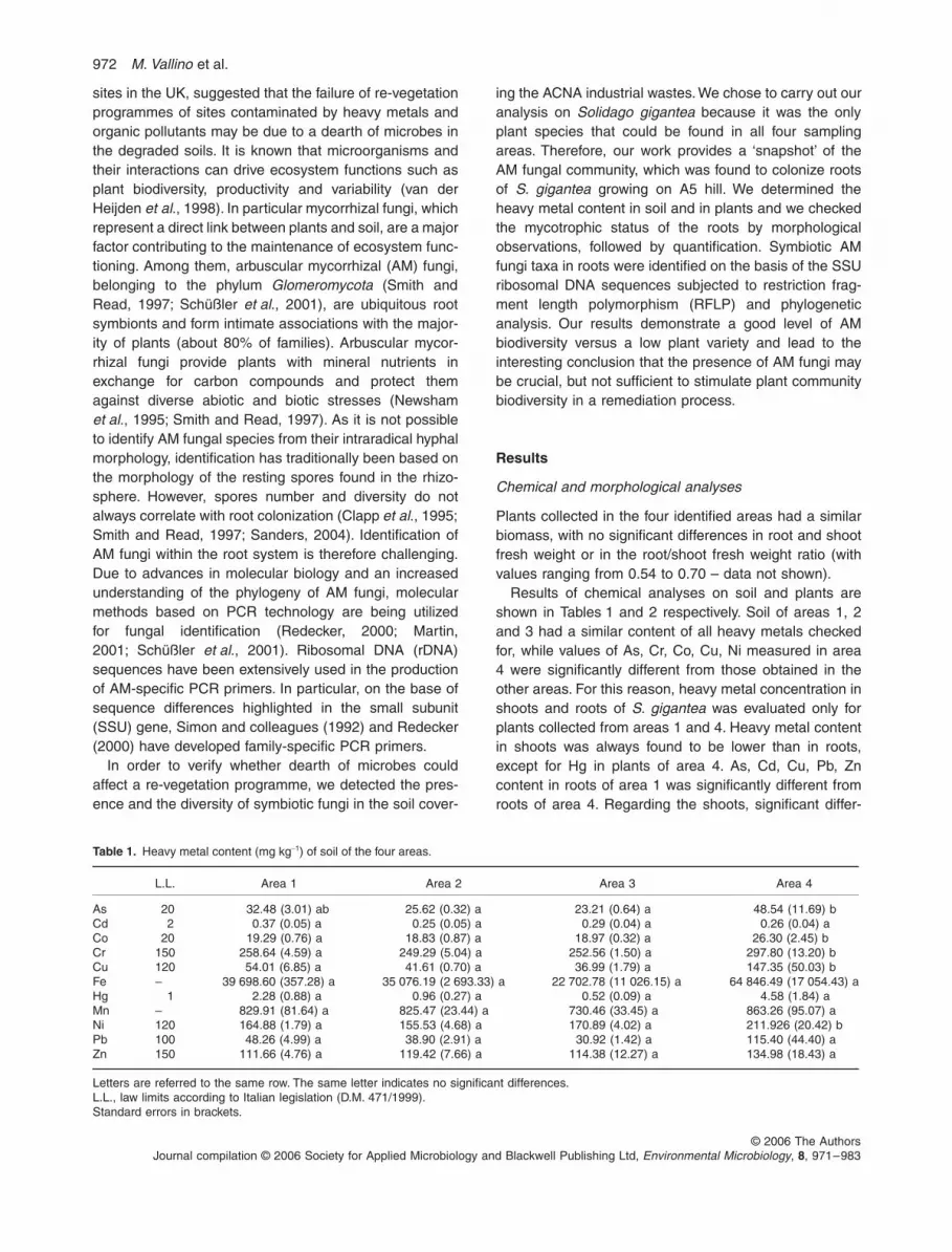
Results of chemical analyses on soil and plants areshown in Tables 1 and 2 respectively Soil of areas 1 2and 3 had a similar content of all heavy metals checkedfor while values of As Cr Co Cu Ni measured in area4 were significantly different from those obtained in theother areas For this reason heavy metal concentration inshoots and roots of
S gigantea
was evaluated only forplants collected from areas 1 and 4 Heavy metal contentin shoots was always found to be lower than in rootsexcept for Hg in plants of area 4 As Cd Cu Pb Zncontent in roots of area 1 was significantly different fromroots of area 4 Regarding the shoots significant differ-
Table 1
Heavy metal content (mg kg
minus
1
) of soil of the four areas
LL Area 1 Area 2 Area 3 Area 4
As 20 3248 (301) ab 2562 (032) a 2321 (064) a 4854 (1169) bCd 2 037 (005) a 025 (005) a 029 (004) a 026 (004) aCo 20 1929 (076) a 1883 (087) a 1897 (032) a 2630 (245) bCr 150 25864 (459) a 24929 (504) a 25256 (150) a 29780 (1320) bCu 120 5401 (685) a 4161 (070) a 3699 (179) a 14735 (5003) bFe ndash 39 69860 (35728) a 35 07619 (2 69333) a 22 70278 (11 02615) a 64 84649 (17 05443) aHg 1 228 (088) a 096 (027) a 052 (009) a 458 (184) aMn ndash 82991 (8164) a 82547 (2344) a 73046 (3345) a 86326 (9507) aNi 120 16488 (179) a 15553 (468) a 17089 (402) a 211926 (2042) bPb 100 4826 (499) a 3890 (291) a 3092 (142) a 11540 (4440) aZn 150 11166 (476) a 11942 (766) a 11438 (1227) a 13498 (1843) a
Letters are referred to the same row The same letter indicates no significant differencesLL law limits according to Italian legislation (DM 4711999)Standard errors in brackets
AM fungal diversity in an Italian polluted soil
973
copy 2006 The AuthorsJournal compilation copy 2006 Society for Applied Microbiology and Blackwell Publishing Ltd
Environmental Microbiology
8
971ndash983
ences between area 1 and area 4 were detected for allthe metals except Mn and Hg
The degree of root colonization by AM fungi and thepresence of arbuscules were determined microscopicallyafter methyl blue staining (Fig 1) Colonization rangedfrom 04 to 244 root length (Table 3) The lowestmycorrhizal colonization rates were found in samples ofarea 3 colonization values around or above 20 werefound in samples of all the areas Mean values shown inFig 2 indicate that the degree of mycorrhization of plantroots coming from different areas was not significantlydifferent
Sequences amplified using NS31AM1
Template DNA was amplified with the primers pair AM1NS31 As expected DNA from leaves was not amplified
while DNA from 91 of all mycorrhizal roots gave a bandof about 550 bp We chose one plant from each area (1B2B 3B 4B) which yielded enough PCR product for cloningand creating a SSU rRNA mini-library (Table 3)
Overall 170 clones were screened by PCR of which137 contained the SSU rRNA gene fragment In order togain a good overview of the fungal richness present in theroots our first aim was to find the minimum number ofclones to be processed from each sample We thereforedetermined the number of RFLP patterns obtained afteranalysing groups of five clones from each area (Fig 3) Aplateau was reached with the analysis of 25 clones in area1 and area 3 20 clones in area 2 15 clones in area 4Therefore it seemed likely that analysing 30 clonessam-ple was sufficient to detect the degree of biodiversity inour system For this reason a total of 120 clones wereanalysed with RFLP providing 12 different patterns
Table 2
Heavy metals concentration (mg kg
minus
1
) in roots and shoots of
S gigantea
collected in area 1 and area 4
Roots Shoots
Area 1 Area 4 Area 1 Area 4
As 170 (024) a 569 (025) b 017 (002) a 265 (064) bCd 090 (006) a 071 (002) b 032 (004) a 011 (001) bCo 362 (031) a 439 (022) a 040 (004) a 207 (047) bCr 3440 (407) a 3329 (306) a 376 (058) a 1822 (417) bCu 1323 (075) a 11798 (1644) b 828 (078) a 1732 (180) bFe 664667 (127115) a 708099 (61477) a 67609 (6217) a 522250 (197838) bHg 011 (001) a 010 (000) a 007 (001) a
lt
01 (000) aMn 13067 (1024) a 13750 (753) a 7921 (438) a 9780 (2565) aNi 2670 (292) a 2952 (249) a 356 (040) a 1490 (342) bPb 932 (078) a 1901 (111) b 200 (027) a 859 (182) bZn 5836 (150) a 7947 (849) b 3267 (240) a 4424 (335) b
The same letter indicates no significant differences Letters are referred to the same row and should be considered separately for roots and shootsStandard errors in brackets
Fig 1
Arbuscular mycorrhizal fungal colonization in
S gigantea
roots sampled from area 2 (A) and area 4 (B C) Roots were stained with methyl blue Vesicles (A) coils (B) and arbuscules (C) are clearly visible
A C B
20 microm 20 microm 20 microm
974
M Vallino
et al
copy 2006 The AuthorsJournal compilation copy 2006 Society for Applied Microbiology and Blackwell Publishing Ltd
Environmental Microbiology
8
971ndash983
(Fig 4A) Two to six clones for each RFLP type weresequenced for a total of 32 sequences the other 88sequences were classified by RFLP typing
RFLP type analysis on NS31AM1 amplified fragments
Figure 5 shows the frequencies and the distribution of theRFLP patterns detected in the roots of
S gigantea
in thefour areas considered in this study Of the 12 RFLP pat-terns detected RFLP2 and RFLP3 are dominant (repre-senting 23 and 22 of the analysed clones) andtogether with RFLP5 are found in all four areas RFLP4and RFLP10 are present in three areas The remainingpatterns look specific for one single area RFLP1 for area1 RFLP9 RFLP11 RFLP12 for area 3 and RFLP6RFLP7 RFLP8 for area 4 In area 1 six different RFLPpatterns are present RFLP1 and RFLP2 being predomi-nant In area 2 only four patterns were found RFLP3having the highest percentage Area 3 and area 4 haveeight and seven different patterns respectively no one ofthem being predominant
Identification of AM fungal groups
BLASTN
searches in the GenBank database showed that
all sequences have high similarity (97ndash100 identity) toAM fungi and belong to members of the
Glomeromycota
phylum Alignment of the sequences obtained in this studywith those corresponding to the closest matches fromGenBank produces a tree which reveals that thesequences belong to the groups of
Glomerales
and
Diver-sisporales
(Fig 6) No sequences were detected withinthe
Archaeosporales
or
Paraglomerales
this wasexpected because AM1 primer does not match the highlydivergent 18S-sequence of these taxa (Redecker 2000)
The phylogenetic relationships among the sequencesclearly reveal discrete sequence groups and allow us toidentify 13 potential taxonomic units (TUs) or fungal types11 belong to the
Glomeraceae
one to the
Diversispora-ceae
and one to the
Gigasporaceae
All sequence clus-ters are supported by a bootstrap
gt
75 In two clusters(acnaGlo3 acnaGlo8) two separate sequences typesappear to exist but they are not well supported by thebootstrap value nor are they distinguishable by the HinfI
Table 3
Comparison between root mycorrhization and AM1NS31PCR amplification of roots of all sampled plants of
S gigantea
M A Plant AM1NS31
Area 1 2439 1074 1A ndash1368 694 1B
+
1269 539 1C
+
1492 339 1D
+
1432 760 1E
+
1022 361 1F
+
Mean value 1504 628
Area 2 1194 789 2A
+
1575 1057 2B
+
1983 1309 2C
+
1065 613 2D
+
1955 1658 2E
+
896 589 2F
+
Mean value 1445 1003
Area 3 036 000 3A ndash2315 1219 3B +2076 1737 3C
+
330 206 3D
+
154 047 3E
+
Mean value 982 642
Area 4 1985 1649 4A
+
1758 931 4B
+
1107 883 4C
+
2014 1130 4D
+
1797 1410 4E
+
1149 661 4F
+
Mean value 1635 1111
M degree of AM fungi colonization A presence of arbuscules
+
ndash presenceabsence of amplified bandGrey shaded cells indicate plants chosen for cloning (see text)
Fig 2
Degree of AM fungal colonization (M) and presence of arbuscules (A) in roots of
S gigantea
sampled in the four areas of hill A5 Mean values and standard errors are presented Statistical analysis was done separately for M and for A The same letter indicates no significant differences
a a
a
a
b
b
b
b
0
2
4
6
8
10
12
14
16
18
20
area 1 area 2 area 3 area 4
per
cen
tag
e
M
A
Fig 3
Number of RFLP patterns obtained after HinfI and Hsp92II digestions of 5 10 15 20 25 30 clones from each area
012
3456
789
5 10 15 20 25 30number of analysed clones
To
tal n
um
ber
of
RF
LP
pat
tern
s
area 1area 2area 3area 4
AM fungal diversity in an Italian polluted soil
975
copy 2006 The AuthorsJournal compilation copy 2006 Society for Applied Microbiology and Blackwell Publishing Ltd
Environmental Microbiology
8
971ndash983
and Hsp92II RFLP patterns for this reason they are clas-sified as single type
Clades acnaGig6 acnaGlo8 acnaGlo11 includesequences with 98 100 98 identity to thesequences of
Gigaspora decipiens
strain BEG45
Glomusmosseae
strain BEG69 and
Glomus intraradices
BEG121respectively all other AM fungal types do not clusterclosely with any sequences from AM fungi in culture
Taxonomic groups are mainly consistent with theRFLP types (Fig 6) with few exceptions For examplesequence 3B34 gives a RFLP2 pattern but it does notcluster with the other sequences having this pattern(1B1 4D41 1B3) On the contrary the latter sequencescluster with clone 3B28 which has a RFLP12 patternFinally sequences 3B12 and 3B26 have the sameRFLP pattern but they cannot be assigned to a singlefungal group
Sequences amplified using ARCH1311NS8
The same DNA samples used for AM1NS31 amplification(plants 1B 2B 3B 4B) were used in nested PCR reac-tions with NS5ITS4 as the first step and ARCH1311 andNS8 as the second step All products amplified byARCH1311NS8 were sufficient for cloning A total of 84
clones were screened for the presence of the insert and41 clones resulted positive unfortunately no single cloneof area 4 was positive
All 41 clones were then analysed with RFLP providingeight different patterns (Fig 4B) One to four clones foreach RFLP type were sequenced for a total of 11sequences
BLASTN
searches in the GenBank database showed thata small percentage of the analysed sequences belong to
Paraglomerales
Only the sequences corresponding tothe RFLPc gave high homology (94 of identity) with
Glomus occultum
and they were only present in area 3All the other sequences gave homology with
Ascomycota
(RLFPd and RFLPh ndash 39)
Basidiomycota
(RFLPf ndash 2)and plants (RFLPa RFLPb RFLPe and RFLPg ndash 44)Therefore out of the 41 clones 44 are of plant origin41 are of no AM fungal origin and 15 belong to
Paraglomeraceae
Discussion
ACNA activity poured into the surrounding area a hugeamount of pollutants which strongly inhibited any form oflife This phenomenon was evident in the core of the hillswhere only a very small number of bacterial species
Fig 4
RFLP patterns of SSU rRNA PCR products obtained from
S gigantea
rootsA AM1NS31 amplification H1ndashH9 restriction pattern obtained with enzyme HinfI P1ndashP7 restriction pattern obtained with enzyme Hsp92IIB ARCH1311NS8 amplification HandashHh restriction pattern obtained with enzyme HinfI PandashPb restriction pattern obtained with enzyme Hsp92II
RFLP patternsH1-P1 RFLP1H2-P2 RFLP2H2-P1 RFLP3H3-P1 RFLP4H4-P6 RFLP5H5-P1 RFLP6H6-P4 RFLP7H4-P5 RFLP8H9-P7 RFLP9H7-P5 RFLP10H8-P5 RFLP11H2-P7 RFLP12
RFLP patternsHa-Pa RFLPaHb-Pa RFLPbHc-Pb RFLPcHd-Pa RFLPdHe-Pa RFLPeHf-Pa RFLPfHg-Pa RFLPgHh-Pa RFLPh
H1 H2 H3 H4 H5 H6 H7 H8 H9
244 nt190 nt
280 nt334 nt
141 nt
50 nt
25 nt
pUC18
P1 P2 P4 P5 P6 P7
290 nt
170 nt
90 nt116 nt
pUC18
Pa Pb
450 nt
170 nt
90 nt
pUC18
Ha Hb Hc Hd He Hf Hg Hh
241 nt199 nt
331 nt
165 nt
107 nt
80 nt
pUC18
A BNS31AM1 amplified fragments ARCH1311NS8 amplified fragments
976
M Vallino
et al
copy 2006 The AuthorsJournal compilation copy 2006 Society for Applied Microbiology and Blackwell Publishing Ltd
Environmental Microbiology
8
971ndash983
occurred (Avidano
et al
2005) The ACNA hills have rep-resented a very interesting situation to investigatebecause they offered the opportunity to evaluate thedynamics of plants microbes and animals starting from aground zero level Among the studies found in literatureon lsquobiological desertsrsquo resulting from industrial wastes andemissions there are cases of investigations in whichbesides human plant and animal health also the micro-bial communities had been considered (for example theareas contaminated by metal smelters in Finland North-ern Sweden and Canada ndash Gunn
et al
1995 Pennanen
et al
1996 Palmborg
et al
1998) However to ourknowledge only few of them analysed the microbial com-ponent represented by mycorrhizal fungi (for example
Niepolomice Forest ndash Turnau 1988 Kapusta
et al
2004Chernobyl ndash Zhdanova
et al
2004) Our work presents asurvey of AM colonization in A5 hill using morphologicaland molecular methods We could not extend our analysisin time or in number of plant species because furthersampling was not allowed Notwithstanding this our inves-tigation is one of the few attempts to evaluate the capacityof AM fungi to colonize plants and to establish an under-ground network in an industrial dump
We chose to carry out our analysis on
S gigantea
because it was the only plant species that could befound in all four sampling areas
Solidago gigantea
(
Asteraceae
) is a rhizomatous perennial herb native toNorthern America and introduced in Europe about
Fig 5 A Distribution and frequencies in the four areas of A5 hill of the 12 RFLP patterns obtained after HinfI and Hsp92II digestions of 120 NS31AM1 amplified fragments The last row is the legend for the below pie-chartsB Approximate tri-dimensional map of A5 hill showing the location of the four analysed areas (area 1 area 3 area 4 ovals area 2 perimeter) The pie-charts associated to each area refer to the RFLP types the legend for the segments of the pie-charts is presented in the last row of the above table
RFLP1 RFLP2 RFLP3 RFLP4 RFLP5 RFLP6 RFLP7 RFLP8 RFLP9 RFLP10 RFLP11 RFLP1222313191 AERA
AREA 2 4 16 4 2155155443 AERA
AREA 4 7 5 1 4 2 9 1total 9 28 26 12 10 4 2 9 1 8 5 1
A
AREA 2AREA 3
AREA 4
B
AREA 1
AM fungal diversity in an Italian polluted soil 977
copy 2006 The AuthorsJournal compilation copy 2006 Society for Applied Microbiology and Blackwell Publishing Ltd Environmental Microbiology 8 971ndash983
250 years ago (Weber and Jakobs 2005) In a study onthe invasiveness of this species Jakobs and colleagues(2004) found that in Europe it was present both in wetand dry habitats fewer population were directly adjacentto rivers and lakes and more were in disturbed areasand besides roads This species turned out to be a goodrepresentative to work on as roots were clear and easyto handle
Chemical analysis of soil sampled in the S gigantearhizosphere revealed that all areas of A5 hill had a highconcentration of heavy metals and that area 4 was themost polluted one Heavy metal concentrations in roots ofS gigantea was much lower than concentrations in soilexcept for Cd which was about three times higher in rootsthan in soil Metal content in plants of area 4 was alwaysequal to or higher than metal content in plants of area 1
Fig 6 Neighbour-joining tree showing relationships between NS31-AM1 rDNA sequences from S gigantea and reference sequences from GenBank Corallochytrium limacisporum a putative choanoflagellate (Cavalier-Smith and Allsopp 1996) was used as outgroup All bootstrap values gt50 are shown (100 replicates) Individual clones are identified by site (first number 1 2 3 4) and clone number (example A43 B6 D1) On the right group identifiers (for example acnaGlo7) are compared with RFLP types (see text)
4D14D51
AF074371 Glomus spGlo5AJ276087 Glomus lamellosumAJ276080 Glomus claroideum
AF481661 Glomus spGlo264B6
4B9AJ567845 Gigaspora margarita
U96146 Gigaspora decipiensAY129642 Scutellospora spScut
Z14010 Gigaspora giganteaX58726 Gigaspora rosea
1B21B7
Y17644 Glomus etunicatumY17650 Glomus spurcum
AJ315524 Glomus spssGloyelp11B33
3B432B11
2B74D6
2A43AY129625 Glomus spGlo18
AJ418867 Glomus spMO G43B34
AY512347 Glomus spGlo48AF480158 Glomus sp5014b25Lla
3B391B1
AF074353 Glomus spGlo44D41
1B33B28
AJ418889 Glomus spMO G9AF437660 Glomus spGlo13AJ496108 Glomus spMO G12
2B122B13AF074370 Glomus spGlo7
AJ418880 Glomus spMO G61B132B14D9
3B1AJ506090 Glomus constrictum
AJ505813 Glomus viscosum4B3Z14007 Glomus mosseae
4D34D7AY243568 Glomus spGlo1BU96141 Glomus mosseae BEG69
1B313B83B11
AY129607 Glomus spGlo10AJ536822 Glomus intraradices
3B12AJ496053 Glomus spMO G3
Y17640 Glomus fasciculatumX58725 Glomus intraradices
3B26AJ309462 Glomus spGlo8AF481660 Glomus spGlo23
AJ496059 Glomus spMO G13
L42528 Corallochytriumlimacisporum
acnaGlo7
acnaGig6
acnaDiv1
acnaGlo3
acnaGlo2
acnaGlo1
acnaGlo2
acnaGlo4
acnaGlo5
acnaGlo8
acnaGlo11
acnaGlo12a
acnaGlo12b
RFLP7
RFLP6
RFLP11
RFLP10
RFLP8
RFLP5
RFLP4
RFLP12
RFLP2
RFLP2
RFLP9
RFLP3
RFLP1
94100
100
94
100
100
100
76
70
57
80
5279
92
100
100
100
100
6695
54
70
69
78
57
94
10071
81
100
66
98
52
59
88
0005 substitutionssite
978 M Vallino et al
copy 2006 The AuthorsJournal compilation copy 2006 Society for Applied Microbiology and Blackwell Publishing Ltd Environmental Microbiology 8 971ndash983
Cd being again an exception in fact Cd concentration inboth roots and shoots of area 4 was lower than that ofarea 1 Significant differences in soil metal contentbetween area 1 and area 4 was not mirrored in root metalcontent (Cu was the only case where significant differ-ences were found in roots and soil) From this we con-clude that metal concentration in plant roots was notnecessarily related to soil metal concentration asobserved also by Ortiz and Alcaniz (2006)
Heavy metal concentration in S gigantea shoots wasalways lower than in roots (except Hg) suggesting a lowlevel of translocation between plant organs However thecontent of some metals in shoots of plant from area 4 washigher than area 1 and fell beyond the normal backgroundranges detected in plants growing in unpolluted regions(Djingova et al 2004) This suggests that in area 4 metaltranslocation increased For As Pb Cu Zn it was prob-ably a consequence of a higher root concentration In thecase of Co Cr Fe Ni in which metal content was signif-icantly different in the soil of the two areas and in shootsbut not in roots we postulate that metals reached a sat-uration point in roots and were therefore transported tothe aerial part of the plants
The morphology of AMF colonization within the roots ofS gigantea was as usual the presence of arbusculessuggests that the symbiosis was active Notwithstandingthis the values of colonization were rather low It is knownthat heavy metals can delay reduce and even eliminatespore germination and AM colonization del Val and col-leagues (1999) showed that the total number of AMFspores correlated negatively with the metal content of thesoil However in previous studies (Griffioen et al 1994Weissenhorn and Leyval 1994) no correlation was foundbetween the concentration of metals such as Cu Cd Znand AMF population Furthermore Pawlowska and col-leagues (1996) found a relatively high rate of mycorrhizalcolonization in plants growing in very polluted soils In ourcase it is noteworthy that the many pollutants did notaffect fungal morphogenesis once the fungus was insidethe root By contrast the low success of infection asmeasured by the number of colonized root fragmentsmakes us wonder whether the spore could be directlyaffected by pollutants in the rhizosphere as here they aredirectly exposed to them
As root colonization had an even distribution in almostall the plants we were confident in taking random frag-ments of roots to be used for molecular analysis insteadof selecting them under the microscope As expectedPCR results were positive for almost all samples In orderto minimize the number of clones to be processed and yetto obtain a picture of the fungal diversity within the rootsas close as possible to the real one RFLP analysis on agrowing number of sequences was performed until reach-ing saturation of the different RFLP patterns Saturation
was reached after the digestion of 25 sequences for eacharea Thus we concluded that analysing a total of 120clones was sufficient to evaluate the AM fungal speciesrichness This result is consistent with the work ofLandeweert and colleagues (2003) on biodiversity of ecto-mycorrhizal fungi in soil in which by applying a statisticalmethod they found that the most common species canbe detected when 30 clonessample are analysed
The primer combination NS31-AM1 is targeted at aregion of the SSU and amplifies most but not all gloma-lean fungi It has been used to detect the presence offungal sequences from the four families of GlomeraceaeAcaulosporaceae Diversisporaceae and Gigasporaceae(order Glomales and Diversisporales) in different ecosys-tems such as agricultural sites (Helgason et al 1998Daniell et al 2001) broadleaved wood (Helgason et al1998) seminatural woodland (Helgason et al 1999)tropical forest (Husband et al 2002) and grassland (Van-denkoornhuyse et al 2002) Recently it has been discov-ered that certain species which have in the past beenconsidered to belong to the genera Glomus or Acau-lospora on the basis of their morphology have SSUsequences that are much more divergent For these fungitwo new orders have been suggested Archaeosporalesand Paraglomerales (Morton and Redecker 2001) Thecharacterization of these ancestral lineages (Redecker2000) showed that the AM1 primer sequence is not appro-priate to detect such deeply divergent clades ARCH1311designed by Redecker (2000) is the first specific primerfor these lineages Therefore by using the two primerpairs AM1-NS31 and ARCH1311-NS8 in our work wecould reliably detect and identify all currently known AMfungi while discriminating against other fungi
Only a small percentage of the sequences obtainedafter amplification with ARCH1311 and NS8 belong toParaglomerales all the others are of plant or non-AMfungi origin The use of the same primer pair on the DNAof liverworts exerted similar results (E Lumini perscomm) Problems of primers specificity were alsoencountered by Wubet and colleagues (2003ab) whoamplified a number of AM fungal types belonging to theGlomeraceae using ARCH1311 in combination with ITS4on mycorrhizal roots of Prunus africana from forests ofEthiopia
The combination of BLAST searches and phylogeneticanalysis allowed us to assign the fungal sequences iso-lated from S gigantea roots to 14 glomeralean groupsTwelve groups belong to the genus Glomus one toGigaspora and one to Paraglomus The dominance of thegenus Glomus is not new Predominance of the Glomer-aceae has also been reported for other ecosystems suchas agricultural sites (Daniell et al 2001) or tropical forests(Onguene and Kuyper 2001 Husband et al 2002 Wubetet al 2003b) Daniell and colleagues (2001) argued that
AM fungal diversity in an Italian polluted soil 979
copy 2006 The AuthorsJournal compilation copy 2006 Society for Applied Microbiology and Blackwell Publishing Ltd Environmental Microbiology 8 971ndash983
Glomus types dominate the colonization of arable cropsbecause Glomus species are better adapted to disturbedenvironments where their high sporulation rates mayenable them to recover more readily Whitfield and col-leagues (2004) drew the same conclusion from the pre-dominance of Glomus species in the metal-contaminatedsoils along the River South Tyne (UK) which is regularlyaffected by flooding which presumably causes major soildisturbance One possible reason why Glomus speciesare dominant also in an ecosystem disturbed by chemicalpollution may be found in the ability of the Glomeraceaeto colonize via fragments of mycelium or mycorrhizal rootspieces while Gigasporaceae are only capable of propa-gation via spores (Biermann and Linderman 1983Daniell et al 2001) Pawlowska and Charvat (2004) sug-gested that hyphal extension from colonized root frag-ments is a better predictor of an AM fungal performanceunder metal stress than spore germination and that AMfungi are able to survive and propagate in metal-pollutedenvironments by thriving in relatively uncontaminatedmicrosites If this is the case we may postulate that rootsonce they are colonized function as safe microenviron-ment and as inoculum for Glomus species which thusbecome more competitive in infecting roots other thanGigasporaceae
Three taxonomic units were found in all four areasinvestigated suggesting that dominant fungi formedmycelial networks with an extension of up to 4000 m2 Twotaxonomic units were found in three areas and nine werefound only in single areas showing a patchy distributionof AM fungal population also described in agriculturalsystems (Stukenbrock and Rosendahl 2005) and inundisturbed vegetation (Rosendahl and Stukenbrock2004)
Interestingly in the most polluted area 4 the most abun-dant fungal type was acnaGlo8 which can be assignedto G mosseae In morphological and molecular analysisof roots of plants colonizing zinc wastes Turnau and col-leagues (2001) showed that G mosseae was the onlyfungus which was always present in roots fragments butthe authors cannot say whether G mosseae is a heavymetal-tolerant species
In Helgason and colleagues (1999) 141 analysedclones from bluebell roots growing in seminatural wood-land gave eight different sequence groups In morecomplex works Helgason and colleagues (1998) andDaniell and colleagues (2001) detected 6 and 10 TUsrespectively analysing the AM fungal community in agri-cultural sites from four different plant species In contrasta much greater diversity was reported for dry and tropicalforests (Husband et al 2002 Wubet et al 2003b) andgrassland (Vandenkoornhuyse et al 2002) in which 2330 24 fungal types respectively were detected from twodifferent plant species Whitfield and colleagues (2004)
compared AM colonization in roots of Thymus polytrichusgrowing in sites with a long-term history of heavy metalcontamination they found 19 different AM fungal SSUsequence types and they considered this number an indexof relatively large AM species diversity Also Ridgewayand Fitter (2003) found that the variety of AM fungal typesgrowing in UK contaminated soils was surprisingly highand at a similar level to some natural woodlands andgrasslands Our data are in accordance with these twolatter works in fact the isolation of 14 TUs from the rootsof S gigantea indicates a good richness of AM fungi inthe cover-soil of hill A5 The level of AM fungal biodiversityis not mirrored by the degree of biodiversity of plantsgrowing on the hill which appears low (Andreucci et al2006) Therefore in this case study fungal presence andbiodiversity do not seem to be the key factor that promotesplant variety in a recovering site AM fungi may be anobligate but not exclusive requisite in a re-vegetationprogramme
In conclusion our work based on a combination ofchemical morphological and molecular analyses pro-vides useful parameters for evaluating the health statusof a contaminated site and the validity of a remediationprogramme It demonstrates in fact how a remediationprotocol based on cover-soil has been efficient in restoringa community of AM fungi which have been tolerantenough to proliferate in a still contaminated soil
Experimental procedures
Sampling
The investigation was carried out in ACNA (Cengio SavonaItaly) an ex-industrial site at 402 m above sea level in whicha very serious degree of contamination by several pollutantsis present both in the soils and the surface waters In factfrom 1929 to the end of its activity ACNA produced anddiscarded in the surrounding area phenols aromatic aminesdioxins furans naphtalensulfonics nitrobenzenes aromaticand aliphatic alogenate compounds PAH heavy metals(Canepa 2002) we focused our attention on the latter
All samples were collected in March 2003 from the hillnumber A5 located near the Bormida river in a securityzone inside the factory with no access permission (exceptwith special pass) and particular safety rules (helmets pro-tective clothing gloves masks) This hill consisted of indus-trial wastes derived from the ACNA activity about 25 yearsago during a first step of decontamination activity the hillwas covered with 1 m of cover-soil coming from a non-pol-luted area As expected re-vegetation eventually occurredand on the base of the current floristic aspect four areashave been identified on the hill surface (Andreucci et al2006) Area 1 is a meadow covered mainly by graminaceousspecies area 2 is the edge of the hill in which trees andshrubs are present area 3 is a meadow covered by a mix-ture of species but mainly legumes and composites area 4is the only spot in which the deeper part of the hill has been
980 M Vallino et al
copy 2006 The AuthorsJournal compilation copy 2006 Society for Applied Microbiology and Blackwell Publishing Ltd Environmental Microbiology 8 971ndash983
excavated and brought to the surface bringing upcontaminants
During a first survey it was concluded that S giganteaAiton was the only grass that was present in all the fourchosen areas and involved in AM symbiosis it was thereforeselected as reference host Soil blocks with a diameter anddepth of about 15 cm including five to six plants were exca-vated and stored in polyethylene bags for transport to thelaboratory where the plants were carefully washed to sepa-rate fine roots from the soil Roots were then briefly rinsedquickly dried on paper and used partly for chemical partlyfor morphological and partly for molecular analysis
A total of 23 plants of S gigantea Aiton were sampled sixfrom areas 1 2 4 and five from area 3
Chemical analysis samples preparation and heavy metals determination
Soil samples were oven dried at 40degC for 2 days thencrushed and sieved to 2 mm through a nylon sieve Plantsamples were carefully washed with distilled water dividedin shoot and root and oven dried at 80degC for 3 days Thenthey were ground to powder with mortar and pestle 05 g ofeach sample was digested in 6 ml of 65 nitric acid using amicrowave oven (ETHOS 900 Microwave Labstation com-bined with MILESTONE Microwave Laboratory System Ter-minal 240) All glassware used for digestion and stocksolutions was soaked in 20 nitric acid (vv) overnight andrinsed three times with MilliQ reagent grade water
Total heavy metal concentrations were determined afterdigestion according to Italian Legislation (DM 4711999)Digested samples were diluted with purified water (MilliQreagent grade) and then analysed by ICP-MS (X-series ICP-MS ThermoElemental) Data obtained were processedwith the ThermoElemental-PlasmaLab 213 software NISTtomato leaves standard (1573a) and chemical standardswere used to calibrate the instrument
Results were statistically analysed by one-way ANOVA andFisherrsquos test using STATVIEW 49 program Differences wereconsidered significant when P-value was lt005
Morphological analysis
Roots were cleared in 10 KOH at 60degC for 30 min thenstained with 1 methyl blue in lactic acid and the intensity ofroots cortex colonization by AM fungi (M) and the presenceof arbuscules (A) was determined as described by Trou-velot and colleagues (1986) Results were statistically anal-ysed as for chemical analysis
DNA extraction and PCR
Total DNA was extracted from a leaf piece of one plant andfour pieces (1ndash2 cm each) of mycorrhizal roots of each plantaccording to the protocol described in Perotto and colleagues(1996) The DNA was resuspended in 20 microl of water Severaldilutions of extracted DNA (110 150 1250 11000) weretested with universal primers ITS1 and ITS4 (White et al1990) to verify amplifiability PCR reactions were performedin 25 microl reactions containing 01 microg microlminus1 BSA 015 mM
dNTPs 17 pmol of each primer 07 units of RedTaq DNApolymerase (Sigma) and the supplied buffer (PCR conditions3 min at 95degC for one cycle 45 s at 94degC 45 s at 55degC 45 sat 72degC for 35 cycles 10 min at 72degC for one cycle)
Partial ribosomal SSU DNA fragments were then amplifiedusing 07 units of RedTaq DNA polymerase (Sigma) and twodifferent sets of primers
One set consisted of a universal eukaryotic primer NS31(Simon et al 1992) and the primer AM1 designed to amplifyAM fungal SSU sequences but not plant sequences (Helga-son et al 1998) Reactions were performed using 01 microgmicrolminus1 BSA 02 mM dNTPs 12 pmol of each primer and thesupplied reaction buffer to a final volume of 25 microl (PCR con-ditions 95degC for 5 min then 35 cycles at 94degC for 1 min 58degCfor 1 min 72degC for 2 min then 72degC for 10 min)
Concerning the other primer set a two-step procedure(nested PCR) was conducted The first amplification withthe universal primers NS5 and ITS4 (White et al 1990)was performed as described above for AM1NS31 with1 mM MgCl2 instead of BSA and an annealing temperatureof 56degC Aliquots of 5 microl were run on agarose gel to esti-mate the quantity of PCR product When no PCR productwas visible the reactions were diluted 120 and 150 wherebands were visible the reactions were diluted 150 and1100 These dilutions were then used as template for thesecond PCR step The second step was conducted with thespecific primer ARCH1311 (Redecker 2000) in combinationwith NS8 (White et al 1990) Reactions were performedusing 1 mM MgCl2 02 mM dNTPs 06 microM of each primerand the supplied reaction buffer to a final volume of 25 microl(PCR conditions 95degC for 5 min then 35 cycles at 94degC for1 min 58degC for 1 min 72degC for 1 min then 72degC for10 min)
All the PCR reactions were run on a Perkin Elmer CetusDNA Thermal Cycler
Cloning and sequencing
PCR products were purified following the QIAquick protocol(Qiagen) Cloning was done using the pGEM-T vector system(Promega) and transformed into Escherichia coli (Xl1 blue)Within each resulting SSU rRNA gene library putative posi-tive transformants were screened using a second NS31AM1or ARCH1311NS8 amplification with the same conditions asdescribed above Positive clones from each sample weretested for RFLP by independent digestion with HinfI andHsp92II according to the manufacturerrsquos instructions(Promega) and analysed on 3 agarose (in TBE) gel elec-trophoresis Examples of each RFLP type were chosen forsequencing They were either re-amplified using the vectorprimers SP6 and T7 and purified using the QIAquick protocol(Qiagen) or grown in liquid culture and the plasmid extractedusing the QIAprep Spin Miniprep kit (Qiagen) The sequenc-ing was done by GeneLab (Roma Italy) using the universalprimers SP6 and T7
Sequence editing was done using the programSEQUENCHER version 414 (Gene Codes Corporation) Thesequences have been deposited at the National Centre forBiotechnology Information (NCBI) GenBank (httpwwwncbinlmnihgov) under Accession numbers DQ164806ndashDQ164837
AM fungal diversity in an Italian polluted soil 981
copy 2006 The AuthorsJournal compilation copy 2006 Society for Applied Microbiology and Blackwell Publishing Ltd Environmental Microbiology 8 971ndash983
Phylogenetical analysis
Sequence similarities were determined using the BLASTN
sequence similarity search tool (Altschul et al 1997) pro-vided by GenBank Phylogenetic analysis was carried out onthe sequences obtained in this study and those correspond-ing to the closest matches from GenBank Sequences werealigned with other published glomeralean sequences usingthe program CLUSTALX (Thompson et al 1997) and thealignment was adjusted manually in GeneDoc (Nicholas andNicholas 1997) We ran neighbour-joining (NJ) phylogeneticanalyses (Saitou and Nei 1987) with the program PAUP408b(Swofford 2002) using the default parameters The putativechoanozoan Corallochytrium limacisporum a close relativeof fungi (Cavalier-Smith and Allsopp 1996) was chosen asoutgroup and used to root the trees
Acknowledgements
Many thanks to Claude Murat (University of Torino) for thephylogenetic analysis the interpretation of the data and help-ful suggestions thanks to Daniela Rullo and Mohamed Al-Yahyarsquoei for collaboration in molecular analyses Thanks toCaroline Gutjahr (University of Torino) and David Milne forthe critical reading of the manuscript as well as to the twoanonymous reviewers for their helpful comments Theauthors gratefully acknowledge the assistance of StefanoLeoni ndash ACNArsquos Temporary Administrator ndash and Gian PaoloCossa ndash Micro-pollutants Pole of ARPA Piemonte (Alessan-dria) M Vallino acknowledges the financial support fromUniversity of Torino and University of Piemonte OrientaleResearch was funded by the Italian Ministry of the Environ-ment (Valle Bormida Remediation Program OM 29861999) CEBIOVEM program (DM 1932003) Cassa di Ris-parmio di Torino (CRT) and IPP-CNR (Biodiversity Project ndashTorino)
References
Altschul SF Madden TL Schaumlffer AA Zhang JZhang Z Miller W and Lipman DJ (1997) GappedBLAST and PSI-BLAST a new generation of protein databasesearch programs Nucleic Acids Res 25 3389ndash3402
Andreucci F Barbato R Massa N and Berta G (2006)Phytosociological phenological and photosynthetic analy-sis on the vegetal component of a highly polluted site PlantBiosyst (in press)
Avidano L Gamalero E Cossa GP and Carraro E(2005) Characterization of soil health in an Italian pollutedsite by using microorganisms as bioindicators Appl SoilEcol 30 21ndash33
Biermann B and Linderman RG (1983) Use of vesicular-arbuscular mycorrhizal roots intraradical vesicles andextraradical vesicles as inoculum New Phytol 95 97ndash105
Bonassi S Merlo F Pearce N and Puntoni R (1989)Bladder cancer and occupational exposure to polycyclicaromatic hydrocarbons Int J Cancer 44 648ndash651
Canepa P (2002) Sisifo Project Results 1st Year Activity2001ndash02 Soil Remediation Series No 1 Marghera (VE)Italy INCA ISBN 88-88214-02-X
Cavalier-Smith T and Allsopp MTEP (1996) Coral-lochytrium an enigmatic non-flagellate protozoan relatedto choanoflagellates Eur J Protistol 32 306ndash310
Clapp JP Young JPW Merryweather JW and FitterAH (1995) Diversity of fungal symbionts in arbuscularmycorrhias from a natural community New Phytol 130259ndash265
Conte P Agretto A Spaccini R and Piccolo A (2005)Soil remediation humic acids as natural surfactants in thewashings of highly contaminated soils Environ Pollut 135515ndash522
Daniell TJ Husband R Fitter AH and Young JPW(2001) Molecular diversity of arbuscular mycorrhizal fungicolonising arable crops FEMS Microbiol Ecol 36 203ndash209
Djingova R Kuleff I and Market B (2004) Chemical fin-gerprinting of plants Ecol Res 19 3ndash11
Esposito A Del Borghi A and Veglio F (2002) Investiga-tion of naphtalene sulfonate compounds sorption in a soilartificially contaminated using batch and column assaysWaste Manag 22 937ndash943
Griffioen WAJ Ietswaart JH and Ernst WHO (1994)Mycorrhizal infection of an Agrostis capillaris population ona copper contaminated soil Plant Soil 158 83ndash89
Gunn J Keller W Negusanti J Potvin R Beckett Pand Winterhalder K (1995) Ecosystem recovery afteremission reductions Sudbury Canada Water Air Soil Poll85 1783ndash1788
Hauptmanns U (2005) A risk-based approach to land-useplanning J Hazard Mater 125 1ndash9
van der Heijden MGA Klironomos JN Ursic M Mou-toglis P Streitwolf-Engel R Boller T et al (1998)Mycorrhizal fungal diversity determines plant biodiversityecosystem variability and productivity Nature 396 69ndash72
Helgason T Daniell TJ Husband R Fitter AH andYoung JPW (1998) Ploughing up the wood-wide webNature 394 431
Helgason T Fitter AH and Young JPW (1999) Molec-ular diversity of arbuscular mycorrhizal fungi colonisingHyacinthoides non-scripta (Bluebell) in a seminaturalwoodland Mol Ecol 8 659ndash666
Husband R Herre EA Turner SL Gallery R andYoung JPW (2002) Molecular diversity of arbuscularmycorrhizal fungi and patterns of host association overtime and space in a tropical forest Mol Ecol 11 2669ndash2678
Jakobs G Weber E and Edwards PJ (2004) Introducedplants of the invasive Solidago gigantea (Asteraceae) arelarger and grow denser than conspecifics in the nativerange Divers Distrib 10 11ndash19
Kapusta P Szarek-Lukaszewska G and Kiszka J (2004)Spatial analysis of lichen species richness in a disturbedecosystem (Niepolomice Forest S Poland) Lichenologist36 249ndash260
Landeweert R Leeflang P Kuyper TW Hoffland ERosling A Wernars K and Smit E (2003) Molecularidentification of ectomycorrhizal mycelium in soil horizonsAppl Environ Microbiol 69 327ndash333
Martin F (2001) Frontiers in molecular mycorrhizal researchGenes loci dots and spins New Phytol 150 499ndash507
982 M Vallino et al
copy 2006 The AuthorsJournal compilation copy 2006 Society for Applied Microbiology and Blackwell Publishing Ltd Environmental Microbiology 8 971ndash983
Morton JB and Redecker D (2001) Two new families ofGlomales Archaeosporaceae and Paraglomaceae withtwo new genera Archaeospora and Paraglomus based onconcordant molecular and morphological charactersMycologia 93 181ndash195
Newsham KK Fitter AH and Watkinson AR (1995)Multifunctionality and biodiversity in arbuscular mycorrhi-zas Trends Ecol Evol 10 407ndash411
Nicholas KB and Nicholas HB Jr (1997) GeneDoc anal-ysis and visualization of genetic variation URL httpwwwpscedubiomedgenedoc
Onguene NA and Kuyper TW (2001) Mycorrhizal asso-ciations in the rain forest of south Cameroon Forest EcolManag 140 277ndash287
Ortiz O and Alcaniz JM (2006) Bioaccumulation of heavymetals in Dactylis glomerata L growing in a calcareous soilamended with sewage sludge Bioresour Technol 97 545ndash552
Palmborg C Nordgren A and Baath E (1998) Multivariatemodelling of soil microbial variables in forest soil contami-nated by heavy metals using wet chemical analyses andpyrolysis GCMS Soil Biol Biochem 30 345ndash357
Pawlowska TE Blazkowski J and Ruhling A (1996) Themycorrhizal status of plants colonizing a calamine spoilmound in southern Poland Mycorrhiza 6 499ndash505
Pawlowska TE and Charvat I (2004) Heavy-metal stressand developmental patterns of arbuscular mycorrhizalfungi Appl Environ Microbiol 70 6643ndash6649
Pennanen T Frostegard A Fritze H and Baath E (1996)Phospholipid fatty acid composition and heavy metal toler-ance of soil microbial communities along two heavy metal-polluted gradients in coniferous forests Appl EnvironMicrobiol 62 420ndash428
Perotto S Actis-Perino E Perugini J and Bonfante P(1996) Molecular diversity of fungi from ericoid mycorrhizalroots Mol Ecol 5 123ndash131
Puntoni R Bolognesi C Bonassi S Merlo F Mari AMerialdo G and Santi L (1988) Cancer risk evaluationin an area with high density of chemical plants Ann N YAcad Sci 534 808ndash816
Redecker D (2000) Specific PCR primers to identify arbus-cular mycorrhizal fungi within colonized roots Mycorrhiza10 73ndash80
Ridgeway K and Fitter A (2003) Polluted soils arenrsquot asdead as they seem Planet Earth Spring 13
Rosendahl S and Stukenbrock EH (2004) Communitystructure of arbuscular mycorrhizal fungi in undisturbedvegetation revealed by analyses of LSU rDNA sequencesMol Ecol 13 3179ndash3186
Saitou N and Nei M (1987) The Neighbor-Joining methoda new method for reconstructing phylogenetic trees MolBiol Evol 4 406ndash425
Sanders IA (2004) Plant and arbuscular mycorrhizal fungaldiversity ndash are we looking at the relevant level of diversityand are we using the right techniques New Phytol 164415ndash418
Schuumlszligler A Schwarzott D and Walker C (2001) A newfungal phylum the Glomeromycota phylogeny and evolu-tion Mycol Res 105 1413ndash1421
Simon LM Lalonde TD and Bruns TD (1992) Specificamplification of 18S fungal ribosomal genes from vesicular
arbuscular endomycorrhizal fungi colonising roots ApplEnviron Microbiol 58 291ndash295
Smith SE and Read DJ (1997) Mycorrhizal SymbiosisLondon UK Academic Press
Stukenbrock EH and Rosendahl S (2005) Clonal diversityand population genetic structure of arbuscular mycorrhizalfungi (Glomus spp) studied by multilocus genotyping ofsingle spores Mol Ecol 14 743ndash752
Swofford DL (2002) PAUP Phylogenetic Analysis UsingParsimony (and Other Methods) Version 408b10 forMachintosh Sunderland MA USA Sinauer Associates
Thompson JD Gibson TJ Plewniak F Jeanmougin Fand Higgins DG (1997) The ClustalX windows interfaceflexible strategies for multiple sequence alignment aidedby quality analysis tools Nucleic Acids Res 24 4876ndash4882
Trouvelot A Kough JL and Gianinazzi-Pearson V(1986) Mesure du taux de mycorhization VA drsquoun systegravemeradiculaire Recherche de meacutethodes drsquoestimation ayantune signification fonctionnelle In Physiological and Gen-etical Aspects of Mycorrhizae Gianinazzi-Pearson Vand Gianinazzi S (eds) Paris France INRA Press pp217ndash221
Turnau K (1988) The influence of industrial dust on themycoflora of Pino-Quercetum forest near Cracow III Theinfluence of industrial dust on the mycorrhiza of Vaccin-ium myrtillus Sci Papers of Krakow Agric Acad 226 135ndash145
Turnau K Ryszka P Gianinazzi-Pearson V and vanTuinen D (2001) Identification of arbuscular mycorrhizalfungi in soils and roots of plants colonizing zinc wastes insouthern Poland Mycorrhiza 10 169ndash174
del Val C Barea JM and Azcon-Aguilar C (1999) Diver-sity of arbuscular mycorrhizal fungus populations in heavy-metal-contaminated soils Appl Environ Microbiol 65 718ndash723
Vandenkoornhuyse P Husband R Daniell TJ WatsonIJ Duck JM Fitter AH and Young JPW (2002)Arbuscular mycorrhizal community composition associatedwith two plant species in a grassland ecosystem Mol Ecol11 1555ndash1564
Weber E and Jakobs G (2005) Biological flora of centralEurope Solidago gigantea Aiton Flora 200 109ndash118
Weissenhorn I and Leyval C (1994) Differential toleranceto Cd and Zn of arbuscular mycorrhizal fungal spores iso-lated from heavy metal-polluted and unpolluted soils PlantSoil 167 189ndash196
White TJ Bruns T Lee S and Taylor J (1990) Amplifi-cation and direct sequencing of fungal ribosomal RNAgenes for phylogenetics In PCR ndash Protocols and Applica-tions ndash A Laboratory Manual Innis MA Gelfand DHSninsky JJ and White TJ (eds) San Diego CA USAAcademic pp 315ndash322
Whitfield L Richards AJ and Rimmer DL (2004) Rela-tionships between soil heavy metal concentration and myc-orrhizal colonisation in Thymus polytrichus in northernEngland Mycorrhiza 14 55ndash62
Wubet T Weiszlig M Kottke I and Oberwinkler F (2003a)Morphology and molecular diversity of arbuscular mycor-rhizal fungi in wild and cultivated yew (Taxus baccata) CanJ Bot 81 255ndash266
AM fungal diversity in an Italian polluted soil 983
copy 2006 The AuthorsJournal compilation copy 2006 Society for Applied Microbiology and Blackwell Publishing Ltd Environmental Microbiology 8 971ndash983
Wubet T Weiszlig M Kottke I Teketay D and OberwinklerF (2003b) Molecular diversity of arbuscular mycorrhizalfungi in Prunus africana an endangered medicinal treespecies in dry Afromontane forests of Ethiopia New Phytol161 517ndash528
Zhdanova NN Tugay T Dighton J Zheltonozhsky Vand McDermott P (2004) Ionizing radiation attracts soilfungi Mycol Res 108 1089ndash1096
972
M Vallino
et al
copy 2006 The AuthorsJournal compilation copy 2006 Society for Applied Microbiology and Blackwell Publishing Ltd
Environmental Microbiology
8
971ndash983
sites in the UK suggested that the failure of re-vegetationprogrammes of sites contaminated by heavy metals andorganic pollutants may be due to a dearth of microbes inthe degraded soils It is known that microorganisms andtheir interactions can drive ecosystem functions such asplant biodiversity productivity and variability (van derHeijden
et al
1998) In particular mycorrhizal fungi whichrepresent a direct link between plants and soil are a majorfactor contributing to the maintenance of ecosystem func-tioning Among them arbuscular mycorrhizal (AM) fungibelonging to the phylum
Glomeromycota
(Smith andRead 1997 Schuumlszligler
et al
2001) are ubiquitous rootsymbionts and form intimate associations with the major-ity of plants (about 80 of families) Arbuscular mycor-rhizal fungi provide plants with mineral nutrients inexchange for carbon compounds and protect themagainst diverse abiotic and biotic stresses (Newsham
et al
1995 Smith and Read 1997) As it is not possibleto identify AM fungal species from their intraradical hyphalmorphology identification has traditionally been based onthe morphology of the resting spores found in the rhizo-sphere However spores number and diversity do notalways correlate with root colonization (Clapp
et al
1995Smith and Read 1997 Sanders 2004) Identification ofAM fungi within the root system is therefore challengingDue to advances in molecular biology and an increasedunderstanding of the phylogeny of AM fungi molecularmethods based on PCR technology are being utilizedfor fungal identification (Redecker 2000 Martin2001 Schuumlszligler
et al
2001) Ribosomal DNA (rDNA)sequences have been extensively used in the productionof AM-specific PCR primers In particular on the base ofsequence differences highlighted in the small subunit(SSU) gene Simon and colleagues (1992) and Redecker(2000) have developed family-specific PCR primers
In order to verify whether dearth of microbes couldaffect a re-vegetation programme we detected the pres-ence and the diversity of symbiotic fungi in the soil cover-
ing the ACNA industrial wastes We chose to carry out ouranalysis on
Solidago gigantea
because it was the onlyplant species that could be found in all four samplingareas Therefore our work provides a lsquosnapshotrsquo of theAM fungal community which was found to colonize rootsof
S gigantea
growing on A5 hill We determined theheavy metal content in soil and in plants and we checkedthe mycotrophic status of the roots by morphologicalobservations followed by quantification Symbiotic AMfungi taxa in roots were identified on the basis of the SSUribosomal DNA sequences subjected to restriction frag-ment length polymorphism (RFLP) and phylogeneticanalysis Our results demonstrate a good level of AMbiodiversity versus a low plant variety and lead to theinteresting conclusion that the presence of AM fungi maybe crucial but not sufficient to stimulate plant communitybiodiversity in a remediation process
Results
Chemical and morphological analyses
Plants collected in the four identified areas had a similarbiomass with no significant differences in root and shootfresh weight or in the rootshoot fresh weight ratio (withvalues ranging from 054 to 070 ndash data not shown)
Results of chemical analyses on soil and plants areshown in Tables 1 and 2 respectively Soil of areas 1 2and 3 had a similar content of all heavy metals checkedfor while values of As Cr Co Cu Ni measured in area4 were significantly different from those obtained in theother areas For this reason heavy metal concentration inshoots and roots of
S gigantea
was evaluated only forplants collected from areas 1 and 4 Heavy metal contentin shoots was always found to be lower than in rootsexcept for Hg in plants of area 4 As Cd Cu Pb Zncontent in roots of area 1 was significantly different fromroots of area 4 Regarding the shoots significant differ-
Table 1
Heavy metal content (mg kg
minus
1
) of soil of the four areas
LL Area 1 Area 2 Area 3 Area 4
As 20 3248 (301) ab 2562 (032) a 2321 (064) a 4854 (1169) bCd 2 037 (005) a 025 (005) a 029 (004) a 026 (004) aCo 20 1929 (076) a 1883 (087) a 1897 (032) a 2630 (245) bCr 150 25864 (459) a 24929 (504) a 25256 (150) a 29780 (1320) bCu 120 5401 (685) a 4161 (070) a 3699 (179) a 14735 (5003) bFe ndash 39 69860 (35728) a 35 07619 (2 69333) a 22 70278 (11 02615) a 64 84649 (17 05443) aHg 1 228 (088) a 096 (027) a 052 (009) a 458 (184) aMn ndash 82991 (8164) a 82547 (2344) a 73046 (3345) a 86326 (9507) aNi 120 16488 (179) a 15553 (468) a 17089 (402) a 211926 (2042) bPb 100 4826 (499) a 3890 (291) a 3092 (142) a 11540 (4440) aZn 150 11166 (476) a 11942 (766) a 11438 (1227) a 13498 (1843) a
Letters are referred to the same row The same letter indicates no significant differencesLL law limits according to Italian legislation (DM 4711999)Standard errors in brackets
AM fungal diversity in an Italian polluted soil
973
copy 2006 The AuthorsJournal compilation copy 2006 Society for Applied Microbiology and Blackwell Publishing Ltd
Environmental Microbiology
8
971ndash983
ences between area 1 and area 4 were detected for allthe metals except Mn and Hg
The degree of root colonization by AM fungi and thepresence of arbuscules were determined microscopicallyafter methyl blue staining (Fig 1) Colonization rangedfrom 04 to 244 root length (Table 3) The lowestmycorrhizal colonization rates were found in samples ofarea 3 colonization values around or above 20 werefound in samples of all the areas Mean values shown inFig 2 indicate that the degree of mycorrhization of plantroots coming from different areas was not significantlydifferent
Sequences amplified using NS31AM1
Template DNA was amplified with the primers pair AM1NS31 As expected DNA from leaves was not amplified
while DNA from 91 of all mycorrhizal roots gave a bandof about 550 bp We chose one plant from each area (1B2B 3B 4B) which yielded enough PCR product for cloningand creating a SSU rRNA mini-library (Table 3)
Overall 170 clones were screened by PCR of which137 contained the SSU rRNA gene fragment In order togain a good overview of the fungal richness present in theroots our first aim was to find the minimum number ofclones to be processed from each sample We thereforedetermined the number of RFLP patterns obtained afteranalysing groups of five clones from each area (Fig 3) Aplateau was reached with the analysis of 25 clones in area1 and area 3 20 clones in area 2 15 clones in area 4Therefore it seemed likely that analysing 30 clonessam-ple was sufficient to detect the degree of biodiversity inour system For this reason a total of 120 clones wereanalysed with RFLP providing 12 different patterns
Table 2
Heavy metals concentration (mg kg
minus
1
) in roots and shoots of
S gigantea
collected in area 1 and area 4
Roots Shoots
Area 1 Area 4 Area 1 Area 4
As 170 (024) a 569 (025) b 017 (002) a 265 (064) bCd 090 (006) a 071 (002) b 032 (004) a 011 (001) bCo 362 (031) a 439 (022) a 040 (004) a 207 (047) bCr 3440 (407) a 3329 (306) a 376 (058) a 1822 (417) bCu 1323 (075) a 11798 (1644) b 828 (078) a 1732 (180) bFe 664667 (127115) a 708099 (61477) a 67609 (6217) a 522250 (197838) bHg 011 (001) a 010 (000) a 007 (001) a
lt
01 (000) aMn 13067 (1024) a 13750 (753) a 7921 (438) a 9780 (2565) aNi 2670 (292) a 2952 (249) a 356 (040) a 1490 (342) bPb 932 (078) a 1901 (111) b 200 (027) a 859 (182) bZn 5836 (150) a 7947 (849) b 3267 (240) a 4424 (335) b
The same letter indicates no significant differences Letters are referred to the same row and should be considered separately for roots and shootsStandard errors in brackets
Fig 1
Arbuscular mycorrhizal fungal colonization in
S gigantea
roots sampled from area 2 (A) and area 4 (B C) Roots were stained with methyl blue Vesicles (A) coils (B) and arbuscules (C) are clearly visible
A C B
20 microm 20 microm 20 microm
974
M Vallino
et al
copy 2006 The AuthorsJournal compilation copy 2006 Society for Applied Microbiology and Blackwell Publishing Ltd
Environmental Microbiology
8
971ndash983
(Fig 4A) Two to six clones for each RFLP type weresequenced for a total of 32 sequences the other 88sequences were classified by RFLP typing
RFLP type analysis on NS31AM1 amplified fragments
Figure 5 shows the frequencies and the distribution of theRFLP patterns detected in the roots of
S gigantea
in thefour areas considered in this study Of the 12 RFLP pat-terns detected RFLP2 and RFLP3 are dominant (repre-senting 23 and 22 of the analysed clones) andtogether with RFLP5 are found in all four areas RFLP4and RFLP10 are present in three areas The remainingpatterns look specific for one single area RFLP1 for area1 RFLP9 RFLP11 RFLP12 for area 3 and RFLP6RFLP7 RFLP8 for area 4 In area 1 six different RFLPpatterns are present RFLP1 and RFLP2 being predomi-nant In area 2 only four patterns were found RFLP3having the highest percentage Area 3 and area 4 haveeight and seven different patterns respectively no one ofthem being predominant
Identification of AM fungal groups
BLASTN
searches in the GenBank database showed that
all sequences have high similarity (97ndash100 identity) toAM fungi and belong to members of the
Glomeromycota
phylum Alignment of the sequences obtained in this studywith those corresponding to the closest matches fromGenBank produces a tree which reveals that thesequences belong to the groups of
Glomerales
and
Diver-sisporales
(Fig 6) No sequences were detected withinthe
Archaeosporales
or
Paraglomerales
this wasexpected because AM1 primer does not match the highlydivergent 18S-sequence of these taxa (Redecker 2000)
The phylogenetic relationships among the sequencesclearly reveal discrete sequence groups and allow us toidentify 13 potential taxonomic units (TUs) or fungal types11 belong to the
Glomeraceae
one to the
Diversispora-ceae
and one to the
Gigasporaceae
All sequence clus-ters are supported by a bootstrap
gt
75 In two clusters(acnaGlo3 acnaGlo8) two separate sequences typesappear to exist but they are not well supported by thebootstrap value nor are they distinguishable by the HinfI
Table 3
Comparison between root mycorrhization and AM1NS31PCR amplification of roots of all sampled plants of
S gigantea
M A Plant AM1NS31
Area 1 2439 1074 1A ndash1368 694 1B
+
1269 539 1C
+
1492 339 1D
+
1432 760 1E
+
1022 361 1F
+
Mean value 1504 628
Area 2 1194 789 2A
+
1575 1057 2B
+
1983 1309 2C
+
1065 613 2D
+
1955 1658 2E
+
896 589 2F
+
Mean value 1445 1003
Area 3 036 000 3A ndash2315 1219 3B +2076 1737 3C
+
330 206 3D
+
154 047 3E
+
Mean value 982 642
Area 4 1985 1649 4A
+
1758 931 4B
+
1107 883 4C
+
2014 1130 4D
+
1797 1410 4E
+
1149 661 4F
+
Mean value 1635 1111
M degree of AM fungi colonization A presence of arbuscules
+
ndash presenceabsence of amplified bandGrey shaded cells indicate plants chosen for cloning (see text)
Fig 2
Degree of AM fungal colonization (M) and presence of arbuscules (A) in roots of
S gigantea
sampled in the four areas of hill A5 Mean values and standard errors are presented Statistical analysis was done separately for M and for A The same letter indicates no significant differences
a a
a
a
b
b
b
b
0
2
4
6
8
10
12
14
16
18
20
area 1 area 2 area 3 area 4
per
cen
tag
e
M
A
Fig 3
Number of RFLP patterns obtained after HinfI and Hsp92II digestions of 5 10 15 20 25 30 clones from each area
012
3456
789
5 10 15 20 25 30number of analysed clones
To
tal n
um
ber
of
RF
LP
pat
tern
s
area 1area 2area 3area 4
AM fungal diversity in an Italian polluted soil
975
copy 2006 The AuthorsJournal compilation copy 2006 Society for Applied Microbiology and Blackwell Publishing Ltd
Environmental Microbiology
8
971ndash983
and Hsp92II RFLP patterns for this reason they are clas-sified as single type
Clades acnaGig6 acnaGlo8 acnaGlo11 includesequences with 98 100 98 identity to thesequences of
Gigaspora decipiens
strain BEG45
Glomusmosseae
strain BEG69 and
Glomus intraradices
BEG121respectively all other AM fungal types do not clusterclosely with any sequences from AM fungi in culture
Taxonomic groups are mainly consistent with theRFLP types (Fig 6) with few exceptions For examplesequence 3B34 gives a RFLP2 pattern but it does notcluster with the other sequences having this pattern(1B1 4D41 1B3) On the contrary the latter sequencescluster with clone 3B28 which has a RFLP12 patternFinally sequences 3B12 and 3B26 have the sameRFLP pattern but they cannot be assigned to a singlefungal group
Sequences amplified using ARCH1311NS8
The same DNA samples used for AM1NS31 amplification(plants 1B 2B 3B 4B) were used in nested PCR reac-tions with NS5ITS4 as the first step and ARCH1311 andNS8 as the second step All products amplified byARCH1311NS8 were sufficient for cloning A total of 84
clones were screened for the presence of the insert and41 clones resulted positive unfortunately no single cloneof area 4 was positive
All 41 clones were then analysed with RFLP providingeight different patterns (Fig 4B) One to four clones foreach RFLP type were sequenced for a total of 11sequences
BLASTN
searches in the GenBank database showed thata small percentage of the analysed sequences belong to
Paraglomerales
Only the sequences corresponding tothe RFLPc gave high homology (94 of identity) with
Glomus occultum
and they were only present in area 3All the other sequences gave homology with
Ascomycota
(RLFPd and RFLPh ndash 39)
Basidiomycota
(RFLPf ndash 2)and plants (RFLPa RFLPb RFLPe and RFLPg ndash 44)Therefore out of the 41 clones 44 are of plant origin41 are of no AM fungal origin and 15 belong to
Paraglomeraceae
Discussion
ACNA activity poured into the surrounding area a hugeamount of pollutants which strongly inhibited any form oflife This phenomenon was evident in the core of the hillswhere only a very small number of bacterial species
Fig 4
RFLP patterns of SSU rRNA PCR products obtained from
S gigantea
rootsA AM1NS31 amplification H1ndashH9 restriction pattern obtained with enzyme HinfI P1ndashP7 restriction pattern obtained with enzyme Hsp92IIB ARCH1311NS8 amplification HandashHh restriction pattern obtained with enzyme HinfI PandashPb restriction pattern obtained with enzyme Hsp92II
RFLP patternsH1-P1 RFLP1H2-P2 RFLP2H2-P1 RFLP3H3-P1 RFLP4H4-P6 RFLP5H5-P1 RFLP6H6-P4 RFLP7H4-P5 RFLP8H9-P7 RFLP9H7-P5 RFLP10H8-P5 RFLP11H2-P7 RFLP12
RFLP patternsHa-Pa RFLPaHb-Pa RFLPbHc-Pb RFLPcHd-Pa RFLPdHe-Pa RFLPeHf-Pa RFLPfHg-Pa RFLPgHh-Pa RFLPh
H1 H2 H3 H4 H5 H6 H7 H8 H9
244 nt190 nt
280 nt334 nt
141 nt
50 nt
25 nt
pUC18
P1 P2 P4 P5 P6 P7
290 nt
170 nt
90 nt116 nt
pUC18
Pa Pb
450 nt
170 nt
90 nt
pUC18
Ha Hb Hc Hd He Hf Hg Hh
241 nt199 nt
331 nt
165 nt
107 nt
80 nt
pUC18
A BNS31AM1 amplified fragments ARCH1311NS8 amplified fragments
976
M Vallino
et al
copy 2006 The AuthorsJournal compilation copy 2006 Society for Applied Microbiology and Blackwell Publishing Ltd
Environmental Microbiology
8
971ndash983
occurred (Avidano
et al
2005) The ACNA hills have rep-resented a very interesting situation to investigatebecause they offered the opportunity to evaluate thedynamics of plants microbes and animals starting from aground zero level Among the studies found in literatureon lsquobiological desertsrsquo resulting from industrial wastes andemissions there are cases of investigations in whichbesides human plant and animal health also the micro-bial communities had been considered (for example theareas contaminated by metal smelters in Finland North-ern Sweden and Canada ndash Gunn
et al
1995 Pennanen
et al
1996 Palmborg
et al
1998) However to ourknowledge only few of them analysed the microbial com-ponent represented by mycorrhizal fungi (for example
Niepolomice Forest ndash Turnau 1988 Kapusta
et al
2004Chernobyl ndash Zhdanova
et al
2004) Our work presents asurvey of AM colonization in A5 hill using morphologicaland molecular methods We could not extend our analysisin time or in number of plant species because furthersampling was not allowed Notwithstanding this our inves-tigation is one of the few attempts to evaluate the capacityof AM fungi to colonize plants and to establish an under-ground network in an industrial dump
We chose to carry out our analysis on
S gigantea
because it was the only plant species that could befound in all four sampling areas
Solidago gigantea
(
Asteraceae
) is a rhizomatous perennial herb native toNorthern America and introduced in Europe about
Fig 5 A Distribution and frequencies in the four areas of A5 hill of the 12 RFLP patterns obtained after HinfI and Hsp92II digestions of 120 NS31AM1 amplified fragments The last row is the legend for the below pie-chartsB Approximate tri-dimensional map of A5 hill showing the location of the four analysed areas (area 1 area 3 area 4 ovals area 2 perimeter) The pie-charts associated to each area refer to the RFLP types the legend for the segments of the pie-charts is presented in the last row of the above table
RFLP1 RFLP2 RFLP3 RFLP4 RFLP5 RFLP6 RFLP7 RFLP8 RFLP9 RFLP10 RFLP11 RFLP1222313191 AERA
AREA 2 4 16 4 2155155443 AERA
AREA 4 7 5 1 4 2 9 1total 9 28 26 12 10 4 2 9 1 8 5 1
A
AREA 2AREA 3
AREA 4
B
AREA 1
AM fungal diversity in an Italian polluted soil 977
copy 2006 The AuthorsJournal compilation copy 2006 Society for Applied Microbiology and Blackwell Publishing Ltd Environmental Microbiology 8 971ndash983
250 years ago (Weber and Jakobs 2005) In a study onthe invasiveness of this species Jakobs and colleagues(2004) found that in Europe it was present both in wetand dry habitats fewer population were directly adjacentto rivers and lakes and more were in disturbed areasand besides roads This species turned out to be a goodrepresentative to work on as roots were clear and easyto handle
Chemical analysis of soil sampled in the S gigantearhizosphere revealed that all areas of A5 hill had a highconcentration of heavy metals and that area 4 was themost polluted one Heavy metal concentrations in roots ofS gigantea was much lower than concentrations in soilexcept for Cd which was about three times higher in rootsthan in soil Metal content in plants of area 4 was alwaysequal to or higher than metal content in plants of area 1
Fig 6 Neighbour-joining tree showing relationships between NS31-AM1 rDNA sequences from S gigantea and reference sequences from GenBank Corallochytrium limacisporum a putative choanoflagellate (Cavalier-Smith and Allsopp 1996) was used as outgroup All bootstrap values gt50 are shown (100 replicates) Individual clones are identified by site (first number 1 2 3 4) and clone number (example A43 B6 D1) On the right group identifiers (for example acnaGlo7) are compared with RFLP types (see text)
4D14D51
AF074371 Glomus spGlo5AJ276087 Glomus lamellosumAJ276080 Glomus claroideum
AF481661 Glomus spGlo264B6
4B9AJ567845 Gigaspora margarita
U96146 Gigaspora decipiensAY129642 Scutellospora spScut
Z14010 Gigaspora giganteaX58726 Gigaspora rosea
1B21B7
Y17644 Glomus etunicatumY17650 Glomus spurcum
AJ315524 Glomus spssGloyelp11B33
3B432B11
2B74D6
2A43AY129625 Glomus spGlo18
AJ418867 Glomus spMO G43B34
AY512347 Glomus spGlo48AF480158 Glomus sp5014b25Lla
3B391B1
AF074353 Glomus spGlo44D41
1B33B28
AJ418889 Glomus spMO G9AF437660 Glomus spGlo13AJ496108 Glomus spMO G12
2B122B13AF074370 Glomus spGlo7
AJ418880 Glomus spMO G61B132B14D9
3B1AJ506090 Glomus constrictum
AJ505813 Glomus viscosum4B3Z14007 Glomus mosseae
4D34D7AY243568 Glomus spGlo1BU96141 Glomus mosseae BEG69
1B313B83B11
AY129607 Glomus spGlo10AJ536822 Glomus intraradices
3B12AJ496053 Glomus spMO G3
Y17640 Glomus fasciculatumX58725 Glomus intraradices
3B26AJ309462 Glomus spGlo8AF481660 Glomus spGlo23
AJ496059 Glomus spMO G13
L42528 Corallochytriumlimacisporum
acnaGlo7
acnaGig6
acnaDiv1
acnaGlo3
acnaGlo2
acnaGlo1
acnaGlo2
acnaGlo4
acnaGlo5
acnaGlo8
acnaGlo11
acnaGlo12a
acnaGlo12b
RFLP7
RFLP6
RFLP11
RFLP10
RFLP8
RFLP5
RFLP4
RFLP12
RFLP2
RFLP2
RFLP9
RFLP3
RFLP1
94100
100
94
100
100
100
76
70
57
80
5279
92
100
100
100
100
6695
54
70
69
78
57
94
10071
81
100
66
98
52
59
88
0005 substitutionssite
978 M Vallino et al
copy 2006 The AuthorsJournal compilation copy 2006 Society for Applied Microbiology and Blackwell Publishing Ltd Environmental Microbiology 8 971ndash983
Cd being again an exception in fact Cd concentration inboth roots and shoots of area 4 was lower than that ofarea 1 Significant differences in soil metal contentbetween area 1 and area 4 was not mirrored in root metalcontent (Cu was the only case where significant differ-ences were found in roots and soil) From this we con-clude that metal concentration in plant roots was notnecessarily related to soil metal concentration asobserved also by Ortiz and Alcaniz (2006)
Heavy metal concentration in S gigantea shoots wasalways lower than in roots (except Hg) suggesting a lowlevel of translocation between plant organs However thecontent of some metals in shoots of plant from area 4 washigher than area 1 and fell beyond the normal backgroundranges detected in plants growing in unpolluted regions(Djingova et al 2004) This suggests that in area 4 metaltranslocation increased For As Pb Cu Zn it was prob-ably a consequence of a higher root concentration In thecase of Co Cr Fe Ni in which metal content was signif-icantly different in the soil of the two areas and in shootsbut not in roots we postulate that metals reached a sat-uration point in roots and were therefore transported tothe aerial part of the plants
The morphology of AMF colonization within the roots ofS gigantea was as usual the presence of arbusculessuggests that the symbiosis was active Notwithstandingthis the values of colonization were rather low It is knownthat heavy metals can delay reduce and even eliminatespore germination and AM colonization del Val and col-leagues (1999) showed that the total number of AMFspores correlated negatively with the metal content of thesoil However in previous studies (Griffioen et al 1994Weissenhorn and Leyval 1994) no correlation was foundbetween the concentration of metals such as Cu Cd Znand AMF population Furthermore Pawlowska and col-leagues (1996) found a relatively high rate of mycorrhizalcolonization in plants growing in very polluted soils In ourcase it is noteworthy that the many pollutants did notaffect fungal morphogenesis once the fungus was insidethe root By contrast the low success of infection asmeasured by the number of colonized root fragmentsmakes us wonder whether the spore could be directlyaffected by pollutants in the rhizosphere as here they aredirectly exposed to them
As root colonization had an even distribution in almostall the plants we were confident in taking random frag-ments of roots to be used for molecular analysis insteadof selecting them under the microscope As expectedPCR results were positive for almost all samples In orderto minimize the number of clones to be processed and yetto obtain a picture of the fungal diversity within the rootsas close as possible to the real one RFLP analysis on agrowing number of sequences was performed until reach-ing saturation of the different RFLP patterns Saturation
was reached after the digestion of 25 sequences for eacharea Thus we concluded that analysing a total of 120clones was sufficient to evaluate the AM fungal speciesrichness This result is consistent with the work ofLandeweert and colleagues (2003) on biodiversity of ecto-mycorrhizal fungi in soil in which by applying a statisticalmethod they found that the most common species canbe detected when 30 clonessample are analysed
The primer combination NS31-AM1 is targeted at aregion of the SSU and amplifies most but not all gloma-lean fungi It has been used to detect the presence offungal sequences from the four families of GlomeraceaeAcaulosporaceae Diversisporaceae and Gigasporaceae(order Glomales and Diversisporales) in different ecosys-tems such as agricultural sites (Helgason et al 1998Daniell et al 2001) broadleaved wood (Helgason et al1998) seminatural woodland (Helgason et al 1999)tropical forest (Husband et al 2002) and grassland (Van-denkoornhuyse et al 2002) Recently it has been discov-ered that certain species which have in the past beenconsidered to belong to the genera Glomus or Acau-lospora on the basis of their morphology have SSUsequences that are much more divergent For these fungitwo new orders have been suggested Archaeosporalesand Paraglomerales (Morton and Redecker 2001) Thecharacterization of these ancestral lineages (Redecker2000) showed that the AM1 primer sequence is not appro-priate to detect such deeply divergent clades ARCH1311designed by Redecker (2000) is the first specific primerfor these lineages Therefore by using the two primerpairs AM1-NS31 and ARCH1311-NS8 in our work wecould reliably detect and identify all currently known AMfungi while discriminating against other fungi
Only a small percentage of the sequences obtainedafter amplification with ARCH1311 and NS8 belong toParaglomerales all the others are of plant or non-AMfungi origin The use of the same primer pair on the DNAof liverworts exerted similar results (E Lumini perscomm) Problems of primers specificity were alsoencountered by Wubet and colleagues (2003ab) whoamplified a number of AM fungal types belonging to theGlomeraceae using ARCH1311 in combination with ITS4on mycorrhizal roots of Prunus africana from forests ofEthiopia
The combination of BLAST searches and phylogeneticanalysis allowed us to assign the fungal sequences iso-lated from S gigantea roots to 14 glomeralean groupsTwelve groups belong to the genus Glomus one toGigaspora and one to Paraglomus The dominance of thegenus Glomus is not new Predominance of the Glomer-aceae has also been reported for other ecosystems suchas agricultural sites (Daniell et al 2001) or tropical forests(Onguene and Kuyper 2001 Husband et al 2002 Wubetet al 2003b) Daniell and colleagues (2001) argued that
AM fungal diversity in an Italian polluted soil 979
copy 2006 The AuthorsJournal compilation copy 2006 Society for Applied Microbiology and Blackwell Publishing Ltd Environmental Microbiology 8 971ndash983
Glomus types dominate the colonization of arable cropsbecause Glomus species are better adapted to disturbedenvironments where their high sporulation rates mayenable them to recover more readily Whitfield and col-leagues (2004) drew the same conclusion from the pre-dominance of Glomus species in the metal-contaminatedsoils along the River South Tyne (UK) which is regularlyaffected by flooding which presumably causes major soildisturbance One possible reason why Glomus speciesare dominant also in an ecosystem disturbed by chemicalpollution may be found in the ability of the Glomeraceaeto colonize via fragments of mycelium or mycorrhizal rootspieces while Gigasporaceae are only capable of propa-gation via spores (Biermann and Linderman 1983Daniell et al 2001) Pawlowska and Charvat (2004) sug-gested that hyphal extension from colonized root frag-ments is a better predictor of an AM fungal performanceunder metal stress than spore germination and that AMfungi are able to survive and propagate in metal-pollutedenvironments by thriving in relatively uncontaminatedmicrosites If this is the case we may postulate that rootsonce they are colonized function as safe microenviron-ment and as inoculum for Glomus species which thusbecome more competitive in infecting roots other thanGigasporaceae
Three taxonomic units were found in all four areasinvestigated suggesting that dominant fungi formedmycelial networks with an extension of up to 4000 m2 Twotaxonomic units were found in three areas and nine werefound only in single areas showing a patchy distributionof AM fungal population also described in agriculturalsystems (Stukenbrock and Rosendahl 2005) and inundisturbed vegetation (Rosendahl and Stukenbrock2004)
Interestingly in the most polluted area 4 the most abun-dant fungal type was acnaGlo8 which can be assignedto G mosseae In morphological and molecular analysisof roots of plants colonizing zinc wastes Turnau and col-leagues (2001) showed that G mosseae was the onlyfungus which was always present in roots fragments butthe authors cannot say whether G mosseae is a heavymetal-tolerant species
In Helgason and colleagues (1999) 141 analysedclones from bluebell roots growing in seminatural wood-land gave eight different sequence groups In morecomplex works Helgason and colleagues (1998) andDaniell and colleagues (2001) detected 6 and 10 TUsrespectively analysing the AM fungal community in agri-cultural sites from four different plant species In contrasta much greater diversity was reported for dry and tropicalforests (Husband et al 2002 Wubet et al 2003b) andgrassland (Vandenkoornhuyse et al 2002) in which 2330 24 fungal types respectively were detected from twodifferent plant species Whitfield and colleagues (2004)
compared AM colonization in roots of Thymus polytrichusgrowing in sites with a long-term history of heavy metalcontamination they found 19 different AM fungal SSUsequence types and they considered this number an indexof relatively large AM species diversity Also Ridgewayand Fitter (2003) found that the variety of AM fungal typesgrowing in UK contaminated soils was surprisingly highand at a similar level to some natural woodlands andgrasslands Our data are in accordance with these twolatter works in fact the isolation of 14 TUs from the rootsof S gigantea indicates a good richness of AM fungi inthe cover-soil of hill A5 The level of AM fungal biodiversityis not mirrored by the degree of biodiversity of plantsgrowing on the hill which appears low (Andreucci et al2006) Therefore in this case study fungal presence andbiodiversity do not seem to be the key factor that promotesplant variety in a recovering site AM fungi may be anobligate but not exclusive requisite in a re-vegetationprogramme
In conclusion our work based on a combination ofchemical morphological and molecular analyses pro-vides useful parameters for evaluating the health statusof a contaminated site and the validity of a remediationprogramme It demonstrates in fact how a remediationprotocol based on cover-soil has been efficient in restoringa community of AM fungi which have been tolerantenough to proliferate in a still contaminated soil
Experimental procedures
Sampling
The investigation was carried out in ACNA (Cengio SavonaItaly) an ex-industrial site at 402 m above sea level in whicha very serious degree of contamination by several pollutantsis present both in the soils and the surface waters In factfrom 1929 to the end of its activity ACNA produced anddiscarded in the surrounding area phenols aromatic aminesdioxins furans naphtalensulfonics nitrobenzenes aromaticand aliphatic alogenate compounds PAH heavy metals(Canepa 2002) we focused our attention on the latter
All samples were collected in March 2003 from the hillnumber A5 located near the Bormida river in a securityzone inside the factory with no access permission (exceptwith special pass) and particular safety rules (helmets pro-tective clothing gloves masks) This hill consisted of indus-trial wastes derived from the ACNA activity about 25 yearsago during a first step of decontamination activity the hillwas covered with 1 m of cover-soil coming from a non-pol-luted area As expected re-vegetation eventually occurredand on the base of the current floristic aspect four areashave been identified on the hill surface (Andreucci et al2006) Area 1 is a meadow covered mainly by graminaceousspecies area 2 is the edge of the hill in which trees andshrubs are present area 3 is a meadow covered by a mix-ture of species but mainly legumes and composites area 4is the only spot in which the deeper part of the hill has been
980 M Vallino et al
copy 2006 The AuthorsJournal compilation copy 2006 Society for Applied Microbiology and Blackwell Publishing Ltd Environmental Microbiology 8 971ndash983
excavated and brought to the surface bringing upcontaminants
During a first survey it was concluded that S giganteaAiton was the only grass that was present in all the fourchosen areas and involved in AM symbiosis it was thereforeselected as reference host Soil blocks with a diameter anddepth of about 15 cm including five to six plants were exca-vated and stored in polyethylene bags for transport to thelaboratory where the plants were carefully washed to sepa-rate fine roots from the soil Roots were then briefly rinsedquickly dried on paper and used partly for chemical partlyfor morphological and partly for molecular analysis
A total of 23 plants of S gigantea Aiton were sampled sixfrom areas 1 2 4 and five from area 3
Chemical analysis samples preparation and heavy metals determination
Soil samples were oven dried at 40degC for 2 days thencrushed and sieved to 2 mm through a nylon sieve Plantsamples were carefully washed with distilled water dividedin shoot and root and oven dried at 80degC for 3 days Thenthey were ground to powder with mortar and pestle 05 g ofeach sample was digested in 6 ml of 65 nitric acid using amicrowave oven (ETHOS 900 Microwave Labstation com-bined with MILESTONE Microwave Laboratory System Ter-minal 240) All glassware used for digestion and stocksolutions was soaked in 20 nitric acid (vv) overnight andrinsed three times with MilliQ reagent grade water
Total heavy metal concentrations were determined afterdigestion according to Italian Legislation (DM 4711999)Digested samples were diluted with purified water (MilliQreagent grade) and then analysed by ICP-MS (X-series ICP-MS ThermoElemental) Data obtained were processedwith the ThermoElemental-PlasmaLab 213 software NISTtomato leaves standard (1573a) and chemical standardswere used to calibrate the instrument
Results were statistically analysed by one-way ANOVA andFisherrsquos test using STATVIEW 49 program Differences wereconsidered significant when P-value was lt005
Morphological analysis
Roots were cleared in 10 KOH at 60degC for 30 min thenstained with 1 methyl blue in lactic acid and the intensity ofroots cortex colonization by AM fungi (M) and the presenceof arbuscules (A) was determined as described by Trou-velot and colleagues (1986) Results were statistically anal-ysed as for chemical analysis
DNA extraction and PCR
Total DNA was extracted from a leaf piece of one plant andfour pieces (1ndash2 cm each) of mycorrhizal roots of each plantaccording to the protocol described in Perotto and colleagues(1996) The DNA was resuspended in 20 microl of water Severaldilutions of extracted DNA (110 150 1250 11000) weretested with universal primers ITS1 and ITS4 (White et al1990) to verify amplifiability PCR reactions were performedin 25 microl reactions containing 01 microg microlminus1 BSA 015 mM
dNTPs 17 pmol of each primer 07 units of RedTaq DNApolymerase (Sigma) and the supplied buffer (PCR conditions3 min at 95degC for one cycle 45 s at 94degC 45 s at 55degC 45 sat 72degC for 35 cycles 10 min at 72degC for one cycle)
Partial ribosomal SSU DNA fragments were then amplifiedusing 07 units of RedTaq DNA polymerase (Sigma) and twodifferent sets of primers
One set consisted of a universal eukaryotic primer NS31(Simon et al 1992) and the primer AM1 designed to amplifyAM fungal SSU sequences but not plant sequences (Helga-son et al 1998) Reactions were performed using 01 microgmicrolminus1 BSA 02 mM dNTPs 12 pmol of each primer and thesupplied reaction buffer to a final volume of 25 microl (PCR con-ditions 95degC for 5 min then 35 cycles at 94degC for 1 min 58degCfor 1 min 72degC for 2 min then 72degC for 10 min)
Concerning the other primer set a two-step procedure(nested PCR) was conducted The first amplification withthe universal primers NS5 and ITS4 (White et al 1990)was performed as described above for AM1NS31 with1 mM MgCl2 instead of BSA and an annealing temperatureof 56degC Aliquots of 5 microl were run on agarose gel to esti-mate the quantity of PCR product When no PCR productwas visible the reactions were diluted 120 and 150 wherebands were visible the reactions were diluted 150 and1100 These dilutions were then used as template for thesecond PCR step The second step was conducted with thespecific primer ARCH1311 (Redecker 2000) in combinationwith NS8 (White et al 1990) Reactions were performedusing 1 mM MgCl2 02 mM dNTPs 06 microM of each primerand the supplied reaction buffer to a final volume of 25 microl(PCR conditions 95degC for 5 min then 35 cycles at 94degC for1 min 58degC for 1 min 72degC for 1 min then 72degC for10 min)
All the PCR reactions were run on a Perkin Elmer CetusDNA Thermal Cycler
Cloning and sequencing
PCR products were purified following the QIAquick protocol(Qiagen) Cloning was done using the pGEM-T vector system(Promega) and transformed into Escherichia coli (Xl1 blue)Within each resulting SSU rRNA gene library putative posi-tive transformants were screened using a second NS31AM1or ARCH1311NS8 amplification with the same conditions asdescribed above Positive clones from each sample weretested for RFLP by independent digestion with HinfI andHsp92II according to the manufacturerrsquos instructions(Promega) and analysed on 3 agarose (in TBE) gel elec-trophoresis Examples of each RFLP type were chosen forsequencing They were either re-amplified using the vectorprimers SP6 and T7 and purified using the QIAquick protocol(Qiagen) or grown in liquid culture and the plasmid extractedusing the QIAprep Spin Miniprep kit (Qiagen) The sequenc-ing was done by GeneLab (Roma Italy) using the universalprimers SP6 and T7
Sequence editing was done using the programSEQUENCHER version 414 (Gene Codes Corporation) Thesequences have been deposited at the National Centre forBiotechnology Information (NCBI) GenBank (httpwwwncbinlmnihgov) under Accession numbers DQ164806ndashDQ164837
AM fungal diversity in an Italian polluted soil 981
copy 2006 The AuthorsJournal compilation copy 2006 Society for Applied Microbiology and Blackwell Publishing Ltd Environmental Microbiology 8 971ndash983
Phylogenetical analysis
Sequence similarities were determined using the BLASTN
sequence similarity search tool (Altschul et al 1997) pro-vided by GenBank Phylogenetic analysis was carried out onthe sequences obtained in this study and those correspond-ing to the closest matches from GenBank Sequences werealigned with other published glomeralean sequences usingthe program CLUSTALX (Thompson et al 1997) and thealignment was adjusted manually in GeneDoc (Nicholas andNicholas 1997) We ran neighbour-joining (NJ) phylogeneticanalyses (Saitou and Nei 1987) with the program PAUP408b(Swofford 2002) using the default parameters The putativechoanozoan Corallochytrium limacisporum a close relativeof fungi (Cavalier-Smith and Allsopp 1996) was chosen asoutgroup and used to root the trees
Acknowledgements
Many thanks to Claude Murat (University of Torino) for thephylogenetic analysis the interpretation of the data and help-ful suggestions thanks to Daniela Rullo and Mohamed Al-Yahyarsquoei for collaboration in molecular analyses Thanks toCaroline Gutjahr (University of Torino) and David Milne forthe critical reading of the manuscript as well as to the twoanonymous reviewers for their helpful comments Theauthors gratefully acknowledge the assistance of StefanoLeoni ndash ACNArsquos Temporary Administrator ndash and Gian PaoloCossa ndash Micro-pollutants Pole of ARPA Piemonte (Alessan-dria) M Vallino acknowledges the financial support fromUniversity of Torino and University of Piemonte OrientaleResearch was funded by the Italian Ministry of the Environ-ment (Valle Bormida Remediation Program OM 29861999) CEBIOVEM program (DM 1932003) Cassa di Ris-parmio di Torino (CRT) and IPP-CNR (Biodiversity Project ndashTorino)
References
Altschul SF Madden TL Schaumlffer AA Zhang JZhang Z Miller W and Lipman DJ (1997) GappedBLAST and PSI-BLAST a new generation of protein databasesearch programs Nucleic Acids Res 25 3389ndash3402
Andreucci F Barbato R Massa N and Berta G (2006)Phytosociological phenological and photosynthetic analy-sis on the vegetal component of a highly polluted site PlantBiosyst (in press)
Avidano L Gamalero E Cossa GP and Carraro E(2005) Characterization of soil health in an Italian pollutedsite by using microorganisms as bioindicators Appl SoilEcol 30 21ndash33
Biermann B and Linderman RG (1983) Use of vesicular-arbuscular mycorrhizal roots intraradical vesicles andextraradical vesicles as inoculum New Phytol 95 97ndash105
Bonassi S Merlo F Pearce N and Puntoni R (1989)Bladder cancer and occupational exposure to polycyclicaromatic hydrocarbons Int J Cancer 44 648ndash651
Canepa P (2002) Sisifo Project Results 1st Year Activity2001ndash02 Soil Remediation Series No 1 Marghera (VE)Italy INCA ISBN 88-88214-02-X
Cavalier-Smith T and Allsopp MTEP (1996) Coral-lochytrium an enigmatic non-flagellate protozoan relatedto choanoflagellates Eur J Protistol 32 306ndash310
Clapp JP Young JPW Merryweather JW and FitterAH (1995) Diversity of fungal symbionts in arbuscularmycorrhias from a natural community New Phytol 130259ndash265
Conte P Agretto A Spaccini R and Piccolo A (2005)Soil remediation humic acids as natural surfactants in thewashings of highly contaminated soils Environ Pollut 135515ndash522
Daniell TJ Husband R Fitter AH and Young JPW(2001) Molecular diversity of arbuscular mycorrhizal fungicolonising arable crops FEMS Microbiol Ecol 36 203ndash209
Djingova R Kuleff I and Market B (2004) Chemical fin-gerprinting of plants Ecol Res 19 3ndash11
Esposito A Del Borghi A and Veglio F (2002) Investiga-tion of naphtalene sulfonate compounds sorption in a soilartificially contaminated using batch and column assaysWaste Manag 22 937ndash943
Griffioen WAJ Ietswaart JH and Ernst WHO (1994)Mycorrhizal infection of an Agrostis capillaris population ona copper contaminated soil Plant Soil 158 83ndash89
Gunn J Keller W Negusanti J Potvin R Beckett Pand Winterhalder K (1995) Ecosystem recovery afteremission reductions Sudbury Canada Water Air Soil Poll85 1783ndash1788
Hauptmanns U (2005) A risk-based approach to land-useplanning J Hazard Mater 125 1ndash9
van der Heijden MGA Klironomos JN Ursic M Mou-toglis P Streitwolf-Engel R Boller T et al (1998)Mycorrhizal fungal diversity determines plant biodiversityecosystem variability and productivity Nature 396 69ndash72
Helgason T Daniell TJ Husband R Fitter AH andYoung JPW (1998) Ploughing up the wood-wide webNature 394 431
Helgason T Fitter AH and Young JPW (1999) Molec-ular diversity of arbuscular mycorrhizal fungi colonisingHyacinthoides non-scripta (Bluebell) in a seminaturalwoodland Mol Ecol 8 659ndash666
Husband R Herre EA Turner SL Gallery R andYoung JPW (2002) Molecular diversity of arbuscularmycorrhizal fungi and patterns of host association overtime and space in a tropical forest Mol Ecol 11 2669ndash2678
Jakobs G Weber E and Edwards PJ (2004) Introducedplants of the invasive Solidago gigantea (Asteraceae) arelarger and grow denser than conspecifics in the nativerange Divers Distrib 10 11ndash19
Kapusta P Szarek-Lukaszewska G and Kiszka J (2004)Spatial analysis of lichen species richness in a disturbedecosystem (Niepolomice Forest S Poland) Lichenologist36 249ndash260
Landeweert R Leeflang P Kuyper TW Hoffland ERosling A Wernars K and Smit E (2003) Molecularidentification of ectomycorrhizal mycelium in soil horizonsAppl Environ Microbiol 69 327ndash333
Martin F (2001) Frontiers in molecular mycorrhizal researchGenes loci dots and spins New Phytol 150 499ndash507
982 M Vallino et al
copy 2006 The AuthorsJournal compilation copy 2006 Society for Applied Microbiology and Blackwell Publishing Ltd Environmental Microbiology 8 971ndash983
Morton JB and Redecker D (2001) Two new families ofGlomales Archaeosporaceae and Paraglomaceae withtwo new genera Archaeospora and Paraglomus based onconcordant molecular and morphological charactersMycologia 93 181ndash195
Newsham KK Fitter AH and Watkinson AR (1995)Multifunctionality and biodiversity in arbuscular mycorrhi-zas Trends Ecol Evol 10 407ndash411
Nicholas KB and Nicholas HB Jr (1997) GeneDoc anal-ysis and visualization of genetic variation URL httpwwwpscedubiomedgenedoc
Onguene NA and Kuyper TW (2001) Mycorrhizal asso-ciations in the rain forest of south Cameroon Forest EcolManag 140 277ndash287
Ortiz O and Alcaniz JM (2006) Bioaccumulation of heavymetals in Dactylis glomerata L growing in a calcareous soilamended with sewage sludge Bioresour Technol 97 545ndash552
Palmborg C Nordgren A and Baath E (1998) Multivariatemodelling of soil microbial variables in forest soil contami-nated by heavy metals using wet chemical analyses andpyrolysis GCMS Soil Biol Biochem 30 345ndash357
Pawlowska TE Blazkowski J and Ruhling A (1996) Themycorrhizal status of plants colonizing a calamine spoilmound in southern Poland Mycorrhiza 6 499ndash505
Pawlowska TE and Charvat I (2004) Heavy-metal stressand developmental patterns of arbuscular mycorrhizalfungi Appl Environ Microbiol 70 6643ndash6649
Pennanen T Frostegard A Fritze H and Baath E (1996)Phospholipid fatty acid composition and heavy metal toler-ance of soil microbial communities along two heavy metal-polluted gradients in coniferous forests Appl EnvironMicrobiol 62 420ndash428
Perotto S Actis-Perino E Perugini J and Bonfante P(1996) Molecular diversity of fungi from ericoid mycorrhizalroots Mol Ecol 5 123ndash131
Puntoni R Bolognesi C Bonassi S Merlo F Mari AMerialdo G and Santi L (1988) Cancer risk evaluationin an area with high density of chemical plants Ann N YAcad Sci 534 808ndash816
Redecker D (2000) Specific PCR primers to identify arbus-cular mycorrhizal fungi within colonized roots Mycorrhiza10 73ndash80
Ridgeway K and Fitter A (2003) Polluted soils arenrsquot asdead as they seem Planet Earth Spring 13
Rosendahl S and Stukenbrock EH (2004) Communitystructure of arbuscular mycorrhizal fungi in undisturbedvegetation revealed by analyses of LSU rDNA sequencesMol Ecol 13 3179ndash3186
Saitou N and Nei M (1987) The Neighbor-Joining methoda new method for reconstructing phylogenetic trees MolBiol Evol 4 406ndash425
Sanders IA (2004) Plant and arbuscular mycorrhizal fungaldiversity ndash are we looking at the relevant level of diversityand are we using the right techniques New Phytol 164415ndash418
Schuumlszligler A Schwarzott D and Walker C (2001) A newfungal phylum the Glomeromycota phylogeny and evolu-tion Mycol Res 105 1413ndash1421
Simon LM Lalonde TD and Bruns TD (1992) Specificamplification of 18S fungal ribosomal genes from vesicular
arbuscular endomycorrhizal fungi colonising roots ApplEnviron Microbiol 58 291ndash295
Smith SE and Read DJ (1997) Mycorrhizal SymbiosisLondon UK Academic Press
Stukenbrock EH and Rosendahl S (2005) Clonal diversityand population genetic structure of arbuscular mycorrhizalfungi (Glomus spp) studied by multilocus genotyping ofsingle spores Mol Ecol 14 743ndash752
Swofford DL (2002) PAUP Phylogenetic Analysis UsingParsimony (and Other Methods) Version 408b10 forMachintosh Sunderland MA USA Sinauer Associates
Thompson JD Gibson TJ Plewniak F Jeanmougin Fand Higgins DG (1997) The ClustalX windows interfaceflexible strategies for multiple sequence alignment aidedby quality analysis tools Nucleic Acids Res 24 4876ndash4882
Trouvelot A Kough JL and Gianinazzi-Pearson V(1986) Mesure du taux de mycorhization VA drsquoun systegravemeradiculaire Recherche de meacutethodes drsquoestimation ayantune signification fonctionnelle In Physiological and Gen-etical Aspects of Mycorrhizae Gianinazzi-Pearson Vand Gianinazzi S (eds) Paris France INRA Press pp217ndash221
Turnau K (1988) The influence of industrial dust on themycoflora of Pino-Quercetum forest near Cracow III Theinfluence of industrial dust on the mycorrhiza of Vaccin-ium myrtillus Sci Papers of Krakow Agric Acad 226 135ndash145
Turnau K Ryszka P Gianinazzi-Pearson V and vanTuinen D (2001) Identification of arbuscular mycorrhizalfungi in soils and roots of plants colonizing zinc wastes insouthern Poland Mycorrhiza 10 169ndash174
del Val C Barea JM and Azcon-Aguilar C (1999) Diver-sity of arbuscular mycorrhizal fungus populations in heavy-metal-contaminated soils Appl Environ Microbiol 65 718ndash723
Vandenkoornhuyse P Husband R Daniell TJ WatsonIJ Duck JM Fitter AH and Young JPW (2002)Arbuscular mycorrhizal community composition associatedwith two plant species in a grassland ecosystem Mol Ecol11 1555ndash1564
Weber E and Jakobs G (2005) Biological flora of centralEurope Solidago gigantea Aiton Flora 200 109ndash118
Weissenhorn I and Leyval C (1994) Differential toleranceto Cd and Zn of arbuscular mycorrhizal fungal spores iso-lated from heavy metal-polluted and unpolluted soils PlantSoil 167 189ndash196
White TJ Bruns T Lee S and Taylor J (1990) Amplifi-cation and direct sequencing of fungal ribosomal RNAgenes for phylogenetics In PCR ndash Protocols and Applica-tions ndash A Laboratory Manual Innis MA Gelfand DHSninsky JJ and White TJ (eds) San Diego CA USAAcademic pp 315ndash322
Whitfield L Richards AJ and Rimmer DL (2004) Rela-tionships between soil heavy metal concentration and myc-orrhizal colonisation in Thymus polytrichus in northernEngland Mycorrhiza 14 55ndash62
Wubet T Weiszlig M Kottke I and Oberwinkler F (2003a)Morphology and molecular diversity of arbuscular mycor-rhizal fungi in wild and cultivated yew (Taxus baccata) CanJ Bot 81 255ndash266
AM fungal diversity in an Italian polluted soil 983
copy 2006 The AuthorsJournal compilation copy 2006 Society for Applied Microbiology and Blackwell Publishing Ltd Environmental Microbiology 8 971ndash983
Wubet T Weiszlig M Kottke I Teketay D and OberwinklerF (2003b) Molecular diversity of arbuscular mycorrhizalfungi in Prunus africana an endangered medicinal treespecies in dry Afromontane forests of Ethiopia New Phytol161 517ndash528
Zhdanova NN Tugay T Dighton J Zheltonozhsky Vand McDermott P (2004) Ionizing radiation attracts soilfungi Mycol Res 108 1089ndash1096
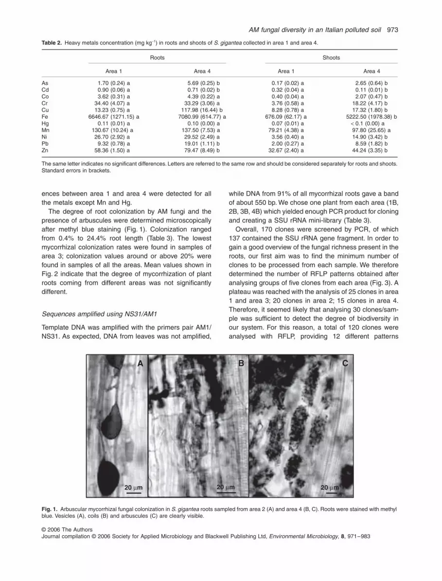
AM fungal diversity in an Italian polluted soil
973
copy 2006 The AuthorsJournal compilation copy 2006 Society for Applied Microbiology and Blackwell Publishing Ltd
Environmental Microbiology
8
971ndash983
ences between area 1 and area 4 were detected for allthe metals except Mn and Hg
The degree of root colonization by AM fungi and thepresence of arbuscules were determined microscopicallyafter methyl blue staining (Fig 1) Colonization rangedfrom 04 to 244 root length (Table 3) The lowestmycorrhizal colonization rates were found in samples ofarea 3 colonization values around or above 20 werefound in samples of all the areas Mean values shown inFig 2 indicate that the degree of mycorrhization of plantroots coming from different areas was not significantlydifferent
Sequences amplified using NS31AM1
Template DNA was amplified with the primers pair AM1NS31 As expected DNA from leaves was not amplified
while DNA from 91 of all mycorrhizal roots gave a bandof about 550 bp We chose one plant from each area (1B2B 3B 4B) which yielded enough PCR product for cloningand creating a SSU rRNA mini-library (Table 3)
Overall 170 clones were screened by PCR of which137 contained the SSU rRNA gene fragment In order togain a good overview of the fungal richness present in theroots our first aim was to find the minimum number ofclones to be processed from each sample We thereforedetermined the number of RFLP patterns obtained afteranalysing groups of five clones from each area (Fig 3) Aplateau was reached with the analysis of 25 clones in area1 and area 3 20 clones in area 2 15 clones in area 4Therefore it seemed likely that analysing 30 clonessam-ple was sufficient to detect the degree of biodiversity inour system For this reason a total of 120 clones wereanalysed with RFLP providing 12 different patterns
Table 2
Heavy metals concentration (mg kg
minus
1
) in roots and shoots of
S gigantea
collected in area 1 and area 4
Roots Shoots
Area 1 Area 4 Area 1 Area 4
As 170 (024) a 569 (025) b 017 (002) a 265 (064) bCd 090 (006) a 071 (002) b 032 (004) a 011 (001) bCo 362 (031) a 439 (022) a 040 (004) a 207 (047) bCr 3440 (407) a 3329 (306) a 376 (058) a 1822 (417) bCu 1323 (075) a 11798 (1644) b 828 (078) a 1732 (180) bFe 664667 (127115) a 708099 (61477) a 67609 (6217) a 522250 (197838) bHg 011 (001) a 010 (000) a 007 (001) a
lt
01 (000) aMn 13067 (1024) a 13750 (753) a 7921 (438) a 9780 (2565) aNi 2670 (292) a 2952 (249) a 356 (040) a 1490 (342) bPb 932 (078) a 1901 (111) b 200 (027) a 859 (182) bZn 5836 (150) a 7947 (849) b 3267 (240) a 4424 (335) b
The same letter indicates no significant differences Letters are referred to the same row and should be considered separately for roots and shootsStandard errors in brackets
Fig 1
Arbuscular mycorrhizal fungal colonization in
S gigantea
roots sampled from area 2 (A) and area 4 (B C) Roots were stained with methyl blue Vesicles (A) coils (B) and arbuscules (C) are clearly visible
A C B
20 microm 20 microm 20 microm
974
M Vallino
et al
copy 2006 The AuthorsJournal compilation copy 2006 Society for Applied Microbiology and Blackwell Publishing Ltd
Environmental Microbiology
8
971ndash983
(Fig 4A) Two to six clones for each RFLP type weresequenced for a total of 32 sequences the other 88sequences were classified by RFLP typing
RFLP type analysis on NS31AM1 amplified fragments
Figure 5 shows the frequencies and the distribution of theRFLP patterns detected in the roots of
S gigantea
in thefour areas considered in this study Of the 12 RFLP pat-terns detected RFLP2 and RFLP3 are dominant (repre-senting 23 and 22 of the analysed clones) andtogether with RFLP5 are found in all four areas RFLP4and RFLP10 are present in three areas The remainingpatterns look specific for one single area RFLP1 for area1 RFLP9 RFLP11 RFLP12 for area 3 and RFLP6RFLP7 RFLP8 for area 4 In area 1 six different RFLPpatterns are present RFLP1 and RFLP2 being predomi-nant In area 2 only four patterns were found RFLP3having the highest percentage Area 3 and area 4 haveeight and seven different patterns respectively no one ofthem being predominant
Identification of AM fungal groups
BLASTN
searches in the GenBank database showed that
all sequences have high similarity (97ndash100 identity) toAM fungi and belong to members of the
Glomeromycota
phylum Alignment of the sequences obtained in this studywith those corresponding to the closest matches fromGenBank produces a tree which reveals that thesequences belong to the groups of
Glomerales
and
Diver-sisporales
(Fig 6) No sequences were detected withinthe
Archaeosporales
or
Paraglomerales
this wasexpected because AM1 primer does not match the highlydivergent 18S-sequence of these taxa (Redecker 2000)
The phylogenetic relationships among the sequencesclearly reveal discrete sequence groups and allow us toidentify 13 potential taxonomic units (TUs) or fungal types11 belong to the
Glomeraceae
one to the
Diversispora-ceae
and one to the
Gigasporaceae
All sequence clus-ters are supported by a bootstrap
gt
75 In two clusters(acnaGlo3 acnaGlo8) two separate sequences typesappear to exist but they are not well supported by thebootstrap value nor are they distinguishable by the HinfI
Table 3
Comparison between root mycorrhization and AM1NS31PCR amplification of roots of all sampled plants of
S gigantea
M A Plant AM1NS31
Area 1 2439 1074 1A ndash1368 694 1B
+
1269 539 1C
+
1492 339 1D
+
1432 760 1E
+
1022 361 1F
+
Mean value 1504 628
Area 2 1194 789 2A
+
1575 1057 2B
+
1983 1309 2C
+
1065 613 2D
+
1955 1658 2E
+
896 589 2F
+
Mean value 1445 1003
Area 3 036 000 3A ndash2315 1219 3B +2076 1737 3C
+
330 206 3D
+
154 047 3E
+
Mean value 982 642
Area 4 1985 1649 4A
+
1758 931 4B
+
1107 883 4C
+
2014 1130 4D
+
1797 1410 4E
+
1149 661 4F
+
Mean value 1635 1111
M degree of AM fungi colonization A presence of arbuscules
+
ndash presenceabsence of amplified bandGrey shaded cells indicate plants chosen for cloning (see text)
Fig 2
Degree of AM fungal colonization (M) and presence of arbuscules (A) in roots of
S gigantea
sampled in the four areas of hill A5 Mean values and standard errors are presented Statistical analysis was done separately for M and for A The same letter indicates no significant differences
a a
a
a
b
b
b
b
0
2
4
6
8
10
12
14
16
18
20
area 1 area 2 area 3 area 4
per
cen
tag
e
M
A
Fig 3
Number of RFLP patterns obtained after HinfI and Hsp92II digestions of 5 10 15 20 25 30 clones from each area
012
3456
789
5 10 15 20 25 30number of analysed clones
To
tal n
um
ber
of
RF
LP
pat
tern
s
area 1area 2area 3area 4
AM fungal diversity in an Italian polluted soil
975
copy 2006 The AuthorsJournal compilation copy 2006 Society for Applied Microbiology and Blackwell Publishing Ltd
Environmental Microbiology
8
971ndash983
and Hsp92II RFLP patterns for this reason they are clas-sified as single type
Clades acnaGig6 acnaGlo8 acnaGlo11 includesequences with 98 100 98 identity to thesequences of
Gigaspora decipiens
strain BEG45
Glomusmosseae
strain BEG69 and
Glomus intraradices
BEG121respectively all other AM fungal types do not clusterclosely with any sequences from AM fungi in culture
Taxonomic groups are mainly consistent with theRFLP types (Fig 6) with few exceptions For examplesequence 3B34 gives a RFLP2 pattern but it does notcluster with the other sequences having this pattern(1B1 4D41 1B3) On the contrary the latter sequencescluster with clone 3B28 which has a RFLP12 patternFinally sequences 3B12 and 3B26 have the sameRFLP pattern but they cannot be assigned to a singlefungal group
Sequences amplified using ARCH1311NS8
The same DNA samples used for AM1NS31 amplification(plants 1B 2B 3B 4B) were used in nested PCR reac-tions with NS5ITS4 as the first step and ARCH1311 andNS8 as the second step All products amplified byARCH1311NS8 were sufficient for cloning A total of 84
clones were screened for the presence of the insert and41 clones resulted positive unfortunately no single cloneof area 4 was positive
All 41 clones were then analysed with RFLP providingeight different patterns (Fig 4B) One to four clones foreach RFLP type were sequenced for a total of 11sequences
BLASTN
searches in the GenBank database showed thata small percentage of the analysed sequences belong to
Paraglomerales
Only the sequences corresponding tothe RFLPc gave high homology (94 of identity) with
Glomus occultum
and they were only present in area 3All the other sequences gave homology with
Ascomycota
(RLFPd and RFLPh ndash 39)
Basidiomycota
(RFLPf ndash 2)and plants (RFLPa RFLPb RFLPe and RFLPg ndash 44)Therefore out of the 41 clones 44 are of plant origin41 are of no AM fungal origin and 15 belong to
Paraglomeraceae
Discussion
ACNA activity poured into the surrounding area a hugeamount of pollutants which strongly inhibited any form oflife This phenomenon was evident in the core of the hillswhere only a very small number of bacterial species
Fig 4
RFLP patterns of SSU rRNA PCR products obtained from
S gigantea
rootsA AM1NS31 amplification H1ndashH9 restriction pattern obtained with enzyme HinfI P1ndashP7 restriction pattern obtained with enzyme Hsp92IIB ARCH1311NS8 amplification HandashHh restriction pattern obtained with enzyme HinfI PandashPb restriction pattern obtained with enzyme Hsp92II
RFLP patternsH1-P1 RFLP1H2-P2 RFLP2H2-P1 RFLP3H3-P1 RFLP4H4-P6 RFLP5H5-P1 RFLP6H6-P4 RFLP7H4-P5 RFLP8H9-P7 RFLP9H7-P5 RFLP10H8-P5 RFLP11H2-P7 RFLP12
RFLP patternsHa-Pa RFLPaHb-Pa RFLPbHc-Pb RFLPcHd-Pa RFLPdHe-Pa RFLPeHf-Pa RFLPfHg-Pa RFLPgHh-Pa RFLPh
H1 H2 H3 H4 H5 H6 H7 H8 H9
244 nt190 nt
280 nt334 nt
141 nt
50 nt
25 nt
pUC18
P1 P2 P4 P5 P6 P7
290 nt
170 nt
90 nt116 nt
pUC18
Pa Pb
450 nt
170 nt
90 nt
pUC18
Ha Hb Hc Hd He Hf Hg Hh
241 nt199 nt
331 nt
165 nt
107 nt
80 nt
pUC18
A BNS31AM1 amplified fragments ARCH1311NS8 amplified fragments
976
M Vallino
et al
copy 2006 The AuthorsJournal compilation copy 2006 Society for Applied Microbiology and Blackwell Publishing Ltd
Environmental Microbiology
8
971ndash983
occurred (Avidano
et al
2005) The ACNA hills have rep-resented a very interesting situation to investigatebecause they offered the opportunity to evaluate thedynamics of plants microbes and animals starting from aground zero level Among the studies found in literatureon lsquobiological desertsrsquo resulting from industrial wastes andemissions there are cases of investigations in whichbesides human plant and animal health also the micro-bial communities had been considered (for example theareas contaminated by metal smelters in Finland North-ern Sweden and Canada ndash Gunn
et al
1995 Pennanen
et al
1996 Palmborg
et al
1998) However to ourknowledge only few of them analysed the microbial com-ponent represented by mycorrhizal fungi (for example
Niepolomice Forest ndash Turnau 1988 Kapusta
et al
2004Chernobyl ndash Zhdanova
et al
2004) Our work presents asurvey of AM colonization in A5 hill using morphologicaland molecular methods We could not extend our analysisin time or in number of plant species because furthersampling was not allowed Notwithstanding this our inves-tigation is one of the few attempts to evaluate the capacityof AM fungi to colonize plants and to establish an under-ground network in an industrial dump
We chose to carry out our analysis on
S gigantea
because it was the only plant species that could befound in all four sampling areas
Solidago gigantea
(
Asteraceae
) is a rhizomatous perennial herb native toNorthern America and introduced in Europe about
Fig 5 A Distribution and frequencies in the four areas of A5 hill of the 12 RFLP patterns obtained after HinfI and Hsp92II digestions of 120 NS31AM1 amplified fragments The last row is the legend for the below pie-chartsB Approximate tri-dimensional map of A5 hill showing the location of the four analysed areas (area 1 area 3 area 4 ovals area 2 perimeter) The pie-charts associated to each area refer to the RFLP types the legend for the segments of the pie-charts is presented in the last row of the above table
RFLP1 RFLP2 RFLP3 RFLP4 RFLP5 RFLP6 RFLP7 RFLP8 RFLP9 RFLP10 RFLP11 RFLP1222313191 AERA
AREA 2 4 16 4 2155155443 AERA
AREA 4 7 5 1 4 2 9 1total 9 28 26 12 10 4 2 9 1 8 5 1
A
AREA 2AREA 3
AREA 4
B
AREA 1
AM fungal diversity in an Italian polluted soil 977
copy 2006 The AuthorsJournal compilation copy 2006 Society for Applied Microbiology and Blackwell Publishing Ltd Environmental Microbiology 8 971ndash983
250 years ago (Weber and Jakobs 2005) In a study onthe invasiveness of this species Jakobs and colleagues(2004) found that in Europe it was present both in wetand dry habitats fewer population were directly adjacentto rivers and lakes and more were in disturbed areasand besides roads This species turned out to be a goodrepresentative to work on as roots were clear and easyto handle
Chemical analysis of soil sampled in the S gigantearhizosphere revealed that all areas of A5 hill had a highconcentration of heavy metals and that area 4 was themost polluted one Heavy metal concentrations in roots ofS gigantea was much lower than concentrations in soilexcept for Cd which was about three times higher in rootsthan in soil Metal content in plants of area 4 was alwaysequal to or higher than metal content in plants of area 1
Fig 6 Neighbour-joining tree showing relationships between NS31-AM1 rDNA sequences from S gigantea and reference sequences from GenBank Corallochytrium limacisporum a putative choanoflagellate (Cavalier-Smith and Allsopp 1996) was used as outgroup All bootstrap values gt50 are shown (100 replicates) Individual clones are identified by site (first number 1 2 3 4) and clone number (example A43 B6 D1) On the right group identifiers (for example acnaGlo7) are compared with RFLP types (see text)
4D14D51
AF074371 Glomus spGlo5AJ276087 Glomus lamellosumAJ276080 Glomus claroideum
AF481661 Glomus spGlo264B6
4B9AJ567845 Gigaspora margarita
U96146 Gigaspora decipiensAY129642 Scutellospora spScut
Z14010 Gigaspora giganteaX58726 Gigaspora rosea
1B21B7
Y17644 Glomus etunicatumY17650 Glomus spurcum
AJ315524 Glomus spssGloyelp11B33
3B432B11
2B74D6
2A43AY129625 Glomus spGlo18
AJ418867 Glomus spMO G43B34
AY512347 Glomus spGlo48AF480158 Glomus sp5014b25Lla
3B391B1
AF074353 Glomus spGlo44D41
1B33B28
AJ418889 Glomus spMO G9AF437660 Glomus spGlo13AJ496108 Glomus spMO G12
2B122B13AF074370 Glomus spGlo7
AJ418880 Glomus spMO G61B132B14D9
3B1AJ506090 Glomus constrictum
AJ505813 Glomus viscosum4B3Z14007 Glomus mosseae
4D34D7AY243568 Glomus spGlo1BU96141 Glomus mosseae BEG69
1B313B83B11
AY129607 Glomus spGlo10AJ536822 Glomus intraradices
3B12AJ496053 Glomus spMO G3
Y17640 Glomus fasciculatumX58725 Glomus intraradices
3B26AJ309462 Glomus spGlo8AF481660 Glomus spGlo23
AJ496059 Glomus spMO G13
L42528 Corallochytriumlimacisporum
acnaGlo7
acnaGig6
acnaDiv1
acnaGlo3
acnaGlo2
acnaGlo1
acnaGlo2
acnaGlo4
acnaGlo5
acnaGlo8
acnaGlo11
acnaGlo12a
acnaGlo12b
RFLP7
RFLP6
RFLP11
RFLP10
RFLP8
RFLP5
RFLP4
RFLP12
RFLP2
RFLP2
RFLP9
RFLP3
RFLP1
94100
100
94
100
100
100
76
70
57
80
5279
92
100
100
100
100
6695
54
70
69
78
57
94
10071
81
100
66
98
52
59
88
0005 substitutionssite
978 M Vallino et al
copy 2006 The AuthorsJournal compilation copy 2006 Society for Applied Microbiology and Blackwell Publishing Ltd Environmental Microbiology 8 971ndash983
Cd being again an exception in fact Cd concentration inboth roots and shoots of area 4 was lower than that ofarea 1 Significant differences in soil metal contentbetween area 1 and area 4 was not mirrored in root metalcontent (Cu was the only case where significant differ-ences were found in roots and soil) From this we con-clude that metal concentration in plant roots was notnecessarily related to soil metal concentration asobserved also by Ortiz and Alcaniz (2006)
Heavy metal concentration in S gigantea shoots wasalways lower than in roots (except Hg) suggesting a lowlevel of translocation between plant organs However thecontent of some metals in shoots of plant from area 4 washigher than area 1 and fell beyond the normal backgroundranges detected in plants growing in unpolluted regions(Djingova et al 2004) This suggests that in area 4 metaltranslocation increased For As Pb Cu Zn it was prob-ably a consequence of a higher root concentration In thecase of Co Cr Fe Ni in which metal content was signif-icantly different in the soil of the two areas and in shootsbut not in roots we postulate that metals reached a sat-uration point in roots and were therefore transported tothe aerial part of the plants
The morphology of AMF colonization within the roots ofS gigantea was as usual the presence of arbusculessuggests that the symbiosis was active Notwithstandingthis the values of colonization were rather low It is knownthat heavy metals can delay reduce and even eliminatespore germination and AM colonization del Val and col-leagues (1999) showed that the total number of AMFspores correlated negatively with the metal content of thesoil However in previous studies (Griffioen et al 1994Weissenhorn and Leyval 1994) no correlation was foundbetween the concentration of metals such as Cu Cd Znand AMF population Furthermore Pawlowska and col-leagues (1996) found a relatively high rate of mycorrhizalcolonization in plants growing in very polluted soils In ourcase it is noteworthy that the many pollutants did notaffect fungal morphogenesis once the fungus was insidethe root By contrast the low success of infection asmeasured by the number of colonized root fragmentsmakes us wonder whether the spore could be directlyaffected by pollutants in the rhizosphere as here they aredirectly exposed to them
As root colonization had an even distribution in almostall the plants we were confident in taking random frag-ments of roots to be used for molecular analysis insteadof selecting them under the microscope As expectedPCR results were positive for almost all samples In orderto minimize the number of clones to be processed and yetto obtain a picture of the fungal diversity within the rootsas close as possible to the real one RFLP analysis on agrowing number of sequences was performed until reach-ing saturation of the different RFLP patterns Saturation
was reached after the digestion of 25 sequences for eacharea Thus we concluded that analysing a total of 120clones was sufficient to evaluate the AM fungal speciesrichness This result is consistent with the work ofLandeweert and colleagues (2003) on biodiversity of ecto-mycorrhizal fungi in soil in which by applying a statisticalmethod they found that the most common species canbe detected when 30 clonessample are analysed
The primer combination NS31-AM1 is targeted at aregion of the SSU and amplifies most but not all gloma-lean fungi It has been used to detect the presence offungal sequences from the four families of GlomeraceaeAcaulosporaceae Diversisporaceae and Gigasporaceae(order Glomales and Diversisporales) in different ecosys-tems such as agricultural sites (Helgason et al 1998Daniell et al 2001) broadleaved wood (Helgason et al1998) seminatural woodland (Helgason et al 1999)tropical forest (Husband et al 2002) and grassland (Van-denkoornhuyse et al 2002) Recently it has been discov-ered that certain species which have in the past beenconsidered to belong to the genera Glomus or Acau-lospora on the basis of their morphology have SSUsequences that are much more divergent For these fungitwo new orders have been suggested Archaeosporalesand Paraglomerales (Morton and Redecker 2001) Thecharacterization of these ancestral lineages (Redecker2000) showed that the AM1 primer sequence is not appro-priate to detect such deeply divergent clades ARCH1311designed by Redecker (2000) is the first specific primerfor these lineages Therefore by using the two primerpairs AM1-NS31 and ARCH1311-NS8 in our work wecould reliably detect and identify all currently known AMfungi while discriminating against other fungi
Only a small percentage of the sequences obtainedafter amplification with ARCH1311 and NS8 belong toParaglomerales all the others are of plant or non-AMfungi origin The use of the same primer pair on the DNAof liverworts exerted similar results (E Lumini perscomm) Problems of primers specificity were alsoencountered by Wubet and colleagues (2003ab) whoamplified a number of AM fungal types belonging to theGlomeraceae using ARCH1311 in combination with ITS4on mycorrhizal roots of Prunus africana from forests ofEthiopia
The combination of BLAST searches and phylogeneticanalysis allowed us to assign the fungal sequences iso-lated from S gigantea roots to 14 glomeralean groupsTwelve groups belong to the genus Glomus one toGigaspora and one to Paraglomus The dominance of thegenus Glomus is not new Predominance of the Glomer-aceae has also been reported for other ecosystems suchas agricultural sites (Daniell et al 2001) or tropical forests(Onguene and Kuyper 2001 Husband et al 2002 Wubetet al 2003b) Daniell and colleagues (2001) argued that
AM fungal diversity in an Italian polluted soil 979
copy 2006 The AuthorsJournal compilation copy 2006 Society for Applied Microbiology and Blackwell Publishing Ltd Environmental Microbiology 8 971ndash983
Glomus types dominate the colonization of arable cropsbecause Glomus species are better adapted to disturbedenvironments where their high sporulation rates mayenable them to recover more readily Whitfield and col-leagues (2004) drew the same conclusion from the pre-dominance of Glomus species in the metal-contaminatedsoils along the River South Tyne (UK) which is regularlyaffected by flooding which presumably causes major soildisturbance One possible reason why Glomus speciesare dominant also in an ecosystem disturbed by chemicalpollution may be found in the ability of the Glomeraceaeto colonize via fragments of mycelium or mycorrhizal rootspieces while Gigasporaceae are only capable of propa-gation via spores (Biermann and Linderman 1983Daniell et al 2001) Pawlowska and Charvat (2004) sug-gested that hyphal extension from colonized root frag-ments is a better predictor of an AM fungal performanceunder metal stress than spore germination and that AMfungi are able to survive and propagate in metal-pollutedenvironments by thriving in relatively uncontaminatedmicrosites If this is the case we may postulate that rootsonce they are colonized function as safe microenviron-ment and as inoculum for Glomus species which thusbecome more competitive in infecting roots other thanGigasporaceae
Three taxonomic units were found in all four areasinvestigated suggesting that dominant fungi formedmycelial networks with an extension of up to 4000 m2 Twotaxonomic units were found in three areas and nine werefound only in single areas showing a patchy distributionof AM fungal population also described in agriculturalsystems (Stukenbrock and Rosendahl 2005) and inundisturbed vegetation (Rosendahl and Stukenbrock2004)
Interestingly in the most polluted area 4 the most abun-dant fungal type was acnaGlo8 which can be assignedto G mosseae In morphological and molecular analysisof roots of plants colonizing zinc wastes Turnau and col-leagues (2001) showed that G mosseae was the onlyfungus which was always present in roots fragments butthe authors cannot say whether G mosseae is a heavymetal-tolerant species
In Helgason and colleagues (1999) 141 analysedclones from bluebell roots growing in seminatural wood-land gave eight different sequence groups In morecomplex works Helgason and colleagues (1998) andDaniell and colleagues (2001) detected 6 and 10 TUsrespectively analysing the AM fungal community in agri-cultural sites from four different plant species In contrasta much greater diversity was reported for dry and tropicalforests (Husband et al 2002 Wubet et al 2003b) andgrassland (Vandenkoornhuyse et al 2002) in which 2330 24 fungal types respectively were detected from twodifferent plant species Whitfield and colleagues (2004)
compared AM colonization in roots of Thymus polytrichusgrowing in sites with a long-term history of heavy metalcontamination they found 19 different AM fungal SSUsequence types and they considered this number an indexof relatively large AM species diversity Also Ridgewayand Fitter (2003) found that the variety of AM fungal typesgrowing in UK contaminated soils was surprisingly highand at a similar level to some natural woodlands andgrasslands Our data are in accordance with these twolatter works in fact the isolation of 14 TUs from the rootsof S gigantea indicates a good richness of AM fungi inthe cover-soil of hill A5 The level of AM fungal biodiversityis not mirrored by the degree of biodiversity of plantsgrowing on the hill which appears low (Andreucci et al2006) Therefore in this case study fungal presence andbiodiversity do not seem to be the key factor that promotesplant variety in a recovering site AM fungi may be anobligate but not exclusive requisite in a re-vegetationprogramme
In conclusion our work based on a combination ofchemical morphological and molecular analyses pro-vides useful parameters for evaluating the health statusof a contaminated site and the validity of a remediationprogramme It demonstrates in fact how a remediationprotocol based on cover-soil has been efficient in restoringa community of AM fungi which have been tolerantenough to proliferate in a still contaminated soil
Experimental procedures
Sampling
The investigation was carried out in ACNA (Cengio SavonaItaly) an ex-industrial site at 402 m above sea level in whicha very serious degree of contamination by several pollutantsis present both in the soils and the surface waters In factfrom 1929 to the end of its activity ACNA produced anddiscarded in the surrounding area phenols aromatic aminesdioxins furans naphtalensulfonics nitrobenzenes aromaticand aliphatic alogenate compounds PAH heavy metals(Canepa 2002) we focused our attention on the latter
All samples were collected in March 2003 from the hillnumber A5 located near the Bormida river in a securityzone inside the factory with no access permission (exceptwith special pass) and particular safety rules (helmets pro-tective clothing gloves masks) This hill consisted of indus-trial wastes derived from the ACNA activity about 25 yearsago during a first step of decontamination activity the hillwas covered with 1 m of cover-soil coming from a non-pol-luted area As expected re-vegetation eventually occurredand on the base of the current floristic aspect four areashave been identified on the hill surface (Andreucci et al2006) Area 1 is a meadow covered mainly by graminaceousspecies area 2 is the edge of the hill in which trees andshrubs are present area 3 is a meadow covered by a mix-ture of species but mainly legumes and composites area 4is the only spot in which the deeper part of the hill has been
980 M Vallino et al
copy 2006 The AuthorsJournal compilation copy 2006 Society for Applied Microbiology and Blackwell Publishing Ltd Environmental Microbiology 8 971ndash983
excavated and brought to the surface bringing upcontaminants
During a first survey it was concluded that S giganteaAiton was the only grass that was present in all the fourchosen areas and involved in AM symbiosis it was thereforeselected as reference host Soil blocks with a diameter anddepth of about 15 cm including five to six plants were exca-vated and stored in polyethylene bags for transport to thelaboratory where the plants were carefully washed to sepa-rate fine roots from the soil Roots were then briefly rinsedquickly dried on paper and used partly for chemical partlyfor morphological and partly for molecular analysis
A total of 23 plants of S gigantea Aiton were sampled sixfrom areas 1 2 4 and five from area 3
Chemical analysis samples preparation and heavy metals determination
Soil samples were oven dried at 40degC for 2 days thencrushed and sieved to 2 mm through a nylon sieve Plantsamples were carefully washed with distilled water dividedin shoot and root and oven dried at 80degC for 3 days Thenthey were ground to powder with mortar and pestle 05 g ofeach sample was digested in 6 ml of 65 nitric acid using amicrowave oven (ETHOS 900 Microwave Labstation com-bined with MILESTONE Microwave Laboratory System Ter-minal 240) All glassware used for digestion and stocksolutions was soaked in 20 nitric acid (vv) overnight andrinsed three times with MilliQ reagent grade water
Total heavy metal concentrations were determined afterdigestion according to Italian Legislation (DM 4711999)Digested samples were diluted with purified water (MilliQreagent grade) and then analysed by ICP-MS (X-series ICP-MS ThermoElemental) Data obtained were processedwith the ThermoElemental-PlasmaLab 213 software NISTtomato leaves standard (1573a) and chemical standardswere used to calibrate the instrument
Results were statistically analysed by one-way ANOVA andFisherrsquos test using STATVIEW 49 program Differences wereconsidered significant when P-value was lt005
Morphological analysis
Roots were cleared in 10 KOH at 60degC for 30 min thenstained with 1 methyl blue in lactic acid and the intensity ofroots cortex colonization by AM fungi (M) and the presenceof arbuscules (A) was determined as described by Trou-velot and colleagues (1986) Results were statistically anal-ysed as for chemical analysis
DNA extraction and PCR
Total DNA was extracted from a leaf piece of one plant andfour pieces (1ndash2 cm each) of mycorrhizal roots of each plantaccording to the protocol described in Perotto and colleagues(1996) The DNA was resuspended in 20 microl of water Severaldilutions of extracted DNA (110 150 1250 11000) weretested with universal primers ITS1 and ITS4 (White et al1990) to verify amplifiability PCR reactions were performedin 25 microl reactions containing 01 microg microlminus1 BSA 015 mM
dNTPs 17 pmol of each primer 07 units of RedTaq DNApolymerase (Sigma) and the supplied buffer (PCR conditions3 min at 95degC for one cycle 45 s at 94degC 45 s at 55degC 45 sat 72degC for 35 cycles 10 min at 72degC for one cycle)
Partial ribosomal SSU DNA fragments were then amplifiedusing 07 units of RedTaq DNA polymerase (Sigma) and twodifferent sets of primers
One set consisted of a universal eukaryotic primer NS31(Simon et al 1992) and the primer AM1 designed to amplifyAM fungal SSU sequences but not plant sequences (Helga-son et al 1998) Reactions were performed using 01 microgmicrolminus1 BSA 02 mM dNTPs 12 pmol of each primer and thesupplied reaction buffer to a final volume of 25 microl (PCR con-ditions 95degC for 5 min then 35 cycles at 94degC for 1 min 58degCfor 1 min 72degC for 2 min then 72degC for 10 min)
Concerning the other primer set a two-step procedure(nested PCR) was conducted The first amplification withthe universal primers NS5 and ITS4 (White et al 1990)was performed as described above for AM1NS31 with1 mM MgCl2 instead of BSA and an annealing temperatureof 56degC Aliquots of 5 microl were run on agarose gel to esti-mate the quantity of PCR product When no PCR productwas visible the reactions were diluted 120 and 150 wherebands were visible the reactions were diluted 150 and1100 These dilutions were then used as template for thesecond PCR step The second step was conducted with thespecific primer ARCH1311 (Redecker 2000) in combinationwith NS8 (White et al 1990) Reactions were performedusing 1 mM MgCl2 02 mM dNTPs 06 microM of each primerand the supplied reaction buffer to a final volume of 25 microl(PCR conditions 95degC for 5 min then 35 cycles at 94degC for1 min 58degC for 1 min 72degC for 1 min then 72degC for10 min)
All the PCR reactions were run on a Perkin Elmer CetusDNA Thermal Cycler
Cloning and sequencing
PCR products were purified following the QIAquick protocol(Qiagen) Cloning was done using the pGEM-T vector system(Promega) and transformed into Escherichia coli (Xl1 blue)Within each resulting SSU rRNA gene library putative posi-tive transformants were screened using a second NS31AM1or ARCH1311NS8 amplification with the same conditions asdescribed above Positive clones from each sample weretested for RFLP by independent digestion with HinfI andHsp92II according to the manufacturerrsquos instructions(Promega) and analysed on 3 agarose (in TBE) gel elec-trophoresis Examples of each RFLP type were chosen forsequencing They were either re-amplified using the vectorprimers SP6 and T7 and purified using the QIAquick protocol(Qiagen) or grown in liquid culture and the plasmid extractedusing the QIAprep Spin Miniprep kit (Qiagen) The sequenc-ing was done by GeneLab (Roma Italy) using the universalprimers SP6 and T7
Sequence editing was done using the programSEQUENCHER version 414 (Gene Codes Corporation) Thesequences have been deposited at the National Centre forBiotechnology Information (NCBI) GenBank (httpwwwncbinlmnihgov) under Accession numbers DQ164806ndashDQ164837
AM fungal diversity in an Italian polluted soil 981
copy 2006 The AuthorsJournal compilation copy 2006 Society for Applied Microbiology and Blackwell Publishing Ltd Environmental Microbiology 8 971ndash983
Phylogenetical analysis
Sequence similarities were determined using the BLASTN
sequence similarity search tool (Altschul et al 1997) pro-vided by GenBank Phylogenetic analysis was carried out onthe sequences obtained in this study and those correspond-ing to the closest matches from GenBank Sequences werealigned with other published glomeralean sequences usingthe program CLUSTALX (Thompson et al 1997) and thealignment was adjusted manually in GeneDoc (Nicholas andNicholas 1997) We ran neighbour-joining (NJ) phylogeneticanalyses (Saitou and Nei 1987) with the program PAUP408b(Swofford 2002) using the default parameters The putativechoanozoan Corallochytrium limacisporum a close relativeof fungi (Cavalier-Smith and Allsopp 1996) was chosen asoutgroup and used to root the trees
Acknowledgements
Many thanks to Claude Murat (University of Torino) for thephylogenetic analysis the interpretation of the data and help-ful suggestions thanks to Daniela Rullo and Mohamed Al-Yahyarsquoei for collaboration in molecular analyses Thanks toCaroline Gutjahr (University of Torino) and David Milne forthe critical reading of the manuscript as well as to the twoanonymous reviewers for their helpful comments Theauthors gratefully acknowledge the assistance of StefanoLeoni ndash ACNArsquos Temporary Administrator ndash and Gian PaoloCossa ndash Micro-pollutants Pole of ARPA Piemonte (Alessan-dria) M Vallino acknowledges the financial support fromUniversity of Torino and University of Piemonte OrientaleResearch was funded by the Italian Ministry of the Environ-ment (Valle Bormida Remediation Program OM 29861999) CEBIOVEM program (DM 1932003) Cassa di Ris-parmio di Torino (CRT) and IPP-CNR (Biodiversity Project ndashTorino)
References
Altschul SF Madden TL Schaumlffer AA Zhang JZhang Z Miller W and Lipman DJ (1997) GappedBLAST and PSI-BLAST a new generation of protein databasesearch programs Nucleic Acids Res 25 3389ndash3402
Andreucci F Barbato R Massa N and Berta G (2006)Phytosociological phenological and photosynthetic analy-sis on the vegetal component of a highly polluted site PlantBiosyst (in press)
Avidano L Gamalero E Cossa GP and Carraro E(2005) Characterization of soil health in an Italian pollutedsite by using microorganisms as bioindicators Appl SoilEcol 30 21ndash33
Biermann B and Linderman RG (1983) Use of vesicular-arbuscular mycorrhizal roots intraradical vesicles andextraradical vesicles as inoculum New Phytol 95 97ndash105
Bonassi S Merlo F Pearce N and Puntoni R (1989)Bladder cancer and occupational exposure to polycyclicaromatic hydrocarbons Int J Cancer 44 648ndash651
Canepa P (2002) Sisifo Project Results 1st Year Activity2001ndash02 Soil Remediation Series No 1 Marghera (VE)Italy INCA ISBN 88-88214-02-X
Cavalier-Smith T and Allsopp MTEP (1996) Coral-lochytrium an enigmatic non-flagellate protozoan relatedto choanoflagellates Eur J Protistol 32 306ndash310
Clapp JP Young JPW Merryweather JW and FitterAH (1995) Diversity of fungal symbionts in arbuscularmycorrhias from a natural community New Phytol 130259ndash265
Conte P Agretto A Spaccini R and Piccolo A (2005)Soil remediation humic acids as natural surfactants in thewashings of highly contaminated soils Environ Pollut 135515ndash522
Daniell TJ Husband R Fitter AH and Young JPW(2001) Molecular diversity of arbuscular mycorrhizal fungicolonising arable crops FEMS Microbiol Ecol 36 203ndash209
Djingova R Kuleff I and Market B (2004) Chemical fin-gerprinting of plants Ecol Res 19 3ndash11
Esposito A Del Borghi A and Veglio F (2002) Investiga-tion of naphtalene sulfonate compounds sorption in a soilartificially contaminated using batch and column assaysWaste Manag 22 937ndash943
Griffioen WAJ Ietswaart JH and Ernst WHO (1994)Mycorrhizal infection of an Agrostis capillaris population ona copper contaminated soil Plant Soil 158 83ndash89
Gunn J Keller W Negusanti J Potvin R Beckett Pand Winterhalder K (1995) Ecosystem recovery afteremission reductions Sudbury Canada Water Air Soil Poll85 1783ndash1788
Hauptmanns U (2005) A risk-based approach to land-useplanning J Hazard Mater 125 1ndash9
van der Heijden MGA Klironomos JN Ursic M Mou-toglis P Streitwolf-Engel R Boller T et al (1998)Mycorrhizal fungal diversity determines plant biodiversityecosystem variability and productivity Nature 396 69ndash72
Helgason T Daniell TJ Husband R Fitter AH andYoung JPW (1998) Ploughing up the wood-wide webNature 394 431
Helgason T Fitter AH and Young JPW (1999) Molec-ular diversity of arbuscular mycorrhizal fungi colonisingHyacinthoides non-scripta (Bluebell) in a seminaturalwoodland Mol Ecol 8 659ndash666
Husband R Herre EA Turner SL Gallery R andYoung JPW (2002) Molecular diversity of arbuscularmycorrhizal fungi and patterns of host association overtime and space in a tropical forest Mol Ecol 11 2669ndash2678
Jakobs G Weber E and Edwards PJ (2004) Introducedplants of the invasive Solidago gigantea (Asteraceae) arelarger and grow denser than conspecifics in the nativerange Divers Distrib 10 11ndash19
Kapusta P Szarek-Lukaszewska G and Kiszka J (2004)Spatial analysis of lichen species richness in a disturbedecosystem (Niepolomice Forest S Poland) Lichenologist36 249ndash260
Landeweert R Leeflang P Kuyper TW Hoffland ERosling A Wernars K and Smit E (2003) Molecularidentification of ectomycorrhizal mycelium in soil horizonsAppl Environ Microbiol 69 327ndash333
Martin F (2001) Frontiers in molecular mycorrhizal researchGenes loci dots and spins New Phytol 150 499ndash507
982 M Vallino et al
copy 2006 The AuthorsJournal compilation copy 2006 Society for Applied Microbiology and Blackwell Publishing Ltd Environmental Microbiology 8 971ndash983
Morton JB and Redecker D (2001) Two new families ofGlomales Archaeosporaceae and Paraglomaceae withtwo new genera Archaeospora and Paraglomus based onconcordant molecular and morphological charactersMycologia 93 181ndash195
Newsham KK Fitter AH and Watkinson AR (1995)Multifunctionality and biodiversity in arbuscular mycorrhi-zas Trends Ecol Evol 10 407ndash411
Nicholas KB and Nicholas HB Jr (1997) GeneDoc anal-ysis and visualization of genetic variation URL httpwwwpscedubiomedgenedoc
Onguene NA and Kuyper TW (2001) Mycorrhizal asso-ciations in the rain forest of south Cameroon Forest EcolManag 140 277ndash287
Ortiz O and Alcaniz JM (2006) Bioaccumulation of heavymetals in Dactylis glomerata L growing in a calcareous soilamended with sewage sludge Bioresour Technol 97 545ndash552
Palmborg C Nordgren A and Baath E (1998) Multivariatemodelling of soil microbial variables in forest soil contami-nated by heavy metals using wet chemical analyses andpyrolysis GCMS Soil Biol Biochem 30 345ndash357
Pawlowska TE Blazkowski J and Ruhling A (1996) Themycorrhizal status of plants colonizing a calamine spoilmound in southern Poland Mycorrhiza 6 499ndash505
Pawlowska TE and Charvat I (2004) Heavy-metal stressand developmental patterns of arbuscular mycorrhizalfungi Appl Environ Microbiol 70 6643ndash6649
Pennanen T Frostegard A Fritze H and Baath E (1996)Phospholipid fatty acid composition and heavy metal toler-ance of soil microbial communities along two heavy metal-polluted gradients in coniferous forests Appl EnvironMicrobiol 62 420ndash428
Perotto S Actis-Perino E Perugini J and Bonfante P(1996) Molecular diversity of fungi from ericoid mycorrhizalroots Mol Ecol 5 123ndash131
Puntoni R Bolognesi C Bonassi S Merlo F Mari AMerialdo G and Santi L (1988) Cancer risk evaluationin an area with high density of chemical plants Ann N YAcad Sci 534 808ndash816
Redecker D (2000) Specific PCR primers to identify arbus-cular mycorrhizal fungi within colonized roots Mycorrhiza10 73ndash80
Ridgeway K and Fitter A (2003) Polluted soils arenrsquot asdead as they seem Planet Earth Spring 13
Rosendahl S and Stukenbrock EH (2004) Communitystructure of arbuscular mycorrhizal fungi in undisturbedvegetation revealed by analyses of LSU rDNA sequencesMol Ecol 13 3179ndash3186
Saitou N and Nei M (1987) The Neighbor-Joining methoda new method for reconstructing phylogenetic trees MolBiol Evol 4 406ndash425
Sanders IA (2004) Plant and arbuscular mycorrhizal fungaldiversity ndash are we looking at the relevant level of diversityand are we using the right techniques New Phytol 164415ndash418
Schuumlszligler A Schwarzott D and Walker C (2001) A newfungal phylum the Glomeromycota phylogeny and evolu-tion Mycol Res 105 1413ndash1421
Simon LM Lalonde TD and Bruns TD (1992) Specificamplification of 18S fungal ribosomal genes from vesicular
arbuscular endomycorrhizal fungi colonising roots ApplEnviron Microbiol 58 291ndash295
Smith SE and Read DJ (1997) Mycorrhizal SymbiosisLondon UK Academic Press
Stukenbrock EH and Rosendahl S (2005) Clonal diversityand population genetic structure of arbuscular mycorrhizalfungi (Glomus spp) studied by multilocus genotyping ofsingle spores Mol Ecol 14 743ndash752
Swofford DL (2002) PAUP Phylogenetic Analysis UsingParsimony (and Other Methods) Version 408b10 forMachintosh Sunderland MA USA Sinauer Associates
Thompson JD Gibson TJ Plewniak F Jeanmougin Fand Higgins DG (1997) The ClustalX windows interfaceflexible strategies for multiple sequence alignment aidedby quality analysis tools Nucleic Acids Res 24 4876ndash4882
Trouvelot A Kough JL and Gianinazzi-Pearson V(1986) Mesure du taux de mycorhization VA drsquoun systegravemeradiculaire Recherche de meacutethodes drsquoestimation ayantune signification fonctionnelle In Physiological and Gen-etical Aspects of Mycorrhizae Gianinazzi-Pearson Vand Gianinazzi S (eds) Paris France INRA Press pp217ndash221
Turnau K (1988) The influence of industrial dust on themycoflora of Pino-Quercetum forest near Cracow III Theinfluence of industrial dust on the mycorrhiza of Vaccin-ium myrtillus Sci Papers of Krakow Agric Acad 226 135ndash145
Turnau K Ryszka P Gianinazzi-Pearson V and vanTuinen D (2001) Identification of arbuscular mycorrhizalfungi in soils and roots of plants colonizing zinc wastes insouthern Poland Mycorrhiza 10 169ndash174
del Val C Barea JM and Azcon-Aguilar C (1999) Diver-sity of arbuscular mycorrhizal fungus populations in heavy-metal-contaminated soils Appl Environ Microbiol 65 718ndash723
Vandenkoornhuyse P Husband R Daniell TJ WatsonIJ Duck JM Fitter AH and Young JPW (2002)Arbuscular mycorrhizal community composition associatedwith two plant species in a grassland ecosystem Mol Ecol11 1555ndash1564
Weber E and Jakobs G (2005) Biological flora of centralEurope Solidago gigantea Aiton Flora 200 109ndash118
Weissenhorn I and Leyval C (1994) Differential toleranceto Cd and Zn of arbuscular mycorrhizal fungal spores iso-lated from heavy metal-polluted and unpolluted soils PlantSoil 167 189ndash196
White TJ Bruns T Lee S and Taylor J (1990) Amplifi-cation and direct sequencing of fungal ribosomal RNAgenes for phylogenetics In PCR ndash Protocols and Applica-tions ndash A Laboratory Manual Innis MA Gelfand DHSninsky JJ and White TJ (eds) San Diego CA USAAcademic pp 315ndash322
Whitfield L Richards AJ and Rimmer DL (2004) Rela-tionships between soil heavy metal concentration and myc-orrhizal colonisation in Thymus polytrichus in northernEngland Mycorrhiza 14 55ndash62
Wubet T Weiszlig M Kottke I and Oberwinkler F (2003a)Morphology and molecular diversity of arbuscular mycor-rhizal fungi in wild and cultivated yew (Taxus baccata) CanJ Bot 81 255ndash266
AM fungal diversity in an Italian polluted soil 983
copy 2006 The AuthorsJournal compilation copy 2006 Society for Applied Microbiology and Blackwell Publishing Ltd Environmental Microbiology 8 971ndash983
Wubet T Weiszlig M Kottke I Teketay D and OberwinklerF (2003b) Molecular diversity of arbuscular mycorrhizalfungi in Prunus africana an endangered medicinal treespecies in dry Afromontane forests of Ethiopia New Phytol161 517ndash528
Zhdanova NN Tugay T Dighton J Zheltonozhsky Vand McDermott P (2004) Ionizing radiation attracts soilfungi Mycol Res 108 1089ndash1096
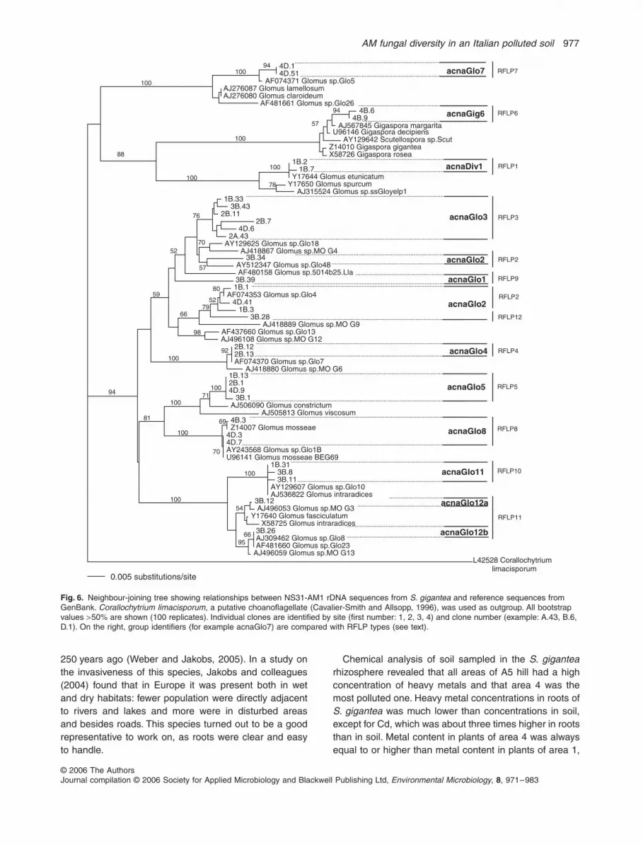