an arabidopsis mutant with altered hypersensitive response to xanthomonas campestris pv. campestris,...
TRANSCRIPT

MOLECULAR PLANT PATHOLOGY
(2004)
5
(5 ) , 453–464 DOI : 10 .1111/ J .1364-3703.2004.00245.X
© 2004 BLACKWELL PUBL ISH ING LTD
453
Blackwell Publishing, Ltd.
An
Arabidopsis
mutant with altered hypersensitive response to
Xanthomonas campestris
pv.
campestris
,
hxc1
, displays a complex pathophenotype
MAR IE LUMMERZHE IM
1
,
2
, THOMAS KROJ
1
, MARC IO FERRE IRA
3
, MAUR ICE TRONCHET
1
, FRANÇOIS GODARD
1
, MARK VAN MONTAGU
2
AND DOMIN IQUE ROBY
1
*
1
Laboratoire des Interactions Plantes–Microorganismes, UMR CNRS/INRA 2594, 31326 Castanet-Tolosan, France
2
Laboratorium voor Genetika, Universiteit Gent, Ledeganckstraat 35, 9000 Gent, Belgium
3
Laboratório de Genética Molecular Vegetal, Universidade Federal de Rio de Janeiro, Brazil
SUMMARY
The
hxc1
mutant was identified by screening an EMS (ethyl-methane sulphonate) mutagenized population of
Arabidopsis
Col-0 plants for an altered hypersensitive response (HR), afterspray inoculation with an HR-inducing isolate of
Xanthomonascampestris
pv
. campestris
(
Xcc
) (strain 147). The
hxc1
mutantshows a susceptible phenotype several days after initiation of theinteraction with the avirulent strain. This macroscopically observedphenotype was confirmed by measurement of
in planta
bacterialgrowth and by microscopical analysis. Interestingly, the
hxc1
mutation acts very specifically.
Hxc1
displays a pathophenotypeidentical to that observed in the wild-type with several extensivelycharacterized avirulent and virulent bacteria, except in responseto
Pseudomonas syringae
pv
. tomato
strain DC3000/
avrRpm1
,for which a partial loss of resistance was observed. Finally, themutation causes an attenuation of expression of several defencemarkers regulated through different signalling pathways. Together,these data underline the complexity of this novel defence mutant,and support the hypothesis of a mutation affecting a key compon-ent acting during the first steps of the plant defence responseleading to resistance to
Xcc147
and
Pseudomonas syringae
pv
.
tomato
containing the
avr
gene
, avrRpm1
.
INTRODUCTION
In plants, resistance to pathogenic micro-organisms frequentlyresults from specific recognition of pathogen-derived proteins,called avirulence (avr) proteins. In most cases, this recognition
event leads to the activation of host defence responses(Glazebrook, 2001). One common plant resistance response isthe hypersensitive response (HR), which involves rapid andlocalized host cell death at the site of attempted infection(Klement, 1982). Although the HR involves some tissue damage,it effectively protects the plant by depriving the pathogen ofliving host cells, and limits the spread of infection. Many studieshave shown that the HR is accompanied by physiological changesin the plant cells both at the site of infection and at a distance.These changes involve intracellular signalling processes such asion fluxes across the plant cell membrane (Atkinson
et al
., 1985),generation of active oxygen species (Lamb and Dixon, 1997; Levine
et al
., 1994) and changes in protein phosphorylation (Zhou
et al
.,1995), which are followed by disruption of the cell membrane,decompartmentalization of the cell contents, and finally result incellular collapse and formation of a limited necrosis (Mansfieldand Richardson, 1981). In addition, signalling to the cells in theimmediate vicinity of this necrotic lesion is most likely responsiblefor the establishment of a local defence response. Finally, systemicsignalling from the location of infection site to distant tissuesinitiates transcriptional activation of plant defence genes (includ-ing the biosynthesis and accumulation of antimicrobial compounds)(Glazebrook, 2001). Although a definitive causal relationshipbetween HR and resistance remains to be established (Greenberg,1997), the HR constitutes a phenotype that has been instrumentalfor the cloning of genes involved in pathogen resistance.
Genetic screens provided a means to isolate mutant linesaffected in their recognition of specific pathogens. Several
R
geneswere cloned and, based on their sequences, similarities in theirstructural organization could be predicted, suggesting their roleas receptors of
avr
gene-encoded signals and in downstreamsignalling (Hammond-Kosack and Jones, 1997). However, thesignalling cascades activated after recognition are just beginningto be elucidated, emphasizing the necessity to isolate mutantsaffected in these pathways. Several genetically distinguishable
*
Correspondence
: Tel.: 05 61 28 53 26; Fax: 05 61 28 50 61; E-mail: [email protected]
454
M. LUMMERZHEIM
et al.
MOLECULAR PLANT PATHOLOGY
(2004)
5
(5 ) , 453–464 © 2004 BLACKWELL PUBL ISH ING LTD
pathogen response pathways have been identified, involving (ornot) the NDR1 and EDS1 proteins (Aarts
et al
., 1998; Glazebrook,2001; Thomma
et al
., 2001). Mutations such as
pad4
and
pbs1
and
2
alter resistance; PAD4-mediated resistance is conditionedby the same spectrum of
R
genes as EDS1 (Glazebrook
et al
.,1997);
pbs1
affects only resistance to
RPS5
(Warren
et al
., 1999).Interestingly, the recent discovery of two downstream resistancecomponents, RAR1 and SGT1, introduced further complexities inour understanding of HR. These components are required for thefunction of most known R proteins (Shirasu and Schulze-Lefert,2003). In addition, a number of
Arabidopsis
mutants exhibitingsigns of pathogen-independent HR have been isolated by Dietrich
et al
. (1994), Greenberg
et al
. (1994) and Lorrain
et al
. (2003).These mutations affect loci that are thought to be involved insignal transduction activating a cell death programme.
New screens for isolating mutants that are affected in theirquantitative and/or qualitative responses to a given stimulus shouldfurther contribute to the unravelling of the signal-transducingsteps downstream from the receptor. Thus,
Arabidopsis
-enhanceddisease susceptibility or resistance mutants have been identified(Rogers and Ausubel, 1997; Tierens
et al
., 2002; Vogel andSomerville, 2000), which has allowed (and will still allow) newdefence-related functions to be defined.
Adopting this kind of approach, we have screened for
Arabi-dopsis
mutants displaying a spatial and/or temporal alteration inthe HR when challenged with an avirulent bacterial pathogen,
Xcc
, the causal agent of black rot of the Brassicaceae (Parker
et al
., 1993). Resistance to
Xcc
involves different mechanismsin
Arabidopsis
, depending on the bacterial strain considered.Somerville and collaborators showed that the
Arabidopsis
ecotypeCol-0 is able to support a substantial
Xcc
2D520 population in theintercellular leaf space and that this tolerance is governed by adominant allele of the single nuclear gene
RXC1
(Tsuji
et al
.,1991). The same authors reported the existence of three otherloci:
RXC2
, which governs resistance to
Xcc
2D250 by restrictingpathogen growth, and
RXC3
and
4
, which suppress symptomformation (Buell and Sommerville, 1997). We have shown theinduction of an HR on
Arabidopsis
leaves after spray inoculationwith the isolate 147 of
Xcc
, and characterized the observed lesionsat a phenotypic, microscopic and molecular level (Lummerzheim
et al
., 1993). More recently, we reported the identification ofthree classes of mutations causing loss-of-HR (complete orpartial), hyper-responsiveness and susceptibility to the avirulentbacterial strain. One mutant, belonging to the susceptible class,
hxc2
(for hypersensitivity to
Xanthomonas
campestris
), has beenextensively characterized (Godard
et al
., 2000). The present studydescribes another mutant,
hxc1
, belonging to the same mutantclass but not to the same complementation group. This mutationconfers a complex pathophenotype, which suggests that the
HXC1
locus might be a new component of the resistance signallingpathways in
Arabidopsis
.
RESULTS
Phenotypic characterization of
hxc1
, an
Arabidopsis
mutant with an altered hypersensitive response to
Xcc
Among the different mutants that we isolated (Godard
et al
.,2000), we focused our interest on a mutant,
hxc1
, which displayeda complex pathophenotype in response to the
Xcc
strain 147,which is avirulent on plants of the wild-type, Col-0. Twenty-four hours after spray-inoculation, Col-0 exhibits small necroticlesions, which dry out 2–3 days post-inoculation (dpi), as shownin Fig. 1C,D and no further symptom development can beobserved later. Similar necrotic lesions are visible on
hxc1
leaves1 dpi, but during the second day of infection, a halo of chlorosissurrounds the individual lesions in the mutant, and 3 dpi ageneralized spreading chlorosis can be observed (Fig. 1A,B).After inoculation by infiltration, a similar sequence of eventscould be observed (Fig. 1E). Twenty-four hours post-inoculation,necrotic symptoms limited to the infiltrated area appear in Col-0,and a similar necrotic area can be observed on the mutant
hxc1
.Three days post-inoculation, no chlorotic symptoms are seen inCol-0, whereas in the mutant
hxc1
strong chlorosis progresses farbeyond the infiltration zone, gaining the whole leaf and rapidlyevolving to necrosis 6 dpi. The disease phenotypes of the sus-ceptible ecotype, Sf-2 infiltrated with the
Xcc147
strain, andof Col-0 infiltrated with the virulent strain 8004, are shown(Fig. 1F) in comparison with this disease-like mutant phenotype.In both cases, a spreading chlorosis, evolving to necrosis,can be observed. However, they only rarely gain the wholeinfected leaf.
In planta
bacterial growth in
hxc1One characteristic of plant resistance is the restriction of
in planta
growth of bacteria. In order to quantify the effect of the
hxc1
mutation on bacterial growth, we evaluated the growth of thestrain 147 in leaves of the
hxc1
mutant, as compared withthe resistant wild-type Col-0 and the susceptible ecotype Sf-2,2, 3, 5 and 7 dpi (Fig. 2). The population of
Xcc147
in
hxc1
mutant leaves was 2–3 orders of magnitude higher than in thewild-type, Col-0 (144-fold higher, 3 dpi and 2845-fold higher5 dpi) and was similar, or slightly higher, as compared with Sf-2leaves, demonstrating that the
hxc1
/
Xcc147
interaction iscompatible.
Response of the
hxc1
mutant to other avirulent bacteria
In order to determine the specificity of the
hxc1
mutation, weanalysed the pathophenotype of
hxc1
in response to inoculationby various virulent or avirulent bacterial pathogens. We infiltratedthe mutant with several
Xanthomonas
isolates inducing anasymptomatic resistance response:
Xcc
8004/
avrXca
, the mutant
XchB2
, or the strain 2D520, and the virulent strain 8004. All these

A hypersensitive response mutant of
Arabidopsis
455
© 2004 BLACKWELL PUBL ISH ING LTD
MOLECULAR PLANT PATHOLOGY
(2004)
5
(5 ) , 453–464
Fig. 1 Macroscopic observation of the hxc1 mutant phenotype. Col-0 (C and D) and hxc1 (A and B) after spray-inoculation with the avirulent strain of Xcc147. Col-0, Sf-2 and hxc1 (E and F) after infiltration inoculation with virulent (8004) and avirulent (147) Xcc strains.

456
M. LUMMERZHEIM
et al.
MOLECULAR PLANT PATHOLOGY
(2004)
5
(5 ) , 453–464 © 2004 BLACKWELL PUBL ISH ING LTD
bacteria induced in
hxc1
a response identical to that observed inthe wild-type, as judged by analysis of symptom development(Table 1). We also used different isolates of
Pseudomonassyringae
pv.
tomato
(
P. s. tomato
), with each being diagnostic fora different
R
gene, to determine whether
hxc1
mutation affectedresistance/HR to
P. s. tomato. Thus, P. s. tomato strain DC3000either expressing or not expressing an avirulence gene, wasinvestigated by evaluating growth of the bacteria at differenttimes after inoculation in hxc1, the wild-type Col-0 and amutant with reduced resistance (pad4-1 for avrRps4 and ndr1-1for avrRpm1, avrRpt2 and avrPphB) or enhanced susceptibility(pad4-1) (Fig. 3, and data not shown). In all cases but one, hxc1behaved identically to the wild-type, and neither reducedresistance nor enhanced susceptibility could be observed in hxc1.Only in the case of DC3000/avrRpm1 did we observe a partial butsignificant loss of resistance that was similar or slightly weaker
than that found in ndr1. These results were consistent in threeindependent experiments and indicate that disease resistanceconferred by different R loci is not affected by the hxc1 mutation,except in the case of RPM1.
Fig. 2 In planta growth of Xcc147 in the hxc1 mutant (triangles), as compared with Col-0 (circles) and Sf-2 (squares) ecotypes. Leaves were pressure-infiltrated with Xcc147 (108 cfu/mL) and the bacterial growth was determined as described in Experimental procedures. Each data point represents the mean and standard error of four replicates of each treatment, with five leaves per plant and four plants for each time point. The experiment was performed twice.
Table 1 Phenotypes of the mutant hxc1 and the wild-type ecotype Col-0 after inoculation with different Xanthomonas strains (R, resistant; S, susceptible; 0, null response).
Bacterial strain Isolate Reference Col-0 hxc1
Xanthomonas 147 Lummerzheim et al. (1993) R Scampetris pv. 8004 Daniels et al. (1984) S Scampestris 8004/avrXca Parker et al. (1993) R R
XchB2 Arlat et al. (1993) 0 0Xcc2D520 Buell and Sommerville (1997) 0 0
Fig. 3 In planta growth of different strains of Pseudomonas syringae pv. tomato in the hxc1 mutant (squares), as compared with the wild-type Col-0 (diamonds) and a mutant used as a control (triangles): pad4-1 (for DC3000 and DC3000/avrRps4) and ndr1 (for DC3000/avrPphB and DC3000/avrRpm1). Leaves were infiltrated with the strains DC3000 (A), DC3000/avrRps4 (B), DC3000/avrPphB (C) and DC3000/avrRpm1 (D) (105 cfu/mL) and the bacterial growth was determined as described in Experimental procedures. Each data point represents the mean and standard error of four replicates of each treatment, with three leaves per plant and four plants for each time point. The experiment was performed three times.

A hypersensitive response mutant of Arabidopsis 457
© 2004 BLACKWELL PUBL ISH ING LTD MOLECULAR PLANT PATHOLOGY (2004) 5 (5 ) , 453–464
Genetic analysis of the mutant hxc1
Genetic analysis of hxc1 is shown in Table 2. The mutant wasback-crossed to Col-0 (Lummerzheim et al., 1993) and F1 and F2progenies were inoculated with Xcc147 and scored 6 dpi forappearance of HR lesions and lack of spreading chlorosis. All theF1 plants developed a typical HR, indicating that the mutationwas recessive. The segregation of resistance and susceptibilityin the F2 progenies was consistent with a 3 : 1 ratio, and thesymptoms observed in the resistant progeny were indistinguish-able from those observed in the wild-type, Col-0. We can thusconclude that the susceptible phenotype in the hxc1 mutantplants is caused by a single recessive mutation.
In order to determine whether HXC1 is allelic to the resistancegene RXC5, governing resistance to Xcc strain 147 in Col-0, across between hxc1 and the susceptible ecotype Sf-2 wasperformed. All the F1 plants developed a typical HR (Table 2), andin the F2 generation resistance and susceptibility segregatedwith a 9 : 7 ratio, indicating that hxc1 is not allelic with thesusceptibility locus present in Sf-2.
Finally, the hxc1 mutant was crossed to the previouslyidentified hxc2 mutant (Godard et al., 2000). All the F1 plantstested exhibited a clear, unambiguous HR, demonstrating thatthe two mutations are not allelic.
Microscopic characterization of hxc1
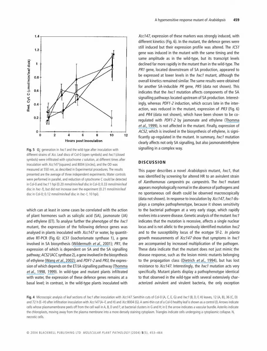
Figure 4 shows transverse leaf sections of Col-0 and hxc1, 12, 36and 72 h after infiltration-inoculation with Xcc147. No apparentdifferences between Col-0 and hxc1 could be observed at 12 hpost-inoculation (hpi) (Fig. 4A,B), suggesting that the mutantand the wild-type do not differ qualitatively at this time point. Inboth leaf sections, cells can be found with their plasma membranepulled away from the cell wall, indicating the onset of plasmolysis.Thirty-six hours post-inoculation, significant differences can be
seen in the leaf section of the wild-type compared with themutant (Fig. 4C,D). In Col-0, most cells are undergoing a generaldecompartmentation preceding a cytoplasmic collapse. Thechloroplasts are moving away from the plasma membrane into amore densely staining cytoplasm. They are swelling, assuminga more rounded shape, and their boundary membranes aredisintegrating (Fig. 4C). In hxc1 the symptoms are far moreattenuated: the plasmolysis observed 12 hpi is now generalizedand chloroplast breakdown is initiated (Fig. 4D). The Col-0 leafmesophyll 72 hpi (for sections made in the infiltrated area) iscompletely necrotic and cell structures are no longer recognizable,with the exception of the rigid lignin structure of the sieve tubes(Fig. 4E). Leaf mesophyll of Col-0 outside the necrotic zoneappears healthy (data not shown), in contrast to hxc1, wheredifferent degenerative stages can be observed: cells with anadvanced plasmolysis, cells with disrupted membrane systemsand organelles, and collapsed cells with amorphous necrotic cyto-plasm (Fig. 4F). As shown in Fig. 4G,H, hxc1 and Col-0, infiltratedwith Xcc147 and Xcc8004, respectively, show both similar andtypical features of a compatible interaction 72 hpi (Brown et al.,1993; Jones and Fett, 1985; Lummerzheim et al., 1993).
Defence responses in the hxc1 mutant
Rapid and sustained generation is induced in hxc1Early production of or derivatives of superoxide anion is ahallmark of the plant defence response and has been moreparticularly shown to be involved in HR to attempted infection byavirulent pathogens (for reviews see Doke et al., 1994; Lamb andDixon, 1997).
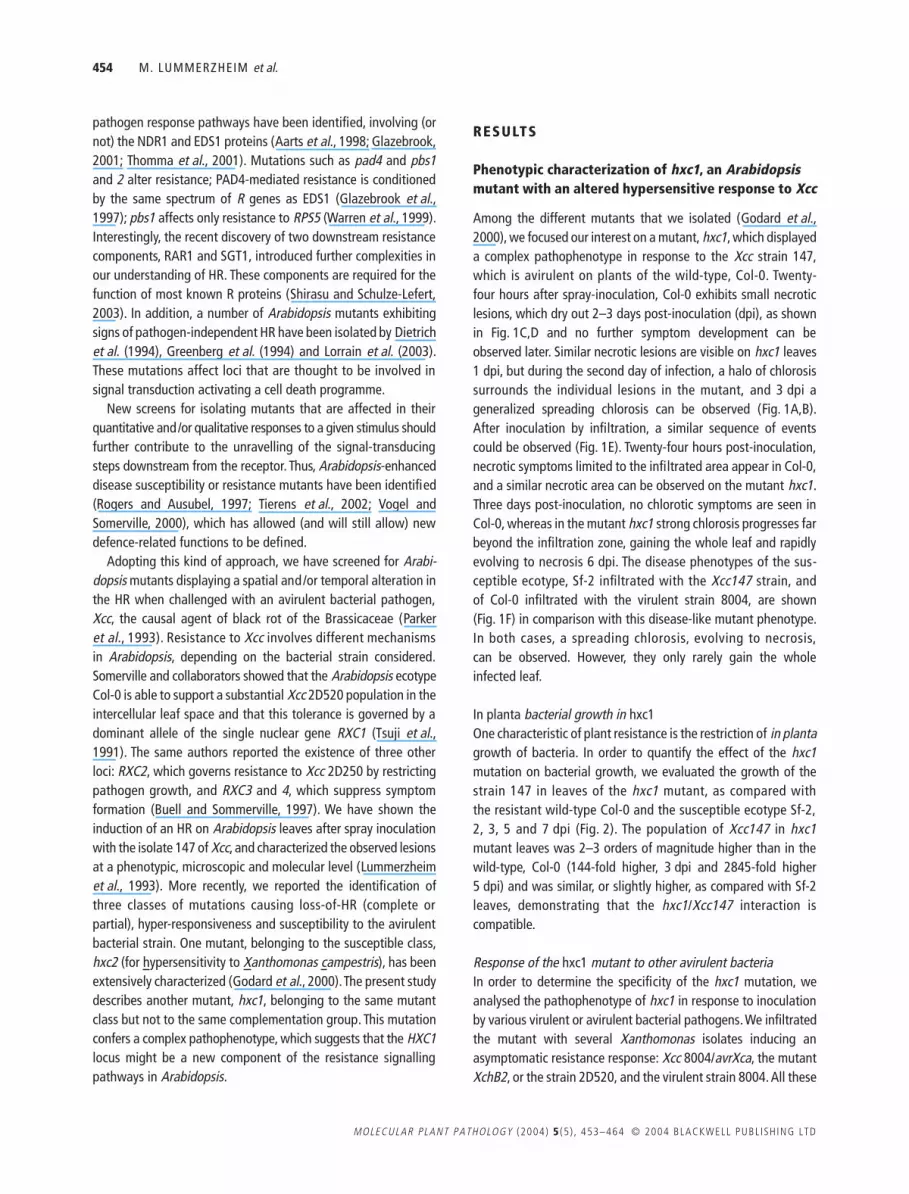
In order to evaluate the oxygen radical release, reduction ofextracellular cytochrome c was measured spectrophotometrically(Doke, 1983) in leaves of Col-0 and hxc1 infiltrated with the strains147 and 8004 (Fig. 5). In control leaves (infiltrated with water),production of could be measured 1 h after mock inoculationin Col-0 and hxc1 and did not increase over the experiment (datanot shown). In Col-0 leaf discs infiltrated with strain 147, a tran-sient burst of release was measured 3–5 hpi (Fig. 5). At thesetime points a slightly delayed but more substantial releasecould be detected in hxc1 leaf discs. This release increaseduntil 10 hpi, whereas in Col-0, no significant release could bedetected any longer. When infiltrating Col-0 and hxc1 leaves withthe virulent strain 8004, a 2-h delay was observed before beingable to measure release values equivalent to the highest valuesobtained in Col-0 during the incompatible interaction. The production increased further until 10 hpi, reaching rates comparablewith those monitored in hxc1 leaf discs infiltrated with Xcc147.
Defence gene induction in the hxc1 mutantInfection of Arabidopsis with Xcc147 results in the activationof different defence-related genes (Lummerzheim et al., 1993),
Table 2 Genetic analysis of the mutant hxc1.
Cross Generation
Number of plants*
‘Susceptible’ HR Total χ2
Col-0 × hxc1 F1 — 13 13 —F2 94 268 362 0.18
hxc1 × Col-0 F1 — 10 10 —F2 37 105 142 0.084
hxc2 × Sf-2† F1 — 32 32 —F2 54 66 110 0.076
hxc1 × hxc2 F1 — 8 8 —hxc2 × hxc1 F1 — 12 12 —
*Leaves (five) of each plant were infiltrated with Xcc147 (5 × 10⁷ cfu/mL) and scored for symptoms at 6 days post-inoculation.†The numbers presented here result from crosses hxc2 × Sf-2 and Sf-2 × hxc2.
O2−
O2−
O2−
O2−
O2−
O2−
O2−
O2−
O2−

458 M. LUMMERZHEIM et al.
MOLECULAR PLANT PATHOLOGY (2004) 5 (5 ) , 453–464 © 2004 BLACKWELL PUBL ISH ING LTD
A hypersensitive response mutant of Arabidopsis 459
© 2004 BLACKWELL PUBL ISH ING LTD MOLECULAR PLANT PATHOLOGY (2004) 5 (5 ) , 453–464
which can at least in some cases be correlated with the actionof plant hormones such as salicylic acid (SA), jasmonate (JA)and ethylene (ET). To analyse further the phenotype of the hxc1mutant, the expression of the following defence genes wasanalysed in plants inoculated with Xcc147 or water, by quantit-ative RT-PCR (Fig. 6): ICS1 (isochorismate synthase 1), a geneinvolved in SA biosynthesis (Wildermuth et al., 2001); PR1, theexpression of which is dependent on SA and the SA signallingpathway; ACS2 (ACC synthase 2), a gene involved in the biosynthesisof ethylene (Wang et al., 2002); and PDF1-2 and PR3, the expres-sion of which depends on the ET/JA signalling pathway (Thommaet al., 1998, 1999). In wild-type and mutant plants infiltratedwith water, the expression of these defence genes remains at abasal level; in contrast, in the wild-type plants inoculated with
Xcc147, expression of these markers was strongly induced, withdifferent kinetics (Fig. 6). In the mutant, the defence genes werestill induced but their expression profile was altered. The ICS1gene was induced in the mutant with the same timing and thesame amplitude as in the wild-type, but its transcript levelsdeclined far more rapidly in the mutant than in the wild-type. ThePR1 gene, located downstream of SA production, appeared tobe expressed at lower levels in the hxc1 mutant, although theoverall kinetics remained similar. The same results were obtainedfor another SA-inducible PR gene, PR5 (data not shown). Thisindicates that the hxc1 mutation affects components of the SAsignalling pathways located upstream of SA production. Interest-ingly, whereas PDF1-2 induction, which occurs late in the inter-action, was reduced in the mutant, expression of PR3 (Fig. 6)and PR4 (data not shown), which have been shown to be co-regulated with PDF1-2 by jasmonate and ethylene (Thommaet al., 1999), is not affected in the mutant. Finally, expression ofACS2, which is involved in the biosynthesis of ethylene, is signi-ficantly up-regulated in the mutant. In summary, hxc1 mutationclearly affects not only SA signalling, but also jasmonate/ethylenesignalling in a complex way.
DISCUSSION
This paper describes a novel Arabidopsis mutant, hxc1, thatwas identified by screening for altered HR to an avirulent strainof Xanthomonas campestris pv. campestris. The hxc1 mutantappears morphologically normal in the absence of pathogens andno spontaneous cell death could be observed macroscopically(data not shown). In response to inoculation by Xcc147, hxc1 dis-plays a complex pathophenotype, because it shows sensitivityto the bacterial pathogen at a very early stage, which rapidlyevolves into a severe disease. Genetic analysis of the mutant hxc1indicates that the mutation is recessive, affects a single nuclearlocus and is not allelic to the previously identified mutation hxc2and to the susceptibility locus of the ecotype Sf-2. In plantagrowth measurements of Xcc147 show that symptoms in hxc1are accompanied by increased multiplication of the pathogen.These data indicate that the mutant does not just mimic thedisease response, such as the lesion mimic mutants belongingto the propagation class (Dietrich et al., 1994), but has lostresistance to Xcc147. Interestingly, the hxc1 mutation acts veryspecifically. Mutant plants display a pathophenotype identicalto that observed in the wild-type with several extensively char-acterized avirulent and virulent bacteria, the only exception
Fig. 5 generation in hxc1 and the wild-type after inoculation with different strains of Xcc. Leaf discs of Col-0 (open symbols) and hxc1 (closed symbols) were infiltrated with cytochrome c solution, at different times after inoculation with Xcc147 (squares) and 8004 (circles), and the OD was measured at 550 nm, as described in Experimental procedures. The results presented are the average of three independent experiments. Water controls were performed in parallel, and reduction of cytochrome C could be detected in Col-0 and hxc1 1 hpi (0.20 mmol/min/leaf disc in Col-0; 0.33 mmol/min/leaf disc in hxc-1), but did not increase over the experiment (0.21 mmol/min/leaf disc in Col-0; 0.12 mmol/min/leaf disc in hxc-1, 10 hpi).
O2–
Fig. 4 Microscopic analysis of leaf sections of hxc1 after inoculation with Xcc147. Semithin cuts of Col-0 (A, C, E, G) and hxc1 (B, D, F, H) leaves, 12 (A, B), 36 (C, D) and 72 h (E–H) after infiltration inoculation with Xcc147 (A–F, and H) and Xcc 8004 (G). A semi-thin cut of a Col-0 healthy leaf is shown as a control (I). Arrows indicate cells whose plasmamembrane peels off from the cell wall in A, B, D and F; at bacterial clusters in G and H; in E the arrow indicates a vascular bundle. Asteriks indicate the chloroplasts, moving away from the plasma membrane into a more densely staining cytoplasm. Triangles indicate cells undergoing a cytoplasmic collapse. N, necrotic cells.

460 M. LUMMERZHEIM et al.
MOLECULAR PLANT PATHOLOGY (2004) 5 (5 ) , 453–464 © 2004 BLACKWELL PUBL ISH ING LTD
being the response to P. s. tomato/avrRpm1. Finally, the mutationcauses an attenuation of expression of several defence markersregulated through different signalling pathways.
All these data allow us to gain an insight into the position ofthe hxc1 mutation in the disease resistance signalling pathwaysso far identified in Arabidopsis. HXC1 is required for recognitionof the Xcc isolate 147 but it is not identical to the R gene govern-ing resistance to this bacterium. It is rather an early componentof the signalling pathway leading to resistance, and resembles inthis the previously described, non-allelic HXC2 gene. Our resultsalso suggest that HXC1 is different from the genes RXC-2, 3 and4, which condition resistance to Xcc strain 2D520 (Buell andSommerville, 1997) because (i) different mechanisms of resist-ance are involved in response to Xcc2D520 and 147 (absence ofsymptom and hypersensitive response, respectively), and (ii)the hxc1 mutant is not affected in its response to Xcc2D520. Thehxc1 mutation was found to act very specifically and does notresemble any of the known loci required for R-gene-dependentresponses. Tests with virulent P. s. tomato DC3000 and Xcc8004indicate that it is not a component of the basal resistanceresponse, and experiments with P. s. tomato strain DC3000carrying various avr genes demonstrated that the function of anyof the corresponding R genes was not impaired (Fig. 3), with theonly exception being RPM1. In response to DC3000/avrRpm1, weobserved repeatedly a partial but significant loss of resistancethat was similar or slightly weaker than the reduction of resist-ance observed in ndr1-1. ndr1-1 is also known to impair RPM1function only partially, but whereas ndr1 plants showed strongand rapid development of chlorosis in the interaction withDC3000/avrRpm1, hxc1 at a similar bacterial titre showed onlymoderate disease symptoms. We suggest that the hxc1 mutationaffects a common component of the signalling pathways lead-ing to resistance to P. s. tomato/avrRpm1 and Xcc147. Recently,mutations (lra2) in a single Arabidopsis cytosolic HSP90 isoform(HSP90.2) have been shown to compromise the function ofRPM1, and not that of related disease resistance proteins (Hubertet al., 2003). lra2, similarly to hxc1, presents only a partial lossof resistance to DC3000/avrRpm1, and is not affected in basalresistance against virulent pathogens, suggesting that hxc1 andlra2 might affect the same signalling component. However, theRPM1-mediated HR is severely attenuated in lra2, but does notseem to be compromised in hxc1 as in ndr1. Experiments toinvestigate these hypotheses are under way.
The hxc1 mutation affects resistance to Xcc in a pleiotropicway, suggesting that it plays a role in the regulation of pathogen-induced defence responses, rather than acting directly bypathogen containment. This hypothesis is supported by ourobservations at microscopic, biochemical and molecular levels.Microscopic observation of leaf sections at different stages ofthe infectious process clearly demonstrates that the mutant is nolonger able to mount an HR and shows a susceptible phenotype
Fig. 6 Defence gene expression in wild-type and hxc1 mutant plants after inoculation with Xcc147. Transcript levels of ICS-1, PR-1, PDF1-2, PR3 and ACS2 were determined by quantitative PCR, in plants inoculated with Xcc147 (5 × 10⁷ cfu/mL) or water (symbols: diamonds—Col-0 treated with water, squares—hxc1 treated with water, triangles—Col-0 inoculated with Xcc147, crosses—hxc1 inoculated with Xcc147 ). See Experimental procedures for further details.

A hypersensitive response mutant of Arabidopsis 461
© 2004 BLACKWELL PUBL ISH ING LTD MOLECULAR PLANT PATHOLOGY (2004) 5 (5 ) , 453–464
in response to Xcc147. Measurement of early production ofreactive oxygen intermediates (ROI) in the mutant vs. the wild-type is in line with these data. During plant–pathogen inter-actions, ROI may fulfil multiple physiological functions. They canact directly as toxic defence agents against pathogens (Levineet al., 1994), play a role in peroxidase-catalysed cross-linking ofcell wall polymers (Brisson et al., 1994), or serve as inducersof various defence reactions (Lamb and Dixon, 1997). ROI alsocould play an important signalling role in the initiation of the HR(Torres et al., 2002). It has indeed been widely observed that HRis preceded by a rapid outburst of , H2O2 and OH (Doke, 1983;Keppler et al., 1989; Levine et al., 1994). In our experiments, asignificant increase in production was not only observed inthe incompatible interaction between 3 and 5 hpi, but also duringthe compatible interaction, starting at 6 hpi. Surprisingly, almost nodata are available about AOS (active oxygen species) generationduring a compatible interaction, probably owing to the experimentalsetup using very time-limited experiments (latest time point 4–6 hpi). The delayed but substantial production measured canbe explained by the fact that release could be a consequenceand not a cause of plant cell membrane disruption. At earlytime points (3–5 hpi) hxc1 produced an release almost aslarge as the transient burst observed in the resistant wild-type Col-0. However, unlike Col-0, production in hxc1increased over time reaching, at 10 hpi, the values that wereotherwise only obtained during a compatible interaction at thattime point. This production profile of hxc1 can be interpretedeither (1) as the addition of an early defence response (theHR) with the establishment of a compatible interaction, or (2)as the consequence of an enhanced susceptibility to Xcc147, ascompared with the wild-type compatible interaction Col-0/Xcc8004. The first hypothesis is supported by the observation that areduced SA-response has been found in the hxc1 mutant, whichcould lead to a less active hypersensitive/defence response, andconsequently to enhanced susceptibility. However, the up-regulated expression of ACS2, encoding an enzyme involved inethylene biosynthesis, without (or with little) alteration of expres-sion of defence markers for this pathway, is in favour of thesecond hypothesis.
The signalling pathways that lead to disease resistance arecomplex. Previous work had established that some but not allR genes require salicylic acid accumulation and NPR1 function(Glazebrook, 2001). ET- and JA-dependent signals also can influ-ence R gene function (Clarke et al., 2000). The data reported inhere are in favour of an alteration of SA biosynthesis and relateddefence by hxc1.
Surprisingly, alteration of the SA pathway in hxc1 results inthe decrease not only in PR1 expression, but also in PDF1-2gene expression, suggesting that the well-described antagonismbetween SA- and JA/ET defence pathways (Clarke et al., 2000;Gupta et al., 2000; Jirage et al., 2001) is no longer operating in
hxc1. In addition, PR3 and PR4, two genes regulated throughET/JA pathways, are not affected in hxc1, demonstrating thecomplexity of regulatory crosstalk between these signalling path-ways (Kunkel and Brooks, 2002). Transcriptome analyses suggestthat SA and JA/ET pathways can act together (Glazebrook et al.,2003; Schenk et al., 2000), some genes being induced by bothpathways. However, PDF1-2 do not belong to this class of genes,suggesting that in hxc1, this negative crosstalk between the twopathways is no longer functional, or that hxc1 acts upstream inthe regulatory pathway. It also suggests that PR3 and PR4 wouldbelong to a set of genes differentially regulated in this context.
An even more surprising result was the observation that ACS2is up-regulated in the mutant. Ethylene production as a responseto stress has been observed many times, and more particularly inresponse to infection by pathogens (Pegg, 1976). This hormoneis thought to be a modulator of cell death processes, such assenescence, or those observed during interactions with patho-gens (Greenberg, 1997). In this latter case, its role was not onlydemonstrated in the activation of disease resistance mechanisms,but also in disease lesion development (Lund et al., 1998). Therate-limiting step in ethylene production is ACC synthase (ACS ),which is encoded by a multigene family in Arabidopsis (Lianget al., 1992), and transcription of ACS genes is precisely regulated(Wang et al., 2002). We chose to analyse the expression patternof ACS2, a member of the multigene family which is known to beexpressed in response to diverse stresses (Liang et al., 1992) andalso in response to elicitor treatment (Oetiker et al., 1997). Takingin account that the expression of ACS2 is expressed at higherlevels during compatible interactions with Xcc strain 8004, thanin response to Xcc147 (data not shown), this enhanced expres-sion can be interpreted as an amplification of the expression ofthe symptoms.
Taken together, these results suggest that the ET biosynthesisand signalling pathways are affected in the mutant, in addition tothe SA pathway. Measurement of SA, JA and ET in the mutant andthe wild-type during the interaction with Xcc147 would certainlyhelp the interpretation of this complex defence phenotype.
The biochemical and molecular markers used to characterizehxc1 underline the complexity of this novel defence mutant andsupport the hypothesis of a mutation affecting a key componentactivated during the first steps of the plant defence responseleading to resistance. This unique phenotype is not entirelyunexpected in light of the recent data unveiling the complexityof the R-gene-mediated signal transduction pathways.
EXPERIMENTAL PROCEDURES
Plant growth conditions
Arabidopsis thaliana plants were maintained as describedpreviously by Lummerzheim et al. (1993). pad4-1 and ndr1-1
O2−
O2−
O2−
O2−
O2−
O2−
O2−
O2−

462 M. LUMMERZHEIM et al.
MOLECULAR PLANT PATHOLOGY (2004) 5 (5 ) , 453–464 © 2004 BLACKWELL PUBL ISH ING LTD
plants were obtained from the NASC stock centre. Four- to 5-week-old plants were used for all experiments unless otherwisestated.
Bacterial strains
Bacterial strains have been described elsewhere: Xcc strain 147by Lummerzheim et al. (1993), Xcc strain 8004 by Daniels et al.(1984), Pseudomonas syringae pv. maculicola strain m2, m4, andm4/avrRpm1 by Debener et al. (1991), Pseudomonas syringae pv.tomato strain DC3000 and DC3000/avrB by Staskawicz et al.(1984), DC3000/avrRpt2 by Whalen et al. (1991), DC3000/avrPphB by Simonich and Innes (1995) and DC3000/avrRps4by Hinsch and Staskawicz (1996).
Inoculation of plants and in planta bacterial growth measurements
Arabidopsis leaves were infiltrated with bacteria as describedby Lummerzheim et al. (1993). The Xanthomonas strains wereprepared as described by Lummerzheim et al. (1993), except thatthe overnight cultures were washed and adjusted to the desiredOD600 of 0.1, which was equivalent to 108 cfu/mL. Plants grownin a growth chamber were hand-inoculated with a spontaneousrifampicin-resistant mutant of Xcc147 with an inoculumconcentration of 108 cfu/mL. The bacterial growth in planta wasmeasured 0, 2, 3, 5 and 7 dpi. The leaves were washed once in0.1% SDS and rinsed three times in distilled water. Bacterialgrowth was assayed by plating serial dilutions of the extractson to bacterial growth medium. Five leaves from four plantswere harvested per assay, and four replicates of each assaywere performed. Each experiment was repeated twice. For thedetermination of in planta bacterial growth of Pseudomonasstrains, four leaves of 4–6 plants per genotype were infiltratedwith a bacterial suspension of 2 × 10⁵ cfu/mL using a bluntsyringae. With a cork borer, three leaf discs per plant wereharvested at 0, 2 and 3 dpi and ground in 1 mL 10 mM MgCl2.A series of 10-fold dilutions of this extract was prepared in10 mM MgCl2, plated on King’s B medium and incubated at28 °C for 2 days.
Genetic analysis
The mutant hxc1, carrying the gl1 mutation, was back-crossed towild-type Col-0. All F1 plants had normal trichomes, and the F2
progeny segregated in a ratio of three wild-type to one glabrous;segregation of hxc1 and gl1 was independent.
Allelism tests were performed by crossing the hxc1 mutantwith the hxc2 mutant and the ecotype Sf-2. Individuals from F1
progeny were scored for their mutant phenotype by infiltrationinoculation with Xcc147.
Microscopy
Bright-field light microscopyThe leaves were infiltrated with a bacterial suspension adjustedto 5 × 10⁷ cfu/mL. Sections of control and infected leaves wereprepared and prefixed under vacuum for 30 min in 2.5% glutar-aldehyde in 0.15 M sodium cacodylate buffer (pH 7.4). Furtherimpregnation was continued at room temperature for 3 h, underconstant agitation. After rinsing in cacodylate buffer (0.2 M,pH 7.4), fixation was carried out in 2% OsO4, 0.8% K3Fe(CN)6 incacodylate buffer for 2 h at room temperature. After washingwith buffer, the samples were step-dehydrated in ethanol andembedded in Epon epoxy resin.
Transverse sections of control and infected leaves were preparedusing a Micron Stemi SV 6 microtome. The 1-µm-thick sectionswere stained with toluidine blue O (1% in 1% borax), examinedand photographed through a Carl Zeiss Jenalumar microscope.
Assay of generation
Determination of generated by inoculated leaf discs wasperformed by assaying the reduction of extracellular cyto-chrome c spectrophotometrically (adapted from Chai and Doke,1987). Five leaves per plant were infiltrated with bacteria asdescribed by Lummerzheim et al. (1993). Leaf discs (10 mm indiameter) were cut from the middle of the infiltrated area usinga cork borer at 0, 1, 3, 5, 10 and 20 hpi. Ten leaf discs per assaywere immediately immersed in 3 mL 0.05 M K-phosphate buffer(pH 7.8) containing 0.1 mM deferoxamine (Desferal, Ciba), and0.1 mM ortho-phenanthroline (Sigma Chemical Co.). Cytochromec at 0.02 mM (Type VI, from horse heart, Sigma Chemical Co.) wasadded just before vacuum infiltration. In each assay, 1 mL of thereaction solution was pipetted out, and the optical density (OD)at 550 nm was measured by a double-beam spectrophotometer(Kontron, Uvikon 810). We verified the almost linear increase inOD550 in our system for 10 min after initiation of the reaction(Chai and Doke, 1987). Cytochrome c reducing activity of thediscs was expressed as micromoles reduced cytochrome c /disk /min by using an extinction coefficient of 21.1.
RT PCR
Total RNA was isolated from infected and control Arabidopsisleaves using Nucleospin RNA plant kit (Macherey-Nagel, Düren,Germany) according to the manufacturer’s instructions. cDNAswere synthesized from 5 µg of total DNA-free RNA, usingSuperscript II reverse transcriptase (Invitrogen, Carlsbad, CA),following the manufacturer’s instructions. In order to verifyhomogeneous efficiency of reverse transcription, we introducedas a spike 5 fg of in vitro produced human nebuline RNA beforereverse transcription. Quantitative PCR was performed with the
O2−
O2−

A hypersensitive response mutant of Arabidopsis 463
© 2004 BLACKWELL PUBL ISH ING LTD MOLECULAR PLANT PATHOLOGY (2004) 5 (5 ) , 453–464
light cycler technology (Roche Diagnostics, Meylan, France) accord-ing to the manufacturer’s recommendations. The primers usedhad the following sequences: 5′-GTCCCGAAGCTTACACATGA-3′ and 5′-GCCATACATCCAGCCTTCATCA-3′ (nebulin), 5′-GGA-GCTACGCAGAACAACTAAGA-3′ and 5′-CCCACGAGGATCATAGTT-CAACTGA-3′ (PR1), 5′-TCATGGCTAAGTTTGCTTCC-3′ and 5′-AATACACACGATTTAGCACC-3′ (PDF1.2), 5′-GCCGTCTCTGAAC-TCAAATCTCAA-3′ and 5′-GTTACGAGCAAGAACAACCTTGTT-3′(ICS1), 5′-CGCTTGTCCTGCTAGAGGTT-3′ and 5′-GCTCGGTTC-ACAGTAGTCTGA-3′ (PR3).
ACKNOWLEDGEMENTS
We gratefully acknowledge J. L. Montillet for his suggestions andassistance in the generation experiments, M. Nicole for hishelp in microscopy data interpretation, S. Camut for her technicalassistance, and C. Balagué and N. Grimsley for critical reading ofthe manuscript. This research was supported by the InstitutNational de la Recherche Agronomique (INRA) (A.I.P. INRA Gènesde resistance). M.L. was supported by fellowships from the ECand INRA.
REFERENCES
Aarts, N., Metz, M., Holub, E., Staskawicz, B.J., Daniels, M.J. andParker, J.E. (1998) Different requirements for EDS1 and NDR1 bydisease resistance genes define at least two R gene-mediated sign-aling pathways in Arabidopsis. Proc. Natl Acad. Sci. USA, 95, 10306–10311.
Arlat, M., Gough, C.L., Barber, C.E., Boucher, C. and Daniels, M.J.(1991) Xanthomonas campestris contains a cluster of hrp genes relatedto the larger hrp cluster of Pseudomonas solanacearum. Mol. Plant–Microbe Interact. 4, 593–601.
Atkinson, M.M., Huang, J.S. and Knopp, J.A. (1985) The hypersensitivereaction of tobacco to Pseudomonas syringae pv. pisi. Activation ofplasmalemma K+/H+ exchange mechanism. Plant Physiol. 79, 843–847.
Brisson, L.F., Tenhaken, R. and Lamb, C. (1994) Function of oxidativecross-linking of cell-wall structural proteins in plant disease resistance.Plant Cell, 6, 1703–1712.
Brown, I., Mansfield, J., Irlam, I., Conrads-Srauch, J. and Bonas, U.(1993) Ultrastructure of interactions between Xanthomonas campestrispv. vesicatoria and pepper, including immuno-cytochemical localizationof extracellular polyssacharides and the avrBS3 protein. Mol. Plant–Microbe Interact. 6, 376–386.
Buell, C.R. and Sommerville, S.C. (1997) Use of Arabidopsis recombinantinbred lines reveals a monogenic and a novel digenic resistance mech-anism to Xanthomonas campestris pv. campestris. Plant J. 12, 21–29.
Chai, H.B. and Doke, N. (1987) Superoxide anion generation: a response ofpotato leaves to infection with Phytophthora infestans. Phytopathology,77, 645–649.
Clarke, J.D., Volko, S.M., Ledford, H., Ausubel, F.M. and Dong, X.(2000) Role of salicilic acid, jasmonic acid, and ethylene in cpr-inducedresistance in Arabidopsis. Plant Cell, 12, 2175–2190.
Daniels, M.J., Barber, C.E., Turner, P.C., Sawcyc, M.K., Byrde, R.J.W.and Fielding, A.H. (1984) Cloning of genes involved in pathogenicity of
Xanthomonas campestris pv. campestris using the broad host rangecosmid pLAFR1. EMBO J. 3, 3323–3328.
Debener, T., Lehnakers, H., Arnold, M. and Dangl, J.L. (1991) Identifi-cation and molecular mapping of a single Arabidopsis thaliana locusdetermining resistance of a phytopathogenic Pseudomonas syringaeisolate. Plant J. 1, 289–302.
Dietrich, R.A., Delaney, T.P., Uknes, S.J., Ward, E.R., Ryals, J.A. andDangl, J.L. (1994) Arabidopsis mutants simulating disease resistanceresponse. Cell, 77, 565–577.
Doke, N. (1983) Generation of superoxide anion by potato tuber proto-plasts during the hypersensitive response to hyphal wall components ofPhytophthora infestans and specific inhibition of the reaction bysuppressors of hypersensitivity. Physiol. Plant Pathol. 23, 359–367.
Doke, N., Miura, Y., Sanchez, L.M. and Kawakita, K. (1994) Involvementof superoxide in signal transduction: responses to attack by pathogens,physical and chemical shocks, and UV radiation. In: Causes of Photooxi-dative Stress and Amelioration of Defence Systems in Plants (Foyer, C.H.and M.P.M., eds), pp. 177–197. London: CRC Press.
Glazebrook, J. (2001) Genes controlling expression of defense responsesin Arabidopsis-2001 status. Curr. Opin. Plant Biol. 4, 301–308.
Glazebrook, J., Chen, W., Estes, B., Chang, H.S., Nawrath, C., Metrauk, J.P.,Zhu, T. and Katagiri, F. (2003) Topology of the network integrating sal-icylate and jasmonate signal transduction derived from global expressionphenotyping. Plant J. 34, 217–228.
Glazebrook, J., Zook, M., Mert, F., Kagan, I., Rogers, E.E., Crute, I.R.,Holub, E.B., Hammerschmidt, R. and Ausubel, F.M. (1997)Phytoalexin-deficient mutants of Arabidopsis reveal that PAD4 encodesa regulatory factor and that four PAD genes contribute to downie mildewresistance. Genetics, 146, 381–392.
Godard, F., Lummerzheim, M., Saindrenan, P., Balagué, C. and Roby, D.(2000) hxc2, an Arabidopsis mutant with altered hypersensitive responseto Xanthomonas campestris pv. campestris. Plant J. 24, 749–761.
Greenberg, J.T. (1997) Programmed cell death in plant pathogen inter-actions. Annu. Rev. Plant Physiol. Plant Mol. Biol. 48, 525–545.
Greenberg, J.T., Guo, A., Klessig, D.F. and Ausubel, F.M. (1994)Programmed cell death in plants: a pathogen-triggered responseactivated coordinately with multiple defence functions. Cell, 77, 551–563.
Gupta, V., Willits, M.G. and Glazebrook, J. (2000) Arabidopsis thalianaEDS4 contributes to salicylic acid (SA)-dependent expression of defenseresponses: evidence for inhibition of jamonic acid signaling by SA. Mol.Plant–Microbe Interact. 13, 503–511.
Hammond-Kosack, K.E. and Jones, J.D.G. (1997) Plant disease resistancegenes. Annu. Rev. Plant Physiol. Plant Mol. Biol. 48, 575–607.
Hinsch, M. and Staskawicz, B.J. (1996) Identification of a new Arabidopsisdisease resistance locus RPS4, and cloning of the correspondingavirulence gene, avrRps4, from Pseudomonas syringae pv. pisi. Mol.Plant–Microbe Interact. 9, 55–61.
Hubert, D.A., Tornero, P., Belkhadir, Y., Krishna, P., Takahashi, A.,Shirasu, K. and Dangl, J.L. (2003) Cytosolic HSP90 associates with andmodulates the Arabidopsis RPM1 disease resistance protein. EMBO J.22, 5679–5689.
Jirage, D., Zhou, N., Cooper, B., Clarke, J.D., Dong, X. and Glazebrook, J.(2001) Constitutive salicylic acid-dependent signaling in cpr1 and cpr6mutants requires PAD4. Plant J. 26, 395–407.
Jones, S.B. and Fett, W.F. (1985) Fate of Xanthomonas campestrisinfiltrated into soybean leaves: an ultrastructural study. Phytopathology,75, 733–741.
O2−

464 M. LUMMERZHEIM et al.
MOLECULAR PLANT PATHOLOGY (2004) 5 (5 ) , 453–464 © 2004 BLACKWELL PUBL ISH ING LTD
Keppler, L.D., Baker, C.J. and Atkinson, M.M. (1989) Active oxygenproduction during bacteria induced hypersensitive reaction in tobaccosuspension cells. Phytopathology, 79, 974–978.
Klement, Z. (1982) Hypersensitivity. In: Phytopathogenic Procaryotes(Mount, M.S. and Lacy, G.H., eds), pp. 149–177. New York: AcademicPress.
Kunkel, B.N. and Brooks, D.M. (2002) Cross talk between signalingpathways in pathogen defense. Curr. Opin. Plant. Biol. 5, 325–331.
Lamb, C. and Dixon, R.A. (1997) The oxidative burst in plant diseaseresistance. Annu. Rev. Plant Physiol. Plant Mol. Biol. 48, 251–275.
Levine, A., Tenhaken, R., Dixon, R. and Lamb, C. (1994) H2O2 from theoxidative burst orchestrates the plant hypersensitive disease resistanceresponse. Cell, 79, 583–593.
Liang, X., Abel, S., Keller, J.A., Shen, N.F. and Theologis, A. (1992) The1-aminocyclopropane-1-carboxylate synthase gene family of Arabidopsisthaliana. Proc. Natl Acad. Sci. USA, 89, 11046–11050.
Lorrain, S., Vailleau, F., Balagué, C. and Roby, D. (2003) Lesion mimicmutants: keys for deciphering cell death and defense pathways inplants? Trends Plant Sci. 8, 263–271.
Lummerzheim, M., de Olivera, D., Castresana, C., Miguens, F.C.,Louzada, E., Roby, D., Van Montagu, M. and Timmerman, B. (1993)Identification of compatible and incompatible interactions betweenArabidopsis thaliana and Xanthomonas campestris pv. campestris andcharacterization of the hypersensitive response. Mol. Plant–MicrobeInteract. 6, 532–544.
Lund, S.T., Stall, R.E. and Klee, H.J. (1998) Ethylene regulates the suscep-tible response to pathogen infection in tomato. Plant Cell, 10, 371–382.
Mansfield, J.W. and Richardson, A. (1981) The ultrasructure of inter-actions between Botrytis species and broad bean leaves. Physiol. PlantPathol. 19, 41–48.
Oetiker, J.H., Olson, D.C., Shiu, O.Y. and Yang, S.F. (1997) Differentialinduction of seven 1-aminocyclopropane-carboxylate synthase genes byelicitor in suspension cultures of tomato (Lycopersicon esculentum).Plant. Mol. Biol. 34, 275–286.
Parker, J.E., Barber, C.E., Fan, M.J. and D.M. (1993) Interaction ofArabidopsis thaliana with Xanthomonas campestris. In: ArabidopsisThaliana as a Model for Plant–Pathogen Interactions (Davis, K.R. andHammerschmidt, R., eds), pp. 61–72. St Paul: APS Press.
Pegg, G.F. (1976) The Involvement of Ethylene in Plant Pathogenesis.Heidelberg: Springer Verlag.
Rogers, E.E. and Ausubel, F.M. (1997) Arabidopsis enhanced diseasesusceptibility mutants exhibit enhanced susceptibility to several bacterialpathogens and alterations in PR-1 gene expression. Plant Cell, 9, 305–316.
Schenk, P.M., Kazan, K., Wilson, I., Anderson, J.P., Richmond, T.,Somerville, S.C. and Manners, J.M. (2000) Coordinated plant defenseresponses in Arabidopsis revealed by microarray analysis. Proc. NatlAcad. Sci. USA. 97, 11655–11660.
Shirasu, K. and Schulze-Lefert, P. (2003) Complex formation, promiscuityand multi-functionality: protein interactions in disease-resistancepathways. Trends Plant Sci. 8, 252–258.
Simonich, M.T. and Innes, R.W. (1995) A disease resistance genein Arabidopsis with specificity for the avrPph3 gene of Pseu-domonas syringae pv. phaseolicola. Mol. Plant–Microbe Interact. 8,637–640.
Staskawicz, B.J., Dahlbeck, D. and Keen, N.T. (1984) Cloned avirulencegene of Pseudomonas syringae pv. glycinea determines race-specificincompatibility on Glycine max (L.) Merr. Proc. Natl Acad. Sci. USA, 81,6024–6028.
Thomma, B.P.H.J., Eggermont, K., Penninckx, I.A.M.A., Mauch-Mani, B.,Vogelsang, R., Cammue, B.P.A. and Broekaert, W.F. (1998) Separatejasmonate-dependent and salicylic-dependent defense response path-ways in Arabidopsis are essential for resistance to distinct microbialpathogens. Proc. Natl Acad. Sci. USA, 95, 15107–15111.
Thomma, B.P., Eggermont, K., Tierens, K.F. and Broekaert, W.F. (1999)Requirement of functional ethylene-insensitive 2 gene for efficientresistance of Arabidopsis to infection by Botrytis cinerea. Plant Physiol.121, 1093–1102.
Thomma, B.P., Penninckx, I.A., Broekaert, W.F. and Cammue, B.P.(2001) The complexity of disease signaling in Arabidopsis. Curr. Opin.Immunol. 13, 63–68.
Tierens, K.F.M.J., Thomma, B.P.H.J., Bari, R.P., Garmier, M.,Eggermont, K., Brouwer, M., Penninckx, I.A.M.A., Broeckaert, W.F.and Cammue, B.P.A. (2002) Esa1, an Arabidopsis mutant withenhanced susceptibility to a range of necrotrophic fungal pathogens,shows a distorted induction of defence responses by reactive oxygengenerating compounds. Plant J. 29, 131–140.
Torres, M.A., Dangl, J.L. and Jones, J.D.G. (2002) Arabidopsis gp91phox
homologues AtrbohD and AtrbohF are required for accumulation ofreactive oxygen intermediates in the plant defense response. Proc. NatlAcad. Sci. USA, 99, 517–522.
Tsuji, J., Sommerville, S.C. and Hammershmidt, R. (1991) Identificationof a gene in Arabidopsis thaliana that controls resistance in Xanthomonascampestris pv. campestris. Physiol. Mol. Plant Pathol. 3, 57–65.
Vogel, J. and Somerville, S. (2000) Isolation and characterization ofpowdery mildew-resistant Arabidopsis mutants. Proc. Natl Acad. Sci.USA, 97, 1897–1902.
Wang, K.L.C., Li, H. and Ecker, J.R. (2002) Ethylene biosynthesis andsignaling networks. Plant Cell, Suppl., S131–S151.
Warren, R.F., Merritt, P.M., Holub, E. and Innes, R.W. (1999) Identifica-tion of three putative signal transduction genes involved in R gene-specified resistance in Arabidopsis. Genetics, 152, 401–412.
Whalen, M.C., Innes, R.W., Bent, A.F. and Staskawicz, B.J. (1991)Identification of Pseudomonas syringae pathogens of Arabidopsis and abacterial locus determining avirulence on both Arabidopsis and soybean.Plant Cell, 3, 49–59.
Wildermuth, M.C., Dewdney, J., Wu, G. and Ausubel, F.M. (2001)Isochorismate synthase is required to synthesize salicylic acid for plantdefence. Nature, 414, 562–565.
Zhou, J., Loh, Y.-T., Bressan, R.A. and Martin, G.B. (1995) The tomatogene Pti1 encodes a serine/threonine kinase that is phosphorylated byPto and is involved in the hypersensitive response. Cell, 83, 925–935.