alpha-linolenic acid supplementation during human pregnancy does not effect cognitive functioning
TRANSCRIPT

Fatty acids,P re g n a n c y,and Cog n i t i o n
Renate H.M. de Groot
NP

Stellingenbehorende bij het proefschrift
Fatty acids, Pregnancy, and Cognition
1. Zwangerschap is geassocieerd met verminderde geheugenfuncties (dit proefschrift).
2. Een snel herstel van de docosahexaeenzuur beschikbaarheid na de bevalling verlaagt de kans op depressieve symptomen bij de moeder (dit proefschrift).
3. In het menselijk lichaam wordt alfa-linoleenzuur nauwelijks omgezet indocosahexaeenzuur; dit geldt ook tijdens de zwangerschap(dit proefschrift).
4. In de weken na de bevalling wordt het functioneren van de moeder gekenmerktdoor een verminderde snelheid en efficiëntie van informatieverwerking (dit proefschrift).
5. De initiële stijging in plasma docosahexaeenzuurconcentraties bij zwangerevrouwen wordt mogelijk veroorzaakt door mobilisatie van dit vetzuur uit dematernale hersenen.
6. Bij het gebruik van plasmaconcentraties als proxy voor weefselsamenstelling dient men zich het volgende te realiseren:“It is the tissue that is the issue” (naar J. Hibbeln, ISSFAL conference 2002).
7. Een geheugenklacht kan een gevolg zijn van een verminderde snelheid vaninformatieverwerking.
8. Veranderingen in de voedingsgewoonten zouden de toename in het aantal patiënten met psychische klachten de afgelopen jaren gedeeltelijk kunnen verklaren.
9. All nutrients are poisons.There is none, which is not a poison.The right amount differentiates a poison and a proper diet (naar Paracelsus 1493-1541)
10. Cogito, ergo possideo DHA (I think, therefore I have DHA) (T. Brenna, ISSFAL conference 2002)
Renate H.M. de Groot6 november 2003

FATTY ACIDS, PREGNANCY, AND COGNITION

The studies presented in this thesis were performed at the Nutrition and ToxicologyResearch Institute Maastricht (NUTRIM), which participates in the Graduate School VLAG(Food technology, Agrobiotechnology, Nutrition and Health Sciences), accredited by theRoyal Netherlands Academy of Arts and Sciences.
Studies of this thesis were financially supported by Unilever Research and Development,Vlaardingen, The Netherlands.
Financial support by Sigma Tau Ethifarma B.V. and Nestlé Nederland B.V. for the publica-tion of this thesis is gratefully acknowledged. Additionally, the author wishes to expressher gratitude to Unilever Research and Development Vlaardingen, Mead JohnsonNutritionals and Numico Research Wageningen for their financial support.
Renate H.M. de Groot, Maastricht 2003
Cover design and illustration: J. G. H. Derwall, Scientific IllustratorLayout: J. G. H. Derwall and H. M. J. LemmensPrinted by: Drukkerij Deurenberg Kerkrade B.V.
ISBN 90 70245 46 5

Fatty acids, Pregnancy, and Cognition
PROEFSCHRIFT
ter verkrijging van de graad van doctor aan de Universiteit Maastricht,op gezag van de Rector Magnificus, Prof. Dr. A.C. Nieuwenhuizen Kruseman,
volgens het besluit van het College van Decanen, in het openbaar te verdedigen op donderdag 6 november 2003 om 12.00 uur
door
Renate Helena Maria de Groot
Geboren te Nijmegen op 15 augustus 1975

Promotores:Prof. Dr. G. HornstraProf. Dr. J. Jolles
Beoordelingscommissie:Prof. Dr. M.A. van den Hout (voorzitter)Prof. Dr. Ir. P.A. van den BrandtProf. Dr. J.G. NijhuisProf. Dr. W. Riedel Prof. Dr. G.J. van der Vusse

Voor mijn opa
Ieder mens wordt drie keer geboren:Eén keer uit zijn vader,
één keer uit zijn moederen één keer uit zichzelf.
(Oude Perzische wijsheid)


7
INHOUDSOPGAVE
1 Introduction . . . . . . . . . . . . . . . . . . . . . . . . . . . . . . . . . . . . . . . . . . . . . . . . . . . . . . . . .9
2 Effect of alpha-linolenic acid supplementation during pregnancy on maternal and neonatal polyunsaturated fatty acid status and pregnancy outcome . . . . . . . . . . . . . . . . . . . . . . . . . . . . . . . . . . . . . . . . . . . . . . . . . .23
3 Memory performance, but not information processing speed, may be reduced during early pregnancy . . . . . . . . . . . . . . . . . . . . . . . . . . . . . . . . . .45
4 Differences in cognitive performance during pregnancy and early motherhood . . . . . . . . . . . . . . . . . . . . . . . . . . . . . . . . . . . . . . . . . . . . . . . .57
5 Selective attention deficits during human pregnancy . . . . . . . . . . . . . . . . . . . . . . . .71
6 Alpha-linolenic acid supplementation during human pregnancy does not effect cognitive functioning . . . . . . . . . . . . . . . . . . . . . . . . . . . . . . . . . . . .81
7 Increased risk of postpartum depressive symptomsis associated with slower normalization after pregnancy of the functional docosahexaenoic acid status . . . . . . . . . . . . . . . . . . . . . . . . . . . . .97
8 General discussion . . . . . . . . . . . . . . . . . . . . . . . . . . . . . . . . . . . . . . . . . . . . . . . . . .111
Abbreviations . . . . . . . . . . . . . . . . . . . . . . . . . . . . . . . . . . . . . . . . . . . . . . . . . . . . . .123
Summary . . . . . . . . . . . . . . . . . . . . . . . . . . . . . . . . . . . . . . . . . . . . . . . . . . . . . . . . .127
Samenvatting . . . . . . . . . . . . . . . . . . . . . . . . . . . . . . . . . . . . . . . . . . . . . . . . . . . . . .131
Dankwoord en Curriculum Vitae . . . . . . . . . . . . . . . . . . . . . . . . . . . . . . . . . . . . . .135
Publications . . . . . . . . . . . . . . . . . . . . . . . . . . . . . . . . . . . . . . . . . . . . . . . . . . . . . . .141
Appendices . . . . . . . . . . . . . . . . . . . . . . . . . . . . . . . . . . . . . . . . . . . . . . . . . . . . . . . .145

8

INTRODUCTION

10
Several studies have shown that the essential fatty acid (EFA) status of pregnant womenchanges during pregnancy (1, 2). These fatty acid changes are not unique for the Dutch popu-lation, but are found in other nationalities as well (3). As these fatty acids are importantbuilding blocks for all cell membranes and especially for those of the central nervous sys-tem, they might play an important role in normal brain functioning. In addition, the sug-gestion exists that the current EFA status of neonates born to healthy mothers is sub-optimal (4). Since there is a strong correlation between maternal and neonatal fatty acidstatus (5) it seems important to improve the maternal EFA status in order to prevent cog-nitive changes and optimize brain development and function.
The predominant long-chain polyunsaturated fatty acids (LCPUFAs) in brain tissue aredocosahexaenoic acid (22:6n-3, DHA) and arachidonic acid (20:4n-6, AA). A shortage ofthese fatty acids, and DHA in particular has also been mentioned to play a possible role inseveral psychiatric disorders like depression ( 6 - 1 3 ), schizophrenia ( 1 4 - 1 7 ) and attention-deficit/hyperactivity disorder (ADHD) (18-20). Therefore, it will be investigated in the pres-ent thesis whether the DHA status of pregnant women and their neonates could beincreased without changing their AA levels by supplementation with a margarine rich inalpha-linolenic acid (18:3n-3, ALA) and linoleic acid (18:2n-6, LA), the precursors of DHAand AA, respectively. In addition, the effect of EFAs and their longer-chain, more unsatu-rated derivatives (LCPUFAs) on certain aspects of brain functioning were studied.
In the next paragraphs relevant background information about fatty acids, and cogni-tion are given. Subsequently, the effect of pregnancy on the maternal fatty acid status andthe effect on the fetus are described. Thereafter, this chapter will focus on cognition andthe possible relation between pregnancy and cognition. Finally, the outline of this thesiswill be presented.
Fatty acidsFatty acids consist of a chain of carbon atoms (C-atoms) with at one end a carboxyl ter-minus (COOH) and at the other end a methyl head group (CH3) (see figure 1.1). The car-bon chain can vary in length and degree of unsaturation. The nomenclature of fatty acidsis based on these characteristics.
Figure 1.1 General chemical structure of a saturated fatty acid
Fatty acids with no double bonds are called ‘saturated’; those with at least one doublebond ‘unsaturated’. The most common fatty acids of the last group are fatty acids fromthe n-3, n-6, n-7 and n-9 families. In these families the first double bond is locatedbetween the 3rd and 4th, 6 th and 7th, 7th and 8th or 9th and 10th C-atom, as counted from themethyl end (n-designation), respectively. A variety of notations is available to indicate fattyacids. In this thesis, a system is used in which the first number refers to the number of C-atoms and the second number to the number of double bonds, followed by the familyassignment (for example, ALA is 18:3n-3). As illustrated in figure 1.2, the parent fattyacids of the n-3 and n-6 families can be further metabolized to their longer-chain, more
ChapterChapter 1
CH3 CH2 CH2 (CH2)n C
OH
On ∆
methyl group carboxyl terminus

11
Introduction
Figure 1.2 Schematic presentation of the major pathways of fatty acid metabolism

12
unsaturated derivatives, the long-chain polyenes (LCPs) by means of an alternatingsequence of chain desaturation and elongation reactions, followed by one cycle of β-oxi-dation.
Desaturation introduces a double bond between the carboxyl group and the nearestdouble bond, so that molecules successively formed, retain the position of the first dou-ble bond in relation to the methyl end of the carbon chain (21). Elongation results in theincorporation of a two-carbon unit from malonylCoA into the activated fatty acid chainproximal to the carboxyl group (22).
The n-6 family and the n-3 family are called the families of the essential fatty acids(EFAs). Reason for this is that mammals, in contrast to plants, lack the enzymes necessaryto insert double bonds at n-6 or n-3 positions (23). So, human cannot synthesize the parentEFAs, LA and ALA de novo, although these fatty acids are needed for several body func-tions (24, 25). Therefore, they must be provided by the diet. LA and ALA are mainly presentin seed oils (ALA also in green leaves).
Once taken up by the body, these EFAs can be further desaturated and elongated intomore unsaturated derivatives, the long-chain polyenes (LCPs), which are also essential forhuman health. In the process of desaturation and elongation the first ∆6-desaturation is arate limiting step (23). Because all fatty acid families use the same desaturase enzymes, theycompete mutually for these enzymes. However, ∆6-desaturase prefers ALA as a sub-strate, followed by, in order, LA, oleic acid (18:1n-9) and palmitoleic acid (16:1n-7). Inaddition, ∆6-desaturase activity is influenced by availability of the substrate and concentra-tion of the product. Thus, at the moment that ALA levels are sub-optimal, LA will bemetabolized, followed by oleic acid, consequently increasing the levels of Mead acid(20:3n-9). In this way Mead acid (20:3n-9) is a marker for sub-optimal levels of fatty acidsof the n-3 + n-6 families. Osbond acid (22:5n-6, ObA) is a specific deficiency marker forDHA, because in case of insufficient DHA, the conversion of adrenic acid (22:4n-6, AdrA)into ObA increases. As the conversion from the parent fatty acids ALA and LA into themajor LCPs of the central nervous system, DHA and AA, respectively, occur inefficiently,it seems favorable to consume these important fatty acids with the diet. AA and DHA canbe found in lean meat. Besides, egg yolk is an important source of AA and fatty fish ofDHA.
Effect of pregnancy on maternal fatty acid patternsPregnancy is generally associated with a marked hyperlipidemia i.e. a 65% increase in plas-ma phospholipid concentration from first trimester through term (26, 27). For this thesis weconcentrate on the changes in fatty acid patterns and of DHA, AA, and their status mark-ers in particular, as an effect of pregnancy. Otto et al studied the fatty acid status of preg-nant women during early gestation ( 2 ). The fatty acid status from week 10 of gestationuntil delivery was studied by Al et al (1), while Otto et al focused on fatty acid pattern inthe postpartum period (28) in both a lactating and non-lactating group. These results aresummarized in figure 1.3.
It appeared that between early pregnancy and delivery, plasma concentrations (mg/L)of phospholipid associated EFAs and their LCPs increase. However, non-essential unsatu-rated fatty acids increase considerably more. These results combined, reveal that theincrease in total absolute amounts of fatty acids is to a greater extend caused by anincrease in non-essential fatty acids (>65%) and to a lesser extend by an increase in essen-tial polyunsaturated fatty acids (PUFAs) (40%), resulting in a decreased overall EFA index((n-3+n-6)/(n-7+n-9)) during pregnancy.
Chapter 1
12

13
In this thesis we are especially interested in DHA and AA (see figure 1.3), becausethese are the predominant fatty acids in brain tissue. AA decreases during pregnancy andDHA shows a very peculiar pattern as a result of pregnancy; an initial increase until week18 of pregnancy and afterwards a decline. This suggests that the maternal DHA status issub-optimal during pregnancy. In addition, the decline after parturition is stronger in lac-tating than in non-lactating women. The sub-optimal status of DHA is supported by anincrease in the general PUFA status marker Mead acid, and of the specific DHA statusmarker ObA during gestation. The initial increment in DHA could result from a change indietary habits during pregnancy, however this was not confirmed by results from Al et al ( 2 9 ).Another explanation could be increased DHA synthesis from its precursors, however thisprocess is said to occur at a low rate only ( 3 0 ). A third possible explanation for theincrease in DHA is increased mobilization from maternal stores, like fat stores or braintissue. What the exact mechanism(s) is (are) remain(s) to be investigated.
Figure 1.3 Calculated patterns of percental changes of relative amounts of DHA, AA, and ObA in maternal plasma phospholipids during pregnancy and the postpar-tum period (results are based on data from Al et al and Otto et al)
Relation between fatty acids status of the mother and her childEFAs do play a crucial role in brain development that takes place especially at the begin-ning of pregnancy and during its third trimester. To obtain these EFAs, the fetus primarilydepends on EFA transport via the placenta from the mother. Thus, it is not surprisinglythat there is a strong correlation between the maternal EFA status and the neonatal EFAstatus at birth (1). Fatty acid composition of the umbilical cord, both plasma and tissue,suggest that the EFA status in newborns is sub-optimal, because the EFA status is lowerdownstream (arteries) than in the upstream (vein) area (5).
Supplementation of pregnant women with LA causes a higher neonatal n-6 status, how-ever the neonatal n-3 fatty acid status decreased concordantly (31). Supplementation duringpregnancy with fish oil, which is rich in n-3 fatty acids, showed a reverse effect: theneonatal n-3 fatty acid status increased, whereas the n-6 fatty acids decreased ( 3 2 ). A decrement in n-3 or n-6 fatty acid status is considered an undesirable situation, as fattyacids from both families are present in brain tissue.
In conclusion, it can be said that there is a strong correlation between the maternaland fetal/neonatal EFA status. The fatty acid status of the latter may be sub-optimal and, if
Introduction
75
100
125
150
175
200
225
0 10 partus
Time
%DHA
lactating women
non-lactating women
0 10 partus
Time
%AA
Time0 10 partus
%ObA

14
so, it would be favorable to improve it, because of the important role of EFAs in brainfunction. This improvement can be obtained by fatty acid supplementation, however themost efficient way still remains to be determined. In addition, also the functional effects ofan optimal EFA status of both mother and child are still unclear, although strong evidenceexists for a possible role in cognitive functioning, at least in childhood (33-35).
Cognitive performanceIn this thesis it will be investigated whether changes in cognitive performance can befound during pregnancy, which are related to the pregnancy induced changes in fattyacids, especially DHA and AA. Reports are available which show that the fatty acid statusat birth affects cognitive development at 4 years of age (36). This implies that fatty acidsmay play an important role in cognitive functioning indeed. Therefore, it was decided touse various aspects of cognitive functioning as outcome parameters to investigate changesin cognitive performance during pregnancy and the possible effect of fatty acid supplemen-tation. Some background information with respect to the most important cognitiveprocesses is given in the remainder of this paragraph.
The term ‘Cognition’ originates from the Latin word “cognoscere”, meaning 'to know'.In general it can be said that cognition is a term, which is used to describe the mentalactivities involved in judgment, decision making, problem solving, imagination, and otheraspects of complex thought.
Figure 1.4 Model of human cognition (based upon Bernstein et al (37))
The information-processing approach offers a general model of human cognition (figure 1.4). According to this model, between the presentation of a stimulus and the exe-cution of a response, information is received, transformed, and manipulated through aseries of stages (37). In the first stage, information about the stimulus reaches the brain byway of sensory information. This stage does not require attention. In the second stage,information must be perceived and recognized. In order to recognize a stimulus the per-ceived pattern has to be matched with a pattern in long-term memory. In addition, vari-ous kinds of encoding are used to hold new information in memory. In the third stage,
ChapterChapter 1
Stimulus
Stage 1
Sensoryprocessing
Stage 2
Perception
Stage 3
Decision/re-sponse selection
Stage 4
Responseexecution
Response
Attention
Short-term
MEMORY
Long-term
Procedural/implicit
Declarative/explicit
episodic semantic

15
once a stimulus has been recognized, it is neccessary to decide what to do with it. Thisstage demands more attention than perception does. The information may simply bestored in memory. If, however, the decision is made to take some action, a responsemust also be selected before the fourth stage -execution of the response- can occur.
The cognitive processes, which are most relevant for this thesis, have to do with themodel of central information processing, which includes m e m o r y and a t t e n t i o n.The choice to concentrate on these processes has been based upon the finding that preg-nant women complain about concentration problems and everyday forgetting in particular( 3 8 - 4 1 ).
Memory as a process can be classified in three parts, namely encoding of information,storage of information, and retrieval of information ( 4 2 ). Over the years there has beensome evolution in the conception of memory. A major aspect of memory, which is nowa-days considered very important, is the ‘working memory’, which can be envisaged as a‘temporary holding device’. This device enables the processing of new information intomore long-term stores, and processing of information that is already stored. Importantterms to describe other aspects of the memory system are short-term memory, alsocalled immediate memory (less than minutes), and long-term (hours or longer) memory.Long-term memory is traditionally divided in procedural memory and declarative memory ( 3 7 ).Procedural memory, also named implicit or non-declarative memory, refers to knowledgewe have no conscious access to, like skills and habits. Knowledge we have consciousaccess to is called declarative or explicit memory. It can be distinguished again in twodomains: episodic memory is that part that represents episodes in our personal history(events), while semantic memory is world knowledge that one remembers in the absenceof any circumstances about learning it (facts). A wide variety of tests is available, whichare used to measure the aspects of memory mentioned above. In this thesis, we will con-fine ourselves to the use of a word learning test ( 4 3 ), measuring encoding and retrievalfrom long-term memory, and the Fluency test, which concentrates on semantic memory ( 4 4 ).
Apart from memory performance, an important place is given to the measurement of‘Central information processing speed’. The basic notion is, that the efficiency atwhich the brain processes incoming sensory information, compares it with informationwhich is already stored in the long term memory, and decides as to the optimal behaviorand motor action which has to be taken, is manifested in the speed at which particularcognitive actions are performed. There are tests of general information processing speedat many cognitive domains. Here we use the Letter-Digit-Substitution-Test ( 4 5 ) and theConcept-Shifting-Test (46).
Attention can be described as the process of directing and focussing certain psycho-logical resources, usually by voluntary control, to enhance information processing, per-formance, and mental experience. Attentive behaviors have a hierarchical structure (42). Atthe most global levels are states of alertness such as sleep and wakefulness. Wakefulnessincludes more and less attentive states, such as drowsiness, alertness, and hyper-alert-ness. The attentive states can be further divided in selective attention by ignoring certainstimuli or by paying selective attention to certain stimuli. In the present thesis, the Strooptest (47) and the Finger precuing task (48, 49) are used to measure selective attention.
Choice for the above-mentioned tests was based upon their effectiveness shown inorther studies, concerning mild cognitive impairments ( 5 0 - 5 3 ). Whether the above-men-tioned cognitive tests show effectiveness in measuring cognitive performance during preg-nancy will be further elucidated in the later chapters of this thesis.
Introduction
16
Cognition and pregnancyMany pregnant women complain about reduced memory and attention deficits duringpregnancy. Research into the relationship between pregnancy and cognitive functionreceived already attention as far back as the sixties (39, 54).
Since that time, several case reports have been published (38, 41, 55, 56), all reporting a nega-tive change in cognitive functioning during pregnancy. This was confirmed by a number ofcross-sectional studies investigating subjective cognitive decline (40, 57), objective cognitivedecline (58), or both (59, 60). However, several other studies only reported subjective memoryloss, whereas objectively no effects were found (39, 61, 62). Christensen et al even demonstrat-ed that pregnancy may confer a selective cognitive advantage (63).
Summarizing the available longitudinal data (54, 64-69), it can be said that almost all authorsreport an effect (subjective or objective) of pregnancy on cognition, however whetherthis is in the first, second, or third trimester or even in the postpartum period is equivo-cal. In addition, the question which factor or factors is/are responsible for this effect isunclear. Furthermore, the role of fatty acids needs to be studied more precisely. Moreover,in some studies a reliable control group was lacking (54, 64) or cases and controls were nottested in the same testing room ( 6 6 ). Therefore, we conducted a study in which cognitiveperformance of pregnant women, which were followed from week 14 of pregnancy until32 weeks after delivery, was compared with cognitive performance of a group of non-pregnant women matched for age and education. Because fatty acids play a possible rolein cognitive performance during pregnancy, the effect of EFA supplementation duringpregnancy on cognitive performance was also studied.
Outline of this thesisThe aim of the current thesis was twofold. The first aim was to improve the maternal andneonatal fatty acid status with a LA containing margarine rich in ALA, the ultimate dietaryprecursors of AA and DHA, without changing the AA status. The second was to studycognitive changes during pregnancy and its potential relationship with the EFA status.
At first we investigated whether our supplement, a combination of n-3 and n-6 fattyacids, was effective in improving the maternal and neonatal fatty acid status. We thereforeconducted a study in which pregnant women were supplemented with either a controlmargarine (LA only) or an experimental margarine with ALA and LA. The effects onmaternal and neonatal EFA status and the effect on birth weight and gestational age werestudied (Chapter 2).
In order to investigate whether there is an effect of early pregnancy on cognitive func-tioning, performance on a cognitive test battery of a group of pregnant women at week14 of gestation was compared with performance of a group of non-pregnant women. Thisstudy is described in Chapter 3.
As has been mentioned by other researchers, different cognitive functions can beaffected by pregnancy. These discrepant findings may be due to differences in the durationof pregnancy when the subjects were studied. We studied cognitive performance in preg-nant women in a longitudinal design from week 14 of pregnancy up until 32 weeks afterdelivery and compared the results again with those of a matched non-pregnant controlgroup tested at comparable time intervals. The results of this study are presented anddiscussed in Chapter 4. Chapter 5 concentrates on one specific aspect of cognition,namely selective attention.
As fatty acids are important constituents of all cell membranes, including those of thecentral nervous system, it might be possible that EFA supplementation during pregnancy
ChapterChapter 1

17
improves maternal cognitive functioning. Whether or not EFA supplementation duringpregnancy affects maternal cognition during and after pregnancy is described in Chapter 6.
Whether fatty acids influence other aspects of brain functioning is illustrated inChapter 7. In this chapter we investigated the possible association between fatty acidsand postpartum depression.
Finally, in Chapter 8 the conclusions of these studies and their possible implicationsare discussed.
Introduction
18
REFERENCES
1. Al M. D., v. Houwelingen A. C., Kester A. D., Hasaart T. H., de Jong A. E., Hornstra G. Maternalessential fatty acid patterns during normal pregnancy and their relationship to the neonatalessential fatty acid status. Br J Nutr 1995;74:55-68.
2. Otto S. J., v. Houwelingen A. C., Badart-Smook A., Hornstra G. Changes in the maternalessential fatty acid profile during early pregnancy and the relation of the profile to diet. Am JClin Nutr 2001;73:302-7.
3. Otto S. J., v. Houwelingen A. C., Antal M., et al. Maternal and neonatal essential fatty acid sta-tus in phospholipids: an international comparative study. Eur J Clin Nutr 1997;51:232-42.
4. Hornstra G. Essential fatty acids in mothers and their neonates. Am J Clin Nutr 2000;71:1262S-9S.5. Al M. D., Hornstra G., van der Schouw Y. T., Bulstra-Ramakers M. T., Huisjes H. J. Biochemical
EFA status of mothers and their neonates after normal pregnancy. Early Hum Dev 1990;24:239-48.6. Adams P. B., Lawson S., Sanigorski A., Sinclair A. J. Arachidonic acid to eicosapentaenoic acid ratio in
blood correlates positively with clinical symptoms of depression. Lipids 1996;31 Suppl:S157-61.7. Edwards R., Peet M., Shay J., Horrobin D. Omega-3 polyunsaturated fatty acid levels in the
diet and in red blood cell membranes of depressed patients. J Affect Disord 1998;48:149-55.8. Hibbeln J. R., Salem N., Jr. Dietary polyunsaturated fatty acids and depression: when choles-
terol does not satisfy. Am J Clin Nutr 1995;62:1-9.9. Hibbeln J. R. Fish consumption and major depression [letter] [see comments]. Lancet
1998;351:1213.10. Peet M., Murphy B., Shay J., Horrobin D. Depletion of omega-3 fatty acid levels in red blood
cell membranes of depressive patients. Biol Psychiatry 1998;43:315-9.11. Tanskanen A., Hibbeln J. R., Tuomilehto J., et al. Fish consumption and depressive symptoms
in the general population in Finland. Psychiatr Serv 2001;52:529-31.12. Maes M., Smith R., Christophe A., Cosyns P., Desnyder R., Meltzer H. Fatty acid composition
in major depression: decreased omega 3 fractions in cholesteryl esters and increased C20: 4omega 6/C20:5 omega 3 ratio in cholesteryl esters and phospholipids. J Affect Disord1996;38:35-46.
13. Maes M., Christophe A., Delanghe J., Altamura C., Neels H., Meltzer H. Y. Lowered omega 3 polyunsaturated fatty acids in serum phospholipids and cholesteryl esters of depressedpatients. Psychiatry Res 1999;85:275-91.
14. Assies J., Lieverse R., Vreken P., Wanders R. J., Dingemans P. M., Linszen D. H. Significantlyreduced docosahexaenoic and docosapentaenoic acid concentrations in erythrocyte mem-branes from schizophrenic patients compared with a carefully matched control group. BiolPsychiatry 2001;49:510-22.
15. Yao J. K., Leonard S., Reddy R. D. Membrane phospholipid abnormalities in postmortembrains from schizophrenic patients. Schizophr Res 2000;42:7-17.
16. Peet M., Laugharne J. D., Mellor J., Ramchand C. N. Essential fatty acid deficiency in erythro-cyte membranes from chronic schizophrenic patients, and the clinical effects of dietary sup-plementation. Prostaglandins Leukot Essent Fatty Acids 1996;55:71-5.
17. Laugharne J. D., Mellor J. E., Peet M. Fatty acids and schizophrenia. Lipids 1996;31 Suppl:S1635.18. Burgess J. R., Stevens L., Zhang W., Peck L. Long-chain polyunsaturated fatty acids in c h i l d r e n
with attention- deficit hyperactivity disorder. Am J Clin Nutr 2000;71:327S-30S.19. Richardson A. J., Puri B. K. The potential role of fatty acids in attention-deficit/hyperactivity
disorder. Prostaglandins Leukot Essent Fatty Acids 2000;63:79-87.20. Stevens L. J., Zentall S. S., Deck J. L., et al. Essential fatty acid metabolism in boys with atten-
tion-deficit hyperactivity disorder. Am J Clin Nutr 1995;62:761-8.
ChapterChapter 1
19
21. Sprecher H. Biochemistry of essential fatty acids. Prog Lipid Res 1981;20:13-22.22. Cinti D. L., Cook L., Nagi M. N., Suneja S. K. The fatty acid chain elongation system of mam-
malian endoplasmic reticulum. Prog Lipid Res 1992;31:1-51.23. Bezard J., Blond J. P., Bernard A., Clouet P. The metabolism and availability of essential fatty
acids in animal and human tissues. Reprod Nutr Dev 1994;34:539-68.24. Burr B. O., Burr M. M. A new deficiency disease produced by rigid exclusion of fat from the
diet. J Biol Chem 1929;25:629-638.25. Burr B. O., Burr M. M. On the nature and role of fatty acids essential in nutrition.
J Biol Chem 1930;86:587-621.26. Desoye G., Schweditsch M. O., Pfeiffer K. P., Zechner R., Kostner G. M. Correlation of hor-
mones with lipid and lipoprotein levels during normal pregnancy and postpartum.J Clin Endocrinol Metab 1987;64:704-12.
27. Punnonen R. The relationship between serum oestradiol levels and serum triglyceride, choles-terol and phospholipid levels in normal human pregnancy. Br J Obstet Gynaecol 1977;84:838-45.
28. Otto S. J., v. Houwelingen A. C., Badart-Smook A., Hornstra G. The postpartum docosa-hexaenoic acid status of lactating and nonlactating mothers. Lipids 1999;34:S227.
29. Al M. D., Badart-Smook A., v. Houwelingen A. C., Hasaart T. H., Hornstra G. Fat intake ofwomen during normal pregnancy: relationship with maternal and neonatal essential fatty acidstatus. J Am Coll Nutr 1996;15:49-55.
30. Voss A., Reinhart M., Sprecher H. Differences in the interconversion between 20- and 22-carbon (n - 3) and (n - 6) polyunsaturated fatty acids in rat liver. Biochim Biophys Acta 1992;1127:33-40.
31. Al M. D., v. Houwelingen A. C., Badart-Smook A., Hornstra G. Some aspects of neonatalessential fatty acid status are altered by linoleic acid supplementation of women during preg-nancy. J Nutr 1995;125:2822-30.
32. v. Houwelingen A. C., Sorensen J. D., Hornstra G., et al. Essential fatty acid status in neonatesafter fish-oil supplementation during late pregnancy. Br J Nutr 1995;74:723-31.
33. Agostoni C., Trojan S., Bellu R., Riva E., Bruzzese M. G., Giovannini M. Developmental quotientat 24 months and fatty acid composition of diet in early infancy: a follow up study. Arch DisChild 1997;76:421-4.
34. Willatts P., Forsyth J. S., DiModugno M. K., Varma S., Colvin M. Effect of long-chain polyunsat-urated fatty acids in infant formula on problem solving at 10 months of age. Lancet 1998;352:688-91.
35. Bakker E. C. Long-chain polyunsaturated fatty acids and child development (thesis). HumanBiology. Maastricht: Maastricht University, 2002:128.
36. Helland I. B., Smith L., Saarem K., Saugstad O. D., Drevon C. A. Maternal supplem e n t a t i o nwith very-long-chain n-3 fatty acids during pregnancy and lactation augments children's IQ at 4 years of age. Pediatrics 2003;111:e39-44.
37. Bernstein D. A., Clarke-Stweart A., Roy E. J., Wickens C. D. Psychology. 4 ed. Boston NewYork: Houghton Mifflin Company, 1997.
38. Baildam E. Doctor as mum. BMJ 1991;303:424.39. Kane F. J., Jr., Harman W. J., Jr., Keeler M. H., Ewing J. A. Emotional and cognitive disturbance
in the early puerperium. Br J Psychiatry 1968;114:99-102.40. Parsons C., Redman S. Self-reported cognitive change during pregnancy. Aust J Adv Nurs
1991;9:20-9.41. Welch J. Labouring brains. BMJ 1991;303:253.42. Gazzaniga M. S., Ivry R. B., Mangun G. R. Cognitive neuroscience: the biology of the mind.
New York, London: W.W. Norton & Company, 1998.
Introduction

20
43. Brand N., Jolles J. Learning and retrieval rate of words presented auditorily and visually. J GenPsychol 1985;112:201-10.
44. Lezak M. D. Neuropsychological Assessment. 3 ed. New York: Oxford University Press,1995.
45. Smith A. The symbol Digit Modalities Test: A neuropsychologic test for economic s c r e e n i n gof learning and other cerebral disorders. Learning Disorders 1968;3:83-91.
46. Houx P. Cognitive aging and health-related factors (thesis). Psychiatry and NeuropsychologyMaastricht: Rijksuniversiteit Limburg, 1991.
47. Stroop J. Studies of interference in serial verbal reaction. J.Exp.Psychol. 1935;18:643-662.48. Adam J. J., Paas F. G., Teeken J. C., et al. Effects of age on performance in a finger-precuing
task. J Exp Psychol Hum Percept Perform 1998;24:870-83.49. Miller J. Discrete versus continuous stage models of human information processing: in search
of partial output. J Exp Psychol Hum Percept Perform 1982;8:273-96.50. Engelberts N. H., Klein M., van der Ploeg H. M., et al. Cognition and health-related quality of
life in a well-defined subgroup of patients with partial epilepsy. J Neurol 2002;249:294-9.51. van Boxtel M. P., Buntinx F., Houx P. J., Metsemakers J. F., Knottnerus A., Jolles J. The rela-
tion between morbidity and cognitive performance in a normal aging population. J Gerontol ABiol Sci Med Sci 1998;53:M147-54.
52. Klein M., Houx P. J., Jolles J. Long-term persisting cognitive sequelae of traumatic brain injuryand the effect of age. J Nerv Ment Dis 1996;184:459-67.
53. Bohnen N., Twijnstra A., Jolles J. Performance in the Stroop color word test in relationshipto the persistence of symptoms following mild head injury. Acta Neurol Scand 1992;85:116-21.
54. Jarrahi-Zadeh A., Kane F. J., Jr., Van de Castlf R. L., Lachenbruch P. A., Ewing J. A. Emotionaland cognitive changes in pregnancy and early puerperium. Br J Psychiatry 1969;115:797-805.
55. Condon J. T. Altered cognitive functioning in pregnant women: a shift towards primaryprocess thinking. Br J Med Psychol 1987;60:329-34.
56. Purvin V. A., Dunn D. W. Caffeine and the benign encephalopathy of pregnancy [letter]. Acta Neurol Scand 1987;75:76-7.
57. Poser C. M., Kassirer M. R., Peyser J. M. Benign encephalopathy of pregnancy. Preliminaryclinical observations. Acta Neurol Scand 1986;73:39-43.
58. Eidelman A. I., Hoffmann N. W., Kaitz M. Cognitive deficits in women after childbirth. ObstetGynecol 1993;81:764-7.
59. Brindle P. M., Brown M. W., Brown J., Griffith H. B., Turner G. M. Objective and subjectivememory impairment in pregnancy. Psychol Med 1991;21:647-53.
60. Sharp K., Brindle P. M., Brown M. W., Turner G. M. Memory loss during pregnancy [see com-ments]. Br J Obstet Gynaecol 1993;100:209-15.
61. Casey P., Huntsdale C., Angus G., Janes C. Memory in pregnancy. II: Implicit, incidental,explicit, semantic, short-term, working and prospective memory in primigravid, multigravidand postpartum women. J Psychosom Obstet Gynaecol 1999;20:158-64.
62. McDowall J., Moriarty R. Implicit and explicit memory in pregnant women: an analysis of data-driven and conceptually driven processes. Q J Exp Psychol A 2000;53:729-40.
63. Christensen H., Poyser C., Pollitt P., Cubis J. Pregnancy may confer a selective cognitiveadvantage. Journal of reproductive and infant psychology 1999;17:7-25.
64. Buckwalter J. G., Stanczyk F. Z., McCleary C. A., et al. Pregnancy, the postpartum, and steroidhormones: effects on cognition and mood [published erratum appears in Psychoneuro-endocrinology 1999 Jul;24(5):581]. Psychoneuroendocrinology 1999;24:69-84.
ChapterChapter 1

21
65. Buckwalter J. G., Buckwalter D. K., Bluestein B. W., Stanczyk F. Z. Pregnancy and post par-tum: changes in cognition and mood. Prog Brain Res 2001;133:303-19.
66. Harris N. D., Deary I. J., Harris M. B., Lees M. M., Wilson J. A. Peripartal cognitive impair-ment: Secondary to depression? Br J Health Psy 1996;1:127-36.
67. Keenan P. A., Yaldoo D. T., Stress M. E., Fuerst D. R., Ginsburg K. A. Explicit memory inpregnant women. Am J Obstet Gynecol 1998;179:731-7.
68. Schneider Z. Cognitive performance in pregnancy. Aust J Adv Nurs 1989;6:40-7.69. Silber M., Almkvist O., Larsson B., Uvnas-Moberg K. Temporary peripartal impairment in
memory and attention and its possible relation to oxytocin concentration. Life Sci 1990;47:57-65.
Introduction

22

EFFECT OF ALPHA-LINOLENICACID SUPPLEMENTATIONDURING PREGNANCY ON
MATERNAL AND NEONATALPOLYUNSATURATED
FATTY ACID STATUS ANDPREGNANCY OUTCOME
accepted for publication in American Journal Clinical Nutrition
Renate H.M. de Groot, Gerard Hornstra, Adriana C. v. Houwelingen, and Frans Roumen.

24
ABSTRACT
Background: The maternal essential fatty acid status declines during preg-
nancy, as a result of which the neonatal concentrations of docosahexaenoic
acid (DHA, 22:6n-3) and arachidonic acid (AA, 20:4n-6) may not be optimal.
Objective: Improving the maternal and neonatal fatty acid status by sup-
plementation of pregnant women with a combination of α-linolenic acid
(ALA, 18:3n-3) and linoleic acid (LA, 18:2n-6), the ultimate dietary precur-
sors of DHA and AA, respectively.
Design: From week 14 of gestation until delivery, pregnant women con-
sumed 25 g of a margarine supplying either 2.8 g ALA + 9.0 g LA (experi-
mental group, n = 29) or 10.9 g LA (control group, n = 29) per day. Venous
blood was collected for plasma phospholipid fatty acid analyses at weeks 14,
26 and 36 of pregnancy, at delivery, and 32 weeks postpartum. Umbilical
cord blood and vascular tissue samples were collected to study the neona-
tal fatty acid status also. Pregnancy outcome variables were assessed.
Results: ALA + LA supplementation did not prevent the maternal DHA and
AA declines during pregnancy and compared to LA supplementation it did not
increase the maternal and neonatal DHA concentrations, although eicosapen-
taenoic acid (20:5n-3, EPA) and docosapentaenoic acid (22:5n-3, DPA) concen-
trations were increased. In addition, it lowered the neonatal AA status. No
differences in pregnancy outcome variables were found.
Conclusion: Maternal ALA + LA supplementation did not promote the
neonatal DHA + AA status, although the lower concentrations of Osbond acid
(22:5n-6) in maternal plasma and umbilical arterial vessel walls phospholipids
suggest that the functional DHA status improves upon ALA+LA supplementation.
Chapter 2
25
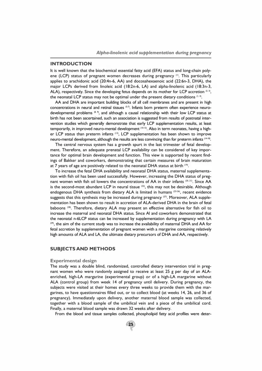
INTRODUCTIONIt is well known that the biochemical essential fatty acid (EFA) status and long-chain poly-ene (LCP) status of pregnant women decreases during pregnancy ( 1 ). This particularlyapplies to arachidonic acid (20:4n-6, AA) and docosahexaenoic acid (22:6n-3, DHA), themajor LCPs derived from linoleic acid (18:2n-6, LA) and alpha-linolenic acid (18:3n-3,ALA), respectively. Since the developing fetus depends on its mother for LCP accretion (2, 3),the neonatal LCP status may not be optimal under the present dietary conditions (1, 4).
AA and DHA are important building blocks of all cell membranes and are present in highconcentrations in neural and retinal tissues ( 5 - 7 ). Infants born preterm often experience neuro-developmental problems (8, 9), and although a causal relationship with their low LCP status atbirth has not been ascertained, such an association is suggested from results of postnatal inter-vention studies which generally demonstrate that early LCP supplementation results, at leasttemporarily, in improved neuro-mental development ( 1 0 - 1 2 ). Also in term neonates, having a high-er LCP status than preterm infants ( 1 3 ), LCP supplementation has been shown to improveneuro-mental development, although the results are less convincing than for preterm infants ( 1 4 - 1 8 ).
The central nervous system has a growth spurt in the last trimester of fetal develop-ment. Therefore, an adequate prenatal LCP availability can be considered of key impor-tance for optimal brain development and function. This view is supported by recent find-ings of Bakker and coworkers, demonstrating that certain measures of brain maturationat 7 years of age are positively related to the neonatal DHA status at birth (19).
To increase the fetal DHA availability and neonatal DHA status, maternal supplementa-tion with fish oil has been used successfully. However, increasing the DHA status of preg-nant women with fish oil lowers the concentrations of AA in their infants (20, 21). Since AAis the second-most abundant LCP in neural tissue (22), this may not be desirable. Althoughendogenous DHA synthesis from dietary ALA is limited in humans (23-26), recent evidencesuggests that this synthesis may be increased during pregnancy (27). Moreover, ALA supple-mentation has been shown to result in accretion of ALA-derived DHA in the brain of f e t a lbaboons ( 2 8 ). Therefore, dietary ALA may present an effective alternative for fish oil toincrease the maternal and neonatal DHA status. Since Al and coworkers demonstrated thatthe neonatal n-6LCP status can be increased by supplementation during pregnancy with LA( 2 9 ), the aim of the current study was to increase the availability of maternal DHA and AA forfetal accretion by supplementation of pregnant women with a margarine containing relativelyhigh amounts of ALA and LA, the ultimate dietary precursors of DHA and AA, respectively.
SUBJECTS AND METHODS
Experimental designThe study was a double blind, randomized, controlled dietary intervention trial in preg-nant women who were randomly assigned to receive at least 25 g per day of an ALA-enriched, high-LA margarine (experimental group) or of a high-LA margarine withoutALA (control group) from week 14 of pregnancy until delivery. During pregnancy, thesubjects were visited at their homes every three weeks to provide them with the mar-garines, to have questionnaires filled out, or to collect blood (at weeks 14, 26, and 36 ofpregnancy). Immediately upon delivery, another maternal blood sample was collected,together with a blood sample of the umbilical vein and a piece of the umbilical cord.Finally, a maternal blood sample was drawn 32 weeks after delivery.
From the blood and tissue samples collected, phospholipid fatty acid profiles were deter-
Alpha-linolenic acid supplementation during pregnancy

26
mined by gas chromatography, to investigate the effect of intervention on the EFA and LCP sta-tus of the mothers during pregnancy and at 32 weeks after delivery, and of the neonates at birth.
SubjectsThe pregnant subjects were recruited by midwives in the regions Maastricht, Heerlen,
and Sittard in the Southeastern part of the Netherlands, and by the Departments ofObstetrics and Gynecology of hospitals in the same area (University Hospital Maastricht,Atrium Medical Center in Heerlen, and Maasland Hospital in Sittard). The selection crite-ria for study entry were: Caucasian origin, a gestational age less than 14 weeks, normalhealth, i.e. not suffering from any hypertensive, metabolic, cardiovascular, renal, psychi-atric, or neurological disorder, and fish consumption less than twice a week.
Earlier studies from our group revealed an average plasma phospholipid DHA concen-tration of 4.07 %wt/wt at week 14 and of 3.80 %wt/wt at week 36 of pregnancy (30). SinceDHA reduction between weeks 14 and 36 of pregnancy was 0.27 %wt/wt, and we aimedat preventing this decline by the ALA supplementation, the target for the difference inDHA concentrations between experimental and control group at 36 weeks of pregnancywas set at 0.27 wt/wt%. At the SD of our DHA measurement (0.34 wt/wt%) and a powerof 90% (at alpha = 0.05), the study size needed was calculated to be 27 volunteers in eachgroup. However, the women were over-sampled because of expected withdrawals anddropouts during the study. In total 79 women enrolled in the study, which was approvedby the Medical Ethics Committees of the University Hospital Maastricht and MaaslandHospital Sittard. Written consent was obtained from all participants.
SupplementsThe margarines, provided by Unilever Research and Development, Vlaardingen, the
Netherlands, contained 79.5% fat, while the remaining consisted of water (20%), vitamins(0.04%), flavor (0.04%), lecithin (0.3%) and BHT (0.12%). The fatty acid compositions ofthe margarines, determined after lipid extraction by gas-chromatographic analysis, aregiven in table 2.1. The experimental margarine contained 45.4% LA and 14.2% ALA oftotal fatty acids, providing 9.02 g LA and 2.82 g ALA per day with the requested con-sumption of 25 g per day. This amount of margarine is about equal to their habitual mar-garine or butter intake (24.9 g) as calculated by food frequency questionnaires. The mar-garine of the control group contained 55.02% LA and 0.17% ALA of total fatty acids,providing 10.94 g LA and 0.03 g ALA per 25 g per day.
Table 2.1 Fatty acid composition of the margarines (% of total fatty acids)
Fatty acids Control Experimental
Saturated 25.26 22.59 Monounsaturated 18.72 17.41LA (18:2n-6) 55.02 45.36 Total n-6 55.04 45.41 ALA (18:3n-3) 0.17 14.18 Total n-3 0.22 14.21 Trans 0.45 0.19 Unidentified 0.17 0.09
Chapter 2

27
Composition of the ALA containing margarine was based on the calculations that thedaily consumption of 25 gram of this margarine would result in an n-3/n-6 ratio of 1:5 inthe total diet of the experimental group. Choice for this ratio was based upon various(inter)national authorities, which have issued PUFA guidelines stating that for optimumbenefit, the n-3/n-6 ratio should be between 1/4 and 1/10, preferably around 1/5 (31-33).
Every three weeks the volunteers received three tubs containing 250 g of margarineeach. The leftovers were collected and weighed, in order to estimate total consumption.Subjects were instructed to use the margarines primarily on bread. If their consumptionwas lower than the required 25 g/day, the volunteers were advised to use the margarineon top of potatoes or pasta instead of the habitual sauce. It was not allowed to use themargarine for baking, because of possible adverse effects on the PUFA content of themargarines. The subjects were allowed to maintain their usual diets during the entirecourse of the study, with the exception of the use of butter or their usual brand of mar-garine, which needed to be replaced by the experimental or control margarines.
QuestionnairesAssessment of the dietary fat intakeBecause dietary intake can influence the plasma fatty acid profile, the subjects’ fat intakewas measured, using a well-validated, pre-structured food frequency questionnaire (FFQ) ( 3 ).This FFQ was especially designed to collect data on fat consumption. Frequency of con-sumption had to be recorded and amounts eaten had to be indicated in household unitsor grams.
The women completed this FFQ at weeks 14 and 36 of gestation, to monitor whetherthe fat intake changed during pregnancy. After returning, the FFQs were checked by anexperienced dietitian and, if required, corrected after an interview by telephone. Thefood consumption data were converted into dietary intake data, including fatty acids, byusing the computer program Komeet (34). This program is based on the database of theDutch Nutrient Databank (NEVO) (35).
Other questionnairesAt the start of the study (week 14), an additional questionnaire was filled out, whichincluded items about age, pre-pregnancy weight, height of the mother, height and weightof the father, smoking habits, education, the use of supplements, and medical history,including former pregnancies and medical treatments. Education was scored on an 8-pointscale, ranging from primary education to higher vocational training and university (36). Afterdelivery, a medical questionnaire was filled out about the course of the current pregnan-cy, blood transfusion, gestational age at delivery, course of parturition, gender of the new-born, birth weight, and Apgar score.
Fatty acid analysisPlasma was separated from blood cells by centrifugation and collected in tubes, whichwere closed under nitrogen and stored at -80 0C until fatty acid analysis. From the piecesof umbilical cords, umbilical arteries and veins were isolated. From each cord, the veinand both arteries were frozen in liquid nitrogen, pulverized and freeze-dried before lipidextraction (see below).
Fatty acid profiles of phospholipids isolated from maternal venous plasma, umbilicalvenous plasma, and umbilical venous and arterial vessel walls were determined as previously described by Al et al. (1) and Otto et al. (37). Briefly, after addition of an internal
Alpha-linolenic acid supplementation during pregnancy

28
standard (1,2 dinonadecanoyl-sn-glycero-3-phosphocholine), plasma or tissue total lipidextracts were prepared by a modified Folch extraction method (38), and phospholipid frac-tions were isolated from the lipid extracts by using aminopropyl (500mg/4.0ml) Extract-Clean columns (39). Heptadecenoic acid (17:1) was added to the samples to check for anycarry-over of free fatty acids during the isolation of phospholipids. The phospholipid frac-tions were hydrolyzed and the fatty acids methylated with boron trifluoride in methanol(40). The fatty acid composition of the phospholipids was then determined by capillary gaschromatography using a WCOT fused silica 50 m x 0.25 mm ID, CP-SIL 88 fame column(film thickness 0.2µm) (Varian, Bergen op Zoom, the Netherlands), with helium as carriergas. The injection and detection temperatures were 300 °C. The starting temperature ofthe column was 160 °C. After 10 minutes, the temperature was increased (3.2 °C/min)up to 190 °C, and kept constant for 15 minutes. Finally, the temperature was increasedup to 230 °C with a rate of 5 °C/min.
Total amounts of phospholipid-associated fatty acid concentrations are expressed asmg/L plasma or mg/kg tissue, and relative fatty acid concentrations as percentages of thetotal amount of phospholipid-associated fatty acids (%wt/wt). Forty-two fatty acids wereidentified, but only the following selection will be reported (for full results see the appen-dices): LA, dihomo-gamma-linolenic acid (20:3n-6, DGLA), AA, adrenic acid (22:4n-6,AdrA), Osbond acid (22:5n-6, ObA), ALA, eicosapentaenoic acid (20:5n-3, EPA), docos-apentaenoic acid (22:5n-3, DPA), and DHA. In addition, the following fatty acid combina-tions and ratios are presented: sum of the saturated fatty acids (SAFA), sum of themonounsaturated fatty acids (MUFA), sum of the n-7 fatty acids, sum of the n-9 fattyacids, total amount of the long-chain polyenes of the n-3 and the n-6 family (LCP n-3 andLCP n-6, LCPs defined as fatty acids with at least 20 C-atoms and 3 of more doublebonds), the essential fatty acid status index (EFA-index = (sum n-3 + sum n-6 fattyacids)/(sum n-7 + sum n-9 fatty acids)), the docosahexaenoic acid deficiency index(DHADI = 22:5n-6/22:4n-6) and the docosahexaenoic sufficiency index (DHASI = 22:6n-3/22:5n-6).
StatisticsThe various statistical techniques used to evaluate the data will be detailed when describ-ing the results. The statistical package SPSS 10.0 was used to perform the statistical analy-ses and data are presented as mean ± SD. In this study, three fatty acids were consideredof primary interest: DHA, AA, and ObA. This latter fatty acid is generally accepted as thedeficiency indicator of DHA, because ObA synthesis increases if there is a functionalDHA shortage (41). These fatty acids of primary interest were studied separately from theother fatty acids and fatty acid combinations. For these principal fatty acids, the p-valuerequired for significance was set at p-values < 0.05. Analyses of the other fatty acid datarequired adaptation of this p-value to p < 0.01 because of multiple testing.
RESULTS
SubjectsIn total, 79 pregnant women enrolled in the study. However, 21 women were not fol-lowed up completely: 3 subjects had a premature delivery (before week 36 of gestation, 2 in the experimental group and 1 in the control group), 4 persons were not motivatedto complete the study because they considered it too time consuming (1 in the control
Chapter 2

29
group and 3 in the experimental group), and 3 others were excluded for non-compliance(1 in the control group and 2 in the experimental group). Two women dropped outbecause they did not like the margarine (both in the control group), and 2 subjects with-drew because they experienced too much morning sickness (both groups 1 subject). Onesubject in the control group had to be removed from the study because of stillbirth and 1 woman was removed because she developed diabetes mellitus gravidae (control group).Two women were lost due to long-term hospitalization during the study (1 in eachgroup), 1 woman because of a lengthy stay abroad (experimental group), and 2 subjectsbecause insufficient blood samples were available for analysis (1 in each group). In total,58 women completed the study until delivery, 29 in the control group and 29 women inthe experimental group.
During the post-delivery period 2 subjects, both in the experimental group, droppedout; 1 subject could not be reached in time due to moving away from the research area,and 1 subject withdrew because of postpartum depression. All remaining mothers haduncomplicated pregnancies and delivered full term singleton newborns.
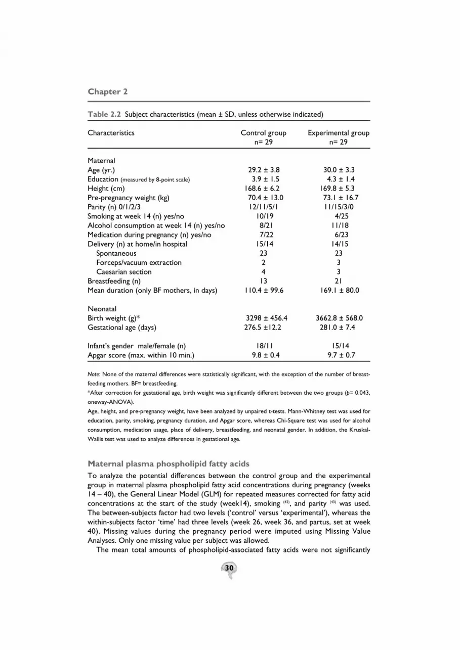
Maternal characteristicsThe maternal characteristics are shown in table 2.2. Comparison between the groups wasperformed using either the unpaired t-test or the Mann-Whitney test (continuous vari-ables), or the Chi-square test (discrete variables). No significant differences were observed.
Significantly more mothers in the experimental than in the control group breastfedtheir babies (Chi-square test, p= 0.020). Mean duration of breastfeeding by the lactatingmothers was not significantly different between the two groups, although a tendency wasobtained for a longer duration of breastfeeding in the experimental group (unpaired t-test, p= 0.095).
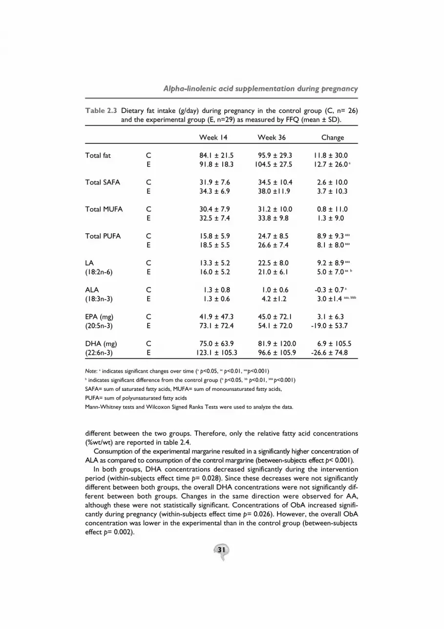
Maternal dietary fatty acid intakeThe average daily margarine consumption was 27.4 ± 3.2 g (mean ± SDdd) for the controlgroup and 27.8 ± 3.3 for the experimental group. This difference was not significant(unpaired t-test). Table 2.3 shows the dietary fatty acid intake, measured by FFQ, inweeks 14 and 36 of pregnancy. At week 14, the differences were not significant betweenthe two groups (Mann-Whitney test). The same holds true for week 36, with the excep-tion of the ALA intake, which was significantly higher in the experimental group (Mann-Whitney test, p < 0.000)
During the intervention period, the intakes of total PUFAs and LA increased signifi-cantly in both groups, whereas total fat intake increased only significantly in the experi-mental group (Wilcoxon Signed Ranks Test). The increase for LA was significantly largerin the control group than in the experimental group (Mann-Whitney U test). ALA con-sumption significantly increased in the experimental group and decreased in the controlgroup (Wilcoxon Signed Ranks Test), making the response difference between the groupshighly significant (Mann-Whitney U test).The higher DHA consumption in the experimental group at week 14 of pregnancy showed a trend for significance (p= 0.074, Mann-Whitney U test) compared to the con-trol group at week 14. The changes in DHA and EPA consumptions during pregnancywere not significantly different between the two groups (Mann-Whitney U test).However, the decreases in DHA and EPA intakes in the experimental group showed atrend for significance (p= 0.051, and p= 0.076, respectively).
Alpha-linolenic acid supplementation during pregnancy
30
Table 2.2 Subject characteristics (mean ± SD, unless otherwise indicated)
Characteristics Control group Experimental groupn= 29 n= 29
Maternal Age (yr.) 29.2 ± 3.8 30.0 ± 3.3 Education (measured by 8-point scale) 3.9 ± 1.5 4.3 ± 1.4 Height (cm) 168.6 ± 6.2 169.8 ± 5.3 Pre-pregnancy weight (kg) 70.4 ± 13.0 73.1 ± 16.7 Parity (n) 0/1/2/3 12/11/5/1 11/15/3/0 Smoking at week 14 (n) yes/no 10/19 4/25 Alcohol consumption at week 14 (n) yes/no 8/21 11/18 Medication during pregnancy (n) yes/no 7/22 6/23 Delivery (n) at home/in hospital 15/14 14/15
Spontaneous 23 23Forceps/vacuum extraction 2 3Caesarian section 4 3
Breastfeeding (n) 13 21 Mean duration (only BF mothers, in days) 110.4 ± 99.6 169.1 ± 80.0
NeonatalBirth weight (g)* 3298 ± 456.4 3662.8 ± 568.0 Gestational age (days) 276.5 ±12.2 281.0 ± 7.4
Infant’s gender male/female (n) 18/11 15/14 Apgar score (max. within 10 min.) 9.8 ± 0.4 9.7 ± 0.7
Note: None of the maternal differences were statistically significant, with the exception of the number of breast-
feeding mothers. BF= breastfeeding.
*After correction for gestational age, birth weight was significantly different between the two groups (p= 0.043,
oneway-ANOVA).
Age, height, and pre-pregnancy weight, have been analyzed by unpaired t-tests. Mann-Whitney test was used for
education, parity, smoking, pregnancy duration, and Apgar score, whereas Chi-Square test was used for alcohol
consumption, medication usage, place of delivery, breastfeeding, and neonatal gender. In addition, the Kruskal-
Wallis test was used to analyze differences in gestational age.
Maternal plasma phospholipid fatty acidsTo analyze the potential differences between the control group and the experimentalgroup in maternal plasma phospholipid fatty acid concentrations during pregnancy (weeks14 – 40), the General Linear Model (GLM) for repeated measures corrected for fatty acid concentrations at the start of the study (week14), smoking (42), and parity (43) was used.The between-subjects factor had two levels (‘control’ versus ‘experimental’), whereas thewithin-subjects factor ‘time’ had three levels (week 26, week 36, and partus, set at week4 0 ) . Missing values during the pregnancy period were imputed using Missing ValueAnalyses. Only one missing value per subject was allowed.
The mean total amounts of phospholipid-associated fatty acids were not significantly
Chapter 2
31
Table 2.3 Dietary fat intake (g/day) during pregnancy in the control group (C, n= 26) and the experimental group (E, n=29) as measured by FFQ (mean ± SD).
Week 14 Week 36 Change
Total fat C 84.1 ± 21.5 95.9 ± 29.3 11.8 ± 30.0E 91.8 ± 18.3 104.5 ± 27.5 12.7 ± 26.0 a
Total SAFA C 31.9 ± 7.6 34.5 ± 10.4 2.6 ± 10.0E 34.3 ± 6.9 38.0 ±11.9 3.7 ± 10.3
Total MUFA C 30.4 ± 7.9 31.2 ± 10.0 0.8 ± 11.0E 32.5 ± 7.4 33.8 ± 9.8 1.3 ± 9.0
Total PUFA C 15.8 ± 5.9 24.7 ± 8.5 8.9 ± 9.3 aaa
E 18.5 ± 5.5 26.6 ± 7.4 8.1 ± 8.0 aaa
LA C 13.3 ± 5.2 22.5 ± 8.0 9.2 ± 8.9 aaa
(18:2n-6) E 16.0 ± 5.2 21.0 ± 6.1 5.0 ± 7.0 aa b
ALA C 1.3 ± 0.8 1.0 ± 0.6 -0.3 ± 0.7 a
(18:3n-3) E 1.3 ± 0.6 4.2 ±1.2 3.0 ±1.4 aaa, bbb
EPA (mg) C 41.9 ± 47.3 45.0 ± 72.1 3.1 ± 6.3(20:5n-3) E 73.1 ± 72.4 54.1 ± 72.0 -19.0 ± 53.7
DHA (mg) C 75.0 ± 63.9 81.9 ± 120.0 6.9 ± 105.5(22:6n-3) E 123.1 ± 105.3 96.6 ± 105.9 -26.6 ± 74.8
Note: a indicates significant changes over time (a p<0.05, aa p<0.01, aaap<0.001)b indicates significant difference from the control group (b p<0.05, bb p<0.01, bbb p<0.001)
SAFA= sum of saturated fatty acids, MUFA= sum of monounsaturated fatty acids,
PUFA= sum of polyunsaturated fatty acids
Mann-Whitney tests and Wilcoxon Signed Ranks Tests were used to analyze the data.
different between the two groups. Therefore, only the relative fatty acid concentrations(%wt/wt) are reported in table 2.4.
Consumption of the experimental margarine resulted in a significantly higher concentration ofALA as compared to consumption of the control margarine (between-subjects effect p< 0.001).
In both groups, DHA concentrations decreased significantly during the interventionperiod (within-subjects effect time p= 0.028). Since these decreases were not significantly different between both groups, the overall DHA concentrations were not significantly dif-ferent between both groups. Changes in the same direction were observed for AA,although these were not statistically significant. Concentrations of ObA increased signifi-cantly during pregnancy (within-subjects effect time p= 0.026). However, the overall ObAconcentration was lower in the experimental than in the control group (between-subjectseffect p= 0.002).
Alpha-linolenic acid supplementation during pregnancy

32
Table 2.4 Maternal plasma phospholipid-associated fatty acid values over time of the control group (C) and the experimental group (E) (mean ± SD)
Fatty acids week 14 week 26 week 36 week 40 week 72(% wt/wt) n= 29/29 n= 29/29 n=28/29 n= 26/27 n= 28/28
Total (mg/l)aa C 1533 ± 278 1849 ± 345 1985 ± 420 1880 ± 400 1338 ± 239 E 1530 ± 307 1870 ± 374 2015 ± 374 2001 ± 377 1386 ± 254
18:2n-6 C 21.08 ± 2.15 22.74 ± 2.18 22.60 ± 2.61 21.96 ± 2.05 21.80 ± 2.44 (LA) E 21.39 ± 2.88 23.00 ± 2.49 23.52 ± 2.66 22.17 ± 2.67 22.77 ± 3.15
20:3n-6 * C 3.51 ± 0.62 3.58 ± 0.65 3.55 ± 0.75 3.69 ± 0.79 3.19 ± 0.56 (DGLA) E 3.54 ± 0.72 3.38 ± 0.63 3.23 ± 0.61 3.30 ± 0.64 3.29 ± 0.83
20:4n-6 C 9.71 ± 1.73 8.72 ± 1.26 8.25 ± 1.22 8.73 ± 1.26 10.03 ± 1.71(AA) E 9.56 ± 1.70 8.06 ± 1.62 7.80 ± 1.51 8.19 ± 1.36 9.54 ± 1.93
22:4n-6***, aa C 0.38 ± 0.09 0.40 ± 0.10 0.38 ± 0.08 0.38 ± 0.06 0.32 ± 0.08(AdrA) E 0.34 ± 0.07 0.31 ± 0.06 0.29 ± 0.06 0.31 ± 0.07 0.32 ± 0.10
22:5n-6**, a C 0.36 ± 0.16 0.40 ± 0.16 0.44 ± 0.14 0.47 ± 0.19 0.21 ± 0.09(ObA) E 0.30 ± 0.08 0.28 ± 0.08 0.30 ± 0.11 0.33 ± 0.11 0.21 ± 0.08
LCP n-6 * C 13.95 ± 1.85 13.10 ± 1.53 12.61 ± 1.56 13.26 ± 1.55 13.76 ± 2.00 E 13.75 ± 1.89 12.03 ± 1.99 11.62 ± 2.00 12.13 ± 1.86 13.36 ± 2.45
18:3n-3 *** C 0.22 ± 0.08 0.23 ± 0.07 0.21 ± 0.06 0.20 ± 0.07 0.15 ± 0.06(ALA) E 0.22 ± 0.08 0.43 ± 0.13 0.39 ± 0.13 0.38 ± 0.14 0.20 ± 0.07##
20:5n-3 ** C 0.52 ± 0.58 0.36 ± 0.28 0.26 ± 0.11 0.25 ± 0.11 0.54 ± 0.32(EPA) E 0.52 ± 0.31 0.51 ± 0.38 0.39 ± 0.21 0.47 ± 0.25 0.73 ± 0.44
22:5n-3 *** C 0.69 ± 0.23 0.54 ± 0.16 0.50 ± 0.15 0.51 ± 0.17 0.70 ± 0.27(DPA) E 0.70 ± 0.21 0.64 ± 0.16 0.59 ± 0.16 0.62 ± 0.19 0.80 ± 0.25
22:6n-3 a C 3.97 ± 0.85 3.68 ± 0.90 3.64 ± 0.92 3.46 ± 0.84 3.10 ± 1.01(DHA) E 4.48 ± 1.01 3.89 ± 0.68 3.95 ± 0.87 3.94 ± 0.96 3.10 ± 0.80
LCP n-3 a C 5.18 ± 1.36 4.58 ± 1.18 4.40 ± 1.08 4.23 ± 1.02 4.35 ± 1.31 E 5.70 ± 1.39 5.04 ± 1.02 4.93 ± 1.16 5.03 ± 1.31 4.63 ± 1.17
sum n-7 C 1.81 ± 0.30 1.72 ± 0.26 1.75 ± 0.24 1.84 ± 0.34 1.85 ± 0.31 E 1.76 ± 0.21 1.62 ± 0.24 1.61 ± 0.23 1.76 ± 0.32 1.70 ± 0.30
sum n-9 C 10.90 ± 1.21 10.34 ± 1.13 10.76 ± 1.29 10.82 ± 1.17 10.96 ± 0.95 E 10.33 ± 1.13 10.10 ± 1.10 10.49 ± 1.12 10.81 ± 1.18 10.41 ± 1.53
Chapter 2
33
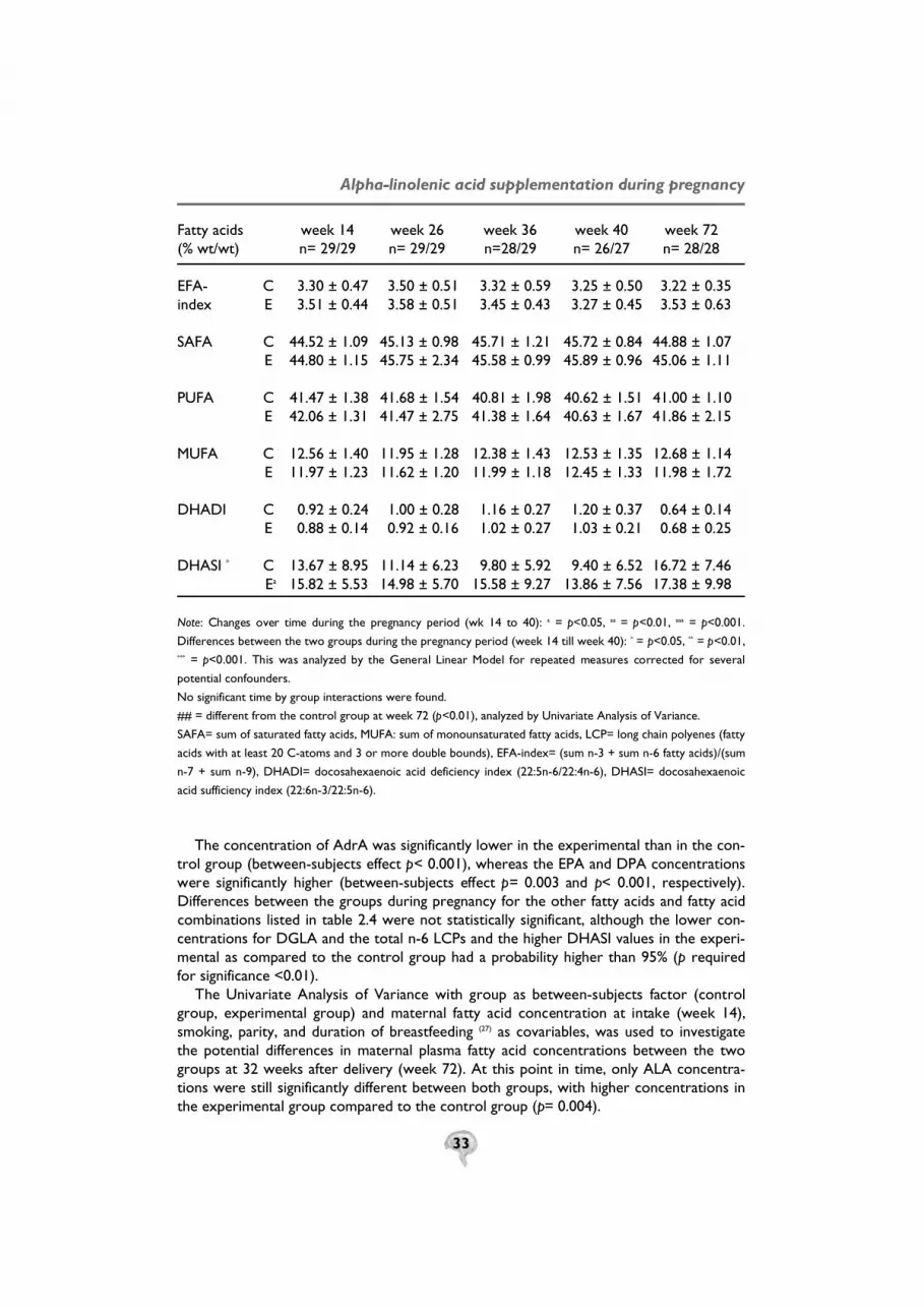
Fatty acids week 14 week 26 week 36 week 40 week 72(% wt/wt) n= 29/29 n= 29/29 n=28/29 n= 26/27 n= 28/28
EFA- C 3.30 ± 0.47 3.50 ± 0.51 3.32 ± 0.59 3.25 ± 0.50 3.22 ± 0.35 index E 3.51 ± 0.44 3.58 ± 0.51 3.45 ± 0.43 3.27 ± 0.45 3.53 ± 0.63
SAFA C 44.52 ± 1.09 45.13 ± 0.98 45.71 ± 1.21 45.72 ± 0.84 44.88 ± 1.07 E 44.80 ± 1.15 45.75 ± 2.34 45.58 ± 0.99 45.89 ± 0.96 45.06 ± 1.11
PUFA C 41.47 ± 1.38 41.68 ± 1.54 40.81 ± 1.98 40.62 ± 1.51 41.00 ± 1.10 E 42.06 ± 1.31 41.47 ± 2.75 41.38 ± 1.64 40.63 ± 1.67 41.86 ± 2.15
MUFA C 12.56 ± 1.40 11.95 ± 1.28 12.38 ± 1.43 12.53 ± 1.35 12.68 ± 1.14E 11.97 ± 1.23 11.62 ± 1.20 11.99 ± 1.18 12.45 ± 1.33 11.98 ± 1.72
DHADI C 0.92 ± 0.24 1.00 ± 0.28 1.16 ± 0.27 1.20 ± 0.37 0.64 ± 0.14 E 0.88 ± 0.14 0.92 ± 0.16 1.02 ± 0.27 1.03 ± 0.21 0.68 ± 0.25
DHASI * C 13.67 ± 8.95 11.14 ± 6.23 9.80 ± 5.92 9.40 ± 6.52 16.72 ± 7.46 Ea 15.82 ± 5.53 14.98 ± 5.70 15.58 ± 9.27 13.86 ± 7.56 17.38 ± 9.98
N o t e: Changes over time during the pregnancy period (wk 14 to 40): a = p<0.05, a a = p<0.01, a a a = p< 0 . 0 0 1 .
Differences between the two groups during the pregnancy period (week 14 till week 40): * = p<0.05, ** = p<0.01,* * * = p<0.001. This was analyzed by the General Linear Model for repeated measures corrected for several
potential confounders.
No significant time by group interactions were found.
## = different from the control group at week 72 (p<0.01), analyzed by Univariate Analysis of Variance.
SAFA= sum of saturated fatty acids, MUFA: sum of monounsaturated fatty acids, LCP= long chain polyenes (fatty
acids with at least 20 C-atoms and 3 or more double bounds), EFA-index= (sum n-3 + sum n-6 fatty acids)/(sum
n-7 + sum n-9), DHADI= docosahexaenoic acid deficiency index (22:5n-6/22:4n-6), DHASI= docosahexaenoic
acid sufficiency index (22:6n-3/22:5n-6).
The concentration of AdrA was significantly lower in the experimental than in the con-trol group (between-subjects effect p< 0.001), whereas the EPA and DPA concentrationswere significantly higher (between-subjects effect p= 0.003 and p< 0.001, respectively).Differences between the groups during pregnancy for the other fatty acids and fatty acidcombinations listed in table 2.4 were not statistically significant, although the lower con-centrations for DGLA and the total n-6 LCPs and the higher DHASI values in the experi-mental as compared to the control group had a probability higher than 95% (p requiredfor significance <0.01).
The Univariate Analysis of Variance with group as between-subjects factor (controlgroup, experimental group) and maternal fatty acid concentration at intake (week 14),smoking, parity, and duration of breastfeeding (27) as covariables, was used to investigatethe potential differences in maternal plasma fatty acid concentrations between the twogroups at 32 weeks after delivery (week 72). At this point in time, only ALA concentra-tions were still significantly different between both groups, with higher concentrations inthe experimental group compared to the control group (p= 0.004).
Alpha-linolenic acid supplementation during pregnancy

34
Neonatal fatty acidsThe mean total amounts of phospholipid-associated fatty acids in umbilical plasma phos-pholipids and in phospholipids of umbilical vessel walls (both venous and arterial) werenot significantly different between the two groups (unpaired t-tests). Therefore, only therelative amounts of fatty acids (%wt/wt) are reported in table 2.5.
The differences in neonatal fatty acid concentrations between the two groups wereanalyzed by Univariate Analysis of Variance with correction for gestational age, parity,maternal smoking and maternal fatty acid concentrations at study intake (week14), if nec-essary after log-transformation to ascertain normality.
Table 2.5 Neonatal phospholipid-associated fatty acids in the control (C) and experi-mental (E) group (mean ± SD).
Fatty acids neonatal plasma venous wall arterial wall%wt/wt n=26/28 n=28/28 n=28/28
Total mg/l C 653 ± 107 479 ± 149 610 ± 176 plasma or mg/kg tissue E 648 ± 131 546 ± 151 624 ± 210
18:2n-6 C 7.62 ± 1.24 1.99 ± 0.41 1.19 ± 0.24(LA) E 8.01 ± 1.16 2.02 ± 0.39 1.33 ± 0.27
20:3n-6 C 4.81 ± 0.82 1.93 ± 0.44 1.13 ± 0.26(DGLA) E 4.92 ± 0.79 1.99 ± 0.28 1.26 ± 0.22a
20:4n-6 C 17.34 ± 1.35 18.50 ± 1.02 13.23 ± 2.02(AA) E 16.05 ± 1.43
aa18.36 ± 1.18 12.89 ± 1.60
22:4n-6 C 0.81 ± 0.19 4.85 ± 0.66 2.83 ± 0.52(AdrA) E 0.75 ± 0.18 4.97 ± 0.77 2.93 ± 0.65
22:5n-6 C 0.71 ± 0.32 2.87 ± 0.62 3.10 ± 0.53 (ObA) E 0.58 ± 0.23 2.48 ± 0.47 2.70 ± 0.46a
LCP n-6 C 23.67 ± 1.56 28.16 ± 1.50 20.29 ± 2.68E 22.30 ± 1.64aa 27.80 ± 1.38 19.79 ± 2.41
18:3n-3 C n.d. 0.07 ± 0.01 0.11 ± 0.03(ALA) E n.d. 0.07 ± 0.02 0.12 ± 0.03
20:5n-3 C 0.13 ± 0.10 n.d. n.d. (EPA) E 0.26 ± 0.12aaa n.d. n.d.
22:5n-3 C 0.41 ± 0.29 0.23 ± 0.07 0.19 ± 0.08 (DPA) E 0.51 ± 0.21 0.35 ± 0.10aaa 0.29 ± 0.09aaa
22:6n-3 C 5.37 ± 1.39 4.88 ± 0.70 4.84 ± 0.87(DHA) E 5.65 ± 1.47 5.43 ± 0.81 5.21 ± 0.94
Chapter 2

35
Fatty acids neonatal plasma venous wall arterial wall%wt/wt n=26/28 n=28/28 n=28/28
LCP n-3 C 5.91 ± 1.65 5.14 ± 0.74 5.06 ± 0.94 E 6.42 ± 1.65 5.82 ± 0.86a 5.54 ± 0.99
sum n-7 C 3.12 ± 0.54 2.69 ± 0.24 3.18 ± 0.27 E 2.92 ± 0.35 2.64 ± 0.28 3.20 ± 0.35
sum n-9 C 10.11 ± 2.12 13.04 ± 1.17 21.66 ± 3.61 E 10.50 ± 1.96 13.01 ± 1.53 22.08 ± 3.19
EFA-index C 3.02 ± 0.65 2.36 ± 0.25 1.15 ± 0.35 E 2.92 ± 0.64 2.40 ± 0.30 1.12 ± 0.25
SAFA C 47.67 ± 1.40 47.02 ± 1.37 46.79 ± 1.84 E 48.04 ± 2.15 46.81 ± 1.22 46.20 ± 1.80
PUFA C 38.81 ± 2.09 37.42 ± 1.41 31.81 ± 2.63 E 38.36 ± 2.54 37.75 ± 1.16 32.33 ± 2.10
MUFA C 12.88 ± 2.29 15.04 ± 1.25 20.28 ± 2.61 E 13.08 ± 2.02 14.92 ± 1.42 20.41 ± 2.38
DHADI C 0.88 ± 0.29 0.60 ± 0.17 1.13 ± 0.28 E 0.79 ± 0.31 0.51 ± 0.11a 0.96 ± 0.26
DHASI C 9.02 ± 4.43 1.79 ± 0.55 1.62 ± 0.49E 11.28 ± 5.10 2.30 ± 0.69a 1.98 ± 0.50
Note: For legend see table 2.4.
n.d.= not reliably detectable; concentrations ≤ 0.05%a Difference from the control group a p<0.05, aa p<0.01, aaa p<0.001
In the Univariate Analysis of Variance corrections were made for gestational age, parity, maternal smoking, and
maternal fatty acid concentrations at the start of the study (week14).
GLM was used to evaluate the arterio-venous differences (data not shown). Significant within-subjects effects
were found for totFA, LA, ObA, sum n-9, MUFA, DHADI, and DHASI, whereas significant between-subjects
effects were found for ObA, DPA, DHADI, and DHASI.
No interaction-effects were found.
Umbilical venous plasmaUmbilical venous plasma samples were available for 26 newborns in the control group and28 neonates in the experimental group (see table 2.5).
No statistically significant difference was found in umbilical plasma phospholipids forDHA. In contrast, the AA concentration was significantly lower in the experimental group
Alpha-linolenic acid supplementation during pregnancy

36
as compared to the control group (p= 0.004). After log-transformation and correction forthe potential confounders, ObA concentrations were not significantly different betweenthe two groups (p= 0.095).
The average concentration of EPA was two times higher in the experimental as com-pared to the control group (p< 0.001), whereas total n-6 LCP levels were significantlylower (p= 0.004). Values for other fatty acids and fatty acid combinations were not signifi-cantly different between both groups.
Umbilical vein wallsFrom both groups, 28 samples of umbilical veins (and arteries) were available for analysis.Results are also shown in table 2.5. After correction for the potential confounders, nei-ther the DHA (p= 0.183), nor the ObA (p= 0.085) concentrations were significantly dif-ferent between the two groups. Also, AA concentrations were not significantly differentbetween the two groups.
DPA was significantly higher in the experimental group as compared to the controlgroup (p< 0.001). The DHASI and LCPn-3 showed trends for higher values (p= 0.023 andp= 0.038, respectively) in the experimental group, whereas the DHADI (22:5n-6/22:4n-6)showed a trend for a lower value (p= 0.034). Other fatty acids and fatty acid combina-tions were not significantly different between the two groups.
Umbilical arterial wallsAfter correction for the potential confounders, ObA showed significantly lower concen-trations in the phospholipids of the experimental group compared to the control group(p= 0.037). The differences for DHA and AA concentrations did not reach statistical significance.
DPA concentrations were again significantly higher in the experimental group (p< 0.001).Differences between both groups for the other fatty acids and fatty acid combinations(see table 2.5) were not significant at all.
Arterio-venous differences As demonstrated by General Linear Model, significant within-subjects effects for AV-dif-ferences for many fatty acids (totFA, LA, ObA, sum n-9, MUFA, and DHADI) listed intable 2.5 were found (data not shown), whereas the interaction effects group by AV-dif-ferences were not significant. Consequently, the AV-differences in the experimentalgroup were not significantly different from those in the control group. However, signifi-cant between-subjects effects were found for ObA, DPA, DHADI, and DHASI, indicatingthat the average amount of fatty acid phospholipids in arterial and venous vessel walls wasrespectively lower for DPA and DHASI, and higher for ObA and DHADI in the controlgroup.
Neonatal outcome parametersUsing a one-way ANOVA, it appeared that neonates in the experimental group had, onaverage, a significantly higher birth weight compared to newborns in the placebo group(p= 0.043; see table 2.2). Mean pregnancy duration was 4.5 days longer in the experimen-tal than in the control group (Table 2.2). This difference tended to be significant as well(Kruskal-Wallis test, p= 0.091). No significant differences between both groups wereobserved for Apgar score (Mann-Whitney test) and gender (Chi-square test, p> 0.05).
Chapter 2

37
DISCUSSION
GeneralFrom longitudinal studies it is known that under the present dietary conditions, relativeAA and DHA concentrations in maternal plasma phospholipids decrease during the secondand third trimester of pregnancy (1, 37, 44, 45). It was suggested that supplementation with acombination of n-6 and n-3 fatty acids would be required for an optimal fatty acid status ( 4 6 ).Although endogenous DHA production from dietary ALA is known to be low (23-26), Ottoet al recently obtained some indications that LCP synthesis from EFA precursors may beenhanced during pregnancy (27). Therefore, in the present study a margarine high in ALAand LA was tested for its efficacy to increase maternal and neonatal DHA concentrationswithout reducing the AA concentrations. In this study some neonatal outcome parame-ters were measured as well. Some of the results obtained need special attention and willbe discussed here.
Maternal dietary fat intakeAnalysis of the food frequency questionnaires (FFQs) showed that in the present studytotal fat intake of the participants increased during pregnancy, although this incrementwas significant for the experimental group only and the difference in intake between thetwo groups was not significant. Al and coworkers, using the same FFQ, did not observe achange in habitual fat consumption during the second and third trimester of pregnancy (3).Therefore, the difference we observed likely resulted from the study regime. This wasconfirmed by an item analysis of the FFQs, revealing a rather frequent habitual use of lightmargarines at week 14, which in the study were replaced by the full fat experimental andcontrol intervention products. The higher fat intake provided about 108 Kcal (0.026 MJ)per day. This compensates partly for the higher energy intake advised during pregnancy ( 4 7 ).
Maternal plasma fatty acid concentrations during pregnancyIn the present study, supplementation with margarines rich in ALA+LA or in LA only didnot prevent the well-known reductions in DHA and AA concentrations during the sec-ond and third trimester of pregnancy (Table 2.4). In addition, supplementation with theALA+LA rich margarine did not result in significantly higher plasma DHA concentrationsthan a comparable margarine without ALA. Although it cannot be excluded that a higherALA dose may increase plasma DHA concentrations, this seems rather unlikely, sinceFrancois et al showed that even an ALA dose of 10.7 g/day did not lead to increased plas-ma DHA levels in lactating women (48). Interestingly, the lower ObA concentration in theexperimental group of our study indicates that the functional DHA status may have beenslightly higher upon ALA supplementation, since ObA is generally accepted as a functionalshortage marker for DHA (41). This suggests that any additional DHA that may have beenproduced from the supplemented ALA was transferred directly to certain (fetal) targettissues and, therefore, did not increase the DHA concentration of maternal plasma phos-pholipids.
Maternal fatty acids 32 weeks postpartumIn this study also the effect of ALA supplementation during pregnancy on maternal post-partum plasma fatty acid concentrations was studied in blood collected 32 weeks afterdelivery. The choice for this moment in time was based on an earlier study by Otto et al,showing that 32 weeks after delivery the maternal EFA and LCP status had returned to
Alpha-linolenic acid supplementation during pregnancy
38
pre-pregnancy levels, irrespective of the fact whether the mother had been breastfeedingor not (27). We confirmed that at 32 weeks postpartum the concentrations of most fattyacids had returned to early pregnancy values or beyond, and were not significantly differ-ent between both groups. The only exception was ALA, which was still higher in theexperimental group. Although this may indicate a long-term effect of the dietary interven-tion, a difference due to selection bias cannot be ruled out.
Neonatal phospholipid fatty acidsThe lower umbilical arterial vessel walls concentration of ObA suggests a higher functionalDHA status in infants of ALA-supplemented mothers, although the DHA concentrationitself was not higher. This suggestion is supported by higher values for the DHA sufficien-cy index (DHASI) and lower values for the DHA deficiency index (DHADI) in theseneonates, which were significant for the umbilical venous vessel wall phospholipids.Moreover, maternal ALA supplementation during pregnancy resulted in higher neonatalconcentrations of EPA (plasma only), and DPA (umbilical vessel walls). This was associat-ed with significantly lower neonatal concentrations of AA and total n-6 LCP values (signif-icant in plasma only). These results demonstrate that maternal ALA consumption duringpregnancy hardly increases the neonatal DHA status, and does not prevent the reductionin n-6 LCPs, also observed after fish oil supplementation during pregnancy (20, 21).
Neonatal outcome parametersHigher birth weight has been associated with lower risk for the development of adultheart disease, Type II diabetes mellitus and related risk factors such as hypertension anddyslipidemia (49-52). Consequently, the significantly higher birth weight of neonates born toALA-supplemented mothers may be relevant indeed. We, therefore, performed a post-hoc stepwise multiple regression analysis to detect possible confounding variables. Basedupon the literature, gestational age, alcohol consumption, neonatal gender, maternal pre-pregnancy weight, maternal height, length and weight of the father, smoking and drugusage during pregnancy were included in the model. Gestational age, maternal pre-preg-nancy weight, smoking and height of the father contributed significantly to the model (p< 0.05), and after correction for these possible confounders no statistically significant dif-ference in birth weight remained between the experimental group and the control group.
In conclusionSupplementation during pregnancy with ALA+LA does not prevent the maternal LCPdecrease during pregnancy, whereas the small improvement of the neonatal DHA statusis accompanied by a reduction in the neonatal n-6 LCP status. Since supplementation withDHA is more efficient than ALA in promoting the DHA status (53), we suggest that a mix-ture of DHA and AA may be a more efficient option to optimize the maternal and neona-tal LCP status.
ACKNOWLEDGEMENTSThe authors like to thank all mothers and their babies for participating in this study. Theparticipation of the midwives from the region of Southern Limburg and the medical staffof the departments of Obstetrics and Gynecology of the hospitals in the same area isgreatly appreciated. We acknowledge Unilever Research and Development Vlaardingen(URDV) for the donation of the margarines and their financial support. The authors wish
Chapter 2

39
to thank Mrs. H. Aydeniz for her technical assistance, Dr. A. Kester for his statisticaladvice and Mrs. A. Badart-Smook, Mrs. J. Breedveld (NutriScience) and Mrs. I. Verkooijen(NutriScience) for their help with the analyses of the food frequency questionnaires, andDr. E. de Deckere (URDV) for critically reading the manuscript.
Alpha-linolenic acid supplementation during pregnancy
40
REFERENCES
1. Al M. D., v. Houwelingen A. C., Kester A. D., Hasaart T. H., de Jong A. E., Hornstra G.Maternal essential fatty acid patterns during normal pregnancy and their relationship to theneonatal essential fatty acid status. Br J Nutr 1995;74:55-68.
2. Crawford M. Placental delivery of arachidonic and docosahexaenoic acids: implications for thelipid nutrition of preterm infants. Am J Clin Nutr 2000;71:275S-84S.
3. Al M. D., Badart-Smook A., v. Houwelingen A. C., Hasaart T. H., Hornstra G. Fat intake ofwomen during normal pregnancy: relationship with maternal and neonatal essential fatty acidstatus. J Am Coll Nutr 1996;15:49-55.
4. Hornstra G. Essential fatty acids in mothers and their neonates. Am J Clin Nutr 2000;71:1262S-9S.5. Fliesler S. J., Anderson R. E. Chemistry and metabolism of lipids in the vertebrate retina. Prog
Lipid Res 1983;22:79-131.6. Sastry P. S. Lipids of nervous tissue: composition and metabolism. Prog Lipid Res 1985;24:69-176.7. Svennerholm L. Distribution and fatty acid composition of phosphoglycerides in normal
human brain. J Lipid Res 1968;9:570-9.8. Wolke D., Meyer R. Cognitive status, language attainment, and prereading skills of 6-year-old
very preterm children and their peers: the Bavarian Longitudinal Study. Dev Med ChildNeurol 1999;41:94-109.
9. Wolke D., Ratschinski G., Ohrt B., Riegel K. The cognitive outcome of very preterm infantsmay be poorer than often reported: an empirical investigation of how methodological issuesmake a big difference. Eur J Pediatr 1994;153:906-15.
10. Simmer K. Longchain polyunsaturated fatty acid supplementation in preterm infants.Cochrane Database Syst Rev 2000:CD000375.
11. SanGiovanni J. P., Parra-Cabrera S., Colditz G. A., Berkey C. S., Dwyer J. T. Meta-analysis ofdietary essential fatty acids and long-chain polyunsaturated fatty acids as they relate to visualresolution acuity in healthy preterm infants. Pediatrics 2000;105:1292-8.
12. O’Connor D. L., Hall R., Adamkin D., et al. Growth and development in preterm infants fedlong-chain polyunsaturated fatty acids: a prospective, randomized controlled trial. Pediatrics2001;108:359-71.
13. Foreman-van Drongelen M. M., Al M. D., v. Houwelingen A. C., Blanco C. E., Hornstra G.Comparison between the essential fatty acid status of preterm and full-term infants, meas-ured in umbilical vessel walls. Early Hum Dev 1995;42:241-51.
14. Simmer K. Longchain polyunsaturated fatty acid supplementation in infants born at term.Cochrane Database Syst Rev 2001:CD000376.
15. SanGiovanni J. P., Berkey C. S., Dwyer J. T., Colditz G. A. Dietary essential fatty acids, long-chain polyunsaturated fatty acids, and visual resolution acuity in healthy fullterm infants: a sys-tematic review. Early Hum Dev 2000;57:165-88.
16. Agostoni C., Trojan S., Bellu R., Riva E., Giovannini M. Neurodevelopmental quotient ofhealthy term infants at 4 months and feeding practice: the role of long-chain polyunsaturatedfatty acids. Pediatr Res 1995;38:262-6.
17. Willatts P., Forsyth J. S., DiModugno M. K., Varma S., Colvin M. Effect of long-chain polyunsat-urated fatty acids in infant formula on problem solving at 10 months of age. Lancet1998;352:688-91.
18. Birch E. E., Garfield S., Hoffman D. R., Uauy R., Birch D. G. A randomized controlled trial ofearly dietary supply of long-chain polyunsaturated fatty acids and mental development in terminfants. Dev Med Child Neurol 2000;42:174-81.
Chapter 2

41
19. Bakker E. C. Long-chain polyunsaturated fatty acids and child development (thesis). HumanBiology. Maastricht: Maastricht University, 2002.
20. Helland I. B., Saugstad O. D., Smith L., et al. Similar effects on infants of n-3 and n-6 fatty acidssupplementation to pregnant and lactating women. Pediatrics 2001;108:E82.
21. v. Houwelingen A. C., Sorensen J. D., Hornstra G., et al. Essential fatty acid status in neonatesafter fish-oil supplementation during late pregnancy. Br J Nutr 1995;74:723-31.
22. Lauritzen L., Hansen H. S., Jorgensen M. H., Michaelsen K. F. The essentiality of long chain n-3fatty acids in relation to development and function of the brain and retina. Prog Lipid Res2001;40:1-94.
23. Burdge G. C., Jones A. E., Wootton S. A. Eicosapentaenoic and docosapentaenoic acids arethe principal products of alpha-linolenic acid metabolism in young men*. Br J Nutr2002;88:355-63.
24. Burdge G. C., Wootton S. A. Conversion of alpha-linolenic acid to eicosapentaenoic, docos-apentaenoic and docosahexaenoic acids in young women. Br J Nutr 2002;88:411-20.
25. Gerster H. Can adults adequately convert alpha-linolenic acid (18:3n-3) to eicosapentaenoicacid (20:5n-3) and docosahexaenoic acid (22:6n-3)? Int J Vitam Nutr Res 1998;68:159-73.
26. Vermunt S. H., Mensink R. P., Simonis M. M., Hornstra G. Effects of dietary alpha-linolenicacid on the conversion and oxidation of 13C-alpha-linolenic acid. Lipids 2000;35:137-42.
27. Otto S. J., v. Houwelingen A. C., Badart-Smook A., Hornstra G. The postpartum docosa-hexaenoic acid status of lactating and nonlactating mothers. Lipids 1999;34:S227.
28. Greiner R. C., Winter J., Nathanielsz P. W., Brenna J. T. Brain docosahexaenoate accretion infetal baboons: bioequivalence of dietary alpha-linolenic and docosahexaenoic acids. PediatrRes 1997;42:826-34.
29. Al M. D., v. Houwelingen A. C., Badart-Smook A., Hornstra G. Some aspects of neonatalessential fatty acid status are altered by linoleic acid supplementation of women during preg-nancy. J Nutr 1995;125:2822-30.
30. Al M. D. M. Essential fatty acids, pregnancy, and pregnancy outcome: relationship betweenmother and child (thesis). Human Biology. Maastricht: Maastricht University, 1994.
31. Reports of the Scientific Committee for Food, Nutrient and energy intakes for the EuropeanCommunity, 1993:52-60.
32. Scientific Review committee. Nutrition Recommendations Ottawa: Minister of nationalHealth and Welfare, Canada, 1990.
33. FAO/WHO. Fats and oils in human nutrition (Report of a joint expert consultation), Fat andNutrition paper 57, Rome: FAO, 1994.
34. Scholte B. A. Komeet 1996 version 2.0. 2.0d ed. Arnhem, 1995.35. Netherlands Bureau of Nutrition Education. Dutch food composition Table 1996. The Hague, 1996.36. Bie de S. E. Standaardvragen 1987: Voorstellen voor uniformering van vraagstellingen naar
achtergrondkenmerken en interviews [Standard questions 1987: Proposal for uniformisationof questions regarding background variables and interviews]. 2 ed. Leiden, The Netherlands:Leiden University Press, 1987.
37. Otto S. J., v. Houwelingen A. C., Antal M., et al. Maternal and neonatal essential fatty acid sta-tus in phospholipids: an international comparative study. Eur J Clin Nutr 1997;51:232-42.
38. Bligh E. G., Dyer W. J. A rapid method of total lipid extraction and purification. Can. J. Biochem. Physiol. 1959;37:911-917.
39. Kaluzny M. A., Duncan L. A., Merritt M. V., Epps D. E. Rapid separation of lipid classes in highyield and purity using bonded phase columns. J Lipid Res 1985;26:135-40.
40. Morrison W. R., Smith L. M. Preparation of fatty acid methyl esters and dimethylacetals fromlipids with boron fluoride-methanol. J Lipid Res 1964;5:600-608.
Alpha-linolenic acid supplementation during pregnancy

42
41. Neuringer M., Connor W. E., Lin D. S., Barstad L., Luck S. Biochemical and functional effectsof prenatal and postnatal omega 3 fatty acid deficiency on retina and brain in rhesus monkeys.Proc Natl Acad Sci U S A 1986;83:4021-5.
42. Rise P., Marangoni F., Galli C. Regulation of PUFA metabolism: pharmacological and toxico-logical aspects. Prostaglandins Leukot Essent Fatty Acids 2002;67:85-9.
43. Al M. D., v. Houwelingen A. C., Hornstra G. Long-chain polyunsaturated fatty acids, pregnancy,and pregnancy outcome. Am J Clin Nutr 2000;71:285S-91S.
44. Matorras R., Ruiz J. I., Perteagudo L., et al. Longitudinal study of fatty acids in plasma and ery-throcyte phospholipids during pregnancy. J Perinat Med 2001;29:293-7.
45. Sanjurjo P., Matorras R., Ingunza N., Alonso M., Rodriguez-Alarcon J., Perteagudo L. Cross-sectional study of percentual changes in total plasmatic fatty acids during pregnancy. HormMetab Res 1993;25:590-2.
46. Hornstra G., Al M. D., v. Houwelingen A. C., Foreman-van Drongelen M. M. Essential fatty acidsin pregnancy and early human development. Eur J Obstet Gynecol Reprod Biol 1995;61:57-62.
47. Pitkin R. M. Nutritional requirements in normal pregnancy. Diabetes Care 1980;3:472-5.48. Francois C. A., Connor S. L., Bolewicz L. C., Connor W. E. Supplementing lactating women with
flaxseed oil does not increase docosahexaenoic acid in their milk. Am J Clin Nutr 2003;77:226-33.49. Barker D. J., Winter P. D., Osmond C., Margetts B., Simmonds S. J. Weight in infancy and
death from ischaemic heart disease. Lancet 1989;2:577-80.50. Hales C. N., Barker D. J., Clark P. M., et al. Fetal and infant growth and impaired glucose tol-
erance at age 64. BMJ 1991;303:1019-22.51. Osmond C., Barker D. J., Winter P. D., Fall C. H., Simmonds S. J. Early growth and death
from cardiovascular disease in women. BMJ 1993;307:1519-24.52. Barker D. J., Hales C. N., Fall C. H., Osmond C., Phipps K., Clark P. M. Type 2 (non-insulin-
dependent) diabetes mellitus, hypertension and hyperlipidaemia (syndrome X): relation toreduced fetal growth. Diabetologia 1993;36:62-7.
53. Brenna J. T. Efficiency of conversion of alpha-linolenic acid to long chain n-3 fatty acids inman. Curr Opin Clin Nutr Metab Care 2002;5:127-32.
Chapter 2

43

44
MEMORY PERFORMANCE,BUT NOT INFORMATION
PROCESSING SPEED,MAY BE REDUCED DURING
EARLY PREGNANCY
Published in Journal of Clinical
and Experimental Neuropsychology, 2003; 25: 483-489
Renate H.M. de Groot, Gerard Hornstra, Nico Rozendaal, and Jelle Jolles.
46
ABSTRACT
Several studies have investigated aspects of cognitive functioning during late
pregnancy or in the period around delivery. The present paper describes a
controlled study of neurocognitive functioning in an early phase of pregnanc y
(14 weeks). Seventy-one pregnant women and 57 control subjects matched
for age and education were tested with a cognitive test battery. Intentional
learning was tested with the Verbal Learning Test, retrieval from semantic
memory with the Fluency Test, and speed of information processing with
the Concept Shifting Test, the Stroop test, and the Letter Digit Substitution
Test. Results show that performance on tests measuring intentional learning
and retrieval from semantic memory were lower in the pregnant group
during early pregnancy as compared to a closely matched non-pregnant
group. In contrast, speed of information processing was not different
between the two groups. The differences observed in memory performance
were not large and further research is needed to establish their clinical
significance. In addition, the results should be interpreted with care,
because our study has a cross-sectional design, which has limitations
concerning the fact that pre-existing performance differences might be
possible. Therefore, longitudinal studies are essential to ascertain clear
associations between pregnancy and cognitive performance.
Chapter 3

INTRODUCTIONForgetfulness and changed attentional functions have frequently been reported by preg-nant women (1). The majority of studies have reported decreased performance on neu-rocognitive tasks during pregnancy (2-8), but some have found no effect of pregnancy (9, 10) oreven a positive effect on some cognitive functions (11) (for review see Brett et al (12)). Thesediscrepant findings may be due to differences in the cognitive tasks used, or differences inthe stage of pregnancy of the subjects investigated. Thus, there is no consistent answer tothe question whether or not cognitive functioning is affected during pregnancy.
Most of the above-mentioned studies investigated only one or some aspects of cogni-tive functioning, and only two studies investigated whether cognitive functioning is alteredin an early stage of pregnancy (7, 13). The possibility that early pregnancy is already charac-terized by cognitive changes is of importance as there is not yet a profound change in psy-chosocial circumstances around the pregnant woman. Possible changes in cognitive func-tioning could therefore more directly be ascribed to biological changes in the bodybrought about by hormones or by a redistribution of nutrients essential for brain func-tioning (e.g. long-chain highly unsaturated essential fatty acids) in favor of the unbornchild.
The present study investigated whether cognitive functioning is different during earlypregnancy (14 weeks after the last menstrual period) and, if so, whether specific cognitivefunctions are affected. A controlled design was chosen in which pregnant women werecompared to carefully matched controls, and where performance of memory tasks wascompared to performance on tests of information processing. The aspects of cognitionwe measured in this study were focussed on memory and speed of information process-ing. The reason for this choice was that pregnant women complain about concentrationproblems and everyday forgetting in particular (5, 14-16). Sensitive tests are needed in orderto detect changes in cognition during pregnancy with reliability and validity, because sucha study concerns a healthy population, where there is no reason to expect profoundchanges in cognitive performance. The tests we decided to use, have already shown theireffectiveness in earlier research into neurocognitive functioning in healthy subjects and insubjects with only mild cognitive complaints (17-19).
MATERIALS AND METHODS
SubjectsPregnant women were recruited by midwives in the region Maastricht, Heerlen, andSittard, located in the south-eastern part of the Netherlands, and by the Departments ofObstetrics and Gynecology of hospitals in the same area (University Hospital Maastricht,Atrium Medical Center in Heerlen, and Maasland Hospital in Sittard). The selection crite-ria for study entry were a gestational age less than 14 weeks, and a normal health, that is,not suffering from any metabolic, cardiovascular, renal, psychiatric, or neurological disor-ders. Exclusion criteria were diastolic blood pressure higher than 90 mmHg, multiplepregnancy, consumption of dietary supplements rich in polyunsaturated fatty acids, andnot being of Caucasian origin, in order to limit scatter of the measuring results due tolarge differences in genetic, social, and cultural patterns.
The non-pregnant women in the control group were friends or family members of thepregnant women, or were recruited by advertisements in local newspapers. All in- andexclusion criteria were the same as for the pregnant population with exception of the
47
Cognition during early pregnancy

48
pregnancy-related criteria. Their last pregnancy, if any, was at least 32 weeks before studyentry and if they had been pregnant before, they were not breastfeeding during the study.
Seventy-one pregnant women and 57 control subjects volunteered to participate inthis study. Groups were matched for age and education (measured on an 8-point scale,ranging from primary education to higher vocational training and university (20)). All testswere administered in the subject’s home, in a situation, which enables testing under favor-able conditions.
The study was approved by the Medical Ethics Committee of the University HospitalMaastricht and written consent was obtained from each participant.
Tests for cognitive functioningVisual Verbal Word Learning Task (WLT). To assess learning capacity as well asrecall and retrieval from long-term memory, a visual verbal word learning task ( 2 1 ) w a scompleted by the subjects, according to the protocol of the MAAS study (22). This test isan improved version of a test originally devised by Rey (23). Fifteen words were presentedone after the other at 2-s intervals. After presentation, the participants were asked torecall as many words as possible in any order. This procedure was repeated three times.After 20 minutes (within this time the tasks mentioned below were performed), delayedrecall was measured. Dependent variables were the mean recall over three trials(WLTtot) and the delayed recall (WLTdr) after 20 min. Four different test versions wereused across the population to prevent exchange of the words, used in the test betweenfamily and friends in our test population. The test versions used in the Words LearningTest were parallel test versions. The words in the lists consisted of frequently used mean-ingful monosyllabic nouns matched for word frequency. All versions contained the sameamount of living versus non-living words, the words were emotionally neutral and had onaverage the same amount of letters.
The Concept Shifting Test (CST) was used to measure visual conceptual andvisuomotor tracking (18, 24). This test consists of four test sheets. On each test sheet, 16small circles are grouped in a larger circle. On the first sheet, numbers appear in a fixedrandom order, on the second sheet letters, and on the third sheet both. Subjects wererequested to cross out the items in the right order (1-2-3-4 (Subtask I), A-B-C-D(Subtask II), 1A-2B-3C (Subtask III), respectively); the time required to do this was meas-ured with a stopwatch. A fourth sheet with empty circles (Subtask IV), which had to becrossed out as fast as possible, was used to correct for motor speed. Mean speed forsubtasks I and II, corrected for motor speed (Subtask IV), was used as a measure of gen-eral information processing speed. Subtask III, also corrected for motor speed, was usedas a measure of concept shifting ability.
Fluency (FLU) The subjects completed a fluency task (25) as a measure of retrievalfrom long-term semantic memory. Subjects had to name as many animals as possiblewithin 1 min. Fluency is a measure of the adequate, strategy-driven retrieval of informa-tion from semantic memory. The number of animals named was used as dependent vari-able.
The Stroop Color-Word Interference Test was used to test selective attention (26, 27). The test involves three cards displaying 40 stimuli each: color names printed in black(Subtask I), colored patches (Subtask II), and color names printed in one of the other col-ors (Subtask III). For Subtask I subjects had to read aloud the printed items, for Subtask IIthey had to name the color of the patches and for Subtask III they had to name the inkcolor the words were printed in. The amount of extra time needed to discard irrelevant
ChapterChapter 3

49
but very salient information (reading) in favor of a less obvious aspect (color naming) wasrecorded. Mean speed for Subtask I and II was used as a measure of general informationprocessing speed. Subtask III was used as a measure of color word interference susceptibility.
The Letter-Digit-Substitution-Test (LDST) is related to the Symbol-DigitModalities Test developed by Smith ( 2 8 ), which originates from the Digit Symbol Testdeveloped by Wechsler (29). The LDST was used to measure the efficiency of operations inworking memory. At the top of the test sheet, a box is presented with nine numbers cou-pled with nine letters in a random order. On the rest of the page, boxes are presentedwith just letters. Subjects were asked to fill in as many corresponding numbers as possiblewithin 1 min. The total number of digits correctly related to a letter was used as thedependent variable.
StatisticsOne pregnant subject did not have reliable test results, because the conditions underwhich she performed the cognitive tests were unacceptable. Data for this person wereexcluded from the analyses. Individual data were screened for extreme values based onnormative data from the Maastricht Aging Study (19); Five datapoints were considered out-liers, with values outside the range of +/- 3*SD, and were excluded from the analyses.
Subject characteristics were examined for statistical differences between the twogroups by unpaired t-tests. The Mann-Whitney U-test analyzed the differences in educa-tion and number of pregnancies. The group differences for the dependent variables wereanalyzed by ANOVA with education and total number of pregnancies as covariates In theanalysis of the WLT a correction for test version was applied too. The CST was also ana-lyzed by a two-way ANOVA for group x repeated measures (two variables), to find outwhether there was a differential effect for concept shifting (CST III) or general speed(mean CST I/II). The same was done for the Stroop test. A p level of <0.05 was consid-ered statistically significant.
RESULTS
Subject characteristics Table 3.1 shows the subject characteristics. With an unpaired t-test no statistically signifi-cant differences were found between the two groups for age, height and weight. The con-trol subjects tended to have attained a higher level of education (p= 0.0628). We there-fore decided to use a statistical design in which education was controlled for. The averagetotal number of pregnancies was statistically different between the two groups (p= 0.0014).Therefore, we corrected for both education and total number of pregnancies.
Intentional learning task and retrieval from semantic memoryThe control group had a significantly higher score on the WLTtot (mean±SD 11.77±1.34, n= 57)than the pregnant group (11.07±1.35, n= 70, p= 0.015). The same was found for WLTdelayed recall task: 12.75±1.83 for the control group (n= 57) versus 11.69±2.06 for the preg-nant group (n= 70, p= 0.006) (see table 3.2).
With respect to retrieval from long-term semantic memory, the pregnant group had a sig-nificantly lower score for the fluency test than did the control group (24.80±5.46, n= 70 versus27.84±5.79, n= 57, p= 0.006 after correction for education and number of pregnancies).
Cognition during early pregnancy

50
Table 3.1 Subject characteristics (mean±SD)
Characteristics pregnant group control groupn=71 n=57
Age (yr)1 29.5 ± 3.7 30.5 ± 4.2 Education2 3.9 ± 1.5 4.4 ± 1.2 Height (cm) 168 ± 5 168 ± 6 Weight (kg) 70.15 ± 14.86 70.27 ± 13.94 Number of pregnancies* n (%) n (%)
0 0 (0%) 17 (29.8%)1 25 (35.2%) 11 (19.3%)2 31 (43.7%) 17 (29.8%)3 9 (12.7%) 8 (14.0%)>3 6 (8.4%) 4 (7.1%)
1 The age range is 21-39 years2 Education was measured on an 8-point scale
* significant
Analyses without correction for number of pregnancies revealed comparable results(WLTtot p= 0.013, WLTdr p= 0.004, FLU p= 0.020).
Speed of information processingGeneral speed of processing and concept shifting in the concept shifting test were not dif-ferent between the two groups (see also table 3.2). Two scores on this test were analyzed;the mean of Version I and II as the general speed of information processing (p= 0.421) andthe score of Version III as a measure of concept shifting ability (p= 0.303). By two-wayrepeated measures ANOVA no differential effect was found for concept shifting.
Likewise, there were no significant group differences in scores on the Stroop test (p=0.872 for the group differences concerning the mean of Stroop Version I and II and p= 0.491 for Version III). Also, two-way ANOVA for repeated measures did not showany statistically significant differences. Finally, scores on the Letter Digit Substitution Testwere not statistically different between the pregnant and the control groups (p= 0.624).
DISCUSSIONThe aim of this study was to investigate whether memory and/or information processingspeed is different in early pregnancy as compared to the non-pregnant state and, if so,which specific aspects are different. Our results show a clear difference between womenin early pregnancy and their matched controls with regard to performance on theWLTtot, its delayed recall, and the fluency test. These findings suggest that intentionallearning and retrieval from semantic memory are compromised. In contrast, speed ofinformation processing under simple and more complex conditions was not different.Thus, memory functions in particular are affected in early pregnancy without conse-quences for attention and speed tasks. However, the differences in cognitive performance
ChapterChapter 3

51
Table 3.2 Cognitive test performance in the pregnant group versus the control group (mean±SD)
Tests pregnant group control group pn=71 n=57
WLTtot (# words recalled) 11.07 ± 1.35 11.77 ± 1.34 0.015* WLTdr (# words recalled) 11.69 ± 2.06 12.75 ± 1.83 0.006* Fluency (# words/min) 24.80 ± 5.46 27.84 ± 5.79 0.006* CST (I+II)/2 (s) 12.89 ± 3.13 12.24 ± 3.13 0.421 CST (III) (s) 20.68 ± 6.21 18.88 ± 5.42 0.303 Stroop (I+II)/2 (s) 17.09 ± 2.64 16.52 ± 2.29 0.872 Stroop (III) (s) 33.10 ± 5.44 31.67 ± 7.05 0.491 LDST (# numbers filled in/min) 40.69 ± 6.31 42.32 ± 6.59 0.624
* significant
Note: Differences between the pregnant group and the control group were corrected for education levels and
total number of pregnancies. WLT was also corrected for test version.
WLT= Words Learning Test, tot= total (mean of trial I, II and III) dr= delayed recall, CST= Concept Shifting
Test, LDST= Letter Digit Substitution Test
between the two groups, although statistically significant, are not large and may not be clinical-ly important. Nevertheless, there is a possibility that these differences are relevant, since theymay cause sub-optimum functioning. Therefore, further research is needed to establish theclinical significance of the observed differences. In addition, it should be noted that our studyhad a cross-sectional design, the results of which can be influenced by selection-bias. Thisimplies that pre-existing performance differences could have been present between thegroups. Although the careful matching between the groups makes it improbable that there is asystematic difference between the groups, a longitudinal study is needed in order to rule outthis possibility. Till then the results of this cross-sectional study should be interpreted withc a u t i o n .
As far as we know, there are only few studies, which investigated cognition in early preg-nancy (7, 13). In our study, explicit memory (as measured by WLTtot and WLTdr) was signifi-cantly impaired in the pregnant group compared to the control group. These results are con-sistent with those of Keenan and colleagues ( 1 3 ), who, however, only found statisticallysignificant differences in explicit memory in the third trimester of pregnancy. Their groupsizes, however, were small (10 pregnant women and 10 women in the control group). Sharpet al ( 7 ) detected deficits in explicit memory in all trimesters of pregnancy. Studies performedlater in pregnancy have yielded conflicting data. Casey, Huntsdale, Angus and Janes ( 3 0 ), as wellas Mc Dowall and Moriarty ( 3 1 ) did not find any effect of pregnancy on different memory func-tions. Brindle et al ( 2 ) found that implicit memory was significantly impaired in primigravidae,while explicit memory was unimpaired in contrast to our findings.
In our study, speed of information processing did not differ between the pregnant womenand the control women. In contrast, Buckwalter et al ( 3 ) showed a lower performance on tasksrequiring speed of cognitive processing and conceptual tracking during pregnancy as comparedwith performance after delivery. However, they administered the tasks late in pregnancy (19.8days prior to delivery), in contrast to our study, and it may be that these cognitive functions
Cognition during early pregnancy

52
are only affected later in pregnancy. Thus, in early pregnancy performance on intentionallearning tests may be a more sensitive indicator of changed cognitive functioning than per-formance on speed tasks. This is especially since women in the earlier phases of pregnancymay still have sufficient attentional ‘resources’ to cope with the demands of speed tasks.
There is uncertainty about the mechanisms underlying the performance deficits seen inpregnancy. Changes in mood and even depression, as well as biological factors, or a combina-tion of biological and psychosocial factors have been mentioned. Concerning the correlationbetween mood during pregnancy and cognitive functioning, Harris, Deary, Harris, Lees andWilson ( 3 2 ) observed that, after they corrected for depression pregnancy was no longer associ-ated with cognitive dysfunction. However, Keenan et al ( 1 3 ) found that although pregnantwomen scored higher on both depression and anxiety scales, the pregnancy-related decline inmemory was not attributable to these factors. It is not likely that our findings were influencedby mood changes or depression, because our study was performed in early pregnancy, whenmood changes are minor, if present, in contrast to late pregnancy, close to delivery, when asubstantial proportion of pregnant women experience sometimes profound psychologicalchanges. However, pre-existing mood differences between our two groups cannot be com-pletely ruled out, as this is one of the limitations of a cross-sectional study design as has beenstated before.
Several biological factors may underlie the cognitive decline during pregnancy. One factormay be hormonal, because the levels of estrogen, progesterone, testosterone, and dehy-droepiandosterone change considerably during pregnancy. Buckwalter and colleagues ( 3 ) t r i e dto find a relation between impaired memory and the above-mentioned hormones. They con-firmed that cognitive functioning decreased during pregnancy, especially aspects of verbalmemory, and that negative mood states were reported more often during pregnancy thanafter delivery. However, no consistent correlation was found between impaired memory andnegative mood state or steroid hormone concentrations. Oxytocin has been mentioned as apossible factor, influencing cognitive function, but a correlation between oxytocin concentra-tion and cognitive function during pregnancy has not been found ( 8 ). Moreover, because theoxytocin concentration does not change in early pregnancy, this factor cannot explain thecompromised intentional learning and retrieval from semantic memory found in our study.
Another biological factor, which may be causally implicated in the cognitive changes duringpregnancy, is nutrition. This particular applies to longer-chain, highly unsaturated derivatives ofessential fatty acids(EFAs), the so-called LCPUFAs. The latter compounds are very importantstructural components in brain tissue, where they perform an array of membrane-associatedfunctions ( 3 3 ). For instance, they can influence membrane fluidity and thus affect the activities ofneurotransmitters, peptides, releasing factors, and hormones. A change in LCPUFA availabilitycould, therefore, explain our findings. During pregnancy, accretion of maternal, placental andfetal tissue occurs and, consequently, the LCPUFA requirements of pregnant women and theirdeveloping fetuses are high ( 3 4 ). To obtain these LCPUFAs, the fetus mainly depends on placen-tal transfer and thus on the EFA status and/or supply of the mother ( 3 5 ). The overall biochemi-cal LCPUFA status of women declines during pregnancy ( 3 6 ), which may have functional conse-quences on maternal cognition.
In summary this cross-sectional study demonstrated less adequate memory performance inan early pregnancy group as compared to a matched group of non-pregnant women. A longitu-dinal study will be needed to establish clear association with pregnancy, especially any causalrelation, and to find out whether the observed differences in early pregnancy disappear atsome point after delivery. Currently, such a study is in progress in our laboratory and resultswill become available later this year.
ChapterChapter 3

53
REFERENCES
1. Condon J. T. Altered cognitive functioning in pregnant women: a shift towards primaryprocess thinking. Br J Med Psychol 1987;60:329-34.
2. Brindle P. M., Brown M. W., Brown J., Griffith H. B., Turner G. M. Objective and subjectivememory impairment in pregnancy. Psychol Med 1991;21:647-53.
3. Buckwalter J. G., Stanczyk F. Z., McCleary C. A., et al. Pregnancy, the postpartum, and steroidhormones: effects on cognition and mood [published erratum appears in Psychoneuro-endocrinology 1999 Jul;24(5):581]. Psychoneuroendocrinology 1999;24:69-84.
4. Eidelman A. I., Hoffmann N. W., Kaitz M. Cognitive deficits in women after childbirth. ObstetGynecol 1993;81:764-7.
5. Parsons C., Redman S. Self-reported cognitive change during pregnancy. Aust J Adv Nurs1991;9:20-9.
6. Poser C. M., Kassirer M. R., Peyser J. M. Benign encephalopathy of pregnancy. Preliminaryclinical observations. Acta Neurol Scand 1986;73:39-43.
7. Sharp K., Brindle P. M., Brown M. W., Turner G. M. Memory loss during pregnancy [see com-ments]. Br J Obstet Gynaecol 1993;100:209-15.
8. Silber M., Almkvist O., Larsson B., Uvnas-Moberg K. Temporary peripartal impairment inmemory and attention and its possible relation to oxytocin concentration. Life Sci 1990;47:57-65.
9. Morris N., Toms M., Easthope Y., Biddulph J. Mood and cognition in pregnant workers [seecomments]. Appl Ergon 1998;29:377-81.
10. Schneider Z. Cognitive performance in pregnancy. Aust J Adv Nurs 1989;6:40-7.11. Christensen H., Poyser C., Pollitt P., Cubis J. Pregnancy may confer a selective cognitive
advantage. Journal of reproductive and infant psychology 1999;17:7-25.12. Brett M., Baxendale S. Motherhood and memory: a review. Psychoneuroendocrinology
2001;26:339-62.13. Keenan P. A., Yaldoo D. T., Stress M. E., Fuerst D. R., Ginsburg K. A. Explicit memory in
pregnant women. Am J Obstet Gynecol 1998;179:731-7.14. Baildam E. Doctor as mum. BMJ 1991;303:424.15. Kane F. J., Jr., Harman W. J., Jr., Keeler M. H., Ewing J. A. Emotional and cognitive disturbance
in the early puerperium. Br J Psychiatry 1968;114:99-102.16. Welch J. Labouring brains. BMJ 1991;303:253.17. Engelberts N. H., Klein M., van der Ploeg H. M., et al. Cognition and health-related quality of
life in a well-defined subgroup of patients with partial epilepsy. J Neurol 2002;249:294-9.18. Houx P. Cognitive aging and health-related factors (t h e s i s). Psychiatry & Neuropsychology.
Maastricht: Rijksuniversiteit Limburg, 1991.19. van Boxtel M. P., Buntinx F., Houx P. J., Metsemakers J. F., Knottnerus A., Jolles J. The rela-
tion between morbidity and cognitive performance in a normal aging population. J Gerontol ABiol Sci Med Sci 1998;53:M147-54.
20. de Bie S. E. Standaardvragen 1987: Voorstellen voor uniformering van vraagstellingen naarachtergrondkenmerken en interviews [Standard questions 1987: Proposal for uniformisationof questions regarding background variables and interviews]. 2 ed. Leiden, The Netherlands:Leiden University Press, 1987.
21. Brand N., Jolles J. Learning and retrieval rate of words presented auditorily and visually. J GenPsychol 1985;112:201-10.
22. Jolles J., Houx P., Boxtel van M., Ponds R. Maastricht aging study; Determinants of cognitiveaging. Maastricht: Neuropsych Publishers, 1995.
Cognition during early pregnancy

54
23. Rey A. L’examen psychologique dans les cas d’encephalopathie traumatique [Psychologicalassessment in cases of traumatic brain injury]. Paris: Presses Universitaires de France, 1964.
24. Vink M., Jolles J. A new version of the Trail Making Test as a n information processing task.Journal of Clinical Neuropsychology 1985;7:162.
25. Lezak M. D. Neuropsychological Assessment. 3 ed. New York: Oxford University Press,1995.
26. Klein M., Ponds R. W., Houx P. J., Jolles J. Effect of test duration on age-related differences inStroop interference. J Clin Exp Neuropsychol 1997;19:77-82.
27. Stroop J. Studies of interference in serial verbal reaction. J.Exp.Psychol. 1935;18:643-662.28. Smith A. The symbol Digit Modalities Test: A neuropsychologic test for economic screening
of learning and other cerebral disorders. Learning Disorders 1968;3:83-91.29. Wechsler D. The measurement and appraisal of adult intelligence. Baltimore: Williams &
Wilkins, 1958.30. Casey P., Huntsdale C., Angus G., Janes C. Memory in pregnancy. II: Implicit, incidental,
explicit, semantic, short-term, working and prospective memory in primigravid, multigravidand postpartum women. J Psychosom Obstet Gynaecol 1999;20:158-64.
31. McDowall J., Moriarty R. Implicit and explicit memory in pregnant women: an analysis of data-driven and conceptually driven processes. Q J Exp Psychol A 2000;53:729-40.
32. Harris N. D., Deary I. J., Harris M. B., Lees M. M., Wilson J. A. Peripartal cognitive impair-ment: Secondary to depression? Br J Health Psy 1996;1:127-36.
33. Sastry P. S. Lipids of nervous tissue: composition and metabolism. Prog Lipid Res 1985;24:69-176.
34. Clandinin M. T., Chappell J. E., Leong S., Heim T., Swyer P. R., Chance G. W. Intrauterinefatty acid accretion rates in human brain: implications for fatty acid requirements. Early HumDev 1980;4:121-9.
35. Innis S. M. Essential fatty acids in growth and development. Prog Lipid Res 1991;30:39-103.36. Al M. D., v. Houwelingen A. C., Hornstra G. Long-chain polyunsaturated fatty acids, pregnan-
cy, and pregnancy outcome. Am J Clin Nutr 2000;71:285S-91S.
ChapterChapter 3

55

56
DIFFERENCES IN COGNITIVEPERFORMANCE DURING
PREGNANCY AND EARLYMOTHERHOOD
under revision
Renate H.M. de Groot, Eric F.P.M.Vuurman, Gerard Hornstra, and Jelle Jolles2, PhD.

58
ABSTRACT
Pregnancy has often been associated with cognitive deficits, but results are
equivocal and longitudinal data are scarce. In this study we compared the
performance of 57 pregnant women and 50 matched non-pregnant women
on a cognitive test battery measuring different aspects of memory and
speed of information processing, at weeks 14, 17, 29, and 36 of pregnancy
and at 32 weeks after delivery. Results revealed that early pregnancy (week
14) was characterized by impairments in all memory functions measured,
while none of the speed functions were affected. During the second and
third trimesters of pregnancy (weeks 17-36) encoding and semantic memo-
ry were still worse in the pregnant group than in the control group.
Unexpectedly, performance in the period of ”early motherhood” also was
different between the two groups: 32 weeks after delivery the new mothers
performed worse than the control group on tasks of speed of information
processing. These findings indicate that there is a shift in cognitive perform-
ance from deficits in memory during pregnancy to deficits in speed of infor-
mation processing during early motherhood.
ChapterChapter 4

INTRODUCTIONPregnancy is a major life-event for many women and greatly affects many psycho-socialand physiological variables. During the last decades there have been several reports thatpregnancy also acts upon cognitive function (for review see Brett et al (1)), but the resultsare inconclusive. Some authors reported impaired cognition during pregnancy (2-8), whileothers did not find such an effect (9, 10). One study showed a relationship between pregnancyand improved performance on cognitive tasks (11).
In an earlier study we showed that memory performance, but not information process-ing speed, is compromised during early pregnancy ( 1 2 ). Another study demonstrated adecrease in speed of information processing and attention tasks in women in late preg-nancy (i.e. around delivery) (8). These apparently discordant findings suggest that cognitivefunctioning may change during pregnancy and that the various cognitive functions mayalter in different ways. Unfortunately, there are few longitudinal data, and thus, a study inwhich the cognitive functioning of pregnant women is monitored during pregnancy withtests, measuring different aspects of cognition, is warranted. The present study addressesthis deficit. The study is part of a larger project investigating the effect of dietary supple-mentation on pregnancy outcome (results published in Chapter 2).
We enrolled pregnant women and carefully matched non-pregnant controls in a longi-tudinal study. The aims of the study were to assess memory and speed of informationprocessing because several reports have shown that pregnant women complain aboutconcentration problems and forgetfulness in everyday life (5, 13-15). Cognitive functioning 32weeks after delivery, during ”early motherhood” was also evaluated. Two opposinghypotheses can be formulated about cognitive functioning after delivery. On the onehand, it can be expected that the effects of pregnancy on cognitive functioning will havedisappeared because most physiological variables will have returned to pre-pregnancy lev-els at 32 weeks post-partum. On the other hand, there may be differences between newmothers and non-pregnant women, as a result of psycho-social circumstances related to‘motherhood’ (16).
Therefore, in this study, we investigated whether cognitive functioning changes duringpregnancy and in the post-natal period. The study started when women were in an earlystage of pregnancy (week 14 of gestation) and lasted until 32 weeks after delivery (58 weeks in total).
METHODS
DesignThe study had a parallel group design with repeated measures. A group of pregnantwomen was tested, using a cognitive test battery, at weeks 14, 17, 29, and 36 of pregnan-cy and at 32 weeks after delivery (further named as week 72). A non-pregnant controlgroup matched for age and education was tested at the same time intervals and with thesame procedure.
Four parallel versions of the cognitive test battery were used to measure cognitiveperformance during pregnancy. The order of the versions was randomized over the totalstudy population to exclude possible differences in level of difficulty between the fourversions. At the end of pregnancy, all subjects had received all test versions (for examplesubject 1 performed the tests in the order A-B-C-D, subject 2 D-B-A-C, and subject 3 C-A-B-D and so on). Three parallel test versions were used at week 72 (randomized).
Cognition during pregnancy and postpartum
59

60
All tests were administered in the subject’s home, without any family members around,the door bell and the telephone switched off and, when possible, always at the same time.These precautions were taken to prevent environmental differences and to reduce therisk of drop out. Two interviewers, both trained at the same institute, administered thetests. Each interviewer had her own group of subjects, so that inter-observer reliabilitywas ensured.
The Medical Ethics Committee of the University Hospital Maastricht approved thestudy and written consent was obtained from each participant.
Study populationPregnant subjects were recruited by midwives in the region Maastricht, Heerlen, andSittard, in the south-eastern part of the Netherlands, and by the Departments ofObstetrics and Gynecology of hospitals in the same area (University Hospital Maastricht,Atrium Medical Center in Heerlen, and Maasland Hospital in Sittard). The selection crite-ria were a gestational age less than 14 weeks at study entry and a normal health, i.e., notsuffering from any metabolic, cardiovascular, renal, psychiatric, or neurological disorders.Exclusion criteria were diastolic blood pressure higher than 90 mmHg, multiple pregnan-cy, or not being of Caucasian origin.
The non-pregnant control group was matched for age and education. The subject’shighest level of education was measured on an 8-point scale, ranging from primary educa-tion to higher vocational training and university (17). Women in the control group werefriends or family members of the pregnant women or were recruited by advertisementsplaced in local newspapers. All inclusion and exclusion criteria were the same as for thepregnant population with exception of the pregnancy-related criteria. At the commence-ment of the study, the control participant’s last pregnancy was at least 32 weeks ago andthe participants did not intend becoming pregnant during the study period (next 58weeks).
A total of 80 pregnant and 59 non-pregnant women agreed to participate in the study.Pregnant women were over-sampled because of an increased probability of drop out duringthe study (e.g. due to miscarriages). Of the 80 pregnant women, three entered the studybefore the set-up was ready, and thus had incomplete data. Their data were excludedfrom analysis. One subject was excluded from the larger project due to dietary reasonsand hence was excluded from this study. During the study 16 subjects dropped out: 4 subjects gave birth prematurely (before week 36 of gestation), 7 subjects were insuf-ficiently motivated, 1 subject experienced severe morning sickness and did not want tocontinue the study, and 4 subjects dropped out for dietary reasons (did not like the sup-plement from the main study). After delivery, 3 women did not participate in the followup at week 72: 1 subject because of postpartum depression, 1 because of loss of herbaby, and 1 because she had moved away from the area. Thus the final data analysis wasbased on data from 57 pregnant women.
In the control group, 2 of the 59 subjects were excluded at the start of the studybecause they used medication. Data were incomplete for 7 subjects: 4 subjects becamepregnant during the study and 3 subjects could not be reached in time for follow up. Thusdata for 50 subjects in the control group were used in the final analysis.
Table 4.1 shows the subject characteristics. No statistically significant differences werefound between the groups in terms of age, education, height, or weight.
ChapterChapter 4

61
Table 4.1 Subject characteristics (mean±SD)
Characteristics Control group Pregnant groupn=50 n=57
Age (yr.)1 30.6 ± 4.2 29.9 ± 3.8 Education2 4.4 ± 1.2 4.1 ± 1.5 Height (cm)169 ± 6 169 ± 5Weight (kg)71 ± 14 71 ± 14Parity 0 15 23
1 9 262 20 73 3 1
>3 3 0Gravidity 0 14 0
1 10 192 15 253 8 6
>3 3 7
1 The age range is 21-39 years2 Education was measured on an 8-point scale
Tests for cognitive functioningThe following neuropsychological tests were chosen on the basis of earlier research intoneurocognitive functioning in healthy subjects or subjects with mild cognitive complaints ( 1 8 - 2 1 ):Visual Verbal Word Learning Task (WLT). To assess learning capacity as well asrecall and retrieval from long-term memory, a visual verbal word learning task ( 2 2 ) w a scompleted by the subjects, according to the protocol of the MAAS study (23). A set of 15frequently used monosyllabic words was presented in a fixed order at 2 s. intervals ineach of three trials. Each trial ended with a free recall. The mean recall (WLTtot) overthe first two trials was used as a measure of encoding. Twenty min. after the last trial(within this time the tasks mentioned below were performed) the subjects were asked toreproduce the set of words (delayed recall WLTdr) to determine retrieval.
The Concept Shifting Test (CST) is a test of behavioral planning (24, 25) . This testconsists of four test sheets. On each test sheet, 16 small circles are grouped in a largercircle. On the first sheet numbers appear in a fixed random order, on the second sheetletters, and on the third sheet both. Subjects were requested to cross out the items inthe right order (1-2-3-4 (Subtask I), A-B-C-D (Subtask II), 1A-2B-3C-4D (Subtask III),respectively); the time taken to do this was measured using a stopwatch. A fourth sheetwith empty circles (Subtask IV), which had to be crossed out as fast as possible was usedto correct for motor speed (CST0). Mean speed for I and II, corrected for motor speed,was used as a measure of general information processing speed (CST(I+II)/2). Subtask III,also corrected for motor speed, was used as a measure of concept shifting ability(CST(III)).
Cognition during pregnancy and postpartum

62
Fluency (FLU) The fluency task ( 2 6 ) is a measure of adequate, strategy-drivenretrieval of information from long-term semantic memory. At week 14 subjects had toname as many animals as possible within 1 min. For the tests later in the study, subjectswere asked to make as many 4-letter words as possible with a given letter, within 1 min.Starting letters were S, T or B (again randomized). Nonsense words and names were notaccepted. The number of correct responses was used as dependent variable.
The Stroop Color-Word Interference Test was used to test selective attention(27, 28). The test consists of three subtests, involving three cards displaying 40 stimuli each:color names printed in black (Subtask I), colored patches (Subtask II), and color namesprinted in one of the other colors (Subtask III). For Subtask I subjects had to read aloudthe printed items, for Subtask II they had to name the color of the patches, and forSubtask III they had to name the ink color of the printed words. The outcome parametersof this test were the time needed to complete each subtest. Mean speed for Subtasks Iand II was used as a measure of general information processing speed (Str(I+II)/2).Subtask III was used as a measure of color word interference susceptibility (Str(III)).
The Letter-Digit-Substitution-Test (LDST) is a modified version of the Symbol-Digit Modalities Test ( 2 9 ). The LDST was used to measure basic information processingspeed. At the top of the test sheet, a box was presented with nine numbers coupled withnine letters in a random order. Subjects were asked to copy as many corresponding num-bers as possible to boxes on the rest of the page which contained only the letters. Thetotal number of corresponding numbers copied in 1 min. was used as the dependent variable.
StatisticsDifferences in the subject characteristics between the pregnant group and the controlgroup were analyzed by unpaired t-tests.
Three aspects of cognitive functioning were used. The primary dependent variableswere: General speed (CST(I+II)/2, Str(I+II)/2 and LDST), Complex speed (CST(III) andStr(III)) and Memory (WLTtot, WLTdr and Fluency). To test whether the dependent vari-ables were different between the two groups at week 14, one-way ANOVA correctedfor education was used. The ANOVA repeated measures procedure was used to testwhether there was a difference in cognition between control non-pregnant women andpregnant women during their 17th to 36th week of gestation. Group (pregnant or control)as between-subjects factor and time (weeks 17, 29, and 36) as within-subjects factor wereused in the model. One-way ANOVA corrected for education was used to test whetherthere was an effect of motherhood on cognition. Although education was not statisticallysignificantly different between the two groups, we decided to enter education as covari-able in the models because it is an important confounder in this type of research. The sta-tistical package used for these analyses was SPSS10.0 for Windows and the significancelevel was set at p< 0.05.
RESULTSThe results of the cognitive tests at different time points during the study are shown inFigure 4.1 and the results of the statistical analyses in Table 4.2.
General speed No significant differences were found between the women in thecontrol group and the women in the pregnant group for all three tests measuring generalspeed neither at week 14 of pregnancy nor later in pregnancy (weeks 17-36).
ChapterChapter 4

pregnant group (n=57)control group (n=50)
11
12
13
14
10 20 30 P 72weeks
CST(I+II)/2
*
15
16
17
18
10 20 30 P 72weeks
Str(I+II)/2
40
42
44
46
10 20 30 P 72weeks
LDST*
18
19
20
21
22
23
10 20 30 P 72weeks
CST(III)
28
30
32
34
36
10 20 30 P 72weeks
Str(III)
10
11
12
13
14
15
10 20 30 P 72weeks
WLTtot
** *
weeks
10
11
12
13
14
15
10 20 30 P 72
WLTdr
*
10
15
20
25
30
10 20 30 P 72weeks
FLU*
P
* p< 0.05partus
63
Figure 4.1 Average cognitive scores of the pregnant group versus the control group over the study period (mean ± sem)
General Speed
Complex Speed
Memory
Note: Higher score on speed tasks means longer duration to fulfil the task, so lower performace in speed of
information processing. CST= Concept Shifting Test, Str= Stroop Test, LDST= Litter Digit Substitution Test,
WLT= Words Learning Test, tot= total, dr= delayed recall, FLU= Fluency Task
Cognition during pregnancy and postpartum
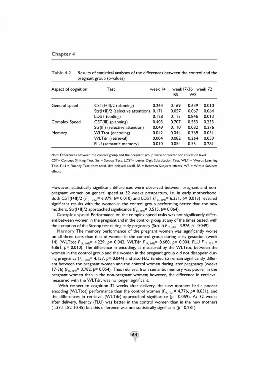
64
Table 4.2 Results of statistical analyses of the differences between the control and thepregnant group (p-values)
Aspect of cognition Test week 14 week17-36 week 72BS WS
General speed CST(I+II)/2 (planning) 0.264 0.169 0.639 0.010 Str(I+II)/2 (selective attention) 0.171 0.057 0.067 0.064LDST (coding) 0.128 0.113 0.846 0.013
Complex Speed CST(III) (planning) 0.403 0.707 0.553 0.233Str(III) (selective attention) 0.049 0.110 0.082 0.276
Memory WLTtot (encoding) 0.042 0.044 0.769 0.031WLTdr (retrieval) 0.004 0.082 0.264 0.059FLU (semantic memory) 0.010 0.054 0.551 0.281
Note: Differences between the control group and the pregnant group were corrected for education level.
CST= Concept Shifting Test, Str = Stroop Test, LDST= Letter Digit Substitution Test, WLT = Words Learning
Test, FLU = Fluency Test, tot= total, dr= delayed recall, BS = Between Subjects effects, WS = Within Subjects
effects
However, statistically significant differences were observed between pregnant and non-pregnant women on general speed at 32 weeks postpartum, i.e. in early motherhood.Both CST(I+II)/2 (F (1, 102)= 6.979, p= 0.010) and LDST (F (1, 104)= 6.331, p= 0.013) revealedsignificant results with the women in the control group performing better than the newmothers. Str(I+II)/2 approached significance (F(1, 103)= 3.515, p= 0.064).
Complex speed Performance on the complex speed tasks was not significantly differ-ent between women in the pregnant and in the control group at any of the times tested, withthe exception of the Stroop test during early pregnancy (Str(III) F (1, 103)= 3.976, p= 0.049).
Memory The memory performance of the pregnant women was significantly worseon all three tests than that of women in the control group during early gestation (week14) (WLTtot F (1, 102)= 4.229, p= 0.042, WLTdr F (1, 102)= 8.680, p= 0.004, FLU F (1, 103) =6.861, p= 0.010). The difference in encoding, as measured by the WLTtot, between thewomen in the control group and the women in the pregnant group did not disappear dur-ing pregnancy (F(1, 102)= 4.157, p= 0.044) and also FLU tended to remain significantly differ-ent between the pregnant women and the control women during later pregnancy (weeks17-36) (F(1, 104)= 3.782, p= 0.054). Thus retrieval from semantic memory was poorer in thepregnant women than in the non-pregnant women; however, the difference in retrieval,measured with the WLTdr, was no longer significant.
With respect to cognition 32 weeks after delivery, the new mothers had a poorerencoding (WLTtot) performance than the control women (F(1, 102)= 4.776, p= 0.031), andthe differences in retrieval (WLTdr) approached significance (p= 0.059). At 32 weeksafter delivery, fluency (FLU) was better in the control women than in the new mothers(1.37;11.82-10.45) but this difference was not statistically significant (p= 0.281).
ChapterChapter 4

65
DISCUSSIONThis study investigated whether there are short and/or long-term differences in variousaspects of cognitive functioning between pregnant and non-pregnant women. Our findingsrevealed that pregnant women have cognitive deficits, the type and severity of whichappear to depend on the stage of pregnancy. Cognitive deficits were also detected 32weeks after delivery. In the following we will successively discuss the specific deficits inmemory during pregnancy and subsequently during early motherhood. In addition, thisdiscussion will also focus on the fact that deficits in speed of information processing weredetected during early motherhood but not during pregnancy. Ultimately, we will finishwith a summary of our main conclusions.
With respect to memory performance there was a clear difference between pregnantwomen and non-pregnant women in memory tasks during early gestation (week 14 ofpregnancy) which lasted during pregnancy and, in some instances, to 32 weeks after deliv-ery. Intentional learning, as measured by an explicit memory task (WLT), was worse at allassessments in the pregnant women than in the non-pregnant control women. This wasespecially pronounced for encoding (WLTtot): Marginal differences in retrieval (WLTdr)were found in later pregnancy and early motherhood. Semantic memory, as measured bythe Fluency task (FLU), also differed between pregnant women and control women withthe control women performing better than the pregnant women. This difference was nolonger statistically significantly different during early motherhood (32 weeks postpartum).
Longitudinal studies of memory during pregnancy are scarce. Keenan et al (30) tested 10pregnant women in all three trimesters of pregnancy and also postpartum and comparedthem with 10 matched non-pregnant women. Using the Wechsler Memory Scale-RevisedLogical Memory and the Californian Discourse Memory Test, they observed a deteriora-tion in the immediate as well as the delayed recall of paragraph-length in the pregnantsubjects as compared to the control subjects. This difference, however, was only signifi-cant in the third trimester. The results of increased deficits in explicit memory duringpregnancy presented here support the findings of Keenan et al. In addition, deficits in earlypregnancy (week14) and to lesser extent postpartum were also observed in this study.Keenan and coworkers did not find any difference postpartum, whereas we observed thatexplicit memory was still compromised. This discrepancy could be attributed to the factthat our study was larger and, consequently, had more power than Keenan’s.
Our findings of impaired memory throughout pregnancy could, in theory, be due toselection bias, i.e. differences in cognitive performance between the two groups at studyintake. However, our findings are consistent with those of the cross-sectional study ofSharp and colleagues (7), who demonstrated an objective impairment of both implicit andexplicit memory during pregnancy. Deficits were found in all trimesters and were compa-rable for multigravids and primigravids. Those authors used different neuropsychologicaltests, such as recall and recognition of words from a priming list (i.e. an implicit memorytest) and recall and recognition of photographs and household objects (explicit memory),which strengthens the findings of both studies.
Even though we did not expect a priori the effect of motherhood, it is a very plausiblefinding. Early motherhood is characterized by numerous environmental changes and isstressful, because of an enhanced information processing load. McVeigh investigated theearly motherhood experiences of 79 first-time mothers. The main outcomes included thetiredness the women experienced, their feelings of incompetence, and the lack of time forthemselves (16). All these factors can have an effect on cognitive functioning.
One of the few studies that evaluated cognition after delivery was conducted by
Cognition during pregnancy and postpartum
66
Buckwalter et al ( 8 ). They concluded that women performed memory tasks better 26.5days after delivery than during pregnancy. However, they did not use a non-pregnant con-trol group in their study, and thus the findings could reflect a learning effect.
On the basis of subjective tests, Casey and coworkers (31) concluded that pregnant andpostpartum women reported significantly more everyday forgetting than non-pregnantcontrols. Although objective tests did not confirm this, our results are consistent withCasey’s subjective findings.
Eidelman and colleagues ( 6 ) investigated cognitive performance in the days followingdelivery by comparing the performance of 100 new mothers with that of non-pregnantchildless women, third-trimester high-risk pregnant women, and fathers of newborns.Their results suggest that parturients had significant memory deficits on the first day post-partum, but that this deficit was no longer apparent 2 or 3 days after delivery. Theseresults imply that memory impairment would not be present 32 weeks after delivery,which contrasts with our results showing that memory impairment was still present dur-ing early motherhood. In Eidelman’s study, however, motherhood factors such as sleepdeprivation, feelings of responsibility, and sadness, would not have negatively affected per-formance because most women were still in hospital and could share responsibilities withthe medical team. Besides, the cumulative effects of sleepless nights begin to have animpact at about 4 weeks postpartum (32). It is probable that these factors played a role inour study, because the women had been caring for themselves and their babies for morethan half a year (16). This could explain why memory deficits were found in our study at 32weeks post-partum but not in the Eidelman study at 2 or 3 days post-partum.
With respect to speed of information processing, it appears from our results that preg-nant women do not experience more difficulties on tasks requiring general speed of infor-mation processing than do non-pregnant women. However, early motherhood appears tobe characterized by a deterioration in the speed of information processing, because 32weeks after delivery the new mothers performed worse on the CST(I+II)/2 and LDST thanthe control women (Str(I+II)/2 almost reached significance). Earlier studies have alreadyfocussed on this aspect of cognition. Harris et al ( 3 3 ), using tests comparable to ours, tested20 pregnant women at week 36 of pregnancy and 48 hours and 4 weeks postpartum bymeans of the adapted DSST (Digit Symbol Substitution Test) (comparable to the LDST)and the TM (Trail making test) (comparable to CST). The performance of these womenwas compared with that of 20 matched controls tested at comparable times. The pregnantgroup scored worse on the DSST, with the largest performance difference being found 48hours after delivery. We observed poorer performance on the LDST 32 weeks postpar-tum. In contrast to our study, Harris et al did not find any effects on planning as measuredby the TM. However, some differences should be taken into account. First Harris andcoworkers tested their pregnant subjects at three different locations: a quiet place in theantenatal clinic, next to the participant’s bed, and at the mother’s home. On all occasionswe tested our participants in their homes, without any family members around, the doorbell and the telephone switched off and, when possible, always at about the same time ofthe day. This was all done in order to prevent any environmental differences. Secondly,Harris colleagues stated that their sample size was too small, which could lead to failure tofind significant differences in tests, such as the TM. In the study presented here there werenearly three times as many study participants. Finally, we used revised versions of the TMand the DSST, which are more sensitive in detecting possible differences ( 2 4 ). These testsproved their effectiveness in earlier research into the neurocognitive functioning of healthysubjects and subjects with only mild cognitive complaints (21, 25, 34, 35).
ChapterChapter 4
67
In our study, pregnancy and early motherhood did not affect performance on complexspeed tasks. This suggests that the changes in pregnant women (and new mothers) areconfined to ‘general speed’ and do not extend to higher aspects of cognitive functioning.Thus, concept shifting ability and planning appeared not to be compromised, which indi-cates that the findings are indeed subtle.
In conclusion, our study demonstrates that both pregnancy and early motherhood areassociated with specific differences in cognitive performance as compared with that ofmatched, non-pregnant control women tested at the same time intervals. Pregnancy ischaracterized by deficits in all measured memory functions: encoding, retrieval andsemantic memory, whereas early motherhood appears to be associated with deficits inmemory and in speed of information processing.
Thus, our results suggest that there is a shift in cognitive performance from early preg-nancy to early motherhood. Further studies are warranted to unravel the possible causesof the differences in cognitive performance.
ACKNOWLEDGMENTSThe authors wish to thank all the women who participated in this study, Arnold Kesterand Nico Rozendaal for the statistical support, and Carel Thijs for critically reviewing thismanuscript. We gratefully acknowledge the cooperation of the midwives and medical staffof the hospitals in our research area.
Cognition during pregnancy and postpartum

68
REFERENCES
1. Brett M., Baxendale S. Motherhood and memory: a review. Psychoneuroendocrinology2001;26:339-62.
2. Poser C. M., Kassirer M. R., Peyser J. M. Benign encephalopathy of pregnancy. Preliminaryclinical observations. Acta Neurol Scand 1986;73:39-43.
3. Silber M., Almkvist O., Larsson B., Uvnas-Moberg K. Temporary peripartal impairment inmemory and attention and its possible relation to oxytocin concentration. Life Sci 1990;47:57-65.
4. Brindle P. M., Brown M. W., Brown J., Griffith H. B., Turner G. M. Objective and subjectivememory impairment in pregnancy. Psychol Med 1991;21:647-53.
5. Parsons C., Redman S. Self-reported cognitive change during pregnancy. Aust J Adv Nurs1991;9:20-9.
6. Eidelman A. I., Hoffmann N. W., Kaitz M. Cognitive deficits in women after childbirth. ObstetGynecol 1993;81:764-7.
7. Sharp K., Brindle P. M., Brown M. W., Turner G. M. Memory loss during pregnancy [see com-ments]. Br J Obstet Gynaecol 1993;100:209-15.
8. Buckwalter J. G., Stanczyk F. Z., McCleary C. A., et al. Pregnancy, the postpartum, and steroidhormones: effects on cognition and mood [published erratum appears in Psychoneuroendo-crinology 1999 Jul;24(5):581]. Psychoneuroendocrinology 1999;24:69-84.
9. Schneider Z. Cognitive performance in pregnancy. Aust J Adv Nurs 1989;6:40-7.10. Morris N., Toms M., Easthope Y., Biddulph J. Mood and cognition in pregnant workers [see
comments]. Appl Ergon 1998;29:377-81.11. Christensen H., Poyser C., Pollitt P., Cubis J. Pregnancy may confer a selective cognitive
advantage. Journal of reproductive and infant psychology 1999;17:7-25.12. de Groot R. H. M., Hornstra G., Rozendaal N., Jolles J. Memory performance, but not infor-
mation processing speed, is compromised during early pregnancy. Journal of Clinical andExperimental Neuropsychology 2003;25:483-489.
13. Kane F. J., Jr., Harman W. J., Jr., Keeler M. H., Ewing J. A. Emotional and cognitive disturbancein the early puerperium. Br J Psychiatry 1968;114:99-102.
14. Welch J. Labouring brains. BMJ 1991;303:253.15. Baildam E. Doctor as mum. BMJ 1991;303:424.16. McVeigh C. Motherhood experiences from the perspective of first-time mothers. Clin Nurs
Res 1997;6:335-48.17. de Bie S. E. Standaardvragen 1987: Voorstellen voor uniformering van vraagstellingen naar
achtergrondkenmerken en interviews [Standard questions 1987: Proposal for uniformisationof questions regarding background variables and interviews]. 2 ed. Leiden, The Netherlands:Leiden University Press, 1987.
18. Bohnen N., Twijnstra A., Jolles J. Performance in the Stroop color word test in relationship tothe persistence of symptoms following mild head injury. Acta Neurol Scand 1992;85:116-21.
19. Houx P. J., Jolles J. Age-related decline of psychomotor speed: effects of age, brain health, sex,and education. Percept Mot Skills 1993;76:195-211.
20. Klein M., Houx P. J., Jolles J. Long-term persisting cognitive sequelae of traumatic brain injuryand the effect of age. J Nerv Ment Dis 1996;184:459-67.
21. Moller J. T., Cluitmans P., Rasmussen L. S., et al. Long-term postoperative cognitive dysfunc-tion in the elderly ISPOCD1 study. ISPOCD investigators. International Study of Post-Operative Cognitive Dysfunction. Lancet 1998;351:857-61.
ChapterChapter 4
69
22. Brand N., Jolles J. Learning and retrieval rate of words presented auditorily and visually. J GenPsychol 1985;112:201-10.
23. Jolles J., Houx P., Boxtel van M., Ponds R. Maastricht aging study; Determinants of cognitiveaging. Maastricht: Neuropsych Publishers, 1995.
24. Vink M., Jolles J. A new version of the Trail Making Test as an information processing task.Journal of Clinical Neuropsychology 1985;7:162.
25. Houx P. Cognitive aging and health-related factors (t h e s i s). Psychiatry & Neuropsychology.Maastricht: Rijksuniversiteit Limburg, 1991.
26. Lezak M. D. Neuropsychological Assessment. 3 ed. New York: Oxford University Press,1995.
27. Stroop J. Studies of interference in serial verbal reaction. J.Exp.Psychol. 1935;18:643-662.28. Klein M., Ponds R. W., Houx P. J., Jolles J. Effect of test duration on age-related differences in
Stroop interference. J Clin Exp Neuropsychol 1997;19:77-82.29. Smith A. The symbol Digit Modalities Test: A neuropsychologic test for economic screening
of learning and other cerebral disorders. Learning Disorders 1968;3:83-91.30. Keenan P. A., Yaldoo D. T., Stress M. E., Fuerst D. R., Ginsburg K. A. Explicit memory in
pregnant women. Am J Obstet Gynecol 1998;179:731-7.31. Casey P., Huntsdale C., Angus G., Janes C. Memory in pregnancy. II: Implicit, incidental,
explicit, semantic, short-term, working and prospective memory in primigravid, multigravidand postpartum women. J Psychosom Obstet Gynaecol 1999;20:158-64.
32. Kalil K. Psychosocial stress, anxiety and pregnancy complications: Issues for public policy. Pre-and Peri-Natal Psychology 1987;1:239-243.
33. Harris N. D., Deary I. J., Harris M. B., Lees M. M., Wilson J. A. Peripartal cognitive impair-ment: Secondary to depression? Br J Health Psy 1996;1:127-36.
34. van Boxtel M. P., Buntinx F., Houx P. J., Metsemakers J. F., Knottnerus A., Jolles J. The rela-tion between morbidity and cognitive performance in a normal aging population. J Gerontol ABiol Sci Med Sci 1998;53:M147-54.
35. Engelberts N. H., Klein M., van der Ploeg H. M., et al. Cognition and health-related quality oflife in a well-defined subgroup of patients with partial epilepsy. J Neurol 2002;249:294-9.
Cognition during pregnancy and postpartum

70
SELECTIVE ATTENTIONDEFICITS DURING
HUMAN PREGNANCY
published in Neuroscience Letters, 2003; 340: 21-24
Renate H.M. de Groot, Jos J.Adam, G. Hornstra.

72
ABSTRACT
Using a longitudinal design we provide evidence that selective attention
- a key component of cognition concerned with selection and preparation -
is compromised during pregnancy. Selective attention was operationalized
by means of the finger precuing technique, which selectively prepares two
of four finger responses. The precuing benefit was taken as a measure of
selective attention. Pregnant women showed a significant smaller precuing
benefit at week 36 of pregnancy than did the control women, indicating loss
of selective attention. Thirty-two weeks after childbirth this performance
decrement had vanished, reflecting a functional recovery.
Chapter 5

73
During pregnancy the maternal body changes drastically. Physiological evidence comesfrom studies demonstrating complex hormonal changes ( 1 ), alterations in fatty acid levels ( 2 ),and, of course, increments in body weight (3). In contrast - and despite abundant anecdotalreports suggesting deficits in memory and attention - serious scientific examination ofcognitive changes during pregnancy has been limited to a few studies that mainly focussedon memory loss (1). In addition not many longitudinal studies about the relation betweenpregnancy and cognition are available. The purpose of this study was to investigate if, andif so how, pregnancy affects selective attention. More specifically, we aimed to determine(1) whether selective attention was different between pregnant women and non-pregnantwomen matched for age and education during pregnancy; and (2) whether there was adifference in selective attention between the two groups after delivery.
Many studies have established that human subjects can make use of advance informa-tion to improve perceptual analyses (4), but also to speed up motor processes (5). In thespatial precuing task, subjects are forewarned by a cue about the location of an upcomingtarget stimulus. Typically, valid precues yield substantial benefits in speed and accuracy ofresponding. This has been shown for target detection (6), target identification (7), and targetdiscrimination tasks (8), but also for response selection (or choice reaction time) tasks (5).The emerging consensus is that precuing benefits are mediated by selective attention: Theprecue directs a process of selective attention whereby perceptual and motor processesare prepared for, readied, and tuned to the most relevant source of information (9, 10). Theresulting effect is a shortening of the processing time when the target stimulus arrives.
In this study we investigated whether pregnancy would affect visual selective attentionin a specific version of the spatial precuing task, the so-called finger precuing task ( 1 1 ). In this task, the spatial precue provides visual information about which finger to use forresponding (Figure. 5.1a and b). At base, the finger precuing task is a 4-choice reactiontime task, with the precue reducing the number of possible response choices from 4 to 2.That is, the spatial precue specifies two possible target locations, thereby informing abouttwo possible finger responses. Since the Hick-Hyman Law (12, 13) holds that 2-choice reac-tion times are shorter than 4-choice reaction times, precue effectiveness can be inferredfrom a significant reaction time or precuing benefit in the cued (i.e., 2-choice) conditionsover the control, uncued (i.e., 4-choice) condition.
The finger precuing task requires subjects to respond to spatial-location stimuli dis-played on a computer screen with discrete button-press responses by index and middlefingers of both hands placed on four keys (Z, X, ., and /) of the computer keyboard (thetwo left-most and two right-most keys on the bottom row of the keyboard). The taskinvolves a spatial precuing method in which two of four possible stimulus-response loca-tions are precued during a preparation interval of 1 s. The display consists of threehorizontal rows, representing warning, precue, and target stimulus, respectively. First thewarning stimulus is presented; it consists of four plus (+) signs that indicate the four pos-sible stimulus-response locations. After 750 ms the precue is presented; it consists of plussigns either in all four positions (uncued condition) or in only two of the four possiblepositions (the cued condition). One thousand milliseconds later the reaction signal is pre-sented; it consists of one plus sign, indicating the final target location. The subject’s task isto respond as quickly as possible to the position in which the reaction signal occurs bypressing the appropriate response button. Target signal and response button are mapped
Selective attention deficits during human pregnancy

74
Chapter 5
20
30
40
50
60
10 20 30 40
Weeks
*
72
pregnant
control
pregnancy post-partum
Fig. 5.1(a) Temporal sequence of events in the finger precuing task
Fig. 5.1 (b) Schematic representation of the different precuing conditions in the fingerprecuing task.
Fig. 5.1(c) Experimental results. Mean precuing benefit (and sem) for the pregnant andcontrol subjects as a function of time. (* p< 0.05).

onto each other in a spatially compatible manner such that a target appearing in the left-most position has to be responded to with the left-middle finger pressing the left-mostresponse button, etc. The finger precuing task has been devised by Miller (11), who modi-fied Rosenbaum’s (14) movement precuing technique.
Two main precue conditions are distinguished. In the cued condition, the precue con-sists of plus signs in only two of the four possible positions, specifying two fingers on thesame hand (e.g., the left-middle and left-index fingers), specifying the same finger on differ-ent hands (e.g., the left-index and right-index fingers), or specifying different fingers on dif-ferent hands (e.g., the left-middle and right-index fingers). In the u n c u e d condition, theprecue provides no advance information (it contains plus signs in all four possible stimuluslocations), and thus precludes selective preparation of any combination of two fingerresponses. Each subject received a block of 80 test trials. Within a block of 80 test trials,there were 20 trials for the uncued condition (5 for each of the four stimulus positions)and 20 trials for each of the hand-cued, finger-cued, and neither-cued conditions (also 5for each of the four stimulus positions). The order of these cuing conditions within ablock of 80 trials was ad random. Twenty practice trials preceded the block of 80 test trials.
Our choice for the finger precuing task was motivated by several considerations. First,the cued and uncued conditions are matched for motor output, because the same fingerresponses are involved in both conditions. Consequently, the precuing benefit, indexed asthe difference in reaction time between cued and uncued conditions, reflects selectiveattention and not motor execution processes or reaction time per se. Second, the fingerprecuing task has been applied successfully to the study of cognitive ageing: Older sub-jects have been shown to generate a substantial smaller precuing benefit than youngersubjects do, suggesting a pronounced loss of attentional capacity as one grows older (15).Third, as revealed in a recently completed neuroimaging study, the attentional mecha-nisms operating in the finger precuing task appear to be controlled by a network of brainstructures that includes specific areas in the prefrontal cortex, the parietal cortex, andthe basal ganglia ( 1 6 ). All these three brain areas have been implicated in the control ofselective attention (17-19), bolstering the notion that precuing benefits in the finger precuingtask are indeed mediated by attentional operations. Together, the above considerationssuggest that the finger precuing task may be an adequate behavioural tool to trace possi-ble pregnancy-related changes in selective attention.
Fourty-eight pregnant women (mean ± SD age is 29.7 ± 3.9 years) performed the fin-ger precuing task several times during their pregnancy (weeks 14, 17, 29, 36 of pregnan-cy) and also 32 weeks after childbirth. A control group of forty-eight non-pregnantwomen (mean ± SD age is 30.5 ± 4.2 years), matched for age and education, performedthe same task at the same time intervals. The original sample contained 100 subjects, butfour subjects were excluded, because they failed to show a consistent preparation benefit.The exclusion criterion was a negative preparation benefit (i.e., longer RTs in cued than inuncued conditions) in at least three of the four weeks during pregnancy. Educational levelwas measured an 8-point scale, ranging form primary education to higher vocational train-ing and university (20) and was not different between the two groups as measured by aMann-Whitney test (p= 0.3992, 4.4±1.23 versus 4.2±1.4 (mean ± SD) for the controlgroup and the pregnant group, respectively).
Performance in the finger precuing task during pregnancy (figure 5.1c) was examinedwith a two-way split-plot analysis of variance (ANOVA). The within-subject factor, week,
75
Selective attention deficits during human pregnancy

76
had four levels (weeks 14, 17, 29, and 36 of pregnancy). The between-subject factor,group, had two levels (pregnant, control). The dependent variable was the precuing bene-fit, indexed as the difference in reaction time between conditions with and withoutinformative precues (i.e., uncued minus cued). Education level was entered as a covariatein the analysis, because it is known as a possible confounder in this type of research. Weused the Greenhouse-Geisser corrected significance values to adjust the tests involvingthe within-subject factor for heterogeneity of variance and covariances. The main effectsof group and week were not significant (ps> 0.217), but there was a significant interactionbetween group and week (F= 3.213; df= 3, 279; p= 0.025). Tests on simple effects of thefactor week indicated that the pregnant and control group generated similar precuingbenefits at weeks 14, 17 and 29 of pregnancy (ps> 0.265). However, at week 36 of preg-nancy, the pregnant group showed a significant smaller precuing benefit than did the con-trol group (p= 0.017). This important finding indicates that pregnancy negatively affectedselective attention at week 36 of pregnancy. This performance decrement, however, haddisappeared 32 weeks after childbirth (F= 0.689; d f= 1, 93; p= 0.409; education levelentered as covariate). Responses were very accurate as mean error was 1.65% and 1.46%for the pregnant and the control subjects, respectively. The slight increase in precuingbenefit for both groups during early pregnancy (from week 14 to 17) probably reflects alearning effect.
Selective attention is an essential brain function that mediates the selection of informa-tion to control action. It is difficult to specify the nature of the pregnancy-related atten-tional deficit. An early, perceptual but also a late, motoric locus seems possible. It shouldbe noted, however, that recent theories of attention emphasize the close link betweenperception and action, making the “locus” question less relevant. For example, the “pre-motor” theory of attention (21) claims that there is no need to postulate an attention sys-tem separate from systems that controls action: The system that controls action is alsothe system that controls attention. According to premotor theory, spatial maps (in theparietal and frontal cortex) not only code the locations of objects but also are involved inpreparing goal-directed motor actions to these locations. Hence, the act of attending to aspatial location is equivalent to the act of preparing some type of motor response to thatlocation. A similar view is articulated in the Visual Attention Model (VAM) proposed bySchneider (22). According to this model, visual attention not only selects objects for per-ceptual processing, but also, and concurrently, provides the spatial parameters for actionstowards these objects. Finally, the integrated competition hypothesis (23) posits that atten-tion emerges as an integrated state in which multiple brain systems converge to process aselected object together with its implications for action. According to these views, thedichotomy between perceptual and motoric processes becomes blurred and probablyless relevant.
The behavioural advantage of “paying attention” is efficiency and coherence of res-ponding. The importance of selective attention for our everyday life should not be under-estimated, and is probably best illustrated by referring to what happens when it fails.“Slips of action” - like car accidents or small accidents in or around the house - oftenarise when we are not “paying attention”. Patients with attentional deficits - such aspatients with frontal lobe damage-typically show passive, inert and distracted behaviour(24). The results of the present study indicate that the efficiency of attentional operationsas measured by the finger precuing task (that is, attentional selection and preparation)may be compromised in the final stages of pregnancy. However, on the upside, our find-ings also indicate that this deficit is not irreversible, but that it appears to recover after
Chapter 5

77
childbirth. As to the mechanisms of this pregnancy-related impairment in selective atten-tion one can only speculate, but it is possible that hormonal changes, changes in fattyacids levels or the need of the unborn baby for important nutrients at the expense of themother may play a role.
One possible explanation for the attention deficit during pregnancy is the fatty acidhypothesis. Fatty acids are important building blocks for all cell membranes, includingthose of the central nervous system. In particular, the amount of docosahexaenoic acid(22:6n-3, DHA) drops, after an initial increase, in the later stages of pregnancy, and hasrecovered 32 weeks after delivery (25, 26). Interestingly, this pattern of DHA-variation duringpregnancy closely parallels the present variation in precuing-benefit during pregnancy.Hence, it seems possible that maternal DHA status might be related to brain function. Analternative direction for possible interpretation is to look at the influence of hormones.Even though the available evidence is somewhat mixed, there are reports indicating thathormones like oestrogens, progesterone, glucocorticoids, and oxytocin might be relatedto memory impairments during pregnancy ( 1 ). Obviously, clarification of the underlying mech-anism of the observed attentional deficit during pregnancy awaits further experimentation.
We believe that the present research is important because it represents a necessaryfirst-step in an exciting, cross-disciplinary journey of inquiry, serving to flesh out ourunderstanding of pregnancy-related neurocognitive changes.
Selective attention deficits during human pregnancy

78
REFERENCES
1. Brett M., Baxendale S. Motherhood and memory: a review. Psychoneuroendocrinology2001;26:339-62.
2. Al M. D., v. Houwelingen A. C., Hornstra G. Long-chain polyunsaturated fatty acids, pregnan-cy, and pregnancy outcome. Am J Clin Nutr 2000;71:285S-91S.
3. Soltani H., Fraser R. B. A longitudinal study of maternal anthropometric changes in normalweight, overweight and obese women during pregnancy and postpartum. Br J Nutr2000;84:95-101.
4. Posner M. I. Orienting of attention. Q J Exp Psychol 1980;32:3-25.5. Reeve T. G., Proctor R. W. On the advance preparation of discrete finger responses. J Exp
Psychol Hum Percept Perform 1984;10:541-53.6. Posner M. I., Snyder C. R., Davidson B. J. Attention and the detection of signals. J Exp Psychol
1980;109:160-74.7. Eriksen C. W., Rohrbaugh J. W. Some factors determining efficiency of selective attention.
American Journal of Psychology 1970;83:330-342.8. Henderson J. M. Stimulus discrimination following covert attentional orienting to an exoge-
nous cue. J Exp Psychol Hum Percept Perform 1991;17:91-106.9. Duncan J. Converging levels of analysis in the cognitive neuroscience of visual attention. In:
Humphreys GW, Duncan J, Treisman A, eds. Attention, space and action. Oxford: OxfordUniversity Press, 1999:112-129.
10. LaBerge D. Attention. In: Bly BM, Rumelhart DE, eds. Cognitive science. London: AcademicPress, 1999:43-97.
11. Miller J. Discrete versus continuous stage models of human information processing: in searchof partial output. J Exp Psychol Hum Percept Perform 1982;8:273-96.
12. Hick W. E. On the rate of gain of information. Quarterly Journal of Experimental Psychology1952;4:11-26.
13. Hyman R. Stimulus information as a determinant of reaction time. Journal of ExperimentalPsychology 1953;45:188-196.
14. Rosenbaum D. A. The movement precuing technique: Assumptions, applications, and exten-sions. In: Magill RA, ed. Memory and control of action. Amsterdam: North-Holland,1983:231-274.
15. Adam J. J., Paas F. G., Teeken J. C., et al. Effects of age on performance in a finger-precuingtask. J Exp Psychol Hum Percept Perform 1998;24:870-83.
16. Adam J. J., Backes W., Rijcken J., Hofman P., Kuipers H., Jolles J. Rapid visuomotor prepara-tion in the human brain: A functional MRI study. Cogn Brain Res 2003;16:1-10.
17. Parasuraman R. The attentive brain. Cambridge: MA:MIT Press, 1998.18. Robbins T. W., Rogers R. D. Functioning of frontostriatal anatomical ”loops” in mechanisms
of cognitive control. In: Monsell S, Driver J, eds. Control of cognitive processes - Attentionand Performance XVIII. Cambridge: MA:MIT Press, 2000:475-509.
19. Ravizza S. M., Ivry R. B. Comparison of the basal ganglia and cerebellum in shifting attention. JCogn Neurosci 2001;13:285-97.
20. de Bie S. E. Standaardvragen 1987: Voorstellen voor uniformering van vraagstellingen naarachtergrondkenmerken en interviews [Standard questions 1987: Proposal for uniformisationof questions regarding background variables and interviews]. 2 ed. Leiden, The Netherlands:Leiden University Press, 1987.
Chapter 5

79
21. Rizzolatti G., Riggio L., Dascola I., Umilta C. Reorienting attention across the horizontal andvertical meridians: evidence in favor of a premotor theory of attention. Neuropsychologia1987;25:31-40.
22. Schneider W. X. A neuro-cognitive model for visual attention control of segmentation, objectrecognition, and space-based motor action. Visual Cognition 1995;2:331-376.
23. Duncan J. Coorperating brain systems in selective perception and action. In: Inui T,McClelland JL, eds. Attention and performance XVI. London: The MIT Press, 1996:549-578.
24. Fuster J. M. The prefrontal cortex; Anatomy, physiology, and neuropsychology of the frontallobe. New York: Raven, 1980.
25. Al M. D., v. Houwelingen A. C., Kester A. D., Hasaart T. H., de Jong A. E., Hornstra G.Maternal essential fatty acid patterns during normal pregnancy and their relationship to theneonatal essential fatty acid status. Br J Nutr 1995;74:55-68.
26. Otto S. J., v. Houwelingen A. C., Badart-Smook A., Hornstra G. The postpartum docosa-hexaenoic acid status of lactating and nonlactating mothers. Lipids 1999;34:S227.
Selective attention deficits during human pregnancy

80

ALPHA-LINOLENIC ACID SUPPLEMENTATION DURING
HUMAN PREGNANCY DOESNOT EFFECT COGNITIVE
FUNCTIONING
Accepted for publication in Prostaglandins,
Leukotrienes, and Essential Fatty Acids
Renate H.M. de Groot, Jos J.Adam, Jelle Jolles, and Gerard Hornstra.

82
ABSTRACT
Increasing evidence suggests a positive association between docosa-
hexaenoic acid (22:6n-3, DHA) and cognitive performance. In addition,
pregnancy is associated with a reduction of the DHA status and cognitive
deficits. In the current study, cognition was assessed in pregnant women
receiving a margarine enriched with alpha-linolenic acid (18:3n-3, ALA, the
ultimate dietary precursor of DHA) and some linoleic acid (18:2n-6, LA, to
prevent a possible reduction in n-6 fatty acids). A control group received a
margarine enriched with LA only.
ALA supplementation hardly affected the maternal DHA status and no sig-
nificant differences were found in cognitive performance between the two
groups. This indicates that ALA supplementation during pregnancy does not
affect cognitive performance during and 32 weeks after gestation. At week
14 of pregnancy and 32 weeks after delivery, higher plasma DHA levels
were associated lower cognitive performance as indicated by longer reac-
tion times on the finger precuing task (partial correlation coefficients 0.3705
and 0.4633, respectively, p<0.01). These latter results certainly need further
i n v e s t i g a t i o n .
ChapterChapter 6
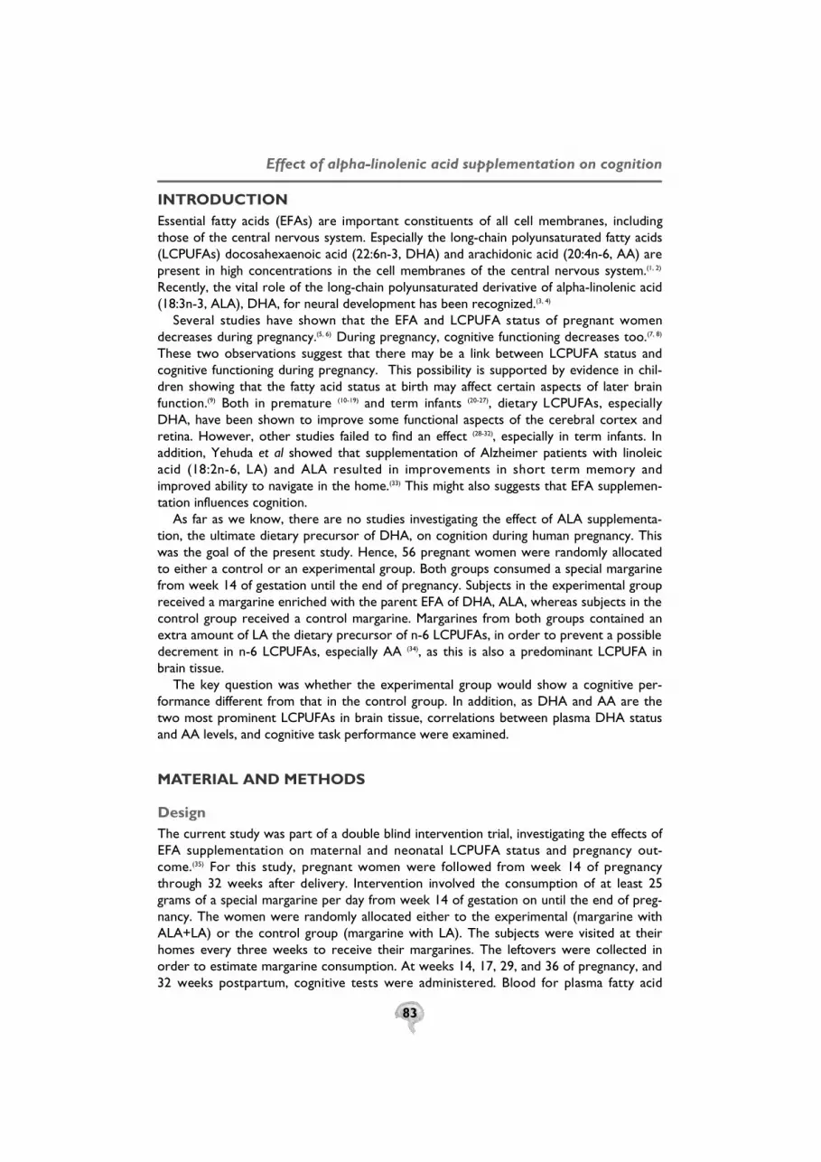
83
INTRODUCTIONEssential fatty acids (EFAs) are important constituents of all cell membranes, includingthose of the central nervous system. Especially the long-chain polyunsaturated fatty acids(LCPUFAs) docosahexaenoic acid (22:6n-3, DHA) and arachidonic acid (20:4n-6, AA) arepresent in high concentrations in the cell membranes of the central nervous system.(1, 2)
Recently, the vital role of the long-chain polyunsaturated derivative of alpha-linolenic acid(18:3n-3, ALA), DHA, for neural development has been recognized.(3, 4)
Several studies have shown that the EFA and LCPUFA status of pregnant womendecreases during pregnancy.(5, 6) During pregnancy, cognitive functioning decreases too.(7, 8)
These two observations suggest that there may be a link between LCPUFA status andcognitive functioning during pregnancy. This possibility is supported by evidence in chil-dren showing that the fatty acid status at birth may affect certain aspects of later brainf u n c t i o n .( 9 ) Both in premature ( 1 0 - 1 9 ) and term infants ( 2 0 - 2 7 ), dietary LCPUFAs, especiallyDHA, have been shown to improve some functional aspects of the cerebral cortex andretina. However, other studies failed to find an effect (28-32), especially in term infants. Inaddition, Yehuda et al showed that supplementation of Alzheimer patients with linoleicacid (18:2n-6, LA) and ALA resulted in improvements in short term memory andimproved ability to navigate in the home.(33) This might also suggests that EFA supplemen-tation influences cognition.
As far as we know, there are no studies investigating the effect of ALA supplementa-tion, the ultimate dietary precursor of DHA, on cognition during human pregnancy. Thiswas the goal of the present study. Hence, 56 pregnant women were randomly allocatedto either a control or an experimental group. Both groups consumed a special margarinefrom week 14 of gestation until the end of pregnancy. Subjects in the experimental groupreceived a margarine enriched with the parent EFA of DHA, ALA, whereas subjects in thecontrol group received a control margarine. Margarines from both groups contained anextra amount of LA the dietary precursor of n-6 LCPUFAs, in order to prevent a possibledecrement in n-6 LCPUFAs, especially AA (34), as this is also a predominant LCPUFA inbrain tissue.
The key question was whether the experimental group would show a cognitive per-formance different from that in the control group. In addition, as DHA and AA are thetwo most prominent LCPUFAs in brain tissue, correlations between plasma DHA statusand AA levels, and cognitive task performance were examined.
MATERIAL AND METHODS
DesignThe current study was part of a double blind intervention trial, investigating the effects ofEFA supplementation on maternal and neonatal LCPUFA status and pregnancy out-c o m e .( 3 5 ) For this study, pregnant women were followed from week 14 of pregnancythrough 32 weeks after delivery. Intervention involved the consumption of at least 25grams of a special margarine per day from week 14 of gestation on until the end of preg-nancy. The women were randomly allocated either to the experimental (margarine withALA+LA) or the control group (margarine with LA). The subjects were visited at theirhomes every three weeks to receive their margarines. The leftovers were collected inorder to estimate margarine consumption. At weeks 14, 17, 29, and 36 of pregnancy, and32 weeks postpartum, cognitive tests were administered. Blood for plasma fatty acid
Effect of alpha-linolenic acid supplementation on cognition

84
analyses was sampled at weeks 14, 26, and 36 of gestation, at partus, and at 32 weeksafter delivery. Correlations between fatty acid levels and cognitive performance were cal-culated for weeks 14, 26/29, and 36 of pregnancy, and 32 weeks after delivery.
Study populationSubjects were recruited by midwives in the regions Maastricht, Heerlen and Sittard andby the Department of Obstetrics and Gynecology of hospitals in the same area:University Hospital Maastricht, Atrium Medical Centre in Heerlen and Maasland Hospitalin Sittard. Inclusion criteria were a gestational age less than 14 weeks at study entry, andgood general health. Exclusion criteria were diastolic blood pressure higher than 90mmHg., multiple pregnancy, use of medication, use of (LC)PUFA rich supplements, originother than Caucasian, or fish consumption higher than two times a week.
Each subject gave written consent to participate in the study, which was approved by theMedical Ethics Committees of the University Hospital Maastricht and Maasland Hospital Sittard.
SupplementsAs described in detail before (35), the experimental margarine provided 2.82 g ALA and9.02 g LA per day with the requested consumption of 25 g per day. The margarine of thecontrol group provided 0.03 g ALA and 10.94 g LA per 25 g per day.
Cognitive test batteryCognitive performance was measured, using different neuropsychological tests withproven sensitivity to assess cognitive functions (36-39), as recently described by de Groot etal.(7, 8) In the present study we used the following tests in five parallel versions, which wererandomly allocated to the subjects.
The Visual Verbal Word Learning Task (WLT) evaluates the ability to acquireand retain new verbal information.( 4 0 ) After presentation of fifteen words, participantswere asked to recall as many words as possible (repeated three times). Dependent vari-ables were the mean number of words recalled over the first two trials (WLTtot) inorder to measure encoding, and the numbers of words recalled after 20 minutes, thedelayed recall (WLTdr), in order to measure retrieval.
The Concept Shifting Test (CST) was used to measure the capability to shift betweenc o n c e p t s .( 4 1 ) This test consists of three subtasks: the subject had to cross out, in the properorder, 16 circles containing numbers, (1 to 16; Subtask I), letters (A to P; Subtask II), or both(1A-2B-3C, etc. Subtask III). The variables of interest were the average times to completeSubtask I and II as a measure for general information processing speed (CST(I+II)/2), and thetime to complete Subtask III as a measure for concept shifting ability (CST(III)).
The Stroop Color-Word Interference Test as introduced by Stroop et al (42) is atest of selective visual attention and interference susceptibility. Subjects had to read alouda series of colors presented in black, to name the colors of colored patches presented,and to mention the incongruously ink color the color names were printed in. Mean speedfor Subtasks I and II was used as a measure of information processing speed (Str(I+II)/2).Subtask III was used as a measure of color word interference susceptibility (Str(III)).
The Letter-Digit-Substitution-Test (LDST) is a measure for coding, includingprocesses such as visual perception, attention, and memory.(43) Subjects were instructedto copy digits in cells indexed by a letter. The letters referred to nine letter/digit combi-nations at the top of the form. The total number of digits correctly related to a letter wasused as the dependent variable.
ChapterChapter 6

85
Finger precuing task. Selective attention was operationalized by means of this test,which involves the preparation and selection of 2 out of 4 possible responses. This taskrequires subjects to respond to spatial stimuli with discrete responses from index andmiddle finger from both hands. The subtraction score between the reaction time on theoriginal four choices reaction time tasks (uncued condition) and any of the other twochoices conditions is called the preparation benefit. Mean reaction time (RT(ms)) andmean preparation benefit (PB(ms)) were taken as measures of selective attention.(44)
Fatty acid analysesThe fatty acid composition of the plasma phospholipids has been reported before by deGroot et al.(35)
For the present study it was decided to focus on the predominant LCPUFAs in braintissue, DHA, and AA (%wt/wt). In addition, Osbond acid (22:5n-6, ObA), which is generallyaccepted as the deficiency marker for DHA received attention.
QuestionnairesAt the start of the study (week 14), a questionnaire was filled out in both groups. Thisquestionnaire included items about age, pre-pregnancy weight, height, smoking habits,supplement usage, and medical history, including former pregnancies and medical treat-ments. In addition, education was measured on an 8-point scale, ranging from primaryeducation to higher vocational training and university.(45) Thirty-two weeks after deliveryanother questionnaire was filled out, which included items about course and duration ofpregnancy and breastfeeding.
StatisticsUnpaired t-tests were used to determine whether the characteristics age, weight, height,and margarine consumption were different between the control and the experimentalgroup. The Mann-Whitney U Test was used in order to test whether differences existedbetween the two groups in education levels, parity, and gravidity. To detect possible dif-ferences in alcohol consumption or smoking the Chi-square Test was applied.
To investigate whether the supplement was effective in changing the fatty acid levelsduring pregnancy, repeated measures analyses of variance (ANOVA) was used. Differencesin cognitive performance between the experimental group and the control group wereanalyzed by repeated measures ANOVA, too. Time during pregnancy (weeks 17, 29 and36) was used as within-subjects factor, and group (experimental group or control group)as between-subjects factor. In all analyses, corrections were made for possible differencesin starting levels by including test performance on week 14 as a covariable. Educationlevel was included as a covariable, too. The Greenhouse-Geisser corrected significancevalues were used to adjust the tests involving the within-subjects factor heterogeneity ofvariance and covariances.
For both groups together, partial correlation coefficients were calculated betweenselected fatty acid levels as independent variables and cognition parameters as dependentvariables at weeks 14, 26/29, and 36 of gestation and 32 weeks after delivery.Adjustments were made for level of education. In addition, outliers as indicated by highCook’s distance were deleted from the data set (3 subjects maximum per analysis).
The statistical package SPSS 10.0 was used to analyze the results. Because of multipletesting, p-values <0.01 were considered significant, unless otherwise indicated.
Effect of alpha-linolenic acid supplementation on cognition
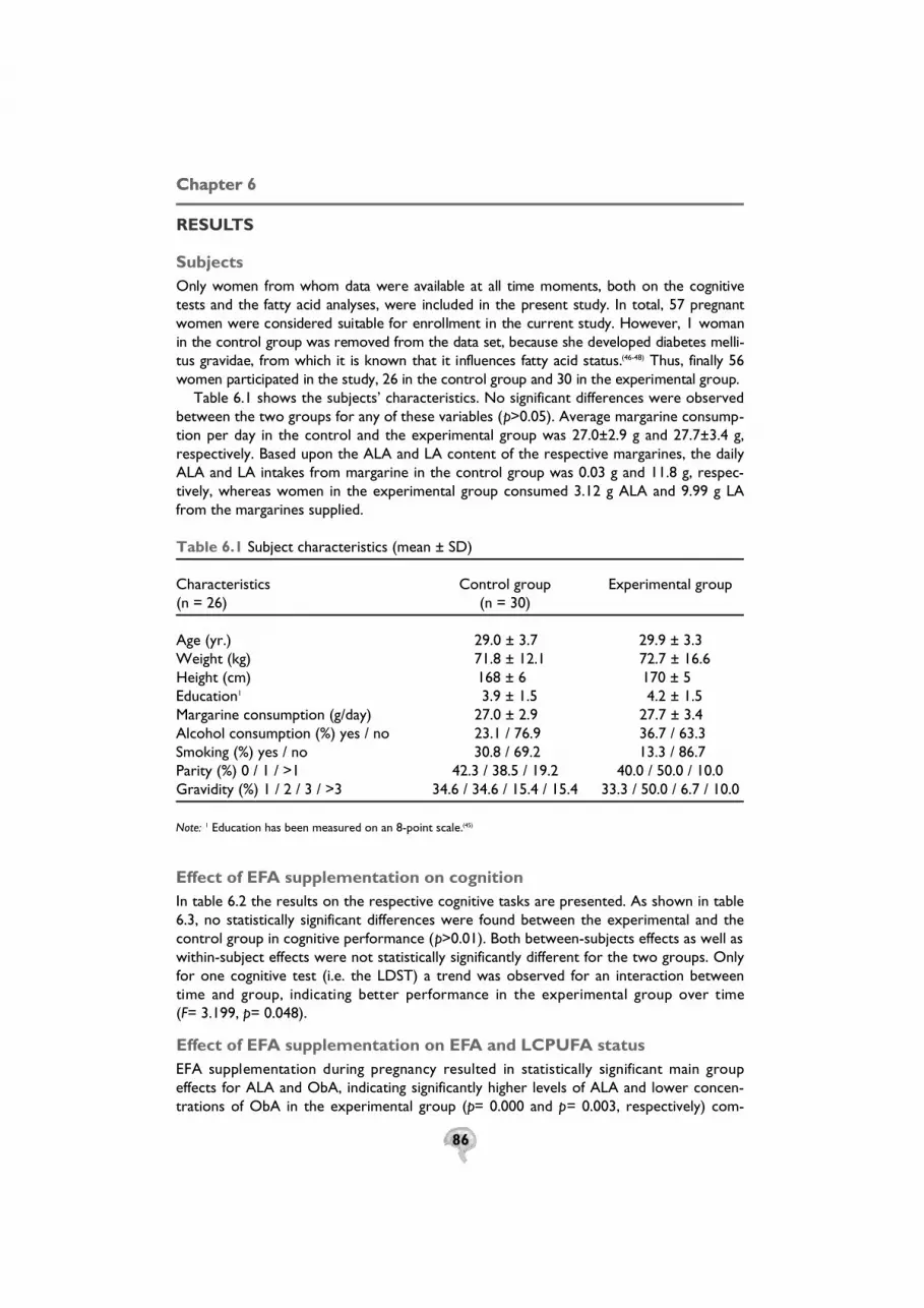
86
RESULTS
Subjects Only women from whom data were available at all time moments, both on the cognitivetests and the fatty acid analyses, were included in the present study. In total, 57 pregnantwomen were considered suitable for enrollment in the current study. However, 1 womanin the control group was removed from the data set, because she developed diabetes melli-tus gravidae, from which it is known that it influences fatty acid status.( 4 6 - 4 8 ) Thus, finally 56women participated in the study, 26 in the control group and 30 in the experimental group.
Table 6.1 shows the subjects’ characteristics. No significant differences were observedbetween the two groups for any of these variables (p>0.05). Average margarine consump-tion per day in the control and the experimental group was 27.0±2.9 g and 27.7±3.4 g,respectively. Based upon the ALA and LA content of the respective margarines, the dailyALA and LA intakes from margarine in the control group was 0.03 g and 11.8 g, respec-tively, whereas women in the experimental group consumed 3.12 g ALA and 9.99 g LAfrom the margarines supplied.
Table 6.1 Subject characteristics (mean ± SD)
Characteristics Control group Experimental group (n = 26) (n = 30)
Age (yr.) 29.0 ± 3.7 29.9 ± 3.3 Weight (kg) 71.8 ± 12.1 72.7 ± 16.6 Height (cm) 168 ± 6 170 ± 5 Education1 3.9 ± 1.5 4.2 ± 1.5 Margarine consumption (g/day) 27.0 ± 2.9 27.7 ± 3.4 Alcohol consumption (%) yes / no 23.1 / 76.9 36.7 / 63.3 Smoking (%) yes / no 30.8 / 69.2 13.3 / 86.7 Parity (%) 0 / 1 / >1 42.3 / 38.5 / 19.2 40.0 / 50.0 / 10.0 Gravidity (%) 1 / 2 / 3 / >3 34.6 / 34.6 / 15.4 / 15.4 33.3 / 50.0 / 6.7 / 10.0
Note: 1 Education has been measured on an 8-point scale.(45)
Effect of EFA supplementation on cognition In table 6.2 the results on the respective cognitive tasks are presented. As shown in table6.3, no statistically significant differences were found between the experimental and thecontrol group in cognitive performance (p>0.01). Both between-subjects effects as well aswithin-subject effects were not statistically significantly different for the two groups. Onlyfor one cognitive test (i.e. the LDST) a trend was observed for an interaction betweentime and group, indicating better performance in the experimental group over time (F= 3.199, p= 0.048).
Effect of EFA supplementation on EFA and LCPUFA statusEFA supplementation during pregnancy resulted in statistically significant main groupeffects for ALA and ObA, indicating significantly higher levels of ALA and lower concen-trations of ObA in the experimental group (p= 0.000 and p= 0.003, respectively) com-
ChapterChapter 6

87
pared to the control group during pregnancy (table 6.4). AA and DHA levels were notsignificantly different between the two groups. No significant group*time effects werefound, indicating that none of the investigated fatty acids changed over time.
Thirty-two weeks after delivery, the two groups were not significantly different fromeach other in terms of fatty acid levels, with the exception of ALA, which was still signifi-cantly higher in the experimental group (p= 0.005).
Table 6.2 Cognitive performance during pregnancy in the control (C) and e x p e r i m ental (E) group (mean ± SD).
Test Week 14 Week 17 Week 29 Week 36 3 2 weeks pp
WLTtot C 10.5 ± 1.8 10.3 ± 2.2 11.1 ± 1.6 11.4 ± 1.5 10.8 ± 1.5# words
E 9.9 ± 1.3 9.8 ± 2.0 10.7 ± 2.2 10.9 ± 1.5 10.3 ± 1.1
WLTdr C 12.3 ± 2.0 12.0 ± 2.3 12.6 ± 2.1 13.0 ± 1.9 12.7 ± 1.6#words
E 11.5 ± 1.9 11.8 ± 2.5 12.8 ± 2.2 12.8 ± 1.6 12.5 ± 1.7
CST (I+II)/2 C 13.6 ± 3.3 13.3 ± 3.2 12.6 ± 2.7 13.5 ± 3.8 12.3 ± 3.5(s.)
E 12.6 ± 3.1 12.1 ± 3.4 11.8 ± 2.3 12.2 ± 2.9 12.0 ± 2.9
CST (III) C 21.7 ± 7.6 22.1 ± 7.2 19.5 ± 6.3 21.5 ± 7.3 21.4 ± 1.6(s.)
E 20.9 ± 7.2 19.6 ± 6.2 19.3 ± 1.1 18.5 ± 0.9 19.1 ± 5.8
Str (I+II)/2 C 17.5 ± 2.3 17.0 ± 2.0 17.1 ± 2.1 17.4 ± 0.4 16.5 ± 1.6(s.)
E 17.2 ± 3.4 16.6 ± 2.5 16.4 ± 2.7 16.4 ± 2.3 16.0 ± 2.4
Str (III) C 35.4 ± 6.4 31.9 ± 1.1 32.5 ± 3.8 32.5 ± 6.6 30.4 ± 5.4(s.)
E 33.9 ± 8.0 30.7 ± 8.3 31.2 ± 7.4 29.8 ± 6.1 29.0 ± 7.3
LDST C 38.7 ± 6.9 39.6 ± 6.4 39.9 ± 1.1 40.2 ± 1.2 39.7 ± 6.7#numbers/min.
E 40.8 ± 5.4 40.9 ± 0.9 42.5 ± 0.9 43.7 ± 1.0 42.9 ± 1.0
RT C 4144 ± 764 3974 ± 594 3906 ± 490 3938 ± 541 3746 ± 460(ms.)
E 3834 ± 465 3666 ± 426 3731 ± 478 3651 ± 451 3589 ± 442
PB C 18.9 ± 33.5 30.2 ± 25.3 17.8 ± 19.5 21.8 ± 20.1 26.0 ± 18.8(ms.)
E 32.0 ± 27.5 29.7 ± 25.1 27.9 ± 18.5 27.5 ± 15.1 33.3 ± 21.2
Note: pp= postpartum, WLT= Words Learning Test, tot= total (mean of trial 1 and 2), dr= delayed recall, CST=
Concept Shifting Test, Str= Stroop test, LDST= Letter Digit Substitution Test, RT= reaction time on the Finger
Precuing Task, PB= prebaration benefit on the Finger Precuing Task.
Effect of alpha-linolenic acid supplementation on cognition
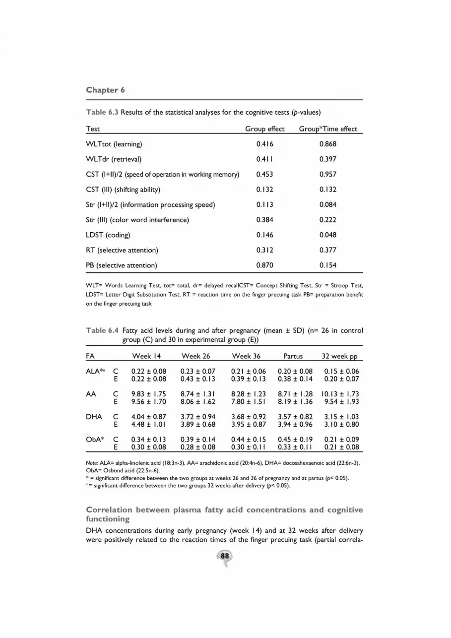
88
Table 6.3 Results of the statistical analyses for the cognitive tests (p-values)
Test Group effect Group*Time effect
WLTtot (learning) 0.416 0.868
WLTdr (retrieval) 0.411 0.397
CST (I+II)/2 (speed of operation in working memory) 0.453 0.957
CST (III) (shifting ability) 0.132 0.132
Str (I+II)/2 (information processing speed) 0.113 0.084
Str (III) (color word interference) 0.384 0.222
LDST (coding) 0.146 0.048
RT (selective attention) 0.312 0.377
PB (selective attention) 0.870 0.154
WLT= Words Learning Test, tot= total, dr= delayed recallCST= Concept Shifting Test, Str = Stroop Test,
LDST= Letter Digit Substitution Test, RT = reaction time on the finger precuing task PB= preparation benefit
on the finger precuing task
Table 6.4 Fatty acid levels during and after pregnancy (mean ± SD) (n= 26 in control group (C) and 30 in experimental group (E))
FA Week 14 Week 26 Week 36 Partus 32 week pp
ALA*# C 0.22 ± 0.08 0.23 ± 0.07 0.21 ± 0.06 0.20 ± 0.08 0.15 ± 0.06 E 0.22 ± 0.08 0.43 ± 0.13 0.39 ± 0.13 0.38 ± 0.14 0.20 ± 0.07
AA C 9.83 ± 1.75 8.74 ± 1.31 8.28 ± 1.23 8.71 ± 1.28 10.13 ± 1.73 E 9.56 ± 1.70 8.06 ± 1.62 7.80 ± 1.51 8.19 ± 1.36 9.54 ± 1.93
DHA C 4.04 ± 0.87 3.72 ± 0.94 3.68 ± 0.92 3.57 ± 0.82 3.15 ± 1.03 E 4.48 ± 1.01 3.89 ± 0.68 3.95 ± 0.87 3.94 ± 0.96 3.10 ± 0.80
ObA* C 0.34 ± 0.13 0.39 ± 0.14 0.44 ± 0.15 0.45 ± 0.19 0.21 ± 0.09 E 0.30 ± 0.08 0.28 ± 0.08 0.30 ± 0.11 0.33 ± 0.11 0.21 ± 0.08
N o t e : ALA= alpha-linolenic acid (18:3n-3), AA= arachidonic acid (20:4n-6), DHA= docosahexaenoic acid (22:6n-3),ObA= Osbond acid (22:5n-6).* = significant difference between the two groups at weeks 26 and 36 of pregnancy and at partus (p< 0.05).# = significant difference between the two groups 32 weeks after delivery (p< 0.05).
C o rrelation between plasma fatty acid concentrations and cog n i t i vefunctioningDHA concentrations during early pregnancy (week 14) and at 32 weeks after deliverywere positively related to the reaction times of the finger precuing task (partial correla-
ChapterChapter 6

89
tion coefficients 0.3705 and 0.4633, respectively, p< 0.01 for both). Thus, reaction timeswere longer (and, consequently, this aspect of cognition lower) with higher plasma DHAconcentrations. Other significant correlations were not observed.
Neither AA nor ObA was significantly related to performance on any of the cognitive tests.
DISCUSSIONThe aim of the current study was to investigate the effect of supplementation with ALA,the parent fatty acid of DHA, on various aspects of cognitive functioning during and 32weeks after human pregnancy. Additional LA was given in order to prevent a decrementin AA concentration, which frequently accompanies an increased consumption of n-3 fattyacids.(34)
Performance on none of the cognitive tests was different between the two groups.Thus, it can be concluded that we did not find an effect of ALA supplementation on cog-nitive functioning during pregnancy.
DHA and AA are the predominant LCPUFAs in brain tissue and although ALA+LAsupplementation did not increase DHA levels in the plasma phospholipid fatty acids of theexperimental group, the lower ObA levels in this group indicated a better functionalDHA status indeed. (35) Alterations in brain function are presumed to follow from the bio-chemical consequences of modifying membrane PUFA content.(49) Known effects includemodifications in membrane fluidity, in the activities of membrane-associated functionalproteins, such as transporters, enzymes, and receptors, and in the production of impor-tant signaling molecules derived from EFA, such as prostaglandins and eicosanoids.( 5 0 - 5 2 )
A possible explanation for the fact that no effect of EFA supplementation on cognitionwas found in the current study could be that the ALA and LA conversion into DHA andAA, respectively, was too limited, since these processes have been shown to be ratherinefficient, indeed.(35) In this study the women were supplemented with ALA and LA, and,as has been shown repeatedly, these parent fatty acids are desaturated and elongated intotheir longer-chain more-unsaturated fatty acids DHA and AA in low amounts.(53-57)
Fatty acids of the n-3 fatty acid family do not necessarily affect all neural domains tothe same extent. Thus, it has been shown that deficiency of n-3 fatty acids in monkeyslead to slower visual processing speed (58), but did not influence learning or memory per-formance.(59) In the current study, we did find correlations between plasma DHA levelsand reaction time on the finger precuing task, which measures selective attention.However, these correlations were negative, indicating that a higher DHA concentrationin maternal plasma phospholipids was associated with lower cognitive performance.Whether this finding can be attributed to chance or whether this is a realistic finding, witha plausible biochemical explanation needs to be further elucidated.
The above-mentioned negative correlation was only found at early pregnancy (week 14of gestation) and at 32 weeks after delivery, but not during advanced pregnancy.However, a potential association between plasma DHA concentrations and cognition dur-ing mid- and late-pregnancy might be overruled by other factors influencing cognition,such as hormones (60), mood (61, 62), and sleep deprivation (63), which are all striking featuresof pregnancy. Further research is needed to clarify this issue.
In conclusion it can be said that ALA supplementation during pregnancy does not influ-ence cognitive performance. As it can be expected that supplementation with DHA willaffect brain DHA levels more efficiently than ALA supplementation, a supplementationstudy with DHA (preferably in combination with AA) is warranted to obtain further infor-
Effect of alpha-linolenic acid supplementation on cognition

90
mation about the possible role of LCPUFAs in cognitive performance. However, such astudy should be restricted to men and/or non-pregnant women, as long as the causality ofthe negative relation observed in th present study between plasma DHA concentrationand selective attention has not been refuted.
ACKNOWLEDGEMENTSWe thank all women for participating is this study and we gratefully acknowledge thedonation of the margarines by Unilever Research and Development, Vlaardingen. In addi-tion, the authors like to thank Hasibe Aydeniz for the fatty acid analyses, Arnold Kesterfor his statistical advice, and Suzie Otto for critically reading the manuscript.
ChapterChapter 6

91
REFERENCES
1. Sastry P. S. Lipids of nervous tissue: composition and metabolism. Prog Lipid Res 1985;24:69-176.2. Svennerholm L. Distribution and fatty acid composition of phosphoglycerides in normal
human brain. J Lipid Res 1968;9:570-9.3. Connor W. E., Neuringer M., Reisbick S. Essential fatty acids: the importance of n-3 fatty
acids in the retina and brain. Nutr Rev 1992;50:21-9.4. Neuringer M., Anderson G. J., Connor W. E. The essentiality of n-3 fatty acids for the devel-
opment and function of the retina and brain. Annu Rev Nutr 1988;8:517-41.5. Al M. D., v. Houwelingen A. C., Kester A. D., Hasaart T. H., de Jong A. E., Hornstra G.
Maternal essential fatty acid patterns during normal pregnancy and their relationship to theneonatal essential fatty acid status. Br J Nutr 1995;74:55-68.
6. Otto S. J., v. Houwelingen A. C., Antal M., et al. Maternal and neonatal essential fatty acid sta-tus in phospholipids: an international comparative study. Eur J Clin Nutr 1997;51:232-42.
7. de Groot R. H. M., Adam J. J., Hornstra G. Selective attention deficits during human pregnancy.Neurosci Lett 2003;340:21-4.
8. de Groot R. H. M., Vuurman E. F. P. M., Hornstra G., Jolles J. Differences in cognitive per-formance during pregnancy and early motherhood. under revision.
9. Bakker E. C. Long-chain polyunsaturated fatty acids and child development (thesis). HumanBiology . Maastricht: Maastricht University, 2002.
10. Uauy R. D., Birch D. G., Birch E. E., Tyson J. E., Hoffman D. R. Effect of dietary omega-3 fattyacids on retinal function of very-low-birth-weight neonates. Pediatr Res 1990;28:485-92.
11. Bjerve K. S., Thoresen L., Bonaa K., Vik T., Johnsen H., Brubakk A. M. Clinical studies withalpha-linolenic acid and long-chain n-3 fatty acids. Nutrition 1992;8:130-2.
12. Birch E. E., Birch D. G., Hoffman D. R., Uauy R. Dietary essential fatty acid supply and visualacuity development. Invest Ophthalmol Vis Sci 1992;33:3242-53.
13. Bjerve K. S., Brubakk A. M., Fougner K. J., Johnsen H., Midthjell K., Vik T. Omega-3 fattyacids: essential fatty acids with important biological effects, and serum phospholipid fatty acidsas markers of dietary omega 3-fatty acid intake. Am J Clin Nutr 1993;57:801S-805S; discus-sion 805S-806.
14. Carlson S. E., Werkman S. H., Rhodes P. G., Tolley E. A. Visual-acuity development in healthypreterm infants: effect of marine-oil supplementation. Am J Clin Nutr 1993;58:35-42.
15. Carlson S. E., Werkman S. H., Peeples J. M., Wilson W. M., 3rd. Growth and development ofpremature infants in relation to omega 3 and omega 6 fatty acid status. World Rev Nutr Diet1994;75:63-9.
16. Carlson S. E., Werkman S. H., Peeples J. M., Wilson W. M. Long-chain fatty acids and earlyvisual and cognitive development of preterm infants. Eur J Clin Nutr 1994;48 Suppl 2:S27-30.
17. Carlson S. E., Werkman S. H., Tolley E. A. Effect of long-chain n-3 fatty acid supplementationon visual acuity and growth of preterm infants with and without bronchopulmonary dysplasia.Am J Clin Nutr 1996;63:687-97.
18. Werkman S. H., Carlson S. E. A randomized trial of visual attention of preterm infants feddocosahexaenoic acid until nine months. Lipids 1996;31:91-7.
19. O’Connor D. L., Hall R., Adamkin D., et al. Growth and development in preterm infants fedlong-chain polyunsaturated fatty acids: a prospective, randomized controlled trial. Pediatrics2001;108:359-71.
20. Agostoni C., Trojan S., Bellu R., Riva E., Giovannini M. Neurodevelopmental quotient ofhealthy term infants at 4 months and feeding practice: the role of long-chain polyunsaturatedfatty acids. Pediatr Res 1995;38:262-6.
Effect of alpha-linolenic acid supplementation on cognition

92
21. Carlson S. E., Ford A. J., Werkman S. H., Peeples J. M., Koo W. W. Visual acuity and fatty acidstatus of term infants fed human milk and formulas with and without docosahexaenoate andarachidonate from egg yolk lecithin. Pediatr Res 1996;39:882-8.
22. Birch E. E., Hoffman D. R., Uauy R., Birch D. G., Prestidge C. Visual acuity and the essentialityof docosahexaenoic acid and arachidonic acid in the diet of term infants. Pediatr Res1998;44:201-9.
23. Willatts P., Forsyth J. S., DiModugno M. K., Varma S., Colvin M. Effect of long-chain polyunsat-urated fatty acids in infant formula on problem solving at 10 months of age. Lancet1998;352:688-91.
24. Willatts P., Forsyth J. S., DiModugno M. K., Varma S., Colvin M. Influence of long-chainpolyunsaturated fatty acids on infant cognitive function. Lipids 1998;33:973-80.
25. Birch E. E., Garfield S., Hoffman D. R., Uauy R., Birch D. G. A randomized controlled trial ofearly dietary supply of long-chain polyunsaturated fatty acids and mental development in terminfants. Dev Med Child Neurol 2000;42:174-81.
26. Makrides M., Neumann M. A., Simmer K., Gibson R. A. A critical appraisal of the role ofdietary long-chain polyunsaturated fatty acids on neural indices of term infants: a randomized,controlled trial. Pediatrics 2000;105:32-8.
27. Helland I. B., Smith L., Saarem K., Saugstad O. D., Drevon C. A. Maternal supplementationwith very-long-chain n-3 fatty acids during pregnancy and lactation augments children’s IQ at4 years of age. Pediatrics 2003;111:e39-e44.
28. Auestad N., Montalto M. B., Hall R. T., et al. Visual acuity, erythrocyte fatty acid composition,and growth in term infants fed formulas with long chain polyunsaturated fatty acids for oneyear. Ross Pediatric Lipid Study. Pediatr Res 1997;41:1-10.
29. Lucas A., Stafford M., Morley R., et al. Efficacy and safety of long-chain polyunsaturated fattyacid supplementation of infant-formula milk: a randomised trial. Lancet 1999;354:1948-54.
30. Makrides M., Neumann M. A., Jeffrey B., Lien E. L., Gibson R. A. A randomized trial of differ-ent ratios of linoleic to alpha-linolenic acid in the diet of term infants: effects on visual func-tion and growth. Am J Clin Nutr 2000;71:120-9.
31. Auestad N., Halter R., Hall R. T., et al. Growth and development in term infants fed long-chain polyunsaturated fatty acids: a double-masked, randomized, parallel, prospective, multi-variate study. Pediatrics 2001;108:372-81.
32. Helland I. B., Saugstad O. D., Smith L., et al. Similar effects on infants of n-3 and n-6 fatty acidssupplementation to pregnant and lactating women. Pediatrics 2001;108:E82.
33. Yehuda S., Rabinovtz S., Carasso R. L., Mostofsky D. I. Essential fatty acids preparation (SR-3)improves Alzheimer’s patients quality of life. Int J Neurosci 1996;87:141-9.
34. Hornstra G. Importance of polyunsaturated fatty acids of the n-6 and n-3 families for earlyhuman development. Eur. J. Lipid Sci. Technol. 2001;103:379-389.
35. de Groot R. H. M., Hornstra G., v. Houwelingen A. C., Roumen F. Effect of a-linolenic acidsupplementation during pregnancy on maternal and neonatal polyunsaturated fatty acid statusand pregnancy outcome. accepted 2003.
36. Bohnen N., Twijnstra A., Jolles J. Performance in the Stroop color word test in relationshipto the persistence of symptoms following mild head injury. Acta Neurol Scand 1992;85:116-21.
37. Houx P. J., Jolles J. Age-related decline of psychomotor speed: effects of age, brain health, sex,and education. Percept Mot Skills 1993;76:195-211.
38. Klein M., Houx P. J., Jolles J. Long-term persisting cognitive sequelae of traumatic brain injuryand the effect of age. J Nerv Ment Dis 1996;184:459-67.
ChapterChapter 6

93
39. Moller J. T., Cluitmans P., Rasmussen L. S., et al. Long-term postoperative cognitive dysfunc-tion in the elderly ISPOCD1 study. ISPOCD investigators. International Study of Post-Operative Cognitive Dysfunction. Lancet 1998;351:857-61.
40. Brand N., Jolles J. Learning and retrieval rate of words presented auditorily and visually. J GenPsychol 1985;112:201-10.
41. Houx P. Cognitive aging and health-related factors (thesis) Psychiatry and Neuropsychology.Maastricht: Rijksuniversiteit Limburg, 1991.
42. Stroop J. Studies of interference in serial verbal reaction. J.Exp.Psychol. 1935;18:643-662.43. Smith A. The symbol Digit Modalities Test: A neuropsychologic test for economic screening
of learning and other cerebral disorders. Learning Disorders 1968;3:83-91.44. Miller J. Discrete versus continuous stage models of human information processing: in search
of partial output. J Exp Psychol Hum Percept Perform 1982;8:273-96.45. de Bie S. E. Standaardvragen 1987: Voorstellen voor uniformering van vraagstellingen naar
achtergrondkenmerken en interviews [Standard questions 1987: Proposal for uniformisationof questions regarding background variables and interviews]. 2 ed. Leiden, The Netherlands:Leiden University Press, 1987.
46. Couch S. C., Philipson E. H., Bendel R. B., Wijendran V., Lammi-Keefe C. J. Maternal and cordplasma lipid and lipoprotein concentrations in women with and without gestational diabetesmellitus. Predictors of birth weight? J Reprod Med 1998;43:816-22.
47. Wijendran V., Bendel R. B., Couch S. C., et al. Maternal plasma phospholipid polyunsaturatedfatty acids in pregnancy with and without gestational diabetes mellitus: relations with maternalfactors. Am J Clin Nutr 1999;70:53-61.
48. Wijendran V., Bendel R. B., Couch S. C., Philipson E. H., Cheruku S., Lammi-Keefe C. J. Fetalerythrocyte phospholipid polyunsaturated fatty acids are altered in pregnancy complicatedwith gestational diabetes mellitus. Lipids 2000;35:927-31.
49. Fernstrom J. D. Effects of dietary polyunsaturated fatty acids on neuronal function. Lipids1999;34:161-9.
50. Brenner R. R. Effect of unsaturated acids on membrane structure and enzyme kinetics. ProgLipid Res 1984;23:69-96.
51. Spector A. A., Yorek M. A. Membrane lipid composition and cellular function. J Lipid Res1985;26:1015-35.
52. Uauy R., Mena P., Rojas C. Essential fatty acids in early life: structural and functional role.Proc Nutr Soc 2000;59:3-15.
53. Burdge G. C., Jones A. E., Wootton S. A. Eicosapentaenoic and docosapentaenoic acids arethe principal products of alpha-linolenic acid metabolism in young men*. Br J Nutr2002;88:355-63.
54. Burdge G. C., Wootton S. A. Conversion of alpha-linolenic acid to eicosapentaenoic, docos-apentaenoic and docosahexaenoic acids in young women. Br J Nutr 2002;88:411-20.
55. Vermunt S. H., Mensink R. P., Simonis M. M., Hornstra G. Effects of dietary alpha-linolenicacid on the conversion and oxidation of 13C-alpha-linolenic acid. Lipids 2000;35:137-42.
56. Gerster H. Can adults adequately convert alpha-linolenic acid (18:3n-3) to eicosapentaenoicacid (20:5n-3) and docosahexaenoic acid (22:6n-3)? Int J Vitam Nutr Res 1998;68:159-73.
57. Su H. M., Huang M. C., Saad N. M., Nathanielsz P. W., Brenna J. T. Fetal baboons convert18:3n-3 to 22:6n-3 in vivo. A stable isotope tracer study. J Lipid Res 2001;42:581-6.
58. Reisbick S., Neuringer M., Gohl E., Wald R., Anderson G. J. Visual attention in infant mon-keys: effects of dietary fatty acids and age. Dev Psychol 1997;33:387-95.
Effect of alpha-linolenic acid supplementation on cognition

94
59. Reisbick S., Neuringer M., Connor W. E. Effect of n-3 fatty acid deficiency in nonhuman pri-mates. In: Bindels JG, Goedhart AC, Visser HKA, eds. Recent developments in infant nutri-tion. Dordrecht: Kluwer Academic Publishers, 1996:157-172.
60. Sherwin B. B. Estrogenic effects on memory in women. Ann N Y Acad Sci 1994;743:213-30;discussion 230-1.
61. Cohen R. M., Weingartner H., Smallberg S. A., Pickar D., Murphy D. L. Effort and cognition indepression. Arch Gen Psychiatry 1982;39:593-7.
62. Lichtenberg P. A., Ross T., Millis S. R., Manning C. A. The relationship between depressionand cognition in older adults: a cross-validation study. J Gerontol B Psychol Sci Soc Sci1995;50:P25-P32.
63. Dotto L. Sleep stages, memory and learning. Cmaj 1996;154:1193-6.
ChapterChapter 6

95

96

INCREASED RISK OF POSTPARTUM DEPRESSIVE SYMPTOMS IS ASSOCIATED
WITH SLOWER NORMALIZATION AFTER
PREGNANCY OF THE FUNCTIONAL
DOCOSAHEXAENOIC ACID STATUS
Published in Prostaglandins, Leukotrienes and Essential Fatty Acids,
2003; 69(4): 237-243
Suzie J. Otto, Renate H.M. de Groot, Gerard Hornstra.
98
ABSTRACT
Observational studies suggest an association between a low docosa-
hexaenoic acid (22:6n-3, DHA) status after pregnancy and the occurrence
of postpartum depression. However, a comparison of the actual biochemi-
cal plasma DHA status among women with and without postpartum
depression has not been reported yet.
The contents of DHA and of its status indicator Osbond acid (22:5n-6, ObA)
were measured in the plasma phospholipids of 112 women at delivery and
32 weeks postpartum. At this latter time point, the Edinburgh Postnatal
Depression Scale (EPDS) questionnaire was completed to measure postpar-
tum depression retrospectively. The EPDS cutoff score of 10 was used to
define ‘possibly depressed’ (EPDS score ≥10) and non-depressed women
(EPDS score <10). Odds ratios (OR) were calculated using a multiple logistic
regression analysis with the EPDS cutoff score as dependent and fatty acid
concentrations and ratios as explanatory variables, while controlling for differ-
ent covariables. The results demonstrated that the postpartum increase of the
functional DHA status, expressed as the ratio DHA/ObA, was significantly
lower in the ‘possibly depressed’ group compared to the non-depressed group
(2.34 ± 5.56 versus 4.86 ± 5.41, respectively; OR=0.88, p=0.03).
Lactating women were not more predisposed than non-lactating women were
to develop depressive symptoms. From this observation it seems that the avail-
ability of DHA in the postpartum period is less in women developing depressive
symptoms. Although further studies are needed for confirmation, increasing the
dietary DHA intake during pregnancy and postpartum, seems prudent.
ChapterChapter 7

99
INTRODUCTIONAccumulating evidence supports the hypothesis that the occurrence of depression isassociated with a low n-3 long chain polyunsaturated fatty acid (LCPUFA) status (1-5). Thus,epidemiological studies have shown that populations with high intakes of n-3 fatty acidshave lower rates of depression than populations with low consumption of these fattyacids (6). In addition, the proportions of n-3 LCPUFAs in plasma and/or erythrocytes haverepeatedly been reported to be significantly lower in depressed patients compared tohealthy control subjects (6-10).
Up till now, only one study demonstrated a significant association between lower lev-els of n-3 fatty acids, especially DHA, and postpartum depression. In this study, Hibbelnshowed that both lower DHA content in mothers’ milk and lower seafood consumptionwere associated with higher rates of postpartum depression in 23 different countries (11).However, studies investigating the association between biochemical fatty acid markersand the prevalence of postpartum depression are still lacking.
Previously, Holman and colleagues and Al and coworkers demonstrated that pregnancyis associated with a marked reduction in the biochemical DHA status (12, 13). After an initialincrease during the first trimester of pregnancy, a relatively strong decrease was observedin the fraction of this fatty acid in maternal plasma phospholipids. This decrease wasshown to persist after delivery, particularly in women breastfeeding their infants ( 1 4 ).Although it has been hypothesized that changes in the maternal DHA status during preg-nancy and after delivery may contribute to the development of depression in the postpar-tum period (5) and supportive evidence from an ecological study has been obtained (11), sofar no studies have been reported which directly compare the postpartum changes in thematernal DHA status and the occurrence of postpartum depression. Therefore, we decid-ed to investigate the relationship between postpartum depression and the changes in DHAconcentrations in maternal plasma phospholipids after delivery and during lactation.
In case of a functional DHA shortage, the conversion of adrenic acid (22:4n-6, AdrA)to Osbond acid acid (22:5n-6, ObA) increases, resulting in relatively high levels of this lat-ter fatty acid (15). Hence, the ratio 22:6n-3/22:5n-6, the DHA sufficiency index, has beenformulated to indicate a more functional DHA status (16). Therefore, in the present study,we not only focussed on the DHA content of plasma phospholipids, but on this morefunctional measure of the DHA status also.
As the postpartum decline in the DHA status is significantly stronger in lactating thanin non-lactating women, we also examined whether breastfeeding is related to an increasedrisk of postpartum depression.
SUBJECTS AND METHODS
SubjectsSubjects included in the present study participated in two earlier studies conducted toinvestigate maternal essential fatty acid (EFA) status in pregnancy and postpartum. In thefirst study (Study 1), the postpartum LCPUFA courses were studied in lactating and non-lactating women, together with the maternal dietary fatty acid intake in this period (14).The second study, a double blind, randomized-controlled intervention trial (Study 2),aimed at answering the question whether an increase in the nutritional ratio of alpha-linolenic acid (18:3n-3, ALA) to linoleic acid (18:2n-6, LA) during pregnancy wouldimprove the maternal and neonatal (EFA) and LCPUFA status ( 1 7 ). Both studies were
DHA status and postpartum depre s s i o n

100
approved by the Medical Ethics Committee of the University Hospital Maastricht, andwritten informed consent was obtained from each participant.
As described in detail before, (14, 17) the subjects had been recruited through midwives in thearea of Southern Limburg (south of the Netherlands) and Departments of Obstetrics andGynecology of hospitals in the same area. The eligibility for study entry was the absence of anymetabolic, cardiovascular (including hypertension, diastolic blood pressure ≥ 90 mm Hg),renal, psychiatric or neurological disorders, no medication except multivitamins and iron sup-plementation, singleton pregnancy, term delivery (≥37 weeks of gestation), and no bloodtransfusion in the perinatal period. Additional inclusion criteria in Study 2 were a gestational ageless than 14 weeks at entry, Caucasian origin, and fish consumption less than two times a week.
Assessment of postpartum depressionIn both studies, postpartum depression was assessed retrospectively at week 32 afterdelivery, using the validated Dutch version of The Edinburgh Postnatal Depression Scale(EPDS) questionnaire. This questionnaire was developed for screening and monitoring ofdepression in women in the postpartum period (18) and has also been validated for clinicalscreening of depression in non-postpartum women (19). The EPDS has been translated intoDutch by Pop and coworkers in 1992 and was found to have good psychometric proper-ties (20). Since then, this version has been applied in studies in the Netherlands, including avalidation for assessing depression in menopausal women (21, 22).
The EPDS is a 10-items self-reporting questionnaire. Each item is scored on a four-point scale (0-3) and thus total scores can vary between 0 (not at all depressed) and 30(very depressed). Usually, cut-off scores of 10 or 13 are used to identify the possiblydepressed women ( 1 8 ). In the present study the cut-off score 10 was used to definewomen as “possibly depressed” (EPDS score ≥10) or “non-depressed” (EPDS score <10).Reason for this choice was based on the fact that the postpartum depressive symptomswere measured retrospectively, so the most sensitive depression indicator was needed.
Blood sample collection and fatty acids analysisVenous blood samples were collected into EDTA containing tubes at week 36 of preg-nancy, immediately after delivery, and 32 weeks postpartum. After blood collection, plas-ma was separated from the erythrocytes by centrifugation. Aliquots of the plasma sampleswere divided over storage cups and closed tightly under nitrogen. These samples werestored at -80°C until fatty acid analysis.
The fatty acids of phospholipids were isolated and analyzed as previously described (14, 17).Because this method was slightly different between the two studies, a dummy variable for‘study’ (1 or 2) was included in the statistical analyses (see below).
Fatty acids were quantified with the help of an internal standard and expressed inabsolute amounts (mg/L plasma) or in relative levels (% of total fatty acids, wt/wt). Hereonly relative levels are reported, because the total absolute amounts of plasma phospho-lipid-associated fatty acids (mg/L) at delivery and their postpartum changes were not sig-nificantly different between the ‘possibly depressed’ and the ‘non-depressed groups’. As the emphasis is on the maternal DHA status, the following fatty acids were included inthe first analyses: DHA, ObA, and the ratio DHA to ObA, 22:6n-3/22:5n-6.
In additional analyses we explored associations with the following fatty acids: LA, diho-mo-gamma-linolenic acid (20:3n-6, DHGLA), arachidonic acid (20:4n-6, AA), adrenic acid(22:4n-6, AdrA), sum of the n-6 LCPUFAs, ALA, eicosapentaenoic acid (20:5n-3, EPA),docosapentaenoic acid (22:5n-3, DPA), and sum of the n-3 LCPUFAs.
Chapter 7

101
The postpartum changes in the fatty acids or fatty acid ratios were calculated by sub-tracting the value at delivery from the value observed at week 32 postpartum.
Other variablesInformation on parity, educational level, breastfeeding, smoking and alcohol use wasrecorded in both studies. Educational level, measured on an 8-point scale, ranging fromprimary education to higher vocational training and university (23), was dichotomized into‘low’ and ‘medium/high’, and parity into ‘primiparous’ and ‘multiparous’. Finally, eachwoman was asked about her self-perceived general health.
Statistical analysesThe DHA status resulting from the supplementation in Study 2 was assumed to be func-tionally equivalent to the DHA status caused by habitual feeding. Therefore, it was feltjustified to combine the fatty acid data of both studies in the current analysis. The dataare presented as mean ± SD. In order to evaluate whether differences in subjects’ charac-teristics existed between the groups originating from the two different studies (Study 1and Study 2) the Mann-Whitney test and the Chi-square test were applied for comparisonof continuous and dichotomous variables, respectively.
Possible correlations between the population characteristics and total EPDS scoreswere assessed by the Spearman rank correlation test. To test whether there were differ-ences in subject characteristics between the women diagnosed as ”possibly depressed”(EPDS score ≥10) or as ”non-depressed” (EPDS score <10), the Mann-Whitney test andthe Chi-square test were used.
Multiple linear regression analyses were used in order to investigate which of the fattyacids or fatty acid changes were associated with depression. In these analyses, the totalEPDS score (continuous) was included as the dependent variable and the fatty acids ofinterest or their postpartum changes as independent variables. Maternal age at testmoment, parity, educational level, breast-/bottle-feeding, smoking, and use of alcoholwere included as covariables. In addition, a dummy variable was included for the two ini-tial studies (see above). To analyze the potential fatty acid differences between “possiblydepressed” and “non-depressed” women (EPDS scores dichotomized at the cutoff score ≥10),multiple logistic regression analyses were performed.
The same statistical regression models were applied to examine the relationshipbetween the total EPDS score and maternal feeding practice, breast- or bottle-feeding, asindependent variable. Statistical differences were considered significant at p-values <0.05(two-sided). All analyses were performed using the statistical package SPSS 10 forWindows (release 10.0.7, SPSS Inc., Chicago, Illinois).
RESULTSOnly women who completed the Edinburgh Postnatal Depression Scale (EPDS) question-naire (see below) and for whom fatty acid data at both delivery and at week 32 postpar-tum were available were included. Ultimately, the study comprised 112 women, of which57 were derived from Study 1 and 55 from Study 2.
Characteristics of the study populationsTable 7.1 shows the characteristics of the study populations, originating from the two ini-tial studies. The average duration of pregnancy in Study 1 tended to be significantly
DHA status and postpartum depre s s i o n

102
longer, approximately 5 days, than that observed in Study 2 (p= 0.076). The distributions of educational level (low or medium/high) were significantly different between the twostudy populations (p= 0.046). The majority of the women (87.7%) included in Study 1 hadcompleted a school of medium or higher educational level, whereas this was 72.2 % inStudy 2. However, further analyses within the respective studies showed no significantcorrelation between educational level and the EPDS scores. Therefore, it was decided toanalyze both data sets jointly.
Table 7.1 Characteristics of the study populations 1,2
Study 1 Study 2 All
n= 57 n=55 n=112
Age (yr) at week 32 postpartum 30.81 ± 3.13 30.51 ± 3.46 30.66 ± 3.28
Height (cm) 168.86 ± 6.37 169.02 ± 5.68 168.94 ± 6.02
Weight (kg) before pregnancy 66.49 ± 10.47 71.06 ± 14.16 68.74 ± 12.58
Gestational age (wk) at delivery 40.3 ± 1.22 39.7 ± 1.463 40.0 ± 1.37
Breastfeeding (yes/no) 35/22 33/22 68/44
Educational level (low/medium + high) 7/50 15/404 22/90
Parity (primi-/multiparous) 28/29 22/33 50/62
1 Data are mean ± SD.2 Entry in Study 1 is week 36 of pregnancy and in Study 2 before week 14 of pregnancy.3 Tendency for significant difference between studies 1 and 2 (p= 0.076, Mann-Whitney test). 4 Significant difference between the two studies (p=0.046, Chi-square test; Pearson).
Edinburgh Postnatal Depression Scale (EPDS) scoresRelation with population characteristicsTwenty one percent (=24 women) of the 112 women in the present study had a totalEPDS score of 10 or above and were, therefore, diagnosed as “possibly depressed” in thepostpartum period. No correlation was found between the EPDS score treated as a con-tinuous variable and any of the population characteristics (maternal age at moment oftesting, educational level, parity, smoking or alcohol use).
Of the 24 ”possibly depressed” women, 75% (n=18) rated their general health status inthe months after delivery as ‘not healthy’. For the women in the ”non-depressed” groupthis was only 6% (n=5). This difference is significant (p=0.005). The other above-men-tioned population characteristics were not significantly different between the two groups.
Relationship between total EPDS scores and DHA statusThe average EPDS score for the total study population was 6.5 ± 5.1.In Table 7.2 the mean plasma levels of DHA, ObA, and their ratio are shown for the totalgroup of women. No significant relationship was observed between DHA, ObA, or theratio DHA to ObA (22:6n-3/22:5n-6) and the total EPDS scores.
Chapter 7

103
Table 7.2 Plasma DHA status and the partial correlation coefficients of the relation with total EPDS scores1
At delivery Postpartum change
Fatty acids mean ± SD r2 p-value mean ± SD r2 p-value
DHA (22:6n-3) 3.91 ± 0.94 -0.06 0.563 -0.69 ± 0.94 0.04 0.694
ObA (22:5n-6) 0.47 ± 0.19 -0.01 0.927 -0.21 ± 0.16 0.04 0.681
22:6n-3/22:5n-6 9.83 ± 5.73 -0.01 0.913 4.32 ± 5.51 -0.08 0.430
1 mean EPDS scores = 6.5 ± 5.12 with correction for dummy variable, parity, educational level, maternal age at test moment, breast-/bottle-
feeding, smoking and alcohol use
Table 7.3 Plasma DHA status at delivery and postpartum increase in women with EPDS scores <10 (non depressed) or ≥10 (possibly depressed)1
At delivery Postpartum change
Fatty acids (% wt/wt) <10 ≥10 <10 ≥10
n=88 n=24 n=88 n=24
DHA (22:6n-3) 3.93 ± 0.97 3.82 ± 0.82 -0.68 ± 0.98 -0.74 ± 0.78
ObA (22:5n-6) 0.48 ± 0.19 0.44 ± 0.19 -0.22 ± 0.16 -0.18 ± 0.15
22:6n-3/22:5n-6 9.52 ± 5.35 10.97 ± 6.97 4.86 ± 5.41 2.34 ± 5.56 2
Data are mean ± SD1 Mean EPDS scores: cutoff <10 = 4.5 ± 2.4 and cutoff ≥10= 14.2 ± 4.9
2 Significant outcome (logistic regression analysis, df=1):b(s.e.)= -0.13 (0.06), p=0.030, with correction for
dummy variable, parity, educational level, maternal age at test moment, breast-/bottle-feeding, smoking and alco-
hol consumption
Difference in DHA status between “non-depressed” and “possiblydepressed” women The fatty acid compositions of plasma phospholipids at delivery and the postpartumchanges are shown in Table 7.3 for women in the ”non-depressed” group (EPDS scores<10) and for those in the ”possibly depressed” group (EPDS scores ≥10), respectively.Statistically significant relationships between depression and fatty acid status were notobserved with DHA or ObA, neither for the levels at delivery, nor for their postpartumchanges. However, the improvement of the DHA status during the postpartum period, asreflected by the increase of the 22:6n-3/22:5n-6 ratio during this period, was higher in the
DHA status and postpartum depre s s i o n

104
non-depressed than in the depressed women (OR=0.90 (b(se)=-0.10 (0.05), p= 0 . 0 4 0 ) .This indicates that the women with a slower improvement in this ratio had a 10% higher riskfor the development of postpartum depression. This significant finding remained after con-comitant adjustments for dummy variable (Study 1 or 2), parity, educational level, maternal ageat test moment, breastfeeding, smoking and alcohol use (OR=0.88 (b(se)=-0.13 (0.06), p=0.030).
Relationship with breastfeedingNo statistically significant relationship was observed between the maternal feeding prac-tice, breast- or bottle-feeding, and the continuous EPDS scores (r=-0.04, p=0.669, linearregression). Similarly, no statistically significant outcome was found for the EPDS scoresdichotomized at the cutoff score of ≥10 (OR= 2.10; p=0.160, logistic regression).
Additional exploratory analysesThe other n-6 and n-3 LCPUFAs considered and the sum of these LCPUFAs showed norelationship with either the continuous or the dichotomized EPDS scores (Table 7.4).
DISCUSSIONThe prevalence of postpartum depression varies between 10-15%.( 2 4 ) According to theDiagnostic and Statistical Manual of mental disorders IV, this condition arises within 4weeks after delivery ( 2 5 ) and may still persist at 12 months after parturition ( 2 6 ). In theNetherlands, Pop et al have shown that about 20% of women who have recently givenbirth is affected by postpartum depression (27). In our present study, 21% of the womenscored 10 or above on the validated Dutch version of the Edinburgh Postnatal D e p r e s s i o nScale (EPDS), indicating that these women had been possibly depressed. The majority ofthese women, 75%, reported a poor self-perception of their general health status.
The reductions of the n-3 fatty acids during pregnancy were hypothesized as a risk fac-tor for postpartum depression (5). Recently Hibbeln has published evidence from a cross-national analysis supporting this hypothesis (11). Our results demonstrate that the slowerpostpartum normalization of the functional DHA status, which is expressed as the 22:6n-3/22:5n-6 ratio and becomes reduced during pregnancy, is related to the higher occur-rence of depressive symptoms. The DHA status at delivery per se, did not correlate withthe depressive symptoms. Thus, a rapid recovery of the postpartum availability of DHAseems to lower the risk of postpartum depression, whereas a relatively slow postpartumincrease seems to confer a 10% higher risk for the development of this condition.
Previously, we have shown that lactating women have a delayed normalization of thepostpartum DHA status (14). Therefore, breastfeeding could increase the maternal vulnera-bility for postpartum depression. Hence, we also investigated whether postpartumdepression is more prevalent among lactating mothers. Although the odds ratio (OR=2.10) of the multiple logistic regression analysis suggested that lactating women were pos-sibly more predisposed, the association between maternal feeding practice and thedepressive symptoms did not reach statistical significance. Lactating mothers seem not tobe at higher risk than mothers bottle-feeding their infant are. This is consistent with thefinding recently reported by Josefsson et al that lactating women were not moredepressed than the non-lactating women (28). Thus, our present findings suggest that irre-spective of their feeding practice, women showing depressive symptoms after parturitionapparently have a slower normalization of their DHA status.
Chapter 7

105
DHA status and postpartum depre s s i o n

106
Our finding points to a retarded improvement of the postpartum DHA status as a pos-sible cause for postpartum depression. However, the reduction in the DHA status thatstarts after the second trimester of pregnancy (13) might already be a trigger for such amood disorder. This hypothesis is supported by the results of a recent study, showingthat depressive symptoms were already present in the second trimester of pregnancy andreached a peak at week 32 (29).
CONCLUSION Although postpartum depression was measured retrospectively, our observations indicatethat a slower postpartum improvement in DHA status is possibly associated with a higherrisk to develop depressive symptoms. Further prospective observational studies with larg-er sample sizes are required to confirm our findings, but ultimately intervention studiesare required to proof the causality of this association. In the mean time, it seems prudentthat women who have recently given birth increase their DHA intake.
ACKNOWLEDGEMENTSThe authors gratefully acknowledge the participation of all women in the above-men-tioned studies and wish to thank Hasibe Aydeniz for her technical assistance.
Chapter 7
107
REFERENCES
1. Edwards R. W., Peet M. Essential fatty acid intake in relation to depression. In: Peet M, Glen I,Horrobin DF, eds. Phospholipid disorder in psychiatry. Lancashire: Marius Press, 1999:211-221.
2. Freeman M. P. Omega-3 fatty acids in psychiatry: a review. Ann Clin Psychiatry 2000;12:159-65.3. Hibbeln J. R. Long-chain polyunsaturated fatty acids in depression and related conditions. In:
Peet M, Glen I, Horrobin DF, eds. Phospholipid spectrum disorders in psychiatry. Lancashire:Marius Press, 1999:195-210.
4. Tanskanen A., Hibbeln J. R., Tuomilehto J., et al. Fish consumption and depressive symptomsin the general population in Finland. Psychiatr Serv 2001;52:529-31.
5. Hibbeln J. R., Salem N., Jr. Dietary polyunsaturated fatty acids and depression: when choles-terol does not satisfy. Am J Clin Nutr 1995;62:1-9.
6. Hibbeln J. R. Fish consumption and major depression [letter] [see comments]. Lancet1998;351:1213.
7. Edwards R., Peet M., Shay J., Horrobin D. Omega-3 polyunsaturated fatty acid levels in thediet and in red blood cell membranes of depressed patients. J Affect Disord 1998;48:149-55.
8. Peet M., Murphy B., Shay J., Horrobin D. Depletion of omega-3 fatty acid levels in red bloodcell membranes of depressive patients. Biol Psychiatry 1998;43:315-9.
9. Maes M., Smith R., Christophe A., Cosyns P., Desnyder R., Meltzer H. Fatty acid compositionin major depression: decreased omega 3 fractions in cholesteryl esters and increased C20: 4omega 6/C20:5 omega 3 ratio in cholesteryl esters and phospholipids. J Affect Disord1996;38:35-46.
10. Maes M., Christophe A., Delanghe J., Altamura C., Neels H., Meltzer H. Y. Lowered omega3polyunsaturated fatty acids in serum phospholipids and cholesteryl esters of depressedpatients. Psychiatry Res 1999;85:275-91.
11. Hibbeln J. R. Seafood consumption, the DHA content of mothers’ milk and prevalence ratesof postpartum depression: a cross-national, ecological analysis. J Affect Disord 2002;69:15-29.
12. Holman R. T., Johnson S. B., Ogburn P. L. Deficiency of essential fatty acids and membranefluidity during pregnancy and lactation. Proc Natl Acad Sci U S A 1991;88:4835-9.
13. Al M. D., v. Houwelingen A. C., Kester A. D., Hasaart T. H., de Jong A. E., Hornstra G.Maternal essential fatty acid patterns during normal pregnancy and their relationship to theneonatal essential fatty acid status. Br J Nutr 1995;74:55-68.
14. Otto S. J., v. Houwelingen A. C., Badart-Smook A., Hornstra G. Comparison of the peripar-tum and postpartum phospholipid polyunsaturated fatty acid profiles of lactating and nonlac-tating women. Am J Clin Nutr 2001;73:1074-9.
15. Makrides M., Neumann M. A., Byard R. W., Simmer K., Gibson R. A. Fatty acid compositionof brain, retina, and erythrocytes in breast- and formula-fed infants. Am J Clin Nutr1994;60:189-94.
16. Hoffman D. R., Uauy R. Essentiality of dietary omega 3 fatty acids for premature infants: plas-ma and red blood cell fatty acid composition. Lipids 1992;27:886-95.
17. de Groot R. H. M., Hornstra G., v. Houwelingen A. C., Roumen F. J. M. E. Effect of alpha-linolenic acid supplementation during pregnancy on maternal and neonatal LCP status andpregnancy outcome. Accepted 2003.
18. Cox J. L., Holden J. M., Sagovsky R. Detection of postnatal depression. Development of the10-item Edinburgh Postnatal Depression Scale. Br J Psychiatry 1987;150:782-6.
19. Cox J. L., Chapman G., Murray D., Jones P. Validation of the Edinburgh Postnatal DepressionScale (EPDS) in non-postnatal women. J Affect Disord 1996;39:185-9.
DHA status and postpartum depre s s i o n

108
20. Pop V. J., Komproe I. H., van Son M. J. Characteristics of the Edinburgh Post NatalDepression Scale in The Netherlands. J Affect Disord 1992;26:105-10.
21. Becht M. C., Van Erp C. F., Teeuwisse T. M., Van Heck G. L., Van Son M. J., Pop V. J.Measuring depression in women around menopausal age: towards a validation of theEdinburgh Depression Scale. J Affect Disord 2001;63:209-13.
22. Brouwers E. P., van Baar A. L., Pop V. J. Does the Edinburgh Postnatal Depression Scalemeasure anxiety? J Psychosom Res 2001;51:659-63.
23. de Bie S. E. Standaardvragen 1987: Voorstellen voor uniformering van vraagstellingen naarachtergrondkenmerken en interviews [Standard questions 1987: Proposal for uniformisationof questions regarding background variables and interviews]. 2 ed. Leiden, The Netherlands:Leiden University Press, 1987.
24. Hodiamont P. P. G. Diagnostiek van stemmingsstoornissen. In: den Boer JA, Ormet J, vanPraag HM, Wetsenberg HGM, D’haenen H, eds. Handboek Stemmingsstoornissen. Maarssen:Elsevier/De Tijdstroom, 1999.
25. American Psychiatric Association. Diagnostic and statistical manual of mental disorders.fourth edition ed. Washington D.C., 1994.
26. Cox J. L., Murray D., Chapman G. A controlled study of the onset, duration and prevalenceof postnatal depression. Br J Psychiatry 1993;163:27-31.
27. Pop V. J. Thyroid dysfunction and depression in the postpartum period: incidence, prevalenceand course (thesis). General Practice. Maastricht: Maastricht University, 1991.
28. Josefsson A., Angelsioo L., Berg G., et al. Obstetric, somatic, and demographic risk factors forpostpartum depressive symptoms. Obstet Gynecol 2002;99:223-8.
29. Evans J., Heron J., Francomb H., Oke S., Golding J. Cohort study of depressed mood duringpregnancy and after childbirth. BMJ 2001;323:257-60
Chapter 7

109

110

GENERAL DISCUSSION
112
INTRODUCTIONPregnancy is associated with a decreasing long-chain polyunsaturated fatty acid (LCPUFA)status, which concerns docosahexaenoic acid (22:6n-3, DHA) and arachidonic acid (20:4n-6, AA) in particular (1-3). It has been suggested that this decreasing LCPUFA status mayhave consequences for fetal development, as it has been shown that DHA and AA mightbe essential for optimal brain functioning (4). One of the studies described in this thesiswere carried out to investigate whether it was possible to improve the maternal as wellas the fetal LCPUFA status by supplementation of pregnant women with the essentialfatty acids (EFAs) alpha-linolenic acid (18:3n-3, ALA) and linoleic acid (18:2n-6, LA), theultimate dietary precursors of DHA and AA, respectively.
Since pregnancy is associated with a reduced LCPUFA status and LCPUFAs are essen-tial for optimum brain function, the pregnancy associated LCPUFA reduction may havefunctional implications for pregnant women, such as memory loss and other cognitivedeficits. Because objective assessments of cognitive performance during pregnancy arescarce, neurocognitive functioning throughout pregnancy was studied from week 14onwards through 32 weeks after delivery and the results were compared with perform-ance of a non-pregnant but otherwise comparable population. After it was observed thatpregnancy is associated with neurocognitive deficits, we considered it of interest whetherALA supplementation could influence brain functioning during pregnancy. In order to inves-tigate this, performance on a neurocognitive test battery was compared between pregnantwomen supplemented with ALA + LA (experimental group) or LA only (control group).
The current chapter evaluates the main results of the studies in this thesis in relationto various methodological and statistical considerations, and gives some suggestions forfurther research.
E f fect of EFA supplementation on maternal and neonatal fatty acid statusSupplementation of pregnant women from week 14 of gestation until the end of pregnancywith a margarine enriched with ALA and LA did not prevent the maternal DHA and AAdeclines generally observed during pregnancy (1, 3). However, when compared with the LAsupplemented group, higher docosapentaenoic acid (22:5n-3, DPA) and eicosapentaenoicacid (20:5n-3, EPA) levels were found in the ALA+LA group, indicating enhanced conver-sion of ALA into these LCPUFAs. The fact that in the ALA+LA group no improvement inDHA concentration is found suggests that the last step of the ALA conversion in whichDHA is formed, is the limiting factor. Possibly the ‘∆4-desaturase step’, which includeselongation of DPA followed by ∆6-desaturation of the resulting fatty acid and one cycle ofperoxisomal β-oxidation, is working on its limits already. However, the lower Osbond acid(22:5n-6, ObA) concentration in the ALA+LA group suggests that the f u n c t i o n a l DHA sta-tus in this group is higher, since ObA is generally accepted as a shortage marker for DHA.This indicates that ALA supplementation may have resulted in the formation of additionalDHA after all, which may have been transferred directly to certain ‘target tissues’, such asthe developing fetus. Small differences in the n-3 LCPUFA levels of some fetal domainsindeed suggest a slightly improved n-3 LCPUFA status, but this was not a general finding.Moreover, indications were obtained that maternal ALA supplementation slightly loweredthe fetal n-6 LCPUFA status. Therefore, ALA supplementation during pregnancy did notappear to be an effective way to improve the maternal and/or fetal LCPUFA status.
As the LCPUFA concentrations in the umbilical vein and arteries are a reflection of theLCPUFA status of the fetal supply and return, respectively, the arterio-venous differencesmay provide some qualitative estimates of the fetal LCPUFA consumption. As shown in
Chapter 8

113
Chapter 2, and in agreement with earlier findings by our group (2, 5, 6), the fetus particularlyconsumes EFAs and LCPUFAs of both the n-6 and n-3 families, with the apparent exceptionof DHA and ObA. ObA levels are often even h i g h e r in arteries than in veins (in Chapter 2not statistically significant because of high variability in the control group). This indicates afunctional shortage of DHA in umbilical arteries as compared to the vein, although the arterio-venous difference for DHA is very small and not at all significant. For this deviating arterio-venous pattern for DHA and ObA is no explanation yet. It may indicate, though, that fetalDHA synthesis is considerable.
Arterial values are significantly higher than venous values for n-7 and n-9, which is indicativeof a rather active de novo fatty acid synthesis and fatty acid desaturation in the fetus. Togetherwith the lower arterial values for most LCPUFAs, this pattern is characteristic for a lowerEFA/LCPUFA status of ‘downstream’ as compared to ‘upstream’ fetal tissue (2, 5, 6).
As compared to the supplementation with the ALA-poor margarine, administration of ALA-rich margarine did not result in significantly different arterio-venous gradients for any of thefatty acids tested. Similar results have been published for supplementation with fish oil ( 7 ).
Many variables other than pregnancy may exert an influence on the maternal and neonatalEFA status. Maternal plasma LCPUFA status during pregnancy, for example, is known to beassociated with parity ( 8 ) and smoking ( 9 ). Moreover, it is likely that pre-pregnancy plasmaEFA and/or LCPUFA concentrations affect EFA and/or LCPUFA levels later in pregnancy.Since these variables can confound the possible effect of the margarines, they were includedin our statistical analyses. Another important confounder could have been the habitualdietary fatty acid consumption. For this reason, subjects who consumed fish, which is a richsource of DHA, more than two times a week, were excluded from participation. Additionalstatistical analyses showed that taking into account dietary fatty acid consumption, as measuredby a food frequency questionnaire, did not change the main outcomes of our study.
Negative side effects of our supplement were not reported, as is also indicated by thenumber of dropouts, which was almost equal in the two groups. However, it is worth-while to notice that the average duration of pregnancy in our study tended to be signifi-cantly shorter, approximately 5 days, than that observed in the study of Otto et al ( 1 0 )
(p= 0.076) (see Chapter 7), although it was still within the normal range (39.7 weeks).The lower gestational age in our population might be the result of the rather high n-6fatty acid intake in our study compared to that of Otto et al (±22 versus ±13 g/day).However, this explanation is not confirmed by the results of a LA supplementation studyin pregnant women, in which no effect of LA supplementation (10 g/day) on pregnancyduration was found (11). Moreover, an additional analysis with our data set revealed no sig-nificant correlations between maternal plasma LA or total n-6 fatty acid contents at delivery,and duration of pregnancy. Therefore the slightly lower pregnancy duration in our studyis likely to be a chance finding. In Chapter 2, the significant difference in birth weightsbetween the control and the experimental groups was explained by differences in mater-nal prepregnancy weight, smoking, height of the father, and gestational age. Interestingly,gestational age tended to be significantly longer (p= 0.091) in the experimental group,which received ALA+LA. This suggests a potential role for ALA in prolonging the dura-tion of pregnancy, as the experimental group had a higher ALA intake than the controlgroup (4.2±1.2 g/day and 1.0±0.6 g/day, respectively), whereas LA intakes were almostcomparable (21.0±6.1 g/day versus 22.5±8.0 g/day). A comparable effect on pregnancyduration has been suggested for the n-3 LCPUFAs by Olsen and colleagues (12, 13). Thus, thepotential effects of maternal LA and ALA intakes during pregnancy on pregnancy durationcertainly need further study.
General Discussion

114
Pregnancy and brain functioningAs shown in Chapter 3, women in early pregnancy had a lower performance on memorytasks compared to non-pregnant women, whereas no performance differences wereobserved for tasks measuring speed of information processing. A comparable pattern wasfound during later pregnancy (Chapter 4). In the postpartum period different results wereobserved. Thirty-two weeks after delivery, the new mothers scored lower on the memo-ry tasks compared to the women in the control group. In addition, speed of informationprocessing seemed to be negatively affected by early motherhood.
Although we showed that memory performance was lower in pregnant women com-pared to non-pregnant women, several pregnancy-unrelated factors could be responsiblefor this association. Among these, selection bias is an important possible confounder. This might have also been the reason why the differences in cognitive performanceobserved between pregnant and non-pregnant women persisted during the entire studyperiod. To prevent this kind of problem, subjects should have been recruited beforepregnancy started. However this was impossible due to practical reasons. In an earlierstudy (3) it appeared very difficult to recruit volunteers who are planning a pregnancy and,consequently the time needed to include sufficient subjects for the required power wouldmost likely not fit the available time frame of two years. Secondly, the time intervalbetween the cognitive tests would vary too much between the subjects, because not all ofthem would have become pregnant at the same time. Another option to check for thepossible selection bias would have been a long-term follow up of the subjects (for exam-ple two years after delivery) to investigate whether cognitive performance would becomeequal in both groups. Such a procedure would enable a comparison between the twogroups of women, not compromised by the possible effects of ‘early motherhood’. Theseeffects on cognitive functioning are potentially substantial and involve waking up at nightas well as physical and mental fatigue. However, it seems unlikely that our study suffersfrom the weakness of ‘selected sampling’, since we had a relatively large study population(60 pregnant versus 60 non-pregnant women). This makes it quite improbable that thegroup of pregnant women is inferior in cognitive performance just by chance. Accordingly,some aspects of cognition may change as result of pregnancy.
Brain functioning in the current study was measured with a standardized neurocogni-tive test battery, which had already proven its effectiveness in studies investigating theeffects of mild neurocognitive impairments (14-16). With respect to the possible significanceof the decrement in cognitive performance found in the pregnant women, it is of rele-vance to know that ongoing research in a large scale population study, the MaastrichtAging Study (MAAS), has revealed that in a comparable age group a decline of 1 word inthe 15 Words Learning Task corresponds to about 10 years of ageing. Accordingly, thedifferences between the groups found in the present study can be considered to be morethan of statistical importance. Furthermore, earlier research has shown that poorer per-formance on a task measuring selective attention was significantly correlated with higheraccident rate in professional bus drivers (17). As we showed in Chapter 5 that selectiveattention, as measured by the Finger Precuing Task, decreases with time of pregnancypregnant women should be careful with tasks requiring selective attention, such as cardriving, especially in the later stages of pregnancy.
One of the restrictions of the use of a neurocognitive test battery is the risk on loss ofreliability through inter-observer differences. In order to minimize this effect the subjectsin our study were always tested by the same person. In addition, to prevent circadianeffects, we tested the subjects always at about the same time of the day.
Chapter 8

115
To investigate associations between pregnancy and either LCPUFA status or cognitiveperformance, pregnant women were compared with nonpregnant women. However, it isstill unclear whether the LCPUFA status of plasma phospholipids or cognitive perform-ance is affected by the menstrual cycle. With respect to cognitive performance during themenstrual cycle, different studies have reported conflicting results (18-25). Thus, it remainsunclear whether the cognitive tests used in the current study and the fatty acid status areinfluenced by the menstrual cycle. Further studies are needed to elucidate this.
EFA and brain functioningFrom Chapter 6 it can be concluded that ALA+LA supplementation during pregnancydoes not result in different cognitive performance than LA supplementation does.Support for the hypothesis that higher concentrations of EFAs and their LCPUFAs lead tobetter cognitive performance derives from studies investigating associations between plas-ma fatty acid concentrations and cognitive development in infants (26-44). In addition Heudeet al have shown that a high proportion of n-3 fatty acids in the erythrocyte membrane ofhealthy elderly people (aged 63-74 y) is associated with a lower risk of cognitive declineas measured by the Mini-Mental State Examination (45). These studies suggest that higherDHA and possibly AA concentrations are associated with better cognitive development.As it is known that the maternal DHA status decreases during pregnancy (1, 2), we hypothe-sized that this might also affect maternal cognition. However, the results presented inChapter 6 did not indicate differences in cognitive performance upon ALA+LA supple-mentation. This might be attributed to the fact that our supplement was not effective inimproving the DHA concentrations, which can be considered the most important fattyacid for optimal brain functioning.
Many variables, other than LCPUFAs, exert an influence on cognition. Cognitive per-formance is known to be sensitive to education, psychosocial factors, genetic background,and many other factors (46). Therefore, in the current study a design was used in which thetarget group of pregnant women was compared to carefully matched subjects in the non-pregnant group. For this last group, preferably family members or friends of the subjectsin the pregnant group were recruited. Age (30.6 versus 29.9 years) and education (4.4versus 4.1, measured on an 8-point scale) were thus actively and successfully matched.
Another possible confounding factor is ‘mood’, since it is generally accepted that moodaffects cognitive performance (46). Therefore, an additional analysis was performed in orderto investigate whether mood may have affected cognitive performance in our study. Itwas tested whether at week 32 postpartum the women who were ‘possibly depressed’,as measured by the Edinburgh Postpartum Depression Scale (EPDS score ≥10), scoredlower on a battery of cognitive tests, each measuring different aspects of cognition, com-pared to the women in the non-depressed group (EPDS score <10). No significant differ-ences in cognitive performance were found between the two groups, indicating that inthis study depressed mood after delivery was not accompanied by a decrement in cogni-tive performance on the tests used.
In an exploratory analysis to further study the potential relationship between LCPUFAstatus and cognitive performance, we observed a negative association between plasmaDHA concentrations and performance on the finger-precuing task in our pregnant popu-lation at week 14 of pregnancy and 32 weeks after delivery (Chapter 6). This indicatesthat with increasing plasma DHA concentrations selective attention may decrease at thosem o m e n t s that a woman is in early pregnancy or after her pregnancy. This rather unexpect-ed result gave rise to further investigations in our nonpregnant population. Unfortunately, we
General Discussion
116
were only able to perform these additional analyses at the start of the study and 22 weekslater, because we only had blood samples taken at these two moments. The additional analy-sis revealed that the negative association between plasma DHA concentration and selectiveattention was not confirmed in our nonpregnant population. Moreover, at the start of thestudy, none of the fatty acids showed any correlation with the other tests measuring cog-nitive performance when a significance level of 0.01 was used. This relatively low p-valuewas considered necessary because of multiple testing. However, further analyses showedthat 22 weeks later, plasma DHA concentrations were negatively correlated with per-formance on the Word Learning Task (r= -0.3407, p= 0.011). In addition, plasma EPAconcentrations showed negative correlations with performance on both subtasks of theConcept Shifting Test (r= -0.3301, p= 0.014 and r= -0.3581, p= 0.007, respectively) andon the Letter Digit Substitution Test (r= -0.4027, p= 0.002). These results suggest that, atleast after repeated testing, there may be negative associations between plasma n-3LCPUFAs and cognitive performance, which are not pregnancy related. In contrast, otherinvestigators have shown positive correlations between n-3 LCPUFAs and cognitive func-tioning. Thus, Heude et al (45) showed that a higher concentration of DHA, as well as EPA,in erythrocyte membranes of healthy subjects aged 63-74 years, was associated with alower risk of cognitive decline, as measured by the Mini-Mental State Examination(MMSE). In the Rotterdam Study (47) consumption of fish, which is a rich source of DHAand EPA, was inversely related to incident dementia and Alzheimer’s Disease in particular.Thus, these two studies show positive associations between higher n-3 LCPUFA concen-trations and cognitive performance in contrast to our findings. The inconsistency of theseand our observational results demonstrate that further studies into the role of fatty acidsin cognitive functioning are necessary before clear conclusions can be drawn.
Breast milk is a rich source of DHA, and lactation is associated with a decreasedmaternal DHA status (10). If DHA plays a role in cognitive functioning, than this might sug-gest that women breastfeeding their children will score lower on tests measuring cogni-tive performance than women bottle-feeding their infants. A further evaluation of ourdata did not confirm this. Performance on 8 cognitive tests (as described in Chapter 6)was determined in a group of 33 women breastfeeding their children and 20 women bot-tle-feeding their babies. Differences in cognitive performance between the two groupswere determined by ANOVA after correction for educational level and group (control orexperimental group). Only 1 test, measuring learning capacity (15-Words Learning Task)revealed statistically significant results (p= 0.028), with the breastfeeding group perform-ing better than the bottle-feeding group. However, after correction for multiple testing,this value was not statistically significant.
Fatty acids and postpartum depressionIn this study, information about depressed mood after delivery was collected retrospec-tively. At 32 weeks after delivery, the women were asked to fill in the EdinburghPostpartum Depression Scale (EPDS). In general, the internal validity of data collected ret-rospectively is lower than data collected prospectively (48). Reasons for this are the oftennonspecific measurement of past proceedings, and the absence of a proper measurementof covariables. Nevertheless, this does not seem to be the case for our study, because theprevalence of postpartum depression measured in the current study (21%) (Chapter 7) isidentical to the percentages found in the Netherlands in general (20%) (49).
Although our results show that the slower postpartum normalization of the functionalDHA status, expressed as the DHA/ObA ratio, was related to the higher occurrence of
Chapter 8

117
depressive symptoms, the results also demonstrated that breast feeding, which leads todecreased maternal DHA levels, was not associated with increased risk for these depres-sive symptoms. This suggests that depressed mood after delivery is not a reason to stopbreastfeeding.
Concluding remarksThe current study has shown that EFA supplementation during pregnancy hardly affectsthe maternal LCPUFA status. Therefore, if an increased LCPUFA status is considered rel-evant, DHA and AA intakes should be promoted. However, it should be recalled thatsome negative associations have been observed between DHA concentrations and cogni-tive performance, not only during pregnancy, but in nonpregnant women as well. This dis-turbing finding requires further study, before an altered LCPUFA intake during pregnancycan be advocated. However, one important notion, which needs to be brought underattention again, is the proven benefit of a higher maternal LCPUFA status upon theneonatal LCPUFA status and on neonatal development (26, 43). Therefore, it still seems ben-eficial to improve the maternal LCPUFA status.
In conclusion, it can be said that maternal supplementation during pregnancy with theEFAs ALA and LA does not prevent the pregnancy-associated LCPUFA decline and hardlyincreases the neonatal LCPUFA status. In addition, the current study is the first, whichinvestigates cognitive performance during pregnancy and after delivery over such a longperiod. Hence this study reveals useful information with respect to cognition during preg-nancy and the possible potential role of LCPUFAs in this. As the period of pregnancy isvery important for future development of the fetus in both childhood, as well as adult-hood (50), and in this way affects future generations, it seems worthwhile to further investi-gate the potential role of LCPUFAs in cognitive functioning.
General Discussion

118
REFERENCES
1. Al M. D., v. Houwelingen A. C., Kester A. D., Hasaart T. H., de Jong A. E., Hornstra G.Maternal essential fatty acid patterns during normal pregnancy and their relationship to theneonatal essential fatty acid status. Br J Nutr 1995;74:55-68.
2. Otto S. J., v. Houwelingen A. C., Antal M., et al. Maternal and neonatal essential fatty acidstatus in phospholipids: an international comparative study. Eur J Clin Nutr 1997;51:232-42.
3. Otto S. J., v. Houwelingen A. C., Badart-Smook A., Hornstra G. Changes in the maternalessential fatty acid profile during early pregnancy and the relation of the profile to diet. Am JClin Nutr 2001;73:302-7.
4. Horrocks L. A., Yeo Y. K. Health benefits of docosahexaenoic acid (DHA). Pharmacol Res1999;40:211-25.
5. Al M. D., Hornstra G., van der Schouw Y. T., Bulstra-Ramakers M. T., Huisjes H. J. Bio-chemi-cal EFA status of mothers and their neonates after normal pregnancy. Early Hum Dev1990;24:239-48.
6. Hornstra G., v. Houwelingen A. C., Simonis M., Gerrard J. M. Fatty acid composition of umbili-cal arteries and veins: possible implications for the fetal EFA-status. Lipids 1989;24:511-7.
7. v. Houwelingen A. C., Sorensen J. D., Hornstra G., et al. Essential fatty acid status in neonatesafter fish-oil supplementation during late pregnancy. Br J Nutr 1995;74:723-31.
8. Al M. D., v. Houwelingen A. C., Hornstra G. Long-chain polyunsaturated fatty acids, pregnancy,and pregnancy outcome. Am J Clin Nutr 2000;71:285S-91S.
9. Rise P., Marangoni F., Galli C. Regulation of PUFA metabolism: pharmacological and toxico-logical aspects. Prostaglandins Leukot Essent Fatty Acids 2002;67:85-9.
10. Otto S. J., v. Houwelingen A. C., Badart-Smook A., Hornstra G. Comparison of the peripar-tum and postpartum phospholipid polyunsaturated fatty acid profiles of lactating and nonlac-tating women. Am J Clin Nutr 2001;73:1074-9.
11. Al M. D., v. Houwelingen A. C., Badart-Smook A., Hornstra G. Some aspects of neonatalessential fatty acid status are altered by linoleic acid supplementation of women during preg-nancy. J Nutr 1995;125:2822-30.
12. Olsen S. F., Sorensen J. D., Secher N. J., et al. Randomised controlled trial of effect of fish-oilsupplementation on pregnancy duration. Lancet 1992;339:1003-7.
13. Olsen S. F., Secher N. J. Low consumption of seafood in early pregnancy as a risk factor forpreterm delivery: prospective cohort study. BMJ 2002;324:447.
14. Engelberts N. H., Klein M., van der Ploeg H. M., et al. Cognition and health-related quality oflife in a well-defined subgroup of patients with partial epilepsy. J Neurol 2002;249:294-9.
15. Houx P. Cognitive aging and health-related factors (t h e s i s). Psychiatry & Neuropsychology.Maastricht: Rijksuniversiteit Limburg, 1991.
16. van Boxtel M. P., Buntinx F., Houx P. J., Metsemakers J. F., Knottnerus A., Jolles J. The rela-tion between morbidity and cognitive performance in a normal aging population. J Gerontol ABiol Sci Med Sci 1998;53:M147-54.
17. Kahneman D., Ben-Ishai R., Lotan M. Relation of a test of attention to road accidents. Journalof Applied Psychology 1973;58:113-115.
18. Broverman D. M., Vogel W., Klaiber E. L., Majcher D., Shea D., Paul V. Changes in cognitivetask performance across the menstrual cycle. J Comp Physiol Psychol 1981;95:646-54.
19. Ward M. M., Stone S. C., Sandman C. A. Visual perception in women during the menstrualcycle. Physiol Behav 1978;20:239-43.
20. Hampson E. Estrogen-related variations in human spatial and articulatory-motor skills.Psychoneuroendocrinology 1990;15:97-111.
Chapter 8

119
21. Hampson E. Variations in sex-related cognitive abilities across the menstrual cycle. BrainCogn 1990;14:26-43.
22. Hampson E., Kimura D. Reciprocal effects of hormonal fluctuations on human motor and per-ceptual-spatial skills. Behav Neurosci 1988;102:456-9.
23. Sherwin B. B. Estrogenic effects on memory in women. Ann N Y Acad Sci 1994;743:213-30;discussion 230-1.
24. Gordon H. W., Corbin E. D., Lee P. A. Changes in specialized cognitive function followingchanges in hormone levels. Cortex 1986;22:399-415.
25. Gordon H. W., Lee P. A. No difference in cognitive performance between phases of the men-strual cycle. Psychoneuroendocrinology 1993;18:521-31.
26. Bakker E. C. Long-chain polyunsaturated fatty acids and child development (thesis). HumanBiology. Maastricht: Maastricht University, 2002:128.
27. Uauy R. D., Birch D. G., Birch E. E., Tyson J. E., Hoffman D. R. Effect of dietary omega-3 fattyacids on retinal function of very-low-birth-weight neonates. Pediatr Res 1990;28:485-92.
28. Bjerve K. S., Brubakk A. M., Fougner K. J., Johnsen H., Midthjell K., Vik T. Omega-3 fattyacids: essential fatty acids with important biological effects, and serum phospholipid fatty acidsas markers of dietary omega 3-fatty acid intake. Am J Clin Nutr 1993;57:801S-805S; discus-sion 805S-806.
29. Bjerve K. S., Thoresen L., Bonaa K., Vik T., Johnsen H., Brubakk A. M. Clinical studies withalpha-linolenic acid and long-chain n-3 fatty acids. Nutrition 1992;8:130-2.
30. Birch E. E., Birch D. G., Hoffman D. R., Uauy R. Dietary essential fatty acid supply and visualacuity development. Invest Ophthalmol Vis Sci 1992;33:3242-53.
31. Birch E. E., Hoffman D. R., Uauy R., Birch D. G., Prestidge C. Visual acuity and the essentialityof docosahexaenoic acid and arachidonic acid in the diet of term infants. Pediatr Res1998;44:201-9.
32. Birch E. E., Garfield S., Hoffman D. R., Uauy R., Birch D. G. A randomized controlled trial ofearly dietary supply of long-chain polyunsaturated fatty acids and mental development in terminfants. Dev Med Child Neurol 2000;42:174-81.
33. Carlson S. E., Werkman S. H., Peeples J. M., Wilson W. M., 3rd. Growth and development ofpremature infants in relation to omega 3 and omega 6 fatty acid status. World Rev Nutr Diet1994;75:63-9.
34. Carlson S. E., Werkman S. H., Peeples J. M., Wilson W. M. Long-chain fatty acids and earlyvisual and cognitive development of preterm infants. Eur J Clin Nutr 1994;48 Suppl 2:S27-30.
35. Carlson S. E., Werkman S. H., Rhodes P. G., Tolley E. A. Visual-acuity development in healthypreterm infants: effect of marine-oil supplementation. Am J Clin Nutr 1993;58:35-42.
36. Carlson S. E., Werkman S. H., Tolley E. A. Effect of long-chain n-3 fatty acid supplementationon visual acuity and growth of preterm infants with and without bronchopulmonary dysplasia.Am J Clin Nutr 1996;63:687-97.
37. Carlson S. E., Ford A. J., Werkman S. H., Peeples J. M., Koo W. W. Visual acuity and fatty acidstatus of term infants fed human milk and formulas with and without docosahexaenoate andarachidonate from egg yolk lecithin. Pediatr Res 1996;39:882-8.
38. Makrides M., Neumann M. A., Simmer K., Gibson R. A. A critical appraisal of the role ofdietary long-chain polyunsaturated fatty acids on neural indices of term infants: a randomized,controlled trial. Pediatrics 2000;105:32-8.
39. Willatts P., Forsyth J. S., DiModugno M. K., Varma S., Colvin M. Effect of long-chain polyunsat-urated fatty acids in infant formula on problem solving at 10 months of age. Lancet1998;352:688-91.
General Discussion
120
40. Willatts P., Forsyth J. S., DiModugno M. K., Varma S., Colvin M. Influence of long-chainpolyunsaturated fatty acids on infant cognitive function. Lipids 1998;33:973-80.
41. Werkman S. H., Carlson S. E. A randomized trial of visual attention of preterm infants feddocosahexaenoic acid until nine months. Lipids 1996;31:91-7.
42. O’Connor D. L., Hall R., Adamkin D., et al. Growth and development in preterm infants fedlong-chain polyunsaturated fatty acids: a prospective, randomized controlled trial. Pediatrics2001;108:359-71.
43. Helland I. B., Smith L., Saarem K., Saugstad O. D., Drevon C. A. Maternal supplementationwith very-long-chain n-3 fatty acids during pregnancy and lactation augments children’s IQ at4 years of age. Pediatrics 2003;111:e39-e44.
44. Agostoni C., Trojan S., Bellu R., Riva E., Giovannini M. Neurodevelopmental quotient ofhealthy term infants at 4 months and feeding practice: the role of long-chain polyunsaturatedfatty acids. Pediatr Res 1995;38:262-6.
45. Heude B., Ducimetiere P., Berr C. Cognitive decline and fatty acid composition of erythro-cyte membranes—The EVA Study. Am J Clin Nutr 2003;77:803-8.
46. Lezak M. D. Neuropsychological assessment. 3 ed. New York: Oxford University Press, 1995.47. Kalmijn S., Launer L. J., Ott A., Witteman J. C., Hofman A., Breteler M. M. Dietary fat intake
and the risk of incident dementia in the Rotterdam Study. Ann Neurol 1997;42:776-82.48. Bouter L. M., v. Dongen M. C. J. M. Epidemiologisch onderzoek: opzet en interpretatie
(in Dutch). Houten: Bohn Stafleu Van Loghum, 1991.49. Pop V. J. Thyroid dysfunction and depression in the postpartum period: incidence, prevalence
and course (thesis). General Practice. Maastricht: Maastricht University, 1991.50. Barker D.J.P. Mothers, babies and health in later life. Second ed. Edinburgh: Churchill
Livingstone, 1998.
Chapter 8

121

122

ABBREVIATIONSFatty acid nomenclature and list of units and abbreviations

124
ABBREVIATIONS
16:0 palmitic acid16:1n-7 palmitoleic acid18:0 stearic acid18:1n-9 oleic acid18:2n-6 linoleic acid18:3n-3 α-linolenic acid18:3n-6 γ-linolenic acid18:4n-3 stearidonic acid20:3n-6 dihomo-γ-linolenic acid20:3n-9 Mead acid or eicosatrienoic acid20:4n-3 eicosatetraenoic acid20:4n-6 arachidonic acid20:5n-3 eicosapentaenoic acid or timnodonic acid22:3n-9 dihomo-Mead acid22:4n-6 adrenic acid or docosatetraenoic acid22:5n-3 docosapentaenoic acid or clupanodonic acid22:5n-6 Osbond acid or docosapentaenoic acid22:6n-3 docosahexaenoic acid or cervonic acid
AA arachidonic acidADHD attention-deficit/hyperactivity disorderAdrA adrenic acidALA α-linolenic acidANOVA analysis of varianceBF breastfeedingC control groupcm centimeterCST concept shifting testdf degrees of freedomDGLA dihomo-γ-linolenic acidDHA docosahexaenoic acidDHADI docosahexaenoic acid deficiency indexDHASI docosahexaenoic acid sufficiency indexDPA n-3 docosapentaenoic acidE experimental groupEFA essential fatty acidEPA eicosapentaenoic acidEPDS Edinburgh postnatal depression scaleFFQ food frequency questionnaireg gramGA gestational ageGLM general linear modelkg kilograml literLA linoleic acidLCP long-chain polyene

125
LCPUFA long-chain polyunsaturated fatty acidLDST letter digit substitution testmg milligrammin minuteml millilitermm millimeterMUFA monounsaturated fatty acidn number of subjectsn.d. not detectedn.s. not statistically significantObA Osbond acidOR odds ratiop probability valuePB preparation benefitPUFA polyunsaturated fatty acidr correlation coefficientRT reaction timeSAFA saturated fatty acidsSD standard deviationse standard errorStr stroop testwk weekWLTtot words learning test totalWLTdr words learning test delayed recallyr year
Abbreviations

126

SUMMARYFatty acids, Pregnancy, and Cognition

128
SUMMARYEssential fatty acids (EFAs) and their longer-chain more-unsaturated derivatives, theLCPUFAs, are important for human development and health. This is especially true forthe LCPUFAs docosahexaenoic acid (22:6n-3, DHA) and arachidonic acid (20:4n-6, AA),which are the predominant LCPUFAs in brain tissue. Previous studies have revealed adecrease of the biochemical EFA status during pregnancy, which could possibly have con-sequences for the cognitive development of the neonate. In addition, it has been shownthat different neuropsychiatric disorders are associated with decreased plasma DHA con-centrations. Since anecdotal evidence suggests that pregnant women are suffering frommemory loss and concentration problems, we proposed that there could be an associa-tion with the decreasing DHA level during pregnancy.
In order to investigate a possible association between EFAs, cognition, and pregnancy,a group of 60 pregnant women was followed from week 14 of gestation until 32 weeksafter delivery. Half of this group consumed a margarine enriched with the EFAs alpha-linolenic acid (ALA, 18:3n-3) and linoleic acid (LA, 18:2n-6), the precursor fatty acids ofDHA and AA, respectively, whereas the other half of the group received a margarineenriched with LA only. Moreover, a group of 60 nonpregnant women, matched for ageand education, was followed for a comparable time span. From both pregnant groups aswell as from the non-pregnant group performances on a battery of neurocognitive taskswere recorded repeatedly and medical and demographic variables were collected. In addi-tion, blood samples were drawn for fatty acid analyses. More information concerning thebackground and the purpose of this study are given in Chapter 1.
Previous studies have shown that the maternal EFA concentrations might become sub-optimal during pregnancy, while these fatty acids, and DHA especially, are very importantfor development of the fetus. In addition, it has been demonstrated that increment of theDHA status by fish oil supplementation results in a decrement of the AA status. Sincehardly anything is known about the structural role of this fatty acid in the central nervoussystem, it seems prudent to prevent such a decrease. Therefore, in Chapter 2 an experi-mental study is described in which it was tried to stabilize the maternal LCPUFA status -and thereby improve that of their neonates- by means of supplementation during preg-nancy with the precursor fatty acids ALA and LA. Intake of the experimental margarine(enriched with ALA and LA) resulted in higher maternal plasma concentrations of ALA,eicosapentaenoic acid (20:5n-3, EPA), and docosapentaenoic acid (22:5n-3, DPA), andlower maternal plasma concentrations of adrenic acid (22:4n-6, AdrA), and Osbond acid(22:5n-6, ObA) compared to the pregnant women in the control group (LA only).However, the well known pregnancy-associated decreases of relative DHA and AA con-centrations in maternal plasma phospholipids were not prevented by intake of theALA+LA supplement. The lower ObA concentration in the experimental group nonethe-less suggests that the functional DHA status might have been somewhat increased. Thiscould imply that any additional DHA that may have been produced from the supplement-ed ALA was directly transferred to certain (fetal?) target tissues. With respect to theneonatal fatty acid concentrations it was found that maternal ALA+LA supplementationhardly increased the neonatal DHA status and did not prevent the reduction of AA. Nosignificant differences between the two groups were found regarding the neonatal out-come parameters.
Because pregnancy is characterized by a decreased DHA and AA status and becausethese fatty acids could be important for optimal brain functioning, a possible associationbetween pregnancy and cognitive functioning was investigated as described in Chapter 3.

129
Performances on a neurocognitive test battery of women during early pregnancy werecompared to performances of nonpregnant women on this battery. Women during week14 of pregnancy performed worse on cognitive tasks compared to nonpregnant women.This was especially the case for those tasks measuring aspects of memory, whereas per-formance on tasks, measuring speed of information processing, was not different betweenthe two groups. These results suggest that various memory functions are affected differ-ently by early pregnancy. However, because the study had a cross-sectional design it isnot allowed to interpret the findings in terms of causality.
The difference in the course of cognitive functioning between pregnant and nonpreg-nant women is highlighted in Chapter 4. Cognitive performance of the two groups wascompared at different moments in time during pregnancy as well as after delivery (or atcomparable moments in time for the nonpregnant group). The reduced memory per-formances of the pregnant women in week 14 of gestation, as has been seen before inChapter 3, persisted during pregnancy. In addition, some aspects of memory were stillworse 32 weeks after delivery in the pregnant compared to the nonpregnant group.However, because of the cross-sectional design of the study it is still possible that thisfinding is the consequence of selection bias. Therefore, a longitudinal follow-up study,starting in advance of conception is necessary in order to investigate the causality of theassociation. The period of ‘early motherhood’ (32 weeks postpartum) appeared to becharacterized by a decreased performance on tasks measuring speed of information pro-cessing, while these cognitive functions were not reduced during pregnancy as comparedto the non-pregnant control group. Consequently, here a causal relationship might be true.
Chapter 5 focuses on selective attention, as measured by the ‘finger precuing task’during and after pregnancy. By means of this finger precuing task performance differencesbetween pregnant and non-pregnant women at weeks 14, 17, 29, and 36 of gestation aswell as 32 weeks after delivery were studied. The results of this study show that selectiveattention decreases during the course of pregnancy and that the difference between preg-nant and non-pregnant women is largest at week 36 of pregnancy. Interestingly, 32 weeksafter delivery the difference between the groups were comparable to the situation duringearly pregnancy and no longer statistically significant. These results indicate that selectiveattention decreases during the course of pregnancy, but normalizes after delivery.
Since it has been shown in the previous chapters that pregnancy can be associated withdifferences in cognitive functioning, cognitive performance was also compared betweenthe two groups of the supplementation study as described in Chapter 2. However, cogni-tive functioning of pregnant women in the experimental group (ALA+LA) was not signifi-cantly different from that of pregnant women in the control group (LA). This indicatesthat ALA supplementation during pregnancy has no significant effect on neurocognitivefunctioning during pregnancy and 32 weeks after delivery (Chapter 6). This could beascribed to the fact that ALA+LA supplementation was not able to increase the plasmaDHA concentrations. (See Chapter 2). To further explore the possible relationshipbetween DHA status and cognition, partial correlation coefficients were calculatedbetween plasma DHA concentrations and the performance on the various cognitive tests.It appeared that higher plasma DHA concentrations were associated with decreased per-formance on the finger precuing task in week 14 of gestation and 32 weeks postpartum.These last results suggest a negative, rather than a positive association between DHA andcertain aspects of cognitive functioning. In Chapter 8 (general discussion) this is furtherdiscussed.
Recent literature points out the possible importance of DHA in preventing postpartum
Summary
130
depression. Therefore, as described in Chapter 7, we investigated the associationbetween the DHA status of women and the presence of depressive symptoms after deliv-ery. The Edinburgh Postnatal Depression Scale (EPDS) was used 32 weeks after deliveryto evaluate retrospectively the possible presence of depressive symptoms. The DHA sta-tus itself did not correlate with the presence of depressive symptoms. However, a rapidpostpartum recovery of the DHA availability was associated with a reduced risk on post-partum depressive symptoms, whilst a relatively slow postpartum increase was associatedwith a 10 percent higher chance on development of postpartum depressive complaints.
Finally, in Chapter 8 the findings of the above-mentioned studies are discussed andfurther evaluated with additional analyses. Since ALA+LA supplementation during preg-nancy could not avoid the usual decreasing DHA and AA concentrations in this period, itis proposed that supplementation during pregnancy with DHA and AA might lead tomore promising results. However, additional analyses as described for pregnant women inChapter 6, showed that also in a group of nonpregnant women some negative associa-tions exist between plasma n-3 LCPUFA concentrations and cognitive performance. Sincethis is in contrast to findings by others, further studies are necessary before clear conclu-sions can be drawn about the possible role of fatty acids in cognitive functioning.Therefore, future studies in which subjects will be supplemented with DHA and AA needto be conducted with care.

SAMENVATTINGVetzuren, zwangerschap en cognitie

132
SAMENVATTINGEssentiële vetzuren en hun langere-keten, hoger-onverzadigde derivaten, de LCPUFA’s,zijn belangrijk voor de ontwikkeling en de gezondheid van de mens. Dit geldt met namevoor de LCPUFA’s docosahexaeenzuur (22:6n-3, DHA) en arachidonzuur (20:4n-6, AA),die in hoge concentraties in het hersenweefsel voorkomen. Eerdere studies hebbenaangetoond dat de biochemisch gemeten essentiële vetzuurstatus van vrouwen daalt tij-dens de zwangerschap en dat dit consequenties kan hebben voor de cognitieve ontwikkel-ing van het kind. Daarnaast is aangetoond dat bepaalde neuropsychologische functiestoor-nissen gepaard kunnen gaan met een verlaagde plasma DHA concentratie. Daar regelmatiggerapporteerd wordt dat zwangeren last kunnen hebben van geheugenverlies en concen-tratiestoornissen hebben wij geopperd dat dergelijke cognitieve problemen mogelijk ver-band houden met de dalende DHA status tijdens de zwangerschap.
Om het verband tussen essentiële vetzuren, cognitie en zwangerschap te onderzoekenwerd een groep van 60 zwangere vrouwen gevolgd vanaf week 14 van hun zwangerschaptot en met 32 weken na de bevalling. De helft van deze groep consumeerde iedere dag eenmargarine die verrijkt was met de twee essentiële vetzuren alfa-linoleenzuur (18:3n-3, ALA)en linolzuur (18:2n-6, LA). Dit zijn de voorlopervetzuren van respectievelijk DHA en AA.De andere helft van de groep kreeg een margarine die alleen extra LA bevatte. Daarnaastwerd een groep van 60 niet-zwangere vrouwen, die was gematched op leeftijd en opleid-ing, gedurende een vergelijkbare tijdspanne gevolgd. Van zowel de zwangere als de niet-zwangere vrouwen werden diverse gegevens verzameld, zoals prestatie op diverse neu-rocognitieve tests, maar ook informatie met betrekking tot medische en demografischegegevens. Daarnaast werden bloedmonsters verzameld voor vetzuuranalyse.Meer informatie over de achtergrond van en de aanleiding tot dit onderzoek is gegeven inHoofdstuk 1.
Uit eerdere studies is bekend geworden dat de LCPUFA status van de moeder gedurendede zwangerschap mogelijkerwijs suboptimaal wordt, terwijl deze vetzuren zeer belangrijk zijnvoor de ontwikkeling van haar kind. Dit geldt met name voor DHA. Verhoging van de DHAstatus door suppletie met visolie heeft echter een daling van de AA status tot gevolg.Aangezien over de structurele functie van dit vetzuur in het centrale zenuwstelsel nauwelijksiets bekend is, lijkt het verstandig een dergelijke daling te voorkomen. Daarom is inHoofdstuk 2 een experimenteel onderzoek beschreven, waarin gepoogd werd de LCPUFAstatus van zwangeren te stabiliseren –en daarmee die van hun babies te verhogen- door mid-del van suppletie tijdens de zwangerschap met de voorlopervetzuren ALA en LA.Consumptie van de experimentele margarine (verrijkt met ALA en LA) leidde tot hogerematernale plasma concentraties van ALA, eicosapentaeenzuur (20:5n-3, EPA) en docosapen-taeenzuur (22:5n-3, DPA) en lagere maternale plasma concentraties van adrinezuur (22:4n-6,AdrA) en Osbondzuur (22:5n-6, ObA) vergeleken met de zwangere vrouwen in de controlegroep (alleen extra LA). De gewoonlijk waargenomen reductie in plasma DHA en AA con-centraties van de moeder tijdens zwangerschap kon echter niet worden voorkomen doorconsumptie van het ALA+LA supplement, hoewel de lagere ObA concentratie in de experi-mentele groep aangaf dat de f u n c t i o n e l e DHA status mogelijkerwijs iets was gestegen. Dit sug-gereert dat additioneel gevormd DHA uit het gesuppleerde ALA onmiddellijk naar bepaalde(foetale?) doelweefsels is getransporteerd. Wat betreft de neonatale vetzuurconcentratieswerd gevonden dat maternale suppletie met ALA+LA nauwelijks de neonatale DHA statusverbeterde en de reductie in AA niet kon voorkomen. Geen significante verschillen werdengevonden in de zwangerschapsuitkomst tussen de twee groepen.

133
Omdat zwangerschap gepaard gaat met een afnemende DHA en AA status en deze vet-zuren van belang worden geacht voor een optimale hersenfunctie is een onderzoek naarde mogelijke associatie tussen zwangerschap en cognitie beschreven in Hoofdstuk 3.Prestatie op een neurocognitieve testbatterij van vrouwen tijdens week 14 van hunzwangerschap werd vergeleken met de prestatie van niet-zwangere vrouwen. Vrouwen inde vroege zwangerschap (week 14) bleken slechter op cognitieve tests te presteren danniet-zwangere vrouwen. Dit was met name het geval voor de cognitieve tests die diverseaspecten van het geheugen in kaart brachten, terwijl tests, die snelheid van informatiever-werking objectiveerden, geen verschillen tussen de twee groepen lieten zien. Gesuggereerdwerd dat diverse geheugenfuncties kunnen zijn aangedaan door de vroege zwangerschap.Aangezien er sprake was van een cross-sectionele proefopzet, dient deze suggestie echtermet voorzichtigheid te worden gehanteerd.
Het verschil in cognitief functioneren tussen zwangeren en niet-zwangeren tijdens hetverdere verloop van de zwangerschap en na de bevalling is verder belicht in Hoofdstuk 4.Cognitieve prestaties van de twee groepen werden vergeleken op diverse tijdstippen,zowel tijdens de zwangerschap als ook daarna (of op vergelijkbare tijdstippen voor deniet-zwangere groep). De verminderde geheugenprestaties van de zwangeren in week 14,zoals eerder gezien in hoofdstuk 3, zetten zich voort tijdens de verdere zwangerschap, enenkele aspecten daarvan waren ook 32 weken na de bevalling nog slechter vergelekenmet die van de groep niet-zwangere vrouwen. Door de cross-sectionele aard van destudie blijft het echter mogelijk dat dit het gevolg is van selectie-bias. Daarom is longitudi-naal vervolgonderzoek, beginnend voor de conceptie, nodig om de causaliteit van derelatie nader te bestuderen. De periode van ‘vroege moederschap’ (32 weken postpar-tum) werd gekenmerkt door verminderde prestaties op taken die snelheid van infor-matieverwerking betroffen, terwijl deze cognitieve functies juist niet verminderd warentijdens de zwangerschap. Hier is dus mogelijk wél sprake van een causaal verband.
In Hoofdstuk 5 is ingegaan op het functioneren van selectieve aandacht tijdens en nade zwangerschap zoals gemeten met de ‘finger precuing task’. Met behulp van deze testwerden prestatieverschillen tussen zwangeren en niet-zwangeren op week 14, 17, 29 en36 alsmede 32 weken na de bevalling in kaart gebracht. De resultaten van dit onderzoektonen aan dat selectieve aandacht daalt tijdens het vorderen van de zwangerschap en dathet verschil tussen zwangeren en niet-zwangeren het grootst is op week 36 van dezwangerschap. Echter, 32 weken na de bevalling was geen statistisch significant verschilmeer aanwezig tussen de twee groepen. Dit betekent dat selectieve aandacht daalt methet vorderen van de zwangerschap, maar dat deze situatie 32 weken na de bevallinggenormaliseerd is.
Nu in de voorafgaande hoofdstukken aangetoond was dat zwangerschap gepaard kangaan met verschillen in cognitief functioneren, werden de cognitieve prestaties in de tweegroepen van de suppletiestudie, zoals beschreven in Hoofdstuk 2, met elkaar vergeleken.Daarbij werd geen significant verschil geconstateerd tussen zwangere vrouwen in deexperimentele groep (ALA+LA) en die in de controle groep (LA). ALA suppletie tijdenszwangerschap bleek dus geen significant effect te hebben op het cognitief functioneren tijdens en 32 weken na de zwangerschap (Hoofdstuk 6). Dit zou kunnen wordentoegeschreven aan het feit dat het ALA+LA supplement niet in staat was de DHA statuswezenlijk te verhogen (zie ook hoofdstuk 2). Daarom werd de mogelijke relatie tussen deDHA status en cognitief functioneren verder exploratief onderzocht door het berekenenvan partiële correlatiecoëfficienten tussen de plasma DHA concentraties en de prestatieop de diverse cognitieve tests. Hieruit bleek dat een hogere plasma DHA concentratie
Samenvatting

134
gepaard ging met verminderde prestatie op de “finger precuing task” in week 14 van dezwangerschap en 32 weken na de bevalling. Deze laatste resultaten suggereren eerdereen negatief, dan een positief verband tussen de DHA status en bepaalde aspecten vanhet cognitief functioneren. In de algemene discussie (hoofdstuk 8) wordt hierop verderingegaan.
Recente literatuur wijst op het mogelijke belang van DHA voor het voorkomen vanpostpartum depressie. Daarom hebben wij, zoals in Hoofdstuk 7 staat beschreven, derelatie tussen de DHA status van vrouwen en de aanwezigheid van depressieve symp-tomen na de bevalling onderzocht. De Edinburgh Postnatal Depression Scale (EPDS) werd32 weken na de bevalling afgenomen om retrospectief de aanwezigheid van deze symp-tomen te meten. De DHA status op zich correleerde niet significant met de aanwezigheidvan depressieve symptomen. Echter een sneller herstel van de DHA beschikbaarheid nade bevalling ging samen met een kleiner risico op symptomen van postpartum depressie,terwijl een relatief langzame postpartum stijging gepaard ging met een 10% verhoogdekans op de ontwikkeling van depressieve klachten na de bevalling.
Tenslotte worden in Hoofdstuk 8 de bevindingen van bovengenoemde studies bedis-cussieerd en met behulp van additionele analyses geëvalueerd. Omdat door ALA+LA sup-pletie tijdens de zwangerschap de dalende DHA en AA concentraties tijdens de zwanger-schap niet voorkomen konden worden werd gesuggereerd dat suppletie met DHA+AAtijdens de zwangerschap tot een beter resultaat zou kunnen leiden. Additionele analyseszoals in Hoofdstuk 6 bij zwangeren werden uitgevoerd lieten echter zien dat ook binneneen groep niet-zwangere vrouwen eveneens negatieve associaties bestaan tussen plasman-3 LCPUFA concentraties en cognitieve prestaties, dit in tegenstelling tot andere studies.Deze inconsistentie toont aan dat verdere studies noodzakelijk zijn alvorens een een-duidige conclusie getrokken kan worden over de mogelijke rol van vetzuren in cognitieffunctioneren. Een vervolgstudie waarin gesuppleerd wordt met DHA+AA dient dan ookmet de nodige voorzichtigheid te worden uitgevoerd.

DANKWOORD EN CURRICULUM VITAE
136
DANKWOORDHet is er dan. Hét boekje. Meer een proeve van doorzettingsvermogen. Ik realiseer medat het een open deur is, maar een cliché is er om bevestigd te worden: het tot standkomen van een proefschrift is alleen mogelijk met de hulp van anderen.
De onderzoeken beschreven in dit proefschrift waren niet mogelijk geweest zonder deenthousiaste deelname van alle proefpersonen. Ik dank jullie allen hartelijk voor de samen-werking, maar ook voor de gezellige uurtjes die ik bij jullie doorgebracht heb en de bijzon-dere momenten die ik met sommigen van jullie heb mogen beleven. Eerdergenoemdeproefpersonen had ik natuurlijk nooit kunnen vinden zonder de medewerking van vele ver-loskundigen en gynaecologen uit de regio Maastricht, Heerlen en Sittard. Ik ben u allenzeer erkentelijk.
Promotor, Prof. Dr. G. Hornstra, beste Gerard: jouw enthousiasme en wetenschap-pelijke honger zijn niet in dit dankwoord tot uiting te brengen, maar eenieder die joukent, zal je in deze woorden herkennen. Je was als geen ander in staat om op diemomenten van het onderzoek waarop ik even dacht er iets van te snappen mij door dieene vraag weer het tegendeel te doen laten geloven. Toch heb ik het idee dat er onderjouw kritische blik iets moois tot stand gekomen is. Hartelijk dank daarvoor!
Tweede promotor, Prof. Dr. J. Jolles, beste Jelle: onze samenwerking heeft in de loopvan de tijd steeds duidelijker vorm gekregen, wat ik erg waardeer. Ik bewonder je gedulden vertrouwen dat je steeds in me gesteld hebt en ik ben blij dat je me als AiO hebtwillen ‘adopteren’. Ik hoop dat we onze samenwerking nog lange tijd kunnen voortzetten.
Dr. J. J. Adam, beste Jos: het tempo waarin wij hebben samengewerkt was niet te fil-men, artikelen kwamen nog nooit zo snel tot stand. Met jou samenwerken is een grootplezier!
Zodra de dataverzameling van een onderzoek voltooid is, komt er ineens een hoopwerk om de hoek kijken. Beste Hasibe, hartelijk bedankt voor het analyseren van al diebloedmonsters en navelstrengen en natuurlijk de fijne samenwerking en gezellige lunch-pauzes. Anita, jou wil ik hartelijk danken voor de vele proefpersonen die je getest hebt ende voedingsvragenlijsten die je bewerkt hebt. Enne, ondanks de tweeling, is het toch noggoed gekomen met jouw geheugen, hé? Ik vind het tenminste heel erg fijn dat je naast mewilt staan als paranimf en ik hoop dat je, indien dat nodig mocht zijn, me ter plekke metraad en daad bij wilt staan, dan kunnen we dat geheugen van jou ook nog even testen......Ook José Breedveld en Inge Verkooijen wil ik bedanken voor het inspringen bij de analy-ses van de voedingsvragenlijsten. Lucy Meijer ben ik dankbaar voor het mee-testen van deproefpersonen tijdens de drukste periode. Annemieke Lindl en Kitty Rotteveel wil ik eveneens bedanken. Jullie waren fijne stagiaires.
Als alle data in de computer staan, wordt het tijd om een en ander statistisch te gaantesten. Het meeste in deze periode, en misschien wel het meeste van deze hele AiO-tijd,heb ik geleerd van Arnold Kester. Beste Arnold, dank voor je statistische ondersteuningen ik weet nu dat op het moment dat je statistiek snapt, het ook nog leuk kan zijn!
Nico Rozendaal, Eric Vuurman, Martin van Boxtel en Peter Houx (†) wil ik bedanken voorhet mij wegwijs maken in de wereld van de neuropsychologie. Hartelijk bedankt voor julliekritische blik op mijn resultaten en de vaak leerzame discussies. Hopelijk volgen er nog vele.Prof. Dr. R. Mensink ben ik erkentelijk voor het stellen van de moeilijkste vragen en het feit
137
dat hij tijdelijk mijn begeleiding heeft overgenomen. Ronald, bedankt dat ik altijd zomaar bij jebinnen mocht komen lopen!
Naast bovenstaande collega’s zijn er nog vele anderen die mij menig aangenaam uurtjezowel binnen als buiten de universiteit, bezorgd hebben. Bedankt Andrea, Antoine,Berber, Claudia, Egbert, Esther, Freek, Joan, Johan, Joris, Jos, Kaatje, Loek, Margriet,Marieke, Mariëlle, Marion, Marleen, Mirjam, Suzanne, Tanja en Wendy voor de gezelligetijd. Wat kan een luisterend oor toch fijn zijn! En nu maar hopen dat nog vele feestjeszullen volgen.........
Sommige collega’s hebben toch een streepje voor op de rest. Twee personen die metstip bovenaan staan zijn Suzie en Esther. Suzie, het was erg prettig om met jou samen opeen kamer te zitten. Wat heb ik als beginnelingetje van jou als ‘oude rot in het vak’ veelgeleerd. Je was altijd even behulpzaam en eigenlijk ben je dat nog steeds! Het was erg leukmet jou samen een artikel te schrijven. Jammer dat er maar twee paranimfen mogen zijn!
Beste Esther, eigenlijk zitten we al lange tijd samen in hetzelfde schuitje, of betergezegd mijn schuitje vaart achter het jouwe aan. Eerst beiden gezondheidswetenschappengestudeerd, beiden een AiO-baan op hetzelfde gebied en ook nog allebei aan een echteLimburger blijven plakken in het Limburgse land (Zullen we dan toch echt aan wafels inplaats van aan oliebollen moeten gaan geloven op Oudjaarsavond?). We hebben altijd eenhoop te be’kletsen’ op zowel wetenschappelijk als privé gebied. Ik vind het dan ook ergleuk dat je me als paranimf wilt bijstaan en ik hoop dat jij en Harold ook ons Limburgsonderkomen in de toekomst weten blijven te vinden.
Wanneer het einde van een promotie in zicht komt, moet er weer een hoop geregeldworden. Ik ben John Derwall en Eric Lemmens dan ook erg dankbaar dat zij mij de layoutvan dit boekje uit handen hebben genomen en er zo’n mooi eindresultaat van gemaakthebben. Beste John, ik ben erg blij hoe je mijn idee voor het kaftontwerp uiteindelijk oppapier hebt gekregen!
Elsa Misdom wil ik hartelijk bedanken voor de ondersteuning bij de laatste loodjes,waaronder het taalkundig kritisch doorlezen van enkele stukken.
Het belangrijkste leeswerk is echter toch voor de beoordelingscommissie. Ik ben Prof.Dr. M. van den Hout, Prof. Dr. Ir. P. van den Brandt, Prof. Dr. J. Nijhuis, Prof. Dr. W. R i e d e len Prof. Dr. G. van der Vusse dan ook zeer erkentelijk voor het kritisch bestuderen vanmijn manuscript.
Toch waren deze laatste loodjes een stuk zwaarder geworden indien ik niet de steun,het begrip en de ijsjes van mijn ‘nieuwe’ collega’s had gehad. Astrid, Ellis, Dick, Freddy (Ga nu toch maar vis eten, ook voor vegetariërs is dit gezond!), Geert, Jeanette, Lisbeth,Martine, Netty, Petra, Pauline, Ria, Willemien en Wim, bedankt!
Ondanks het feit dat bovenstaande opsomming misschien anders doet vermoeden,bestaat er ook nog leven naast het werk. Lieve Anne, David, Mirjam, Stefan, Jacqueline,Mark, Irene, René, Desiree, Erica en Sander hartelijk dank voor jullie steun en vriend-schap de afgelopen jaren. Ik besef dat ik lang niet meer overal tijd voor had en sommigeafspraken en telefoontjes er wel eens bij in schoten, maar toch waren jullie er altijd voormij. Wat ben ik blij met zulke vrienden!!!!
Dear Barbara, once a friend always a friend, whatever the distance is!!! Thank you verymuch for critically reviewing my manuscript and I hope to see you and Ian very soon again!
Dankwoord en Curruculum Vitae

138
Lieve pa en ma, jullie lezen het goed, het boekje is af!!!! Zou het nu dan eindelijkafgelopen zijn met mijn drukbezette leventje? Of zou het in de aard van het beestje zitten?Heel erg bedankt voor jullie onvoorwaardelijke steun en vertrouwen, ook in tijden dathet allemaal even niet zo makkelijk was. Mam, ik zal je geen slapeloze nachten meerbezorgen!
Lieve Frank, we hebben het er vaak over gehad; wat zal ik in de laatste regeltjes vanmijn dankwoord zetten? Het is eigenlijk de gewoonte om die laatste plaats voor een heelbijzonder iemand te reserveren. Maar weet je wat? Ik reserveer gewoon de rest van mijnleven voor jou!!!

139
CURRICULUM VITAERenate Helena Maria de Groot werd op 15 augustus 1975 geboren te Nijmegen. In dezestad behaalde zij in 1993 haar Gymnasium diploma aan het Elshof College. Eveneens in 1993begon zij met de studie Gezondheidswetenschappen aan de toentertijd RijksuniversiteitLimburg, nu Universiteit Maastricht. Daar volgde zij de afstudeerrichting Biologische Gezond-heidkunde, die zij afsloot met twee stages. De eerste stage vond plaats aan de UniversiteitMaastricht op de afdeling Humane Biologie onder begeleiding van prof. Dr. W.H.M. Saris endr. W.P.H.G. Verboeket-van de Venne. Haar tweede stage doorliep zij in Australië aan deUniversity of Wollongong, department of Biomedical Sciences, in de periode november1997 t/m mei 1998, waar zij zich bezighield met verschillende aspecten van het lipid metab-olisme onder de supervisie van prof. L. Storlien en prof. P. Howe. Na haar terugkomst inNederland is zij enkele maanden werkzaam geweest als onderzoeksassistent op de afdelingHumane Biologie, waar zij vooral assisteerde bij laboratoriumwerkzaamheden. D a a r n a a s twerkte zij op de afdeling Psychologie, waar zij betrokken was bij onderzoek naar het effect vandrugsgebruik op de rijvaardigheid. In september 1998 studeerde zij af, waarna zij kon beginnen alsAssistent in Opleiding op de afdeling Humane Biologie van de Universiteit Maastricht, alwaar ditproefschrift tot stand kwam. Sinds januari van dit jaar is Renate als wetenschappelijk m a n a g e rwerkzaam op de afdeling Psychiatrie & Neuropsychologie van de Universiteit Maastricht.
Dankwoord en Curruculum Vitae

140

PUBLICATIONS

142

PUBLICATIONS
Full papers1. de Groot R. H. M., Adam J. J., Hornstra G. Selective attention deficits during human
pregnancy. Neurosci Lett 2003;340:21-4.2. de Groot R. H. M., Adam J. J., Jolles J., Hornstra G. Alpha-linolenic acid supplemen-
tation during human pregnancy does not effect cognitive functioning. ProstaglandinsLeukot Essent Fatty Acids, in press.
3. de Groot R. H. M., Hornstra G., Houwelingen van A. C., Roumen F. Effect of alpha-linolenic acid supplementation during pregnancy on maternal and neonatal polyun-saturated fatty acid status and pregnancy outcome. Am J Clin Nutr, in press.
4. de Groot R. H. M., Hornstra G., Rozendaal N., Jolles J. Memory performance, butnot information processing speed, may be reduced during early pregnancy. J ClinExp Neuropsychol 2003;25:482-488.
5. de Groot R. H. M., Vuurman E. F. P. M., Hornstra G., Jolles J. Differences in cogni-tive performance during pregnancy and early motherhood, under revision.
6. Otto S. J., de Groot R. H. M., Hornstra G. Increased risk of postpartum depressivesymptoms is associated with slower normalization after pregnancy of the functionaldocosahexaenoic acid status. Prostaglandins Leukot Essent Fatty Acids 2003;69:237-43.
7. Tapsell L., Groot de R. H. M. Dietitian-nutritionists in the Australian food industry:an educational needs assessment. Aust J Nutr Diet 1999;56:86-90.
Abstracts1. de Groot R. H. M. A possible role of essential fatty acids in pregnancy-related cogni-
tive impairment. Lipids 2001;36(9):1062.
143

144

APPENDICES

146
Appendix 1. The relative fatty acid composition (% of total fatty acids) of maternal venousplasma phospholipids during pregnancy and 32 weeks after delivery in the control (LA)and experimental (ALA+LA) groups. Results are given as mean±sd.
week 14
control group experimental groupn=29 n=29
total 1533.27 ± 277.77 1530.25 ± 307.06unknowns 1.13 ± 0.51 0.99 ± 0.18
12:0 0.02 ± 0.03 0.03 ± 0.0514:0 0.43 ± 0.11 0.43 ± 0.1415:0 0.19 ± 0.05 0.20 ± 0.0516:0 28.06 ± 1.08 28.38 ± 1.2617:0 0.38 ± 0.05 0.38 ± 0.0518:0 11.48 ± 0.77 11.62 ± 0.7820:0 0.53 ± 0.08 0.52 ± 0.1021:0 0.08 ± 0.02 0.07 ± 0.0122:0 1.57 ± 0.35 1.51 ± 0.3823:0 0.62 ± 0.12 0.58 ± 0.1124:0 1.16 ± 0.22 1.08 ± 0.20SAFA 44.52 ± 1.09 44.80 ± 1.15
16:1n-7 0.45 ± 0.14 0.43 ± 0.1518:1n-7 1.35 ± 0.21 1.33 ± 0.16sum n-7 1.81 ± 0.30 1.76 ± 0.21
18:1-9 8.24 ± 1.29 7.82 ± 0.9618:2n-9 0.07 ± 0.04 0.06 ± 0.0320:1n-9 0.16 ± 0.05 0.16 ± 0.0420:3n-9 0.07 ± 0.03 0.06 ± 0.0222:1n-9 0.14 ± 0.15 0.14 ± 0.1324:1n-9 2.21 ± 0.48 2.08 ± 0.56sum n-9 10.90 ± 1.21 10.33 ± 1.13
18:2n-6 21.08 ± 2.15 21.39 ± 2.8818:3n-6 0.05 ± 0.02 0.05 ± 0.0220:2n-6 0.40 ± 0.11 0.40 ± 0.0920:3n-6 3.51 ± 0.62 3.54 ± 0.7220:4n-6 9.71 ± 1.73 9.56 ± 1.7022:2n-6 0.04 ± 0.05 0.04 ± 0.0422:4n-6 0.38 ± 0.09 0.34 ± 0.0722:5n-6 0.36 ± 0.16 0.30 ± 0.0824:2n-6 0.26 ± 0.07 0.25 ± 0.09sum n-6 35.79 ± 1.89 35.88 ± 2.17LCP n-6 13.95 ± 1.85 13.75 ± 1.89
18:3n-3 0.22 ± 0.08 0.22 ± 0.0818:4n-3 0.14 ± 0.04 0.13 ± 0.0520:5n-3 0.52 ± 0.58 0.52 ± 0.3122:5n-3 0.69 ± 0.23 0.70 ± 0.2122:6n-3 3.97 ± 0.85 4.48 ± 1.01sum n-3 5.54 ± 1.35 6.06 ± 1.42LCPn-3 5.18 ± 1.36 5.70 ± 1.39
16:1n-7tr 0.18 ± 0.06 0.18 ± 0.0518:1tr 1.26 ± 0.52 0.99 ± 0.2618:2n-6tr 0.07 ± 0.03 0.07 ± 0.02
EFAindex 3.30 ± 0.47 3.51 ± 0.44PUFA 41.47 ± 1.38 42.06 ± 1.31MUFA 12.56 ± 1.40 11.97 ± 1.23DHADI 0.92 ± 0.24 0.88 ± 0.14DHASI 13.67 ± 8.95 15.82 ± 5.53
week 26
control group experimental groupn=29 n=29
1848.65 ± 344.94 1869.94 ± 373.891.02 ± 0.62 0.92 ± 0.24
0.02 ± 0.04 0.02 ± 0.030.41 ± 0.11 0.43 ± 0.120.17 ± 0.04 0.18 ± 0.05
29.63 ± 1.21 29.89 ± 1.580.33 ± 0.05 0.34 ± 0.06
10.61 ± 0.70 10.83 ± 0.810.52 ± 0.08 0.53 ± 0.090.07 ± 0.02 0.07 ± 0.011.59 ± 0.49 1.71 ± 0.380.60 ± 0.10 0.61 ± 0.111.18 ± 0.18 1.14 ± 0.23
45.13 ± 0.98 45.75 ± 2.34
0.46 ± 0.16 0.43 ± 0.171.26 ± 0.19 1.19 ± 0.161.72 ± 0.26 1.62 ± 0.24
7.97 ± 1.07 7.80 ± 1.040.06 ± 0.03 0.05 ± 0.020.15 ± 0.04 0.14 ± 0.040.06 ± 0.03 0.05 ± 0.030.11 ± 0.12 0.10 ± 0.122.00 ± 0.40 1.95 ± 0.41
10.34 ± 1.13 10.10 ± 1.10
22.74 ± 2.18 23.00 ± 2.490.04 ± 0.02 0.04 ± 0.020.45 ± 0.12 0.43 ± 0.093.58 ± 0.65 3.38 ± 0.638.72 ± 1.26 8.06 ± 1.620.03 ± 0.04 0.03 ± 0.030.40 ± 0.10 0.31 ± 0.060.40 ± 0.16 0.28 ± 0.080.27 ± 0.07 0.26 ± 0.08
36.64 ± 1.69 35.79 ± 2.7313.10 ± 1.53 12.03 ± 1.99
0.23 ± 0.07 0.43 ± 0.130.12 ± 0.05 0.11 ± 0.040.36 ± 0.28 0.51 ± 0.380.54 ± 0.16 0.64 ± 0.163.68 ± 0.90 3.89 ± 0.684.93 ± 1.20 5.58 ± 1.014.58 ± 1.18 5.04 ± 1.02
0.14 ± 0.05 0.15 ± 0.051.06 ± 0.39 0.96 ± 0.280.06 ± 0.03 0.07 ± 0.02
3.50 ± 0.51 3.58 ± 0.5141.68 ± 1.54 41.47 ± 2.7511.95 ± 1.28 11.62 ± 1.201.00 ± 0.28 0.92 ± 0.16
11.14 ± 6.23 14.98 ± 5.70

147
week 36
control group experimental groupn=29 n=29
total 1985.17 ± 420.46 2014.67 ± 374.01unknowns 0.88 ± 0.46 0.77 ± 0.14
12:0 0.02 ± 0.03 0.02 ± 0.0314:0 0.41 ± 0.11 0.41 ± 0.1215:0 0.16 ± 0.03 0.17 ± 0.0416:0 30.66 ± 1.01 30.73 ± 1.0017:0 0.30 ± 0.04 0.31 ± 0.0518:0 10.39 ± 0.79 10.43 ± 0.7120:0 0.51 ± 0.08 0.52 ± 0.0921:0 0.06 ± 0.01 0.06 ± 0.0122:0 1.61 ± 0.35 1.41 ± 0.3823:0 0.55 ± 0.09 0.53 ± 0.1124:0 1.03 ± 0.18 0.99 ± 0.21SAFA 45.71 ± 1.21 45.58 ± 0.99
16:1n-7 0.58 ± 0.22 0.47 ± 0.1518:1n-7 1.17 ± 0.16 1.14 ± 0.15sum n-7 1.75 ± 0.24 1.61 ± 0.23
18:1-9 8.51 ± 1.20 8.23 ± 1.0318:2n-9 0.06 ± 0.03 0.05 ± 0.0320:1n-9 0.14 ± 0.04 0.13 ± 0.0320:3n-9 0.07 ± 0.04 0.06 ± 0.0322:1n-9 0.10 ± 0.11 0.10 ± 0.1224:1n-9 1.88 ± 0.38 1.86 ± 0.41sum n-9 10.76 ± 1.29 10.49 ± 1.12
18:2n-6 22.60 ± 2.61 23.52 ± 2.6618:3n-6 0.05 ± 0.03 0.03 ± 0.0220:2n-6 0.40 ± 0.07 0.40 ± 0.0920:3n-6 3.55 ± 0.75 3.23 ± 0.6120:4n-6 8.25 ± 1.22 7.80 ± 1.5122:2n-6 0.03 ± 0.03 0.03 ± 0.0422:4n-6 0.38 ± 0.08 0.29 ± 0.0622:5n-6 0.44 ± 0.14 0.30 ± 0.1124:2n-6 0.26 ± 0.06 0.24 ± 0.07sum n-6 35.96 ± 2.03 35.85 ± 2.14LCP n-6 12.61 ± 1.56 11.62 ± 2.00
18:3n-3 0.21 ± 0.06 0.39 ± 0.1318:4n-3 0.12 ± 0.05 0.11 ± 0.0520:5n-3 0.26 ± 0.11 0.39 ± 0.2122:5n-3 0.50 ± 0.15 0.59 ± 0.1622:6n-3 3.64 ± 0.92 3.95 ± 0.087sum n-3 4.73 ± 1.09 5.42 ± 1.20LCPn-3 4.40 ± 1.08 4.93 ± 1.16
16:1n-7tr 0.14 ± 0.04 0.15 ± 0.0418:1tr 0.92 ± 0.27 0.84 ± 0.2218:2n-6tr 0.07 ± 0.03 0.07 ± 0.03
EFAindex 3.32 ± 0.59 3.45 ± 0.43PUFA 40.81 ± 1.98 41.38 ± 1.64MUFA 12.38 ± 1.43 11.99 ± 1.18DHADI 1.16 ± 0.27 1.02 ± 0.27DHASI 9.80 ± 5.92 15.58 ± 9.27
Appendices
week 40
control group experimental groupn=29 n=29
1880.49 ± 400.47 2000.68 ± 377.330.79 ± 0.15 0.94 ± 0.45
0.02 ± 0.03 0.02 ± 0.030.37 ± 0.09 0.41 ± 0.110.15 ± 0.04 0.17 ± 0.04
31.18 ± 1.29 31.37 ± 1.070.30 ± 0.05 0.30 ± 0.049.92 ± 0.77 9.98 ± 0.560.51 ± 0.08 0.52 ± 0.080.06 ± 0.01 0.07 ± 0.021.67 ± 0.35 1.52 ± 0.390.54 ± 0.11 0.54 ± 0.121.01 ± 0.19 1.00 ± 0.23
45.72 ± 0.84 45.89 ± 0.96
0.59 ± 0.21 0.58 ± 0.201.25 ± 0.24 1.18 ± 0.191.84 ± 0.34 1.76 ± 0.32
8.56 ± 1.04 8.75 ± 1.060.06 ± 0.02 0.06 ± 0.040.14 ± 0.03 0.13 ± 0.030.08 ± 0.04 0.07 ± 0.030.09 ± 0.12 0.12 ± 0.121.90 ± 0.46 1.87 ± 0.40
10.82 ± 1.87 10.81 ± 1.18
21.96 ± 2.05 22.17 ± 2.670.04 ± 0.02 0.04 ± 0.020.39 ± 0.08 0.36 ± 0.103.69 ± 0.79 3.30 ± 0.648.73 ± 1.26 8.19 ± 1.360.02 ± 0.03 0.03 ± 0.030.38 ± 0.06 0.31 ± 0.070.47 ± 0.19 0.33 ± 0.110.26 ± 0.09 0.26 ± 0.07
35.94 ± 1.94 34.98 ± 2.1913.26 ± 1.55 12.13 ± 1.86
0.20 ± 0.08 0.38 ± 0.140.12 ± 0.06 0.12 ± 0.05
0.225 ± 0.11 0.47 ± 0.250.51 ± 0.17 0.62 ± 0.193.46 ± 0.84 3.94 ± 0.964.54 ± 1.02 5.53 ± 1.344.23 ± 1.02 5.03 ± 1.31
0.14 ± 0.05 0.16 ± 0.050.93 ± 0.41 0.85 ± 0.300.07 ± 0.02 0.06 ± 0.03
3.25 ± 0.50 3.27 ± 0.4540.62 ± 1.51 40.63 ± 1.6712.53 ± 1.35 12.45 ± 1.331.20 ± 0.37 1.03 ± 0.219.40 ± 6.52 13.86 ± 7.56

148
week 72
control group experimental groupn=29 n=29
total 1338.00 ± 239.31 1385.96 ± 254.02unknowns 1.11 ± 0.33 1.18 ± 0.41
12:0 0.02 ± 0.04 0.03 ± 0.0414:0 0.37 ± 0.10 0.39 ± 0.1415:0 0.16 ± 0.03 0.17 ± 0.0416:0 27.38 ± 1.63 26.95 ± 2.0817:0 0.37 ± 0.04 0.36 ± 0.0518:0 11.97 ± 1.56 12.77 ± 1.6420:0 0.61 ± 0.06 0.57 ± 0.0821:0 0.07 ± 0.01 0.07 ± 0.0222:0 1.92 ± 0.36 1.81 ± 0.3323:0 0.63 ± 0.15 0.63 ± 0.1124:0 1.37 ± 0.29 1.32 ± 0.26SAFA 44.88 ± 1.07 45.06 ± 1.11
16:1n-7 0.45 ± 0.14 0.44 ± 0.1618:1n-7 1.41 ± 0.24 1.26 ± 0.22sum n-7 1.85 ± 0.31 1.70 ± 0.30
18:1-9 8.14 ± 0.91 7.83 ± 1.4018:2n-9 0.06 ± 0.03 0.06 ± 0.0220:1n-9 0.16 ± 0.03 0.15 ± 0.0820:3n-9 0.07 ± 0.03 0.07 ± 0.0422:1n-9 0.13 ± 0.18 0.13 ± 0.1424:1n-9 2.40 ± 0.44 2.17 ± 0.43sum n-9 10.96 ± 0.95 10.14 ± 1.53
18:2n-6 21.80 ± 2.44 22.77 ± 3.1518:3n-6 0.05 ± 0.02 0.07 ± 0.0320:2n-6 0.34 ± 0.09 0.32 ± 0.0620:3n-6 3.19 ± 0.56 3.29 ± 0.8320:4n-6 10.03 ± 1.71 9.54 ± 1.9322:2n-6 0.04 ± 0.05 0.03 ± 0.0422:4n-6 0.32 ± 0.08 0.32 ± 0.1022:5n-6 0.21 ± 0.09 0.21 ± 0.0824:2n-6 0.25 ± 0.08 0.23 ± 0.06sum n-6 36.24 ± 1.64 36.78 ± 2.08LCP n-6 13.76 ± 2.00 13.36 ± 2.45
18:3n-3 0.15 ± 0.06 0.20 ± 0.0718:4n-3 0.13 ± 0.05 0.12 ± 0.0520:5n-3 0.54 ± 0.32 0.73 ± 0.4422:5n-3 0.70 ± 0.27 0.80 ± 0.2522:6n-3 3.10 ± 1.01 3.10 ± 0.80sum n-3 4.63 ± 1.32 4.95 ± 1.17LCPn-3 4.35 ± 1.31 4.63 ± 1.17
16:1n-7tr 0.16 ± 0.04 0.17 ± 0.0518:1tr 1.22 ± 0.48 0.99 ± 0.3918:2n-6tr 0.06 ± 0.02 0.06 ± 0.03
EFAindex 3.22 ± 0.35 3.53 ± 0.63PUFA 41.00 ± 1.10 41.86 ± 2.15MUFA 12.68 ± 1.14 11.98 ± 1.72DHADI 0.64 ± 0.14 0.68 ± 0.25DHASI 16.72 ± 7.46 17.38 ± 9.98

149
Appendix 2. The relative fatty acid composition (% of total fatty acids) of phospholipids from neonatal venousplasma at delivery in the control (LA) and experimental (ALA+LA) groups. Results are given as mean±sd.
Appendices
control group experimental groupn=29 n=29
total 652.64 ± 107.14 648.01± 130.63unknowns 1.20 ± 0.29 1.49 ± 0.87
12:0 0.06 ± 0.12 0.06 ± 0.0914:0 0.47 ± 0.18 0.49 ± 0.2115:0 0.12 ± 0.03 0.13 ± 0.0316:0 27.44 ± 1.28 27.31 ± 1.8417:0 0.33 ± 0.07 0.38 ± 0.0818:0 14.84 ± 1.05 14.97 ± 1.3120:0 0.94 ± 0.14 0.98 ± 0.1321:0 0.07 ± 0.03 0.07 ± 0.0322:0 1.70 ± 0.30 1.82 ± 0.3123:0 0.20 ± 0.10 0.22 ± 0.1524:0 1.52 ± 0.33 1.62 ± 0.29SAFA 47.67 ± 1.40 48.04 ± 2.15
16:1n-7 0.74 ± 0.24 0.68 ± 0.1718:1n-7 2.38 ± 0.39 2.24 ± 0.29sum n-7 3.12 ± 0.54 2.92 ± 0.35
18:1n-9 7.14 ± 2.00 7.40 ± 1.6718:2n-9 0.05 ± 0.05 0.05 ± 0.0320:1n-9 0.08 ± 0.03 0.08 ± 0.0420:3n-9 0.31 ± 0.19 0.30 ± 0.2022:1n-9 0.23 ± 0.37 0.33 ± 0.3324:1n-9 2.30 ± 0.46 2.37 ± 0.43sum n-9 10.11 ± 2.12 10.50 ± 1.96
18:2n-6 7.62 ± 1.24 8.01 ± 1.1618:3n-6 0.07 ± 0.03 0.06 ± 0.0320:2n-6 0.29 ± 0.05 0.29 ± 0.0920:3n-6 4.81 ± 0.82 4.92 ± 0.7920:4n-6 17.34 ± 1.35 16.05 ± 1.4322:2n-6 0.08 ± 0.07 0.09 ± 0.0822:4n-6 0.81 ± 0.19 0.75 ± 0.1822:5n-6 0.71 ± 0.32 0.58 ± 0.2324:2n-6 0.70 ± 0.20 0.71 ± 0.20sum n-6 32.42 ± 1.91 31.46 ± 1.85LCP n-6 23.67 ± 1.56 22.30 ± 1.64
18:3n-3 0.01 ± 0.02 0.03 ± 0.0318:4n-3 0.10 ± 0.08 0.11 ± 0.0820:5n-3 0.13 ± 0.10 0.26 ± 0.1222:5n-3 0.41 ± 0.29 0.51 ± 0.2122:6n-3 5.37 ± 1.39 5.65 ± 1.47sum n-3 6.03 ± 1.69 6.56 ± 1.66LCP n-3 5.91 ± 1.65 6.42 ± 1.65
16:1n-7tr 0.11 ± 0.20 0.08 ± 0.0518:1tr 0.50 ± 0.37 0.45 ± 0.3418:2n-6tr 0.03 ± 0.03 0.04 ± 0.07
EFA index 3.02 ± 0.65 2.92 ± 0.64PUFA 38.81 ± 2.09 38.36 ± 2.54MUFA 12.88 ± 2.29 13.08 ± 2.02DHADI 0.88 ± 0.29 0.79 ± 0.31DHASI 9.02 ± 4.43 11.28 ± 5.10

150
Appendix 3. The relative fatty acid composition (% of total fatty acids) of phospholipidsisolated from umbilical arterial wall phospholipids in the control (LA) and experimental(ALA+LA) groups. Results are given as mean±sd.
control group experimental groupn=28 n=28
mg/kg 0.036 ± 0.01 0.036 ± 0.01 total 609.53 ± 176.46 623.87 ± 210.28unknowns 8.44 ± 2.62 8.11 ± 2.27
14:0 1.19 ± 0.19 1.15 ± 0.2115:0 0.65 ± 0.10 0.67 ± 0.15 16:0 25.08 ± 1.24 24.55 ± 1.46 17:0 0.95 ± 0.11 0.97 ± 0.12 18:0 13.43 ± 0.81 13.40 ± 0.62 20:0 0.54 ± 0.06 0.53 ± 0.06 21:0 0.03 ± 0.05 0.03 ± 0.03 22:0 1.67 ± 0.27 1.66 ± 0.30 23:0 0.15 ± 0.05 0.16 ± 0.08 24:0 3.11 ± 0.41 3.09 ± 0.53 SAFA 46.79 ± 1.84 46.20 ± 1.80
16:1n-7 0.44 ± 0.06 0.46 ± 0.06 18:1n-7 2.75 ± 0.27 2.74 ± 0.32 sumn7 3.18 ± 0.27 3.20 ± 0.35
18:1n-9 11.29 ± 1.79 11.29 ± 1.67 18:2n-9 0.04 ± 0.01 0.04 ± 0.01 20:1n-9 0.53 ± 0.18 0.56 ± 0.15 20:3n-9 3.04 ± 0.98 3.19 ± 0.89 22:1n-9 0.10 ± 0.06 0.10 ± 0.05 22:3n-9 1.49 ± 0.48 1.63 ± 0.36 24:1n-9 5.18 ± 0.73 5.27 ± 0.60 sumn9 21.66 ± 3.61 22.08 ± 3.19
18:2n-6 1.19 ± 0.24 1.33 ± 0.27 18:3n-6 0.02 ± 0.01 0.02 ± 0.01 20:2n-6 0.13 ± 0.06 0.15 ± 0.07 20:3n-6 1.13 ± 0.26 1.26 ± 0.22 20:4n-6 13.23 ± 2.02 12.89 ± 1.60 22:2n-6 0.05 ± 0.06 0.07 ± 0.06 22:4n-6 2.83 ± 0.52 2.93 ± 0.65 22:5n-6 3.10 ± 0.53 2.70 ± 0.46 24:2n-6 0.20 ± 0.08 0.21 ± 0.07 sumn6 21.87 ± 2.88 21.57 ± 2.47 LCPn-6 20.29 ± 2.68 19.79 ± 2.41
18:3n-3 0.11 ± 0.03 0.12 ± 0.03 18:4n-3 0.19 ± 0.11 0.25 ± 0.15 20:5n-3 0.03 ± 0.01 0.04 ± 0.02 22:5n-3 0.19 ± 0.08 0.29 ± 0.09 22:6n-3 4.84 ± 0.87 5.21 ± 0.94 sumn3 5.37 ± 0.90 5.90 ± 0.95 LCPn-3 5.06 ± 0.94 5.54 ± 0.99
16:1n-7tr 0.43 ± 0.11 0.42 ± 0.08 18:1tr 0.61 ± 0.47 0.53 ± 0.44 18:2n-6tr 0.09 ± 0.07 0.11 ± 0.08
EFAindex 1.15 ± 0.35 1.12 ± 0.25 PUFA 31.81 ± 2.63 32.33 ± 2.10 MUFA 20.28 ± 2.61 20.41 ± 2.38 DHADI 1.13 ± 0.28 0.96 ± 0.26 DHASI 1.62 ± 0.49 1.98 ± 0.50

151
Appendix 4. The relative fatty acid composition (% of total fatty acids) of phospholipidsisolated from umbilical venous wall phospholipids in the control (LA) and experimental(ALA+LA) groups. Results are given as mean±sd.
Appendices
control group experimental groupn=28 n=28
mg/kg 0.025 ± 0.01 0.030 ± 0.01total 479.02 ± 149.40 545.60 ± 150.88unknowns 7.21 ± 2.45 6.89 ± 2.11
14:0 0.96 ± 0.16 0.96 ± 0.1815:0 0.63 ± 0.10 0.67 ± 0.1616:0 26.53 ± 1.35 26.36 ± 1.3017:0 0.98 ± 0.19 1.03 ± 0.1318:0 13.35 ± 0.62 13.35 ± 0.5420:0 0.48 ± 0.05 0.47 ± 0.0621:0 0.03 ± 0.07 0.05 ± 0.1422:0 1.36 ± 0.20 1.31 ± 0.2423:0 0.13 ± 0.03 0.13 ± 0.0324:0 2.57 ± 0.31 2.49 ± 0.38SAFA 47.02 ± 1.37 46.81 ± 1.22
16:1n-7 0.47 ± 0.09 0.48 ± 0.0618:1n-7 2.22 ± 0.22 2.16 ± 0.25sumn7 2.69 ± 0.24 2.64 ± 0.28
18:1n-9 7.79 ± 0.81 7.71 ± 0.7318:2n-9 0.03 ± 0.01 0.03 ± 0.0120:1n-9 0.25 ± 0.04 0.25 ± 0.0620:3n-9 0.37 ± 0.16 0.39 ± 0.2122:1n-9 0.06 ± 0.05 0.08 ± 0.0622:3n-9 0.29 ± 0.11 0.32 ± 0.1724:1n-9 4.25 ± 0.44 4.24 ± 0.56sumn9 13.04 ± 1.17 13.01 ± 1.53
18:2n-6 1.99 ± 0.41 2.02 ± 0.3918:3n-6 0.03 ± 0.01 0.03 ± 0.0020:2n-6 0.37 ± 0.12 0.36 ± 0.1120:3n-6 1.93 ± 0.44 1.99 ± 0.2820:4n-6 18.50 ± 1.02 18.36 ± 1.1822:2n-6 0.08 ± 0.06 0.08 ± 0.0222:4n-6 4.85 ± 0.66 4.97 ± 0.7722:5n-6 2.87 ± 0.62 2.48 ± 0.4724:2n-6 0.80 ± 0.16 0.77 ± 0.17sumn6 31.42 ± 1.44 31.04 ± 1.46LCPn-6 28.16 ± 1.50 27.80 ± 1.38
18:3n-3 0.07 ± 0.01 0.07 ± 0.0218:4n-3 0.10 ± 0.09 0.08 ± 0.0820:5n-3 0.02 ± 0.01 0.04 ± 0.0222:5n-3 0.23 ± 0.07 0.35 ± 0.1022:6n-3 4.88 ± 0.70 5.43 ± 0.81sumn3 5.31 ± 0.72 5.97 ± 0.85LCPn-3 5.14 ± 0.74 5.82 ± 0.86
16:1n-7tr 0.16 ± 0.04 0.16 ± 0.0318:1tr 0.34 ± 0.21 0.34 ± 0.2018:2n-6tr 0.03 ± 0.02 0.02 ± 0.02
EFAindex 2.36 ± 0.25 2.40 ± 0.30PUFA 37.42 ± 1.41 37.75 ± 1.16MUFA 15.04 ± 1.25 14.92 ± 1.42DHADI 0.60 ± 0.17 0.51 ± 0.11DHASI 1.79 ± 0.55 2.30 ± 0.69


ISBN 90 70246 46 5