alkylated chitosans of low molecular weight as non-viral transfection vectors for gene therapy
TRANSCRIPT
ISSN 1070-3632, Russian Journal of General Chemistry, 2008, Vol. 78, No. 5, pp. 1093–1102. © Pleiades Publishing, Ltd., 2008. Original Russian Text © Xin Zhang, S. Ercelen, V.E. Tikhonov, S.Z. Karaeva, A.V. Slita, V.V. Zarubaev, Y. Mély, G. Duportail, V.G. Babak, 2008, published in Rossiiskii Khimicheskii Zhurnal, 2007, Vol. 51, No. 6, pp. 81–88.
1093
Alkylated Chitosans of Low Molecular Weight as Non-Viral Transfection Vectors for Gene Therapy
Xin Zhanga, S. Ercelenb, V. E. Tikhonovс, S. Z. Karaevad, A. V. Slitae, V. V. Zarubaeve, Y. Mélya, G. Duportaila, and V. G. Babakb
aDepartment of Pharmacology and Physicochemistry at Institut Gilbert Laustriat, Pharmaceutical Faculty, Lois Pasteur University, France
b Scientific Center TUBITAK Marmara, Turkey cNesmeyanov Institute of Organoelemental Compounds, Russian Academy of Sciences,
uд.Vavilova 28, Moscow, 119991 Russia phone: (499)135-65-02
fax: (499)135-50-85 e-mail: [email protected]
d Laboratory of Experimental Physics at the Complex Research Institute, Russian Academy of Sciences, Groznyi, Russian Federation
eResearch Institute of Grippe, Russian Academy of Medical Sciences, ul. Prof. Popova 15/17, Saint-Petersburg, 197376 Russia
phone: (812)234-67-25 fax: (812)234-59-73
Received August 22, 2007
Abstract—Low molecular weight chitosans (5 kDa) hydrophobically modified with 3, 10, and 18 mol % of tetradecenoyl (TDC) groups have been synthesized. Their good solubility at neutral pH, their surface activity and micelle-forming properties as well as their ability to interact with negatively charged phospholipid vesicles mimicking the internal layer of cell plasma membranes, allow us to consider them as potential non-viral transfection vectors for gene therapy.
INTRODUCTION
Successes of gene therapy are underlain by existence of accessible vectors capable of efficient and low-toxic transmission of genetic material into target cells. Synthetic cationic lipids and polymers form electrostatic complexes with polyanions, in particular, also DNA molecules, resulting in condensation of the latter, namely, curling the macromolecular chain into compact aggregates. The interpolyelectrolyte complexing between the oppositely charged polymers is virtually irreversible and leads to the formation of polydisperse microspecies composed of several hundreds of DNA molecules. These complexes are dissimilar in composition, size (50–500 nm), and form (toruses, bars, aggregates) [1]. Large size of the aggregates favors their efficient contact with the cell
surface at sedimentation and facilitates effective transfection of DNA into cell cultures. Yet the use of these aggregates in experiments in vivo is hardly probable due to their low diffusion mobility.
On the contrary, small compact complexes with a single DNA molecule can be prepared based on cationic surfactants [2–5]. It was demonstrated that in these complexes the DNA macroion was coiled around micelle-like aggregates of the surfactant and as a result the complexes were negatively charged thus preventing their aggregation due to mutual electrostatic repulsion. The complexes with a single DNA molecule easily go through the physiologic barrier because of their small size.. On the other hand, these complexes are unstable and dissociate at the contact with the plasmid membrane [3] that results in their weak
DOI: 10.1134/S1070363208050423
RUSSIAN JOURNAL OF GENERAL CHEMISTRY Vol. 78 No. 5 2008
XIN ZHANG et al. 1094
trasfection efficiency. Moreover, these complexes are cytotoxic are provoke cell lysis [6].
Therefore the target of this study was development and characteristic of new types of vectors underlain by oligochitosan molecules capable of micelle formation due to the presence of long alkyl chains covalently linked thereto. This approach is authorized for chitosans are nontoxic even at elevated concentration in contrast to other non-viral vectors like poly(ethyl-eneimine) or cationic lipids.
Chitosans are copolymers of 2-amino-2-deoxy-β-D-glucosamine and 2-acetamido-2-deoxy-β-D- glucosamine linked by β (1→4) bonds, prepared by alkaline deacetylation of chitin extracted mostly from armors of arthropods, insects, and mushrooms. Func-tional characteristics of these cationic polyelectrolytes depend on the molecular weight, deacetylation degree, and microstructure (comonomers distribution along the chain). Numerous chitosan derivatives are nontoxic, biodegradable, biocompatible, and weakly immuno-genic [7, 8]. These cationic polyelectrolytes may be regarded as potential vectors of gene transfer [9–12]. Actually, they form complexes with the negatively charged DNA macromolecules preventing the degradation of the latter by nucleases [13–17]. DNA-chitosan complexes are used in DNA transfection experiments in vitro [18–20].
The chitosan is a weak base of pKa = 6.5 close to the pKa value of the D-glucosamine residues [21]. The high-molecular chitosan is virtually insoluble in water at neutral pH limiting its application to biomedicine. This problem can be overcome by the use of chitosans of low molecular weight that are water-soluble in a wide pH range and retain their ability of condensation with DNA, but show low efficiency in transfection [22–26]. Similar behavior was observed also in quaternized oligochitosans where quaternary ammonium groups were introduced to increase the solubility in water and to strengthen the electrostatic interaction with the negatively charged DNA [25]. It was suggested that the relatively low efficiency of transfection was due to low hydrophobicity of the fairly bulky macromolecular aggregates formed at the condensation of DNA with these oligochitosans .
In this connection imparting oligochitosans with surfactant qualities might be an interesting opportunity of improving their efficiency as non-viral transfection vectors. This goal can be attained by alkylating chitosans of low molecular weight on condition that their solubility in water would not be considerably
reduced. To this end we synthesized a series of oligochitosans with a variable content of tetradecenoyl (TDC) chains [27]. We report here on the surface activity of these oligochitosans and on results of the study of their interaction with phospholipid liposomes that we regard as models of biological membranes.
Objects and Methods of the Study
Chemical substances. L-α-Phosphatidylcholined-imyristyl (DMPC) and L-α-phosphatidyl DL-glycerol (DMPG) was purchased from Sigma; 2-(3-diphenyl-hexatrienyl)propanoyl-1-hexadecanoyl-syn-glycero-3-phosphocholine (DPHpPC), from Molecular Probes, and high-molecular chitosan (M = 300 kDa, degree of acetylation 18%), from BioChit, Moscow, Russia.
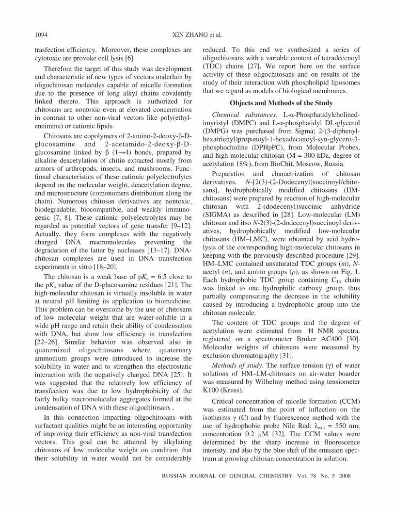
Preparation and charactrization of chitosan derivatives. N-[2(3)-(2-Dodecenyl)succinoyl/chito-sans], hydrophobically modified chitosans (HM-chitosans) were prepared by reaction of high-molecular chitosan with 2-(dodecenyl)succinic anhydride (SIGMA) as described in [28]. Low-molecular (LM) chitosan and itsо N-2(3)-(2-dodecenyl)succinoyl deriv-atives, hydrophobically modified low-molecular chitosans (HM–LMC), were obtained by acid hydro-lysis of the corresponding high-molecular chitosans in keeping with the previously described procedure [29]. HM–LMC contained unsaturated TDC groups (m), N-acetyl (n), and amino groups (p), as shown on Fig. 1. Each hydrophobic TDC group containing C14 chain was linked to one hydrophilic carboxy group, thus partially compensating the decrease in the solubility caused by introducing a hydrophobic group into the chitosan molecule.
The content of TDC groups and the degree of acetylation were estimated from 1H NMR spectra. registered on a spectrometer Bruker AC400 [30]. Molecular weights of chitosans were measured by exclusion chromatography [31].
Methods of study. The surface tension (γ) of water solutions of HM–LM-chitosans on air-water boarder was measured by Wilhelmy method using tensiometer K100 (Kruss).
Critical concentration of micelle formation (CCM) was estimated from the point of inflection on the isotherms γ (C) and by fluorescence method with the use of hydrophobic probe Nile Red: λexit = 550 nm; concentration 0.2 μM [32]. The CCM values were determined by the sharp increase in fluorescence intensity, and also by the blue shift of the emission spec-trum at growing chitosan concentration in solution.
ALKYLATED CHITOSANS OF LOW MOLECULAR WEIGHT
RUSSIAN JOURNAL OF GENERAL CHEMISTRY Vol. 78 No. 5 2008
1095
Fig 1. Hydrophobically modified low-molecular chitosans.
Table 1. Characteristics of HM–LM chitosans
Sample Acetylation, % TDC, % n14 Mw Mn Mw/Mn
HM(3%)-LMC 3 3 0.75 4720 4220 1.12
HM(10%)-LMC 3 10 2.5 4780 3840 1.26
HM(18%)-LMC 4 18 4.5 3580 3300 1.09
Unilamellar liposomes of large size were obtained by extrusion of a suspension of multilamellar liposomes (MLV) through a polycarbonate filter (Nucleopore) on a temperature-controlled extruder (Lipex Biomembranes) [3–5, 33]. The initial pore size of the filter was 0.2 μm (seven filtrations), then 0.1 μm (ten filtrations). As a result a homogeneous population of liposomes was obtained of the size from 0.11 to 0.12 μm measured by the method of quasielastic light scattering (Nanosizer N4SD Coultronics).
The measurement of fluorescence anisotropy in a stationary state (r) was carried out on a spectro-fluorimeter SLM 8000 [3]. The measurement results were treated using Biokine routine developed at Biologic (Claix, France). The micelles size and that of HM–LMC complexes were estimated by dynamic light scattering on a Zetamaster 3000 instrument (Malvern Instruments, France) at the following parameters: time of measurement 30 s, viscosity of medium 1.054 Pa, refraction index 1.45 (equal to that of liposomes), scattering angle 90°, temperature 25°С.
Cytotoxicity of HM–LMC was evaluated using 3-(4,5-dimethyl-2-thiazolyl)-2,5-diphenyltetrazolium bromide (МТТ) [3]. HeLa cells were inoculated into
96-well plates with the density 5 × 10⁴ cells per well and were incubated for 24 h. HM–LMC was added to the cells in the presence of 10% fetal bovine serum or without it. After 3.5 h of transfection the cells were washed with phosphate buffered saline and 250 μl of solution with 0.5 mg ml–1 of MTT in DMEM(Dulbecco Modificated Eagle’s Medium) was added into each well; it was left for additional incubation for 1 h at 37°C. The medium containing MTT was then removed, and 100 μl of dimethyl sulfoxide (DMSO) was added into each well to dissolve formazan crystals formed in mitochondrions during cell breathing. Absorption measurements were carried out on 535 nm on a Labsystems IEMS Microplate Reader instrument.
RESULTS AND DISCUSSION
Characteristics of Hydrophobically Modified Low-Molecular Chitosans
Three samples of HM–LMC were prepared by the formerly described procedure [27] and characterized by NMR and exclusion chromatography (Table 1). Their molecular weight was 5 kDa and the degree of acetylation attained ~3 mol %. Consequently all these
RUSSIAN JOURNAL OF GENERAL CHEMISTRY Vol. 78 No. 5 2008
XIN ZHANG et al. 1096
(a) (b)
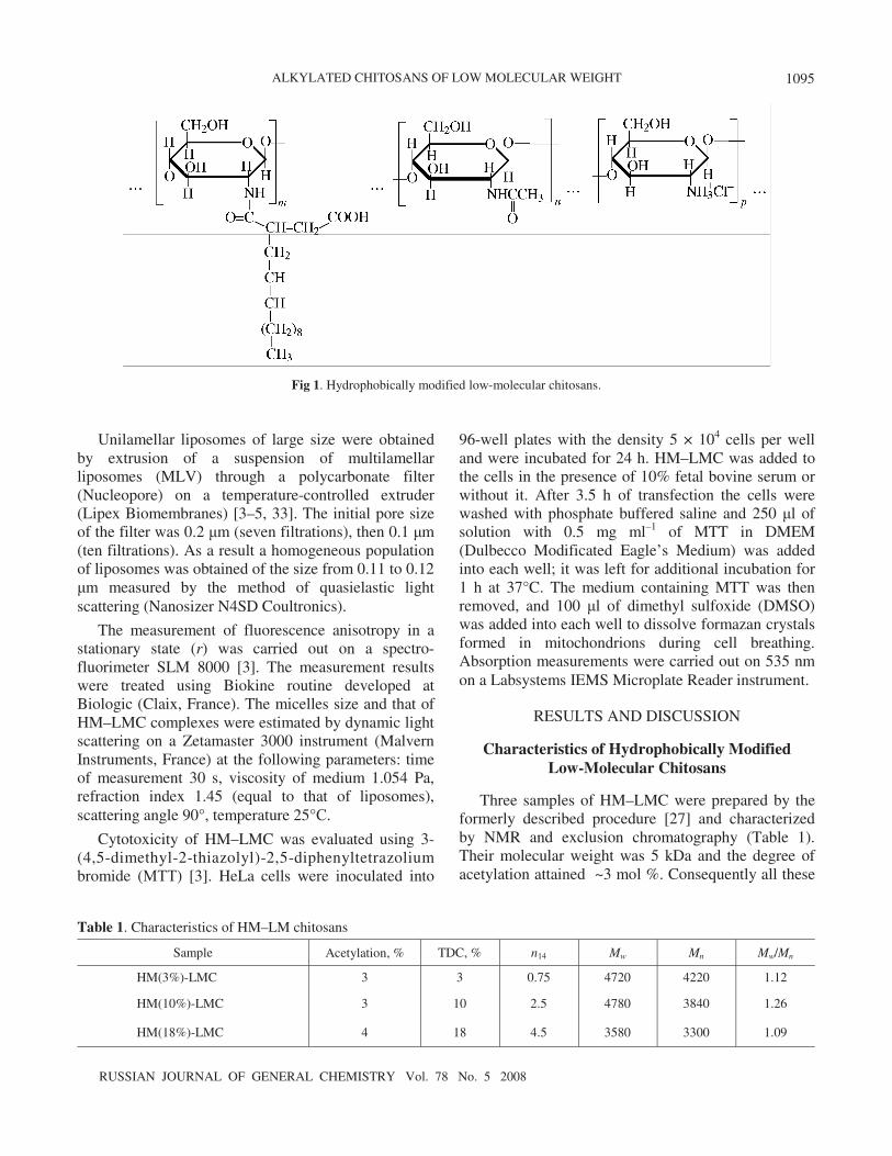
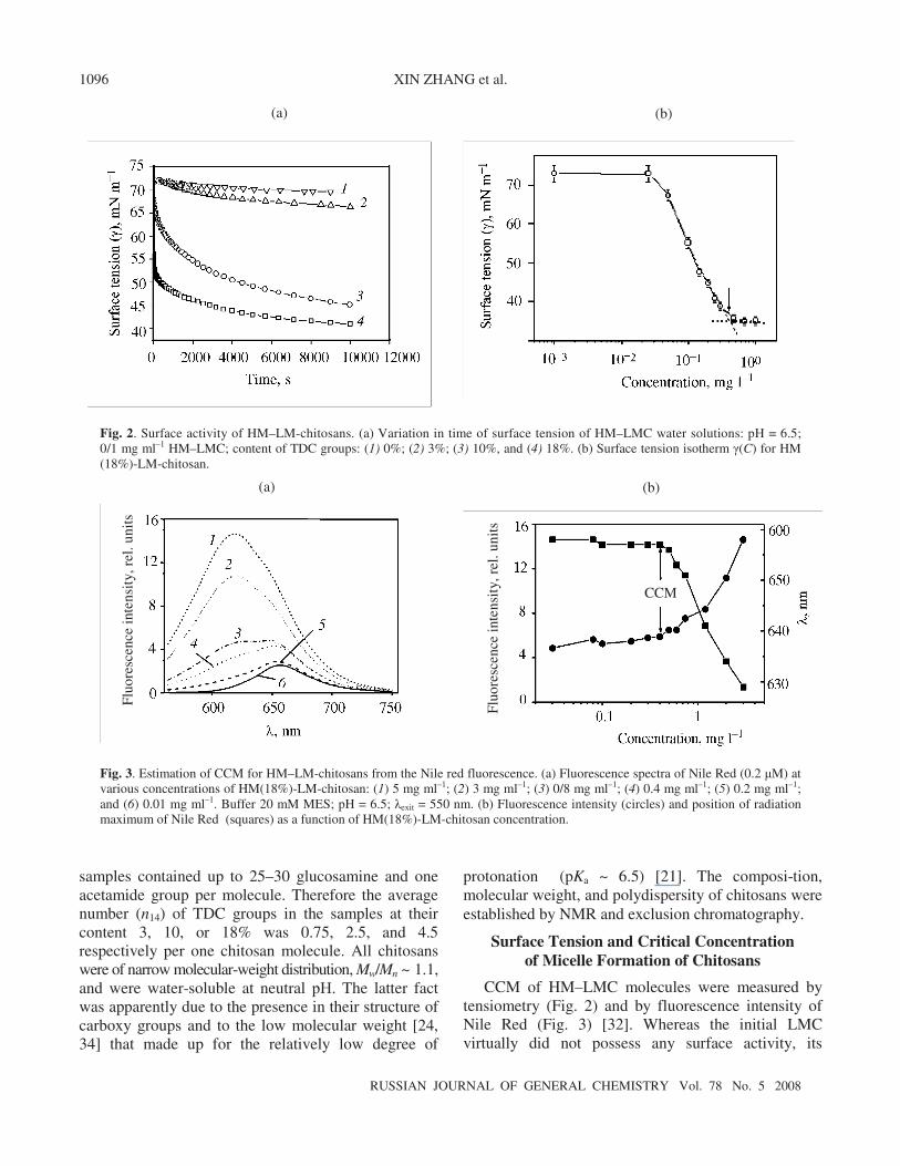
Fig. 2. Surface activity of HM–LM-chitosans. (a) Variation in time of surface tension of HM–LMC water solutions: pH = 6.5; 0/1 mg ml–1 HM–LMC; content of TDC groups: (1) 0%; (2) 3%; (3) 10%, and (4) 18%. (b) Surface tension isotherm γ(C) for HM(18%)-LM-chitosan.
samples contained up to 25–30 glucosamine and one acetamide group per molecule. Therefore the average number (n14) of TDC groups in the samples at their content 3, 10, or 18% was 0.75, 2.5, and 4.5 respectively per one chitosan molecule. All chitosans were of narrow molecular-weight distribution, Mw/Mn ~ 1.1, and were water-soluble at neutral рН. The latter fact was apparently due to the presence in their structure of carboxy groups and to the low molecular weight [24, 34] that made up for the relatively low degree of
Fig. 3. Estimation of CCM for HM–LM-chitosans from the Nile red fluorescence. (a) Fluorescence spectra of Nile Red (0.2 μM) at various concentrations of HM(18%)-LM-chitosan: (1) 5 mg ml–1; (2) 3 mg ml–1; (3) 0/8 mg ml–1; (4) 0.4 mg ml–1; (5) 0.2 mg ml–1; and (6) 0.01 mg ml–1. Buffer 20 mM MES; pH = 6.5; λexit = 550 nm. (b) Fluorescence intensity (circles) and position of radiation maximum of Nile Red (squares) as a function of HM(18%)-LM-chitosan concentration.
(a) (b) Fl
uore
scen
ce in
tens
ity,
rel
. uni
ts
Fluo
resc
ence
inte
nsit
y, r
el. u
nits
CCM
protonation (pKa ~ 6.5) [21]. The composi-tion, molecular weight, and polydispersity of chitosans were established by NMR and exclusion chromatography.
Surface Tension and Critical Concentration of Micelle Formation of Chitosans
CCM of HM–LMC molecules were measured by tensiometry (Fig. 2) and by fluorescence intensity of Nile Red (Fig. 3) [32]. Whereas the initial LMC virtually did not possess any surface activity, its
ALKYLATED CHITOSANS OF LOW MOLECULAR WEIGHT
RUSSIAN JOURNAL OF GENERAL CHEMISTRY Vol. 78 No. 5 2008
1097
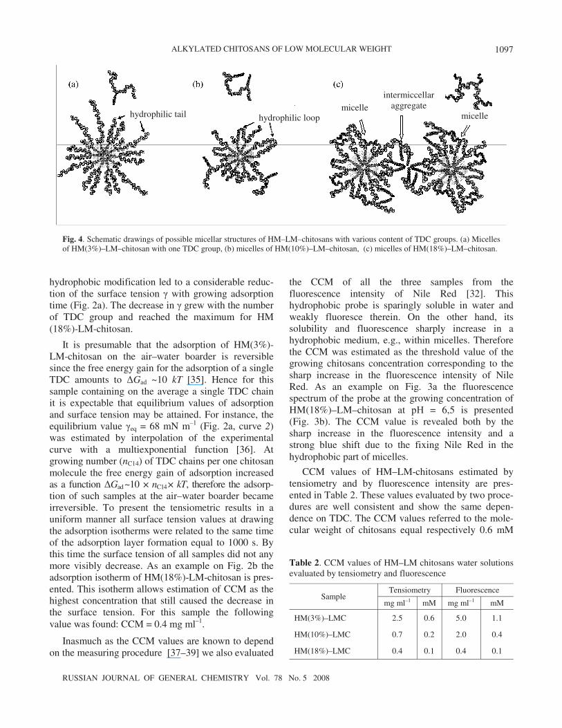
Fig. 4. Schematic drawings of possible micellar structures of HM–LM–chitosans with various content of TDC groups. (a) Micelles of HM(3%)–LM–chitosan with one TDC group, (b) micelles of HM(10%)–LM–chitosan, (c) micelles of HM(18%)–LM–chitosan.
hydrophilic tail hydrophilic loop micelle
micelle
intermiccellar aggregate
Table 2. CCM values of HM–LM chitosans water solutions evaluated by tensiometry and fluorescence
Sample Tensiometry Fluorescence
mg ml–1 mM mg ml–1 mM
HM(3%)–LMC 2.5 0.6 5.0 1.1
HM(10%)–LMC 0.7 0.2 2.0 0.4
HM(18%)–LMC 0.4 0.1 0.4 0.1
hydrophobic modification led to a considerable reduc-tion of the surface tension γ with growing adsorption time (Fig. 2а). The decrease in γ grew with the number of TDC group and reached the maximum for HM(18%)-LM-chitosan.
It is presumable that the adsorption of HM(3%)-LM-chitosan on the air–water boarder is reversible since the free energy gain for the adsorption of a single TDC amounts to ΔGad ~10 kT [35]. Hence for this sample containing on the average a single TDC chain it is expectable that equilibrium values of adsorption and surface tension may be attained. For instance, the equilibrium value γeq = 68 mN m–1 (Fig. 2а, curve 2) was estimated by interpolation of the experimental curve with a multiexponential function [36]. At growing number (nC14) of TDC chains per one chitosan molecule the free energy gain of adsorption increased as a function ΔGad ~10 × nC14 × kT, therefore the adsorp-tion of such samples at the air–water boarder became irreversible. To present the tensiometric results in a uniform manner all surface tension values at drawing the adsorption isotherms were related to the same time of the adsorption layer formation equal to 1000 s. By this time the surface tension of all samples did not any more visibly decrease. As an example on Fig. 2b the adsorption isotherm of HM(18%)-LM-chitosan is pres-ented. This isotherm allows estimation of CCM as the highest concentration that still caused the decrease in the surface tension. For this sample the following value was found: CCM = 0.4 mg ml–1.
Inasmuch as the CCM values are known to depend on the measuring procedure [37–39] we also evaluated
the CCM of all the three samples from the fluorescence intensity of Nile Red [32]. This hydrophobic probe is sparingly soluble in water and weakly fluoresce therein. On the other hand, its solubility and fluorescence sharply increase in a hydrophobic medium, e.g., within micelles. Therefore the CCM was estimated as the threshold value of the growing chitosans concentration corresponding to the sharp increase in the fluorescence intensity of Nile Red. As an example on Fig. 3а the fluorescence spectrum of the probe at the growing concentration of HM(18%)–LM–chitosan at рН = 6,5 is presented (Fig. 3b). The CCM value is revealed both by the sharp increase in the fluorescence intensity and a strong blue shift due to the fixing Nile Red in the hydrophobic part of micelles.
CCM values of HM–LM-chitosans estimated by tensiometry and by fluorescence intensity are pres-ented in Table 2. These values evaluated by two proce-dures are well consistent and show the same depen-dence on TDC. The CCM values referred to the mole-cular weight of chitosans equal respectively 0.6 mM
RUSSIAN JOURNAL OF GENERAL CHEMISTRY Vol. 78 No. 5 2008
XIN ZHANG et al. 1098
and 0.1 mM. The strong dependence of CCM values on TDC suggests that the hydrophobic interactions between the TDC groups present the main driving force in the aggregation of these molecules. This conclusion is in agreement with the common view on micelle formation that the free energy gain at transfer of hydrophobic chains from the water environment into the internal part of micelle becomes equal to the loss in the entropy of translation of the dissolved surfactant molecules and their counter ions [40].
The structure of micelles formed from various HM–LM–chitosans depends on the degree of the hydrophobic modification of these samples (Fig. 4). The sample of HM(3%)–LM–chitosan having on the average only one alkyl group per macroion formed presumably “interpolyelectrolyte complexes” resembl-ing the micelles of “classic” ionic surfactants (Fig. 4а). These micelles possess an external crown formed by partially ionized hydrophilic glucosamine groups. The attraction between non-ionized glucosamine groups that are hydrophobic and in the unprotonated form are capable of hydrogen bonds formation apparently also facilitates the micelle formation.
In contrast to HM(3%)–LM–chitosan the samples modified to 10 and 18% contain on the average 2 and 4.5 alkyl chains per one molecule respectively, and they are able to form micellar aggregates by interaction of al-kyl chains not involved into the micelle core (Figs. 4b, 4c).
The micelle crown in these aggregates consists both of hydrophilic tails and loops and of alkyl chains not involved into the hydrophobic core. The interaction between these alkyl chains leads to the formation of intermicellar clusters yielding nano- and microparticles of gel [41]. The smaller number of alkyl chains in HM(10%)–LM–chitosan results in their lesser density on the micelle surface compared with HM(18%)–LM–chitosan; consequently, the intermicellar clusters from HM(10%)–LM–chitosan would be less stable.
The comparison of CCM = 0.6 mM of cationic HM(3%)–LM–chitosan with the CCM values of classic cationic surfactants from the alkyltrimethylammonium bromides series like dodecyl- (DTAB), tetradecyl- (TTAB), and cetyltrimethylammonium (CTAB) bro-mides with alkyl chains of 12, 14, and 16 carbon atoms equal respectively 15 mM, 3.5 mM, and 0.92 mM, [42, 43] shows virtual equiality of CCM of HM(3%)-LM-chitosan and that of CTAB possessing a hydrophobic chain C16. This equiality means that the free energy consumed in the concentration of the positively charged glucosamine groups on the micelle surface
(Fig. 4а) is close to the corresponding energy for the concentration of the trimethylammonium groups. This fact is fairly unexpected taking into account the large difference in the volume of the polar groups; it can be understood considering the small contribution of the electrostatic repulsion into the free energy of micelle formation [40, 44, 45].
Actually, it was demonstrated that the average dis-tance between the trimethylammonium groups [l(N(CH3)3] of cationic surfactants was equal to the characteristic Bjerrum distance in water (lB = е2/4πε0εkT = 0.7 nm) [42, 43]; it meant that the energy of electrostatic repulsion between these groups was of 1 kT order of magnitude. This energy is less by an order of magnitude than that of hydrophobic interaction between the alkyl chains in the core of the micelle [35]. Considering the large size of the glucosamine groups the average distance between amino groups on the surface of HM(3%)–LM–chitosan micelle should be significantly larger than Bjerrum distance and consequently the electrostatic repulsion between them should be notably less. The reduced energy of electrostatic repulsion is compensated by the large number of charged groups of the chitosan sample resulting in identical CCM values of HM(3%)–LM–chitosan and CTAB. It should be also taken into account that about a half of amino groups of this chitosan is not ionized at рН = 6.5 [21] and the corresponding glucosamine groups can contribute to the energy of micelle formation considering their weak hydrophobicity [29].
On formal representation of CCM through the concentration of alkyl chains in solution, CTDC = nC14CCM, we obtain values 0.45 mM, 0.42 mM, and 0.45 mM for chitosan samples having on the average nC14 = 0.75, 2.5, and 4.5 alkyl chains per chitosan molecule. This surprising equiality of CTDC values indicates that the molecules of HM–LM–chitosans behave in a solution like an ensemble of free molecules of micelle-forming surfactants.
The Nile Red fluorescence was also applied to the study of the dependence of CCM of HM(18%)–LM–chitosan on рН and salt concentration. At рН = 6.5 the CCM value decreased from 0.4 mg ml–1 (without salt) to 0.25 mg ml–1 (0.5 M NaCl). In the same manner as with cationic surfactants [40] this decrease in the CCM might be due to the stronger condensation of counter ions at a larger salt concentration. Actually, the stronger condensation corresponds to a lesser loss in translational entropy and reduces the free energy of
ALKYLATED CHITOSANS OF LOW MOLECULAR WEIGHT
RUSSIAN JOURNAL OF GENERAL CHEMISTRY Vol. 78 No. 5 2008
1099
micelle formation. It was also shown that in the absence of a salt the growth in рН from 5.8 to 7.0, i.e., amino groups deprotonation, resulted in decrease in CCM from 0.6 mg ml–1 to 0.25 mg ml–1. This CCM diminishing may be attributed to the decrease in the charge at amino groups deprotonation leading in its turn to lesser loss in the translational entropy, free energy of micelle formation, and CCM.
Interaction with Model Membranes
One among the important stages of transfection is the interaction of complexes DNA/non-viral vector with the cell membranes. This type of interaction plays a significant part in all the stages of transfection including the initial interaction between these complexes and plasma membranes, their liberation from endosomes and interaction with the membrane of the cell nucleus. Inasmuch as the HM–LMC exhibited the surfactant qualities we aimed first to establish whether the free molecules of these chitosans interacted with the membranes and whether they altered the characteristics of the membranes. To this end we investigated the interaction of HM(18%)–LM–chitosan with large unilamellar liposomes (LUV) that served as models of the cell membranes. The neutral and anionic liposomes were previously shown to adequately mimic respectively the external ant cytoplasmatic layers of plasma membranes [3–5, 33]. Thus we studied the interaction of HM(18%)–LM–
(a) (b)
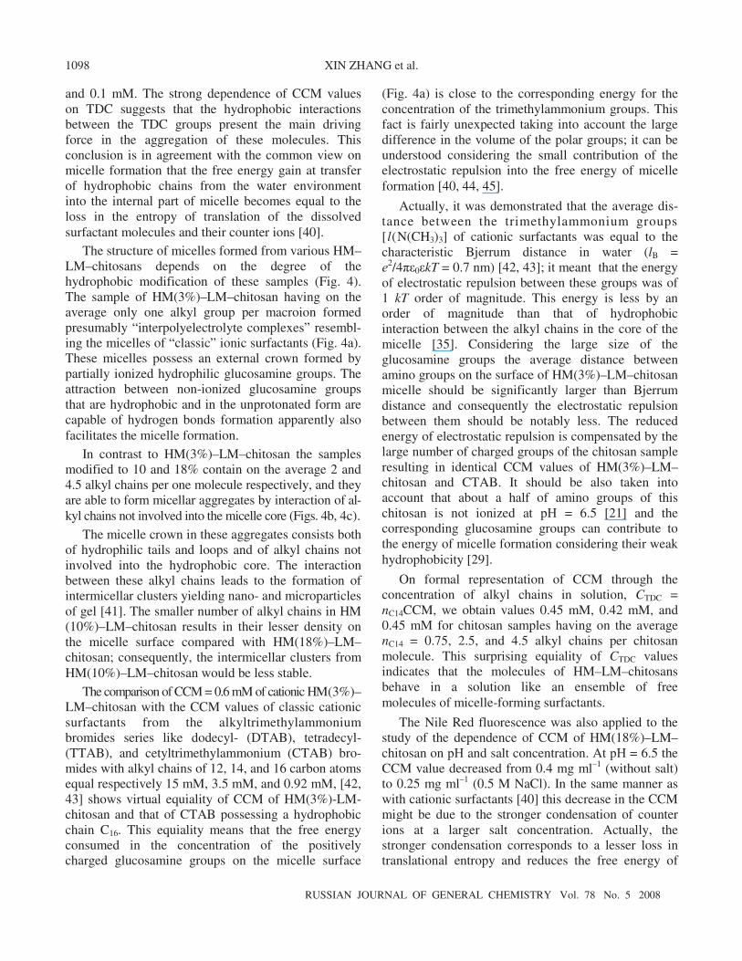
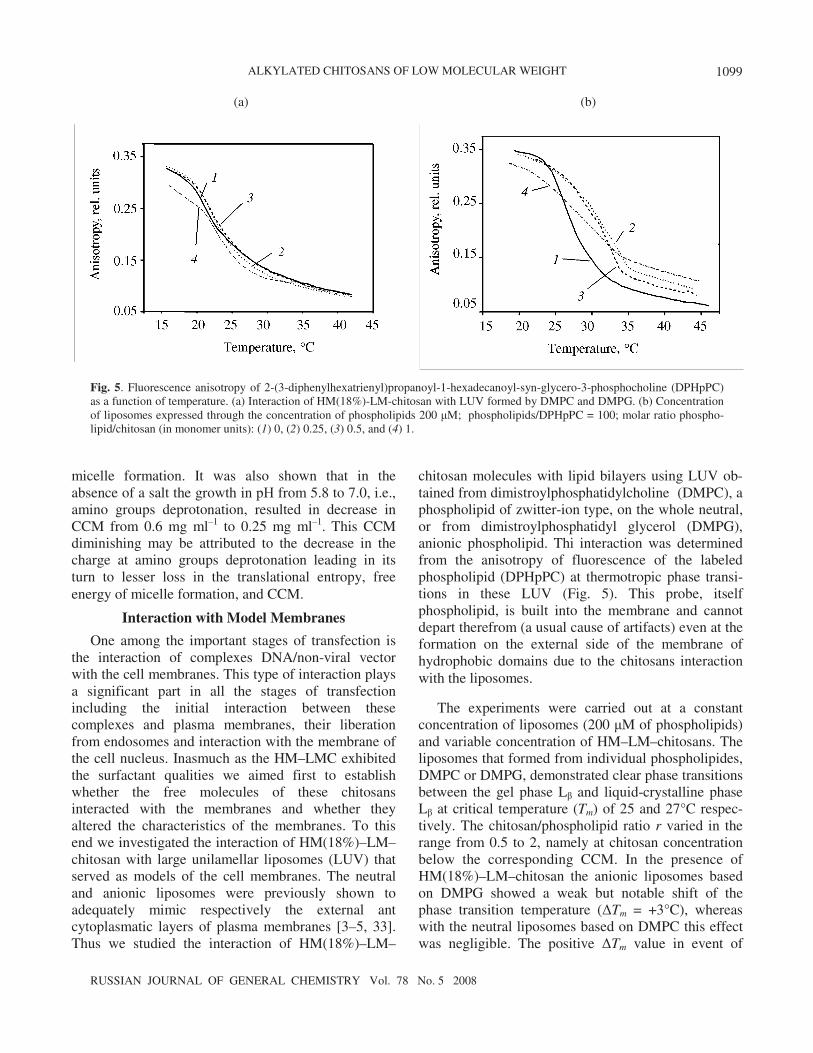
Fig. 5. Fluorescence anisotropy of 2-(3-diphenylhexatrienyl)propanoyl-1-hexadecanoyl-syn-glycero-3-phosphocholine (DPHpPC) as a function of temperature. (a) Interaction of HM(18%)-LM-chitosan with LUV formed by DMPC and DMPG. (b) Concentration of liposomes expressed through the concentration of phospholipids 200 μM; phospholipids/DPHpPC = 100; molar ratio phospho-lipid/chitosan (in monomer units): (1) 0, (2) 0.25, (3) 0.5, and (4) 1.
chitosan molecules with lipid bilayers using LUV ob-tained from dimistroylphosphatidylcholine (DMPC), a phospholipid of zwitter-ion type, on the whole neutral, or from dimistroylphosphatidyl glycerol (DMPG), anionic phospholipid. Thi interaction was determined from the anisotropy of fluorescence of the labeled phospholipid (DPHpPC) at thermotropic phase transi-tions in these LUV (Fig. 5). This probe, itself phospholipid, is built into the membrane and cannot depart therefrom (a usual cause of artifacts) even at the formation on the external side of the membrane of hydrophobic domains due to the chitosans interaction with the liposomes.
The experiments were carried out at a constant concentration of liposomes (200 μM of phospholipids) and variable concentration of HM–LM–chitosans. The liposomes that formed from individual phospholipides, DMPC or DMPG, demonstrated clear phase transitions between the gel phase Lβ and liquid-crystalline phase Lβ at critical temperature (Tm) of 25 and 27°C respec-tively. The chitosan/phospholipid ratio r varied in the range from 0.5 to 2, namely at chitosan concentration below the corresponding CCM. In the presence of HM(18%)–LM–chitosan the anionic liposomes based on DMPG showed a weak but notable shift of the phase transition temperature (ΔTm = +3°C), whereas with the neutral liposomes based on DMPC this effect was negligible. The positive ΔTm value in event of
RUSSIAN JOURNAL OF GENERAL CHEMISTRY Vol. 78 No. 5 2008
XIN ZHANG et al. 1100
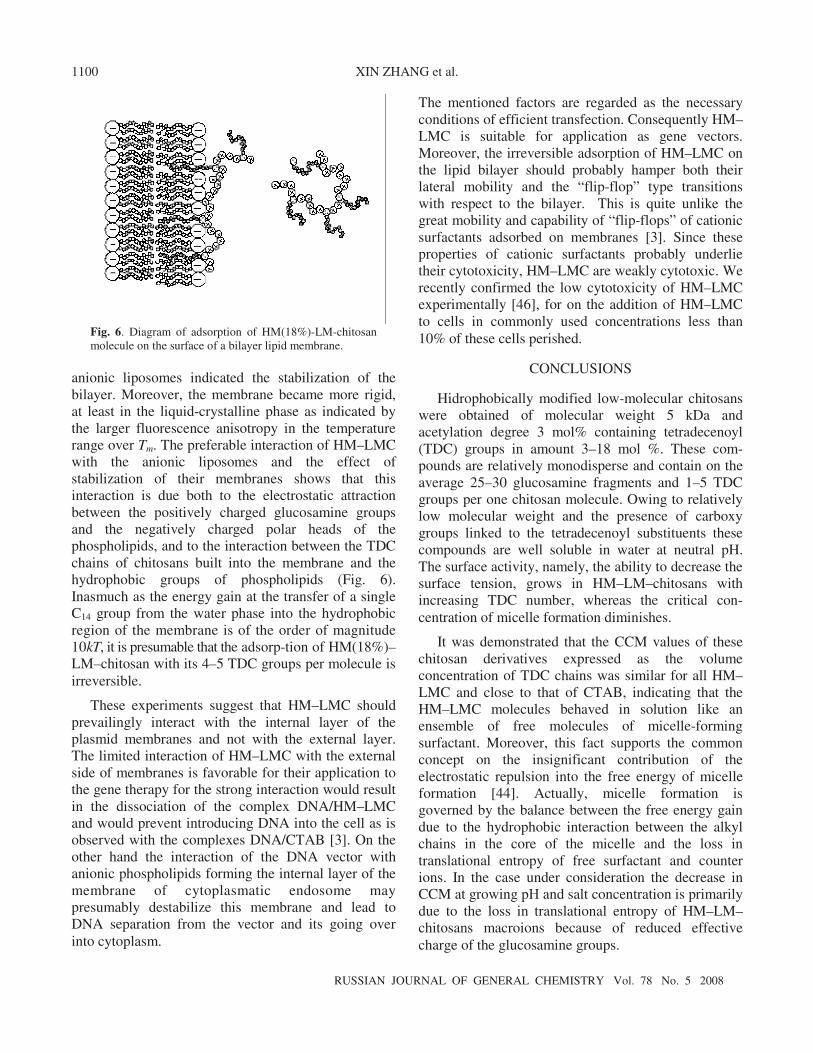
Fig. 6. Diagram of adsorption of HM(18%)-LM-chitosan molecule on the surface of a bilayer lipid membrane.
anionic liposomes indicated the stabilization of the bilayer. Moreover, the membrane became more rigid, at least in the liquid-crystalline phase as indicated by the larger fluorescence anisotropy in the temperature range over Tm. The preferable interaction of HM–LMC with the anionic liposomes and the effect of stabilization of their membranes shows that this interaction is due both to the electrostatic attraction between the positively charged glucosamine groups and the negatively charged polar heads of the phospholipids, and to the interaction between the TDC chains of chitosans built into the membrane and the hydrophobic groups of phospholipids (Fig. 6). Inasmuch as the energy gain at the transfer of a single C14 group from the water phase into the hydrophobic region of the membrane is of the order of magnitude 10kT, it is presumable that the adsorp-tion of HM(18%)–LM–chitosan with its 4–5 TDC groups per molecule is irreversible.
These experiments suggest that HM–LMC should prevailingly interact with the internal layer of the plasmid membranes and not with the external layer. The limited interaction of HM–LMC with the external side of membranes is favorable for their application to the gene therapy for the strong interaction would result in the dissociation of the complex DNA/HM–LMC and would prevent introducing DNA into the cell as is observed with the complexes DNA/CTAB [3]. On the other hand the interaction of the DNA vector with anionic phospholipids forming the internal layer of the membrane of cytoplasmatic endosome may presumably destabilize this membrane and lead to DNA separation from the vector and its going over into cytoplasm.
The mentioned factors are regarded as the necessary conditions of efficient transfection. Consequently HM–LMC is suitable for application as gene vectors. Moreover, the irreversible adsorption of HM–LMC on the lipid bilayer should probably hamper both their lateral mobility and the “flip-flop” type transitions with respect to the bilayer. This is quite unlike the great mobility and capability of “flip-flops” of cationic surfactants adsorbed on membranes [3]. Since these properties of cationic surfactants probably underlie their cytotoxicity, HM–LMC are weakly cytotoxic. We recently confirmed the low cytotoxicity of HM–LMC experimentally [46], for on the addition of HM–LMC to cells in commonly used concentrations less than 10% of these cells perished.
CONCLUSIONS
Hidrophobically modified low-molecular chitosans were obtained of molecular weight 5 kDa and acetylation degree 3 mol% containing tetradecenoyl (TDC) groups in amount 3–18 mol %. These com-pounds are relatively monodisperse and contain on the average 25–30 glucosamine fragments and 1–5 TDC groups per one chitosan molecule. Owing to relatively low molecular weight and the presence of carboxy groups linked to the tetradecenoyl substituents these compounds are well soluble in water at neutral pH. The surface activity, namely, the ability to decrease the surface tension, grows in HM–LM–chitosans with increasing TDC number, whereas the critical con-centration of micelle formation diminishes.
It was demonstrated that the CCM values of these chitosan derivatives expressed as the volume concentration of TDC chains was similar for all HM–LMC and close to that of CTAB, indicating that the HM–LMC molecules behaved in solution like an ensemble of free molecules of micelle-forming surfactant. Moreover, this fact supports the common concept on the insignificant contribution of the electrostatic repulsion into the free energy of micelle formation [44]. Actually, micelle formation is governed by the balance between the free energy gain due to the hydrophobic interaction between the alkyl chains in the core of the micelle and the loss in translational entropy of free surfactant and counter ions. In the case under consideration the decrease in CCM at growing рН and salt concentration is primarily due to the loss in translational entropy of HM–LM–chitosans macroions because of reduced effective charge of the glucosamine groups.
ALKYLATED CHITOSANS OF LOW MOLECULAR WEIGHT
RUSSIAN JOURNAL OF GENERAL CHEMISTRY Vol. 78 No. 5 2008
1101
HM–LMC interact with the negatively charged lipid liposomes mimicking the internal layer of plasmid membranes. The decrease in the fluidity (growth of rigidity) of liposome membranes in the liquid-crystalline state originating from the interaction with HM–LM–chitosans may be ascribed to the inclusion of TDC chains covalently bound to glucosamine groups into the lipide bilayer formed by alkyl groups of phospholipids. Unlike cationic surfactants HM–LMC suffer difficulties regarding lateral and transverse diffusion with respect to the membrane resulting in their low toxicity. Besides the prevailing interaction of HM–LMC with the internal layer of biomembranes leads to destabilization of endosomal membranes facilitating the liberation of DNA into cytoplasm. On the whole, the results obtained indicate that HM–LMC possess interesting characteristics suggesting their application as non-viral DNA transfection vectors. The investigation results of physicochemical properties of these chitosans complexes with DNA and the efficiency of their transfection both in vitro and in vivo will be published elsewhere [46].
ACKNOWLEDGMENTS
The study was carried out under a financial support of the French Agency against Myopathy (AFM) and of the program Eco-Net of the Foreign Ministry of France within the framework of collaboration of France, Russian Federation, and Ukraine. V. Babak worked as an Invited Professor at the Lois Pasteur University. The autors are grateful to Doctor C. Grandfils from the Institute of Chemistry of Liege University (Belgium) and Professor J. Desbrières from the Laboratory of the physical chemistry of polymers from the Pau University (France) for the help in characterization of LM-chitosans by NMR and exclusion chromatography.
REFERENCES
1. Xu, Y. and Szoka, C.F., Biochemistry, 1996, vol. 35, p. 5616.
2. Mel’nikov, S.M., Sergeyev, V.G., and Yoshikawa, K., J. Am. Chem. Soc., 1995, vol. 117, p. 2401.
3. Clamme, J.-P., Bernacchi, S., Vuilleumier, C., Dupor- tail, G., and Mély, Y., Biochim. Biophys. Acta, 2000, vol. 1467, p. 347.
4. Llères, D., Dauty, E., Behr, J.P., Mély, Y., and Dupor- tail, G., Chem. Phys. Lipids, 2001, vol. 111, p. 59.
5. Llères, D., Clamme, J.-P., Dauty, E., Blessing, T., Krishnamoorthy, G., Duportail, G., and Mély, Y., Langmuir, 2002, vol. 18, p. 10340.
6. Sasaki, T., Kawai, K., Saijo-Kurita, K., and Ohno, T., Toxicol. in vitro, 1992, vol. 6, p. 451.
7. Muzzarelli, R.A.A., in Chitosan in Natural Chelating Polymers; Alginic Acid, Chitin, and Chitosan, Belcher, R., Ed., Oxford: Pergamon Press, 1973, p. 144.
8. Muzzarelli, R.A.A., Carbohydr. Polym., 1996, vol. 29, p. 309.
9. Romoren, K., Thu, B.J., and Evensen, O., J. Controlled Release, 2002, vol. 85, p. 215. 10. Borchard, G., Adv. Drug Delivery Rev., 2001, vol. 52, p. 143. 11. Rolland, A.P., Crit. Rev. Ther. Drug Carrier Syst., 1998, vol. 15, p. 143. 12. Sato, T., Ishii, T., and Okahata, Y., Biomaterials, 2001, vol. 22, p. 2075. 13. Venkatesh, S. and Smith, T.J., Biotechnol. Appl. Biochem., 1998, vol. 27, p. 265. 14. Cui, Z. and Mumper, R.J., J. Controlled Release, 2001, vol. 75, p. 409. 15. Illum, L., Jabbal-Gill, I., Hinchcliffe, M., Fisher, A.N., and Davis, S.S., Adv. Drug Delivery Rev., 2001, vol. 51, p. 81. 16. Hejazi, R. and Amiji, M., J. Controlled Release, 2003, vol. 89, p. 151. 17. Fang, N., Chan, V., Mao, H.-Q., and Leong, K.W., Biomacromolecules, 2001, vol. 2, p. 1161. 18. Mansouri, S., Lavigne, P., Corsi, K., Benderdour, M., Beaumont, E., and Fernandes, J.C., Eur. J. Pharm. Biopharm., 2004, vol. 57, p. 1. 19. Corsi, K., Chellat, F., Yahia, L., and Fernandes, J.C., Biomaterials, 2003, vol. 24, p. 1255. 20. Erbacher, P., Zou, S., Bettinger, T., Steffan, A.-M., and Rémy, J.-S., Pharm. Res., 1998, vol. 15, p. 1332. 21. Rinaudo, M., Pavlov, G., and Desbrières, J., Polymer, 1999, vol. 40, p. 7029.
22. MacLaughlin, F.C., Mumper, R.J., Wang, J., Tagliaferri J.M., Gill, I., Hinchcliffe, M., amd Rolland, A.P., J. Controlled Release, 1998, vol. 56, p. 259. 23. Koping-Hoggard, M., Mel’.nikova, Y.S., Varum, K.M., Lindman, B., and Artursson, P., J. Gene. Med., 2003, vol. 5, p. 130.
24. Lee, M., Nah, J.-W., Kwon, Y., Koh, J.J., Ko, K.S., and Kim, S.W., Pharm. Res., 2001, vol. 18, p. 427.
25. Thanou, M., Florea, B.I., Geldof, M., Junginger, H.E., and Borchard, G., Biomaterials, 2002, vol. 23, p. 153.
26. Tommeraas, K., Varum, K.M., Christensen, B.E., and Smidsrod, O., Carbohydr. Res., 2001, vol. 333, p. 137. 27. Tikhonov, V.E., Stepnova, E.A., Babak, V.G., Yam- skov, I.A., Palma-Guerrero, J., Jansson, H.-B., Lopez- Llorca, L.V., Salinas, J., Gerasimenko, D.V., Avdien- ko, I.D., and Varlamov, V.P., Carbohydr. Polym., 2006, vol. 64, p. 66.
RUSSIAN JOURNAL OF GENERAL CHEMISTRY Vol. 78 No. 5 2008
XIN ZHANG et al. 1102
28. Hirano, S., Ohe, Y., and Ono, H., Carbohydr Res., 1976, vol. 47, p. 315.
29. Domard, A. and Carter, N., in Chitin and Chitosan, Skjak-Braek, G., Anthonsen, T., and Sandford, P., Eds., London–New York: Elsevier Applied Science, 1989, p. 383.
30. Rinaudo, M., Dung, P.L., Gey, C., and Milas, M., Int. J. Biol. Macr., 1992, vol. 14, p. 121.
31. Brugnerotto, J., Desbrières, J., Roberts, G., and Rina- udo, M., Polymer, 2001, vol. 42, p. 9921.
32. Countinho, P.J.G., Castanheira, E.M.S., Rei, M.C., and Oliveira, M.E.C.D.R., J. Phys. Chem. B, 2002, vol. 106, p. 12841.
33. Cullis, P.R. and Hope, M.J., in Biochemistry of Lipids and Membranes, Vance, D.E. and Vance, J.E., New York: Benjamin/Cummings, 1985, p. 27.
34. Richardson, S.C.W., Kolbe, H.V.J., and Duncan, R., Int. J. Pharm., 1999, vol. 178, p. 231.
35. Tanford, C., The Hydrophobic Effect: Formation of Micelles and Biological Membranes, New York: Wiley, 1973, p. 10.
36. Babak, V.G., Desbrières, J., and Tikhonov, V.E., Colloid. Surf. A, 2005, vol. 255, p. 119.
37. Rosen, M.J., Surfactants and Interfacial Phenomena, New York: Wiley, 1989, 2nd ed.
38. Babak, V.G., Pavlov, A.N., Svitova, T.F., Danilen- ko, A.N., Egorov, V.V., and Varlamova, E.A., Kolloid. Zh., 1996, vol. 58, p. 1.
39. Chakrabort,y T., Ghosh, S., and Moulik, S.P., J. Phys. Chem. B, 2005, vol. 109, p. 14813.
40. Konop, A.J. and Colby, R.H., Langmuir, 1999, vol. 15, p. 58.
41. Lee, K.Y., Jo, W.H., Kwon, I.C., Kim, Y.-H., and Jeong, S.Y., Langmuir, 1998, vol. 14, p. 2329.
42. Zana, R., J. Colloid Interf. Sci., 1980, vol. 78, p. 330.
43. Nagamine, N. and Nakamura, H., Anal. Sci., 1998, vol. 14, p. 405.
44. Murray, R.C. and Hartley, G.S., Trans. Faraday Soc., 1935, vol. 31, p. 183.
45. Attwood, D. and Florence, A.T., Surfactant Systems: Their Chemistry, Pharmacy and Biology, New York: Chapman and Hall, 1983, p. 203.
46. Zhang, X., Ercelen, S., Duportail, G., Schaub, E., Tikhonov, V., Slita, A., Zarubaev, V., Babak, V., and Mély, Y., J. Gene Med., 2007 (sent).