acute cytomegalovirus infection in kenyan hiv-infected infants
TRANSCRIPT
C
Acute cytomegalovir
us infection in KenyanHIV-infected infantsJennifer A. Slykera,b, Barbara L. Lohman-Payneb,c,
Grace C. John-Stewartb,g, Elizabeth Maleche-Obimboc, Sandra Emeryd,
Barbra Richardsone, Tao Donga, Astrid K.N. Iversena,
Dorothy Mbori-Ngachac, Julie Overbaughd, Vincent C. Emeryf
and Sarah L. Rowland-Jonesa
opyright © L
aMRC Human ImmWashington, USA,eDepartment of BiSchool of BiomediWashington, Seatt
Correspondence to
Tel: +1 206 744 3Received: 5 May
DOI:10.1097/QAD
ISSN
Objective: Cytomegalovirus (CMV) coinfection may influence HIV-1 disease pro-gression during infancy. Our aim was to describe the incidence of CMV infectionand the kinetics of viral replication in Kenyan HIV-infected and HIV-exposed unin-fected infants.
Methods: HIV-1 and CMV plasma viral loads were serially measured in 20 HIV-exposed uninfected and 44 HIV-infected infants born to HIV-infected mothers.HIV-infected children were studied for the first 2 years of life, and HIV-exposeduninfected infants were studied for 1 year.
Results: CMV DNA was detected frequently during the first months of life; by 3 months ofage,CMVDNAwasdetected in90%ofHIV-exposeduninfected infantsand93%of infantswhohadacquiredHIV-1 inutero.CMVviralloadswerehighestinthe1–3monthsfollowingthe first detection of virus and declined rapidly thereafter. CMV peak viral loads weresignificantlyhigher in theHIV-infected infantscomparedwith theHIV-exposeduninfectedinfants (mean3.2versus2.7 log10 CMV DNA copies/ml, respectively,P¼0.03).Thedetec-tion of CMV DNA persisted to 7–9 months post-CMV infection in both the HIV-exposeduninfected (8/17, 47%) and HIV-infected (13/18, 72%, P¼0.2) children. Among HIV-infected children, CMV DNA was detected in three of the seven (43%) surviving infantstested between 19 and 21 months post-CMV infection. Finally, a strong correlation wasfound between peak CMV and HIV-1 viral loads (r¼0.40, P¼0.008).
Conclusion: Acute CMV coinfection is common in HIV-infected Kenyan infants. HIV-1infection was associated with impaired containment of CMV replication.
� 2009 Wolters Kluwer Health | Lippincott Williams & Wilkins
AIDS 2009, 23:2173–2181
Keywords: acute infection, cytomegalovirus, opportunistic infection, paediatricHIV, pathogenesis
Introduction
Cytomegalovirus (CMV) is a major viral cause ofcongenital disease globally, affecting 0.2–3% of live
ippincott Williams & Wilkins. Unauth
unology Unit, Oxford University, Oxford, UK,cDepartment of Paediatrics, University of Nairob
ostatistics, University of Washington, Seattle, Wacal and Life Sciences, University College London,le, Washington, USA.
Jennifer A. Slyker, PhD, Harborview Medical Ce
786; fax: +1 206 744 3693; e-mail: [email protected]; revised: 17 June 2009; accepted: 25 June
.0b013e32833016e8
0269-9370 Q 2009 Wolters Kluwer Hea
births in high-income and 4–14% in low-income regions[1–6]. CMV prevalence varies among populationsaccording to socioeconomic conditions [7], with poorercommunities having a relatively higher prevalence and
orized reproduction of this article is prohibited.
bDepartment of Medicine, University of Washington, Seattle,i, Nairobi, Kenya, dFred Hutchinson Cancer Research Center,shington, USA, fCentre for Virology, Department of Infection,London, UK, and gDepartment of Epidemiology, University of
nter, 325 9th Avenue, Box 359931, Seattle, WA 98104, USA.
ashington.edu2009.
lth | Lippincott Williams & Wilkins 2173
Co
2174 AIDS 2009, Vol 23 No 16
earlier incidence. A recent estimate reported approxi-mately 54% of American adults in their thirties to beCMV seropositive [8], whereas approximately 85% ofGambian infants acquire CMV before they are a year old[9].
Although CMV does not typically cause disease in healthyindividuals, the virus has clinical significance duringprimary infection or reactivation in the immunosup-pressed. CMV infection is associated with HIV-1 diseaseprogression and mortality in adults [10–13]. In theabsence of HAART, patients with CD4 cell counts below100 cells/ml are at a high risk for CMV-associated retinitisand gastrointestinal and neurologic disease [14–17]. InUnited States cohorts, CMV coinfection has been notedin up to 40% of HIV-infected infants during the first yearof life and is associated with an approximately 2.5-foldincreased risk of disease progression [18,19]. In Kenya, werecently reported that the detection of maternal CMVDNA in the blood near the time of delivery was associatedwith a three to four times increased rate of mortality inHIV-infected infants [20]; this relationship remainedsignificant after controlling for other strong predictors ofinfant mortality, including maternal CD4 cell count,CD4 cell percentage, HIV-1 RNA viral load andmaternal death.
If CMV presents a significant risk factor for HIV-1 diseaseprogression, its impact may be particularly important inAfrican children where both viruses are commonlyacquired in infancy [2,21]. In order to understand themechanisms that underlie the relationship between CMVcoinfection and rapid HIV-1 disease progression, it is firstnecessary to determine the incidence of CMV infectionamong HIV-infected infants and to describe its naturalhistory. Although risk factors associated with verticalCMV transmission are well defined, very few studies[22,23] have measured CMV replication quantitatively ininfants, and only one longitudinal study [24] hasdescribed infant CMV viral load in the setting of HIV-1. The purpose of our study was to describe the incidenceand timing of CMV infection and the kinetics of CMVviral replication in HIV-infected and HIV-exposeduninfected Kenyan infants.
Methods
Participants and study designStudy protocols were approved by the Ethics ReviewCommittee of Kenyatta National Hospital and theInstitutional Review Board of the University ofWashington. A cohort of infants born to HIV-infectedwomen was used to study acute infant CMV infection. Aspart of a larger cohort study of perinatal HIV-1transmission, HIV-infected pregnant women wererecruited in Nairobi between 1999 and 2003 [25,26].
pyright © Lippincott Williams & Wilkins. Unauthor
The women received short-course zidovudine forprevention of HIV-1 transmission [27]. Mothers andinfants were followed during pregnancy, delivery and for1–2 years postpartum, during which time serial bloodspecimens were obtained in pregnancy, at delivery andmonths 1, 3, 6, 9, 12, 15, 18, 21 and 24 postpartum.Sixty-four infants were selected from the larger cohortbased on survival to at least 3 months of age and theavailability of a plasma specimen by 1 month of age.Infants were followed until death or exit from the studyat 1 year (HIV-exposed uninfected) or 2 years of life(HIV infected).
HIV-1 diagnosis and quantificationDiagnosis of infant HIV-1 infection was made using PCRamplifying HIV-1 gag DNA from dried blood spottedonto filter paper as previously described [28]. HIV-1RNA viral loads were measured using the Gen-Probeassay [29]. HIV-1 infection was defined as the detection ofeither HIV-1 DNA or RNA; the timing of HIV-1infection was estimated as the midpoint between the lastHIV-negative test and the first HIV-1-positive test.Infants were grouped according to first HIV-1 detection:in utero (within 48 h of birth, n¼ 15), peripartum(uninfected at birth, infected at 1 month, n¼ 16) or late(infected after 1 month, n¼ 13). In the ‘late’ infectiongroup, estimated infection times were: 2 months (sixinfants), 2.5 months (one infant), 4.5 months (one infant),5 months (one infant), 6 months (one infant), 7.5 months(two infants) and 10.5 months (one infant). Peak and set-point HIV-1 viral load were used to describe thedynamics and control of HIV-1 replication during earlyinfection. We defined peak HIV-1 viral load as the highestmeasurement in the first 6 months after infection; HIV-1set-point was defined as the first viral load observationmeasured at least 6 weeks after the peak [30].
Cytomegalovirus viral load measurementsCord blood was used to diagnose in utero CMVtransmission. Following delivery of the placenta, umbi-lical cords were clamped in two locations and swabbed toremove maternal blood. Blood was then collected with asyringe and transferred to EDTA Vacutainer tubes(Becton Dickinson Diagnostics, Franklin Lakes, NewJersey, USA). At all other time-points, venous blood wascollected. Viral nucleic acids were extracted from 50–200 ml of plasma using the Qiagen UltraSens virusextraction kit (Qiagen, Valencia, California, USA).Quantitative PCR was used to detect the glycoproteinB gene [31], and copy number was determined with theaid of a standard curve derived from known quantities ofcloned amplicon DNA. Each individual’s viral load wasdetermined by calculating the mean of three replicatereactions. The lower limit of detection was one copy perreaction. Negative (no DNA detection) and indetermi-nate (less than one copy per reaction) PCR assays werenot included in calculations of median or peak viral loadand were categorized as negative for CMV DNA.
ized reproduction of this article is prohibited.
C
Acute CMV in Kenyan HIV-infected infants Slyker et al. 2175
Indeterminate PCR assays were assigned a valueequivalent to the midpoint between the limit of detectionand zero for longitudinal modelling but were notincluded in the calculation of peak CMV viral load.Peak CMV viral load was defined as the highest viral loadobserved for each CMV-infected infant during the first6 months after infection.
Statistical analysisSTATA SE version 9 for Macintosh (STATA Corp.,College Station, Texas, USA) was used for the statisticalanalysis. Viral loads were base 10 log-transformed(log10). The t-test was used to compare continuousvariables, and Fisher’s exact tests were used to compareproportions. S-Plus (S-Plus 2000; Mathsoft, Inc.,Seattle, Washington, USA) was used to create non-parametric smoothers for CMV DNA over time usingFreidman’s super smoother. Area under the curve(AUC) for each child’s log viral load from 3 to 12 months oflife was estimated using SPSS version 15.0 (SPSS Inc.,Chicago, Illinois, USA). The t-test was used to comparemean AUC of longitudinal viral loads between groupsof infants. Spearman’s rank correlation coefficientwas used to describe the correlation between HIV-1and CMV viral load. All P values reported are fortwo-tailed tests.
Results
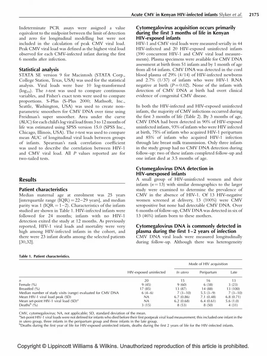
Patient characteristicsMedian maternal age at enrolment was 25 years[interquartile range (IQR)¼ 22–29 years], and medianparity was 1 (IQR¼ 1–2). Characteristics of the infantsstudied are shown in Table 1. HIV-infected infants werefollowed for 24 months; infants with no HIV-1detection exited the study at 12 months. As previouslyreported, HIV-1 viral loads and mortality were veryhigh among HIV-infected infants in the cohort, andthere were 23 infant deaths among the selected patients[30,32].
opyright © Lippincott Williams & Wilkins. Unauth
Table 1. Patient characteristics.
HIV-
nFemale (%)Breastfed (%)Median number of study visits (range) evaluated for CMV DNAMean HIV-1 viral load peak (SD)Mean set-point HIV-1 viral load (SD)a
Deathsb (%)
CMV, cytomegalovirus; NA, not applicable; SD, standard deviation of theaSet-point HIV-1 viral loads were not defined for infants who died before thein utero group, three infants in the peripartum group and three infants inbDeaths during the first year of life for HIV-exposed uninfected infants, de
Cytomegalovirus acquisition occurs primarilyduring the first 3 months of life in KenyanHIV-exposed infantsHIV-1 and CMV viral loads were measured serially in 44HIV-infected and 20 HIV-exposed uninfected infants(390 concurrent HIV-1 and CMV viral load measure-ments). Plasma specimens were available for CMV DNAassessment at birth from 51 infants and by 1 month of agefrom all 64 infants. CMV DNA was detected in the cordblood plasma of 29% (4/14) of HIV-infected newbornsand 2.7% (1/37) of infants who were HIV-1 RNAnegative at birth (P¼ 0.02). None of the infants withdetection of CMV DNA at birth had overt clinicalevidence of congenital CMV disease.
In both the HIV-infected and HIV-exposed uninfectedinfants, the majority of CMV infections occurred duringthe first 3 months of life (Table 2). By 3 months of age,CMV DNA had been detected in 90% of HIV-exposeduninfected infants, 93% of infants who were HIV infectedat birth, 75% of infants who acquired HIV-1 peripartumand 85% of infants who acquired HIV-1 infectionthrough late breast milk transmission. Only three infantsin the study group had no CMV DNA detection duringfollow-up: two of these infants completed follow-up andone infant died at 3.5 months of age.
Cytomegalovirus DNA detection inHIV-unexposed infantsA small group of HIV-uninfected women and theirinfants (n¼ 13) with similar demographics to the largerstudy were examined to determine the prevalence ofCMV in the absence of HIV-1. Of 13 HIV-negativewomen screened at delivery, 13 (100%) were CMVseropositive but none had detectable CMV DNA. Over6 months of follow-up, CMV DNA was detected in six of13 (46%) infants born to these mothers.
Cytomegalovirus DNA is commonly detected inplasma during the first 1–2 years of infectionCMV DNA viral loads were measured longitudinallyduring follow-up. Although there was heterogeneity
orized reproduction of this article is prohibited.
Mode of HIV acquisition
exposed uninfected In utero Peripartum Late
20 15 16 139 (45) 9 (60) 6 (38) 3 (23)
17 (85) 13 (87) 14 (88) 13 (100)6 (4–6) 7 (3–10) 5.5 (3–9) 7 (3–10)
NA 6.7 (0.86) 7.1 (0.48) 6.8 (0.71)NA 6.2 (0.68) 6.4 (0.61) 5.6 (1.0)
3 (15) 8 (53) 8 (50) 4 (31)
mean.ir first postpeak viral load measurement; this included one infant in thethe late group.aths during the first 2 years of life for the HIV-infected infants.
Co
2176 AIDS 2009, Vol 23 No 16
Table 2. Cytomegalovirus detection in children grouped by mode of HIV-1 acquisition.
Cumulative % CMV DNA detected
Age HIV-uninfecteda, n¼20 HIV in utero, n¼15 HIV peripartum n¼16 HIV late, n¼13
Birthb 6.3 29 0 01 month 20 53 13 233 months 90 93 75 856 months 90 93 81 929 months 90 100 81 9212 months 95 100 88 9215 months –c 100 88 9218 months –c 100 88 9221 months –c 100 94 9224 months –c 100 94 92
aInfants with no HIV-1 detection exited the study at 12 months of age.bCord blood tested at birth.cNot tested.
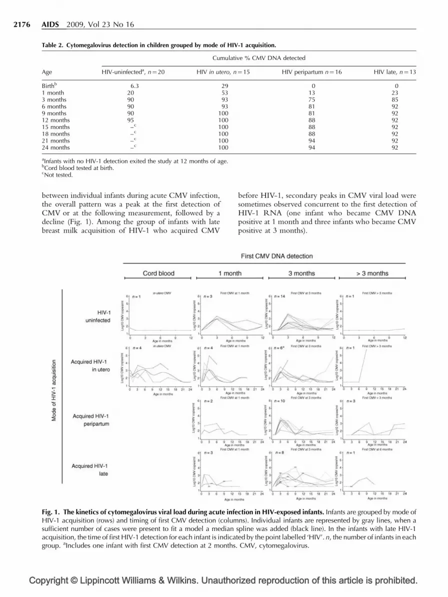
between individual infants during acute CMV infection,the overall pattern was a peak at the first detection ofCMV or at the following measurement, followed by adecline (Fig. 1). Among the group of infants with latebreast milk acquisition of HIV-1 who acquired CMV
pyright © Lippincott Williams & Wilkins. Unauthor
Fig. 1. The kinetics of cytomegalovirus viral load during acute infeHIV-1 acquisition (rows) and timing of first CMV detection (columsufficient number of cases were present to fit a model a medianacquisition, the time of first HIV-1 detection for each infant is indicatgroup. aIncludes one infant with first CMV detection at 2 months.
before HIV-1, secondary peaks in CMV viral load weresometimes observed concurrent to the first detection ofHIV-1 RNA (one infant who became CMV DNApositive at 1 month and three infants who became CMVpositive at 3 months).
ized reproduction of this article is prohibited.
ction in HIV-exposed infants. Infants are grouped by mode ofns). Individual infants are represented by gray lines, when a
spline was added (black line). In the infants with late HIV-1ed by the point labelled ‘HIV’. n, the number of infants in eachCMV, cytomegalovirus.
C
Acute CMV in Kenyan HIV-infected infants Slyker et al. 2177
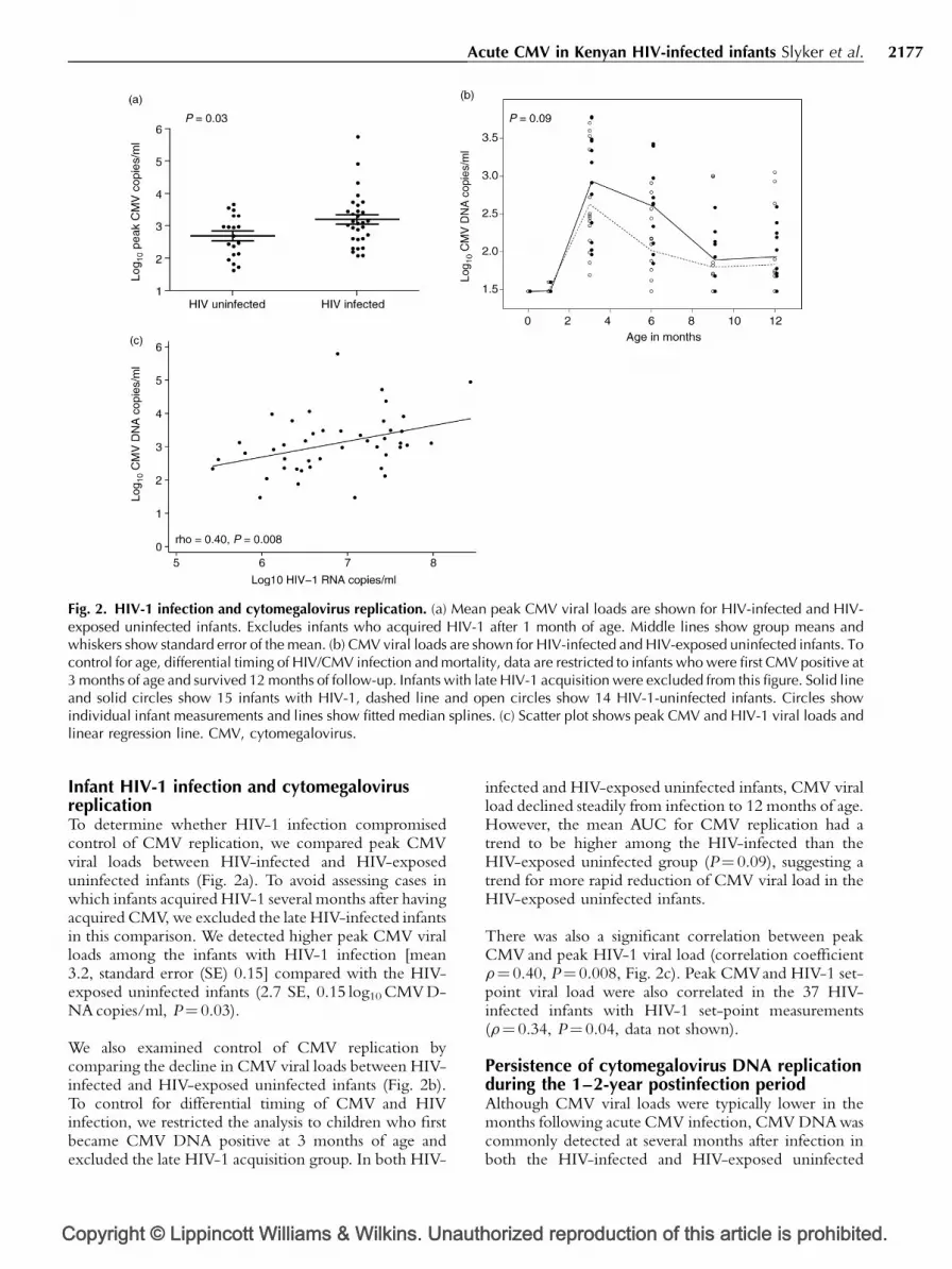
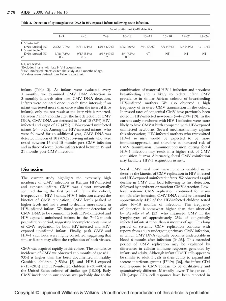
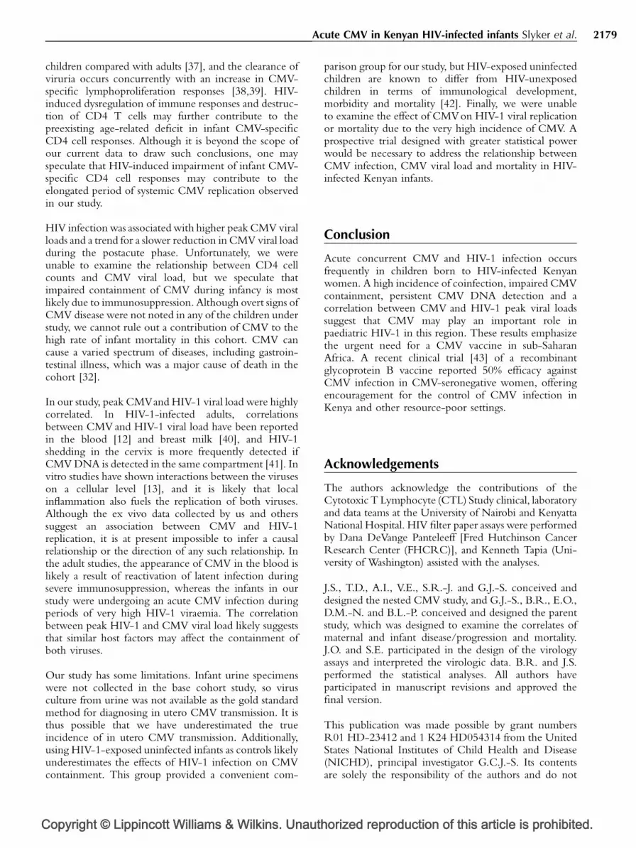
Fig. 2. HIV-1 infection and cytomegalovirus replication. (a) Mean peak CMV viral loads are shown for HIV-infected and HIV-exposed uninfected infants. Excludes infants who acquired HIV-1 after 1 month of age. Middle lines show group means andwhiskers show standard error of the mean. (b) CMV viral loads are shown for HIV-infected and HIV-exposed uninfected infants. Tocontrol for age, differential timing of HIV/CMV infection and mortality, data are restricted to infants who were first CMV positive at3 months of age and survived 12 months of follow-up. Infants with late HIV-1 acquisition were excluded from this figure. Solid lineand solid circles show 15 infants with HIV-1, dashed line and open circles show 14 HIV-1-uninfected infants. Circles showindividual infant measurements and lines show fitted median splines. (c) Scatter plot shows peak CMV and HIV-1 viral loads andlinear regression line. CMV, cytomegalovirus.
Infant HIV-1 infection and cytomegalovirusreplicationTo determine whether HIV-1 infection compromisedcontrol of CMV replication, we compared peak CMVviral loads between HIV-infected and HIV-exposeduninfected infants (Fig. 2a). To avoid assessing cases inwhich infants acquired HIV-1 several months after havingacquired CMV, we excluded the late HIV-infected infantsin this comparison. We detected higher peak CMV viralloads among the infants with HIV-1 infection [mean3.2, standard error (SE) 0.15] compared with the HIV-exposed uninfected infants (2.7 SE, 0.15 log10 CMV D-NA copies/ml, P¼ 0.03).
We also examined control of CMV replication bycomparing the decline in CMV viral loads between HIV-infected and HIV-exposed uninfected infants (Fig. 2b).To control for differential timing of CMV and HIVinfection, we restricted the analysis to children who firstbecame CMV DNA positive at 3 months of age andexcluded the late HIV-1 acquisition group. In both HIV-
opyright © Lippincott Williams & Wilkins. Unauth
infected and HIV-exposed uninfected infants, CMV viralload declined steadily from infection to 12 months of age.However, the mean AUC for CMV replication had atrend to be higher among the HIV-infected than theHIV-exposed uninfected group (P¼ 0.09), suggesting atrend for more rapid reduction of CMV viral load in theHIV-exposed uninfected infants.
There was also a significant correlation between peakCMV and peak HIV-1 viral load (correlation coefficientr¼ 0.40, P¼ 0.008, Fig. 2c). Peak CMVand HIV-1 set-point viral load were also correlated in the 37 HIV-infected infants with HIV-1 set-point measurements(r¼ 0.34, P¼ 0.04, data not shown).
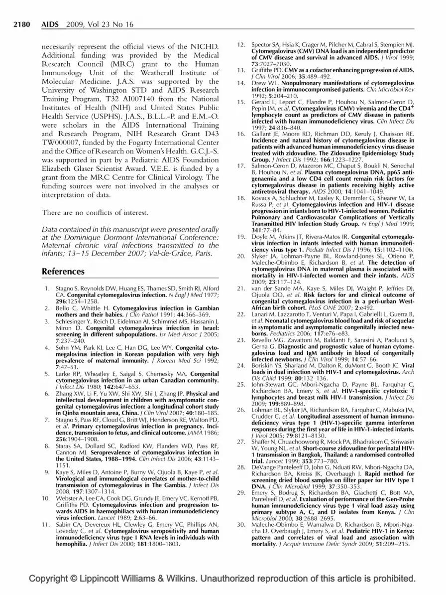
Persistence of cytomegalovirus DNA replicationduring the 1–2-year postinfection periodAlthough CMV viral loads were typically lower in themonths following acute CMV infection, CMV DNA wascommonly detected at several months after infection inboth the HIV-infected and HIV-exposed uninfected
orized reproduction of this article is prohibited.
Co
2178 AIDS 2009, Vol 23 No 16
Table 3. Detection of cytomegalovirus DNA in HIV-exposed infants following acute infection.
Months after first CMV detection
1–3 4–6 7–9 10–12 13–15 16–18 19–21 22–24
HIV infecteda
DNAþ/tested (%) 20/22 (91%) 15/21 (71%) 13/18 (72%) 6/12 (50%) 7/10 (70%) 4/9 (44%) 3/7 (43%) 0/1 (0%)HIV uninfectedb
DNAþ/tested (%) 13/18 (72%) 9/17 (53%) 8/17 (47%) 3/4 (75%) NT NT NT NTPc 0.2 0.3 0.2 0.6
NT, not tested.aExcludes infants with late HIV-1 acquisition.bHIV-uninfected infants exited the study at 12 months of age.cP values were derived from Fisher’s exact test.
infants (Table 3). As infants were evaluated every3 months, we examined CMV DNA detection in3-monthly intervals after first CMV DNA detection.Infants were counted once in each time interval; if aninfant was tested more than once within the interval (fiveinfants), only the test result at the later visit is reported.Between 7 and 9 months after the first detection of CMVDNA, CMV DNA was detected in 13 of 18 (72%) HIV-infected and eight of 17 (47%) HIV-exposed uninfectedinfants (P¼ 0.2). Among the HIV-infected infants, whowere followed for an additional year, CMV DNA wasdetected in seven of 10 (70%) surviving infants who weretested between 13 and 15 months post-CMV infectionand in three of seven (43%) infants tested between 19 and21 months post-CMV infection.
Discussion
The current study highlights the extremely highincidence of CMV infection in Kenyan HIV-infectedand exposed infants. CMV was almost universallyacquired during the first year of life in the cohort,irrespective of HIV-1 status. HIV-1 infection altered thekinetics of CMV replication; CMV levels peaked athigher levels and had a trend to decline more slowly inHIV-infected infants. We found persistent detection ofCMV DNA to be common in both HIV-1-infected andHIV-exposed uninfected infants in the 7–12-monthpostinfection period, suggesting incomplete containmentof CMV replication by both HIV-infected and HIV-exposed uninfected infants. Finally, peak CMV andHIV-1 viral loads were highly correlated, suggesting thatsimilar factors may affect the replication of both viruses.
CMV was acquired rapidly in this cohort. The cumulativeincidence of CMV we observed at 6 months of age (81–93%) is higher than has been documented in healthyGambian children (�53%) [2] and HIV-1-exposed(�15–20%) and HIV-infected children (�30–40%) inthe United States cohorts of similar age [18,33]. EarlyCMV incidence in our cohort was probably due to the
pyright © Lippincott Williams & Wilkins. Unauthor
combination of maternal HIV-1 infection and prevalentbreastfeeding and is likely to reflect infant CMVprevalence in similar African cohorts of breastfeedingHIV-infected mothers. We also observed a highfrequency of in utero CMV transmission in the cohort.Increased rates of congenital CMV have previously beennoted in HIV-infected newborns (�4–20%) [19]. In thecurrent study, newborns with HIV-1 infection were morelikely to have CMVat birth compared with HIV-exposeduninfected newborns. Several mechanisms may explainthis observation; HIV-infected mothers who transmittedHIV-1 in utero would be expected to be moreimmunosuppressed, and therefore at increased risk ofCMV transmission. Immunosuppression during foetalHIV-1 infection may result in a higher risk of CMVacquisition in utero. Alternately, foetal CMV coinfectionmay facilitate HIV-1 acquisition in utero.
Serial CMV viral load measurements enabled us todescribe the kinetics of CMV replication in HIV-infectedand HIV-exposed uninfected infants. We observed a rapiddecline in CMV viral load following its first detection,followed by persistent or transient CMV detection. Low-level systemic CMV replication continued for manymonths after infection; CMV DNA could be detected inapproximately 44% of the HIV-infected children testedafter 16–18 months of infection. This frequencyof detection is somewhat higher than that observedby Revello et al. [23] who measured CMV in thelymphocytes of approximately 25% of congenitallyinfected infants at more than 4 months of age. This longperiod of systemic CMV replication contrasts withreports from adults undergoing primary CMV infection,in which CMV DNA typically becomes undetectable inblood 6 months after infection [34,35]. This extendedperiod of CMV replication may be explained bydifferences in cellular immune responses generated byinfants and adults. Although infant CD8 T cells appear tobe similar to adult T cells in their ability to expand andsecrete interferon-gamma (IFNg) [36], the infant CD4cell response to CMV appears to be qualitatively andquantitatively different. Markedly lower T-helper cell 1(Th1)-type CD4 cell responses have been reported in
ized reproduction of this article is prohibited.
C
Acute CMV in Kenyan HIV-infected infants Slyker et al. 2179
children compared with adults [37], and the clearance ofviruria occurs concurrently with an increase in CMV-specific lymphoproliferation responses [38,39]. HIV-induced dysregulation of immune responses and destruc-tion of CD4 T cells may further contribute to thepreexisting age-related deficit in infant CMV-specificCD4 cell responses. Although it is beyond the scope ofour current data to draw such conclusions, one mayspeculate that HIV-induced impairment of infant CMV-specific CD4 cell responses may contribute to theelongated period of systemic CMV replication observedin our study.
HIV infection was associated with higher peak CMV viralloads and a trend for a slower reduction in CMV viral loadduring the postacute phase. Unfortunately, we wereunable to examine the relationship between CD4 cellcounts and CMV viral load, but we speculate thatimpaired containment of CMV during infancy is mostlikely due to immunosuppression. Although overt signs ofCMV disease were not noted in any of the children understudy, we cannot rule out a contribution of CMV to thehigh rate of infant mortality in this cohort. CMV cancause a varied spectrum of diseases, including gastroin-testinal illness, which was a major cause of death in thecohort [32].
In our study, peak CMVand HIV-1 viral load were highlycorrelated. In HIV-1-infected adults, correlationsbetween CMV and HIV-1 viral load have been reportedin the blood [12] and breast milk [40], and HIV-1shedding in the cervix is more frequently detected ifCMV DNA is detected in the same compartment [41]. Invitro studies have shown interactions between the viruseson a cellular level [13], and it is likely that localinflammation also fuels the replication of both viruses.Although the ex vivo data collected by us and otherssuggest an association between CMV and HIV-1replication, it is at present impossible to infer a causalrelationship or the direction of any such relationship. Inthe adult studies, the appearance of CMV in the blood islikely a result of reactivation of latent infection duringsevere immunosuppression, whereas the infants in ourstudy were undergoing an acute CMV infection duringperiods of very high HIV-1 viraemia. The correlationbetween peak HIV-1 and CMV viral load likely suggeststhat similar host factors may affect the containment ofboth viruses.
Our study has some limitations. Infant urine specimenswere not collected in the base cohort study, so virusculture from urine was not available as the gold standardmethod for diagnosing in utero CMV transmission. It isthus possible that we have underestimated the trueincidence of in utero CMV transmission. Additionally,using HIV-1-exposed uninfected infants as controls likelyunderestimates the effects of HIV-1 infection on CMVcontainment. This group provided a convenient com-
opyright © Lippincott Williams & Wilkins. Unauth
parison group for our study, but HIV-exposed uninfectedchildren are known to differ from HIV-unexposedchildren in terms of immunological development,morbidity and mortality [42]. Finally, we were unableto examine the effect of CMVon HIV-1 viral replicationor mortality due to the very high incidence of CMV. Aprospective trial designed with greater statistical powerwould be necessary to address the relationship betweenCMV infection, CMV viral load and mortality in HIV-infected Kenyan infants.
Conclusion
Acute concurrent CMV and HIV-1 infection occursfrequently in children born to HIV-infected Kenyanwomen. A high incidence of coinfection, impaired CMVcontainment, persistent CMV DNA detection and acorrelation between CMV and HIV-1 peak viral loadssuggest that CMV may play an important role inpaediatric HIV-1 in this region. These results emphasizethe urgent need for a CMV vaccine in sub-SaharanAfrica. A recent clinical trial [43] of a recombinantglycoprotein B vaccine reported 50% efficacy againstCMV infection in CMV-seronegative women, offeringencouragement for the control of CMV infection inKenya and other resource-poor settings.
Acknowledgements
The authors acknowledge the contributions of theCytotoxic T Lymphocyte (CTL) Study clinical, laboratoryand data teams at the University of Nairobi and KenyattaNational Hospital. HIV filter paper assays were performedby Dana DeVange Panteleeff [Fred Hutchinson CancerResearch Center (FHCRC)], and Kenneth Tapia (Uni-versity of Washington) assisted with the analyses.
J.S., T.D., A.I., V.E., S.R.-J. and G.J.-S. conceived anddesigned the nested CMV study, and G.J.-S., B.R., E.O.,D.M.-N. and B.L.-P. conceived and designed the parentstudy, which was designed to examine the correlates ofmaternal and infant disease/progression and mortality.J.O. and S.E. participated in the design of the virologyassays and interpreted the virologic data. B.R. and J.S.performed the statistical analyses. All authors haveparticipated in manuscript revisions and approved thefinal version.
This publication was made possible by grant numbersR01 HD-23412 and 1 K24 HD054314 from the UnitedStates National Institutes of Child Health and Disease(NICHD), principal investigator G.C.J.-S. Its contentsare solely the responsibility of the authors and do not
orized reproduction of this article is prohibited.
Co
2180 AIDS 2009, Vol 23 No 16
necessarily represent the official views of the NICHD.Additional funding was provided by the MedicalResearch Council (MRC) grant to the HumanImmunology Unit of the Weatherall Institute ofMolecular Medicine. J.A.S. was supported by theUniversity of Washington STD and AIDS ResearchTraining Program, T32 AI007140 from the NationalInstitutes of Health (NIH) and United States PublicHealth Service (USPHS). J.A.S., B.L.L.-P. and E.M.-O.were scholars in the AIDS International Trainingand Research Program, NIH Research Grant D43TW000007, funded by the Fogarty International Centerand the Office of Research on Women’s Health. G.C.J.-S.was supported in part by a Pediatric AIDS FoundationElizabeth Glaser Scientist Award. V.E.E. is funded by agrant from the MRC Centre for Clinical Virology. Thefunding sources were not involved in the analyses orinterpretation of data.
There are no conflicts of interest.
Data contained in this manuscript were presented orallyat the Dominique Dormont International Conference:Maternal chronic viral infections transmitted to theinfants; 13–15 December 2007; Val-de-Grace, Paris.
References
1. Stagno S, Reynolds DW, Huang ES, Thames SD, Smith RJ, AlfordCA. Congenital cytomegalovirus infection. N Engl J Med 1977;296:1254–1258.
2. Bello C, Whittle H. Cytomegalovirus infection in Gambianmothers and their babies. J Clin Pathol 1991; 44:366–369.
3. Schlesinger Y, Reich D, Eidelman AI, Schimmel MS, Hassanin J,Miron D. Congenital cytomegalovirus infection in Israel:screening in different subpopulations. Isr Med Assoc J 2005;7:237–240.
4. Sohn YM, Park KI, Lee C, Han DG, Lee WY. Congenital cyto-megalovirus infection in Korean population with very highprevalence of maternal immunity. J Korean Med Sci 1992;7:47–51.
5. Larke RP, Wheatley E, Saigal S, Chernesky MA. Congenitalcytomegalovirus infection in an urban Canadian community.J Infect Dis 1980; 142:647–653.
6. Zhang XW, Li F, Yu XW, Shi XW, Shi J, Zhang JP. Physical andintellectual development in children with asymptomatic con-genital cytomegalovirus infection: a longitudinal cohort studyin Qinba mountain area, China. J Clin Virol 2007; 40:180–185.
7. Stagno S, Pass RF, Cloud G, Britt WJ, Henderson RE, Walton PD,et al. Primary cytomegalovirus infection in pregnancy. Inci-dence, transmission to fetus, and clinical outcome. JAMA 1986;256:1904–1908.
8. Staras SA, Dollard SC, Radford KW, Flanders WD, Pass RF,Cannon MJ. Seroprevalence of cytomegalovirus infection inthe United States, 1988–1994. Clin Infect Dis 2006; 43:1143–1151.
9. Kaye S, Miles D, Antoine P, Burny W, Ojuola B, Kaye P, et al.Virological and immunological correlates of mother-to-childtransmission of cytomegalovirus in The Gambia. J Infect Dis2008; 197:1307–1314.
10. Webster A, Lee CA, Cook DG, Grundy JE, Emery VC, Kernoff PB,Griffiths PD. Cytomegalovirus infection and progression to-wards AIDS in haemophiliacs with human immunodeficiencyvirus infection. Lancet 1989; 2:63–66.
11. Sabin CA, Devereux HL, Clewley G, Emery VC, Phillips AN,Loveday C, et al. Cytomegalovirus seropositivity and humanimmunodeficiency virus type 1 RNA levels in individuals withhemophilia. J Infect Dis 2000; 181:1800–1803.
pyright © Lippincott Williams & Wilkins. Unauthor
12. Spector SA, Hsia K, Crager M, Pilcher M, Cabral S, Stempien MJ.Cytomegalovirus (CMV) DNA load is an independent predictorof CMV disease and survival in advanced AIDS. J Virol 1999;73:7027–7030.
13. Griffiths PD. CMV as a cofactor enhancing progression of AIDS.J Clin Virol 2006; 35:489–492.
14. Drew WL. Nonpulmonary manifestations of cytomegalovirusinfection in immunocompromised patients. Clin Microbiol Rev1992; 5:204–210.
15. Gerard L, Leport C, Flandre P, Houhou N, Salmon-Ceron D,Pepin JM, et al. Cytomegalovirus (CMV) viremia and the CD4R
lymphocyte count as predictors of CMV disease in patientsinfected with human immunodeficiency virus. Clin Infect Dis1997; 24:836–840.
16. Gallant JE, Moore RD, Richman DD, Keruly J, Chaisson RE.Incidence and natural history of cytomegalovirus disease inpatients with advanced human immunodeficiency virus diseasetreated with zidovudine. The Zidovudine Epidemiology StudyGroup. J Infect Dis 1992; 166:1223–1227.
17. Salmon-Ceron D, Mazeron MC, Chaput S, Boukli N, SenechalB, Houhou N, et al. Plasma cytomegalovirus DNA, pp65 anti-genaemia and a low CD4 cell count remain risk factors forcytomegalovirus disease in patients receiving highly activeantiretroviral therapy. AIDS 2000; 14:1041–1049.
18. Kovacs A, Schluchter M, Easley K, Demmler G, Shearer W, LaRussa P, et al. Cytomegalovirus infection and HIV-1 diseaseprogression in infants born to HIV-1-infected women. PediatricPulmonary and Cardiovascular Complications of VerticallyTransmitted HIV Infection Study Group. N Engl J Med 1999;341:77–84.
19. Doyle M, Atkins JT, Rivera-Matos IR. Congenital cytomegalo-virus infection in infants infected with human immunodefi-ciency virus type 1. Pediatr Infect Dis J 1996; 15:1102–1106.
20. Slyker JA, Lohman-Payne BL, Rowland-Jones SL, Otieno P,Maleche-Obimbo E, Richardson B, et al. The detection ofcytomegalovirus DNA in maternal plasma is associated withmortality in HIV-1-infected women and their infants. AIDS2009; 23:117–124.
21. van der Sande MA, Kaye S, Miles DJ, Waight P, Jeffries DJ,Ojuola OO, et al. Risk factors for and clinical outcome ofcongenital cytomegalovirus infection in a peri-urban West-African birth cohort. PLoS ONE 2007; 2:e492.
22. Lanari M, Lazzarotto T, Venturi V, Papa I, Gabrielli L, Guerra B,et al. Neonatal cytomegalovirus blood load and risk of sequelaein symptomatic and asymptomatic congenitally infected new-borns. Pediatrics 2006; 117:e76–e83.
23. Revello MG, Zavattoni M, Baldanti F, Sarasini A, Paolucci S,Gerna G. Diagnostic and prognostic value of human cytome-galovirus load and IgM antibody in blood of congenitallyinfected newborns. J Clin Virol 1999; 14:57–66.
24. Boriskin YS, Sharland M, Dalton R, duMont G, Booth JC. Viralloads in dual infection with HIV-1 and cytomegalovirus. ArchDis Child 1999; 80:132–136.
25. John-Stewart GC, Mbori-Ngacha D, Payne BL, Farquhar C,Richardson BA, Emery S, et al. HIV-1-specific cytotoxic Tlymphocytes and breast milk HIV-1 transmission. J Infect Dis2009; 199:889–898.
26. Lohman BL, Slyker JA, Richardson BA, Farquhar C, Mabuka JM,Crudder C, et al. Longitudinal assessment of human immuno-deficiency virus type 1 (HIV-1)-specific gamma interferonresponses during the first year of life in HIV-1-infected infants.J Virol 2005; 79:8121–8130.
27. Shaffer N, Chuachoowong R, Mock PA, Bhadrakom C, SiriwasinW, Young NL, et al. Short-course zidovudine for perinatal HIV-1 transmission in Bangkok, Thailand: a randomised controlledtrial. Lancet 1999; 353:773–780.
28. DeVange Panteleeff D, John G, Nduati RW, Mbori-Ngacha DA,Richardson BA, Kreiss JK, Overbaugh J. Rapid method forscreening dried blood samples on filter paper for HIV type 1DNA. J Clin Microbiol 1999; 37:350–353.
29. Emery S, Bodrug S, Richardson BA, Giachetti C, Bott MA,Panteleeff D, et al. Evaluation of performance of the Gen-Probehuman immunodeficiency virus type 1 viral load assay usingprimary subtype A, C, and D isolates from Kenya. J ClinMicrobiol 2000; 38:2688–2695.
30. Maleche-Obimbo E, Wamalwa D, Richardson B, Mbori-Nga-cha D, Overbaugh J, Emery S, et al. Pediatric HIV-1 in Kenya:pattern and correlates of viral load and association withmortality. J Acquir Immune Defic Syndr 2009; 51:209–215.
ized reproduction of this article is prohibited.
C
Acute CMV in Kenyan HIV-infected infants Slyker et al. 2181
31. Mattes FM, Hainsworth EG, Hassan-Walker AF, Burroughs AK,Sweny P, Griffiths PD, Emery VC. Kinetics of cytomegalovirusload decrease in solid-organ transplant recipients after pre-emptive therapy with valganciclovir. J Infect Dis 2005; 191:89–92.
32. Obimbo EM, Mbori-Ngacha DA, Ochieng JO, Richardson BA,Otieno PA, Bosire R, et al. Predictors of early mortality in acohort of human immunodeficiency virus type 1-infectedAfrican children. Pediatr Infect Dis J 2004; 23:536–543.
33. Chandwani S, Kaul A, Bebenroth D, Kim M, John DD, Fidelia A,et al. Cytomegalovirus infection in human immunodeficiencyvirus type 1-infected children. Pediatr Infect Dis J 1996;15:310–314.
34. Zanghellini F, Boppana SB, Emery VC, Griffiths PD, Pass RF.Asymptomatic primary cytomegalovirus infection: virologicand immunologic features. J Infect Dis 1999; 180:702–707.
35. Revello MG, Zavattoni M, Sarasini A, Percivalle E, Simoncini L,Gerna G. Human cytomegalovirus in blood of immunocompe-tent persons during primary infection: prognostic implicationsfor pregnancy. J Infect Dis 1998; 177:1170–1175.
36. Marchant A, Appay V, Van Der Sande M, Dulphy N, Liesnard C,Kidd M, et al. Mature CD8(R) T lymphocyte response to viralinfection during fetal life. J Clin Invest 2003; 111:1747–1755.
opyright © Lippincott Williams & Wilkins. Unauth
37. Tu W, Chen S, Sharp M, Dekker C, Manganello AM, TongsonEC, et al. Persistent and selective deficiency of CD4R T cellimmunity to cytomegalovirus in immunocompetent youngchildren. J Immunol 2004; 172:3260–3267.
38. Reynolds DW, Dean PH, Pass RF, Alford CA. Specific cell-mediated immunity in children with congenital and neonatalcytomegalovirus infection and their mothers. J Infect Dis 1979;140:493–499.
39. Pass RF, Stagno S, Britt WJ, Alford CA. Specific cell-mediatedimmunity and the natural history of congenital infection withcytomegalovirus. J Infect Dis 1983; 148:953–961.
40. Gantt S, Carlsson J, Shetty AK, Seidel KD, Qin X, Mutsvangwa J,et al. Cytomegalovirus and Epstein–Barr virus in breast milk areassociated with HIV-1 shedding but not with mastitis. AIDS2008; 22:1453–1460.
41. Lurain NS, Robert ES, Xu J, Camarca M, Landay A, Kovacs AA,Reichelderfer PS. HIV type 1 and cytomegalovirus coinfectionin the female genital tract. J Infect Dis 2004; 190:619–623.
42. Filteau S. The HIV-exposed, uninfected African child. Trop MedInt Health 2009; 14:276–287.
43. Pass RF, Zhang C, Evans A, Simpson T, Andrews W, Huang ML,et al. Vaccine prevention of maternal cytomegalovirus infection.N Engl J Med 2009; 360:1191–1199.
orized reproduction of this article is prohibited.