acetate on glomerular filtration rate and sodium and water
TRANSCRIPT
THE ACTION OF CORTISONE AND DESOXYCORTICOSTERONEACETATE ON GLOMERULAR FILTRATION RATE AND
SODIUM AND WATER EXCHANGE IN THEADRENALECTOMIZED DOG
By OLIVER GARROD,' S. A. DAVIES, AND GEORGE CAHILL, JL
(From the College of Physicians and Surgeons, Columbia University, New York, N. Y.)
(Submitted for publication December 1, 1953; accepted February 2, 1955)
Adrenal failure causes a complex disturbanceof renal function and of sodium and water ex-change. In the dog, as in man, the ability to con-serve sodium is impaired (1, 2), there is a fall inglomerular filtration rate and renal blood flow(3-6), and diuresis from water ingestion is muchdelayed (7, 8).There have been many reports of the effects of
corticosteroids on discrete renal functions suchas filtration rate or excretion of water and electro-lytes in adrenal insufficiency. There have, how-ever, been few attempts to correlate the changesin these renal functions, one with another and withthe level of bodily hydration. Thus, from existingdata, the effects of changes in filtration rate onother renal functions, or of changes in bodily hy-dration on filtration rate, cannot be clearly definedin relation to the action of corticosteroids.The steroids most examined for their renal ef-
fects have been desoxycorticosterone acetate(DCA) and, to a much lesser extent, cortisone.Though DCA appears to act mainly on electrolyteexcretion, yet, when given in larger amounts thanare needed for optimal sodium balance, it maythen correct the low filtration rate of adrenalec-tomized dogs (4, 8, 9). It is not clear, however,whether this last effect is mainly or wholly de-pendent on excessive sodium retention, for nocomparable studies appear to have been made atdeliberately optimal levels of sodium balance(10, 11).
Cortisone seems to be more active than DCAin restoring filtration rate (12-14) and waterdiuresis (15, 16), and, by mechanisms which arenot yet understood, it can either increase or di-minish the sodium output (14, 15, 17-19). There
1During tenure of a Rockefeller Travelling Fellow-ship in Medicine, 1951-1952. Present address: Instituteof Clinical Research, The Middlesex Hospital, London,W. 1.
is little information about its effects in the adrenal-ectomized dog (14, 20).The investigations reported in this paper are
an attempt to define more precisely the renal ac-tions of cortisone and DCA, with special refer-ence to their effects on filtration rate and on so-dium and water exchange.
EXPERIMENTAL PLAN AND METHODS
Renal function was studied in two trained female mon-grel dogs, before and after bilateral adrenalectomy, withthe following objects: (a) to measure the effects ofadrenalectomy on GFR and renal excretion of water andelectrolytes, under controlled conditions of bodily hy-dration, but during escape from direct corticosteroideffects; (b) to compare the acute effects of cortisone andDCA on these renal functions, after adrenalectomy; and(c) to correlate the changes in these different renalfunctions with one another and with the state of bodilyhydration.
Maintenance of the dogs
The dogs weighed 162 and 20 Kgm. They lived underoptimal conditions on a constant diet to which 2 gm. ofsodium chloride was added.3 They were fed between1 and 3 P.M. and had free access to water. After a two-stage adrenalectomy, a state of optimal bodily hydrationwas maintained by daily intramuscular injection, at thetime of each meal, of 0.6 to 0.75 mg. of DCA in sesameoil. In dog A changes in bodily hydration were in-duced by raising or lowering the DCA dosage (range,0.5 to 1.5 mg. a day). Therapy was controlled by dailyweighing and frequent measurements of fasting plasmaspecific gravity, electrolyte concentrations, blood urea,and hematocrit. Plasma volumes were measured by theT-1824 dye method (21), using a Beckmann spectro-photometer.The diurnal patterns of water, creatinine, and electro-
lyte excretion were determined, in relation to meals andDCA injections.
2 Dog A was not fully grown at the beginning of thesestudies (initial weight, 15.5 Kgm.; final weight, 18 Kgm.).
8 The total daily intake was approximately 90 mEq.sodium and 62 mEq. potassium.
761
OLIVER GARROD, S. A. DAVIES, AND GEORGE CAHILL, JR.
Measurements of renal functionUsual procedure. After free access to water overnight,
15-minute urine collections were started between 8 and9 A.M. Half an hour later, a priming infusion of cre-atinine and PAH4 was slowly injected, followed by asustaining infusion4 which was continued throughout theexperiment. A 45-minute period was allowed for equili-bration. After three or more clearance periods, 40 ml. ofwarm water per Kgm. were given by stomach tube.Clearances were then measured for a further three tosix hours.The following renal functions were determined: GFR,
ERPF, urinary pH, and urinary outputs of sodium, po-tassium, chloride, and phosphate.Blood was taken initially and at the midpoint of every
third or fourth clearance period, and the red cells rein-fused at the end of each experiment. Plasma creatinine,PAH, specific gravity, and electrolyte concentrationswere measured in each sample.The above-mentioned procedure was carried out under
the following conditions: (i) before adrenalectomy, (ii)after adrenalectomy, while on optimal DCA therapy,and, in dog A, also when on inadequate and excessiveDCA therapy and after intravenous saline infusion, andin both dogs during acute administration of cortisoneand DCA.To observe its effects, both with and without a water
load, cortisone was given, in separate experiments, threehours before the water dose, or at the time of the waterdose, or three hours after it. The cortisone was givenin one of three different ways: (i) intravenously, as amicrocrystalline suspension of 50 mg. cortisone acetate in1 ml. distilled water, dose 10 to 50 mg.5; (ii) intra-venously, as cortisone tricarballylate,6 dose 30 to 90 mg.freshly dissolved in a few ml. of isotonic saline, or (iii)orally, as cortisone acetate (Cortone@, Merck), dose 50mg.DCA was injected intravenously as a microcrystalline
suspension in distilled water, made to the same specifica-tions as the cortisone acetate,5 dose 2.5 to 5.0 mg. It wasgiven three hours after the water dose.
Other procedures. On 47 occasions the diuretic re-sponse to the standard water dose (supra) and the ac-companying outputs of sodium and potassium were meas-ured for three hours without determining GFR andERPF.
4 The priming infusion consisted of hypertonic creati-nine and PAH in about 60 ml. of distilled water. Thesustaining infusion consisted of approximately isotoniccreatinine and PAH, and was given at a steady rate of 24to 30 ml. per hour. The effects of these infusions onwater and electrolyte excretion were measured duringequilibration, and on GFR and ERPF during succeedingperiods totalling 45 to 60 minutes, before the water dosewas given.
6 Specially prepared by Chemical Specialities, Inc.,N. Y.
8 A water-soluble but unstable ester, specially pre-pared by Merck, Ltd., N. J.
In another group of experiments the effects of intra-venous cortisone acetate, and of DCA, on excretion of wa-ter, electrolytes, and endogenous creatinine were meas-ured but, without the complicating effects of an infusionor water dose.
Chemical methodsThe methods used for the following chemical estima-
tions are indicated by the references: sodium and potas-sium, by an internal lithium standard flame photometer(22) having an error of less than one per cent; plasmaand urinary chloride (23); plasma and urinary creatinine(24); PAH (25); urinary phosphate (26); blood ureanitrogen (27); plasma specific gravity (28, 29). Theurinary pH was measured by indicators within one-halfminute of urine collection.
RESULTS
I. Water and Electrolyte Balance in Relation toChronic DCA Therapy After Adrenalectomy
Assessment of bodily hydrationAdequacy of the DCA therapy was assessed
by observing the weight, appetite, and activity
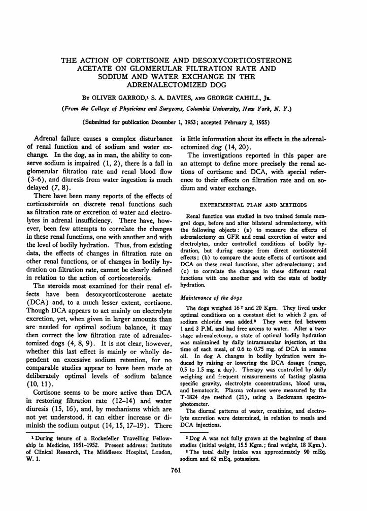
FIG. 1. THE EFmcETs OF DCA THERAPY ON FASTINGPLASMA CONCENTRATIONS IN THE Two ADRENALECTO-MIZED DOGS
In dog A, optimal therapy was achieved on 0.6 to0.75 mg. DCA a day, in dog B on 0.75 mg. a day. Theweight balances in dog A are the means of daily devia-tions from the growth-weight curve during the total pe-riod of the studies. The slight rise in weight during in-adequate DCA therapy was due to water retention, andthe rise during excessive DCA therapy to salt and waterretention.
762
ACUTE EFFECTS OF CORTISONE AND DCA ON RENAL FUNCTION
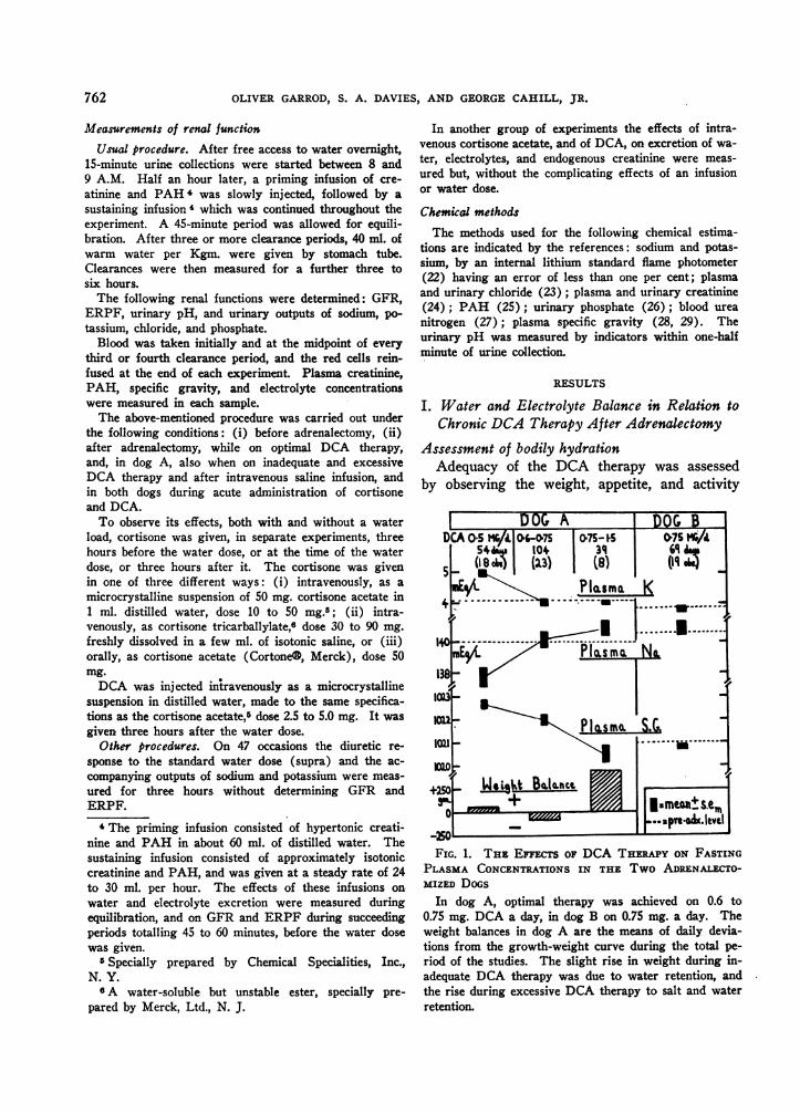
FIG. 2. WATER DIURESIS AND PLASMA CONCENTRA-TIONS RELATIVE TO DCA THERAPY IN ADRENALECTO-MIZED DOG ATwo sample periods are shown. Diet and sodium in-
take were constant. The body weight and plasma con-centrations were obtained after 18 hours' fasting withfree access to water, before giving the standard waterdose of 40 ml. per Kgm. body weight. Dotted lines in-dicate the delay in diuresis before adrenalectomy. Thebody weights have not been corrected for growth.
of the dogs, and by comparing their fasting plasmaspecific gravities and electrolyte concentrationswith the levels obtained before adrenalectomy(Figure 1). In dog A, a state of optimal healthand bodily hydration was achieved on 0.6 to 0.75mg. DCA per day, in dog B on 0.75 mg. per day.At this dosage their plasma volumes varied be-tween 5 and 5.7 per cent of body weight, figurescomparable with the mean of 5.4 per cent re-ported by Courtice in 29 intact mongrel dogs(30). In dog A the dose of DCA was deliberatelyvaried so that the effects of changes in bodily hy-dration could be studied. Inadequate DCA ther-apy (e.g., 0.5 to 0.6 mg.) was followed by a risein hematocrit, plasma specific gravity and potas-sium and a fall in plasma sodium; the expectedloss of weight did not always occur (Figure 1)because of water retention, apparently intracel-lular, from the repeated water doses (Figure 2).
Excessive DCA therapy (e.g., 1 to 1.5 mg.)caused excessive gain in weight, a fall in plasmaspecific gravity, and a slight rise in plasma sodiumconcentration (Figures 1 and 2). The plasmaspecific gravity was the most sensitive index of hy-dration; the hematocrit and plasma sodium andpotassium were almost as sensitive to inadequate,but not to excessive, therapy; blood urea was anunreliable index.
In order that renal function could be assessedin relation to bodily hydration, the renal data weregrouped as follows: 1) optimal therapy, all pe-riods on 0.6 to 0.75 mg. DCA per day,7 2) in-adequate therapy all periods on less than 0.6 mg.per day, 3) excessive therapy, all periods on 1 to1.5 mg. per day, and in dog A, on 0.75 mg. formore than six weeks.7
DCA04MGC.M100 _F06 K output
llk
I
0TIME Hus 120
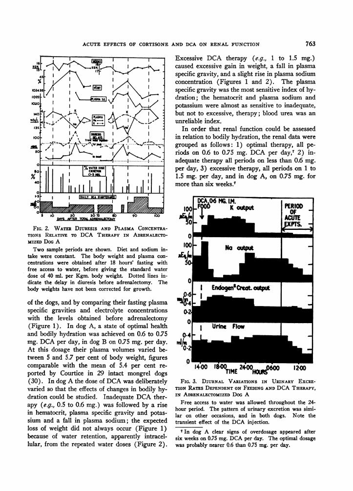
FIG. 3. DIURNAL VARIATIONS IN URINARY EXCRE-TION RATPs DEPENDENT ON FEEDING AND DCA THERAPY,IN ADRENALECTOMIZED DOG A
Free access to water was allowed throughout the 24-hour period. The pattern of urinary excretion was simi-lar on other occasions, and in both dogs. Note thetransient effect of the DCA injection.
7 In dog A clear signs of overdosage appeared aftersix weeks on 0.75 mg. DCA per day. The optimal dosagewas probably nearer 0.6 than 0.75 mg. per day.
763
I E-,A--- Creco.. -0-909W--0.
10-4
hj.0.
OLIVER GARROD, S. A. DAVIES, AND GEORGE CAHILL, JR.
I .rI I%?
C"o
°"NN__z :I oo041
_ __nlM'*
o* I I I 21
to I aSCK "
.44N%%"__N
en
-0 NO Rr4oeqw O4-4wq4%q"
r*
1` 0 I
U)0N
eq N e___-
%n 0 tOOV.00 n4 "_"
1111f
10 -0%eq0"
eq -0%e U004ON C
Ninc-ocoUcv0m N "
00
eq% 0-4 -0U)-0* C-eo eq U)0)-4
I I ",
1 I °Iz
vaftin'
'" I v U1% 4
$I 1 ! % -r
C4Go%Z
tNU)N'C m) N WQ4
I'C)O O eq e C
U)0
II oN m
.10
.4e+I++
I I .4 oUs *"R
I I "
¢<<
b
U) 0
.q . . >.*q. .-4>>> ->5 "
'0
"
a
30~AO
s
0
aqex41qV.m
m
+494)2
I OW)
f) eq e U)VU) U) U)t'.
1!0I
II
I v
N
.- .> .4
"O0_
U)
-_ liiiIIIIO-U
vM11G I
eqeq eq00Us)l
inv v ) W r. u>v.4
IUvA
:b90
0i04
I1 II
I I
I I I
11 I 1
U)+wio_eq m 0%.-4 .- .: .
45'ao
.9LIAII
1" ag
081 t8 1Noro.a_
_"4 N_
r- e4$Nvt> oN
I I
I I
-o
_0
I
o
_e0
4
oo
U)l'cNe nm ) eqU) 0.0%-->ee
U)U) N) U)U)0n W) %lo " %n
ew1 4 1%N0oWo ) 000
U).N4Ne Neqo0
g o
1+
I i .>I4IIII
>>>5>5
ON
-HIt
90
4
'0co
I II Iaq In'
*0It%
,V
:
W)
0U)
0o 0
08t
*t t
v
U..
0
E
4-
g
S '
.is
6.
0.!4I
01
Ao
a0
>6
.I"
764
II
II
II
II
I I
U)U)
U)
0
0II U.4)
I II I
I
1%,I
04
0
1
ao ~o W
*e
%4.t
n
41.
.r
Ila
bco0:1.
i4
4.
0
EV , 3.
60
co
co
0
la
I I
I I
I I
q-U)
.4
z
';4,
O4..
-
* :
W--
to_:
I..
ACUTE EFFECTS OF CORTISONE AND DCA ON RENAL FUNCTION
Duration of action of the therapeutic DCA in-jections
The sodium-retaining action of the DCA injec-tions ceased after about eight hours in dog A(Figure 3) and after 12 to 18 hours in dog B.There was then a delayed excretion of the dietarysodium. From 18 to 24 hours after the DCA,which is the period when renal function was stud-ied, the sodium excretion remained steady or fellfurther; the kidneys had then escaped from DCAeffect.
II. Renal Function
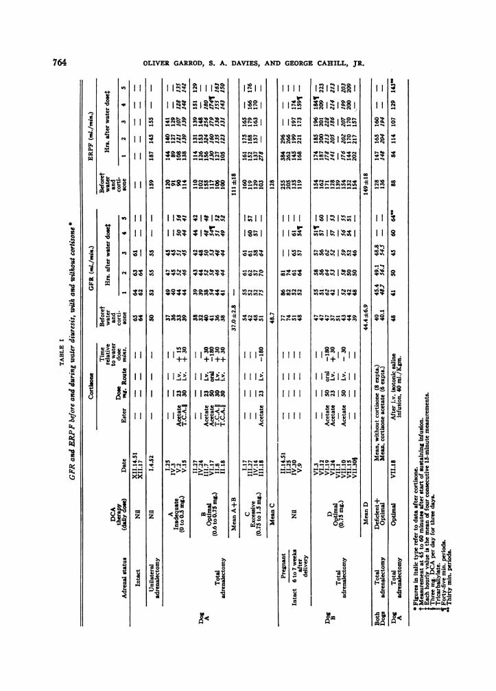
Glomerular filtration rate (GFR) and renal plasmaflow (ERPF)8
(a) Effects of adrenalectomy, relative to DCAtherapy (Table I). In dog A, after adrenalec-tomy, GFR was consistently subnormal when onoptimal DCA therapy, but rose after water in-gestion. On excessive DCA therapy GFR washigher and sometimes became normal after wateringestion. Changes in ERPF were essentiallyparallel.
In dog B, GFR fell to subnormal levels duringthe second month after adrenalectomy, butthroughout the post-operative period it rose lessafter water than before adrenalectomy. A shortperiod of excessive DCA therapy in this dog (3mg. a day for three days) did not raise the GFR(Table I), but there was no accompanying re-tention of sodium and water.
(b) Acute effects of DCA on GFR. Intrave-nous DCA (2.5 to 5 mg.), though it caused almostmaximal reabsorption of sodium by the tubules,did not increase GFR, ERPF, or endogenous
8 The effects on GFR and ERPF of the infusions usedin measuring them need to be considered. During nineexperiments in dog A, there was a consistent rise in GFRof 11 ± s.d. 3.7 ml. per minute, and in ERPF of 8 ± s.d.2 ml. per min. between 45 and 90 minutes after startingthe sustaining infusion. Though a similar rise did notoccur in dog B, a rise in this dog's sodium excretionwithin 30 mins. of starting the sustaining infusion sug-gested that GFR had already risen during the period ofequilibration when it could not be measured directly.Thus, in both dogs the true fasting GFR was probablyless than that of the first period of creatinine clearanceat the end of equilibration. To ensure standardization,the fasting GFR and ERPF have been taken from thefirst 15-minute clearances after equilibration, i.e., 45 to 60minutes after starting the sustaining infusion.
DCA OCA DCA26MM 0 CA CTCA CACOM EG.6M C MMmMG W
Pal ORAL
J;4 0 C 6m
120r SODIUM
a1iKEU:I1LiiHOURS AFTER WATER INGESTION
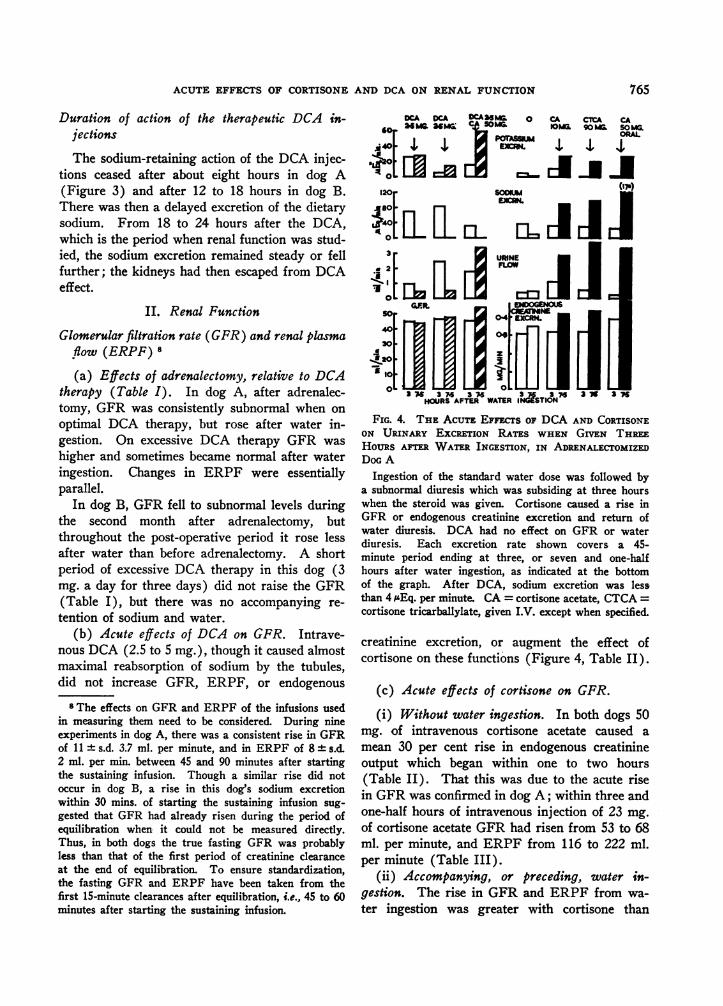
FIG. 4. THIE ACUTrE EFFECTS OF DCA AND CORTISONEON URINARY EXCsRETON RATES WHEN GIVEN TH:REEHOURS AIRE WATE:R INGESTION, IN ADRENALECTOMIZEDDOG A
Ingestion of the standard water dose was followed bya subnormal diuresis which was subsiding at three hourswhen the steroid was given. Cortisone caused a rise inGFR or endogenous creatinine excretion and return ofwater diuresis. DCA had no effect on GFR or waterdiuresis. Each excretion rate shown covers a 45-minute period ending at three, or seven and one-halfhours after water ingestion, as indicated at the bottomof the graph. After DCA, sodium excretion was lessthan 4 i'Eq. per minute. CA = cortisone acetate, CTCA =cortisone tricarbalylate, given I.V. except when specified.
creatinine excretion, or augment the effect ofcortisone on these functions (Figure 4, Table II).
(c) Acute effects of cortisone on GFR.(i) Without water ingestion. In both dogs 50
mg. of intravenous cortisone acetate caused amean 30 per cent rise in endogenous creatinineoutput which began within one to two hours(Table II). That this was due to the acute risein GFR was confirmed in dog A; within three andone-half hours of intravenous injection of 23 mg.Of cortisone acetate GFR had risen from 53 to 68ml. per minute, and ERPF from 116 to 222 ml.per minute (Table III).
(ii) Accompanying, or preceding, water in-gestion. The rise in GFR and ERPF from wa-ter ingestion was greater with cortisone than
765
766 OLIVER GARROD, S. A. DAVIES, AND GEORGE CAHILL, JR.
TABLE II
The acute effects of cortisone acdat and DCA on fasting urinary excretion rates in two adrenakectomized dogs(on steady optimal DCA therapy) *
Dog A: 5 to 6 months after total adrenalectomy Dog B: 8 to 11 weeks after total
Excretion rates Excretion rates
Intra- Endog- Endog-venous enous enoussteroid creati- creati(micro Tm Urine nine Na K Cl PO4t Urine nine Na K Cl P04crystaj) Time ./ %Of - Urine mI./ %oflaUSnat 0 mins. mix$. mix. weod &Eg./min. pH$ min. period Eq./mix. pH
0 0.41 100 94 16 77 27 7.5 0.25 100 37 21 27 13 7.560 0.39 98 89 13 78 22 7.5 0.29 97 55 14 30 12 7.8
0 120 0.32 93 62 7.6 59 12 7.45 0.27 100 49 10.6 31 9.5 7.8180 0.28 88 56 6.5 51 10 7.5 0.30 101 58 9.1 31 8.1 7.8240 0.27 90 42 S.9 39 7.5 7.55 0.26 103 46 8.2 32 6.1 7.8300 0.24 91 35 4.4 34 6.1 7.6 0.28 99 48 7.3 34 5.2 7.7
0 0.45 100 87 30 87 27 7.4 0.36 100 91 12 70 7.4 8.060 0.38 91 73 22 72 25 7.1 0.35 99 79 8 92 4.6 8.2
DCA 120 0.28 90 20 37 49 17 6.7 0.23 91 32 21 54 2.9 7.42.5 mg. 180 0.19 92 4.7 23 27 12 6.8 0.26 102 31 27 57 4.3 7.3
240 0.19 99 3.1 23 26 13 7.1 0.25 108 33 27 61 9.1 7.4300 0.21 104 2.8 26 24 16.5 7.3 0.25 104 29 16.2 45 7.1 7.3
(mean of 3 experiments) (mean of 2 experiments)0 0.40 100 73 16.2 60 31 7.2 0.35 100 85 9.1 78 14 7.5
Cortisone 60 0.37 98 90 33 101 23 7.6 0.36 107 83 22 73 11 8.1acetate 120 0.29 103 33 39 50 12 7.25 0.77 120 154 52 176 11 7.950 mg. 180 0.72 115 24 47 48 19 7.3 0.54 124 109 46 133 9.7 7.5
240 0.86 128 89 56 61 34 8.0 0.76 129 119 35 118 16 7.8300 0.91 128 146 47 53 36 8.25 0.83 132 145 17 78 27 8.2
5Each hourly output is the mean of two consecutive one-half-hourly outputs. Plasma changes are shown in Table IVCorrected for unne pH.Measured by indicators within 30 seconds of urine collection.
TABLE III
The effect of intravenous cortisone on renal function and water bakrnce in adrenalectomised dog A *
Cumulative water balance (md.)Corrected for Na ss
Urn.Uncorrected wariDin ex- Plasma contra
Urine Excretion rates Total cessof Correctedflow GFR ERPF pEg./min. Water Urine water isotonic water Na K
Time infused vol. balance sodium balance Time mEa./ mE./mix$. ml./mix. Na K Cl (W) (UV) (W -UV) (U) (W-UW) mix$. S.G. L. L.
0-15 .58 61 17 55 0 10 - 1016 Start of priming infusion (creatinine and PAH in 58 ml. distilled water)25 Start of sustaining infusion, continued at 30 ml./hr. throughout experiment55 .93 - 76 16 53 82 69 + 1370 .68 51 103 95 12 65 90 80 + 10 32 + 58 63 1.0205 -85 .69 53 109 156 17 76 97 89 + 8 26 + 71100 .69 52 114 139 18 75 105 98 + 7 22 + 83115 .69 57 137 135 17 80 112 108 + 4 18 + 94 108 1.0205 143 3.8117 23 mg. cortisone acetate microcrystals intravenously160 .75 53 141 125 19 94 134 142 - 8 15 +119 153 1.0205 143 3.7205 3.19 62 170 252 83 299 155 288 -133 85 + 70 198 1.0215 143 3.5250 1.37 66 183 93 65 117 177 350 -173 119 + 58 243 1.0215 140 3.2295 2.16 64 187 68 40 57 198 456 -258 205 - 7 288 1.0220 141 3.2340 2.14 68 222 149 45 71 220 562 -342 246 - 26 333 1.0215 142 3.0
* The dog was on excessive DCA maintenance (0.75 mg. per day for seven weeks).Each output shown after cortisone injection is the mean of three consecutive 15-minute observations.
ACUTE EFFECTS OF CORTISONE AND DCA ON RENAL FUNCTION
without (Table I). This increment occurred atabout one and one-half hours after 23 mg. ofcortisone acetate, and at three to three and one-
half hours after an equimolar dose (i.e., 30 mg.)of cortisone tricarballylate, irrespective of the levelof DCA therapy (Table I).
(iii) Three hours after water ingestion (Fig-ure 4). When cortisone was given three hoursafter the water dose, orally as the acetate, or in-travenously as the acetate or tricarballylate, en-
dogenous creatinine excretion rose by 25 to 33per cent. Without cortisone it did not rise.
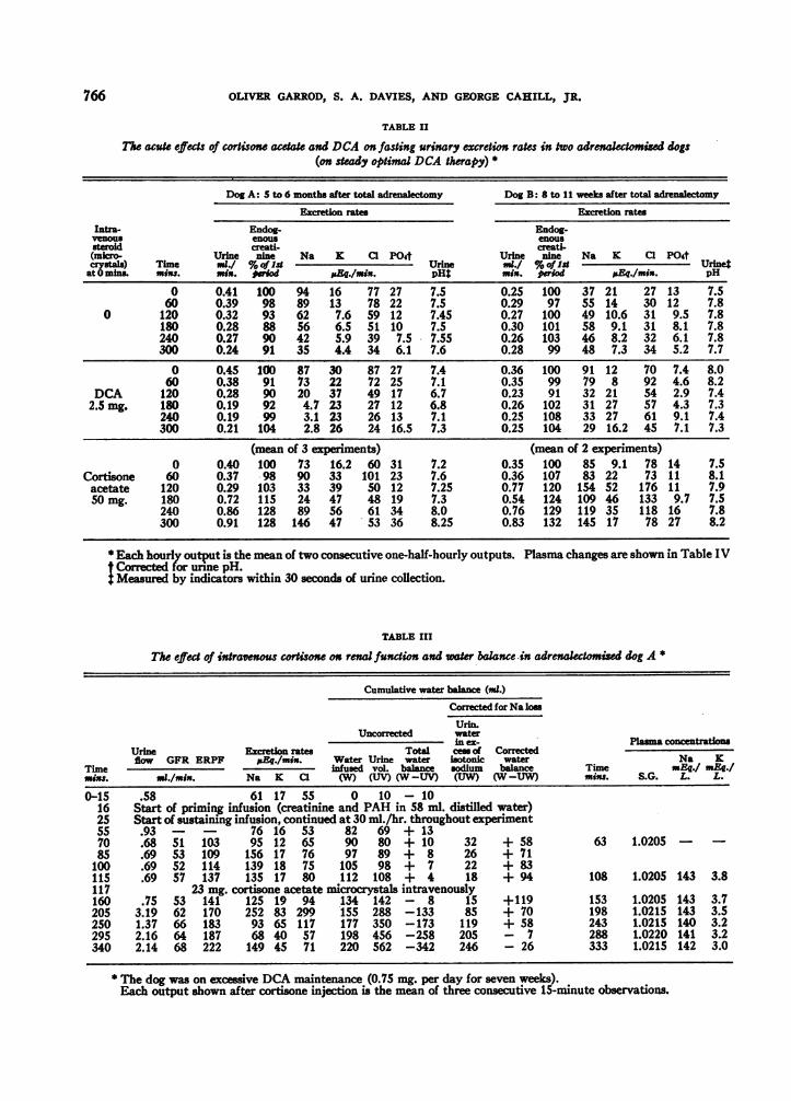
(d) GFR relative to plasma volume and spe-cific gravity. Plasma specific gravity was used as
an index of hemodilution and was measured con-
currently with GFR during all experiments.Grouped data from the periods without cortisoneshowed a high inverse correlation between GFRand plasma specific gravity (r = - 0.84).Grouped data from the periods between one andsix hours after cortisone showed no such corre-
lation (r = - .034), but GFR was higher rela-tive to plasma specific gravity (Figure 5).
Again, in four experiments unaccompanied byinfusion or water ingestion, 50 mg. of intravenouscortisone acetate had no effect during four hourson plasma volume, specific gravity or electrolyteconcentrations; concurrently, however, the en-
PLASMA SPECIFIC GRAVITY
FIG. 5. GFR IN RELATION TO PLASMA SPECIFICGRAviTy, WITH AND WITHOUT CORTISONE, IN TwoADRENALECTOMIZED DoGSEach GFR plotted is the mean of three consecutive 1S-
minute measurements. Plasma specific gravity (28) wasmeasured at the midpoint of each 45-minute GFR pe-riod. All data have been included except those from thefirst two months after adrenalectomy in dog A.
TABLE IV
Changes in plasma in rektson to changes in endogenous creatinine excretio after cortisone and DCA
Per cent change at three and three-fourths hours after steroid
PlasmaSteroid at Endogenous0 mins. creatinine Na Cl
Subject (Intravenous) outputt Vol. S.G.1 cone. conc.
Cortisone +33 - 4.5 -1.3 +0.4 +1.650 mg.
Dog A 50 mg. +36 - 1.4 +1.6 -0.6 -0.550mg. +21 +1.7 -1.4
Dog B 50 mg. +27 +12.7 +0.4 -0.2 +0.450 mg. +36 - 2.9 +1.0 -0.5 -0.2
Mean of 5 experiments (both dogs) +30.6 + 1.0 +0.7 -0.46 +0.32
Dog A DCA 2.5 mg. - 2 -0.5 +0.7 +2.1Dog B DCA 2.5 mg. + 1 - +0.9 +1.0 0Dog A 0 -10 - -1.3 -1.0 0DogB 0 +33
* These experiments were made during a five-week period of steady optimal DCA therapy, after 18 hours' fastingwith free access to water.
t Calculated from the output three and one-half to four hours after, the steroid injection divided by the outputdurng the 60 minutes immediately preceding, the steroid injection.
$ These measurements were made 15 to 30 minutes before, and repeated three and three-fourths hours after, thesteroid injection.
I Measured by the gradient method of Lowry and Hunter (29).
767
. 0
0
0. 0* * -
0
0.
0 0
0
OLIVER GARROD, S.
(DM A)IgTACT X
ECTD. *CORTISONE+DCAL DA
0
00
0o
0
IwX,o
*0-
0
4
% ~0004
0 00
* a °o:0k* 0
114~
0o 0R
aap SOML
40 . s
G.FR AL/MINA
A. DAVIES, AND GEORGE CAHILL, JR.
250
20o
ISO
i!wlo
B
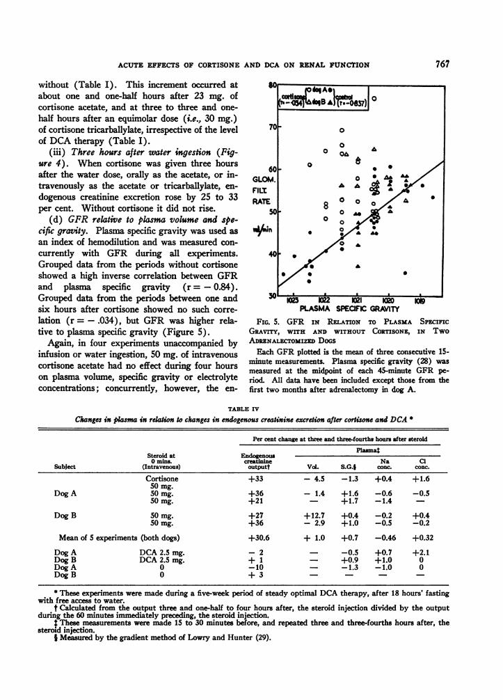
FIG. 6A AND B. SODIUM EXCRETION IN RELATION TO GFR: THE EFFECTS OF CORTISONE AND DCAEach plotted GFR and sodium excretion rate is the mean of three consecutive 15-minute measurements, without
overlap. In dog A all data have been included. In dog B, data from the first month after adrenalectomy have beenexcluded on account of dietary irregularities and unsteady sodium intake.
dogenous creatinine output rose by 30 per cent(Table IV). These findings suggested that theacute effects of cortisone on GFR were not de-pendent on an increase in plasma volume.
(e) The effect of saline infusion on GFR(Table I). This experiment was designed to findout whether the chronic effects of excessive DCAtherapy on GFR could be reproduced acutely,when on optimal DCA therapy, by infusing iso-tonic saline. Isotonic saline, 40 ml. per Kgm. ofbody weight, was infused during 75 minutes.When the water dose was given at the end of thisinfusion, GFR was 10 ml. higher than the mean
fasting level on optimal DCA therapy (37.1s.d. 2.8 ml. per min.). Four hours later GFRhad risen to its pre-adrenalectomized level (TableI). This effect was preceded by defecation, slightedema, and restlessness, suggesting mild waterintoxication. Thus, the chronic effects of exces-
sive DCA therapy on GFR were reproducedacutely by overloading with sodium and water;water diuresis, however, was largely inhibited.9
9 Defecation during diuresis experiments in the intactdog is usually accompanied by prolonged antidiuresis(31). This was also our experience on other occasions,in the adrenalectomized dog.
Sodium excretion 'O
(a) In relation to water diuresis. Beforeadrenalectomy, even when GFR rose after wateringestion, the fasting sodium output remained lowor fell further (Figure 6). After adrenalectomy,water ingestion always raised GFR (Table I),and there was then a parallel rise in the fastingsodium excretion, an effect which was further ac-
centuated when on excessive DCA therapy10(Figure 6).
(b) In relation to GFR, without cortisone. Onanalysis of grouped data from the periods afteradrenalectomy, there was a high correlation be-tween GFR and sodium output (r = 0.84 and0.82 in the two dogs) (Figure 6).1
10Before adrenalectomy the sodium output, 18 hoursafter food, was always less than 25 isEq. per minute.After adrenalectomy it was always higher (40 to 100;&Eq. per minute), due partly to delayed excretion ofdietary sodium which had been retained during the pe-riod of action of the injected DCA. This "escape effect"was much greater during excessive DCA therapy, owingto the longer action of the larger DCA injections and thecompensating effect of higher GFR (infra).I"Because the changes in plasma sodium concentration
were small, when this data was calculated as the quan-tity of sodium filtered (Na' = Nat X GFR) and the per-centage reabsorbed by the tubules (Na R/F), the corre-lations were essentially similar.
768
ISO
Soo
I
Ii
INTACT X (DM s)I_. :e .orgwasm) 0
0.
00*0
00 0
00~ 0
0. 00 0 (
0.0 0 0609 ° 0.000.000
00 Nh3 N N
- 2f.wt. NN. N
so
ACUTE EFFECTS OF CORTISONE AND DCA ON RENAL FUNCTION
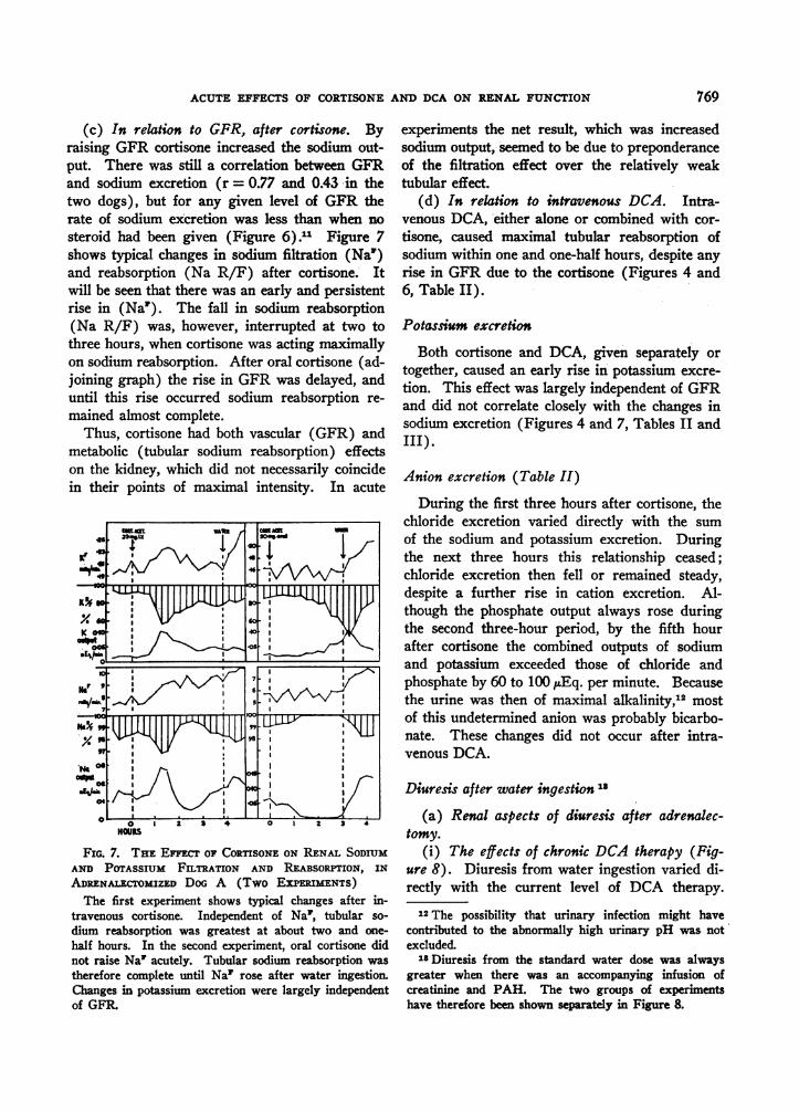
(c) In relation to GFR, after cortisone. Byraising GFR cortisone increased the sodium out-put. There was still a correlation between GFRand sodium excretion (r = 0.77 and 0.43 in thetwo dogs), but for any given level of GFR therate of sodium excretion was less than when nosteroid had been given (Figure 6).11 Figure 7shows typical changes in sodium filtration (Na')and reabsorption (Na R/F) after cortisone. Itwill be seen that there was an early and persistentrise in (Na1). The fall in sodium reabsorption(Na R/F) was, however, interrupted at two tothree hours, when cortisone was acting maximallyon sodium reabsorption. After oral cortisone (ad-joining graph) the rise in GFR was delayed, anduntil this rise occurred sodium reabsorption re-mained almost complete.
Thus, cortisone had both vascular (GFR) andmetabolic (tubular sodium reabsorption) effectson the kidney, which did not necessarily coincidein their points of maximal intensity. In acute
FIG. 7. THE EFFECT OF CORTISONE ON RENAL SODIUMAND POTASSIUM FILTRATION AND REABSORPTION, IN
ADRENALECTOMIZED DoG A (Two EXPERuMENTS)The first experiment shows typical changes after in-
travenous cortisone. Independent of Na', tubular so-
dium reabsorption was greatest at about two and one-
half hours. In the second experiment, oral cortisone didnot raise Na' acutely. Tubular sodium reabsorption was
therefore complete until Na' rose after water ingestion.Changes in potassium excretion were largely independentof GFR.
experiments the net result, which was increasedsodium output, seemed to be due to preponderanceof the filtration effect over the relatively weaktubular effect.
(d) In relation to intravenous DCA. Intra-venous DCA, either alone or combined with cor-tisone, caused maximal tubular reabsorption ofsodium within one and one-half hours, despite anyrise in GFR due to the cortisone (Figures 4 and6, Table II).
Potassium excretion
Both cortisone and DCA, given separately ortogether, caused an early rise in potassium excre-tion. This effect was largely independent of GFRand did not correlate closely with the changes insodium excretion (Figures 4 and 7, Tables II andIII).
Anion excretion (Table II)
During the first three hours after cortisone, thechloride excretion varied directly with the sumof the sodium and potassium excretion. Duringthe next three hours this relationship ceased;chloride excretion then fell or remained steady,despite a further rise in cation excretion. Al-though the phosphate output always rose duringthe second three-hour period, by the fifth hourafter cortisone the combined outputs of sodiumand potassium exceeded those of chloride andphosphate by 60 to 100 /AEq. per minute. Becausethe urine was then of maximal alkalinity,12 mostof this undetermined anion was probably bicarbo-nate. These changes did not occur after intra-venous DCA.
Diuresis after water ingestion18
(a) Renal aspects of diuresis after adrenalec-tomy.
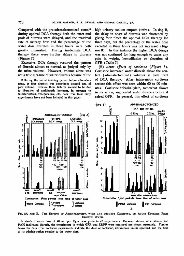
(i) The effects of chronic DCA therapy (Fig-ure 8). Diuresis from water ingestion varied di-rectly with the current level of DCA therapy.
12The possibility that urinary infection might havecontributed to the abnormally high urinary pH was notexcluded.
lsDiuresis from the standard water dose was alwaysgreater when there was an accompanying infusion ofcreatinine and PAH. The two groups of experimentshave therefore been shown separately in Figure 8.
769
OLIVER GARROD, S. A. DAVIES, AND GEORGE CAHILL, JR.
Compared with the pre-adrenalectomized state,",during optimal DCA therapy both the onset andpeak of diuresis were delayed, and the maximalrate of urinary flow and the percentage of thewater dose excreted in three hours were bothgreatly diminished. During inadequate DCAtherapy there were further delays in diuresis(Figure 2).Excessive DCA therapy restored the pattern
of diuresis almost to normal, as judged only bythe urine volume. However, volume alone wasnot a true measure of water diuresis because of the14During the initial training period before adrenalec-
tomy, at first diuresis was sometimes delayed and ofpoor volume. Because these failures seemed to be dueto liberation of antidiuretic hormone, in response tocatheterization, venepuncture, etc., data from these earlyexperiments have not been included in this paper.
high urinary sodium outputs (infra). In dog B,the delay in onset of diuresis was shortened bygiving four times the optimal DCA therapy forthree days, but the percentage of the water doseexcreted in three hours was not increased (Fig-ure 8). In this instance the higher DCA dosagewas not continued for long enough to cause anygain in weight, hemodilution or elevation ofGFR (Table I).
(ii) Acute effects of cortisone (Figure 8).Cortisone increased water diuresis above the con-trol (adrenalectomized) volumes at each levelof DCA therapy. After intravenous cortisoneacetate this effect was seen within 60 to 90 min-utes. Cortisone tricarballylate, somewhat slowerin its action, augmented water diuresis before itraised GFR. In general, this effect of cortisone
(Dog B) I..ADRENALECTOMIZED
- ADRENALECTOMIZED (Dog A)
U..
b%'I
mean enov9 mean 14 30 20 men 102 expF experimnts e. - experiments
mq at +30mmConsecutive 34 hr. periods from time of water dose
IWith Cortisone Cortisone Cortisonel~rtolA artians I Tricarbaflylee [J acetate
A
DCA dose per day:3mg. for
Z 00.75mg 0.75m 3-4days
81~~~~~~~~~~~~~~~~~~4
As~~~~~~~~~~~~~~k.-
at at at+30-180 -30
2-5 6-9 10weeks after adrenalectomy
Consecutive 3/4hr periods from time of water dose
IWithaut Cortsone With Cortisone
B
FIG. 8A AND B. THE EFECTs OF ADRENALECTOMY, WITH AND WrrHOUT CORTISONE, ON AcuTE, Diuaxsis FROMINGESTED WATER
A standard water dose of 40 ml. per Kgm. was given in all experiments. Because infusion of creatinine andPAH facilitated diuresis, the experiments in which GFR and ERPF were measured are shown separately. Figuresbelow the data from cortisone experiments indicate the dose of cortisone, intravenous unless specified, and the timeof its administration relative to the water dose.
770
ACUTE EFFECTS OF CORTISONE AND DCA ON RENAL FUNCTION
8 (osMA)8~~~~~~ 0
6 0 0
0
4 0 00
0
2 AdI0NT= K MrT* ADME3t. o HOCOwa SONE
10 WITH CORTISONE
50 60 70GFR AT PEAK URINE FLOW
ml/mmin.A
z
M.
9--J
w
z
w
a.
X (OOsM)
8
0 0
6 K-xx 4q)
4
0
2 * INT x INeIAO .l-0 wSnu CORTISON
0 WITH CORTIO
50 60 70 80GFR AT PEAK URINE FLOW% ML/MIN
B
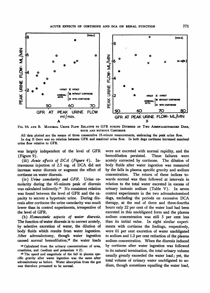
FIG. 9A AND B. MAXIMAL URINE FLOW REATIVE TO GFR DURING Diujsis IN Two ADENALECTOMIZED DOGS,WITH AND WITHOUT CORTISONE
All data plotted are the means of three consecutive 15-minute measurements, embracing the peak urine flow.In dog B there was no relation between GFR and maximal urine flow. In both dogs cortisone increased maximal
urine flow relative to GFR
was largely independent of the level of GFR(Figure 9).
(iii) Acute effects of DCA (Figure 4). In-travenous injection of 2.5 mg. of DCA did notincrease water diuresis or augment the effect ofcortisone on water diuresis.
(iv) Urine osmolarity and GFR. Urine os-
molarity during the 45-minute peak of diuresiswas calculated indirectly.15 No consistent relationwas found between the level of GFR and the ca-
pacity to secrete a hypotonic urine. During diu-resis after cortisone the urine osmolarity was muchlower than in control experiments, irrespective ofthe level of GFR.
(b) Homeostatic aspects of water diuresis.The function of water diuresis is to correct acutely,by selective excretion of water, the dilution ofbody fluids which results from water ingestion.After adrenalectomy, though water ingestioncaused normal hemodilution,"' the water loads
15 Calculated from the urinary concentrations of urea,creatinine, and (sodium and potassium X 2).
16 The speed and magnitude of the fall in plasma spe-cific gravity after water ingestion was the same afteradrenalectomy as before. Water absorption from the gutwas therefore presumed to be normal.
were not excreted with normal rapidity, and thehemodilution persisted. These failures were
acutely corrected by cortisone. The dilution ofbody fluids after water ingestion was measuredby the falls in plasma specific gravity and sodiumconcentration. The return of these indices to-wards normal was then followed at intervals inrelation to the total water excreted in excess ofurinary isotonic sodium (Table V). In seven
control experiments in the two adrenalectomizeddogs, excluding the periods on excessive DCAtherapy, at the end of three and three-fourthshours only 22 per cent of the water load had beenexcreted in this unobligated form and the plasmasodium concentration was still 5 per cent lessthan its initial value. In eight similar experi-ments with cortisone the findings, respectively,were 61 per cent excretion of water unobligatedto sodium and 1.3 per cent reduction of the plasmasodium concentration. When the diuresis inducedby cortisone after water ingestion was followedto its natural termination, the total urinary volumeusually greatly exceeded the water load; yet, thetotal volume of urinary water unobligated to so-
dium, though sometimes equalling the water load,
z
-J
zorD
771
OLIVER GARROD, S. A. DAVIES, AND GEORGE CAHILL, JR.
TABLE V
Plasma specific graviy and sodium concentration in relationto total water and sodium excretion, with
and without cortisone(two adrenalectomized dogs) *
Water without Water+At three and three-fourths cortisone cortisone
hours after water (7 expts.) 18 expts.)ingestion
(40 ,I./Kgm.) Mean + S.D. Mean i S.D.
Plasma Na conc.:% change from initiallevel -54 1.1 -1.3± 2.1
Plasma S.G.:%change from initiallevel -7.34 2.8 -2.1 is 2.4
Per cent of water doseexcreted as total uri-nary water 45.3±13.7 92.1*22.6
Per cent of water doseexcreted as water inexcess of urinary iso-tonic sodium 22.34 8.7 60.8±+17.1
* All experiments were accompanied by a standard infu-sion of creatinine and PAH the volume of which has beenincluded in the total water dose. Experiments on dog Aduing excessive DCA therapy have been excluded. Thedifferences after cortisone are significant (p < .01).
HOURS
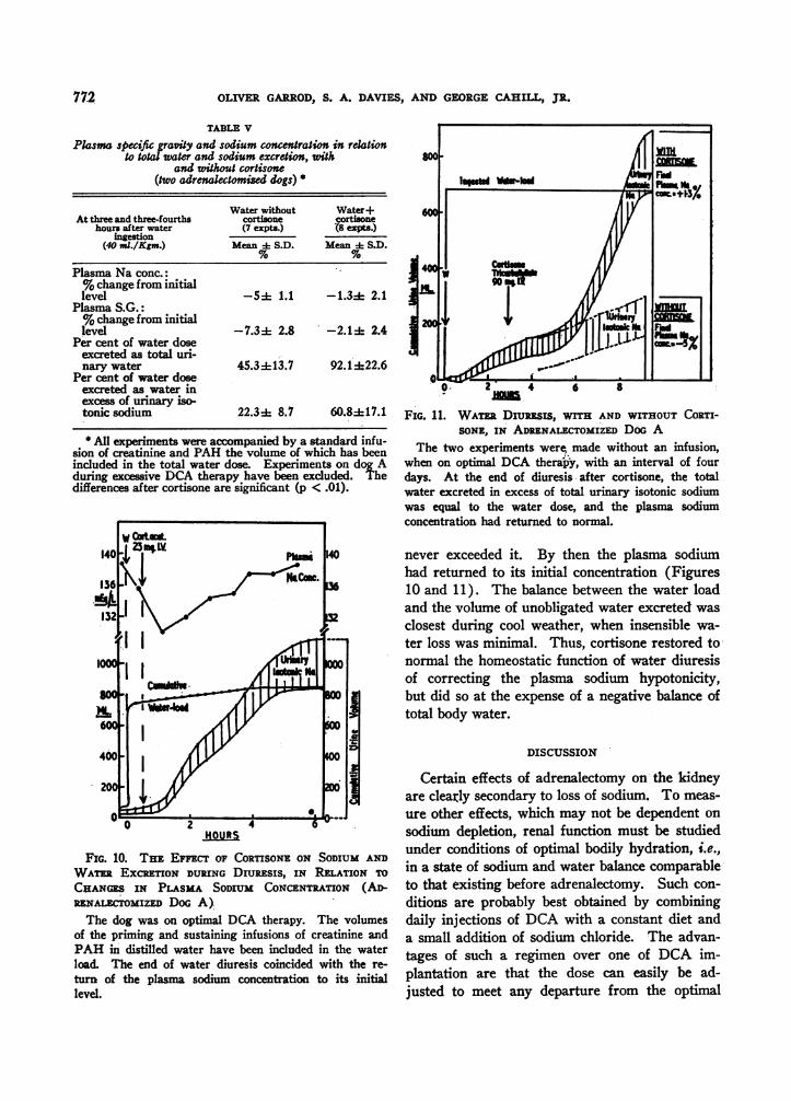
FIG. 10. THE EFFEcT OF CORTISONE ON SODIUM AND
WATR EXCRETION DURING DrumESIS, IN RELATION TO
CHANGES IN PLASMA SODIUM CONCENTRATION (AD-RENALEmmIZED DoG A)The dog was on optimal DCA therapy. The volumes
of the priming and sustaining infusions of creatinine andPAH in distilled water have been included in the waterload. The end of water diuresis coincided with the re-
turn of the plasma sodium concentration to its initiallevel.
o0 2 4 6 8
FIG. 1 1. WATr DIUREsIS, WITH AND WITHOUT CORTI-SONE, IN ADRENALECTOMIZED DOG A
The two experiments were, made without an infusion,when on optimal DCA therapy, with an interval of fourdays. At the end of diuresis after cortisone, the totalwater excreted in excess of total urinary isotonic sodiumwas equal to the water dose, and the plasma sodiumconcentration had returned to normal.
never exceeded it. By then the plasma sodiumhad returned to its initial concentration (Figures10 and 11). The balance between the water loadand the volume of unobligated water excreted wasclosest during cool weather, when insensible wa-ter loss was minimal. Thus, cortisone restored tonormal the homeostatic function of water diuresisof correcting the plasma sodium hypotonicity,but did so at the expense of a negative balance oftotal body water.
DISCUSSION
Certain effects of adrenalectomy on the kidneyare clearly secondary to loss of sodium. To meas-ure other effects, which may not be dependent onsodium depletion, renal function must be studiedunder conditions of optimal bodily hydration, i.e.,in a state of sodium and water balance comparableto that existing before adrenalectomy. Such con-ditions are probably best obtained by combiningdaily injections of DCA with a constant diet anda small addition of sodium chloride. The advan-tages of such a regimen over one of DCA im-plantation are that the dose can easily be ad-justed to meet any departure from the optimal
772
ACUTE EFFECTS OF CORTISONE AND DCA ON RENAL FUNCTION
state, and that renal function can be assessedduring its escape from DCA effect.Using dogs of around 10 Kgm. weight, on an
added salt intake of 2 gm. a day, other authorshave shown that the minimal amounts of DCAneeded for normal health and hydration are be-tween 0.25 and 0.5 mg. a day (10, 11). With thesame added salt, the dogs reported here needed0.6 to 0.75 mg. a day which, allowing for theirgreater weight, is closely equivalent to the aboveamounts. Raising or lowering the dose of DCAby as little as 0.15 mg. was followed by consistentchanges in body weight or in plasma specificgravity and electrolyte concentrations. Criticaladjustment of the DCA therapy is thus essentialfor obtaining a steady optimal state, a precautionapparently overlooked in most previous studies.
After adrenalectomy the filtration rate seemedto vary with the state of bodily hydration, as wassuggested by its inverse correlation with theplasma specific gravity, and by the over-retentionof water which always accompanied the rise of fil-tration, whether chronic, as during excessiveDCA therapy, or acute, from water ingestion orsaline infusion. However, since filtration andrenal blood flow remained subnormal even afteroptimal therapy had corrected any sodium deple-tion, at least a part of this failure could not beascribed merely to reduction of the intravascularor extracellular fluid volumes. Some data oncardiac output are needed to throw further lighton this problem.The absence of any acute effect of DCA on
filtration was in marked contrast with the rise inthis function which occurred when excessive DCAtherapy had caused over-retention of sodium andwater. Though Davis and Howell found a sub-acute rise of filtration rate in the intact dog whenDCA was given intramuscularly at more thantwenty times the dosage used intravenously in thepresent experiments (32), this effect was con-siderably less than after cortisone or ACTH; andit is not clear from their data whether any sodiumretention preceded the rise in filtration. It ispossible, therefore, that DCA may have someprimary action on the filtration rate when givenin doses that are greatly excessive in terms ofelectrolyte effects.The action of cortisone on the filtration rate
and renal blood flow differs fundamentally from
that of DCA in its rapidity of onset and its ap-parent independence of plasma volume or sodiumbalance. It would seem that cortisone has a"permissive" action of allowing a higher and morenormal proportion to exist between the filtra-tion rate and the level of bodily hydration.Whether this effect is confined to the kddneys oris part of a more widespread circulatory responsecannot be decided from the present data.The normal dog excretes a sodium load more
rapidly than does man, and puts out only negligi-ble amounts of sodium after 18 hours of fasting.The sodium output falls further during sustainedmaximal water diuresis (31), and, as has beenshown in the present studies, either falls or in-creases only slightly when the filtration rate risesduring acute water diuresis. After adrenalectomy,however, there is a partial loss of tubular abilityto conserve sodium, and the filtration rate becomesthe main regulator of sodium output. Theadrenalectomized dog comes to resemble the salt-loaded normal dog in that both show a direct re-lation between sodium filtration and sodium out-put (33). This could be explained by assumingthat salt-loading of the intact animal may sup-press the secretion of a powerful sodium-retain-ing steroid, whereas in the normal fasting statethis steroid 17 may already be present in amountslarge enough to prevent any increase in sodiumoutput when filtration rises after water ingestion.Although its sodium-retaining action is well
recognized (4), cortisone often increases sodiumexcretion (15, 18, 19, 36, 37). To explain thisparadox, it has been suggested that under certainconditions, notably in the presence of DCA, cor-tisone may inhibit tubular reabsorption of sodium(15); and a concept has arisen of intratubularcompetition between cortisone and a steroid withmore potent sodium-retaining effects, which corti-sone is supposed to displace (17). This hypothe-sis ignores the important role of filtration ratein regulating sodium output. In the present stud-ies it has been shown that after cortisone there isstill a clear correlation between filtration rate andsodium output, though the latter is slightly less,and the fraction of filtered sodium reabsorbedslightly higher, than at parallel levels of filtrationwithout cortisone. Thus, though cortisone in-
17 Probably aldosterone (34, 35).
773
OLIVER GARROD, S. A. DAVIES, AND GEORGE CAHILL, JR.
creases sodium reabsorption, this effect is notenough to prevent a net increase in sodium outputin the presence of increased filtration of this ion.Similarly, Davis and Howell found in the intactdog that a great natriuresis accompanied the risein filtration induced by cortisone or ACTH (32);and, though Roberts and Pitts did not find thiseffect in the adrenalectomized dog, it should benoted that there were no changes in filtrationrate during the short periods of their experiments(14).When DCA and cortisone were given together,
reabsorption of sodium became almost complete,despite increased filtration. Similarly, Robertsand Pitts have shown that these two steroids havea summative effect on sodium reabsorption (14).Since the natriuretic effects of cortisone can nowbe adequately explained by increased filtration,there is no further need to invoke the hypothesisof competition between these steroids at the renaltubular level.
It is still a matter of dispute why adrenal fail-ure should inhibit the capacity for water diuresis.The main issue concerns the cause of the renalfailure to excrete a more hypotonic urine afterwater ingestion, despite adequate hemodilution (8,13). Theoretically, this failure could be dueeither to extrarenal factors, such as continued se-cretion of antidiuretic hormone (ADH), or tointrinsic renal disturbances, conditioned by lackof adrenal hormones, and independent of thepresence or absence of ADH. To consider firstthe present data on renal factors: when a normalfiltration rate was restored by forced water in-gestion during excessive DCA therapy, the abilityto excrete a hypotonic urine, though still dimin-ished, was greatly improved. Because the possi-bility of there having been any direct effect ofDCA during these experiments seems to have beenexcluded by the high sodium outputs, and becauseDCA did not acutely correct filtration rate or wa-ter diuresis, it could be argued that the improveddiuresis was somehow related to increased filtra-tion. On the other hand, when cortisone restoredthe capacity for a normal water diuresis, the in-creased output of a more hypotonic urine some-times occurred before filtration had risen. It isthus probable that an abnormality of tubular func-tion, whether primary or secondary to ADH, isthe essential factor in causing inhibition of water
diuresis, though lowered filtration rate probablyplays an additional role.To consider next the evidence concerning ex-
trarenal factors: it has been suggested that theaction of ADH may be prolonged unduly inadrenal failure because of delayed inactivation ofthis hormone by the liver (38). However, thisdoes not explain why water diuresis should endprematurely, after only a small fraction of thewater load has been excreted, instead of continu-ing to increase slowly as the inactivation of ADHproceeds. If ADH be the paramount factor, itmust therefore be postulated that this hormone isbeing continuously secreted during the long pe-riod of delayed diuresis. The secretion of ADHmight so persist either because the osmorecep-tors cannot function at their normal thresholdwhen deprived of adrenal hormones, or becausethere is a further unidentified stimulus to ADHsecretion, possibly humoral or arising from vol-ume receptors.
SUMMARY
Changes in GFR, ERPF, electrolyte excre-tion, and water diuresis were studied in twoadrenalectomized dogs in relation to bodily hy-dration and to acute administration of cortisoneand DCA.The dogs received daily DCA therapy which
was assessed by its effects on bodily hydration asinadequate, optimal or excessive. Renal functionwas measured during the daily escape from DCAeffect.
After adrenalectomy GFR and ERPF were sub-normal on optimal DCA therapy, but rose afterwater-loading or saline infusion. DCA had noacute effect on GFR and raised it only when caus-ing excessive retention of sodium and water.Cortisone raised GFR acutely, without change inplasma volume and despite a negative sodiumbalance.
After adrenalectomy, during escape from DCAeffect, the sodium output varied directly withGFR. DCA caused acute maximal sodium re-tention. Cortisone usually increased the sodiumoutput by raising GFR, thereby obscuring itsother effect of slightly increasing tubular sodiumreabsorption at any given level of filtration.
After adrenalectomy, water diuresis was greatlydiminished, and was acutely restored by cortisone,
774
ACUTE EFFECTS OF CORTISONE AND DCA ON RENAL FUNCTION
but not by DCA. This effect of cortisone waslargely independent of GFR and electrolyte out-puts.
After adrenalectomy, water ingestion causedprolonged hyponatremia which cortisone correctedby allowing excretion of the water load plus avolume of water isotonically equivalent to theurinary sodium. The plasma sodium concentra-tion was thereby restored at the expense of totalbody water.
These results suggest that in the adrenalecto-mized dog: 1) There is a direct relationship be-tween extracellular hydration and GFR, and be-tween GFR and sodium output; and 2) that twoadrenal steroids are needed to maintain normalrenal function: (a) cortisone or hydrocortisone,to maintain normal GFR and water diuresis, and(b) a DCA-like steroid, to prevent excessive so-dium excretion at normal or high levels of GFR.
AddendumSince this paper was submitted for publication, acute
effects of cortisone and DCA on GFR and sodium ex-cretion in the adrenalectomized dog, which are similarto those reed here, have been described by, Liddle,Pechet, and Bartter (39).
ACKNOWLEDGMENTS
The authors wish to express their gratitude to Dr. R.F. Loeb for encouragement and advice and for providingthe facilities for this study; to Dr. Alfred Gilman foradvice; to Dr. David Habif for undertaking the adrenal-ectomies; to Dr. T. Allen for assistance in measuringplasma volumes; and to Dr. Russell T. Fraser for criti-cism of the manuscriptThey also wish to thank Merck and Co. of New Jersey
for providing the cortisone tricarballylate, and ChemicalSpecialities, Inc., of New York, for providing the mi-crocrystalline suspensions of cortisone acetate and DCA.
REFERENCES
1. Loeb, R. F., Atchley, D. W., Benedict, E. M., andLeland, J. L., Electrolyte balance studies in adrenal-ectomised dogs with particular reference to theexcretion of sodium. J. Exper. Med., 1933, 57, 775.
2. Harrop, G. A., Jr., Soffer, L J., Ellsworth, R., andTrescher, J. H., Studies on the suprarenal cortex;III. Plasma electrolytes and electrolyte excretionduring suprarenal insufficiency in dog. J. Exper.Med., 1933, 58, 17.
3. Harrison, H. E., and Darrow, D. C., Renal functionin experimental adrenal insufficiency. Am. J.Physiol., 1939, 125, 631.
4. Gaudino, M., and Levitt, M. F., Influence of theadrenal cortex on body water distribution andrenal function. J. Gin. Invest., 1949, 28, 1487.
5. Talbott, J. H., Pecora, L. J., Melville, R. S., andConsolazio, W. V., Renal function in patients withAddison's disease and in patients with adrenalinsufficiency secondary to pituitary pan-hypofunc-tion. J. Clin. Invest., 1942, 21, 107.
6. Waterhouse, C., and Keutmann, E. H., Kidney func-tion in adrenal insufficiency. J. Gin. Invest., 1948,27, 372.
7. Kottke, F. J., Code, C. F., and Wood, E. I., Urinedilution and concentration tests in adrenalectomiseddogs. Am. J. Physiol., 1942, 136, 229.
8. Roemmelt, J. C., Sartorius, 0. W., and Pitts, R. F.,Excretion and reabsorption of sodium and waterin the adrenalectomised dog. Am. J. Physiol.,1949, 159, 124.
9. White, H. L., Heinbecker, P., and Rolf, D., Furtherobservations on the depression of renal functionfollowing hypophysectomy. Am. J. Physiol., 1949,156, 67.
10. Remington, J. W., Parkins, W. M., Swingle, W. W.,and Drill, V. A., Efficacy of desoxycorticosteroneacetate as replacement therapy in adrenalectotnizeddogs. Endocrinology, 1941, 29, 740.
11. Cleghorn, R. A., Fowler, J. L A., Wenzel, J. S., andClarke, A. P. W., The desoxycorticosterone acetaterequirement of the adrenalectomised dog. Endo-crinology, 1941, 29, 535.
12. Burnett, C. H., The actions of ACTH and cortisoneon renal function in man in Renal Function, Trans-actions of the Second Conference, Oct. 1950, S. E.Bradley, ed., New York, Josiah Macy, Jr. Founda-tion, 1951, p. 106.
13. Burston, R. A., and Garrod, O., The variability ofthe lowered glomerular filtration rate in Addison'sdisease and panhypopituitarism and the effect ofcortisone thereon. Clin. Sc., 1952, 11, 129.
14. Roberts, K. E., and Pitts, R. F., The influence of cor-tisone on renal function and electrolyte excretionin the adrenalectomized dog. Endocrinology, 1952,SO, 51.
15. Thorn, G. W., Forsham, P. H., Bennett, L. L., Roche,M., Reiss, R. S., Slessor, A., Flink, E. B., andSomerville, W., Clinical and metabolic changesin Addison's disease following the administrationof compound E acetate. Tr. A. Am. Physicians,1949, 62, 233.
16. Garrod, O., and Burston, R. A., The diuretic re-sponse to ingested water in Addison's disease andpanhypopituitarism and the effect of cortisonethereon. Clin. Sc., 1952, 11, 113.
17. Adrenal Cortex: Transactions of the Third Confer-ence, Nov. 1951, E. P. Ralli, ed., New York, JosiahMacy, Jr. Foundation, 1952.
18. Thorn, G. W., Engel, L. L., and Lewis, R. A., Theeffect of 17-hydroxycorticosterone and relatedadrenal cortical steroids on sodium and chlorideexcretion. Science, 1941, 94, 348.
775
OLIVER GARROD, S. A. DAVIES, AND GEORGE CAHILL, JR.
19. Davis, A. K, Bass, A. C., and Overman, R. R., Com-parative effects of cortisone and DCA on ionicbalance and fluid volume of normal and adrenalec-tomised dogs. Am. J. Physiol., 1951, 166, 493.
20. Swingle, W. W., Collins, E., Barlow, G., and Fedor,E. J., Bioassay and physiological effects of corti-sone on adrenalectomized dogs. Am. J. Physiol.,1952, 169, 270.
21. Gregersen, M. I., and Stewart, J. D., Simultaneousdetermination of the plasma volume with T-1824,and the "available fluid" volume with thiocyanate.Am. 3. Physiol., 1939, 125, 142.
22. Berry, 3. W., Chappel, D. G., and Barnes, R. B., Animproved method of flame photometry. Indust. &Engin. Chem. (Anal. Ed.), 1946, 18, 19.
23. Van Slyke, D. D., The determination of chlorides inblood and tissues. J. Biol. Chem., 1923, 58, 523.
24. Folin, O., and Wu, H., A system of blood analysis.3. Biol. Chem., 1919, 38, 81.
25. Smith, H. W., Finkelstein, N., Aliminosa, L, Craw-ford, B., and Graber, M., The renal clearances ofsubstituted hippuric acid derivatives and otheraromatic acids in dog and man. J. Chin. Invest.,1945, 24, 388.
26. Fiske, C. H., and Subbarow, Y., The calorimetricdetermination of phosphorus. J. Biol. Chem., 1925,66, 375.
27. Gentskow, C. J., An accurate method for the deter-mination of blood urea nitrogen by direct nessleri-zation. J. Biol. Chem., 1942, 143, 531.
28. Phillips, R. A., Van Slyke, D. D., Dole, V. P., Emer-son, K., Jr., Hamilton, P. B., and Archibald, R. M.,Copper Sulphate Method for Measuring SpecificGravities of Whole Blood and Plasma. Glendale,Calif., Dan. Baxter, Inc., 1945.
29. Lowry, 0. H., and Hunter, T. H., The deermina-tion of serum protein concentration with a gradienttube. J. Biol. Chem., 1945, 159, 465.
30. Courtice, F. C., The blood volume of normal animals.J. Physiol., 1943, 102, 290.
31. Gilman, A., Personal Communications.32. Davis, J. O., and Howell, D. S., Comparative effect
of ACTH, cortisone and DCA on renal function,electrolyte excretion and water exchange in normaldogs. Endocrinology, 1953, 52, 245.
33. Thompson, D. D., and Pitts, R. F., Effects of altera-tions of renal arterial pressure on sodium and wa-ter excretion. Am. J. Physiol., 1952, 168, 490.
34. Simpson, S. A., Tait, J. F., and Bush, I. E., Secre-tion of a salt-retaining hormone by the mammalianadrenal cortex. Lancet, 1952, 2, 226.
35. Mach, R. S., Fabre, J., Duckert, A., Borth, R., andDucommun, P., Action clinique et metabolique deI'Aldosterone (electrocortine). Schweiz. med.Wchnschr., 1954, 84, 407.
36. Marcus, F., Romanoff, L P., and Pincus, G., Theelectrolyte-excreting activity of adrenocortical sub-stances. Endocrinology, 1952, 50, 286.
37. Perera, G. A., Pines, K. L., Hamilton, H. B., andVislocky, K., Clinical and metabolic study 11-dehydro-17-corticosterone acetate (Kendall com-pound E) in hypertension, Addison's disease anddiabetes mellitus. Am. J. Med., 1949, 7, 56.
38. Birnie, J. H., The inactivation of posterior pituitaryantidiuretic hormone by liver extracts. Endocrin-ology, 1953, 52, 33.
39. Liddle, G. W., Pechet, M. M., and Bartter, F. C.,Enhancement of biological activities of corticoster-oids by substitution of halogen atoms in 9 a position.Science, 1954, 120, 496.
SPECIAL NOTICE TO SUBSCRIBERS
Post Offices will no longer forward the Journal when you move.
Please notify The Journal of Clinical Investigation, BusinessOffice, 622 West 168th Street, New York 32, N. Y. at once whenyou have a change of address, and do not omit the zone number ifthere is one.
776