abnormality of vergence latency in children with vertigo
TRANSCRIPT

J Neurol (2004) 251 : 204–213DOI 10.1007/s00415-004-0304-8 ORIGINAL COMMUNICATION
Maria Pia BucciZoï KapoulaQing YangSylvette Wiener-VacherDominique Brémond-Gignac
Abnormality of vergence latency in children with vertigo
JON
130
4
Introduction
Vergence eye movements are essential for clear binocu-lar vision but also for depth vision, accurate distanceevaluation and stereopsis [14]. It is also known that pos-tural stability depends on viewing distance [4] and a re-cent survey shows balance problems in non-correctedhyperopic children [2]. Anon-Tanon et al. [1] reportedthat more than 5 % of the 520 children consulting theENT service for vertigo, headaches and equilibrium dis-orders showed normal vestibular function but revealed
signs of vergence abnormalities assessed by orthoptictests [29]. These authors pointed out that deficits in ver-gence could impair gaze stabilization during move-ments of the body and thus, cause double or blurry vi-sion which can lead to vertigo and sensation ofimbalance. Despite the convincing clinical evidence ofthe link between vertigo symptoms and vergence ab-normalities there have been no studies of such abnorm-alities with objective eye movement recordings. In-deed, the orthoptic clinical tests provide subjective in-formation, i. e. sense of double vision, which is attrib-uted to incapacity to perform the appropriate vergence
Received: 11 March 2003Received in revised form: 29 September 2003Accepted: 7 October 2003
Dr. M. P. Bucci (�) · Z. Kapoula · Q. YangLaboratoire de Physiologie de la Perceptionet de l’Action, UMR 9950CNRS-College de France11, place Marcelin Berthelot75005 Paris, FranceTel.: +33-1/4427-1636Fax: +33-1/4427-1382E-Mail:[email protected]. YangLaboratory of Neurology of ShanghaiInstitute of Physiology and Laboratory of Visual Information Processing ofBiophysics InstituteChinese Academy of SciencesShanghai, ChinaS. Wiener-VacherHôpital Robert Debré, service d’O. R. L.Unité de VestibulométrieParis, FranceD. Brémond-GignacHôpital Robert Debréservice d’OphtalmologieParis, France
■ Abstract It is well known thatvergence movements are importantfor distance appreciation, depth vi-sion and stereopsis. Moreover, ver-gence movements are very proba-bly used by the CNS during headand body motion to adjust the gainof the vestibulo-ocular reflex(VOR) according to the viewingdistance. A recent clinical study ofAnoh-Tanon et al. suggested thatvertigo in children with normalvestibular function could be asso-ciated with abnormal vergenceclinically assessed. The purpose ofthis study was to test this hypothe-sis with objective vergence eyemovement recordings. We exam-ined the latency of vergence, sac-cades and combined movements intwelve children with the complaintof vertigo but without vestibularabnormality. Convergence and sac-cades combined with convergenceor with divergence had abnormally
long latencies (relative to normalchildren of matched age). In con-trast, divergence and isolated sac-cades showed only mild latencyincrease relative to normals.Lengthening of latency could bedue to impaired cortical control.Orthoptic vergence training re-duced all latencies; however, eventhe reduced latency of vergenceand of combined movements wasstill abnormal. The improvementafter orthoptic vergence trainingcould be due to increased visualattention, although such mecha-nism cannot eliminate completelythe initiation deficit of vergencemovements. Objective eye move-ment recordings are thus useful fora diagnosis and treatment of chil-dren with vertigo.
■ Key words latency · saccade ·vergence · combined movements ·vertigo
204-213_Bucci_JON-1304 21.01.2004 11:19 Uhr Seite 204

205
movements in static condition. However, these tests donot give any information about the vergence movementsthemselves and their temporal aspects such as their la-tency.
To prepare and perform an eye movement, e. g. a sac-cade, several processes at the cortical and sub-corticallevel take place: the visual information from the retina issent to visual cortex, parietal cortex, frontal lobe, supe-rior colliculus and then, via the brain-stem the motorcommand is sent to the extra-ocular muscles. The la-tency of eye movements is the preparation time for per-forming the movement and includes several processes:shift of the attention to the visual target, disengagementof oculomotor fixation and computation of the move-ment parameters [9, 10]. All these processes involve theactivation of several cortical areas, particularly of theparietal cortex and of the frontal lobe [20]. The latencyis an important measure that provides informationabout cortical function. It is not known whether in sub-jects with vergence abnormalities and vertigo there areabnormal eye movement latencies.The goal of this studywas to examine the latency of vergence, saccades andcombined saccade – vergence movements in a youngpopulation with vertigo with normal vestibular func-tion and a vergence abnormality in the results of or-thoptic tests. In the second part of this study we addressthe question whether orthoptic vergence training canimprove the latency of eye movements, and particularlyvergence eye movements.
Methods
■ Selection of the patients.
The patients included in the study had the following characteristics:completely normal responses to the vestibular tests, normal neuro-logical and otological clinical examination but one to three abnormalvalues at the orthoptic tests (see Table 1 A). They all were referred tohave the vestibular tests for vertigo, and headache was a secondarysymptom associated or not with the vertigo.
Although the symptoms were initially described as vertigo, the re-ported sensations were in fact sometimes rotation, but also displace-ment of the environment, rolling or translation. They were never in-tense rotatory vertigo lasting for hours but rather brief sensationsrelated to movements of the head or the gaze. Their timing was oftenrelated to fatigue (at school or at the end of the day, or in the evening)and often after long exposure to computer or television screens.
Twelve subjects, between 6 and 15 years old participated in thestudy. The investigation adhered to the principles of the Declarationof Helsinki and was approved by our institutional human experi-mentation committee. Informed parental consent was obtained foreach subject after the nature of the procedure had been explained.
■ Vestibular tests
All subjects underwent a complete vestibular, otological and neuro-logical examination. Clinical vestibular examination was always com-pleted with a quantitative vestibular function evaluation (canal andotolith vestibular function) including several tests which had been
adapted to children and fully explained and detailed in previous arti-cles [31, 32]. The vestibulo-ocular responses were recorded by theelectro-oculography technique during canal vestibular stimulations(caloric test, earth vertical axis rotation, and pendular rotation) andduring otolith vestibular stimulations (off vertical axis rotation) bymeans of a computer controlled rotating chair in a completely darkroom [30–32]. The results of all these tests were normal and are listedbelow.
All eye movements recorded during vestibular tests were monoc-ular at fixed viewing distance and were run on a different day fromthe binocular saccades and vergence eye movements recording (seeoculomotor tests).
The tests performed and normal results were as follows. For thecaloric test: relative directional preponderance < 15 %, relative reflec-tivity < 15 %. For the earth vertical axis rotation: maximum initialslow phase velocity symmetrical for clockwise and counterclockwiserotation and equal to 40 ± 10°/s- time constant symmetrical (depen-dent of age, varying from 8 s to 20 s for 6 to 15 years old subjects). Forthe pendular rotation: responses symmetrical in dark and completelyinhibited by the fixation of a lead target. For the off vertical axis rota-tion: horizontal directional preponderance (< 0.35 ± 0.30°/s), verticaldirectional preponderance (< 0.90 ± 0.69°/s) and horizontal modula-tion amplitude (3.0 ± 1.4°/s) vertical modulation amplitude(4.8 ± 3.3°/s). Conventional hearing tests (tonal and speech audio-metric techniques) were also performed to assess the function of theinner ear and all patients had normal hearing.
Orthoptic tests
Subjects underwent complete ophthalmological and orthoptic exami-nation that is summarized next. All subjects had normal binocularvision, evaluated with the TNO random dot test for stereoscopic depth discrimination (the threshold of stereoacuity was 60 s of arc orbetter). No child wore spectacles and their visual acuity was 8/10 orbetter in both eyes. Orthoptic evaluation of vergence (made by usingthe cover test,bar of prisms,Maddox rod and synoptophore technique)revealed, for all subjects, one or more signs of vergence abnormalities(see Table 1 A,children examined and Table 1 B,normal children); e. g.distant near point of convergence (≥ 10 cm), exophoria (i. e. latentdeviation of one eye when the other eye is covered) at far viewing thatwas larger than 3 ∆, and exophoria at near viewing larger than 6 ∆; therange of fusional vergence at far and at close viewing was also limited.According to Rouse et al. [23, 24] and Rainey [22] subjects S1, S3–S7,S10 and S12 were classified as having convergence abnormalities whilesubjects S2, S8, S9 and S11 had divergence abnormalities.
Orthoptic training
For all subjects vergence abnormalities were treated with twelve ses-sions (three times per week) of orthoptic training that consisted inseveral exercises (e. g. converging and diverging the eyes to follow asmall pen-light or a letter moving in depth) in order to improve therange of vergence fusional amplitude [see for details www. chil-drensvision.com].
Eight of the subjects (S1, S3, S4, S7, S9, S10, S11 and S12) had a sec-ond orthoptic clinical examination 1–2 weeks after the end of thetwelve sessions of orthoptic vergence training.
Oculomotor testing
■ Visual display. Two isovergence circles horizontally placed on asurface were used one at 20 cm and the other at 150 cm from the sub-ject. The target used was a LED; on the circle close to the subject threeLEDs were placed; one at the center and the other at ± 20°. The re-quired mean vergence angle for fixating any of these three LEDs was17°. On the circle far from the subject, five LEDs were placed: one atthe center, two at ± 10° and two at ± 20°; fixation to any of these LEDs required a vergence angle of 2.3°. A computer directed the tar-get-LED presentation.
204-213_Bucci_JON-1304 21.01.2004 11:19 Uhr Seite 205
206
■ Eye movement recordings. Data collection was directed by REX,software developed for real-time experiments. Horizontal eye move-ments from both eyes were recorded simultaneously with a photo-electric device (OCULOMETER, BOUIS). This system has an optimalresolution of 2” of arc. Eye-position signals were low-pass filteredwith a cut off frequency of 200 Hz and digitized with a 12-bit ana-logue-to-digital converter. Each channel was sampled at 500 Hz.
■ Procedure of oculomotor testing. Subject was seated in a chair,which could be adjusted for height, with the head stabilized by a fore-head and chin support. The experiment was run in a completely darkroom to ensure that stimulus detection and localization was quasi au-tomatic, avoiding distraction from other objects [16].
The subject viewed binocularly and faced the surface containingthe LEDs so that the close isovergence circle was at 20 cm from her/hiseyes.
Each trial started by lighting a fixation LED at the center of one of the two circles (far or close). After a 2.5 s fixational period thecentral LED was turned off and a target-LED appeared for 2 s. Whenthe target-LED was at the same circle it called for a pure saccade(rightward or leftward). When it was on the center of the other circleit called for a pure vergence eye movement along the median plane(convergence or divergence), and when it was lateral and on the othercircle the required eye movement was a combined saccade and ver-gence eye movement. The required saccade amplitude was always 20°,and the required vergence change along the median plane (for purevergence) or along lateral axes (for combined movements) was 15°.In each block, the three types of trials were interleaved randomly.Each block contained 24 trials, i. e. four saccades at far, four saccadesat close, four pure convergence, four pure divergence, four saccadescombined with divergence, and four saccades combined with conver-gence. For the majority of subjects four blocks were run. At the be-ginning and at the end of each block, a calibration task was run:subjects followed a target-LED that stepped from the center to ± 10°and ± 20° at the far isovergence surface.
Saccades, vergence and combined movements were recorded forall twelve subjects, and for eight of them, the same oculomotorrecording was also done 1–2 weeks after the end of the orthoptic ver-gence training.
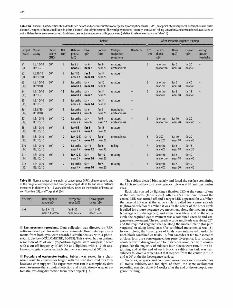
Table 1A Clinical Characteristics of children tested before and after reeducation of vergence by orthoptic exercises. NPC (near point of convergence), heterophoria (in prismdiopters), vergence fusion amplitude (in prism diopters) clinically measured. The vertigo symptoms (rotatory, translation rolling sensations and unsteadiness) associated ornot with headache are also reported. Bold characters indicate abnormal orthoptic values (relative to references shown in Table 1B)
Before After orthoptic vergence training
Subject Visual Stereo NPC Hetero- Diver Conver Vertigo: Headache NPC Hetero- Diver Conver Vertigo(years) acuity acuity (cm) phoria (pD) (pD) subjective (cm) phoria (pD) (pD) and/or
(TNO) (pD) sensations (pD) headache
S1 LE: 10/10 60” 6 far 2 E far 6 far 6 rotatory, 6 far ortho far 6 far 30 –(6) RE: 10/10 near 6 E near 6 near 20 unsteadiness near ortho near 18 near 40
S2 LE:10:10 60” 6 far 1 E far 3 far 14 rotatory(8) RE: 10:10 near 1 X near 10 near 20
S3 LE: 10/10 60” 6 far ortho far 4 far 10 rotatory 6 far ortho far 6 far 40 –(10) RE: 9/10 near 8 X near 10 near 30 near 2 X near 18 near 40
S4 LE: 10/10 60” 15 far ortho far 4 far 14 rotatory + 6 far ortho far 8 far 18 –(11) RE: 10/10 near 8 X near 6 near 20 near 4 X near 18 near 40
S5 LE: 10/10 60” 6 far ortho far 4 far 14 rotatory +(11) RE: 10/10 near 2 X near 14 near 18
S6 LE: 8/10 60” 6 far ortho far 6 far 8 translation, +(12) RE: 8/10 near 8 X near 8 near 20 unsteadiness
S7 LE: 10/10 60” 10 far ortho far 4 far 6 rotatory, + 6 far ortho far 10 far 20(12) RE: 10/10 near 2 X near 6 near 10 unsteadiness near ortho near 20 near 40 –
S8 LE: 10/10 60” 6 far 4 E far 1 far 18 rotatory +(12) RE: 10/10 near 2 X near 4 near 20
S9 LE: 10/10 60” 10 far 10 X far 10 far 6 unsteadiness 6 far 2 X far 10 far 20 –(12) RE: 10/10 near 6 X near 12 near 20 near 2 X near 18 near 40
S10 LE: 10/10 60” 10 far ortho far 14 far 6 rolling + 7 far ortho far 8 far 18 –(14) RE: 10/10 near 4 X near 12 near 30 near 4 X near 18 near 30
S11 LE: 10/10 60” 6 far 12 X far 4 far 10 rotatory 4 far ortho far 6 far 40 –(14) RE: 10/10 near 6 X near 14 near 26 near ortho near 18 near 40
S12 LE: 10/10 60” 10 far ortho far 4 far 4 rotatory + 6 far ortho far 4 far 40 –(15) RE: 10/10 near 4 X near 10 near 26 near 4 X near 20 near 40
Table 1B Normal values of near point of convergence (NPC), of heterophoria andof the range of convergence and divergence amplitude at far and close distancemeasured in children of 6–15 years old; values based on the studies of Evans [8],von Noorden [29], and Ygge et al. [34]
NPC (cm) Heterophoria Divergence Convergencerange (pD) range (pD) range (pD)
< 6 far 3 X-1 E far 4–10 far 11–17near 6 X-ortho near 17–25 near 15–27
204-213_Bucci_JON-1304 21.01.2004 11:19 Uhr Seite 206

207
Data analysis
The analysis methods are similar to those used in prior studies [17,33]. Briefly, a linear function was used to calibrate the individual eyeposition signals. From these two signals, we calculated the saccadesignal [(left eye + right eye)/2] and the vergence signal (left eye – righteye).Fig. 1 shows an example of pure rightward saccade (A),pure con-vergence movement (B) and a saccade movement combined with con-vergence (C). For each type of eye movements the saccade or conju-gate component and the vergence or disconjugate components areshown. For pure convergence (Fig. 1B) the position trace from the in-dividual eye which gives information about the symmetry of the re-sponse from the two eyes have been added.
Markers indicating the start of the movement were placed on eacheye position signal automatically, and were verified by an investiga-tor. The criteria used are standard: the onset of the conjugate saccadiccomponent was defined as the time when the eye velocity reached 5 %of the saccadic peak velocity. The onset of the vergence signals (forpure vergence movements and for the vergence component of thecombined movements) were defined as the time point when the eyevelocity exceeded 5 %. Eye movements that were in wrong directionor contaminated by blinks were rejected. Also, eye movements withlatencies shorter than 100 ms or longer than 1000 ms were rejected.Note that the percentage of the rejected movements before trainingwas between 10 %–40 % for pure vergence movements, 6 %–34 % forcombined movements and 0 %–9 % for pure saccades; after trainingthe percentage of rejected movements was between 0 %–34 % for purevergence, 0 %–13 % for combined movements and 0 %–5 % for pure
saccades. After training an increase of the percentage of the move-ments analysed was observed for subjects S1, S3 and S10, but not forsubjects S4, S5, S6, S12.
For each type of eye movement (saccade, vergence and combinedmovements) we measured the latency in ms, that is the time betweenthe onset of the target-LED and the beginning of the movements. Forcombined movements latency was measured for each component(saccade, vergence component). Analysis of variance (ANOVA) wasperformed with subject as a random factor and the latency of diffe-rent types of eye movements as a fixed factor.The group mean latencyof each type of eye movements before and after the orthoptic ver-gence training was compared with the mean latency values from nor-mal subjects of comparable age extracted from the study of Yang et al.[33]. Statistical comparison was done by using the Student’s t-test.
Results
■ Pure saccades and vergence movements
Fig. 2A shows the individual mean latency for pure sac-cades at far and close viewing distance. For all subjects(except S4 and S5) the latency was shorter for the sac-cades at near distance. The group mean latency was
Fig. 1 Examples of eye movements. Onthe right side is shown the excursion ofthe target. The saccade or conjugate sig-nal is the average of the position signalof the two eyes [(LE + RE/2)]; the ver-gence or disconjugate signal is the dif-ference between the two signals (LE –RE). For pure convergence, the positiontrace of each eye is also showed. The tar-get-LED appeared at time zero. The ar-rows indicate the onset of the move-ment. (A) Pure rightward saccade; (B)Convergence movement along the me-dian plane; (C) Rightward saccade com-bined with convergence. Note that thesaccade component starts before thevergence component
204-213_Bucci_JON-1304 21.01.2004 11:19 Uhr Seite 207
208
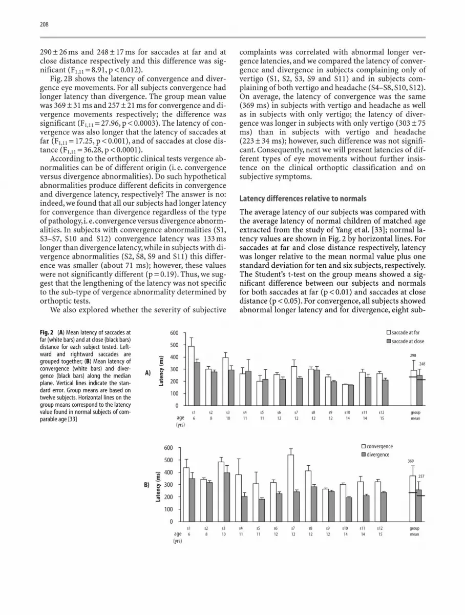
290 ± 26 ms and 248 ± 17 ms for saccades at far and atclose distance respectively and this difference was sig-nificant (F1,11 = 8.91, p < 0.012).
Fig. 2B shows the latency of convergence and diver-gence eye movements. For all subjects convergence hadlonger latency than divergence. The group mean valuewas 369 ± 31 ms and 257 ± 21 ms for convergence and di-vergence movements respectively; the difference wassignificant (F1,11 = 27.96, p < 0.0003). The latency of con-vergence was also longer that the latency of saccades atfar (F1,11 = 17.25, p < 0.001), and of saccades at close dis-tance (F1,11 = 36.28, p < 0.0001).
According to the orthoptic clinical tests vergence ab-normalities can be of different origin (i. e. convergenceversus divergence abnormalities). Do such hypotheticalabnormalities produce different deficits in convergenceand divergence latency, respectively? The answer is no:indeed, we found that all our subjects had longer latencyfor convergence than divergence regardless of the typeof pathology,i. e.convergence versus divergence abnorm-alities. In subjects with convergence abnormalities (S1,S3–S7, S10 and S12) convergence latency was 133 mslonger than divergence latency,while in subjects with di-vergence abnormalities (S2, S8, S9 and S11) this differ-ence was smaller (about 71 ms); however, these valueswere not significantly different (p = 0.19). Thus, we sug-gest that the lengthening of the latency was not specificto the sub-type of vergence abnormality determined byorthoptic tests.
We also explored whether the severity of subjective
complaints was correlated with abnormal longer ver-gence latencies, and we compared the latency of conver-gence and divergence in subjects complaining only ofvertigo (S1, S2, S3, S9 and S11) and in subjects com-plaining of both vertigo and headache (S4–S8, S10, S12).On average, the latency of convergence was the same(369 ms) in subjects with vertigo and headache as wellas in subjects with only vertigo; the latency of diver-gence was longer in subjects with only vertigo (303 ± 75ms) than in subjects with vertigo and headache(223 ± 34 ms); however, such difference was not signifi-cant. Consequently, next we will present latencies of dif-ferent types of eye movements without further insis-tence on the clinical orthoptic classification and onsubjective symptoms.
Latency differences relative to normals
The average latency of our subjects was compared withthe average latency of normal children of matched ageextracted from the study of Yang et al. [33]; normal la-tency values are shown in Fig. 2 by horizontal lines. Forsaccades at far and close distance respectively, latencywas longer relative to the mean normal value plus onestandard deviation for ten and six subjects, respectively.The Student’s t-test on the group means showed a sig-nificant difference between our subjects and normalsfor both saccades at far (p < 0.01) and saccades at closedistance (p < 0.05). For convergence, all subjects showedabnormal longer latency and for divergence, eight sub-
Fig. 2 (A) Mean latency of saccades atfar (white bars) and at close (black bars)distance for each subject tested. Left-ward and rightward saccades aregrouped together; (B) Mean latency ofconvergence (white bars) and diver-gence (black bars) along the medianplane. Vertical lines indicate the stan-dard error. Group means are based ontwelve subjects. Horizontal lines on thegroup means correspond to the latencyvalue found in normal subjects of com-parable age [33]
204-213_Bucci_JON-1304 21.01.2004 11:19 Uhr Seite 208
209
jects had latency longer than normal values. Similarly tosaccades, the group means of convergence latency aswell as divergence latency were significantly differentfrom those of normals (p < 0.01).
■ Combined saccade – vergence eye movements
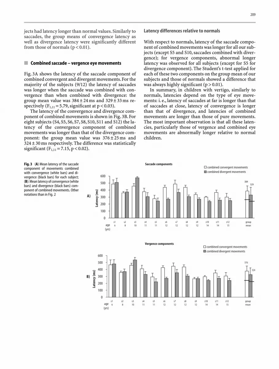
Fig. 3A shows the latency of the saccade component ofcombined convergent and divergent movements.For themajority of the subjects (9/12) the latency of saccadeswas longer when the saccade was combined with con-vergence than when combined with divergence: thegroup mean value was 384 ± 24 ms and 329 ± 33 ms re-spectively (F1,11 = 5.79, significant at p < 0.03).
The latency of the convergence and divergence com-ponent of combined movements is shown in Fig. 3B. Foreight subjects (S4, S5, S6, S7, S8, S10, S11 and S12) the la-tency of the convergence component of combinedmovements was longer than that of the divergence com-ponent: the group mean value was 376 ± 25 ms and324 ± 30 ms respectively. The difference was statisticallysignificant (F1,11 = 7.15, p < 0.02).
Latency differences relative to normals
With respect to normals, latency of the saccade compo-nent of combined movements was longer for all our sub-jects (except S5 and S10, saccades combined with diver-gence); for vergence components, abnormal longerlatency was observed for all subjects (except for S5 fordivergence component). The Student’s t-test applied foreach of these two components on the group mean of oursubjects and those of normals showed a difference thatwas always highly significant (p > 0.01).
In summary, in children with vertigo, similarly tonormals, latencies depend on the type of eye move-ments: i. e., latency of saccades at far is longer than thatof saccades at close, latency of convergence is longerthan that of divergence, and latencies of combinedmovements are longer than those of pure movements.The most important observation is that all these laten-cies, particularly those of vergence and combined eyemovements are abnormally longer relative to normalchildren.
Fig. 3 (A) Mean latency of the saccadecomponent of movements combinedwith convergence (white bars) and di-vergence (black bars) for each subject;(B): Mean latency of convergence (whitebars) and divergence (black bars) com-ponent of combined movements. Othernotations than in Fig. 2
204-213_Bucci_JON-1304 21.01.2004 11:19 Uhr Seite 209

210
■ Additional observation: effect of orthopticreeducation
Effects on vestibular and orthoptic tests (see Table 1A, After training)
All eight subjects did not feel vertigo after training.Moreover, the range of vergence improved for all sub-jects. Indeed, the near point of convergence that beforetraining was 9 ± 3 cm reduced to 5 ± 0.8 cm after train-ing; the exophoria at close viewing that was before train-ing 6 ± 2.4 pD reduced to 1.7 ± 1.6 pD after training. Fi-nally, the convergence amplitude at close distanceimproved: from 21 ± 7 pD before training to 39 ± 3 pD af-ter training.
Effect on latency
Fig. 4 shows the group means of latency before and afterorthoptic vergence training for saccades at far and closedistance (A), for convergence and divergence (B), andfor the saccades (C) and the vergence components (D) ofcombined movements, respectively. Before training, themean latency value of all these types of movements was
significantly longer (t-test significant at p < 0.01) thanthe mean latency observed in normals (showed by thehorizontal line on the bar graphs).
After training the mean latency was shortened: forsaccades at far distance the ANOVA applied on the meanlatencies before and after orthoptic vergence trainingtends to be significant (F1,7 = 5.33, p > 0.057); in contrast,for saccades at close distance that reduction was not sig-nificant (F1,7 = 1.25, p = 0.25). Note, however, that aftertraining, the average latency of both saccades at eitherdistance was not different from normal values (t-test,p = 0.05).
The training reduced substantially the latency ofconvergence by 140 ms (F1,7 = 10.65, p < 0.01), while thelatency of divergence decreased by a small amount (only9 ms); the change was not significantly different(F1,7 = 0.42, p = 0.54). Importantly, the mean latency ofboth convergence and divergence observed after thetraining (see Fig. 4B) was still significantly longer thannormal values (t-test, significant at p < 0.01).
After training, the latency of the saccade as well as ofthe vergence components of combined movements de-creased significantly (Fig. 4C and 4D).The effect was sig-nificant for both saccades combined with convergence
Fig. 4 Group mean (on eight subjects)of the latency before and after orthopticvergence training for saccades at far andclose distance (A), for convergence anddivergence (B), and for combined move-ments (saccade components C and ver-gence components D). Vertical lines in-dicate the standard error. A significantbefore-after training change in the la-tency value is shown by an asterisk. Thehorizontal lines indicate latency valuesrecorded in normal subjects of matchedage [33]
204-213_Bucci_JON-1304 21.01.2004 11:19 Uhr Seite 210
211
and divergence (F1,7 = 20.79, p < 0.003 and F1,7 = 6.21,p < 0.02, respectively), and for both, convergence anddivergence components (F1,7 = 23.16, p < 0.002 andF1,7 = 8.97, p < 0.02, respectively). Note, however, that alllatencies of combined movements after training were stilllonger than normal values (t-test, significant at p < 0.01).
In summary, the orthoptic vergence training short-ened the latency of all eye movements, but the decreasewas significant only for convergence and combinedmovements. Importantly, after training the latency ofsaccades at far and close distance was similar to that ofnormals; in contrast, the latency of vergence and com-bined movements did not reach normal values.
Discussion
■ Latency differences between types of eye movements
As in normals, in children with vertigo, latencies of var-ious types of eye movements were different: the latencyof saccades at close was shorter than that of saccades atfar distance; the latency of convergence movements waslonger with respect to that of divergence but also withrespect to all types of saccades (at far and close dis-tance). Combined movements also showed long latency,particularly when combined with convergence; the la-tency of both saccade and vergence components of com-bined movements were longer than those of the corre-sponding pure movements. These results will be brieflydiscussed below.
Saccades latency: effect of distance
The shorter latency of saccades at near distance has beenobserved also in another study on normal children andadults using the same experimental conditions [33] andis compatible with other studies examining the far [11]or the close distance [27].As suggested by Yang et al. [33]the shorter latency at close distance could be due to sen-sory, motor or attentional factors.
Interpretation of the different latencies observedbetween convergence and divergence
Longer latency for convergence than for divergencemovements was also observed in normal adults by Kri-shan et al. [18] and in some subjects by Takagi et al. [27];however, other studies [15, 25] showed opposite results.These differences could be due to different experimen-tal procedures used. More convincing is the study ofYang et al. [33] who showed systematically longer la-tency for convergence for both normal children andadults using the same experimental conditions of thepresent study. Our findings for subjects with vertigo arecompatible with this study [33].
Such latency difference between convergence and di-vergence suggests different neuro-physiologicalprocesses involved in the preparation of these move-ments. The cortical substrate of vergence in humans isnot yet well explored. Hasebe et al. [13] showed with aPET study activation in the parietal and temporo-occip-ital cortex in relation to vergence eye movements;Kapoula et al. [17] showed that transcranial magneticstimulation of the posterior parietal cortex lengthenedthe latency of both saccades as well as vergence eyemovements. More importantly,Tzelepi et al. [28] showedhigher amplitude of EEG responses in the parietal areasfor stimuli calling for convergence than for divergenceor for saccades. All these observations suggest that sev-eral cortical areas control the triggering of vergence,andthat the cortical circuitry for the initiation of conver-gence and divergence movements seems to be not ab-solutely identical.
Combined movements: longer latency
The increased latency of combined movements with re-spect to that of pure movements could be due to the ad-ditional time needed to prepare two or more movementsrather than one. This interpretation is compatible withother studies examining sequences of saccades report-ing increase of latency as the number of the targets in-creases [3].
■ Differences of latencies from normal population
As mentioned in Methods, we compared the data in chil-dren with vertigo with those of children without disor-ders tested under identical conditions [33]. The latencyof saccades at far and close distance was 50 ms and 10 msrespectively longer in our subjects than in normal chil-dren of comparable age. More interesting is that the la-tency of vergence was 145 ms and 50 ms (for conver-gence and divergence, respectively) longer than those ofnormals. The latency of the saccade component of com-bined movements was 115 ms longer than normal; fi-nally, the latency of convergence and divergence compo-nent of combined movements was 130 ms and 110 mslonger than that of normal children.
Thus, the movements that have naturally longerlatencies are the ones that are more severely affected insubjects with vertigo. In general, for normal subjects,the longer is the latency of a movement, the most voli-tional the movement is believed to be [10]; on the otherhand, in patients with cortical lesions latencies increasebecause of cortical dysfunction [21]. Thus, our findingssuggest the presence of a central deficit in the initiationof eye movements, particularly for convergence andcombined movements, which are, perhaps, the mostvolitional movements.
204-213_Bucci_JON-1304 21.01.2004 11:19 Uhr Seite 211

212
■ Effect of orthoptic vergence training
Orthoptic vergence training is widely used by cliniciansto treat vergence abnormalities; moreover, it is wellknown [e. g., 6, 7, 12, 19] that even normal subjects canbenefit from orthoptic training: indeed these authorspointed out in normal subjects improvement of ver-gence performance after orthoptic training. Note alsothat for normals, even if the training procedure was sim-ilar for all subjects, the improvement due to the trainingwas variable between the subjects [19].
Nevertheless, objective eye movement studies show-ing the effect of the orthoptic training are rather scarce.The study by van Leeuwen et al. [19] is, to our knowl-edge, the only one showing an increase, after orthopticvergence training, of the amplitude of vergence (pureand combined with saccades) in normal subjects as wellas in one subject with convergence insufficiency.
Our findings show, for the first time, that orthopticvergence training can: (i) eliminate vertigo; (ii) improvevergence capabilities clinically assessed; (iii) shorten la-tency of eye movements. The decrease of the latency wassignificant for the movements, which before traininghad the longer latency, i. e., pure convergence eye move-ments and both components of combined movements.
As mentioned in the Introduction, latency reflectsseveral processes, such as disengagement of attention, ofoculomotor fixation, and computation of the metric ofthe movement [9, 10]. Thus, shortening of the latencycould be mediated by shortening of the time needed byone or several of these sub-processes. Namely, atten-tional factors could be sensitive and modulated by train-ing or by visual experience. Our data do not allow us todetermine if the latency decrease is due to the orthoptictraining itself or to spontaneous improvement related tonatural visual experience. Our findings are compatiblewith the study of Fischer [10] who reported in a normalsubject significant decrease of saccade latency aftertraining; the training consisted of a repetition of 200saccades per day for a period of 10 days. The author sug-gested that training helped the subject to disengage theattention more rapidly. Consequently, we conclude thatnormal subjects as well as subjects with vergence ab-normalities can benefit by the training and that atten-tion can be responsible for the decrease of eye move-ment’s latencies.
Finally, one could argue that latencies in childrenwith vertigo before training were simply extreme values,that is, a little standard deviation from normals, and thattraining brought these values back to normals. This,however, was not the case.As we pointed out, despite thereduction of latencies for all types of eye movements af-ter training, latencies remained still longer than those ofnormals, particularly for vergence and for combinedmovements (see Fig. 4). Hence we suggest that childrenwith vertigo have a specific problem with the initiation
of vergence and combined movements. This deficit canbe reduced by attentional learning mechanism but can-not be eliminated completely.
■ Vergence initiation abnormalities and vertigo
During head movements or body motion, the vestibulo-ocular system generates compensatory eye movementsthat stabilize images on the retina. Several electrophysi-ological and behavioral studies in the last ten years haveshown that the responses generated by the vestibulo-oc-ular reflex (VOR) are a complex function of head move-ment and target distance [20]. The modulation of thegain of the vestibular system by the viewing distance isan aspect integrated in most recent models of vestibularocular function. The CNS uses knowledge (propriocep-tive input or corollary discharge related to vergence an-gle) to adjust the gain of the VOR as a function of theviewing distance. Importantly, Snyder et al. [26] have re-ported that during vergence eye movements the changeof the gain of the VOR anticipates the vergence, whichsuggests the possibility for predictive influence of ver-gence on the VOR system. Coenen [5] proposed a modelin which the cerebellum could construct by learning apredictive vergence input to control the VOR gain. Be-cause of the abnormally longer vergence latency in oursubjects the contribution to the VOR adjustment couldbe reduced or inappropriate. Consequently, we suggestthat inappropriate vestibular function relative to dis-tance could lead to vertigo and headache as reported byBrandt [4]. However, this hypothesis should be con-firmed further by quantitative studies examining thelink between the quality of vergence control, namely theslowness of its initiation, and the gain of the VOR at dif-ferent viewing distances. This field is to our knowledgeunexplored both in normals and in subjects with ver-tigo.
■ Clinical considerations
The study of Anon-Tanon et al. [1] suggested for the firsttime that vertigo is associated with abnormal results insubjective orthoptic evaluation of vergence. Our studysubstantiates this report as it shows abnormal vergencelatency in such subjects. The two studies are compatibleand indicate that orthoptic testing of vergence is valu-able even though it cannot provide temporal informa-tion about eye movements. Objective recording of eyemovements together with clinical orthoptic tests is themost useful approach for a fine diagnosis and treatmentof children with vertigo.
Finally, it should be noted that for a long time, ver-gence abnormalities have been thought to be related toperipheral muscular deficiency only [29]. Our study
204-213_Bucci_JON-1304 21.01.2004 11:19 Uhr Seite 212

213
suggests that vergence abnormalities such as slownessof vergence initiation are most likely due to dysfunctionof the cortical circuitry that controls the initiation ofsuch movements.
■ Acknowledgements This study was supported by INSERM (con-tract n° 4M105E) and by MRT (Cognitive Dysfonctionment). M. P.
Bucci was supported by EDF and FRM; Q. Yang was supported byMENRT. The authors thank the orthoptist F. Afkami for conductingorthoptic examination and H. Puech (optometrist, Atol Les Opti-ciens) for her helpful discussion on clinical characteristics of thesubjects. The authors are particularly grateful to Dr. G. A. Oliva andher department of Cibernetica e Biofisica (CNR of Genoa, Italy) forlending the Dr. Bouis Oculometer to the Parisian group.
References
1. Anoh-Tanon MJ, Brémond-Gignac D,Wiener-Vacher S (2000) Vertigo is anunderstimated symptom of ocular dis-orders: dizzy children do not alwaysneed MRI. Pediatr Neurol 23:49–53
2. Atkinson J, Anker S, Nardini M, Brad-dick O, Hughes C, Rae S, Wattam-Bell J,Atkinson S (2002) Infant vision screen-ing predicts failures on motor and cog-nitive tests up to school age. Strabis-mus 10:187–198
3. Becker W (1991) Saccades. Eye Move-ments. Vision and visual dysfunction.RHS Carpenter. 2nd edition, London,Lion, pp 112–117
4. Brandt T (1999) Vertigo. Its multisen-sory syndromes. 2nd ed Springer
5. Coenen OJMD (1998) Modeling theVestibulo-Ocular Reflex and the Cere-bellum: Analytical & ComputationalApproaches. PhD Dissertation. Univer-sity of California, San Diego, PhysicsDepartment
6. Daum KM (1982) The course and ef-fect of visual training on the vergencesystem. Am J Optomet Physiol Optics59:223–227
7. Daum KM (1984) Classification crite-rion for success in the treatment ofconvergence insufficiency. Am J Op-tomet Physiol Optics 61:10–15
8. Evans BJW (1997) Pickwell’s binocularvision anomalies: investigation andtreatment. Butterworth-Heinemann,Oxford
9. Findlay JM, Walker R (1999) A frame-work for saccadic eye movement con-trol based on parallel processing andcompetitive inhibition. Behav Brain Sci22:661–721
10. Fischer B (1987) The preparation ofvisually-guided saccades. Rev PhysiolBiochem Pharmacol 106:1–35
11. Fukushima J, Hatta T, Fukushima K(2002) Development of voluntary con-trol of saccadic eye movements I. Age-related changes in normal children.Brain Dev 22:173–180
12. Griffin JR (1987) Efficacy of visiontherapy for nonstrabismic vergenceanomalies. Am J Optomet PhysiolOptics 64:411–414
13. Hasebe H, Oyamada H, Kinomura Set al. (1999) Human cortical areas acti-vated in relation to vergence eye move-ments – a PET study. Neuroimage 10:200–208
14. Howard IP, Rogers BI (2002) Seeing indepth. Porteous I (ed) Toronto press
15. Hung GK, Zhu HM, Ciuffreda KJ(1997) Convergence and divergenceexhibit different response characteris-tics to symmetric stimuli. Vision Res37:1197–1205
16. Jones R, Stephen GL (1989) Horizontalfusional amplitudes. Invest Ophthal-mol Vis Sci 30:1638–1642
17. Kapoula Z, Isotalo E, Müri RM, BucciMP, Rivaud-Péchoux S (2001) Effectsof transcranial magnetic of the poste-rior parietal cortex on saccades andvergence. Neuroreport 12:4041–4046
18. Krishnan VV, Farazian F, Stark L(1973) An analysis of latencies andprediction in the fusional vergencesystem. Am J Optom Arch Am AcadOptom 50:933–939
19. Leeuwen AF, Westen MJ, van der SteenJ, de Faber JTHN, Collewijn H (1999)Gaze-shift dynamics in subjects withand without symptoms of convergenceinsufficiency: influence of monocularpreference and the effect of training.Vision Res 39:3095–3107
20. Leigh RJ, Zee DS (1999) The Neurologyof Eye Movements 3rd ed New York:Oxford University Press
21. Pierrot-Deseilligny C, Rivaud S, Gay-mard B, Müri R, Vermersch AI (1995)Cortical control of saccades. Ann Neu-rol 37:557–567
22. Rainey BB (2002) V 755: Angle of devi-ation. Clinical characteristics and diag-nostic considerations. The Trustees ofIndiana University. www.indiana.edu
23. Rouse MW, Borsting E, Hyman L et al.(1999) Frequency of convergence in-sufficiency among fifth and sixgraders. Optom Vis Sci 76:643–649
24. Rouse MW, Hyman L, Hussen M et al.(1998) Frequency of convergence in-sufficiency in optometry clinic setting.Optom Vis Sci 75:88–96
25. Semmlow JL, Wetzel P (1979) Dynamiccontributions of the components ofbinocular vergence. J Opt Soc Am A69:639–645
26. Snyder LH, Lawrence DM, King WM(1992) Changes in the vestibulo-ocularreflex (VOR) anticipate changes in ver-gence angle in monkey. Vision Res32(3):569–575
27. Takagi M, Frohman EM, Zee DS (1995)Gap-overlap effects on latencies of sac-cades, vergence and combined ver-gence-saccades in humans. Vision Res35:3373–3388
28. Tzelepi A, Kapoula Z, Luts A (2002)EEG stimulus locked activity preced-ing saccades, vergence and combinedeye movements. Society of Neuro-sciences, Orlando, USA
29. von Noorden GK (2002) Binocular vi-sion and ocular motility. Theory andmanagement of strabismus. 6th ed. St.Luis, Mosby
30. Wiener-Vacher SR, Amanou L, DeniseP, Narcy P, Manach Y (1999) Vestibularfunction in children with the CHARGEassociation. Arch Otolaryngol HeadNeck Surg 125:342–347
31. Wiener-Vacher SR, Mazda K (1998)Asymmetric otolith vestibulo-ocularresponses in children with idiopathicscoliosis. J Pediatr 132:1028–1032
32. Wiener-Vacher S, Toupet F, Narcy P(1996) Canal and otolith vestibulo-oc-ular reflexes to vertical and off verticalaxis rotations in children learning towalk. Acta Otolaryngol (Stockh) 116:657–665
33. Yang Q, Bucci MP, Kapoula Z (2002)The latency of saccades, vergence andcombined movements in children andin adults. Investigative Ophthalmol VisSci 43:2939–2949
34. Ygge J, Lennerstrand G, Rydberg A,Wijecoon S, Petterson BM (1993) Ocu-lomotor functions in a Swedish popu-lation of dyslexic and normally read-ing children. Acta Ophthalmol 71:10–21
204-213_Bucci_JON-1304 21.01.2004 11:19 Uhr Seite 213