a thionine-modified carbon paste amperometric biosensor for catechol and bisphenol a determination
TRANSCRIPT

Ab
MNa
b
c
a
ARRAA
KCBTET
1
mcpsiprdh(ri
Rnae
C
0d
Biosensors and Bioelectronics 25 (2010) 2003–2008
Contents lists available at ScienceDirect
Biosensors and Bioelectronics
journa l homepage: www.e lsev ier .com/ locate /b ios
thionine-modified carbon paste amperometric biosensor for catechol andisphenol A determination
. Portaccioa,b, D. Di Tuoroa,b, F. Arduinib,c, M. Leporea,b, D.G. Mitaa,b,∗,. Dianoa,b, L. Mitaa,b, D. Mosconeb,c
Dipartimento di Medicina Sperimentale, Seconda Università di Napoli, Naples, ItalyConsorzio Interuniversitario Biostrutture e Biosistemi “INBB”, Viale Medaglie d’oro,305 Roma, ItalyDipartimento di Scienze e Tecnologie Chimiche, Università di Tor Vergata, Roma, Italy
r t i c l e i n f o
rticle history:eceived 13 November 2009eceived in revised form 12 January 2010ccepted 21 January 2010vailable online 1 February 2010
a b s t r a c t
A thionine-modified carbon paste electrode for catechol and Bisphenol A (BPA) detection is presented.Graphite powder was modified by adsorbing thionine as electrochemical mediator. The electrochemicalresponse of the modified carbon paste electrode (CPE) was determined before electrode modification withtyrosinase. Then, tyrosinase was added in order to assemble a biosensor. Once established the best oper-ative conditions, an interelectrode reproducibility around 7% was obtained and the resulting biosensor
eywords:atecholisphenol Ayrosinasendocrine disruptorhionine
showed improved sensitivities and (S = 139.6 ± 1.1 nA/�M for catechol and S = 85.4 ± 1.5 nA/�M for BPA)in comparison with the biosensor constructed without thionine (S = 104.4 ± 0.5 nA/�M for catechol andS = 51.1 ± 0.6 nA/�M for BPA) and low detection limits (0.15 �M for both the electrodes and analytes). Alsothe comparison with the results reported in the literature showed higher sensitivity and lower detectionlimit for our biosensor. Moreover the functioning of the thionine-tyrosinase CPE was validated followinga biodegradation process of water polluted by BPA and comparing the time changes of BPA concentration
calib
inferred by the biosensor. Introduction
Concerns are growing among the public and the scientific com-unity owing to the awareness that a group of chemicals, known
ollectively as endocrine disrupting compounds (EDCs), are sus-ected of interfering with the normal function of the endocrineystem causing adverse effects for humans and wildlife. EDCsnclude a range of synthetic estrogens, pesticides, plasticizers andhenol compounds (Colborn et al., 1996). Concerns have beenaised, in particular, about the contribution of alkylphenols (break-own products of detergent alkylphenol polyethoxylates) whichave been found in drinking water supplies and waste streamsBruchet and Janex-Habibi, 2004). These chemicals have phenolicings, a structure also found in natural estrogens. A similar structures present also in Bisphenol A (BPA) and catechol.
Bisphenol A [BPA, 2,2-(4,4-dihydroxydiphenyl) propane; CAS
egistry No. 80-05-7] is produced by combining acetone and phe-ol and is an important organic chemical owing to its wide uses an intermediate in the manufacture of polycarbonate plastics,poxy resins, and flame retardants (Staples et al., 1998; Mutou et∗ Corresponding author at: Institute of Genetics and Biophysics of CNR, Via Pietroastellino, 111-80131, Naples, Italy. Tel.: +39 81 6132608; fax: +39 81 6132608.
E-mail address: [email protected] (D.G. Mita).
956-5663/$ – see front matter © 2010 Elsevier B.V. All rights reserved.oi:10.1016/j.bios.2010.01.025
ration curve and those determined by means of HPLC measurements.© 2010 Elsevier B.V. All rights reserved.
al., 2006). Its main use is for coating of cans, powder paints, dentalfillings, and antioxidant in plastics. It has also been used as an inertingredient in pesticides, antioxidants and polyvinyl chloride stabi-lizer (Staples et al., 1998; Rodriguez-Mozaz et al., 2005). BPA showsestrogenic potential both in vitro and in vivo (Matthews et al., 2000;Segner et al., 2003) and also acute toxicity towards aquatic organ-isms (Staples and Davis, 2002; Suzuki and Hattori, 2003) and humancultured cells (Nakagawa et al., 2000) In particular it has been foundthat BPA binds to the estrogen receptor (Matthews et al., 2001)produces meiotic aneuploidy in the female mouse (Hunt et al.,2003) and increases cell proliferation in the male and female sex-ual organs. Additionally, although still controversial, BPA may eveninduce a decrease of sperm quality in humans (Maffini et al., 2006).
BPA pollution occurs owing to its release from many commodi-ties, such as the inside cans coating, polycarbonate baby bottles, andplastic waste, as well as accidental spills (Fukazawa et al., 2001;Fromme et al., 2002; Sajiki and Yonekubo, 2004; Maragou et al.,2006).
The results cited above suggest that it is necessary to removeBPA from the environment and to determine its presence also in
trace.Concerning the first priority, we have demonstrated thatit is possible, through enzymatic processes carried out withnon-isothermal bioreactors, to remove BPA and other phenolcompounds from the aquatic environment (Diano et al., 2007;
2 d Bioe
Gopbobs
iterocisecapbAr
dLnhImoot
bnotbeiattp(aod
cTbirwo
2
2
btv
004 M. Portaccio et al. / Biosensors an
eorgieva et al., 2008; Mita et al., 2009). The success in the removalf BPA has been demonstrated not only analytically through high-erformance liquid chromatography (HPLC) determination but alsoy means of the disappearance, after enzyme treatment, of somef the biological effects previously observed in the absence ofioremediation (Ricupito et al., 2009). These last experiments alsohowed that the enzyme reaction products were not estrogenic.
Concerning the second priority, i.e. the necessity of determin-ng BPA even in trace, this is currently done using classic analyticalechniques such as gas chromatography (GC) and HPLC (Watabet al., 2004; Sun et al., 2006). Immunochemical techniques wereecently reported (Ballesteros-Gomez et al., 2009) using mon-clonal and polyclonal mammalian (Ohkuma et al., 2002) andhicken (De Meulenaer et al., 2002) antibodies in enzyme linkedmmunosorbent assays (ELISA’s). Mass spectrometry is also con-idered a useful technique. However, all these technologies arexpensive and time-consuming. Electrochemical biosensors, on theontrary, represent a rapid and less expensive method and have thedvantage of high sensitivity, potential for miniaturization, and theossibility of in situ analysis (Rogers et al., 1999). The use of car-on paste as electrode material was initially reported in 1958 bydams (Adams, 1958). Carbon paste offers high versatility, surfaceenewability, low noise, low background currents, and low cost.
Tyrosinase-modified carbon paste electrodes have beenescribed earlier (Marko-Varga et al., 1995; Lutz et al., 1995;indgren et al., 1996; Petit et al., 1996; Wang et al., 1997). Tyrosi-ase is a copper containing metalloprotein which catalyses theydroxylation and oxidation of mono and diphenols to o-quinones.
t can exist as a mono and diphenolase (Hedenmo et al., 1997). Theono form can be inactive (met) or active (oxy) and the formation
f a significant amount of the active form depends on the presencef catechol. When phenol is used as substrate, only a small part ofhe enzyme is active due to the absence of the diphenol ‘activator’.
In this paper we report the results obtained with tyrosinaseased carbon paste electrode modified with the addition of thio-ine as a mediator. Thionine is an artificial organic dye derivativef phenothiazine, and it is used as a mediator since its formal poten-ial is between 0.08 and −0.25 V, near the redox potential of manyiomolecules (Shobha Jeykumari et al., 2007). Thionine was firstmployed for the electrocatalytic oxidation of NADH by crosslink-ng it onto a graphite electrode (Ohtani et al., 1997; Hajizadeh etl., 1991). Cosnier (Cosnier et al., 2001) reported the mediated elec-rochemical detection of catechol using thionine covalently boundo a poly(dicarbazole) backbone. Thionine has also been used asolythionine by means of electropolymerisation of the monomerYang et al., 1999; Liu et al., 2008). Later on, Dempsey (Dempsey etl., 2004) developed a biosensor using thionine electropolymerisedn a glassy carbon electrode in the presence of tyrosinase for theetection of synthetic estrogens (BPA) and phenolic compounds.
In the present work thionine was used in the construction of aarbon paste electrode after its adsorption onto the carbon powder.he carbon paste was then modified with tyrosinase. Finally, theiosensor was then challenged in amperometric mode. Linear cal-
bration curves have been obtained both for BPA and catechol. Ouresults have been compared with those obtained by other authorsith and without thionine, and with thionine electropolymerised
n glassy carbon electrode.
. Materials and methods
.1. Materials
Tyrosinase (E.C. 1.14.18.1, 2033 Units/mg) from mushrooms haseen used as enzyme. It must be noted that the specific activity ofyrosinase here employed is lower than that we used in our pre-ious work (Mita et al., 2007). It was not possible to find anymore
lectronics 25 (2010) 2003–2008
tyrosinase with higher specific activity. It is known that this fact cancause a lower response and thus problems in the reproducibilityand in the comparison among different catalytic system (Rescignoet al., 2007).
Catechol and BPA were used as substrate representative of phe-nolic compounds and endocrine disruptors, respectively. Carbonpowder (<0.1 mm in size) was purchased from Fluka. The mineraloil (cod. 69794), employed as pasting material, catechol, BPA and allthe other chemicals (of pure pro-analysis grade), were purchasedfrom Sigma (Sigma–Aldrich, Milan, Italy) and used without furtherpurification.
2.2. Methods
Carbon paste was prepared by hand mixing graphite powder,thionine (when it was the case), mineral oil and tyrosinase in anappropriate weight ratio. The resulting pastes were packed intothe well of the working electrode as described below. The body ofthe working electrode was a Teflon tube (3 mm in diameter) withan electrical contact provided by a copper wire. The working elec-trodes were prepared by packing the Teflon tube with a doublelayer of carbon paste: an inner layer containing only graphite pow-der mixed to mineral oil, and an outer layer composed of carbonpaste, tyrosinase and thionine (when indicated). The inner layer(about 2/3 of total volume) was prepared by mixing 60% of graphitepowder and 40% of mineral oil, for a total weight of 4.8 mg. Theouter layer (about 1/3 of the total volume) with a total weight of2.5 mg, was prepared by a carbon paste obtained from two steps.During the first step, 600 mg of graphite powder are mixed for10 min with 1 mL of thionine (1 mM or 10 mM), and the thionine-modified powder was put in an oven at 60 ◦C for about 15 h. In thesecond step, the thionine/graphite powder was mixed with min-eral oil and tyrosinase in the ratio (w/w) of 50:40:10, respectively.For each electrode we used about 0.25 mg of tyrosinase which cor-respond to about 500 U. To prepare thionine free electrode wefollowed only the second step, mixing graphite powder, mineral oiland enzyme in the ratio before used. The electrode surface exposedto the solution was polished on weighing paper to give a smoothfinish before use. Amperometric measurements were carried outusing a PalmSens instrument (Palm Instruments, the Netherlands)coupled to a PC. All experiments were carried out in a three elec-trode electrochemical cell with a volume of 10 mL (0.1 M sodiumphosphate, pH 6.5, containing 0.1 M KCl) with the enzyme modi-fied CPE as working electrode, an Ag/AgCl electrode as referenceelectrode and a platinum wire as auxiliary electrode. The workingelectrode was operated at −200 mV vs. Ag/AgCl and the transientcurrents were allowed to decay to a steady-state value. A magneticstirrer and a stirring bar provided the convective transport in theelectrolytic cell. All measurements were performed at room tem-perature (25.0 ± 0.5 ◦C). Each experimental point in Figs. 4 and 5represent the average of five experiments carried out under thesame conditions. The experimental error never exceeded 4.6%.
3. Results and discussion
3.1. Cyclic voltammetry measurements
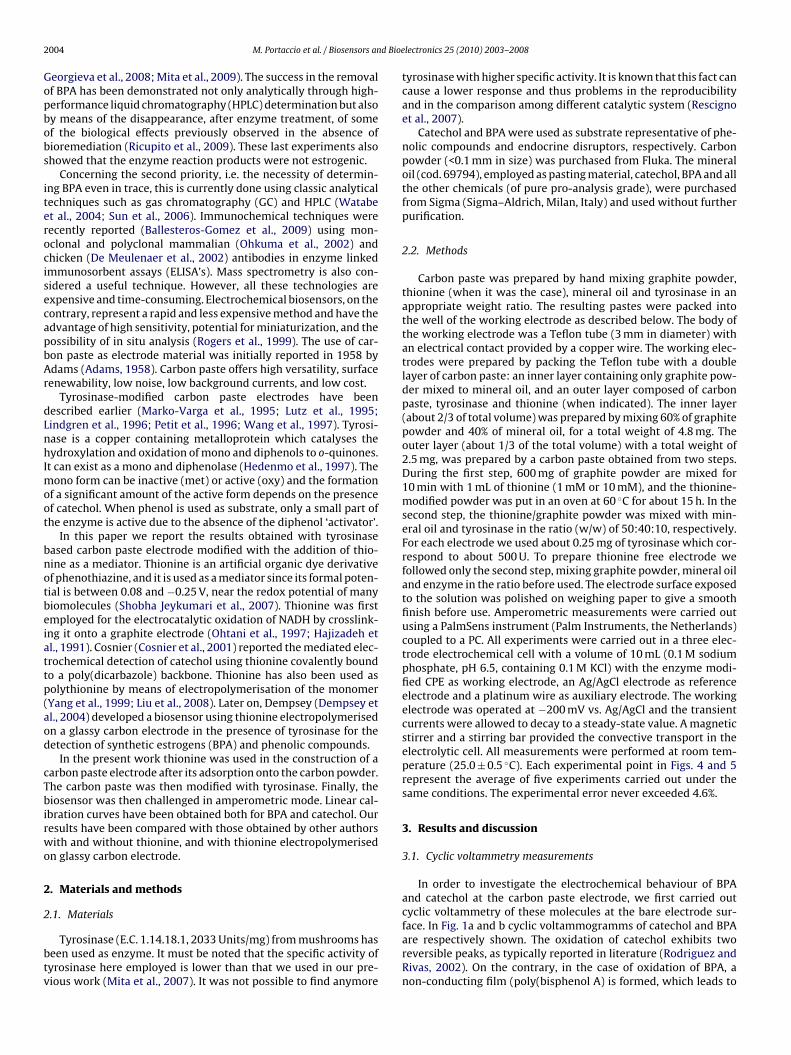
In order to investigate the electrochemical behaviour of BPAand catechol at the carbon paste electrode, we first carried outcyclic voltammetry of these molecules at the bare electrode sur-
face. In Fig. 1a and b cyclic voltammogramms of catechol and BPAare respectively shown. The oxidation of catechol exhibits tworeversible peaks, as typically reported in literature (Rodriguez andRivas, 2002). On the contrary, in the case of oxidation of BPA, anon-conducting film (poly(bisphenol A) is formed, which leads to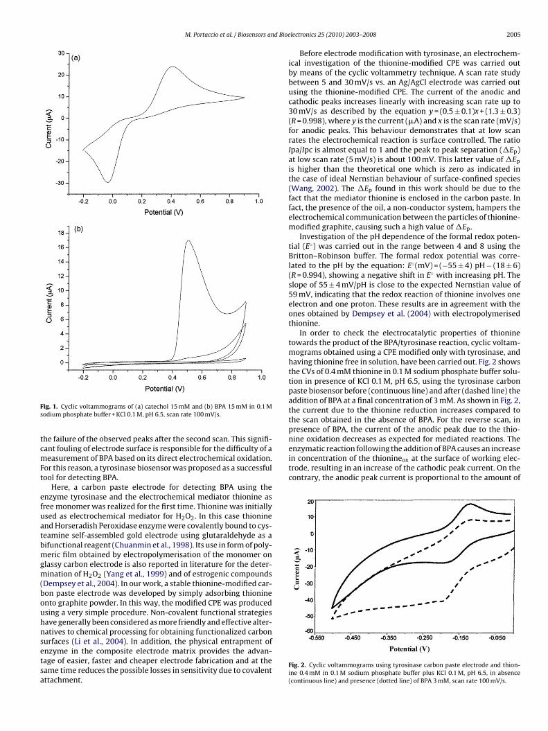
M. Portaccio et al. / Biosensors and Bioe
Fs
tcmFt
efuatbmgm(bouhnsetsa
nine oxidation decreases as expected for mediated reactions. Theenzymatic reaction following the addition of BPA causes an increasein concentration of the thionineox at the surface of working elec-trode, resulting in an increase of the cathodic peak current. On thecontrary, the anodic peak current is proportional to the amount of
ig. 1. Cyclic voltammograms of (a) catechol 15 mM and (b) BPA 15 mM in 0.1 Modium phosphate buffer + KCl 0.1 M, pH 6.5, scan rate 100 mV/s.
he failure of the observed peaks after the second scan. This signifi-ant fouling of electrode surface is responsible for the difficulty of aeasurement of BPA based on its direct electrochemical oxidation.
or this reason, a tyrosinase biosensor was proposed as a successfulool for detecting BPA.
Here, a carbon paste electrode for detecting BPA using thenzyme tyrosinase and the electrochemical mediator thionine asree monomer was realized for the first time. Thionine was initiallysed as electrochemical mediator for H2O2. In this case thioninend Horseradish Peroxidase enzyme were covalently bound to cys-eamine self-assembled gold electrode using glutaraldehyde as aifunctional reagent (Chuanmin et al., 1998). Its use in form of poly-eric film obtained by electropolymerisation of the monomer on
lassy carbon electrode is also reported in literature for the deter-ination of H2O2 (Yang et al., 1999) and of estrogenic compounds
Dempsey et al., 2004). In our work, a stable thionine-modified car-on paste electrode was developed by simply adsorbing thioninento graphite powder. In this way, the modified CPE was producedsing a very simple procedure. Non-covalent functional strategiesave generally been considered as more friendly and effective alter-atives to chemical processing for obtaining functionalized carbonurfaces (Li et al., 2004). In addition, the physical entrapment of
nzyme in the composite electrode matrix provides the advan-age of easier, faster and cheaper electrode fabrication and at theame time reduces the possible losses in sensitivity due to covalentttachment.lectronics 25 (2010) 2003–2008 2005
Before electrode modification with tyrosinase, an electrochem-ical investigation of the thionine-modified CPE was carried outby means of the cyclic voltammetry technique. A scan rate studybetween 5 and 30 mV/s vs. an Ag/AgCl electrode was carried outusing the thionine-modified CPE. The current of the anodic andcathodic peaks increases linearly with increasing scan rate up to30 mV/s as described by the equation y = (0.5 ± 0.1)x + (1.3 ± 0.3)(R = 0.998), where y is the current (�A) and x is the scan rate (mV/s)for anodic peaks. This behaviour demonstrates that at low scanrates the electrochemical reaction is surface controlled. The ratioIpa/Ipc is almost equal to 1 and the peak to peak separation (�Ep)at low scan rate (5 mV/s) is about 100 mV. This latter value of �Ep
is higher than the theoretical one which is zero as indicated inthe case of ideal Nernstian behaviour of surface-confined species(Wang, 2002). The �Ep found in this work should be due to thefact that the mediator thionine is enclosed in the carbon paste. Infact, the presence of the oil, a non-conductor system, hampers theelectrochemical communication between the particles of thionine-modified graphite, causing such a high value of �Ep.
Investigation of the pH dependence of the formal redox poten-tial (E◦) was carried out in the range between 4 and 8 using theBritton–Robinson buffer. The formal redox potential was corre-lated to the pH by the equation: E◦(mV) = (−55 ± 4) pH − (18 ± 6)(R = 0.994), showing a negative shift in E◦ with increasing pH. Theslope of 55 ± 4 mV/pH is close to the expected Nernstian value of59 mV, indicating that the redox reaction of thionine involves oneelectron and one proton. These results are in agreement with theones obtained by Dempsey et al. (2004) with electropolymerisedthionine.
In order to check the electrocatalytic properties of thioninetowards the product of the BPA/tyrosinase reaction, cyclic voltam-mograms obtained using a CPE modified only with tyrosinase, andhaving thionine free in solution, have been carried out. Fig. 2 showsthe CVs of 0.4 mM thionine in 0.1 M sodium phosphate buffer solu-tion in presence of KCl 0.1 M, pH 6.5, using the tyrosinase carbonpaste biosensor before (continuous line) and after (dashed line) theaddition of BPA at a final concentration of 3 mM. As shown in Fig. 2,the current due to the thionine reduction increases compared tothe scan obtained in the absence of BPA. For the reverse scan, inpresence of BPA, the current of the anodic peak due to the thio-
Fig. 2. Cyclic voltammograms using tyrosinase carbon paste electrode and thion-ine 0.4 mM in 0.1 M sodium phosphate buffer plus KCl 0.1 M, pH 6.5, in absence(continuous line) and presence (dotted line) of BPA 3 mM, scan rate 100 mV/s.
2006 M. Portaccio et al. / Biosensors and Bioelectronics 25 (2010) 2003–2008
Fa1s
ttto
3
tbt
tdtsFjatttt5saApiitp1
uI
tmnaht
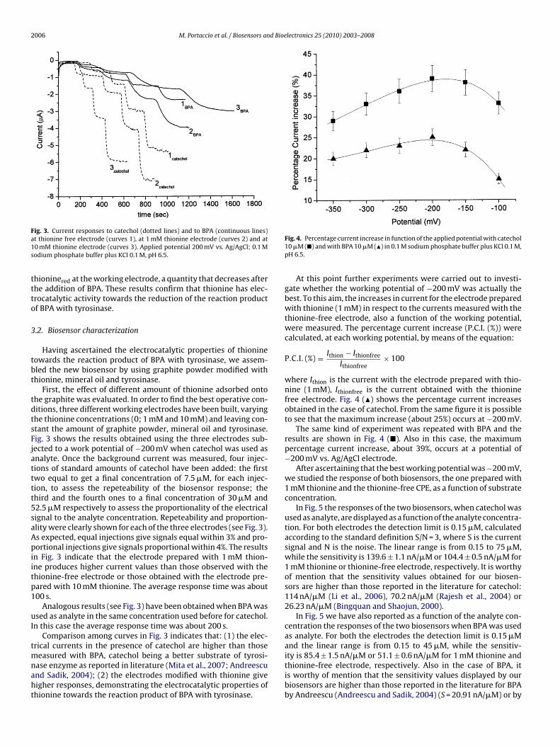
ig. 3. Current responses to catechol (dotted lines) and to BPA (continuous lines)t thionine free electrode (curves 1), at 1 mM thionine electrode (curves 2) and at0 mM thionine electrode (curves 3). Applied potential 200 mV vs. Ag/AgCl; 0.1 Modium phosphate buffer plus KCl 0.1 M, pH 6.5.
hioninered at the working electrode, a quantity that decreases afterhe addition of BPA. These results confirm that thionine has elec-rocatalytic activity towards the reduction of the reaction productf BPA with tyrosinase.
.2. Biosensor characterization
Having ascertained the electrocatalytic properties of thionineowards the reaction product of BPA with tyrosinase, we assem-led the new biosensor by using graphite powder modified withhionine, mineral oil and tyrosinase.
First, the effect of different amount of thionine adsorbed ontohe graphite was evaluated. In order to find the best operative con-itions, three different working electrodes have been built, varyinghe thionine concentrations (0; 1 mM and 10 mM) and leaving con-tant the amount of graphite powder, mineral oil and tyrosinase.ig. 3 shows the results obtained using the three electrodes sub-ected to a work potential of −200 mV when catechol was used asnalyte. Once the background current was measured, four injec-ions of standard amounts of catechol have been added: the firstwo equal to get a final concentration of 7.5 �M, for each injec-ion, to assess the repeteability of the biosensor response; thehird and the fourth ones to a final concentration of 30 �M and2.5 �M respectively to assess the proportionality of the electricalignal to the analyte concentration. Repeteability and proportion-lity were clearly shown for each of the three electrodes (see Fig. 3).s expected, equal injections give signals equal within 3% and pro-ortional injections give signals proportional within 4%. The results
n Fig. 3 indicate that the electrode prepared with 1 mM thion-ne produces higher current values than those observed with thehionine-free electrode or those obtained with the electrode pre-ared with 10 mM thionine. The average response time was about00 s.
Analogous results (see Fig. 3) have been obtained when BPA wassed as analyte in the same concentration used before for catechol.
n this case the average response time was about 200 s.Comparison among curves in Fig. 3 indicates that: (1) the elec-
rical currents in the presence of catechol are higher than those
easured with BPA, catechol being a better substrate of tyrosi-ase enzyme as reported in literature (Mita et al., 2007; Andreescund Sadik, 2004); (2) the electrodes modified with thionine giveigher responses, demonstrating the electrocatalytic properties ofhionine towards the reaction product of BPA with tyrosinase.
Fig. 4. Percentage current increase in function of the applied potential with catechol10 �M (�) and with BPA 10 �M (�) in 0.1 M sodium phosphate buffer plus KCl 0.1 M,pH 6.5.
At this point further experiments were carried out to investi-gate whether the working potential of −200 mV was actually thebest. To this aim, the increases in current for the electrode preparedwith thionine (1 mM) in respect to the currents measured with thethionine-free electrode, also a function of the working potential,were measured. The percentage current increase (P.C.I. (%)) werecalculated, at each working potential, by means of the equation:
P.C.I. (%) = Ithion − Ithionfree
Ithionfree× 100
where Ithion is the current with the electrode prepared with thio-nine (1 mM), Ithionfree is the current obtained with the thioninefree electrode. Fig. 4 (�) shows the percentage current increasesobtained in the case of catechol. From the same figure it is possibleto see that the maximum increase (about 25%) occurs at −200 mV.
The same kind of experiment was repeated with BPA and theresults are shown in Fig. 4 (�). Also in this case, the maximumpercentage current increase, about 39%, occurs at a potential of−200 mV vs. Ag/AgCl electrode.
After ascertaining that the best working potential was −200 mV,we studied the response of both biosensors, the one prepared with1 mM thionine and the thionine-free CPE, as a function of substrateconcentration.
In Fig. 5 the responses of the two biosensors, when catechol wasused as analyte, are displayed as a function of the analyte concentra-tion. For both electrodes the detection limit is 0.15 �M, calculatedaccording to the standard definition S/N = 3, where S is the currentsignal and N is the noise. The linear range is from 0.15 to 75 �M,while the sensitivity is 139.6 ± 1.1 nA/�M or 104.4 ± 0.5 nA/�M for1 mM thionine or thionine-free electrode, respectively. It is worthyof mention that the sensitivity values obtained for our biosen-sors are higher than those reported in the literature for catechol:114 nA/�M (Li et al., 2006), 70.2 nA/�M (Rajesh et al., 2004) or26.23 nA/�M (Bingquan and Shaojun, 2000).
In Fig. 5 we have also reported as a function of the analyte con-centration the responses of the two biosensors when BPA was usedas analyte. For both the electrodes the detection limit is 0.15 �Mand the linear range is from 0.15 to 45 �M, while the sensitiv-
ity is 85.4 ± 1.5 nA/�M or 51.1 ± 0.6 nA/�M for 1 mM thionine andthionine-free electrode, respectively. Also in the case of BPA, itis worthy of mention that the sensitivity values displayed by ourbiosensors are higher than those reported in the literature for BPAby Andreescu (Andreescu and Sadik, 2004) (S = 20.91 nA/�M) or by
M. Portaccio et al. / Biosensors and Bioe
F1Ap
Dti2
w
fwr
wiptietdbf1t
Fvm
ig. 5. Calibration curves obtained at thionine free electrode (full symbols) or atmM thionine electrode (empty symbols) for catechol (�,�) and for BPA (�,�).pplied potential 200 mV vs. Ag/AgCl; 0.1 M sodium phosphate buffer plus KCl 0.1 M,H 6.5.
empsey (Dempsey et al., 2004) (S = 0.4 nA/�M), who also usedhionine as electrochemical mediator or also than those reportedn the literature for phenol compounds: 17.1 nA/�M (Rajesh et al.,004) or 2.42 nA/�M (Bingquan and Shaojun, 2000).
The intraelectrode repeatability never exceeded the 2% (n = 5)hile the interelectrode reproducibility was around 7%.
Concerning the stability, the carbon paste electrode was stableor about one month of working. The electrodes were discardedhen their response to a catechol solution of 5 �M decreased 7%
elative to the initial one.Before concluding a significant experiment is described, in
hich the real functioning of our thionine-based biosensor is val-dated during a bioremediation process of an aqueous solutionolluted by BPA. 10 mL of 1 mM BPA aqueous solution were allowedo react with a membrane on which laccase was immobilized. Themmobilization method can be found in a previous paper (Mitat al., 2009). At zero time 200 �l were withdrawn from the reac-ion vessel for the subsequent measurements. 100 �L of those areiluted in 10 mL of buffer solution and put in contact with the
iosensor, while 25 �L were processed in the HPLC. The procedureor HPLC assay is described in the same paper above indicated. Every0 min the same operation was repeated until 100 min. In Fig. 6he average results of five experiments are reported. Data in fig-ig. 6. Time dependence of biosensor response (continuous line, right scale) andalidation of the response through the analysis of BPA concentrations (left scale) byeans of HPLC (©) and the calibration curve (�) of Fig. 5.
lectronics 25 (2010) 2003–2008 2007
ure show: (i) the time decrease of the current intensity measuredby the biosensor during the bioremediation process (continuousline and right scale); (ii) the time dependence of BPA concentration(left scale) as measured by HPLC (©) and as obtained by the cali-bration curve reported in Fig. 5 (�). It is interesting to note how: (i)a reduction of 29% in the current value corresponds to a reductionof 25% in the concentration value; and (ii) the BPA concentrationvalues measured by HPLC are very similar to those obtained fromthe calibration curve. The conclusion is that the functioning of ourthionine-tyrosinase based biosensor appears excellent.
4. Conclusions
This paper reports the simple and effective preparation of a car-bon paste biosensor for catechol and BPA determination. It wasprepared by using thionine as mediator and the enzyme tyrosinaseas bioactive element. The thionine adsorption onto the graphitepowder turned out to be a simple way to obtain a stable thionine-modified CPE. In addition, the use of thionine as monomer in thetyrosinase carbon paste biosensor seems to improve the analyticalperformances with respect to those exhibited by the glassy carbonpaste modified with electropolymerised thionine and tyrosinase(Dempsey et al., 2004). In the latter case, in fact, lower sensitivity(0.4 nA/�M) and higher detection limit (23 �M) were observed forBPA. This behaviour can probably be ascribed to the better solubilityof BPA in a hydrophobic system such as the one with oil present inthe CPE. The oil, in fact, seems to play a significant role as reportedin our previous paper (Mita et al., 2007). In that paper we havefound with reference to the BPA graphite carbon powder biosen-sor the following characteristics: linear range from 0.1 to 15 �M;S = 68 ± 4 nA/�M; and detection limit = 0.1 �M. In the present work,thanks to thionine, we have further improved our BPA graphitecarbon powder biosensor having found: linear range from 0.15 to45 �M; S = 85.4 ± 1.5 nA/�M; and detection limit = 0.15 �M. In thisway we have demonstrated that the combination of graphite pow-der, oil and thionine as mediator can be a useful system for BPA andcatechol detection when tyrosinase is used as bioactive element.
Moreover, it is possible to improve the performance of ourbiosensor, for example, by using an enzyme with higher specificactivity. Studies in this direction are being carried out in our labo-ratories.
Acknowledgments
This work has been partially supported by the “Istituto Superioreper la Prevenzione e Sicurezza sul Lavoro” (ISPESL) in the frame ofthe Strategic Project on “Interferenti Endocrini: valutazione del ris-chio e degli effetti”–convenzione n. PMS/40/06/P2/UO7 and underthe National Strategic Project “Salute della donna” and “Innovativeapproaches in the evaluation and prevention of the food exposureto contaminating toxic persistent and emergent, through the studyof the diet and the debugging of innovative methods of survey”supported by Ministry of Health.
References
Adams, R.N., 1958. Anal. Chem. 30, 1576.Andreescu, S., Sadik, O.M., 2004. Anal. Chem. 76 (3), 552–560.Ballesteros-Gomez, A., Rubio, S., Perez-Bendito, D., 2009. J. Chromatogr. A 1216,
449–469.Bingquan, W., Shaojun, D., 2000. J. Electroanal. Chem. 487, 45–50.Bruchet, A., Janex-Habibi, M.L., 2004. Tech. Sci. Methods 4, 81–90.Chuanmin, R., Ru, Y., Xiaohong, C., Jiaqi, D., 1998. J. Electroanal. Chem. 455, 121–125.
Colborn, T., Dumenoski, D., Uyers, J.P., 1996. Our Stolen Future. Book AD, New York.Cosnier, S., Szunerits, S., Marks, R.S., Lellouche, J.P., Perie, K., 2001. J. Biochem. Bio-phys. Methods 50 (1), 65–77.De Meulenaer, B., Baert, K., Lanckriet, H., Van Hoed, V., Huyghebaert, A., 2002. J. Agric.
Food Chem. 50, 5273–5282.Dempsey, E., Diamond, D., Collier, E., 2004. Biosens. Bioelectron. 20, 367–377.
2 d Bioe
D
F
F
G
HH
H
L
LL
L
L
M
M
M
M
MM
M
008 M. Portaccio et al. / Biosensors an
iano, N., Grano, V., Fraconte, L., Caputo, P., Ricupito, A., Attanasio, A., Bianco, M.,Bencivenga, U., Rossi, S., Manco, I., Mita, L., Del Pozzo, G., Mita, D.G., 2007. Appl.Catal. 69, 252–261.
romme, H., Küchler, T., Otto, T., Pilz, K., Müller, J., Wenzel, A., 2002. Water Res. 36(6), 1429–1438.
ukazawa, H., Hoshino, K., Shiozawa, T., Matsushita, H., Terao, Y., 2001. Chemosphere44 (5), 973–979.
eorgieva, S., Godjevargova, T., Portaccio, M., Mita, D.G., 2008. J. Mol. Catal. B: Enzym.55, 177–184.
ajizadeh, K., Halsall, H.B., Heineman, W.R., 1991. Anal. Chim. Acta 243, 23–32.edenmo, M., Narváez, A., Domínguez, E., Katakis, I., 1997. J. Electroanal. Chem. 425
(1–2), 1–11.unt, P.A., Koehler, K.E., Susiarjo, M., Hodges, C.A., Ilagan, A., Voigt, R.C., Thomas, S.,
Thomas, B.F., Hassold, T.J., 2003. Curr. Biol. 13, 546–553.i, Y.F., Liu, Z.M., Liu, Y.L., Yang, Y.H., Shen, G.L., Yu, R.Q., 2006. Anal. Biochem. 349,
33–40.i, Q., Zhang, J., Yan, H., He, M., Liu, Z., 2004. Carbon 42, 537–545.indgren, A., Ruzgas, T., Emneus, J., Csoregi, E., Gorton, L., Marko-Varga, G., 1996.
Anal. Lett. 29, 1055–1068.iu, H., Wang, G., Chen, D., Zhang, W., Li, C., Fang, B., 2008. Sens. Actuators B 128,
414–421.utz, M., Burestedt, E., Emneus, J., Liden, H., Gobhadi, S., Gorton, L., Marko-Varga, G.,
1995. Anal. Chim. Acta 305, 8–17.affini, M.V., Rubin, B.S., Sonnenschein, C., Soto, A.M., 2006. Mol. Cell. Endocrinol.
254–255, 179–186.aragou, N.C., Lampi, E.N., Thomaidis, N.S., Koupparis, M.A., 2006. J. Chromatogr. A
1129 (2), 165–173.arko-Varga, G., Emneus, J., Gorton, L., Ruzgas, T., 1995. TrAC Trends Anal. Chem.
14, 319–328.atthews, J., Celius, T., Halgren, R., Zacharewski, T., 2000. J. Steroid Biochem. 74 (4),
223–234.
atthews, J.B., Twomey, K., Zacharewski, T.R., 2001. Chem. Res. Toxicol. 14, 223–234.ita, D.G., Attanasio, A., Arduini, F., Diano, N., Grano, V., Bencivenga, U., Rossi, S.,Amine, A., Moscone, D., 2007. Biosens. Bioelectron. 23, 60–65.ita, D.G., Diano, N., Grano, V., Portaccio, M., Rossi, S., Bencivenga, U., Manco, I.,
Nicolucci, C., Bianco, M., Grimaldi, T., Mita, L., Georgieva, S., Godjevargova, T.,2009. J. Mol. Catal. B: Enzym. 58, 199–207.
lectronics 25 (2010) 2003–2008
Mutou, Y., Ibuki, Y., Terao, Y., Kojima, S., Goto, R., 2006. Environ. Toxicol. Pharmacol.21 (3), 283–289.
Nakagawa, Y., Suzuki, T., Tayama, S., 2000. Toxicology 156 (1), 27–36.Ohtani, M., Kuwabata, S., Yoneyama, H., 1997. J. Electroanal. Chem. 422 (1–2),
45–54.Ohkuma, H., Abe, K., Ito, M., Kokado, A., Kambegawa, A., Maeda, M., 2002. Analyst
127, 93–97.Petit, C., Gonzalez-Cortes, A., Kauffmann, J.M., 1996. Bioelectrochem. Bioenergy 41,
101–106.Rajesh, Takashima, W., Kaneto, K., 2004. React. Funct. Polym. 59, 163–169.Rescigno, A., Zucca, P., Flurkey, A., Inlow, J., Flurkey, W.H., 2007. Enzyme Microb.
Technol. 41, 620–627.Ricupito, A., Del Pozzo, G., Diano, N., Grano, V., Portaccio, M., Marino, M., Bolli, A.,
Galluzzi, P., Bontempo, P., Mita, L., Altucci, L., Mita, D.G., 2009. Environ. Int. 35,21–26.
Rodriguez, M.C., Rivas, G.A., 2002. Anal. Chim. Acta 459, 43–51.Rodriguez-Mozaz, S., Lopez de Alda, M., Barcelo, D., 2005. Water Res. 39, 5071–5079.Rogers, K.R., Becker, J.Y., Wang, J., Lu, F., 1999. Field Anal. Chem. Technol. 3, 161–169.Sajiki, J., Yonekubo, J., 2004. Chemosphere 55 (6), 861–867.Segner, H., Navas, J.M., Schäfers, C., Wenzel, A., 2003. Ecotoxicol. Environ. Saf. 54 (3),
315–322.Shobha Jeykumari, D.R., Ramaprabhu, S., Sriman Narayanan, S., 2007. Carbon 45,
1340–1353.Staples, C.A., Davis, J.W., 2002. Chemosphere 49 (1), 61–73.Staples, C.A., Dome, P.B., Klecka, G.M., Oblock, S.T., Harris, L.R., 1998. Chemosphere
36 (10), 2149–2173.Sun, C., Leong, L.P., Barlow, P.J., Chan, S.H., Bloodworth, B.C., 2006. J. Chromatogr. A
1129, 145–148.Suzuki, N., Hattori, A., 2003. Life Sci. 73 (17), 2237–2247.Wang, J., Lu, F., Kane, S.A., Choi, Y.K., Smyth, M.R., Rogers, K., 1997. Electroanalysis
9, 1102–1106.
Wang, J., 2002. Analytical Electrochemistry, second ed. John Wiley and Sons Inc.,New York.Watabe, Y., Kondo, T., Morita, M., Tanaka, N., Haginaka, J., Hosoya, K., 2004. J. Chro-
matogr. A 1032, 45–49.Yang, R., Ruan, C.M., Dai, W.L., Deng, J.Q., Kong, J.L., 1999. Electrochim. Acta 44,
1585–1596.