detection of hydrogen peroxide in photosystem ii using catalytic amperometric biosensor
TRANSCRIPT

METHODSpublished: 15 October 2015
doi: 10.3389/fpls.2015.00862
Frontiers in Plant Science | www.frontiersin.org 1 October 2015 | Volume 6 | Article 862
Edited by:
Roger Deal,
Emory University, USA
Reviewed by:
Maha Afifi,
California Table Grape Commission,
USA
Franz-Josef Schmitt,
Technical University of Berlin,
Germany
Agnese Magnani,
Università of Siena, Italy
*Correspondence:
Ankush Prasad
†Co-corresponding author.
Specialty section:
This article was submitted to
Technical Advances in Plant Science,
a section of the journal
Frontiers in Plant Science
Received: 27 May 2015
Accepted: 29 September 2015
Published: 15 October 2015
Citation:
Prasad A, Kumar A, Suzuki M,
Kikuchi H, Sugai T, Kobayashi M,
Pospíšil P, Tada M and Kasai S (2015)
Detection of hydrogen peroxide in
Photosystem II (PSII) using catalytic
amperometric biosensor.
Front. Plant Sci. 6:862.
doi: 10.3389/fpls.2015.00862
Detection of hydrogen peroxide inPhotosystem II (PSII) using catalyticamperometric biosensorAnkush Prasad 1*, Aditya Kumar 2, Makoto Suzuki 3, Hiroyuki Kikuchi 3, Tomoya Sugai 3,
Masaki Kobayashi 1, 4, Pavel Pospíšil 2, Mika Tada 1, 5 and Shigenobu Kasai 1, 3 †
1 Biomedical Engineering Research Center, Tohoku Institute of Technology, Sendai, Japan, 2Department of Biophysics,
Faculty of Science, Centre of the Region Haná for Biotechnological and Agricultural Research, Palacký University, Olomouc,
Czech Republic, 3Graduate Department of Environmental Information Engineering, Tohoku Institute of Technology, Sendai,
Japan, 4Graduate Department of Electronics, Tohoku Institute of Technology, Sendai, Japan, 5Center for General Education,
Tohoku Institute of Technology, Sendai, Japan
Hydrogen peroxide (H2O2) is known to be generated in Photosystem II (PSII) via
enzymatic and non-enzymatic pathways. Detection of H2O2 by different spectroscopic
techniques has been explored, however its sensitive detection has always been a
challenge in photosynthetic research. During the recent past, fluorescence probes
such as Amplex Red (AR) has been used but is known to either lack specificity or
limitation with respect to the minimum detection limit of H2O2. We have employed an
electrochemical biosensor for real time monitoring of H2O2 generation at the level of
sub-cellular organelles. The electrochemical biosensor comprises of counter electrode
and working electrodes. The counter electrode is a platinum plate, while the working
electrode is a mediator based catalytic amperometric biosensor device developed by
the coating of a carbon electrode with osmium-horseradish peroxidase which acts
as H2O2 detection sensor. In the current study, generation and kinetic behavior of
H2O2 in PSII membranes have been studied under light illumination. Electrochemical
detection of H2O2 using the catalytic amperometric biosensor device is claimed to
serve as a promising technique for detection of H2O2 in photosynthetic cells and
subcellular structures including PSII or thylakoid membranes. It can also provide a
precise information on qualitative determination of H2O2 and thus can be widely used in
photosynthetic research.
Keywords: photosystem II, superoxide anion radical, hydrogen peroxide, reactive oxygen species, amperometric
biosensor, EPR-spin trapping
INTRODUCTION
Photosystem II (PSII) is a multi-subunit pigment-protein complex which is located in the thylakoidmembrane of chloroplasts of cyanobacteria, algae and higher plants that comprises of morethan 25 proteins and the concomitant cofactors (Ferreira et al., 2004; Loll et al., 2005; Guskovet al., 2009; Kawakami et al., 2011). In plants, photosynthesis has been considered as a sourceof reactive oxygen species (ROS) production which works in close association with regulatedmechanism of antioxidant network under normal conditions. The ROS in plants are known
Prasad et al. Hydrogen peroxide in Photosystem II
to be involved in cell toxicity, defense and signaling, and havebeen recently overviewed (Krieger-Liszkay, 2005; Foyer andShigeoka, 2011; Schmitt et al., 2014).
Chlorophyll pigments of the PSII antenna complex absorblight energy and use it for the oxidation of water moleculesand reduction of plastoquinone. Light energy absorbed bychlorophyll pigments converted into the energy of separatedcharges and consequent water-plastoquinone oxidoreductaseactivity is involuntarily linked with the production of ROS(Pospíšil, 2009, 2012). In unison, released molecular oxygenserves as a forerunner of ROS, which at low concentration play animportant role in cell regulation, whereas if formed in excess, isresponsible for oxidation of biomolecules such as lipid, proteins,and nucleic acid (Halliwell and Gutteridge, 2007). In addition,direct oxidation of proteins and lipids by UV irradiation andtoxic chemicals following subsequent chemical reactions are alsoknown to be associated with formation of ROS (Halliwell andGutteridge, 2007; Prasad and Pospíšil, 2012).
Production of ROS arises when excitation energy transfer tothe PSII reaction center is inadequate or there is inhibition ofelectron transport chain in PSII. The redox couples in PSII coversa broad range of redox potential, it ranges from a very highnegative value of redox couple, Pheo/Pheo− (Em = −610mV)to a very high positive value of redox couple P680+/P680 (Em =
+1250mV) (Rappaport and Diner, 2008; Pospíšil, 2012). PSII iscapable of either oxidizing water molecule or reducing molecularoxygen on the electron donor and on the electron acceptor sideof the membrane, respectively (Pospíšil, 2012). There is leakageof electrons to molecular oxygen during the electron transporton the electron acceptor side of PSII (Pospíšil, 2009).
Formation of O•−
2 results from non-enzymatic and enzymaticone-electron reductions of molecular oxygen. Pheophytin(Pheo•−) (Ananyev et al., 1994; Pospíšil et al., 2004), tightlybound plastosemiquinone (Q•−
A ), (Cleland and Grace, 1999;Pospíšil et al., 2004), loosely bound plastosemiquinone(Q•−
B ), (Zhang et al., 2003; Yadav et al., 2014), and freeplastosemiquinone (PQ•−) (Mubarakshina and Ivanov, 2010)maintains non-enzymatic reduction of molecular oxygen toO•−
2 . Heme iron of low-potential (LP) form of cyt b559 reducesmolecular oxygen to O•−
2 in the enzymatic reaction pathway(Pospíšil et al., 2006; Pospišil, 2011).
One electron reduction of O•−
2 either via non-enzymaticor enzymatic reaction pathway results in the formation ofH2O2. In spontaneous dismutation, O•−
2 provides an additionalelectron to another O•−
2 , and then with protonation bringsabout the formation of H2O2. In enzymatic dismutation,the ferrous heme iron of HP form of cyt b559 drives thecatalysis of one-electron reduction of HO•
2 to H2O2 (Tiwariand Pospísil, 2009; Pospišil, 2011). Spontaneous dismutationis preferred where there is availability of protons e.g., at themembrane edge while PSII metal centers are chosen to catalyzethe dismutation reaction in the interior of the membrane(Pospíšil, 2012). The one-electron reduction of O•−
2 to H2O2
is catalyzed by superoxide dismutase (SOD) and is known tooccur predominantly in the mitochondria, peroxisomes, andcytoplasm. At the physiological pH, the dismutation reaction ispreferably catalyzed by SOD.
Several spectroscopic techniques (fluorescence andchemiluminescence) and chromatographic techniques (highperformance liquid chromatography coupled with peroxyoxalatechemiluminescence detection) have been used in the past forthe determination of H2O2 in living cells (Mills et al., 2007;Chen et al., 2009; Ahammad, 2013). Light induced productionof H2O2 have been measured in PSII membranes by oxidation ofthiobenzamide with lactoperoxidase. Thiobenzamide sulfoxidewas quantified by its absorbance at 370 nm (Schröder andÅkerlund, 1990; Arató et al., 2004; Pospíšil et al., 2004).Production of H2O2 by chloroplasts have been measured bythe AmplexRed fluorescence assays (Mubarakshina and Ivanov,2010; Yadav and Pospíšil, 2012). Hydrogen peroxide (H2O2)detecting probes, 3,3 diaminobenzidine (DAB), Amplex Red(AR), Amplex Ultra Red (AUR), and a europium-tetracyclinecomplex (Eu3Tc) have been compared by infiltrating intotobacco leaves and tested for sensitivity to light, toxicity,subcellular localization, and capacity to detect H2O2 in vivo(Šnyrychová et al., 2009). The induction of H2O2 generation atthe leaf level after 3-acetyl-4-hydroxyl-5-isopropylpyrroline-2-dione (3-AIPTA) or bentazon treatment has been detected byperforming histochemical analysis with 3, 3 DAB staining(Chen et al., 2012). Apart from several spectroscopictechniques (absorbance, fluorescence, chemiluminescence),cyclic voltammetry and histochemical technique, a reportersystem based on HSP70A promoter-luciferase fusions have alsobeen developed in past for the detection of H2O2 in vivo (Shaoet al., 2007).
In the electrochemical method, mediator and non-mediatorbased electrode have been used in the past. Among the mediatorbased modified electrodes, HRP is the most commonly usedmaterial for the modification of the electrode in the lastdecade (Lin et al., 2000; Camacho et al., 2007; Radi et al.,2009; Ahammad, 2013). The sensitivity and selectivity of H2O2
biosensor depends on the material used and modificationsinvolved (Lin et al., 2000; Nakabayashi and Yoshikawa, 2000; Yaoet al., 2005; Wang and Zhang, 2006; Camacho et al., 2007; Radiet al., 2009; Inoue et al., 2010; Li et al., 2013). The mechanismof detection of H2O2 biosensor is described in Scheme 1. Theenzyme HRP is converted to its oxidized form, which is thanreduced at the surface of the carbon electrode by the transferof the electron via the mediator. Different mediators have beenused in the past including methylene blue (Lin et al., 2000; Kafiet al., 2009; Tiwari and Singh, 2011); quinones predominantlyhydroquinones (Lei et al., 2004, 2005; Zhang et al., 2008; Yanget al., 2010); ferrocene (Wang et al., 2005; Senel et al., 2010),and ferrocene carboxylic acid (Tian et al., 2001; Tripathi et al.,2006; Liu et al., 2011; Luo et al., 2011). The detection limit inutmost cases were found to be in the concentration range ofunits of millimolar or micromolar (µM) while a limited reportsshowed detection limit in tens of nanomolar (nM) concentration(Zhang et al., 2008; Loew et al., 2009; Lu et al., 2010). Recently,Os-HRP was compared with native HRP based coated glassycarbon electrode and it was found to possess high sensitivityfor hydroperoxide. The electrodes were tested for H2O2 andhydroperoxide in the concentration range of 0.01–1µM (Loewet al., 2009).
Frontiers in Plant Science | www.frontiersin.org 2 October 2015 | Volume 6 | Article 862

Prasad et al. Hydrogen peroxide in Photosystem II
SCHEME 1 | Working principle of catalytic amperometric biosensor device: schematic illustration shows the working principle of Osmium-horseradish
peroxidase (Os-HRP) modified carbon electrode depicting the oxidation-reduction cycle leading to generation of reduction current for H2O2.
Non-mediator based H2O2 biosensor has also been widelyused in the past; however, it is known that the electron transferbetween HRP and the electrode is difficult due to higherdistance between the active site of HRP and the electrode.The voltammetric detection of H2O2 at carbon electrodes ischallenging due to the slow electron transfer kinetics associatedwith the irreversible oxidation of peroxide. An anodic scanhas been used as an electrochemical pretreatment and a rapid,sensitive and selective voltammetric method has been developedfor the detection of physiological concentrations of H2O2 atuncoated carbon fiber microelectrodes (Sanford et al., 2011).
In this study, we provide an experimental evidenceon the detection of H2O2 by using Osmium (Os) as amediator which promotes shuttling of electrons betweenthe electrode and the enzyme. Detection of H2O2 by usinghighly sensitive and selective Os-HRP modified electrodewas tested in PSII membrane under light illumination. Thecurrent study introduces the use of catalytic amperometricbiosensors in detection of low level of H2O2 production in PSIImembrane.
MATERIALS AND METHODS
Material and Chemical Reagents5-(ethoxycarbonyl)-5-methyl-1-pyrroline N-oxide (EMPO)spin trap was obtained from Alexis Biochemicals (Lausen,Switzerland). Capillary tube used for Electron paramagneticresonance (EPR) measurements was purchased from BlaubrandintraMARK, Brand, Germany. All other chemicals of analyticalgrade were purchased either from Wako Pure ChemicalsIndustries, Ltd. (Osaka, Japan), Sigma-Aldrich chemie Gmbh(Munich, Germany), or Sigma-Aldrich Japan K.K (Tokyo,Japan).
Preparation of PSII MembranePhotosystem II (PSII) membranes were prepared from freshspinach leaves using the method reported previously by Bertholdet al. (1981) with modifications described by Ford and Evans(1983). All steps during the isolation procedure were done at
4◦C in green light using green LED strip (Photon SystemsInstruments (PSI), Drásov, Czech Republic) or under darkcondition using different buffers (A and B). The compositionof buffer A (pH 7.5) being 400mM sucrose, 15mM NaCl,5mM MgCl2, 5mM CaCl2, 40mM HEPES (pH 7.5), 5mMNa-ascorbate, and 2 g/l bovine serum albumin (BSA) whilebuffer B was composed of 400mM sucrose, 15mM NaCl and5mMMgCl2, 40mMMES (pH 6.5). Spinach leaves were washedtwice with deionized water. Na-ascorbate and BSA were addedimmediately before crushing the spinach leaves. Dark adaptedleaves (400 g) were homogenized with 500ml of buffer A. Thisstep was followed by filtering the homogenized mixture through2 layers of nylon bolting cloth. Filtrate was transferred intoice-chilled centrifugation tubes and was centrifuged for 10minat 9950 × g at 4◦C. The supernatant was thrown out andpellet was mixed properly with paint brush. The pellet was thenresuspended in 600ml of buffer B and again centrifuged at9950× g for 10min at 4◦C. After the centrifugation, supernatantwas discarded and pellet was again resuspended in buffer B,at this step concentration of chlorophyll was measured. Thisstep was followed by treating the suspension with 5% Triton X-100 on ice bath with continuous stirring for 17min, and thencentrifugation at 7000 × g for 7min. The pellet was discardedand supernatant was centrifuged again at 48,000 × g for 20minat 4◦C. Pellet was washed for 3 times with buffer B and atthe final step, and chlorophyll concentration was measured.PSII membrane were diluted to final chlorophyll concentration(3–6mg Chl ml−1) and were stored at −80◦C until furtheruse.
Chlorophyll concentration was determined in aqueous 80 %(v/v) acetone by absorbance at 646 and 663 nm according to themethod described by Lichtenthaler (1987).
Light IlluminationPSII membranes were exposed to continuous white light(1,000µmol photons m−2s−1) for time period as requiredduring the different experimental setups. The illumination wasperformed using halogen lamps with a light guide (KL1500Electronic, Schott, Mainz, Germany and PL075, Hoya Candeo
Frontiers in Plant Science | www.frontiersin.org 3 October 2015 | Volume 6 | Article 862
Prasad et al. Hydrogen peroxide in Photosystem II
Optonics, Japan). The light intensity was measured by quantumradiometer LI-189 and LI-185B (LI-COR Inc., Lincoln, U.S.A.).
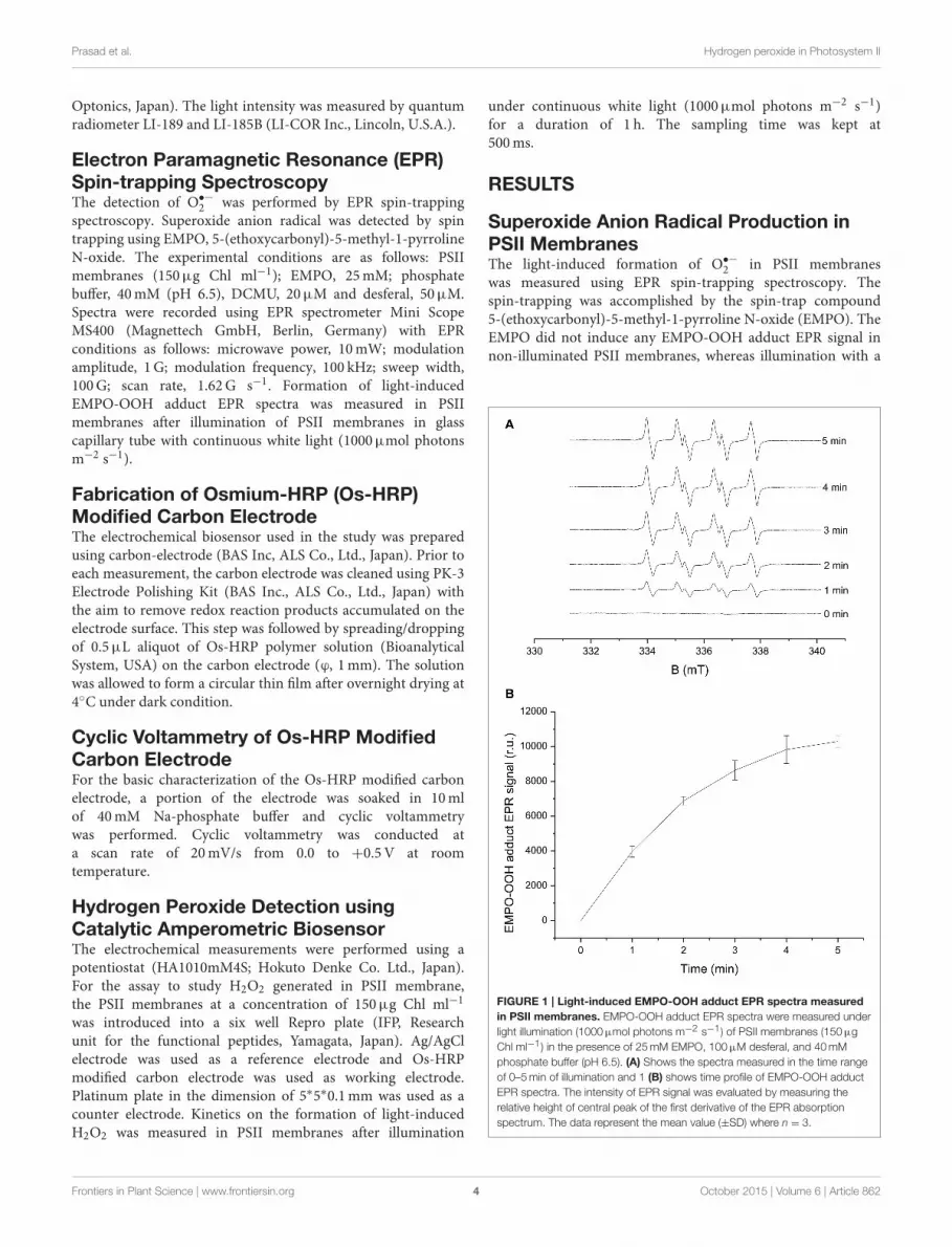
Electron Paramagnetic Resonance (EPR)Spin-trapping SpectroscopyThe detection of O•−
2 was performed by EPR spin-trappingspectroscopy. Superoxide anion radical was detected by spintrapping using EMPO, 5-(ethoxycarbonyl)-5-methyl-1-pyrrolineN-oxide. The experimental conditions are as follows: PSIImembranes (150µg Chl ml−1); EMPO, 25mM; phosphatebuffer, 40mM (pH 6.5), DCMU, 20µM and desferal, 50µM.Spectra were recorded using EPR spectrometer Mini ScopeMS400 (Magnettech GmbH, Berlin, Germany) with EPRconditions as follows: microwave power, 10mW; modulationamplitude, 1G; modulation frequency, 100 kHz; sweep width,100G; scan rate, 1.62G s−1. Formation of light-inducedEMPO-OOH adduct EPR spectra was measured in PSIImembranes after illumination of PSII membranes in glasscapillary tube with continuous white light (1000µmol photonsm−2 s−1).
Fabrication of Osmium-HRP (Os-HRP)Modified Carbon ElectrodeThe electrochemical biosensor used in the study was preparedusing carbon-electrode (BAS Inc, ALS Co., Ltd., Japan). Prior toeach measurement, the carbon electrode was cleaned using PK-3Electrode Polishing Kit (BAS Inc., ALS Co., Ltd., Japan) withthe aim to remove redox reaction products accumulated on theelectrode surface. This step was followed by spreading/droppingof 0.5µL aliquot of Os-HRP polymer solution (BioanalyticalSystem, USA) on the carbon electrode (ϕ, 1mm). The solutionwas allowed to form a circular thin film after overnight drying at4◦C under dark condition.
Cyclic Voltammetry of Os-HRP ModifiedCarbon ElectrodeFor the basic characterization of the Os-HRP modified carbonelectrode, a portion of the electrode was soaked in 10mlof 40mM Na-phosphate buffer and cyclic voltammetrywas performed. Cyclic voltammetry was conducted ata scan rate of 20mV/s from 0.0 to +0.5V at roomtemperature.
Hydrogen Peroxide Detection usingCatalytic Amperometric BiosensorThe electrochemical measurements were performed using apotentiostat (HA1010mM4S; Hokuto Denke Co. Ltd., Japan).For the assay to study H2O2 generated in PSII membrane,the PSII membranes at a concentration of 150µg Chl ml−1
was introduced into a six well Repro plate (IFP, Researchunit for the functional peptides, Yamagata, Japan). Ag/AgClelectrode was used as a reference electrode and Os-HRPmodified carbon electrode was used as working electrode.Platinum plate in the dimension of 5∗5∗0.1mm was used as acounter electrode. Kinetics on the formation of light-inducedH2O2 was measured in PSII membranes after illumination
under continuous white light (1000µmol photons m−2 s−1)for a duration of 1 h. The sampling time was kept at500ms.
RESULTS
Superoxide Anion Radical Production inPSII MembranesThe light-induced formation of O•−
2 in PSII membraneswas measured using EPR spin-trapping spectroscopy. Thespin-trapping was accomplished by the spin-trap compound5-(ethoxycarbonyl)-5-methyl-1-pyrroline N-oxide (EMPO). TheEMPO did not induce any EMPO-OOH adduct EPR signal innon-illuminated PSII membranes, whereas illumination with a
FIGURE 1 | Light-induced EMPO-OOH adduct EPR spectra measured
in PSII membranes. EMPO-OOH adduct EPR spectra were measured under
light illumination (1000µmol photons m−2 s−1) of PSII membranes (150µg
Chl ml−1) in the presence of 25mM EMPO, 100µM desferal, and 40mM
phosphate buffer (pH 6.5). (A) Shows the spectra measured in the time range
of 0–5min of illumination and 1 (B) shows time profile of EMPO-OOH adduct
EPR spectra. The intensity of EPR signal was evaluated by measuring the
relative height of central peak of the first derivative of the EPR absorption
spectrum. The data represent the mean value (±SD) where n = 3.
Frontiers in Plant Science | www.frontiersin.org 4 October 2015 | Volume 6 | Article 862

Prasad et al. Hydrogen peroxide in Photosystem II
continuous white light (1,000µmol photons m−2 s−1) resultedin the formation of the EMPO-OOH adduct EPR signal(Figure 1A). Time dependence of EMPO-OOH adduct EPRsignal shows that formation of O•−
2 is enhanced linearly fora duration upto 5min of light illumination (Figure 1B). Theseresults suggest that illumination of PSII membranes with acontinuous white light (1,000µmol photons m−2 s−1) results inO•−
2 production.
Effect of Superoxide Dismutase andCatalase on O•−
2 Production in PSIIMembranesTo study the contribution of O•−
2 leading to H2O2 formationin PSII membranes under light illumination, the effect of SODand catalase (CAT) were studied on O•−
2 . Upon addition ofexogenous SOD, which is known to catalyze the dismutationof O•−
2 to H2O2 to PSII membranes prior to illumination,EMPO-OOH adduct EPR signal was found to diminishcompletely. The simultaneous addition of SOD and CAT wasalso found to suppress the EMPO-OOH EPR signal completely(Figure 2A). This observation indicates that O•−
2 producedduring light illumination is most likely involved in H2O2
formation in PSII membranes.
Effect of DCMU on O•−
2 Production in PSIIMembranesThe effect of herbicides, DCMU [3-(3,4-dichlorophenyl)-1,1-dimethylurea] (Sigma Aldrich, Germany) was tested for itseffect on EMPO-OOH adduct EPR signal in PSII membranes.The effect of DCMU which is known to block the electrontransfer from QA to QB was studied on light-induced formationof O•−
2 . Figure 2B shows that the addition of DCMUsuppressed EMPO-OOH adduct EPR signal approximately by50% (Figure 2B). These observations indicate that loosely boundplastosemiquinone bound at or after the QB site can contributeto the overall production of O•−
2 via the reduction of molecularoxygen.
Characterization of Os-HRP ModifiedCarbon ElectrodeThe characterization of the Os-HRP modified carbon electrodewas performed using cyclic voltammetry (Figure 3). Cyclicvoltammetry was conducted at a scan rate of 20mV/s from 0.0to + 0.5V at room temperature. The oxidation and reductioncurrent were obtained at 0.3 V vs. Ag/AgCl. Based on thedata obtained, the surface concentration of Os-HRP on thecarbon electrode was calculated to be 6.78 × 10⁻⁹mol/cm2. TheCalibration curve of Os-HRP modified carbon electrode was alsomeasured using standard H2O2 solution (Supplementary Data).
Hydrogen Peroxide Production in PSIIMembranesThe light-induced formation of H2O2 in PSII membranes wasmeasured using catalytic amperometric biosensor. The study ofkinetics of H2O2 production was accomplished using Os-HRPmodified carbon electrode. The reduction current generated in
the presence of H2O2 was monitored. Under dark condition,no change in the reduction current was observed whereasillumination with a continuous white light (1,000µ mol photonsm−2 s−1) resulted in change in the reduction current. Figure 4Ashows typical chronoamperometric responses, the reductioncurrents gradually increased after light illumination followedby a shoulder which continues for a duration of about 1 hwhich than rapidly drops at the switching off the light (darkperiod). The peak value of the reduction current was reached afterabout 15min of light illumination with a maximum change inreduction currents of approximately 400 pA. The data presentedshows continuous generation of H2O2 till the period of lightillumination (Figure 4A). These results shows that illuminationof PSII membranes with continuous white light results in H2O2
production.
Effect of SOD and CAT on H2O2 Productionin PSII MembranesTo monitor the functionality of the sensor, the effect of SODwas measured under light-illumination. Reduction current forH2O2was measured for first 5min of light illumination where
FIGURE 2 | Effect of SOD, CAT, and DCMU on EMPO-OOH adduct EPR
spectra measured in PSII membranes. EMPO-OOH adduct EPR spectra
were measured in PSII membranes in the presence of SOD and CAT under
light illumination. The relative intensity of the light-induced EMPO-OOH adduct
EPR signal measured in the presence of SOD (400U/ml) and SOD+CAT
(400U/ml each) (A) and DCMU (20µM) (B). The other experimental conditions
were the same as described in Figure 1.
Frontiers in Plant Science | www.frontiersin.org 5 October 2015 | Volume 6 | Article 862

Prasad et al. Hydrogen peroxide in Photosystem II
FIGURE 3 | Characterization of Os-HRP modified carbon electrode. Characterization of the Os-HRP modified carbon electrode: cyclic voltammetry was
performed for the basic characterization of the modified electrode. Cyclic voltammetry was conducted at a scan rate of 20mV/s from 0.0 to +0.5 V at room
temperature.
a linear increase was observed similar to control (Figure 4A).Upon addition of exogenous SOD to PSII membranes duringillumination, a fast increase in reduction current was observedbringing a considerable change of about 600 pA. The fast increasewas followed by a rapid drop however; the reduction current wasstill higher as compared to reduction current recorded before theaddition of SOD. The addition of CAT completely suppressed thereduction current by about 100% (Figure 4B). These results showthat under illumination of PSII membranes with a continuouswhite light, H2O2 production in PSII membrane is contributedvia dismutation of O•−
2 .
DISCUSSION
In the current study, we used spectroscopic and amperometrictechniques to measure the production of ROS in spinach PSIImembrane. Our prime objective was to study the effect of highlight stress in PSII membrane reflected by ROS productionprimarily the generation of H2O2 as the stress response formedvia the dismutation of O•−
2 . Superoxide anion radical which isknown to be formed by one-electron reduction of molecularoxygen was measured under light illumination (Figure 1).EPR spin-trapping data obtained using the urea-type herbicide(DCMU) supports the evidence on the involvement of looselybound plastosemiquinone in O•−
2 production (Figure 2B).Suppression of EMPO-OOH adduct EPR signal is in agreementwith our previously published results where contribution ofloosely bound plastosemiquinone at or after the QB site (DCMU-sensitive site) might contribute to the overall O•−
2 production(Yadav et al., 2014). The EMPO-OOH adduct EPR signal in thepresence of DCMU indicates that molecular oxygen is reducedprior to the QB site which is known to occur due to reductionof molecular oxygen by Pheo•− or Q•−
A which serves as electrondonors to molecular oxygen due to their low redox potentials
(Pospíšil, 2012). The observation that the EMPO-OOH adductwas completely suppressed in PSII membranes illuminated inpresence of SOD indicates that O•−
2 formed during the lightillumination dismutates to H2O2 prior to its interaction with spintrap (Figure 2A). It can be concluded here that the H2O2 formedin PSII membrane under light illumination is contributed by thedismutation of O•−
2 .Amperometric methods for the direct detection of H2O2 has
been used since last decades in animal cells (Mouithys-Mickaladet al., 2001; Inoue et al., 2010) however, very limited evidencesexist on its application in plant cells (Cleland and Grace,1999). Amperometric measurements have been implementedat the level of protoplast to study the photosynthetic activityunder the effect of benzoquinone (Yasukawa et al., 1998, 1999).Redox response of benzoquinone, p-hydroquinone and oxygenwas measured by placing a microelectrode close to an algalprotoplast to localize concentration of these species (Yasukawaet al., 1999). Direct detection of H2O2using electrochemicalmethods; however, had never been reported previously inphotosynthetic organelles. H2O2 detection in subchloroplastoxygen-evolving PSII particles and isolated reaction centercomplexes of PSII has been studied using luminol-peroxidasechemiluminescence and pulse photoactivation (Zastrizhnayaet al., 1997). Production of H2O2 was detected in PSIImembranes using AR fluorescent assay (Yadav and Pospíšil,2012). Levels of H2O2 including O•−
2 and singlet oxygen hasbeen determined by using both histochemical and fluorescentprobes in leaves and thylakoids (Zulfugarov et al., 2014), however,there exist arguments over the selectivity of the molecular probesused.
Two different approaches of the detection of H2O2viaelectrochemical methods are being used in animal cells. Thedetection of H2O2 is either via a non-mediator or a mediatorbased biosensor device (Lin et al., 2000; Nakabayashi and
Frontiers in Plant Science | www.frontiersin.org 6 October 2015 | Volume 6 | Article 862

Prasad et al. Hydrogen peroxide in Photosystem II
FIGURE 4 | Real-time monitoring of reduction current for hydrogen peroxide in PSII membrane. (A) Kinetics of the production of H2O2 was measured using
Os-HRP modified carbon electrode during light illumination in PSII membranes. The light illumination was started at 5min from the start of the measurement and the
reduction current was measured for a duration of 1 h. (B) Effect of SOD and CAT on reduction current was measured in the presence of SOD (400U/ml) and SOD +
CAT (400U/ml each).
Yoshikawa, 2000; Yao et al., 2005; Wang and Zhang, 2006;Camacho et al., 2007; Jia, 2008; Radi et al., 2009; Ahammad,2013). In non-mediator based biosensor, the transfer of electronoccurs between the electrode and the enzyme (Ahammad, 2013).The preparation process being very simple, the non-mediatorbased biosensors have been extensively used in the past. Inthe mediator based biosensor device, the mediator plays a keyrole to shuttle electron between the electrode and the enzyme(Scheme 1). Most commonly, methylene blue, ferrocene andcarboxylic acid are used as mediators (Lin et al., 2000; Tian et al.,
2001; Li et al., 2004; Tripathi et al., 2006; Senel et al., 2010).The mediator in the case of HRP is pointed to be importantbecause the shuttling of electron between electrode and HRP islarge as the active site of HRP is located deep in the proteinsheath (Ahammad, 2013). In our current device, Os acts asa mediator where Os2+ is reduced to Os3+ in the process(Scheme 1). Different modified electrode has been introduced inthe past, the detection limit in most case were in the range of µMconcentration with a limited contribution where mediator-basedHRP biosensor reached a lower detection limit in the range of
Frontiers in Plant Science | www.frontiersin.org 7 October 2015 | Volume 6 | Article 862

Prasad et al. Hydrogen peroxide in Photosystem II
tens-hundreds nm concentration (Chen et al., 2006; Zhang et al.,2008; Lu et al., 2010).
Using Os-HRP modified carbon electrode, we have observeda change in the reduction current during illumination of PSIImembranes reflecting the production of H2O2 (Figure 4A).A change of ∼400 pA reflects generation of H2O2 whichwas then found to be stable in the period of 20–60min oflight illumination. The reduction current was found to dropimmediately under dark condition. Based on the considerablechanges monitored in reduction current, the biosensor deviceis claimed to be sensitive for its application in photosyntheticsamples.
ROS generated in the cell or organelles are eliminatedby antioxidant enzymes such as SOD, glutathione peroxidase,CAT or by low molecular antioxidants such as vitamins,glutathione etc. (Halliwell and Gutteridge, 2007). When SODwhich dismutates O•−
2 to H2O2 was added to the illuminatedPSII membrane, the reduction current for H2O2 was observedto gradually enhance leading to the conclusion that the H2O2
generated in the PSII membrane is formed via the formationof O•−
2 . The addition of SOD (Figure 4B) during the lightillumination drastically enhanced the reduction current by 600pA followed by a sudden drop indicate the fast conversion ofO•−
2 into H2O2 available in the PSII pool. This is in agreementwith observed result with EPR signal of the EMPO-OOH adductobserved under the effect of SOD (Figure 2A). The subsequentaddition of CAT which converts H2O2 to H2O and molecularoxygen brings back the reduction current close to the valueobserved under dark condition.
Electrochemical detection of H2O2 is suggested here to be ofgreat importance in addition to other methods that has been usedin the recent past. In the detailed study performed by Šnyrychováand co-workers, different H2O2 detecting probes were tested.Probes such as amplex red and amplex ultra-red were foundto be sensitive to light and thus are not appropriate for thestudy on the generation of H2O2 in plant sample where effectof light stress is frequently studied. Based on the results, theauthors also suggested that these probes should be used withcaution to avoid artifacts (Šnyrychová et al., 2009). In additionto this, the toxicity caused by the exogenous addition of probescannot be completely excluded. The method of electrochemicalmeasurements is also preferred because of the simplicity, low-cost, high-sensitivity and selectivity (Ahammad, 2013). Thesimplicity and low-cost of electrochemical measurements isbecause of the fact that the electrodes can be used for endlesstime with easy fabrication with designated mediator and enzymeand overnight incubation at 4◦C under dark condition or as perstandards. Based on the redox potential of the redox couple,the modified electrode are specific for different species. Os-HRPmodified electrode is specific for H2O2 and thus can be widelyused in photosynthetic research where detection of H2O2 has
always been a challenge. The sensitivity of the modified electrodedepends on the size and the material used. In our currentreport, Os-HRP modified carbon electrode has been claimed tobe sensitive and highly selective among other H2O2 detectiontechniques available till date. This fact opens the possibilityfor using Os-HRP modified electrode for H2O2 detection inphotosystem I (PSI) where H2O2 is formed by the transferof electron from reduced ferredoxin to molecular oxygen viaferredoxin-thioredoxin reductase (Gechev et al., 2006). Plasmamembrane NADPH oxidase complex are considered as themajor producer of ROS including O•−
2 and H2O2 in cells (Sagiand Fluhr, 2001, 2006; Halliwell and Gutteridge, 2007). Inaddition to this, among the enzymatic source of O•−
2 and H2O2,cell wall bound peroxidases, aminooxidases, flavin containingoxidases, oxalate, and plasma membrane oxidases are knownto be involved (Bolwell et al., 2002; Mori and Schroeder, 2004;Svedruzic et al., 2005). In the case of apoplastic oxidative burst,ROS are produced by cell wall bound oxidases, peroxidasesand polyamine oxidases (Minibayeva et al., 1998, 2009). Thus,the current electrode also opens the possibility for measuringgeneration of H2O2 from different and localized structures ofplants and animals cells.
AUTHOR CONTRIBUTIONS
AP and SK contributed to the conception and design of thework. AP, AK, MS, HK, TS performed the measurements. APanalyzed, interpreted the data and drafted the manuscript. AK,PP participated in drafting the manuscript. SK, MK, and MTrevised it critically for important content. All authors approvedthe final version of the manuscript.
ACKNOWLEDGMENTS
This work was funded by the MEXT-Supported Program forthe Strategic Research Foundation at Private Universities, Japan.PP would like to thank Ministry of Education, Youth andSports of the Czech Republic grants no. LO1204 (NationalProgram of Sustainability I), no.CZ.1.07/2.3.00/30.0041 (Supportfor Building Excellent Research Teams and Intersectoral Mobilityat Palacký University) and by Czech Science Foundation grantno. GP13-29294S. We thank Ketaki Vasant Phadke (Departmentof Biophysics, Palacký University) for cross-checking themanuscript for errors.
SUPPLEMENTARY MATERIAL
The Supplementary Material for this article can be foundonline at: http://journal.frontiersin.org/article/10.3389/fpls.2015.00862
Frontiers in Plant Science | www.frontiersin.org 8 October 2015 | Volume 6 | Article 862
Prasad et al. Hydrogen peroxide in Photosystem II
REFERENCES
Ahammad, A. J. S. (2013). Hydrogen peroxide biosensors based on horseradishperoxidase and hemoglobin. J. Biosens. Bioelectron. S9:001. doi: 10.4172/2155-6210.S9-001
Ananyev, G., Renger, G., Wacker, U., and Klimov, V. (1994). The photoproductionof superoxide radicals and the superoxide-dismutase activity of photosystem II.The possible involvement of cytochrome b559. Photosynth. Res. 41, 327–338.doi: 10.1007/BF00019410
Arató, A., Bondarava, N., and Krieger-Liszkay, A. (2004). Production of reactiveoxygen species in chloride- and calcium-depleted photosystem II and theirinvolvement in photoinhibition. Biochim. Biophys. Acta 1608, 171–180. doi:10.1016/j.bbabio.2003.12.003
Berthold, D. A., Babcock, G. T., and Yocum, C. F. (1981). A highly resolved oxygenevolving photosystem II preparation from spinach thylakoid membranes. FEBSLett. 134, 231–234. doi: 10.1016/0014-5793(81)80608-4
Bolwell, G. P., Bindschedler, L. V., Blee, K. A., Butt, V. S., Davies, D. R., Gardner,S. L., et al. (2002). The apoplastic oxidative burst in response to bioticstress in plants: a three-component system. J Exp. Bot. 53, 1367–1376. doi:10.1093/jexbot/53.372.1367
Camacho, C., Matías, J. C., Chico, B., Cao, R., Gómez, L., Simpson,B. K., et al. (2007). Amperometric biosensor for hydrogen peroxide,using supramolecularly immobilized horseradish peroxidase on the β-cyclodextrin-coated gold electrode. Electroanalysis 19, 2538–2542. doi:10.1002/elan.200703993
Chen, S., Yin, C., Strasser, R. J., Govindjee, Yang, C., and Qiang, S. (2012). Reactiveoxygen species from chloroplasts contribute to 3-acetyl-5- isopropyltetramicacid-induced leaf necrosis of Arabidopsis thaliana. Plant Physiol. Biochem. 52,38–51. doi: 10.1016/j.plaphy.2011.11.004
Chen, S., Yuan, R., Chai, Y., Xu, L., Wang, N., Li, X. et al. (2006).Amperometric hydrogen peroxide biosensor based on the immobilization ofHorseradish Peroxidase (HRP) on the layer-by-layer assembly films of goldcolloidal nanoparticles and toluidine blue. Electroanalysis 18, 471–477. doi:10.1002/elan.200503424
Chen, W., Li, B., Xu, C., and Wang, L. (2009). Chemiluminescence flow biosensorfor hydrogen peroxide using DNAzyme immobilized on eggshell membraneas a thermally stable biocatalyst. Biosens. Bioelectron. 24, 2534–2540. doi:10.1016/j.bios.2009.01.010
Cleland, R. E., and Grace, S. C. (1999). Voltammetric detection of superoxideproduction by photosystem II. FEBS Lett. 457, 348–352. doi: 10.1016/S0014-5793(99)01067-4
Ferreira, K. N., Iverson, T. M., Maghlaoui, K., Barber, J., and Iwata, S. (2004).Architecture of the photosynthetic oxygen-evolving center. Science 303,1831–1838. doi: 10.1126/science.1093087
Ford, R. C., and Evans, M. C. W. (1983). Isolation of a photosystem II from higherplants with highly enriched oxygen evolution activity. FEBS Lett. 160, 159–164.doi: 10.1016/0014-5793(83)80957-0
Foyer, C. H., and Shigeoka, S. (2011). Understanding oxidative stress andantioxidant functions to enhance photosynthesis. Plant Physiol. 155, 93–100.doi: 10.1104/pp.110.166181
Gechev, T. S., Van Breusegem, F., Stone, J. M., Denev, I., and Laloi, C (2006).Reactive oxygen species as signals that modulate plant stress responses andprogrammed cell death. Bioessays 28, 1091–1101. doi: 10.1002/bies.20493
Guskov, A., Kern, J., Gabdulkhakov, A., Broser, M., Zouni, A., and Saenger, W.(2009). Cynobacterial photosystem II at 2.9 Å resolution and the role ofquinones, lipids, channels and chloride.Nat. Struct. Mol. Biol. 16, 334–342. doi:10.1038/nsmb.1559
Halliwell, B., and Gutteridge, J. M. C. (2007). Free Radicals in Biology andMedicine,
4th Edn. New York, NY: Oxford University Press.Inoue, K. Y., Ino, K., Shiku, H., Kasai, S., Yasukawa, T., Mizutani, F., et al. (2010).
Electrochemical monitoring of hydrogen peroxide released from leucocyteson horseradish peroxidase redox polymer coated electrode chip. Biosens.
Bioelectron. 25, 1723–1728. doi: 10.1016/j.bios.2009.12.014Jia, J. (2008). Hydrogen peroxide biosensor based on horseradish peroxidase– Au
nanoparticles at a viologen grafted glassy carbon electrode. Microchim. Acta
163, 237–241. doi: 10.1007/s00604-008-0002-9Kafi, A. K., Wu, G., and Chen, A. (2009). A novel hydrogen peroxide biosensor
based on the immobilization of horseradish peroxidase onto Au-modified
titanium dioxide nanotube arrays. Biosens. Bioelectron. 24, 566–571. doi:10.1016/j.bios.2008.06.004
Kawakami, K., Umena, Y., Kamiya, N., and Shen, J.-R. (2011). Structure ofthe catalytic, inorganic core of oxygen-evolving photosystem II at 1.9 Åresolution. J. Photochem. Photobiol. B 104, 9–18. doi: 10.1016/j.jphotobiol.2011.03.017
Krieger-Liszkay, A. (2005). Singlet oxygen production in photosynthesis. J. Exp.Bot. 56, 337–346. doi: 10.1093/jxb/erh237
Lei, C. X., Long, L. P., and Cao, Z. L. (2005). An H2O2 biosensorbased on immobilization of horseradish peroxidase labeled nano-Au insilica sol-gel/alginate composite film. Anal. Lett. 38, 1721–1734. doi:10.1080/00032710500207762
Lei, C. X., Wang, H., Shen, G. L., and Yu, R. Q. (2004). Immobilizationof enzymes on the nano-au film modified glassy carbon electrode for thedetermination of hydrogen peroxide and glucose. Electroanalysis 16, 736–740.doi: 10.1002/elan.200302877
Li, C. X., Deng, K. Q., Shen, G. L., and Yu, R. Q. (2004). Amperometric hydrogenperoxide biosensor based on horseradish peroxidase-labeled nano-Au colloidsimmobilized on poly(2,6-pyridinedicarboxylic acid) layer by cysteamine. Anal.Sci. 20, 1277–1281. doi: 10.2116/analsci.20.1277
Li, Z. H., Guedri, H., Viguier, B., Sun, S. G., and Marty, J. L. (2013). Optimizationof hydrogen peroxide detection for a methyl mercaptan biosensor. Sensors 13,5028–5039. doi: 10.3390/s130405028
Lichtenthaler, H. K. (1987). Chlorophylls and carotenoids: pigments ofphotosynthetic biomembranes. Methods Enzymol. 148, 350–382. doi:10.1016/0076-6879(87)48036-1
Lin, X. Q., Chen, J., and Chen, Z. H. (2000). Amperometric biosensor forhydrogen peroxide based on immobilization of horseradish peroxidase onmethylene blue modified graphite electrode. Electroanalysis 12, 306–310. doi:10.1002/(sici)1521-4109(20000301)
Liu, X., Luo, L., Ding, Y., Xu, Y., and Li, F. (2011). Hydrogen peroxidebiosensor based on the immobilization of horseradish peroxidase on γ-Al2O3
nanoparticles/chitosan film-modified electrode. J. Solid State Electrochem. 15,447–453. doi: 10.1007/s10008-010-1120-y
Loew, N., Wollenberger, U., Scheller, F. W., and Katterle, M. (2009).Direct electrochemistry and spectroelectrochemistry of osmiumsubstituted horseradish peroxidase. Bioelectrochemistry 76, 28–33. doi:10.1016/j.bioelechem.2009.03.015
Loll, B., Kern, J., Saenger, W., Zouni, A., and Biesiadka, J. (2005). Towardscomplete cofactor arrangement in the 3.0 Å resolution structure of photosystemII. Nature 438, 1040–1044. doi: 10.1038/nature04224
Lu, L., Zhang, L., Zhang, X., Wu, Z., Huan, S., Shen, G. et al. (2010). AMgO nanoparticles composite matrix-based electrochemical biosensor forhydrogen peroxide with high sensitivity. Electroanalysis 22, 471–477. doi:10.1002/elan.200900429
Luo, L., Zhu, L., Xu, Y., Shen, L., Wang, X., Ding, Y. et al. (2011).Hydrogen peroxide biosensor based on horseradish peroxidase immobilizedon chitosan-wrapped NiFe2O4 nanoparticles. Microchim.Acta 174, 55–61. doi:10.1007/s00604-011-0591-6
Mills, A., Tommons, C., Bailey, R. T., Tedford, M. C., and Crilly, P. J. (2007).Reversible, fluorescence-based optical sensor for hydrogen peroxide. Analyst132, 566–571. doi: 10.1039/b618506a
Minibayeva, F., Kolesnikov, O., Chasov, A., Beckett, R. P., Lüthje, S., Vylegzhanina,N., et al. (2009). Wound-induced apoplastic peroxidase activities: their rolesin the production and detoxification of reactive oxygen species. Plant Cell.Environ. 32, 497–508. doi: 10.1111/j.1365-3040.2009.01944.x
Minibayeva, F., Kolesnikov, O. P., and Gordon, L. K. (1998). Contribution of aplasma membrane redox system to the superoxide production by wheat rootcells Protoplasma 205, 101–106. doi: 10.1007/BF01279299
Mori, I. C., and Schroeder, J. I. (2004). Reactive oxygen species activation ofplant Ca2+ channels. A signaling mechanism in polar growth, hormonetransduction, stress signaling, and hypothetically mechanotransduction. PlantPhysiol. 135, 702–708. doi: 10.1104/pp.104.042069
Mouithys-Mickalad, A., Deby-Dupont, G., Nys, M., Lamy, M., and Deby,C. (2001). Oxidative processes in human promonocytic cells (THP-1) after differentiation into macrophages by incubation with Chlamydia
pneumoniae extracts. Biochem. Biophys. Res. Commun. 287, 781–788. doi:10.1006/bbrc.2001.5643
Frontiers in Plant Science | www.frontiersin.org 9 October 2015 | Volume 6 | Article 862

Prasad et al. Hydrogen peroxide in Photosystem II
Mubarakshina, M. M., and Ivanov, B. N. (2010). The production andscavenging of reactive oxygen species in the plastoquinone pool of chloroplastthylakoid membranes. Physiol. Plant. 140, 103–110. doi: 10.1111/j.1399-3054.2010.01391.x
Nakabayashi, Y., and Yoshikawa, H. (2000). Amperometric biosensors forsensing of hydrogen peroxide based on electron transfer between horseradishperoxidase and ferrocene as a mediator. Anal. Sci. 16, 609–613. doi:10.2116/analsci.16.609
Pospíšil, P. (2009). Production of reactive oxygen species by photosystem II.Biochim. Biophys. Acta 1787, 1151–1160. doi: 10.1016/j.bbabio.2009.05.005
Pospíšil, P. (2011). Enzymatic function of cytochrome b559 in photosystem II. JPhotochem. Photobiol. B 104, 341–347. doi: 10.1016/j.jphotobiol.2011.02.013
Pospíšil, P. (2012). Molecular mechanisms of production and scavenging ofreactive oxygen species by photosystem II. Biochim. Biophys. Acta 1817,218–231. doi: 10.1016/j.bbabio.2011.05.017
Pospíšil, P., Arató, A., Krieger-Liszkay, A., and Rutherford, A. W. (2004).Hydroxyl radical generation by Photosystem II. Biochemistry 43, 6783–6792.doi: 10.1021/bi036219i
Pospíšil, P., Šnyrychová, I., Kruk, J., Strzałka, K., and Nauš, J. (2006). Evidence thatcytochrome b559 is involved in superoxide production in Photosystem II: effectof synthetic short-chain plastoquinones in a cytochrome b559 tobacco mutant.Biochem. J. 397, 321–327. doi: 10.1042/BJ20060068
Prasad, A., and Pospíšil, P. (2012). Ultra-weak photon emission induced byvisible light and ultraviolet A radiation via photoactivated skin chromophores:in-vivo charge coupled device imaging. J. Biomed. Opt. 17:085004. doi:10.1117/1.JBO.17.8.085004
Radi, A. E., Muñoz-Berbel, X., Cortina-Puig, M., and Marty, J. L. (2009). Athird generation hydrogen peroxide biosensor based on horseradish peroxidasecovalently immobilized on electrografted organic film on screen-printedcarbon electrode. Electroanalysis 21, 1624–1629. doi: 10.1002/elan.200904587
Rappaport, F., and Diner, B. A. (2008). Primary photochemistry and energeticsleading to the oxidation of the (Mn)4Ca cluster and to the evolution ofmolecular oxygen in Photosystem II. Coord. Chem. Rev. 252, 259–272. doi:10.1016/j.ccr.2007.07.016
Sagi, M., and Fluhr, R. (2001). Superoxide production by plant homologuesof the Gp91phox NADPH oxidase. modulation of activity by calcium andby tobacco mosaic virus infection. Plant Physiol. 126, 1281–1290. doi:10.1104/pp.126.3.1281
Sagi, M., and Fluhr, R. (2006). Production of reactive oxygen species by plantNADPH oxidases. Plant Physiol. 141, 336–340. doi: 10.1104/pp.106.078089
Sanford, A. L., Morton, S. W., Whitehouse, K. L., Oara, H. M., Lugo-Morales,L. Z., Roberts, J. G., et al. (2011). Voltammetric detection of hydrogenperoxide at carbon fiber microelectrodes. Anal Chem. 82, 5205–5210. doi:10.1021/ac100536s
Schmitt, F. J., Renger, G., Friedrich, T., Kreslavksi, V. D., Zharmukhadmedov, S. K.,Los, D. A., et al. (2014). Reactive oxygen species: re-evaluation of generation,monitoring and role in stress-signaling in phototrophic organisms. Biochim.
Biophys. Acta 1837, 385–848. doi: 10.1016/j.bbabio.2014.02.005Schröder, W. P., and Åkerlund, H.-E. (1990). “Hydrogen peroxide production in
photosystem II preparations,” in Current Research in Photosynthesis, Vol. 1, edM. Baltscheffsky (Dordrecht: Kluwer Academic Publisher), 901–904.
Senel, M. C., Cevik, E., and Abasıyanık, M. F. (2010). Amperometrichydrogen peroxide biosensor based on covalent immobilization of horseradishperoxidase on ferrocene containing polymeric mediator. Sens. Actuators. BChem. 145, 444–450. doi: 10.1016/j.snb.2009.12.055
Shao, N., Krieger-Liszkay, A., Schroda, M., and Beck Christoph, F. (2007). Areporter system for the individual detection of hydrogen peroxide and singletoxygen: its use for the assay of reactive oxygen species produced in vivo. PlantJ. 50, 475–487. doi: 10.1111/j.1365-313X.2007.03065.x
Šnyrychová, I., Ayaydin, F., and Hideg, É. (2009). Detecting hydrogen peroxidein leaves in vivo—a comparison of methods. Physiol. Plant. 135, 1–18. doi:10.1111/j.1399-3054.2008.01176.x
Svedruzic, D., Jónsson, S., Toyota, C. G., Reinhardt, L. A., Ricagno, S,Lindqvist, Y., et al. (2005). The enzymes of oxalate metabolism: unexpectedstructures and mechanisms. Arch. Biochem. Biophys. 433, 176–192. doi:10.1016/j.abb.2004.08.032
Tian, F., Xu, B., Zhu, L., and Zhu, G. (2001). Hydrogen peroxide biosensorwith enzyme entrapped within electrodeposited polypyrrole based onmediated
sol–gel derived composite carbon electrode. Anal. Chim. Acta 443, 9–16. doi:10.1016/S0003-2670(01)01187-4
Tiwari, A., and Pospísil, P. (2009). Superoxide oxidase and reductase activity ofcytochrome b559 in photosystem II. Biochim. Biophys. Acta 1787, 985–994. doi:10.1016/j.bbabio.2009.03.017
Tiwari, I., and Singh, M. (2011). Preparation and characterization of methyleneblue-SDS-multiwalled carbon nanotubes nanocomposite for the detection ofhydrogen peroxide. Microchim. Acta 174, 223–230. doi: 10.1007/s00604-011-0620-5
Tripathi, V. S., Kandimalla, V. B., and Ju, H. (2006). Amperometricbiosensor for hydrogen peroxide based on ferrocene-bovineserum albumin and multiwall carbon nanotube modified ormosilcomposite. Biosens. Bioelectron. 21, 1529–1535. doi: 10.1016/j.bios.2005.07.006
Wang, G. H., and Zhang, L. M. (2006). Using novel polysaccharide-silica hybridmaterial to construct an amperometric biosensor for hydrogen peroxide.J. Phys. Chem. B 110, 24864–24868. doi: 10.1021/jp0657078
Wang, H. S., Pan, Q. X., and Wang, G. X. (2005). A biosensor based onimmobilization of horseradish peroxidase in chitosan matrix cross-linked withglyoxal for amperometric determination of hydrogen peroxide. Sensors 5,266–276. doi: 10.3390/s5040266
Yadav, D. K., and Pospíšil, P. (2012). Role of chloride ion in hydroxyl radicalproduction in photosystem II under heat stress: electron paramagneticresonance spin-trapping study. J. Bioenerg. Biomembr. 44, 365–372. doi:10.1007/s10863-012-9433-4
Yadav, D. K., Prasad, A., Kruk, J., and Pospíšil, P. (2014). Evidencefor the involvement of loosely bound plastosemiquinones in superoxideanion radical production in photosystem II. PLoS ONE 9:e115466. doi:10.1371/journal.pone.0115466
Yang, Z., Zong, X., Ye, Z., Zhao, B., Wang, Q., and Wang, P. (2010). Theapplication of complex multiple forklike ZnO nanostructures to rapid andultrahigh sensitive hydrogen peroxide biosensors. Biomaterials 31, 7534–7541.doi: 10.1016/j.biomaterials.2010.06.019
Yao, H., Li, N., Wei, Y. L., and Zhu, J. J. (2005). A H2O2 biosensor based onimmobilization of horseradishperoxidase in a gelatine network matrix. Sensors5, 277–283. doi: 10.3390/s5040277
Yasukawa, T., Uchida, I., and Matsue, T. (1998). Permeation of redox speciesthrough a cell membrane of a single, living algal protoplast studied bymicroamperometry. Biochim. Biophys. Acta 1369, 152–158. doi: 10.1016/S0005-2736(97)00220-4
Yasukawa, T., Uchida, I., and Matsue, T. (1999). Microamperometricmeasurements of photosynthetic activity in a single algal protoplast. Biophys. J.76, 1129–1135. doi: 10.1016/S0006-3495(99)77277-2
Zastrizhnaya, O. M., Khorobrykh, A. A., Khristin, M. S., and Klimov, V. V.(1997). Photoinduced production of hydrogen peroxide at the acceptor side ofphotosystem II. Biochemistry 62, 357–361.
Zhang, H. L., Lai, G. S., Han, D. Y., and Yu, A. M. (2008). An amperometrichydrogen peroxide biosensor based on immobilization of horseradishperoxidase on an electrode modified with magnetic dextran microspheres.Anal. Bioanal. Chem. 390, 971–977. doi: 10.1007/s00216-007-1748-3
Zhang, S., Weng, J., Pan, J., Tu, T., Yao, S., and Xu, C. (2003). Study on the photo-generation of superoxide radicals in Photosystem II with EPR spin trappingtechniques. Photosynth. Res. 75, 41–48. doi: 10.1023/A:1022439009587
Zulfugarov, I. S., Tovuu, A., Eu, Y. J., Dogsom, B., Poudyal, R. S., Nath, K., et al.(2014). Production of superoxide fromPhotosystem II in a rice (Oryza sativa L.)mutant lacking PsbS. BMC Plant Biol. 14:242. doi: 10.1186/s12870-014-0242-2
Conflict of Interest Statement: The authors declare that the research wasconducted in the absence of any commercial or financial relationships that couldbe construed as a potential conflict of interest.
Copyright © 2015 Prasad, Kumar, Suzuki, Kikuchi, Sugai, Kobayashi, Pospíšil, Tada
and Kasai. This is an open-access article distributed under the terms of the Creative
Commons Attribution License (CC BY). The use, distribution or reproduction in
other forums is permitted, provided the original author(s) or licensor are credited
and that the original publication in this journal is cited, in accordance with accepted
academic practice. No use, distribution or reproduction is permitted which does not
comply with these terms.
Frontiers in Plant Science | www.frontiersin.org 10 October 2015 | Volume 6 | Article 862