a reduced vwa domain-containing proteasomal ubiquitin receptor of giardia lamblia localizes to the...
TRANSCRIPT
Sinha et al. Parasites & Vectors (2015) 8:120 DOI 10.1186/s13071-015-0737-1
RESEARCH Open Access
A reduced VWA domain-containing proteasomalubiquitin receptor of Giardia lamblialocalizes to the flagellar pore regions inmicrotubule-dependent mannerAbhishek Sinha, Shankari Prasad Datta, Atrayee Ray and Srimonti Sarkar*
Abstract
Background: Giardia lamblia switches its lifecycle between trophozoite and cyst forms and the proteasome plays apivotal role in this switching event. Compared to most model eukaryotes, the proteasome of this parasite has alreadybeen documented to have certain variations. This study was undertaken to characterize the ubiquitin receptor, GlRpn10,of the 19S regulatory particle of the Giardia proteasome and determine its cellular localization in trophozoites, encystingtrophozoites and cysts.
Method: Sequence alignment and domain architecture analyses were performed to characterize GlRpn10. In vitroubiquitin binding assay, functional complementation and biochemical studies verified the protein’s ability to functionas ubiquitin receptor in the context of the yeast proteasome. Immunofluorescence localization was performed withantibody against GlRpn10 to determine its distribution in trophozoites, encysting trophozoites and cysts. Real-timePCR and Western blotting were performed to monitor the expression pattern of GlRpn10 during encystation.
Result: GlRpn10 contained a functional ubiquitin interacting motif, which was capable of binding to ubiquitin. Althoughit contained a truncated VWA domain, it was still capable of partially complementing the function of the yeast Rpn10orthologue. Apart from localizing to the nucleus and cytosol, GlRpn10 was also present at flagellar pores of trophozoitesand this localization was microtubule-dependent. Although there was no change in the cellular levels of GlRpn10 duringencystation, its selective distribution at the flagellar pores was absent.
Conclusion: GlRpn10 contains a noncanonical VWA domain that is partially functional in yeast. Besides the expectednuclear and cytosolic distribution, the protein displays microtubule-dependent flagellar pore localization in trophozoites.While the protein remained in the nucleus and cytosol in encysting trophozoites, it could no longer be detected at theflagellar pores. This absence at the flagellar pore regions in encysting trophozoites is likely to involve redistribution of theprotein, rather than decreased gene expression or selective protein degradation.
Keywords: Proteasome, Giardia, Flagella, Ubiquitin, VWA, Rpn10
* Correspondence: [email protected] of Biochemistry, Bose Institute, P 1/12, C. I. T. Road, Scheme – VII M,Kolkata 700054, West Bengal, India
© 2015 Sinha et al.; licensee BioMed Central. This is an Open Access article distributed under the terms of the CreativeCommons Attribution License (http://creativecommons.org/licenses/by/4.0), which permits unrestricted use, distribution, andreproduction in any medium, provided the original work is properly credited. The Creative Commons Public DomainDedication waiver (http://creativecommons.org/publicdomain/zero/1.0/) applies to the data made available in this article,unless otherwise stated.
Sinha et al. Parasites & Vectors (2015) 8:120 Page 2 of 13
BackgroundGiardia lamblia, a flagellated parasitic protist, colonizes thegut of its hosts and causes the diarrheal disease giardiasis.The parasite has two distinct morphological stagesduring its lifecycle: flagellated motile trophozoites andthe non-motile cysts. While trophozoites are the diseasecausing forms, the environmentally-resistant cysts enablethe parasite to survive outside the host and the infectioncycle commences with ingestion of either water orfood contaminated with cysts [1]. Thus, transition fromtrophozoite to cyst is crucial for disease transmission andthis change is brought about by a change in the intracellularproteome of G. lamblia [2]. Such changes in intracellularproteome require not only new protein synthesis, butalso degradation of existing proteins. Given that theproteasome carries out the bulk of protein degradation incells [3], investigation of proteasomal function of Giardiawill be crucial towards understanding stage transition inthis protist.Proteasomes are large macromolecular assemblies that
carry out polyubiquitin-dependent protein degradationin a highly-regulated manner, as opposed to the largelyunsystematic proteolysis carried out by extracellularproteases. Each proteasome consists of a barrel-shaped20S core particle (CP) that is composed of proteases andthe CP is capped at one or both ends by the 19S regulatoryparticle (RP). The RP is further subdivided into the baseand the lid. The hexameric ring-like base is proximal to theCP and is composed of ATPase subunits, while the lid isdistal to the CP and is composed of non-ATPase subunits.The lid is involved in recognition of polyubiquitinatedsubstrates [4]. The presence of the CP of Giardia was firstreported by Emmerlich et al. [5]. Reports also suggestedthat Giardia has the machinery for protein ubiquitination,viz. the ubiquitin activating enzyme (E1), ubiquitin conju-gating enzymes (E2s), and ubiquitin ligases (E3s) [6].Recent study by Jerlström-Hultqvist et al. [7] has lead tothe identification of the RP components of the Giardiaproteasome by mass spectrometric analyses.A crucial step in the proteasomal degradation of
polyubiquitinated substrates is their recognition bythe proteasome. In yeast Saccharomyces cerevisiae, thefunction of polyubiquitinated substrate recognition isprimarily carried out by the lid subunits Rpn10 andRpn13 [8]. The mode of recognition of ubiquitin bythese two receptors is different; while Rpn10 bindsubiquitin via the ubiquitin-interacting motif (UIM)[9], Rpn13 recognizes it with the pleckstrin-like receptorof ubiquitin (PRU) domain [10,11]. However, additionalubiquitin recognition factors exist in the yeast proteasomeas the double-deletion mutant (rpn10Δ rpn13Δ) is viable[8]. Consistently, another proteasome subunit, Rpt5, hasbeen reported to be cross-linked to polyubiquitin chains[12]. Also, shuttle receptors, viz. Rad23, Dsk2 and Ddi1,
have been identified that have the ability to bind to bothubiquitin and also proteasomal ubiquitin receptors. Thusthey serve as adapters for binding of ubiquitinated sub-strates to the proteasome [13-15]. Given the indispensiblerequirement for recognition of ubiquitinated substrates byproteasomes, there appears to be multiple factors that havethe ability to serve as receptors for ubiquitinated substrates.A recent study provides an idea regarding the possible
subunit composition of G. lamblia proteasome whereinthe authors performed tandem affinity purification bytagging the putative orthologue of Rpt1, followed bytandem mass spectrophotometry [7]. While this study ledto the identification of many of the RP orthologues of theGiardia proteasome, it failed to identify Rpn12 andRpn13. Also the putative Rpn3 of Giardia lacked anyrecognizable PCI domain, which is characteristic of Rpn3in other eukaryotes [16]. Such deviations in the compos-ition of the proteasome may be consistent with the well-documented evolutionary divergence of Giardia [1].Given the apparent absence of Rpn13, an important
ubiquitin receptor in higher eukaryotes, this study hasbeen undertaken to functionally characterize the othermajor ubiquitin receptor, i.e. the Rpn10 orthologue ofGiardia (GlRpn10). The results indicate that althoughGlRpn10 is capable of functioning as an ubiquitin-bindingprotein, it has variations in the VWA domain that appearto be unique to Giardia. Localization studies of this proteinin Giardia also indicate that apart from the anticipatedlocalization in the cytoplasm and nucleus, the protein ispresent in the vicinity of the flagellar pores of trophozoites.While this distribution at the flagellar pore is microtubule-dependent and is lost during encystation, the nuclear andcytoplasmic distributions remain unaltered.
MethodsBioinformatic analysisTo search for ubiquitin receptors of the G. lambliaproteasome, the sequences of Rpn10 and Rpn13 subunitsof S. cerevisiae, B. taurus, H. sapiens etc. were used asquery to BLAST search the G. lamblia database (giardiadb.org). The identified sequence was analyzed using Pfam(pfam.sanger.ac.uk) to ascertain the domain compositionof the identified protein. GlRpn10 sequence was alignedwith other Rpn10/S5a protein sequences of A. mellifera, H.sapiens, S. cerevisiae, M. crystallinum, A. thaliana and C.parvum using CLUSTALW [17] and the multiple sequencealignment was edited and visualized in JALVIEW [18].
In vitro encystations and real-time PCR (RT-PCR)Trophozoites were grown in TY-I–S 33 medium andencystation was induced according to Kane et al. [19].Cysts were harvested by chilling the tubes on iceand trophozoites that did not undergo encystationwere removed by selective lysis, achieved by overnight
Sinha et al. Parasites & Vectors (2015) 8:120 Page 3 of 13
incubation in distilled water. Purified cysts were lysed byhomogenization [19,20]. Total RNA from G. lambliatrophozoites, encysting trophozoites and purified cystswas prepared using TRIZOL (Invitrogen) according tomanufacturer’s instruction. cDNA was prepared from 2 μgof total RNA using Revertaid Reverse Transcriptase(Thermo Scientific). Real-time PCR was performedusing Maxima SYBR green Q-PCR Mastermix (ThermoScientific) with primers corresponding to the internalsequence of the ORFs (Additional file 1: Table S1). ThePCR conditions were as follows: initial denaturationat 95°C for 5 min, followed by 40 cycles of amplification(95°C for 30 s, 60°C for 30 s, 72°C for 30 s).
Construction of plasmidsFor the in vitro ubiquitin binding studies, the portion ofGL50803_15604 ORF encoding the UIM was PCR amp-lified using specific primers (Additional file 1: Table S1)and cloned into pET32a (Novagen) using appropriaterestriction enzymes (sites italicized in primer sequencegiven in Additional file 1: Table S1). The tandem-UIMdomain of Vps27 was PCR amplified from S. cerevisiaegenomic DNA using specific primers (Additional file 1:Table S1) and cloned into pET32a. Constructs used forcomplementation analyses were made using yeast centro-meric vector pUS234 containing the GAL1-10 promoter[21]. ORF GL50803_15604 and S. cerevisiae RPN10 werePCR amplified using genomic DNA of G. lamblia and S.cerevisiae, respectively (primers listed in Additional file 1:Table S1). The PCR products were digested with cor-responding restriction enzymes and ligated into pUS234vector. The deletion mutant constructs used in the com-plementation analyses were created by PCR amplificationwith the respective primers (Additional file 1: Table S1)and cloning in pUS234. For raising antibody againstGlRpn10, GL50803_15604 ORF was PCR amplified usingrespective primer pair (Additional file 1: Table S1) andcloned in pET32a. All clones were sequenced to confirmthe presence of the insert.
In vitro ubiquitin binding assayThe in vitro ubiquitin binding experiment described inShih et al. was adopted, but with minor modifications[22]. For this purpose, 6xHis-tagged fusion proteins ofGiardia and Saccharomyces UIM domains were overex-pressed in E. coli BL21 (DE3) cells. After induction, thecells were resuspended in sonication buffer (300 mMNaCl, 50 mM Na2HPO4, 1 mM PMSF pH 7.0) and lysedby sonication. The UIM fusion proteins, as well as FYVEdomain fusion [23], were allowed to bind with thepre-equilibrated Ni-NTA agarose beads (Qiagen) for1 h at 4°C. The beads were then washed thrice with 20volumes of 1X PBS containing 50 mM imidazole and 0.1%Triton X-100. For the GST-Ubiquitin (GST-Ub) binding
experiment, the fusion proteins immobilized on Ni-NTAbeads were incubated with 2 μg of GST-Ub (BostonBiochem) in 1XPBS containing 50 mM imidazole, for 1 hat 4°C. Subsequently the beads were washed thrice with10 volumes of 1X PBS containing 50 mM imidazole. Thebound proteins were then eluted using 1X PBS containing300 mM imidazole. The amount of eluted protein wasmeasured by Bradford assay [24], loaded equally on a 12%SDS PAGE and subsequently analyzed by Westernblotting with anti-GST antibody (Merck Genei).
Site-directed mutagenesisSite-directed mutagenesis was carried out using Quick-change Site-directed Mutagenesis kit (Stratagene). Pointmutation was inserted into the Giardia UIM usingPCR based site-directed mutagenesis according to themanufacturer’s instruction using primers listed inAdditional file 1: Table S1. The mutation was confirmedby DNA sequencing.
Complementation analysis in Saccharomyces cerevisiaeFor complementation analysis in S. cerevisiae, RPN10was deleted in BY4742 (MATα his3Δ1 leu2Δ0 lys2Δ0ura3Δ0) strain by replacing the sequence with His3MX6module, using PCR-based gene deletion [25]. Forthis purpose, 60 nucleotide-long forward and reverseprimers were designed such that 40 nucleotides fromeach of these primers matched sequences upstream ordownstream of the RPN10 locus and the remaining20 bases correspond to the HIS3 gene (Additional file 1:Table S1). The PCR condition was as follows: denaturationat 95°C for 1 min, annealing at 55°C for 1 min andamplification at 72°C for 1.5 min, with 30 cycles ofamplification. The resulting PCR product (1376 bp)was gel purified and transformed into BY4742 cells.Transformants were selected by plating on YCM platescontaining 2.5 mM 3-amino triazole, but lacking histidineand incubating the plates at 30°C. rpn10Δ mutants wereconfirmed by isolating genomic DNA from putative candi-dates and using the genomic DNA as template in PCRwith primers binding to sequence upstream of RPN10 andwithin the coding sequence of either RPN10 or HIS3(Additional file 1: Table S1).The rpn10Δ and wild-type (BY4742) yeast cells
were transformed with constructs carrying GlRpn10,ScRpn10, and various deletion mutants used for thestudy. For the spot assay, the cells were first grownovernight in liquid YCM medium. Next day, differentdilutions of the cells were spotted onto YCM plateslacking uracil, arginine and glucose, but containing2% galactose, 3% glycerol and 1 μg/ml canavanine.The plates were allowed to grow for 8 days at 30°Cto observe the extent of complementation.
Sinha et al. Parasites & Vectors (2015) 8:120 Page 4 of 13
Protein extraction and Western blottingG. lamblia cells were lysed by resuspending in lysisbuffer (50 mM Tris-Cl, 100 mM NaCl, 2% SDS, 1%Triton X-100, pH 8.0) and kept on ice for 30 min.Next the protein fraction was collected by centrifugationat 12000 rpm for 10 min. Total protein from BY4742and rpn10Δ cells carrying various deletion mutantsused for complementation analysis, were prepared byresuspending the cell in suspension buffer (20 ml of buffercontained 1 ml of 1 M Tris-Cl pH 7.5, 200 μl of 0.5 MEDTA, 2 ml of 2.5 M NaCl, 20 μl of NP-40, 30 μl of 1 MDTT, 200 μl of 0.1 M PMSF and 80 μl of protease inhibi-tor cocktail) and vortexing in presence of glass beads for10 min at 4°C. Centrifugation was carried-out at12000 rpm for 15 min and the supernatant was run ongel. For Western blotting, the membrane was blockedwith 3% BSA in 1X PBS for 1 h. Antibody againstGlRpn10 and anti-ubiquitin antibody (Cell Signaling)was used at 1:800 dilution in 1X PBS carrying 1% BSA.Antibody against 3-PGK (Molecular Probes) was used at1: 2000 dilution in the same buffer. The membranes wereincubated with respective antibody at 4°C overnight.Next the membrane was washed thrice with 1X PBST.Following washing anti-mouse or anti-rabbit peroxidase-conjugated secondary antibody (Santacruz Biotech.) wasused at 1:2500 dilution in 1X PBS for 2 h. Membraneswere washed as previously described and developed usingchemiluminescent substrate (Thermo Scientific).
Raising polyclonal antibody against GlRpn10 in rabbitFor raising antibody against GlRpn10, the clone containingGlRpn10 in pET32a was first transformed into E. coli BL21(DE3) cells. The fusion protein was overexpressed by induc-tion with 0.1 mM IPTG for 4 h at 37°C. After induction thecells were harvested by centrifugation and resuspended insame sonication buffer that was used for in vitro ubiquitinbinding experiment. After sonication the cell extract wasanalyzed by SDS-PAGE to ensure the induction of thedesired protein. The purified protein was handed over toBioBharti LifeSciences (Kolkata, India) for raising antibodyin rabbit.
Immunofluorescence studiesTrophozoites, encysting trophozoites and cysts wereharvested by chilling the culture tubes on ice for 20 min.The cells were collected by centrifugation at 1000 g for10 min and washed twice with 1X PBS. The cells werefixed for 20 min with 3% paraformaldehyde in 1X PBS atroom temperature. After fixation the cells were collectedby centrifugation and treated with 0.1% glycine in 1XPBS for 5 min at room temperature. Subsequently, thecells were collected by centrifugation and permeabilisedwith 0.1% Triton X-100 in 1X PBS solution (v/v) for20 min at room temperature. Cysts were permeabilised
with 0.2% Triton X-100 in PBS solution for 40 min.After permeabilisation, the cells were blocked with 2%BSA solution in 1X PBS for 1 h. The cells were thenincubated overnight with primary antibody at 4°C.Anti-GlRpn10 antibody was used at 1:200 dilution in1X PBS containing 1% BSA. Cells were harvested bycentrifugation and washed thrice in 1X PBS (10 mineach wash). Secondary antibody was diluted 1:400 andincubated for 2 h. Before washing away secondaryantibody, DAPI was added to the cells at 1 μg/mlconcentration and incubated for 15 min. The cellswere then washed thrice with 1X PBS. Finally the cell pelletwas resuspended in adequate volume of antifade-medium(0.1% p-phenelene-diamine in glycerol) and mounted onglass slides. Confocal laser scanning microscope was usedto capture images of cells (Olympus FluoView FV1000).
Statistical analysisFor the analysis of change in gene expression usingreal-time PCR, one-way ANOVA was used.
ResultsCharacterization of the UIM of GlRpn10A recent study has led to the identification of some of thecomponents of the Giardia proteasome by performing tan-dem affinity purification with the tagged Rpt1 orthologue,followed by mass spectrometry [7]. This resulted in theidentification of a putative GlRpn10, which is encoded bythe ORF GL50803_15604. However, no orthologue of theother ubiquitin receptor, Rpn13, was identified. BLASTsearches of the Giardia genome with the Rpn13orthologues from various eukaryotes also failed toidentify any putative orthologue of this protein (AS andSS, unpublished results). Even the putative GlRpn10protein shared very low sequence identity (16.8%) with theS. cerevisiae Rpn10 (ScRpn10), thus raising concernsabout its capability to function as an ubiquitin recep-tor of the proteasome. With the aim of functionallycharacterizing the putative GlRpn10 orthologue, domainarchitecture analysis of the protein sequence was performedusing Pfam and multiple sequence alignment was done tocompare the sequence of putative GlRpn10 with sequencesof Rpn10 orthologues derived from various eukaryotes likeA. mellifera, H. sapiens, S. cerevisiae, M. crystallinum, andC. parvum (Figure 1a and b). The Rpn10 protein is knownto contain two different domains, a VWA domain locatedtowards the N-terminus, and one or more UIMs locatedafter the VWA (Figure 1a). There is variability in the UIMrepeat number; while the S. cerevisiae orthologue has asingle UIM, the human orthologue has two and the flyorthologues (Drosophila and Apis) have three (Figure 1aand b) [8]. Analysis of the predicted amino acid sequenceof GlRpn10 in Pfam indicates that it contains only a singleUIM and no other domain (Figure 1a). The predicted UIM
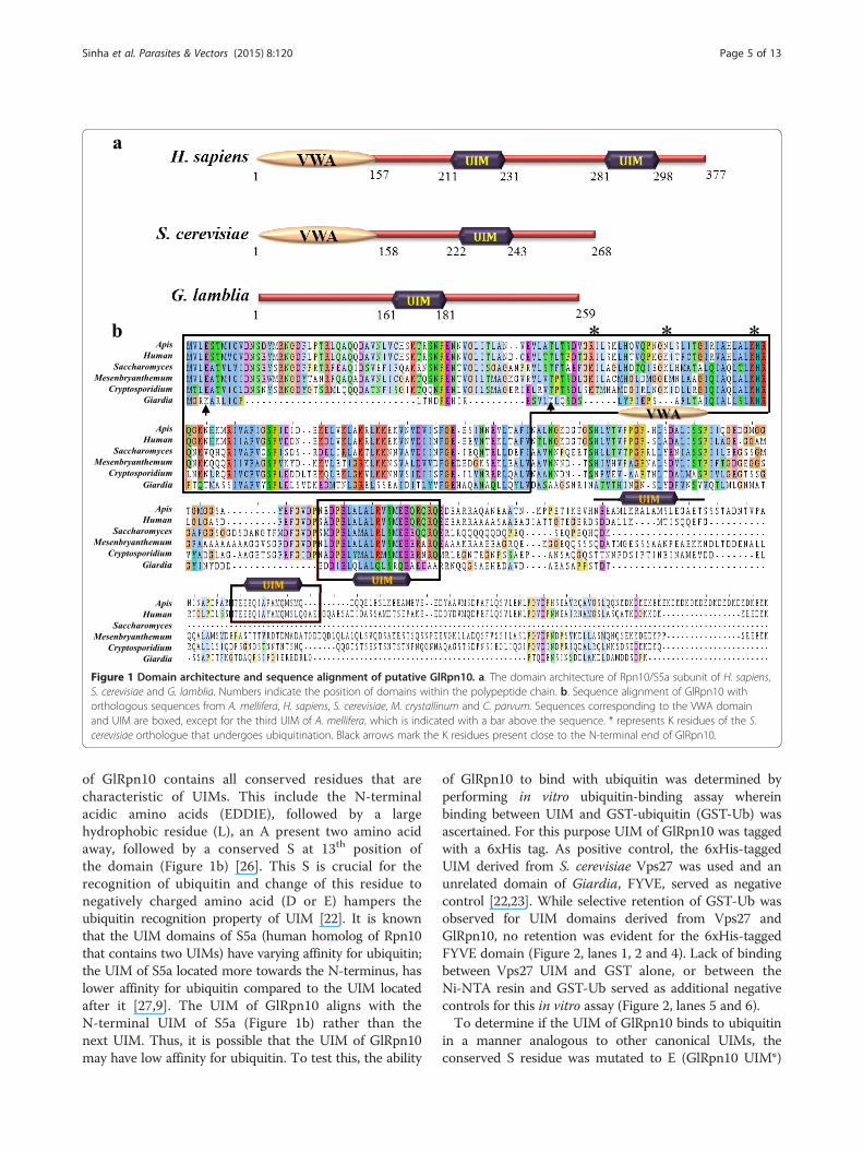
Figure 1 Domain architecture and sequence alignment of putative GlRpn10. a. The domain architecture of Rpn10/S5a subunit of H. sapiens,S. cerevisiae and G. lamblia. Numbers indicate the position of domains within the polypeptide chain. b. Sequence alignment of GlRpn10 withorthologous sequences from A. mellifera, H. sapiens, S. cerevisiae, M. crystallinum and C. parvum. Sequences corresponding to the VWA domainand UIM are boxed, except for the third UIM of A. mellifera, which is indicated with a bar above the sequence. * represents K residues of the S.cerevisiae orthologue that undergoes ubiquitination. Black arrows mark the K residues present close to the N-terminal end of GlRpn10.
Sinha et al. Parasites & Vectors (2015) 8:120 Page 5 of 13
of GlRpn10 contains all conserved residues that arecharacteristic of UIMs. This include the N-terminalacidic amino acids (EDDIE), followed by a largehydrophobic residue (L), an A present two amino acidaway, followed by a conserved S at 13th position ofthe domain (Figure 1b) [26]. This S is crucial for therecognition of ubiquitin and change of this residue tonegatively charged amino acid (D or E) hampers theubiquitin recognition property of UIM [22]. It is knownthat the UIM domains of S5a (human homolog of Rpn10that contains two UIMs) have varying affinity for ubiquitin;the UIM of S5a located more towards the N-terminus, haslower affinity for ubiquitin compared to the UIM locatedafter it [27,9]. The UIM of GlRpn10 aligns with theN-terminal UIM of S5a (Figure 1b) rather than thenext UIM. Thus, it is possible that the UIM of GlRpn10may have low affinity for ubiquitin. To test this, the ability
of GlRpn10 to bind with ubiquitin was determined byperforming in vitro ubiquitin-binding assay whereinbinding between UIM and GST-ubiquitin (GST-Ub) wasascertained. For this purpose UIM of GlRpn10 was taggedwith a 6xHis tag. As positive control, the 6xHis-taggedUIM derived from S. cerevisiae Vps27 was used and anunrelated domain of Giardia, FYVE, served as negativecontrol [22,23]. While selective retention of GST-Ub wasobserved for UIM domains derived from Vps27 andGlRpn10, no retention was evident for the 6xHis-taggedFYVE domain (Figure 2, lanes 1, 2 and 4). Lack of bindingbetween Vps27 UIM and GST alone, or between theNi-NTA resin and GST-Ub served as additional negativecontrols for this in vitro assay (Figure 2, lanes 5 and 6).To determine if the UIM of GlRpn10 binds to ubiquitin
in a manner analogous to other canonical UIMs, theconserved S residue was mutated to E (GlRpn10 UIM*)
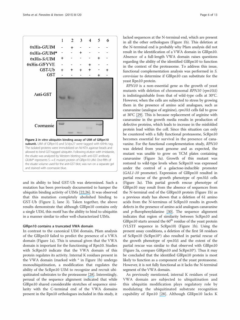
Figure 2 In vitro ubiquitin binding assay of UIM of GlRpn10subunit. UIM of GlRpn10 and ScVps27 were tagged with 6XHis tag.The isolated proteins were immobilized on Ni-NTA agarose beads andallowed to bind GST-tagged ubiquitin. Following elution with imidazole,the eluate was analyzed by Western blotting with anti-GST antibody.GlUIM* represents S→ E mutant protein of GlRpn10 UIM. One-fifth ofthe eluate volume used for the anit-GST blot, was run on a separate geland stained with coomassie blue.
Sinha et al. Parasites & Vectors (2015) 8:120 Page 6 of 13
and its ability to bind GST-Ub was determined. Such amutation has been previously documented to hamper theubiquitin binding activity of UIMs [22,26]. It was observedthat this mutation completely abolished binding toGST-Ub (Figure 2, lane 3). Taken together, the aboveresults demonstrate that although GlRpn10 contains onlya single UIM, this motif has the ability to bind to ubiquitinin a manner similar to other well-characterized UIMs.
GlRpn10 contains a truncated VWA domainIn contrast to the canonical UIM domain, Pfam analysisof the GlRpn10 failed to predict the presence of a VWAdomain (Figure 1a). This is unusual given that the VWAdomain is important for the functioning of Rpn10. Studieswith ScRpn10 indicate that the VWA domain of thisprotein regulates its activity. Internal K residues present inthe VWA domain (marked with * in Figure 1b) undergomonoubiquitination, a modification that regulates theability of the ScRpn10 UIM to recognize and recruit ubi-quitinated substrates to the proteasome [28]. Interestingly,perusal of the sequence alignment indicated that whileGlRpn10 shared considerable stretches of sequence simi-larity with the C-terminal end of the VWA domainspresent in the Rpn10 orthologues included in this study, it
lacked sequences at the N-terminal end, which are presentin all the other orthologues (Figure 1b). This deletion atthe N-terminal end is probably why Pfam analysis did notresult in the identification of a VWA domain in GlRpn10.Absence of a full-length VWA domain raises questionsregarding the ability of the identified GlRpn10 to functionin the context of the proteasome. To address this issue,functional complementation analysis was performed in S.cerevisiae to determine if GlRpn10 can substitute for theyeast Rpn10 protein.RPN10 is a non-essential gene as the growth of yeast
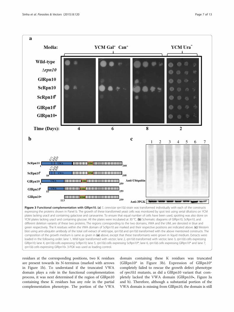
mutants with deletion of chromosomal RPN10 (rpn10Δ)is indistinguishable from that of wild-type cells at 30°C.However, when the cells are subjected to stress by growingthem in the presence of amino acid analogues, such ascanavanine (analogue of arginine), rpn10Δ cells fail to growat 30°C [29]. This is because replacement of arginine withcanavanine in the growth media results in production ofdefective proteins, which leads to increase in the misfoldedprotein load within the cell. Since this situation can onlybe countered with a fully functional proteasome, ScRpn10becomes essential for survival in the presence of cana-vanine. For the functional complementation study, RPN10was deleted from yeast genome and as expected, themutant was unable to grow on YCM plates containingcanavanine (Figure 3a). Growth of this mutant wasrestored to wild-type levels when ScRpn10 was expressedunder the control of a galactose-inducible promoter(GAL1-10 promoter). Expression of GlRpn10 resulted inpartial rescue of the growth phenotype of rpn10Δ cells(Figure 3a). This partial growth rescue phenotype ofGlRpn10 may result from the absence of sequences fromthe N-terminal end of the GlRpn10 protein (Figure 1b) asa previous study has shown that a deletion of 61 aminoacids from the N-terminus of ScRpn10 results in growthdefects in the presence of amino acid analogues canavanineand p-flurophenylalanine [30]. The sequence alignmentindicates that region of similarity between ScRpn10 andGlRpn10 starts around the 60th residue of the yeast protein(VLSTF sequence in ScRpn10) (Figure 1b). Using thepresent assay conditions, a deletion of the first 58 residuesof ScRpn10 (ScRpn10*) also resulted in partial rescue ofthe growth phenotype of rpn10Δ and the extent of thepartial rescue was similar to that observed with GlRpn10(Figure 3a, compare GlRpn10 and ScRpn10*). Thus it maybe concluded that the identified GlRpn10 protein is mostlikely to function as a component of the yeast proteasome.However, it is not fully functional as it lacks the N-terminalsegment of the VWA domain.As previously mentioned, internal K residues of yeast
VWA domain are subjected to ubiquitination andthis ubiquitin modification plays regulatory role bymodulating the ubiquitinated substrate recognitioncapability of Rpn10 [28]. Although GlRpn10 lacks K
c1 2 3 4 5 6 7
Anti-Ubiquitin
Anti-3PGK
Figure 3 Functional complementation with GlRpn10. (a) S. cerevisiae rpn10Δ strain was transformed individually with each of the constructsexpressing the proteins shown in Panel b. The growth of these transformed yeast cells was monitored by spot test using serial dilutions on YCMplates lacking uracil and containing galactose and canavanine. To ensure that equal number of cells have been used, spotting was also done onYCM plates lacking uracil and containing glucose. All the plates were incubated at 30 °C. (b) Schematic diagrams of GlRpn10, ScRpn10, anddifferent deletion variants of these two proteins. The regions corresponding to the two domains, VWA and the UIM, are denoted in blue andgreen respectively. The K residues within the VWA domain of ScRpn10 are marked and their respective positions are indicated above. (c) Westernblot using anti-ubiquitin antibody of the total cell extract of wild-type, rpn10Δ and rpn10Δ transformed with the above mentioned constructs. Thecomposition of the growth medium is same as given in (a) above, except that these transformants were grown in liquid medium. Extracts wereloaded in the following order: lane 1, Wild-type transformed with vector; lane 2, rpn10Δ transformed with vector; lane 3, rpn10Δ cells expressingGlRpn10; lane 4, rpn10Δ cells expressing ScRpn10; lane 5, rpn10Δ cells expressing ScRpn10*; lane 6, rpn10Δ cells expressing GlRpn10* and lane 7,rpn10Δ cells expressing GlRpn10•. 3-PGK was used as loading control.
Sinha et al. Parasites & Vectors (2015) 8:120 Page 7 of 13
residues at the corresponding positions, two K residuesare present towards its N-terminus (marked with arrowsin Figure 1b). To understand if the truncated VWAdomain plays a role in the functional complementationprocess, it was next determined if the region of GlRpn10containing these K residues has any role in the partialcomplementation phenotype. The portion of the VWA
domain containing these K residues was truncated(GlRpn10* in Figure 3b). Expression of GlRpn10*completely failed to rescue the growth defect phenotypeof rpn10Δ mutants, as did a GlRpn10 variant that com-pletely lacked the VWA domain (GlRpn10•, Figure 3aand b). Therefore, although a substantial portion of theVWA domain is missing from GlRpn10, the domain is still
Sinha et al. Parasites & Vectors (2015) 8:120 Page 8 of 13
essential and thus may retain the ability to discharge someof the functions of the full-length version of the domain.A biochemical approach was used to validate the
results of the complementation studies. In absence offunctional Rpn10, yeast cells accumulate ubiquitinatedproteins [29]. The overall levels of ubiquitinated proteinspresent in the cells harboring all the above-mentionedRpn10 variations were determined. Western blottingwith anti-ubiquitin antibody showed that the levels ofubiquitinated proteins, relative to wild-type cells, increasedwhen rpn10Δ mutants were grown in the presence ofcanavanine (Figure 3c, lanes 1 and 2). While the amountof ubiquitinated proteins was restored to wild-type levelswith the expression of ScRpn10, expression of GlRpn10resulted in only partial reduction (Figure 3c, lanes 3 and 4).Expression of ScRpn10* also reduced the ubiquitinatedproteins to levels comparable to that of GlRpn10(Figure 3c, lane 5). However, the expression of GlRpn10*and GlRpn10• failed to cause any detectable reduction ofthe cellular ubiquitinated protein levels compared to thatobserved in rpn10Δ mutants (Figure 3c, lanes 6 and 7).Thus the cellular ubiquitin levels are consistent with thegrowth of these mutants on canavanine plates. Therefore,both genetic and biochemical approaches indicate theGlRpn10 is capable of functioning in context of prote-asome and it encodes a reduced VWA domain that is onlypartially functional compared to the yeast VWA domain.
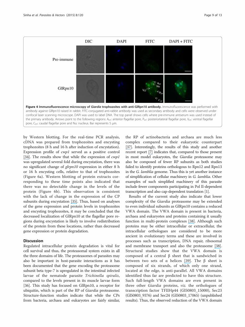
Unique distribution of GlRpn10 in trophozoitesStefanic et al. have previously reported the CP subunitcomponent, Glα7, has both nuclear and cytoplasmicdistribution [31]. To determine if GlRpn10 has a similar cel-lular distribution, polyclonal antibody was raised against therecombinant GlRpn10 in rabbit. The antibody recognized aprotein of approximately 28 kDa that is not detectable withthe pre-immune sera (Additional file 2: Figure S1). This sizeis consistent with the predicted size of GlRpn10, which iscomposed of 259 amino acids. This antibody was used forperforming immunofluorescence experiment and the cellswere observed using confocal laser scanning microscopy.Consistent with the previous report for Glα7, both nuclearand cytoplasmic pools of GlRpn10 was observed (Figure 4,bottom panel and Additional file 3: Video 1). Additionally,GlRpn10 also localized to eight bright spots that are locatedat or near the cell periphery (Figure 4, bottom panel). Thesespots appeared at regions of the cell periphery from wherethe anterior, posteriorlateral, ventral and caudal flagellaemerge, i.e. the flagellar pores. The intensity of the signalwas the maximum at the anterior flagellar pores and leastat the caudal flagellar pores. Thus, in addition to theexpected nuclear and cytoplasmic distribution, GlRpn10also has a unique localization at the flagellar pore regions.Given that the components of CP and the base of the RPdo not localize at flagellar pores [31], this distribution may
arise from a pool of GlRpn10 that is not associated with theproteasome.
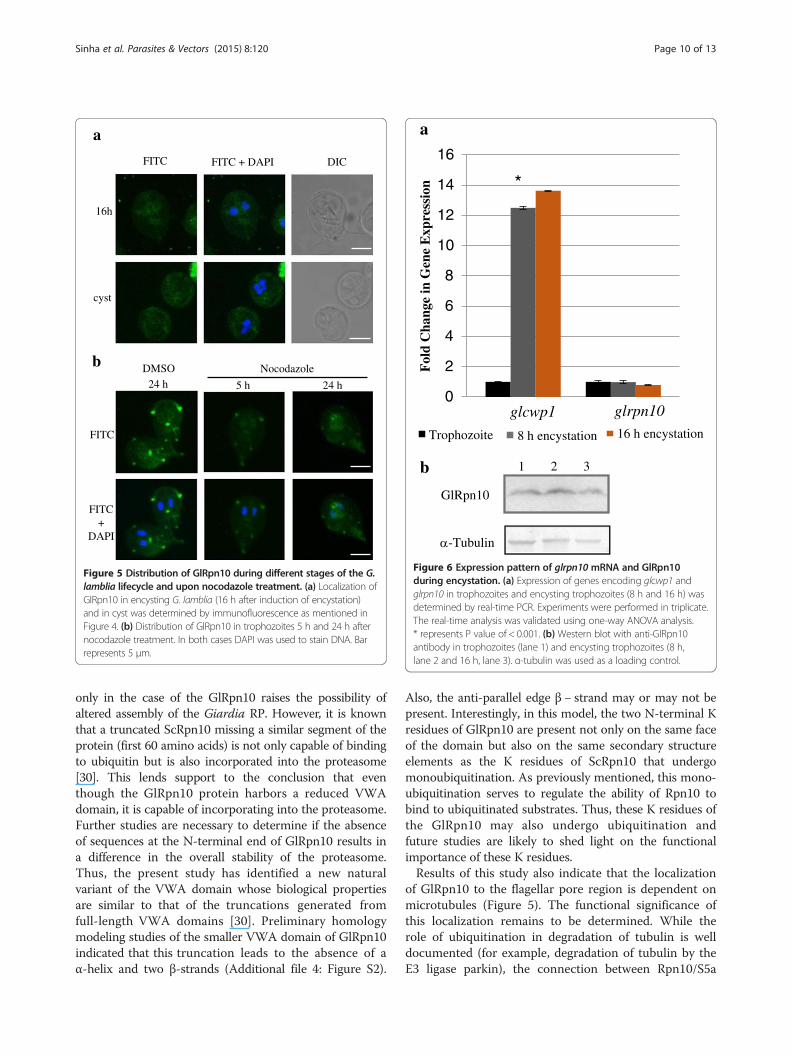
Localization of GlRpn10 to the flagellar pores ismicrotubule-dependentAs GlRpn10 localized to the flagellar pores, the role ofthe flagella, if any, in such a selective localization wasinvestigated. Towards this goal, the distribution ofGlRpn10 was determined in encysting trophozoitesand cysts as flagella start to regress during encystation andare completely internalized in cysts [1]. Trophozoites wereinduced to undergo encystation with bovine bile and thelocalization of GlRpn10 was determined in encysting tro-phozoites (16 h post-induction) and cysts. In the encystingtrophozoites, it was observed that while the signal forGlRpn10 persisted in the cytoplasm and the nucleus, itsdistribution at the flagellar pore region was not evident(Figure 5a). In the tetranucleated cysts, GlRpn10 wasdistributed in the cytoplasm (Figure 5a). Thus, thereappears to be a selective reduction of the GlRpn10 signalonly at the flagellar pores of encysting cells.Since GlRpn10 localizes at the flagellar pores and
flagellar pores are enriched in microtubular structures[32], it was next determined if this localization isdependent on microtubules. As nocodazole hampersmicrotubule polymerization, it was determined if thedistribution of GlRpn10 is altered upon treatmentwith this drug. Based on previous studies, trophozoiteswere exposed to 10 μM nocodazole for 5 h and 24 h;DMSO treatment served as control [32,33]. Following thistreatment, GlRpn10 was immunolocalized and it wasobserved that, in comparison to DMSO-treated controlcells, the presence of GlRpn10 at the flagellar poresdecreased in nocodazole-treated cells (Figure 5b). Whilethe distribution in the nucleus and cytoplasm remainedunaltered, a time-dependent decrease in intensity wasobserved at the flagellar pore regions in the nocodazole-treated cells. Staining of alpha tubulin revealed thedepolymerization of microtubule structures, such as themedian body, in nocodazole-treated cells (data not shown).Taken together, these results indicate that the selectivedistribution of this protein at the flagellar pore region ismicrotubule dependent.
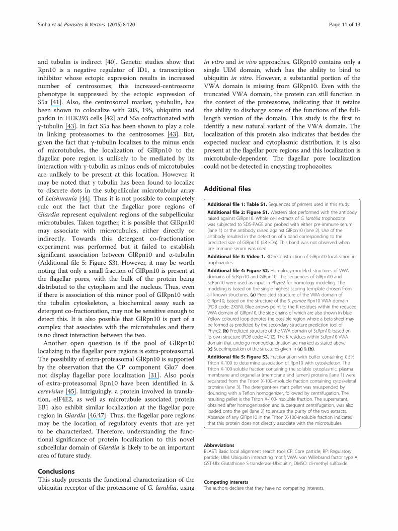
Expression pattern of glrpn10 during encystation in cystsThe disappearance of GlRpn10 from the base of theflagella of encysting cells may also result from decreasedexpression of this protein. To determine if the observedselective distribution of GlRpn10 at the flagellar pores oftrophozoites and the subsequent selective disappearancefrom this location during the process of encystation entailsany change in the cellular levels of GlRpn10, the expressionpattern of the encoding gene was monitored by real-timePCR and modulation of the protein levels was determined
AFPNu
VFPPFP
CFP
Pre-immune
GlRpn10
DIC DAPI FITC DAPI + FITC
Figure 4 Immunofluorescence microscopy of Giardia trophozoites with anti-GlRpn10 antibody. Immunofluorescence was performed withantibody against GlRpn10 raised in rabbit. FITC-conjugated anti-rabbit antibody was used as secondary antibody and cells were observed underconfocal laser scanning microscope. DAPI was used to label DNA. The top panel shows cells where pre-immune antiserum was used instead ofthe primary antibody. Arrows point to the following regions: AFP: anterior flagellar pore, PFP: posteriorlateral flagellar pore, VFP: ventral flagellarpore, CFP: caudal flagellar pore and Nu: nucleus. Bar represents 5 μm.
Sinha et al. Parasites & Vectors (2015) 8:120 Page 9 of 13
by Western blotting. For the real-time PCR analysis,cDNA was prepared from trophozoites and encystingtrophozoites (8 h and 16 h after induction of encystation).Expression profile of cwp1 served as a positive control[34]. The results show that while the expression of cwp1was upregulated several fold during encystation, there wasno significant change of glrpn10 expression in either 8 hor 16 h encysting cells, relative to that of trophozoites(Figure 6a). Western blotting of protein extracts cor-responding to these time points also indicated thatthere was no detectable change in the levels of theprotein (Figure 6b). This observation is consistentwith the lack of change in the expression of the CPsubunits during encystation [35]. Thus, based on analysesof the gene expression and protein levels in trophozoitesand encysting trophozoites, it may be concluded that thedecreased localization of GlRpn10 at the flagellar pore re-gions during encystation is likely to involve redistributionof the protein from these locations, rather than decreasedgene expression or protein degradation.
DiscussionRegulated intracellular protein degradation is vital forcell survival and thus, the proteasomal system exists in allthe three domains of life. The proteasomes of parasites mayalso be important in host-parasite interactions as it hasbeen documented that the gene encoding the proteasomesubunit beta type-7 is upregulated in the intestinal infectedlarvae of the nematode parasite Trichinella spiralis,compared to the levels present in its muscle larvae form[36]. This study has focused on GlRpn10, a receptor forubiquitin, which is part of the RP of Giardia proteasome.Structure-function studies indicate that while the CPsfrom bacteria, archaea and eukaryotes are fairly similar,
the RP of actinobacteria and archaea are much lesscomplex compared to their eukaryotic counterpart[37]. Interestingly, the results of this study and anotherrecent report [7] indicates that, compared to those presentin most model eukaryotes, the Giardia proteasome mayalso be composed of fewer RP subunits as both studiesfailed to identify proteins orthologous to Rpn12 and Rpn13in the G. lamblia genome. Thus this is yet another instanceof simplification of cellular machinery in G. lamblia. Otherexamples of such simplified machinery of this parasiteinclude fewer components participating in Pol II-dependenttranscription and also cap-dependent translation [1].Results of the current study also indicate that lower
complexity of the Giardia proteasome may be extendedto even individual subunits as GlRpn10 contains a reducedVWA domain. The VWA domain is present in bacteria,archaea and eukaryotes and proteins containing it usuallyfunction in multi-protein complexes [38]. Although suchproteins may be either intracellular or extracellular, theintracellular orthologues are considered to be moreancient in evolutionary terms and these are involved inprocesses such as transcription, DNA repair, ribosomaland membrane transport and also the proteasome [38].Structural studies show that the VWA domain iscomposed of a central β sheet that is sandwiched inbetween two sets of α helices [39]. The β sheet iscomposed of six strands, of which only one strand,located at the edge, is anti-parallel. All VWA domainsidentified thus far are predicted to have this structure.Such full-length VWA domains are even present inthree other Giardia proteins, viz. the orthologues oftranscription factor TFIIHp44 (Gl50803_15000), Sec23(Gl50803_9376) and Sec24 (Gl50803_17065) (unpublishedresults). Thus, the observed reduction of the VWA domain
0
2
4
6
8
10
12
14
16
Fol
d C
hang
e in
Gen
e E
xpre
ssio
nTrophozoite 8 h encystation 16 h encystation
glcwp1 glrpn10
*
1 2 3
GlRpn10
-Tubulin
Figure 6 Expression pattern of glrpn10 mRNA and GlRpn10during encystation. (a) Expression of genes encoding glcwp1 andglrpn10 in trophozoites and encysting trophozoites (8 h and 16 h) wasdetermined by real-time PCR. Experiments were performed in triplicate.The real-time analysis was validated using one-way ANOVA analysis.* represents P value of < 0.001. (b) Western blot with anti-GlRpn10antibody in trophozoites (lane 1) and encysting trophozoites (8 h,lane 2 and 16 h, lane 3). α-tubulin was used as a loading control.
16h
cyst
FITC
FITC+
DAPI
FITC FITC + DAPI DIC
DMSO Nocodazole24 h 24 h5 h
a
b
Figure 5 Distribution of GlRpn10 during different stages of the G.lamblia lifecycle and upon nocodazole treatment. (a) Localization ofGlRpn10 in encysting G. lamblia (16 h after induction of encystation)and in cyst was determined by immunofluorescence as mentioned inFigure 4. (b) Distribution of GlRpn10 in trophozoites 5 h and 24 h afternocodazole treatment. In both cases DAPI was used to stain DNA. Barrepresents 5 μm.
Sinha et al. Parasites & Vectors (2015) 8:120 Page 10 of 13
only in the case of the GlRpn10 raises the possibility ofaltered assembly of the Giardia RP. However, it is knownthat a truncated ScRpn10 missing a similar segment of theprotein (first 60 amino acids) is not only capable of bindingto ubiquitin but is also incorporated into the proteasome[30]. This lends support to the conclusion that eventhough the GlRpn10 protein harbors a reduced VWAdomain, it is capable of incorporating into the proteasome.Further studies are necessary to determine if the absenceof sequences at the N-terminal end of GlRpn10 results ina difference in the overall stability of the proteasome.Thus, the present study has identified a new naturalvariant of the VWA domain whose biological propertiesare similar to that of the truncations generated fromfull-length VWA domains [30]. Preliminary homologymodeling studies of the smaller VWA domain of GlRpn10indicated that this truncation leads to the absence of aα-helix and two β-strands (Additional file 4: Figure S2).
Also, the anti-parallel edge β − strand may or may not bepresent. Interestingly, in this model, the two N-terminal Kresidues of GlRpn10 are present not only on the same faceof the domain but also on the same secondary structureelements as the K residues of ScRpn10 that undergomonoubiquitination. As previously mentioned, this mono-ubiquitination serves to regulate the ability of Rpn10 tobind to ubiquitinated substrates. Thus, these K residues ofthe GlRpn10 may also undergo ubiquitination andfuture studies are likely to shed light on the functionalimportance of these K residues.Results of this study also indicate that the localization
of GlRpn10 to the flagellar pore region is dependent onmicrotubules (Figure 5). The functional significance ofthis localization remains to be determined. While therole of ubiquitination in degradation of tubulin is welldocumented (for example, degradation of tubulin by theE3 ligase parkin), the connection between Rpn10/S5a
Sinha et al. Parasites & Vectors (2015) 8:120 Page 11 of 13
and tubulin is indirect [40]. Genetic studies show thatRpn10 is a negative regulator of ID1, a transcriptioninhibitor whose ectopic expression results in increasednumber of centrosomes; this increased-centrosomephenotype is suppressed by the ectopic expression ofS5a [41]. Also, the centrosomal marker, γ-tubulin, hasbeen shown to colocalize with 20S, 19S, ubiquitin andparkin in HEK293 cells [42] and S5a cofractionated withγ-tubulin [43]. In fact S5a has been shown to play a rolein linking proteasomes to the centrosomes [43]. But,given the fact that γ-tubulin localizes to the minus endsof microtubules, the localization of GlRpn10 to theflagellar pore region is unlikely to be mediated by itsinteraction with γ-tubulin as minus ends of microtubulesare unlikely to be present at this location. However, itmay be noted that γ-tubulin has been found to localizeto discrete dots in the subpellicular microtubular arrayof Leishmania [44]. Thus it is not possible to completelyrule out the fact that the flagellar pore regions ofGiardia represent equivalent regions of the subpellicularmicrotubules. Taken together, it is possible that GlRpn10may associate with microtubules, either directly orindirectly. Towards this detergent co-fractionationexperiment was performed but it failed to establishsignificant association between GlRpn10 and α-tubulin(Additional file 5: Figure S3). However, it may be worthnoting that only a small fraction of GlRpn10 is present atthe flagellar pores, with the bulk of the protein beingdistributed to the cytoplasm and the nucleus. Thus, evenif there is association of this minor pool of GlRpn10 withthe tubulin cytoskeleton, a biochemical assay such asdetergent co-fractionation, may not be sensitive enough todetect this. It is also possible that GlRpn10 is part of acomplex that associates with the microtubules and thereis no direct interaction between the two.Another open question is if the pool of GlRpn10
localizing to the flagellar pore regions is extra-proteasomal.The possibility of extra-proteasomal GlRpn10 is supportedby the observation that the CP component Glα7 doesnot display flagellar pore localization [31]. Also poolsof extra-proteasomal Rpn10 have been identified in S.cerevisiae [45]. Intriguingly, a protein involved in transla-tion, eIF4E2, as well as microtubule associated proteinEB1 also exhibit similar localization at the flagellar poreregion in Giardia [46,47]. Thus, the flagellar pore regionsmay be the location of regulatory events that are yetto be characterized. Therefore, understanding the func-tional significance of protein localization to this novelsubcellular domain of Giardia is likely to be an importantarea of future study.
ConclusionsThis study presents the functional characterization of theubiquitin receptor of the proteasome of G. lamblia, using
in vitro and in vivo approaches. GlRpn10 contains only asingle UIM domain, which has the ability to bind toubiquitin in vitro. However, a substantial portion of theVWA domain is missing from GlRpn10. Even with thetruncated VWA domain, the protein can still function inthe context of the proteasome, indicating that it retainsthe ability to discharge some of the functions of the full-length version of the domain. This study is the first toidentify a new natural variant of the VWA domain. Thelocalization of this protein also indicates that besides theexpected nuclear and cytoplasmic distribution, it is alsopresent at the flagellar pore regions and this localization ismicrotubule-dependent. The flagellar pore localizationcould not be detected in encysting trophozoites.
Additional files
Additional file 1: Table S1. Sequences of primers used in this study.
Additional file 2: Figure S1. Western blot performed with the antibodyraised against GlRpn10. Whole cell extracts of G. lamblia trophozoitewas subjected to SDS-PAGE and probed with either pre-immune serum(lane 1) or the antibody raised against GlRpn10 (lane 2). Use of theantibody resulted in the detection of a band corresponding to thepredicted size of GlRpn10 (28 kDa). This band was not observed whenpre-immune serum was used.
Additional file 3: Video 1. 3D-reconstruction of GlRpn10 localization introphozoites.
Additional file 4: Figure S2. Homology-modeled structures of VWAdomains of ScRpn10 and GlRpn10. The sequences of GlRpn10 andScRpn10 were used as input in Phyre2 for homology modeling. Themodeling is based on the single highest scoring template chosen fromall known structures. (a) Predicted structure of the VWA domain ofGlRpn10, based on the structure of the S. pombe Rpn10 VWA domain(PDB code: 2X5N). Blue arrows point to the K residues within the reducedVWA domain of GlRpn10, the side chains of which are also shown in blue.Yellow coloured loop denotes the possible region where a beta-sheet maybe formed as predicted by the secondary structure prediction tool ofPhyre2. (b) Predicted structure of the VWA domain of ScRpn10, based onits own structure (PDB code: 4CR2). The K residues within ScRpn10 VWAdomain that undergo monoubiquitination are marked as stated above.(c) Superimposition of the structures given in (a) & (b).
Additional file 5: Figure S3. Fractionation with buffer containing 0.5%Triton X-100 to determine association of Rpn10 with cytoskeleton. TheTriton X-100-soluble fraction containing the soluble cytoplasmic, plasmamembrane and organellar (membrane and lumen) proteins (lane 1) wereseparated from the Triton X-100-insoluble fraction containing cytoskeletalproteins (lane 3). The detergent-resistant pellet was resuspended bydouncing with a Teflon homogenizer, followed by centrifugation. Theresulting pellet is the Triton X-100-insoluble fraction. The supernatant,obtained after homogenization and subsequent centrifugation, was alsoloaded onto the gel (lane 2) to ensure the purity of the two extracts.Absence of any GlRpn10 in the Triton X-100-insoluble fraction indicatesthat this protein does not directly associate with the microtubules.
AbbreviationsBLAST: Basic local alignment search tool; CP: Core particle; RP: Regulatoryparticle; UIM: Ubiquitin interacting motif; VWA: von Willebrand factor type A;GST-Ub: Glutathione S-transferase-Ubiquitin; DMSO: di-methyl sulfoxide.
Competing interestsThe authors declare that they have no competing interests.
Sinha et al. Parasites & Vectors (2015) 8:120 Page 12 of 13
Authors’ contributionsSS conceived of the study, AS, SPD and AR carried out the experiments, ASand SS analyzed the data. SS wrote the manuscript with help from AS andSPD. All authors have read and approved the final manuscript.
Authors’ informationAS is presently a postdoctoral fellow at the Department of Biology andEnvironment, Faculty of Natural Sciences, University of Haifa at Oranim, Israel.
AcknowledgementsThe authors acknowledge Dr. Alok Sil for valuable suggestions during thecourse of this study and also for critically reading the manuscript. Prof.Parimal C. Sen and Dr. Sandipan Ganguly are acknowledged for helpfuldiscussions and Prof. Pratima Sinha for the gift of pUS234 vector. Authorsalso acknowledge Boni Halder of the DBT-IPLS Confocal Microscopy Facilityof the University of Calcutta for assistance with imaging and the CentralInstrument Facility of Bose Institute for real-time PCR and DNA sequencing.This work was supported by research grants from the Dept. of Biotechnology,Govt. of India (Project No. BT/PR3116/BRB/10/957/2011) and the Bose Institute.AS received fellowship support from Bose Institute and CSIR, SPD is aDST-INSPIRE scholar and AR received fellowship support from UniversityGrants Commission.
Received: 8 August 2014 Accepted: 13 February 2015
References1. Lujẚn HD, Svärd S. Giardia: A model organism. New York: Springer
Wien; 2011.2. Faso C, Bischof S, Hehl AB. The proteome landscape of Giardia lamblia
encystation. PLoS One. 2013;8:12.3. Lee DH, Goldberg AL. Proteasome inhibitors: valuable new tools for cell
biologists. Trends Cell Biol. 1998;8:397–403.4. Jung T, Grune T. Structure of the proteasome. Prog Mol Biol Transl Sci.
2012;109:1–39.5. Emmerlich V, Santarius U, Bakker-Grunwald T, Scholze H. Isolation and
subunit composition of the 20S proteasome of Giardia lamblia.Mol Biochem Parasitol. 1999;100:131–4.
6. Gallego E, Alvarado M, Wasserman M. Identification and expression of theprotein ubiquitination system in Giardia intestinalis. Parasitol Res.2007;101:1–7.
7. Jerlström-Hultqvist J, Stadelmann B, Birkestedt S, Hellman U, Svärd SG.Plasmid vectors for proteomic analyses in Giardia: purification of virulencefactors and analysis of the proteasome. Eukaryot Cell. 2012;11:864–73.
8. Sakata E, Bohn S, Mihalache O, Kiss P, Beck F, Nagy I, et al. Localization ofproteasomal ubiquitin receptors Rpn10 and Rpn13 by electroncryomicroscopy. Proc Natl Acad Sci U S A. 2012;109:1479–84.
9. Wang Q, Young P, Walters KJ. Structure of S5a bound to monoubiquitinprovides a model for polyubiquitin recognition. J Mol Biol. 2005;348:727–39.
10. Husnjak K, Elsasser S, Zhang N, Chen X, Randles L, Shi Y, et al. Proteasomesubunit Rpn13 is a novel ubiquitin receptor. Nature. 2008;453:481–8.
11. Schreiner P, Chen X, Husnjak K, Randles L, Zhang N, Elsasser S, et al.Ubiquitin docking at the proteasome through a novel pleckstrin-homologydomain interaction. Nature. 2008;453:548–52.
12. Lam YA, Lawson TG, Velayutham W, Zweier JL, Pickart CM. A proteasomalATPase subunit recognizes the polyubiquitin degradation signal. Nature.2002;416:763–7.
13. Chen L, Shinde U, Ortolan TG, Madura K. Ubiquitin-associated (UBA) domainsin Rad23 bind ubiquitin and promote inhibition of multi-ubiquitin chainassembly. EMBO Rep. 2001;2:933–8.
14. Funakoshi M, Sasaki T, Nishimoto T, Kobayashi H. Budding Yeast Dsk2p is apolyubiquitin-binding protein that can interact with the proteasome.Proc Natl Acad Sci U S A. 2002;99:745–50.
15. Gomez TA, Kolawa N, Gee M, Sweredoski MJ, Deshaies RJ. Identification of afunctional docking site in the Rpn1 LRR domain for the UBA-UBL domainprotein Ddi1. BMC Biol. 2011;9:33.
16. Hofmann K, Bucher P. The PCI domain: a common theme in threemultiprotein complexes. Trends Biochem Sci. 1998;23:204–5.
17. Thompson JD, Higgins DG, Gibson TJ. CLUSTAL W: improving the sensitivityof progressive multiple sequence alignment through sequence weighting,
position-specific gap penalties and weight matrix choice. Nucleic Acids Res.1994;22:4673–80.
18. Waterhouse AM, Procter JB, Clamp M, Barton GJ. Jalview version 2 amultiple sequence alignment editor and analysis workbench. Bioinformatics.2009;25(9):1189–91.
19. Kane AV, Ward HD, Keusch GT, Pereira MEA. In vitro encystations of Giardialamblia: large scale production of in vitro cysts and strains and clonedifferences in encystations efficiency. J Parasitol. 1991;77:974–81.
20. Weiland MBL, McArthur AG, Morrison HG, Sogin ML, Sva¨rd SG. Annexin-likealpha giardins: a new cytoskeletal gene family in Giardia lamblia. Int JParasitol. 2005;35:617–26.
21. Majumdar S, Ghatak J, Mukerji S, Bhattacharjee H, Bhaduri A. UDPgalactose4-epimerase from Saccharomyces cerevisiae A bifunctional enzyme withaldose 1-epimerase activity. Eur J Biochem. 2004;271:753–9.
22. Shih SC, Katzmann DJ, Schnell JD, Sutanto M, Emr SD, Hicke L. Epsins andVps27p/Hrs contain ubiquitin-binding domains that function in receptorendocytosis. Nat Cell Biol. 2002;4(5):389–93.
23. Sinha A, Mandal S, Banerjee S, Ghosh A, Ganguly S, Sil AK, et al.Identification and characterization of a FYVE domain from the earlydiverging eukaryote Giardia lamblia. Curr Microbiol. 2011;62:1179–84.
24. Bradford MM. A rapid and sensitive method for quantitation of microgramquantities of protein utilizing the principle of protein-dye binding.Analytical Biochem. 1976;72:248–54.
25. Longtine MS, McKenzie III A, Demarini DJ, Shah NG, Wach A, Brachat A, et al.Additional module for versatile and economical PCR-based gene deletion andmodification in Saccharomyces cerevisiae. Yeast. 1998;13:953–61.
26. Fisher RD, Wang B, Alam SL, Higginson DS, Robinson H, Sundquist WI, et al.Structure and ubiquitin binding of the ubiquitin-interacting motif. J BiolChem. 2003;278:28976–84.
27. Young P, Deveraux Q, Beal RE, Pickart CM, Rechsteiner M. Characterizationof two polyubiquitin binding sites in the 26S protease subunit 5a. J BiolChem. 1998;273:5461–7.
28. Isasa M, Katz EJ, Kim W, Yugo V, González S, Kirkpatrick DS, et al.Monoubiquitination of Rpn10 regulates substrate recruitment to theproteasome. Mol Cell. 2010;38:733–45.
29. Lambertson D, Chen L, Madura K. Pleotropic defects caused by loss of theProteasome-Interacting factors Rad23 and Rpn10 of Saccharomyces cerevisiae.Genetics. 1999;153:69–79.
30. Fu H, Sadis S, Rubin DM, Glickman M, van Nocker S, Finley D, et al.Multiubiquitin chain binding and protein degradation are mediated bydistinct domains within the 26S proteasome subunit Mcb1. J Biol Chem.1998;273:1970–81.
31. Stefanic S, Palm D, Svard SG, Hehl AB. Organelle proteomics reveals cargomaturation mechanisms associated with Golgi-like encystation vesicles inthe early diverged protozoan Giardia lamblia. J Biol Chem.2006;281:7595–604.
32. Mariante RM, Vancini RG, Melo AL, Benchimol M. Giardia lamblia: evaluationof the in vitro effects of nocodazole and colchicine on trophozoites. ExpParasitol. 2005;110:62–72.
33. Dawson SC. Primary microtubule structures in Giardia. In: Lujan HD, Svärd S,editors. Giardia A Model Organism. New York: Springer Wien; 2011.p. 275–96.
34. Kim J, Bae SS, Sung MH, Lee KH, Park SJ. Comparative proteomic analysis oftrophozoites versus cysts of Giardia lamblia. Parasitol Res. 2009;104:475–9.
35. Emmerlich V, Scholze H, Gillin FD, Bakker-Grunwald T. Characterization ofproteasome alpha-chain from Giardia lamblia. Parasitol Res. 2001;87:112–5.
36. Yang W, Li L, Liu R, Sun G, Liu C, Zhang S, et al. Molecular identification andcharacterization of Trichinella spiralis proteasome subunit beta type-7.Parasit Vectors. 2015;8:18.
37. Maupin-Furlow J. Proteasomes and protein conjugation across domains oflife. Nat Rev Microbiol. 2012;10:100–11.
38. Whittaker CA, Hynes RO. Distribution and Evolution of von Willebrand/Integrin A domains: widely dispersed domains with roles in cell adhesionand elsewhere. Mol Biol Cell. 2002;13:3369–87.
39. Springer T. A. Complement and the multifaceted functions of VWA andintegrin I domains. Structure. 2006;14:1611–6.
40. Ren Y et al. Perkin binds to alpha/beta tubulin and increases theirubiquitination and degradation. J Neurosci. 2003;23:3316–24.
41. Hasskarl J, Mern DS, Munger K. Interference of the dominant negativehelix-loop-helix protein ID1 with proteasomal subunit S5a causes centrosomalabnormalities. Oncogene. 2008;27:1657–64.

Sinha et al. Parasites & Vectors (2015) 8:120 Page 13 of 13
42. Wigley WC, Fabunmi RP, Lee MG, Marino CR, Maullem S, DeMartino GN,et al. Dynamic association of proteasomal machinery with the centrosome.J Cell Biol. 1999;145:481–90.
43. Puram SV, Kim AH, Park HY, Anckar J, Bonni A. The ubiquitin receptorS5a/Rpn10 links centrosomal proteasomes with dendrite development inthe mammalian brain. Cell Rep. 2013;11:19–30.
44. Libusova L, Sulimenko T, Sulimenko V, Hozak P, Draber P. γ − Tubulin inLeishmania: cell-cycle-dependent changes in subcellular localization andheterogeneity of its isoforms. Exp Cell Res. 2004;295:375–86.
45. Matiuhin Y, Kirkpatrick DS, Ziv I, Kim W, Dakshinamurthy A, Kleifeld O, et al.Extraproteasomal Rpn10 restricts access of the polyubiquitin-binding proteinDsk2 to proteasome. Mol Cell. 2008;32:415–25.
46. Li L, Wang CC. Identification in the ancient protest Giardia lamblia of twoeukaryotic translation initiation factor 4E homologs with distinctivefunctions. Euk Cell. 2005;4:948–59.
47. Dawson SC, Sagolla MS, Mancuso JJ, Woessner DJ, House SA, Fritz-Laylin L,et al. Kinesin-13 regulates flagellar, interphase, and mitotic microtubuledynamics in G. intestinalis. Euk Cell. 2007;6:2354–64.
Submit your next manuscript to BioMed Centraland take full advantage of:
• Convenient online submission
• Thorough peer review
• No space constraints or color figure charges
• Immediate publication on acceptance
• Inclusion in PubMed, CAS, Scopus and Google Scholar
• Research which is freely available for redistribution
Submit your manuscript at www.biomedcentral.com/submit