a new minimally invasive mesotherapy technique for facial rejuvenation
TRANSCRIPT

ORIGINAL RESEARCH
A New Minimally Invasive Mesotherapy Techniquefor Facial Rejuvenation
Antonella Savoia • Simone Landi • Alfonso Baldi
To view enhanced content go to www.dermtherapy-open.comReceived: October 19, 2012 / Published online: January 8, 2013� The Author(s) 2013. This article is published with open access at Springerlink.com
ABSTRACT
Introduction: This study describes a pivotal
clinical trial of a new minimally invasive
mesotherapy technique for facial rejuvenation.
Methods: The authors utilized two
formulations: formulation A with hyaluronic
acid, vitamins, amino acids, minerals,
coenzymes, and antioxidant substances;
formulation B with hyaluronic acid and
idebenone. Fifty participants were enrolled in
the study and divided in two groups. Group 1
(50–65 years) treated with formulation A. Group
2 (35–50 years) treated with formulation B. The
groups underwent four sessions of mesotherapy
involving multiple injections. Treatment was
conducted at 15 day intervals. All participants
had pre- and posttreatment photographs.
Punch biopsies were taken from randomly
selected participants, baseline and after
6 weeks, and stained for interleukin (IL)-6, IL-
1b, matrix metalloproteinase (MMP)-1, and
collagen 1. Clinical evaluation was based on
the Global Aesthetic Scale (GAIS) and on the
Wrinkle Severity Rating Scale (WSRS).
Results: The results produced were statistically
analyzed and resulted in a significant and
long-lasting effect on facial rejuvenation.
Evaluation of photographs at 0, 1, and
2 months revealed significant clinical
improvement: brightness, texture, and
firmness of the skin. The analysis of the
GAIS and WSRS scores in the two groups
demonstrated statistically significant results
after 2 months. The biopsies taken from
randomly selected participants at baseline
and after 3 months showed a decrease in IL-
1b, IL-6, and MMP1, and an increase in
collagen 1.
Conclusion: The new minimally invasive
mesotherapy technique described can improve
the clinical appearance of the skin in different
age groups.
A. Savoia � S. LandiPromoitalia Group S.p.A, Pozzuoli, Naples, Italy
A. Baldi (&)Department of Biochemistry, Sect. Pathology,Second University of Naples, Via L. Armanni 5,80138 Naples, Italye-mail: [email protected]
Enhanced content for this article is
available on the journal web site:
www.dermtherapy-open.com
123
Dermatol Ther (Heidelb) (2013) 3:83–93
DOI 10.1007/s13555-012-0018-2

Keywords: Biorevitalization; Cosmetic
dermatology; Facial rejuvenation; Fillers;
Mesotherapy; Soft-tissue augmentation
INTRODUCTION
Aging is an increasing concern of modern
society, particularly aging of the face. This is a
complex process involving two important
factors: volume loss throughout the face, and
repetitive muscle movements that cause
wrinkles and folds [1]. Traditionally, facial
rejuvenation has focused on various
dermatologic cosmetic procedures such as
carbon dioxide laser resurfacing,
microdermabrasion, and electric stimulation
leading to collagen production in human skin
fibroblasts [2, 3]. In recent years there has been
an increasing emphasis on mesotherapy as an
anti-aging strategy. Mesotherapy is a medical
procedure introduced by Pistor in 1958, which
consists of intradermal injection of
pharmacologic substances, such as nutrients,
hormones, vitamins, enzymes, and other
reagents that have been diluted and are
administered directly into the region to be
treated [4]. The aim of mesotherapy in skin
rejuvenation is maintenance and/or restoration
of healthy and youthful texture of the skin [5,
6]. The desired final effect is firm, bright,
moisturized skin obtained by the injection in
the superficial dermis of suitable products that
are perfectly biocompatible and totally
absorbable [7]. In fact, injection of
mesotherapy products promotes skin
rejuvenation by increasing both hydration and
fibroblast activation [8, 9]. Several experimental
studies have demonstrated that hyaluronic acid
injected into the skin can stimulate fibroblasts
to express collagen type 1 (Col-1), matrix
metalloprotease-1 (MMP-1), and tissue
inhibitor of matrix metalloproteinase-1 (TIMP)
[10]. Another study suggested that dermal
injection of vitamins results in stimulation of
collagen production in skin cells [11]. It is also
well known that antioxidant substances are able
to reverse aging. One the most studied
hypotheses regarding aging is that it is caused
by oxidative stress, and oxidation can damage
proteins, DNA, and lipids.
The present study describes a pivotal
comparative clinical study of a minimally
invasive technique of skin rejuvenation
consisting of multiple intradermal injections
with two formulations with the goal of
maintenance and/or restoration of healthy and
youthful skin texture.
MATERIALS AND METHODS
In this clinical study two different formulations
were used: formulation A, which was composed
of hyaluronic acid, vitamins, amino acids,
minerals, coenzymes, and antioxidants
(Table 1), and formulation B, which was
composed of hyaluronic acid, idebenone,
polysorbate 20, water, acetyl cysteine, sodium
chloride.
Patients
Fifty healthy patients of both genders were
enrolled in the study and were divided into two
groups: group 1 (32 patients), aged between 50
and 60 years (older patients) and group 2 (18
patients), aged between 35 and 50 years
(younger patients). Formulation A was used for
patients in group 1 and formulation B was used
for patients in group 2. Exclusion criteria for the
study were: patients that had undergone other
medical-aesthetical treatments, and pregnant or
breastfeeding women. Inclusion criteria were:
84 Dermatol Ther (Heidelb) (2013) 3:83–93
123

patients in good health, patients that were not
using any other treatments, and patients that
presented mild/moderate to severe photo-aging.
The patients did not modify their lifestyle. All
patients gave written consent for enrollment
into the clinical study, and ethical approval was
obtained for the study.
Treatment and Evaluation Protocol
Each treatment was carried out according to a
default protocol, which was similar for the two
groups. Briefly, all participants underwent four
sessions of mesotherapy involving multiple
injections with a 30 G/4 mm needle in the
face, neck, and decollete. The hands of eight
patients were additionally treated. Treatments
were conducted over 2 months. The objective
examination was carried out with inspection
using a Wood’s light (GIMA S.p.A., Gessate,
Milano, Italy) and palpation to verify cutaneous
elasticity. The areas of the body treated were
examined by the sense of touch and pressure.
The results were defined with a score derived
from the Global Aesthetic Improvement Scale
(GAIS), used as a reference parameter (Table 2);
a summary of the patients’ characteristics and
of GAIS scores obtained is provided in Table 3.
The photographic evaluation was performed at
the beginning and at the end of the treatment.
Table 1 Characteristics of formulation A
Vitamins Amino acids Minerals Nucleotides Coenzymes Others
Tocopherol Arginine Sodium chloride Adenine Inositol Sodium hyaluronate
Ubiquinone Lysine Cytosine Acetyl cysteine Water
Cyanocobalamin Isoleucine Glutamine Polysorbate 20
Folic acid Leucine Guanine
Riboflavin Threonine
Pyridoxine HCL Valine
Thiamine HCL Histidine hydrochloride
Nicotinamide Tyrosine
Calcium pantothenate Phenylalanine
Ascorbic acid Glutamic acid
Aspartic acid
Asparagine
Cysteine
Proline
Serine
Alanine
Methionine
Glycine
Tryptophan
HCL hydrochloride
Dermatol Ther (Heidelb) (2013) 3:83–93 85
123

Moreover, the Wrinkle Severity Rating Scale
(WSRS) was also used to evaluate the condition
of the wrinkles and, therefore, the degree of
aging.
Histology and Immunohistochemistry
Punch biopsies were taken from randomly
selected participants at baseline and after
6 weeks. Excised tissue samples were
transferred in 4% (m/v) paraformaldehyde
solution and were paraffin embedded. Sections
of 5 lm were stained with hematoxylin–eosin,
hematoxylin–Van Gieson, and Periodic acid-
Schiff (PAS)-Alcian blue hematoxylin
(Carlo Erba, Milano, Italy). For
immunohistochemistry, tissue sections were
heated twice in a microwave oven for 5 min
each at 700 W in citrate buffer (pH 6.0). Slides
were then incubated at 4 �C overnight at 1:100
dilution with the following antibodies: mouse
monoclonal to interleukin-1b (IL-1b) (AbD
Serotec, Oxford, UK); mouse monoclonal to IL-
6 (Origene, MD, USA); mouse monoclonal to
MMP1 (Santa Cruz, CA, USA); and rabbit
polyclonal to Col-1 (Santa Cruz, CA, USA).
After three washes in phosphate-buffered
saline (PBS) to remove the excess antiserum,
the slides were incubated with diluted goat anti-
rabbit or anti-mouse biotinylated antibody
(Vector Laboratories, Burlingame, CA, USA) at
1:200 dilution in PBS-3% nonfat dry milk for
1 h. All the slides were then processed by the
avidin–biotin complex (ABC) method (Vector
Laboratories, Burlingame, CA, USA) for 30 min
at room temperature. The ABC method relies on
the strong affinity of avidin or streptavidin for
the vitamin, biotin, for the revelation of the
immune complex formed on a tissue by an
antigen and a specific antibody raised against
this antigen. Diaminobenzidine was used as the
final chromogen and hematoxylin was used as
the nuclear counter stain. Negative controls for
each tissue section were prepared by leaving out
the primary antiserum. All samples were
processed under the same conditions. The
intensity of expression per field (209 original
magnification) using light microscopy was
Table 2 Global aesthetic improvement scale
Degree Description
1 Exceptional
improvement
Excellent corrective result after a session with the Spherofill device
2 Very improved
patient
Marked improvment of the appearance, but not completely optimal. A touch-up would slightly
improve the result
3 Improved patient Improvement of the appearance, better compared to the initial condition, but a touch-up is
advised
4 Unaltered patient The appearance substantially remains the same compared to the original condition
5 Worsened patient The appearance has worsened compared to the original condition
Table 3 Wrinkle severity rating scale
Score Description
1 Absent
2 Slight
3 Moderate
4 Severe
5 Extreme
86 Dermatol Ther (Heidelb) (2013) 3:83–93
123
calculated and compared in different specimens
by two separate observers (A.B. and A.S.) in a
double-blind fashion and described as: score 0
(absent); score 1 (low); score 2 (moderate); score
3 (intense). On average 22 fields were observed
for each specimen. The percentage of
concordance between the two observers was
93% (28 over 30); in the remaining specimens
the final score was reached after collegial
revision.
Statistical Analysis
Descriptive analysis was made using median
values and 95% confidence intervals. The
differences in the GAIS scores at the different
timepoints for each group were performed using
Wilcoxon’s test for nonparametric dependent
continuous variables. Statistical Product and
Service Solutions (SPSS) software (version 17.0,
SPSS, Chicago, IL, USA) was used for statistical
analysis. A P value (two tailed) of less than 0.05
was considered to indicate statistical significance.
RESULTS
The treatment caused minimum discomfort,
without any posttreatment pain. All patients
returned to their everyday activities
immediately after the treatment. In a small
number of patients (n = 15) ecchymosis were
found; moreover, hyperemia secondary to the
traumatic action of the needle and vasoactive
substances was reported in 12 patients, but this
resolved a few days after the application of an
anti-edemigen and anti-inflammatory cream.
Edema reactions associated with moderate
pain due to pressure after the injections were
evident in five patients. On objective
examination, both formulations resulted in an
improvement in the epidermal texture, and an
increase in elasticity and brightness of the skin.
The effect of the contraction of collagen and
production of new natural collagen was
highlighted by the increase in tone and
compactness. Such features were appreciable
by gently skimming over or touching the skin.
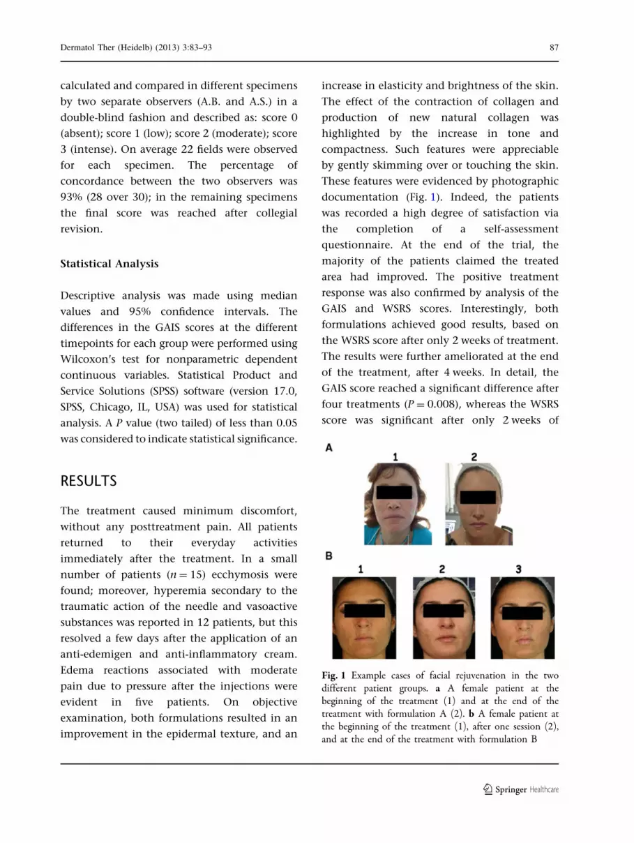
These features were evidenced by photographic
documentation (Fig. 1). Indeed, the patients
was recorded a high degree of satisfaction via
the completion of a self-assessment
questionnaire. At the end of the trial, the
majority of the patients claimed the treated
area had improved. The positive treatment
response was also confirmed by analysis of the
GAIS and WSRS scores. Interestingly, both
formulations achieved good results, based on
the WSRS score after only 2 weeks of treatment.
The results were further ameliorated at the end
of the treatment, after 4 weeks. In detail, the
GAIS score reached a significant difference after
four treatments (P = 0.008), whereas the WSRS
score was significant after only 2 weeks of
Fig. 1 Example cases of facial rejuvenation in the twodifferent patient groups. a A female patient at thebeginning of the treatment (1) and at the end of thetreatment with formulation A (2). b A female patient atthe beginning of the treatment (1), after one session (2),and at the end of the treatment with formulation B
Dermatol Ther (Heidelb) (2013) 3:83–93 87
123

Table 4 Characteristics of the patients enrolled in the study
No. Sex Age (years) Formulation used Treatment site GAIS1 GAIS2 WSRS0 WSRS1 WSRS2
1 F 52 A Face/neck 1 1 4 3 3
2 M 55 A Face/neck 1 1 3 2 2
3 F 64 A Face/decollete 3 2 5 4 4
4 M 56 A Face/neck/hands 2 2 4 4 3
5 F 53 A Face/decollete 2 1 4 3 2
6 M 60 A Face/neck 4 3 4 3 3
7 F 59 A Face/neck/hands 4 2 4 4 3
8 F 57 A Face/neck/hands 2 1 3 3 2
9 F 58 A Face/neck 3 3 4 4 3
10 F 58 A Face/decollete 2 2 3 3 2
11 F 63 A Face/decollete/hands 1 1 3 2 2
12 F 65 A Face/neck/hands 4 3 5 4 3
13 M 51 A Face/neck 2 2 4 3 3
14 F 54 A Face/decollete/hands 3 2 3 2 2
15 F 53 A Face/decollete/neck 3 3 3 3 2
16 F 56 A Face/decollete/hands 3 3 4 4 3
17 F 57 A Face/decollete 2 2 3 3 2
18 F 62 A Face/neck/hands 2 1 4 4 3
19 F 62 A Face/decollete/hands 2 1 4 4 3
20 M 64 A Face/neck 1 1 5 4 4
21 F 61 A Face/decollete 3 2 5 5 4
22 F 58 A Face/decollete/neck 3 3 3 3 2
23 F 59 A Face/decollete/neck 2 2 3 3 2
24 F 54 A Face/neck/hands 2 1 4 4 3
25 F 52 A Face/neck 2 2 3 2 2
26 F 54 A Face/decollete/neck 4 3 4 4 3
27 F 64 A Face/decollete/neck 3 2 4 4 3
28 F 62 A Face/decollete 2 2 4 4 3
29 F 63 A Face/decollete 3 1 4 3 3
30 M 61 A Face/neck 3 1 3 3 2
31 F 60 A Face/decollete/neck 1 1 4 4 3
32 F 60 A Face/decollete/neck 3 2 3 2 2
33 F 37 A Face/decollete/neck 1 1 2 2 1
88 Dermatol Ther (Heidelb) (2013) 3:83–93
123

treatment (P = 0.014), and this significance was
augmented after four treatments (P = 0.002).
These data are summarized in Table 4.
To further characterize the effects of the
skin treatment, histological and
immunohistochemical analysis of the skin of
four patients treated with formulation A
(n = 4) was performed at baseline and after
6 weeks. The histological analysis failed to
show any significant difference at the level
of the epithelium and of the dermis (data not
shown). Conversely, immunohistochemical
analysis revealed a decrease in IL-1b, IL-6,
and MMP-1, and an increase in Col-1. Figure 2
is a histogram depicting the different
immunohistochemical expression levels of
the four markers; these differences were
statistically significant as depicted in the
figure.
DISCUSSION
The primary factor that causes human skin
aging is its direct contact with the environment;
therefore, skin aging can be considered as a
consequence of environmental damage. The
most important environmental factor involved
in human skin aging is ultraviolet (UV)
irradiation from the sun. This sun-induced
skin aging (photo-aging) depends primarily on
Table 4 continued
No. Sex Age (years) Formulation used Treatment site GAIS1 GAIS2 WSRS0 WSRS1 WSRS2
34 F 38 B Face/decollete 2 2 2 2 1
35 M 43 B Face/neck 1 1 3 3 2
36 F 50 B Face/decollete 2 2 4 3 3
37 F 36 B Face/decollete/neck 1 1 2 1 1
38 F 35 B Face/decollete/neck 1 1 3 1 1
39 F 47 B Face/decollete/neck 3 2 3 3 2
40 F 42 B Face/decollete/neck 2 2 3 3 2
41 F 47 B Face/decollete 2 1 4 3 3
42 M 39 B Face/neck 1 1 2 2 1
43 M 40 B Face/neck 1 1 2 2 1
44 F 45 B Face/decollete 2 2 3 2 2
45 F 43 B Face/decollete/neck 1 1 3 2 2
46 F 39 B Face/neck 2 1 2 2 1
47 F 45 B Face/decollete/ 1 1 4 4 3
48 F 35 B Face/decollete/neck 2 2 3 2 1
49 F 36 B Face/decollete/neck 3 2 2 1 1
50 F 49 B Face/decollete 2 1 4 3 2
GAIS1 after two treatments, GAIS2 after four treatments; significantly different from GAIS1 (P = 0.008), WSRS0 at thebeginning of the treatment, WSRS1 after two treatments; significantly different from WSRS0 (P = 0.014), WSRS2 afterfour treatments; significantly different from WSRS1 (P = 0.002)
Dermatol Ther (Heidelb) (2013) 3:83–93 89
123

the degree of sun exposure and skin pigment.
Individuals who have outdoor lifestyles and
those who are lightly pigmented will experience
the greatest degree of photo-aging [12]. The
deleterious effects of solar radiation on the skin
impact primarily on the dermal connective
tissue with accelerated breakdown and
synthesis of collagen, and enhanced
inflammatory processes. These processes are
directly responsible for the clinical appearance
of photo-aged skin: wrinkling, telangiectasia,
laxity, pigment changes, coarseness,
dehydration, and loss of tensile strength [13].
The primary treatment of photo-aging is photo-
protection, but secondary treatment could be
achieved with the use of antioxidants, as
reactive oxygen species (ROS) generated in the
skin by UV-radiation (UVR) exposure are among
the most important factors causing photo-aging
[14]. In fact, antioxidants can regulate the
transfer of electrons or quencher free radicals
escaping from the electron transport chain. It
has to be underlined, however, that no
significant protective effect of ‘‘natural’’ and
synthetic antioxidant are detectable, when
applied topically before UVR expose. Indeed,
UVR-induced skin damage is a rapid event and
antioxidants can prevent such damage when
present at relevant concentrations at the site of
the body exposed to UVR [15]. Nevertheless,
molecular key and histochemical features
underlying age-dependent phenotypic
alterations of human skin include decreased
collagen production in dermal fibroblasts,
leading to the fragmentation and disarray of
collagen fibrils in the dermis [14, 16]. This
structural disturbance results in various
physiological consequences, including
interference with the mechanical properties of
skin and impairment of resident cell function in
the dermis, resulting in disruption of the
normal interaction of cells within the
microenvironment of the extracellular matrix
(ECM) [17].
The aim of mesotherapy for skin
rejuvenation is maintenance and/or recovery
of youthful skin. Mesotherapy increases the
biosynthetic capacity of fibroblasts and the
Fig. 2 Different expression levels of the various proteinsanalyzed, before (marked as A) and after (marked as B)treatment with formulation A. Asterisks indicate the
statistical significance of each expression level. *P = 0.002;**P = 0.011; ***P = 0.001; ****P = 0.0001
90 Dermatol Ther (Heidelb) (2013) 3:83–93
123

reconstruction of an optimal physiologic
environment, the enhancement of cell
activity, and the synthesis of collagen, elastin,
and hyaluronic acid [18]. Currently applied
medications employ microinjection of
hyaluronic acid, vitamins, minerals, and
amino acids into the superficial layer of the
skin. It is generally assumed that many
formulations used in mesotherapy improve the
appearance of skin by increasing the
biosynthetic capacity of human skin
fibroblasts. Moreover, it has to be considered
that exogenous antioxidants, such as vitamins
C–E and many others, cannot be synthesized by
the human body and must be obtained by the
diet. Indeed, mesotherapy treatment can be an
ideal solution to achieve optimal
concentrations of these substances directly in
the skin [19].
The wide range in the age of the participants
enrolled in this study is justified by the
differences in the two groups of patient
treated: younger subjects (group 2) that
present mild/moderate photo-aging and in
which the treatment is preventive, and older
patients (group 1) that present severe photo-
aging and in which the treatment is essentially
therapeutic. The results produced were
statistically analyzed, and treatment resulted
in significant and long-lasting rejuvenation
effects. These data were confirmed both
subjectively and objectively by GAIS and WSRS
score analysis.
In order to investigate the molecular effects
on the skin caused by treatment with
formulation A, the authors analyzed the
expression levels of IL-6, IL-1b, MMP-1, and
Col-1 at the beginning and at the end of the
treatment using immunohistochemistry. The
proteins tested were chosen as it has
previously been demonstrated that their
expression can be modified by topical
antioxidant treatment of the skin [20]. Indeed,
the authors observed a decrease in IL-1b, IL-6,
and MMP-1 and in increase in Col-1. The
immunohistochemical results suggest,
therefore, that treatment with formulation A is
able to interfere with the effects on collagen
production in damaged skin by increasing the
expression of Col-1. Nevertheless, the
downregulation of IL-1b, IL-6, and MMP1
suggests a role for this formulation in
interfering with the inflammatory processes.
Therefore, the authors predict that this
formulation will increase moisturization,
elasticity, and luminosity, and has a
restructuring action in the skin resulting in
anti-aging processes in patients with severe
photo-aging.
The authors assume for formulation B,
enriched in antioxidant substances such as
idebenone, an anti-aging role such as
preventive anti-aging treatment, particularly
for oxidative stress anti-aging (mild/moderate
photo-aging). In general topical antioxidants
exert their effects by downregulating free-
radical-mediated pathways that damage skin
[21]. A wide variety of antioxidant or other
photo-chemicals have been reported to produce
substantial skin photoprotective effects, but
ubiquinone has been recently shown as one of
the most effective [22]. Ubiquinone is an
important lipophilic antioxidant synthesized
by the body and critical for the protection of
mitochondrial membranes [23]. Idebenone is a
synthetic derivative [24]. Both molecules have
been suggested as topical antioxidant
ingredients for the protection of skin from
oxidative damage. Idebenone was ranked the
number one antioxidant in a recent clinical trial
[25]. Consistent with these data, the present
authors have demonstrated that this
Dermatol Ther (Heidelb) (2013) 3:83–93 91
123
formulation with idebenone represents a
promising and efficient treatment for photo-
aging aspects of patients aged 30–60 years.
The wide range in the age of the patients
enrolled in this study is justified by the
differences in the two groups of patient
treated: younger subjects (group 2) that
present mild/moderate photo-aging and in
which the treatment is preventive and older
patients (group 1) that present a severe photo-
aging and in which the treatment is essentially
therapeutic. The data described in this paper
suggest that mesotherapy is an effective and
safe procedure for both preventive and
therapeutic anti-aging. In fact, using different
formulations, it is possible to achieve good
subjective and objective results in two
different cohorts of patients: younger patients,
where the most important effect was on
prevention of aging, and older patients, where
the most relevant effect was a therapeutic
action on aging. It is assumed that the
therapeutic action of formulation A was due to
substances able to restore the dermal
architecture of the skin, whereas for
formulation B, enriched in antioxidant
substances such as idebenone, the action was
mostly a preventive anti-aging role, particularly
for oxidative stress.
Limitations of the study include the small
number of patients enrolled and the short time
of observation. Therefore, it cannot be excluded
that part of the amelioration effectively
registered could disappear after several months.
In conclusion, these new minimally invasive
mesotherapy techniques, can improve the
clinical appearance of the skin in different age
groups, via action on the maintenance and/or
restoration of healthy and youthful texture of
skin, even if it has to be underlined that these
results on the clinical appearance of the skin are
not permanent and tend to diminish after a
period of time.
ACKNOWLEDGMENTS
The work was supported by Second University
grants to A.B. Alfonso Baldi is the guarantor for
this article, and takes responsibility for the
integrity of the work as a whole.
Conflict of interest. The authors have no
conflicts of interest to declare.
Open Access. This article is distributed
under the terms of the Creative Commons
Attribution Noncommercial License which
permits any noncommercial use, distribution,
and reproduction in any medium, provided the
original author(s) and the source are credited.
REFERENCES
1. Buck DW, Alam M, Kim JYS. Injectable fillers forfacial rejuvenation: a review. J Plast Reconstr AesthSurg. 2009;62:11–8.
2. Oringer JS, Kang S, Johnson TM, et al. Connectivetissue remodeling induced by carbon dioxide laserresurfacing of photodamaged human skin. ArchDermatol. 2004;140:1326–32.
3. Tan WQ, Gao ZJ, Xu JH, et al. Inhibiting scarformation in vitro and in vivo by adenovirus-mediated mutant Smad4: a preliminary report.Exp Dermatol. 2011;20:119–24.
4. Pistor M. What is mesotherapy? Chin Dent Fr.1976;46:59–60.
5. Galadari H, Al Faresi F. Mesotherapy. Skinmed2011;9:342–3.
6. Lacarrubba F, Tedeschi A, Nardone B, et al.Mesotherapy for skin rejuvenation: assessment ofthe subepidermal low-echogenic band byultrasound evaluation with cross-sectional B-modescanning. Dermatol Ther. 2008;21(Suppl. 3):S1–5.
92 Dermatol Ther (Heidelb) (2013) 3:83–93
123

7. Iorizzo M, De Padova MP, Tosti A. Biorejuvenation:theory and practice. Clin Dermatol. 2008;26:177–81.
8. Tammi MI, Day AJ, Turley EA. Hyaluronan andhomeostasis: a balancing act. J Bio Chem. 2002;277:4581–4.
9. Yoneda M, Shimizu S, Nishi Y, et al. Hyaluronicacid-dependent change in the extracellular matrixof mouse dermal fibroblasts that is conducive to cellproliferation. J Cell Sci. 1988;90:275–86.
10. Gao F, Liu Y, He Y, et al. Hyaluronanoligosaccharides promote excisional woundhealing through enhanced angiogenesis. MatrixBiol. 2010;29:107–16.
11. Greesin J C, Hendricks L J, Falkenstein PA, et al.Regulation of collagen synthesis by ascorbic acid:characterization of the role of ascorbate-stimulatedlipid peroxidation. Arch Biochem Biophys. 1991;290:127–32.
12. Fisher GJ, Kang S, Varani J, et al. Mechanisms ofphotoaging and chronological skin aging. ArchDermatol. 2002;138:1462–70.
13. Kligman LH. Photoaging. Manifestations,prevention, and treatment. Clin Geriatr Med. 1989;5:235–51.
14. West MD. The cellular and molecular biology ofskin aging. Arch Dermatol. 1994;130:87–95.
15. Sjerobabski Masnec I, Poduje S. Photoaging. CollAntropol. 2008;32:177–80.
16. Warren R, Gartstein V, Klingman A, Montagna W,Allendorf RA, Ridder GM. Age, sunlight, and facialskin: a histologic and quantitative study. J Am AcadDermatol. 1991;25:751–60.
17. Varani J, Dame MK, Rittie L, et al. Decreasedcollagen production in chronologically aged skin:
role of age-dependent alteration in fibroblastfunction and defective mechanical stimulation.Am J Pathol. 2006;186:1861–8.
18. Fisher A, Morley JE. Anti-aging and complementarytherapies. In: Landefeld C, Palmer R, Johnson MA,Johnston C, Lyons W, editors. Current geriatricdiagnosis and treatment. New York: Lange MedicalBooks/McGraw-Hill; 2004. p. 468–81.
19. Atiyeh BS, Ibrahim AE, Dibo SA. Cosmeticmesotherapy: between scientific evidence, sciencefiction, and lucrative business. Aesthet Plast Surg.2008;32:842–9.
20. Mc Daniel DH, Neudecker BA, DiNardo JC, Lewis JA2nd, Mailbach HI. Clinical efficacy assessment inphotodamaged skin of 0.5% and 1.0% idebenone.J Cosmet Dermatol. 2005;4:167–73.
21. Kamel NS, Gammack J, Cepeda O, Flaherty JH.Antioxydants and hormones as antiaging terapies:high hopes, disappointing results. Cleve Clin J.2006;73:1049–56.
22. Crane FL. Biochemical function of coenzymes Q10.J Am Coll Nutr. 2001;20:591–8.
23. Dallner G, Sinderlar PJ. Regulation of ubiquinonemetabolism. Free Radic Biol Med. 2000;29:285–94.
24. Wieland E, Schutz E, Armstrong VW, Kuthe F,Heller C, Oellerich M. Idebenone protects hepaticmicrosomes against oxygen radical-mediateddamage in organ preservation solutions.Transplantation. 1995;60:444–51.
25. McDaniel DH, Neudecker BA, DiNardo JC, Lewis JA2nd. Maibach HI. Idebenone: a new antioxidant—Part I. Relative assessment of oxidative stressprotection capacity compared to commonlyknown antioxidants. J Cosmet Dermatol. 2005;4:10–7.
Dermatol Ther (Heidelb) (2013) 3:83–93 93
123