a new genotype of trypanosoma cruzi associated with bats evidenced by phylogenetic analyses using...
TRANSCRIPT
A new genotype of Trypanosoma cruzi associated with bats
evidenced by phylogenetic analyses using SSU rDNA,
cytochrome b and Histone H2B genes and genotyping based
on ITS1 rDNA
A. MARCILI1, L. LIMA1, M. JR. CAVAZZANA1,2, A. C. V. JUNQUEIRA3, H. H. VELUDO4,
F. MAIA DA SILVA1, M. CAMPANER1, F. PAIVA5, V. L. B. NUNES 6
and M. M. G. TEIXEIRA1*
1Departamento de Parasitologia, Instituto de Ciencias Biomedicas, Universidade de Sao Paulo (USP), Sao Paulo, SP,05508-900, Brasil2Faculdade de Medicina de Catanduva, Catanduva, Sao Paulo, SP, 15809-145, Brasil3Laboratorio de Doencas Parasitarias, Instituto Oswaldo Cruz (FIOCRUZ), Rio de Janeiro, RJ, 21040-360, Brasil4Universidade Federal de Rondonia (UNIR), Porto Velho, RO, 78900-000, Brasil5Departamento de Parasitologia Veterinaria, Universidade Federal do Mato Grosso do Sul, Campo Grande, MS,79070-900, Brasil6Centro de Ciencias Biologicas,Agrarias e da Saude,Universidade para o Desenvolvimento do Estado e da Regiao do Pantanal(UNIDERP), Campo Grande, MS, 79003-010, Brasil
(Received 12 September 2008; revised 17 December 2008 and 30 January 2009; accepted 2 February 2009; first published online 16 April 2009)
SUMMARY
We characterized 15 Trypanosoma cruzi isolates from bats captured in the Amazon, Central and Southeast Brazilian
regions. Phylogenetic relationships among T. cruzi lineages using SSU rDNA, cytochrome b, and Histone H2B genes
positioned all Amazonian isolates into T. cruzi I (TCI). However, bat isolates from the other regions, which had been
genotyped as T. cruzi II (TC II) by the traditional genotyping method based on mini-exon gene employed in this study,
were not nested within any of the previously defined TCII sublineages, constituting a new genotype designated as TCbat.
Phylogenetic analyses demonstrated that TCbat indeed belongs to T. cruzi and not to other closely related bat trypano-
somes of the subgenus Schizotrypanum, and that although separated by large genetic distances TCbat is closest to lineage
TCI. A genotyping method targeting ITS1 rDNA distinguished TCbat from establishedT. cruzi lineages, and from other
Schizotrypanum species. In experimentally infected mice, TCbat lacked virulence and yielded low parasitaemias. Isolates
of TCbat presented distinctive morphological features and behaviour in triatomines. To date, TCbat genotype was found
only in bats from anthropic environments of Central and Southeast Brazil. Our findings indicate that the complexity of
T. cruzi is larger than currently known, and confirmed bats as important reservoirs and potential source of T. cruzi
infections to humans.
Key words: Trypanosoma cruzi lineages, Chagas disease, Chiroptera, genotyping, phylogeny, evolution, bat parasites,
SSU rDNA, cytochrome b, Histone H2B.
INTRODUCTION
Trypanosoma cruzi is the only generalist species of
the subgenus Schizotrypanum; all other species of
this subgenus are restricted to Chiroptera. T. cruzi
is transmitted by triatomine insects and is the agent
of human Chagas disease, a major public health
problem of the American continent. Schizotrypanum
trypanosomes restricted to bats may occur exclus-
ively in the Americas (T. c. marinkellei), or they may
be widespread in the New and OldWorlds. Brazilian
bats are commonly infected by Schizotrypanum spp.,
and the bat-restricted species are more prevalent
than T. cruzi (Marinkelle, 1976; Pinto and da
Costa Bento, 1986; Molyneux, 1991; Fabian, 1991;
Cavazzana et al. 2003; Maia da Silva et al. 2009).
Different populations of T. cruzi circulate in
enzootic cycles from the southern half of North
America to southern South America, infecting
species of virtually all mammalian orders (Gaunt and
Miles, 2000). T. cruzi comprises highly phenotypic
and genotypic heterogeneous populations classified
as T. cruzi I (TCI) and T. cruzi II (TCIIa–e)
lineages, which have been defined based on zymo-
demes, RAPD, ribosomal, mini-exon, and cyto-
chrome b genemarkers (Miles et al. 1981; Souto et al.
1996; Brisse et al. 2001; Marcili et al. 2008). In the
southern cone of South America, isolates from
* Corresponding author: Departamento de Parasitologia,Instituto de Ciencias Biomedicas, Universidade de SaoPaulo, Sao Paulo, SP, 05508-900, Brasil. Tel: +55 1130917268. Fax: +55 11 30917417. E-mail : [email protected]
641
Parasitology (2009), 136, 641–655. f Cambridge University Press 2009
doi:10.1017/S0031182009005861 Printed in the United Kingdom
humans and vectors of domestic and peridomestic
transmission cycles are predominantly of lineages
TCIIb and TCIId/e. TCI has been reported in
the sylvatic cycle throughout Latin America, and
predominantly infects humans in endemic areas
northwest of the Amazon basin. TCIIa has been
sporadically isolated from humans and occurs
mainly in the Brazilian Amazonia. TCIIc is wide-
spread in South America and also is mostly sylvatic
and sporadically found in humans (Miles et al.
1981; Brisse et al. 2003; Yeo et al. 2005; Martins
et al. 2008; Maia da Silva et al. 2008; Marcili et al.
2009).
Ecobiological and phylogenetic analyses have
suggested that ecotopes and preferential mammalian
hosts and vectors may be determining factors of
T. cruzi lineages in sylvatic cycles. Although lineage
association with both mammals and vectors is far
from absolute some relevant correlations have been
observed. TCI is strongly associated with opossums
of Didelphis and with vectors of the genus Rhodnius.
Nevertheless, isolates of this lineage also infect other
didelphids, wild primates, rodents and carnivores,
and can be found in other genera of triatomines.
TCIIc is associated mainly with armadillos and few
other terrestrial mammals, and is transmitted by
triatomines of terrestrial ecotopes. Natural hosts and
ecotopes of TCIIa are not clearly resolved, with re-
cent reports of this lineage in wild primates and in
Rhodnius spp. from the Brazilian Amazonia. TCIIa
has been also reported in racoons and dogs in North
America (Miles et al 1981; Gaunt and Miles, 2000;
Yeo et al. 2005; Roellig et al. 2008; Marcili et al.
2009).
Several studies have reported Brazilian bats in-
fected with T. cruzi, from the Amazonia rainforest
to urban areas and including roofs of human dwell-
ings in Central, Northeast and Southeast Brazil
(Funayama and Barretto, 1970a, b, 1973; Barretto
et al. 1974; Fabian, 1991; Cavazzana et al. 2003;
Lisboa et al. 2008; Maia da Silva et al. 2009).
Although the genetic diversity of T. cruzi isolates
from bats is virtually unknown, recent studies
showed that they can be infected by TCI, TCII and
Z3 isolates (Lisboa et al. 2008; Maia da Silva et al.
2009; Anez et al. 2009). Furthermore, the phylo-
genetic relationships between T. cruzi isolates from
bats and those from other mammals have not yet
been addressed.
Several mammalian and triatomine species sustain
domestic and sylvatic transmission cycles ofT. cruzi,
while domestic (dogs and cats) and peridomestic
(opossums and rodents) animals are responsible for
the interaction between these two cycles (Yeo et al.
2005; Gurtler et al. 2007; Marcili et al. 2009).
Although poorly investigated, bats may play an im-
portant role as a risk factor for human Chagas dis-
ease. Bats harbouring T. cruzi have been observed
in various sylvatic niches, as well as roosting in
buildings, human dwelling lofts, and peridomiciliary
environments, where they can attract triatomines
from nearby ecotopes and serve as a source of in-
fected blood for these vectors. Precipitin tests and
experimental studies confirmed that triatomines feed
on bats, which can be infected through contami-
nation with feces and by ingestion of T. cruzi-
infected bugs (Barreto et al. 1974; Thomas et al.
2007). It was recently reported that bats can be in-
fectedwithT. cruzi by congenital transmission (Anez
et al. 2009).
Identification of T. cruzi from bats requires care-
ful analysis because Schizotrypanum species are
morphologically indistinguishable and generically
named as T. cruzi-like. In addition, more than one
species of trypanosome can infect a given bat species,
and mixed infections are common. Methods em-
ployed for distinguishing species of Schizotrypanum
such as zymodemes (Baker et al. 1978; Barnabe et al.
2003) and restriction analysis of kDNA (Teixeira
et al. 1993; Steindel et al. 1998) are time-consuming,
require a large number of parasites, and cannot de-
tect mixed infections. Moreover, these methods have
not been evaluated for T. cruzi isolates from bats.
Due to difficulties in the identification of Schizo-
trypanum species using methods employed for the
diagnosis of T. cruzi, there are few unquestionable
reports of T. cruzi in bats. T. cruzi can be convinc-
ingly confirmed by the ability to infect mice, a feature
shared by all T. cruzi lineages but lacked by all
T. cruzi-like trypanosomes (Funayama and Barretto,
1970a, b, 1973; Fabian, 1991; Maia da Silva et al.
2009).
A survey of trypanosomes infecting Brazilian bats
disclosed several isolates of the subgenus Schizo-
trypanum. Analysis of SSU rDNA sequences from
these isolates allowed separation of T. cruzi from
other trypanosomes found infecting Brazilian bats:
T. c. marinkellei, T. dionisii-like and T. rangeli
(Cavazzana et al. 2003; Maia da Silva et al. 2009).
In this study, we gathered a multigene data set from
T. cruzi isolates from Brazilian bats to infer phylo-
genetic relationships among isolates from bats and
other hosts representative of established lineages of
T. cruzi. In addition, behavioural features of bat
isolates were evaluated in culture and in exper-
imentally infected mice and triatomines.
MATERIALS AND METHODS
Study areas and selection of T. cruzi isolates
T. cruzi isolates characterized in this study are from
bats captured in the Brazilian States of Amazonia
(North region, Amazonia), Mato Grosso do Sul
(Center region, Cerrado/Pantanal) and Sao Paulo
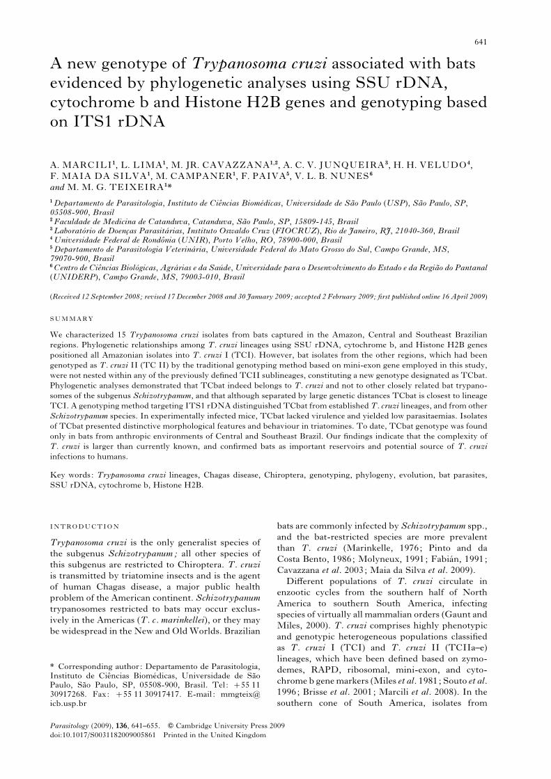
(Southeast region, the Atlantic Forest) (Fig. 1). Bats
were captured using appropriated nets, anaesthe-
tized and manipulated for blood-sample collection
A. Marcili and others 642
according to permits of the IBAMA (Instituto
Brasileiro do Meio Ambiente). Trypanosomes were
isolated by haemoculture (HE), and purified cultures
of T. cruzi from HE mixed with other Schizo-
trypanum spp. were obtained by HE of experimen-
tally infected mice, approximately 30 days after
inoculation of mixed cultures (Maia da Silva et al.
2004a). Isolates were cryopreserved in the Trypano-
somatid Culture Collection of the Department of
Parasitology, University of Sao Paulo. Brazilian
T. cruzi isolates included in this study were from
the following Brazilian States: PA, Para ; AC, Acre;
AM, Amazonas ; SP, Sao Paulo; BA, Bahia; RO,
Rondonia; MG, Minas Gerais ; RN, Rio Grande do
Norte; MS, Mato Grosso do Sul; GO, Goias.
Molecular diagnosis and genotyping of T. cruzi
isolates
DNA from cultured trypanosomes was extracted
using the traditional phenol/chloroform method.
Diagnosis ofT. cruzi isolates was done by PCR based
on kDNA sequences and SSU rDNA sequence
analysis as before (Maia da Silva et al. 2008, 2009).
Genotyping of T. cruzi was done using PCRs based
on mini-exon (Fernandes et al. 2001) and LSU
24Sa-rRNA (Souto et al. 1996) gene sequences.
Reference strains of major T. cruzi lineages were
used as controls: TCI (G), TCIIa (JJ), TCIIb (Y),
TCIIc (MT3663) and TCIId (NRcl3).
PCR amplification, sequencing and data analysis of
SSU rDNA, cytochrome b and Histone H2B genes
PCR amplification of a 900 bp DNA fragment
corresponding to partial sequence of SSU rDNA
(V7–V8 region) was performed using primers and
PCR reactions previously described (Maia da Silva
et al. 2004b). Amplification of 450 bp sequences of
HistoneH2B (H2B) genewas performed as described
by Sturm et al. (2003). Sequences of mitochondrial
cytochrome b (Cyb) were amplified (500 bp) using
primers described by Brisse et al. (2003). New se-
quences from nuclear SSU rDNA (38 sequences),
mitochondrial (Cyb) (33 sequences), and H2B (13
sequences) genes determined in this study were
alignedwith corresponding sequences from reference
strains of T. cruzi and other bat trypanosomes from
GenBank (Table 1). Alignments were made using
ClustalW and manually refined. Phylogenetic in-
ferenceswere done by parsimony (P) (PAUP*4.0b10,
Swofford, 2002) and maximum likelihood (ML)
(RAxML,Stamatakis, 2006), with bootstrap analyses
performed with 100 replicates, as previously de-
scribed (Rodrigues et al. 2006; Ferreira et al. 2008).
PCR-RFLP analysis of ITS1 rDNA from T. cruzi
and other Schizotrypanum species
The primers and PCR conditions employed for
amplification of ITS1 rDNA have been described
previously (Maia da Silva et al. 2004b ; Rodrigues
et al. 2006). Amplified ITS1 rDNA was digested
with several restriction enzymes. The enzyme Bsh
1236 was selected to standardize a PCR-RFLP assay
able to separate lineages ofT. cruzi and to distinguish
T. cruzi fromT. c. marinkellei andT. dionisii. Length
and restriction profiles of amplified ITS1 rDNA
were analysed by electrophoresis in 2.5% agarose gels
stained with ethidium bromide.
RAPD fingerprinting and karyotyping
RAPD profiles from T. cruzi isolates were assessed
using 5 decameric oligonucleotide primers to amplify
Fig. 1. (A) Geographical origin ( ) of isolates of
Trypanosoma cruzi from bats captured in the following
States of different Brazilian biomes: Amazonas (AM)
and Rondonia (RO) in Amazonia biome; Mato Grosso
do Sul (MS) in the Pantanal/Cerrado; and Sao Paulo
(SP) in the Atlantic Forest. DNA profiles generated by
genotyping of isolates of T. cruzi using PCR assays
based on mini-exon (B) and ribosomal (C) markers.
Controls were performed using DNA from reference
strains/isolates of T. cruzi lineages: TCI, G (2) ; TCIIb,
Y ( ) ; TCIIa, JJ (&) ; TCIIc, MT3663 (m) ; TCIId,
NRcl3 ( ) and TCbat ($). Isolates of TCbat: TryCC
203-1112.
Lineages of Trypanosoma cruzi infecting bats 643
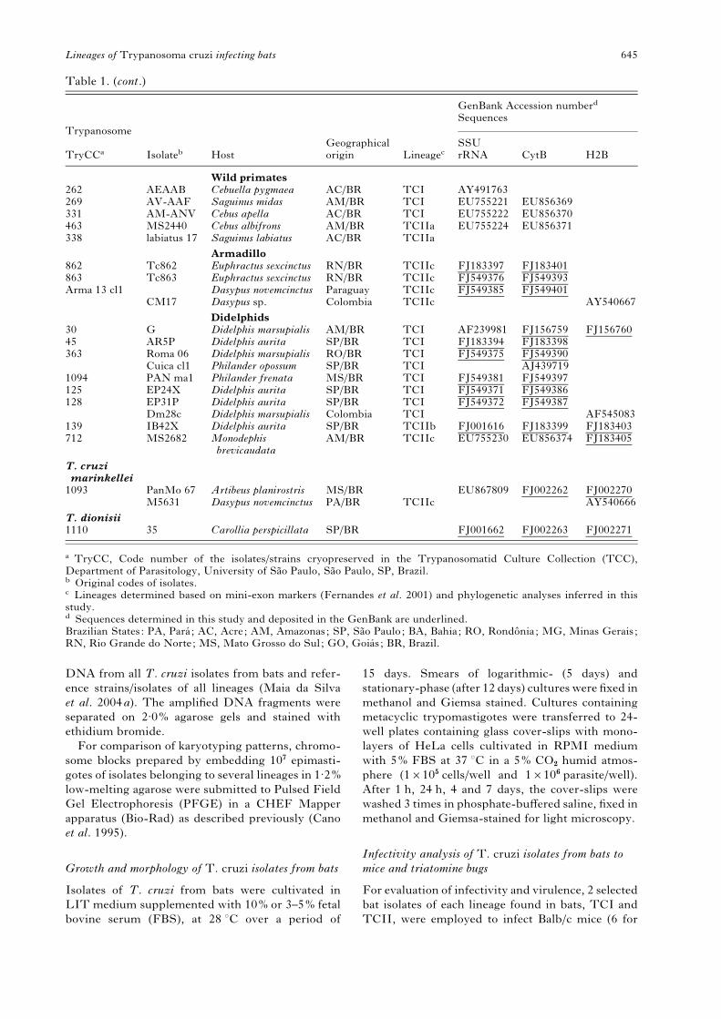
Table 1. Trypanosoma cruzi isolates, host and geographical origin, lineages and sequences of SSU rDNA,
cytochrome b and Histone H2B genes employed in the phylogenetic analyses performed in this study
Trypanosome
Isolateb HostGeographicalorigin Lineagec
GenBank Accession numberd
Sequences
TryCCaSSUrRNA CytB H2B
T. cruziBats
417 M2542 Thyroptera tricolor AM/BR TCI FJ001631 FJ002255 FJ183404507 MO115 Carollia perspicillata RO/BR TCI FJ001632 FJ002256640 MO507 Carollia perspicillata RO/BR TCI FJ001633 FJ549391642 MO92 Carollia perspicillata RO/BR TCI FJ001624 FJ549392203 248 Myotis ruber SP/BR TCbat FJ001617 FJ002253 FJ002264204 519 Myotis albescens SP/BR TCbat FJ001618294 998 Myotis levis SP/BR TCbat FJ001619 FJ002254312 1296 Noctilio albiventris MS/BR TCbat FJ001620480 PaMo122 Noctilio albiventris MS/BR TCbat EU867804499 1336 Myotis nigricans MS/BR TCbat FJ001622 FJ002265597 1361 Myotis nigricans MS/BR TCbat FJ001623 FJ002257 FJ002266793 MO294 Myotis levis SP/BR TCbat FJ001634 FJ002258 FJ002267947 3681 Myotis nigricans SP/BR TCbat FJ001626 FJ002259949 3679 Myotis nigricans SP/BR TCbat FJ001627 FJ002260 FJ0022681122 1122 Myotis albescens SP/BR TCbat FJ001628 FJ002261 FJ002268
Humans971 DRS Homo sapiens AP/BR TCI FJ549378 FJ599394978 XE6863/3 Homo sapiens AP/BR TCI FJ549379 FJ5493951339 Silvio X10 Homo sapiens PA/BR TCI AJ130928 AF545084
CAN III Homo sapiens PA/BR TCIIa AY54066985 Jose Julio Homo sapiens AM/BR TCIIa AY491761 EU856368
M6241 cl6 Homo sapiens PA/BR TCIIa AJ13093334 Y Homo sapiens SP/BR TCIIb AF301912 FJ168768 AY540671335 Sinesio Homo sapiens BR TCIIb FJ001621873 573LU Homo sapiens GO/BR TCIIb FJ0016251146 Basileu Homo sapiens MG/BR TCIIb FJ001629
Peru Homo sapiens Peru TCIIb X53917Esmeraldo Homo sapiens BA/BR TCIIb AJ130931 AF545086CBB Homo sapiens Chile TCIIb AJ439722 AY540670
844 MT3869 Homo sapiens AM/BR TCIIc AF303660Tula14 Homo sapiens Chile TCIIc DQ021895
967 NRcl3 Homo sapiens Chile TCIId AF228685656 Tc656 Homo sapiens Bolivia TCIId FJ183395 FJ183400
9280cl1 Homo sapiens Bolivia TCIId AJ439725MN11 Homo sapiens Chile TCIId DQ021896MN12 Homo sapiens Chile TCIId DQ021897Tula 12 Homo sapiens Chile TCIIe DQ021894
TriatominesSC13 Rhodnius pallescens Colombia TCI AJ130937
884 884 Panstrongylusmegistus
SP/BR TCI FJ549377
1108 1108 Rhodnius stali MS/BR TCI FJ549382 FJ5493981109 1109 Rhodnius stali MS/BR TCI EU867806 FJ5493991116 1116 Rhodnius stali MS/BR TCI EU867807 FJ549400
TEH Triatoma sp. Mexico TCI AJ13093882 RB X Rhodnius brethesi AM/BR TCIIa EU755217 EU856367 FJ183402668 Rr 668 Rhodnius robustus RO/BR TCIIa FJ183396 EU856372
M4167 Rhodnius brethesi AM/BR TCIIa AY540668698 Rr698 Rhodnius robustus RO/BR TCIIa EU755228 EU856373
TU18 Triatoma infestans Bolivia TCIIb AJ130932845 MT3663 Panstrongylus
geniculatusAM/BR TCIIc AF288660 EU856375
1078 QJIII Triatoma rubrovaria RS/BR TCIIc FJ549380 FJ549396185 Tc185 Triatoma infestans Bolivia TCIId FJ549373 FJ549388186 Tc186 Triatoma infestans Bolıvia TCIId FJ001630 FJ549389
SC43cl1 Triatoma infestans Bolivia TCIId AF232214 AJ439721 AY540664CL Brener Triatoma infestans SP/BR TCIIe AF245383 AJ130935 AF545085P63cl1 Triatoma infestans Paraguay TCIIe DQ021893
A. Marcili and others 644
DNA from all T. cruzi isolates from bats and refer-
ence strains/isolates of all lineages (Maia da Silva
et al. 2004a). The amplified DNA fragments were
separated on 2.0% agarose gels and stained with
ethidium bromide.
For comparison of karyotyping patterns, chromo-
some blocks prepared by embedding 107 epimasti-
gotes of isolates belonging to several lineages in 1.2%
low-melting agarose were submitted to Pulsed Field
Gel Electrophoresis (PFGE) in a CHEF Mapper
apparatus (Bio-Rad) as described previously (Cano
et al. 1995).
Growth and morphology of T. cruzi isolates from bats
Isolates of T. cruzi from bats were cultivated in
LIT medium supplemented with 10% or 3–5% fetal
bovine serum (FBS), at 28 xC over a period of
15 days. Smears of logarithmic- (5 days) and
stationary-phase (after 12 days) cultures were fixed in
methanol and Giemsa stained. Cultures containing
metacyclic trypomastigotes were transferred to 24-
well plates containing glass cover-slips with mono-
layers of HeLa cells cultivated in RPMI medium
with 5% FBS at 37 xC in a 5% CO2 humid atmos-
phere (1r105 cells/well and 1r106 parasite/well).
After 1 h, 24 h, 4 and 7 days, the cover-slips were
washed 3 times in phosphate-buffered saline, fixed in
methanol and Giemsa-stained for light microscopy.
Infectivity analysis of T. cruzi isolates from bats to
mice and triatomine bugs
For evaluation of infectivity and virulence, 2 selected
bat isolates of each lineage found in bats, TCI and
TCII, were employed to infect Balb/c mice (6 for
Table 1. (cont.)
Trypanosome
Isolateb HostGeographicalorigin Lineagec
GenBank Accession numberd
Sequences
TryCCaSSUrRNA CytB H2B
Wild primates262 AEAAB Cebuella pygmaea AC/BR TCI AY491763269 AV-AAF Saguinus midas AM/BR TCI EU755221 EU856369331 AM-ANV Cebus apella AC/BR TCI EU755222 EU856370463 MS2440 Cebus albifrons AM/BR TCIIa EU755224 EU856371338 labiatus 17 Saguinus labiatus AC/BR TCIIa
Armadillo862 Tc862 Euphractus sexcinctus RN/BR TCIIc FJ183397 FJ183401863 Tc863 Euphractus sexcinctus RN/BR TCIIc FJ549376 FJ549393Arma 13 cl1 Dasypus novemcinctus Paraguay TCIIc FJ549385 FJ549401
CM17 Dasypus sp. Colombia TCIIc AY540667
Didelphids30 G Didelphis marsupialis AM/BR TCI AF239981 FJ156759 FJ15676045 AR5P Didelphis aurita SP/BR TCI FJ183394 FJ183398363 Roma 06 Didelphis marsupialis RO/BR TCI FJ549375 FJ549390
Cuica cl1 Philander opossum SP/BR TCI AJ4397191094 PAN ma1 Philander frenata MS/BR TCI FJ549381 FJ549397125 EP24X Didelphis aurita SP/BR TCI FJ549371 FJ549386128 EP31P Didelphis aurita SP/BR TCI FJ549372 FJ549387
Dm28c Didelphis marsupialis Colombia TCI AF545083139 IB42X Didelphis aurita SP/BR TCIIb FJ001616 FJ183399 FJ183403712 MS2682 Monodephis
brevicaudataAM/BR TCIIc EU755230 EU856374 FJ183405
T. cruzimarinkellei1093 PanMo 67 Artibeus planirostris MS/BR EU867809 FJ002262 FJ002270
M5631 Dasypus novemcinctus PA/BR TCIIc AY540666
T. dionisii1110 35 Carollia perspicillata SP/BR FJ001662 FJ002263 FJ002271
a TryCC, Code number of the isolates/strains cryopreserved in the Trypanosomatid Culture Collection (TCC),Department of Parasitology, University of Sao Paulo, Sao Paulo, SP, Brazil.b Original codes of isolates.c Lineages determined based on mini-exon markers (Fernandes et al. 2001) and phylogenetic analyses inferred in thisstudy.d Sequences determined in this study and deposited in the GenBank are underlined.Brazilian States: PA, Para ; AC, Acre; AM, Amazonas; SP, Sao Paulo; BA, Bahia; RO, Rondonia; MG, Minas Gerais;RN, Rio Grande do Norte; MS, Mato Grosso do Sul; GO, Goias ; BR, Brazil.
Lineages of Trypanosoma cruzi infecting bats 645
each isolate) by intra-peritoneal inoculation of cul-
tures containing metacyclic forms (y106/animal).
Mice blood samples were examined weekly from 7
to 30 days post-inoculation (p.i.) by the micro-
haematocrit method and chronic infection was con-
firmed by haemoculture after the 30th day p.i.
Smears from the blood of experimentally infected
mice were Giemsa-stained for light microscopy.
Purified T. cruzi cultures were recovered from mice
infected with trypanosomes from mixed cultures
containing T. c. marinkellei or T. dionisii-like besides
T. cruzi as described previously (Maia da Silva et al.
2004a, 2008b).
Eight species of triatomines were used for behav-
ioural analysis of selected bat isolates: Rhodnius
prolixus, R. robustus (genetic population II), R. pic-
tipes, R. domesticus, R. neglectus, Triatoma infestans,
T. vitticeps and Panstrongylus megistus. Mice infected
with bat isolates of T. cruzi were used for xeno-
diagnosis with 20–30 fifth instar nymphs of each
species. The infected triatomines were fed on normal
mice every 15 days, dissected 15, 30, and 60 days
p.i., and their guts examined for the presence of
trypanosomes.
RESULTS
Genotypes of T. cruzi isolates from bats
The 4 bat isolates of T. cruzi from Amazonia were
assigned to lineage TCI, and all isolates fromCentral
and Southeast Brazil were ascribed to TCIIb using
the traditional genotyping method based on mini-
exon gene (Fernandes et al. 2001). Genotyping based
on ribosomal (LSU 24Sa rRNA) markers (Souto
et al. 1996) confirmed all the TCI isolates. However,
using this method all the 11 bat isolates from
Pantanal/Cerrado and the Atlantic Forest genotyped
as TCIIb by mini-exon markers yielded DNA
fragments slightly larger (y140 bp) than that gen-
erated for TCIIb isolates (125 bp), and different
from those of other lineages (Fig. 1C, Table 1).
T. cruzi isolates from bats characterized in this
study were recovered mainly from insectivorous bats
ofMyotis spp. (Vespertilionidae) (9 isolates),Noctilio
albiventris (Noctylionidae) (2 isolates), and 1 was
from Thyroptera tricolor (Tryropteridae). Three iso-
lates from Amazonian bats were from frugivorous/
insectivorous Carollia perspicillata (Phyllostomidae)
(Table 1).
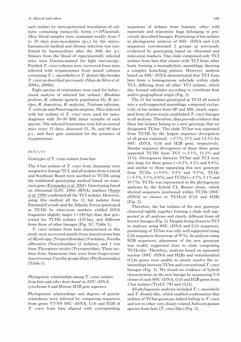
Phylogenetic relationships among T. cruzi isolates
from bats and other hosts based on SSU rDNA,
cytochrome b and Histone H2B gene sequences
Phylogenetic relationships and degrees of genetic
relatedness were inferred by comparing sequences
from genes V7–V8 SSU rDNA, Cyb and H2B of
T. cruzi from bats aligned with corresponding
sequences of isolates from humans, other wild
mammals and triatomine bugs belonging to pre-
viously described lineages. Positioning of bat isolates
in phylogenetic analysis of SSU rDNA and Cyb
sequences corroborated 2 groups as previously
evidenced by genotyping based on ribosomal and
mini-exon markers. One clade comprised only TCI
isolates from bats that cluster with TCI from other
hosts forming a monophyletic assemblage showing
a complex branching pattern. However, analysis
based on SSU rDNA demonstrated that TCI from
bats form a homogeneous subclade within clade
TCI, differing from all other TCI isolates, which
also formed subclades according to vertebrate host
and/or geographical origin (Fig. 2).
The 11 bat isolates genotyped as TCII all nested
into a well-supported assemblage composed exclus-
ively of bat isolates from SP and MS, clearly separ-
ated from all previously established T. cruzi lineages
in all analyses. Therefore, data provide evidence that
these bat isolates belong to a new genotype that we
designated TCbat. The clade TCbat was separated
from TCIIb by the largest sequence divergences
of all genes examined: y5.7%, 11% and 12.3% for
SSU rDNA, Cyb and H2B gene, respectively.
Similar sequence divergences of these three genes
separated TCIIb from TCI (y5.5%, 11.2% and
11%). Divergences between TCbat and TCI were
also large for these genes (y6.2%, 4.2% and 8.4%),
and similar to those separating this new genotype
from TCIIa (y5.0%, 5.5% and 9.5%), TCIIc
(y5.5%, 5.5%, 6.0%), and TCIId (y4.7%, 5.1% and
10.7%). TCIIe was represented in the phylogenetic
analyses by the hybrid CL Brener strain, which
showed sequences positioned within TCIIb (SSU
rDNA) or closest to TCIIc/d (Cyb and H2B)
(Fig. 2).
Therefore, the bat isolates of the new genotype
clustered tightly together forming a clade well sup-
ported in all analyses and clearly different from all
known lineages (Fig. 2). Despite being closest to TCI
in analyses using SSU rDNA and Cyb sequences,
positioning of TCbat was only well supported using
Cyb sequences (bootstrap of 97%). In analysis using
H2B sequences, placement of the new genotype
was weakly supported close to clade comprising
TCIIc/d/e. Therefore, analyses based on separated
nuclear (SSU rDNA and H2B) and mitochondrial
(Cyb) genes were unable to clearly resolve the re-
lationships betweenTCbat and conventionalT. cruzi
lineages (Fig. 2). We found no evidence of hybrid
characteristics in the new lineage by sequencing 5–8
clones of each SSU rDNA, Cyb andH2B genes from
2 bat isolates (TryCC 793 and 1122).
All phylogenetic analyses includedT. c. marinkelei
and T. dionisii-like, which enabled confirmation that
isolates of TCbat genotype indeed belong to T. cruzi
and not to other very closely related Schizotrypanum
species from bats (T. cruzi-like) (Fig. 2).
A. Marcili and others 646
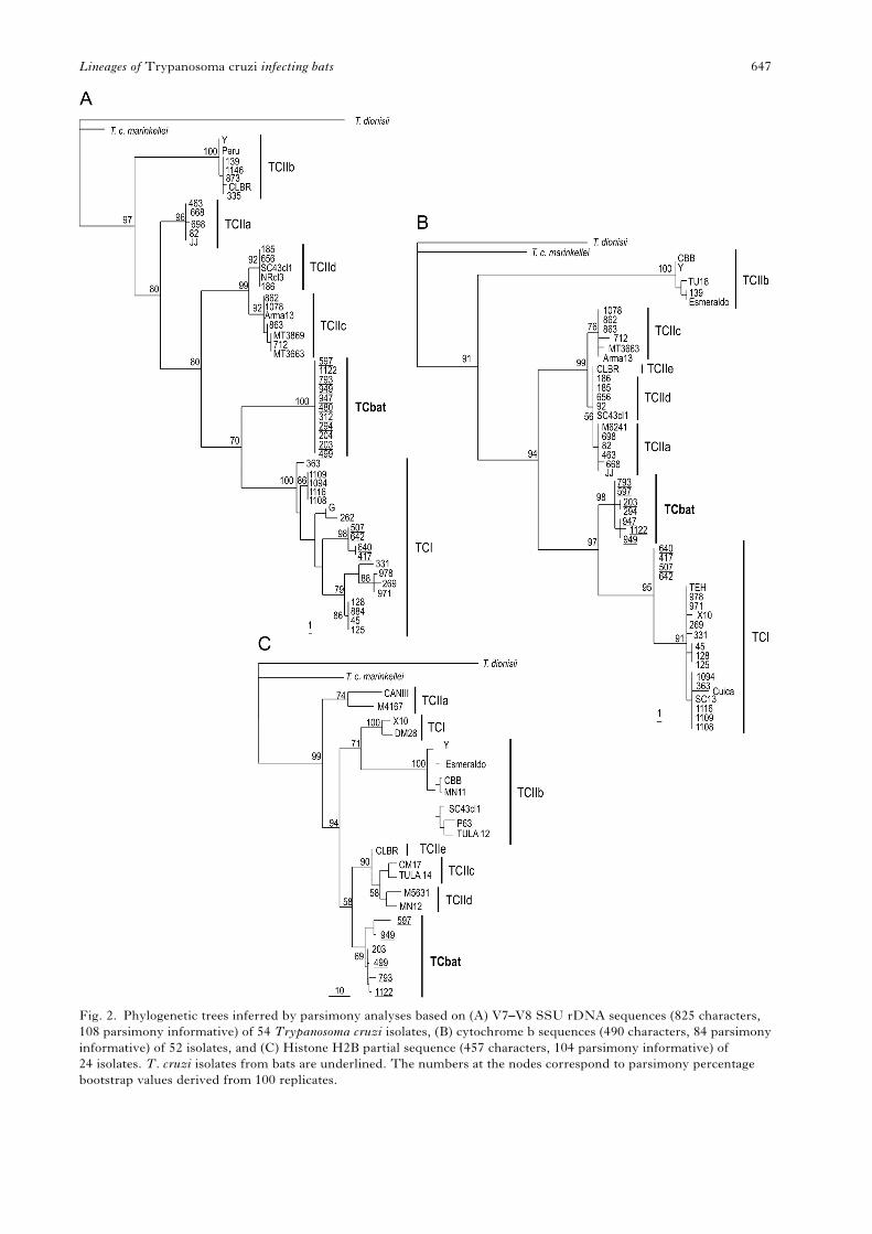
Fig. 2. Phylogenetic trees inferred by parsimony analyses based on (A) V7–V8 SSU rDNA sequences (825 characters,
108 parsimony informative) of 54 Trypanosoma cruzi isolates, (B) cytochrome b sequences (490 characters, 84 parsimony
informative) of 52 isolates, and (C) Histone H2B partial sequence (457 characters, 104 parsimony informative) of
24 isolates. T. cruzi isolates from bats are underlined. The numbers at the nodes correspond to parsimony percentage
bootstrap values derived from 100 replicates.
Lineages of Trypanosoma cruzi infecting bats 647
Phylogenetic relationships among T. cruzi and
T. cruzi-like from bats (Schizotrypanum) inferred
using a combined data set of SSU rDNA,
cytochrome b and Histone H2B sequences
Phylogenetic analyses were performed using 2 data
sets of concatenated aligned sequences aiming to
better resolve the phylogenetic relationships of the
new T. cruzi genotype from bats in relation to pre-
viously established lineages of T. cruzi as well as of
T. cruzi-like trypanosomes. Alignment 1 consists of
SSU rDNA plus Cyb sequences from 40 T. cruzi
isolates plus T. c. marinkelei and T. dionisii-like.
Alignment 2 comprises sequences from SSU rDNA,
Cyb and H2B from 11 T. cruzi isolates plus T. c.
marinkelei and T. dionisii-like. Phylogenetic trees
based on both alignments were inferred by P,
Bayesian (data not shown) andMLmethods (Fig. 3).
In trees generated by the two alignments, the clade
TCbat was always well supported and positioned
closest to TCI. TCIIb was positioned as the basal
lineage of the clade T. cruzi, while the other lineages
were distributed in 5 major clades. Clade TCI and
TCbat formed a monophyletic assemblage in all
analyses using the two combined data sets (Fig. 3).
TCbat was separated from the subclade formed by
bat isolates assigned to TCI by large genetic dis-
tances. Independent of alignments and analytical
methods, T. cruzi-like, T. dionisii and T. c. mar-
inkellei, were always positioned as outgroups of the
clade harbouring all T. cruzi isolates (Fig. 3).
Analyses using combined data sets positioned
TCbat closer to TCI than to any other lineage, and
unequivocally within T. cruzi (Fig. 3). The assem-
blage formed by TCI isolates corroborated the major
subclades revealed by SSU rDNA analysis (Figs 2
and 3). The 4 TCI isolates from bats generated
a subclade formed exclusively by isolates from
Amazonia whereas isolates from opossum, monkeys
and humans from this region clustered separately.
However, these isolates were all genotyped as typi-
cal TCI by traditional PCR methods and tightly
clustered together forming a clade separated from
TCbat. Although TCbat isolates were all from bats
Fig. 3. Phylogenetic trees inferred by ML analyses using combined data sets: (A) SSU rDNA and cytochrome b
sequences from 42 isolates (1315 characters, xLn=3044.252544), and (B) SSU rDNA, cytochrome b and Histone H2B
sequences from 11 isolates (1791 characters, xLn=4761.077323) of Trypanosoma cruzi isolates. T. cruzi isolates from
bats are underlined. Numbers at nodes are bootstrap values derived from 100 replicates.
A. Marcili and others 648
captured in MS and SP states, isolates from didel-
phids and triatomines from these states never nested
within clade TCbat while they were segregated in 2
clades according to their geographical origin (Figs 2
and 3).
Polymorphism analysis of bat isolates by RAPD
and karyotype patterns
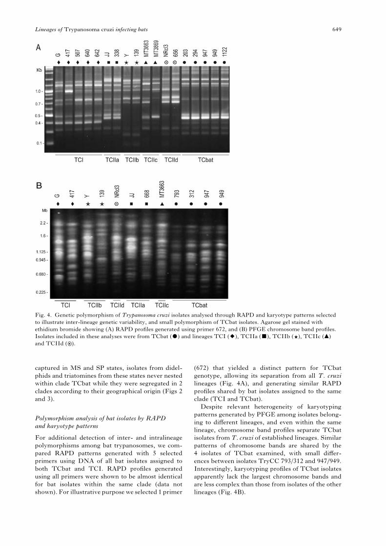
For additional detection of inter- and intralineage
polymorphisms among bat trypanosomes, we com-
pared RAPD patterns generated with 5 selected
primers using DNA of all bat isolates assigned to
both TCbat and TCI. RAPD profiles generated
using all primers were shown to be almost identical
for bat isolates within the same clade (data not
shown). For illustrative purpose we selected 1 primer
(672) that yielded a distinct pattern for TCbat
genotype, allowing its separation from all T. cruzi
lineages (Fig. 4A), and generating similar RAPD
profiles shared by bat isolates assigned to the same
clade (TCI and TCbat).
Despite relevant heterogeneity of karyotyping
patterns generated by PFGE among isolates belong-
ing to different lineages, and even within the same
lineage, chromosome band profiles separate TCbat
isolates from T. cruzi of established lineages. Similar
patterns of chromosome bands are shared by the
4 isolates of TCbat examined, with small differ-
ences between isolates TryCC 793/312 and 947/949.
Interestingly, karyotyping profiles of TCbat isolates
apparently lack the largest chromosome bands and
are less complex than those from isolates of the other
lineages (Fig. 4B).
Fig. 4. Genetic polymorphism of Trypanosoma cruzi isolates analysed through RAPD and karyotype patterns selected
to illustrate inter-lineage genetic variability, and small polymorphism of TCbat isolates. Agarose gel stained with
ethidium bromide showing (A) RAPD profiles generated using primer 672, and (B) PFGE chromosome band profiles.
Isolates included in these analyses were from TCbat ($) and lineages TCI (2), TCIIa (&), TCIIb ( ), TCIIc (m)
and TCIId ( ).
Lineages of Trypanosoma cruzi infecting bats 649
Taken together, similar RAPD and karyotyping
patterns, which are highly sensitive tools for poly-
morphism analyses, besides high conservation of
SSU rDNA, Cyb and H2B sequences, indicated that
TCbat isolates from SP and MS formed a highly
homogeneous group different from other lineages of
T. cruzi.
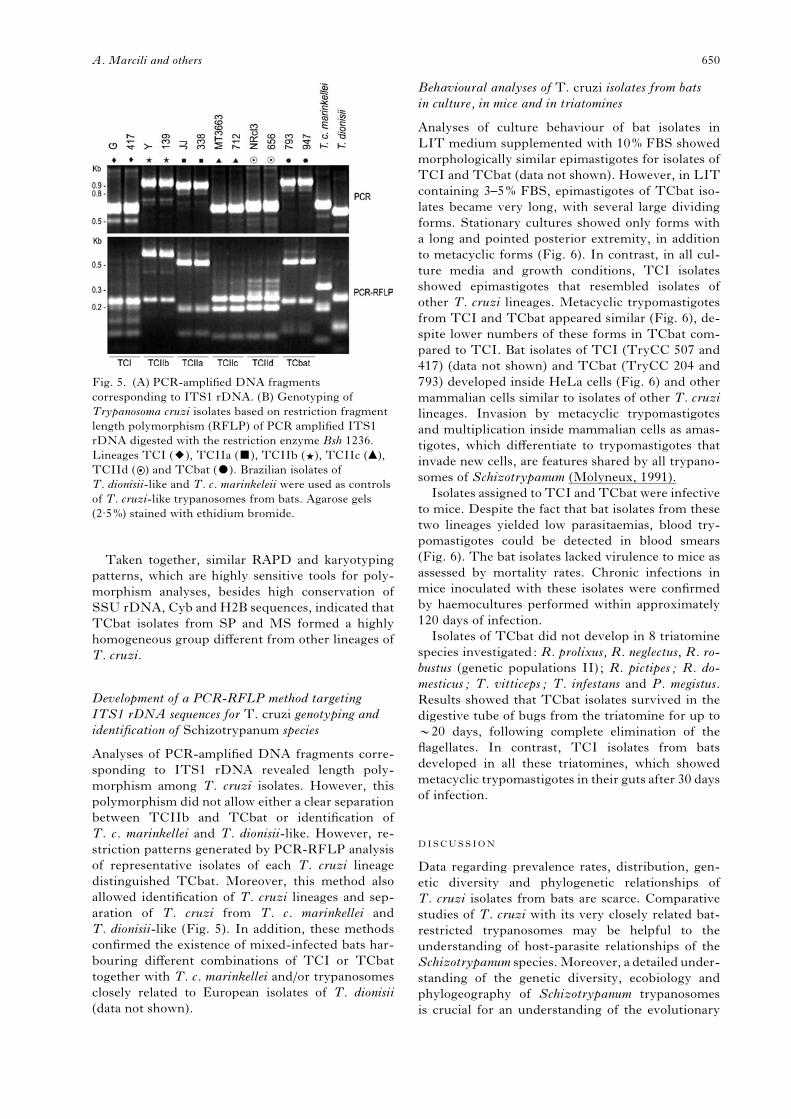
Development of a PCR-RFLP method targeting
ITS1 rDNA sequences for T. cruzi genotyping and
identification of Schizotrypanum species
Analyses of PCR-amplified DNA fragments corre-
sponding to ITS1 rDNA revealed length poly-
morphism among T. cruzi isolates. However, this
polymorphism did not allow either a clear separation
between TCIIb and TCbat or identification of
T. c. marinkellei and T. dionisii-like. However, re-
striction patterns generated by PCR-RFLP analysis
of representative isolates of each T. cruzi lineage
distinguished TCbat. Moreover, this method also
allowed identification of T. cruzi lineages and sep-
aration of T. cruzi from T. c. marinkellei and
T. dionisii-like (Fig. 5). In addition, these methods
confirmed the existence of mixed-infected bats har-
bouring different combinations of TCI or TCbat
together with T. c. marinkellei and/or trypanosomes
closely related to European isolates of T. dionisii
(data not shown).
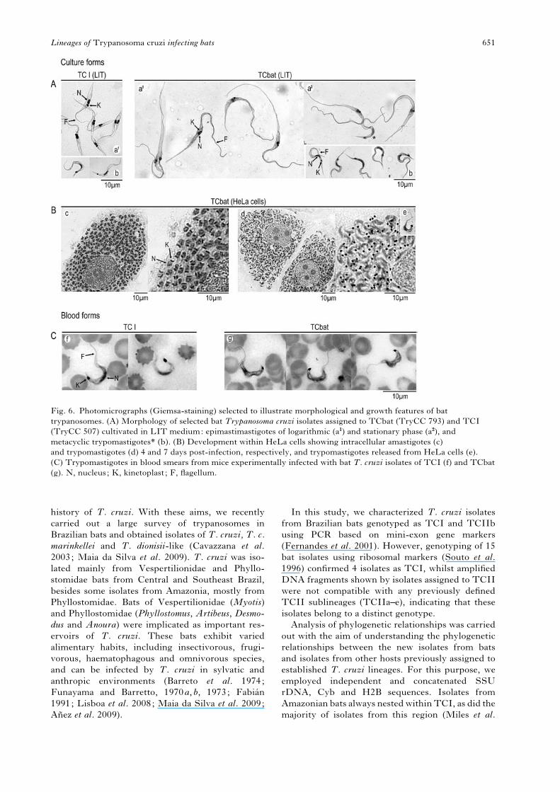
Behavioural analyses of T. cruzi isolates from bats
in culture, in mice and in triatomines
Analyses of culture behaviour of bat isolates in
LIT medium supplemented with 10% FBS showed
morphologically similar epimastigotes for isolates of
TCI and TCbat (data not shown). However, in LIT
containing 3–5% FBS, epimastigotes of TCbat iso-
lates became very long, with several large dividing
forms. Stationary cultures showed only forms with
a long and pointed posterior extremity, in addition
to metacyclic forms (Fig. 6). In contrast, in all cul-
ture media and growth conditions, TCI isolates
showed epimastigotes that resembled isolates of
other T. cruzi lineages. Metacyclic trypomastigotes
from TCI and TCbat appeared similar (Fig. 6), de-
spite lower numbers of these forms in TCbat com-
pared to TCI. Bat isolates of TCI (TryCC 507 and
417) (data not shown) and TCbat (TryCC 204 and
793) developed inside HeLa cells (Fig. 6) and other
mammalian cells similar to isolates of other T. cruzi
lineages. Invasion by metacyclic trypomastigotes
and multiplication inside mammalian cells as amas-
tigotes, which differentiate to trypomastigotes that
invade new cells, are features shared by all trypano-
somes of Schizotrypanum (Molyneux, 1991).
Isolates assigned to TCI and TCbat were infective
to mice. Despite the fact that bat isolates from these
two lineages yielded low parasitaemias, blood try-
pomastigotes could be detected in blood smears
(Fig. 6). The bat isolates lacked virulence to mice as
assessed by mortality rates. Chronic infections in
mice inoculated with these isolates were confirmed
by haemocultures performed within approximately
120 days of infection.
Isolates of TCbat did not develop in 8 triatomine
species investigated: R. prolixus, R. neglectus, R. ro-
bustus (genetic populations II) ; R. pictipes; R. do-
mesticus; T. vitticeps; T. infestans and P. megistus.
Results showed that TCbat isolates survived in the
digestive tube of bugs from the triatomine for up to
y20 days, following complete elimination of the
flagellates. In contrast, TCI isolates from bats
developed in all these triatomines, which showed
metacyclic trypomastigotes in their guts after 30 days
of infection.
DISCUSSION
Data regarding prevalence rates, distribution, gen-
etic diversity and phylogenetic relationships of
T. cruzi isolates from bats are scarce. Comparative
studies of T. cruzi with its very closely related bat-
restricted trypanosomes may be helpful to the
understanding of host-parasite relationships of the
Schizotrypanum species.Moreover, a detailed under-
standing of the genetic diversity, ecobiology and
phylogeography of Schizotrypanum trypanosomes
is crucial for an understanding of the evolutionary
Fig. 5. (A) PCR-amplified DNA fragments
corresponding to ITS1 rDNA. (B) Genotyping of
Trypanosoma cruzi isolates based on restriction fragment
length polymorphism (RFLP) of PCR amplified ITS1
rDNA digested with the restriction enzyme Bsh 1236.
Lineages TCI (2), TCIIa (&), TCIIb ( ), TCIIc (m),
TCIId ( ) and TCbat ($). Brazilian isolates of
T. dionisii-like and T. c. marinkeleii were used as controls
of T. cruzi-like trypanosomes from bats. Agarose gels
(2.5%) stained with ethidium bromide.
A. Marcili and others 650
history of T. cruzi. With these aims, we recently
carried out a large survey of trypanosomes in
Brazilian bats and obtained isolates of T. cruzi, T. c.
marinkellei and T. dionisii-like (Cavazzana et al.
2003; Maia da Silva et al. 2009). T. cruzi was iso-
lated mainly from Vespertilionidae and Phyllo-
stomidae bats from Central and Southeast Brazil,
besides some isolates from Amazonia, mostly from
Phyllostomidae. Bats of Vespertilionidae (Myotis)
and Phyllostomidae (Phyllostomus, Artibeus, Desmo-
dus and Anoura) were implicated as important res-
ervoirs of T. cruzi. These bats exhibit varied
alimentary habits, including insectivorous, frugi-
vorous, haematophagous and omnivorous species,
and can be infected by T. cruzi in sylvatic and
anthropic environments (Barreto et al. 1974;
Funayama and Barretto, 1970a, b, 1973; Fabian
1991; Lisboa et al. 2008; Maia da Silva et al. 2009;
Anez et al. 2009).
In this study, we characterized T. cruzi isolates
from Brazilian bats genotyped as TCI and TCIIb
using PCR based on mini-exon gene markers
(Fernandes et al. 2001). However, genotyping of 15
bat isolates using ribosomal markers (Souto et al.
1996) confirmed 4 isolates as TCI, whilst amplified
DNA fragments shown by isolates assigned to TCII
were not compatible with any previously defined
TCII sublineages (TCIIa–e), indicating that these
isolates belong to a distinct genotype.
Analysis of phylogenetic relationships was carried
out with the aim of understanding the phylogenetic
relationships between the new isolates from bats
and isolates from other hosts previously assigned to
established T. cruzi lineages. For this purpose, we
employed independent and concatenated SSU
rDNA, Cyb and H2B sequences. Isolates from
Amazonian bats always nested withinTCI, as did the
majority of isolates from this region (Miles et al.
Fig. 6. Photomicrographs (Giemsa-staining) selected to illustrate morphological and growth features of bat
trypanosomes. (A) Morphology of selected bat Trypanosoma cruzi isolates assigned to TCbat (TryCC 793) and TCI
(TryCC 507) cultivated in LIT medium: epimastimastigotes of logarithmic (a1) and stationary phase (a2), and
metacyclic trypomastigotes* (b). (B) Development within HeLa cells showing intracellular amastigotes (c)
and trypomastigotes (d) 4 and 7 days post-infection, respectively, and trypomastigotes released from HeLa cells (e).
(C) Trypomastigotes in blood smears from mice experimentally infected with bat T. cruzi isolates of TCI (f) and TCbat
(g). N, nucleus; K, kinetoplast ; F, flagellum.
Lineages of Trypanosoma cruzi infecting bats 651
1981; Fernandes et al. 2001; Maia da Silva et al.
2008; Marcili et al. 2009). The 11 bat isolates pre-
viously genotyped as TCIIb clustered tightly to-
gether constituting a new clade separated by relevant
genetic distances from all known lineages, thus con-
firming that they belong to a new genotype (TCbat).
All analyses performed using separated or combined
data sets separated isolates of genotype TCbat from
isolates of lineage TCIIb. However, results did not
indicate an unquestionable positioning of TCbat
within any established lineages of T. cruzi. Phylo-
genetic analysis of Schizotrypanum trypanosomes
using concatenated alignment of SSU rDNA, Cyb
and H2B genes positioned all bat isolates character-
ized in this study within the clade T. cruzi, which
is more closely related to T. c. marinkellei than to
T. dionisii clades.
Most inferred phylogenetic analyses suggested
that TCbat genotype is closer to TCI than to any
other lineage, although separated by large genetic
distances. In contrast to TCbat, isolates of TCI
shared amplified DNA fragments of the same length
when genotyped using traditional methods. In this
study, we demonstrated that TCbat isolates differed
from TCI isolates from Amazonian bats, and also
from TCI from didelphids and triatomines from the
same regions where bats infected with TCbat have
been found. TCbat is clearly different from any iso-
late assigned to the TCI lineage, even from bat
isolates assigned to TCI by all molecular markers
investigated in this study.
The monophyletic assemblage formed by TCI
from bats, humans, wild monkeys and didelphids
provided evidence of geographical clustering within
TCI. Recent studies have evidenced subclusters
within TCI that could be associated with both mam-
malian hosts and geographical origin. Analysis of the
highly polymorphic intergenic region of the mini-
exon gene separated North American from South
American isolates, and disclosed a subclade related
to Didelphis sp. (O’Connor et al. 2007). Mini-exon
markers revealed 4 haplotypes (Ia–Id) of Colombian
TCI isolates related to distinct transmission cycles
(Herrera et al. 2007). Analysis of Cyb sequences of
TCI isolates from Chile revealed a new genotype
(DTU1b) associated with caviomorph rodents
(Spotorno et al. 2008).
Comparative analyses of TCbat and TCI isolates
from a range of hosts and geographical origin may
help to define recommendations for the description
of new genotypes/lineages closely related to TCI.
Recently, isolates of lineage TCI have been inves-
tigated using polymorphic molecular markers, and
results showed high intralineage genetic diversity
and a complex populational structure of TCI popu-
lations (O’Connor et al. 2007; Herrera et al. 2007;
Spotorno et al. 2008). Our data do not support TCI
and TCII as 2 major lineages within T. cruzi since
TCIIa–e sublineages are not monophyletic and
varied according to the gene analysed, as previously
demonstrated (Machado and Ayala, 2001; Brisse
et al. 2003; Sturm et al. 2003; Westenberger et al.
2005, 2006). Therefore, relationships among lineages
of T. cruzi are far from understood. Further analysis
from more isolates from several lineages may help to
resolve the phylogeny of T. cruzi. Until more data
can be gathered, we have designated the new geno-
type of T. cruzi from bats characterized in this study
as TCbat.
Isolates of TCbat were shown to be distinguish-
able by morphological and biological features. These
isolates showed very large and pointed epimastigotes,
different from those of all other T. cruzi lineages. In
addition, TCbat isolates were unable to yield estab-
lished infection in triatomines of genera Rhodnius,
Triatoma and Panstrongylus, a behaviour shared
by T. c. marinkellei and T. dionisii. T. c. marinkellei
seems to be transmitted only by triatomines of genus
Cavernicola, which are usually associated with
bat colonies, whereas T. dionisii is transmitted by
cimicids (Marinkelle, 1976; Molyneux, 1991).
Triatomines that live in tree holes and caves, palms
and house roofs can transmit T. cruzi among bats.
The majority of T. cruzi-infected bats are insecti-
vorous and likely to be infected by ingestion of
triatomines (Marinkelle, 1976; Thomas et al. 2007).
Vector permissiveness to T. cruzi and association
between lineages/strains and triatomine species de-
pends on both vectors and parasite features, appar-
ently, with superior vector competence of sympatric
sylvatic species, as clearly demonstrated for T. ran-
geli (Maia da Silva et al. 2007). Unfortunately, tria-
tomines of Cavernicola and other sylvatic species
from regions where TCbat isolates originated were
not available for this study. In Central Brazil, several
triatomine species, mainly those inhabiting palms
such as R. neglectus, R. robustus and R. stali could
transmit T. cruzi among bats (Gurgel-Goncalves
et al. 2008). However, the genotypes of T. cruzi cir-
culating in sylvatic triatomines from MS and SP
were completely unknown. Isolates from R. stali
(MS) andP.megistus (SP) included in this study were
genotyped as TCI.
Altogether, sequence divergences and phylogen-
etic analysis of SSU rDNA, Cyb and H2B genes,
morphology and behaviour in triatomines indicate
that isolates of TCbat indeed belong to a new geno-
type of T. cruzi represented, so far, exclusively by
bat isolates from antrophic areas of Central and
Southeast Brazil. Both regions are endemic for
Chagas disease. Distances separating these areas are
easily crossed by bats found to be infected with
TCbat. The limited data regarding T. cruzi geno-
types in wild mammals are insufficient to rule out
other mammals as hosts of TCbat, and also humans
living in houses inhabited by bats.
The hypotheses that T. cruzi evolved from a
trypanosome restricted to bats or vice versa remain
A. Marcili and others 652
to be elucidated. The present-day distribution of
Schizotrypanum species and the ability of bats to
disperse over long distances, including crossing
oceans, are consistent with both hypotheses (Stevens
et al. 1999; Barnabe et al. 2003). The evolutionary
histories of T. cruzi lineages have been correlated
with a long-standing association with vertebrate
hosts. TCI and TCII have been associated respect-
ively with marsupials of Didelphimorpha (opos-
sums) and with placentals of Xenarthra (armadillos),
the early mammals in South America (y65 mya).
Primates and rodents entered South America from
Africa during the Oligocene (y35 mya), whereas
chiropterans dispersed from Africa in the Eocene
(y45 mya), and arrived in the Americas via Beringia
or by a transatlantic route (Eick et al. 2005). Bats are
ancient hosts of T. cruzi-like species or lineages
transmitted by triatomines in the Americas as in-
dicated by the description of Trypanosoma antiquus
in triatomine feces fossilized in Dominican amber,
together with bat hairs (Poinar, 2005). The low
genetic divergence showed in this and in previous
studies is compatible with a recent split between
T. cruzi and the bat-restricted Schizotrypanum, as
well as with a recent diversification of all T. cruzi
lineages (Machado and Ayala, 2001; Brisse et al.
2003; Barnabe et al. 2003).
Besides T. cruzi, other Schizotrypanum species
such as T. c. marinkellei and T. dionisii-like, in ad-
dition to T. rangeli have also been found infecting
bats in the same locations where bats infected with
TCbat were captured (Cavazzana et al. 2003; Lisboa
et al. 2008; Maia da Silva et al. 2009). Taking into
account the ITS1 rDNA polymorphisms among
T. cruzi lineages, we standardized a PCR-RFLP,
targeting this gene that allowed separation ofT. cruzi
from other species of Schizotrypanum and to dis-
tinguish TCbat from other genotypes. This method
was shown to be a sensitive tool for easy detection of
new genotypes of T. cruzi and T. cruzi-like.
Data from this study corroborated the high com-
plexity of T. cruzi, pointing towards the existence of
distinct T. cruzi genotypes waiting to be described,
and the method described in this study can be very
helpful for this purpose. Our results provide evi-
dence that the understanding of enzootic trans-
mission cycles of T. cruzi can be improved with
phylogenetic analysis of more isolates, especially
from poorly investigated sylvatic vertebrate and
invertebrate hosts of unexplored geographical re-
gions and ecotopes.
We would like to thank all students and colleagues for theirinestimable help in the fieldwork, and for providing us withblood samples of bats and reference strains of T. cruzi. Weare grateful to Erney P. Camargo,Michel A.Miles, MartinLlewellyn andMichaelLewis for valuable comments on themanuscript. We are thankful to Valdir Tadei (in memor-iam) and Caroline C. Aires for identification of bats. Workin Rondonia (Amazonia) was done in ICB5-USP. This
work was supported by grants from the Brazilian agenciesCNPq (UNIVERSAL) and FAPESP (PRONEX). F. M.da S. was sponsored by CAPES (PRODOC-PROTAX),and A.M., L.L. and A.C.V.J. were recipients of scholar-ships from CNPq.
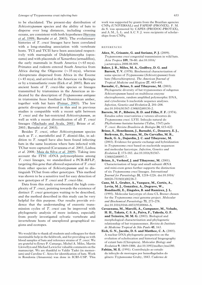
REFERENCES
Anez, N., Crisante, G. and Soriano, P. J. (2009).
Trypanosoma cruzi congenital transmission in wild bats.
Acta Tropica 109, 78–80. doi:10.1016/
j.actatropica.2008.08.009.
Baker, J. R., Miles, M. A., Godfrey, D. G. and
Barrett, T. V. (1978). Biochemical characterization of
some species of Trypanosoma (Schizotrypanum) from
bats (Microchiroptera). The American Journal of
Tropical Medicine and Hygiene 27, 483–491.
Barnabe, C., Brisse, S. and Tibayrenc, M. (2003).
Phylogenetic diversity of bat trypanosomes of subgenus
Schizotrypanum based on multilocus enzyme
electrophoresis, random amplified polymorphic DNA,
and cytochrome b nucleotide sequence analyses.
Infection, Genetics and Evolution 2, 201–208.
doi:10.1016/S1567-1348(02)00130-2.
Barreto, M. P., Ribeiro, R. D. and Filho, F. F. (1974).
Estudos sobre reservatorios e vetores silvestres do
Trypanosoma cruzi. LVII. Infeccao natural do
Phyllostomus hastatus hastatus (Tallas, 1767) pelo
T. cruzi. Revista Brasileira de Biologia 34, 615–622.
Brisse, S., Henriksson, J., Barnabe, C., Douzery, E. J.,
Berkvens, D., Serrano, M., De Carvalho, M. R.,
Buck, G. A., Dujardin, J. C. and Tibayrenc, M.
(2003). Evidence for genetic exchange and hybridization
in Trypanosoma cruzi based on nucleotide sequences
and molecular karyotype. Infection, Genetics and
Evolution 2, 173–183. doi:10.1016/S1567-
1348(02)00097-7.
Brisse, S., Verhoef, J. and Tibayrenc, M. (2001).
Characterisation of large and small subunit rRNA
and mini-exon genes further supports the distinction
of six Trypanosoma cruzi lineages. International
Journal for Parasitology 31, 1218–1226. doi:10.1016/
S0020-7519(01)00238-7.
Cano, M. I., Gruber, A., Vazquez, M., Cortes, A.,
Levin, M. J., Gonzalez, A., Degrave, W.,
Rondinelli, E., Zingales, B. and Ramirez, J. L.
(1995). Molecular karyotype of clone CL Brener chosen
for the Trypanosoma cruzi genome project. Molecular
and Biochemical Parasitology 71, 273–278.
doi:10.1016/0166-6851(95)00066-A.
Cavazzana, M., Marcili, A., Campaner, M., Veludo,
H. H., Takata, C. S. A., Paiva, F., Takeda, G. F.
and Teixeira, M. M. G. (2003). Biological and
morphological characterization and phylogenetic
relationship of bat trypanosomes. Revista do Instituto
de Medicina Tropical de Sao Paulo 45, 163.
Eick, G. N., Jacobs, D. S. and Matthee, C. A. (2005).
A nuclear DNA phylogenetic perspective on the
evolution of echolocation and historical biogeography
of extant bats (Chiroptera). Molecular Biology and
Evolution 9, 1869–1886. doi:10.1093/molbev/msi180.
Fabian, M. E. (1991). Contribuicao ao estudo
da infeccao de morcegos por hemoflagelados do
genero Trypanosoma Gruby, 1843. Cadernos de
Lineages of Trypanosoma cruzi infecting bats 653
Saude Publica, Rio de Janeiro 7, 69–81. doi:10.1590/
S0102-311X1991000100006.
Fernandes, O., Santos, S. S., Cupolillo, E., Mendonca,
B., Derre, R., Junqueira, A. C. V., Santos, L. C.,
Sturm, N. R., Naiff, R. D., Barrett, T. V., Campbell,
D. and Coura, J. R. (2001). A mini-exon multiplex
polymerase chain reaction to distinguish the major
groups of Trypanosoma cruzi and T. rangeli in the
Brazilian Amazon. Transactions of the Royal Society of
Tropical Medicine and Hygiene 95, 97–99. doi:10.1016/
S0035-9203(01)90350-5.
Ferreira, R. C., De Souza, A. A., Freitas, R. A.,
Campaner, M., Takata, C. S. A., Barrett, T. V.,
Shaw, J. J. and Teixeira, M. M. G. (2008). A
phylogenetic lineage of closely related trypanosomes
(Trypanosomatidae, Kinetoplastida) of anurans and
sand flies (Psychodidae, Diptera) sharing the same
ecotopes in Brazilian Amazonia. Journal of Eukaryotic
Microbiology 55, 527–535. doi:10.1111/j.1550-
7408.2008.00342.x.
Funayama, G. K. and Barreto, M. P. (1973). Studies
of wild reservoirs and vectors of Trypanosoma cruzi.
LIV. Natural bat infection, Epitesicus brasiliensis
brasiliensis (Desmarest, 1819) by T. cruzi. Revista
Brasileira de Biologia 33, 439–444.
Funayama, G. K. and Barretto, M. P. (1970a).
Estudo sobre reservatorios e vetores silvestres do
Trypanosoma cruzi. XXXVIII. Infeccao natural do
morcego Desmodus rotundus rotundus (Geoffroy, 1810)
pelo T. cruzi. Revista Brasileira de Biologia 30, 13–19.
Funayama, G. K. and Barretto, M. P. (1970b). Estudo
sobre reservatorios e vetores silvestres do Trypanosoma
cruzi. XLI. Infeccao natural do morcego Tadarida
laticaudata (Geoffroy, 1805) pelo T. cruzi. Revista
Brasileira de Biologia 30, 439–445.
Gaunt, M. and Miles, M. (2000). The ecotopes and
evolution of triatomine bugs (triatominae) and their
associated trypanosomes. Memorias do Instituto
Oswaldo Cruz 95, 557–565. doi:10.1590/
S0074-02762000000400019.
Gurgel-Goncalves, R., Abad-Franch, F., Ferreira,
J. B., Santana, D. B. and Cuba, C. A. (2008). Is
Rhodnius prolixus (Triatominae) invading houses in
central Brazil? Acta Tropica 107, 90–98. doi:10.1016/
j.actatropica.2008.04.020.
Gurtler, R. E., Cecere, M. C., Lauricella, M. A.,
Cardinal, M. V., Kitron, U. and Cohen, J. E. (2007).
Domestic dogs and cats as sources of Trypanosoma cruzi
infection in rural northwestern Argentina. Parasitology
134, 69–82. doi:10.1017/S0031182006001259.
Herrera, C., Bargues, M. D., Fajardo, A., Montilla,
M., Triana, O., Vallejo, G. A. and Guhl, F. (2007).
Identifying four Trypanosoma cruzi I isolate haplotypes
from different geographic regions in Colombia.
Infection, Genetics and Evolution 7, 535–539.
doi:10.1016/j.meegid.2006.12.003.
Lisboa, C. V., Pinho, A. P. S., Herrera, H., Gerhard,
M., Cupolillo, E. and Jansen, A. M. (2008).
Trypanosoma cruzi (Kinetoplastida, Trypanosomatidae)
genotypes in neotropical bats in Brazil.
Veterinary Parasitology 156, 314–318. doi:10.1016/
j.vetpar.2008.06.004.
Maia da Silva, F., Marcili, A., Lima, L., Cavazzana,
Jr. M., Ortiz, P. A., Campaner, M., Takeda, G. F.,
Paiva, F., Nunes, V. L. B., Camargo, E. P. and
Teixeira, M. M. G. (2009)Trypanosoma rangeli isolates
of bats from Central Brazil : genotyping and
phylogenetic analysis enable description of a new lineage
using spliced-leader gene sequences. Acta Tropica 109,
199–207. doi:1016/j.actatropica.2008.11.005.
Maia da Silva, F., Naiff, R. D., Marcili, A., Gordo, M.,
D’Affonseca Neto, J. A., Naiff,M. F., Franco, A. M.,
Campaner, M., Valente, V., Valente, S. A.,
Camargo, E. P., Teixeira, M. M. G. and Miles,
M. A. (2008). Infection rates and genotypes
of Trypanosoma rangeli and T. cruzi infecting
free-ranging Saguinus bicolor (Callitrichidae), a critically
endangered primate of the Amazon Rainforest.
Acta Tropica 107, 168–173. doi:10.1016/
j.actatropica.2008.05.015.
Maia da Silva, F., Junqueira, A. C., Campaner, M.,
Rodrigues, A. C., Crisante, G., Ramirez, L. E.,
Caballero, Z. C., Monteiro, F. A., Coura, J. R.,
Anez,N. andTeixeira,M. M. G. (2007). Comparative
phylogeography of Trypanosoma rangeli and Rhodnius
(Hemiptera: Reduviidae) supports a long coexistence
of parasite lineages and their sympatric vectors.
Molecular Ecology 16, 3361–3373. doi:10.1111/
j.1365-294X.2007.03371.x.
Maia da Silva, F., Noyes, H., Campaner, M.,
Junqueira, A. C., Coura, J. R., Anez, N., Shaw, J. J.,
Stevens, J. R. and Teixeira, M. M. G. (2004b).
Phylogeny, taxonomy and grouping of Trypanosoma
rangeli isolates from man, triatomines and sylvatic
mammals from widespread geographical origin based
on SSU and ITS ribosomal sequences. Parasitology
129, 549–561. doi:10.1017/S0031182004005931.
Maia da Silva, F., Rodrigues, A. C., Campaner, M.,
Takata, C. S., Brigido, M. C., Junqueira, A. C.,
Coura, J. R., Takeda, G. F., Shaw, J. J. and Teixeira,
M. M. G. (2004a). Randomly amplified polymorphic
DNA analysis of Trypanosoma rangeli and allied species
from human, monkeys and other sylvatic mammals
of the Brazilian Amazon disclosed a new group and
a species-specific marker. Parasitology 128, 283–294.
doi:10.1017/S0031182003004554.
Machado, C. A. and Ayala, F. J. (2001). Nucleotide
sequences provide evidence of genetic exchange
among distantly related lineages of Trypanosoma
cruzi. Proceedings of the National Academy
of Sciences, USA 19, 7396–7401.
www.pnas.orgycgiydoiy10.1073ypnas.121187198.
Marcili, A., Valente, V., Valente, A., Junqueira,
A. C. V., Maia da Silva, F., Naiff, R., Campaner, M.,
Coura, J. R., Camargo, E. P., Miles, M. A. and
Teixeira, M. M. G. (2009). Trypanosoma cruzi in
Brazilian Amazonia: lineages TCI and TCIIa in wild
primates, Rhodnius spp. and in humans with Chagas
disease associated with oral transmission. International
Journal for Parasitology 39, 615–623. doi:10.1016/
j.ijpara.2008.09.015.
Marinkelle, C. J. (1976). Biology of the trypanosomes
of bats. In Biology of the Kinetoplastida, Vol 1,
(ed.Lumsden,W. H. R. andEvans,D. A.), pp. 175–216.
Academic Press, London, UK.
Martins, L. P. A., Marcili, A., Castanho, R. E. P.,
Therezo, A. L. S., Oliveira, J. C. P., Suzuki, R. B.,
Teixeira, M. M. G., Rosa, J. A. and Speranca, M. A.
A. Marcili and others 654
(2008). Rural Triatoma rubrovaria from southern
Brazil harbors Trypanosoma cruzi of lineage IIc. The
American Journal of Tropical Medicine and Hygiene 79,
427–434.
Miles, M. A., Povoa, M., De Souza, A. A., Lainson, R.,
Shaw, J. J. and Ketteridge, D. S. (1981). Chagas
disease in the Amazon Basin. II. The distribution of
Trypanosoma cruzi zymodemes 1 and 3 in Para State,
north Brazil. Transactions of the Royal Society of
Tropical Medicine and Hygiene 75, 667–674.
doi:10/1016/0035-9203(81)90145-0.
Molyneux, D. H. (1991). Trypanosomes of bats. In
Parasitic Protozoa, 2ndEdn, (ed.Kreier, J. P. andBaker,
J. R.), pp. 195–223. Academic Press, London, UK.
O’Connor, O., Bosseno, M. F., Barnabe, C., Douzery,
E. J. and Breniere, S. F. (2007). Genetic clustering
of Trypanosoma cruzi I lineage evidenced by intergenic
miniexon gene sequencing. Infection, Genetics and
Evolution 7, 587–593. doi:10.1016/
j.meegid.2007.05.003.
Pinto, A. S. and da Costa Bento, D. N. (1986).
Trypanosoma cruzi-like bloodstrean trypomastigotes
in bats from the state Piaui northeasten. Revista
da Sociedade Brasileira de Medicina Tropical 19,
31–34.
Poinar, G. Jr. (2005). Triatoma dominicana sp. n.
(Hemiptera: Reduviidae: Triatominae), and
Trypanosoma antiquus sp. n. (Stercoraria:
Trypanosomatidae), the first fossil evidence of
a triatomine-trypanosomatid vector association.
Vector – Borne and Zoonotic Disease 5, 72–81.
doi:10.1089/vbz.2005.5.72.
Rodrigues, A. C., Paiva, F., Campaner, M., Stevens,
J. R., Noyes, H. A. and Teixeira, M. M. G. (2006).
Phylogeny of Trypanosoma (Megatrypanum) theileri
and related trypanosomes reveals lineages of isolates
associated with artiodactyl hosts diverging on SSU and
ITS ribosomal sequences. Parasitology 132, 215–224.
doi:10.1017/S0031182005008929.
Roellig, D. M., Brown, E. L., Barnabe, C., Tibayrenc,
M., Steurer, F. J. and Yabsley, M. J. (2008).
Molecular typing of Trypanosoma cruzi isolates,
United States. Emerging Infectious Diseases 14,
1123–1125. doi:10.3201/eid1407.080175.
Souto, R. P., Fernandes, O., Macedo, A. M.,
Campbell, D. A. and Zingales, B. (1996). DNA
markers define two major phylogenetic lineages of
Trypanosoma cruzi. Molecular and Biochemical
Parasitology 83, 141–152. doi:10.1016/S0166-
6851(96)02755-7.
Spotorno, O. A. E., Cordova, L. and Solari, I. A. (2008).
Differentiation of Trypanosoma cruzi I subgroups
through characterization of cytochrome b gene
sequences. Infection, Genetics and Evolution 8, 898–900.
doi:10.1016/j.meegid.2008.08.006.
Stamatakis, A. (2006). RAxML-VI-HPC: maximum
likelihood-based phylogenetic analyses with thousands
of taxa and mixed models. Bioinformatics 1, 2688–2690.
doi:10.1093/bioinformatics/btl446.
Steindel, M., Grisard, E. C., de Carvalho Pinto, C. J.,
Cordeiro, F. D., Ribeiro-Rodrigues, R. and
Romanha, A. J. (1998). Characterization of
trypanosomes from the subgenus Schizotrypanum
isolated from bats, Eptesicus sp. (Chiroptera:
Vespertilionidae), captured in Florianopolis, Santa
Catarina State, Brazil. Journal of Parasitology 84,
601–607.
Stevens, J. R., Noyes, H. A., Dover, G. A. and
Gibson,W. C. (1999). The ancient and divergent origins
of the human pathogenic trypanosomes, Trypanosoma
brucei and T. cruzi. Parasitology 118, 107–116.
doi:10.1017/S0031182098003473.
Sturm, N. R., Vargas, N. S., Westenberger, S. J.,
Zingales, B. and Campbell, D. A. (2003). Evidence
for multiple hybrid groups in Trypanosoma cruzi.
International Journal for Parasitology 33, 269–279.
doi:10.1016/S0020-7519(02)00264-3.
Swofford, D. L. (2002). PAUP*. Phylogenetic Analysis
using Parsimony (* and Other Methods). Version 4.
Sinauer and Associates, Sunderland, MA, USA.
Teixeira, L. F., Goncalves, A. M., Romanha, A. J.,
Steindel, M. and Pinto, A. S. (1993). Schizodeme
and zymodeme analysis of trypanosomes of the subgenus
Schizotrypanum from the bat. Parasitology Research 79,
497–500.
Thomas, M. E., Rasweiler, I. J. J. and D’Alessandro,
A. (2007). Experimental transmission of the parasitic
flagellates Trypanosoma cruzi and Trypanosoma rangeli
between triatomine bugs or mice and captive neotropical
bats. Memorias do Instituto Oswaldo Cruz 102, 559–565.
doi:10.1590/S0074-02762007005000068.
Yeo, M., Acosta, N., Llewellyn, M., Sanchez, H.,
Adamson, S., Miles, G. A., Lopez, E., Gonzalez, N.,
Patterson, J. S., Gaunt, M. W., de Arias, A. R. and
Miles, M. A. (2005). Origins of Chagas disease:
Didelphis species are natural hosts of Trypanosoma cruzi
I and armadillos hosts of Trypanosoma cruzi II,
including hybrids. International Journal for Parasitology
35, 225–233. doi:10.1016/j.ijpara.2004.10.024.
Westenberger, S. J., Sturm,N. R. andCampbell, D. A.
(2006). Trypanosoma cruzi 5S rRNA arrays define
five groups and indicate the geographic origins of an
ancestor of the heterozygous hybrids. International
Journal for Parasitology 36, 337–346. doi:10.1016/
j.ijpara.2005.11.002.
Westenberger, S. J., Barnabe, C., Campbell, D. A.
and Sturn, N. R. (2005). Two hybridization
events define the population structure of
Trypanosoma cruzi. Genetics 171, 527–543. doi:10.1534/
genetics.104.038745.
Lineages of Trypanosoma cruzi infecting bats 655