periodic binding of individual core histones to dna: inadvertent purification of the core histone...
TRANSCRIPT

© 1992 Oxford University Press Nucleic Acids Research, 1992, Vol. 20, No. 24 6673-6680
Periodic binding of individual core histones to DNA:inadvertent purification of the core histone H2B as aputative enhancer-binding factor
Leslie A.Kerrigan and James T.Kadonaga*Department of Biology, 0322 Bonner Hall, room 4409, University of California, San Diego,9500 Gilman Drive, La Jolla, CA 92093, USA
Received August 20, 1992; Revised and Accepted November 24, 1992
ABSTRACT
By using a DNase I footprinting assay, we have purifieda factor by DNA affinity chromatography that binds tothe minimal enhancer region of the Drosophila knirpsgene and subsequently identified the protein as thecore histone H2B. This inadvertent purification of acore histone as a putative sequence-specific DNAbinding protein was due to a previously unknownproperty of H2B to Interact with DNA In a periodicmanner. Moreover, we found that each of the individualcore histones, but not histone H1 or high mobility groupprotein 1, bound to the knirps enhancer to give arepetitive DNase I footprint pattern with a periodicityof about 10 base pairs, which is approximately one turnof the DNA helix. In addition, preparations containingthe core histones H2A-H2B or H3-H4 yieldedIdentical periodic DNase I footprint patterns on severaldifferent promoter and enhancer regions. Thesefindings suggest that there are periodic, homotypicinteractions between DNA-bound core histones thatresult from an alteration of the overall DNA structuresuch as the curvature rather than a specific sequence.We have also shown that histones H2A-H2B canrepress initiation of transcription by RNA polymeraseII. The phenomena described here may reflect histone-DNA interactions in non-nucleosomal stretches ofchromatln and could be Involved In some aspects ofeither rotational or translatlonal positioning ofnucleosomes. Furthermore, these findings Indicate thata repeated 10 bp DNase I ladder, which has previouslybeen considered to be a property of an intactnucleosome, can also be generated with subnucleo-somal components. It will thus be necessary toreevaluate the criteria applied to the analysis ofnucleosomes both in vivo and In vitro.
INTRODUCTION
Gene expression in eukaryotes is often regulated at the level oftranscription initiation. Since there are tens of thousands of genes
transcribed by RNA polymerase II, there must be diverse, yetprecise mechanisms to control their activity. Basal transcriptionby RNA polymerase II is a complex process that requires manydifferent factors in addition to the polymerase to functionaccurately and efficiently (for recent reviews, see refs. 1-3).In addition, factors that bind to promoter and enhancer elementsappear to direct the spatial and temporal patterns of geneexpression in an organism, but it is not yet known how thesesequence-specific factors act to modulate transcription (forreviews, see refs. 4, 5). Another component of the transcriptionreaction is the template DNA, which is packaged into chromatin.A significant body of data indicates that chromatin structure playsan important role in gene activity (for recent reviews, see refs.6-12). It is thus essential to consider transcriptional regulationin the context of the basal transcription factors, the promoter-and enhancer-binding factors, and the chromatin template.
A general strategy for the analysis of the transcriptionalregulation of a gene is to identify the cis-acting DNA elementsthat are required for proper expression of the gene and then tocharacterize the trans-acting factors that interact with thetranscriptional control elements. We have been investigating thetranscriptional regulation of the Drosophila gap genes, which areinvolved in the early steps in the formation of anterior-posteriorboundaries in the embyro (for recent review, see 13). The aimof these experiments has been to illuminate the biochemicalmechanisms by which genes are spatially and temporallyregulated. In earlier work, we had found that the GAGA factor,a Drosophila promoter-binding protein, appears to counteractchromatin-mediated repression of the Drosophila gap geneKrUppel (14, 15). In the present study, we have analyzed theenhancer region of the Drosophila gap gene knirps (kni"), whichhas been recently characterized by Pankratz et al. (16). Inparticular, we sought to identify sequence-specific transcriptionfactors that interact with a 58 base pair (bp) minimal enhancersegment of the kni gene (16). By using a DNase I footprintingassay, we purified a protein that binds in a periodic manner tothe kni minimal enhancer. Unexpectedly, this factor was identifiedas the core histone H2B. In this paper> we present these resultsand discuss the implications of the data.
• To whom correspondence should be addressed
by guest on August 20, 2016
http://nar.oxfordjournals.org/D
ownloaded from
6674 Nucleic Acids Research, 1992, Vol. 20, No. 24
MATERIALS AND METHODSPreparation of protein fractionsThe kni enhancer binding activity (H2B) was purified fromDrosophila embryos as follows. A Drosophila embyro nuclearextract was partially purified by heparin-Sepharose CL-2Bchromatography as described by Wampler et al. (17). The 0.4M to 1.0 M KC1 heparin-Sepharose fraction was then subjectedto sequence-specific DNA affinity chromatography (18, 19) withthe synthetic oligonucleotides knil, 5' GAT CTC GTT ACCTAA TCG CGG, and kni2, 5' GAT CCC GCG ATT AGG TAACGA, which correspond to a portion of the kni minimal enhancer.Core histones from Drosophila embyros (collected 0 to 12 hoursafter fertilization) were prepared by purification ofoligonucleosomes by sucrose gradient centrifugation (20, 21)followed by hydroxylapatite chromatography (ceramichydroxylapatite; American International Chemical; Natick, MA)to yield fractions containing histones H2A—H2B and H3 —H4(22). To prevent proteolytic digestion of the histones, thefollowing protease inhibitors were included in the buffers:phenylmethylsulfonyl fluoride (0.2 mM), N-tosyl-L-phenylalanine chloromethyl ketone (5 /ig/ml), aprotinin (5 /ig/ml),leupeptin (5 /xg/ml), and pepstatin (5 /ig/ml). The histone fractionswere dialyzed into 50 mM Tris-HCl, pH 7.9, buffer containing1 mM EDTA and 10% (v/v) glycerol and stored at either 4°Cor -100°C. The individual core histones were purified byreverse-phase HPLC with a Vydac C4 column as described byHill and Thomas (23). Fractions containing purified histones werecollected and lyophilized to dryness, and the proteins were thendissolved in 50 mM Tris-HCl, pH 7.9, buffer containing 100mM KC1, 1 mM dithiothreitol, 0.1 % (v/v) Nonidet P^tO, and20% (v/v) glycerol. GAL4-Herpes virus protein 16(GAL4-VP16) hybrid protein was purified to approximately75 — 80% homogeneity according to the procedure of Chasmanet al. (24). The soluble nuclear fraction from Drosophilaembyros, which was employed as the source of basal transcriptionfactors for the in vitro transcription experiments, was preparedas described by Kamakaka et al. (25), except that the 0.4 Mpotassium glutamate in the nuclei extraction buffer was substitutedwith 0.1 M KC1.
DNase I footprinting
DNase I footprinting was carried out as follows. The footprintingprobes were prepared by 5'-phosphorylation of restrictionfragments with T4 polynucleotide kinase and [7-32P]ATPfollowed by purification of the labelled fragments bypolyacrylamide gel electrophoresis. In a typical footprintingreaction, 20 fmol of labelled probe was combined with 10 /il of10% (w/v) polyvinyl alcohol and glass-distilled water to a finalvolume of 25 /il and placed on ice. At 4°C, the protein sample(or buffer only, as a control) in Buffer Z [50 mM Tris-HCl, pH7.9, 100 mM KC1, 1 mM dithiothreitol, 0.1% (v/v) NonidetP^40, and 20% (v/v) glycerol] was diluted with Buffer Z to avolume of 25 /il and then combined with the probe mixture (25/il) to give a final volume of 50 /il. This mixture was incubatedon ice for 15 min. DNase I digestion of each sample wasperformed as follows: (1), the sample was placed at roomtemperature for 1 min; (2), 50 /il of a solution of 10 mMMgCl2, 5 mM CaCl2 (solution at room temperature) was addedto the sample, which was then gently mixed and incubated atroom temperature for 1 min; (3), 2 /il of a solution of DNaseI in water (at concentrations ranging from 1 /xg/ml to 10 /ig/ml
DNase I—because histone appears to inhibit digestion of DNAby DNase I, it is necessary to increase the DNase I concentrationas the histone concentration is increased) was added to themixture, and the digestion was allowed to proceed at roomtemperature for 1 min; (4), the reaction was terminated by theaddition of 100 /il of stop solution [20 mM EDTA, pH 8.0, 1 %(w/v) SDS, 0.2 M NaCl, and 250 /ig/ml glycogen]. Each samplewas prepared for gel electrophoresis by digestion with proteinaseK (addition of 5 /il of a 2.5 mg/ml solution of proteinase Kfollowed by incubation at room temperature for 5 min), extractionwith phenol-chloroform, and precipitation with ethanol.
The DNA fragments used for DNase I footprinting were asfollows. pSV07 probe—a 250 bp EcoRI-Hindm fragment(5'-32P-labelled at the EcoRI site) of pSV07 (26) was used. Thisfragment contains the 21 bp repeat elements of the S V40 earlypromoter. pkni200 probe—pHZ50 PL (16), which contains theknirps enhancer region, was digested with Asp718 and EcoRIto give a 500 bp fragment. This fragment was purified by gelelectrophoresis, and the recessed 3' ends were filled-in with DNApolymerase and then flanked with BamHI linkers. The resultingBamHI fragment was inserted into the BamHI site of pUCl 18(27) to give pkni500. pkni500 was digested with TaqI and Asp718to excise a 200 bp fragment that was inserted into the AccI andAsp718 sites of pUC118 (27) to generate pkni200. The knienhancer was generously provided by Drs. Michael Pankratz andHerbert Jackie (Max-Planck Institut fur Biophysikalische Chemie,Gottingen, Germany). For footprinting, a 280 bp EcoRI-Hindmfragment (S'-^P-labelled at the EcoRI site) of pkni200, whichcontains the kni minimal enhancer, was used. pKr9B probe—pBN1.2 was constructed by insertion of a 1.2 kb BamHI-Ncolfragment of the CD1 element of the Drosophila Kriippel enhancerregion (located approximately 5 kb upstream of the transcriptionstart site) into the BamHI and Smal sites of pUC119 (27).Truncation of the 1.2 kb Kriippel fragment by treatment with(1) BamHI, (2) exonuclease HI and mung bean nuclease, and(3) Asp718 resulted in a 390 bp fragment that was inserted intothe Asp718 and Hindi sites of pUCl 19 (27) to give pKr9B. Thefootprint probe containing the Kriippel enhancer region wasprepared by 5'-32P-labelling of a 400 bp Asp718-Bamfflfragment at the Asp718 site. pUbxA-175 probe—a 480 bpAsp718-EcoRI fragment of the Drosophila Ultrabithoraxpromoter region in pUbxA-175 (29), which was 5'-32P-labelledat the Asp 718 site, was used for footprinting. DNase Ifootprinting with DNA adsorbed onto calcium phosphate crystalswas carried out as described by Hayes et al. (30). Equal volumes(25 /tl each) of 50 mM CaCl2 and 80 mM K2HPO4 werecombined to generate a suspension of calcium phosphate crystals.Probe DNA (10 fmol) was immediately added to the suspension.After incubation for 1 hour at 25°C, DNase I footprinting wasperformed as usual.
In vitro transcriptionIn vitro transcription and primer extension analysis of the RNAwas performed as described elsewhere (14, 17, 31). The plasmidspGoE4T (= pE4A-38) and PG5E4T both contain the TATA boxelement and transcription start site of the adenovirus E4 promoter(from - 3 8 to +250 relative to the start site; ref. 32). pG5E4Tcontains five tandem GAL4 binding sites immediately upstreamof the TATA box, whereas pGoE4T does not contain any GAMbinding sites. The adenovirus E4 transcripts were detected byprimer extension analysis as described previously (14).Transcription reactions were performed as follows. Template
by guest on August 20, 2016
http://nar.oxfordjournals.org/D
ownloaded from
Nucleic Acids Research, 1992, Vol. 20, No. 24 6675
o a mCM
> - E5 £
i— f~ vi
W — E CD
I i 11ra o.11
c cofc- w •— — "- *-^: c o o o o
5?<oiio6 6 -4 5 -3 6 -29 -2 4 -2 0 -
14 —
I,H3• H2B•H2A • s
1 2 3 4 5 6 7
1 2 3 4 5 6
CCTAAGCCAGCGATTTCGTTACCTAATCGCGGGATCAGCTTACCTAAGCTGCA
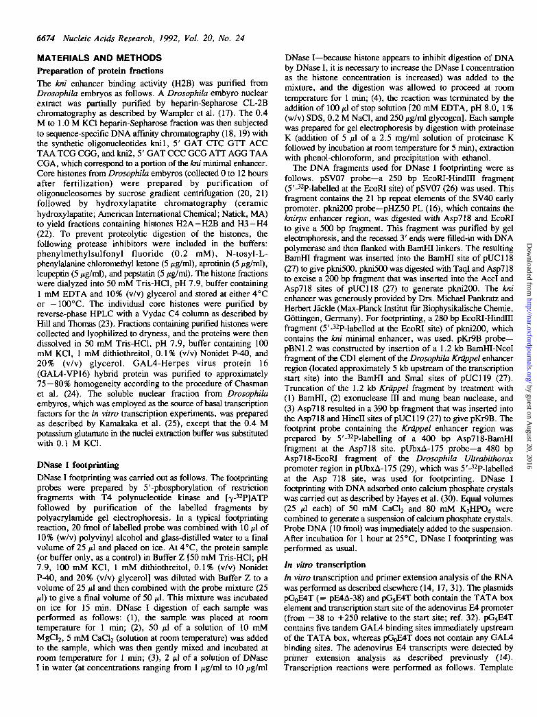
Figure 1. Identification of a factor that binds to the burps minimal enhancerelement as the core histone H2B. (A) Analysis of partially purified protein fractionsand purified core histories by 18% polyacrylamide-SDS gel electrophoresis andstaining with Coomassie blue. Lane 1, molecular mass markers (1 pg total protein);lane 2, 0.4-1.0 M KCI heparin-Sepharose CL-2B fraction (15 ^g); lane 3, kniDNA affinity column eluate (3 /ig); lanes 4 and 7, core histories from Drosophilaembryos (3.3 /ig); lane 5, histones H2A and H2B from Drosophila embryos (2.5/ig); lane 6, purified histone H2B from Drosophila embryos (1.3 jig). The sizesof the molecular mass standards are given in kDa. (B) DNase I footprint analysisof the kni minimal enhancer region (pkni200 probe) with partially purified proteinfractions and purified core histories. Lanes 1 and 6, no added protein (as a negativecontrol); lane 2, 0.4-1.0 M KCI heparin-Sepharose CL-2B fraction (1.3 /ig);lane 3, kni DNA affinity column eluate (0.25 jig); lane 4, histones H2A and H2B(20 ng); lane 5, purified histone H2B (1.3 /ig). Note that purification of H2Bby reverse-phase HPLC results in a significant loss of DNA binding activity.Regions of protection are indicated by brackets. (C) Summary of binding of H2Bto the kni minimal enhancer. The segments of the minimal enhancer that areprotected from DNase I digestion by histone H2B are underlined. The imperfectpalindromic sequences are indicated by the arrows. The portion of the enhancerthat was used for construction of the DNA affinity resin is shaded. Filled circleswere placed at 10 bp intervals to highlight the periodicity of DNase I digestion.
DNA (50 ng) and GAL4-VP16 (either 0 or 32 ng) were incubatedon ice for 15 min. Next, variable amounts (0, 50, 125, or 250ng) of H2A-H2B [in 25 mM Hepes (K+), pH 7.6, buffercontaining 100 mM KCI, 12.5 mM MgCl2, 0.1 mM EDTA, 1mM dithiothreitol, and 10% (v/v) glycerol] were added, and thesamples were incubated on ice for an additional 15 min. Solublenuclear fraction (40 /tg; ref. 25) and ribonucleoside5'-triphosphates were then added, and the transcription reactionswere carried out at 21°C for 30 min. All reactions wereperformed two to four times to ensure reproducibility of the data.Quantitation of the reverse transcription products was performedby using a Molecular Dynamics phosphorimager withImageQuant software.
RESULTS AND DISCUSSIONPurification and identification of a factor that binds to theknirps minimal enhancerAs a first step in the biochemical analysis of the kni transcriptionalcontrol region, we sought to purify a sequence-specific DNAbinding factor that interacts with the minimal enhancer element(16). Such a transcription factor would probably be a centralcomponent in the spatial and temporal regulation of the kni gene.A survey with a variety of purified transcription factors andpartially purified protein fractions yielded a periodic DNase Ifootprint pattern over the kni minimal enhancer with a 0.4-1.0M KCI fraction off a heparin-Sepharose column (Fig. 1A, lane2; Fig. IB, lane 2). Examination of the DNA sequence of thekni minimal enhancer revealed two and a half copies of animperfect 21 bp palindromic sequence, which appeared tocorrelate with the DNase footprint pattern (see Fig. 1C). Weprepared a sequence-specific DNA affinity resin based on a singlecopy of the imperfect palindromic sequence, and subsequentaffinity chromatography resulted in enrichment of a protein withan apparent molecular mass of 15 kDa (Fig. 1A, lane 3; Fig. IB,lane 3).
To determine if the 15 kDa polypeptide was interacting withthe kni minimal enhancer, the enhancer-binding activity wassubjected to further purification and characterization. In the courseof these studies, we found that both the 15 kDa protein and theenhancer-binding activity bound with high affinity to a GC-richaffinity resin with a DNA sequence that did not resemble thekni enhancer. This lack of apparent sequence-specificity of DNAbinding by a protein with an apparent molecular mass of 15 kDasuggested that the 15 kDa protein may be a component ofchromatin such as a histone or high mobility group protein. Totest this hypothesis, we compared the mobility of the 15 kDapolypeptide with that of the Drosophila core histones by SDS-polyacrylamide gel electrophoresis and found that the 15 kDapolypeptide co-migrated with the core histone H2B (Fig. 1A,compare lanes 3 and 4). Furthermore, the Coomassie blue-stained15 kDa polypeptide in the poly aery lamide gel displayed the sameunusual, yet distinctive light-scattering properties (33) as the corehistones. At this point, it was likely that the 15 kDa 'enhancer-binding protein' was identical to histone H2B.
To confirm the hypothesis that a previously unknown periodicDNA binding property of histone H2B resulted in our inadvertentpurification of H2B as a putative enhancer-binding factor, weperformed the following experiments. First, western blot analysisrevealed that antibodies directed against histone H2B cross-reacted with the 15 kDa polypeptide (data not shown). Then,we purified H2A-H2B dimers and H2B alone from Drosophilaembryos (Fig. 1A, lanes 5 and 6) by using well-establishedprocedures (22, 23) and found that both the H2A-H2B fractionand purified H2B bound to the kni enhancer region in a periodicmanner (Fig. IB, lanes 4 and 5). Thus, the putative 'enhancer-binding factor' was unequivocally identified as the core histoneH2B. The periodic binding of H2B to the kni minimal enhanceris summarized in Fig. 1C, and the period of the DNase Iprotection is approximately 10 bp, which is roughly one turn ofthe DNA helix.
The four core histones can each bind to DNA in a periodicmannerTo examine further the unexpected finding that DNase I digestionof DNA-bound histone H2B gives a repeated 10 bp ladder, we
by guest on August 20, 2016
http://nar.oxfordjournals.org/D
ownloaded from
6676 Nucleic Acids Research, 1992, Vol. 20, No. 24
CO CO
w <» < co a;m £ C\J CM CO T g^ S x i x i 2(0 i5 IB 0) J) d -
c -c c c c c -c:a) o o o o aj
5 o ^ ?̂ ^ ^ o5 O X X X I O66
3i2 9 -2 4 - •20 -
14 -
,H3• H2B• H2A
1 2 3 4 5 6 7
1 2 3 4 5 6
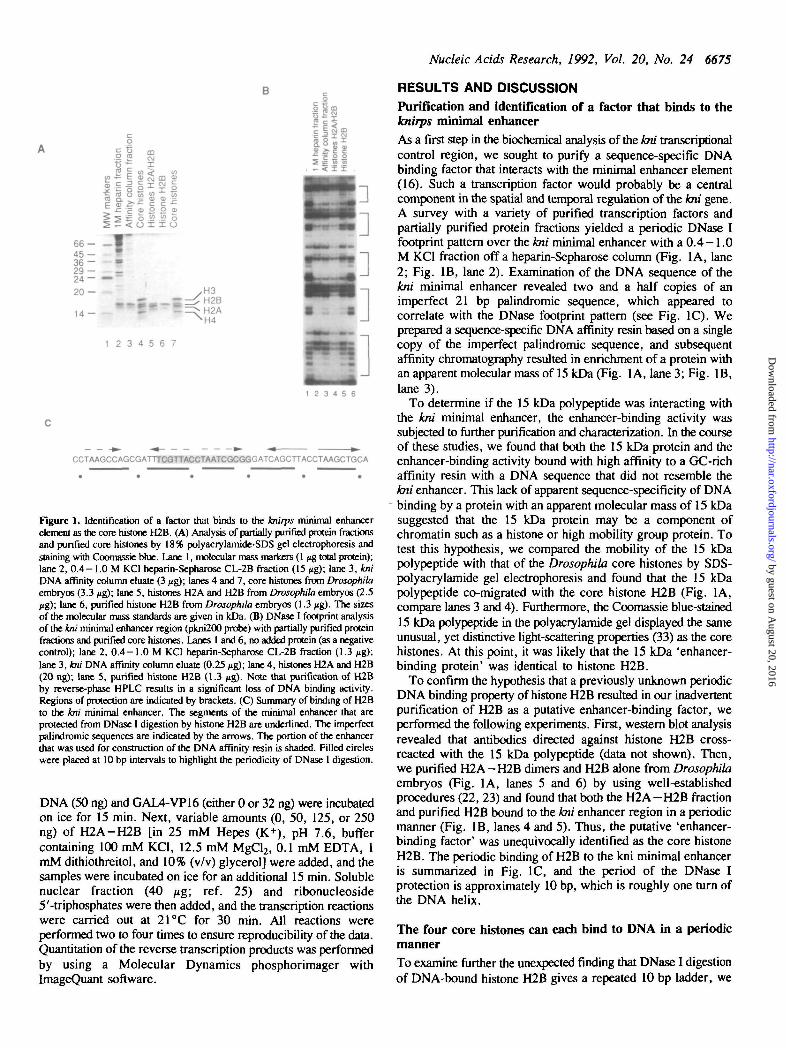
Figure 2. Purification and characterization of individual core histories fromDrosophila embyros. (A) Analysis of purified core histones by 18%polyacrylamide-SDS gel electrophoresis and staining with Coomassie blue. Lane1, molecular mass markers (1 tig)', lanes 2 and 7, total core histones (5 /ig); lanes3 —6, HPLC-purified core histones (1 ng, each, as indicated). (B) DNase I footprintanalysis of the kni minimal enhancer region (pkru200 probe) with purified corehistones. Lanes 1 and 6, no added protein (as a negative control); lanes 2 - 5 ,HPLC-purified core histones (1 Mg each, as indicated). Regions of protectionare indicated by brackets.
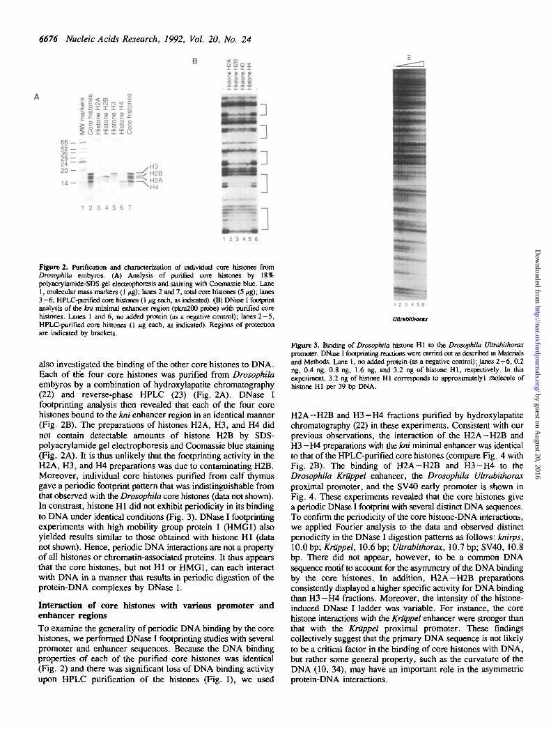
also investigated the binding of the other core histones to DNA.Each of the four core histones was purified from Drosophilaembyros by a combination of hydroxylapatite chromatography(22) and reverse-phase HPLC (23) (Fig. 2A). DNase Ifootprinting analysis then revealed that each of the four corehistones bound to the kni enhancer region in an identical manner(Fig. 2B). The preparations of histones H2A, H3, and H4 didnot contain detectable amounts of histone H2B by SDS-polyacrylamide gel electrophoresis and Coomassie blue staining(Fig. 2A). It is thus unlikely that the footprinting activity in theH2A, H3, and H4 preparations was due to contaminating H2B.Moreover, individual core histones purified from calf thymusgave a periodic footprint pattern that was indistinguishable fromthat observed with the Drosophila core histones (data not shown).In constrast, histone HI did not exhibit periodicity in its bindingto DNA under identical conditions (Fig. 3). DNase I footprintingexperiments with high mobility group protein 1 (HMG1) alsoyielded results similar to those obtained with histone HI (datanot shown). Hence, periodic DNA interactions are not a propertyof all histones or chromatin-associated proteins. It thus appearsthat the core histones, but not HI or HMG1, can each interactwith DNA in a manner that results in periodic digestion of theprotein-DNA complexes by DNase I.
Interaction of core histones with various promoter andenhancer regionsTo examine the generality of periodic DNA binding by the corehistones, we performed DNase I footprinting studies with severalpromoter and enhancer sequences. Because the DNA bindingproperties of each of the purified core histones was identical(Fig. 2) and there was significant loss of DNA binding activityupon HPLC purification of the histones (Fig. 1), we used
1 2 3 4 5 6
Uttrablthormx
Figure 3. Binding of Drosophila histone HI to the Drosophila Ultrabithoraxpromoter. DNase I footprinting reactions were carried out as described in Materialsand Methods. Lane 1, no added protein (as a negative control); lanes 2 - 6 , 0.2ng, 0.4 ng, 0.8 ng, 1.6 ng, and 3.2 ng of histone HI, respectively. In thisexperiment, 3.2 ng of histone HI corresponds to approximately! molecule ofhistone HI per 39 bp DNA.
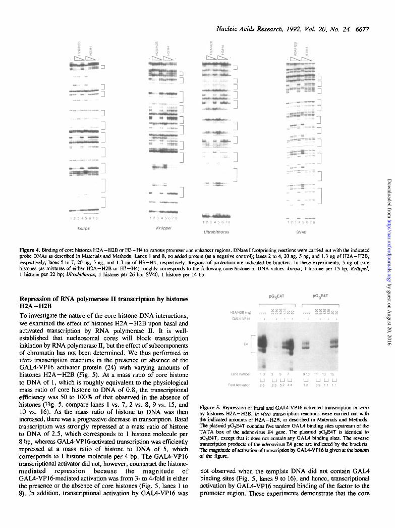
H2A-H2B and H3-H4 fractions purified by hydroxylapatitechromatography (22) in these experiments. Consistent with ourprevious observations, the interaction of the H2A-H2B andH3 — H4 preparations with the kni minimal enhancer was identicalto that of the HPLC-purified core histones (compare Fig. 4 withFig. 2B). The binding of H2A-H2B and H3-H4 to theDrosophila Kriippel enhancer, the Drosophila Ultrabithoraxproximal promoter, and the SV40 early promoter is shown inFig. 4. These experiments revealed that the core histones givea periodic DNase I footprint with several distinct DNA sequences.To confirm the periodicity of the core histone-DNA interactions,we applied Fourier analysis to the data and observed distinctperiodicity in the DNase I digestion patterns as follows: knirps,10.0 bp; Kriippel, 10.6 bp; Ultrabithorax, 10.7 bp; SV40, 10.8bp. There did not appear, however, to be a common DNAsequence motif to account for the asymmetry of the DNA bindingby the core histones. In addition, H2A—H2B preparationsconsistently displayed a higher specific activity for DNA bindingthan H3— H4 fractions. Moreover, the intensity of the histone-induced DNase I ladder was variable. For instance, the corehistone interactions with the Kriippel enhancer were stronger thanthat with the Kriippel proximal promoter. These findingscollectively suggest that the primary DNA sequence is not likelyto be a critical factor in the binding of core histones with DNA,but rather some general property, such as the curvature of theDNA (10, 34), may have an important role in the asymmetricprotein-DNA interactions.
by guest on August 20, 2016
http://nar.oxfordjournals.org/D
ownloaded from
Nucleic Acids Research, 1992, Vol. 20, No. 24 6677
•]
nn
l —1
:
«-I
12 3 4 5 6 7 8
Ultrabithorax
1 2 3 4 5 6 7 6
SV40
Figure 4. Binding of core histories H2A-H2B or H3-H4 to vanous promoter and enhancer regions. DNase I footprinting reactions were carried out with the indicatedprobe DNAs as described in Materials and Methods. Lanes 1 and 8, no added protein (as a negative control); lanes 2 to 4, 20 ng, 5 ng, and 1.3 ng of H2A—H2B,respectively; lanes 5 to 7, 20 ng, 5 ng, and 1.3 ng of H3—H4, respectively. Regions of protection are indicated by brackets. In these experiments, 5 ng of corehistories (as mixtures of either H2A-H2B or H3—H4) roughly corresponds to the following core histone to DNA values: knirps, 1 histone per 15 bp; KrOppel,1 histone per 22 bp; Ultrabithorax, 1 histone per 26 bp; SV40, 1 histone per 14 bp.
Repression of RNA polymerase II transcription by histonesH2A-H2BTo investigate the nature of the core histone-DNA interactions,we examined the effect of histones H2A-H2B upon basal andactivated transcription by RNA polymerase II. It is well-established that nucleosomal cores will block transcriptioninitiation by RNA polymerase n, but the effect of subcomponentsof chromatin has not been determined. We thus performed invitro transcription reactions in the presence or absence of theGAL4-VP16 activator protein (24) with varying amounts ofhistones H2A—H2B (Fig. 5). At a mass ratio of core histoneto DNA of 1, which is roughly equivalent to the physiologicalmass ratio of core histone to DNA of 0.8, the transcriptionalefficiency was 50 to 100% of that observed in the absence ofhistones (Fig. 5, compare lanes 1 vs. 7, 2 vs. 8, 9 vs. 15, and10 vs. 16). As the mass ratio of histone to DNA was thenincreased, there was a progressive decrease in transcription. Basaltranscription was strongly repressed at a mass ratio of histoneto DNA of 2.5, which corresponds to 1 histone molecule per8 bp, whereas GAL4-VP16-activated transcription was efficientlyrepressed at a mass ratio of histone to DNA of 5, whichcorresponds to 1 histone molecule per 4 bp. The GAL4-VP16transcriptional activator did not, however, counteract the histone-mediated repression because the magnitude ofGAL4-VP16-mediated activation was from 3- to 4-fold in eitherthe presence or the absence of core histones (Fig. 5, lanes 1 to8). In addition, transcriptional activation by GAL4-VP16 was
pG5E4T
"i rH2A/H2B (ng) o o
GAL4-VP16 • +
SSooy- • - tr> i n
ILane number 1 2 3 5 7
U U U UFold Activation 2 5 2 3 52 44
910 11 13 15
U U U U12 09 11 11
Figure S. Repression of basal and GAL4-VPI6-activated transcription in vitroby histones H2A-H2B. In vitro transcription reactions were carried out withthe indicated amounts of H2A-H2B, as described in Materials and Methods.The plasmid pG3E4T contains five tandem GAM binding sites upstream of theTATA box of the adenovirus E4 gene. The plasmid pGoE4T is identical topG5E4T, except that it does not contain any GAL4 binding sites. The reversetranscription products of the adenovirus E4 gene are indicated by the brackets.The magnitude of activation of transcription by GAL4-VP16 is given at the bottomof the figure.
not observed when the template DNA did not contain GAL4binding sites (Fig. 5, lanes 9 to 16), and hence, transcriptionalactivation by GAL4-VP16 required binding of the factor to thepromoter region. These experiments demonstrate that the core
by guest on August 20, 2016
http://nar.oxfordjournals.org/D
ownloaded from
6678 Nucleic Acids Research, 1992, Vol. 20, No. 24
c . B .
[-11 2 3
SV40
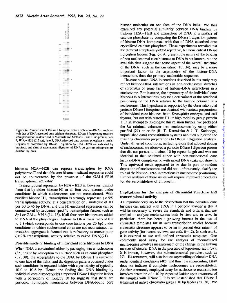
Figure 6. Comparison of DNase I footprint pattern of histone-DNA complexeswith that of DNA adsorbed onto calcium phosphate. DNase I footprinting reactionswere performed as described in Materials and Methods. Lane 1, no protein; lane2, H2A—H2B(2.5 ng); lane 3, DNA adsorbed onto calcium phosphate crystals.Regions of protection by DNase I digestion by H2A-H2B are indicated bybrackets, and sites of accentuated digestion of DNA on calcium phosphate aredenoted by dots.
histones H2A—H2B can repress transcription by RNApolymerase II and that this core histone-mediated repression couldnot be counteracted by the presence of the GAL4-VP16transcriptional activator.
Transcriptional repression by H2A—H2B is, however, distinctfrom that by either histone HI or all four core histones underconditions in which nucleosomes are not reconstituted. Withpurified histone HI, transcription is strongly repressed (<5%transcriptional activity) at a concentration of 1 molecule of H1per 30 to 45 bp DNA, and this HI-mediated repression can becounteracted by sequence-specific transcription factors such asSpl orGAL4-VP16 (14, 15). If all four core histones are addedto DNA at the physiological histone to DNA mass ratio of 0.8to 1 (which corresponds to one core histone per 25 bp) underconditions in which nucleosomal cores are not reconstituted, aninsoluble aggregate is formed that is refractory to transcription( < 2 % transcriptional activity relative to naked DNA)(21).
Possible mode of binding of individual core histones to DNAWhen DNA is constrained either by packaging into a nucleosome(35, 36) or by adsorption to a surface such as calcium phosphate(37, 38), the accessibility to the DNA by DNase I is restrictedto one face of the helix, and the digestion pattern obtained undersuch conditions is repeated ladder with a periodicity of typically10.0 to 10.6 bp. Hence, the finding that DNA binding byindividual core histones yields a repeated DNase I digestion ladderwith a periodicity of roughly 10 bp suggests that there areperiodic, homotypic interactions between DNA-bound core
histone molecules on one face of the DNA helix. We thusexamined any potential similarity between DNA binding byhistones H2A—H2B and adsorption of DNA to a surface ofcalcium phosphate-by comparing the DNase I digestion patternof histone-DNA complexes with that of DNA adsorbed ontocrystallized calcium phosphate. These experiments revealed thatthe different complexes yielded repetitive, but nonidentical DNaseI digestion ladders (Fig. 6). At present, the nature of the bindingof non-nucleosomal core histones to DNA is not known, but theavailable data suggest that some aspect of the overall structureof the DNA, such as the curvature (10, 34), may be a moreimportant factor in the asymmetry of the histone-DNAinteractions than the primary nucleotide sequence.
The core histone-DNA interactions described in this study mayreflect histone-DNA interactions in non-nucleosomal stretchesof chromatin or some facet of histone-DNA interactions in anucleosome. For instance, the asymmetry of the individual corehistone-DNA interactions may be a determinant of the rotationalpositioning of the DNA relative to the histone octamer in anucleosome. This hypothesis is supported by the observation thatperiodic DNase I footprints are obtained with various preparationsof individual core histones from Drosophila embryos and calfthymus, but not with histone HI or high mobility group protein1. In an attempt to investigate this relation further, we packagedthe kni minimal enhancer into nucleosomes by using eitherpurified (21) or crude (R. T. Kamakaka & J. T. Kadonaga,unpublished data) reconstitution systems and then subjected theresulting chromatin preparations to DNase I digestion analysis.Under all tested conditions, including those that allowed slidingof nucleosomes, we observed a periodic DNase I digestion patternthat did not possess a distinct 10 bp repeat length and was notidentical to that obtained either with non-nucleosomal corehistone-DNA complexes or with naked DNA (data not shown).This equivocal result appeared to be due in part to randomdeposition of nucleosomes and did not, unfortunately, clarify therole of the histone-DNA interactions in nucleosome positioning.Further analyses of these issues will require improved proceduresfor the reconstitution of chromatin.
Implications for the analysis of chromatin structure andtranscriptional activityAn important corollary to the observation that the individual corehistones can interact with DNA in a periodic manner is that itwill be necessary to revise the standards and criteria that areapplied to analyze nucleosomes both in vitro and in vivo. Inparticular, there has been a growing interest in the use ofchromatin templates for in vitro transcription studies becausechromatin structure appears to be an important determinant ofgene activity (for recent reviews, see refs. 6-12). In such work,it is essential to use well-defined chromatin templates. Onecommonly used assay for the analysis of reconstitutednucleosomes involves measurement of the change in the linkingnumber of circular DNA in the presence of topoisomerase I (39).It is known, however, that subnucleosomal particles, such asH3 — H4 tetramers, will also induce supercoiling of circular DNAunder identical conditions (40), and thus, the supercoiling assaydoes not indicate if complete nucleosomes are reconstituted.Another commonly employed assay for nucleosome reconstrtutioninvolves detection of a 10 bp repeated ladder upon treatment ofthe samples with DNase I. It is well-established that DNase Itreatment of native chromatin gives a 10 bp ladder (35, 36). We
by guest on August 20, 2016
http://nar.oxfordjournals.org/D
ownloaded from
Nucleic Acids Research, 1992, Vol. 20, No. 24 6679
have shown, however, that individual core histones, or mixturesof H2A-H2B or H3-H4, can also interact with DNA in anasymmetric manner to give a repeated 10 bp DNase I digestionladder. Hence, in the course of nucleosome reconstitution in vitro,it may be difficult to distinguish between the formation ofnucleosomes or subnucleosomal particles by the use of thesupercoiling and DNase I digestion assays.
It is thus apparent that in the future, it will be necessary toapply additional criteria to evaluate the quality of reconstitutednucleosomes. These criteria should probably include micrococcalnuclease digestion analysis (41) to demonstrate the existence ofan array of nucleosomes as well as examination of the polypeptidecomposition of the purified chromatin by SDS-polyacrylamidegel electrophoresis to show that the four core histones are presentin equimolar quantities. In situations where nucleosomes arereconstituted onto relatively short DNA fragments,mononucleosomes could also be characterized by electrophoresisof the intact nucleosomes by nondenaturing gel electrophoresis(42, 43). In addition, electron microscopy (44) would provideuseful information regarding the quality of reconstitutedchromatin.
In this context, it is worthwhile to compare the results ofexperiments in which transcription is repressed by eithernucleosomes or H2A-H2B mixtures. It has been shownpreviously that when GAL4-VP16 is pre-bound to a naked DNAtemplate, it is able to counteract repression of transcription causedby the reconstitution of nucleosomes (45). In contrast, we haveshown here that GAL4-VP16 is not able to counteract repressionof transcription by H2A-H2B, and hence, it appears that thereis a distinction between the ability of GAL4-VP16 to counteractrepression of transcription by nucleosomes or subnucleosomalcomponents. Moreover, it has been demonstrated more recentlythat GAL4-VP16 is able to counteract nucleosomal core-mediatedrepression of transcription with reconstituted chromatin that hasbeen characterized by the supercoiling assay, micrococcalnuclease digestion assay, SDS-polyacrylamide gelelectrophoresis, and electron microscopy (R. T. Kamakaka, M.D. Bulger, and J. T. Kadonaga, in preparation). The availabledata suggest that DNA-bound GAL4-VP16 is able to counteractrepression of transcription that is caused by the deposition ofnucleosomes but not transcriptional repression that is due to thebinding of H2A-H2B alone. Thus, in future studies of therelation between chromatin structure and transcription activity,it will be important to characterize the reconstituted chromatinthoroughly in terms of nucleosomal structure as well as toconsider other factors such as histone modifications and thepresence or absence of histone HI and high mobility groupproteins.
In summary, we describe the identification of a putativeenhancer binding factor as the core histone H2B and show thateach of the individual core histones, but not histone HI or highmobility group protein 1, can interact with DNA in a periodicmanner. From a practical standpoint, the ability of the corehistones to give a repetitive DNase I footprint ladder and to bindto DNA affinity resins should be considered in the course ofpurification and characterization of new sequence-specific DNAbinding proteins. Moreover, these results indicate that the criteriafor the analysis of nucleosomes should extend beyond thegeneration of a repeated 10 bp DNase I digestion ladder. Theperiodic DNA binding by the core histones may reflect someaspects of histone-DN A interactions in the nucleosome or histone-DNA interactions in non-nucleosomal regions of chromatin. For
instance, previous studies involving cross-Unking of core histonesto DNA in vivo have suggested that upon transcriptionalactivation, nucleosome structure is altered while the core histonesremain associated with the promoter sequences (46, 47). It ispossible that the core histone-DNA interactions described in thisstudy may be relevant to such situations. In the future, asystematic and detailed analysis of non-nucleosomal stretches ofchromatin, such as that found in highly transcribed genes, mightprovide additional insight into these phenomena.
ACKNOWLEDGEMENTS
We are most grateful to B. Zimm for assistance with the Fourieranalysis of the footprinting data; B. David Stollar for antibodiesagainst the core histones; Rohinton Kamakaka and Paul Laybournfor suggestions and advice during the course of this work; JeffHayes and Alan Wolffe for helpful advice regarding theadsorption of DNA to calcium phosphate; Betsy Komives foradvice on HPLC purification of the histones; Michael Pankratzand Herbert Jackie for the kni genomic clone and communicationof results prior to publication; and Chris Kerrigan for assistancein the preparation of the figures. We also thank Bruno Zimm,Rohinton Kamakaka, Glenn Croston, Mike Bulger, and SumiParanjape for critical reading of the manuscript. J. T. K. is aLucille P. Markey Scholar and Presidential Faculty Fellow, andthis work was supported in part by grants from the NationalInstitutes of Health, National Science Foundation, Council forTobacco Research, and the Lucille P. Markey Charitable Trust.
REFERENCES
1. Saltzman, A. G., and Weinmann, R. (1989) FASEB Journal 3, 1723-17332. Sawadogo, M., and Sentenac, A. (1990) Amu. Rev. Biochem. 59, 711-7543. Zawel, L., and Reinberg, D. Prog. Nucl. Acids Res. Mol. Biol., in press4. Mitchell, P. J., and Tjian, R. (1989) Science 245, 371-3785. Johnson, P. F., and McKnight, S. L. (1989) Annu. Rev. Biochem. 58,
799-8396. van Holde, K. E. (1989) Chromatin (Springer-Verlag, New York)7. Wolffe, A. P. (1990) New Biologist 2, 211-2188. Grunstein, M. (1990) Annu. Rev. Cell Biol. 6, 643-6789. Elgin, S. C. R. (1990) Cun\ Opin. Cell Biol. 2, 437-445
10. Simpson, R. T. (1991) Prog. Nucl. Acids Res. Mol. Biol. 40, 143-18411. Kornberg, R. D., and Lorch, Y. (1991) Cell 67, 833-83612. Felsenfeld, G. (1992) Nature 355, 219-22313. St. Johnston, D., and Nusslein-Volhard, C. (1992) Cell 68, 201-21914. Kerrigan, L. A., Croston, G. E., Lira, L. M., and Kadonaga, J. T. (1991)
J. Biol. Chem. 266, 574-58215. Croston, G. E., Kerrigan, L. A., Lira, L., Marshak, D. R., and Kadonaga,
J. T. (1991) Science 251, 643-64916. Pankratz, M. J., Busch, M., Hoch, M., Seifert, E., and JSckle, H. (1992)
Science 255, 986-98917. Wampler, S. L., Tyree, C. M., and Kadonaga, J. T. (1990) J. Biol. Chem.
265, 21223-2123118. Kadonaga, J. T., and Tjian, R. (1986) Proc. Natl. Acad. Sci. USA 83,
5889-589319. Kadonaga, J. T. (1991) Methods Enzymol. 208, 10-2320. Butler, P. J. G., and Thomas, J. O. (1980) J. Mol. Biol. 140, 505-52921. Layboum, P. J., and Kadonaga, J. T. (1991) Science 254, 238-24522. Simon, R. H., and Felsenfeld, G. (1979) Nucleic Acids Res. 6, 689-69623. Hill, C. S., and Thomas, J. O. (1990) Eur. J. Biochem. 187, 145-15324. Chasman, D. I., Leatherwood, J., Carey, M., Ptashne, M., and Kornberg,
R. D. (1989) Mol. Cell. Biol. 9, 4746-474925. Kamakaka, R. T., Tyree, C. M., and Kadonaga, J. T. (1991) Proc. Natl.
Acad. Sci. USA 88, 1024-102826. Dynan, W. S., and Tjian, R. (1983) Cell 35, 79-8727. Vieira, J., and Messing, J. (1987) Methods Enzymol. 153, 3-1128. Hoch, M., Schroder, C , Seifert, E., and Jackie, H. (1990) EMBO J. 9,
2587-259529. Biggin, M. D., and Tjian, R. (1988) Cell 53, 699-711
by guest on August 20, 2016
http://nar.oxfordjournals.org/D
ownloaded from
6680 Nucleic Acids Research, 1992, Vol. 20, No. 24
30. Hayes, J. J., Bashkin, J., Tullius, T. D., and Wolffe, A. P. (1991)Biochemistry 30, 8434-8440
31. Kadonaga, J. T. (1990) J. Biol. Chem. 265, 2624-263132. Lin, Y.-S., Carey, M. F., Ptashne, M., and Green, M. R. (1988) Cell 54,
659-66433. Kleinschmidt, J. A., and Franke, W. W. (1982) Cell 29, 799-80934. Travers, A. A. (1987) Trends Biochem. Sci. 12, 108-11235. NoU, M. (1974) Nucleic Acids Res. 1, 1573-157836. Lutter, L. C. (1989) Methods Enzymol. 170, 264-26937. Rhodes, D., and Klug, A. (1980) Nature 286, 573-57838. Behe, M., Zimmerman, S., and Felsenfeld, G. (1981) Nature 293, 233-23539. Germond, J. E., Hirt, B., Oudet, P., Gross-Bellard, M., and Chambon,
P. (1975) Proc. Nad. Acad. Sci. USA 72, 1843-184740. Camerini-Otero, R. D., and Felsenfeld, G. (1977) Nucleic Acids Res. 4,
1159-118141. NoU, M., and Komberg, R. D. (1977) J. Mol. Bid. 109, 393-40442. Varshavsky, A. J., Bakayev, V. V., and Georgiev, G. P. (1976) Nucleic
Acids Res. 3, 477-49243. Nelson, P. P., Albright, S. C , Wiseman, J. M., and Garrard, W. T. (1979)
J. Biol. Chem. 254, 11751-1176044. Sogo, J. M., and Thoma, F. (1989) Methods Enzymol. 170, 142-16545. Workman, J. L., Taylor, I. C. A., and Kingston, R. E. (1991) Cell 64,
533-54446. Nacheva, G. A., Guschin, D. Y., Preobrazhenskaya, O. V., Karpov, V.
L., Ebralidse, K. K., and Mirzabekov, A. D. (1989) Cell 58, 27-3647. Bresnick, E. H., Bustin, M., Marsaud, V., Richard-Foy, H., and Hager,
G. L. (1992) Nucleic Acids Res. 20, 273-278
by guest on August 20, 2016
http://nar.oxfordjournals.org/D
ownloaded from