a membrane-reconstituted multisubunit functional proton pump on mesoporous silica particles
TRANSCRIPT
AMembrane-ReconstitutedMultisubunit Functional Proton PumponMesoporous Silica ParticlesGustav Nordlund,† Jovice Boon Sing Ng,‡ Lennart Bergstrom,‡ and Peter Brzezinski†,*†Department of Biochemistry and Biophysics, Centre for Biomembrane Research, Stockholm University, SE-10691 Stockholm, Sweden, and ‡Materials Chemistry ResearchGroup, Department of Physical, Inorganic and Structural Chemistry, Stockholm University, SE-10691 Stockholm, Sweden
Recent developments in the synthe-sis of self-assembled materials havemade it possible to design and
manufacture surfactant-templated mesopo-
rous silica particles with stable mesostruc-
ture, narrow pore size distribution, adjust-
able pore size, pore connectivity, and
morphology.1�5 The accessible surface area
and pore volume of these particles are typi-
cally very large, making it possible to use
them as vehicles for controlled delivery of,
for example, drugs and genes.6�8 The inter-
nal and external surfaces of the mesoporous
material can be modified to immobilize
and control the release of, for example, an-
ticancer drugs.9�11 Recent works have
shown how coumarin, quantum dots, and
other molecules can open and close the
pores triggering release of specific
molecules.12�14 Durable encapsulation of
the molecules can also be accomplished by
deposition of a lipid membrane, which cov-
ers the entire surface of the particle.15�22
The release of these molecules could be
mediated by specific channels or transport-
ers incorporated into the membrane, which
requires development of methodology and
techniques for incorporation of integral
membrane proteins into the particle-
supported lipid layers. Such
protein�membrane particle systems are
also of significant interest for functional
studies of membrane proteins because they
provide a robust system for mechanistic in-
vestigations of transport mechanisms. In-
deed, recent reports describe successful in-
corporation of a number of simple and
structurally stable membrane proteins, such
as bacteriorhodopsin, into supported mem-
branes onto solid20 and porous23 silica par-
ticles. In the present study, we have used a
multisubunit, transmembrane complex
“molecular machine”, cytochrome c oxi-dase (CytcO, cytochrome aa3) from Rhodo-bacter sphaeroides. In earlier studies meth-ods have been developed to adsorbmembrane-reconstituted CytcO at planarsolid surfaces.24�27 Here, we demonstratethat CytcO can be incorporated into a lipidmembrane supported onto the outer sur-face of cell-mimetic mesoporous silica col-loids with an interior water-filled volume.The mesoporous silica particles are mono-disperse spheres with a diameter of 550 nmand pore-size of 3.0 nm, prepared by amodified Stoeber method using hexadecyl-trimethyl ammonium bromide as the tem-plating molecule.28
The enzyme CytcO is a proton pumpthat is driven by electron transfer from cyto-chrome c to oxygen, which is reduced towater. Cytochrome c initially donates elec-trons to a copper site (CuA), located withinsubunit II near the positive side (see later) ofthe membrane. The electrons are thentransferred consecutively to a heme group
*Address correspondence [email protected].
Received for review May 26, 2009and accepted July 28, 2009.
Published online August 4, 2009.10.1021/nn9005413 CCC: $40.75
© 2009 American Chemical Society
ABSTRACT We have investigated formation of a proteolipid membrane surrounding mesoporous silica
particles with a diameter of 550 nm and pore sizes of 3.0 nm. A multisubunit redox-driven proton pump,
cytochrome c oxidase, was incorporated into the membrane, and we show that the enzyme is functional, both
with respect to catalysis of O2 reduction to water, and charge separation across the membrane. The orientation
of cytochrome c oxidase in the membrane was found to be the same (�70%) in the lipid vesicles and in the silica-
particle-supported lipid membrane, which provides information on the mechanism by which the vesicles adsorb
to the surface. Furthermore, cytochrome c oxidase could maintain a proton electrochemical gradient across the
supported proteomembrane, that is, the membrane system was proton tight, defining an interior particle
compartment that is separated from the surrounding aqueous media. Such a biofunctional cellular interface,
supported onto a colloid that has a connected interior cytoskeleton-like pore structure, provides a basis for
functional studies of membrane-bound transport proteins, and also for applications within pharmaceutical drug
delivery.
KEYWORDS: supported lipid bilayer · mesoporous spheres · nanoparticles ·membrane protein · drug delivery · cytochrome c oxidase
ARTIC
LE
www.acsnano.org VOL. 3 ▪ NO. 9 ▪ 2639–2646 ▪ 2009 2639
(heme a) and then to the catalytic site, which consists
of another heme group, called heme a3, and a copper
ion (CuB) in close vicinity. When the catalytic site is re-
duced, heme a3 binds O2, which is then reduced to wa-
ter accompanied by proton uptake from the negative
side of the membrane. Because electrons are donated
from one side (the positive side) of the membrane,
while protons are taken up from the opposite side (the
negative side, inside of the bacterial cell), the reaction
catalyzed by CytcO results in a charge separation across
the membrane, which is equivalent to moving a posi-
tive charge from the negative to the positive side of the
membrane. In addition, the reaction catalyzed by CytcO
is linked to pumping of one proton across the mem-
brane for each electron transferred from cytochrome c
to O2, thereby increasing the overall charge transloca-
tion stoichiometry making it equivalent to translocation
of two positive charges per electron transferred to the
catalytic site.
The advantage of using CytcO to study proteo-
membrane formation at solid particles is that the pro-
tein has a well-defined optical absorption spectrum,
which is dependent on, for example, ligand binding to
the catalytic site. Furthermore, the protein can be selec-
tively reduced, either only from one or from both sides
of the membrane, providing a simple way to determine
protein orientation without the use of external labels.
Furthermore, the capacity of the membrane to hold a
transmembrane proton electrochemical gradient,
which reflects the integrity of the membrane, can be as-
sessed because the CytcO turnover rate is dependent
on the membrane potential. The orientation of CytcO in
the lipid vesicles was found to be preserved in the silica-
particle supported lipid membrane after the vesicle fu-
sion event. This finding allows us to speculate about the
mechanisms by which the vesicles adsorbed, ruptured,
and fused at the silica particle surface. The CytcO incor-
porated into the supported membrane was active and
did not display any structural changes around the cata-
lytic site. The CytcO-containing lipid membrane sup-
ported by the mesoporous silica particles was suffi-
ciently proton tight to allow for build-up of a proton
electrochemical gradient between the closed compart-
ment within the particles and the surrounding solution.
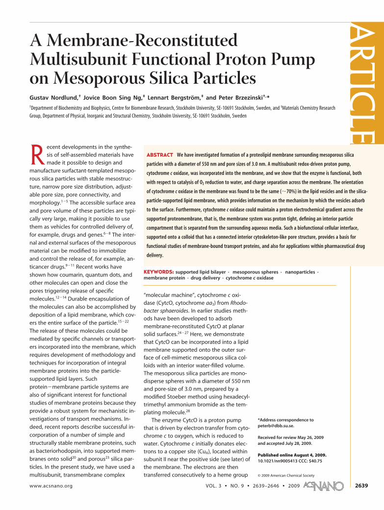
RESULTS AND DISCUSSIONThe calcined particles are monodispersed spheres
with an average particle size of 550 � 30 nm (Figure
1a). Their thin microtomed equatorial slices revealed
bundles of hexagonally ordered cylindrical channels
that extend radially from the sphere core to the sur-
face (Figure 1b). The X-ray diffraction spectra (Figure
1c) exhibit diffraction peaks of an ordered 2-D hexago-
nal mesopore structure. Nitrogen sorption measure-
ments (Figure 1d) showed that the particles have a pore
size of 3.0 nm, a specific area of 1280 m2 g�1, and pore
Figure 1. Characterization of the calcined mesoporous silica particles. (a) A scanning electron micrograph reveals thespherical morphology and monodispersity of the particles. (b) Thin microtomed equatorial slice of the particles viewed un-der transmission electron microscope exhibits pore channels extending radially from the core to the sphere surface. (c) Theparticles exhibit an X-ray diffraction of a 2D hexagonal mesostructure having a sharp 10 peak, with the higher order 11 and20 peaks merged together. (d) The nitrogen sorption isotherm can be classified as type IV isotherms according to the IU-PAC nomenclature, which is typically observed for 2D hexagonal mesoporous structures.1
ART
ICLE
VOL. 3 ▪ NO. 9 ▪ NORDLUND ET AL. www.acsnano.org2640

volume of 0.88 cm3 g�1. Results from previous studies
have shown that the pores are oriented perpendicular
to the external surface.28 The density, �1, of the mesopo-
rous particles is estimated as follow:
where Vpore is the pore volume per unit weight. Assum-
ing that the density of amorphous silica is 2.2 g/cm3,
and the estimated pore volume is 0.88 cm3/g (from the
nitrogen isotherm), �1 is 0.75 g/cm3. This particle den-
sity corresponds to an estimated exterior surface area of
14.6 m2/g (�1�1A/V, where �1 is the particle density, A is
the area and V is the volume), that is, the ratio of the in-
terior and exterior surface area is �90.
CytcO was reconstituted into the small unilamellar
vesicles, and these CytcO-containing vesicles were sub-
sequently used to cover the mesoporous silica par-
ticles. The fluorescence from the CytcO-containing
vesicles (i) with fluorescein-labeled lipids added, (ii)
with maleimide-fluorescein-labeled CytcO, or (iii) with
fluorescein trapped within, was analyzed before (using
a Fluorometer) and after (using flow cytometry) interac-
tion with the silica particles.29 The data show that the
relative fluorescence values for vesicles with (i)
fluorescein-labeled lipids and for (ii) fluorescein-labeled
CytcO were approximately the same before and after
reaction with the particles (Table 1). Because the light
scattering intensity depends strongly on particle size,
the flow cytometer only detects the mesoporous silica
particles and not the much smaller vesicles. Hence,
these measurements show that the CytcO-vesicles were
adsorbed onto the surface of the particles and that the
ratio of fluorescence values originating from pure lipids
and from fluorescein-labeled CytcO was the same for
the vesicle sample and after adsorption to particles. In
other words, the data indicate that the protein�lipid ra-
tio was the same in the vesicles and in the CytcO-
membrane at the particle surface.
The fluorescence from the CytcO-vesicle solutioncontaining (i) fluorescent lipids was �10 times largerthan from that containing (ii) fluorescein-labeled CytcO(Table 1). Each CytcO binds approximately one fluores-cein molecule per enzyme30 and the vesicle�CytcOratio was approximately unity. The number of lipid mol-ecules per vesicle was �5000 (diameter � 25 nm) andthe Fluo-DOPE (fluorescent lipid) mole fraction was�0.07%, which yields an average of �3.5 such lipidsper vesicle. In other words, the ratio of fluorophores invesicles containing Fluo-DOPE and fluorescein-labeledCytcO was �3.5. The larger observed fluorescence ratioof �10 is explained by a fraction (�50%) of vesicleswithout protein in the sample containing fluorescein-labeled CytcO (not all CytcO molecules are reconsti-tuted into the membrane, see Methods section).
For comparison, Table 1 also includes data from ourrecent study29 on adsorption of vesicles, loaded withwater-soluble fluorescein, to silica particle surfaces (seeiii). As seen in the table the relative fluorescence origi-nating from the entrapped fluorescein was much largerfor the vesicles prior to than after interaction with thesilica particles, which indicates that the vesicles breakupon interaction with the particles (see more detaileddiscussion in ref 29).
To examine whether a continuous membrane-CytcOlayer covering the entire surface of the particle wasformed (as compared to isolated patches covering onlypart of the surface), we tested if the interior of the par-ticle was isolated from the surrounding water solution.In the case of CytcO such a test can be done by study-ing the turnover activity of the enzyme itself withoutthe need of adding, for example, additional dye mol-ecules. As outlined above in the introduction section,this activity involves electron transfer from the electrondonor, cytochrome c, which binds on the positive sideof the protein, to the catalytic site, located in the inte-rior of the protein closer to the negative side. The elec-tron transfer is accompanied by proton uptake from thenegative side to the catalytic site, where O2 is eventu-ally reduced to H2O, that is, the overall reaction isequivalent to moving a positive charge from the nega-tive to the positive side, across the membrane. Further-more, CytcO pumps one proton per electron in thesame direction, where both proton pumping and theproton�electron transfer to the catalytic site result inbuilt-up of a transmembrane electrochemical protongradient. Upon formation of this gradient the turnoveractivity of the CytcO decreases. If proton and potassiumionophores are added the electrochemical gradient col-lapses and the activity increases again. The ratio of therates in the presence of the ionophores and in their ab-sence (i.e., the ratio of the slopes after and before addi-tion of ionophores) is referred to as the “respiratory con-trol ratio” (RCR), which is a measure of the tightness ofthe lipid membrane. Values �1 indicate that the mem-brane slows down diffusion of protons back into the
F1 ) 11
FSiO2
+ Vpore
(1)
TABLE 1. Relative Fluorescence Values of Vesicles andMembrane-Covered Particlesa
Flu-lipids (i) Flu-CytcO (ii) Flu inside/Flu-lipidsb (iii)
vesicles 93% 7% 10particles 91% 9% 0.12
aThe relative fluorescence values obtained with (i) fluorescein-labeled lipids (Flu-lipids), (ii) fluorescein-labeled CytcO (Flu-CytcO) in vesicles and (iii) the ratio of fluo-rescence values obtained from measurements with fluorescein entrapped withinvesicles and fluorescein-labeled lipids.29 These values are shown for vesicles only(“vesicles”) and after deposition of the membrane on the silica particle surface (“par-ticles”). The measurements were done using a fluorometer (for vesicles only) or aflow cytometer (for the silica particles). Consequently, the absolute fluorescence val-ues could not be directly compared. The signal originating from light scatteringwas subtracted from the data (particles with adsorbed membrane, but without anyfluorophores). The errors in these numbers are �10%. bData from ref 29.
ARTIC
LE
www.acsnano.org VOL. 3 ▪ NO. 9 ▪ 2639–2646 ▪ 2009 2641
particles such that a transmembrane electrochemicalpotential develops, which diminishes the CytcO activ-ity. It should be noted that an RCR of �1 would also beobtained if intact CytcO-vesicles were adsorbed at thesurface of the silica particles. We exclude this possibil-ity based on the observation that the vesicles breakupon interaction with the surface of the silicaparticles.29
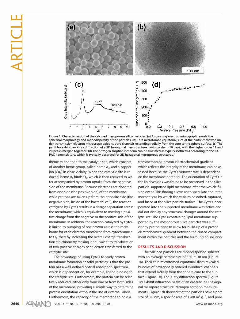
Figure 2 shows O2-consumption as a function oftime for the CytcO-vesicles alone and for CytcO-vesiclesdeposited on the particle surface. The slope (cf., O2-consumption rate) corresponding to the fully un-coupled system (i.e., after the addition of ionophores,which directly reflects the CytcO activity) was �7 timeslarger for the vesicle than for the particle sample (notethe different ordinate scales). This difference in slopes isconsistent with the factor of �10 lower CytcO concen-tration in the latter sample, which indicates that theCytcOs incorporated into the lipid bilayers supportedonto the particle surface are fully active. For the CytcO-containing vesicles the RCR was 4.0 � 0.3 (standard er-ror (SE), 3 measurements). For the CytcO-containingmembrane-covered silica particles the RCR value was
2.7 � 0.6 (SE, 5 measurements). The lower value mea-sured with the CytcO-containing membrane supportedonto the particles as compared to the vesicles alone isreasonable considering that the surface area of the par-ticles is �500 (rparticle
2/rvesicle2 � 2752/12.52) times larger
than that of the vesicles. Here we assume that the leak-age rate increases with increasing membrane surfacearea. It should also be noted that the experiment is sen-sitive to proton leaks, which presumably occur due todynamic fluctuations of the membrane and notthrough “holes” across the membrane (which would al-low protons to move freely across the membrane yield-ing an RCR of unity). Thus, even though there is a spreadin the RCR values, the fact that values of �1 are ob-served for the particles shows that the interior was insu-lated from the exterior by an intact membrane. In otherwords, the entire particles were covered by a mem-brane. In addition, these results also show that theCytcO adsorbed to the particle surface is functional,not only when considering O2-reduction catalysis, butalso when considering charge separation across themembrane.
Another approach to study the integrity of the cata-lytic site of CytcO incorporated into the particle-supported membrane (which reflects the integrity ofthe entire protein) is to examine ligand binding to thesite. When CytcO is reduced under anaerobic conditionsin the presence of carbon monoxide, heme a3 bindsthe CO ligand. The iron-CO bond is photolabile and,consequently, the CO ligand can be fully dissociatedfrom CytcO by means of a short laser flash. When stud-ied in detergent solution, after dissociation, the COligand then recombines with a rate constant of �10ms at 1 mM CO (saturated CO solution)31 As seen in Fig-ure 3 (compare any of the traces in panel a to those inpanel b, see further explanation later) the CO-recombination time constant was about the same forthe particle-supported membrane-bound CytcO as forCytcO in lipid vesicles or in detergent solution, whichalso indicates an intact catalytic site.
The orientation of CytcO in the vesicles was deter-mined by selectively reducing the enzyme from differ-ent sides of the membrane. First, CytcO was reducedwith a membrane-impermeable reducing agent (hex-aminerutheniumchloride, analogue of cytochrome c),which reduces only CytcO with the cytochromec-binding site facing the outside (positive side) solu-tion (“correctly” oriented CytcO). Then, another reduc-ing agent (dithionite), which can reduce also the “incor-rectly” oriented population, was added. The fraction ofreduced CytcO after the additions is typically deter-mined from measurements of the optical absorptionspectra. However, this approach was found to be inac-curate when used to determine the orientation of CytcOin the membranes deposited at the silica particle sur-faces due to significant light scattering of the particles.To circumvent this problem we instead used an ap-
Figure 2. Activity and respiratory-control ratio (RCR). TheO2-consumption activity of CytcO vesicles (a) and CytcO-membrane-covered particles (b) before and after additionof ionophores. The ratio of the activities with and withoutionophores is the respiratory-control ratio (RCR), which is ameasure of proton leaks across the membrane. RCR values of�4 and �2.7 were obtained for CytcO-containing vesiclesand particles, respectively. In both plots a and b all O2 wasconsumed (i.e., final O2 concentration � 0), but due to theslower O2-consumption rate (due to lower CytcO concentra-tion in the particle than in the vesicle sample, see also textfor explanation) plot b is interrupted at t � 400 s. Experimen-tal conditions: 100 mM KCl, 25 mM HEPES at pH 7.4, 6 mMascorbate, 70 nM TMPD, and 30 �M oxidized cytochrome c.At time � 0, 10 �L of a CytcO-vesicle solution (a) or 20 �L ofa CytcO-membrane-particle solution (b) (see Methods sec-tion) was added (gives �10 times higher CytcO concentra-tion in panel a than in panel b) and the O2-consumption ratewas monitored. Then 3 �L of 2 mM valinomycin (K� iono-phore) and 3 �L of 10 mM FCCP (carbonyl cyanide-p-trifluoromethoxyphenylhydrazone) (H� ionophore) wereadded as indicated in the figure.
ART
ICLE
VOL. 3 ▪ NO. 9 ▪ NORDLUND ET AL. www.acsnano.org2642
proach based on measurements of differences in absor-
bance after flash photolysis of the CO ligand from re-
duced CytcO as described above (Figure 3). In these
experiments the CO-dissociation amplitude at time �
0 is a measure of the amount reduced CytcO because
the CO ligand binds exclusively to the reduced form of
heme a3. The advantage of using this approach is that
differences in absorbance are measured at the time of
the laser flash (CO photolysis) rather then the absolute
absorption spectrum of the CytcO. Consequently, the
background “absorbance” (cf. scattering) is less of a
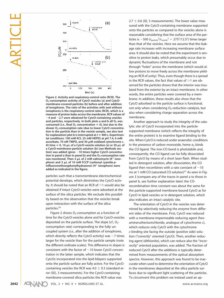
problem. Figure 3a shows flash-induced absorbance
changes of the CytcO-containing vesicles after addi-
tion of a membrane-impermeable (reduces only the
“correctly oriented” CytcO, i.e., from the positive side,
“reduced P”) and membrane-permeable (reduces CytcO
from both sides, “reduced P & N”) reducing agent. The
orientation was determined, from the ratio of the ampli-
tudes at zero time, to be 71 � 4% (SE, n � 5) “cor-
rectly oriented” CytcO. After deposition of the mem-
brane at particles we obtained a ratio of 68 � 5% (SE,
n � 10), which shows that the orientation is maintained
during the coverage process. In a recent study it was
observed that �70% of bacteriorhodopsin molecules
were correctly oriented when incorporated into meso-
porous microspheres-supported-bilayer membranes
even though the bacteriorhodopsin was initially ori-
ented unidirectionally in the predeposited vesicles.23 It
was proposed that upon interaction of the
bacteriorhodopsin-containing membrane with the
silica particle surface two transient bacteriorhodopsin-
membrane populations were formed, which eventually
adsorb onto the surface resulting in the observed orien-
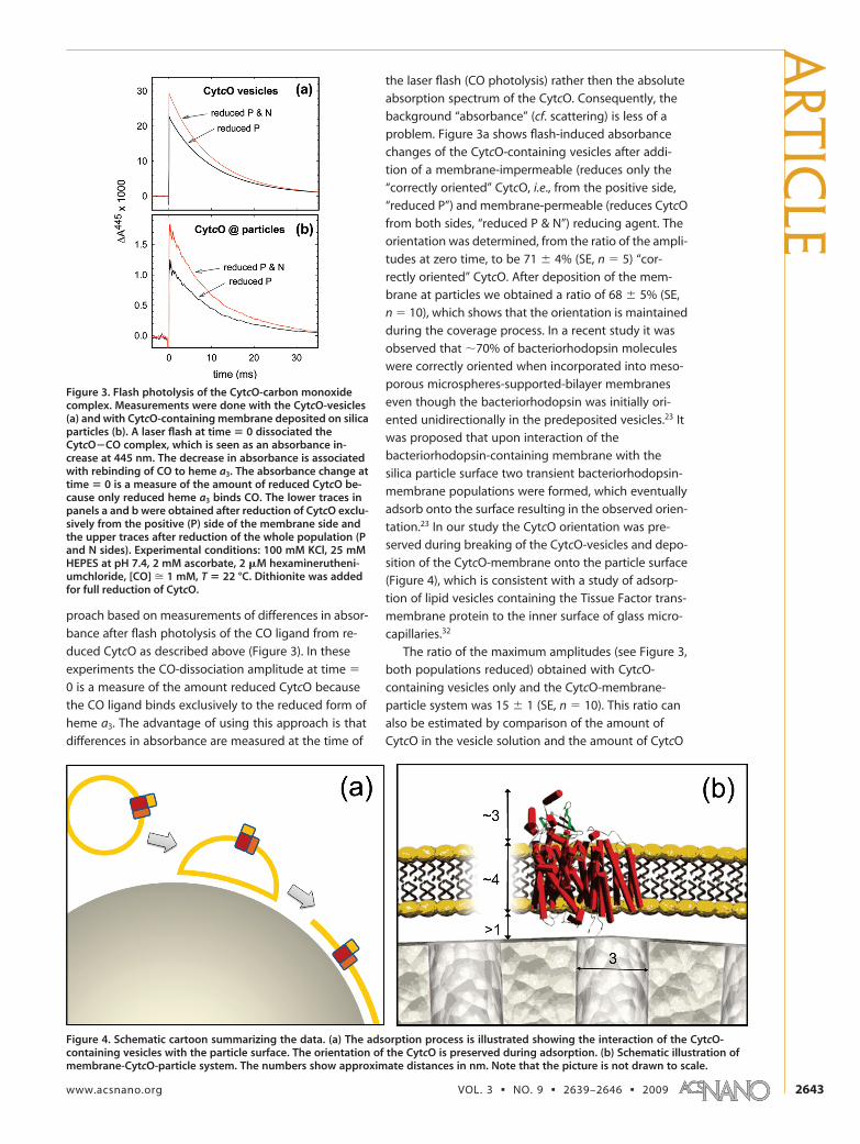
tation.23 In our study the CytcO orientation was pre-
served during breaking of the CytcO-vesicles and depo-
sition of the CytcO-membrane onto the particle surface
(Figure 4), which is consistent with a study of adsorp-
tion of lipid vesicles containing the Tissue Factor trans-
membrane protein to the inner surface of glass micro-
capillaries.32
The ratio of the maximum amplitudes (see Figure 3,
both populations reduced) obtained with CytcO-
containing vesicles only and the CytcO-membrane-
particle system was 15 � 1 (SE, n � 10). This ratio can
also be estimated by comparison of the amount of
CytcO in the vesicle solution and the amount of CytcO
Figure 4. Schematic cartoon summarizing the data. (a) The adsorption process is illustrated showing the interaction of the CytcO-containing vesicles with the particle surface. The orientation of the CytcO is preserved during adsorption. (b) Schematic illustration ofmembrane-CytcO-particle system. The numbers show approximate distances in nm. Note that the picture is not drawn to scale.
Figure 3. Flash photolysis of the CytcO-carbon monoxidecomplex. Measurements were done with the CytcO-vesicles(a) and with CytcO-containing membrane deposited on silicaparticles (b). A laser flash at time � 0 dissociated theCytcO�CO complex, which is seen as an absorbance in-crease at 445 nm. The decrease in absorbance is associatedwith rebinding of CO to heme a3. The absorbance change attime � 0 is a measure of the amount of reduced CytcO be-cause only reduced heme a3 binds CO. The lower traces inpanels a and b were obtained after reduction of CytcO exclu-sively from the positive (P) side of the membrane side andthe upper traces after reduction of the whole population (Pand N sides). Experimental conditions: 100 mM KCl, 25 mMHEPES at pH 7.4, 2 mM ascorbate, 2 �M hexaminerutheni-umchloride, [CO] � 1 mM, T � 22 °C. Dithionite was addedfor full reduction of CytcO.
ARTIC
LE
www.acsnano.org VOL. 3 ▪ NO. 9 ▪ 2639–2646 ▪ 2009 2643
added from that solution to the particle suspension. Ifall vesicles added to the particle suspension were ad-sorbed at the particle outer surface, then we would ex-pect an amplitude ratio equal to the ratio of the totalvesicle and the total particle outer surface area (AV/AP),which was 27 � 1 (SE, n � 10). If part of the CytcO popu-lation would be “deactivated” due to interactions withthe silica colloids then we would expect a CO-dissociation amplitude ratio of �27. The observationof a ratio that is �27 indicates that CO binding is notperturbed by interactions with the silica particles. In ad-dition, the observation of two active CytcO popula-tions (which are the same as in the lipid vesicles) withdifferent orientations in the silica-supported membranealso indicates that none of the orientations is dispropor-tionally deactivated.
As described in the Methods section the particleswith fluorescein-labeled CytcO in the membrane werewashed several times such that the original CytcO-vesicle solution was diluted by a factor of �10�8. Thus,the observed ratio of the absorbance changes of �15shows that the CytcO molecules were associated withthe mesoporous particles and not free vesicles.
The results are consistent with other data showingthat upon deposition of membranes on silica surfacescontaining nm-sized pores, the membrane is able tospan across the pore openings rather then penetratinginto the pores33 (Figure 4). In addition, it has also beenshown that there is a water-filled space between a lipidmembrane and a (dense) silica surface that may be�10 Å thick.34�36 We speculate that the porous natureof the exterior surface of the mesoporous spheres alsocould contribute to a thicker fluid film between themembrane and the solid support because of the de-
crease in the attractive van der Waals interaction be-
tween the lipids and the water�silica composite sur-
face. In this context we note that, as discussed above,
CytcO turnover requires proton uptake from the nega-
tive side of the membrane, which requires a water-filled
space on the inside of the membrane. With the meso-
porous structure the accessible volume on the inside of
the membrane is defined by the surface water film as
well as the entire pore volume; therefore, the inside vol-
ume is significantly larger than that of a solid silica par-
ticle. In future studies a more uniform orientation of
CytcO could be achieved by chemically modifying the
silica surface to allow specific interactions with, for ex-
ample, a histidine tag as has been done previously for
planar gold surfaces.24�27
In summary, the results described above show that
the mesoporous silica particles were entirely covered
by defect-free membranes containing the multisubunit,
redox-driven proton pump, CytcO. The incorporated
enzymes remained active, both with respect to cataly-
sis of reduction of O2 to H2O and transmembrane
charge separation. The use of mesoporous silica par-
ticles may be beneficial to obtain supported lipid bilay-
ers with a large interspace that is sufficient to provide
a proton source and also connect the water space
around the CytcO negative side surface with the entire
volume of the particle through the pores. This supramo-
lecular architecture with an inner pore structure mim-
icking a cytoskeleton yields a solid-supported biofunc-
tional cellular surface, which provides a basis for
functional studies of membrane-bound transport pro-
teins, and also for applications within pharmaceutical
drug delivery.
METHODSSynthesis of the Monodisperse Mesoporous Colloids. Mesostructured
silica spheres were synthesized from dilute solutions of the cat-ionic surfactant, hexadecyltrimethylammonium bromide(C16TAB), in an alkaline/methanol/water solution that containshydrolyzed tetramethylorthosilicate (TMOS) following a previ-ously published procedure.28 The as-synthesized particles werethermally calcined at 550 °C, at a ramp rate of 1 °C per minute, for5 h in air. The morphology of the particles was determined us-ing scanning electron microscopy, JEOL 820 SEM, operating at8�10 keV. The particles were thinly spread onto a carbon filmsupported on a brass stud and gold coated. The XRD patternswere obtained using a XPERT-PRO powder diffractometer (Cu K�radiation, k � 1.5418 Å) over the range of 2 � 1.0�10.0°. Poros-ity measurements were performed volumetrically using a Mi-cromeritics ASAP 2020 analyzer. The particles were degassed at150 °C for 10 h and data collection started typically at �196 °C,following a program consisting of both an adsorption and de-sorption branch. The specific surface areas, SBET (m2 g�1) were cal-culated using the Brunauer� Emmett�Teller (BET) model withinthe 0.05�0.15 p/po relative pressure region.37 The pore volume,Vpore (cm3 g�1), was estimated from the amount of N2-adsorbedat a relative pressure around P/P0 � 0.99. The pore size distribu-tion was calculated based on the NLDFT (nonlocal density func-tional theory)38 method, and the mesopore size, Øpore (nm), wasdetermined from the maximum of the pore size distribution
curve. Transmission electron microscopy (TEM) micrographs ofthin microtomed slices of the calcined particles were obtainedusing a JEOL JEM-3010 microscope operating at an acceleratingvoltage of 300 keV equipped with a CCD camera (model KeenView, SIS analysis) (for details, see ref 28).
Purification of CytcO. The R. sphaeroides bacteria were grownaerobically in shake incubators at 30 °C. The cells were har-vested and the His-tagged CytcO was purified as described pre-viously.39 It consists of four subunits with a total molecularweight of �100 kDa (for discussion of the structure and func-tion of CytcO, see refs 40�44). The enzyme was frozen in liquidnitrogen and stored in 0.1 M Hepes-KOH, pH 7.4, 0.1% dodecyl-D-maltoside in a �80 °C freezer.
Reconstitution of CytcO into Lipid Vesicles. The lipids 1,2-dioleoyl-sn-glycero-3-phosphoethanolamine (DOPE), 1,2-dioleoyl-sn-glycero-3-phosphocholine (DOPC), 1,2-dioleoyl-sn-glycero-3-[phospho-rac-(1-glycerol)] (DOPG) and 1,1=2,2=-tetraoleoyl cardi-olipin (CA) (all from Avanti Polar Lipids Inc.) were dissolved inchloroform and methanol (2:1 volume ratio) and then dried un-der continuous flow of nitrogen during �30 min. The lipid mix-ture at a weight ratio of 9:6:4:1 of DOPE:DOPC:DOPG:CA was sus-pended, at a total lipid concentration of 6 mg/mL, in a solutionof 100 mM KCl, 25 mM HEPES pH 7.4. This lipid mixture was cho-sen to approximately mimic those of Escherichia coli and R.sphaeroides (source of the CytcO in our experiments). In experi-ments where the fluorescein-labeled lipids (1,2-dioleoyl-sn-
ART
ICLE
VOL. 3 ▪ NO. 9 ▪ NORDLUND ET AL. www.acsnano.org2644
glycero-3-phosphoethanolamine-N-(carboxyfluorescein) (Fluo-DOPE)), were used, the weight ratio of the lipids mixture DOPE:DOPC:DOPG:CA:Fluo-DOPE was 9:6:4:1:0.02. This mixture gives aFluo-DOPE mole fraction of 0.07%. The lipid solutions were son-icated (Heat Systems Sonicator XL) in 30 s on/off cycles for a to-tal of 10 min to form small unilamellar vesicles.
Reconstitution of CytcO was performed following publishedprocedures.44,45 In short the sonicated lipids, supplemented with0.3% cholate, were mixed at a 1:1 volume ratio with 2.4 �MCytcO in 0.6% cholate, 100 mM KCl, and 25 mM HEPES at pH7.4. The detergent was then removed by addition of Bio-Beads(see ref 45). The CytcO and vesicle concentrations were chosensuch that the CytcO/vesicle ratio was approximately unity, that is,one CytcO per vesicle. However, not all enzyme molecules werereconstituted into the membrane and a fraction of the vesiclesremained empty (Faxen et al., unpublished data). The diameterof the vesicles was estimated to be 25 � 5 nm using dynamiclight-scattering (Nano Z, Malvern Instruments). The CytcO-vesiclesolution (�0.5 �M CytcO) was transferred to a sealed cuvetteand the atmosphere was exchanged to nitrogen on a vacuumline. The CytcO was then reduced by adding 2 mM ascorbate, 2�M hexaminerutheniumchloride and then the N2 atmospherewas exchanged for CO. Formation of the CytcO�CO complexwas verified using the optical absorption spectrum.
Formation of Supported Lipid Bilayers on Mesoporous Silica Particles.Mesoporous silica particles at a concentration of 5 mg/mL weresuspended in an electrolyte buffer solution that contained 100mM KCl, 25 mM HEPES at pH 7.4 and were sonicated to removeany aggregates. The particle suspension was then mixed for 1 hat room temperature with the lipid vesicles, with or without re-constituted CytcO. The total volume of the mixture was 2 mL andthe ratio between the total vesicle and the total particle outersurface area (AV/AP) was 25�30 (the AV/AP ratio was calculatedas described in ref 29). The number of lipids (cf. vesicle concen-tration) was determined from the total amount ofphosphorus.29,46 After mixing, a buffer solution of 100 mM KCl,25 mM HEPES pH 7.4 was added to make a final volume of 10 mL,and the mixture was centrifuged at 720g for 15 min. The result-ing particle pellet had an approximate volume of 200 �L. The su-pernatant containing superfluous vesicles was discarded andthe pellet was resuspended in 10 mL buffer solution. The cen-trifugation procedure was repeated two times, and the final pel-let was solubilized in 1 mL of a solution of 100 mM KCl, 25 mMHEPES pH 7.4. After this treatment the concentration of the freevesicles in solution was estimated to be �10�6 % of the initialamount added.
Analysis of the CytcO-Membrane Covered Particles. The fluorescenceof different types of CytcO-containing vesicles: (i) with fluores-cent lipids added, (ii) with maleimide-fluorescein-labeled CytcO(the labeling procedure is described in ref 30) (no fluorescent lip-ids), and (iii) with fluorescein trapped within the vesicles wasmeasured using a fluorometer (Varian Cary Eclipse). Four differ-ent types of CytcO-containing membrane covered particles wereprepared and analyzed using flow cytometry (BD FACSCaliburwith BD FACSFlow Sheath Fluid as liquid vector (BD Biosciences,Canada)): type i�iii as described above, and (iv) no fluorophoresadded (used to determine the background “fluorescence” sig-nal originating from light scattering).
Measurements of CytcO Activity. The CytcO activity at 22ŒC wasdetermined by measuring the O2-consumption rate using an ox-ygraph (Hansatech Instruments Ltd., UK). The background (non-enzymatically catalyzed) O2-consumption rate was first deter-mined after adding 1.5 mL of a solution composed of 100 mMKCl, 25 mM HEPES at pH 7.4, 6 mM ascorbate, 70 nM N,N,N=,N=-tetramethyl-p-phenylenediamine (TMPD) and 30 �M oxidizedcytochrome c. Then, 10 �L of a solution with CytcO-containingvesicles (see earlier) or 20 �L of a suspension of particle-supported CytcO-membrane (see earlier) was added and the O2-consumption rate was monitored. The activity was determinedfrom the difference in the slopes after and before the CytcO ad-dition. To determine the CytcO activity in the absence of a pro-ton electrochemical gradient, 3 �L of a 2 mM (4 �M final concen-tration) valinomycin solution (K� ionophore) and 3 �L of a 10mM (20 �M final concentration) FCCP (carbonyl cyanide-p-
trifluoromethoxyphenylhydrazone) solution (H� ionophore)were added.
Laser Flash Photolysis. The CO ligand was dissociated from CytcOusing an 8 ns laser pulse with an energy of �100 mJ at 532 nm(Nd:YAG Quantel Brilliant B, Les Ulis Cedex, France). Time-resolved optical absorbance changes were recorded using aflash photolysis setup from Applied Photophysics (model LKS.60,Surrey, UK)). The cuvette path length was 1.00 cm. Monochroma-tors both in front and after the cuvette were used to select theappropriate wavelength.
Acknowledgment. The project was supported by fundingfrom the Center for Biomembrane Research at Stockholm Univer-sity and the Knut and Alice Wallenberg Foundation.
REFERENCES AND NOTES1. Kresge, C. T.; Leonowicz, M. E.; Roth, W. J.; Vartuli, J. C.;
Beck, J. S. Ordered Mesoporous Molecular SievesSynthesized by a Liquid-Crystal Template Mechanism.Nature 1992, 359, 710–712.
2. Che, S.; Li, H.; Lim, S.; Sakamoto, Y.; Terasaki, O.; Tatsumi, T.Synthesis Mechanism of Cationic Surfactant TemplatingMesoporous Silica under an Acidic Synthesis Process.Chem. Mater. 2005, 17, 4103–4113.
3. Rama Rao, G. V.; López, G. P.; Bravo, J.; Pham, H.; Datye,A. K.; Xu, H. F.; Ward, T. L. Monodisperse Mesoporous SilicaMicrospheres Formed by Evaporation-Induced SelfAssembly of Surfactant Templates in Aerosols. Adv. Mater.2002, 14, 1301–1304.
4. Rathousky, J.; Zukalov, M.; Zukal, A.; Had, J. HomogeneousPrecipitation of Siliceous Mcm-41 and Bimodal Silica.Collect. Czech. Chem. Commun. 1998, 63, 1893–1906.
5. Yang, H.; Coombs, N.; Ozin, G. A. Morphogenesis of Shapesand Surface Patterns in Mesoporous Silica. Nature 1997,386, 692–695.
6. Slowing, I. I.; Vivero-Escoto, J. L.; Wu, C. W.; Lin, V. S. Y.Mesoporous Silica Nanoparticles as Controlled ReleaseDrug Delivery and Gene Transfection Carriers. Adv. DrugDelivery Rev. 2008, 60, 1278–1288.
7. Lai, C. Y.; Trewyn, B. G.; Jeftinija, D. M.; Jeftinija, K.; Xu, S.;Jeftinija, S.; Lin, V. S. Y. A Mesoporous Silica Nanosphere-Based Carrier System with Chemically Removable CdsNanoparticle Caps for Stimuli-Responsive ControlledRelease of Neurotransmitters and Drug Molecules. J. Am.Chem. Soc. 2003, 125, 4451–4459.
8. Salonen, J.; Laitinen, L.; Kaukonen, A. M.; Tuura, J.;Bjorkqvist, M.; Heikkila, T.; Vaha-Heikkila, K.; Hirvonen, J.;Lehto, V. P. Mesoporous Silicon Microparticles for OralDrug Delivery: Loading and Release of Five Model Drugs.J. Controlled Release 2005, 108, 362–374.
9. Wu, E. C.; Park, J. H.; Park, J.; Segal, E.; Cunin, F.; Sailor, M. J.Oxidation-Triggered Release of Fluorescent Molecules orDrugs from Mesoporous Si Microparticles. ACS Nano 2008,2, 2401–2409.
10. Liong, M.; Lu, J.; Kovochich, M.; Xia, T.; Ruehm, S. G.; Nel,A. E.; Tamanoi, F.; Zink, J. I. Multifunctional InorganicNanoparticles for Imaging, Targeting, and Drug Delivery.ACS Nano 2008, 2, 889–896.
11. Lu, J.; Liong, M.; Zink, J. I.; Tamanoi, F. Mesoporous SilicaNanoparticles as a Delivery System for HydrophobicAnticancer Drugs. Small 2007, 3, 1341–1346.
12. Hernandez, R.; Tseng, H. R.; Wong, J. W.; Stoddart, J. F.;Zink, J. I. An Operational Supramolecular Nanovalve. J. Am.Chem. Soc. 2004, 126, 3370–3371.
13. Mal, N. K.; Fujiwara, M.; Tanaka, Y. PhotocontrolledReversible Release of Guest Molecules from Coumarin-Modified Mesoporous Silica. Nature 2003, 421, 350–353.
14. Nguyen, T. D.; Tseng, H. R.; Celestre, P. C.; Flood, A. H.; Liu,Y.; Stoddart, J. F.; Zink, J. I. A Reversible Molecular Valve.Proc. Natl. Acad. Sci. U.S.A. 2005, 102, 10029–10034.
15. Loidl-Stahlhofen, A.; Schmitt, J.; Noller, J.; Hartmann, T.;Brodowsky, H.; Schmitt, W.; Keldenich, J., Solid-SupportedBiomolecules on Modified Silica SurfacesOA Tool for FastPhysicochemical Characterization and High-ThroughputScreening. Adv. Mater. 2001, 13.
ARTIC
LE
www.acsnano.org VOL. 3 ▪ NO. 9 ▪ 2639–2646 ▪ 2009 2645
16. Katagiri, K.; Hashizume, M.; Kikuchi, J. I.; Taketani, Y.;Murakami, M. Creation of Asymmetric Bilayer Membraneon Monodispersed Colloidal Silica Particles. Colloids Surf., B2004, 38, 149–153.
17. Baksh, M. M.; Jaros, M.; Groves, J. T. Detection of MolecularInteractions at Membrane Surfaces through Colloid PhaseTransitions. Nature 2004, 427, 139–141.
18. Moura, S. P.; Carmona-Ribeiro, A. M. Biomimetic Particles:Optimization of Phospholipid Bilayer Coverage on Silicaand Colloid Stabilization. Langmuir 2005, 21,10160–10164.
19. Moura, S. P.; Carmona-Ribeiro, A. M. Cationic BilayerFragments on Silica at Low Ionic Strength: CompetitiveAdsorption and Colloid Stability. Langmuir 2003, 19,6664–6667.
20. Trepout, S.; Mornet, S.; Benabdelhak, H.; Ducruix, A.;Brisson, A. R.; Lambert, O. Membrane Protein SelectivelyOriented on Solid Support and Reconstituted into a LipidMembrane. Langmuir 2007, 23, 2647–54.
21. Mornet, S.; Lambert, O.; Duguet, E.; Brisson, A. TheFormation of Supported Lipid Bilayers on SilicaNanoparticles Revealed by Cryoelectron Microscopy. NanoLett. 2005, 5, 281–285.
22. Liu, J.; Stace-Naughton, A.; Jiang, X.; Brinker, C. J. PorousNanoparticle Supported Lipid Bilayers (Protocells) asDelivery Vehicles. J. Am. Chem. Soc. 2009, 131, 1354–1355.
23. Davis, R. W.; Flores, A.; Barrick, T. A.; Cox, J. M.; Brozik, S. M.;Lopez, G. P.; Brozik, J. A. Nanoporous MicrobeadSupported Bilayers: Stability, Physical Characterization,and Incorporation of Functional Transmembrane Proteins.Langmuir 2007, 23, 3864–3872.
24. Friedrich, M. G.; Plum, M. A.; Santonicola, M. G.; Kirste,V. U.; Knoll, W.; Ludwig, B.; Naumann, R. L. C. In SituMonitoring of the Catalytic Activity of Cytochrome cOxidase in a Biomimetic Architecture. Biophys. J. 2008, 95,1500–1510.
25. Ataka, K.; Richter, B.; Heberle, J. Orientational Control ofthe Physiological Reaction of Cytochrome c OxidaseTethered to a Gold Electrode. J. Phys. Chem. B 2006, 110,9339–9347.
26. Ataka, K.; Giess, F.; Knoll, W.; Naumann, R.; Haber-Pohlmeier, S.; Richter, B.; Heberle, J. Oriented Attachmentand Membrane Reconstitution of His-Tagged Cytochromec Oxidase to a Gold Electrode: In Situ Monitoring bySurface-Enhanced Infrared Absorption Spectroscopy.J. Am. Chem. Soc. 2004, 126, 16199–16206.
27. Giess, F.; Friedrich, M. G.; Heberle, J.; Naumann, R. L.; Knoll,W. The Protein-Tethered Lipid Bilayer: A Novel Mimic ofthe Biological Membrane. Biophys. J. 2004, 87, 3213–3220.
28. Ng, J. B. S.; Vasiliev, P. O.; Bergstrom, L. The RadialDependence of the Spatial Mesostructure ofMonodisperse Mesoporous Silica Spheres. MicroporousMesoporous Mater. 2008, 112, 589–596.
29. Nordlund, G.; Lonneborg, R.; Brzezinski, P. Formation ofSupported Lipid Bilayers on Silica Particles Studied UsingFlow Cytometry. Langmuir 2009, 25, 4601–6.
30. Ojemyr, L.; Sanden, T.; Widengren, J.; Brzezinski, P. LateralProton Transfer between the Membrane and a MembraneProtein. Biochemistry 2009, 48, 2173–2179.
31. Adelroth, P.; Brzezinski, P.; Malmstrom, B. G. InternalElectron Transfer in Cytochrome c Oxidase fromRhodobacter Sphaeroides. Biochemistry 1995, 34,2844–2849.
32. Contino, P. B.; Hasselbacher, C. A.; Ross, J. B. A.; Nemerson,Y. Use of an Oriented Transmembrane Protein to Probethe Assembly of a Supported Phospholipid Bilayer.Biophys. J. 1994, 67, 1113–1116.
33. Simon, A.; Girard-Egrot, A.; Sauter, F.; Pudda, C.; PicolletD’Hahan, N.; Blum, L.; Chatelain, F.; Fuchs, A. Formationand Stability of a Suspended Biomimetic Lipid Bilayer onSilicon Submicrometer-Sized Pores. J. Colloid Interface Sci.2007, 308, 337–43.
34. Koenig, B. W.; Krueger, S.; Orts, W. J.; Majkrzak, C. F.; Berk,N. F.; Silverton, J. V.; Gawrisch, K. Neutron Reflectivity andAtomic Force Microscopy Studies of a Lipid Bilayer in
Water Adsorbed to the Surface of a Silicon Single Crystal.Langmuir 1996, 12, 1343–1350.
35. Johnson, S. J.; Bayerl, T. M.; McDermott, D. C.; Adam, G. W.;Rennie, A. R.; Thomas, R. K.; Sackmann, E. Structure of anAdsorbed Dimyristoylphosphatidylcholine BilayerMeasured with Specular Reflection of Neutrons. Biophys. J.1991, 59, 289–294.
36. Bayerl, T. M.; Bloom, M. Physical Properties of SinglePhospholipid Bilayers Adsorbed to Micro Glass Beads. ANew Vesicular Model System Studied by 2H-NuclearMagnetic Resonance. Biophys. J. 1990, 58, 357–362.
37. Brunauer, S.; Emmett, P. H.; Teller, E. Adsorption of Gasesin Multimolecular Layers. J. Am. Chem. Soc. 1938, 60,309–319.
38. Ravikovitch, P. I.; Neimark, A. V. Characterization of Micro-and Mesoporosity in SBA-15 Materials from AdsorptionData by the NLDFT Method. J. Phys. Chem. B 2001, 105,6817–6823.
39. Mitchell, D. M.; Gennis, R. B. Rapid Purification of Wildtypeand Mutant Cytochrome c Oxidase from RhodobacterSphaeroides by Ni2�-NTA Affinity Chromatography. FEBSLett. 1995, 368, 148–150.
40. Brzezinski, P.; Gennis, R. B. Cytochrome c Oxidase: ExcitingProgress and Remaining Mysteries. J. Bioenerg. Biomembr.2008, 1–11.
41. Hosler, J. P.; Ferguson-Miller, S.; Mills, D. A. EnergyTransduction: Proton Transfer through the RespiratoryComplexes. Annu. Rev. Biochem. 2006, 75, 165–187.
42. Wikstrom, M.; Verkhovsky, M. I. Towards the Mechanism ofProton Pumping by the Haem-Copper Oxidases. Biochim.Biophys. Acta 2006, 1757, 1047–1051.
43. Brzezinski, P.; Adelroth, P. Design Principles of Proton-Pumping Haem-Copper Oxidases. Curr. Opin. Struct. Biol.2006, 16, 465–472.
44. Faxen, K.; Gilderson, G.; Adelroth, P.; Brzezinski, P. AMechanistic Principle for Proton Pumping by Cytochromec Oxidase. Nature 2005, 437, 286–289.
45. Jasaitis, A.; Verkhovsky, M. I.; Morgan, J. E.; Verkhovskaya,M. L.; Wikstrom, M. Assignment and Charge TranslocationStoichiometries of the Major Electrogenic Phases in theReaction of Cytochrome c Oxidase with Dioxygen.Biochemistry 1999, 38, 2697–2706.
46. Chen, P. S.; Toribara, T. Y.; Warner, H. Microdeterminationof Phosphorus. Anal. Chem. 1956, 28, 1756–1758.
ART
ICLE
VOL. 3 ▪ NO. 9 ▪ NORDLUND ET AL. www.acsnano.org2646