a duplicated pair of arabidopsis ring-finger e3 ligases contribute to the rpm1- and rps2-mediated...
TRANSCRIPT
A duplicated pair of Arabidopsis RING-finger E3 ligasescontribute to the RPM1- and RPS2-mediated hypersensitiveresponse
Tsutomu Kawasaki1,†, Jaesung Nam1,‡, Douglas C. Boyes1,§, Ben F. Holt, III1, David A. Hubert1, Aaron Wiig1,– and Jeffery L.
Dangl1,2,3,4,*
1Department of Biology and2Curriculum in Genetics,3Department of Microbiology and Immunology, and4Carolina Center for Genome Sciences, University of North Carolina, Chapel Hill, NC 27599, USA
Received 31 May 2005; revised 13 July 2005; accepted 21 July 2005.*For correspondence (fax 919 962 1625; e-mail [email protected]).†Present address: Graduate School of Biological Sciences, Nara Institute of Science and Technology, 8916-5 Takayama, Ikoma 630-0192, Japan.‡Present address: Faculty of Plant Biotechnology, Dong-A University, 840, Hadan-2dong, Saha-gu, Busan 604-714, South Korea.§Present address: Monsanto Company, 110 T.W. Alexander Dr, Research Triangle Park, NC 27709, USA.–Present address: BASF Plant Science, Research Triangle Park, NC 27709, USA.
Summary
The Arabidopsis RPM1 protein confers resistance to disease caused by Pseudomonas syringae strains
delivering either the AvrRpm1 or AvrB type III effector proteins into host cells. We characterized two closely
related RPM1-interacting proteins, RIN2 and RIN3. RIN2 and RIN3 encode RING-finger type ubiquitin ligases
with six apparent transmembrane domains and an ubiquitin-binding CUE domain. RIN2 and RIN3 are
orthologs of the mammalian autocrine motility factor receptor, a cytokine receptor localized in both plasma
membrane caveolae and the endoplasmic reticulum. RIN2 is predominantly localized to the plasmamembrane,
as are RPM1 and RPS2. The C-terminal regions of RIN2 and RIN3, including the CUE domain, interact strongly
with an RPM1 N-terminal fragment and weakly with a similar domain from the Arabidopsis RPS2 protein. RIN2
and RIN3 can dimerize through their C-terminal regions. The RING-finger domains of RIN2 and RIN3 encode
ubiquitin ligases. Inoculation with P. syringae DC3000(avrRpm1) or P. syringae DC3000(avrRpt2) induces
differential decreases of RIN2 mobility in SDS-PAGE and disappearance of the majority of RIN2. A rin2 rin3
double mutant expresses diminished RPM1- and RPS2-dependent hypersensitive response (HR), but no
alteration of pathogen growth. Thus, the RIN2/RIN3 RING E3 ligases apparently act on a substrate that
regulates RPM1- and RPS2-dependent HR.
Keywords: RING-finger ubiquitin ligase, disease resistance, hypersensitive response, RPM1, RPS2.
Introduction
The plant immune system responds to infection using
both general recognition of microbe-associated molecules
(Gomez-Gomez and Boller, 2000) and specific recognition
of particular pathogen-encoded molecules through the
action of plant disease resistance (R) proteins (Belkhadir
et al., 2004; Dangl and Jones, 2001). The majority of R
proteins defined to date have a central nucleotide binding
site (NB) domain and C-terminal leucine-rich repeats
(LRRs). One subclass of NB-LRR proteins contains a
coiled-coil (CC) motif at the N-terminus (CC-NB-LRR),
while a second carries an N-terminal motif homologous to
the cytoplasmic domain of Drosophila Toll and mamma-
lian interleukin (IL-1) receptors (TIR-NB-LRR) (Dangl and
Jones, 2001; Martin et al., 2003). Recognition of patho-
gens mediated by NB-LRR results in rapid ion fluxes, a
sustained oxidative burst, the enhanced transcriptional
induction of a basal defense transcriptome, production of
antimicrobial compounds and induction of systemic
acquired disease resistance at distal sites (Dangl and
Jones, 2001; Durrant and Dong, 2004; Martin et al., 2003).
Defense responses are often accompanied by a localized
programmed cell death at the infection site, called the
258 ª 2005 Blackwell Publishing Ltd
The Plant Journal (2005) 44, 258–270 doi: 10.1111/j.1365-313X.2005.02525.x
hypersensitive response (HR) (Dangl et al., 1996; Green-
berg and Yao, 2004).
Plant NB-LRR proteins directed against phytopathogenic
bacteria recognize the type III effector proteins that these
pathogens deliver into the host cell. Type III effector proteins
can act as virulence factors, and are delivered to the host cell
by the evolutionarily conserved type III secretion system
(Alfano and Collmer, 2004; Chang et al., 2004; Nimchuk
et al., 2003). It is increasingly likely that the recognition of
many type III effector proteins is indirect, and that NB-LRR
proteins have evolved to monitor the activity of these
virulence factors on host targets (Axtell and Staskawicz,
2003; Dangl and Jones, 2001; Mackey et al., 2003; Shao
et al., 2003). However, the mechanisms by which NB-LRR
proteins transduce signals leading to successful disease
resistance and the associated HR remain unclear.
The Arabidopsis RPM1 and RPS2 genes encode CC-NB-
LRR proteins that confer resistance to Pseudomonas syrin-
gae strains expressing either of two different type III
effectors, avrRpm1 or avrB (Grant et al., 1995) or avrRpt2
(RPS2; Bent et al., 1994; Mindrinos et al., 1994). RPM1 is a
peripheral plasma membrane protein, and it disappears via
an unknown mechanism coincident with the onset of the HR
(Boyes et al., 1998). Degradation of diverse receptors has
been investigated in various animal systems. For example,
some Toll-like receptors (TLRs) that function in the innate
animal immune response are degraded via a ubiquitination
pathway, thus limiting TLR signaling to prevent undesirable
amplification of host responses (Chuang and Ulevitch,
2004). By contrast, RPS2 is also membrane associated, but
it does not disappear following stimulation(Axtell and
Staskawicz, 2003).
Protein degradation mediated by the ubiquitination path-
way is performed by a sequential reaction of three enzymes
(Smalle and Vierstra, 2004). First, a ubiquitin-activating
enzyme (E1) activates ubiquitin by the formation of a
thioester bond between ubiquitin and a cysteine at the E1
active site. The activated ubiquitin is then transferred to a
ubiquitin-conjugating enzyme (E2) to form a thioester link-
age. A ubiquitin–protein ligase (E3) recognizes the target
protein as a substrate, and promotes the formation of an
isopeptide bond between ubiquitin and lysine residues on
the target protein. The ubiquitinated protein is then rapidly
degraded via the proteasome (Vierstra, 2003). Ubiquitination
can regulate target protein function independent of the
proteasome. In these cases, ubiquitination is required for
various subsequent regulatory responses including endo-
cytosis and relocalization of the target protein (Prag et al.,
2003).
In Arabidopsis, E3 ubiquitin ligases are classified into
various groups based on the presence of RING, HECT, or U-
box domains (Vierstra, 2003). There are 469 predicted RING-
proteins in Arabidopsis (Stone et al., 2005). The RING
domains in some of these proteins have demonstrable
ubiquitin ligase activity, and function in hormone, light and
various developmental processes (Vierstra, 2003). Several
targets for RING E3 ligases, including COP1, SINAT5 and
RMA1, have been identified (Matsuda et al., 2001; Saijo
et al., 2003; Seo et al., 2003; Xie et al., 2002).
Participants in NB-LRR protein function have been char-
acterized by genetic screens for mutants that exhibit defects
in disease resistance, HR or both, and by yeast two-hybrid
screens. For example, PBS1, NDR1, EDS1, PAD4, RAR1,
SGT1b and HSP90 encode proteins that are required for the
activity of some or all NB-LRR proteins. (Dangl and Jones,
2001; Muskett and Parker, 2003; Schulze-Lefert, 2004;
Shirasu and Schulze-Lefert, 2003; Swiderski and Innes,
2001; Tornero et al., 2002). Some of these, like RAR1 and
HSP90, function in the stabilization and/or accumulation of
signal competent NB-LRR proteins (Hubert et al., 2003;
Tornero et al., 2002), while others might function in signal
transduction (e.g. PAD4 and EDS1; Hammond-Kosack and
Parker, 2003).
Yeast two-hybrid screening using RPM1 fragments as
baits has identified two other proteins that influence RPM1
function, RIN1 and RIN13 (Al-Daoude et al., 2005; Holt et al.,
2002). RIN1 encodes an ortholog of TIP49a that interacts with
TATA binding protein. RIN1 is a negative regulator of
disease resistance responses and is also required for normal
plant development (Holt et al., 2002). The function of RIN13
is not predicted from its sequence. Overexpression of RIN13
enhances resistance to P. syringae (Pto) expressing avr-
Rpm1 without triggering HR (Al-Daoude et al., 2005). Resist-
ance mediated by RPM1 is inhibited by suppression of RIN13
expression, suggesting that RIN13 may function as a posi-
tive regulator of RPM1. However, the molecular basis of how
RIN13 regulates the RPM1-mediated response is unknown.
A similar screen using AvrB as a bait led to isolation of the
RIN4 protein, a small protein of unknown function that is
acylated into the plasma membrane (H.S. Kim et al., 2005;
M.G. Kim et al., 2005). RIN4 co-immunoprecipitates with
RPM1 and RPS2, and is required for their activation (Axtell
and Staskawicz, 2003; Mackey et al., 2002, 2003). RIN4 is a
target for AvrB, AvrRpm1 and AvrRpt2 and its manipulation
by these unrelated type III effectors drives activation of RPM1
or RPS2 (Axtell and Staskawicz, 2003; Day et al., 2005; H.S.
Kim et al., 2005; M.G. Kim et al., 2005; Mackey et al., 2002,
2003). These examples suggest that yeast two-hybrid screens
followed by reverse genetics and biochemical analyses can
contribute to our understanding of NB-LRR regulation.
We characterize here two new RPM1-interacting proteins,
RIN2 and RIN3. RIN2 and RIN3 are highly related plasma-
membrane localized E3 ubiquitin ligases that contain RING-
finger domains in association with a CUE domain (Prag
et al., 2003). Inoculation of Pto DC3000(avrRpm1) induces
both a gradual decrease of RIN2 mobility in SDS-PAGE
before HR and its degradation at the time of HR. A similar
mobility shift of RIN2 is induced by inoculation of Pto
Transmembrane RING-E3 ligases and the HR 259
ª Blackwell Publishing Ltd, The Plant Journal, (2005), 44, 258–270
DC3000(avrRpt2), concomitant with the RPS2-dependent
HR. A rin2 rin3 double mutant exhibits reduced RPM1- and
RPS2-dependent HR. However, no alteration of pathogen
growth was found in the rin2 rin3 mutant. Thus, the RIN2/
RIN3 RING E3 ligases contribute positively to RPM1- and
RPS2-dependent HR.
Results
Two closely related Arabidopsis RING-finger proteins
interact with RPM1
We isolated RIN2 (RPM1 interacting protein 2; At4g25230)
from a yeast two-hybrid screen using an RPM1 domain
lacking the CC but containing most of the NB domain (amino
acids 55–341; pEG10 in Figure 2; Holt et al., 2002). Homology
searches indicated that the Arabidopsis genome encodes
another protein 83% identical to RIN2; we designated it RIN3
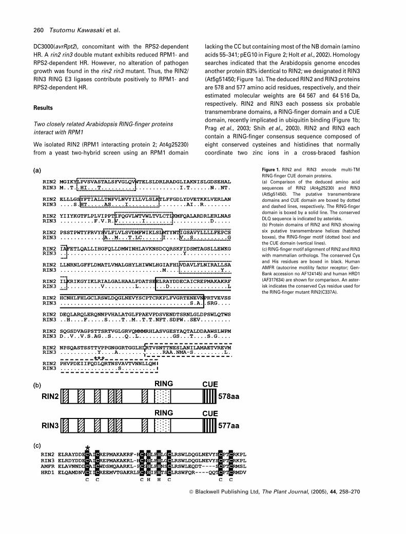
(At5g51450; Figure 1a). The deduced RIN2 and RIN3 proteins
are 578 and 577 amino acid residues, respectively, and their
estimated molecular weights are 64 567 and 64 516 Da,
respectively. RIN2 and RIN3 each possess six probable
transmembrane domains, a RING-finger domain and a CUE
domain, recently implicated in ubiquitin binding (Figure 1b;
Prag et al., 2003; Shih et al., 2003). RIN2 and RIN3 each
contain a RING-finger consensus sequence composed of
eight conserved cysteines and histidines that normally
coordinate two zinc ions in a cross-braced fashion
Figure 1. RIN2 and RIN3 encode multi-TM
RING-finger CUE domain proteins.
(a) Comparison of the deduced amino acid
sequences of RIN2 (At4g25230) and RIN3
(At5g51450). The putative transmembrane
domains and CUE domain are boxed by dotted
and dashed lines, respectively. The RING-finger
domain is boxed by a solid line. The conserved
DLQ sequence is indicated by asterisks.
(b) Protein domains of RIN2 and RIN3 showing
six putative transmembrane helices (hatched
boxes), the RING-finger motif (dotted box) and
the CUE domain (vertical lines).
(c) RING-finger motif alignment of RIN2 and RIN3
with mammalian orthologs. The conserved Cys
and His residues are boxed in black. Human
AMFR (autocrine motility factor receptor; Gen-
Bank accession no AF124145) and human HRD1
(AF317634) are shown for comparison. An aster-
isk indicates the conserved Cys residue used for
the RING-finger mutant RIN2(C337A).
260 Tsutomu Kawasaki et al.
ª Blackwell Publishing Ltd, The Plant Journal, (2005), 44, 258–270
(Figure 1c). The RING-finger domains of RIN2 and RIN3 have
a histidine at the fifth zinc-coordinating residue, indicative of
the RING-H2 subclass reported to have E3 ubiquitin ligase
activity (Jackson et al., 2000; Joazeiro and Weissman, 2000).
RIN2 and RIN3 display significant similarity to mammalian
autocrine motility factor receptor (AMFR) throughout the
coding region, including the order and relative spacing of all
domains (not shown). AMFR is a cell surface cytokine
receptor (Benlimame et al., 1998; Parton and Richards,
2003; Shimizu et al., 1999). Single-copy AMFR-like genes
are present in many organisms including rice, Drosophila,
Caenorhabditis elegans, zebrafish, Xenopus, mouse and rat.
Figure 1(d) presents a phylogenetic tree of AMFR-like genes.
RIN2 and RIN3 showed higher similarity to a rice ortholog
(approximately 48% identity) compared with the animal
orthologs [identities are: human (AF124145) 23%, C. elegans
(NM_060201) 23%, zebrafish (NM_213163) 22%, Xenopus
(BC072063) 23%, mouse (NM_011787) 23%, rat (XM_341644)
21%]. The human HRD1 protein (AF317634), which functions
in protein quality control in the endoplasmic reticulum (ER)
(Gardner et al., 2001), also has similarity to AMFR-like
proteins in the transmembrane and RING-finger domains
(Figure 1c), but lacks the CUE domain.
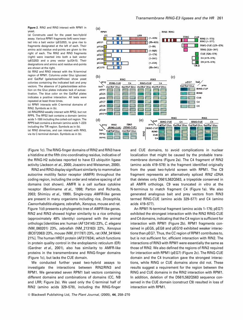
We conducted further yeast two-hybrid assays to
investigate the interactions between RIN2/RIN3 and
RPM1. We generated seven RPM1 bait vectors containing
different domains and combinations of domains (CC, NB
and LRR; Figure 2a). We used only the C-terminal half of
RIN2 (amino acids 329–578), including the RING-finger
and CUE domains, to avoid complications in nuclear
localization that might be caused by the probable trans-
membrane domains (Figure 2a). The C4 fragment of RIN2
(amino acids 419–578) is the fragment identified originally
from the yeast two-hybrid screen with RPM1. The C9
fragment represents an alternatively spliced RIN2 cDNA
that deletes only D561L562Q563, a tripeptide conserved in
all AMFR orthologs. C9 was truncated in vitro at the
N-terminus to match fragment C4 (Figure 1a). We also
generated analogous bait and prey vectors from RIN3
termed RING-CUE (amino acids 329–577) and C4 (amino
acids 419–577).
An RPM1 N-terminal fragment (amino acids 1–176; pEG7)
exhibited the strongest interaction with the RIN2 RING-CUE
and C4 domains, indicating that the C4 region is sufficient for
interaction with RPM1 (Figure 2b). RPM1 fragments con-
tained in pEG5, pEG8 and pEG10 exhibited weaker interac-
tions than pEG7. Thus, the CC region of RPM1 contributes to,
but is not sufficient for, efficient interaction with RIN2. The
interactions of RIN3 with RPM1 were essentially the same as
those of RIN2. We also defined the regions of RIN2 required
for interaction with RPM1 (pEG7) (Figure 2c). The RING-CUE
domain and the C4 truncation gave the strongest interac-
tions, while RING or CUE domains alone did not. These
results suggest a requirement for the region between the
RING and CUE domains in the RIN2 interaction with RPM1.
In addition, deletion of the D561L562Q563 sequence con-
served in the CUE domain (construct C9) resulted in loss of
interaction with RPM1.
(a)
(b) (c)
(d) (e)
Figure 2. RIN2 and RIN3 interact with RPM1 in
yeast.
(a) Constructs used for the yeast two-hybrid
assay. Various RPM1 fragments (left) were inser-
ted into a bait vector (pEG202), to give rise to
fragments designated at the left of each. Their
amino acid residue end-points are given to the
right of each. The RIN2 and RIN3 fragments
(right) were inserted into both a bait vector
(pEG202) and a prey vector (pJG4-5). Their
designations and amino acid residue end-points
are shown at the right.
(b) RIN2 and RIN3 interact with the N-terminal
region of RPM1. Columns under Gluc (glucose)
and Gal/Raf (galactose/raffinose) show yeast
colonies containing the indicated bait and prey
vectors. The absence of b-galactosidase activa-
tion on the Gluc plates indicates lack of autoac-
tivation. The blue color on the Gal/Raf plates
indicates a positive interaction. All tests were
repeated at least three times.
(c) RPM1 interacts with C-terminal domains of
RIN2. Symbols as in (b).
(d) RIN2/RIN3 weakly interact with RPS2, but not
RPP5. The RPS2 bait contains a domain (amino
acids 1–159) including the coiled-coil region. The
RPP5 bait contains a domain (amino acids 1–223)
including the TIR region. Symbols as in (b).
(e) RIN2 dimerizes, and can interact with RIN3,
via its C-terminal domain. Symbols as in (b).
Transmembrane RING-E3 ligases and the HR 261
ª Blackwell Publishing Ltd, The Plant Journal, (2005), 44, 258–270
We tested whether the interaction of RIN2 with RPM1 was
specific or could also be observed using similar domains
from related NB-LRR proteins (Figure 2d). We tested RPS2
(Bentet al., 1994; Mindrinoset al., 1994) and RPP5 (resistance
to Peronospora parasitica isolate Noco2; van der Biezen
et al., 2000; Parker et al., 1997). RPS2 is a CC-NBS-LRR
protein, as is RPM1. RPP5, by contrast, contains an N-terminal
TIR domain (Dangl and Jones, 2001). RIN2 and RIN3 weakly
interacted with RPS2, but not at all with RPP5, consistent with
a requirement for the CC domain in this interaction.
RIN2 and RIN3 can homo- and heterodimerize, and this
was strongest when using the C4 domain from each
(Figure 2e). The CUE domains alone could weakly bind C4,
but not the RING-CUE domain. This indicates that sequences
upstream of CUE in C4 are required for homo- and/or hetero-
oligomerization.
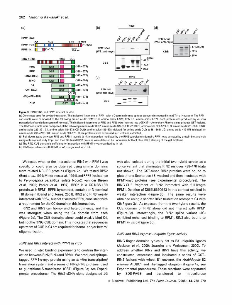
RIN2 and RIN3 interact with RPM1 in vitro
We used in vitro binding experiments to confirm the inter-
action between RIN2/RIN3 and RPM1. We produced epitope-
tagged RPM1-c-myc protein using an in vitro transcription/
translation system and a series of RIN2/RIN3 proteins fused
to glutathione-S-transferase (GST) (Figure 3a; see Experi-
mental procedures). The RIN2 cDNA clone designated JG
was also isolated during the initial two-hybrid screen as a
splice variant that eliminates RIN2 residues 438–470 (data
not shown). The GST-fused RIN2 proteins were bound to
glutathione Sepharose 4B, washed and then incubated with
RPM1-myc proteins (see Experimental procedures). The
RING-CUE fragment of RIN2 interacted with full-length
RPM1. Deletion of D561L562Q563 in this context resulted in
weaker interaction (Figure 3b). The same results were
obtained using a shorter RIN2 truncation (compare C4 with
C9; Figure 3c). As expected from the two-hybrid results, the
CUE domain of RIN2 alone did not interact with RPM1
(Figure 3c). Interestingly, the RIN2 splice variant (JG)
exhibited enhanced binding to RPM1. RIN3 also bound to
RPM1 in vitro (Figure 3d).
RIN2 and RIN3 express ubiquitin ligase activity
RING-finger domains typically act as E3 ubiquitin ligases
(Jackson et al., 2000; Joazeiro and Weissman, 2000). To
address whether RIN2 and RIN3 have this activity, we
constructed, expressed and incubated a series of GST–
RIN2 fusions with wheat E1 enzyme, the Arabidopsis E2
enzyme AtUBC1 and His-tagged ubiquitin (Figure 4a; see
Experimental procedures). These reactions were separated
by SDS-PAGE and transferred to nitrocellulose
(a) (b) (d)
(c)
Figure 3. RIN2/RIN3 and RPM1 interact in vitro.
(a) Constructs used for in vitro interaction. The indicated fragments of RPM1 with a C-terminal c-myc epitope tag were introduced into pET14b (Novagen). The RPM1
constructs were composed of the following amino acids: RPM1-Full, amino acids 1–926; RPM1-N, amino acids 1–177. Each protein was produced by in vitro
transcription/translation system (Promega). The indicated fragments of RIN2 and RIN3 were inserted into pGEX4T-1(Amersham Pharmacia) to produce GST fusions.
The RIN2 constructs were composed of the following amino acids: RIN2, amino acids 329–578; RIN2(-DLQ), amino acids 329–578(-DLQ, amino acids 561–563); RING,
amino acids 329–391; C4, amino acids 419–578; C9(-DLQ), amino acids 419–578 (deleted for amino acids DLQ at 561–563); JG, amino acids 419–578 (deleted for
amino acids 438–470); CUE, amino acids 526–578. These proteins were expressed in E. coli and extracted.
(b) Pull-down assay between RIN2 and RPM1 reveals in vitro interaction mediated by the RIN2 cytoplasmic domain. RPM1 was detected by protein blot analysis
using anti-myc antibody (top), and the GST-fused RIN2 proteins were detected by Coomassie brilliant blue (CBB) staining of the gel (bottom).
(c) The RIN2 CUE domain is sufficient for interaction with RPM1-myc; organized as in (b).
(d) RIN3 also interacts with RPM1 in vitro; organized as in (b).
262 Tsutomu Kawasaki et al.
ª Blackwell Publishing Ltd, The Plant Journal, (2005), 44, 258–270
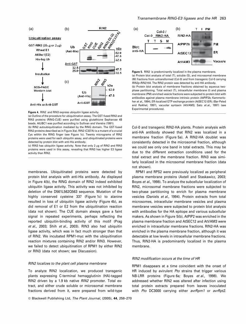
membranes. Ubiquitinated proteins were detected by
protein blot analysis with anti-His antibody. As displayed
in Figure 4(b), the RING domain of RIN2 indeed exhibited
ubiquitin ligase activity. This activity was not inhibited by
deletion of the D561L562Q563 sequence. Mutation of the
highly conserved cysteine 337 (Figure 1c) to alanine
resulted in loss of ubiquitin ligase activity (Figure 4b), as
did removal of E1 or E2 from the ubiquitination reaction
(data not shown). The CUE domain always gave a faint
signal in repeated experiments, perhaps reflecting the
reported ubiquitin-binding activity of this motif (Prag
et al., 2003; Shih et al., 2003). RIN3 also had ubiquitin
ligase activity, which was in fact much stronger than that
of RIN2. We incubated RPM1-myc with the ubiquitination
reaction mixtures containing RIN2 and/or RIN3. However,
we failed to detect ubiquitination of RPM1 by either RIN2
or RIN3 (data not shown; see Discussion).
RIN2 localizes to the plant cell plasma membrane
To analyze RIN2 localization, we produced transgenic
plants expressing C-terminal hemagglutinin (HA)-tagged
RIN2 driven by a 1.9 kb native RIN2 promoter. Total ex-
tract, and either crude soluble or microsomal membrane
fractions derived from it, were prepared from wild-type
Col-0 and transgenic RIN2-HA plants. Protein analysis with
anti-HA antibody showed that RIN2 was localized in a
membrane fraction (Figure 5a). A RIN2-HA doublet was
consistently detected in the microsomal fraction, although
we could see only one band in total extracts. This may be
due to the different extraction conditions used for the
total extract and the membrane fraction. RIN3 was simi-
larly localized in the microsomal membrane fraction (data
not shown).
RPM1 and RPS2 were previously localized as peripheral
plasma membrane proteins (Axtell and Staskawicz, 2003;
Boyes et al., 1998). To analyze the subcellular localization of
RIN2, microsomal membrane fractions were subjected to
two-phase partitioning to enrich for plasma membrane
vesicles (Daniels et al., 1994). Protein extracts from total
microsomes, intracellular membrane vesicles and plasma
membrane vesicles were subjected to protein blot analysis
with antibodies for the HA epitope and various subcellular
makers. As shown in Figure 5(b), AtPIP2 was enriched in the
plasma membrane fraction and AtSEC12 and AtVAM3 were
enriched in intracellular membrane fractions. RIN2-HA was
enriched in the plasma membrane fraction, although it was
detectable at low levels in intracellular membrane fractions.
Thus, RIN2-HA is predominantly localized in the plasma
membrane.
RIN2 modification occurs at the time of HR
RPM1 disappears at a time coincident with the onset of
HR induced by avirulent Pto strains that trigger various
NB-LRR proteins (Figure 6a; Boyes et al., 1998). We
addressed whether RIN2 was altered after infection using
total protein extracts prepared from leaves inoculated
with Pto DC3000 carrying either avrRpm1 or avrRpt2.
(a) (b)
(c)
Figure 4. RIN2 and RIN3 express ubiquitin ligase activity.
(a) Outline of the procedure for ubiquitination assay. The GST-fused RIN2 and
RIN3 proteins (RING-CUE) were purified using glutathione Sepharose 4B
beads. AtUBC1 was purified according to Sullivan and Vierstra (1991).
(b) RIN2 autoubiquitination mediated by the RING domain. The GST-fused
RIN2 proteins described as in Figure 3(a). RIN2 (C337A) is a mutant of a crucial
Cys within the RING finger (see Figure 1c). Twenty micrograms of RIN2
proteins were used for each ubiquitin assay, and ubiquitinated proteins were
detected by protein blot with anti-His antibody.
(c) RIN3 has ubiquitin ligase activity. Note that only 2 lg of RIN2 and RIN3
proteins were used in this assay, revealing that RIN3 has higher E3 ligase
activity than RIN2.
(a) (b)
Figure 5. RIN2 is predominantly localized in the plasma membrane.
(a) Protein blot analysis of total (T), soluble (S), and microsomal membrane
(M) fractions from untransformed (Col-0) and from transgenic Col-0 carrying
RIN2p-RIN2:HA. The RIN2 protein was detected by anti-HA antibody.
(b) Protein blot analysis of membrane fractions obtained by aqueous two-
phase partitioning. Total extract (T), intracellular membrane (I) and plasma
membrane (PM) enriched vesicle fractions were subjected to protein blot with
antibodies against plasma membrane intrinsic protein (AtPIP2b; Kammerlo-
her et al., 1994), ER-localized GTP-exchange protein (AtSEC12 (ER); (Bar-Peled
and Raikhel, 1997), vacuolar syntaxin (AtVAM3; Sato et al., 1997) (see
Experimental procedures).
Transmembrane RING-E3 ligases and the HR 263
ª Blackwell Publishing Ltd, The Plant Journal, (2005), 44, 258–270
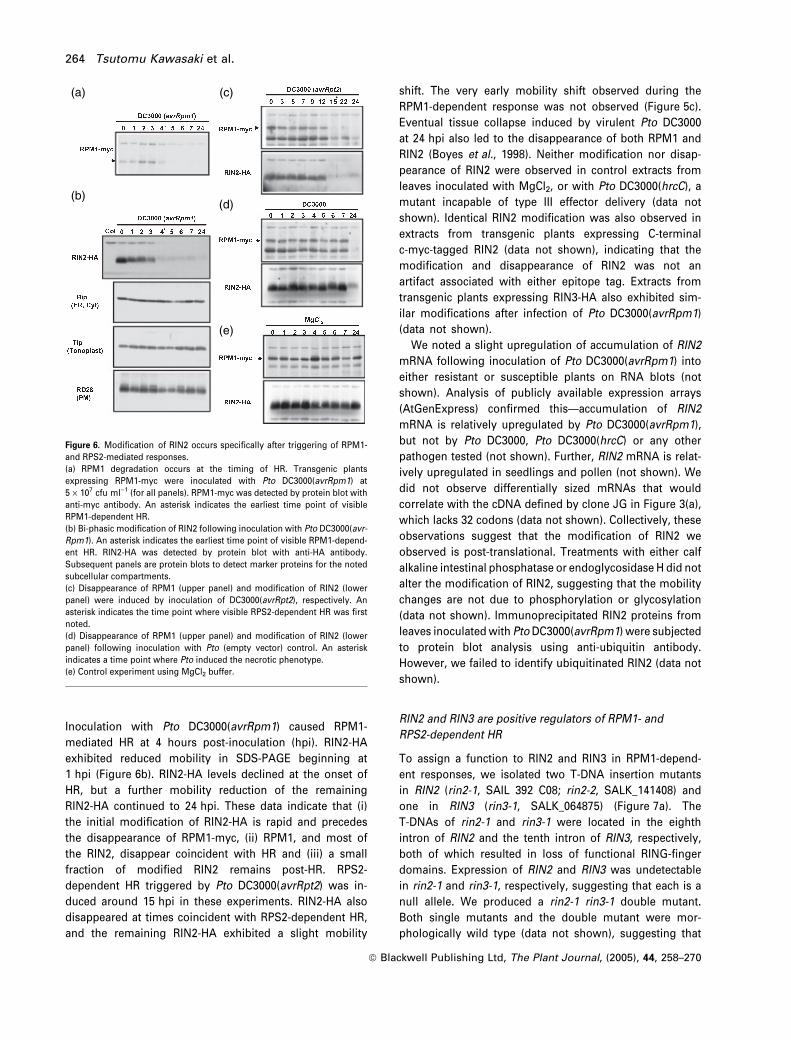
Inoculation with Pto DC3000(avrRpm1) caused RPM1-
mediated HR at 4 hours post-inoculation (hpi). RIN2-HA
exhibited reduced mobility in SDS-PAGE beginning at
1 hpi (Figure 6b). RIN2-HA levels declined at the onset of
HR, but a further mobility reduction of the remaining
RIN2-HA continued to 24 hpi. These data indicate that (i)
the initial modification of RIN2-HA is rapid and precedes
the disappearance of RPM1-myc, (ii) RPM1, and most of
the RIN2, disappear coincident with HR and (iii) a small
fraction of modified RIN2 remains post-HR. RPS2-
dependent HR triggered by Pto DC3000(avrRpt2) was in-
duced around 15 hpi in these experiments. RIN2-HA also
disappeared at times coincident with RPS2-dependent HR,
and the remaining RIN2-HA exhibited a slight mobility
shift. The very early mobility shift observed during the
RPM1-dependent response was not observed (Figure 5c).
Eventual tissue collapse induced by virulent Pto DC3000
at 24 hpi also led to the disappearance of both RPM1 and
RIN2 (Boyes et al., 1998). Neither modification nor disap-
pearance of RIN2 were observed in control extracts from
leaves inoculated with MgCl2, or with Pto DC3000(hrcC), a
mutant incapable of type III effector delivery (data not
shown). Identical RIN2 modification was also observed in
extracts from transgenic plants expressing C-terminal
c-myc-tagged RIN2 (data not shown), indicating that the
modification and disappearance of RIN2 was not an
artifact associated with either epitope tag. Extracts from
transgenic plants expressing RIN3-HA also exhibited sim-
ilar modifications after infection of Pto DC3000(avrRpm1)
(data not shown).
We noted a slight upregulation of accumulation of RIN2
mRNA following inoculation of Pto DC3000(avrRpm1) into
either resistant or susceptible plants on RNA blots (not
shown). Analysis of publicly available expression arrays
(AtGenExpress) confirmed this—accumulation of RIN2
mRNA is relatively upregulated by Pto DC3000(avrRpm1),
but not by Pto DC3000, Pto DC3000(hrcC) or any other
pathogen tested (not shown). Further, RIN2 mRNA is relat-
ively upregulated in seedlings and pollen (not shown). We
did not observe differentially sized mRNAs that would
correlate with the cDNA defined by clone JG in Figure 3(a),
which lacks 32 codons (data not shown). Collectively, these
observations suggest that the modification of RIN2 we
observed is post-translational. Treatments with either calf
alkaline intestinal phosphatase or endoglycosidase H did not
alter the modification of RIN2, suggesting that the mobility
changes are not due to phosphorylation or glycosylation
(data not shown). Immunoprecipitated RIN2 proteins from
leaves inoculated with Pto DC3000(avrRpm1) were subjected
to protein blot analysis using anti-ubiquitin antibody.
However, we failed to identify ubiquitinated RIN2 (data not
shown).
RIN2 and RIN3 are positive regulators of RPM1- and
RPS2-dependent HR
To assign a function to RIN2 and RIN3 in RPM1-depend-
ent responses, we isolated two T-DNA insertion mutants
in RIN2 (rin2-1, SAIL 392 C08; rin2-2, SALK_141408) and
one in RIN3 (rin3-1, SALK_064875) (Figure 7a). The
T-DNAs of rin2-1 and rin3-1 were located in the eighth
intron of RIN2 and the tenth intron of RIN3, respectively,
both of which resulted in loss of functional RING-finger
domains. Expression of RIN2 and RIN3 was undetectable
in rin2-1 and rin3-1, respectively, suggesting that each is a
null allele. We produced a rin2-1 rin3-1 double mutant.
Both single mutants and the double mutant were mor-
phologically wild type (data not shown), suggesting that
(a)
(b)
(c)
(d)
(e)
Figure 6. Modification of RIN2 occurs specifically after triggering of RPM1-
and RPS2-mediated responses.
(a) RPM1 degradation occurs at the timing of HR. Transgenic plants
expressing RPM1-myc were inoculated with Pto DC3000(avrRpm1) at
5 · 107 cfu ml)1 (for all panels). RPM1-myc was detected by protein blot with
anti-myc antibody. An asterisk indicates the earliest time point of visible
RPM1-dependent HR.
(b) Bi-phasic modification of RIN2 following inoculation with Pto DC3000(avr-
Rpm1). An asterisk indicates the earliest time point of visible RPM1-depend-
ent HR. RIN2-HA was detected by protein blot with anti-HA antibody.
Subsequent panels are protein blots to detect marker proteins for the noted
subcellular compartments.
(c) Disappearance of RPM1 (upper panel) and modification of RIN2 (lower
panel) were induced by inoculation of DC3000(avrRpt2), respectively. An
asterisk indicates the time point where visible RPS2-dependent HR was first
noted.
(d) Disappearance of RPM1 (upper panel) and modification of RIN2 (lower
panel) following inoculation with Pto (empty vector) control. An asterisk
indicates a time point where Pto induced the necrotic phenotype.
(e) Control experiment using MgCl2 buffer.
264 Tsutomu Kawasaki et al.
ª Blackwell Publishing Ltd, The Plant Journal, (2005), 44, 258–270
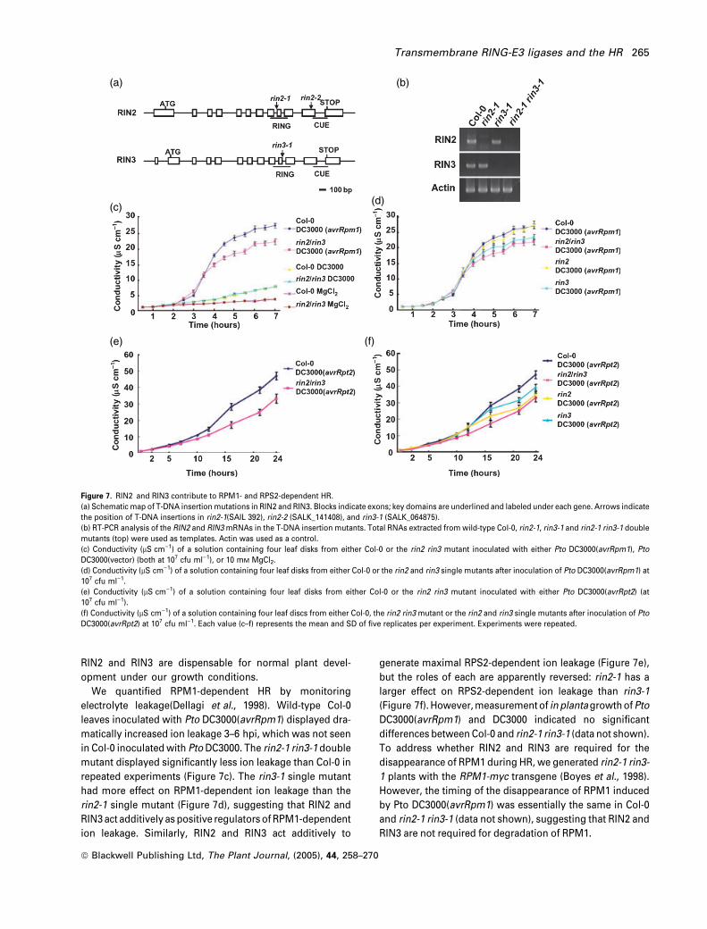
RIN2 and RIN3 are dispensable for normal plant devel-
opment under our growth conditions.
We quantified RPM1-dependent HR by monitoring
electrolyte leakage(Dellagi et al., 1998). Wild-type Col-0
leaves inoculated with Pto DC3000(avrRpm1) displayed dra-
matically increased ion leakage 3–6 hpi, which was not seen
in Col-0 inoculated with Pto DC3000. The rin2-1 rin3-1 double
mutant displayed significantly less ion leakage than Col-0 in
repeated experiments (Figure 7c). The rin3-1 single mutant
had more effect on RPM1-dependent ion leakage than the
rin2-1 single mutant (Figure 7d), suggesting that RIN2 and
RIN3 act additively as positive regulators of RPM1-dependent
ion leakage. Similarly, RIN2 and RIN3 act additively to
generate maximal RPS2-dependent ion leakage (Figure 7e),
but the roles of each are apparently reversed: rin2-1 has a
larger effect on RPS2-dependent ion leakage than rin3-1
(Figure 7f). However, measurement of inplantagrowth ofPto
DC3000(avrRpm1) and DC3000 indicated no significant
differences between Col-0 and rin2-1 rin3-1 (data not shown).
To address whether RIN2 and RIN3 are required for the
disappearance of RPM1 during HR, we generated rin2-1 rin3-
1 plants with the RPM1-myc transgene (Boyes et al., 1998).
However, the timing of the disappearance of RPM1 induced
by Pto DC3000(avrRpm1) was essentially the same in Col-0
and rin2-1 rin3-1 (data not shown), suggesting that RIN2 and
RIN3 are not required for degradation of RPM1.
(a)
(c)(d)
(b)
(e) (f)
Figure 7. RIN2 and RIN3 contribute to RPM1- and RPS2-dependent HR.
(a) Schematic map of T-DNA insertion mutations in RIN2 and RIN3. Blocks indicate exons; key domains are underlined and labeled under each gene. Arrows indicate
the position of T-DNA insertions in rin2-1(SAIL 392), rin2-2 (SALK_141408), and rin3-1 (SALK_064875).
(b) RT-PCR analysis of the RIN2 and RIN3 mRNAs in the T-DNA insertion mutants. Total RNAs extracted from wild-type Col-0, rin2-1, rin3-1 and rin2-1 rin3-1 double
mutants (top) were used as templates. Actin was used as a control.
(c) Conductivity (lS cm)1) of a solution containing four leaf disks from either Col-0 or the rin2 rin3 mutant inoculated with either Pto DC3000(avrRpm1), Pto
DC3000(vector) (both at 107 cfu ml)1), or 10 mM MgCl2.
(d) Conductivity (lS cm)1) of a solution containing four leaf disks from either Col-0 or the rin2 and rin3 single mutants after inoculation of Pto DC3000(avrRpm1) at
107 cfu ml)1.
(e) Conductivity (lS cm)1) of a solution containing four leaf disks from either Col-0 or the rin2 rin3 mutant inoculated with either Pto DC3000(avrRpt2) (at
107 cfu ml)1).
(f) Conductivity (lS cm)1) of a solution containing four leaf discs from either Col-0, the rin2 rin3 mutant or the rin2 and rin3 single mutants after inoculation of Pto
DC3000(avrRpt2) at 107 cfu ml)1. Each value (c–f) represents the mean and SD of five replicates per experiment. Experiments were repeated.
Transmembrane RING-E3 ligases and the HR 265
ª Blackwell Publishing Ltd, The Plant Journal, (2005), 44, 258–270
Discussion
RIN2 and RIN3 encode new RPM1 partners that are
conserved in higher eukaryotes
To date, five proteins, RIN1, RIN4, RAR1, HSP90 and
RIN13, have been identified as partners of RPM1 using
various assays (Al-Daoude et al., 2005; Belkhadir et al.,
2004; Schulze-Lefert, 2004). Here, we identified RIN2 and
RIN3, membrane-bound RING-E3 ligases, as new RPM1
partners. A rin2 rin3 double mutant displayed weakened
RPM1- and RPS2-dependent HR, as measured by dimi-
nution of electrolyte leakage. Yet this phenotype was not
correlated with a loss of RPM1- or RPS2-dependent
restriction of bacterial growth, at least at the level of
resolution afforded by available assays. We infer that
RIN2 and RIN3 act additively as positive regulators of
RPM1- and RPS2-dependent HR.
RIN2 and RIN3 encode ubiquitin E3 ligases
In Arabidopsis, there are 469 deduced RING domain-con-
taining proteins (Stone et al., 2005). The RING-finger
domain contains an octet of cysteines and histidines that
coordinate zinc (Jackson et al., 2000). The RING-finger
domains are classified based upon the spacing of the
metal-binding amino acids or substitutions at one or
more of the metal-bound positions (Stone et al., 2005).
Based upon this classification, the RING-finger domains of
RIN2 and RIN3 are assigned to the type RING-H2 that
contains six cysteines and two histidines (Stone et al.,
2005).
The RING-finger domain of RIN2 itself is sufficient for
in vitro ubiquitin ligase activity that is abolished by a
mutation at one of the conserved cysteines, indicating that
RIN2 encodes a typical ubiquitin E3 ligase. In addition, the
in vitro ligase activity of RIN3 is much stronger than that of
RIN2, although both RING-finger domains are closely rela-
ted. We anticipate that this difference is due to different
biochemical properties of the C-terminal cytoplasmic
domains of RIN2 and RIN3.
RIN2 and RIN3 each possess a CUE domain at their
C-terminus. The CUE domain was initially identified as a
domain similar to yeast Cue1 protein and was proposed
to recruit E2 ubiquitin-conjugating enzyme (Ponting and
Birney, 2000). Recent observations indicate that the CUE
domain is a monoubiquitin-binding motif that promotes
intramolecular monoubiquitination. Monoubiquitination is
a key cell regulatory event, involved in endocytosis and
regulation of protein activity and localization (Hicke, 2001;
Prag et al., 2003). In our in vitro ubiquitination experi-
ments of RIN2, the CUE domain always gave a weak
signal, suggesting that it has monoubiqutin-binding
activity. This interpretation is supported by the fact that
ubiquitin was identified as a RIN2 interactor in further
two-hybrid screening using the CUE domain as a bait
(data not shown).
RIN2 and RIN3 are closely related to the mammalian
autocrine motility factor (AMF) receptor. AMF is a cytoki-
ne secreted by some tumor cells that stimulate cell
migration in vitro and, perhaps, metastasis in vivo
(Watanabe et al., 1996). AMFR functions as a cell surface
receptor for AMF. Because the crystal structure of AMF
indicates dimerization, an AMF homodimer may interact
with an AMFR homodimer to form an AMF/AMFR com-
plex (Tanaka et al., 2002). The interaction between RIN2
and RIN3 via their C-terminal regions, including the CUE
domain, supports the idea that RIN2 and RIN3 form
homo- or heterodimers.
AMFR is localized to PM caveolae and membranes of the
smooth ER at steady state (Benlimame et al., 1998). Endo-
cytosis of AMFR delivers it to the ER (Le et al., 2002). RIN2 is
predominantly localized in the plasma membrane consistent
with RPM1 localization (Boyes et al., 1998). However, we
also found low levels of RIN2 in our intracellular membrane
pool, suggesting that RIN2 also might be localized in the ER
as found in AMFR. Recently, AMFR has been reported to be
involved in ER-associated degradation (ERAD) for protein
quality control (Fang et al., 2001). The barley MLO protein is
localized to the plasma membrane and plays a critical role in
resistance responses to powdery mildew (Devoto et al.,
1999). The MLO protein is regulated by ERAD-mediated
protein quality control (Muller et al., 2005). RIN2 and RIN3
were tested for, but do not participate in, ERAD of the MLO
protein (Muller et al., 2005).
RIN2 and RIN3 interact with RPM1
RIN2 and RIN3 interact with the N-terminal region of RPM1,
including the CC motif. However, the N-terminal fragment
(pEG10) that is deleted for the CC motif binds RIN2 or RIN3
very weakly. Thus, the CC region of RPM1 contributes sig-
nificantly to, but is not sufficient for, full interaction with
RIN2. Similarly, RIN2 and RIN3 bind weakly to the CC-NBS-
LRR protein RPS2, but not to similar baits derived from the
TIR-NBS-LRR protein RPP5. In addition, RIN2 and RIN3 both
interact in vitro with full-length RPM1 produced by in vitro
translation, but not in yeast. This might be explained by the
fact that full-length RPM1 protein is made in relatively low
abundance in yeast (Holt et al., 2002), or by a requirement
for conformational changes in RPM1 that might accompany
its activation (Belkhadir et al., 2004; Moffett et al., 2002).
Examination of in vivo interactions between RIN2 and RPM1
by co-immunoprecipitation using plants expressing both
RPM1-myc and RIN2-HA were unsuccessful, probably
because extraction buffers suitable for solubilization of RIN2
(a multipass integral membrane protein) disrupted RPM1
localized at the periphery of the plasma membrane. Future
266 Tsutomu Kawasaki et al.
ª Blackwell Publishing Ltd, The Plant Journal, (2005), 44, 258–270
experiments using cell biological methods might allow
co-localization of RIN2 and RPM1 in vivo.
The C-terminal region of RIN2, including the CUE domain,
interacts with RPM1. Deletion of the DLQ sequence con-
served in the CUE domain results in reduced binding of
RPM1. The CUE domain alone does not interact with RPM1,
indicating that the CUE domain is required, but not sufficient
for, full interaction. This result also suggests that the region
between the RING and CUE domains is necessary for
interaction with RPM1. Thus, the C-terminal regions of
RIN2 and RIN3 are required for both homo- and hetero-
dimerization and for interaction with RPM1. This suggests
that RPM1 (or RPS2) may regulate RIN2/RIN3 dimerization.
RIN2 and RIN3 are not required for activation-dependent
disappearance of RPM1
The RPM1 protein disappears at the time of HR induced
by inoculation with Pto DC3000(avrRpm1), as well as by
other NB-LRR-mediated recognition events (Boyes et al.,
1998). The disappearance of RPM1 seems to be mediated
by ubiquitination because treatment with a proteasome
inhibitor inhibits it (J. Nam, D. Mackey and J. Dangl,
unpublished results). Therefore, we considered the pos-
sibility that RIN2 and RIN3 are involved in degradation of
RPM1. However, we could not detect RIN2- and RIN3-
dependent ubiquitination of RPM1 in vitro or in vivo.
Furthermore, the kinetics of the disappearance of RPM1 is
not altered in rin2 rin3 (data not shown). Thus, it is
unlikely that disappearance of RPM1 is regulated by a
RIN2/RIN3-mediated pathway.
RIN2 and RIN3 are positive regulators for RPM1-mediated
HR
Inoculation of Pto DC3000(avrRpm1) induces various appar-
ent modifications of RIN2 in SDS-PAGE: a rapid mobility shift
at 1 hpi, a second and gradual mobility coincident with HR
and disappearance also coincident with HR. The very early
modification of RIN2 mobility was not detected following
inoculation with Pto DC3000(avrRpt2), and neither modifi-
cation was seen using Pto DC3000, Pto DC3000(hrcC) or
MgCl2. Thus, these molecular events are unlikely to be
induced by the other type III effectors delivered into the
plant cell from Pto DC3000 or by a wound response. The
rapid timing of the RPM1-specific RIN2 mobility shift at
1 hpi is essentially coincident with type III effector deliv-
ery (Grant et al., 2000), supporting our contention that
this first modification of RIN2 may be a very early signature
of AvrRpm1-RPM1 signaling. The second modification,
and disappearance of RIN2, occur at the same time as
disappearance of RPM1. We proposed (Boyes et al., 1998)
that disappearance of RPM1 is a desensitization mechanism
by which a receptor is eliminated to prevent excessive host
responses after activation (Chuang and Ulevitch, 2004). Data
presented here are not inconsistent with that idea.
RIN2 and RIN3 function additively as positive regulators
of RPM1- and RPS2-mediated HR, but they have no
obvious role in regulating pathogen growth restriction.
This finding could be interpreted as a separation of HR
regulation from the mechanism(s) of disease resistance.
Alternatively, this finding could merely suggest that a
partial loss of HR is not sufficient to alter the effectiveness
of disease resistance. Further definition of the regulators
of HR and disease resistance will allow us to differentiate
whether HR is the cause or simply a consequence of the
mechanisms that ultimately stop proliferation of patho-
gens.
Experimental procedures
Plant cultivation, Agrobacterium transformation and
herbicide selection
Plants were cultivated in growth chambers under a 9 h light/15 hdark regime at 22�C and 60% constant relative humidity. The RIN2p-RIN2:HA gene, including 1912 bp of promoter sequence upstreamof the translation start codon, was subcloned into binary vectorpBAR1 (Holt et al., 2002) and transformed into AgrobacteriumGV3101 by electroporation. Agrobacterium containing the constructwere used for transformation into plants as described (Bechtold andPelletier, 1998). Basta (glufosinate-ammonium) selection was per-formed according to Holt et al. (2002).
Yeast two-hybrid constructs and methods
The yeast two-hybrid library was produced as described (Holt et al.,2002). All RPM1, RIN2 and RIN3 yeast two-hybrid baits were clonedinto the pEG202 vector. RIN2 was originally identified by two-hybridscreening using pEG10 as bait (Gyuris et al., 1993). The prey vectorscontaining the fragments of RIN2 and RIN3 were made using thepJG4-5 vector (Gyuris et al., 1993). For two-hybrid assays, bait andprey vectors were co-transformed into yeast EGY48-competent cellsprepared by the Frozen-EZ yeast transformation II Kit (ZymoResearch, CA, USA). All screening, interaction assays, and plasmidpurification from yeast were done according to (Ausubel et al.,1987).
In vitro interaction experiments
Full-length (amino acids 1–926) and N-terminal region (aminoacids 1–177) RPM1 fragments with C-terminal c-myc epitope-tagswere cloned into pET14b (Novagen, Madison, WI, USA). RPM1-Myc proteins were produced by the TNT Quick Coupled Tran-scription/Translation System (Promega, Madison, WI, USA). ThecDNA fragments of RIN2 and RIN3 were cloned into pGEX4T-1(Amersham Pharmacia, Piscataway, NJ, USA) to produce GST-fused proteins. GST, GST-RIN2 and GST-RIN3 proteins coupledto glutathione Sepharose 4B beads were incubated with theRPM1-Myc proteins in TEDM buffer [80 mM Tris-HCl (pH 7.5),150 mM NaCl, 0.5 mM EDTA, 20 mM MgCl2, 4 mM DTT and 1·protease inhibitor cocktail (Sigma, St Louis, MO, USA)] at 4�C for2 h. After incubation, the beads were washed five times with a
Transmembrane RING-E3 ligases and the HR 267
ª Blackwell Publishing Ltd, The Plant Journal, (2005), 44, 258–270
wash buffer [50 mM Tris-HCl (pH 7.5), 150 mM NaCl, 0.5 mM
EDTA, 0.5% Triton X-100, 0.5 mM DTT and 1· protease inhibitorcocktail]. After the final wash, bound proteins were eluted in 3·SDS loading buffer at the same volume as the beads. SDS-PAGE,immunoblotting, and Coomassie staining were performed usingstandard protocols.
E3 ubiquitin ligase assay
The AtUBC1 protein was purified as previously described (Sullivanand Vierstra, 1991). Either 20 lg or 2 lg of GST-fused RIN2 and RIN3proteins was incubated with 30 ng of yeast E1 enzyme (BostonBiochem, Cambridge, MA, USA), 2 lg His-tagged ubiquitin (Calbi-ochem, San Diego, CA, USA), 1 lg of AtUBC1 in a reaction buffer[50 mM Tris-HCl (pH 7.5), 2 mM MgCl2, 0.5 mM DTT and 4 mM ATP]at 30�C for 90 min. The reaction was stopped by heating at 95�C for2 min. After SDS-PAGE and blotting, the ubiquitinated proteinswere detected by anti-His antibody.
Two-phase partitioning
Tissue was homogenized on ice in an extraction buffer [100 mM
Tris-HCl (pH 7.5), 12% sucrose, 1 mM EDTA, 1· protease inhibitorcocktail (Sigma)] with a polytron. Total proteins were prepared byfiltration through one layer of miracloth, and pelleting of the insol-uble debris by centrifugation at 2000 g for 10 min at 4�C. The sol-uble and microsomal membrane proteins were separated bycentrifugation at 100 000 g for 1 h. Aqueous two-phase partitioningwas done with a polymer concentration of 6.6% (wt/vol) as des-cribed previously (Boyes et al., 1998). The anti-AtPIP2b and anti-AtSEC12 antibodies were purchased from Rose Biotechnology(Rose Biotechnology, Hayward, CA, USA). The AtVAM3 antibodywas provided by Dr Masa H. Sato (Sato et al., 1997).
Modification of RIN2
Two 8.5 mm diameter leaf disks were homogenized in an extractionbuffer [50 mM Tris-HCl (pH 8.0), 10 mM EDTA, 1% SDS], and incu-bated on ice for 1 h. Total proteins were prepared by pelleting of theinsoluble debris by centrifugation at 2000 g for 10 min at 4�C, andsubjected to immunodetection using 10% SDS-PAGE. The anti-RD28 and anti-Tip antibodies were provided by Dr Maarten Chris-peels, and the anti-Bip antibody was provided by Dr RebeccaBoston.
Pathogen culture and inoculation
Pto DC3000 carrying either pVSP61 or derivatives of this plasmidcontaining avr genes have been described (Bisgrove et al., 1994;Grant et al., 1995). For analyzing the modification of RIN2 and RIN3and degradation of RPM1, Pto was resuspended at 5 · 107 colony-forming units (cfu) ml)1 in 10 mM MgCl2, and infiltrated into leavesof 4- to 5-week-old plants. For measurements of electrolyte leakage,Pto at 107 cfu ml)1 was infiltrated into leaves of 4- to 5-week-oldplants. Four 8.5 mm diameter leaf disks were collected from theinfiltrated area and washed with water for 50 min, and then placedin a tube with 15 ml of water. Conductivity was measured from fivereplicates for each treatment using a Yokogawa (Yokogawa, Mu-sashino, Tokyo, Japan) conductivity meter, model SC82. For assaysof bacterial growth, Pto at 105 cfu ml)1 was infiltrated, and numbersof bacteria were measured as described previously (Mackey et al.,2003).
DNA/RNA manipulation
For analysis of RNA, tissue was ground by mortar and pestle inliquid nitrogen. Total RNA was extracted with an RNeasy plant minikit (Qiagen, Valencia, CA, USA). RT-PCR was done using primersspecific to RIN2 (sense, 5¢-GCTCTCCATGCAGCCCTCCC-3¢; anti-sense, 5¢-CTGGCACCTCAGCAGGAATAA-3¢), RIN3 (sense,5¢-GCAACTTCAGAAGAGCTACGGGA-3¢; antisense, 5¢-AGCCTGCA-GAGGAACCCACA-3¢) and actin (sense, 5¢-GAGAGATTCAGGTGCC-CAG-3¢; antisense, 5¢-AGAGCGAGAGCGGGTTTTCA-3¢).
Double-mutant construction
The T-DNA insertion lines for rin2-2 (SALK_141408), and rin3-1(SALK_064875) were identified by searches of the Salk T-DNAinsertion mutant collection (http://signal.salk.edu/cgi-bin/tdnaex-press). The seeds were obtained from the ABRC (Columbus, OH,USA). rin2-1 (SAIL 392 C08) was identified by searching SAIL(the Syngenta Arabidopsis Insertion Library). Genomic DNA wasisolated from the mutant lines, and T-DNA insertions wereconfirmed by PCR reactions using T-DNA left border primers andgene-specific primers, followed by sequencing of the PCR products.The rin2 rin3 double mutants were produced by a cross of bothlines, and identified by PCR.
Acknowledgements
This work was supported by NSF Arabidopsis 2010 grant(IBN-0114795) to J.L.D. T.K. was supported by a fellowship fromthe Ministry of Education, Science, Sports and Culture of Japan.We thank Professor Richard Vierstra (University of Wisconsin,Madison, WI, USA), Professor Judy Callis (University of Califor-nia, Davis, CA, USA) and Dr Jonathan Jones (Sainsbury Lab,Norwich, UK) for their generous gift of reagents and for theirhelpful discussions. We thank Drs Masa H. Sato (Kyoto Univer-sity, Japan), Maarten Chrispeels (University of California, SanDiego, CA, USA) and Rebecca Boston (North Carolina StateUniversity, NC, USA) for antibodies. We gratefully acknowledgethe NSF-sponsored Salk T-DNA collection and ABRC for providingthese resources and we thank Syngenta for making the SAILcollection available.
References
Al-Daoude, A., de Torres Zabala, M., Ko, J.H. and Grant, M. (2005)RIN13 is a positive regulator of the plant disease resistance pro-tein RPM1. Plant Cell, 17, 1016–1028.
Alfano, J.R. and Collmer, A. (2004) Type III secretion system effectorproteins: double agents in bacterial disease and plant defense.Annu. Rev. Phytopathol. 42, 385–414.
Ausubel, F.M., Brent, R., Kingston, R.E., Moore, D.D., Seidman, J.G.,
Smith, J.A. and Struhl, K. (1987) Current Protocols in MolecularBiology. New York, NY: Wiley.
Axtell, M.J. and Staskawicz, B.J. (2003) Initiation of RPS2-specifieddisease resistance in Arabidopsis is coupled to the AvrRpt2-directed elimination of RIN4. Cell, 112, 369–377.
Bar-Peled, M. and Raikhel, N.V. (1997) Characterization of AtSEC12and AtSAR1. Proteins likely involved in endoplasmic reticulumand Golgi transport. Plant Physiol. 114, 315–324.
Bechtold, N. and Pelletier, G. (1998) In planta Agrobacterium-mediated transformation of adult Arabidopsis thaliana plants byvacuum infiltration. Methods Mol. Biol. 82, 259–266.
268 Tsutomu Kawasaki et al.
ª Blackwell Publishing Ltd, The Plant Journal, (2005), 44, 258–270
Belkhadir, Y., Subramaniam, R. and Dangl, J.L. (2004) Plant diseaseresistance protein signaling: NBS-LRR proteins and their part-ners. Curr. Opin. Plant Biol. 7, 391–399.
Benlimame, N., Le, P.U. and Nabi, I.R. (1998) Localization of auto-crine motility factor receptor to caveolae and clathrin-independ-ent internalization of its ligand to smooth endoplasmic reticulum.Mol. Biol. Cell, 9, 1773–1786.
Bent, A.F., Kunkel, B.N., Dahlbeck, D., Brown, K.L., Schmidt, R.,
Giraudat, J., Leung, J. and Staskawicz, B.J. (1994) RPS2 of Ara-bidopsis thaliana: a leucine-rich repeat class of plant diseaseresistance genes. Science, 265, 1856–1860.
van der Biezen, E.A., Sun, J., Coleman, M.J., Bibb, M.J. and
Jones, J.D. (2000) Arabidopsis RelA/SpoT homologs implicate(p)ppGpp in plant signaling. Proc. Natl Acad. Sci. USA, 97,3747–3752.
Bisgrove, S.R., Simonich, M.T., Smith, N.M., Sattler, A. and Innes,
R.W. (1994) A disease resistance gene in Arabidopsis withspecificity for two different pathogen avirulence genes. Plant Cell,6, 927–933.
Boyes, D.C., Nam, J. and Dangl, J.L. (1998) The Arabidopsis thali-ana RPM1 disease resistance gene product is a peripheral plas-ma membrane protein that is degraded coincident with thehypersensitive response. Proc. Natl Acad. Sci. USA, 95, 15849–15854.
Chang, J.H., Goel, A.K., Grant, S.R. and Dangl, J.L. (2004) Wake ofthe flood: ascribing functions to the wave of type III effectorproteins of phytopathogenic bacteria. Curr. Opin. Microbiol. 7,11–18.
Chuang, T.H. and Ulevitch, R.J. (2004) Triad3A, an E3 ubiquitin-protein ligase regulating Toll-like receptors. Nat. Immunol. 5,495–502.
Dangl, J.L. and Jones, J.D. (2001) Plant pathogens and integrateddefence responses to infection. Nature, 411, 826–833.
Dangl, J.L., Dietrich, R.A. and Richberg, M.H. (1996) Death don’thave no mercy: cell death programs in plant–microbe interac-tions. Plant Cell, 8, 1793–1807.
Daniels, M.J., Mirkov, T.E. and Chrispeels, M.J. (1994) The plasmamembrane of Arabidopsis thaliana contains a mercury-insensit-ive aquaporin that is a homolog of the tonoplast water channelprotein TIP. Plant Physiol. 106, 1325–1333.
Day, B., Dahlbeck, D., Huang, J., Chisholm, S.T., Li, D. and Stas-
kawicz, B.J. (2005) Molecular basis for the RIN4 negative regu-lation of RPS2 disease resistance. Plant Cell, 17, 1292–1305.
Dellagi, A., Brisset, M.N., Paulin, J.P. and Expert, D. (1998) Dual roleof desferrioxamine in Erwinia amylovora pathogenicity. Mol.Plant Microbe Interact. 11, 734–742.
Devoto, A., Piffanelli, P., Nilsson, I., Wallin, E., Panstruga, R., von
Heijne, G. and Schulze-Lefert, P. (1999) Topology, subcellularlocalization, and sequence diversity of the Mlo family in plants.J. Biol. Chem. 274, 34993–35004.
Durrant, W.E. and Dong, X. (2004) Systemic acquired resistance.Annu. Rev. Phytopathol. 42, 185–209.
Fang, S., Ferrone, M., Yang, C., Jensen, J.P., Tiwari, S. and Weiss-
man, A.M. (2001) The tumor autocrine motility factor receptor,gp78, is a ubiquitin protein ligase implicated in degradation fromthe endoplasmic reticulum. Proc. Natl Acad. Sci. USA, 98, 14422–14427.
Gardner, R.G., Shearer, A.G. and Hampton, R.Y. (2001) In vivo actionof the HRD ubiquitin ligase complex: mechanisms of endoplas-mic reticulum quality control and sterol regulation. Mol. Cell. Biol.21, 4276–4291.
Gomez-Gomez, L. and Boller, T. (2000) FLS2: an LRR receptor-likekinase involved in the perception of the bacterial elicitor flagellinin Arabidopsis. Mol. Cell, 5, 1003–1011.
Grant, M.R., Godiard, L., Straube, E., Ashfield, T., Lewald, J., Sattler,
A., Innes, R.W. and Dangl, J.L. (1995) Structure of the ArabidopsisRPM1 gene enabling dual specificity disease resistance. Science,269, 843–846.
Grant, M., Brown, I., Adams, S., Knight, M., Ainslie, A. and Mans-
field, J. (2000) The RPM1 plant disease resistance gene facilitatesa rapid and sustained increase in cytosolic calcium that isnecessary for the oxidative burst and hypersensitive cell death.Plant J. 23, 441–450.
Greenberg, J.T. and Yao, N. (2004) The role and regulation ofprogrammed cell death in plant–pathogen interactions. Cell.Microbiol. 6, 201–211.
Gyuris, J., Golemis, E., Chertkov, H. and Brent, R. (1993) Cdi1, ahuman G1 and S phase protein phosphatase that associates withCdk2. Cell, 75, 791–803.
Hammond-Kosack, K.E. and Parker, J.E. (2003) Deciphering plant–pathogen communication: fresh perspectives for molecularresistance breeding. Curr. Opin. Biotechnol. 14, 177–193.
Hicke, L. (2001) Protein regulation by monoubiquitin. Nat. Rev. Mol.Cell Biol. 2, 195–201.
Holt, B.F., 3rd, Boyes, D.C., Ellerstrom, M., Siefers, N., Wiig, A.,
Kauffman, S., Grant, M.R. and Dangl, J.L. (2002) An evolutionarilyconserved mediator of plant disease resistance gene function isrequired for normal Arabidopsis development. Dev. Cell, 2, 807–817.
Hubert, D.A., Tornero, P., Belkhadir, Y., Krishna, P., Takahashi, A.,
Shirasu, K. and Dangl, J.L. (2003) Cytosolic HSP90 associates withand modulates the Arabidopsis RPM1 disease resistance protein.EMBO J. 22, 5679–5689.
Jackson, P.K., Eldridge, A.G., Freed, E., Furstenthal, L., Hsu, J.Y.,
Kaiser, B.K. and Reimann, J.D. (2000) The lore of the RINGs:substrate recognition and catalysis by ubiquitin ligases. TrendsCell Biol. 10, 429–439.
Joazeiro, C.A. and Weissman, A.M. (2000) RING finger proteins:mediators of ubiquitin ligase activity. Cell, 102, 549–552.
Kammerloher, W., Fischer, U., Piechottka, G.P. and Schaffner, A.R.
(1994) Water channels in the plant plasma membrane cloned byimmunoselection from a mammalian expression system. Plant J.6, 187–199.
Kim, H.S., Desveaux, D., Singer, A.U., Patel, P., Sondek, J. and
Dangl, J.L. (2005) The Pseudomonas syringae effector AvrRpt2cleaves its C-terminally acylated target, RIN4, from Arabidopsismembranes to block RPM1 activation. Proc. Natl Acad. Sci. USA,102, 6496–6501.
Kim, M.G., Cunha, L.D., McFall, A.J., Belkhadir, Y., DebRoy, S.,
Dangl, J.L. and Mackey, D. (2005) Two Pseudomonas syringaetype III effectors inhibit RIN4-regulated basal defense in Arabid-opsis. Cell, 121, 749–759.
Le, P.U., Guay, G., Altschuler, Y. and Nabi, I.R. (2002) Caveolin-1 is anegative regulator of caveolae-mediated endocytosis to theendoplasmic reticulum. J. Biol. Chem. 277, 3371–3379.
Mackey, D., Holt, B.F., Wiig, A. and Dangl, J.L. (2002) RIN4 interactswith Pseudomonas syringae type III effector molecules and isrequired for RPM1-mediated resistance in Arabidopsis. Cell, 108,743–754.
Mackey, D., Belkhadir, Y., Alonso, J.M., Ecker, J.R. and Dangl, J.L.
(2003) Arabidopsis RIN4 is a target of the type III virulence effectorAvrRpt2 and modulates RPS2-mediated resistance. Cell, 112, 379–389.
Martin, G.B., Bogdanove, A.J. and Sessa, G. (2003) Understandingthe functions of plant disease resistance proteins. Annu. Rev.Plant Biol. 54, 23–61.
Matsuda, N., Suzuki, T., Tanaka, K. and Nakano, A. (2001) Rma1, anovel type of RING finger protein conserved from Arabidopsis to
Transmembrane RING-E3 ligases and the HR 269
ª Blackwell Publishing Ltd, The Plant Journal, (2005), 44, 258–270
human, is a membrane-bound ubiquitin ligase. J. Cell Sci. 114,1949–1957.
Mindrinos, M., Katagiri, F., Yu, G.L. and Ausubel, F.M. (1994) TheA. thaliana disease resistance gene RPS2 encodes a proteincontaining a nucleotide-binding site and leucine-rich repeats.Cell, 78, 1089–1099.
Moffett, P., Farnham, G., Peart, J. and Baulcombe, D.C. (2002)Interaction between domains of a plant NBS-LRR proteinin disease resistance-related cell death. EMBO J. 21, 4511–4519.
Muller, J., Piffanelli, P., Devoto, A., Miklis, M., Elliott, C., Ortmann,
B., Schulze-Lefert, P. and Panstruga, R. (2005) Conserved ERAD-like quality control of a plant polytopic membrane protein. PlantCell, 17, 149–163.
Muskett, P. and Parker, J. (2003) Role of SGT1 in the regulation ofplant R gene signalling. Microbes Infect. 5, 969–976.
Nimchuk, Z., Eulgem, T., Holt, B.F. and Dangl, J.L. (2003) Recogni-tion and response in the plant immune system. Annu. Rev. Genet.37, 579–609.
Parker, J.E., Coleman, M.J., Szabo, V., Frost, L.N., Schmidt, R., van
der Biezen, E.A., Moores, T., Dean, C., Daniels, M.J. and Jones,
J.D. (1997) The Arabidopsis downy mildew resistance gene RPP5shares similarity to the toll and interleukin-1 receptors with N andL6. Plant Cell, 9, 879–894.
Parton, R.G. and Richards, A.A. (2003) Lipid rafts and caveolae asportals for endocytosis: new insights and common mechanisms.Traffic, 4, 724–738.
Ponting, C.P. and Birney, E. (2000) Identification of domains fromprotein sequences. Methods Mol. Biol. 143, 53–69.
Prag, G., Misra, S., Jones, E.A., Ghirlando, R., Davies, B.A., Hor-
azdovsky, B.F. and Hurley, J.H. (2003) Mechanism of ubiquitinrecognition by the CUE domain of Vps9p. Cell, 113, 609–620.
Saijo, Y., Sullivan, J.A., Wang, H., Yang, J., Shen, Y., Rubio, V., Ma,
L., Hoecker, U. and Deng, X.W. (2003) The COP1-SPA1 interactiondefines a critical step in phytochrome A-mediated regulation ofHY5 activity. Genes Dev. 17, 2642–2647.
Sato, M.H., Nakamura, N., Ohsumi, Y., Kouchi, H., Kondo, M., Hara-
Nishimura, I., Nishimura, M. and Wada, Y. (1997) The AtVAM3encodes a syntaxin-related molecule implicated in the vacuolarassembly in Arabidopsis thaliana. J. Biol. Chem. 272, 24530–24535.
Schulze-Lefert, P. (2004) Plant immunity: the origami of receptoractivation. Curr. Biol. 14, R22–R24.
Seo, H.S., Yang, J.Y., Ishikawa, M., Bolle, C., Ballesteros, M.L. and
Chua, N.H. (2003) LAF1 ubiquitination by COP1 controls photo-morphogenesis and is stimulated by SPA1. Nature, 423, 995–999.
Shao, F., Golstein, C., Ade, J., Stoutemyer, M., Dixon, J.E. and
Innes, R.W. (2003) Cleavage of Arabidopsis PBS1 by a bacterialtype III effector. Science, 301, 1230–1233.
Shih, S.C., Prag, G., Francis, S.A., Sutanto, M.A., Hurley, J.H. and
Hicke, L. (2003) A ubiquitin-binding motif required for intramo-lecular monoubiquitylation, the CUE domain. EMBO J. 22, 1273–1281.
Shimizu, K., Tani, M., Watanabe, H., Nagamachi, Y., Niinaka, Y.,
Shiroishi, T., Ohwada, S., Raz, A. and Yokota, J. (1999) Theautocrine motility factor receptor gene encodes a novel type ofseven transmembrane protein. FEBS Lett. 456, 295–300.
Shirasu, K. and Schulze-Lefert, P. (2003) Complex formation, pro-miscuity and multi-functionality: protein interactions in disease-resistance pathways. Trends Plant Sci. 8, 252–258.
Smalle, J. and Vierstra, R.D. (2004) The ubiquitin 26s proteasomeproteolytic pathway. Annu. Rev. Plant Biol. 55, 555–590.
Stone, S.L., Hauksdottir, H., Troy, A., Herschleb, J., Kraft, E. and
Callis, J. (2005) Functional analysis of the RING-typeubiquitin ligase family of Arabidopsis. Plant Physiol. 137,13–30.
Sullivan, M.L. and Vierstra, R.D. (1991) Cloning of a 16-kDa ubiquitincarrier protein from wheat and Arabidopsis thaliana. J. Biol.Chem. 266, 23878–23885.
Swiderski, M.R. and Innes, R.W. (2001) The Arabidopsis PBS1resistance gene encodes a member of a novel protein kinasesubfamily. Plant J. 26, 101–112.
Tanaka, N., Haga, A., Uemura, H., Akiyama, H., Funasaka, T.,
Nagase, H., Raz, A. and Nakamura, K.T. (2002) Inhibition mech-anism of cytokine activity of human autocrine motility factorexamined by crystal structure analyses and site-directed muta-genesis studies. J. Mol. Biol. 318, 985–997.
Tornero, P., Merritt, P., Sadanandom, A., Shirasu, K., Innes, R.W.
and Dangl, J.L. (2002) RAR1 and NDR1 contribute quantitativelyto disease resistance in Arabidopsis, and their relative contri-butions are dependent on the R gene assayed. Plant Cell, 14,1005–1015.
Vierstra, R.D. (2003) The ubiquitin/26S proteasome pathway, thecomplex last chapter in the life of many plant proteins. TrendsPlant Sci. 8, 135–142.
Watanabe, H., Takehana, K., Date, M., Shinozaki, T. and Raz, A.
(1996) Tumor cell autocrine motility factor is the neuroleukin/phosphohexose isomerase polypeptide. Cancer Res. 56, 2960–2963.
Xie, Q., Guo, H.S., Dallman, G., Fang, S., Weissman, A.M. and Chua,
N.H. (2002) SINAT5 promotes ubiquitin-related degradation ofNAC1 to attenuate auxin signals. Nature, 419, 167–170.
270 Tsutomu Kawasaki et al.
ª Blackwell Publishing Ltd, The Plant Journal, (2005), 44, 258–270