a cationic cytofectin with long spacer mediates favourable transfection in transformed human...
TRANSCRIPT

International Journal of Pharmaceutics 309 (2006) 189–198
Pharmaceutical Nanotechnology
A cationic cytofectin with long spacer mediates favourabletransfection in transformed human epithelial cells
Moganavelli Singh, Mario Ariatti∗Department of Biochemistry, Westville Campus, University of KwaZulu-Natal, P Bag X54001, Durban 4000, South Africa
Received 5 August 2005; received in revised form 11 November 2005; accepted 15 November 2005Available online 27 December 2005
Abstract
The synthesis and transfection potential of a novel cationic cholesterol cytofectin with a dimethylamino head group and along 12 atom, 15̊A spacer incorporating relatively polar amido and dicarbonyl hydrazine linkages are reported. ThusN,N-dimethylaminopropylamidosuccinylcholesterylformylhydrazide (MS09) in equimolar admixture with dioleoylphosphatidylethanolamine (DOPE)forms stable unilamellar liposomes (80–150 nm) which cluster into very effective transfecting, serum nuclease-resistant, lipoplexes with DNA(180–200 nm) at a liposome+/DNA− molar charge ratio of 2.8:1 (12:1, w/w). Gel retardation and ethidium displacement assays confirmedt thelial celll carcinoma( to that inH Oc G2, HeLaa tion. Thesel ion) cells.©
K
1
ma
aDdDp(2bfNhR
withlar.hys-
t
ndo-ds isionic,
-acts of
delyicronughega-d, are
0d
hat DNA was fully liposome-associated and maximally compacted at this ratio. Transfection levels in three human transformed epiines, as established by luciferase transgene activity, was found to be optimal at this charge ratio and in the following order: cervicalHeLa) > oesophageal carcinoma (SNO) > hepatoblastoma (HepG2). Activity in the murine fibroblast line NIH-3T3 was comparableepG2 cells. MS09 lipoplexes achieved approximately three-times and two-times greater activity than Lipofectin® complexes in HeLa and SNells, respectively, whilst comparable levels were recorded in HepG2 and NIH-3T3 cells. MS09 lipoplexes were well tolerated by Hepnd SNO cells with cell numbers found to be 80, 85 and 75% of untreated cultures, respectively, at the optimal transfection concentra
ipoplexes also exhibited high activity in the presence of 10% foetal bovine serum (FBS) in HeLa (17% inhibition) and HepG2 (33% inhibit2005 Elsevier B.V. All rights reserved.
eywords: Cationic liposome; Transfection; Transformed epithelial cells
. Introduction
Efforts to develop non-viral gene transfer vectors in mam-alian systems with the design capability for whole organismpplications are gaining impetus. This process is fuelled, in part,
Abbreviations: BCA, bicinchoninic acid; Chol-T, 3�[N-(N′,N′-dimethyl-minopropane)-carbamoyl] cholesterol; DCC, dicyclohexylcarbodiimide;C-Chol, dimethyl aminoethane carbamoyl cholesterol 3�[N-(N′,N′-imethylaminoethane)-carbamoyl] cholesterol; DMF, dimethylformamide;MSO, dimethylsulfoxide; DOSPER, 1,3-dioleoyloxy-2-(6-carboxyspermyl)-ropylamide; DOPE, dioleoylphosphatidylethanolamine; DOTAP,N-(1-2,3-dioleyloxy) propyl)-N,N,N-trimethylammonium methylsulfate; HEPES,-[4-(2-hydroxyethyl)-piperazinyl]-ethanesulfonic acid; HBS, HEPESuffered saline; MEM, minimum essential medium; MS04, cholesteryl-ormylhydrazide; MS08, cholesterylformylhydrazidehemisuccinate; MS09,,N-dimethylpropylamidosuccinylcholesterylformylhydrazide; NHS,N-ydroxysuccinimide; PBS, phosphate buffered saline; PEI, polyethyleneimine;LU, relative light units; TEM, transmission electron microscopy∗ Corresponding author. Tel.: +27 31 2607981; fax: +27 31 2607942.
E-mail address: [email protected] (M. Ariatti).
by a variety of safety and production difficulties associatedalternative viral methods and retroviral modalities in particuNon-viral DNA transfer approaches are varied and include pical approaches such as electroporation (Meaking et al., 1995),the biolistic or gene gun method (Yang et al., 1999), and direcmicroinjection (Wolff et al., 1990), which deliver naked DNAinto cells by penetration of the plasma membrane avoiding esomes and lysosomes. A growing list of chemical methoreceiving evermore attention however. Amongst these, catpolymers form a sizeable group that includes PEI (Boussif et al.1995), poly-l-lysine (Wu and Wu, 1987), polybrene (Mumperet al., 1996), chitosan (Corsi et al., 2003) and oligoaminosiloxanes (Kichler et al., 2003). These polymers bind and compDNA and transfect mammalian cells with varying degreeefficiency.
Cationic liposomes, however, are arguably the most wiemployed non-viral gene transfer vehicles. These submvesicles which form electrostatic complexes with DNA throinteraction of their positively charged amphiphiles and the ntively charged phosphodiester backbone of the nucleic aci
378-5173/$ – see front matter © 2005 Elsevier B.V. All rights reserved.
oi:10.1016/j.ijpharm.2005.11.023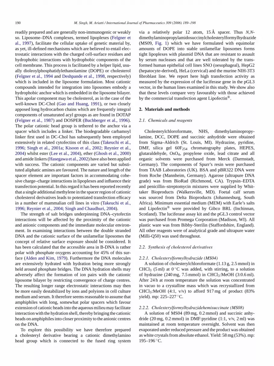
190 M. Singh, M. Ariatti / International Journal of Pharmaceutics 309 (2006) 189–198
readily prepared and are generally non-immunogenic or weaklyso. Liposome–DNA complexes, termed lipoplexes (Felgner etal., 1997), facilitate the cellular uptake of genetic material by,as yet, ill-defined mechanisms which are believed to entail elec-trostatic interactions with the charged cell-surface residues andhydrophobic interactions with hydrophobic components of thecell membrane. This process is facilitated by a helper lipid, usu-ally dioleoylphosphatidylethanolamine (DOPE) or cholesterol(Felgner et al., 1994andDeshpande et al., 1998, respectively)which is included in the liposome formulation. Most cationiccompounds intended for integration into liposomes embody ahydrophobic anchor which is embedded in the liposome bilayer.This apolar component may be cholesterol, as in the case of thewell-known DC-Chol (Gao and Huang, 1991), or two closelyapposed long hydrocarbon chains which are frequently integralcomponents of unsaturated acyl groups as are found in DOTAP(Felgner et al., 1987) and DOSPER (Buchberger et al., 1996).The polar cationic head group is tethered to the anchor via aspacer which includes a linker. The biodegradable carbamoyllinker first used in DC-Chol has subsequently been employedextensively in related cytofectins of this class (Takeuchi et al.,1996; Singh et al., 2001a; Kisoon et al., 2002; Reynier et al.,2004) whilst ester (Lee et al., 2004), ether (Ghosh et al., 2000)and amide linkers (Hasegawa et al., 2002) have also been appliedwith success. The cationic components are varied but substi-tuted aliphatic amines are favoured. The nature and length of thes cohes thet entlt ionicc caci ,1
ctini nica ironm ndeD thec ed.h rathep sur-f esa nglyh maa nicl tresT thenb lturem e tha voure litatei onich ntreso
reda inoh stem
via a relatively polar 12 atom, 15̊A spacer. ThusN,N-dimethylaminopropylamidosuccinylcholesterylformylhydrazide(MS09, Fig. 1) which we have formulated with equimolaramounts of DOPE into stable unilamellar liposomes formstight lipoplexes with plasmid DNA that are resistant to assaultby serum nucleases and that are well tolerated by the trans-formed human epithelial cell lines SNO (oesophageal), HepG2(hepatocyte-derived), HeLa (cervical) and the murine NIH-3T3fibroblast line. We report here high transfection activity asmeasured by the expression of the luciferase gene in the pGL3vector, in the human lines examined in this study. We show alsothat these levels compare very favourably with those achievedby the commercial transfection agent Lipofectin®.
2. Materials and methods
2.1. Chemicals and reagents
Cholesterylchloroformate, NHS, dimethylaminopropy-lamine, DCC, DOPE and succinic anhydride were obtainedfrom Sigma–Aldrich (St. Louis, MI). Hydrazine, pyridine,DMF, silica gel 60F254 chromatography plates, HEPES,glutaraldehyde, OsO4, propylene oxide, lead citrate and allorganic solvents were purchased from Merck (Darmstadt,Germany). The components of Spurr’s resin were purchasedfrom TAAB Laboratories (UK). BSA and pBR322 DNA weref NAg TAa hit-t umw outhA ltsa n,S ectorw . Allp d).A ater(
2
2) in
C ono .A ratedi romC %y
2hy-
d asm thene taineda mp:1
pacer element are important factors in accommodatingive charge–charge interactions in lipoplexes and influenceransfection potential. In this regard it has been reported rechat a single additional methylene in the spacer region of catholesterol derivatives leads to potentiated transfection effin a number of mammalian cell lines in vitro (Takeuchi et al.996; Reynier et al., 2004; Singh and Chaudhuri, 2004).
The strength of salt bridges underpinning DNA–cytofenteractions will be affected by the proximity of the cationd anionic components and the immediate molecular envent. In examining interactions between the double straNA and the cationic surface of the unilamellar liposomesoncept of relative surface exposure should be consideras been calculated that the accessible area in B-DNA isolar with phosphate oxygens accounting for 45% of this
ace (Alden and Kim, 1979). Furthermore the DNA moleculre extensively hydrated with hydration being more stroeld around phosphate bridges. The DNA hydration shellsdversely affect the formation of ion pairs with the catio
iposome bilayer by restricting convergence of charge cenhe resulting longer range electrostatic interactions maye more easily destabilized by ions and polyions in cell cuedium and serum. It therefore seems reasonable to assummphiphiles with long, somewhat polar spacers which faxtension of cationic heads into the aqueous milieu may faci
nteraction with the hydration shell, thereby bringing the catieads on amphiphiles into closer proximity to the anionic cen the DNA.
To explore this possibility we have therefore prepacholesteryl derivative bearing a cationic dimethylam
ead group which is connected to the fused ring sy
-iry
y
-d
Itr
y
.
at
rom Roche (Mannheim, Germany). Agarose (ultrapure Drade) was from BioRad (Richmond, CA). Trypsin–EDnd penicillin–streptomycin mixtures were supplied by W
aker Bioproducts (Walkersville, MD). Foetal calf seras sourced from Delta Bioproducts (Johannesburg, Sfrica). Minimum essential medium (MEM) with Earle’s sand Lipofectin® were provided by Gibco BRL (Inchinnacotland). The luciferase assay kit and the pGL3 control vas purchased from Promega Corporation (Madison, WI)lastic ware was from Bibby-Sterilin (Staffordshire, Englanll other reagents were of analytical grade and ultrapure w
Milli-Q50) was used throughout.
.2. Synthesis of cholesterol derivatives
.2.1. Cholesterylformylhydrazide (MS04)A solution of cholesterylchloroformate (1.13 g, 2.5 mmol
HCl3 (5 ml) at 0◦C was added, with stirring, to a solutif hydrazine (240 mg, 7.5 mmol) in CHCl3:MeOH (3:0.6 ml)fter 24 h at room temperature the solution was concent
n vacuo to a crystalline mass which was recrystallized fHCl3:MeOH (4:1, v/v) to afford 917 mg of product (83ield). mp: 225–227◦C.
.2.2. Cholesterylformylhydrazidehemisuccinate (MS08)A solution of MS04 (89 mg, 0.2 mmol) and succinic an
ride (20 mg, 0.2 mmol) in DMF:pyridine (1:1, v/v, 2 ml) waintained at room temperature overnight. Solvent was
vaporated under reduced pressure and the product was obs white crystals from absolute ethanol. Yield: 58 mg (53%).95–196◦C.

M. Singh, M. Ariatti / International Journal of Pharmaceutics 309 (2006) 189–198 191
Fig. 1. Scheme outlining the synthesis ofN,N-dimethylaminpropylamidosuccinylcholesterylformylhydrazide (MS09) from cholesteryl chloroformate.
2.2.3. N-hydroxysuccinimide ester ofcholesterylformylhydrazidehemisuccinate
MS08 (82 mg, 0.15 mmol), DCC (62 mg, 0.3 mmol) andN-hydroxysuccinimide (35 mg, 0.3 mmol) were dissolved in DMF(1 ml) and the progress of the reaction was monitored by TLC onsilica gel 60F254 plates developed in CHCl3:MeOH (9:1, v/v).After 48 h the dicyclohexylurea crystals were removed by filtra-tion and the crude product obtained by evaporation of the solventin vacuo. The product was dissolved in CHCl3 and this wasextracted with water to remove excessN-hydroxysuccinimide.After evaporation of the solvent the residue was extracted withpetroleum ether (60–80◦C) to remove traces of DCC. Finally
the product was obtained as white crystals from EtOH. Yield:53 mg (55%).
2.2.4. N,N-dimethylpropylamidosuccinylcholesterylformylhydrazide(MS09)
The N-hydroxysuccinimide ester of cholesterylformyl-hydrazidehemisuccinate (53 mg, 0.083 mmol) and dimethy-laminopropylamine (36 mg, 0.35 mmol) were dissolved inwater:pyridine:DMF (13:7:10, v/v/v, 1.5 ml). The reaction wasmonitored by TLC in CHCl3:MeOH (95:5, v/v) and the productwas purified on four 10 cm× 20 cm 60F254 TL plates devel-

192 M. Singh, M. Ariatti / International Journal of Pharmaceutics 309 (2006) 189–198
oped in the same solvent system. Yield: 20 mg (38%). mp:155–156◦C.
2.3. Plasmid DNA
The 5256 bp pGL3 control vector (Promega, Madison, WI)used in this study encodes thePhotinus pyralis luciferase gene(luc+) flanked by SV40 promoter and enhancer sequences whichafford strong expression in a range of mammalian cell types.The 4363 bp pBR322 plasmid (Roche Diagnostics, Mannheim,Germany) was used in the dye displacement assay only.
2.4. Liposome preparation and transmission electronmicroscopy
Liposomes were prepared by a method adapted from thatof Gao and Huang (1991). Briefly, MS09 (2�mol) and DOPE(2�mol) were dissolved in CHCl3 (1 ml). The lipids weredeposited as a thin film on the inner wall of a test tube byrotary evaporation of the solvent at 20◦C. The thin film wasrehydrated in sterile HBS (20 mM HEPES, 150 mM NaCl, pH7.5, 1 ml) overnight and finally the suspension was vortexedbefore sonicating for 5 min on a Transsonic bath-type sonicator.The resulting liposomes were stored at 4◦C. Size and lamellar-ity of liposomes were established by TEM. In brief, liposomes -t /v)g O( withp andfi eadc copa
2
2ng
lH onpA faceo ighta pe a6
2t
2g mMT rel -R mini1 ply,T ide
(1�g/ml running buffer) for 30 min and viewed under transil-lumination at 300 nm and images captured on a Gene GeniusBioimaging System (Syngene, Cambridge, UK).
2.5.3. Nuclease digestion assayLiposome:DNA mixtures were incubated in HBS (20�l) for
20 min at 20◦C, and thereafter, foetal bovine serum was addedto a final concentration of 10%. Samples were incubated for afurther 4 h at 37◦C before receiving EDTA and SDS to finalconcentrations of 10 mM and 0.5% (w/v), respectively. Aftera further 20 min at 55◦C loading buffer (3�l) was added tomixtures before subjecting to electrophoresis as described inSection2.5.2.
2.5.4. Ethidium displacement assayLipoplex formation was also followed by the displacement
of intercalated ethidium cation upon association of plasmidDNA with liposomes in a Shimadzu RF-551 spectrofluoro-metric detector (excitation and emission wavelengths of 520and 600 nm, respectively). The assay was adapted from thatdescribed byTros de Ilarduya et al. (2002). A solution of ethid-ium bromide (ETBr, 1�g) in HBS (500�l) was used to providea baseline reading of fluorescence (0%). Thereafter, pBR322DNA (6 �g) was added and this solution was used to set theinstrument to 100% fluorescence. Aliquots of MS09 liposomes(10�g) were then added and readings taken after mixing at eachs
2
romH rep -t G,1H G2)em
2
telyt den-sr enta bledim freem latesw edbf d ofS ew ,w e-h andw ere
uspensions (50�l) were mixed with a 5% (w/v) BSA soluion in Tris–HCl (100 mM, pH 7.2) and treated with 25% (wlutaraldehyde (50�l). Gels were diced and treated with Os424 h). After dehydration, gels were treated successivelyropylene oxide, propylene oxide:Spurr’s resin (1:1, v/v)nally Spurr’s resin. After staining in uranyl acetate and litrate, sections were viewed in a Jeol 1010 electron microst 60 kV.
.5. Lipoplex characterization
.5.1. Transmission electron microscopyLipoplex suspensions (50�l), which were prepared by addi
iposomes (4�mol lipid/ml HBS) to plasmid DNA (1�g/�lBS) at 20◦C and allowing to mature for 20 min, were placedarafilm sheets and mixed with 0.5% w/v uranyl acetate (50�g).fter 3 min, the mixtures were transferred to the matt surf formvar coated copper grids. Discs were air dried overnnd viewed in a Jeol 1010 transmission electron microsco0 kV.
.5.2. Gel retardation assayLiposome:DNA mixtures in HBS (10�l) were incubated a
0◦C for 30 min and mixed with 3�l gel loading buffer (50%lycerol, 0.5% bromophenol blue, 0.5% xylene cyanol, 72ris–HCl, 60 mM NaH2PO4 and 20 mM EDTA at pH 7.5) befo
oading onto a 1% agarose gel in a Mini-Sub® apparatus (Bioad, Richmond, CA). Electrophoresis was conducted for 90
n TPE running buffer (36 mM Tris–HCl, 30 mM NaH2PO4,0 mM EDTA, pH 7.5) at 50 V (Consort E455 power supurnhout, Belgium). The gel was stained with ethidium brom
e
t
tep until 90�g MS09 liposomes had been added.
.6. Cell lines and their maintenance
HeLa, HepG2, SNO and NIH-3T3 cells were obtained fighveld Biological (Pty) Ltd., Kelvin, South Africa. Cells weropagated at 37◦C in 25 cm2 flasks in 5 ml of MEM con
aining 10% (v/v) foetal bovine serum, 100 U/ml penicillin00�g/ml streptomycin sulphate, 10 mM NaHCO3 and 20 mMEPES (pH 7.5). Cells were split in a ratio of 1:3 (1:2 Hepvery 3–4 days, and stored in a biofreezer (−80◦C) in completeedium containing 10% (v/v) DMSO.
.7. Growth inhibition assays
HeLa, HepG2, SNO and NIH-3T3 cells were separarypsinized and seeded into 24-well plates at the followingities: 2.0× 10⁴, 1.8× 10⁴, 1.8× 10⁴ and 2.1× 10⁴ per well,espectively. Cells were incubated for 24 h to allow attachmnd growth to semi-confluency. Lipoplexes were assem
n HEPES (10�l) and incubated at 20◦C for 20 min. Growthedium was removed from cells and replaced with serumedium (0.5 ml). Lipoplexes were then added to wells and pere incubated at 37◦C for 4 h. The medium was then replacy complete medium and cells were incubated at 37◦C for a
urther 48 h. Adherent cells were quantified by the methochellekens and Stitz (1980). In brief, cells were washed twicith PBS and stained with 200�l crystal violet solution (0.5%/v crystal violet; 0.8%, w/v sodium chloride; 5%, v/v formaldyde; 50%, v/v, ethanol) for 20 min. Stain was removedells washed extensively with water. The multi-well plates w

M. Singh, M. Ariatti / International Journal of Pharmaceutics 309 (2006) 189–198 193
then dried for 24 h and the stain was extracted from fixed cellsinto 2-methoxyethanol (0.5 ml) over a period of 36 h with gen-tle rocking (20 revolutions/min) on a Stuart Scientific STR 6platform shaker Surrey, UK). Absorbance values of extractswere measured at 550 nm in a Novaspec spectrophotometer(Biochrom, Cambridge, UK).
2.8. Transfections
Cells were seeded into 24-well plates at a density of 2.3× 10⁴
(HeLa), 1.9× 10⁴ (HepG2), 1.8× 10⁴ (SNO) and 2.0× 10⁴
(NIH-3T3) in complete medium and grown to semi-confluency.MS09 liposome–DNA complexes were assembled as describedin Section2.7 while Lipofectin®–DNA complexes were pre-pared according to the manufacturers of the transfecting agent.After washing cells with PBS serum free medium (0.5 ml) wasintroduced into wells. Transfection complexes were then addedto wells and after 4 h incubation at 37◦C the medium wasremoved, and replaced with complete growth medium and cellscultured for a further 48 h. In some experiments transfectioncomplexes were introduced to cells in complete medium at theoutset.
2.9. Luciferase assay
PBS( sisr ndcsm uredo aaf,N ineu yw
3
3
ievefh pre-p en-s inatew hah polaa ctedb phob tivesw ico-lG asss Ther
3.1.1. Cholesterylformylhydrazide (MS04)IR (film) 3416 (b, N H), 2929 (st, C H), 1731 (m, C O),
1495 (m, C C) cm−1. 1H NMR (DMSO d6) δ 0.66 (s, 3H,C CH3), 0.86 (d, 6H, CH CH3), 0.91 (d, 3H, CH CH3), 0.99(s, 3H, C CH3), 3.38 (bs, 2H, NH2), 4.38 (m, 1H, CholH3�),5.33 (d, 1H, CholH6), 7.93 (s, 1H, NH). Ms,m/z, ES-TOF445.44 [M + H+], 467.39 [M + Na+].
3.1.2. Cholesterylformylhydrazidehemisuccinate (MS08)IR (film) 3452 (b, N H), 2934 (m, C H), 1675 (m, C O),
1567 (w, C C) cm−1. 1H NMR (DMSO d6) δ 0.64 (s, 3H,C CH3), 0.83 (d, 6H, CH CH3), 0.89 (d, 3H, CH CH3), 0.96(s, 3H, C CH3), 4.3 (Chol H3�), 5.34 (d, 1H, CholH6). Ms,m/z, ES-TOF 545.09 [M + H+], 567.63 [M + Na+].
3.1.3. N,N-dimethylaminopropylamidosuccinylcholesterylformylhydrazide(MS09)
IR (film) 3441 (b, N H), 2945 (m, C H), 1634 (m, C O),1557 (w, C C) cm−1. 1H NMR (CDCl3) δ 0.65 (s, 3H, CCH3),0.84 (d, 6H, CH CH3), 0.88 (d, 3H, CH CH3), 0.98 (s, 3H,C CH3), 2.26 (s, 6H, N CH3), 3.28 (q, CH2 CH2 NH), 4.48(m, 1H, Chol H3�), 5.35 (bs, 1H, CholH6). Ms,m/z, ES-TOF629.82 [M + H+].
3
olarrw da nantlys d byT esw oreur d ofs )wa me-w tures(
3
thec byet em e-a singl 2:1( Aw tainc bouth omeo f
The medium was aspirated and cells washed with2 ml× 0.5 ml). Cell lysis was effected by cell culture lyeagent (80�l/well) over 15 min. Lysates were collected aentrifuged (12,000× g) to pellet cellular debris. To 20�l of theupernatant was added luciferase assay reagent (100�l). Afterixing, relative light units (RLU) of each sample were meas
ver a 10 s period in a Lumac Biocounter M1500 (Landgretherlands). Protein content of supernatants was determsing the BCA assay (Smith et al., 1985) and luciferase activitas expressed as RLU/mg protein.
. Results
.1. Chemical syntheses and spectral analyses
The synthesis of the target compound MS09 was achrom cholesterylchloroformate in four steps (Fig. 1). Theydrazide MS04, which was obtained in high yield, wasared to afford a convenient amino functionality for extion by subsequent succinylation (MS08). The hemisuccas then coupled toN,N-dimethylaminopropylamine througn amide linkage following carboxyl group activation byN-ydroxysuccinimide. In the process, a 12 atom spacer withmido and 1,2-dicarbonyl hydrazine linkages was construetween the basic dimethylamino head group and the hydroic cholesteryl skeleton. Structures of cholesteryl derivaere confirmed by infrared (IR) spectrophotometry on a N
et Impact 420 spectrophotometer,1H (300 MHz) NMR on aemini 300 instrument and electrospray time of flight mpectrometry on a Waters APIQ-TOF Ultima instrument.esults were as follows:
d
d
r
-
.2. Ultrastructure of liposomes and lipoplexes
Liposomes prepared from MS09 and DOPE in a 1:1 matio at a concentration of 4�mol total lipid per milliliter of HBSere stable for several months when stored at 4◦C. Embeddend sectioned samples appeared unilamellar and predomipherical or oval in the 80–150 nm size range when vieweEM (Fig. 2A). Although some size dispersity of liposomas observed, on addition of plasmid DNA, lipoplexes of mniform size were generated. At a Lip+/DNA− molar chargeatio of 2.8 globular aggregates (180–200 nm) comprisemaller vesicles not unlike those reported byPercot et al. (2004ere observed by TEM after negative staining (Fig. 2B). Atmarginally higher charge ratio of 3.3 particles were sohat larger (280–350 nm) and spherical liposome-like struc
50–70 nm) were clearly discernible in the aggregates (Fig. 2C).
.3. Liposome–plasmid association
The formation of electrostatic complexes betweenationic liposomes and pGL3 plasmid DNA was followedlectrophoresis retardation assays on agarose gels.Fig. 3shows
he effect an increasing Lip+/DNA− charge ratio has on thigration of pGL3 plasmid DNA. The amount of liposomssociated DNA retained in the wells increases with increa
iposome concentration until at a liposome/DNA ratio of 1w/w), corresponding to a +/− charge ratio of 2.8 all the DNas liposome-bound. At this ratio, lipoplexes appear to conlusters of unilamellar vesicles, and in consequence only aalf the cationic charges are exposed to the DNA (liposuter leaflet). This would give an effective +/− charge ratio o

194 M. Singh, M. Ariatti / International Journal of Pharmaceutics 309 (2006) 189–198
Fig. 2. Transmission electron micrographs (TEMs) of unilamellar liposomesprepared from MS09 and DOPE in 1:1 molar ratio, (A); lipoplexes at a liposome:pGL3 DNA molar charge ratio of 2.8:1 (B) and 3.3:1 (C).
1.4 at complete DNA retardation (Fig. 3, lane 8). Similar argu-ments have been advanced before to explain the apparent excesspositive charge in cluster complexes of this nature at full DNAretardation (Cao et al., 2000; Kisoon et al., 2002; Percot et al.,2004). This association was also explored in an ethidium dis-placement experiment. On intercalation of the planar aromaticcationic fluorophore into the naked DNA a marked enhancementof fluorescence, a result of steric protection from molecular oxy-gen induced quenching, is noted (Even-Chen and Barenholz,2000). Cationic liposome preparations are known to displaceDNA-associated ethidium (Xu et al., 1999) in a process whichis usually complete within 30 s (Xu and Szoka, 1996). Thus onstepwise addition of MS09 liposomes a steady decrease in fluorescence was observed (Fig. 4) until a point of inflection was
Fig. 3. Gel retardation study of MS09 cationic liposome:DNA complexes. Incu-bation mixtures (10�l) in 20 mM HEPES, 150 mM NaCl (pH 7.5) containedvarying amounts of cationic liposomes in lanes 1–8 (0, 0.5, 1.0, 2.0, 3.0, 4.0,5.0, 6.0�g) and pGL3 DNA (0.5�g).
reached at a liposome:DNA ratio of 12:1 (w/w) which approx-imated 60% displacement and corresponded with the completeassociation of DNA with liposomes as determined in the gelretardation assay (Fig. 3). A further increase in MS09 up toa ratio of 20:1 resulted in additional, but modest, reduction influorescence suggesting that the plasmid DNA did not undergosignificant additional condensation.
The integrity of the DNA in serum-containing medium iscrucial in gene delivery systems. Of particular concern is thepossible degradation of the nucleic acid by serum nucleases.Results presented inFig. 5 confirm that complete protectionfrom digestion in medium containing 10% foetal bovine serumis afforded to liposome-bound DNA over a wide liposome:DNArange (4:1–14:1, w/w). It has already been suggested that electro-
F omesa
-ig. 4. Ethidium intercalation displacement assay for MS09 cationic lipost varying liposome:DNA (w/w) ratios.

M. Singh, M. Ariatti / International Journal of Pharmaceutics 309 (2006) 189–198 195
Fig. 5. Serum nuclease protection assay for MS09 lipoplexes. pBR322 DNA(1�g) was incubated with varying amounts of MS09 liposomes (0, 4, 6, 8, 10,12, 14�g, lanes 2–8) and exposed to 10% FBS for 4 h whereupon mixtureswere applied to a 1% agarose gel. Uncomplexed and untreated pBR322 DNA isshown in lane 1.
static forces between positively charged cationic liposomes andnegatively charged DNA lead to the formation of highly orga-nized supramolecular structures where the DNA is condensedand protected against nuclease degradation (Pitard, 2002).
3.4. Cell growth inhibition studies
Cytotoxicity may present an important yet sometimes over-looked obstacle to effective non-viral gene transfer. This param-eter was measured in all four cell lines studied, under the con-ditions selected for gene transfer experiments. Lipoplexes weregenerally well tolerated over the entire lipid concentration rangeof 8.5–20�M with maximum growth inhibitions of 20% forHeLa, 25% for NIH-3T3, 35% for SNO and 31% for HepG2 cellsrecorded at the highest lipid concentration (Fig. 6). It has beensuggested that toxicity attributed to cationic lipids arises mostlyfrom the increasing cell membrane permeability and creation oftransmembrane pores (Lasic, 1997), while the interaction withanionic lipids such as cardiolipin found in mitochondrial mem-branes would adversely affect the basic energy reactions of thecell (Dass and Burton, 1999).
3.5. Transfection activity of MS09 lipoplexes
The transfection activity of MS09 lipoplexes was measuredin three human transformed epithelial cell lines (HeLa, HepG2,S usL 10%F ipo-s l
Fig. 6. Growth inhibition studies of MS09 liposome:pGL3 DNA complexes inthree transformed human epithelial lines (HeLa, SNO, HepG2) and one murinefibroblast line (NIH-3T3). Cells in multiwell plates bathed in 0.5 ml MEM wereexposed to lipoplexes containing 0.5�g DNA and varying amounts of MS09liposomes as indicated. Data are presented as means± S.D. (n = 4).
the plasmid DNA was liposome-bound (Fig. 3) and near max-imally condensed (Fig. 4). Highest levels were achieved inHeLa cells followed by SNO cells, whilst the lowest levelsof luciferase activity were recorded in HepG2 and NIH 3T3cells. The presence of 10% FBS in the initial 4 h incubation oflipoplexes with cells reduced luciferase activity in the cell linesby varying degrees. In the human lines, transfection activitiesat the optimal Lip+/DNA− ratio were reduced to 83, 67 and40% in HepG2, HeLa and SNO cells, respectively. In all casesthe inhibitory effect was less apparent at Lip+/DNA− ratiosabove and below the optimum. In all transfection experimentswith MS09 lipoplexes, 0.5�g of pGL3 DNA was used in eachwell while the liposome concentration was varied. Lipofectin®,which was used as a comparator in transfection studies, dis-played greatest transfection activity in the cell lines studied whencombined with pGL3 DNA at a ratio of 8:1 (w/w). Unlike MS09liposomes however, highest levels were achieved in the murineNIH 3T3 line (Fig. 8). Although the plasmid concentrationadopted in the Lipofectin® experiments was twice that used inthe MS09 studies, the MS09 lipoplexes achieved approximatelythree-times and two-times greater activity than Lipofectin® inHeLa cells and oesophageal SNO cells, respectively, whilstcomparable levels were noted for HepG2 and NIH 3T3cells.
4. Discussion
eent cru-c ;W chi entsiox
NO) and one murine fibroblast line (NIH 3T3) at varioip+/DNA− charge ratios in the presence or absence ofBS. In all cases maximum activity was obtained at a lome: DNA ratio of 12:1 (w/w) (Fig. 7A–D), at which al
It has been known for some time that the distance betwhe cationic head group and the hydrophobic anchor is aial factor in the design of cytofectins (Remy et al., 1995heeler et al., 1996). The dimethylamino head group, whi
s a feature of several cationic cholesterol transfecting agncluding DC-Chol (Gao and Huang, 1991), cholesteryl-3�-xysuccinamidoethylenedimethylamine, cholesteryl-3�-carbo-yamidoethylenedimethylamine (Farhood et al., 1992), chole-

196 M. Singh, M. Ariatti / International Journal of Pharmaceutics 309 (2006) 189–198
Fig. 7. Transfection studies of MS09 liposome:pGL3 DNA complexes, with and without 10% FBS in HeLa cells (A), SNO cells (B), HepG2 cells (C) and NIH-3T3cells (D). Experiments were conducted in multiwell dishes and cells were exposed to 0.5�g pGL3 DNA complexed to varying amounts of MS09 liposomes (3, 5,6, 7�g). Control 1 shows endogenous luminescence of unexposed cells while control 2 shows luciferase activity of cells exposed to pGL3 DNA alone. Data arepresented as mean± S.D. (n = 4).
Fig. 8. Transfection studies of Lipofectin®:pGL3 DNA complexes in threetransformed human epithelial lines (HepG2, SNO, HeLa) and one murine fibrob-last line (NIH-3T3). Cells in multiwell plates exposed to varying amounts ofLipofectin® (4, 6, 8, 10�g) and pGL3 DNA (1�g). In control 1, cells wereunexposed while in control 2, cells were exposed to pGL3 DNA alone. Data arepresented as mean± S.D. (n = 4).
steryl-3�-carboxyamidopropanedimethylamine (Takeuchi etal., 1996), cholesteryl-3�-oxysuccinamidopropanedimethyl-amine (Singh et al., 2001a) and cholesteryl-3�-oxycarboxy-amidopropanedimethylamine (Chol-T) (Takeuchi et al., 1996;Singh et al., 2001b), has been selected for the work reportedherein. In this study, a cationic cholesterol derivative MS09,in which the dimethylamino function and the hydrophobiccholesteryl moiety are separated by a considerably greater dis-tance, has been prepared and screened in three transformedhuman epithelial cell lines and one murine cell line. We havedesigned this derivative to explore the possibility of improvingthe electrostatic interaction between cationic and anionic cen-tres in the cytofectin and DNA components of lipoplexes byincreasing the spacer length to 12 atoms. The spacer incorpo-rates hydrophilic features (amido and 1,2-dicarbonyl hydrazinelinkages) to promote extension and stability in the hydrated envi-ronment surrounding the nucleic acid and liposome. In supportof this notion, it has already been suggested that liposomal for-mulations with good surface hydration will lead to increasedtransfection activity (Bennett et al., 1996). The derivative wasformulated into stable unilamellar liposomes with DOPE, a co-lipid which has been reported to assist in the formation of lipo-somes, that have significantly increased transfection efficiency,

M. Singh, M. Ariatti / International Journal of Pharmaceutics 309 (2006) 189–198 197
and which tends to reduce the cytotoxicity of cationic surfac-tants (Sternberg et al., 1994). Lipoplexes were well-definedaggregates in appearance (TEM) with a substructure of glob-ular vesicles (50–70 nm) (Fig. 2B and C). In earlier studies withlipoplexes containing Chol-T, a cationic cholesterol cytofectinwith a much shorter propylcarbamoyl spacer/linker, we hadobserved similar aggregates (Moodley et al., 2002). It seems fairto conclude from these observations and those of others (Cao etal., 2000; Percot et al., 2004) that cationic liposomes comprisingDOPE in equimolar admixture with monocationic cholesterolderivatives embodying substituted amino head groups and car-bamoyl linkers readily assemble with DNA into compact clusterswhich increase in size as the Lip+/DNA− ratio is raised.
It is noteworthy that in this study maximum transgene activ-ity was achieved in four unrelated cell types by MS09 lipoplexesof the same Lip+/DNA− molar charge ratio (2.8:1), a ratiowhich coincided with complete association of DNA with lipo-somes (Fig. 3) and maximal compaction of the nucleic acid(Fig. 4). Protection against serum nuclease digestion, however,was clearly conferred on liposome-associated plasmid DNAeven at a far lower liposome:DNA ratio (4:1, w/w,1:1 molarcharge) (Fig. 5).
Importantly, MS09-derived lipoplexes were well toleratedby human hepatoblastoma, cervical carcinoma and oesophagealcarcinoma cells at the lipoplex level resulting in the highestluciferase activity, with cell numbers at 80, 85 and 75% ofu -c heru anc ls int duct effi-c rsea edt sula ,2 xesw cellst ialt S09l eLac H-3 mab t thaa earsr eLac
asec ichd ctedh nil-a ofs G2)c forN de( ento our
laboratory for possible inclusion in transfection systems withtargeting capabilities.
Acknowledgements
This work was supported, in part, by a University ofDurban–Westville research grant. M.S. held a USAID staffdevelopment grant.
References
Alden, C.J., Kim, S.-H., 1979. Accessible surface areas of nucleic acids andtheir relation to folding, conformational transition, and protein recog-nition. In: Sarma, R.H. (Ed.), Stereodynamics of Molecular Systems.Pergamon Press, New York, pp. 331–350.
Bennett, M.J., Aberle, A.M., Balasubramanian, R.P., Molone, J.G., Nantz,M.H., Malone, R.W., 1996. Considerations for the design of improvedcationic amphiphile-based transfection reagents. J. Liposome Res. 6,545–565.
Boussif, O., Lezoualc’h, F., Zanta, M.A., Mergny, M.P., Scherman, D., Deme-niex, B., Behr, J.P., 1995. A versatile vector for gene and oligonucleotidedelivery into cells in culture and in vivo: polyethyleneimine. Proc. Natl.Acad. Sci. U.S.A. 92, 7297–7314.
Buchberger, B., Fernholz, E., Bantle, E., Weigert, M., Borowski, E., van derEltz, H., Hinzpeter, M., 1996. DOSPER liposomal transfection agent: areagent with unique transfection properties. Biochemica 2, 7–10.
Cao, A., Briane, D., Coudet, R., Reynier, P., Bouchemal, N., Lievre, N.,Hantz, E., Salzmann, J.L., Rigaut, J.P., Taillandier, E., 2000. Delivery
es69–
C NA313–
C stemicles.
D arm.
D land,s for
E pre-509,
F ionictivity.
F , R.,y ands. J.
F , M.,hly
cad.
F g, L.,, G.,Gene
G ient179,
G kagegene
ntreated cultures, respectively (Fig. 6). This favourable bioompatibility of MS09 lipoplexes with human lines was furtnderscored particularly in the hepatoblastoma (HepG2)ervical carcinoma (HeLa) cells where transfection levehe presence of 10% FBS resulted in only moderate reion of transfection activity. It has been reported that theiency of liposome-mediated gene expression is often adveffected (Yang and Huang, 1997) while it has also been report
hat in some cases a total inhibition of transfection may refter incubation with serum for as little as 2 h (Nchinda et al.002). Of interest also was the finding that MS09 lipopleere rather more effective in transfecting HeLa and SNO
han Lipofectin® although in experiments with the commercransfecting agent DNA levels were doubled. Moreover, Mipoplexes showed their highest transfection activity in Hells whilst Lipofectin® appeared to be most effective in NIT3 cells. It has been suggested, however, that HeLa cellse more easily transfected than fibroblasts due to the facfter translocation of the DNA into the nucleus, it disappapidly in fibroblasts, but remains for longer periods in Hells (Coonrod et al., 1997; Zhdanov et al., 2002).
In summary, we have prepared a new cholesterol-bationic derivative with a relatively long 12 atom spacer whisplays favourable transfection characteristics in seleuman cancer lines when formulated with DOPE into umellar liposomes. Low cytotoxicity and good toleranceerum by lipoplexes in cervical (HeLa) and hepato (Heparcinoma cells vindicate the design criteria adopted,N-dimethylpropylamidosuccinylcholesterylformylhydrazi
MS09). These findings augur well for the further developmf this system which is currently being investigated in
d
-
ly
t
yt
d
and pathway in MCF7 cells of DNA vectorized by cationic liposomderived from cholesterol. Antisense Nucl. Acid Drug Dev. 10, 3380.
oonrod, A., Li, F.Q., Horwitz, M., 1997. On the mechanism of Dtransfection: efficient gene transfer without viruses. Gene Ther. 4, 11321.
orsi, K., Chellat, F., Yahia, L., Fernandes, J.C., 2003. Mesenchymalcells, MG63 and HEK293 transfection using chitosan–DNA nanopartBiomaterials 24, 1255–1264.
ass, C.R., Burton, M.A., 1999. Lipoplexes and tumours. A review. J. PhPharmacol. 51, 755–770.
eshpande, D., Blezinger, P., Pillai, R., Duguid, J., Freimark, B., RolA., 1998. Target specific optimization of cationic lipid-based systempulmonary gene therapy. Pharm. Res. 15, 1340–1347.
ven-Chen, S., Barenholz, Y., 2000. DOTAP cationic liposomesfer relaxed over supercoled plasmids. Biochim. Biophys. Acta 1176–188.
arhood, H., Bottega, R., Epand, R.M., Huang, L., 1992. Effect of catcholesterol derivatives on gene transfer and protein kinase C acBiochim. Biophys. Acta 1111, 239–246.
elgner, J.H., Kumar, R., Sridhar, R., Wheeler, C., Tsai, Y.L., BorderRamsay, P., Martin, M., Felgner, P.L., 1994. Enhanced gene delivermechanism studies with a novel series of cationic lipid formulationBiol. Chem. 269, 2550–2561.
elgner, P.L., Gadek, T.R., Holm, M., Roman, R., Chan, H.W., WenzNorthrop, J.P., Ringold, G.M., Danielson, M., 1987. Lipofection: a higefficient lipid-mediated DNA-transfection procedure. Proc. Natl. ASci. U.S.A. 84, 7413–7417.
elgner, P.L., Barenholz, Y., Behr, J.P., Cheng, S.H., Cullis, P., HuanJesse, J.A., Seymour, L., Szoka, F., Thierry, A.R., Wagner, E., Wu1997. Nomenclature of synthetic gene delivery systems. HumanTher. 8, 511–512.
ao, X., Huang, L., 1991. A novel cationic liposome reagent for effictransfection of mammalian cells. Biochem. Biophys. Res. Commun.280–285.
hosh, Y.K., Viswewariah, S.S., Bhattacharya, S., 2000. Nature of linbetween the cationic headgroup and cholesteryl skeleton controlstransfection efficiency. FEBS Lett. 473, 341–344.

198 M. Singh, M. Ariatti / International Journal of Pharmaceutics 309 (2006) 189–198
Hasegawa, S., Hirashima, N., Nakanishi, M., 2002. Comparative study oftransfection efficiency of cationic cholesterols mediated by liposomes-based gene delivery. Bioorg. Med. Chem. Lett. 12, 1299–1302.
Kichler, A., Sabourault, N., D́ecor, R., Leborgne, C., Schmutz, M., Valleix,A., Danos, O., Wagner, A., Mioskowski, C., 2003. Preparation and evalu-ation of a new class of gene transfer agents: poly(-alkylaminosiloxanes).J. Control. Release 93, 403–414.
Kisoon, N., Ariatti, M., Moodley, T., 2002. A novel cationic cholesterolderivative, its formulation into liposomes, and the efficient transfection ofthe human cell lines HepG2 and HeLa. Drug Deliv. 9, 161–167.
Lasic, D.D., 1997. Liposomes in Gene Delivery. CRC Press, Boca Raton,New York.
Lee, Y., Koo, H., Lim, Y.B., Lee, Y., Mo, H., Park, J.S., 2004. New cationiclipids for gene transfer with high efficiency and low toxicity: T-shapecholesterol ester derivatives. Bioorg. Med. Chem. Lett. 14, 2637–2641.
Meaking, W.S., Edgerton, J., Wharton, C.W., Meldrum, R.A., 1995.Electroporation-induced damage in mammalian cell DNA. Biochim. Bio-phys. Acta 1264, 357–362.
Moodley, T., Singh, M., Kisoon, N., Naidoo, Y., Ariatti, M., 2002. Structureof lipoplexes derived from novel cationic liposomes and DNA. In: Pro-ceedings of the 15th International Congress on Electron Microscopy, pp.815–816.
Mumper, R.J., Duguid, J.G., Anwer, K., Barron, M.K., Nitta, H., Rolland,A.P., 1996. Polyvinyl derivatives as novel interactive polymers for con-trolled gene delivery to muscle. Pharm. Res. 13, 701–709.
Nchinda, G., Uberla, K., Zshornig, O., 2002. Characterization of cationiclipid DNA transfection complexes differing in susceptibility to seruminhibition. BMC Biotechnol. 2, 12–21.
Percot, A., Briane, D., Coudet, R., Reynier, P., Bouchemal, N., Lievre, N.,Hantz, E., Salzmann, J.L., Cao, A., 2004. A hydroxyethylated cholesterol-based cationic lipid for DNA delivery: effect of conditioning. Int. J.
P t. Ce
R etedDNAruses
R ieresand
ion in
S owth00.
S gene
Singh, M., Kisoon, N., Ariatti, M., 2001b. Receptor-mediated gene deliveryto HepG2 cells by tertiary assemblies containing cationic liposomes andcationized asialoorosomucoid. Drug Deliv. 8, 29–34.
Singh, R.S., Chaudhuri, A., 2004. Single additional methylene group in thehead-group region imparts high gene transfer efficacy to a transfection-incompetent cationic lipid. FEBS Lett. 556, 86–90.
Smith, P.K., Krohn, R.I., Hermanson, G.T., Mallia, A.K., Gartner, F.H.,Provenzano, M.D., Fujimoto, E.K., Goeke, N.M., Olson, B.J., Klenk,D.C., 1985. Measurement of protein using bicinchoninic acid. Analyt.Biochem. 150, 76–85.
Sternberg, B., Sorgi, F.L., Huang, L., 1994. New structures in complex forma-tion between DNA and cationic liposomes visualized by freeze-fractureelectron microscopy. FEBS Lett. 356, 361–366.
Takeuchi, K.-I., Ishikara, M., Kawaura, C., Noji, M., Furuno, T., Nakanishi,M., 1996. Effect of zeta potential of cationic liposomes containing cationiccholesterol derivatives on gene transfection. FEBS Lett. 397, 207–209.
Tros de Ilarduya, C., Arangoa, M.A., Moreno-Aliaga, M.J., Duzgunes,N., 2002. Enhanced gene delivery in vitro and in vivo by improvedtransferrin-lipoplexes. Biochim. Biophys. Acta 1561, 209–221.
Wheeler, C.J., Sukhu, L., Yang, G., Tsai, Y.J., Bustamenta, C., Felgner, P.,Norman, J., Manthrope, M., 1996. Converting an alcohol to an aminein a cationic lipid dramatically alters the co-lipid requirements, cellulartransfection activity and the ultrastructure of DNA–cytofectin complexes.Biochim. Biophys. Acta 1280, 1–11.
Wolff, J.A., Malone, R.W., Williams, P., Chong, W., Ascaldi, G., Jani, A.,Felgner, P.L., 1990. Direct gene transfer into mouse muscle in vivo. Sci-ence 247, 1465–1468.
Wu, G.Y., Wu, C.H., 1987. Receptor-mediated in vitro gene transformationby a soluble DNA carrier system. J. Biol. Chem. 263, 14621–14624.
Xu, Y., Szoka, F.C., 1996. Mechanism of DNA release from cationic35,
X arac-41–
Y onNA.
Y deliv-ung,
emic
Z NAmistry
Pharm. 278, 143–163.itard, B., 2002. Supramolecular assemblies of DNA systems. Soma
Mol. Genet. 27, 5–15.emy, J.S., Kichler, A., Mordvinov, V., Schiber, F., Behr, J.P., 1995. Targ
gene transfer into hepatoma cells with lipopolyamine-condensedparticles presenting galactose ligands: a stage toward artificial viProc. Natl. Acad. Sci. U.S.A. 92, 1744–1748.
eynier, P., Briane, D., Coudert, R., Fadda, G., Bouchemal, N., BissP., Taillandier, E., Cao, A., 2004. Modifications in the head groupin the spacer of cholesterol-based cationic lipids promote transfectmelanoma B16-F10 cells and tumours. J. Drug Target 12, 25–38.
chellekens, H., Stitz, L.W., 1980. Simple method for measuring grinhibition by interferon of cells in monolayer. J. Virol. Meth. 1, 197–2
ingh, M., Balram, S., Ariatti, M., 2001a. Lipoplex-mediated stabletransfer in HeLa cels. Nucleos. Nucleot. Nucl. 20, 889–891.
ll
.
,
liposome:DNA complexesused in cell transfection. Biochemistry5616–5623.
u, Y., Hui, S.-W., Frederik, P., Szoka, F., 1999. Physicochemical chterization and purification of cationic liposomes. Biophys. J. 77, 3353.
ang, J.P., Huang, L., 1997. Overcoming the inhibitory effect of serumlipofection by increasing the charge ratio of cationic liposomes to DGene Ther. 4, 950–960.
ang, N.S., Hogge, G.S., MacEwen, E.G., 1999. Particle mediated geneery applications to canine and larger animal systems. In: Huang, L., HM.-C., Wagner, E. (Eds.), Non-Viral Vectors for Gene Therapy. AcadPress, San Diego, pp. 171–190.
hdanov, R.I., Podobed, O.V., Vlassov, V.V., 2002. Cationic lipid–Dcomplexes—lipoplexes for gene transfer and therapy. Bioelectroche58, 53–64.