4-approach to bleeding disorders.pdf - ksumsc
TRANSCRIPT
Important.Extra.
.Doctor’s notes
Objectives:• To know the function of platelets and the relationship between the
platelet count in peripheral blood and the extent of abnormal bleeding.
• To know about the diseases associated with 1) a failure of platelet production 2) a shortened platelet lifespan, especially immune thrombocytopenic purpura (ITP).
• To know the principles of investigation of patient suspected of having a haemostatic defect.
• To understand the role of platelets, blood vessel wall and coagulation factors in normal haemostasis.
• To know the classification of haemostatic defects. • To know the platelet morphology and life span. • To know the platelet function and diseases due to platelet function
disorders. • To know the causes of thrombocytopenic purpura and non-
thrombocytopenic purpura
References:436 girls & boys’ slides435 teamwork slides
Editing file
Do you have any suggestions? Please contact us!
@haematology436 E-mail: [email protected]
or simply use this form
Approach to Bleeding Disorders
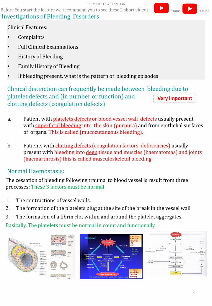
Investigations of Bleeding Disorders:
Clinical Features:
• Complaints
• Full Clinical Examinations
• History of Bleeding
• Family History of Bleeding
• If bleeding present, what is the pattern of bleeding episodes
Clinical distinction can frequently be made between bleeding due to platelet defects and (in number or function) and clotting defects (coagulation defects)
a. Patient with platelets defects or blood vessel wall defects usually present with superficial bleeding into the skin (purpura) and from epithelial surfaces of organs. This is called (mucocutaneous bleeding).
b. Patients with clotting defects (coagulation factors deficiencies) usually present with bleeding into deep tissue and muscles (haematomas) and joints (haemarthrosis) this is called musculoskeletal bleeding.
Normal Haemostasis:
The cessation of bleeding following trauma to blood vessel is result from three processes: These 3 factors must be normal
1. The contractions of vessel walls.
2. The formation of the platelets plug at the site of the break in the vessel wall.
3. The formation of a fibrin clot within and around the platelet aggregates.
Basically, The platelets must be normal in count and functionally.
2
HEMATOLOGY TEAM 436
Very important
Plt for platelet count
We must have fibrin for a stable hemostatic plug
Before You start the lecture we recommend you to see these 2 short videos: 2 mins 4 mins

Classification of haemostatic defects• The action of platelets and the clotting mechanism are closely intertwined in
the prevention of bleeding. However, bleeding arise from defects in one of the three processes:
1) Thrombocytopenia (a low platelet count) (the commonest cause).
2) A defect in the clotting mechanism (the second commonest cause).
3) Abnormal platelet function. 3rd most common.
• Patients with clotting defects usually present with bleeding into deep tissues; that is, muscles or joints. Patients with a deficiency of platelets usually present with mucocutaneous bleeding; that is, bleeding into the skin and from the epithelial surfaces of the nose, uterus and other organs.
• Petechial haemorrhages and ecchymoses and bleeding from other sites may occur when the number of platelets falls below 50 X 109/L. At levels between 20 and 50 X 109/L, petechiae, ecchymoses and nose bleeds are the commonest symptoms, but below 20 X 109/L, gross haemorrhage (melaena, haematemesis, haematuria) becomes increasingly common.
• Signs appear after severe loss (if it drops below 50 x109 /L) Hemorrhage appears if it drops below 20 x109 /L.
Large ecchymoses on both the upper arms of a woman with ITP
Multiple pin-point hemorrhages مثل الدبابيس
(petechiae) on the legs of a patient with idopathic thrombocytopenic purpura (ITP)
Hereditary Vascular Disorders (Not required in the Exam *EXTRA*):1. Hereditary Haemorrhagic Telangiectasia (Rendu-weber- osler syndrome) The most common one
2. Kasabach-merritt syndrome (Haemangioma –Thrombocytopenia)
3. Ehlers-Danlos syndrome
4. Pseudoxanthoma elasticum
5. Homocystinuria
6. Marfan syndrome
7. Osteogenesis imperfecta
Only number 1 is important the rest
you only need to know by name.
3
HEMATOLOGY TEAM 436
Petechiae is the most important sign of ITP
Anything ending with “syndrome” is hereditary.
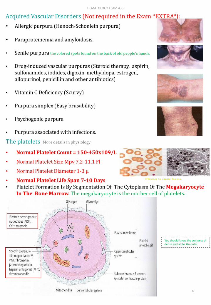
Acquired Vascular Disorders (Not required in the Exam *EXTRA*):
• Allergic purpura (Henoch-Schonlein purpura)
• Paraproteinemia and amyloidosis.
• Senile purpura the colored spots found on the back of old people’s hands.
• Drug-induced vascular purpuras (Steroid therapy, aspirin, sulfonamides, iodides, digoxin, methyldopa, estrogen, allopurinol, penicillin and other antibiotics)
• Vitamin C Deficiency (Scurvy)
• Purpura simplex (Easy brusability)
• Psychogenic purpura
• Purpura associated with infections.
The platelets More details in physiology
• Normal Platelet Count = 150-450x109/L
• Normal Platelet Size Mpv 7.2-11.1 Fl
• Normal Platelet Diameter 1-3 µ
• Normal Platelet Life Span 7-10 Days • Platelet Formation Is By Segmentation Of The Cytoplasm Of The Megakaryocyte
In The Bone Marrow. The megakaryocyte is the mother cell of platelets.
4
HEMATOLOGY TEAM 436
You should know the contents of dense and alpha Granules.

Platelet Activation
• Stickiness
• Shape Change
• Internal Contraction
• Secretion
Physiology
haemostatic plug at sites of damage to vascular endothelium.
• The platelets are also stimulated to produce the prostaglandin, thromboxane A2 from arachidonic acid derived from the cell membrane. The release of ADP and thromboxane A2 causes an interaction of other platelets with the adherent platelets and with each other (secondary platelet aggregation), thus leading to the formation of a platelet plug (primary haemostasis).
• At the site of injury, tissue factor (TF) is expressed and the TF-VIIa complex initiates the formation of a fibrin clot within and around the platelet plug (secondary haemostasis)
• Platelets are also responsible for the contraction of the fibrin clot once it has been formed.
Measurements of Platelet Function
Tests of platelet function The first thing you should do is CBC to check the platelet count.
Bleeding timeThe bleeding time is estimated by making small wounds in the skin of the forearm after applying a blood pressure cuff to the upper arm and inflating it to 40mmHg; the average time that elapses until bleeding ceases is then measured.
PFA - 100 / PFA – 200 the new way (Machine name)
The bleeding time has largely been replaced by an in vitro estimation of primary haemostasis using a machine called a PFA-100.
5
HEMATOLOGY TEAM 436
كأنه طالع منها مخالب
Static form
Activated form
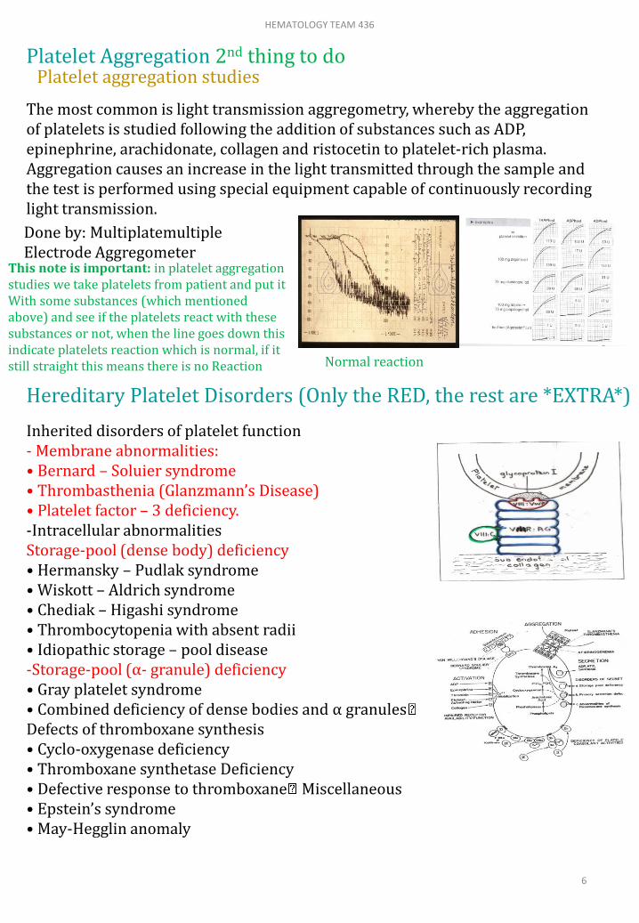
Platelet Aggregation 2nd thing to doPlatelet aggregation studies
The most common is light transmission aggregometry, whereby the aggregation of platelets is studied following the addition of substances such as ADP, epinephrine, arachidonate, collagen and ristocetin to platelet-rich plasma. Aggregation causes an increase in the light transmitted through the sample and the test is performed using special equipment capable of continuously recording light transmission.
Done by: MultiplatemultipleElectrode Aggregometer
Hereditary Platelet Disorders (Only the RED, the rest are *EXTRA*)
Inherited disorders of platelet function - Membrane abnormalities:• Bernard – Soluier syndrome• Thrombasthenia (Glanzmann’s Disease)• Platelet factor – 3 deficiency.-Intracellular abnormalities Storage-pool (dense body) deficiency• Hermansky – Pudlak syndrome• Wiskott – Aldrich syndrome• Chediak – Higashi syndrome• Thrombocytopenia with absent radii• Idiopathic storage – pool disease -Storage-pool (α- granule) deficiency• Gray platelet syndrome• Combined deficiency of dense bodies and α granulesDefects of thromboxane synthesis• Cyclo-oxygenase deficiency• Thromboxane synthetase Deficiency• Defective response to thromboxane Miscellaneous• Epstein’s syndrome• May-Hegglin anomaly
6
HEMATOLOGY TEAM 436
This note is important: in platelet aggregation studies we take platelets from patient and put it With some substances (which mentioned above) and see if the platelets react with these substances or not, when the line goes down this indicate platelets reaction which is normal, if it still straight this means there is no Reaction Normal reaction

Inherited Glanzmann’s disease commonThis is a rare but severe platelet disorder caused by a lack of glycoprotein IIb/IIIa receptors. Inheritance is autosomal recessive and platelets are normal in morphology and number.
Bernard-Soulier diseaseThis is a platelet disorder caused by a lack of glycoprotein Ib receptors. Inheritance is autosomal recessive. Platelets are larger than normal and usually the platelet count is reduced.
Haemostatic profile from a patient with Bernard-Soulier syndrome.
Test Patient Reference Range
PT 12.8s11.5 -13.2s
APTT 27s 26-32s
Fibrinogen (Clauss)2.9g/L 2-4 g/L
Platelets 45 x 109/L 150-450 x 109/L
7
HEMATOLOGY TEAM 436
No reactions Only reacting with ristocetin
Reacting with all substances EXCEPT rstocetin. )Glanzmann’s disease عكس )
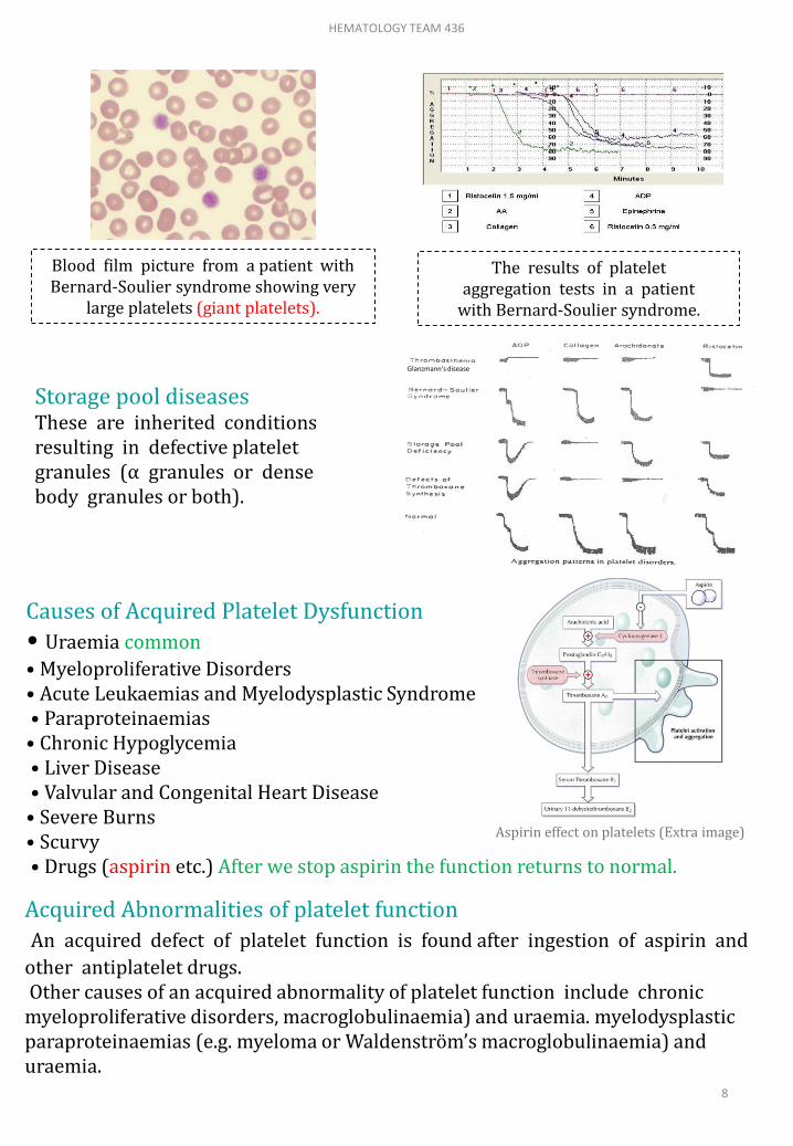
Storage pool diseasesThese are inherited conditions resulting in defective platelet granules (α granules or dense body granules or both).
Blood film picture from a patient with Bernard-Soulier syndrome showing very
large platelets (giant platelets).
The results of platelet aggregation tests in a patient
with Bernard-Soulier syndrome.
Causes of Acquired Platelet Dysfunction
• Uraemia common
• Myeloproliferative Disorders • Acute Leukaemias and Myelodysplastic Syndrome• Paraproteinaemias
• Chronic Hypoglycemia• Liver Disease• Valvular and Congenital Heart Disease
• Severe Burns • Scurvy• Drugs (aspirin etc.) After we stop aspirin the function returns to normal.
Acquired Abnormalities of platelet function
An acquired defect of platelet function is found after ingestion of aspirin and
other antiplatelet drugs.Other causes of an acquired abnormality of platelet function include chronic
myeloproliferative disorders, macroglobulinaemia) and uraemia. myelodysplastic paraproteinaemias (e.g. myeloma or Waldenström’s macroglobulinaemia) and uraemia.
8
HEMATOLOGY TEAM 436
Glanzmann’s disease
Aspirin effect on platelets (Extra image)

Thrombocytopenia the function is okay but the platelet count is low
Causes of thrombocytopenia:1- Bone Marrow failure of platelet production:
2- Increased consumption of platelets in the peripheral blood:• ImmuneAutoimmune (idiopathic)Associated with systemic lupus erythematosus, Chronic lymphocytic leukaemia or lymphomaFeto-maternal alloimmune thrombocytopenia Post-transfusional purpura • Infections: HIV, other viruses, malaria• Drug-induced (e.G. Heparin induced thrombocytopenia)• Disseminated intravascular coagulation• Thrombotic thrombocytopenic purpura• Abnormal distribution of platelets: splenomegaly* • Dilutional loss: massive transfusion of stored blood to bleeding
3- Increased splenic poolingA normal spleen contains within its microcirculation about 30% of all the blood platelets.The splenic platelet pool increases with increasing splenic size, so that in patients with moderate to massive splenomegaly it may account for 50-90% of all blood platelets, thus causing thrombocytopenia.
Selective megakaryocyte depression in the bone marrow
Rare congenital defects (amegakaryocyticaplasia) they are born
without megakaryocytes.
Drugs, chemicals, viral infections
Part of general bone marrow failure
Cytotoxic drugs
Radio-therapy -Aplastic anaemia-Megaloblastic anaemia
Myelodysplastic syndromes
Myelo-fibrosis
Bone marrow infiltration e.G.Carcinoma, lymphoma
-Leukaemia-Multiple myeloma
HIV infection
A newborn baby with allo-immune thrombocytopenia showing
widespread purpura all over the body.
9
HEMATOLOGY TEAM 436
*Normally the spleen holds 30% on the peripheral platelet count, but in splenomegaly the spleen will hold 70-80%.
Immune thrombocytopenic purpura (ITP)ITP is characterized by petechiae, bruising, spontaneous bleeding from mucous membranes and a reduction in the platelet count. The disease presents in both an acute and a chronic form.
Acute ITP: Clinical FeaturesThis is seen at all ages but is most common before the age of 10years. Two-thirds of patients give a history of a common childhood viral infection (e.g. upper respiratory tract infection, chicken pox, measles) 2-3 weeks preceding the purpura.Platelet counts are often less than 20 X 109/L. In most patients the disease runs a self-limiting course of 2-4 weeks, but in approximately 20% it becomes chronic; that is, it lasts more than 6 months.
The mortality is low, the main danger being intracranial bleeding.
Diagnosis by taking history and CBC- Children with the appropriate clinical features, acute thrombocytopenia and an otherwise normal blood count (i.e. no evidence of acute leukaemia).- In ITP, bone marrow megakaryocytes are normal or increased in number (up to four-or eightfold) and increased in size.
Laboratory features of immune thrombocytopenia:Thrombocytopenia with increased numbers of large platelets (>3μ)Increased numbers and size of megakaryocytes.Reduced intravascular platelet survival.
Elevated levels of platelet-associated IgG or IgM.
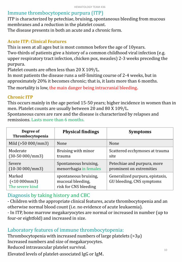
Degree of Thrombocytopenia
Physical findings Symptoms
Mild (>50 000/mm3) None None
Moderate(30-50 000/mm3)
Bruising with minor trauma
Scattered ecchymoses at trauma site
Severe (10-30 000/mm3)
Spontaneous bruising, menorrhagia in females
Petechiae and purpura, more prominent on extremities
Marked(<10 000mm3)
The severe kind
spontaneous bruising, mucosal bleeding,risk for CNS bleeding
Generalized purpura, epistaxis, GU bleeding, CNS symptoms
10
HEMATOLOGY TEAM 436
Chronic ITPThis occurs mainly in the age period 15-50 years; higher incidence in women than in men. Platelet counts are usually between 20 and 80 X 109/L. Spontaneous cures are rare and the disease is characterized by relapses and remissions. Lasts more than 6 months.

Treatment Acute ITPOver 80% of patients recover without any treatment.- Corticosteroids are widely used- High doses of intravenous immunoglobulin (Ig) causes a rapid increase in the platelet count if corticosteroids didn’t work.
Chronic ITPTreatment is usually not needed in patients with platelet counts above 30-50 X 109/L who have no significant spontaneous bleeding. - High-dose corticosteroid therapy increases the platelet count to more than 50 X 109/L- Prednisolone 60 mg/day- Splenectomy because the spleen takes up too much of the platlets.- Second-line treatment - Azathioprine, cyclophosphamide, danazol, dapsone, cyclosporine A, mycophenolate mofetil and rituximab have all been used, particularly inpatients who fail to respond to splenectomy.- High dose of intravenous Ig (e.g. 1 g/kg/day for2 days) has also been found to increase the platelet count to greater than 50 X 109/L in 80%of patients with chronic ITP.
Thrombocytopenia as a result of drugs or toxins:• Bone marrow suppression• Predictable (dose-related): Ionizing radiation, cytotoxic drugs (heparin), ethanol• Occasional• Chloramphenicol, co-trimoxazole, idoxuridine, penicillamine, organic arsenicals, benzene, etc.• Immune mechanisms (proven or probable)• Analgesics, anti-inflammatory drugs, gold salts• Antimicrobials: Penicillins, sulphonamides, trimethoprim, rifampicin• Sedatives, anticonvulsants: Dizepam, sodium valproate, carbamazepine• Diuretics: Acetazolamide, chlorathiazides, frusemide• Antidiabetics: Chlorpropamide, tolbutamide• Others: Digitoxin, heparin, methyldopa, oxyprenolol, quinine, quinidine• Platelet aggregation: Ristocetin, heparin
Blood film of ITP:Severe thrombocytopenia (No platelets seen) 11
HEMATOLOGY TEAM 436
The whole slide is *EXTRA*
Thrombotic thrombocytopenic purpura (TTP) 1:40 mins
- In healthy individuals a VWF-cleaving protease (ADAMTS 13) cleaves the Tyr 842-Met 843 peptide bond in VWF to produce the characteristic multimer profile.- In the absence of the protease, ultra-large VWF multimers are released that lead
to platelet aggregation and the disease known as ‘thrombotic thrombocytopenic purpura’ (TTP).- This is a serious illness characterized by widespread arteriolar platelet thrombi leading to fragmentation of red cells (schistocytes), thrombocytopenia, neurological symptoms and renal impairment.
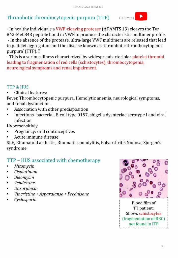
Blood film of TT patient:
Shows schistocytes(fragmentation of RBC)
not found in ITP
TTP & HUS• Clinical features:Fever, Thrombocytopenic purpura, Hemolytic anemia, neurological symptoms, and renal dysfunction.• Association with other predisposition• Infections- bacterial, E-coli type 0157, shigella dysnteriae serotype I and viral
infectionHypersensitiviy• Pregnancy: oral contraceptives• Acute immune diseaseSLE, Rhumatoid arthritis, Rhumatic spondylitis, Polyarthritis Nodosa, Sjorgen’ssyndrome
TTP – HUS associated with chemotherapy• Mitomycin• Cisplatinum• Bleomycin• Vendestine• Doxorubicin • Vincristine + Asparalanse + Prednisone• Cyclosporin
12
HEMATOLOGY TEAM 436

Platelet transfusions : EXTRA• It is often possible to raise the platelet count temporarily by platelet transfusions. • The main indication for platelet transfusion is severe haemorrhage caused by: (i) Thrombocytopenia due to diminished platelet production or DIC; or (ii)Abnormal platelet function.
• Transfusion may also be indicated in a patient with thrombocytopenia or defective platelet function prior to surgery. • Another indication for platelet transfusion is thrombocytopenia (platelets <50 X 109/L) in patients receiving massive blood transfusions. • Platelet counts need only be maintained above 10-20 X 109/L, since severe bleeding is rare above this level.
Blood count film
Normal platelet countLow platelet count
I. Bleeding timeII. Platelet aggregation studies with
ADP, Adrenalin, collagen and ristocetin
III. Other special platelet tests e.gAdhesion studies, nucleotide, pool measurement
IV. Favtor VIII clotting assayvWF assayvWF antigen AssayNo need to test the bone marrow
I. Bone marrow examinationII. Platelet antibodiesIII. Screen tests for DIC
13
HEMATOLOGY TEAM 436

platelets
normal count 150-450x109/L
life span 7-10 days
formation by segmentation of the cytoplasm of the megakaryocyte in thebone marrow.
Classification of haemostatic defects (Bleeding Disorders) مهم
platelets defects (in number (Thrombocytopenia)(commonest) or
function)
clotting defects (coagulation factors deficiencies)
superficial bleeding into the skin (purpura) bleeding into deep tissue and muscles (haematomas) and joints (haemarthrosis)
Called mucocutaneous bleeding Called musculoskeletal bleeding.
Most common symptoms
Num. of platelets falls below 50 Petechial haemorrhages and ecchymoses
between 20 and 50 petechiae, ecchymoses and nose bleeds
below 20 gross haemorrhage (melaena, haematemesis, haematuria)
Inherited disorders of platelet function
Membrane abnormality:
Glanzmann’s disease -caused by a lack of glycoprotein IIb/IIIa receptors.
Bernard-Soulierdisease
-caused by a lack of glycoprotein Ib receptors.-larger Platelets / low platelet count.
Storage pool diseases -resulting in defective platelet granules (α granules or dense body granules or both)
Thrombocytopenia
Causesمهم
-Bone Marrow failure of platelet production (radiotherapy,leukaemia,viral infections,..)-increased consumption of platelets in the peripheral blood (Immune, infections,..)-Increased splenic pooling (splenomegaly)
Immune thrombocytopenic purpura (ITP)
مهم
characterized by petechiae, bruising, spontaneous bleeding from mucous membranes and a reduction in the platelet count.
AcuteITP(main danger being intracranial bleeding)-ChronicITP(relapses and remissions)
Reduced intravascular platelet survival. + increased size&number of megakaryocytes
Thrombotic thrombocytopenic purpura (TTP)
a serious illness characterized by widespread arteriolar platelet thrombi due to absence of VWF-cleavingprotease
Summary
14
HEMATOLOGY TEAM 436

MCQs:
Team members:
Ghada AlMazrou
Jawaher Alhayyal
Rana Barasain
Shrooq Alsomali
Aseel Alsulimani
Team Leaders
Safa Al-Osaimi
Abdulaziz Al-Hussainy
1) Deficiency of which of the following will cause Glanzmann's Thrombastheniadisease?A. GPIa B. GPIbC. GPIIIa D. GPIIIbAns: C
Q2) Platelet life span:A- 2-5 days B- 10-20 daysC- 7-10 days D- 5-10 daysAns: C
Q3) Bernard Soulier syndrome is a manifestation of:A- Deficiency of GPIIb B- Deficiency of GPIaC- Deficiency of GPIIIa D- Deficiency of GPIbAns: D
Q4) Bleeding into deep tissue and muscles and joint is due to:A- Platelet defects B- WBCs defectsC- Clotting defects D- Epithelial defectsAns: C
Q5) platelets defects will lead to which bleeding type of the following? A- Deep tissue bleeding B- superficial bleedingC- Joint bleeding D- Hematomas Ans: B
Good Luck!
15
HEMATOLOGY TEAM 436