2010 - metabolic and structural rearrangement during dark-induced autophagy in soybean ( glycine max...
TRANSCRIPT
Planta (2010) 231:1495–1504
DOI 10.1007/s00425-010-1148-3ORIGINAL ARTICLE
Metabolic and structural rearrangement during dark-induced autophagy in soybean (Glycine max L.) nodules: an electron microscopy and 31P and 13C nuclear magnetic resonance study
Pierre Vauclare · Richard Bligny · Elisabeth Gout · Valentine De Meuron · François Widmer
Received: 21 December 2009 / Accepted: 26 February 2010 / Published online: 1 April 2010© Springer-Verlag 2010
Abstract The eVects of dark-induced stress on the evolu-tion of the soluble metabolites present in senescent soybean(Glycine max L.) nodules were analysed in vitro using 13C-and 31P-NMR spectroscopy. Sucrose and trehalose were thepredominant soluble storage carbons. During dark-inducedstress, a decline in sugars and some key glycolytic metabo-lites was observed. Whereas 84% of the sucrose disap-peared, only one-half of the trehalose was utilised. Thisdecline coincides with the depletion of Gln, Asn, Ala andwith an accumulation of ureides, which reXect a hugereduction of the N2 Wxation. Concomitantly, phosphodiest-ers and compounds like P-choline, a good marker of mem-brane phospholipids hydrolysis and cell autophagy,accumulated in the nodules. An autophagic process wasconWrmed by the decrease in cell fatty acid content. In addi-tion, a slight increase in unsaturated fatty acids (oleic andlinoleic acids) was observed, probably as a response toperoxidation reactions. Electron microscopy analysis revealed
that, despite membranes dismantling, most of the bacter-oids seem to be structurally intact. Taken together, ourresults show that the carbohydrate starvation induced insoybean by dark stress triggers a profound metabolic andstructural rearrangement in the infected cells of soybeannodule which is representative of symbiotic cessation.
Keywords Peribacteroid membrane · Glycine · Metabolic NMR · Nodules · Senescence
AbbreviationsFru6P Fructose 6-phosphateGABA �-Aminobutyric acidGlc6P Glucose 6-phosphateGlyc3P Glycerol 3-phosphateGPC Glycerylphosphoryl-cholineGPE Glycerylphosphoryl-ethanolamineGPG Glycerylphosphoryl-glycerolGPI Glycerylphosphoryl-inositolMan6P Mannose 6-phosphatePBM Peribacteroid membraneP-cho Phosphoryl-cholineP-eth Phosphoryl-ethanolamineSEM Scanning electron microscopyTEM Transmission electron microscopy
Introduction
Legumes play an important role in agricultural food pro-duction (Doyle and Luckow 2003). Among them, soybean(Glycine max) is a major crop cultivated for oil and proteinproduction. Like other legumes, soybean develops specia-lised root organs, called nodules, in which host cells estab-lish a symbiotic association with rhizobia (Perret et al.
Electronic supplementary material The online version of this article (doi:10.1007/s00425-010-1148-3) contains supplementary material, which is available to authorized users.
P. Vauclare · V. De Meuron · F. WidmerLaboratory of Plant Biology and Physiology, Biology Building UNIL, room 5449, 1015 Lausanne, Switzerland
R. Bligny · E. GoutLaboratoire de Physiologie Cellulaire Végétale, Unité Mixte de Recherche 5168, Institut de Recherche en Technologie et Sciences pour le Vivant CEA, 17, rue des Martyrs, 38054 Grenoble Cedex 9, France
Present Address:P. Vauclare (&)22, rue du Lavoir, 26120 Malissard, Francee-mail: [email protected]
123
1496 Planta (2010) 231:1495–1504
2000; Broughton 2003). DiVerentiated bacteria, called bac-teroids, express the nitrogenase enzyme complex, whichreduces atmospheric nitrogen (N2) to ammonia at theexpense of 16 ATP per mol of N2. Ammonia is then sup-plied to the host cells and converted into organic com-pounds (amides or ureides), which are exported to theshoots (for a review, see Tajima et al. 2004). In return,leaves deliver carbohydrates into the nodules via thephloem to provide the energy required for N2 Wxation (for areview see Schubert 1986; Prell and Poole 2006). Becausethis symbiotic relationship is the largest source of availablenitrogen on earth, culture of legumes reduces the need forexpensive fertilizers, reducing the pollution of the ecosys-tem (Newton 2000). The active period of N2 Wxation is lim-ited during nodule development because nodule senescenceoccurs rapidly after Xowering and during seed maturation(Espinosa-Victoria et al. 2000; Puppo et al. 2005). Thissenescence is also observed in leguminous plants inresponse to environmental perturbations like drought, lowtemperature, defoliation, external addition of nitrogen andpathogenic attack (Cots et al. 2002; Patriarca et al. 2004;Puppo et al. 2005). In addition, nodule senescence can beartiWcially triggered by exposing soybean plants to pro-longed darkness (Cohen et al. 1986; Gordon et al. 1993;Fargeix et al. 2004). Although dark-induced senescence is apremature phenomenon, it shares many of the molecular,physiological and ultrastructural characteristics found innaturally senescing nodules, such as the loss of nitrogenaseactivity, the increase in proteolytic activity and the greenishcolour of nodule tissues (PfeiVer et al. 1983; Cohen et al.1986; Jacobi et al. 1994; Fargeix et al. 2004). Because natu-ral senescence is an irreversible phenomenon, understand-ing the physiological processes which occur during inducedsenescence could help to Wnd strategies that delay this phe-nomenon and extend the active period of rhizobium–plantsymbiosis (Espinosa-Victoria et al. 2000). Recently, a tran-scriptomic study has been performed on the senescence ofindeterminate nodules in Medicago truncatula (Van deVelde et al. 2006). During this complex process a widevariety of defence and stress-response genes are transcribedin senescing M. truncatula nodules, an event which ismarked by the transition of the nodule from a carbon sink toa general nutrient source (Van de Velde et al. 2006). Inter-estingly, although determinate (soybean) and indeterminatenodule types show a very diVerent anatomical structure,which makes comparison with their senescing physiologi-cal responses diYcult (Puppo et al. 2005), some quite com-mon modiWcations have been observed. One of the mainevents was observed in soybean as well as in M. truncatulausing transmission electron microscopy (TEM). It concernsthe alteration of the structure of the symbiosomes, an orga-nelle-like compartment in which bacteroids are surroundedby a specialised membrane called the peribacteroid membrane
(Fargeix et al. 2004; Van de Velde et al. 2006). However,while this alteration is considered as the earliest observableevent associated with a loss of nitrogen-Wxing activity, littleinformation is available on the metabolic modiWcationsduring nodule senescence in soybean (Cohen et al. 1986;Fargeix et al. 2004; Van de Velde et al. 2006). For thesereasons, we performed further experiments to demonstrateunambiguously the autophagic process in relation to thedown-regulation of the nitrogen metabolism. This promptedus to draw up the metabolite proWle to characterise someeasily identiWable marker(s) of autophagy and peribacteroidmembrane (PBM) evolution in senescing soybean nodulesby using in vitro 13C- and 31P-nuclear magnetic resonancespectroscopy, scanning electron microscopy (SEM) andbiochemical measurements.
Materials and methods
Plant materials and growth conditions
Soybean seeds (Glycine max L., var Mapple arrow; Schwe-izer Samen AG, Thoune, Switzerland) were thoroughlywashed in water, incubated for 20 min at 42°C and thenwashed a second time in water. The swollen seeds werethen transferred into a mixture of vermiculite and pottingsoil (Triohum; Mauser Samen, Winterthur, Switzerland)without fertilizer [1:1]. Plants were grown in a greenhouseunder controlled conditions with a light phase of 16 h, andtemperatures of 20°C (day) and 18.5°C (night).
Bradyrhizobium japonicum (strain 110, spc 4, wild type)was grown in sterilised liquid culture [4% bactopeptone(p/v), 1 mM KH2PO4, 2 mM MgSO4·7H2O, pH 6.8] withspectinomycine (100 �g/ml) for 6 days at 28°C. Centri-fuged bacteria were diluted in phosphate-buVered salinemedium (0.14 M NaCl, 2.5 mM KCl, 4 mM Na2HPO4,2 mM KH2PO4, pH 7.4). Seven days after imbibition, eachplant was inoculated with 108–109 B. japonicum strain. Oneweek after infection, a fertilizer (Basisdünger Hauert-Flory,GrossaVoltern, Switzerland) was added and this wasrepeated once a week; 28–32 days of growth after inocula-tion were required to obtain mature nodules. For the experi-ments, 30-day-old plants were placed in darkness or undernormal growth conditions for 4, 7, 11 and 14 days.
In vitro NMR spectroscopy
Perchloric acid (PCA) extracts were prepared from 8 g offresh nitrogen frozen soybean nodules according to themethod described by Gout et al. (2000). Spectra of neutra-lised PCA extracts were obtained on an NMR spectrometer(AMX 400, Bruker, Billeria, MA, USA) equipped with a10-mm multinuclear probe tuned at 162 or 100.6 MHz for
123
Planta (2010) 231:1495–1504 1497
31P- or 13C-NMR studies, respectively. The deuterium reso-nance of D2O (100 �l added per millilitre of extract) wasused as a lock signal.
Conditions used for 13C-NMR acquisition were as fol-lows: 90° radio-frequency pulses (19 �s) at 6 s intervals;spectral width, 20,000 Hz; 900 scans; and Waltz-16 1Hdecoupling sequence (with two levels of decoupling: 2.5 Wduring acquisition time, 0.5 W during delay). Free induc-tion decays were collected as 16,000 data points, zero Wlledto 32,000 and processed with a 0.2-Hz exponential linebroadening. The 13C-NMR spectra are referenced to hexa-methyldisiloxane at 2.7 ppm. Mn2+ ions were chelated bythe addition of 2 �M 1,2-cyclohexylenedinitrilotetraaceticacid (CDTA) and the pH was adjusted to 7.5 (sample vol-ume 2.5 ml). The spectra of nodules were compared withthe spectra of a PCA extract of sycamore cells.
The conditions used for 31P-NMR acquisition were asfollows: 70° radio-frequency pulses (15 �s) at 3.6 s inter-vals; spectral width 8,200 Hz; 1,024 scans; Waltz-16 1Hdecoupling sequence (with two levels of decoupling: 1 Wduring acquisition time, 0.5 W during delay). Free induc-tion decays were collected as 8,000 data points, zero Wlledto 16,000 and processed with a 0.2-Hz exponential linebroadening. The 31P-NMR spectra were referenced tomethylene diphosphonic acid, pH 8.9, at 16.38 ppm. Before31P-NMR analyses, divalent paramagnetic cations (Ca2+,Mg2+, Mn2+, etc.,) were chelated by addition of appropriateamounts of CDTA ranging from 100 to 150 �M (samplevolume of 2.5 ml). The pH was buVered by the addition of75 �mol Hepes and adjusted to 7.5. IdentiWed compoundswere quantiWed by comparison of the areas of their reso-nance peaks with those of known amounts of maleateand methylphosphonate as internal standards for 13C- and31P-NMR analyses, respectively.
Scanning electron microscopy
Samples were frozen in liquid N2, fragmented, and then Wxedovernight in 2% OsO4 and 3% CrO3 at room temperature.Dehydration was carried out on ice by successive 10 minincubations in 30, 50, 70, 80, 90 and 100% acetone solutions,and samples were stored at ¡20°C. A critical drying (CriticalPoint Dryer, Baltec-Leica Microsystems, Wetzlar, Germany)was performed, followed by coating with gold for 3 min, andsamples were observed with a Jeol JSM-6300F (Tokyo,Japan) microscope at an accelerating voltage of 5 kV.
Isolation of peribacteroid membranes (PBM) from soybean nodules
Membrane fractions were prepared as described by Bassa-rab et al. (1989) with slight modiWcations. Fresh nodules
(4–8 g) stocked overnight at 4°C (Day et al. 1987) werecrushed, using a chilled mortar and pestle, in 10 ml ofcold buVer containing 75 mM Tris/HCl at pH 7.5, 10 mMKCl, 1 mM MgCl2, 1 mM EDTA, 1 mM phenylmethyl-sulfonyl Xuoride (PMSF), 2 mM 1,4-dithioerythritol(DTT) and 12% sucrose. The crushed material was Wlteredthrough three layers of moist Miracloth, and then centri-fuged for 10 min at 750g. The supernatant was transferredto the top of a continuous sucrose gradient (24 ml of 60–30% sucrose which in turn was layered over 5 ml of 20%sucrose) and centrifuged for 2 h at 90,000g. Fractions cor-responding to various cell compartments were identiWedby using the following marker enzymes: alcohol dehydro-genase, NADH cytochrome-c reductase and cytochrome-coxidase for cytosol, endoplasmic reticulum, and mito-chondria, respectively (Quail 1979; Lord 1987; Lee et al.1995).
The band of peribacteroid units (PBUs) or symbiosomeswere collected, adjusted to 20% sucrose with a buVer(5 mM Tris/HCl, pH 7.5, 1 mM EDTA, 1 mM PMSF,2 mM DTT), and centrifuged for 30 min at 6,000g. Thepellet was re-suspended in 6 ml of 6% sucrose; PBUswere pressed 10 times through a hypodermic needle(0.6 £ 30 mm, Microlance 3), and the suspension wastransferred to the top of 10 ml of 35% sucrose and centri-fuged for 30 min at 50,000g. The identiWcation of PBMfraction was identiWed by immunodetection using a nodu-lin-26 (NOD 26) antibody with a procedure described byWeaver et al. (1991). The interface containing PBM wasrecovered, adjusted to 6% sucrose in buVer (5 mM Tris/HCl, pH 7.5, 1 mM EDTA, 1 mM PMSF, 2 mM DTT) andcentrifuged for 30 min at 50,000g. The PBM was recoveredfrom the pellet and diluted in 1 ml of a buVer containing25 mM Tris–Mes at pH 6.5, 0.25 M sucrose, 1 mM DTTand 20% glycerol. Samples were stored at ¡20°C after sat-uration with N2.
QuantiWcation of fatty acid
Lipids from frozen PBM were extracted by the methodof Bligh and Dyer (1959). To measure fatty acids, phos-pholipids were transesteriWed according to Bassarabet al. (1989). Myristic acid (14:0) was used as an internalstandard. Methyl-ester derivates were analysed on aHewlett-Packard 5890 gas chromatograph [SP TM-2330glass column from Supelco (Sigma-Aldrich, Buchs,Switzerland) 30 m long £ 0.75 mm i.d., 0.2 �m Wlmthickness] equipped with a Xame ionisation detector andan automatic data analysis. Programme conditions wereas follows: from 100 to 160°C (25°C min¡1), from 160to 200°C (8°C min¡1) and Wnally 220 to 100°C(25°C min¡1).
123
1498 Planta (2010) 231:1495–1504
Results
Metabolite proWling of soybean nodules’ cells
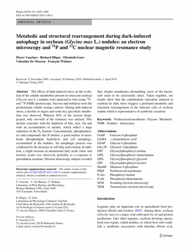
In vitro 13C- and 31P-NMR spectroscopy from perchloricacid (PCA) extracts was performed to obtain the metabolicproWle of 41-day-old soybean nodules infected withB. japonicum (Fig. 1a, Table 1). Among the diVerent reso-nance signals detected by 13C-NMR (Table 1), the highestcorresponded to the 13C of D-chiro-inositol and myo-inosi-tol, the two major polyols present in soybean nodules[8.6 �mol g¡1 fresh weight (FW)] (Streeter 1987). Theother major resonances observed correspond to those ofthe carbons of sucrose (5.78 �mol g¡1 FW) and trehalose(4.2 �mol g¡1 FW), a non-reducing disaccharide sugarrestricted to root nodules (Table 1). As expected, malate(0.625 �mol g¡1 FW) was also detected at high concentra-tions (Table 1), as this dicarboxylic acid is eYcientlyrespired by nodule host cells and bacteroids to support N2
Wxation (Day and Copeland 1991; Lodwig and Poole 2003).One of the main amino acids detected was asparagine(6.1 �mol g¡1 FW), which is two to three times higher thanthe ureides (allantoin and allantoic acid) and glutamate(Table 1). There were also two smaller signals emergingfrom the background noise: �-aminobutyric acid (GABA;0.78 �mol g¡1 FW) and alanine (0.94 �mol g¡1 FW)(Table 1). Finally, glutamine signals are almost undetect-able (0.63 �mol g¡1 FW) suggesting a high turnover ofthis amino acid (Table 1). The 31P-NMR spectra showedthat the major compound was the inorganic phosphate(Pi; 7.1 �mol g¡1 FW; Fig. 1a; Table 1). The presence ofsignals corresponding to sugar phosphates [glucose6-phosphate (Glc6P), fructose 6-phosphate (Fru6P),3-phosphoglycerate (PGA) and dihydroxyacetone-phosphate(DHAP)] reXects the glycolytic pathway activity. The pre-cursor of ascorbate biosynthesis, mannose 6-phosphate(Man6P), was also found to accumulate in nodules(Wheeler et al. 1998; Matamoros et al. 2006) (Fig. 1a,Table 1). Nucleoside diphosphate sugars such as UDP-glu-cose (UDP-Glc), UDP-galactose (UDP-Gal) and UDP-N-acetylglucosamine (UDP-GlcNAc) were also detected.Contrary to UDP-Glc, UDP-Gal and UDP-GlcNAc werepresent only at trace levels. Intermediates in the synthesis andhydrolysis of phospholipids like phosphoryl-choline (P-cho)and phosphoryl-ethanolamine (P-eth) and phosphodiestersglycerylphosphoryl-choline (GPC) and glycerylphosphoryl-inositol (GPI) were identiWed (Fig. 1a, Table 1). Amongnucleotides, UTP (0.07 �mol g¡1 FW) was about twice ashigh as ATP. The low ATP concentration compared withthat of AMP (0.88 �mol g¡1 FW, Fig. 1a, Table 1) is typi-cal of a hypoxic metabolism. Finally, we observed the pres-ence of signiWcant amounts of pyridine nucleotides, mainlyNAD and NADPH. In contrast, NADP+ was not detected,
which suggests that the reducing power coupled to thepentose phosphate pathway (Hong and Copeland 1990) isactive in nodules under our culture conditions.
EVects of dark stress on metabolic proWling of nodules
Nodules taken from 30-day control soybeans after a 11-daydark treatment showed important metabolic changes thatcould mimic natural senescence at the physiological level(Fig. 1b, Table 1). A major observation from the 13C-NMRspectra is that sucrose (0.93 �mol g¡1 FW) was reduced by84% during dark stress, whereas the correspondingdecrease in trehalose was only 48% (2.18 �mol g¡1 FW,Table 1). Inositol remained stable during the 11 days ofdarkness. Interestingly, whereas amino acids’ spectra ofglutamine, glutamate, alanine and asparagine presented amarked decrease in their amounts; a three- to Wvefoldincrease in allantoate (5.93 �mol g¡1 FW) and allantoin(5.16 �mol g¡1 FW) levels was observed (Table 1). However,
Fig. 1 Representative in vitro 31P-NMR spectra of excised soybeannodules from 30-day-old plants subsequently kept under normal cul-ture condition for 11 days (a) or kept in total darkness for 11 days (b).Extracts were prepared from 8 g of nodules (on a fresh weight basis)and analysed by 31P-NMR. DHAP, dihydroxyacetone-phosphate;Fru6P, fructose 6-phosphate; Glcn6P, gluconate 6-phosphate; Glc6P,glucose 6-phosphate; Glyc3P, glycerol 3-phosphate; Pi, inorganicphosphate; Man6P, mannose 6-phosphate; PGA, 3-phosphoglycerate;P-eth, phosphoryl-ethanolamine; P-cho, phosphoryl-choline; UDP-glc, UDP-glucose; UDP-gal, UDP-galactose; UDP-GlcNAc, UDP-N-acetylglucosamine
123
Planta (2010) 231:1495–1504 1499
the asparagine signals emerged clearly from the back-ground noise (3.13 �mol g¡1 FW). Concerning the pool ofGABA, its concentration doubled in the dark-treated sam-ple to reach 1.25 �mol g¡1 FW, whereas malate was belowthe threshold of 13C-NMR detection. Of particular interestwas the detection of two intermediates implicated in mem-brane lipid synthesis in stressed nodules, P-cho (peaks at57.77, 54.73 and 54.69 ppm) and free choline (peaks at54.66, 54.62 and 54.58 ppm) corresponding to the threemethyl groups coupled to the N atom of these molecules.Using 31P-NMR spectroscopy, we observed that in dark-treated root nodules, P-cho became the major phosphory-lated compound (2.7 �mol g¡1 FW) whereas P-eth increasedonly slightly (Fig. 1b, Table 1). A new peak correspondingto glycerol 3-phosphate (Glyc3P) was detected. The accu-mulation of P-cho and Glyc3P reveals a membrane hydro-lytic process (Dorne et al. 1987; Roby et al. 1987; Aubertet al. 1996). Moreover, two new phosphodiesters corre-sponding to glycerylphosphoryl-glycerol (GPG) andglycerylphosphoryl-ethanolamine (GPE) were detected. Theother phosphorylated compounds, including Glc6P andMan6P, the major sugar phosphate compounds, UDP-Glc andUDP-Gal decreased signiWcantly, whereas UDP-GlcNAc,Fru6P and DHAP became undetectable (Fig. 1b, Table 1).Interestingly, we found at 4.72 ppm a new peak corre-sponding to Glcn6P, a hexose-P involved in the pentosephosphate pathway (PPP) and in the regeneration ofNADPH (Hong and Copeland 1990; Anthon and Emerich1990). Finally, we observed a decrease in the total amountof soluble NTP. This was accompanied by a collapse in thecontent of NMP (Fig. 1b, Table 1). We conclude that a longperiod without photosynthesis induces important changesin the metabolite proWle of root nodules in soybean relatedto an autophagy process. Since nodules contain a highproportion of symbiotic rhizobia surrounded by a PBM,we hypothesised that this PBM is particularly aVected insenescing nodules. In order to test this hypothesis, weexamined the fate of PBM during dark stress by SEM and abiochemical approach.
Ultrastructural eVects of senescence on soybean nodules
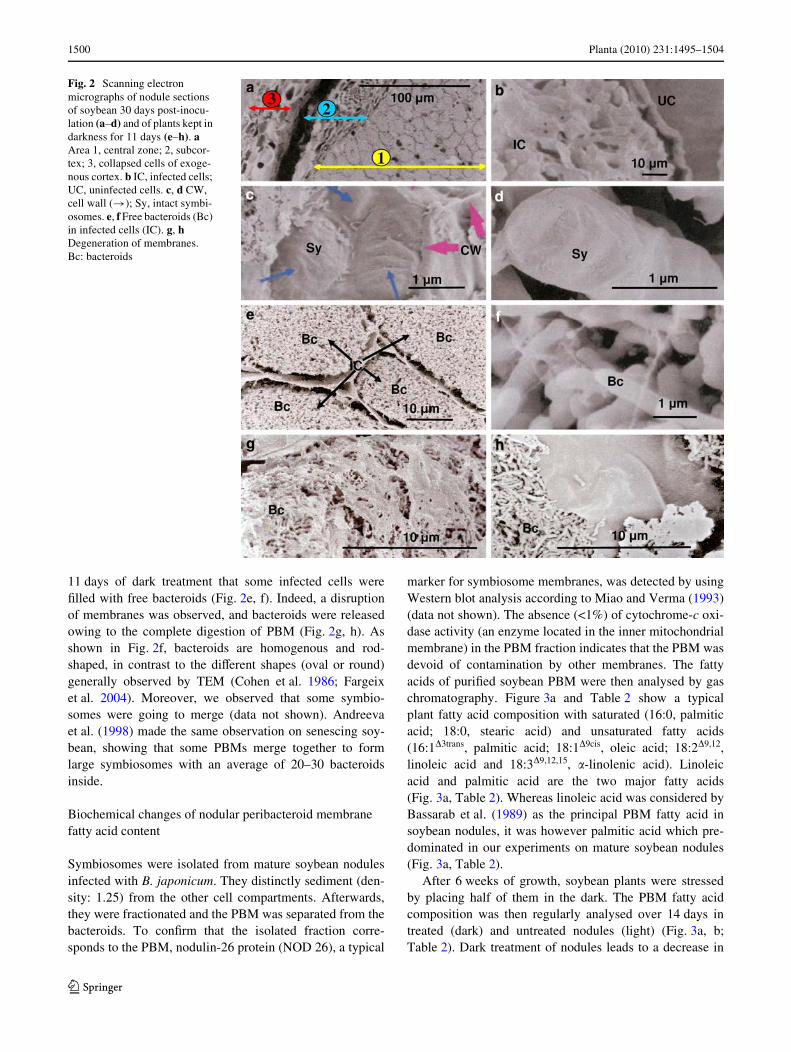
Scanning electron microscopy was used to study the ultra-structural changes of infected cells in mature nodulesduring prolonged darkness. As shown in Fig. 2a and b,three-dimensional observation reveals a central part of thenodules containing cells infected or not. Infected cells incontrol nodules present hundreds of symbiosomes in whichbacteroids were enclosed in an intact PBM (Fig. 2b). Theexternal structure of symbiosomes corresponds to the PBMand looks like a big stick (Fig. 2c, d). We observed after
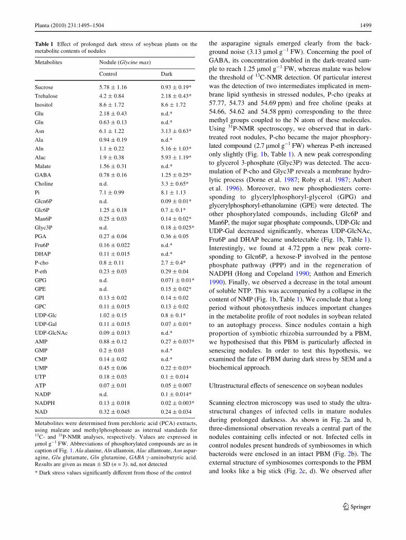
Table 1 EVect of prolonged dark stress of soybean plants on themetabolite contents of nodules
Metabolites were determined from perchloric acid (PCA) extracts,using maleate and methylphosphonate as internal standards for13C- and 31P-NMR analyses, respectively. Values are expressed in�mol g¡1 FW. Abbreviations of phosphorylated compounds are as incaption of Fig. 1. Ala alanine, Aln allantoin, Alac allantoate, Asn aspar-agine, Glu glutamate, Gln glutamine, GABA �-aminobutyric acid.Results are given as mean § SD (n = 3). nd, not detected
* Dark stress values signiWcantly diVerent from those of the control
Metabolites Nodule (Glycine max)
Control Dark
Sucrose 5.78 § 1.16 0.93 § 0.19*
Trehalose 4.2 § 0.84 2.18 § 0.43*
Inositol 8.6 § 1.72 8.6 § 1.72
Glu 2.18 § 0.43 n.d.*
Gln 0.63 § 0.13 n.d.*
Asn 6.1 § 1.22 3.13 § 0.63*
Ala 0.94 § 0.19 n.d.*
Aln 1.1 § 0.22 5.16 § 1.03*
Alac 1.9 § 0.38 5.93 § 1.19*
Malate 1.56 § 0.31 n.d.*
GABA 0.78 § 0.16 1.25 § 0.25*
Choline n.d. 3.3 § 0.65*
Pi 7.1 § 0.99 8.1 § 1.13
Glcn6P n.d. 0.09 § 0.01*
Glc6P 1.25 § 0.18 0.7 § 0.1*
Man6P 0.25 § 0.03 0.14 § 0.02*
Glyc3P n.d. 0.18 § 0.025*
PGA 0.27 § 0.04 0.36 § 0.05
Fru6P 0.16 § 0.022 n.d.*
DHAP 0.11 § 0.015 n.d.*
P-cho 0.8 § 0.11 2.7 § 0.4*
P-eth 0.23 § 0.03 0.29 § 0.04
GPG n.d. 0.071 § 0.01*
GPE n.d. 0.15 § 0.02*
GPI 0.13 § 0.02 0.14 § 0.02
GPC 0.11 § 0.015 0.13 § 0.02
UDP-Glc 1.02 § 0.15 0.8 § 0.1*
UDP-Gal 0.11 § 0.015 0.07 § 0.01*
UDP-GlcNAc 0.09 § 0.013 n.d.*
AMP 0.88 § 0.12 0.27 § 0.037*
GMP 0.2 § 0.03 n.d.*
CMP 0.14 § 0.02 n.d.*
UMP 0.45 § 0.06 0.22 § 0.03*
UTP 0.18 § 0.03 0.1 § 0.014
ATP 0.07 § 0.01 0.05 § 0.007
NADP n.d. 0.1 § 0.014*
NADPH 0.13 § 0.018 0.02 § 0.003*
NAD 0.32 § 0.045 0.24 § 0.034
123
1500 Planta (2010) 231:1495–1504
11 days of dark treatment that some infected cells wereWlled with free bacteroids (Fig. 2e, f). Indeed, a disruptionof membranes was observed, and bacteroids were releasedowing to the complete digestion of PBM (Fig. 2g, h). Asshown in Fig. 2f, bacteroids are homogenous and rod-shaped, in contrast to the diVerent shapes (oval or round)generally observed by TEM (Cohen et al. 1986; Fargeixet al. 2004). Moreover, we observed that some symbio-somes were going to merge (data not shown). Andreevaet al. (1998) made the same observation on senescing soy-bean, showing that some PBMs merge together to formlarge symbiosomes with an average of 20–30 bacteroidsinside.
Biochemical changes of nodular peribacteroid membrane fatty acid content
Symbiosomes were isolated from mature soybean nodulesinfected with B. japonicum. They distinctly sediment (den-sity: 1.25) from the other cell compartments. Afterwards,they were fractionated and the PBM was separated from thebacteroids. To conWrm that the isolated fraction corre-sponds to the PBM, nodulin-26 protein (NOD 26), a typical
marker for symbiosome membranes, was detected by usingWestern blot analysis according to Miao and Verma (1993)(data not shown). The absence (<1%) of cytochrome-c oxi-dase activity (an enzyme located in the inner mitochondrialmembrane) in the PBM fraction indicates that the PBM wasdevoid of contamination by other membranes. The fattyacids of puriWed soybean PBM were then analysed by gaschromatography. Figure 3a and Table 2 show a typicalplant fatty acid composition with saturated (16:0, palmiticacid; 18:0, stearic acid) and unsaturated fatty acids(16:1�3trans, palmitic acid; 18:1�9cis, oleic acid; 18:2�9,12,linoleic acid and 18:3�9,12,15, �-linolenic acid). Linoleicacid and palmitic acid are the two major fatty acids(Fig. 3a, Table 2). Whereas linoleic acid was considered byBassarab et al. (1989) as the principal PBM fatty acid insoybean nodules, it was however palmitic acid which pre-dominated in our experiments on mature soybean nodules(Fig. 3a, Table 2).
After 6 weeks of growth, soybean plants were stressedby placing half of them in the dark. The PBM fatty acidcomposition was then regularly analysed over 14 days intreated (dark) and untreated nodules (light) (Fig. 3a, b;Table 2). Dark treatment of nodules leads to a decrease in
Fig. 2 Scanning electron micrographs of nodule sections of soybean 30 days post-inocu-lation (a–d) and of plants kept in darkness for 11 days (e–h). a Area 1, central zone; 2, subcor-tex; 3, collapsed cells of exoge-nous cortex. b IC, infected cells; UC, uninfected cells. c, d CW, cell wall (!); Sy, intact symbi-osomes. e, f Free bacteroids (Bc) in infected cells (IC). g, h Degeneration of membranes. Bc: bacteroids
123
Planta (2010) 231:1495–1504 1501
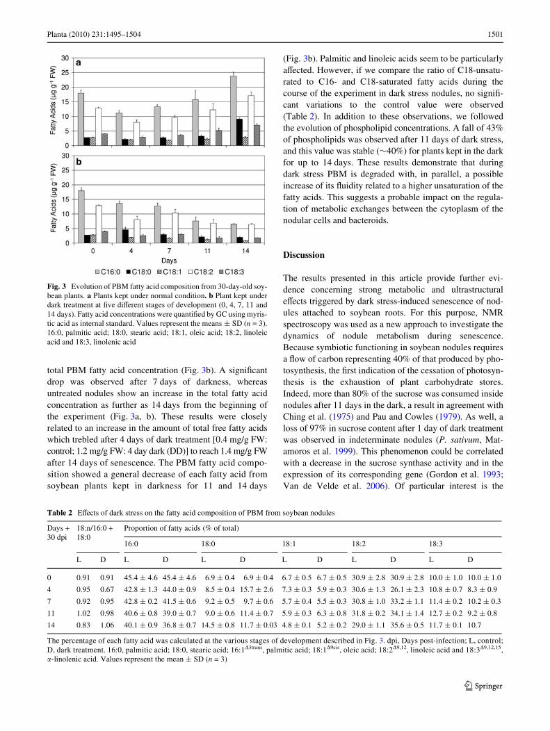
total PBM fatty acid concentration (Fig. 3b). A signiWcantdrop was observed after 7 days of darkness, whereasuntreated nodules show an increase in the total fatty acidconcentration as further as 14 days from the beginning ofthe experiment (Fig. 3a, b). These results were closelyrelated to an increase in the amount of total free fatty acidswhich trebled after 4 days of dark treatment [0.4 mg/g FW:control; 1.2 mg/g FW: 4 day dark (DD)] to reach 1.4 mg/g FWafter 14 days of senescence. The PBM fatty acid compo-sition showed a general decrease of each fatty acid fromsoybean plants kept in darkness for 11 and 14 days
(Fig. 3b). Palmitic and linoleic acids seem to be particularlyaVected. However, if we compare the ratio of C18-unsatu-rated to C16- and C18-saturated fatty acids during thecourse of the experiment in dark stress nodules, no signiW-cant variations to the control value were observed(Table 2). In addition to these observations, we followedthe evolution of phospholipid concentrations. A fall of 43%of phospholipids was observed after 11 days of dark stress,and this value was stable (»40%) for plants kept in the darkfor up to 14 days. These results demonstrate that duringdark stress PBM is degraded with, in parallel, a possibleincrease of its Xuidity related to a higher unsaturation of thefatty acids. This suggests a probable impact on the regula-tion of metabolic exchanges between the cytoplasm of thenodular cells and bacteroids.
Discussion
The results presented in this article provide further evi-dence concerning strong metabolic and ultrastructuraleVects triggered by dark stress-induced senescence of nod-ules attached to soybean roots. For this purpose, NMRspectroscopy was used as a new approach to investigate thedynamics of nodule metabolism during senescence.Because symbiotic functioning in soybean nodules requiresa Xow of carbon representing 40% of that produced by pho-tosynthesis, the Wrst indication of the cessation of photosyn-thesis is the exhaustion of plant carbohydrate stores.Indeed, more than 80% of the sucrose was consumed insidenodules after 11 days in the dark, a result in agreement withChing et al. (1975) and Pau and Cowles (1979). As well, aloss of 97% in sucrose content after 1 day of dark treatmentwas observed in indeterminate nodules (P. sativum, Mat-amoros et al. 1999). This phenomenon could be correlatedwith a decrease in the sucrose synthase activity and in theexpression of its corresponding gene (Gordon et al. 1993;Van de Velde et al. 2006). Of particular interest is the
Fig. 3 Evolution of PBM fatty acid composition from 30-day-old soy-bean plants. a Plants kept under normal condition. b Plant kept underdark treatment at Wve diVerent stages of development (0, 4, 7, 11 and14 days). Fatty acid concentrations were quantiWed by GC using myris-tic acid as internal standard. Values represent the means § SD (n = 3).16:0, palmitic acid; 18:0, stearic acid; 18:1, oleic acid; 18:2, linoleicacid and 18:3, linolenic acid
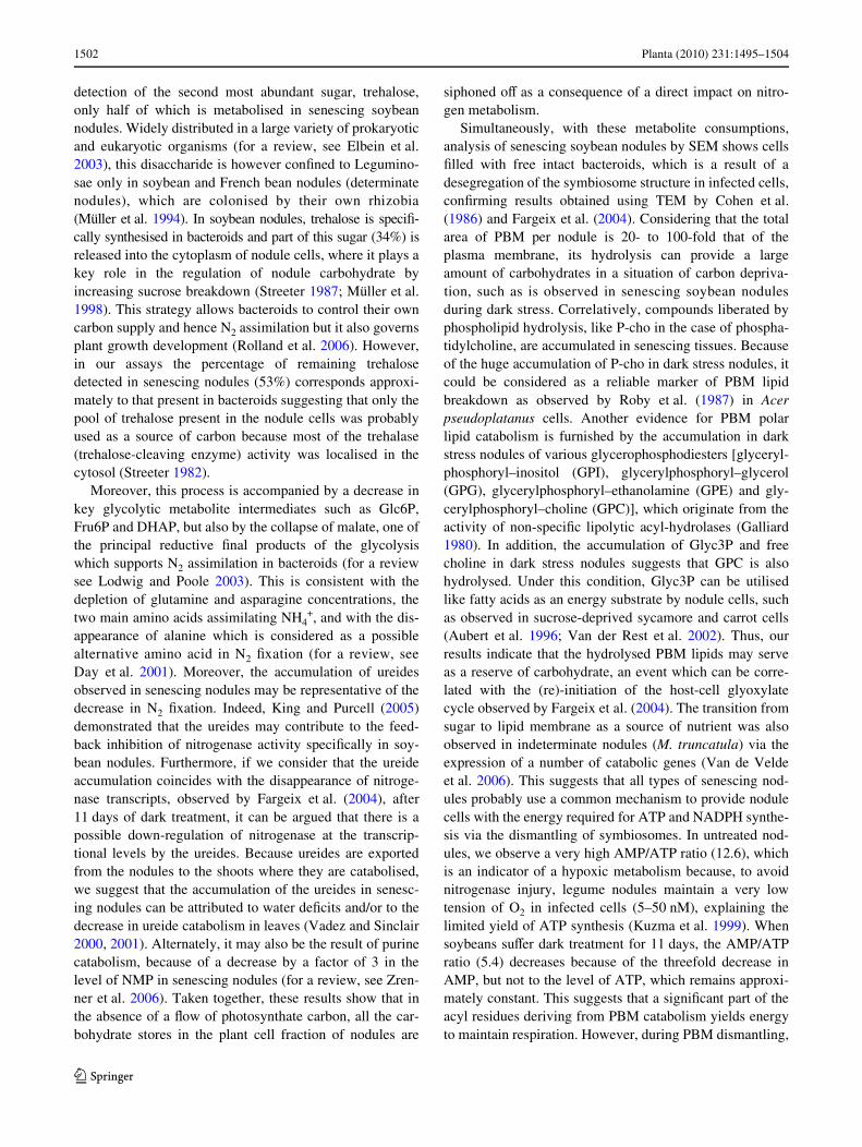
Table 2 EVects of dark stress on the fatty acid composition of PBM from soybean nodules
The percentage of each fatty acid was calculated at the various stages of development described in Fig. 3. dpi, Days post-infection; L, control;D, dark treatment. 16:0, palmitic acid; 18:0, stearic acid; 16:1�3trans, palmitic acid; 18:1�9cis, oleic acid; 18:2�9,12, linoleic acid and 18:3�9,12,15,�-linolenic acid. Values represent the mean § SD (n = 3)
Days +30 dpi
18:n/16:0 +18:0
Proportion of fatty acids (% of total)
16:0 18:0 18:1 18:2 18:3
L D L D L D L D L D L D
0 0.91 0.91 45.4 § 4.6 45.4 § 4.6 6.9 § 0.4 6.9 § 0.4 6.7 § 0.5 6.7 § 0.5 30.9 § 2.8 30.9 § 2.8 10.0 § 1.0 10.0 § 1.0
4 0.95 0.67 42.8 § 1.3 44.0 § 0.9 8.5 § 0.4 15.7 § 2.6 7.3 § 0.3 5.9 § 0.3 30.6 § 1.3 26.1 § 2.3 10.8 § 0.7 8.3 § 0.9
7 0.92 0.95 42.8 § 0.2 41.5 § 0.6 9.2 § 0.5 9.7 § 0.6 5.7 § 0.4 5.5 § 0.3 30.8 § 1.0 33.2 § 1.1 11.4 § 0.2 10.2 § 0.3
11 1.02 0.98 40.6 § 0.8 39.0 § 0.7 9.0 § 0.6 11.4 § 0.7 5.9 § 0.3 6.3 § 0.8 31.8 § 0.2 34.1 § 1.4 12.7 § 0.2 9.2 § 0.8
14 0.83 1.06 40.1 § 0.9 36.8 § 0.7 14.5 § 0.8 11.7 § 0.03 4.8 § 0.1 5.2 § 0.2 29.0 § 1.1 35.6 § 0.5 11.7 § 0.1 10.7
123
1502 Planta (2010) 231:1495–1504
detection of the second most abundant sugar, trehalose,only half of which is metabolised in senescing soybeannodules. Widely distributed in a large variety of prokaryoticand eukaryotic organisms (for a review, see Elbein et al.2003), this disaccharide is however conWned to Legumino-sae only in soybean and French bean nodules (determinatenodules), which are colonised by their own rhizobia(Müller et al. 1994). In soybean nodules, trehalose is speciW-cally synthesised in bacteroids and part of this sugar (34%) isreleased into the cytoplasm of nodule cells, where it plays akey role in the regulation of nodule carbohydrate byincreasing sucrose breakdown (Streeter 1987; Müller et al.1998). This strategy allows bacteroids to control their owncarbon supply and hence N2 assimilation but it also governsplant growth development (Rolland et al. 2006). However,in our assays the percentage of remaining trehalosedetected in senescing nodules (53%) corresponds approxi-mately to that present in bacteroids suggesting that only thepool of trehalose present in the nodule cells was probablyused as a source of carbon because most of the trehalase(trehalose-cleaving enzyme) activity was localised in thecytosol (Streeter 1982).
Moreover, this process is accompanied by a decrease inkey glycolytic metabolite intermediates such as Glc6P,Fru6P and DHAP, but also by the collapse of malate, one ofthe principal reductive Wnal products of the glycolysiswhich supports N2 assimilation in bacteroids (for a reviewsee Lodwig and Poole 2003). This is consistent with thedepletion of glutamine and asparagine concentrations, thetwo main amino acids assimilating NH4
+, and with the dis-appearance of alanine which is considered as a possiblealternative amino acid in N2 Wxation (for a review, seeDay et al. 2001). Moreover, the accumulation of ureidesobserved in senescing nodules may be representative of thedecrease in N2 Wxation. Indeed, King and Purcell (2005)demonstrated that the ureides may contribute to the feed-back inhibition of nitrogenase activity speciWcally in soy-bean nodules. Furthermore, if we consider that the ureideaccumulation coincides with the disappearance of nitroge-nase transcripts, observed by Fargeix et al. (2004), after11 days of dark treatment, it can be argued that there is apossible down-regulation of nitrogenase at the transcrip-tional levels by the ureides. Because ureides are exportedfrom the nodules to the shoots where they are catabolised,we suggest that the accumulation of the ureides in senesc-ing nodules can be attributed to water deWcits and/or to thedecrease in ureide catabolism in leaves (Vadez and Sinclair2000, 2001). Alternately, it may also be the result of purinecatabolism, because of a decrease by a factor of 3 in thelevel of NMP in senescing nodules (for a review, see Zren-ner et al. 2006). Taken together, these results show that inthe absence of a Xow of photosynthate carbon, all the car-bohydrate stores in the plant cell fraction of nodules are
siphoned oV as a consequence of a direct impact on nitro-gen metabolism.
Simultaneously, with these metabolite consumptions,analysis of senescing soybean nodules by SEM shows cellsWlled with free intact bacteroids, which is a result of adesegregation of the symbiosome structure in infected cells,conWrming results obtained using TEM by Cohen et al.(1986) and Fargeix et al. (2004). Considering that the totalarea of PBM per nodule is 20- to 100-fold that of theplasma membrane, its hydrolysis can provide a largeamount of carbohydrates in a situation of carbon depriva-tion, such as is observed in senescing soybean nodulesduring dark stress. Correlatively, compounds liberated byphospholipid hydrolysis, like P-cho in the case of phospha-tidylcholine, are accumulated in senescing tissues. Becauseof the huge accumulation of P-cho in dark stress nodules, itcould be considered as a reliable marker of PBM lipidbreakdown as observed by Roby et al. (1987) in Acerpseudoplatanus cells. Another evidence for PBM polarlipid catabolism is furnished by the accumulation in darkstress nodules of various glycerophosphodiesters [glyceryl-phosphoryl–inositol (GPI), glycerylphosphoryl–glycerol(GPG), glycerylphosphoryl–ethanolamine (GPE) and gly-cerylphosphoryl–choline (GPC)], which originate from theactivity of non-speciWc lipolytic acyl-hydrolases (Galliard1980). In addition, the accumulation of Glyc3P and freecholine in dark stress nodules suggests that GPC is alsohydrolysed. Under this condition, Glyc3P can be utilisedlike fatty acids as an energy substrate by nodule cells, suchas observed in sucrose-deprived sycamore and carrot cells(Aubert et al. 1996; Van der Rest et al. 2002). Thus, ourresults indicate that the hydrolysed PBM lipids may serveas a reserve of carbohydrate, an event which can be corre-lated with the (re)-initiation of the host-cell glyoxylatecycle observed by Fargeix et al. (2004). The transition fromsugar to lipid membrane as a source of nutrient was alsoobserved in indeterminate nodules (M. truncatula) via theexpression of a number of catabolic genes (Van de Veldeet al. 2006). This suggests that all types of senescing nod-ules probably use a common mechanism to provide nodulecells with the energy required for ATP and NADPH synthe-sis via the dismantling of symbiosomes. In untreated nod-ules, we observe a very high AMP/ATP ratio (12.6), whichis an indicator of a hypoxic metabolism because, to avoidnitrogenase injury, legume nodules maintain a very lowtension of O2 in infected cells (5–50 nM), explaining thelimited yield of ATP synthesis (Kuzma et al. 1999). Whensoybeans suVer dark treatment for 11 days, the AMP/ATPratio (5.4) decreases because of the threefold decrease inAMP, but not to the level of ATP, which remains approxi-mately constant. This suggests that a signiWcant part of theacyl residues deriving from PBM catabolism yields energyto maintain respiration. However, during PBM dismantling,
123
Planta (2010) 231:1495–1504 1503
H2O2 was probably generated by �-oxidation of fatty acids(for a review see Vauclare et al. 2003). Indeed, GABAaccumulation in senescing nodules could reXect an increasein hypoxic conditions, an acidiWcation of cytosolic pH andan increase in the oxidative stress (for a review, see Bouchéand Fromm 2004). This suggests that nodules’ cells need toincrease their anti-oxidant defence. However, we observedan accumulation of NADP and Glcn6P in senescing nod-ules, which indicates a diminution of the production ofreducing power in nodules via the pentose phosphate path-way, a situation which favours oxidative stress because theascorbate–glutathione cycle, which is the main anti-oxidantdefence of nodules, needs NADPH to work (Dalton et al.1986). The diminution in the level of the ascorbate precur-sor, mannose 6-phosphate (Man6P), in senescing nodulesmay also aVect the ascorbate biosynthetic pathway and,subsequently, the nodule anti-oxidant defence capacitiesas observed in indeterminate nodules (P. sativum) byMatamoros et al. (1999). Consequently, this constitutes arisk for PBM lipid peroxidation by reactive oxygen species(ROS; for a review, see Puppo et al. 2005). In addition, thetwo PBM unsaturated fatty acids (oleic and linoleic acid)from 11 days of darkness could enhance fatty acid peroxi-dation. This is considered by Puppo et al. (1991) as one ofthe major events involved in the modiWcation of PBMs’permeability and their eventual rupture, with a direct eVecton symbiosis.
To summarise, the present study reveals that dark-induced senescence in soybean triggers a complex processof autophagy at the level of symbiosomes, which couldphysiologically mimic programmed cell death. This pro-cess starts with the exhaustion of the reserve carbohydratesfollowed by a decrease in primary metabolism, a collapseof the nitrogen metabolism and PBM phospholipid hydro-lysis, which release substrates required to maintain a mini-mum physiological activity. The decrease of anti-oxidantcapacities in the nodules makes it easier for ROS to breakdown the PBM, suggesting that, once the autophagic pro-cess is started in senescing nodules, the process becomeautocatalytic. Because of the key properties of the PBM insymbiosis (for a review, see Udvardi and Day 1997), itshydrolysis during senescence probably cuts oV the symbi-otic relationship established between the plant and themicrosymbionts. For this reason and on the basis of theresult obtained by Werner et al. (1985), we can speculatethat bacteroids released in the host cytoplasm could berecognised as parasites by the senescing plant. Furtheranalyses are required to provide greater insight into themetabolic adaptation of the bacteroids and nodule hostcells of soybean when symbiosis is broken oV. Likewise, itwill be important to establish whether the autophagicprocess induced in the dark can be reversed in stressednodules.
Acknowledgments We are grateful to Dr. John Lomas (ITODYSUMR 7086, Université Paris-Diderot, Paris 7) for his Wne work incorrection of English text in the manuscript. We also thank JosianeBonetti for her excellent bibliographic assistance. This work wassupported by grants from the University of Lausanne (Switzerland).
References
Andreeva IN, Kozharinova GM, Izmailov SF (1998) Senescence oflegume nodules. Russ J Plant Physiol 45:101–112
Anthon GE, Emerich DW (1990) Developmental regulation of enzymesof sucrose and hexose metabolism in eVective and ineVectivesoybean nodules. Plant Physiol 92:346–351
Aubert S, Gout E, Bligny R, Marty-Mazars D, Barrieu F, AlabouvetteJ, Marty F, Douce R (1996) Ultrastructural and biochemical char-acterization of autophagy in higher plant cells subjected to carbondeprivation: control by supply of mitochondria with respiratorysubstrates. J Cell Biol 133:1251–1263
Bassarab S, Schenk SU, Werner D (1989) Fatty acid composition ofthe peribacteroid membrane and the ER in nodules of Glycinemax varies after infection by diVerent strains of the microsymbi-ont Bradyrhizobium japonicum. Bot Acta 102:196–201
Bligh EG, Dyer WJ (1959) A rapid method of total lipid extraction andpuriWcation. Can J Biochem Physiol 37:911–917
Bouché N, Fromm H (2004) GABA in plants: just a metabolite?Trends Plant Sci 9:110–115
Broughton WJ (2003) Roses by other names: taxonomy of the Rhizo-biaceae. J Bacteriol 185:2975–2979
Ching TM, Hedtke S, Russel SA, Evans HJ (1975) Energy state anddinitrogen Wxation in soybean nodules of dark-grown plants.Plant Physiol 55:796–798
Cohen HP, Sarath G, Lee K, Wagner FW (1986) Soybean root noduleultrastructure during dark-induced stress and recovery. Protoplas-ma 132:69–75
Cots J, Fargeix C, Gindro K, Widmer F (2002) Pathogenic attack andcarbon reallocation in soybean leaves (Glycine max L.): reinitia-tion of the glycoxylate cycle as a defense reaction. J Plant Physiol159:91–96
Dalton DA, Russell SA, Hanus FJ, Pascoe GA, Evans HJ (1986) Enzy-matic reactions of ascorbate and glutathione that prevent peroxidedamage in soybean root nodules. Proc Natl Acad Sci USA83:3811–3815
Day DA, Copeland L (1991) Carbon metabolism and compartmenta-tion in nitrogen-Wxing legume nodules. Plant Physiol Biochem29:185–201
Day DA, Price GD, Gresshof PM (1987) A comparison of mitochon-dria from soybean nodules, roots and cotyledons. In: Moore AL,Beechey RB (eds) Plant mitochondria: structural, functional andphysiological aspects. Plenum Press, New York, pp 207–210
Day DA, Poole PS, Tyerman SD, Rosendahl L (2001) Ammonia andamino acid transport across symbiotic membranes in nitrogen-Wxing legume nodules. Cell Mol Life Sci 58:61–71
Dorne A-J, Bligny R, Rébeillé F, Roby C, Douce R (1987) Fatty aciddisappearance and phosphorylcholine accumulation in higherplant cells after a long period of sucrose deprivation. Plant PhysiolBiochem 25:589–595
Doyle JJ, Luckow MA (2003) The rest of the iceberg Legume diver-sity and evolution in a phylogenetic context. Plant Physiol131:900–910
Elbein AD, Pan YT, Pastuszak I, Carroll D (2003) New insights on tre-halose: a multifunctional molecule. Glycobiology 13(4):17R–27R
Espinosa-Victoria D, Vance CP, Graham PH (2000) Host variation intraits associated with crown nodule senescence in soybean. CropSci 40:103–109
123
1504 Planta (2010) 231:1495–1504
Fargeix C, Gindro K, Widmer F (2004) Soybean (Glycine max L.) andbacteroid glyoxylate cycle activities during nodular senescence.J Plant Physiol 161:183–190
Galliard T (1980) Degradation of acyl lipids: hydrolytic and oxidativeenzymes. In: Stumpf PK (ed) The biochemistry of plants. A com-prehensive treatise, vol 4. Academic Press, New York, pp 85–116
Gordon AJ, Ougham HJ, James CL (1993) Changes in the levels ofgene transcripts and their corresponding proteins in nodulesof soybean plant subjected to dark-induced stress. J Exp Bot44:1453–1460
Gout E, Aubert S, Bligny R, Rébeillé F, Nonomura AR, Benson AA,Douce R (2000) Metabolism of methanol in plant cells. Carbon-13nuclear magnetic resonance studies. Plant Physiol 123:287–296
Hong ZQ, Copeland L (1990) Pentose phosphate pathway enzymesin nitrogen-Wxing leguminous root nodules. Phytochemistry29:2437–2440
Jacobi A, Katinakis P, Werner D (1994) ArtiWcially induced senes-cence of soybean root nodules aVects diVerent polypeptides andnodulins in the symbiosome membrane compared to physiologi-cal ageing. J Plant Physiol 144:533–540
King CA, Purcell LC (2005) Inhibition of N2 Wxation in soybean isassociated with elevated ureides and amino acids. Plant Physiol137:1389–1396
Kuzma MM, Winter H, Storer P, Oresnik II, Atkins CA, Layzell DB(1999) The site of oxygen limitation in soybean nodules. PlantPhysiol 119:399–408
Lee JW, Zhang Y, Weaver CD, Shomer NH, Louis CF, Roberts DM(1995) Phosphorylation of nodulin 26 on serine 262 aVects itsvoltage-sensitive channel activity in planar lipid bilayers. J BiolChem 270:27051–27057
Lodwig E, Poole P (2003) Metabolism of Rhizobium bacteroids. CritRev Plant Sci 22:37–78
Lord JM (1987) Isolation of endoplasmic reticulum: general princi-ples, enzymatic markers, and endoplasmic reticulum bound poly-somes. Methods Enzymol 148:576–584
Matamoros MA, Baird LM, Escurado PR, Dalton DA, Minchin FR,Iturbe-Ormaetxe I, Rubio MC, Moran JF, Gordon AJ, Becana M(1999) Stress-induced legume root nodule senescence physiological,biochemical, and structural alteration. Plant Physiol 121:97–111
Matamoros MA, Loscos J, Coronado MJ, Ramos J, Sato S, TestillanoPS, Tabata S, Becana M (2006) Biosynthesis of ascorbic acid inlegume root nodules. Plant Physiol 141:1068–1077
Miao GH, Verma DP (1993) Soybean nodulin-26 gene encoding achannel protein is expressed only in the infected cells of nodulesand is regulated diVerently in roots of homologous and heterolo-gous plants. Plant Cell 5:781–794
Müller J, Xie Z-P, Staehelin C, Mellor RB, Boller T, Wiemken A(1994) Trehalose and trehalase in root nodules from variouslegumes. Physiol Plant 90:86–92
Müller J, Boller T, Wiemken A (1998) Trehalose aVects sucrose syn-thase and invertase activities in soybean (Glycine max [L.] Merr.)roots. J Plant Physiol 153:255–257
Newton WE (2000) Nitrogen Wxation. In: Pedrosa FO, Hungria M,Yates MG, Newton WE (eds) From molecules to crop productiv-ity. Kluwer, Dordrecht, pp 3–8
Patriarca EJ, Tate R, Ferraioli S, Laccarino M (2004) Organogenesisof legume root nodules. Int Rev Cytol 234:201–262
Pau AS, Cowles JR (1979) EVect of induced nodule senescence onparameters related to dinitrogen Wxation, bacteroid size, andnucleic acid content. J Gen Microbiol 111:101–107
Perret X, Staehelin C, Broughton WJ (2000) Molecular basis of sym-biotic promiscuity. Microbiol Mol Biol Rev 64:180–201
PfeiVer NE, Malik NSA, Wagner FW (1983) Reversible dark-inducedsenescence of soybean root nodules. Plant Physiol 71:393–399
Prell J, Poole P (2006) Metabolic changes of rhizobia in legume nod-ules. Trends Microbiol 14:1–8
Puppo A, Herrada G, Rigaud J (1991) Lipid peroxidation in peribacter-oid membrane from French bean nodules. Plant Physiol 96:826–830
Puppo A, Groten K, Bastian F, Carzaniga R, Soussi M, MercedesLucas M, Rosaria de Felipe M, Harisson J, Vanacker H, Foyer CH(2005) Legume nodule senescence: roles for redox and hormonesignalling in the orchestration of the natural aging process.New Phytol 165:683–701
Quail PH (1979) Plant cell fractionation. Annu Rev Plant Physiol30:425–484
Roby C, Martin JB, Bligny R, Douce R (1987) Biochemical changesduring sucrose deprivation in higher plant cells. Phosphorus-31nuclear magnetic resonance studies. J Biol Chem 262:5000–5007
Rolland F, Baeana-Gonzalez E, Sheen J (2006) Sugar sensing and sig-nalling in plants: conserved and novel mechanisms. Annu RevPlant Biol 57:675–709
Schubert KR (1986) Products of biological nitrogen Wxation in higherplants; synthesis, transport and metabolism. Annu Rev PlantPhysiol 37:539–574
Streeter JG (1982) Enzymes of sucrose, maltose and alpha, alpha-tre-halose catabolism in soybean root nodules. Planta 155:112–115
Streeter JG (1987) Carbohydrate, organic acid, and amino acid compo-sition of bacteroids and cytosol from soybean nodules. PlantPhysiol 85:768–773
Tajima S, Nomura M, Kouchi H (2004) Ureide biosynthesis in legumenodules. Front Biosci 9:1374–1381
Udvardi MK, Day DA (1997) Metabolite transport across symbioticmembranes of legume nodules. Annu Rev Plant Physiol PlantMol Biol 48:493–523
Vadez V, Sinclair TR (2000) Ureide degradation pathways in intactsoybean leaves. J Exp Bot 51:1459–1465
Vadez V, Sinclair TR (2001) Leaf ureide degradation and N2 Wxationtolerance to water deWcit in soybean. J Exp Bot 52:153–159
Van de Velde W, Pérez Guerra JC, De Keyser A, De Rycke R, Rom-bauts S, Maunoury N, Mergaert P, Kondorosi E, Holsters M,Goormachtig S (2006) Aging in legume symbiosis A molecularview on nodule senescence in Medicago truncatula. Plant Physiol141:711–720
Van der Rest B, Boisson A-M, Gout E, Bligny R, Douce R (2002)Glycerophosphocholine metabolism in higher plant cells evi-dence of a new glyceryl-phosphodiester phosphodiesterase. PlantPhysiol 130:244–255
Vauclare P, Cots J, Gindro K, Widmer F (2003) The glyoxylate cycleas an essential step in carbon reallocation mechanisms. Adv PlantPhysiol 5:97–132
Weaver CD, Crombie B, Stacey G, Roberts DM (1991) Calcium-dependent phosphorylation of symbiosome membrane proteinsfrom nitrogen-Wxing soybean nodules: evidence for phosphoryla-tion of Nodulin-26. Plant Physiol 95:222–227
Werner D, Mellor RB, Hahn MG, Grisebach H (1985) Soybean rootresponse to symbiotic infection Glyceollin I accumulation in anineVective type of soybean nodules with an early loss of theperibacteroid membrane. Z Naturfosch 40:179–181
Wheeler GL, Jones MA, SmirnoV N (1998) The biosynthetic pathwayof vitamin C in higher plants. Nature 393:365–369
Zrenner R, Stitt M, Sonnewald U, Boldt R (2006) Pyrimidine andpurine biosynthesis and degradation in plants. Annu Rev PlantBiol 57:805–836
123