13815359.pdf - enlighten: theses
TRANSCRIPT
https://theses.gla.ac.uk/
Theses Digitisation:
https://www.gla.ac.uk/myglasgow/research/enlighten/theses/digitisation/
This is a digitised version of the original print thesis.
Copyright and moral rights for this work are retained by the author
A copy can be downloaded for personal non-commercial research or study,
without prior permission or charge
This work cannot be reproduced or quoted extensively from without first
obtaining permission in writing from the author
The content must not be changed in any way or sold commercially in any
format or medium without the formal permission of the author
When referring to this work, full bibliographic details including the author,
title, awarding institution and date of the thesis must be given
Enlighten: Theses
https://theses.gla.ac.uk/
Chemomechanical Removal of Dental Caries — An in vitro Study
byHak Kong Yip, B.D.S.
Thesis submitted to the University of Glasgow in partial fulfilment of the requirements of the degree of Doctor of
Philosophy, May 1992
This research was conducted in the Oral Biology Group and
Department of Conservative Dentistry,
Glasgow Dental Hospital and School, 378 Sauchiehall Street,
Glasgow G2 3JZ,UK.
i
© H. K. Yip, 1992.
ProQuest Number: 13815359
All rights reserved
INFORMATION TO ALL USERS The quality of this reproduction is dependent upon the quality of the copy submitted.
In the unlikely event that the author did not send a com p le te manuscript and there are missing pages, these will be noted. Also, if material had to be removed,
a note will indicate the deletion.
uestProQuest 13815359
Published by ProQuest LLC(2018). Copyright of the Dissertation is held by the Author.
All rights reserved.This work is protected against unauthorized copying under Title 17, United States C ode
Microform Edition © ProQuest LLC.
ProQuest LLC.789 East Eisenhower Parkway
P.O. Box 1346 Ann Arbor, Ml 48106- 1346

GLASGOW |UNIVERSITYLIBRARY
SUMMARY
The removal of dental caries is usually carried out by mechanical excavation using hand and /or rotary instruments. The carious process in human teeth, in particular dentine caries, and the development of various techniques of caries removal have been reviewed. The evolution of the
dental engine has enabled gradual improvements in the mechanical devices used to prepare cavities. No chemical means of removing caries has ever gained general accept
ance in restorative dentistry. This is partly due to the advantages to the dentist of mechanically prepared cavities and partly because of the difficulties in finding a reagent that would remove caries effectively without causing any damage to the underlying sound dentine and pulpal tissue. Although a purely chemical caries removal system has never been devised, a chemomechanical caries removal system was first introduced in the early nineteen seventies in the USA. The system was marketed as the C a r i d e x ™ Caries Removal System in the early nineteen
eighties after approval by the Food and Drug Administra
tion (USA). The active ingredient of the caries removal
agent is N-monochloro-D,L-2-aminobutyric acid (NMAB) which
is generated by mixing sodium hypochlorite and aminobutyric acid. It has been suggested that NMAB reacts with the partially degraded collagen of the carious den
tine making it more soluble thereby enabling the carious
ii
material to be more easily excavated. In this study a simulated system in which the parameters are more strictly controlled than in the C a r i d e x ™ system was constructed.
Possible improvements in the effectiveness of chemomechanical caries removal were studied by incorporating various protein de-naturing agents into the existing system. Initially a series of in vitro studies was carried out to investigate the effectiveness of various caries removal agents in extracted permanent and deciduous teeth with carious lesions and attempts were made to improve the present formulation. The results have indicated that NMAB containing urea resulted in an improvement in the effectiveness of caries removal when compared with NMAB alone. This improvement seemed to be more effective in carious deciduous than permanent teeth, the difference was, however, not statistically significant. Attempts were made to assess whether caries removal was complete by means of two caries detector dyes (0.5% basic fuchsin and 1.0% acid
red in propylene glycol) but neither reagent had adequate
specificity. In a subsequent in vitro study, a more
"standardised" group of extracted carious deciduous teeth was used and these showed a higher percentage of teeth in which complete caries removal was achieved than in the
preliminary study. The microscopic features of the dentine
remaining after caries removal were studied using light and scanning electron microscopy. The surfaces of the
cavities after chemomechanical caries removal had a very uneven appearance with many undermined areas; "dentine scales", patent and occluded dentinal tubules could also be observed. The differences in the dentinal surfaces of
cavities with complete caries removal may represent a range of differences in the interface between carious and
sound dentine. Few bacteria were found after chemomechani
cal caries removal. Backscattered electron imaging andelectron probe X-ray microanalysis of the dentine remain-
jshowed the dentine ing after chemomechanical caries removal^ was sound andnormally calcified and suitable for the application of
restorative materials. The advantages and disadvantages of the chemomechanical caries removal system using the improved reagents are discussed and future research suggested .
iv
CONTENTSPage
TITLE iSUMMARY iiCONTENTS vLIST OF FIGURES xiiiLIST OF TABLES xxACKNOWLEDGEMENTS xx i i i
DECLARATION xxvABBREVIATIONS xxviCHAPTER 1 INTRODUCTION 11.1 The Disease : Dental Caries 11.2 Early Theories of Caries Aetiology 11.3 Alternative Theories of Caries Aetiology 31.4 Current Theories of Caries Aetiology 41.5 Treatment of The Caries Process 51.6 Basic Dental Structure 6
1.6.1 Enamel 61.6.2 Dentine 9
1.7 The Carious Process 141.7.1 Enamel Caries 151.7.2 Coronal Dentine Caries 161.7.3 Microbiology of Caries 17
1.7.4 Morphological and Biochemical Aspects of 21
Cariesv
1.7.5 Smooth-surface and Fissure Caries 291.7.6 Deciduous Caries 301.7.7 Active and Arrested Caries 321.8 Pulpo-Dentinal Reactions 36
1.8.1 Tubular Sclerosis 371.8.2 Dead Tracts 38
1.8.3 Caries Crystals 391.8.4 Reactionary Dentine and Atubular 40
Calcification1.8.5 Pulpitis : Inflammation Within the Pulp 411.9 Removal of Carious Dentine 441.9.1 The Mechanical Rotary Technique 441.9.2 The Air-abrasive Technique 501.9.3 The Ultrasonic Technique 511.9.4 The Air-polishing Technique 521.9.5 The Enzyme Technique 521.9.6 The Laser Technique 53
1.9.7 The Chemomechanical Technique 55
1.9.8 The Current Status of Caries Removal 551.10 The Chemomechanical Caries Removal System 571.10.1 Historical Development 571.10.2 The C a r i d e x ™ Caries Removal System 58
1.10.3 Mode of Action 601.10.4 Toxicity and Pulpal Biocompatibility 62
1.10.5 Evaluation of Effectiveness 64
1.10.6 Advantages 67vi
1.10.7 Limitations 721.10.8 Improving the Formulation 7 5
1.11 Criteria for Caries Removal 761.11.1 Caries Detector Dyes 771.12 Aims 78
CHAPTER 2 MATERIALS AND METHODS 812.1 Introduction 81
2.2 Overview of the Plan of the Investigations 822.2.1 Specimen Teeth 832.2.2 Caries Removal Agents 842.2.3 Measurement of pH and Osmolality 892.2.4 Chemomechanical Caries Removal Apparatus 902.2.5 Experimental Design 912.2.6 Chemomechanical Caries Removal Technique 922.2.7 Recording of Results 932.3 Light Microscopy 942.3.1 Introduction 942.3.2 Specimen Preparation 96
2.4 Scanning Electron Microscopy (SEM) 992.4.1 Introduction 992.4.2 Specimen Preparation 103
2.5 Backscattered Electron Imaging (BSE) 1062.5.1 Introduction 106
2.5.2 Specimen Preparation 108
2.6 Electron Probe Microanalysis (EPMA) 110
vii
2.6.1 Introduction 1102.6.2 Quantitation 1112.6.3 Specimen Preparation 1132.7 Caries Detector Dyes 1142.7.1 Introduction 1142.7.2 In vitro Staining Studies 115
CHAPTER 3 THE CHEMOMECHANICAL REMOVAL OF DENTAL CARIES 118 IN PERMANENT AND DECIDUOUS TEETH
3.1 Introduction 1183.1.1 Improving the Formulation 1183.1.2 Considerations of Osmotic Balance 1203.1.3 Aims of the Studies 1213.2 Materials and Methods 1213.2.1 Protein De-naturing Agents 1213.2.2 Optimum Urea Concentration in NMAB 1233.2.3 Measurement of pH and Osmolality 1243.2.4 In vitro Study 1243.3 Results 1253.3.1 Protein De-naturing Agents 1253.3.2 Optimum Urea Concentration 126
3.3.3 pH and Osmolality 1273.3.4 In vitro Study 128
3.4 Discussion 1303.4.1 Selection of the Protein De-naturing Agent 1303.4.2 In vitro Study 131
vii i
3.4.3 pH and Osmolality 1353.4.4 Problems Associated with the Assessment 136
of "Complete Caries Removal"3.5 Conclusions 137
CHAPTER 4 MICROSCOPIC FEATURES OF THE CAVITY FLOORS 139AFTER CHEMOMECHANICAL CARIES REMOVAL
4.1 Introduction 139
4.2 Materials and Methods 1404.3 Results 1414.3.1 NMAB 141
4.3.2 NMAB-Urea 1424.3.3 NaOCl, Urea and Isotonic Saline 1434.4 Discussion and Conclusions 1444.5 Summary 147
CHAPTER 5 THE INTERFACE BETWEEN CARIOUS AND SOUND 148DENTINE
5.1 Introduction 148
5.2 Materials and Methods 1495.3 Results 1505.3.1 Scanning Electron Microscopy 1505.3.2 Light Microscopy 152
5.4 Discussion and Conclusions 153
5.5 Summary 160
ix
CHAPTER 6 THE USE OF TWO CARIES DETECTOR DYES - AN IN 161 VITRO STUDY
6.1 Introduction 1616.2 Materials and Methods 161
6.3 Results 1626.3.1 0.5% Basic Fuchsin 1626.3.2 1.0% Acid Red 1636.4 Discussion and Conclusions 164
6.5 Summary 168
CHAPTER 7 CHEMOMECHANICAL REMOVAL OF DENTAL CARIES IN 169 DECIDUOUS TEETH
7.1 Introduction 1697.2 Materials and Methods 1707.2.1 In vitro Study 170
7.3 Results 1717.3.1 In vitro Study 1 7 1
7.3.2 Light Microscopy 1727.3.3 Scanning Electron Microscopy 1 7 3
7.4 Discussion and Conclusions 1 7 3
7.5 Summary 1 7 7
CHAPTER 8 MINERAL CONTENT OF THE DENTINE REMAINING 178AFTER CHEMOMECHANICAL CARIES REMOVAL
8.1 Introduction 1 7 8
8.2 Materials and Methods 180x
8.3 Results8.5 Discussion and Conclusions
8.6 Summary
183187192
CHAPTER 9 CONCLUDING DISCUSSION 1949.1 Introduction 1949.2 In vitro Study 1949.2.1 Comparison of Permanent and Deciduous Teeth 1989.2.2 Mode of Action 1999.3 Dentine Remaining after Caries Removal 2009.3.1 Surface Morphology and Topography 2009.3.2 Types of Dentine 2039.3.3 The Fate of Organisms Remaining in Dentine 2049.3.4 Decalcification of Dentine 2079.4 Application of a Chemomechanical Caries Removal 209
System9.5 Future Research 211
APPENDIX 1 MATERIALS 214
APPENDIX 2 CRITICAL POINT DRYING 219
APPENDIX 3 PARAFFIN WAX PROCESSING 221
APPENDIX 4 HAEMATOXYLIN AND EOSIN STAINING 223PROCEDURE
xi
APPENDIX 5 MODIFIED GRAM-WEIGERT STAINING 227TECHNIQUE
APPENDIX 6 STANDARD PROCEDURE FOR PREPARING 231
EPOXY RESIN EMBEDDING MEDIA
REFERENCES 233
Publications 252
xii
LIST OF FIGURES
Fig. 1.1 The circles diagrammatically
represent the parameters involved in the
carious process.Fig. 1.2 Diagrammatic illustration of thehistological appearance of various zones of a coronal dentinal lesion after the cavitation of enamel has occurred and micro-organisms have invaded the dentine.Fig. 1.3 Scheme depicting possible routes of collagen fibre degradation during dentinal caries.Fig. 1.4 Diagrammatic illustration showing left to right, normal dentine, tubular sclerosis dead tracts and reparative dentine.
Fig. 1.5 The C a r i d e x ™ Caries Removal System.Fig. 1.6 Interchangeable Applicator Tips of
the C a r i d e x ™ Caries Removal System.Fig. 2.1 Diagrammatic Representation of the
Simulated Set-up of a Chemomechanical Caries Removal System.
Fig. 2.2 Diagrammatic Representation of the
"Applicator Tip" of a Chemomechanical Caries Removal System.
xiii
FollowingPage
5
24
27
36
59
59
90
90
Fig. 2.3 The various effects of electron -specimen interaction.
Fig. 2.4 Schematic diagram showing a pear-shaped excited volume in a bulk specimen exposed to the primary electron beam.Fig. 3.1 Changes in pH of NMAB and NMAB-Urea
with Time.Fig. 4.1 Scanning electron micrograph of the
dentinal surface of a carious lesion in a permanent molar after treatment with NMAB.Fig. 4.2 Scanning electron micrograph of the dentinal surface of a carious lesion in a deciduous molar after treatment with NMAB.Fig. 4.3 Scanning electron micrograph of the dentinal surface of a carious lesion in a permanent molar conventionally prepared cavity.Fig. 4.4 Scanning electron micrograph of the dentinal surface in a fractured permanent molar. Fig. 4.5a Scanning electron micrograph of the
dentinal surface of a carious lesion in a permanent molar after treatment with NMAB-Urea.Fig. 4.5b Scanning electron micrograph of the
dentinal surface of a carious lesion in a permanent
molar after treatment with NMAB-Urea.Fig. 4.6 Scanning electron micrograph of the
dentinal surface of a carious lesion in a deciduousxiv
102
102
127
141
141
142
1 42
142
142
1 42
molar after treatment with NMAB-Urea.Fig. 4.7 Scanning electron micrograph of the
dentinal surface of a carious lesion in a permanent molar after treatment with NaOCl.Fig. 4.8 Scanning electron micrograph of the
dentinal surface of a carious lesion in a deciduous molar after treatment with NaOCl.Fig. 4.9 Scanning electron micrograph of the dentinal surface of a carious lesion in a permanent molar after treatment with urea.
Fig. 4.10 Scanning electron micrograph of the dentinal surface of a carious lesion in a deciduous molar after treatment with urea.Fig. 4.11 Scanning electron micrograph of the dentinal surface of a carious lesion in a permanent molar after treatment with isotonic saline.Fig. 4.12 Scanning electron micrograph of the dentinal surface of a carious lesion in a deciduous
molar after treatment with isotonic saline.Fig. 5.1 Scanning electron micrograph of the
dentinal surface of a permanent molar after treatment with NMAB-Urea but without washing prior to preparation for SEM.
Fig. 5.2 Scanning electron micrograph of the
dentinal surface of a deciduous molar after treatment with NMAB-Urea.
xv
143
143
143
143
143
143
150
Fig. 5.3 Scanning electron micrograph of the
dentinal surface of a permanent molar after treatment with NMAB.Fig. 5.4 Scanning electron micrograph of the
dentinal surface of a permanent molar after treatment with NMAB.Fig. 5.5 Scanning electron micrograph of the
dentinal surface of a permanent molar after treatment with NMAB.Fig. 5.6 Scanning electron micrograph of the
dentinal surface of a permanent molar after treatment with NMAB-Urea.Fig. 5.7 Scanning electron micrograph of thedentinal surface of a permanent molar after treatment with NMAB-Urea.Fig. 5.8 Scanning electron micrograph of thedentinal surface of a permanent molar after
treatment with NMAB-Urea.Fig. 5.9 Scanning electron micrograph of thedentinal surface of a permanent molar after treatment with NMAB-Urea.Fig. 5.10 Scanning electron micrograph of the
dentinal surface of a permanent molar after treatment with NMAB-Urea.
Fig. 5.11 Scanning electron micrograph of the
dentinal surface of a permanent molar after
xvi
151
151
151
151
151
152
1 52
152
152
treatment with NMAB-Urea.Fig. 5.12 Decalcified section through the middle of 152
the cavity floor of a permanent molar after treatment with NMAB-Urea.Fig. 5.13 Decalcified section through the middle of 152 the cavity floor of a deciduous molar after treatment
with NMAB-Urea.Fig. 5.14 Decalcified section through the middle of 153 the cavity floor of a permanent molar after treatment with NMAB-Urea.Fig. 5.15 Decalcified section through the middle of 153 the cavity floor of a deciduous molar after treatment with NMAB-Urea.Fig. 5.16 Decalcified section through the middle of 153 the cavity floor of a carious deciduous molar.Fig. 6.1 Cavity of a permanent molar after mechanical 162 excavation of carious material in conjunction with the use of 0.5% basic fuchsin (Group A).Fig. 6.2 Pulpal floor of a control cavity in a 162permanent molar stained with 0.5% basic fuchsin
(Group B ) .
Fig. 6.3 Dentino-enamel junction of a control cavity 162of permanent molar stained with 0.5% basic fuchsin (Group E ).
Fig. 6.4 Carious lesion in a permanent molar stained 162with 0.5% basic fuchsin (half tooth) (Group E).
xvii
Fig. 6.5 Longitudinal ground sections of a carious 162lesion of a deciduous molar stained with 0.5% basic fuchsin (left) and 1.0% acid red (right) (Group C ) .
Fig. 6.6 Transverse ground sections of carious lesion 162 of a deciduous incisor stained with 0.5% basic fuchsin (left) and 1.0% acid red (right) (Group D ) .Fig. 6.7 Carious cavity of a permanent molar 163mechanically excavated with the aid of 1.0% acid red (Group A).Fig. 6.8 Carious lesion of a permanent molar stained 163with 1.0% acid red (half tooth) (Group B).Fig. 7.1 Scanning electron micrograph of the dentinal 173 surface of a deciduous molar after treatment with NMAB. Fig. 7.2 Scanning electron micrograph of the dentinal 173 surface of a deciduous molar after treatment with NMAB-Urea.Fig. 7.3 Scanning electron micrograph of the dentinal 173 surface of a deciduous molar after treatment with NMAB.
Fig. 7.4 Scanning electron micrograph of the dentinal 173
surface of a deciduous molar after treatment with NMAB-Urea.
Fig. 8.1 SEM photomicrograph and line scan across the 183dentinal surface of a cavity of a carious deciduousmolar after treatment with NMAB.
Fig. 8.2 BSE photomicrograph of the cavity surface 183of a carious deciduous molar after treatment with
xviii
NMAB.Fig. 8.3 BSE photomicrograph of a carious deciduous molar after treatment with NMAB-Urea.
Fig. 8.4 BSE photomicrographs of a carious deciduous molar after treatment with NMAB-Urea.
Fig. 8.5 A dot map showing the overall distribution of mineral density of a carious deciduous molar after treatment with NMAB-Urea.
Fig. 8.6 The distribution of calcium and phosphorusin a carious deciduous molar after treatment withNMAB-Urea.Fig. 8.6 BSE photograph of a carious deciduous molar.Fig. 8.7 BSE photomicrograph of a carious permanentmo l a r .Fig. 8.8 A dot map showing the distribution of mineral density of a carious permanent molar.Fig. 8.9 The distribution of calcium and phosphorus in a carious permanent molar.
185
185
185
185
185186
186
186
xix
LIST OF TABLESFollowing
Page
Table 1.1 The Three Layers of Carious Dentine. 29Table 1.2 Criteria for Identification of Active 35and Arrested Carious Lesions in Dentine.Table 1.3 Summary of Some Clinical Trials of the 65
Chemomechanical Caries Removal System.Table 2.1 Summary of Scoring for Caries Removal. 93
Table 3.1 Classes of Carious Cavities of 126Permanent Teeth Treated Using Various Concentrations of NMAB-Urea.Table 3.2 Types of Permanent Teeth Treated Using 126Various Concentrations of NMAB-Urea.Table 3.3 Average Time taken and Volume of the 127solution used to Achieve "Complete Caries Removal"(CCR) in Permanent Carious Lesions Using Various
Concentrations of NMAB-Urea.Table 3.4 Classes of Carious Cavities Treated. 128
Table 3.5 Types of Teeth Treated. 128Table 3.6 Average Time taken and Volume of the 128
solution used to Achieve "Complete Caries Removal"
(CCR) in Permanent and Deciduous Carious Lesions.
Table 3.7 Statistical Analysis of the "Complete 128Caries Removal" (CCR) of the Five Caries Removal Agents Using Chi-squared Test.
xx
Table 3.8 Statistical Analysis of the "Complete Caries Removal" (CCR) of Permanent (P) and
Deciduous (D) Teeth Using Chi-squared Test.Table 3.9 Statistical Analysis of the Time taken and Volume of the solution used to achieve "Complete Caries Removal" of Permanent Teeth Using Student's
T-test.Table 3.10 Statistical Analysis of the Time taken and Volume of the solution used to achieve "Complete
Caries Removal" of Deciduous Teeth Using Student's T-test.Table 3.11 Statistical Analysis of the Time taken and Volume of the solution used to achieve "Complete Caries Removal" between Permanent and Deciduous Teeth Using Student's T-test.Table 6.1 Groups of teeth used in caries detector dye studies.Table 7.1 Classes of Deciduous Carious Cavities
Treated.Table 7.2 Types of Deciduous Teeth Treated.Table 7.3 Average Time taken and Volume of the solution used to Achieve "Complete Caries Removal"
(CCR) in Deciduous Carious Lesions.
Table 7.4 Statistical Analysis of the "Completel>Caries Removal of Deciduous Teeth Using Chi-squared
Te s t .xxi
1 29
130
130
130
161
1 71
171 1 71
172
Table 7.5 Statistical Analysis of the Time taken and Volume of the solution used to Achieve "Complete Caries Removal" of Deciduous Teeth Using
Student's T-test.Table 7.6 Number of Cavities with "Complete Caries
Removal" with no Bacteria Detected in Histological Sections.Table 7.7 The Presence of Reactionary Dentine in the Carious Lesions with "Complete Caries Removal". Table 8.1 Calcium and Phosphorus levels (percentage weight of total elements present and Ca : P ratios) on the dentinal surface of cavities after caries removal by various chemical agents.Table 8.2 Calcium and Phosphorus levels (percentage weight of total elements present), in deciduous carious dentine of control lesions at varying depths, after caries removal by various caries removal agents.
Table 8.3 Levels of Calcium, Phosphorus and other
elements (percentage weight of total elements present), in carious dentine of a control carious lesion in a permanent tooth at varying depths.
172
172
1 72
183
1 84
186
xxii
ACKNOWLEDGEMENTS
I should like to thank Dr. J.A. Beeley and Mr. A.G. Stevenson for their advice and supervision. I am indebted to Professor D.A.M. Geddes (Head of Oral Biology Group) for guidance and support when it is most needed.
I also wish to express my thanks to the following :
Dr H. Elder and Dr. I Montgomery (Institute of Physiology) for advice and provision of scanning electron microscope.
Sir Alwyn Williams, Dr. S. Hazeldine and Mr. P. Ainsworth (Department of Geology and Applied Geology) for advice and use of scanning electron microscope facilities for electron probe microanalysis.
Professor R.C. Paterson, Mr. A. McLundie and Dr. A. Watts (Department of Conservative Dentstry) for helpful discussion throughout the research project.
Professor D. McGowan and Dr. D. Stenhouse for allow
ing me to collect specimen teeth in the Department of Oral Surgery.
Professor D.G. MacDonald and Mr. W. Marshall
(Department of Oral Pathology and Oral Medicine) for provision of an osmometer.
Mr. D. Sweeney (formly Oral Biology Group) for help and invaluable criticism of the construction of the
simulated set-up for the chemomechanical caries removal
xxiii
system and Mr. J. Brown (Department of Prosthodontics) for assistance in making the "applicator tips".
Mr. J. Gillespie and Mr. R. Foye (Oral Biology Group) and their team of technical staff for general advice.
Mr. J.B. Davis, Mrs. K. Shepherd, Mrs. G. Drake and
Mr. D. Woodward (Department of Dental Illustration) for photographic and computer graphic assistance.
Mr. I. Ramsden (Medical Illustration) for artistic
assistance.Mrs. A. Shaw (Department of Oral Pathology and Oral
Medicine) for assistance with statistical analysis.Mrs. I. Roger and Mrs. E. Bulloch for general assis
tance .All those who have kindly collected the specimen
teeth used in my study.The financial assistance of a grant from the Scottish
Home and Health Department (K/MRS/S0/C1380) is gratefully acknowledged.
The list is by no means exhaustive and I would extend my gratitude to those who have helped me in the past.
I would like to thank my parents and the rest of the family for their support, understanding and encouragement over the past years.
xxiv
DECLARATION
This thesis embodies the results of my own special work. I declare that it has been composed by myself and it
does not include work forming part of a thesis presented for a degree in this or another University.
H.K. Yip
XXV
ABBREVIATIONS
USA United States of AmericaADA American Dental Association& andn/a not applicableBSE Backscattered electron imaging
°C degree Celsiuscm centimetrech. ChapterCMCR Chemomechanical Caries RemovalCMCRS Chemomechanical Caries Removal
SystemCCR Complete caries removalconc. concentrationDEJ Dentino-enamel junctionEPMA Electron probe X-ray
microanalysisEDTA ethylenediaminetetra-acetic acidF FemaleFig. FigureFDA Food and Drug Administration
(USA)
g gram
H & E Haematoxylin and Eosin
h hourkV kilovoltLA Local anaestheticLS Longitudinal section
M Malepm micrometre
xxvi
mm3m m 3/min.MPml
mmHg
mosmolminNMABNMG-ve
PH
n
P-pp.ppm%wt. %
+vepsirpm
secSEM
SE
SDSsp.
cubic millimetrecubic millimetre per minuteMelting point
millilitre
millimetre mercurymilliosmol/kg
minuteN-monochloro-DL-2-aminobutyrateN-monochloroglycinenegativenegative decimal log of molar hydrogen ion concentrationnot statednumberpagepagesparts per million percentpercentage weight of total element presentplus or minus
positivepounds per square inch
revolutions per minute second
Scanning electron microscope Secondary electron imaging
Sodium dodecyl sulphate species
xxvii
spp. species (plural)SD Standard deviation
™ Trade markTS Transverse sectionUK United Kingdom
v o l . volumev/v volume per volumewt. weightw/v weight per volume+ yes
xxviii
CHAPTER 1
INTRODUCTION
1.1 The Disease : Dental CariesDental caries is a complex, dynamic and continous
biological process with an imprecise beginning (Johnson, 1991). It takes place within a microbial deposit covering
a tooth surface. At any given site, over time the action of micro-organisms on fermentable carbohydrate results in a disturbance of the equilibrium between the hard tissues (enamel, dentine, cementum) and the fluid immediately surrounding them. This leads to a net loss of mineral and eventually a localised destruction of the mineralised tissues of the tooth (Kidd and Joyston-Bechal, 1987).
1.2 Early Theories of Caries AetiologyThe agent first thought to be responsible for carious
lesion formation was the tooth worm. This idea appears to have been universal at one time, and references to it have
been discovered on clay tablets dating from about 5,000 BC
excavated from an ancient city in the Mesopotamian area
and from Chinese characters on oracle bones dating back to
the Shang dynasty around 1,000 BC (Newbrun, 1983). Even today, this theory is still held in some parts of the
1
world.From the end of the 18th century until the middle of
the 19th century, the vital theory of tooth decay was dominant. Here it was postulated that caries originated from within the tooth itself, analogous to bone gangrene (Nikiforuk, 1985). Other theories put forward at this time included the chemical theory of Parmly in 1819, and the parasitic or septic theory of Erdl (1843). The chemical theory proposed that an unidentified chemical agent was
responsible for the caries, and that the process began on the surface of enamel. Support was given to this concept by Robertson in 1835, who proposed that caries was caused by acid formed by the fermentation of food particles around the teeth. The parasitic theory was based on the fact that micro-organisms had been detected by van Leeuwenhoek (1632 - 1723) from material taken from carious cavities, and it was therefore proposed that these bacteria could cause decomposition of the tooth tissues.
However no explanation was given as to how these organisms could destroy the tooth. The chemical-parasitic theory, a combination of the above two theories, was proposed by W.D. Miller (1890) in "The Micro-Organisms of the Human
Mouth". His theory was based both on his own experimental
work and on previous communications from other workers. He
identified carbohydrate as the bacterial substrate, and
2
noted that the decalcification of enamel produced by bacterial acids was the major factor resulting in destruction
of the tissue. He failed, however, to identify plaque as the source of bacteria, and assumed that the acids were
produced by the fermentation of impacted foodstuffs by salivary bacteria. G.V. Black (1898) considered that the acid attack was produced by bacteria in situ on the teeth. This was supported by Williams (1898), who observed dental plaque on the surface of enamel and considered that this was a means of localising acids produced by bacteria in
contact with the tooth, as well as partially preventing the dilution and neutralisation of the acid by saliva.
1.3 Alternative Theories of Caries AetiologyThe proteolysis theory of Gottlieb (1947) suggests
that the organic element of the enamel is first attacked by proteolytic bacteria, and that the inorganic component is then subsequently lost either by acid dissolution (Frisbie and Nuckolls, 1947) or by the mechanical loss of physically unbounded prisms (Pincus, 1949). A number of
criticisms have been levelled against this theory, in par
ticular the fact that the organic component comprises such a small fraction of the enamel (Jenkins, 1978).
The proteolytic-chelation theory (Schatz & Martin,
1962) suggested that products of proteolysis of tooth sub
3
stance, and possibly also of the acquired pellicle and foods, may act as chelating agents, thereby releasing mineral ions from enamel. Whilst the amount of chelating agent released by proteolytic degradation of the organic
phase of enamel is likely to be negligible (Jenkins,1978), calcium chelation may indeed occur; some of the histological features of enamel caries can be simulated in vitro by using chelating agents (Mortimer and Tranter, 1971) and many natural chelators (e.g. lactate and some amino acids) are present in plaque (Morch et a l ., 1971).
An intrinsic concept of caries aetiology has been proposed by Jackson and co-workers (1973) who suggested that specific regions of odontoblasts within the pulp of atooth are damaged by an auto-immune process and concludedthat caries should be regarded as a degenerative disease. This theory is based on epidemiological evidence and has been criticised by Edgar (1974) and Sofaer (1982).
1 .4 Current Theories on Caries Aetiology
Today it is universally accepted that caries is a multifactorial process, with the development of the lesion being due to the interaction of three primary factors, the
host, the microflora and the diet. For caries to occur, favourable conditions within each of these groups must ex
ist concomitantly for a sufficient length of time, i.e. a
4
susceptible host, a cariogenic flora and a suitable substrate (Fig. 1.1).
Dental caries may be classified as primary enamel
caries, dentine caries, root surface caries and recurrent caries associated with existing restorations. Dentine caries includes both coronal and root caries. These can be subdivided into either active or arrested caries.
1.5 Treatment of The Caries ProcessThe treatment of a disease process e.g. dental
caries, can be subdivided into the following categories :a. Primary Prevention
Procedures carried out to prevent a disease before it occurs .b. Secondary PreventionThe early detection of the disease, halting its progress by simple repair or remedial measures. Full recovery to
the "normal state" may be possible and recurrence
prevented.c. Tertiary PreventionThe treatment of a well-established disease in order to minimise or eliminate the gross destruction which has al
ready occurred. At this stage preventive procedures will
help to prevent further episodes of the disease.
The restoration of carious lesions involving dentine
5
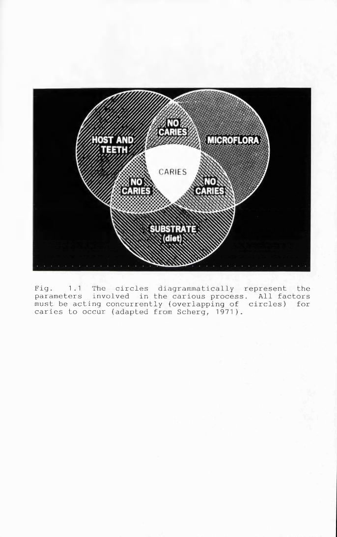
Fig. 1.1 The circles diagrammatically represent the parameters involved in the carious process. All factors must be acting concurrently (overlapping of circles) for caries to occur (adapted from Scherg, 1971).
is a tertiary preventive measure in dental treatment. The purposes of treating carious dentine are to :a. arrest the progress of the carious lesion,b. promote healing of the remaining dentine and pulp, andc. restore the tooth to a "normal" structure.
The work in this thesis is concerned with the investigation into an alternative method of tertiary prevention of coronal carious lesions using a chemomechanical caries removal system (CMCRS).
1.6 Basic Dental Structure
1.6.1 EnamelEnamel is the most highly mineralised tissue known,
consisting of 96% mineral and 4% organic material and water. The organic content of deciduous enamel is higher than that of permanent enamel. The inorganic content of enamel consists of a crystalline calcium phosphate known
as hydroxyapatite Caio(P04)e(0H)2. However the crystals lack stoichiometry due to deficiencies in the three primary constituents (i.e. Ca2- , PCU3- and OH- ), and their replacement in the lattice by COa2-, HPO-*2- and trace ele
ments. Some of these ions, particularly carbonate, are relatively easily released from enamel during demineralisation, and positions in the lattice where COa2-
6
ions are present are believed to be particularly vulnerable to the effects of acids (Featherstone et a l ., 1979). Other ions, for example fluoride, may be included in the apatite proper and are only released when the crystal dissolves (Nikiforuk, 1985). The organic materials are largely proteinaceous and contain some polysaccharides
(ten Cate, 1989 ) .Clinically, normal sound enamel appears hard and
shiny. It consists of hydroxyapatite crystals packed so tightly that the enamel has a glass-like appearance. The yellow-white colour of teeth is therefore a result of the
dentine "showing through" the overlying enamel layer. The crystals in the enamel are arranged in an orderly fashion forming rods and interrod enamel. The packing of crystals is slightly looser in the rod periphery compared with the rod and interrod enamel. The intercrystalline spaces are filled with water and organic material. These also form a fine network of potential diffusion pathways which are referred to as micropores in enamel.
If mineral is removed from the enamel by carious dis
solution, the individual crystals diminish in size. This
results in an enlargement of the intercrystalline spaces which can be observed as an increase in tissue porosity. For this reason quantification of changes in tissue porosity can be used as an indicator of loss of mineral
7
from the tissue. If the total mineral surface formed by the mass of tightly packed crystals is considered, it can be understood that an extremely modest loss of mineral from each of the crystals involved results in a propor
tionately much more pronounced increase in the spaces between the crystals. For this reason changes in enamel porosity are a very sensitive indicator of even a very slight loss of mineral in the enamel (Thylstrup & Fejerskov, 1986).
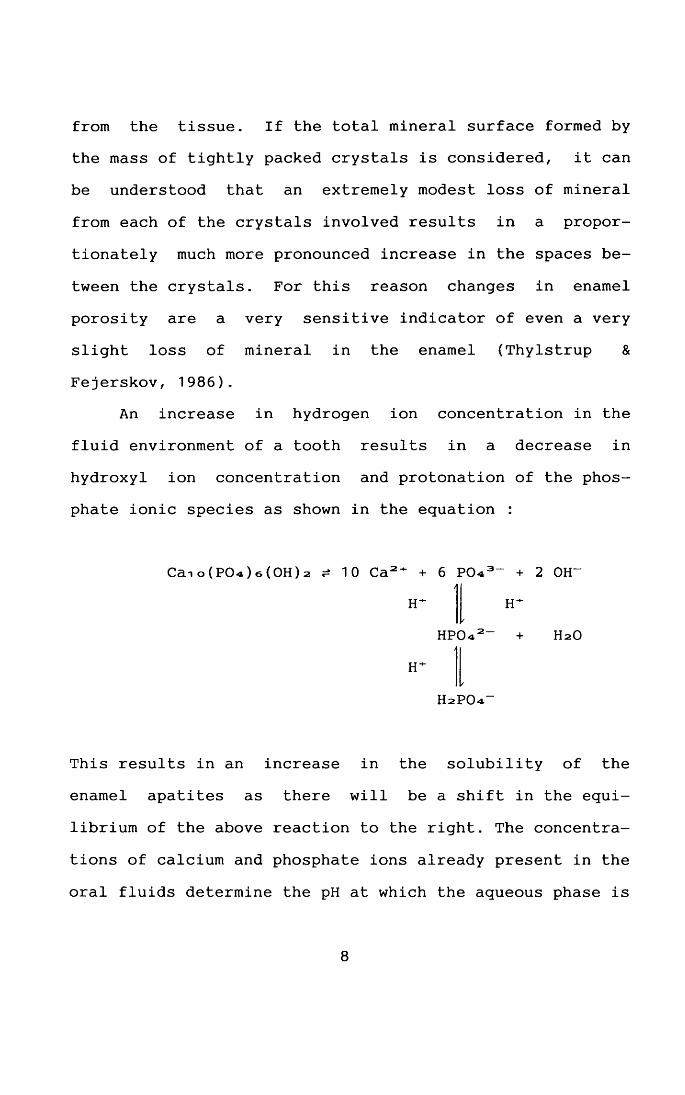
An increase in hydrogen ion concentration in the fluid environment of a tooth results in a decrease in hydroxyl ion concentration and protonation of the phosphate ionic species as shown in the equation :
Cai o (P04 )6(0H>2 ^ 10 Ca2+ + 6 PCU3- + 2 0H~/
H"" H*HPCU2- + H 2 O
H2PO4-
This results in an increase in the solubility of the enamel apatites as there will be a shift in the equilibrium of the above reaction to the right. The concentra
tions of calcium and phosphate ions already present in the oral fluids determine the pH at which the aqueous phase is
8
saturated with respect to enamel apatites. The pH at which saliva is exactly saturated with respect to apatite is often referred to the "critical pH", and will depend on the
concentrations of calcium and phosphate in the saliva of the individual. Clinical assessment shows that the critical pH varies between pH 5.2 and 5.5.
For the first few years after the eruption of a
tooth, secondary maturation of enamel takes place. This may be considered to be the result of the ongoing de- and remineralisation that takes place during the establishment of the tooth in the oral cavity. During this phase, mineral is deposited from the oral fluids into the fine fluid-filled pores in the enamel, and there is a release of the more readily soluble mineral components and uptake of fluoride (Larsen & Bruun, 1986).
1.6.2 DentineDentine is the hard tissue portion of the dentino-
pulpal complex and forms the bulk of the tooth. Mature dentine consists chemically (by weight) of approximately 70% inorganic material, 20% organic material, and 10%
water (adsorbed onto the surface of the mineral or in the
interstices between crystals); these constitute 45%, 33%,
and 22% respectively by volume. The inorganic component consists mainly of hydroxyapatite, and the organic phase
9
is type I collagen with fractional inclusions ofglycosaminoglycans, proteoglycans, phosphoproteins and glycoproteins, together with some plasma proteins. About
56% of the mineral phase is within the collagen matrix.
The inorganic phase makes dentine slightly harder than bone and softer than enamel. Physically, dentine has an elastic quality which is important for the properfunctioning of the tooth, because it provides flexibility, thus preventing fracture of the overlying brittle enamel.
Almost 90% of the organic matrix in dentine consists of collagen, whereas the reminder is non-collagenousprotein and proteoglycan. The major component of the non- collagenous proteins is a group of proteins with different degree of phosphorylation known as phosphophorins. These highly phosphorylated phosphoprotein molecules are unique to dentine and are probably involved in the regulation of mineralisation when the dentine is formed (Linde, 1984). Another group of specific non-collagenous proteins are the
a-carboxyglutamate-containing proteins of the osteocalcin
type (Gla-proteins). Proteoglycans constitute a third group of non-collagenous components in dentine. These are macromolecules with a number of carbohydrate side chains covalently bound to a central protein core. Other non-
collagenous proteins are in general acidic glycoproteins.
In addition, there are some lipid-containing components
10
which are constituents of dentine, but these have not been investigated in great detail. Dentine and predentine are devoid of fibronectin and collagen type III, both of which occur in most, if not all unmineralised connective
tissues. (Klont & ten Cate, 1987).Dentine is characterised by the presence of a multi
tude of closely packed dentinal tubules that traverse its entire thickness and contain the cytoplasmic extensions of the odontoblasts which once formed the dentine and now maintain it. The cell bodies of the odontoblasts are aligned along the inner aspect of the dentine, where they also form the peripheral boundary of the dental pulp.
The tubules are not straight, but have a gentle S-shaped curve, particularly obvious in the cervical region. The diameter of a tubule near the outer surface is about 1.2 pm, and near the pulp it is about 2.5 pm. Because of the difference in surface area between the outer and inner surfaces, the tubules are more widely separated
at the outer surface (on average, 20,000 /mm2 ) than at the pulpal surface (on average, 45,000 /mm2 ). (Garberoglio & Brannstrom, 1976). The length of the odontoblast process in the mature tooth is uncertain, but it extends through
at least the inner third of the dentine thickness (Thomas,1979). The mature cell itself is about 40 pm. If the odontoblast process extended through the thickness of the den-
11
tine it would be about 2 mm long (2000 p m ) . The remainder
of the lumen of the tubule is occupied by the tissue fluid, at a hydrostatic pressure of about 10 mmHg. Since the pulpal pressure is about 30 mmHg, there is a pressure gradient outwards. A small proportion of tubules also contain a nerve fibre. These extend only a short distance
into the tubules (0.1 - 0.4 mm). They are probablymechanoreceptors which cause sharp pain with slight deformation. There do not appear to be sufficient numbers of nerve fibres to account for the sensitivity of dentine. The dentine surrounding the tubules is intertubular den
tine. Its main components, like those of all hard connective tissue, are collagen fibres and calcium hydroxyapatite crystallites. In intertubular dentine, the collagen fibres are quite fine, about 0.05 pm in diameter, and they weave between the tubules, at approximately right angles to them. On their surface, and also within the fibres, lie the hydroxyapatite crystallites. Another type of dentine, peritubular dentine, lies within the tubules, narrowing them. It is much harder than the intertubular
dentine, and consists almost entirely of hydroxyapatite.
Peritubular dentine increases in thickness throughout the
life of the tooth, progressively narrowing the tubule and reducing the permeability of the dentine. It may eventually occlude the tubules in a particular area. This
12
gives the area involved a glassy appearance. This is
called sclerotic or translucent dentine. It is most common in the apical third of the root. Another example of continued cellular activity is the formation of secondary dentine. All the dentine formed prior to completion of the root is termed primary dentine. Further dentine laid down on the pulpal surface is called secondary (regular) den
tine. It is formed much more slowly than primary dentine, but has a similar number of tubules which are similarly regularly arranged, and is therefore of similar per
meability. It gradually encroaches on the pulp. The pulp horns become reduced in height, and much less vulnerable. Reactionary (irregular secondary, tertiary) dentine is formed in response to increased stimulation (e.g. in the course of a carious attack). It is an excellent defence for the pulp because it blocks the pulpal openings of the dentinal tubules. It contains very few tubules (Scott & Weber, 1977) and they are irregularly arranged. It is
found in areas where the original odontoblasts have been killed and new cells have differentiated.
Dentine and pulp are embryologically, histologically, and functionally the same tissue and should therefore be
considered together (ten Cate, 1989). The dental pulp is
the soft connective tissue that occupies the central por
tion of the tooth and which contains the vascular, nerve
13

and lymphatic supply to the tooth. Next to the odon
toblasts is a zone in which there are few cells. This is termed the cell-free zone (of Weil). It is most distinct in the crown. There is an important nerve plexus here -
the plexus of Raschkow. Further in is a layer with more
cells than elsewhere. This is the cell-rich zone. The
majority of the cells are fibroblasts. Also in this zone is a capillary plexus. These zones only become established after eruption.
1.7 The Carious ProcessCaries occurs as a result of the prolonged exposure
of the tooth surface to the end-products of the metabolism of plaque micro-organisms which are found on most enamel surfaces (Loesche, 1986). The sites which commonly develop lesions are therefore those which favour plaque retention.
During eruption, when the teeth are not yet in occlusion, plaque can readily accumulate on their surfaces.
This results in frequent episodes of de- and remineralisation at a subclinical level. When the teeth reach occlusion, most of these active lesions tend to become inactive as the microbial deposits on these surfaces are regularly disturbed by the shearing forces produced by chewing and by the action of saliva (Thylstrup & Fejerskov, 1986). The
exceptions to this are the pit and fissure regions which
14
remain more sheltered from these protective influences. When interproximal contacts are formed, the bacterial deposits are removed from the contact area, but an ideal site for plaque accumulation develops below the contact area , and this area may favour lesion formation.
(Thylstrup & Qvist, 1987).
1.7.1 Enamel CariesThe cariogenic process is a gradual destruction of
the inorganic and organic substances of the tooth. In
general, organic acids are produced in the oral cavity and in the dental plaque from dietary carbohydrates and, in particular, from simple saccharides (Geddes, 1991). These acids contribute to the demineralisation of the enamel, especially in those areas where it displays congenital or acquired structural or crystallographic defects. (McCabe et al. , 1991). Although various strains of oral strep
tococci show an acidogenic potential at a pH of 6 to 7, only Streptococcus mutans appears to produce acid at lower
values of about pH 5 (Tinanoff et. a l . , 1978; Loesche,1986). Lactic and possibly formic acids are the main active substances responsible for the dissolution of enamel and for the formation of white spot lesions (Guggenheim,
1983).
If, however, the local environmental factors which
15
favour demineralisation prevail for prolonged periods of time, the number of crystals undergoing dissolution increases, and the crystal arrangement becomes disorganised.
The calcified tissue then becomes more porous and cavitates thus leading to further irreversible progression of the lesion.
1.7.2 Coronal Dentine CariesPlaque bacteria can invade enamel before initiation
of caries can be clinically observed (Seppa, 1984), and at
an early stage of caries while the surface is still intact (Seppa et a l ., 1985). Micro-organisms may multiply deepinside a lesion which has no cavitation at the surface. In some advanced attacks there is not only lateral spread of infection along the dentino-enamel junction (DEJ), there is also outward as well as pulpward destruction, evenbefore cavitation of the surface (Brannstrom et al.,1980a).
In summary, the carious process destroys dentine by a
combination of acid demineralisation and enzymatic degradation. The breakdown products in combination with
bacterial metabolites and serum proteins theoretically
provide a pool from which tissue irritants and inflammatory stimuli may be derived. (Trowbridge, 1981).
16
1.7.3 Microbiology of Caries
Enamel CariesIt is possible that plaque bacteria may infiltrate
the lesion as intermittent demineralisation and remineralisation occurs in the enamel surface. A slight dissolution of the enamel surface could trigger bacterial invasion through the superficial openings. However, should such openings become remineralised, their role as a route of entry would not be permanent. Bacteria may also be able
to penetrate through developmental and other irregularities and microdefects in the enamel. This has indeed been suggested by scanning electron microscopy (Haikel et a l ., 1983; Louma et a l ., 1984; Seppa et a l .,1989). S. mutans is considered to be a prime candidate in the initiation of carious lesions in enamel. They are numerous in plaque associated with white spot lesions and higher proportions of S. mutans are found in plaque sampled over white spot surfaces than in plaque from caries-free sites (Hoerman & Keene, 1972; Duchin & van
Houte, 1978; Boyar & Bowden, 1985). S. mutans has the
ability to cause extensive caries in animal models and is
very acidogenic (Loesche, 1986). The progress of enamel caries is possibly dependent on the types of invading bacteria. Coccus-like organisms could be present in higher
17
numbers than rod-shaped bacteria inside white spot le
sions. Coccal forms may be more important in the initiation of enamel caries whilst rod-shaped bacteria may be responsible for the carious progression of the white spot.
(McCabe et al., 1991 ) .
Coronal Dentine CariesBacterial invasion into the dentinal tubules requires
direct exposure of the dentine to the masses of bacteria harboured in the carious lesion. Their penetration relates directly to the stages of enamel destruction i.e. deeper penetration as enamel destruction becomes more advanced. (Thylstrup & Qvist, 1987).
The micro-organisms, which are initially confined to the tubules and their lateral branches, now invade the peritubular and intertubular dentine following acid dissolution of the apatite crystals. Typical cross-striated collagen fibrils were observed in close contact with invading Gram-positive micro-organisms (Frank, 1990). Small aggregations of bacteria and necrotic tissue coalesce to
form what are known as liquefaction foci. The distribution
of the infected tubules is not uniform, as uninfected
tubules are frequently found interspersed between infected ones. Bacteriological studies of dentinal caries have shown that the predominant micro-organisms in the tubules
18
and cavities are cocci and gram-positive bacilli; filamentous forms of the actinomyces type are less commonly found.
The microflora on exposed smooth intact dentine sur
faces is composed of approximately two-thirds gram- positive and one-third gram-negative bacteria (Marsh & Martin, 1984). The predominant cultiv able genera are the gram-positive Streptococci and Actinomyces; and the gram- negative Veillonella, Neisseria and Bacteroides. S. sanguis , S. mitis /mitior, Actinomyces viscosus and A. naes- lundii are species which are also regularly found. The number of S. mutans is variable while lactobacilli are usually present in low numbers. Although almost all bacterial groups are able to metabolise carbohydrates, large variations do exist both for the rate at which acid is produced (acid production rate) and also in the pH at which growth and carbohydrate metabolism cease (acidurance).
The microflora of caries in enamel-covered dentine
can be divided into those in the more superficial soft necrotic dentine and those in the partly demineralised deep areas containing the microbial front. Gram-positive bacteria dominate in both areas and the numbers of lac
tobacilli and the groups containing Eubacterium spp. and
Propionibacterium spp. are large. The number of Strep
19
tococci and gram-negative bacteria decrease from soft necrotic dentine to the deep dentine. (Edwardsson, 1987).
The organisms isolated from the superficial layers of infected dentine with enamel cavitation are acid-producing (acidogenic) and capable of surviving under acid conditions (aciduric). Although a number of species is often present, the flora is less complex than that of plaque on
the enamel surface (Silverstone et a l . , 1981). Lactobacilli are especially common (McKay, 1976). Successive samples taken further into the body of the lesion show that the flora becomes progressively more mixed and includes many more proteolytic species. It contains a mixture of aerobic, microaerophilic and anaerobic bacteria and varies considerably from tooth to tooth and from site to site in the same lesion. With more slowly progressing lesions, in which the dentine defence reactions slow the rate of invasion, the second wave of bacterial ingrowth may overtake the first. Nevertheless, lactobacilli still
constitute at least 20 % of the dentine flora and are at
least as common as cariogenic streptococci including S.
mutans. (Loesche & Syed, 1973; Hahn et. a l . , 1991). The invasiveness of S. mutans, 53. intermedius and P. acnes in dentine appears to be associated primarily with their proteolytic activities and also with their capacity to survive in an anaerobic environment ( Kobayashi et_ a l . ,
20
1992).It is always wise, however, to bear in mind the pos
sibility that large numbers of a particular type of organism may be the result of particularly favourable growth conditions rather than the primary cause of disease. Con
ditions in the deep dentine lesion are likely to vary from place to place depending on substrate availability, pH and oxygen tension. This may favour the dominance of certain organisms at certain sites.
1.7.4 Morphological and Biochemical Aspects of Caries
Enamel CariesThe earliest visible change seen on the smooth sur
faces of enamel is a loss of transparency of the enamelresulting in an opaque chalky region or "white spot" lesion. In the early stages of lesion formation there is minimal damage to the outer surface of the enamel, but considerable demineralisation below it (Darling, 1956;
Soni & Brudevold, 1959). These white spots are the result of subsurface dissolution of enamel crystals by the acid.
This dissolution may produce channels which are important
for the diffusion of dissolved minerals in and out of theenamel along the intercrystalline spaces (Haikel et al.,1983; Frank 1990). With charged ionic species e.g. H"-,
21
diffusion can occur against a concentration gradient in
order to maintain electrical neutrality. A similar phenomenon at the enamel surface could explain subsurface demineralisation (Anderson & Elliott, 1987). At the
ultrastructural level, the first alteration seen is a random destruction of individual apatite crystals both within
the enamel prisms and at their boundaries. As this dissolution progresses, there is a broadening of the intercrystalline spaces. Measurement of crystal sizes within a lesion shows that in the surface and dark zones, the crystal diameters are larger than in sound enamel. This suggests that recrystallisation has taken place and it is evident that the carious process involves remineralisation as well as demineralisation (Mortimer & Tranter, 1971; Newbrun, 1983; Kidd & Joyston-Bechal, 1987). Thus, thewhite spot lesion can be said to be the result of a dynamic process consisting of periods of intermittent demineralisation and remineralisation rather than a simple
continuing dissolution (Newbrun, 1983; Stephens et a l .,1987). The early lesion may be reversed at this stage if effective measures are implemented (Kidd, 1984).
At this stage the lesion can be divided histologi
cally into four zones which are usually clearly distin
guishable by light microscopy (Silverstone, 1981). There
is a translucent zone at the inner advancing front of the
22
lesion which represents the first observable change in the enamel structure. Superficial to that, lies a dark zone apparently formed by a reprecipitation of minerals. The
body of the lesion represents the third zone, which con
tains the largest proportion of carious enamel. This zone shows a considerable loss of minerals and is located between the dark zone and the undamaged enamel surface. The relatively unaffected surface is the fourth zone; it represents an area of reprecipitation of minerals derived
both from the plaque and from the demineralised zones located deeper in the lesion. (Newbrun, 1983; Shellis et al., 1987; Stephens et a l ., 1987). The intact surfacelayer was explained by a specific biochemical composition consisting of a higher level of mineralisation and trace elements - such as fluorides, zinc, lead, and chlorides, etc. - and a lower water and carbonate content (Hallsworth et a l ., 1973). Some authorities (Aoba et a l ., 1981; Silverstone, 1988) consider the apparently intact surface layer to be a result of redeposition of dissolved
minerals.
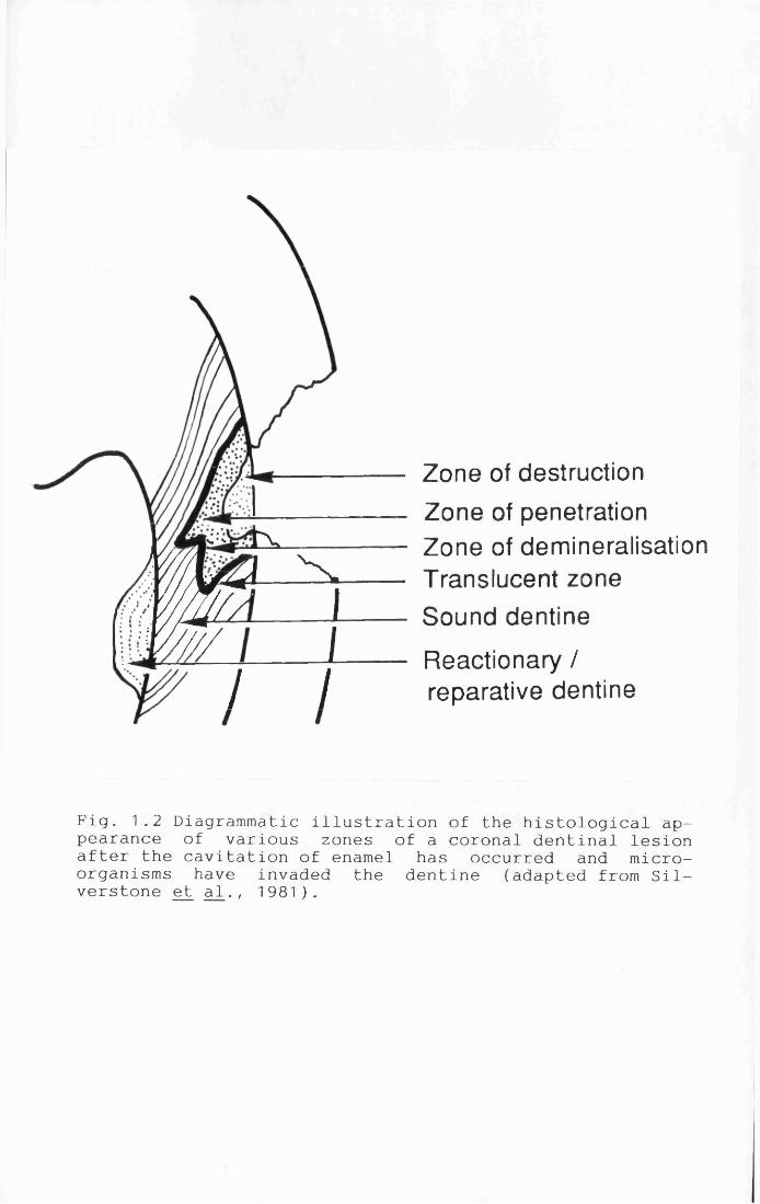
Dentine CariesWhen the advancing front of an enamel caries lesion
approaches the DEJ, acids, enzymes and other stimuli reach the peripheral dentine as a result of the increased per
23
meability of the enamel. At the immediate apex of the enamel lesion, demineralisation occurs in the dentine. The
demineralisation spreads laterally along the DEJ and towards the pulp. This zone is called the zone of demineralisation (Fig. 1.2). In the dentinal tubules pul- pal to the demineralised area as well as in those immediately lateral to it, a tubular sclerosis is seen. This reaction gives rise to the occurrence of the so-called translucent zone. It is thought that odontoblasts may be responsible for this reaction and also initiate this response all around the zone of demineralisation, even extending, at the periphery, to the DEJ. (Thylstrup & Fejerskov, 1986). The process of sclerosis in dentine is thought to be an acceleration of the otherwise normal mechanism of peritubular dentine formation (Bradford, 1960; Johnson et a l ., 1969). The sclerosis of the tubules retards but does not entirely prevent the invasion by bacteria, since the tubules again become permeable as a result of further demineralisation (Ogawa et a l ., 1983).
This distribution of destructive processes, enclosed by the zone of sclerosis, is brought about because the first wave of bacteria infecting the dentine are primarily
acidogenic. The pH in the deepest layer of carious dentine is low (Johansen & Parks, 1961; Dirksen et a l ., 1963), and
softening of the dentine precedes the penetration of
24
p
1 - K1 ' w
W af > to--------1r — — 1
/ J1/ 1
1
Zone of destructionZone of penetration Zone of demineralisation Translucent zoneSound dentine
Reactionary / reparative dentine
Fig. 1.2 Diagrammatic illustration of the histological appearance of various zones of a coronal dentinal lesion after the cavitation of enamel has occurred and microorganisms have invaded the dentine (adapted from Sil- verstone et al. , 1981).
micro-organisms and the discolouration of the dentine (Fusayama et a l ., 1966). Michelich et al. (1980) havedemonstrated that bacteria easily penetrate the tubules in
acid-etched dentine, but do not enter the tubules in un
treated dentine in vitro. Collagen fibres in the zone of
demineralisation are exposed as a result of the removal of attached apatite crystals by acid (Ohgushi & Fusayama, 1975). Collagen denatured by acids is broken down by bacterial proteolytic enzymes more efficiently than intact collagen (Armstrong, 1958). These observations suggest that the organic acids produced by micro-organisms may cause demineralisation of inorganic materials, with associated enhancement of bacterial penetration and proteolytic destruction of organic materials. Lactate, acetate, and propionate are the major acids involved and altogether account for about 90% of the total acid found in carious dentine. The organic acid profiles of carious dentine vary considerably between different subjects. Moreover, the acid profile of the shallow layer is similar
to that of the deep layer in the same carious lesion. These observations suggest that even if the acid profile in dentine varies greatly among the samples, changes in its composition do not occur within a short time, as is
the case in dental plaque (Hojo et a l ., 1991).Frequently, groups of dentinal tubules which have
25
been located in the centre of the demineralised dentine appear empty because sclerosis of these tubules has taken place. The micro-organisms invade or penetrate these tubules. The bacteria may appear in small groups or, more
often, in large numbers confined to the dentinal tubules and their many lateral branches. This is known as the zone
of penetration. (Thylstrup & Fejerskov, 1986).Destruction of the organic matrix follows
demineralisation. As the lesion progresses, the apparently firmly bound material disappears more or less simultaneously with the degradation of the collagenous matrix by proteinases and /or by nonenzymatic processes (Johansen & Park, 1961; Selvig, 1968). Towards the DEJ the bacterial populations are more heterogeneous, increasing the number of proteolytic and hydrolytic enzymes which add to the tissue destruction caused by the acid (Larmas, 1972). This results in the destruction of the organic matrix of the tissue. This appears first in the peritubular zone where the collagen fibrils are finest and extends eventually to the intertubular matrix where the fibres appear to be in
tact even in the advanced stages of demineralisation. In- termolecular crosslinking of collagen fibres decreases and the concentration of collagen precursors increases (Kuboki et a l . , 1977). The organic framework of the dentine therefore breaks apart and this zone is designated the zone of
26
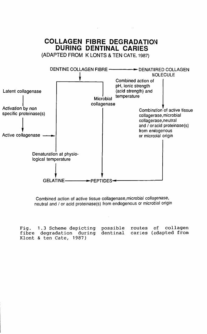
destruction. It is apparent that the collagenous matrix of dentine is altered during the carious process. It is not clear by which mechanisms collagen degradation actually takes place. Possibly this degradation is the result of the processes involving microbial collagenase, active tissue collagenase, neutral and /or acid proteinases of endogenous or microbial origin and acid strength as sum
marised in Fig. 1.3. In vitro experiments have shown that dentine, as a result of carious breakdown, becomes more resistant to proteinase activity (Young & Massler, 1963). In this context, Young and Massler had already suggested in 1963 that at least theoretically, dentinal caries could be a self-limiting process, a suggestion that is still valid today (Klont & ten Cate, 1987).
In some areas, demineralisation and proteolysis occur so rapidly that several groups of tubules coalesce to form so-called liquefaction foci. Furthermore, thedemineralisation and destruction may follow the incremental lines so that transverse clefts occurs in the tissue.
(Thylstrup & Fejerskov, 1986).
The dissolution of the crystallites is followed by a
process of remineralisation in the area in which bacterial invasion was actively taking place, as well as in the odontoblast processes in the zone of sclerosis (Takuma & Kurahashi, 1962; Takuma et: a l . , 1967; Mjor, 1987). This
27
COLLAGEN FIBRE DEGRADATION! DURING DENTINAL CARIES
(ADAPTED FROM K LONTS & TEN CATE, 1987)
DENTINE COLLAGEN FIBRE
Latent collagenase
Activation by non specific proteinase(s)
Active collagenase
DENATURED COLLAGENMOLECULE
Combined action of pH, ionic strength (acid strength) and
Microbial ,emPera,ure collagenase
Denaturation at physiological temperature
GELATINE-
Combination of active tissue collagerase,microbial collagerase.neutral and / or acid proteinase(s) from endogenous or microoial origin
-PEPTIDES-
Combined action of active tissue collagenase,microbial collagenase, neutral and / or acid proteinase(s) from endogenous or microbial origin
Fig. 1.3 Scheme depicting possible routes of collagen fibre degradation during dentinal caries (adapted from Klont & ten Cate, 1987)
involves the formation of caries crystals, either as needle-shaped crystals of octacalcium phosphate or rhom- bohedral crystals of whitlockite (Johnson et ad., 1969;
Mjor, 1985). Two distinctively different mineralisation patterns may be identified; either the j peri-odontoblastic
space mineralises first and calcification within the odontoblastic process follows, or mineral deposits may first
be observed in the cytoplasmic process with subsequent mineralisation of the extracellular periodontoblastic space (Frank &\ voegel, 1980). Daculsi and co-authors (1987) suggested that large Mg-substituted 6-tricalcium phosphate (TCP) crystals were due to initial dissolution of the dentine mineral followed by reprecipitation of Mg-substituted 8—T C P .
Once there is cavitation of enamel and bacteria have reached the dentine, progress of the lesion is likely to be more rapid. The different zones (Fig. 1.2) are only discrete and distinguishable as separate entities in
slowly advancing carious lesions (chronic); they tend to
merge and become indistinguishable in more rapidly progressing lesions (acute).
In contrast to the histological classification out
lined above, some authors classify coronal dentine caries according to the staining and hardness of the lesion
(Fusayama et a l ., 1966). Carious dentine is regarded as
28
consisting of a first or outer layer in which the organic material is substantially degraded and not remineralisable and a second or inner layer with limited collagen degradation which is capable of being remineralised (Fusayama et
a l . , 1966; Kato & Fusayama, 1970; Fusayama & Kurosaki,1972; Kuboki et al., 1977; Table 1.1).
1.7.5 Smooth—surface and Fissure CariesOnce a carious lesion in enamel reaches the DEJ, its
spread is usually relatively rapid along this interface, the anatomical discontinuity between the two tissues apparently being less resistant to the penetration of the destructive agents involved. Fissure and smooth-surface lesions tend to develop different overall shapes : in the case of lesions on the smooth surfaces of teeth, the enamel lesion tends to be conical with its apex touching the DEJ.
Lateral spread from this point results in a broadened base in the dentine lesion which then itself becomes coni
cal in shape; it follows the primary curvature of the dentinal tubules so that its slightly narrower apex approaches the pulpal surface more cervically than the level
at which the lesion entered the dentine. In the case of
fissure caries, the enamel lesion spread is guided by the orientation of the prisms and it broadens as it approaches
29
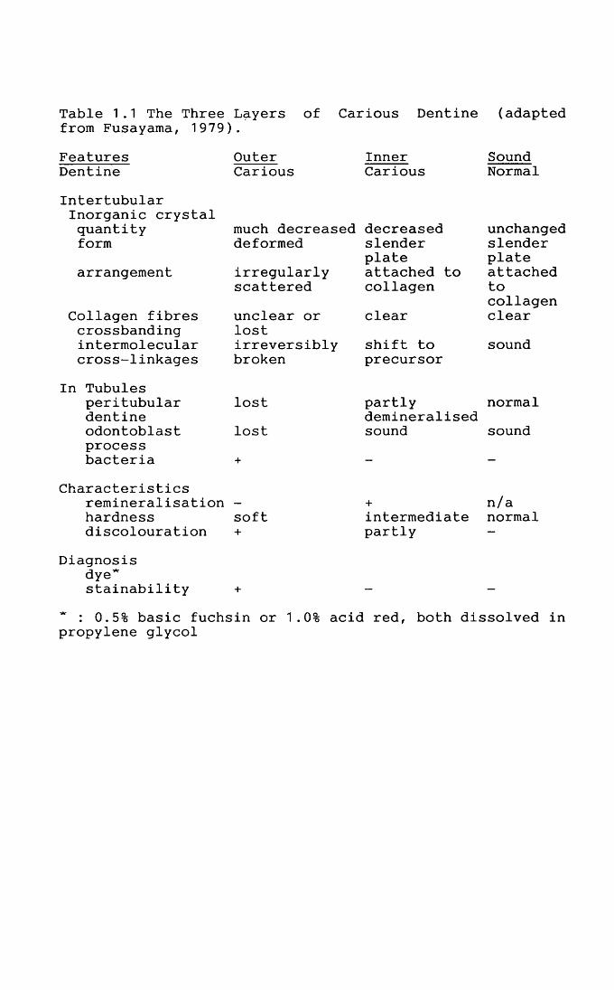
Table 1.1 The Three Layers of Carious Dentine (adapted from Fusayama, 1979).
FeaturesDentine
OuterCarious
InnerCarious
SoundNormal
Intertubular Inorganic crystal quantity formarrangement
Collagen fibres crossbanding intermolecular cross-linkages
In Tubulesperitubulardentineodontoblastprocessbacteria
much decreased decreaseddeformedirregularlyscatteredunclear or lostirreversiblybroken
lostlost
slenderplateattached to collagen
clear
shift to precursor
unchangedslenderplateattachedtocollagenclear
sound
partly normaldemineralised sound sound
Characteristicsremineralisation - hardness softdiscolouration +
Diagnosisdye*stainability +
intermediatepartly
n/a normal
* : 0.5% basic fuchsin or 1.0% acid red, both dissolved in propylene glycol
the dentine. With lateral spread at the DEJ, the area of
dentine involved is initially larger than in a smooth- surface lesion. The tubules are relatively straight over the occlusal aspect of the pulp chamber and do not taper so much toward the pulp. This explains why, when an apparently small occlusal lesion is excavated, it is often found to have extensively undermined enamel and a surpris
ingly large area of unsound dentine.Nevertheless dentinal lesions, wherever they arise,
are characteristically conical, the shape initially being determined by the distribution of the translucent zone. The lateral border of the carious dentine runs parallel to the direction of the dentinal tubules and is fairly sharply defined. Together with the peritubular dentine, the dentinal tubules assist in restricting the lateral spread of caries. The fact that the level of mineralisation is marginally lower in mantle dentine encourages the lateral spread of the carious lesion just below the DEJ. The deeper border of the lesion in dentine is generally
much harder to identify (Jones & Boyde, 1987).
1.7.6 Deciduous CariesFew studies have dealt with caries in the dentine of
deciduous teeth and few have attempted to correlate ultrastructural observations with the better-known fea
30

tures seen by light microscopy.
Enamel CariesThe rate of progress of the artificial carious lesion
is faster in deciduous enamel than permanent (Featherstone & Mellberg, 1981). The histological features of caries in deciduous enamel are essentially similar to those in the
enamel of permanent teeth. However, enamel in deciduous teeth is approximately half the thickness of that in permanent teeth and the pulp chambers are relatively much larger. Thus, the carious process needs to travel a shorter distance to reach the pulp in a deciduous molar than in a molar from the permanent dentition. (Silverstone et a l ., 1981).
Dentine CariesCarious lesions in the dentine of deciduous molars
characteristically contain a peripheral translucent zone enclosing a broad zone of bacterial invasion which may in turn surround a zone of more severe destruction (Johnson
et a l ., 1969). Electron microscopic examination has shown
that most of the tubules in the translucent zone are oc
cluded by mineral deposits, closely resembling peritubular dentine. It has been postulated that this may represent a defence mechanism on the part of the tooth, but which may
31
be inhibited if the initial carious attack is sufficiently severe to cause the death of a large number of odontoblasts (Johnson et a l ., 1969). In the outer parts of the zone of bacterial penetration caries crystals (see Section
1.8.3) were present in many tubules. Throughout most of the zone of penetration there is extensive demineralisation of the intertubular dentine (Lester & Boyde, 1968; Johnson et al. , 1969). Bacteria, however, remain confined
to the tubules where they are incompletely surrounded by highly mineralised tissue which probably represents the remnants of previously sclerosed tubules or of peritubular dentine. The pathology of the carious process in the dentine of deciduous molars appears to be fundamentally similar to that observed in permanent teeth. The deciduous teeth, however, showed a much higher percentage of cavities with bacteria remaining in the dentine after removal of all softened dentine than did permanent teeth (Whitehead et a l ., 1960).
1.7.7 Active and Arrested Caries
Enamel Caries
Although saliva, due to its physiological supersaturation with respect to apatite, should effect a remineralisation of demineralised subsurface enamel, this
32
apparently occurs infrequently in vivo. For example, when a once-demineralised area along the gingival margin is freed from the close relation to the gingiva by its retraction, it remains as white spot enamel for the life
of the tooth. It is often observed that the surface layer takes up mineral and becomes hard. However, the subsurface porous area remains, presumably due to a number of factors. The small pores, which narrow in pace with the surface remineralisation of the surface layer limit the diffusion through the surface layer and therefore the content of organic materials within the pores of central lesions occupies the volume where the mineral should be deposited.
It is now well established that the primary mode of action of topical fluoride is its influence in enhancing natural salivary remineralisation of the early enamel lesion - the so-called "white spot lesion". As far back as 1961, Koulourides and his colleagues demonstrated an eightfold increase in the rate of in vitro remineralisation upon addition of small quantities of fluoride (0.05 mmol/L) to a calcium phosphate solution. It seemed
reasonable to suppose that the addition of small quan
tities of fluoride to the oral environment might produce a
comparable effect in vivo, and indeed there has been considerable laboratory and clinical evidence to support this theory (Creanor & Strang, 1989). A careful and frequent
33
application of topical fluoride to such active initial le
sions results in the deposition of relatively large amounts of calcium fluoride within the lesion, from where
it is only released very slowly. Calcium fluoride within a carious lesion may act as a store from which fluoride is slowly released, resulting in a high concentration of free F— within the lesion, thus preventing further dissolution
of mineral from its interior. (Thylstrup & Fejerskov, 1986). The production of a fluoridated mineral with a high resistance to acid attack is the aim of any topical fluoride regime.
Dentine CariesMassler (1967) has called attention to the intermit
tent nature of the carious attack with periods of rapid demineralisation, alternating with periods of inactivity. Depending on the balance between attacking and defensive forces, the rate of progress of caries in dentine is
therefore highly variable. Accordingly, it is not supris- ing that under suitable environmental conditions lesion progress can be completely arrested and the lesion may even regress (Massler, 1967). If the cariogenicity of the environment is controlled, and particularly if the cavity
has become open and more readily kept free of accumula
tions of food and bacterial plaque, the caries process may
34
become arrested (Silverstone et a l . , 1981). Arrestment/remineralisation of carious dentine does not occur in the components of the organic matrix but by the growth of residual crystals in the lesions (Levine & Rowles, 1973;
Daculsi et a l ., 1979; Klont & ten Cate, 1991). Destruction of dentine occurs both by demineralisation and proteolytic breakdown of the collagenous matrix, and whilst this means that the lesion has a rate of progress which is governed less by purely physiochemical interactions, it also means that remineralisation of the lesion is not such a simple process as in enamel since the organic matrix to be mineralised has been changed (Jones & Boyde, 1987).
The most striking remineralisation takes place on and within the tooth surface exposed to the oral environment. This layer contains reformed crystals in a matrix derived from saliva, food and bacterial products. The crystals are predominantly apatitic (Takuma et al., 1975) but arelarger than those of sound dentine with a high Ca : P
ratio (Levine, 1973) and high F content (Levine, 1972). A number of non-apatitic calcium phosphates may also be
found (Rowles & Levine, 1973). Carious coronal dentine lesions can be classified on the basis of their clinical, and gross histological characteristics, into active and arrested lesions (Table 1.2).
35
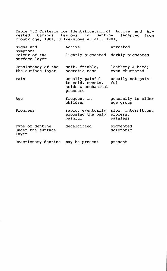
Table 1.2 Criteria for Identification of Active and Arrested Carious Lesions in Dentine (adapted from Trowbridge, 1981; Silverstone et al., 1981)Signs and Symptoms Colour of the surface layerConsistency of the the surface layerPain
Age
Progress
Type of dentine under the surface layer
Activelightly pigmented
soft, friable, necrotic mass
frequent in childrenrapid, eventually exposing the pulp, painfuldecalcified
Arrested
darkly pigmented
leathery & hard; even eburnated
generally in older age groupslow, intermittent process, painless
pigmented,sclerotic
usually painful usually not pain- to cold, sweets, ful acids & mechanical pressure
Reactionary dentine may be present present
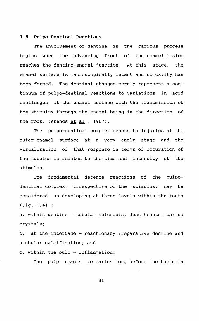
1 .8 Pulpo-Dentinal ReactionsThe involvement of dentine in the carious process
begins when the advancing front of the enamel lesion
reaches the dentino-enamel junction. At this stage, the enamel surface is macroscopically intact and no cavity has been formed. The dentinal changes merely represent a continuum of pulpo-dentinal reactions to variations in acid challenges at the enamel surface with the transmission of
the stimulus through the enamel being in the direction of the rods. (Arends et a l ., 1987).
The pulpo-dentinal complex reacts to injuries at the outer enamel surface at a very early stage and the visualisation of that response in terms of obturation of the tubules is related to the time and intensity of the stimulus.
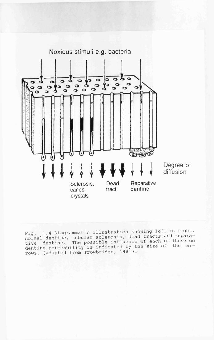
The fundamental defence reactions of the pulpo- dentinal complex, irrespective of the stimulus, may be considered as developing at three levels within the tooth (Fig. 1.4) :
a. within dentine - tubular sclerosis, dead tracts, caries crystals;
b. at the interface - reactionary /reparative dentine and atubular calcification; andc. within the pulp - inflammation.
The pulp reacts to caries long before the bacteria
36
Noxious stimuli e.g. bacteria
Y V YSclerosis,cariescrystals
muiDeadtract
Reparativedentine
Degree of diffusion
Fig. 1.4 Diagrammatic illustration showing left tc right, normal dentine, tubular sclerosis, dead tracts and reparative dentine. The possible influence of each of these on dentine permeability is indicated by the size of the arrows. (adapted from Trowbridge, 1981).

penetrate into the pulp chamber because soluble irritants and inflammatory stimuli diffuse from the carious lesion through the dentinal tubules. These may include biologically active substances such as : bacterial enzymes, bacterial peptides, endotoxins, polysaccharides, lipopolysac-
charides, somatic antigens, antibodies, chemotaxins, complement proteins, organic acids, products of tissue destruction and ammonia. The rate of permeation varies inversely with the molecular radius of the penetrating substance (Pashley et a l ., 1977). Major factors contributingto the resistance to fluid movement through dentine include surface resistance and the degree of intratubular occlusion. In addition to the substances that diffuse from the carious lesion into the pulp, other molecules e.g. serum proteins (Okamura et a l ., 1979) may also move in the opposite direction, i.e. from the pulp towards the carious lesion.
1.8.1 Tubular SclerosisA carious lesion is frequently associated with a
characteristic zone of sclerosis in the underlying den
tine. It is conceivable that this lateral sclerosis is the first reaction to the earlier stages of the peripheral enamel lesion. Sclerosis is produced by the deposition of
mineral closely resembling peritubular dentine and is much
37
more prominent in arrested caries than active caries. It represents an acceleration of the normal physiological process of peritubular dentine formation and is more likely to form in the presence of mild stimulation as op
posed to severe irritation of the odontoblasts. Occlusion
of the tubules by mineralisation results in decreased permeability (Miller & Massler, 1962; Barber & Massler, 1964). Tubular sclerosis leads to translucency of the
tissue because the affected area is structurally more homogeneous : there is less scattering of light as it
passes through the tissue and an area of dentine so affected is called a translucent or sclerotic zone. It is almost a universal feature of the pulpo-dentinal response to caries (Levine, 1974).
1.8.2 Dead TractsIf, as a result of an aggressive carious attack,
odontoblasts are quickly destroyed, dead tracts, rather
than sclerotic dentine, will be formed. Dead tracts are groups of tubules that are no longer filled with living odontoblast processes. Dead tracts are difficult to identify histologically in demineralised specimens, but they
appear black in ground sections because air contained
within the tubules refracts transmitted light. In relation to caries, dead tracts are most commonly seen in young
38
teeth affected by rapidly progressing lesions. Presumably, dead tracts are highly permeable in comparison to sclerotic dentine, suggesting that they may accelerate the carious process. However, they are usually sealed by the deposition of reparative dentine at the pulpal end of the tubules.
1.8.3 Caries CrystalsIn addition to peritubular-like mineralisation,
tubules can become partially blocked by needle-shaped crystals and rhombohedral structures (Lester & Boyde, 1968; Johnson et a l ., 1969). In carious lesions, suchstructures have been referred to as "caries crystals". It has been suggested that caries crystals represent the recrystallisation of calcium and phosphate ions that have been dissolved during demineralisation of the dentine (Takuma et a l ., 1967). The intratubular crystals increase in size and number below the translucent zone nearly fill
ing the dentinal tubules. Further mineralisation may have
taken place centripetally so that the central tubes
decrease in diameter (Mendis & Darling, 1979). These crys
tals gradually became transformed into rhomboid shapes in the translucent zone and were found to be absent in the overlying discoloured layer (Ogawa et al., 1983). These
deposits are found in tubules within the advancing front
39
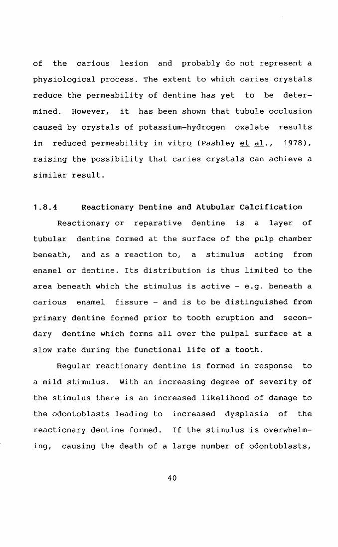
of the carious lesion and probably do not represent a physiological process. The extent to which caries crystals reduce the permeability of dentine has yet to be determined. However, it has been shown that tubule occlusion caused by crystals of potassium-hydrogen oxalate results in reduced permeability in vitro (Pashley et a l ., 1978),raising the possibility that caries crystals can achieve a similar result.
1.8.4 Reactionary Dentine and Atubular Calcification
Reactionary or reparative dentine is a layer of tubular dentine formed at the surface of the pulp chamber beneath, and as a reaction to, a stimulus acting from enamel or dentine. Its distribution is thus limited to the area beneath which the stimulus is active - e.g. beneath a carious enamel fissure - and is to be distinguished from primary dentine formed prior to tooth eruption and secondary dentine which forms all over the pulpal surface at a
slow rate during the functional life of a tooth.
Regular reactionary dentine is formed in response to a mild stimulus. With an increasing degree of severity of the stimulus there is an increased likelihood of damage to
the odontoblasts leading to increased dysplasia of the reactionary dentine formed. If the stimulus is overwhelming, causing the death of a large number of odontoblasts,
40
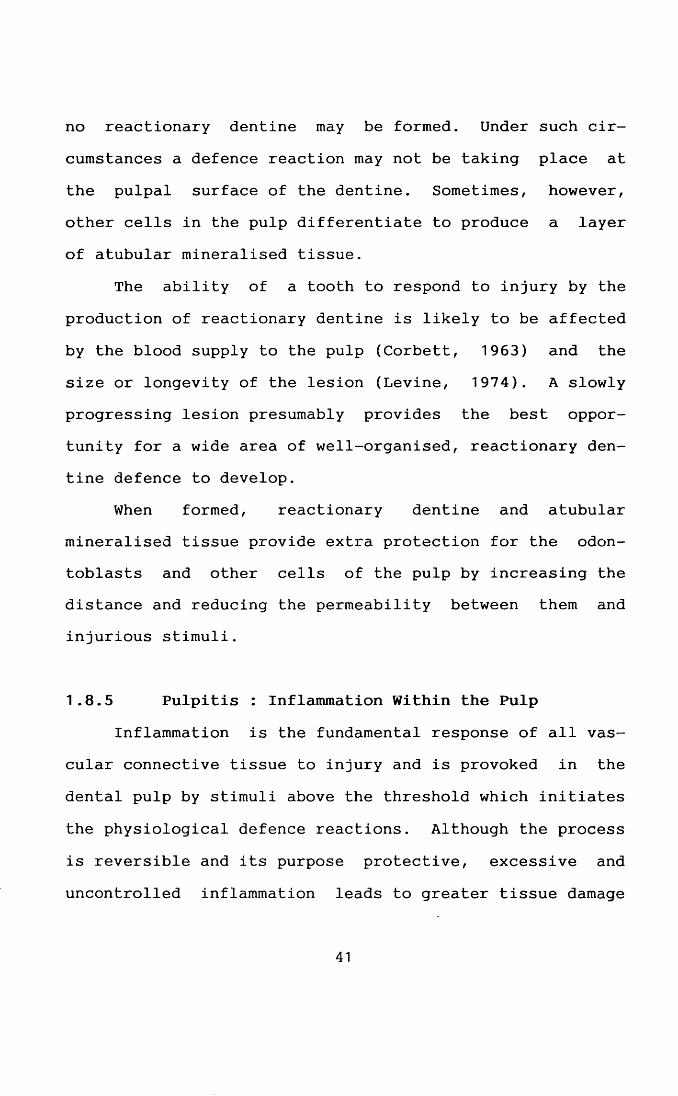
no reactionary dentine may be formed. Under such cir
cumstances a defence reaction may not be taking place at the pulpal surface of the dentine. Sometimes, however, other cells in the pulp differentiate to produce a layer of atubular mineralised tissue.
The ability of a tooth to respond to injury by the
production of reactionary dentine is likely to be affected by the blood supply to the pulp (Corbett, 1963) and the size or longevity of the lesion (Levine, 1974). A slowly progressing lesion presumably provides the best opportunity for a wide area of well-organised, reactionary dentine defence to develop.
When formed, reactionary dentine and atubular mineralised tissue provide extra protection for the odontoblasts and other cells of the pulp by increasing the distance and reducing the permeability between them and injurious stimuli.
1.8.5 Pulpitis : Inflammation Within the PulpInflammation is the fundamental response of all vas
cular connective tissue to injury and is provoked in the
dental pulp by stimuli above the threshold which initiates
the physiological defence reactions. Although the process is reversible and its purpose protective, excessive and
uncontrolled inflammation leads to greater tissue damage
41
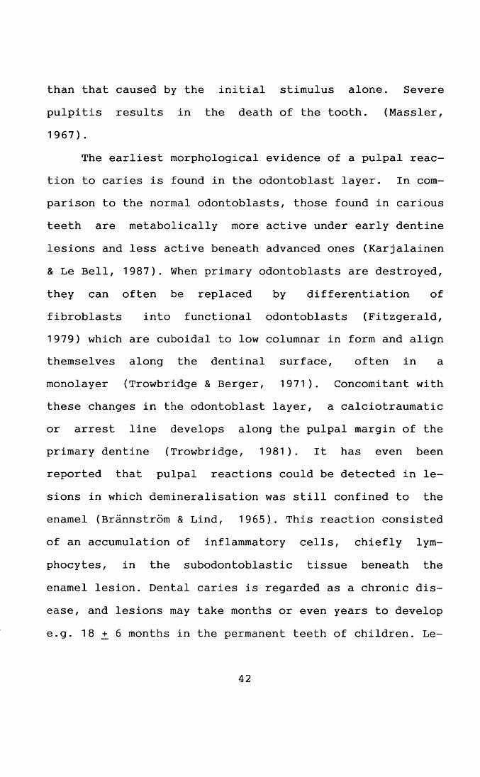
than that caused by the initial stimulus alone. Severe pulpitis results in the death of the tooth. (Massler, 1967).
The earliest morphological evidence of a pulpal reaction to caries is found in the odontoblast layer. In comparison to the normal odontoblasts, those found in carious teeth are metabolically more active under early dentine lesions and less active beneath advanced ones (Karjalainen
& Le Bell, 1987). When primary odontoblasts are destroyed, they can often be replaced by differentiation of
fibroblasts into functional odontoblasts (Fitzgerald, 1979) which are cuboidal to low columnar in form and align themselves along the dentinal surface, often in a monolayer (Trowbridge & Berger, 1971). Concomitant with these changes in the odontoblast layer, a calciotraumatic or arrest line develops along the pulpal margin of the primary dentine (Trowbridge, 1981). It has even been reported that pulpal reactions could be detected in lesions in which demineralisation was still confined to the enamel (Brannstrom & Lind, 1965). This reaction consisted of an accumulation of inflammatory cells, chiefly lym
phocytes, in the subodontoblastic tissue beneath the enamel lesion. Dental caries is regarded as a chronic dis
ease, and lesions may take months or even years to develop e.g. 1 8 ^ 6 months in the permanent teeth of children. Le
42
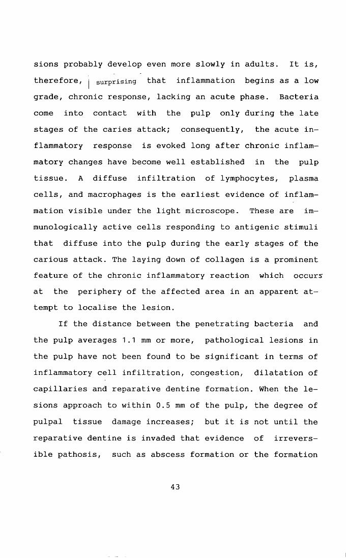
sions probably develop even more slowly in adults. It is,
therefore, | surprising that inflammation begins as a low grade, chronic response, lacking an acute phase. Bacteria come into contact with the pulp only during the late stages of the caries attack; consequently, the acute inflammatory response is evoked long after chronic inflam
matory changes have become well established in the pulp tissue. A diffuse infiltration of lymphocytes, plasma cells, and macrophages is the earliest evidence of inflammation visible under the light microscope. These are im- munologically active cells responding to antigenic stimuli
that diffuse into the pulp during the early stages of the carious attack. The laying down of collagen is a prominent feature of the chronic inflammatory reaction which occurs at the periphery of the affected area in an apparent attempt to localise the lesion.
If the distance between the penetrating bacteria and
the pulp averages 1.1 mm or more, pathological lesions in the pulp have not been found to be significant in terms of inflammatory cell infiltration, congestion, dilatation of capillaries and reparative dentine formation. When the lesions approach to within 0.5 mm of the pulp, the degree of
pulpal tissue damage increases; but it is not until the reparative dentine is invaded that evidence of irreversible pathosis, such as abscess formation or the formation
43
of large amounts of granulation tissue can be observed. (Reeves & Stanley, 1966).
1.9 Removal of Carious Dentine
In the Middle Ages, carious tissue was scooped out with a hand instrument. The restoration of decayed teeth, even in the 19th century, was seldom successful. The na
ture of dental decay was not understood and the need for removal of the softened decayed portion before attempting to fill the cavity was not appreciated.
The methods which have been used to remove caries include :a. mechanical rotary techniquesb. mechanical non-rotary techniques
i. air-abrasive techniquesii. ultrasonic techniquesiii. air-polishing techniques
c. enzyme techniquesd. laser techniques
e. chemomechanical techniques
1.9.1 The Mechanical Rotary Techniques
The use of rotary instruments was the first mechanical technique developed to remove caries. The equipment used includes dental engines, handpieces and dental burs.
44
Dental EnginesThe early models of dental engines and cutting in
struments used for the removal of carious material were, by today's standard, very primitive. In 1846 Amos Westcott developed the drill-stock consisting of a finger ring and simple drill rotated between the thumb and forefinger. In 1850, J.D. Chevalier devised a drill stock operated by a small crank and bevel gears. The tool was held at an angle of 45° to the body of the instrument and could be made to
point towards or away from the handle. There was steady rotation in either direction but the instrument required two hands to operate it.
Charles Merry of St. Louis devised a drill in 1858 which had two handpieces - one to hold the instrument in place and the other as a handle to drive the holder, to which it was connected by a flexible coupling or a universal joint of spirally wound wire. That was the first time
the separation of functions between the two handpieces was ma d e .
A further separation of function in the production of
the rotary movement was the foot treadle which was later achieved by Morrison. In 1864, a new category of instruments in which the source of power was an electric motor was introduced. In 1868, George F. Green invented an en
45
gine in which a foot-driven bellows transferred air in a rubber tube to a handpiece which contained draught screens
which rotated a canula within which various drills could be mounted. This was the direct predecessor of today's air driven dental turbine, (reviewed by Noble, 1985).
Air driven dental turbines were produced by Norlen in 1955 as the "Dentalair" and in 1957 as the Borden Airotor.
Dental BursEarly models of the dental engine and the burs used
with them were not efficient in cutting enamel. It was not until the end of 19th century that an improved range and quality of cutting and polishing burs and discs became available.
When James Morrison invented the foot treadle operated dental engine in 1871 and S.S. White's mechanic, George F. Green, developed the first electric dental drill shortly after, a much greater demand was created for an efficient bur. It is not known who made the first modern bur but it must be assumed the entire process was performed by hand and was very laborious. A small piece of steel was filed and ground into a specific shape with each tiny blade exquisitely hand sharpened to a razor-like
e d g e .
S.S. White revolutionised and set the standard for
46
years to come when the first machine-made bur was introduced in 1891. The bur had a continuous blade or drill edge across its centre. This enabled the bur to cut in the direction of its axis. No blade followed in the path of another; instead it cut across the path of its leader. These early burs were made of steel, and although gradual improvements have been made since in the properties of the
alloy used, they were very similar to modern steel burs (Sturdevant et a l ., 1985).
Rudolph Furke discovered the process of hardening steel with tungsten carbide and applied this technique to the dental bur in 1917 but had to wait for more than 40 years before a method was developed to cuti a tungsten carbide bur to cut efficiently. The early steel burs had only four basic shapes; round, inverted cone, straight fissure and tapered fissure.
In 1957, S.S. White introduced the Borden Airotor capable of 300,000 revolutions per minute. The air turbine dramatically changed the practice of dentistry. There was a scramble to produce the intricate carbide burs capable
of utilising the benefits of this previously unrealised speed.
In the 1960s and early 1970s when plastic and com
posite materials came onto the market, a wide range of burs was developed to cope with the new demands made on
47
them by these materials, (reviewed by Crawford, 1990).
Dental Handpieces
Prior to 1870, dentists had no mechanically driven
rotary tools for the removal of caries and cavity preparation. The procedure of cleaving away over-hanging enamel was undertaken by hand instruments, termed enamel cutters, which enabled the underlying carious dentine to be scooped out with excavators. These instruments were augmented by a wide range of long-handled burs having the same knurled hexagonal handles as the present day hand instruments.
During the period 1850 - 1870 various other instruments were devised to rotate burs in cavities such as Merry's drill with its universal joint which allowed rotation of a bur in a posterior tooth by holding the lower handle still and twisting the upper handle.
Straight handpieces with a variety of intricate chuck-closing mechanisms became well developed during the
1880s and they were permanently linked to the foot-
engine's flexible cable. A later development was the angled handpiece which held the burs in place by so- called "lock-bit attachments" to their front ends. These lock-bits were available in right angle, acute angle, and obtuse angle pattern.
Early 20th century developments included right angled
48
handpieces fitted with a latch-lock mechanism, balanced contra-angled handpieces and jointed engine arms.
Throughout the 1940s little change of note occurred
in relation to dental handpieces and their driving
mechanisms. The subsequent three decades saw radical improvements in the design of handpieces and their driving mechanisms. Handpieces with water jets or water spray nozzles were first manufactured by the Amalgamated Dental Company as the New Era in 1955, thus allowing dentine to be cut wet. Given spray cooling there was now no limit to the maximum speed of burs and diamonds and, as far as enamel removal was concerned, it seemed indeed to be a case of the faster the better. Different factors may contribute to pathological changes in the dentine and in the pulp when rotating instruments are used. The main factors are : speed - desiccation - heat - pressure, in combination with cutting time, depth of the cavity and area of prepared dentine. An efficient waterspray keeping the prepared area under constant water cover has been shown to
be crucial in minimising pulpal damage (Stanley, 1968;
Shovelton, 1972). Handpieces were improved to drive burs
and diamonds much faster than the usual 9000 rpm maximum allowing reduced cutting pressure. Their bearings were also improved to withstand the high speeds achieved, namely 20,000 rpm or more. To allow seated dentistry, the
49
engine cord arm had to be eliminated. Handpieces incorporating their own motors powered by electricity or by
compressed air were manufactured.Even with the advanced airotor, the restoration pro
cedure was still very slow. That was changed when the high speed handpiece was introduced in the early 1960s which marked a new era of restorative dentistry. Both patients and dentists have now benefited from this advancement for more than thirty years.
The three significant advances in handpiece development during the nineteen eighties have been : the pushbutton chuck, the multiple coupling, and fibre optic lighting. The obvious advantage of the multiple coupling is that it enables different types of handpieces to be pushed on to, and pulled off of the coupling in a single quick movement. Air and water connections for propulsion and spray cooling are automatically made on attachment of the handpiece to the coupling and, in addition, the tip of the coupling may house a small light bulb for fibre-optic light transmission through each handpiece connected to it. This fibre optic lighting enhances the illumination of the
bur tip and the cavity, (reviewed by Stephens, 1986).
1.9.2 The Air-abrasive TechniqueThe air-abrasive technique was based on the use of
50
powdered aluminium oxide particles which travelled at high speed to remove hard tooth structure without perceptible vibration, pressure, heat production or pulpal reaction (Black, 1955). This technique was abandoned as a clinical
tool on account of its lack of tactile perception and the difficulty in forming precise margins and angles. The
operator had to return to hand or rotary instruments for finishing procedures. The surface of an ordinary mirror was rendered useless in a short period by rebounding abrasive particles and the dust spreading everywhere - including both the patient's and the dentist's eyes and respiratory systems (Boyde, 1984).
1.9.3 The Ultrasonic TechniqueHigh frequency ultrasonic vibrations in conjunction
with an abrasive slurry have been used to prepare teeth for restorative treatment. This procedure eliminated noise, vibration, heat formation and pressure (Nielsen et
al., 1955; Nielsen & Bethesda, 1955). The technique seemed
to have the same pulpal effect as that produced by rotary instruments and patient acceptance was favourable (Oman and Appelbaum, 1955). The technique, however, did not gain
widespread acceptance because of the limited availability of instrument tips, slowness of action, poor visibility
when using the abrasive slurry and maintenance problems.
51
Caries and resilient restorative materials such as gold could not be removed effectively (Sturdevant et a l ., 1985).
1.9.4 The Air-polishing TechniqueAir-polishing is a technique involving the use of an
air-propelled jet of powder (mainly sodium bicarbonate) shrouded by a concentric water jet. It has been developed as an instrument to remove dental plaque and staining (e.g. coffee and tea stain). Despite its effectiveness, the negative aspects arising from abrasive loss of dentine, cementum and white spots on enamel have raised concern about its use in routine prophylaxis (Boyde, 1984; Cooley et a l ., 1990; Kontturi-Narhi et a l ., 1990).
It has been suggested that air-polishing has thepotential for development as a caries removal technique. It differentially removes carious dentine leaving the
sound dentine intact when used appropriately (Boyde, 1984). However, no further studies have been carried out.
1.9.5 The Enzyme TechniqueIt has been reported that using a bacterial
Acromobacter collagenase, soft carious dentine can be
removed over a period of 2 - 5 hours leaving the sound
layer of dentine beneath the lesions intact (Goldberg &
52
Keil, 1989). No further literature on the clinical application of this technique is available.
1.9.6 The Laser TechniqueTwo general classes of laser (light amplication by
stimulated emission of radiation) exist for medical and
dental applications. These are so-called "soft" lasers (Strang et a l ., 1988) which are a source of cold(athermic) low energy light emitted at wavelengths thought by some to stimulate cellular activity, and "hard" (thermic) lasers utilised in surgery as precise energy
sources to cut, coagulate and vaporise tissues.After initial experiments with the ruby laser, most
clinicians have progressed to using Argon, CO 2 and more recently Nd:YAG (neodymium : yttrium-aluminium-garnet)systems. Currently, most interest in the fields of restorative dentistry and oral surgery centre around the use of the "hard" CO 2 and Nd:YAG lasers. Sterilising as it
cuts, the laser shows promise not only in caries removal
and soft tissue surgery, but also in endodontics and gingival curettage.
It has been claimed that the finer control offered by Nd:YAG lasers over CO 2 systems allows the operator to set the laser up to remove carious dentine without damaging
sound dentine or enamel. Carious dentine being darker than
53
sound tooth, the laser energy can be set at a level where a sufficient amount of laser light is absorbed by the
carious dentine to destroy it, whilst the sound dentine is affected little. Many operators report that patients can frequently be treated without a local anaesthetic. Due to the alteration in the surface structure of the lased tooth, it will be more resistant to caries attack than before treatment (Midda & Renton-Harper, 1991). It has,however, been reported that the CO 2 laser acts better on dental hard tissue regardless of dentine or enamel colour
and its use is preferable to that of Nd:YAG lasers (Zakariasen et a l ., 1991).
The Nd:YAG laser system is already commercially available in the UK (e.g. American Dental Laser) but the cost of installation is still high (around £ 30,000 at the moment). The main area in laser techniques which remains to be improved is the reduction of noise and the flashing light produced during their operation. The use of lasers
on paediatric dental patients is still to be investigated. Only the use of lasers in soft tissue has been approved by the FDA. Unless more research can be carried out, ex
tensive use of this technique in dental clinics will not be realised in the near future.
54
1.9.7 The Chemomechanical TechniqueAlthough the evolution of the dental engine has of
fered a gradual improvement in mechanical devices to
prepare cavities for restoration, no chemical means of caries removal has ever gained ground in restorative dentistry. This is partly due to the advantages of mechanical methods of cavity preparation in terms of speed and ef-
fiency and partly because of the difficulties in finding a chemical that would remove caries effectively without causing any damage to sound dentine and pulpal tissue. Although a purely chemical caries removal system has yet to be devised, a chemomechanical caries removal system (CMCRS) was first introduced during the nineteen seventies in the USA.
The system, which is known as the C a r i d e x ™ Caries Removal System (CRS), consists of a freshly prepared aqueous solution of N-monochloro-DL-2-aminobutyrate (NMAB) which is presumed to react with the demineralised, par
tially degraded collagen of the carious dentine resulting
in a softening of the carious tissue which can then be gently removed.
1.9.8 Current Status of Caries RemovalCurrently, conventional cavity preparation and caries
removal entails a combination of the use of the high speed
55
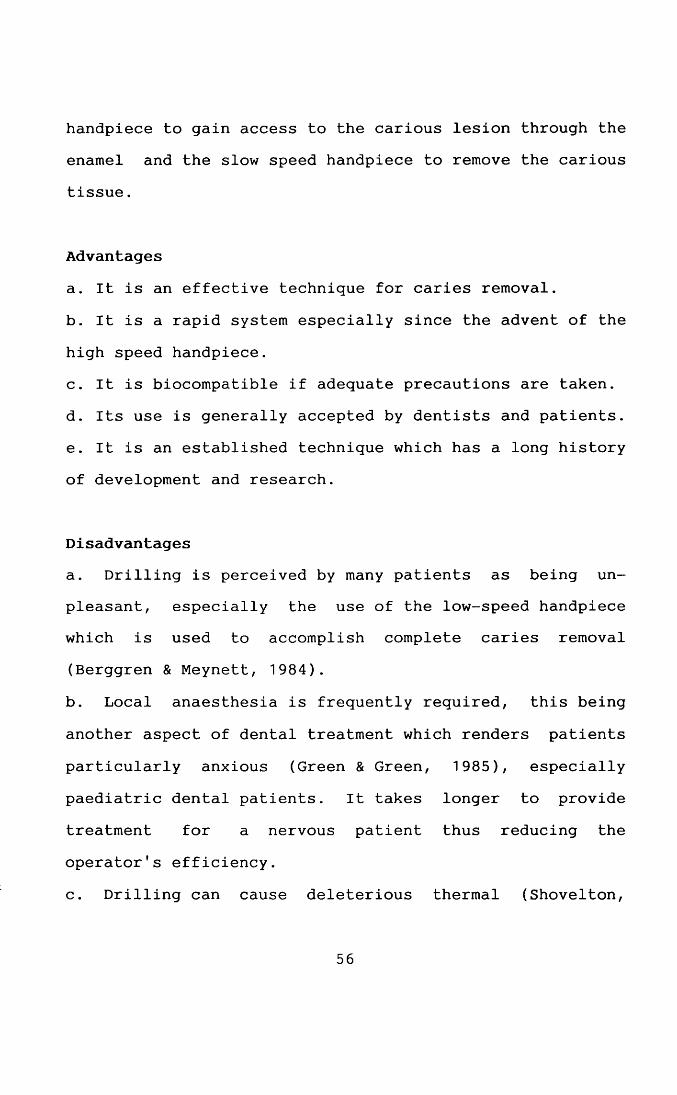
handpiece to gain access to the carious lesion through the enamel and the slow speed handpiece to remove the carious tissue.
Advantagesa. It is an effective technique for caries removal.b. It is a rapid system especially since the advent of thehigh speed handpiece.c. It is biocompatible if adequate precautions are taken.d. Its use is generally accepted by dentists and patients.e. It is an established technique which has a long historyof development and research.
Disadvantagesa. Drilling is perceived by many patients as being unpleasant, especially the use of the low-speed handpiece which is used to accomplish complete caries removal (Berggren & Meynett, 1984).
b. Local anaesthesia is frequently required, this being another aspect of dental treatment which renders patients particularly anxious (Green & Green, 1985), especially paediatric dental patients. It takes longer to provide treatment for a nervous patient thus reducing the
operator's efficiency.c. Drilling can cause deleterious thermal (Shovelton,
56
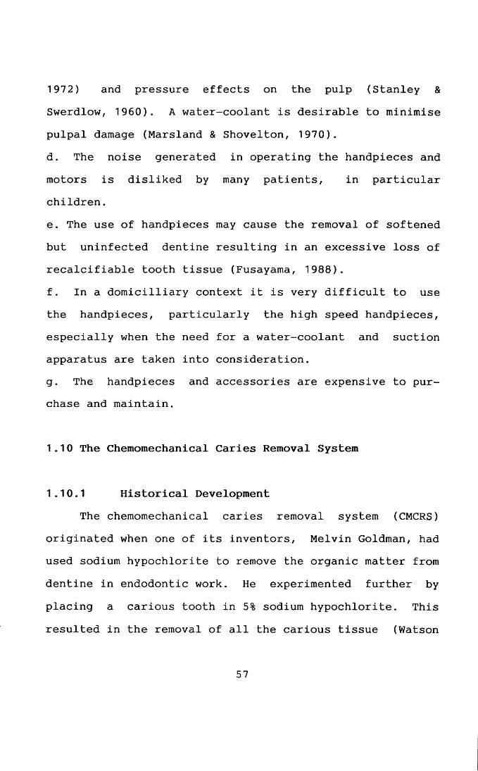
1972) and pressure effects on the pulp (Stanley & Swerdlow, 1960). A water-coolant is desirable to minimise pulpal damage (Marsland & Shovelton, 1970).d. The noise generated in operating the handpieces and motors is disliked by many patients, in particular children.
e. The use of handpieces may cause the removal of softened but uninfected dentine resulting in an excessive loss of recalcifiable tooth tissue (Fusayama, 1988).
f. In a domicilliary context it is very difficult to use the handpieces, particularly the high speed handpieces, especially when the need for a water-coolant and suction apparatus are taken into consideration.g. The handpieces and accessories are expensive to purchase and maintain.
1.10 The Chemomechanical Caries Removal System
1.10.1 Historical DevelopmentThe chemomechanical caries removal system (CMCRS)
originated when one of its inventors, Melvin Goldman, had used sodium hypochlorite to remove the organic matter from
dentine in endodontic work. He experimented further by
placing a carious tooth in 5% sodium hypochlorite. This
resulted in the removal of all the carious tissue (Watson
57
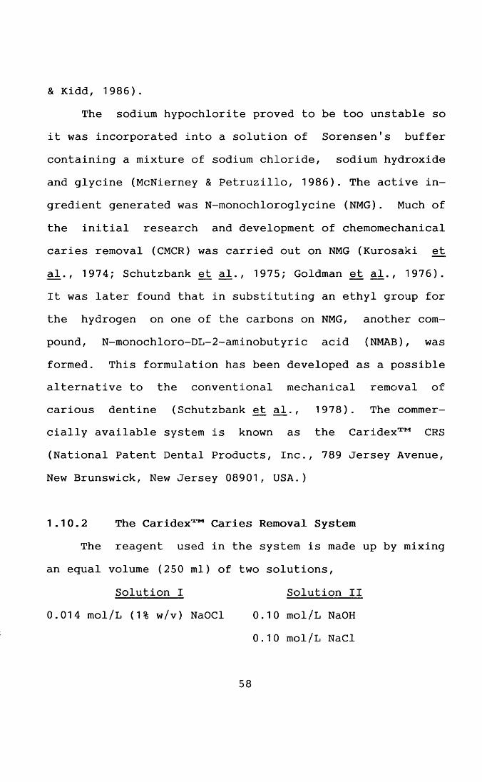
& Kidd, 1986).The sodium hypochlorite proved to be too unstable so
it was incorporated into a solution of Sorensen's buffer containing a mixture of sodium chloride, sodium hydroxide and glycine (McNierney & Petruzillo, 1986). The active in
gredient generated was N-monochloroglycine (NMG). Much of the initial research and development of chemomechanical caries removal (CMCR) was carried out on NMG (Kurosaki et a l ., 1974; Schutzbank et al., 1975; Goldman et al., 1976). It was later found that in substituting an ethyl group for the hydrogen on one of the carbons on NMG, another compound, N-monochloro-DL-2-aminobutyric acid (NMAB), was formed. This formulation has been developed as a possible alternative to the conventional mechanical removal of carious dentine (Schutzbank et a l ., 1978). The commercially available system is known as the C a r i d e x ™ CRS (National Patent Dental Products, Inc., 789 Jersey Avenue, New Brunswick, New Jersey 08901, USA.)
1.10.2 The Caridex™ Caries Removal SystemThe reagent used in the system is made up by mixing
an equal volume (250 ml) of two solutions,
Solution I Solution II
0.014 mol/L (1% w/v) NaOCl 0.10 mol/L NaOH
0.10 mol/L NaCl
58
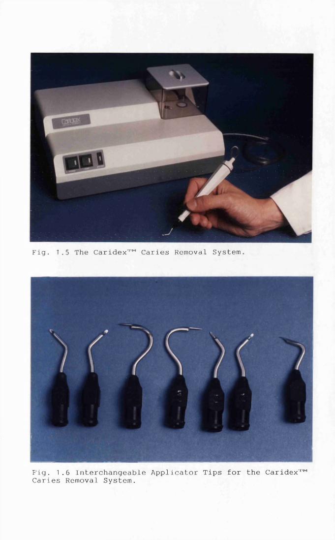
Fig. 1.5 The Caridex™ Caries Removal System
Fig. 1.6 Interchangeable Applicator Tips for the Caridex™ Caries Removal System.
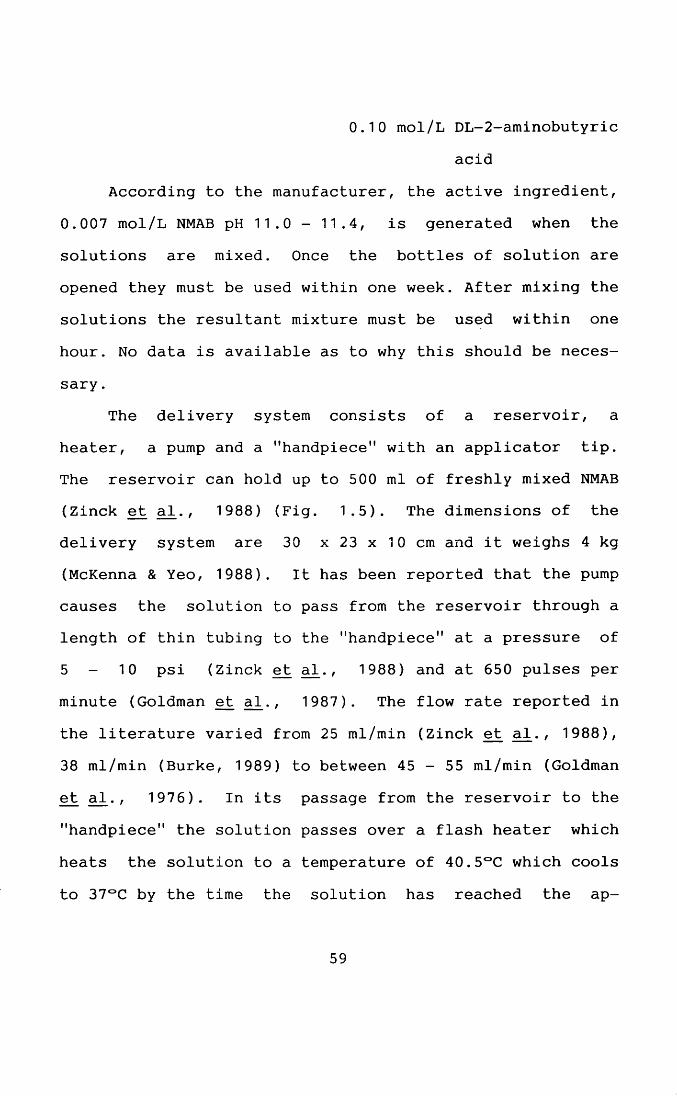
0.10 mol/L DL-2-aminobutyric
acidAccording to the manufacturer, the active ingredient,
0.007 mol/L NMAB pH 11.0 -11.4, is generated when the solutions are mixed. Once the bottles of solution are opened they must be used within one week. After mixing the solutions the resultant mixture must be used within one hour. No data is available as to why this should be neces
sary .The delivery system consists of a reservoir, a
heater, a pump and a "handpiece" with an applicator tip. The reservoir can hold up to 500 ml of freshly mixed NMAB (Zinck et a T ., 1988) (Fig. 1.5). The dimensions of thedelivery system are 30 x 23 x 10 cm and it weighs 4 kg(McKenna & Yeo, 1988). It has been reported that the pump causes the solution to pass from the reservoir through a length of thin tubing to the "handpiece" at a pressure of5 - 10 psi (Zinck et a l ., 1988) and at 650 pulses perminute (Goldman et a l ., 1987). The flow rate reported inthe literature varied from 25 ml/min (Zinck et a l ., 1988), 38 ml/min (Burke, 1989) to between 45 - 55 ml/min (Goldman et a l ., 1976). In its passage from the reservoir to the
"handpiece" the solution passes over a flash heater which
heats the solution to a temperature of 40.5°C which cools to 37°C by the time the solution has reached the ap
59
plicator tip (Robbins, 1987). The rationale for this is that the unanaesthetised patient would not be aware of any temperature difference when the solution was being applied to the dentine. The tips at the end of the "handpiece" are interchangeable (Fig. 1.6). These are the diameter of a
20-gauge hypodermic needle and are flattened towards the tip where the solution emerges. The tips are available
with different angles to facilitate access of the tip into the carious lesion (Goldman & Kronman, 1976; Robbins,1987). A foot pedal controls the pump so that the flow of solution can be activated or stopped at the operator's discretion.
1.10.3 Mode of ActionA biochemical analytical report on the mode of action
of NMG on bovine achilles tendon collagen (Habib et al.,1975) has postulated two possible modes of action of this CMCR agent on carious dentinal collagen :
a. The chlorination of collagen molecules, and
b. the alteration of hydroxyproline, an important constituent in collagen fibre stability (Weissman, 1969), and its glycine dipeptide to pyrrole-2-carboxylic acid and possibly pyrrole-2-carboxylglycine respectively. There is
no study available to confirm the above proposed mode of action. It has also been postulated that NMAB would have
60
the same mode of action although there is no evidence to support this theory (Kronman et a l ., 1979).
This mode of action is based on the previously proposed chlorination by sodium hypochlorite of amino acids and /or proteins (Wright, 1926; Baker, 1947; Kan-
touch & Abdel-Fattah, 1970). The work was carried out using chemical titration and paper chromatography. There has been no literature published in the last twenty years on this subject even though biochemical methodology has improved dramatically in that time.
The effect of NMAB on collagen and its effectiveness relative to NMG have been studied by Kronman et al. (1977, 1979). In that study, bovine achilles tendon collagen samples were treated with NMAB, NMG and a saline control solution at 37°C for 2, 15 or 30 minutes. Samples of collagen were removed and examined using transmission electron microscopy. NMAB and NMG caused disruption of the organisation of the collagen structure and were more effective than the saline control solution. No analytical
reports on the biochemical mode of action of NMAB on dentine collagen are available.
The basic mode of action of the system depends on the fact that the demineralisation of dentine by the carious process leaves free collagen which can then be acted upon
chemically. The collagen in a carious lesion has a par
61
tially degraded fibrillar structure (Oghushi & Fusayama,
1975). It was, therefore, hypothesised that the NMAB solution chlorinates the partially degraded collagen, disrupts the hydrogen bonding and thus affects the secondary and /or quaternary structure of the collagen. The carious dentine becomes more friable and can then be removed more easily.
The application of NMAB to carious dentine softens the first or outer layer which contains denatured collagen. The second or inner layer of carious dentine, con
taining normal collagen fibres, is allegedly unaffected (Kurosaki e_t ad., 1977; Brannstrom et a l . , 1980b).
1.10.4 Toxicity and Pulpal BiocompatibilityThe toxicity of NMAB has been evaluated in rats by
Wedenberg and Bornstein (1990) and its pulpal biocompatibility in human teeth reported (Waltmann et a l ., 1988). No adverse effect on the dental pulp in animals or humans has been noted. The only follow-up report on the short term consequences of chemomechanical pulpal exposure
failed to detect any adverse pulpal response 30 days after
direct pulp capping (Robbins, 1987). The inventors of the
original CMCRS have reported that NMAB has been approved by the U.S. Food and Drug Administrative as safe and effective, and it is recognised as being safe and effective
62
by the American Dental Association (Goldman et a l . , 1987). Among the claims for the system which the FDA approved in 1984 (Brophy & Brophy, 1987) were that it:
a. is safe,b. eliminates the need for local anaesthesia in most
cases,c. will not affect normal tissue,d. conserves healthy tissue,
e. minimises pulp exposure (especially iatrogenic),
f. has no adverse effects on hard or soft tissue,g. is non-irritant to the pulp,h. reduces the use of instrumentation,i. is effective,j. has high patient acceptance.The ADA Council on Dental Materials, Instruments and Equipment has put it in a "recognised" category (Scrabeck and List, 1989 ) .
The toxicological tests carried out by the manufacturer of the C a r i d e x ™ CRS and filed for FDA approval have
not been published in detail and are, therefore, not included in the discussion in this thesis.
63
1.10.5 Evaluation of Effectiveness
In vitro StudiesThe effectiveness of caries removal by NMAB has been
evaluated through in vitro studies on carious dentine and it was claimed to be more effective in removing carious material than a saline placebo (Schutzbank et a l ., 1978).
For the removal of carious tissue of medium-to-hard consistency, NMAB was statistically superior to both saline and NMG (Schutzbank et a l ., 1978).
Clinical TrialsA double-blind study has been used to assess the ef
ficacy of NMG using saline as a control (Goldman et al., 1976). Half of the subjects were treated with NMG and half with saline. The lesion size and nature of the carious dentine were not matched. No information is available on the number of patients who required the use of instrumentation for access or retention. With NMG, 80% of the carious lesions exhibited total caries removal. Difficulty
with access accounted for 75% of the incomplete caries
removal group. Only 10% of the saline group however,
showed total caries removal, this served to differentiate the chemomechanical from the purely mechanical and hydraulic effect of applying a solution to the carious
64
surface.Multiple clinical studies have been conducted which
have reported the effectiveness of NMAB in caries removal (Table 1.3) on :a. all classes of coronal lesions (McCune, 1986; McNierney & Petruzillo et a l ., 1986; Robbins & Ragan, 1988; Punwani
et a l ., 1988; Zinck et a l ., 1988),
b. cervical carious lesions (Robbins 1987; Cavel et a l .,1988), andc. root surface lesions (Tavares et a l ., 1988).
The reported number of teeth with complete caries removal is in the range of 42 - 100% with a majority of
the clinical trials showing a value of over 80% (Table1.3). Incomplete caries removal has been ascribed to :a. difficulty in obtaining access (McCune, 1986), andb. carious dentine having a hard consistency is more difficult to remove (Watson & Kidd, 1986; Gu et a l ., 1987).
Use of hand and /or rotary instruments was necessary to
gain access prior to caries removal in most cases.
The majority of the studies were carried out in permanent teeth and no comparison of the differences in caries removal between permanent and deciduous teeth has been carried out. A procedure involving the use of NMG,
caries detector dye (1% acid red in propylene glycol),
modified Elliot separator and posterior composite resin
65
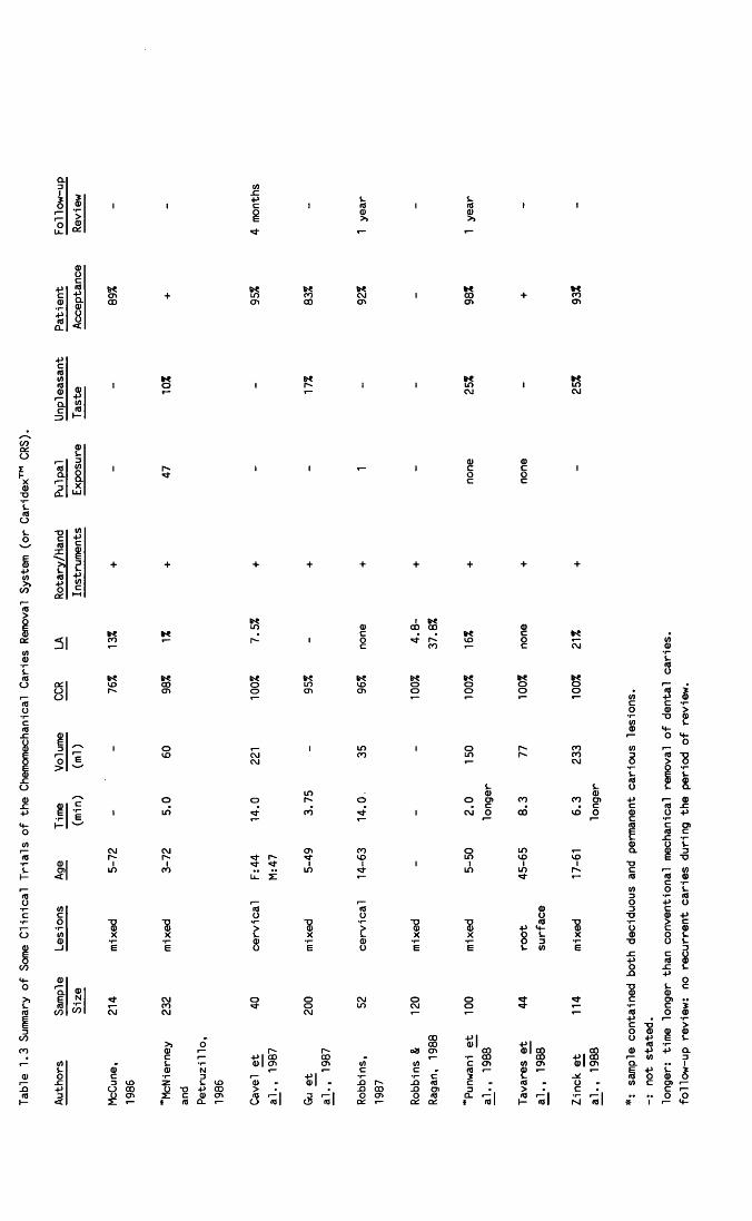
Table
1.3 Summary
of Some Clinical Trials of
the Chemomechanical
Caries Removal
System
(or Caridex™
CRS).
<o+>CLa)oo<00<71
>>m■po
31
I I I
+ + + + + + + + +
1 Nm d> CO CO d>K c c HCO r'~ 1 o <o o' c oo 1 c CM
S'? s? S'? S'? s? « XKO CO o in CD o o o O<71 o <71 <71 o o o o
i O i in 1 o r-> 00<o CM 00 in ooCM ,— CM
t-in d>o o o o Ol 00 ooi in 00 1 CM o CO CD
1 ’
CNJ CM CJ1 oo O inr- O'- r- M" CD in CD COin 00 in in in r-
ul £ *d-
_<9 ro d)O o o
■8 ■8 > ■8 > "8 ■8 ■p <0H- ■8X X i- X i. X X O i- Xd> <1) O 3£ 6 O E O E E s- </) E
o o C\J o oo io CNJ oCNJ *“ *“
CO +*1CO 0)1 +>|r ^ o» ON 00 0)1 58CO CO CO 3
0)1 on ON i/i cn c ON i/i ONc c m 0)+*l c 2 L
0) 0)1 -O .D m c m> -O CO _Q o> 3 >(0 r— I 3 r— | o o> O m CL r - I m r - 1o ml <3 ml a: T— CX cx t ml ml
+>|0)1
I M "m I *: sample contained
both deciduous
and permanent
carious
lesions,
not stated.
longer:
time
longer than conventional mechanical removal
of dental caries,
follow-up
review:
no recurrent
caries during the period of
review.
was reported to be particulary suitable for restorative
treatment of deciduous molars (Yoshida & Motokawa, 1984). The NMAB system has been used with paediatric dental patients as reported by Punwani and McNierney (1988).
Histological StudiesThere seems to be universal agreement in the few
studies which have been published that bacteria are
present in the remaining dentine, both after treatment with NMAB (Katz, 1988; Waltmann et a^., 1988; Roth et a l .,1989) and by conventional means (Katz, 1988; Waltmann et: a l ., 1988). However, it has been reported that NMAB removes approximately 25 - 35% more bacteria than conventional procedures (Katz, 1988). Although it has been suggested that NMAB may exert a bacteriocidal effect (Wedenberg & Bornstein, 1990), one microbial sensitivity study showed that no bacteriocidal effect from the NMAB could be discerned in bacterial samples cultured from carious dentine (Rompen & Charpentier, 1989). It has been
suggested that the cavity prepared by a CMCRS must at least be finished mechanically to remove discoloured or weakly structured marginal segments and to achieve distinct and evenly cut preparation margins (Krejei et al.,
1990).
66
1.10.6 Advantages
Reduced Need for Local AnaesthesiaWith the reduced use of mechanical instrumentation
there is less physical trauma to the dentino-pulpal complex. It has been suggested that warming the solution to 37°C results in less thermal irritation of the pulp which may account for the reduced dentine sensitivity during the operative procedures (Punwani et al., 1988; Tavares eta l . , 1988) and a reduced need for local anaesthesia(McCune, 1986; Robbins, 1987; Punwani et a l ., 1988;
Tavares et a l ., 1988; Zinck et a l ., 1988). Some of theclinical trials which compared CMCR with conventional mechanical caries removal showed that not all patients in the conventionally treated groups required local anaesthesia. There were, however, fewer requests for local anaesthesia with the CMCRS (Table 1.3).
It has been demonstrated that the need for local anaesthesia is operator dependent. An older and more experienced operator used less local anaesthesia on his patients than did a young operator who was more conditioned to the use of local anaesthesia for bothchemomechanical and conventional caries removal techniques
(Robbins & Ragan, 1988).
In the above mentioned studies the need for local
67
anaesthesia may have been further reduced if an adhesive restorative material was used which did not require the use of additional instrumentation to establish retentive features in the cavity. The fact that patients may have
been aware that the NMAB would probably be less painful
could however have contributed to a form of placebo ef
fect .In summary, it is concluded that there may be a
reduced need for local anaesthesia with the CMCRS. This would be of benefit to :a. patients who have management problems (Petruzillo & McNierney, 1988; Zinck et a l ., 1988),b. paediatric dental patients (Rothman, 1985; Punwani et al., 1988),c. patients who do not wish to have a local anaesthetic, andd. those who for medical reasons, should not have a local anaesthetic, for instance haemophiliacs.
Conserves Tooth Structure
The application of NMAB to carious dentine softens
the first or outer layer which contains denatured collagen. The second or inner layer of carious dentine, containing normal collagen fibres, is allegedly unaffected (Kurosaki et a l . , 1977; Brannstrom et a l ., 1980b). The
68
solution will not, therefore, remove sound dentine (Brannstrom et al., 1980b). Before the appearance ofadhesive restorative materials, preparation of retentive features was mandatory when only non-adhesive restorative materials such as amalgam were available. When an adhesive
restorative material is used, no additional use of instrumentation is required to establish retentive features thereby enabling most of the sound tooth structure to be preserved. The combination of CMCRS and adhesive restora
tive materials appears to be mutually advantageous in clinical dentistry (Mclnnes-Ledoux et ad., 1989). However, use of hand and /or rotary instruments may still be necessary to gain access prior to caries removal.
Reduced Risk of Pulpal ExposureIt is theoretically possible that there should be
fewer pulpal exposures with the CMCRS than by use of conventional means. The number of vital pulpal exposures was reported to have been reduced (Goldman and Kronman, 1976)
and it was suggested that traumatic exposure of the pulp should be impossible since the solution will not remove
sound dentine (Kurosaki et a l ., 1977; Brannstrom et a l .,
1980b).Several clinical trials (Table 1.3) have reported on
pulpal exposures with the CMCRS; the percentages ranged
69
from 0% (Tavares et a l ., 1988) to 20% (McNierney &Petruzillo, 1986). No control group using conventional
caries removal methods was included in the latter study nor was any data given on the extent of the lesions
treated. The only report on the short term effects of
chemomechanical pulpal exposure failed to detect any adverse pulpal response 30 days after direct pulp capping (Robbins, 1987 ) .
Improved Bonding with Adhesive Restorative MaterialsThe highly irregular dentinal surfaces of the cavity
floor produced by the CMCRS seem to indicate the possibility of this being used to advantage in the restoration of the lesion. A system providing a mechanical lock for an adhesive restorative material might be possible by utilising an unfilled resin with very low specific gravity to lock into the tubules and undercuts and provide a bond in the same manner as is presently done with acid-etched
enamel. Indeed, the dentinal surface formed is actually a
surface that has been "acid-etched" by the carious process. (Goldman et a l ., 1987, 1988).
The surface energy of chemomechanically treated dentine is greater than that of conventionally treated den
tine (Emanuel & Broome, 1988). The clinical implication is
that chemomechanically treated dentine may have a greater
70
affinity for dentinal adhesives than conventionally
prepared dentine, and therefore result in potentially better bonding. Indeed, some dentine bonding agents have been
shown to have higher bond strengths to dentine in cavities prepared by treatment with NMAB than to that of conventionally prepared cavities (Mclnnes-Ledoux et a l ., 1987;Wolski et_ al. , 1989). The statistical analysis of one ofthe studies (Mclnnes-Ledoux et al., 1987) has been considered weak and raises some doubt about the validity of
the claimed superiority of the NMAB treated and bonded dentine (Soderholm, 1991). What can undoubtedly be concluded from that study is that NMAB treated dentine does not result in an inferior dentine bonding when compared with conventionally treated dentine (Soderholm, 1991). Higher bond strength values of glass polyalkenoate cement to dentine treated with NMAB have also been reported (Burke, 1989; Mclnnes-Ledoux et a l ., 1989).
Patient AcceptanceThe attitudes of patients towards the CMCRS when com
pared with conventional treatment have demonstrated very
favourable acceptance (Table 1.3) ranging from 89%
(McCune, 1986) to 98% (Punwani et a l ., 1988).
71
Domiciliary DentistryDue to the compact size of the C a r i d e x ™ CRS, its use
in domicilliary work may be most appropriate.
1.10.7 Limitations
Use of Hand and /or Rotary InstrumentsThe mode of action of NMAB is based onj its selective
attack on partially degraded dentine collagen in cariouslesions. Tissue or material other than degraded dentine collagen which require removal during the operative procedure still has to be removed by conventional means. Rotary and other hand instruments would therefore still be needed
to :a. establish access to cavities in small lesions (e.g.fissure caries and interproximal carious lesions),b. remove enamel caries,c. remove existing restorations in cases of secondary
caries,d. remove caries or staining along the DEJ which was not
always successfully removed using NMAB,
e. remove darkly stained, hard and eburnated dentine (Watson & Kidd, 1986), andf. prepare the outline form and the resistance and retention form of the cavity for amalgam or other non-adhesive
72
restoration when used.However, it has very recently been suggested that it
is not necessary to remove the discoloured tissue along the DEJ provided that it is clinically sound. The consistency of demineralised dentine may therefore be a better guide to caries activity than its colour (Joyston-Bechal et a l . , 1991). Should this be proven to be the case, thenthe above limitations d. and e. would no longer be valid as far as the CMCRS is concerned and its use would be far more widely applicable than hitherto thought.
Time Required for Caries RemovalSeveral studies comparing the time required for CMCR
with that for conventional procedures confirm that the former requires longer (Gu et a l ., 1987; McCune, 1986;Zinck et a l ., 1988). This varies according to the size and consistency of the carious lesion (Gu et al., 1987; Zincket a l ., 1987). In these studies, the time taken for the
administration and action of local anaesthetic was not
taken into account. The difference in time between the two methods therefore, may not in fact be so great (Table
1.3), if the comparison is based on the time required to carry out the entire procedure. Indeed, it has been reported that patients do not appear to realise that this
procedure takes longer than conventional methods (Zinck et
73
a l ., 1988). For some nervous patients e.g. dental phobics, needle phobics and so on, the time required to carry out a restorative procedure using a CMCRS may compare very
favourably with that of conventional procedures.
TasteBetween 0.5% (McNierney & Petruzillo, 1986) and 3.5%
of patients (Zinck et a T . , 1988) found the taste of thesolution objectionable and the dental procedure had to be terminated. Between 10% (McNierney & Petruzillo, 1986) and
25% (Punwani et_ a l . , 1988; Zinck et a l . , 1988) of patients found the taste unpleasant but allowed the procedure to continue.
CostSpecial equipment must be bought in order to use this
system. It has been estimated that it will require 5,000
patient visits to pay for the equipment investment in a
private dental practice in California (Brophy & Brophy, 1987). The NMAB solution has a half-life of only 60 minutes after which it needs to be replaced. This adds to the inconvenience and expense of the system. Once the bottles of solution are opened they must be used within
seven days and the shelf-life of the unopened solution is three years (Brophy & Brophy, 1987).
74
1.10.8 Improving the FormulationDespite the apparent advantages, the current proce
dure has certain limitations as outlined above. Accordingly, if the procedure is to become clinically useful, a more effective solution must be developed in terms of both
the extent of caries removal and the time involved. Following an extensive literature search, it would appear that since the introduction of the C a r i d e x ™ system in the
1980's there has been no change in the original formulation and no attempt appears to have been made to improve its performance.
Improving the effectiveness of NMAB in caries removal is theoretically possible by incorporating for example other protein denaturing or solublising agent(s) into the present formulation. These additional reagents would require to be effective in assisting NMAB to selectively attack partially degraded dentine collagen as well as being convenient and practical to incorporate into the present
system. Some of the protein denaturing agents commonly used in the laboratory to solubilise proteins including sodium dodecyl sulphate, guanidinium chloride, urea
peroxide, hydrogen peroxide and urea might be used in conjunction with NMAB to remove caries.
75
1.11 Criteria for Caries RemovalThe prime objective of cavity preparation is to carry
out an operative procedure that attempts to remove all carious dental tissues prior to placing a restorative material (Gilmore et a l ., 1982, Pitts, 1991). A carious dentinal lesion has been characterised as consisting of two distinct layers with different ultramicroscopic and chemical structures. The first or outer layer is cbn- taminated with bacteria, the organic matrix is substantially degraded and is not remineralisable and must therefore be removed. The second or inner layer with limited collagen degradation is capable of being remineralised and should be preserved (reviewed by Fusayama, 1979). An ideal method of caries removal would be one which could identify the boundary between remineralisable and unremineralisable dentine and aim to remove only the latter. CMCR may offer this possibility.
Various methods are available to diagnose caries prior to operative procedures (reviewed by Pitts, 1991).
There are few methods that could be deployed to assess complete caries removal. Colour and hardness have been
used as criteria for the clinical assessment of carious dentine. The estimation of hardness of the remaining dentine as felt by hand through an instrument, however, is
not a reliable guide for the clinical removal of caries
76
(Fusayama et aJL. , 1966). Discolouration is considered tobe a reliable guide when removing infected dentine in chronic caries because the discolouration is usually
marked, and the extent of bacterial invasion is demarcated by and follows closely the discolouration front. In acute caries, discolouration is often not marked, and bacterial invasion is usually diffuse and lags far behind the discolouration front (Fusayama et. a l . , 1966; Sato & Fusayama,
1976) and therefore is not a reliable guide for the clinical removal of infected dentine.
The criteria used to assess the adequacy of caries removal have varied widely among clinical trials of the CMCRS. Among the criteria used were :a. clinical judgement (Schutzbank et a l ., 1978; McCune,1986; McNierney & Petruzillo, 1986; Gu et a l ., 1987; Robbins, 1987; Zinck et a l . , 1988), andb. caries detector dyes and clinical judgement (Brannstrom et a l ., 1980b).
In summary, most of the clinical trials on the effectiveness of the CMCRS have used subjective visual and tactile criteria to verify the degree of caries removal.
1.11.1 Caries Detector Dyes
It has been reported that in acute or moderately
acute caries, removal of carious dentine can be assessed
77
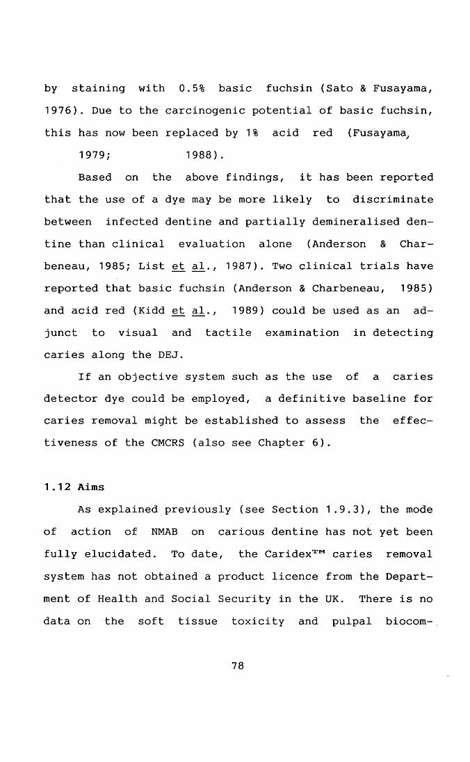
by staining with 0.5% basic fuchsin (Sato & Fusayama,1976). Due to the carcinogenic potential of basic fuchsin, this has now been replaced by 1% acid red (Fusayama,
1979; 1988).Based on the above findings, it has been reported
that the use of a dye may be more likely to discriminate between infected dentine and partially demineralised dentine than clinical evaluation alone (Anderson & Char- beneau, 1985; List et ad., 1987). Two clinical trials have reported that basic fuchsin (Anderson & Charbeneau, 1985)
and acid red (Kidd et a l ., 1989) could be used as an adjunct to visual and tactile examination in detecting caries along the DEJ.
If an objective system such as the use of a caries detector dye could be employed, a definitive baseline for caries removal might be established to assess the effectiveness of the CMCRS (also see Chapter 6).
1.12 AimsAs explained previously (see Section 1.9.3), the mode
of action of NMAB on carious dentine has not yet been fully elucidated. To date, the C a r i d e x ™ caries removal
system has not obtained a product licence from the Department of Health and Social Security in the UK. There is no
data on the soft tissue toxicity and pulpal biocom-
78
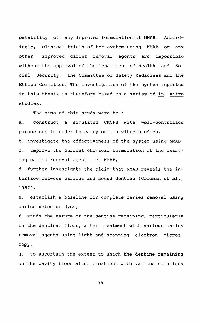
patability of any improved formulation of NMAB. Accordingly, clinical trials of the system using NMAB or any
other improved caries removal agents are impossible without the approval of the Department of Health and Social Security, the Committee of Safety Medicines and the Ethics Committee. The investigation of the system reported in this thesis is therefore based on a series of in vitro studies.
The aims of this study were to :a. construct a simulated CMCRS with well-controlled parameters in order to carry out in vitro studies,b. investigate the effectiveness of the system using NMAB,c. improve the current chemical formulation of the existing caries removal agent i.e. NMAB,d. further investigate the claim that NMAB reveals the interface between carious and sound dentine (Goldman et al., 1987),
e. establish a baseline for complete caries removal using caries detector dyes,f. study the nature of the dentine remaining, particularly in the dentinal floor, after treatment with various caries removal agents using light and scanning electron microscopy,
g. to ascertain the extent to which the dentine remaining
on the cavity floor after treatment with various solutions
79
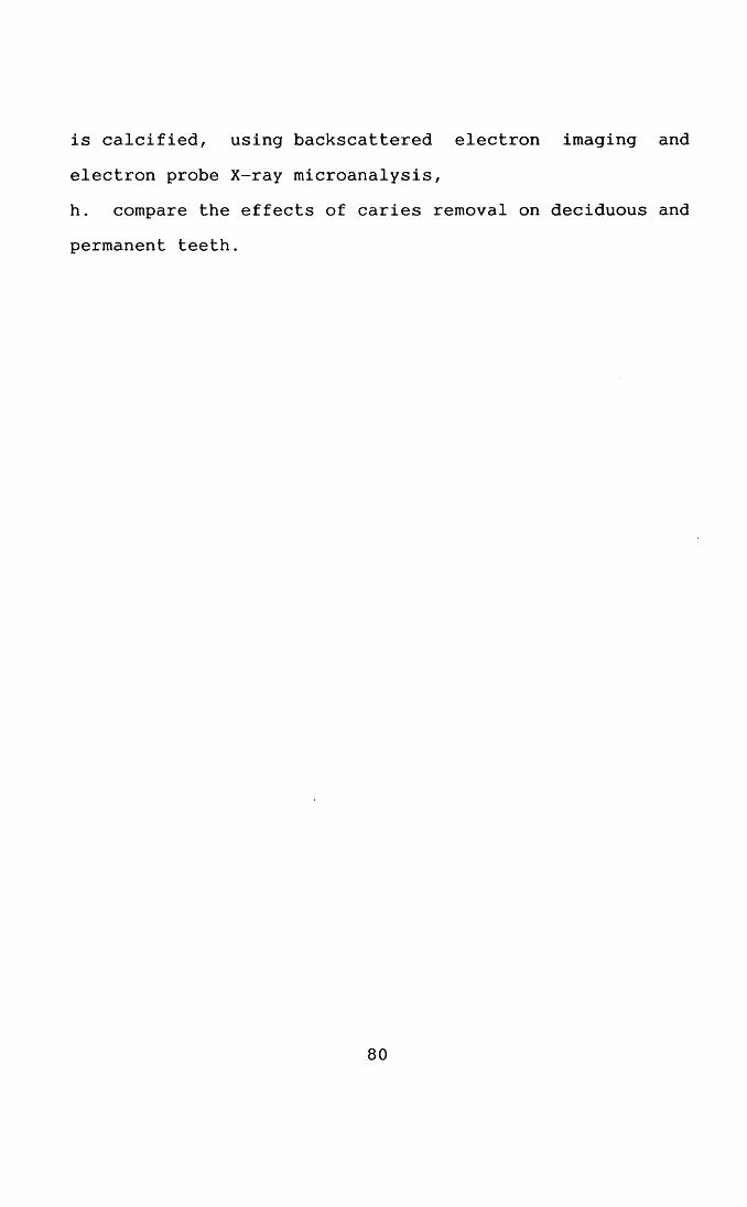
is calcified, using backscattered electron imaging and
electron probe X-ray microanalysis,h. compare the effects of caries removal on deciduous and
permanent teeth.
80
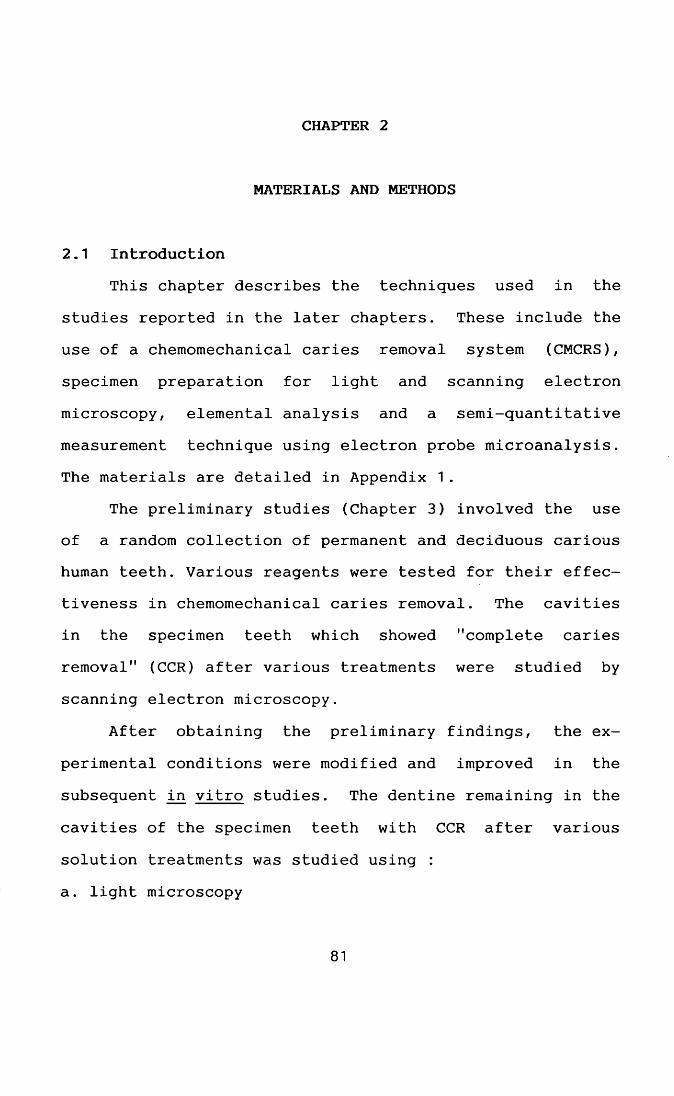
CHAPTER 2
MATERIALS AND METHODS
2.1 IntroductionThis chapter describes the techniques used in the
studies reported in the later chapters. These include the use of a chemomechanical caries removal system (CMCRS), specimen preparation for light and scanning electron microscopy, elemental analysis and a semi-quantitative measurement technique using electron probe microanalysis. The materials are detailed in Appendix 1.
The preliminary studies (Chapter 3) involved the use of a random collection of permanent and deciduous carious human teeth. Various reagents were tested for their effectiveness in chemomechanical caries removal. The cavities in the specimen teeth which showed "complete caries removal" (CCR) after various treatments were studied by scanning electron microscopy.
After obtaining the preliminary findings, the ex
perimental conditions were modified and improved in the subsequent in vitro studies. The dentine remaining in the cavities of the specimen teeth with CCR after various
solution treatments was studied using :a. light microscopy
81
i. visual examination under magnification using a
stereomicroscope at X4 and X10 magnificationsii. histological sections (ground and demineralised)
-haematoxylin and eosin (Bancroft & Stevens, 1984) -Gram-Weigert (Brown & Brenn, 1931)-basic fuchsin and acid red (caries detector dyes)
b. scanning electron microscopy (SEM)i. secondary electron imaging (SE)
ii. backscattered electron imaging (BSE)iii. electron probe microanalysis (EPMA).
2.2 Overview of the Plan of the InvestigationThe work of this thesis started with the construction
in the laboratory of a CMCRS (Chapter 3). The author initially familiarised himself with the use of this equipment and the technique by carrying out a few trials using reagents chosen from the information available in the literature (Chapter 3). A series of chemicals was then tested in an attempt to improve the effectiveness of the
existing formulation for caries removal (Chapter 3). The
pH and osmolality of the improved reagent was tested prior
to a series of in vitro studies comparing the effectiveness of the modified caries removal agent on decay in both permanent and deciduous teeth (Chapter 3). The criteria used to define caries removal were however subjective,
82
using clinical parameters. SEM was used to study the nature of the dentinal surface after complete caries removal (Chapter 4). A more detailed study was also carried out to
study the interface between carious and sound dentine using LM and SEM (Chapter 5). An attempt was made to in
vestigate the possibility of using caries detector dyes to "objectively" assess CCR prior to a more extensive study (Chapter 6 ). A study involving the use of lesions at a more well defined stage in the carious process was carried out to compare the existing and the improved formulation in caries removal (Chapter 7). The mineral content of the dentine remaining after CCR was studied using BSE and EPMA (Chapter 8 ).
2.2.1 Specimen TeethIn the preliminary studies (Chapter 3), freshly ex
tracted permanent teeth with coronal carious lesions were collected from the Oral Surgery Department (Glasgow Dental
Hospital & School). Organic deposits on the teeth such as plaque and food debris were removed gently with both air and water with a three-in-one syringe. Only carious lesions where instrumentation was straight forward and without too many undercuts and free from darkly-stained,
hard and eburnated dentine on the surface were used. The
teeth were bottled individually in phosphate buffered
83
saline (PBS: 8.0 g/L NaCl; 0.2 g/L KC1; 1.15 g/L NazHPCU;0.2 g/L KH 2 PO 4 , pH 7.4) with 0.15% (w/v) thimerosal
added as an antibacterial agent and stored at 4°C.These teeth were used either immediately or within
one month of extraction. The PBS was changed daily for those stored prior to use.
At a later stage (Chapters 5, 7 and 8 ), a more well-defined group of carious lesions was used. The specimen teeth were collected individually in phosphate buffered saline with sodium azide (2 .0 % w/v) added as an antibacterial agent. Sodium azide is an equally effective antibacterial agent but does not contain mercury as does thimerosal (Merck Index Co. Inc., 1960) which would complicate EPMA studies. Storage solutions were changed daily.
In the preliminary studies (Chapter 3), the teeth selected were those designated for extraction because of
gross caries and for which CMCR would not normally be used in a clinical situation. This allowed a large number of specimen teeth to be collected in a short period of time in order to assess the effectiveness of the various caries removal agents.
The effectiveness of caries removal by a caries
removal agent depends on the type of lesion used
(Schutzbank et a l ., 1978). In subsequent studies (Chapter
84
7), it was necessary to be selective in the type of carious lesions used in order to compare the effectiveness of various chemical agents in caries removal more accurately. Teeth used in the later series of in vitro studies (Chapters 5 and 7) were, therefore, limited to carious lesions which could have been restored with either amalgam or adhesive restorative materials in clinical situations. The following criteria were employed in all the selections of samples other than those used in the preliminary studies :a. primary caries,b. coronal dentinal caries,c. no radiographic pulpal involvement,d. easy access with little undermined enamel,e. medium consistency (see below) tested with a sharp dental probe,f. isolated lesion without communication with other lesions in the same tooth,
g. no periapical pathology e.g. periapical granuloma,h. no darkly-stained, hard and eburnated dentine caries onthe surface of the lesion,i. not grossly carious, and
j . in deciduous teeth root resorption was limited to less
than one third of the original root length (in the judgement of the operator).
85
Carious tissue was classified as being of a medium consistency if it was resistant to/probing, not easily removed by mechanical means but readily penetrated by a sharp dental probe (Schutzbank et al., 1978).
All carious lesions were assessed by the author. The assessment and selection of the specimen teeth was carried out in a dry field using an optical loupe with X4 magnification under good standard lighting. Periapical dental radiographs were then taken of the selected teeth using Kodak Ultraspeed films and exposed at a long cone-object
distance of approximately 16 inches with the aid of a film holder. Radiographs were developed in a standard automatic dental X-ray film processor (Velopex Mini-dry, Billericay Dental Supply Co. Ltd., 6 Perry way, Witham, Essex, UK). Only specimen teeth which satisfied the requirements listed above were used in the later studies (Chapters 5, 7 and 8).
The period involved in this series of studies was much longer than the preliminary one due to the difficulty
in acquiring sufficient specimens of both permanent and deciduous teeth with restorable lesions. A smaller sample size was therefore used in the case of permanent teeth in the later study (Chapter 5). The teeth used in this study
were as follows :a. deciduous teeth :
86
extracted during inhalation analgesia, intravenous sedation, and local anaesthesia,
b. permanent teeth :i. extracted for orthodontic reasons under local anaes
thesia or extracted along with deciduous teeth as ina. above,
ii. extracted for prosthetic reasons e.g. dental clearance, and
iii. restorable carious lesions extracted at the patients' requests.
2.2.2 Caries Removal Agents0.007 mol/L (0.11% w/v) N-monochloro-DL-2-
aminobutyrate (NMAB) was prepared by mixing equal volumes
(250 ml) of solutions I and II (Schutzbank et a l ., 1978).This formulation is also marketed under the trade name :C a r i d e x ™ solutions (McCune, 1986).
Solution I Solution II0.014 mol/L (1 % w/v) NaOCl 0.10 mol/L NaOH
0.10 mol/L NaCl
0.10 mol/L DL-2-aminobutyric
acidA number of other different chemical agents were also
tested as potential caries removal agents : guanidinium chloride (4 mol/L)
87
hydrogen peroxide (H2 O 2 ) solution (30% v/v) sodium dodecyl sulphate (SDS) solution (2 mol/L)
two sodium hypochlorite (NaOCl) solutions (0.5% w/v, 1.0% w/v) urea (2 mol/L)
urea hydrogen peroxide (33 - 35% w/v H 2 O 2 ) isotonic saline solution (0.9% w/v)Extracted permanent teeth with coronal carious le
sions were immersed in 10 ml of each of the above solutions and observed for a period of 15 days. The solutions were changed daily. The changes in consistency, surface texture and colour of the carious dentine, if any, were recorded.
8 mol/L guanidinium chloride,| 30% v/v H 2 O 2 , 4 mol/LSDS, 4 mol/L urea and 33 - 35% w/v urea hydrogen peroxide were also individually incorporated into solution II in the NMAB system in order to test their effectiveness in removing caries using the simulated CMCRS (Section 2.2.4).
Prior to this, the author practised with this technique using NMAB in order to acquire the skills necessary to
compare, subjectively, the ease of caries removal of NMAB with that of other caries removal agents.
88
Most of the solutions were either used immediately or stored at 4°C until required (up to a period of one week) except NaOCl which was diluted from concentrated solution freshly prior to use.
NMAB, NMAB containing 2 mol/L urea (NMAB-Urea), 0.5% (w/v) NaOCl, 2 mol/L urea and isotonic saline were selected for the preliminary studies (Chapter 3). Only NMAB, NMAB-Urea and isotonic saline were used in the subsequent studies (Chapters 5, 7 and 8).
2.2.3 Measurement of pH and OsmolalityThe pH of NMAB and NMAB-Urea used in the preliminary
studies (Chapters 3 and 4) were measured before and after mixing solutions I and II. A pH meter (type PHM26, Radiometer, Copenhagen, Denmark) was used to monitor the pH at intervals of 10 min for a period of two hours after mixing. The change in pH with time was expressed graphically (Fig. 3.1).
The osmolality of the NMAB and NMAB-Urea solutions used in the preliminary studies (Chapters 3 and 4) were measured by freezing point depression, at 10 min intervals for two hours after mixing using an electronic osmometer (Electronic Semi-micro Osmometer, Knauer, Wis-
senschaftlicher geratebau, kG Dr.-Ing. Herbert Knauer &
89
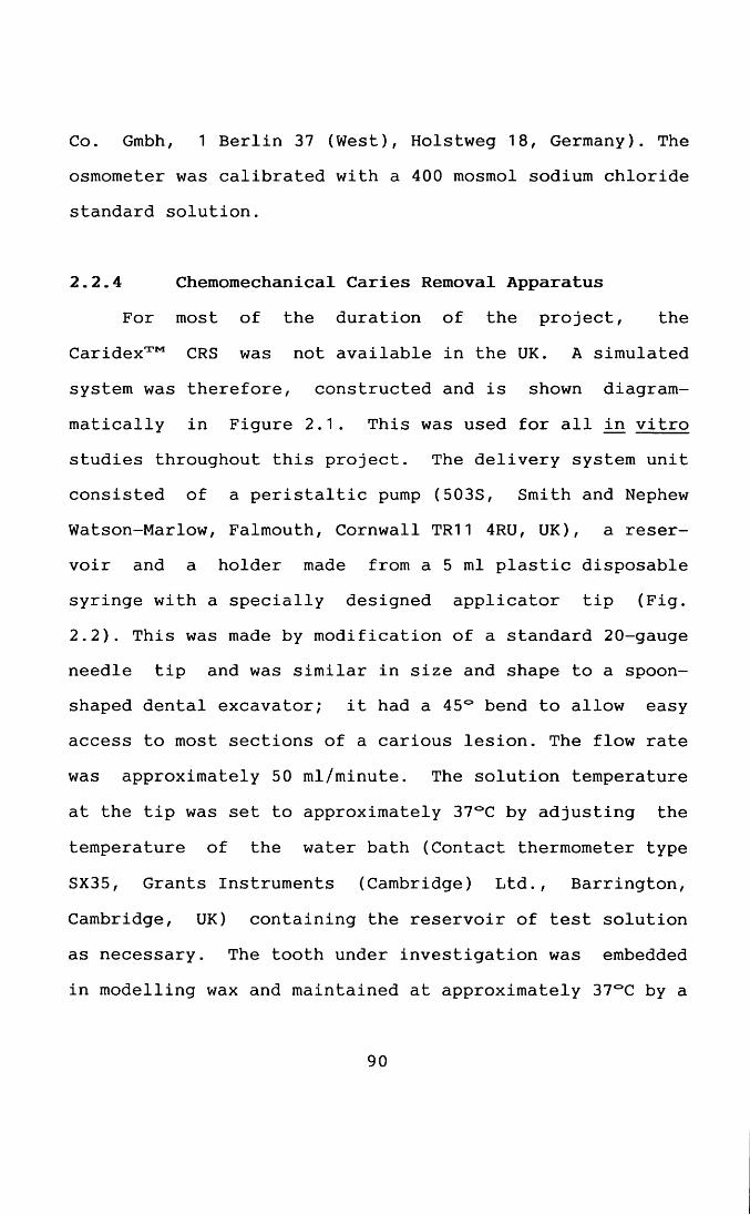
Co. Gmbh, 1 Berlin 37 (West), Holstweg 18, Germany). The osmometer was calibrated with a 400 mosmol sodium chloride standard solution.
2.2.4 Chemomechanical Caries Removal ApparatusFor most of the duration of the project, the
C a r i d e x ™ CRS was not available in the UK. A simulated system was therefore, constructed and is shown diagram- matically in Figure 2.1. This was used for all in vitro studies throughout this project. The delivery system unit consisted of a peristaltic pump (503S, Smith and Nephew Watson-Marlow, Falmouth, Cornwall TR11 4RU, UK), a reservoir and a holder made from a 5 ml plastic disposable syringe with a specially designed applicator tip (Fig. 2.2). This was made by modification of a standard 20-gauge needle tip and was similar in size and shape to a spoonshaped dental excavator; it had a 45° bend to allow easy access to most sections of a carious lesion. The flow rate was approximately 50 ml/minute. The solution temperature at the tip was set to approximately 37°C by adjusting the temperature of the water bath (Contact thermometer type
SX35, Grants Instruments (Cambridge) Ltd., Barrington, Cambridge, UK) containing the reservoir of test solution as necessary. The tooth under investigation was embedded in modelling wax and maintained at approximately 37°C by a
90
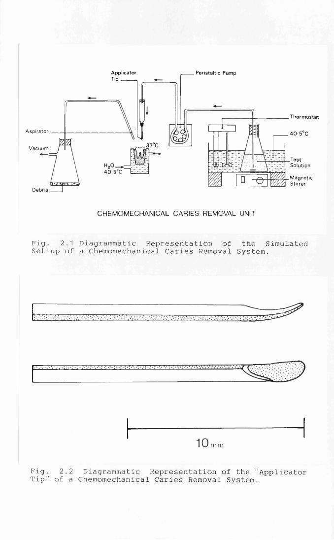
ApplicatorT'P-------
Peristaltic Pump
Thermostat
Aspirator 40 5°C37°CVacuum
.TestSolution
40 5°CMagneticStirrer
Debris
CHEMOMECHANICAL CARIES REMOVAL UNIT
Fig. 2.1 Diagrammatic Representation of the Simulated Set-up of a Chemomechanical Caries Removal System.
m m
Fig. 2.2 Diagrammatic Representation of the "Applicator Tip" of a Chemomechanical Caries Removal System.
water jacket linked to a second water bath (Shandon, London SW7, UK). The temperature settings were as indicated in the diagram (Fig 2.1).
The temperature and flow rate of the C a r i d e x ™ CRS have been reported to fluctuate (Burke, 1989). These parameters were however accurately controlled with the
system used in this project.
2.2.5 Experimental DesignThe experimental design of the in vitro studies took
the form of a blind trial, with the test solutions being prepared and assigned codes by an individual not involved in the study. The code was only broken when the study was complete. The author carried out all of the experimental procedures. The solutions were contained in brown glass bottles in order to avoid :a. any possibility of breaking the code by the operator, andb. photochemical decomposition of the NaOCl solution (British Standard, 1988).
Solutions I and II were pre-incubated at ap
proximately 40°C for 5 - 1 0 minutes before mixing. Water
at the same temperature was pumped through the delivery unit for few minutes to pre-warm it. The outlet tempera
ture was approximately 37°C. The solutions were then
91
poured into the conical flask and magnetically stirred for two minutes before flushing onto the carious lesion. Water was passed through the system for 2 min after each treatment in order to remove any residual caries removal agent. The waste solution and carious debris excavated were
removed by vacuum suction. This experimental designclosely followed that recommended by the manufacturer of
the C a r i d e x ™ CRS and that used in a previous in vitrostudy (Schutzbank et a l ., 1978).
2.2.6 Chemomechanical Caries Removal TechniqueTreatment consisted of directing the stream of solu
tion onto the carious lesions by means of the applicator. The operator wore a pair of surgical gloves and a face mask. A gentle scraping action was applied to the carious lesions. During application, some of the gross debris was lifted up and loosened by the tip and removed by aspiration. The procedure was continued until either CCR was
achieved or no more carious material could be removed by the 500 ml of caries removal agent used. While conducting this aspect of the study, a few preliminary trials using
both permanent and deciduous carious lesions were carried out in order to allow the operator to familiarise himself
with the technique and acquire the necessary skills prior to the actual in vitro studies being carried out.
92
2.2.7 Recording of ResultsThe results were scored using a scale of 0 - 5, this
being indicative of the percentage of caries removal (Table 2.1). The volume of solution used and time taken were also recorded. Only those teeth in which CCR was
achieved were selected for further studies. The criteria for CCR used in all the in vitro studies were those used
clinically during cavity preparation, i.e. :a. removal of soft stained caries from the carious lesion leaving only sound unstained dentine, orb. any stained dentine remaining being firm when tested by a sharp dental probe as judged by the operator. Any staining at the DEJ was also recorded.
After the preliminary studies (Chapter 3), some modifications to the method of examination of the cavities after caries removal were introduced. After treatment with various solutions, the air-dried cavities were examined under good lighting using an operating loupe with X4 magnification, particularly along the DEJ. Those teeth that
were judged to have CCR were then re-examined using a
Wild M3Z stereomicroscope (Wild Leitz (UK) Ltd., Davy Avenue, Knowlhill, Milton Keynes, MK5 8LB, England) at x10
magnification. A colour transparency record was made ofeach of the specimens with CCR at X10 magnification using
93

Table 2.1 Summary of Scoring for Caries Remo1(Schutzbank et a l ., 1978).Observation Removal Approx. % Estimation
Score Removal of CriteriaCarious Lesion
no removal 0 0 obviousperceptible 1 10 justremoval noticeable< 1/2 removal 2 30 10 - 50%> 1/2 removal 3 70 50 - 90%almost complete 4 90 noticeablyremoval completecomplete removal 5 100 obvious
Kodak Ektachrome 50 film, showing the surface of the cavity to the greatest possible advantage for visual reference when sectioning teeth. A dental radiograph was also taken using Kodak Ultraspeed film and exposed at a
long cone-object distance of approximately 16 inches. Radiographs were developed in a standard automatic dental
X-ray film processor.
2.3 Light Microscopy
2.3.1 IntroductionThe earliest attempts to study the microscopic struc
ture of calcified tissues were made on histological sections prepared by grinding down slices of bone or teeth until sufficiently thin to permit examination under the microscope. This type of section has been replaced almost completely by the use of decalcified tissue sections. This is because decalcified tissues can be cut more thinly than calcified tissues, thus allowing the sections to be
studied under a wide range of magnifications (Brain, 1966).
The preparation of decalcified sections for microscopy is divided into several stages, their numbers depending on the medium used for infiltration and embedding. For
example, the preparation of sections embedded in paraffin
94
wax requires nine distinct steps. The function of each stage has been designed to form part of a systematic and complete process (Brain, 1966), which is briefly described
as follows :a. Fixation, i.e. the preservation of the tissues.b. Decalcification, i.e. the removal of the inorganic com
ponents from the hard tissues.c. Washing, i.e. the removal of the decalcifying agent.d. Dehydration, i.e. the removal of the washing fluid.e. Clearing, i.e. the replacement of the dehydrating fluid by a solvent of the infiltrating medium.f. Infiltration, i.e. the saturation of the tissues with a
liquid medium, which can subsequently be solidified.g. Embedding, i.e. encasing the tissues in the solid medium.h. Sectioning.i. Staining and mounting the section.
Lack of contrast makes it difficult to examine microscopically the minute elements of the tissues in un
stained sections by transmitted light. The use of stains allows intercellular substance to be differentiated from
cells and the different constituents in each of these to be recognised. Haematoxylin and Eosin staining is one of the commonest general stains used with decalcified dental
tissues. It enables most of the structural details of a
95
decalcified section to be visualised under transmitted
light. Bacteria may be stained by this method but there is no indication whether they are Gram-positive or Gram- negative micro-organisms. The Gram-Weigert staining procedure is useful as a routine stain for bacteria in tissues and is particularly valuable in the case of mixed infections where both Gram-positive and Gram-negative bacteria may be found and differentiated (Brown & Brenn, 1931).
2.3.2 Specimen PreparationLight microscopy was employed in the study of the
remaining dentine of the cavities with CCR after treatment with various solutions (Chapters 5 and 8). Teeth with CCR were randomly selected and rinsed with distilled water to wash away any salts remaining after treatment. A guidance groove close to the cavity margin was cut using a carborundum disc powered by an electric motor (Type 520, Kal- tenbach und Voigt Leutkirch /A, Germany) and with constant water cooling. Each tooth was then sectioned with an osteotome (6 mm, Skidmore, Vicarey Davidson & Co., Unit 10, 30A Cumberland Street, Glasgow, G5 79Q, Scotland) through
the middle of the cavity. One half was fixed in 70%
ethanol and processed for scanning electron microscopy.
The other half was stored in 10% neutral formalin in an
individual air-tight specimen bottle (Medical Wire and
96
Equipment Co. Ltd., Potley, Corsham, Wiltshire, UK) for one week at room temperature for histological processing and staining. They were then washed for 5 hours prior to being transferred to a plastic specimen container and
decalcified in 11.9% w/v ethylenediaminetetra-acetic acid (EDTA) (with 4.8% w/v dimethylsulphoxide added to aid the penetration of the EDTA into the dental tissue) at pH 7.4, 37°C with constant agitation for 2 - 4 weeks until radiographs showed no evidence of any remaining mineralisation. The EDTA solution was changed weekly. Wijnbergen and van Mullem (1987) reported an in vitro study in which a reduction in the number of micro
organisms and the ability to stain Gram-positive organisms occurred following treatment with various demineralising fluids. The effect appeared to be least severe with an EDTA-based preparation. Decalcified specimens were rinsed for 5 hours and returned to 10% neutral formalin for 1 - 7
days fixation prior to processing for histological ex
amination. Decalcified tissues were dehydrated and em
bedded in paraffin blocks (Appendix 3). The specimen teeth were orientated during the embedding procedure so that the longitudinal sections would pass through the deepest part of the cavity. Three consecutive sections 7 pm thick were
then prepared at representative depths from 3 - 4 separate
areas of the specimens; 9 - 1 5 sections were cut for each
97
specimen, the number of sections obtained being determined mainly by the size of the lesion. Only 2 - 4 consecutive sections from each site were chosen and mounted on glass
slides, deparaffinised, hydrated and stained with haematoxylin and eosin (Appendix 4) or Gram-Weigert (Appendix 5) stain for examination under the light microscope. The remaining sections were retained in case some were lost during the staining procedures or had a less than ideal histological appearance. Photomicrographs of the sections were taken at low and high powers (e.g. X100, X200, X400). Each slide was examined for the types of den
tine present, i.e. secondary or reparative.Ideally, a sterile procedure for specimen collection
and preparation should be adopted if light microscopy is used to detect the presence of bacteria in the remaining dentine after caries removal. Serial sectioning is necessary in order to reduce the possibility that infected tubules may be missed due to the plane in which the sec
tions were cut (Watts & Paterson, 1990). The presence of bacteria in the sections in the light microscopic studies was nevertheless still recorded in order to give an additional indication as to the effectiveness of caries
removal in teeth with clinically CCR (see also Chapters 5 and 7), even though some (or all) of them may have been
acquired subsequent to extraction.
98
2.4 Scanning Electron Microscopy (SEM)
2.4.1 IntroductionThe scanning electron microscope (SEM) produces a
vivid, seemingly three-dimensional picture of the specimen surface over a wide range of magnifications. Scanning electron micrographs are unsurpassed in beauty and
topographical detail. External or internal surfaces of the
specimen may be studied by the use of different preparation techniques and additional accessories now make it possible to analyse the elemental composition of thespecimen.
The SEM can achieve a depth of focus -500 times greater than that of the light microscope at equivalent magnifications. This excellent depth of focus provides us with the opportunity to visualise three-dimensionallyspecimens that could previously be seen only as thin sections, as replicas, or at a low resolution with the light microscope. The ability to view the specimen at an angle under the electron beam enhances the three-dimensional ef
fect. The depth of focus available makes the SEM suitable
even for applications in which the magnification andresolution of the light microscope are inadequate. Images ranging in magnification from X20 to X50,000 can be ob
99
tained with the SEM without any part of the image being out of focus. In other words, the specimen can be observed
at progressively higher magnifications without any change in focus or brightness of image. Generally, a continous shift in magnification can be obtained at the accelerating
voltage of 25 kV. Because the specimen area selected for higher magnification in the SEM is always at the centre of the lower magnification image, it is easily identified. Other advantages of the SEM are : the specimen need not be cut into thin sections before examination, and relatively large areas (~1 cm2 ) of the specimen can be examined which allows reliable estimation of morphological variations in the given specimen.
The success of the SEM with calcified tissues relates to their lower water content, with a consequent reduction in shrinkage due to the drying necessary for examination in the high vacuum system of the microscope. The aim of drying is to leave the solids in their original locations within the specimen and maintain the mechanical and morphological stability of the specimens. The examination of
calcified tissues is, however, not free from problems. So
far as the study of the cells associated with these
tissues is concerned, the problems are greater than with any other natural biological samples when studied by SEM because of the large difference in shrinkage between the
100
cells and mineralised matrices. (Boyde, 1984). The SEM can resolve topographical details of 5 - 10 nm in size,depending on the accelerating voltage used. Another
limitation of the SEM is the vacuum environment in which the specimen must be viewed. Also, the SEM usually fails to discern some of the internal details of the specimen, which are often visible with the light microscope. Furthermore, it lacks colour response which is a means of increasing contrast in light microscopy in addition to differences in light intensity. The findings from both light and scanning electron microscopic studies can therefore be correlated to yield more information about the specimens.
In the conventional SEM, specimens must be capable of withstanding very low pressures. The specimen surface must not become charged when it is exposed to the electron beam, because an irregular charge density on the specimen surface would seriously and unpredictably affect both the incident and secondary electrons. Excess incident electrons would accumulate as a result of the voltages (> 2 kV) needed to obtain satisfactory signal levels (i.e.
signal to noise ratio). The specimen surface must, there
fore, be conductive. To accomplish this, specimens are coated with gold, carbon or other conductive materials.
At low beam voltages (1 - 3 kV), the charging is reduced and a minimum amount of metal is needed on the specimen
101
surface to suppress charging effects. In certain cases, especially at low magnifications (X 1 - 600), it is possible to use uncoated specimens. Uncoated specimens are usually viewed at low accelerating voltages (below 5 kV) in order to reduce both beam penetration and radiation flux impinging on the specimen (Hayat, 1978).
If the specimen is hard and dry (e.g. bone), it needs only to be attached to the specimen stub and coated with a
thin layer of a suitable metal. If, on the other hand, the
specimen is soft and hydrated (e.g. kidney), it may have to undergo fixation, dehydration, and critical point drying before it is mounted on the stub (Boyde, 1984).
In the conventional SEM, the electron beam is focused
to as small a point as possible. This probe is moved over the specimen in a regular pattern, similar to the spot on
a television tube. When an electron beam strikes a solid
specimen a number of interactions occur, the most impor
tant of these being illustrated in Figs. 2.3 & 2.4.
Electrons may be backscattered from the front face of the
specimen with little or no energy loss, or they may inter
act with surface atoms to produce secondary (low energy)
electrons. Some electrons may be absorbed by the specimen
with transfer of energy to heat and sometimes to light.
Transmitted electrons may be unchanged in direction or scattered at different angles. Scattered electrons may be
102
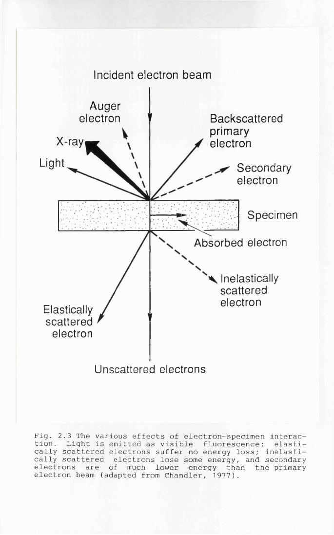
Incident electron beam
Augerelectron Backscattered
primaryelectronX-ray
Light Secondaryelectron
Specimen
Absorbed electron
V Inelastically scattered electron
Elasticallyscattered
electron
Unscattered electrons
Fig. 2.3 The various effects of electron-specimen interaction. Light is emitted as visible fluorescence; elastically scattered electrons suffer no energy loss; inelastically scattered electrons lose some energy, and secondary electrons are of much lower energy than the primary electron beam (adapted from Chandler, 1977).
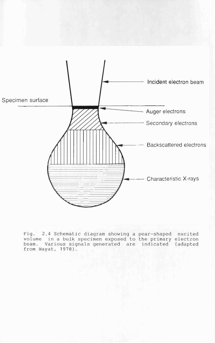
Incident electron beam
Specimen surface
Auger electrons
Secondary electrons
Backscattered electrons
Characteristic X-rays
Fig. 2.4 Schematic diagram showing a pear-shaped excited volume in a bulk specimen exposed to the primary electron beam. Various signals generated are indicated (adapted from Hayat, 1978).
elastic (no energy loss) or inelastic (having lost some energy). If energy is transmitted to the specimen it may
also result in the emission of X-rays as the excess energy is released when an electron in an outer atomic shell
falls into the hole in an inner shell created by the ionisation process. In the alternative mechanism, the excess energy is given to another atomic electron which may then escape from the atom; these electrons are known as Auger electrons. Each of these events can provide information about the specimen.
When operating in the conventional secondary electron mode, the secondary electrons are collected and the resultant electron current will vary depending upon the shape of the specimen surface. These current variations are converted into voltage variations and then displayed on a cathode ray tube, which is scanned in synchrony with the primary beam on the specimen. The result is the production of a picture of the surface topography which has a clear three-dimensional effect.
2.4.2 Specimen Preparation
The teeth with CCR in the preliminary studies (Chapter 3) were randomly selected for SEM and rinsed with
distilled water to wash away any salts remaining after
treatment with the various caries removal agents. The
103
specimens were subjected to critical point drying without fixation (Critical Point Drier Polaron E300Q, Polaron Equipment Limited, 21 Greenhill Crescent, Holywell Industrial Estate, Watford, Herfordshire, WD1 8 XG, England) by placing in ascending concentrations of ethyl alcohol
through to absolute ethanol (one change of 70% ethanol,
two changes of 90% ethanol and then two changes of ab
solute ethanol). Ethanol is known to cause shrinkage in tissues and has a coagulant effect on proteins by displacing water, thereby breaking hydrogen bonds to produce denaturation (Kiernan, 1990). The use of 70% ethanol or acetone in critical point drying has, however, been
reported not to cause any significant volume changes (Boyde, 1984). The ethanol was replaced with liquid carbon dioxide in a critical point drier and the pressure slowly released until normal atmospheric pressure was attained (see Appendix 2).
After critical point drying, each of the specimens
was then attached to a specimen stub and given an
electrically conductive coating of gold approximately 2 0
nm in thickness. For use at low magnifications with secondary electron imaging, problems due to imperfect con
tact of adjacent islands of metal in the conductive coating are easily minimised by applying an excess coating
e.g. 50 nm of gold (Boyde, 1984). Due to the irregularity
104
of the cavities, some tooth specimens required a second coating in order to obtain a reasonable image. Photomicrographs (Ilford FP4 film) of the dentinal sur
faces were taken using the Jeol JSM-T100 scanning electron microscope in the secondary electron imaging mode,
operated at 15 kV. No attempt was made to quantify the proportion of different types of dentinal surface observed. Photomicrographs were taken of typical and atypi
cal areas of the dentinal surfaces of all the cavities
before breaking the treatment code.After the preliminary studies (Chapter 3), some
modifications in specimen preparation were introduced. The
specimen teeth were split into two halves with an osteotome, one half being used for light microscopy (see Section 2.3.2) and the other half for SEM. The specimens to be studied by SEM were then fixed in either 7 0% ethanol (Boyde, 1984) or 3% (w/v) glutaraldehyde buffered in 0.1 mol/L sodium cacodylate to pH 7.4 (Boyde & Vesely, 1972).
The same procedures of critical point drying and gold coating were applied. Scanning electron micrographs of the dentinal surfaces were taken in the secondary electron im
aging mode using a Cambridge Stereoscan 360 scanning electron microscope (Cambridge Instrument, Cambridge, UK),
105
operated at 20 kV. Again, no attempt was made to quantify the proportion of different types of dentinal surface observed .
2.5 Backscattered Electron Imaging (BSE)
2.5.1 IntroductionBackscattered electron imaging of carious lesions
received little attention until recent work (Boyde & Jones, 1983; Jones & Boyde, 1987) demonstrated the potential for this technique in the study of carious lesions in enamel and dentine. Pearce and Nelson (1989) showed that detailed structural information on carious lesions in human enamel could be obtained with this method, and that the signal emanated from about the first 4 pm from the surface and appeared sensitive to changes in mineral density. The images were similar to those obtained with microradiography for enamel lesions, but offered much
higher resolution. The intensity of the backscattered beam
is determined by the scattering properties of particles inside the samples and their geometric distributions. Jones and Boyde (1987) claimed that once these are made
fully quantitative they would have greater resolution than microradiography.
106
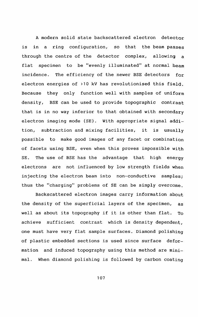
A modern solid state backscattered electron detector is in a ring configuration, so that the beam passes through the centre of the detector complex, allowing a
flat specimen to be "evenly illuminated" at normal beam incidence. The efficiency of the newer BSE detectors for electron energies of >10 kV has revolutionised this field.
Because they only function well with samples of uniform
density, BSE can be used to provide topographic contrast that is in no way inferior to that obtained with secondary electron imaging mode (SE). With appropriate signal addition, subtraction and mixing facilities, it is usually possible to make good images of any facet or combination of facets using BSE, even when this proves impossible with SE. The use of BSE has the advantage that high energy electrons are not influenced by low strength fields when injecting the electron beam into non-conductive samples; thus the "charging" problems of SE can be simply overcome.
Backscattered electron images carry information about the density of the superficial layers of the specimen, as well as about its topography if it is other than flat. To
achieve sufficient contrast which is density dependent, one must have very flat sample surfaces. Diamond polishing of plastic embedded sections is used since surface deformation and induced topography using this method are minimal. When diamond polishing is followed by carbon coating
107
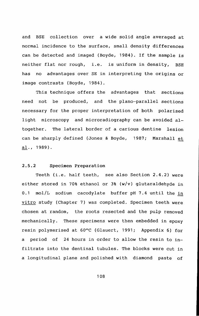
and BSE collection over a wide solid angle averaged at normal incidence to the surface, small density differences can be detected and imaged (Boyde, 1984). If the sample is neither flat nor rough, i.e. is uniform in density, BSE
has no advantages over SE in interpreting the origins or image contrasts (Boyde, 1984).
This technique offers the advantages that sections need not be produced, and the piano-parallel sections necessary for the proper interpretation of both polarised light microscopy and microradiography can be avoided altogether. The lateral border of a carious dentine lesion can be sharply defined (Jones & Boyde, 1987; Marshall et al., 1989).
2.5.2 Specimen PreparationTeeth (i.e. half teeth, see also Section 2.4.2) were
either stored in 70% ethanol or 3% (w/v) glutaraldehyde in 0.1 mol/L sodium cacodylate buffer pH 7.4 until the in
vitro study (Chapter 7) was completed. Specimen teeth were
chosen at random, the roots resected and the pulp removed
mechanically. These specimens were then embedded in epoxy resin polymerised at 60°C (Glauert, 1991; Appendix 6 ) for a period of 24 hours in order to allow the resin to infiltrate into the dentinal tubules. The blocks were cut in a longitudinal plane and polished with diamond paste of
1 08
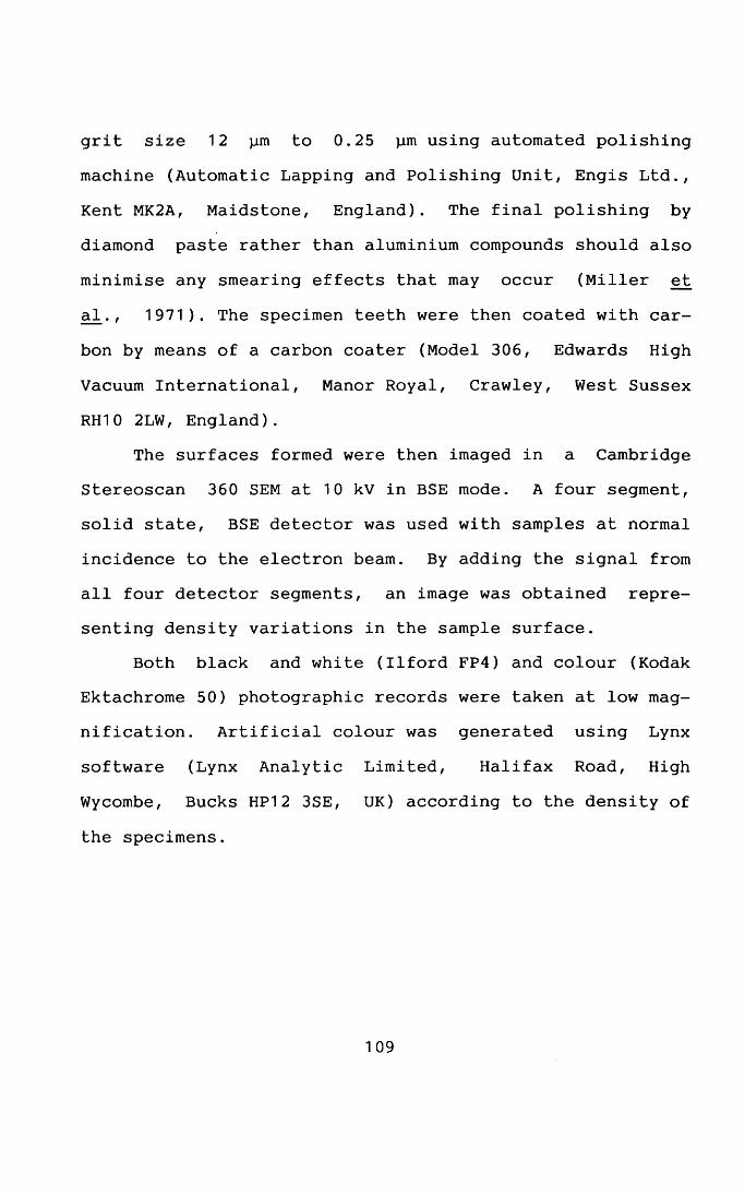
grit size 12 jim to 0.25 jim using automated polishing machine (Automatic Lapping and Polishing Unit, Engis Ltd., Kent MK2A, Maidstone, England). The final polishing by diamond paste rather than aluminium compounds should also minimise any smearing effects that may occur (Miller et
a l . , 1971). The specimen teeth were then coated with car
bon by means of a carbon coater (Model 306, Edwards High Vacuum International, Manor Royal, Crawley, West Sussex RH10 2LW, England).
The surfaces formed were then imaged in a Cambridge Stereoscan 360 SEM at 10 kV in BSE mode. A four segment, solid state, BSE detector was used with samples at normal incidence to the electron beam. By adding the signal from all four detector segments, an image was obtained representing density variations in the sample surface.
Both black and white (Ilford FP4) and colour (Kodak Ektachrome 50) photographic records were taken at low magnification. Artificial colour was generated using Lynx software (Lynx Analytic Limited, Halifax Road, High Wycombe, Bucks HP12 3SE, UK) according to the density of
the specimens.
109
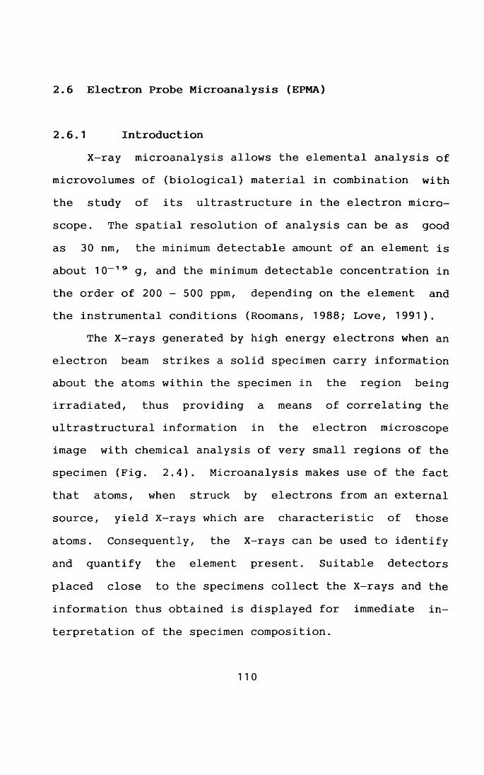
2.6 Electron Probe Microanalysis (EPMA)
2.6.1 Introduction
X-ray microanalysis allows the elemental analysis of microvolumes of (biological) material in combination with the study of its ultrastructure in the electron micro
scope. The spatial resolution of analysis can be as good as 30 nm, the minimum detectable amount of an element is about 1 0 - 1 9 g, and the minimum detectable concentration in the order of 200 - 500 ppm, depending on the element and the instrumental conditions (Roomans, 1988; Love, 1991).
The X-rays generated by high energy electrons when an electron beam strikes a solid specimen carry information about the atoms within the specimen in the region being irradiated, thus providing a means of correlating the ultrastructural information in the electron microscope image with chemical analysis of very small regions of the specimen (Fig. 2.4). Microanalysis makes use of the fact
that atoms, when struck by electrons from an external source, yield X-rays which are characteristic of those atoms. Consequently, the X-rays can be used to identify
and quantify the element present. Suitable detectors placed close to the specimens collect the X-rays and the information thus obtained is displayed for immediate in
terpretation of the specimen composition.
110
Clearly there is a need to combine a method of obtaining high resolution images of a specimen with simultaneous elemental analysis of a non-destructive nature of the same regions of the same specimen. X-ray microanalysisfulfils these needs by providing an in situ means of identifying elements within microvolumes of a specimen to a very high degree of sensitivity and with very precise localisation of the regions being analysed (Morgan, 1985).
2.6.2 Quantitation
The elemental X-ray intensity gives a measure of the mass of an element present. However, of more interest is its concentration. In order to calculate the concentration, it is necessary to know the total mass. The method of choice to perform this is utilisation of the white radiation intensity which forms the major part of the X-ray background upon which the characteristic lines are superimposed; this is proportional to the mass through
which the electrons are passing. The amount of white radiation produced by the specimen is a function of the
total number of atoms, of all kinds, in the specimen,
being unlike the characteristic radiation which is
specific for each element. The ratio of elemental mass to
total mass is the relative weight percentage of the element .
111
It is important to separate this background emission
from other factors contributing to the general background from within the microscope such as the specimen holder. In addition, since there may be a loss of mass because the specimen becomes hot, or a gain in mass because of con
tamination, it is better to calculate the relative concentrations rather than their absolute values. If the concentration of one element is known from an internal reference standard, the relative elemental concentration can then be converted to the absolute concentration of the other element. This technique of elemental analysis has a percentage error of +_ 0.5%.
The X-ray signal may also be used to provide an X-ray distribution map of an element. A specific energy band is chosen in the multi-channel analyser to involve the one X-ray line and the total signal in that energy region is calculated by a cathode ray oscilloscope operated synchronously with the scanning electron beam. Where an element is present in high concentrations in a specimen, signals are produced from the multi-channel analyser in
the chosen energy band and high intensities are shown on
the cathode ray oscilloscope. For this type of measurement
to be meaningful, a large peak to background ratio is necessary. This can be a problem with elements of biological interest and may, for example, involve long exposure
112
times in order to obtain statistically meaningful results. This can in turn produce problems of specimen drift and contamination.
2.6.3 Specimen Preparation
The principal problems and limitations with elements of biological interest are a function of specimen preparation rather than of suitable instrumentation for EPMA (Love, 1991; von Zglinicki, 1992).
The samples prepared for BSE (see Sections 2.4.2 and 2.5.2) were examined using the Cambridge Stereoscan 360 SEM microprobe analyser with a probe size of 100 nm at 10 kV. The background counting rate was 11 - 30 counts/secwith a counting period of 100 seconds. Elemental analyses in step scans (line scans) were carried out using Ca and P on each specimen, from the floor of the cavities to the dentino-pulpal junction. The X-rays generated were absorbed by a semiconductor detector. The signals were then passed through an amplifier system after which they could
be analysed using a computerised multichannel analyser
(Rooman, 1988). The most intense line of intensity in emission was from the transition of electrons from j electron shells L to K (called K lines). These were the lines used
113
in this study to measure the concentrations of the Ca and P in the specimens. Other elements such as Na, K, S, Mg
and 0 were also studied.The results representing the distribution of the ele
ments analysed were recorded using the colour transparency (Kodak Ektachrome 50) and black and white film (Ilford FP4) .
2.7 Caries Detector Dyes
2.7.1 IntroductionVisual and tactile assessment by conventional clini
cal criteria of staining and /or softening of dentine on the floor of a cavity at the end of the caries removal process was quite subjective. This can vary between different operators and between different carious lesions.
Following the preliminary studies (Chapters 3 and 4) and the light microscopy study (Chapter 5), it was found that the subjective visual and tactile criteria used to
verify the adequacy of caries removal may have introduced some error into the findings. Accordingly, it would be
ideal if an objective system such as the use of a caries detector dye could be used to ascertain when complete
removal of caries had been achieved and therefore provide a definitive baseline. An in vitro staining study was
114
therefore carried out in order to test the effectiveness
of 0.5% basic fuchsin and 1% acid red in propylene glycol as caries detector dyes.
2.7.2 In vitro Staining StudiesExtracted human permanent and deciduous teeth with
coronal carious lesions were collected from the Department of Oral Surgery, Glasgow Dental Hospital and School and
immediately stored at 4°C in PBS (pH 7.4, containing thimerosal added as an antibacterial agent). Only teeth with caries extending approximately halfway through the dentine but without pulpal involvement were chosen. Selection was based upon visual, tactile and radiographic examination. Sound premolars extracted for orthodontic reasons were also collected. The teeth were used within one week of collection. All specimens were randomly divided into five groups.
In group A, carious tissue was excavated from the
teeth with a spoon shaped excavator until the cavity was judged to be clinically caries free. The criteria for CCR were those normally used clinically during conventional (i.e. mechanical) cavity preparation i.e. complete removal
of soft, stained dentine caries from the carious lesion leaving clinically sound dentine. Either 1.0% acid red or 0.5% basic fuchsin, both dissolved in propylene glycol,
115
was applied for 1 0 seconds to the dried cavity of the specimen using a pledget of cotton wool. The specimen was then thoroughly washed with distilled water for 1 0
seconds, dried with a three-in-one syringe and reexamined. Any dye-stained dentine was then removed using the same spoon excavator. The cavity was then re-stained with a caries detector dye for a further 1 0 seconds,
washed and any dye-stained dentine further excavated. This procedure was repeated until the cavity floor no longer stained with the caries detector dyes (Boston & Graver, 1989).
In group B, teeth were split into two halves through
the middle of the carious lesions using either an osteotome or a diamond wheel mounted on a tooth sectioning machine (Microslice 2, Malvern instruments Ltd., Spring Lane South, Malvern, Worcestershire, WR14 1AQ, UK). The staining procedures applied were the same as in group A, but without caries removal.
In group C, consecutive longitudinal serial sections
were prepared from the teeth (LS, 100-120 pm) using the same tooth sectioning procedure as in Group B. Some specimens were mounted as ground sections for observation under a light microscope in order to confirm the visual examination. The same staining procedures as in group A
but without caries removal were applied.
116
In group D, consecutive transverse serial sections
were prepared from the teeth (TS, 1 mm) using the same
tooth sectioning machine as in Group B. The staining procedures were the same as in group A but without caries removal. The sections were then examined under a stereomicroscope (X10 magnification).
In group E, Class I occlusal cavities were prepared in sound premolars using a finishing diamond bur in a high
speed handpiece with a constant water spray. The depth of the cavities was kept just 1 mm into dentine.
After staining with caries detector dyes as discussed for all groups, the penetration of staining was recorded. All the specimens were examined using a Wild M3Z stereomicroscope at x10 magnification. A colour transparency (Kodak Ektachrome 50 film) record was made of each of the specimens.
117
CHAPTER 3
THE CHEMOMECHANICAL REMOVAL OF DENTAL CARIES IN PERMANENT AND DECIDUOUS TEETH
3.1 Introduction
3.1.1 Improving the FormulationDespite the apparent advantages, the current
procedure for CMCR has certain limitations as outlined in Section 1.10.7. Accordingly, if the procedure is to become clinically useful, a more effective solution must be developed. The formulation of the caries removal agent was improved when NMG was substituted by NMAB (Schutzbank et a l ., 1978) but subsequently there have been no furtherimprovements.
The possibility of using some of the protein de-naturing agents commonly used in laboratories to
solubilise proteins including sodium dodecyl sulphate, guanidinium chloride, urea hydrogen peroxide, hydrogen peroxide and urea, in conjunction with NMAB to improve the removal of caries was therefore investigated.Sodium Dodecyl Sulphate
This is a detergent that disrupts most protein-protein and protein-lipid interactions. The
118
dodecyl sulphates are amphiphilic 1 2 -carbon alkyl sulphate molecules that act to de-nature the protein by weakening
the hydrophobic bondings. They form a charged micelle around the de-natured molecule and thereby stabilise it. Guanidinium Chloride
High concentrations of guanidinium chloride e.g. 6
mol/L have been used to solubilise collagen in some biochemical experiments (Wohllebe & Carmichael, 1979). This reagent has also been used as a medication for treatment of myasthenia gravis.Urea
Urea has been widely used as a protein de-naturing agent and acts by disrupting hydrogen bonds. It does not affect the intrinsic bonds of the protein. High urea concentrations e.g. 8 mol/L are usually used and complete de-naturation of proteins containing disulphide bonds would also require a thiol reagent. There are, however, few, if any, disulphide bonds in mature collagen.Hydrogen Peroxide
Hydrogen peroxide is an oxidising agent and during
decomposition, oxygen is generated. It has been used as a
caustic or etching substance (30%) and as a mouthrinse (equal volumes of 3% H 2 O 2 and warm water) in the treatment of acute ulcerative gingivitis (Lindhe, 1989).
119
Urea Hydrogen Peroxide
Urea hydrogen peroxide decomposes slowly in air into urea and oxygen and it dissolves in water to form urea and hydrogen peroxide. It is normally used as a disinfectant but might offer the combined properties of urea and hydrogen peroxide.
3.1.2 Considerations of Osmotic Balance
The sodium-potassium ratio of dentinal fluid has been reported to approximate to that of interstitial fluid (Stevens & Gutch, 1960; Coffey et al. , 1970) which would
indicate that the dentinal fluid orginates extracellularly
and is not derived from the cytoplasm of the odontoblast
processes (Paunio & Nanto, 1965; Coffey et al., 1970).Since dentinal fluid contains proteins similar to those found in plasma (Haldi & Wynn, 1963), it was considered to
be a derivative of a capillary transudate or filtrate. The
osmotic pressure of dentinal fluid would presumably
therefore be very similar to that of serum. There is,
however, a report that the dentinal fluid differs
significantly from interstitial fluid (Haljamae & Rockert,
1970). Osmotically active excitants that disturb the
osmotic balance have been reported to cause pain when applied to human dentine (Anderson & Ronning, 1962).
The procedure of chemomechanical removal of carious
1 20
dentine using NMAB has been reported to reduce the need for local anaesthesia (Table 1.3). This has been partially attributed to the fact that warming of the solution to 37°C results in less thermal irritation (Punwani et a l ., 1988; Tavares et a l . , 1988). The osmotic effect of NMAB on human dentine however has not been studied. Whether there is a relationship between the osmotic activity of NMAB and a reduced need for local anaesthesia is still unknown.
3.1.3 Aims of the StudiesThe aims of the present study in this chapter were
therefore :a. to improve the efficacy of the solution currently used,b. to evaluate the effectiveness of NMAB and compare its action with other chemical agents in the removal of carious dentine,c. to determine the pH and osmolality of NMAB and NMAB-Urea, andd. to compare the effectiveness of various caries removal agents in permanent and deciduous teeth.
3.2 Materials and Methods
3.2.1 Protein De-naturing Agents
The selection of an appropriate protein de-naturing
1 21
agent to be incorporated into Solution II was carried out using both immersion and in vitro trials.
Immersion TrialsTwo permanent and two deciduous teeth with coronal
carious lesions (see Section 2.2.1) were immersed in 10 ml of each of the following solutions :
guanidinium chloride (4 mol/L) hydrogen peroxide (H2 O 2 ) solution (30% w/v) sodium dodecyl sulphate (SDS) solution (2 mol/L) two sodium hypochlorite (NaOCl) solutions (0.5% and1 .0 % w / v ) urea (2 mol/L)urea hydrogen peroxide (33 - 35% w/v H 2 O 2 ) isotonic saline solution (0.9% w/v sodium chloride) The solutions were changed daily. The changes in
consistency, surface texture and colour of the carious dentine, if any, were recorded over a period of 15 days.In vitro Trials
8 mol/L guanidinium chloride, 30% H 2 O 2 , 4 mol/L SDS,
4 mol/L urea and 1 tablet of urea hydrogen peroxide were also individually incorporated into solution II in the
NMAB system (see Section 2.2.2) in order to test the effectiveness in removing caries using the simulated set-up of a CMCRS (i.e. to give working concentrations in
122
NMAB the same as those shown above). The ease of caries
removal of NMAB alone was subjectively compared with the
other caries removal agents. Five carious lesions were tested with each solution used. The in vitro study model described in Sections 2.2.4 - 2.2.7 was used.
3.2.2 Optimum Urea Concentration in NMAB
An in vitro study was carried out to determine the optimum urea concentration for inclusion in NMAB. Various
concentrations of urea were incorporated into Solution II to give the following final concentrations of urea when Solution I and II were mixed :a. NMAB + 1 mol/L urea,b. NMAB + 2 mol/L urea,c. NMAB + 3 mol/L urea, andd. NMAB + 4 mol/L urea.
Concentrations of urea higher than 4 mol/L (i.e. 8
mol/L in Solution II) were easily saturated. Forty carious permanent teeth (see Section 2.2.1) were treated with the various concentrations of NMAB-Urea using the in vitro study model described in Sections 2.2.4 - 2.2.7 i.e. 10
teeth were treated with each of the 4 urea-containing
solutions studied.
123

3.2.3 Measurement of pH and OsmolalityThe pH and osmolality of freshly prepared, well mixed
solutions of NMAB and NMAB containing 2 mol/L urea were measured over a period of two hours after mixing Solutions I and II. The procedures were repeated three times with three different samples. In the case of urea, Solution II
with urea and NMAB—Urea, it was necessary to dilute the solutions to give values within the working range of the instrument. The validity of this is dependent on the assumption that they are ideal solutions.
3.2.4 In vitro StudyAn in vitro study involving a larger number of
carious lesions from both permanent and deciduous teeth was carried out in order to investigate the effectiveness of caries removal of the NMAB-Urea system and compare it with the NMAB system. Freshly extracted human carious teeth consisting of 130 permanent teeth and 174 deciduous teeth were collected in phosphate buffered saline
containing an antibacterial agent, thimerosal (see Section 2.2.1). The coronal caries was not restricted to any
specific surface or surfaces of the teeth. All the teeth were used either immediately or within one week after extraction. Carious tissue was removed chemomechanically
using the following solutions :
124
a. 0.007 mol/L NMAB,b. 0.007 mol/L NMAB containing 2 mol/L urea (NMAB-Urea),c. 0.5% (w/v) NaOCl,d. 2 mol/L urea, ande. isotonic saline (0.9% w/v).The methodology for the in vitro study described inSections I 2.2.4 - 2.2.7 was used. :
3.3 Results
3.3.1 Protein De-naturing AgentsImmersion Trials
With the exception of NaOCl and to a lesser extent urea hydrogen peroxide, none of the trial solutions seemed to alter the colour, texture or consistency of the cariousdentine to any noticeable extent after 15 days ofimmersion. NaOCl softened and "bleached" the carious dentine and the superficial layer simply dissolved away
after 24 hours. The deeper layer was soft and could be
excavated easily. Urea hydrogen peroxide had a similar
effect on carious dentine to NaOCl but the changes were, however, not noticeable until after at least 4 days of
immersion. The effects appeared to be similar in both permanent and deciduous teeth.
125
In vitro TrialsNeither guanidinium chloride nor H 2 O 2 enhanced the
removal of dentine caries when incorporated into SolutionII. Isotonic saline seemed to be ineffective in removing caries. SDS did not seem to enhance caries removal andproduced too much foam making the process messy and impractical. Urea and to a lesser extent urea hydrogen
peroxide, however, seemed to enhance the caries removal by NMAB when assessed subjectively. There was, however,gaseous evolution when urea hydrogen peroxide was mixedwith Solution II, probably resulting from decomposition of the hydrogen peroxide.
Hard, darkly stained and eburnated dentine when present was found to be difficult to remove in all cases.
3.3.2 Optimum Urea ConcentrationBecause the preliminary findings indicated that a
combination of urea and NMAB (NMAB-Urea) seemed to be at least equal, if not superior, in caries removal as
compared to NMAB alone, a more detailed study was carried
out. The optimal concentration of urea to be incorporated
into Solution II to improve the effectiveness in caries
removal of NMAB therefore had to be determined. The types of carious lesions and teeth used are shown in Tables 3.1 and 3.2. Molars and Class II lesions were the most
1 26
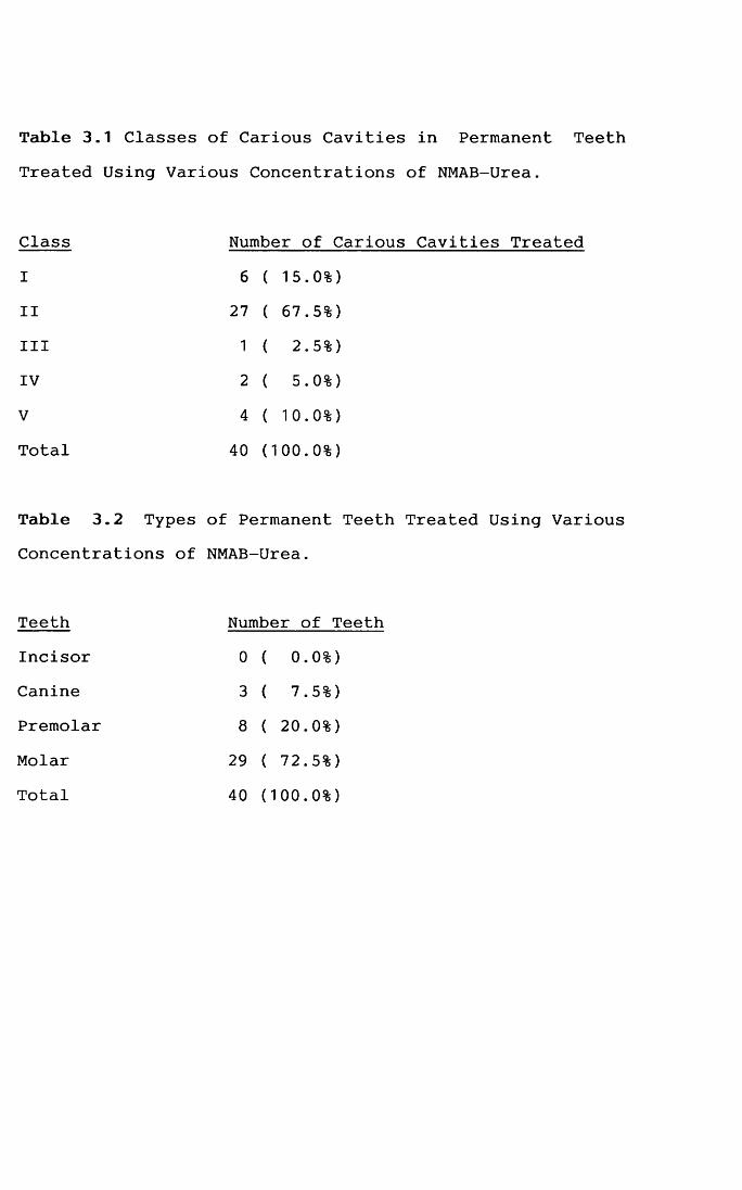
Table 3.1 Classes of Carious Cavities in Permanent Teeth Treated Using Various Concentrations of NMAB-Urea.
Class Number of Carious Cavities Treated
I 6 ( 15.0%)II 27 ( 67.5%)III 1 ( 2.5%)IV 2 ( 5.0%)
V 4 ( 10.0%)
Total 40 (100.0%)
Table 3.2 Types of Permanent Teeth Treated Using Various Concentrations of NMAB-Urea.
Teeth Number of TeethIncisor 0 ( 0.0%)Canine 3 ( 7.5%)Premolar 8 ( 20.0%)
Molar 29 ( 72.5%)Total 40 (100.0%)
commonly used. The results including average times and volumes of caries removal agents used to achieve CCR are shown in Table 3.3. The number of teeth with CCR appeared to show no increase when concentrations of urea higher than 2 mol/L were added to NMAB, even though the sample numbers were small. The criteria for CCR were outlined in Section 2.2.7.
3.3.3 pH and OsmolalityThe changes in pH of NMAB and NMAB-Urea over a two
hour period after mixing of the two constituent solutions were monitored. Typical results are shown in Fig. 3.1. The pH of NMAB dropped from slightly above 11 to 9.5 over a 20 minutes period after the solutions were mixed and remained fairly constant over the next two hours. The pH of NMAB-Urea showed two changes during the first 30 minutes : it dropped from just above 10 to 9.8 and remained constant for a period of 1 0 minutes before dropping again to around9.6 after another 10 minutes. Thereafter it remained constant but the final pH was slightly higher than in the
absence of urea.
No steady readings of osmolality of urea, Solution II
containing urea and NMAB-Urea could be read from the osmometer. In order to measure the osmolalities, these solutions were diluted X100. The osmolalities of NMAB-Urea
127
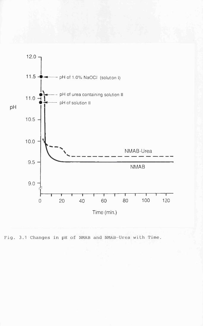
12.0
pH of 1.0% NaOCl (solution I)
pH of urea containing solution
pH of solution II
10.5 -
NMAB-Urea
9.5 -
NMAB
9.0 -
1 0 0 1208060200 40
Time (min.)
Fig. 3.1 Changes in pH of NMAB and NMAB-Urea with Time.
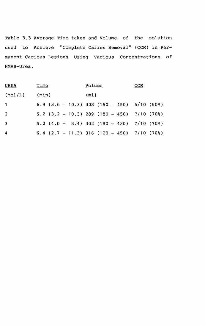
Table 3.3 Average Time taken and Volume of the solution used to Achieve "Complete Caries Removal" (CCR) in Permanent Carious Lesions Using Various Concentrations of NMAB-Urea.
UREA Time Volume CCR(mol/L) (min) (ml)1 6.9 (3.6 - 10.3) 308 (150 - 450) 5/10 (50%)2 5.2 (3.2 - 10.3) 289 (180 - 450) 7/10 (70%)
3 5.2 (4.0 - 8.4) 302 (180 - 430) 7/10 (70%)4 6.4 (2.7 - 11.3) 316 (120 - 450) 7/10 (70%)
remained at 4500 mosmol throughout the 2 hours periods after Solutions I and Solutions II containing 4 mol/L urea were mixed.
The osmolalities of the solutions were :Solution II 22.5 mosmol
Solution II with 5000.0 mosmol
4 mol/L Urea
NaOCl (Solution I) 30.0 mosmol2 mol/L Urea 3700.0 mosmolNMAB 25.0 mosmolNMAB-Urea 4500.0 mosmol
3.3.4 In vitro StudyThe types of carious lesions and teeth used are shown
in Tables 3.4 and 3.5. Molars and Class II lesions were most commonly used in both permanent and deciduous teeth.
The results of the in vitro study involving 130 permanent, 174 deciduous teeth and five different solutions for CMCR are shown in Table 3.6. The number of teeth with CCR with NMAB in this study was 58% in permanent teeth and 65% in deciduous teeth. Although NMAB
was superior to NaOCl, urea and saline in CCR, none of these differences was statistically significant (Table
3.7). The difference was only found to be statistically significant between NMAB-Urea as compared to urea alone in
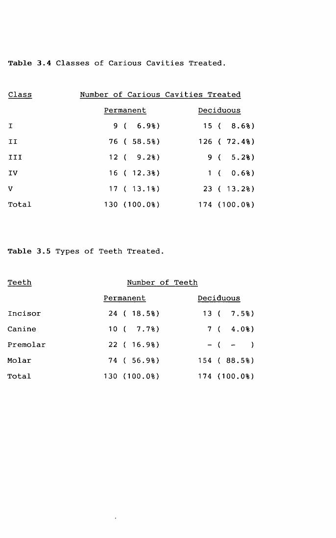
Table 3.4 Classes of Carious Cavities Treated.
Class Number of Carious Cavities TreatedPermanent Deciduous
I 9 ( 6.9%) 15 ( 8 .6 %)II 76 ( 58.5%) 126 ( 72.4%)III 1 2 ( 9.2%) 9 ( 5.2%)IV 16 ( 12.3%) 1 ( 0 .6 %)V 17 ( 13.1%) 23 ( 13.2%)Total 130 (1 0 0 .0 %) 1 74 (1 0 0 .0 %)
Table 3.5 Types of Teeth Treated.
Teeth Number of Teeth
Permanent DeciduousIncisor 24 ( 18..5%) 13 ( 7 ..5%)Canine 1 0 ( 7..7%) 7 ( 4..0 %)Premolar 2 2 ( 16..9%) - ( - )Molar 74 ( 56..9%) 154 ( 8 8 ..5%)Total 130 ( 1
oo .0 %) 174 ( 1 0 0 .,0 %)
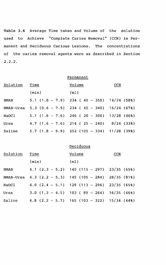
Table 3.6 Average Time taken and Volume of the solution used to Achieve "Complete Caries Removal" (CCR) in Permanent and Deciduous Carious Lesions. The concentrations
of the caries removal agents were as described in Section 2.2.2.
PermanentSolution Time Volume CCR
(min) (ml)NMAB 5.1 ( 1 . 8 - 7.9) 234 ( 40 - 350) 14/24 (58%)NMAB-Urea 5.3 (0.6 - 7.9) 234 ( 45 - 340) 16/24 (67%)
NaOCl 5.1 ( 1 . 6 - 7.6) 246 ( 20 - 300) 13/28 (46%)Urea 4.7 (1.6 - 7.6) 214 ( 25 - 240) 8/24 (33%)Saline 5.7 (1.8 - 9.9) 252 (105 - 334) 11 / 28 (39%)
DeciduousSolution Time Volume CCR
(min) (ml)
NMAB 4.1 (2.3 - 5.2) 140 (115 - 297) 23/35 (65%)NMAB-Urea 4.3 (2.2 - 5.3) 145 (105 - 284) 28/35 (81%)NaOCl 4.0 (2.4 - 5.1 ) 129 (113 - 296) 23/35 (65%)Urea 3.0 (1.3 - 4.5) 103 ( 89 - 264) 16/35 (46%)Saline 4.8 (2.2 - 5.7) 165 (103 - 322) 15/34 (44%)
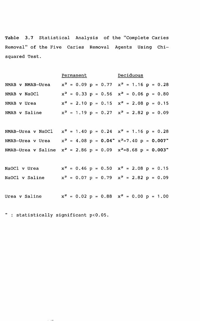
Table Removal
squared Test.
NMAB v NMAB-Urea
NMAB v NaOCl NMAB v Urea NMAB v Saline
NMAB-Urea v NaOCl NMAB-Urea v Urea NMAB-Urea v Saline
NaOCl v Urea NaOCl v Saline
Urea v Saline
Analysis of Caries Removal
Permanentx 2 = 0.09 p = 0.77x 2 = 0.33 p = 0.56x 2 = 2.10 p = 0.15
X K) II vo p = 0.27
x 2 = 1 .40 p = 0.24x 2 = 4.08 p = 0.04X 2 = 2.86 p = 0.09
X 2 = 0.46 p = 0.50x2 = 0.07 p = 0.79
x2 = 0.02 p = 0.88
the "Complete Caries Agents Using Chi-
Deciduousx 2 = 1.16 P = 0.28x2 = 0.06 P = 0.80x 2 = 2.08 P = 0.15x2 = 2.82 P = 0.09
x 2 = 1.16 P = 0.28x 2 = 7 .40 p = 0.007*
X to II 00 .68 p = 0.003*
X 2 = 2.08 P = 0.15x 2 = 2.82 P = 0.09
x 2 = 0.00 P _ 1 .00
3.7 Statistical " of the Five
* : statistically significant p<0.05.
permanent teeth, and between NMAB-Urea as compared to urea or saline in deciduous teeth (Table 3.7). The amount of CCR with NMAB-Urea was increased by 16% in permanent teeth i.e. to 67% and by 25% in deciduous teeth i.e. to 81% as compared to NMAB alone. Urea (2 mol/L) on its own (at neutral pH) was only as effective as the saline control. In general, the amount of CCR in deciduous teeth was higher compared with that in permanent teeth. The order of efficacy of CCR in both permanent and deciduous teeth was found to be as follows :
NMAB-Urea > NMAB, NaOCl > Urea, Saline Although the number of teeth with CCR was higher in
deciduous teeth than in permanent teeth, the difference was not statistically significant (Table 3.8). However the level of significance of NMAB-Urea against urea was higher in the case of deciduous teeth (p=0.007) than in permanent (p=0.05) (Table 3.7) as well as being highly significant when compared to saline (p=0.003).
The average times and volumes of solution used to
remove carious tissue are shown in Table 3.6. In general,
the caries in deciduous teeth required a shorter time and a smaller volume of solution for removal when compared with caries in permanent teeth. The time needed to inspect
the degree of progress of caries removal during and after
the treatment with various solutions was not included. The
129
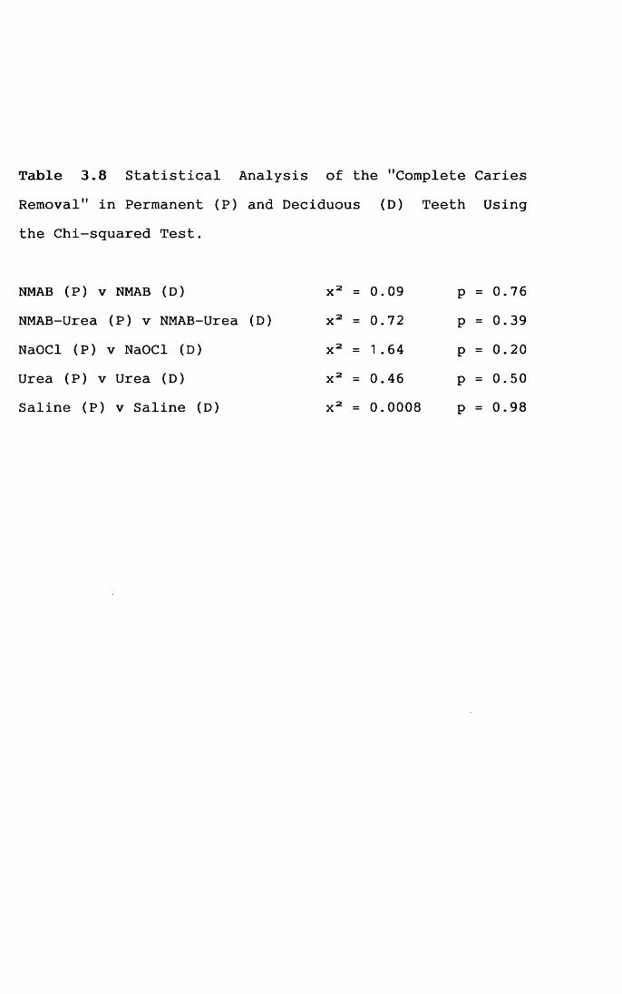
Table 3.8 Statistical Analysis of the "Complete CariesRemoval" in Permanent (P)
the Chi-squared Test.and Deciduous (D) Teeth Using
NMAB (P) v NMAB (D) x 2 = 0.09 p = 0.76NMAB-Urea (P) v NMAB-Urea (D) x 2 = 0.72 p = 0.39
NaOCl (P) v NaOCl (D) x2 = 1 .64 p = 0.20Urea (P) v Urea (D) x 2 = 0.46 p = 0.50Saline (P) v Saline (D) x2 = 0.0008 p = 0.98
time and volume of solution needed to achieve CCR in a carious lesion depended on the individual size, ease of access and the consistency of the lesion. Although the differences in time taken to remove carious tissue in permanent and deciduous teeth were not statistically significant except for urea (p<0.05), the differences
between the volumes of all the caries removal agents used
were of a high statistical signifance (p<0.001 in all cases), the volumes of solution used to remove carious material in deciduous teeth being much smaller (Tables 3.9, 3.10 & 3.11 ).
The caries in deciduous teeth was found to be more easily removed than that in permanent teeth (Tables 3.6 & 3.11). It was observed that the removal of caries in a carious lesion with a harder consistency took longer and was much less likely to achieve CCR irrespective of the caries removal agent used. Firm staining was consistently found along some parts of the DEJ of the treated cavities.
3.4 Discussion
3.4.1 Selection of a Protein De—naturing Agent
From the in vitro trials, urea and urea hydrogen
peroxide seemed to be the only reagents which would enhance the effectiveness of caries removal by - NMAB.
130
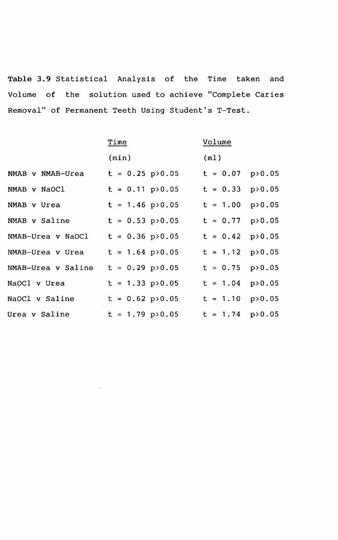
Table 3.9 Statistical Analysis of the Time taken and Volume of the solution used to achieve "Complete Caries Removal" of Permanent Teeth Using Student's T-Test.
Time(min)
NMAB v NMAB-Urea t = 0.NMAB v NaOCl t = 0.NMAB v Urea t = 1 .
NMAB v Saline t = 0.NMAB-Urea v NaOCl t = 0.NMAB-Urea v Urea t = 1 .NMAB-Urea v Saline t = 0.NaOCl v Urea t = 1 .NaOCl v Saline t = 0.Urea v Saline t = 1 .
Volume(ml)
p> 0.05 t = 0.07 p> 0.05p> 0.05 t = 0.33 p> 0.05p> 0.05 t = 1 .00 p> 0.05
p> 0.05 t = 0.77 p> 0.05p> 0.05 t = 0.42 p> 0.05p> 0.05 t = 1.12 p> 0.05p> 0.05 t = 0.75 p> 0.05p> 0.05 t = 1 .04 p> 0.05p> 0.05 t = 1.10 p> 0.05p> 0.05 t = 1 .74 p> 0.05
2511
46
53366429336279
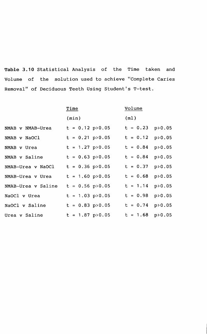
Table 3.10 Statistical Analysis of the Time taken and Volume of the solution used to achieve "Complete Caries Removal" of Deciduous Teeth Using Student's T-test.
Time (min)
NMAB v NMAB-Urea t = 0.NMAB v NaOCl t = 0.NMAB v Urea t = 1 .
NMAB v Saline t = 0.NMAB-Urea v NaOCl t = 0.NMAB-Urea v Urea t = 1 .NMAB-Urea v Saline t = 0.NaOCl v Urea t = 1.NaOCl v Saline t = 0.Urea v Saline t = 1 .
Volume(ml)
p> 0.05 t = 0.23 p> 0.05p> 0.05 t = 0.12 p > 0 .05p> 0.05 t = 0.84 p> 0.05p> 0.05 t = 0.84 p> 0.05p> 0.05 t = 0.37 p> 0.05p> 0.05 t = 0.68 p> 0.05p> 0.05 t = 1.14 p> 0.05p> 0.05 t = 0.98 p> 0.05p> 0.05 t = 0.74 p > 0 .05p> 0.05 t = 1 .68 p> 0.05
12
21
27
63366056038387
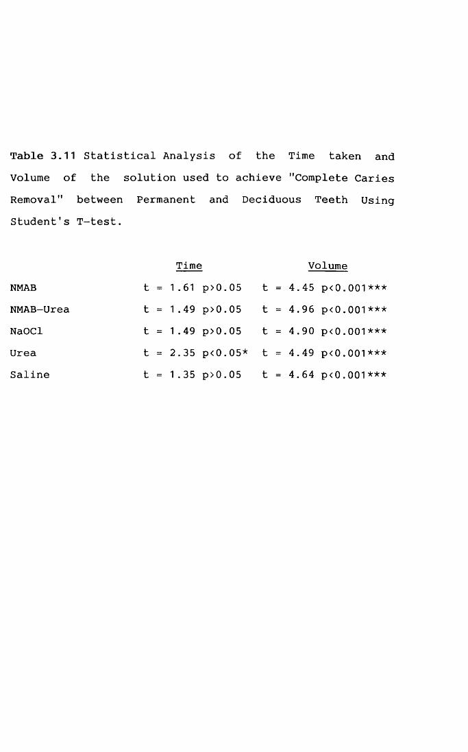
Table 3.11 Statistical Analysis of the Time taken and Volume of the solution used to achieve "Complete Caries Removal" between Permanent and Deciduous Teeth Using Student's T-test.
Time VolumeNMAB t = 1.61 p>0.05 t = 4.45 p<0.001***NMAB-Urea t = 1.49 p>0.05 t = 4.96 p<0.001***NaOCl t = 1.49 p>0.05 t = 4.90 p<0.001***Urea t = 2.35 p<0.05* t = 4.49 p<0.001***Saline t = 1.35 p>0.05 t = 4.64 p<0.001***
Although urea hydrogen peroxide did perform fairly well in the trial studies, it decomposed with gaseous evolution, when mixed with Solution II. It therefore had to be
prepared as a separate solution and mixed with Solution I
and II when in use. The procedure was found to be lengthy and would be impractical in clinical situation. Indeed, as hydrogen peroxide alone had no perceptible effect, the improvement may have resulted solely from the urea. Urea, however, is unlikely to react with any of the componentsin Solution II and the active ingredient (i.e. NMAB) wouldonly be generated when Solutions I and II are mixed. It was, therefore, considered to be a more appropriate agent. A small in vitro study using different concentrations of urea in Solution II indicated that the enhancement of caries removal by NMAB was best achieved by incorporating 2 mol/L or higher concentrations of urea into the NMAB system (Table 3.3). NMAB containing 2 mol/L urea was therefore selected for the subsequent in vitro studies.
3.4.2 In vitro StudyNMAB
The larger scale in vitro blind study comparing efficiency of different solutions for CMCR showed that although NMAB was subjectively superior to both NaOCl and
saline in caries removal, the difference was not
131
statistically significant. Compared with previous clinical trials, which reported a much higher caries removal efficacy, NMAB appeared to be less effective in the
present study. Also in contrast to the previous study no rotary or hand instruments were used before, during or after solution treatment in this study. In the present study, flow-rate and temperature were also more carefully
controlled than previously. This was considered to be more appropriate when only the effectiveness of the caries removal properties of the various chemical agents used was
to be evaluated. The results obtained in the present study could not, therefore, be compared directly with those of most of the clinical trials which were carried out withthe aid of other dental instruments (Chapter 1, Table1.3). Only one clinical trial (Zinck et a l ., 1988) hasreported that a 68.4% of CCR in permanent teeth was achieved without the aid of other dental procedures, which is similar to the finding in this study (58% for NMAB and
67% for NMAB-Urea).
NMAB-UreaWith NMAB containing urea, the number of teeth with
CCR increased by 16% in permanent teeth to 67% and by 25%
in deciduous teeth to 81%. The improvement seemed to be
more marked in deciduous teeth. However NMAB-Urea, unlike NMAB alone, gave results which were more statistically
132
significant than some of the controls, and the level of significance was greater in the case of deciduous teeth (p<0.01) than in that of permanent teeth (p<0.05).
A possible reaction mechanism might involve the two
amino groups of urea being chlorinated by NaOCl to form
mono- or dichloro-derivatives; these intermediate compounds may then attack and break down the collagen in carious dentine analogously to NMAB. The excess urea may
also help to disrupt the hydrogen bonds in the partially degraded collagen thereby increasing its solubility.
However, neither the nature of the products formed in the solutions used nor their possible toxicity has been studied. The effects on pulpal tissue would also need to be investigated should the improved formulation prove to have a potential for clinical application.NaOCl, Urea and Isotonic Saline
Interestingly, the effectiveness of NaOCl in caries removal, though less than that of NMAB, was not
significantly lower. Its use in the present regime of this system is, however, questionable due to the toxic effects it has on oral tissues. It is cytotoxic to all cells except heavily keratinised epithelia (Pashley et al.,
1985). Ingestion of NaOCl may cause hypernatraemia and hyperchloraemic acidosis (Ward & Routledge, 1988).
Neither saline nor urea was effective in CCR, even
133
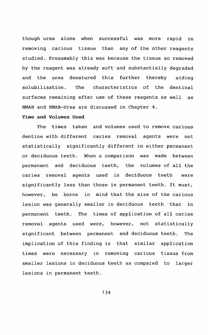
though urea alone when successful was more rapid in
removing carious tissue than any of the other reagents
studied. Presumably this was because the tissue so removed by the reagent was already soft and substantially degraded and the urea denatured this further thereby aiding solubilisation. The characteristics of the dentinal surfaces remaining after use of these reagents as well as NMAB and NMAB-Urea are discussed in Chapter 4.
Time and Volumes UsedThe times taken and volumes used to remove carious
dentine with different caries removal agents were not statistically significantly different in either permanent or deciduous teeth. When a comparison was made between permanent and deciduous teeth, the volumes of all the caries removal agents used in deciduous teeth were significantly less than those in permanent teeth. It must, however, be borne in mind that the size of the carious lesion was generally smaller in deciduous teeth than in permanent teeth. The times of application of all caries
removal agents used were, however, not statistically significant between permanent and deciduous teeth. The
implication of this finding is that similar application
times were necessary in removing carious tissue from smaller lesions in deciduous teeth as compared to larger
lesions in permanent teeth.
134
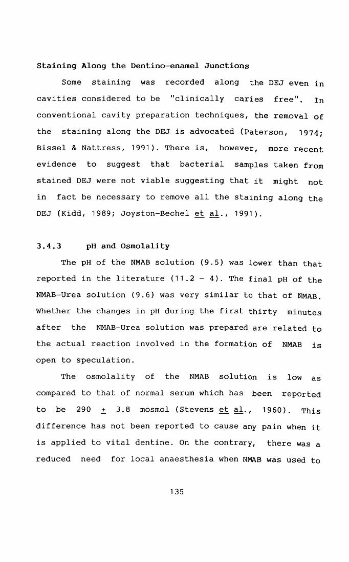
Staining Along the Dentino-enamel JunctionsSome staining was recorded along the DEJ even in
cavities considered to be "clinically caries free". in conventional cavity preparation techniques, the removal of the staining along the DEJ is advocated (Paterson, 1974;
Bissel & Nattress, 1991). There is, however, more recent evidence to suggest that bacterial samples taken from stained DEJ were not viable suggesting that it might not
in fact be necessary to remove all the staining along theDEJ (Kidd, 1989; Joyston-Bechel et a l . , 1991).
3.4.3 pH and OsmolalityThe pH of the NMAB solution (9.5) was lower than that
reported in the literature (11.2 - 4). The final pH of theNMAB-Urea solution (9.6) was very similar to that of NMAB. Whether the changes in pH during the first thirty minutes after the NMAB-Urea solution was prepared are related to the actual reaction involved in the formation of NMAB is open to speculation.
The osmolality of the NMAB solution is low as compared to that of normal serum which has been reported
to be 290 + 3.8 mosmol (Stevens et al. , 1960). This
difference has not been reported to cause any pain when it
is applied to vital dentine. On the contrary, there was a reduced need for local anaesthesia when NMAB was used to
135
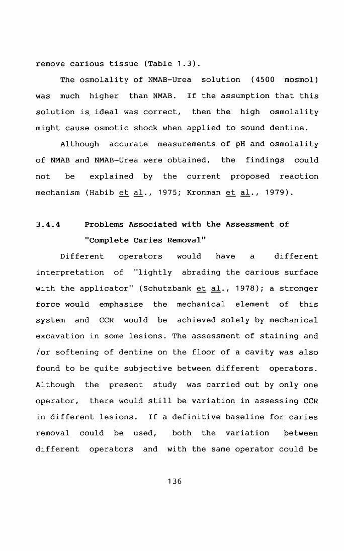
remove carious tissue (Table 1.3).The osmolality of NMAB-Urea solution (4500 mosmol)
was much higher than NMAB. If the assumption that this solution isr ideal was correct, then the high osmolality might cause osmotic shock when applied to sound dentine.
Although accurate measurements of pH and osmolality
of NMAB and NMAB-Urea were obtained, the findings could
not be explained by the current proposed reaction mechanism (Habib et a l ., 1975; Kronman et al., 1979).
3.4.4 Problems Associated with the Assessment of"Complete Caries Removal"
Different operators would have a different interpretation of "lightly abrading the carious surface with the applicator" (Schutzbank et a l ., 1978); a stronger force would emphasise the mechanical element of this system and CCR would be achieved solely by mechanical excavation in some lesions. The assessment of staining and
/or softening of dentine on the floor of a cavity was also
found to be quite subjective between different operators.
Although the present study was carried out by only one operator, there would still be variation in assessing CCR in different lesions. If a definitive baseline for caries removal could be used, both the variation between
different operators and with the same operator could be
136
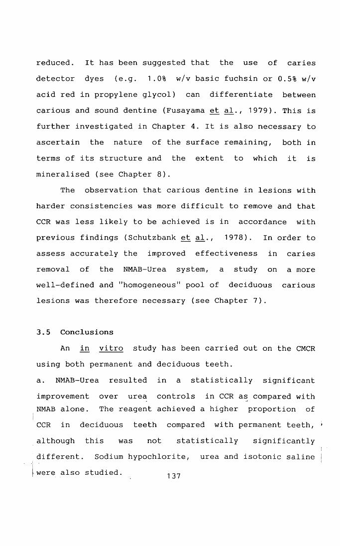
reduced. It has been suggested that the use of caries detector dyes (e.g. 1.0% w/v basic fuchsin or 0.5% w/v acid red in propylene glycol) can differentiate between carious and sound dentine (Fusayama et a l ., 1979). This is further investigated in Chapter 4. It is also necessary to ascertain the nature of the surface remaining, both in
terms of its structure and the extent to which it is mineralised (see Chapter 8).
The observation that carious dentine in lesions with harder consistencies was more difficult to remove and that CCR was less likely to be achieved is in accordance with previous findings (Schutzbank et a l ., 1978). In order toassess accurately the improved effectiveness in caries removal of the NMAB-Urea system, a study on a more well-defined and "homogeneous" pool of deciduous carious lesions was therefore necessary (see Chapter 7).
3.5 ConclusionsAn in vitro study has been carried out on the CMCR
using both permanent and deciduous teeth.
a. NMAB-Urea resulted in a statistically significant
improvement over urea controls in CCR as compared with NMAB alone. The reagent achieved a higher proportion ofCCR in deciduous teeth compared with permanent teeth,although this was not statistically significantlydifferent. Sodium hypochlorite, urea and isotonic salinewere also studied. m
b. The order of efficacy in caries removal was:NMAB-Urea > NMAB, NaOCl > Saline, Urea.
c. The procedure in general has limitations in terms of
treatment time, suitability of cavities and the continued need for some use of mechanical instruments.
d. The improved reagent may be useful in removing dental caries in deciduous teeth. Further investigations using a more "standardised" sample of carious teeth are necessary to properly evaluate the reagent's improved effectiveness in caries removal.
138
CHAPTER 4
MICROSCOPIC FEATURES OF THE CAVITY FLOORS AFTER CARIES REMOVAL
4.1 IntroductionThe dentinal surface of the cavity floor produced by
the CMCRS in permanent teeth using NMAB has been reported to be highly irregular (Goldman et a l ., 1987, 1988) and
also to have a higher surface energy (Emanuel & Broome, 1988) than that of dentine in conventionally prepared cavities. Both the irregular dentinal surface and the high surface energy favour micromechanical bonding with restorative materials. Indeed, adhesive restorative materials have been shown to have higher bond strengths to the dentine of permanent teeth treated with NMAB than to those conventionally prepared (Mclnnes—Ledoux et_ a l . , 1987,1989; Burke, 1989; Wolski et a l ., 1989).
The aims of the study described in this chapter were to use SEM to :a. compare the surface morphology of the dentine remaining
after the carious tissue was removed using various caries
removal agents, and
b. compare the surfaces produced by this method in permanent and deciduous teeth.
139
4.2 Materials and MethodsSixty-five permanent teeth and twenty-five deciduous
teeth with CCR after treatment with the various solutions (see Section 3.2.4) were selected at random from the preliminary studies (Chapter 3). Twenty teeth (15 per
manent, 5 deciduous) had been treated with NMAB, nineteen with NMAB containing urea (14 permanent, 5 deciduous), twenty-two with sodium hypochlorite (17 permanent, 5 deciduous), seventeen with saline (12 permanent, 5 deciduous) and twelve (7 permanent, 5 deciduous) with 2 mol/L urea. Two permanent and two deciduous carious teeth with cavities prepared conventionally (i.e. mechanically) in sound dentine and two sound permanent teeth and two sound deciduous molars fractured through the dentine with an osteotome were also prepared. They were sectioned along a plane mid-way between dentino-enamel and dentino-pulpal junction which was predetermined using periapical dental
radiographs.Each tooth was rinsed thoroughly with distilled water
to remove any salts remaining after treatment with the
various solutions. The specimens were then subjected to critical point drying without prior fixation, and given an
electrically conductive coating of gold approximately 20 nm in thickness. Some of the specimens had to be recoated
1 40
due to excessive "charging" under the SEM. Scanning electron micrographs of the dentinal surfaces were taken using the Jeol JSM-T100 scanning electron microscope,
operated at 15kV. No attempt was made to quantify the
proportions of different types of dentinal surface observed .
4.3 ResultsThe dentinal surfaces of the cavity floors after
treatment with various caries removal agents varied in appearance from site to site within the same and in different cavities. Only the typical appearances are shown and discussed. Further investigation of the different
types of dentinal surfaces formed is described in Chapter 5.
4.3.1 NMABAfter NMAB treatment, scanning electron microscopy
showed that the dentinal floors of the cavities had an ir
regular appearance and although different morphological
features were observed in various specimens, it was noted that these could co-exist on the same cavity floor.
In most of the areas (Figs. 4.1 & 4.2), the dentinal tubules were clearly visible and the surface was highly irregular and roughened. Most of the dentinal tubules were
141
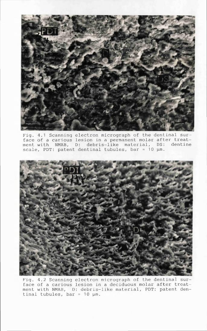
Fig. 4.1 Scanning electron micrograph of the dentinal surface of a carious lesion in a permanent molar after treatment with NMAB, D: debris-like material, DS: dentinescale, PDT: patent dentinal tubules, bar = 10 jam.
Fig. 4.2 Scanning electron micrograph of the dentinal surface of a carious lesion in a deciduous molar after treatment with NMAB, D: debris-like material, PDT: patent dentinal tubules, bar = 10 pm.
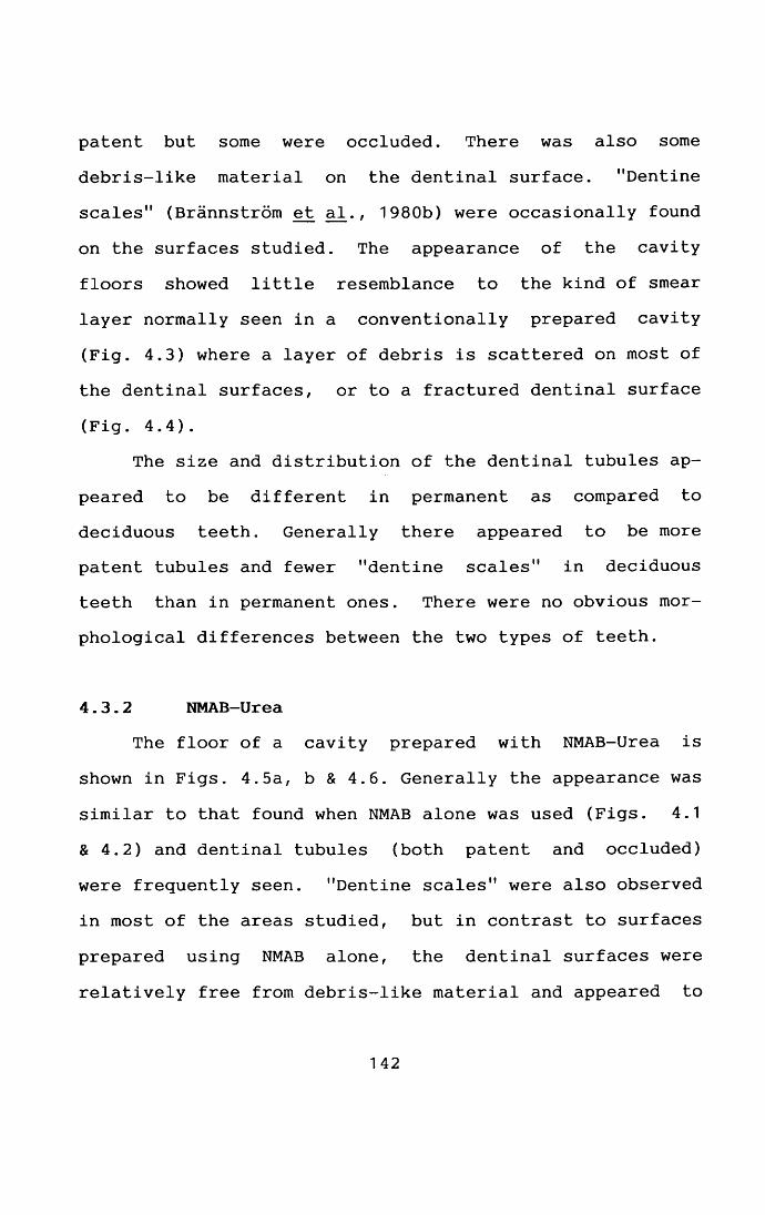
patent but some were occluded. There was also some debris-like material on the dentinal surface. "Dentine scales" (Brannstrom et a l . , 1980b) were occasionally found
on the surfaces studied. The appearance of the cavity floors showed little resemblance to the kind of smear layer normally seen in a conventionally prepared cavity
(Fig. 4.3) where a layer of debris is scattered on most of the dentinal surfaces, or to a fractured dentinal surface
(Fig. 4.4).The size and distribution of the dentinal tubules ap
peared to be different in permanent as compared to deciduous teeth. Generally there appeared to be more patent tubules and fewer "dentine scales" in deciduous teeth than in permanent ones. There were no obvious mor
phological differences between the two types of teeth.
4.3.2 NMAB-UreaThe floor of a cavity prepared with NMAB-Urea is
shown in Figs. 4.5a, b & 4.6. Generally the appearance was
similar to that found when NMAB alone was used (Figs. 4.1 & 4.2) and dentinal tubules (both patent and occluded)
were frequently seen. "Dentine scales" were also observed in most of the areas studied, but in contrast to surfaces
prepared using NMAB alone, the dentinal surfaces were
relatively free from debris-like material and appeared to
142
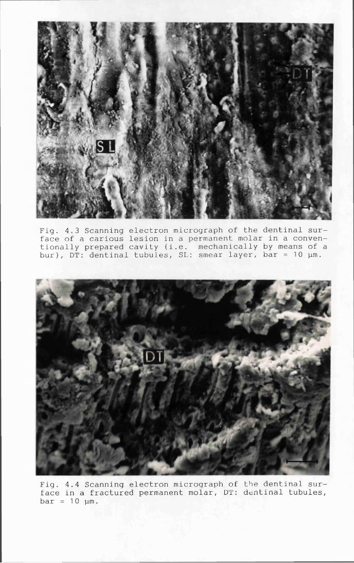
Fig. 4.3 Scanning electron micrograph of the dentinal surface of a carious lesion in a permanent molar in a conventionally prepared cavity (i.e. mechanically by means of a bur), DT: dentinal tubules, SL: smear layer, bar = 10 pm.
Fig. 4.4 Scanning electron micrograph of the dentinal surface in a fractured permanent molar, DT: dentinal tubules, bar = 10 pm.
Fig. 4.5a Scanning electron micrograph of the dentinal surface of a carious lesion in a permanent molar after treatment with NMAB-Urea, D: debris-like material, ODT: occluded dentinal tubules, PDT: patent dentinal tubules,
Fig. 4.5b Scanning electron micrograph of the dentinal surface of a carious lesion of a permanent molar after treatment with NMAB-Urea, D: debris-like material, ODT: occluded dentinal tubules, PDT: patent dentinal tubules, bar = 10 pm.
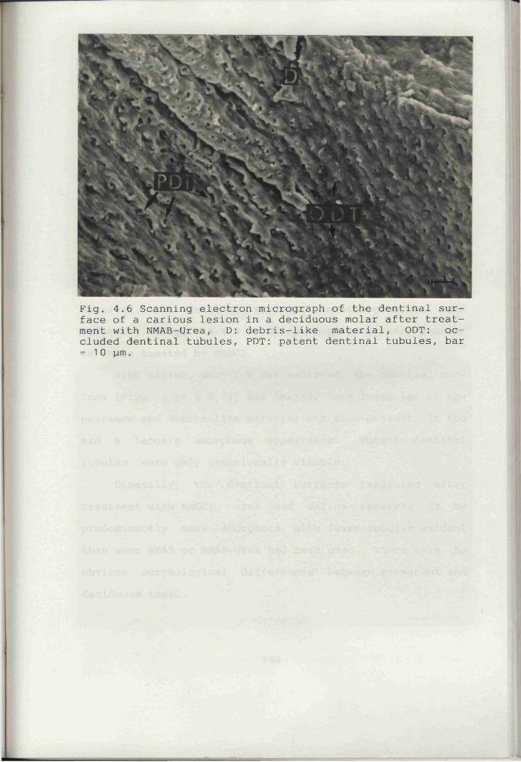
Fig. 4.6 Scanning electron micrograph of the dentinal surface of a carious lesion in a deciduous molar after treatment with NMAB-Urea, D: debris-like material, ODT: occluded dentinal tubules, PDT: patent dentinal tubules, bar = 10 p m .
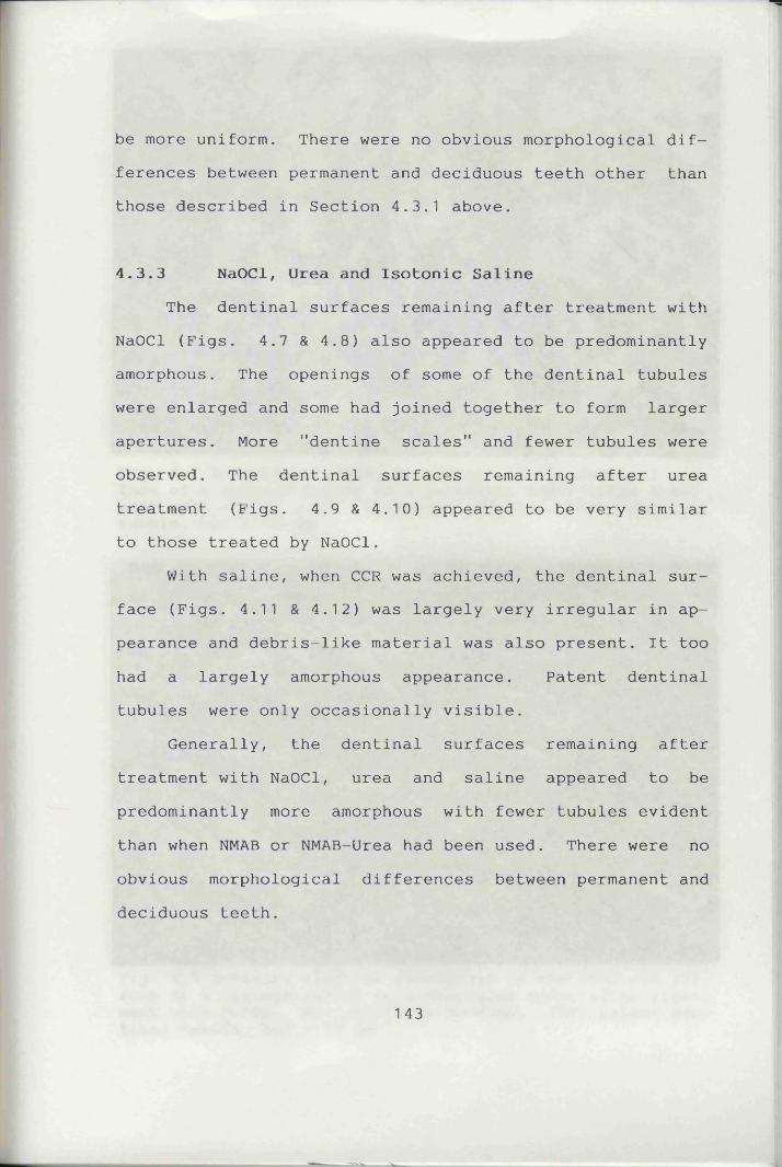
be more uniform. There were no obvious morphological differences between permanent and deciduous teeth other than those described in Section 4.3.1 above.
4.3.3 NaOCl, Urea and Isotonic SalineThe dentinal surfaces remaining after treatment with
NaOCl (Figs. 4.7 & 4.8) also appeared to be predominantly amorphous. The openings of some of the dentinal tubules were enlarged and some had joined together to form larger apertures. More "dentine scales" and fewer tubules were observed. The dentinal surfaces remaining after urea treatment (Figs. 4.9 & 4.10) appeared to be very similar to those treated by NaOCl.
With saline, when CCR was achieved, the dentinal surface (Figs. 4.11 & 4.12) was largely very irregular in appearance and debris-like material was also present. It too had a largely amorphous appearance. Patent dentinal tubules were only occasionally visible.
Generally, the dentinal surfaces remaining after treatment with NaOCl, urea and saline appeared to be predominantly more amorphous with fewer tubules evident than when NMAB or NMAB-Urea had been used. There were no obvious morphological differences between permanent and deciduous teeth.
1 43
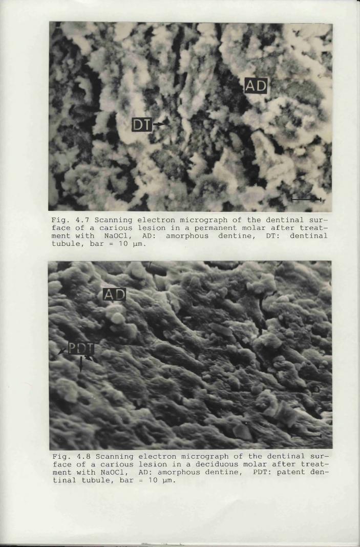
Fig. 4.7 Scanning electron micrograph of the dentinal surface of a carious lesion in a permanent molar after treatment with NaOCl, AD: amorphous dentine, DT: dentinaltubule, bar = 10 pm.
Fig. 4.8 Scanning electron micrograph of the dentinal surface of a carious lesion in a deciduous molar after treatment with NaOCl, AD: amorphous dentine, PDT: patent dentinal tubule, bar = 10 pm.
Fig. 4.9 Scanning electron micrograph of the dentinal surface of a carious lesion in a permanent molar after treatment with urea, bar = 10 pm.
Fig. 4.10 Scanning electron micrograph of the dentinal surface of a carious lesion in a deciduous molar after treatment with urea, bar = 10 pm.
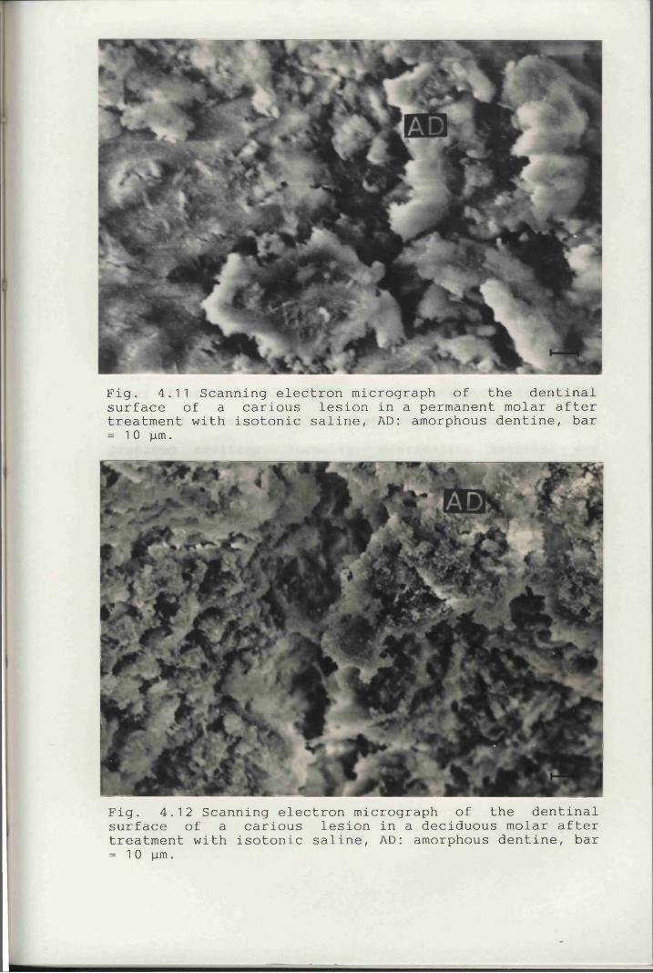
Fig. 4.11 Scanning electron micrograph of the dentinal surface of a carious lesion in a permanent molar after treatment with isotonic saline, AD: amorphous dentine, bar = 10 p m .
Fig. 4.12 Scanning electron micrograph of the dentinal surface of a carious lesion in a deciduous molar after treatment with isotonic saline, AD: amorphous dentine, bar = 10 p m .
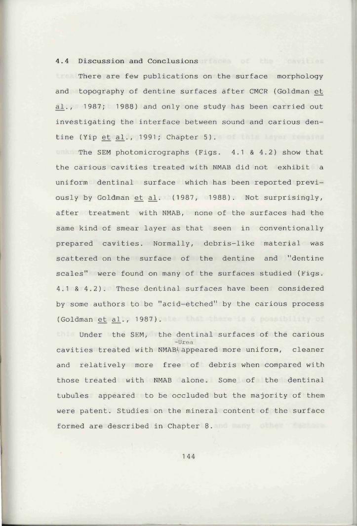
4.4 Discussion and ConclusionsThere are few publications on the surface morphology
and topography of dentine surfaces after CMCR (Goldman e_tal., 1987; 1988) and only one study has been carried outinvestigating the interface between sound and carious dentine (Yip et aJ. , 1991; Chapter 5).
The SEM photomicrographs (Figs. 4.1 & 4.2) show that the carious cavities treated with NMAB did not exhibit a uniform dentinal surface which has been reported previously by Goldman et al. (1987, 1988). Not surprisingly, after treatment with NMAB, none of the surfaces had the same kind of smear layer as that seen in conventionally prepared cavities. Normally, debris-like material was scattered on the surface of the dentine and "dentine scales" were found on many of the surfaces studied (Figs.4.1 & 4.2). These dentinal surfaces have been consideredby some authors to be "acid-etched" by the carious process (Goldman et al., 1987).
Under the SEM, the dentinal surfaces of the carious-Urea
cavities treated with NMABAappeared more uniform, cleaner and relatively more free of debris when compared with those treated with NMAB alone. Some of the dentinal tubules appeared to be occluded but the majority of them were patent. Studies on the mineral content of the surface formed are described in Chapter 8.
1 44
The amorphous dentine surfaces of the cavities treated by NaOCl, urea and isotonic saline showed few or no patent dentinal tubules (Figs. 4.8 - 4.12). These surfaces might consist of both partially demineralised or remineralised dentine in which some of the organic matrix may have been degraded. The nature of this layer remains unknown.
The SEM photomicrographs (Figs. 4.1, 4.2, 4.5 & 4.6) showed that the carious cavities after treatment with NMAB and NMAB-Urea did not exhibit a uniform dentinal surface, and some of the dentinal tubules appeared to be occluded. Whilst these tubules might have become occluded by the gentle "crushing and burnishing action" of the applicator tip on the dentine surface (Sheerer et aT., 1989), it ismore likely that this results from the deposition of reactionary, sclerotic dentine as a result of the caries process. The highly irregular surfaces of the cavity floors seem to indicate that there is a possibility of this being used to advantage in the restoration of the lesion using adhesive restorative materials.
It is, however, important to bear in mind that the size and distribution of dentinal tubules also depends on a number of different factors such as the type of teeth used, depth of dentine cut, stage of development of the teeth, previous carious activity and many other factors
1 45
■
(ten Cate, 1989).The comparison of deciduous and permanent teeth indi
cated that there were only few differences in the morphological appearance of the resulting dentinal surface after treatment with various caries removal agents apart from the fact that "dentine scales" were rarely observedin deciduous teeth and there were also more patent tubules
bein these teeth. This mayjvdue to the differences between the nature of permanent and deciduous dentine and the rate of demineralisation during a carious attack which has shown to be higher in deciduous than in permanent teeth (Featherstone et al., 1981). In the light of these findings and because NMAB and NMAB-Urea appeared to be better suited to caries removal in deciduous teeth than permanent ones (Chapter 3), the different types of dentinal surfaces after treatment with NMAB or NMAB-Urea were further investigated using a more "standardised" batch of specimen teeth (see Sections 2.2.1 and Chapter 5).
The type of dentinal surfaces remaining after treatment probably depends largely on the state of carious process which pertains, i.e. whether it is remineralising or demineralising, and of the latter, whether remineralisation is still possible. This is further investigated in Chapter 5.
146
4.5 Summarya. After CCR by treatment with NMAB, the surfaces formed had a very uneven appearance with many undermined areas; "dentine scales", patent and occluded dentinal tubules could also be observed. The nature of the surfaces produced by this treatment would appear to offer improved micromechanical retention of restorative materials as compared to cavities prepared conventionally (i.e. mechanically).b. NMAB-Urea produced a "cleaner" surface when compared with NMAB alone.c. "Dentine scales" were rarely observed in deciduous teeth, and most of the tubules were patent.
1 47
CHAPTER 5
THE INTERFACE BETWEEN CARIOUS AND SOUND DENTINE
5.1 IntroductionNMAB has been reported to remove the outer or first
layer but not to affect the inner or second layer of carious dentine (Brannstom et al. , 1980b; Kurosaki et. al. , 1977) which remains after treatment with the solution. Some authors regard the dentine remaining after treatment with NMAB as sound and have claimed that NMAB removed only the carious dentine enabling the interface between the carious and sound dentine to be visualised under the SEM (Goldman et al. , 1987). The clinically sound dentine atthe base of lesions from which carious tissue was removed with NMAB has been reported to consist of highly irregular surfaces with numerous undermined areas and patent dentinal tubules (Brannstrom et al. , 1980b; Goldman et al.,1987, 1988). Generally, this is consistent with the previous study (Chapter 4).
Apart from the typical dentinal surfaces observed in the previous study and which formed the major part of the surface concerned (Figs. 4.1, 4.2, 4.5a, b & 4.6), other atypical surfaces were also seen in carious lesions treated with either NMAB or NMAB-Urea. The purpose of this
148

study was to use light and scanning electron microscopy to investigate the types of dentinal surface found in carious lesions after treatment with NMAB and NMAB-Urea in more detail as this may represent the so-called "interface of carious and sound dentine".
5.2 Materials and MethodsCoronal carious lesions from permanent and deciduous
teeth were treated chemomechanically with either NMAB or NMAB containing 2 mol/L urea using the in vitro study model described in Sections 2.2.4 - 2.2.7. Ten permanent and ten deciduous teeth with CCR (five treated with NMAB and five with NMAB-Urea in each type of dentition) were selected at random for this study. These teeth were split into two halves through the middle of the cavities with an osteotome. One half was processed for scanning electron microscopy and the other half for histological staining. Methodologies for specimen preparation for both light and scanning electron microscopy were detailed in Sections2.3.2 and 2.4.2. A total of one hundred and twenty decalcified, stained histological sections were examined (thirty permanent teeth treated with NMAB, thirty permanent teeth treated with NMAB-Urea, thirty deciduous teeth treated with NMAB and thirty deciduous teeth treated with NMAB-Urea). Due to the delay in fixation, the pulpal
149
tissue which was not the the subject of study here, was not well preserved. The aim of the work described in Chapter 4 was to compare the nature of the dentinal surfaces remaining after CCR using various caries removal agents. The aim of this chapter is to correlate these findings with the features of the dentinal surfaces produced at what is in fact the interface between carious and sound dentine, thereby facilitating the possibility of further elucidating the understanding of the carious process. The typical dentinal surfaces which were described in Chapter 4 are not repeatedly reported here.
5.3 Results
5.3.1 Scanning Electron MicroscopyAll the specimen surfaces were rinsed thoroughly with
distilled water prior to critical point drying in order to remove any contaminant salts which had formed crystals on the dentinal surfaces. A specimen which was not rinsed is shown in Fig. 5.1. These contaminant salts may either come from the caries removal agents used or the storage medium which was phosphate buffered saline. The crystals formed are of quite different appearance from anything observed
1 50
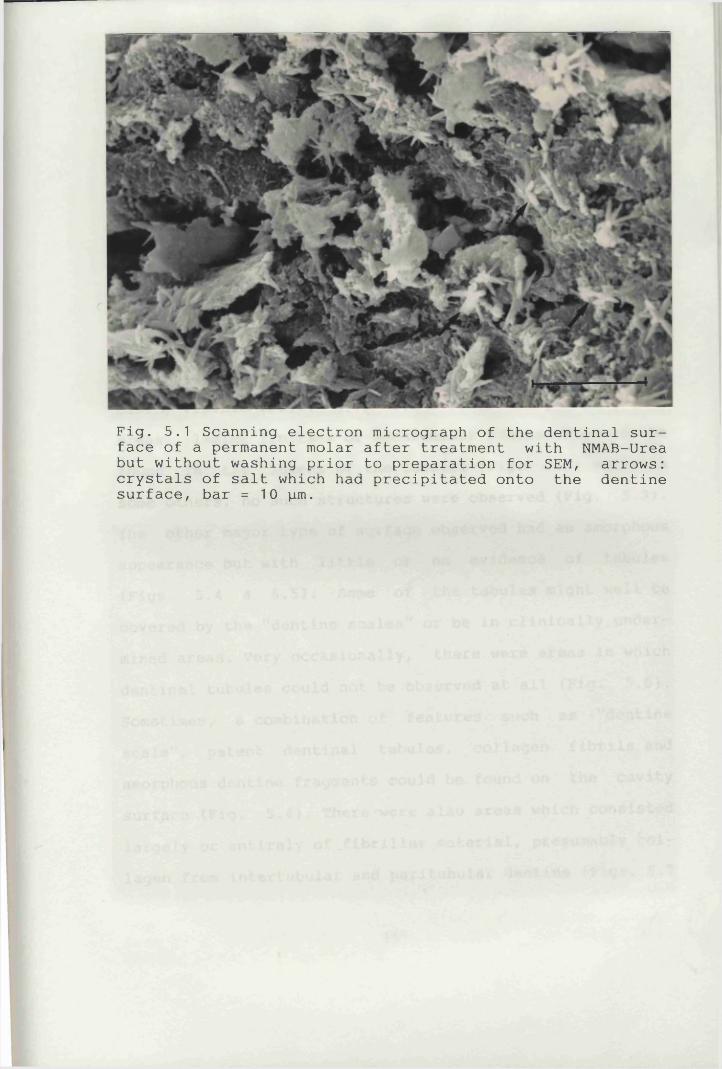
Fig. 5.1 Scanning electron micrograph of the dentinal surface of a permanent molar after treatment with NMAB-Urea but without washing prior to preparation for SEM, arrows: crystals of salt which had precipitated onto the dentine surface, bar = 10 pm.
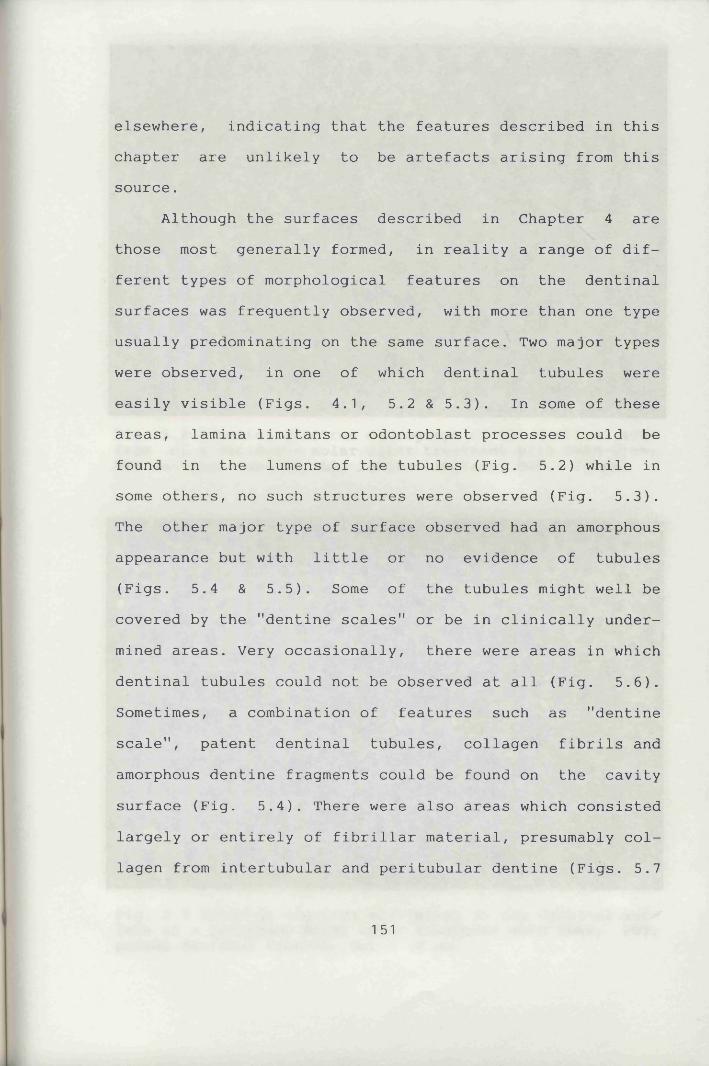
elsewhere, indicating that the features described in this chapter are unlikely to be artefacts arising from this source.
Although the surfaces described in Chapter 4 are those most generally formed, in reality a range of different types of morphological features on the dentinal surfaces was frequently observed, with more than one type usually predominating on the same surface. Two major types were observed, in one of which dentinal tubules wereeasily visible (Figs. 4.1, 5.2 & 5.3). In some of theseareas, lamina limitans or odontoblast processes could be found in the lumens of the tubules (Fig. 5.2) while in some others, no such structures were observed (Fig. 5.3). The other major type of surface observed had an amorphous appearance but with little or no evidence of tubules(Figs. 5.4 & 5.5). Some of the tubules might well becovered by the "dentine scales" or be in clinically undermined areas. Very occasionally, there were areas in which dentinal tubules could not be observed at all (Fig. 5.6). Sometimes, a combination of features such as "dentine scale", patent dentinal tubules, collagen fibrils and amorphous dentine fragments could be found on the cavity surface (Fig. 5.4). There were also areas which consisted largely or entirely of fibrillar material, presumably collagen from intertubular and peritubular dentine (Figs. 5.7
151
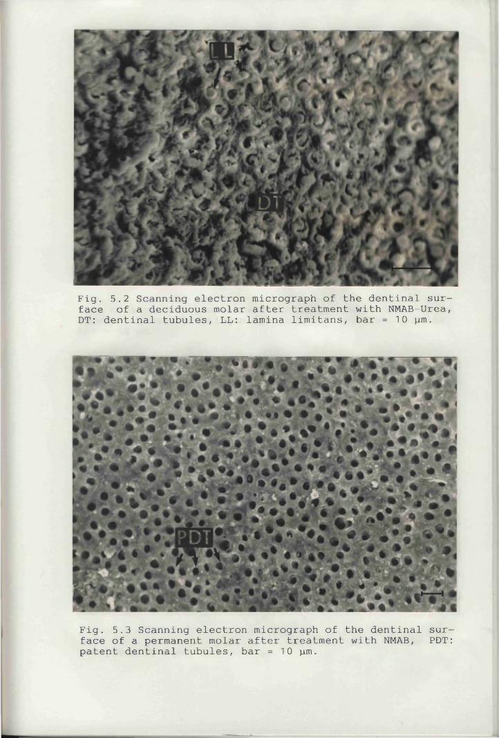
Fig. 5.2 Scanning electron micrograph of the dentinal surface of a deciduous molar after treatment with NMAB-Urea, DT: dentinal tubules, LL: lamina limitans, bar = 10 pm.
%
Fig. 5.3 Scanning electron micrograph of the dentinal surface of a permanent molar after treatment with NMAB, PDT: patent dentinal tubules, bar = 10 pm.
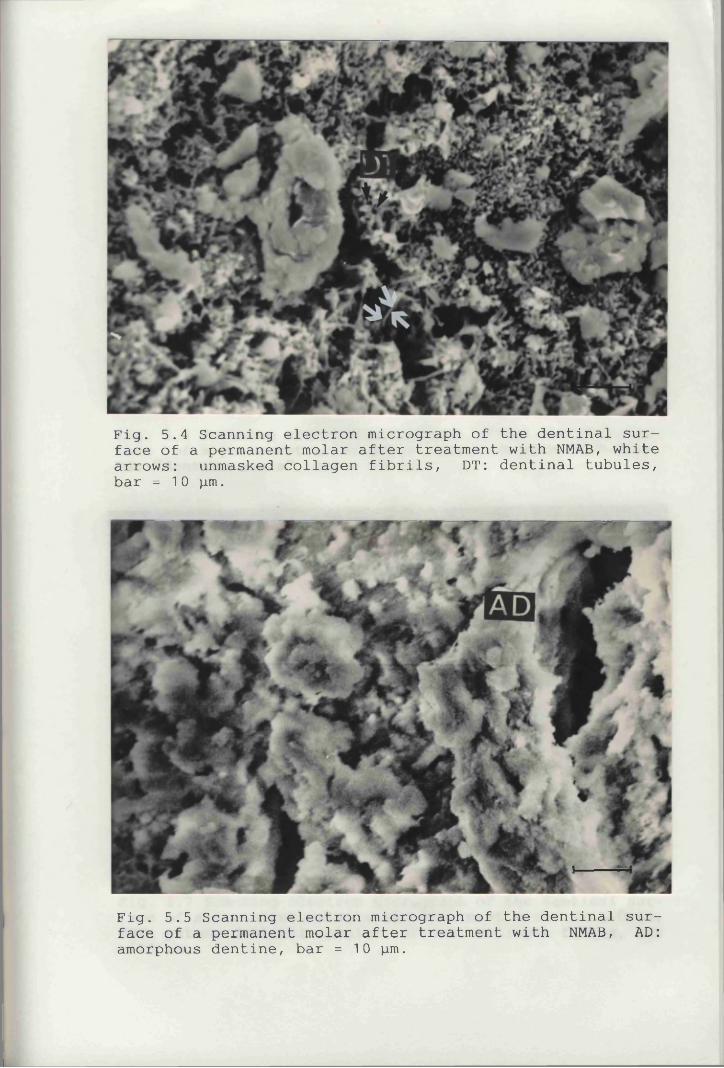
Fig. 5.4 Scanning electron micrograph of the dentinal surface of a permanent molar after treatment with NMAB, white arrows: unmasked collagen fibrils, DT: dentinal tubules,bar = 10 pm.
Fig. 5.5 Scanning electron micrograph of the dentinal surface of a permanent molar after treatment with NMAB, AD: amorphous dentine, bar = 10 pm.
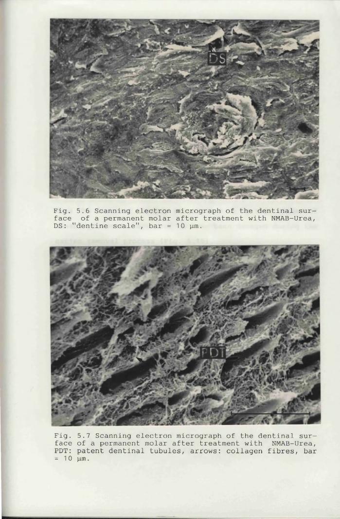
Fig. 5.6 Scanning electron micrograph of the dentinal surface of a permanent molar after treatment with NMAB-Urea, DS: "dentine scale", bar = 10 pm.
Fig. 5.7 Scanning electron micrograph of the dentinal surface of a permanent molar after treatment with NMAB-Urea, PDT: patent dentinal tubules, arrows: collagen fibres, bar= 10 pm.
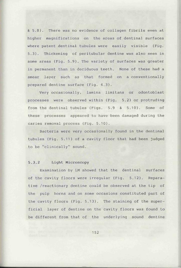
& 5.8). There was no evidence of collagen fibrils even at higher magnifications on the areas of dentinal surfaces where patent dentinal tubules were easily visible (Fig. 5.3). Thickening of peritubular dentine was also seen in some areas (Fig. 5.9). The variety of surfaces was greater in permanent than in deciduous teeth. None of these had a smear layer such as that formed on a conventionally prepared dentine surface (Fig. 4.3).
Very occasionally, lamina limitans or odontoblast processes were observed within (Fig. 5.2) or protruding from the dentinal tubules (Figs. 5.9 & 5.10). Some ofthese processes appeared to have been damaged during the caries removal process (Fig. 5.10).
Bacteria were very occasionally found in the dentinal tubules (Fig. 5.11) of a cavity floor that had been judged to be "clinically" sound.
5.3.2 Light MicroscopyExamination by LM showed that the dentinal surfaces
of the cavity floors were irregular (Fig. 5.12). Reparative /reactionary dentine could be observed at the tip of the pulp horns and on some occasions constituted part of the cavity floors (Fig. 5.13). The staining of the superficial layer of dentine on the cavity floors was found to be different from that of the underlying sound dentine
152
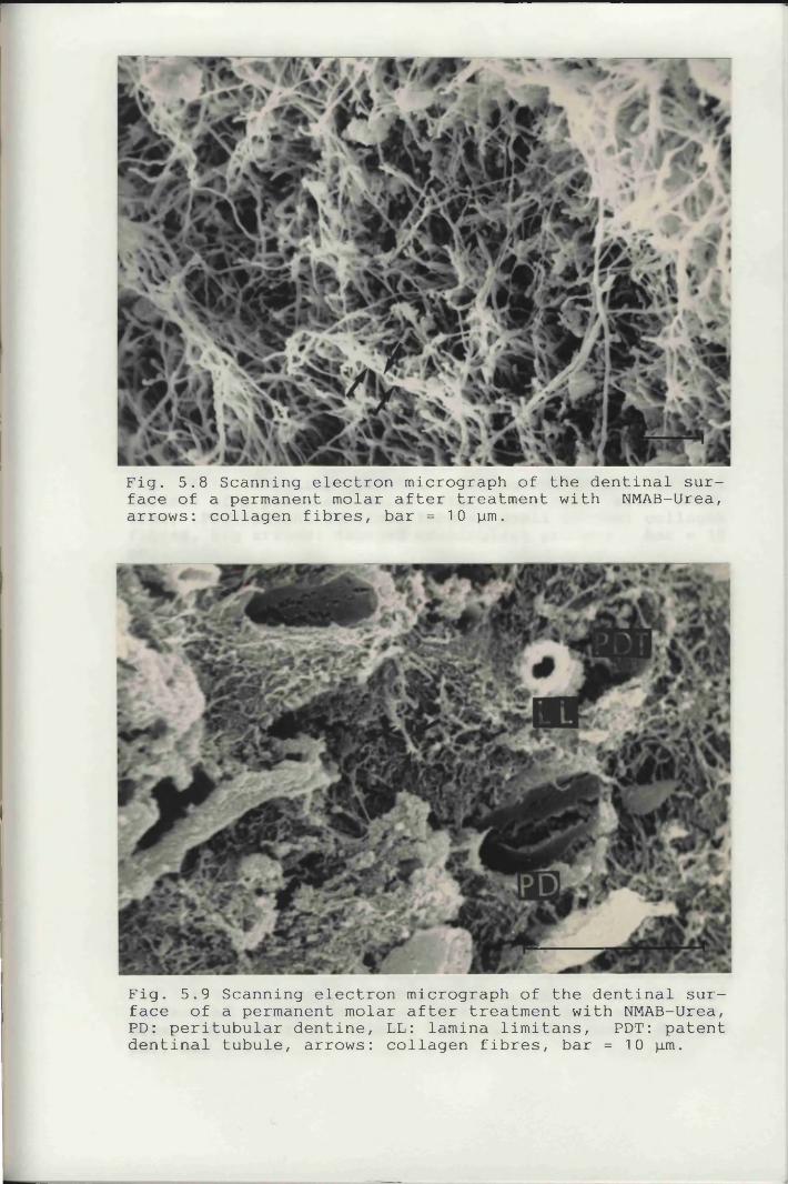
Fig. 5.8 Scanning electron micrograph of the dentinal surface of a permanent molar after treatment with NMAB-Urea, arrows: collagen fibres, bar = 10 pm.
Fig. 5.9 Scanning electron micrograph of the dentinal surface of a permanent molar after treatment with NMAB-Urea, PD: peritubular dentine, LL: lamina limitans, PDT: patent dentinal tubule, arrows: collagen fibres, bar =10 pm.
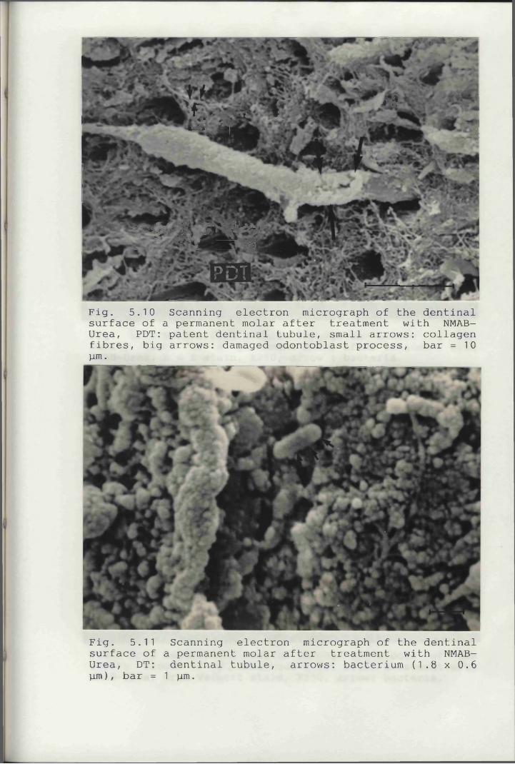
\W
'f • -v <** * J« L H i ____
* 5
lar^L^y'K '' ̂ ^ jM k ̂PDT
Fig. 5.10 Scanning electron micrograph of the dentinal surface of a permanent molar after treatment with NMAB- Urea, PDT: patent dentinal tubule, small arrows: collagen fibres, big arrows: damaged odontoblast process, bar =10 pm.
Fig. 5.11 Scanning electron micrograph of the dentinal surface of a permanent molar after treatment with NMAB- Urea, DT: dentinal tubule, arrows: bacterium (1.8 x 0.6pm), bar = 1 pm.
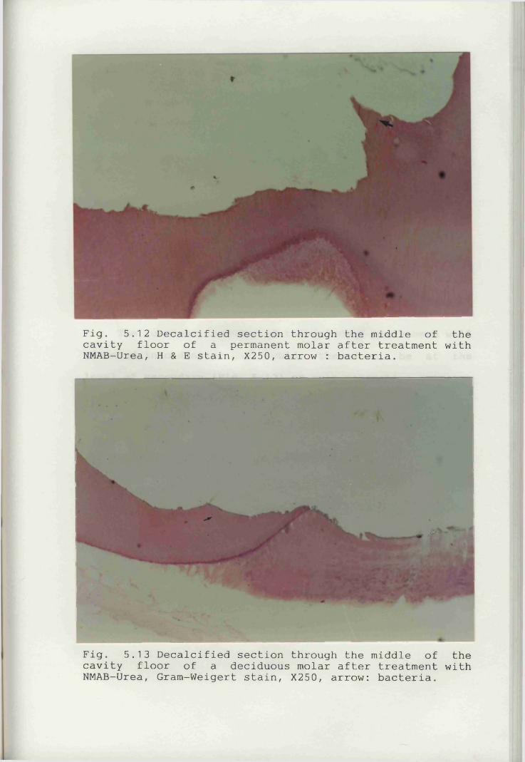
Fig. 5.12 Decalcified section through the middle of the cavity floor of a permanent molar after treatment with NMAB-Urea, H & E stain, X250, arrow : bacteria.
Fig. 5.13 Decalcified section through the middle of the cavity floor of a deciduous molar after treatment with NMAB-Urea, Gram-Weigert stain, X250, arrow: bacteria.
> i.
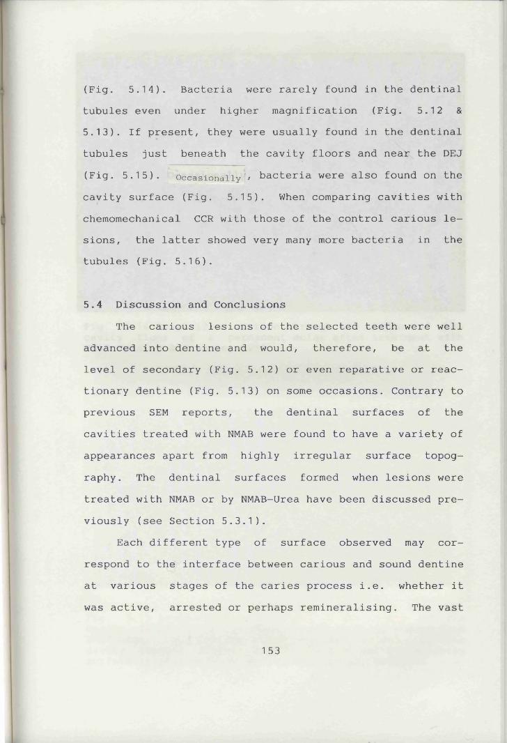
(Fig. 5.14). Bacteria were rarely found in the dentinal tubules even under higher magnification (Fig. 5.12 &5.13). If present, they were usually found in the dentinal tubules just beneath the cavity floors and near the DEJ (Fig. 5.15). Occasionally'/ bacteria were also found on the cavity surface (Fig. 5.15). When comparing cavities with chemomechanical CCR with those of the control carious lesions, the latter showed very many more bacteria in the tubules (Fig. 5.16).
5.4 Discussion and ConclusionsThe carious lesions of the selected teeth were well
advanced into dentine and would, therefore, be at the level of secondary (Fig. 5.12) or even reparative or reactionary dentine (Fig. 5.13) on some occasions. Contrary to previous SEM reports, the dentinal surfaces of the cavities treated with NMAB were found to have a variety of appearances apart from highly irregular surface topography. The dentinal surfaces formed when lesions were treated with NMAB or by NMAB-Urea have been discussed previously (see Section 5.3.1).
Each different type of surface observed may correspond to the interface between carious and sound dentine at various stages of the caries process i.e. whether it was active, arrested or perhaps remineralising. The vast
153
Fig. 5.14 Decalcified section through the middle of the cavity floor of a permanent molar after treatment with NMAB-Urea, H & E stain, X250.
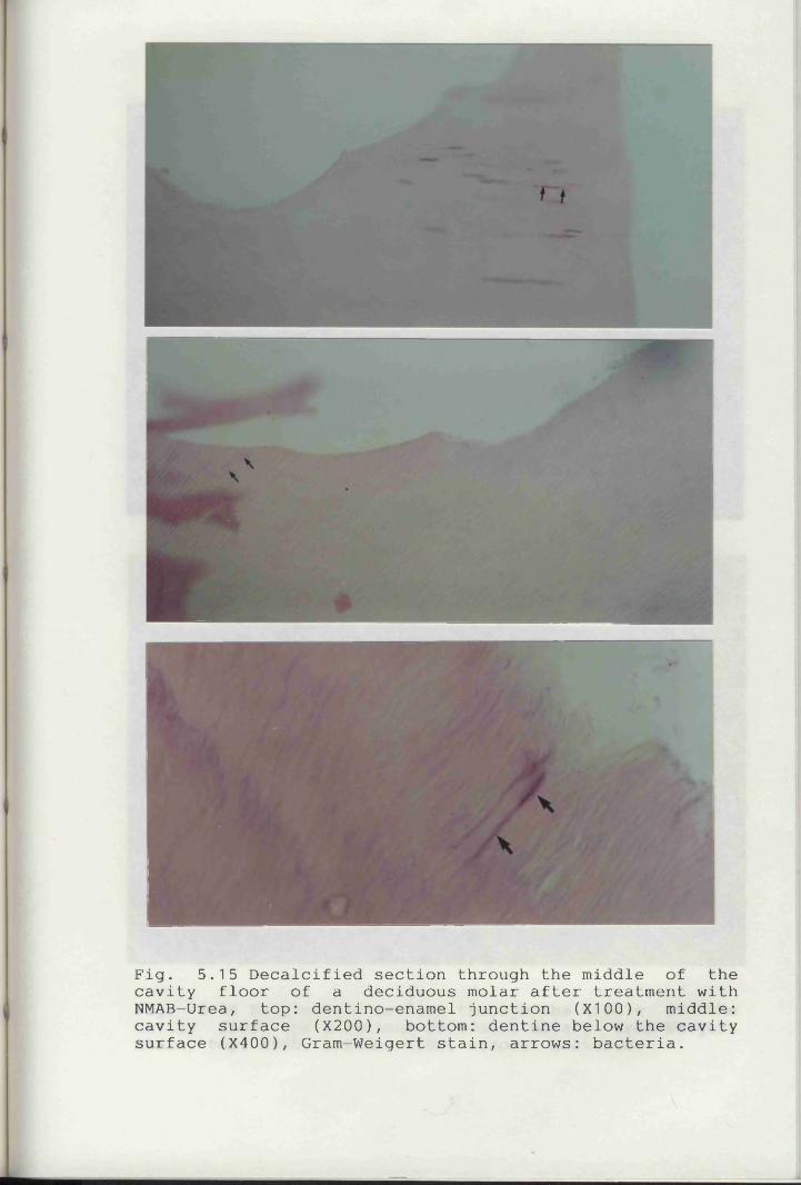
w
Fig. 5.15 Decalcified section through the middle of the cavity floor of a deciduous molar after treatment with NMAB-Urea, top: dentino-enamel junction (X100), middle: cavity surface (X200), bottom: dentine below the cavity surface (X400), Gram-Weigert stain, arrows: bacteria.
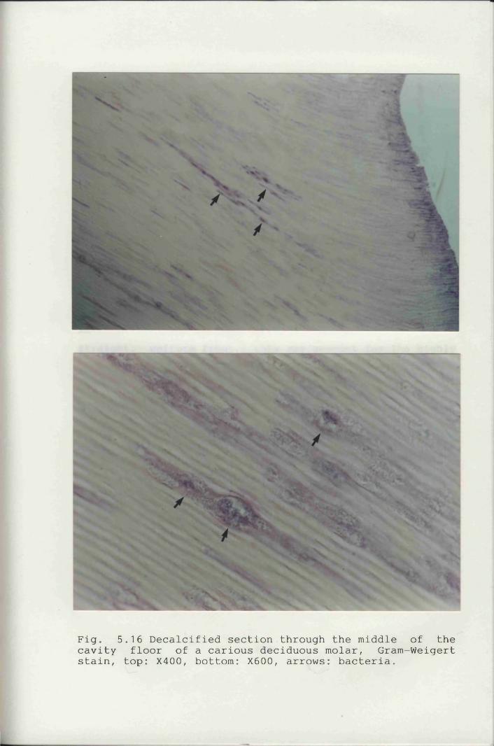
Fig. 5.16 Decalcified section through the middle of the cavity floor of a carious deciduous molar, Gram-Weigert stain, top: X400, bottom: X600, arrows: bacteria.
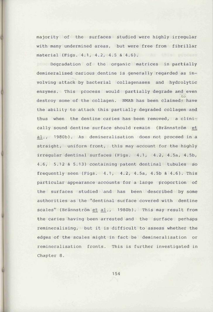
majority of the surfaces studied were highly irregular with many undermined areas, but were free from fibrillar material (Figs. 4.1, 4.2, 4.5 & 4.6).
Degradation of the organic matrices in partially demineralised carious dentine is generally regarded as involving attack by bacterial collagenases and hydrolyticenzymes. This process would partially degrade and even
todestroy some of the collagen. NMAB has been claimed< have the ability to attack this partially degraded collagen and thus when the dentine caries has been removed, a clinically sound dentine surface should remain (Brannstrom et al., 1980b). As demineralisation does not proceed in astraight, uniform front, this may account for the highly irregular dentinal surfaces (Figs. 4.1, 4.2, 4.5a, 4.5b,4.6, 5.12 & 5.13) containing patent dentinal tubules sofrequently seen (Figs. 4.1, 4.2, 4.5a, 4.5b & 4.6). Thisparticular appearance accounts for a large proportion of the surfaces studied and has been described by some authorities as the "dentinal surface covered with dentine scales" (Brannstrom et al., 1980b). This may result fromthe caries having been arrested and the surface perhaps remineralising, but it is difficult to assess whether the edges of the scales might in fact be demineralisation or remineralisation fronts. This is further investigated in Chapter 8.
1 54
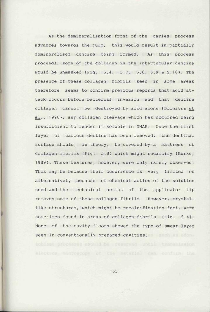
As the demineralisation front of the caries process advances towards the pulp, this would result in partially demineralised dentine being formed. As this process proceeds, some of the collagen in the intertubular dentine would be unmasked (Fig. 5.4, 5.7, 5.8, 5.9 & 5.10). Thepresence of these collagen fibrils seen in some areas therefore seems to confirm previous reports that acid attack occurs before bacterial invasion and that dentine collagen cannot be destroyed by acid alone (Boonstra et al., 1990), any collagen cleavage which has occurred being insufficient to render it soluble in NMAB. Once the first layer of carious dentine has been removed, the dentinal surface should, in theory, be covered by a mattress of collagen fibrils (Fig. 5.8) which might recalcify (Burke, 1989). These features, however, were only rarely observed. This may be because their occurrence is very limited or alternatively because of chemical action of the solution used and the mechanical action of the applicator tip removes some of these collagen fibrils. However, crystallike structures, which might be recalcification foci, were sometimes found in areas of collagen fibrils (Fig. 5.4). None of the cavity floors showed the type of smear layer seen in conventionally prepared cavities.
1 55
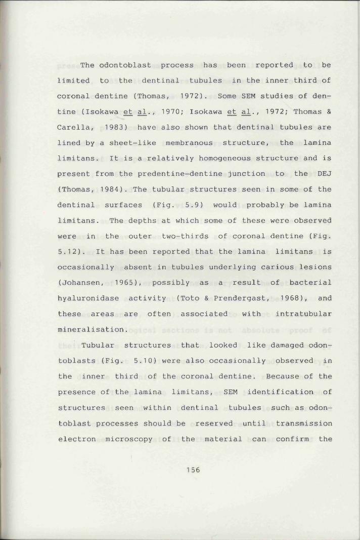
The odontoblast process has been reported to be limited to the dentinal tubules in the inner third of coronal dentine (Thomas, 1972). Some SEM studies of dentine (Isokawa et al., 1970; Isokawa et al., 1972; Thomas & Carella, 1983) have also shown that dentinal tubules are lined by a sheet-like membranous structure, the lamina limitans. It is a relatively homogeneous structure and is present from the predentine-dentine junction to the DEJ (Thomas, 1984). The tubular structures seen in some of the dentinal surfaces (Fig. 5.9) would probably be lamina limitans. The depths at which some of these were observed were in the outer two-thirds of coronal dentine (Fig. 5.12). It has been reported that the lamina limitans is occasionally absent in tubules underlying carious lesions (Johansen, 1965), possibly as a result of bacterial hyaluronidase activity (Toto & Prendergast, 1968), and these areas are often associated with intratubular mineralisation.
Tubular structures that looked like damaged odontoblasts (Fig. 5.10) were also occasionally observed in the inner third of the coronal dentine. Because of the presence of the lamina limitans, SEM identification of structures seen within dentinal tubules such as odontoblast processes should be reserved until transmission electron microscopy of the material can confirm the
1 56
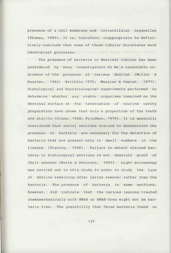
presence of a cell membrane and intracellular organelles (Thomas, 1985). It is, therefore, inappropriate to definitively conclude that some of these tubular structures were odontoblast processes.
The presence of bacteria in dentinal tubules has been considered by many investigators to be a reasonable indicator of the presence of carious dentine (Miller & Massler, 1962; Wirthlin 1970; Massler & Pawlak, 1977). Histological and bacteriological experiments performed to determine whether any viable organisms remained on the dentinal surface at the termination of routine cavity preparation have shown that only a proportion of the teeth are sterile (Crone, 1968; Friedman, 1979). It is generally considered that serial sections stained to demonstrate the presence of bacteria are necessary for the detection of bacteria that are present only in small numbers in the tissues (Stanley, 1968). Failure to detect stained bacteria in histological sections is not absolute proof of their absence (Watts & Paterson, 1990). Light microscopy was carried out in this study in order to study the type of dentine remaining after caries removal rather than the bacteria. The presence of bacteria in some sections, however, did indicate that the carious lesions treated chemomechanically with NMAB or NMAB—Urea might not be bacteria free. The possibility that those bacteria found on
1 57
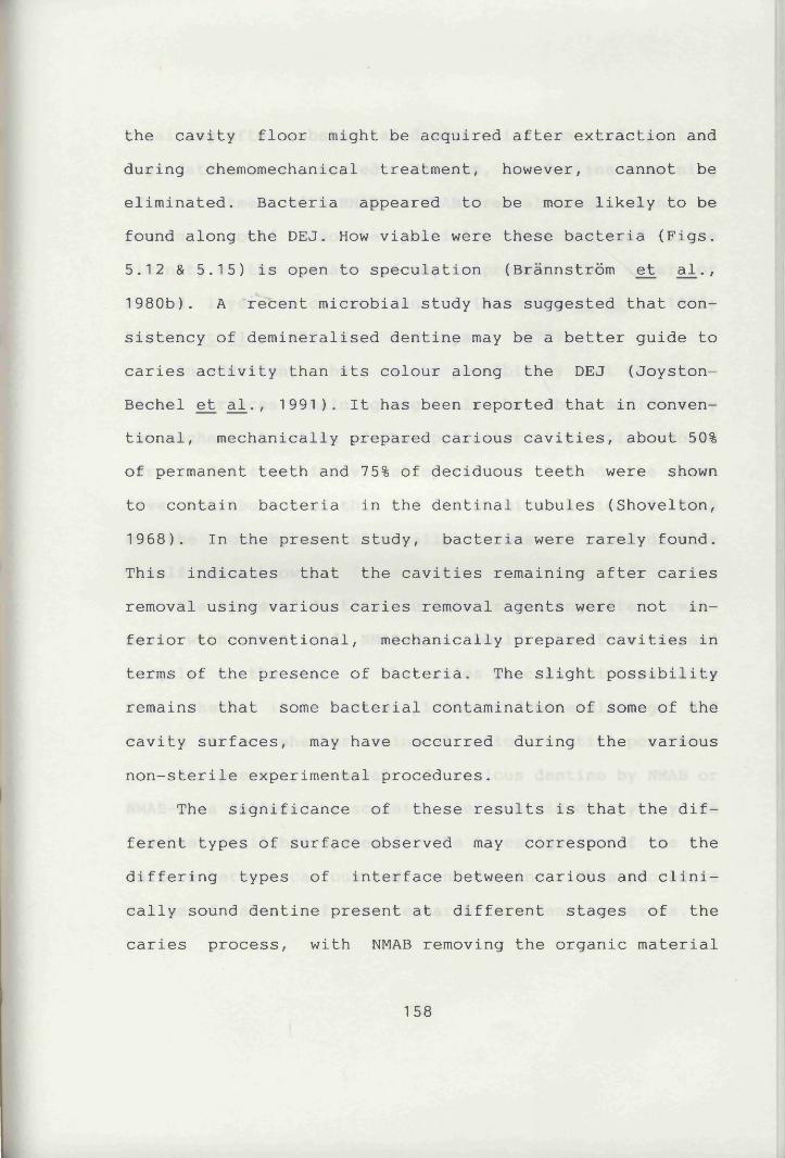
the cavity floor might be acquired after extraction and during chemomechanical treatment, however, cannot be eliminated. Bacteria appeared to be more likely to be found along the DEJ. How viable were these bacteria (Figs. 5.12 & 5.15) is open to speculation (Brannstrom et al., 1980b). A recent microbial study has suggested that consistency of demineralised dentine may be a better guide to caries activity than its colour along the DEJ (Joyston- Bechel et al., 1991). It has been reported that in conventional, mechanically prepared carious cavities, about 50% of permanent teeth and 75% of deciduous teeth were shown to contain bacteria in the dentinal tubules (Shovelton, 1968). In the present study, bacteria were rarely found. This indicates that the cavities remaining after caries removal using various caries removal agents were not inferior to conventional, mechanically prepared cavities in terms of the presence of bacteria. The slight possibility remains that some bacterial contamination of some of the cavity surfaces, may have occurred during the various non-sterile experimental procedures.
The significance of these results is that the different types of surface observed may correspond to the differing types of interface between carious and clinically sound dentine present at different stages of the caries process, with NMAB removing the organic material
158
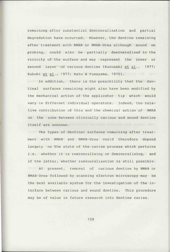
remaining after substantial demineralisation and partial degradation have occurred. However, the dentine remaining after treatment with NMAB or NMAB-Urea although sound on probing, could also be partially demineralised in the vicinity of the surface and may represent the inner or second layer of carious dentine (Kurosaki et al., 1977;Kuboki et al., 1977; Kato & Fusayama, 1970).
In addition, there is the possibility that the dentinal surfaces remaining might also have been modified by the mechanical action of the applicator tip which would vary in different individual operators. Indeed, the relative contribution of this and the chemical action of NMAB on the zone between clinically carious and sound dentine itself are unknown.
The types of dentinal surfaces remaining after treatment with NMAB and NMAB-Urea could therefore depend largely on the state of the caries process which pertains i.e. whether it is remineralising or demineralising, and if the latter, whether remineralisation is still possible.
At present, removal of carious dentine by NMAB or NMAB-Urea followed by scanning electron microscopy may be the best available system for the investigation of the interface between carious and sound dentine. This procedure may be of value in future research into dentine caries.
1 59
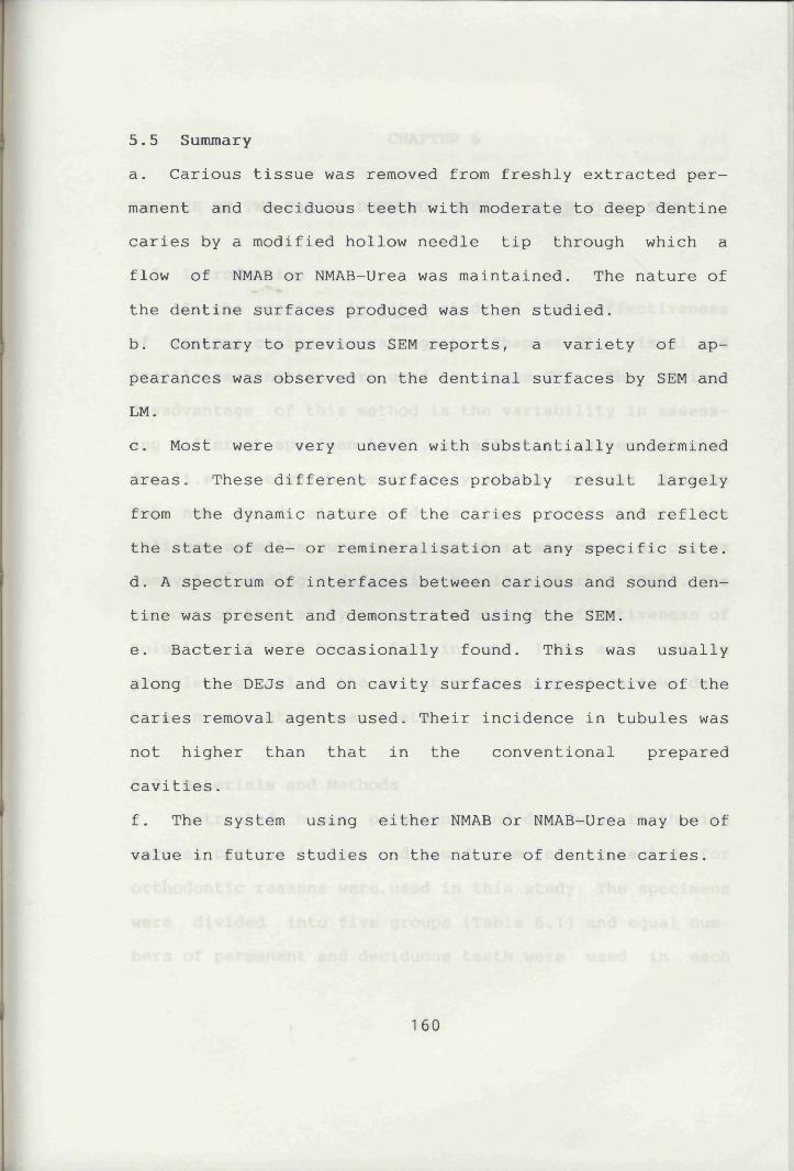
5.5 Summarya. Carious tissue was removed from freshly extracted permanent and deciduous teeth with moderate to deep dentine caries by a modified hollow needle tip through which a flow of NMAB or NMAB-Urea was maintained. The nature of the dentine surfaces produced was then studied.b. Contrary to previous SEM reports, a variety of appearances was observed on the dentinal surfaces by SEM and LM.c. Most were very uneven with substantially undermined areas. These different surfaces probably result largely from the dynamic nature of the caries process and reflectthe state of de- or remineralisation at any specific site.d. A spectrum of interfaces between carious and sound dentine was present and demonstrated using the SEM.e. Bacteria were occasionally found. This was usuallyalong the DEJs and on cavity surfaces irrespective of the caries removal agents used. Their incidence in tubules was not higher than that in the conventional preparedcavities.f. The system using either NMAB or NMAB-Urea may be of value in future studies on the nature of dentine caries.
160
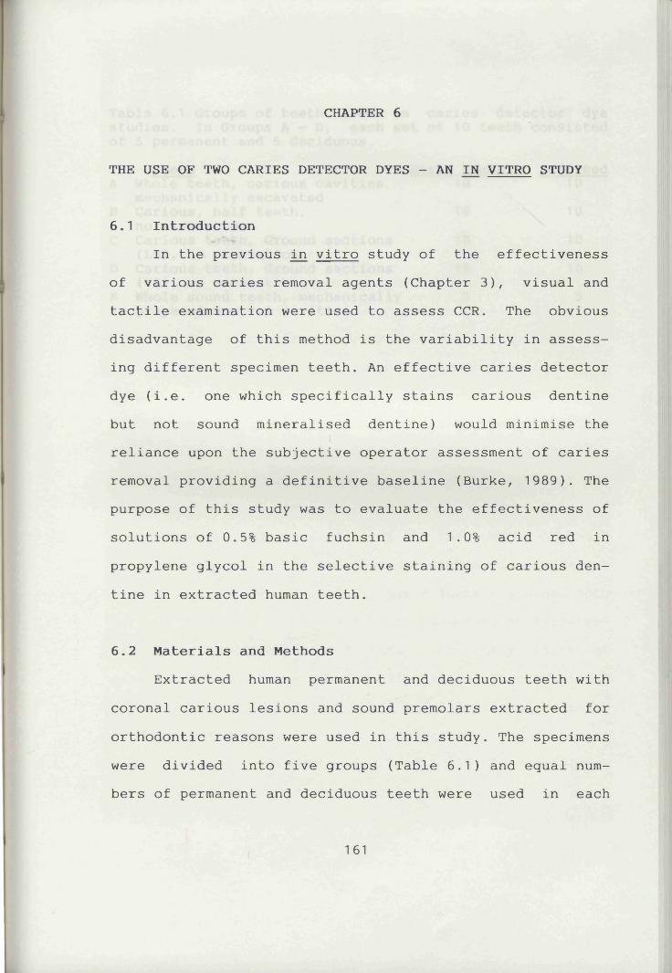
CHAPTER 6
THE USE OF TWO CARIES DETECTOR DYES - AN IN VITRO STUDY
6.1 IntroductionIn the previous in vitro study of the effectiveness
of various caries removal agents (Chapter 3), visual and tactile examination were used to assess CCR. The obvious disadvantage of this method is the variability in assessing different specimen teeth. An effective caries detector dye (i.e. one which specifically stains carious dentine but not sound mineralised dentine) would minimise the
ireliance upon the subjective operator assessment of caries removal providing a definitive baseline (Burke, 1989). The purpose of this study was to evaluate the effectiveness of solutions of 0.5% basic fuchsin and 1.0% acid red in propylene glycol in the selective staining of carious dentine in extracted human teeth.
6.2 Materials and MethodsExtracted human permanent and deciduous teeth with
coronal carious lesions and sound premolars extracted for orthodontic reasons were used in this study. The specimens were divided into five groups (Table 6.1) and equal numbers of permanent and deciduous teeth were used in each
161
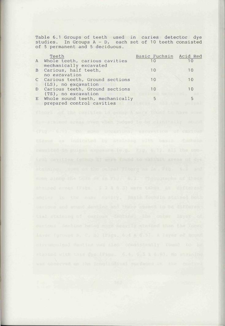
Table 6.1 Groups of teeth used in caries detector dye studies. In Groups A - D, each set of 10 teeth consisted of 5 permanent and 5 deciduous.
Teeth Basic Fuchsin Acid RedA Whole teeth, carious cavities
mechanically excavated1 0 1 0
B Carious, half teeth, no excavation
1 0 10C Carious teeth, Ground sections
(LS), no excavation10 10
D Carious teeth, Ground sections (TS), no excavation
1 0 10E Whole sound teeth, mechanically
prepared control cavities5 5
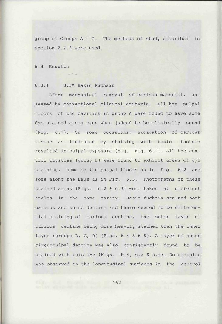
group of Groups A - D. The methods of study described in Section 2.7.2 were used.
6.3 Results
6.3.1 0.5% Basic FuchsinAfter mechanical removal of carious material, as
sessed by conventional clinical criteria, all the pulpal floors of the cavities in group A were found to have some dye-stained areas even when judged to be clinically sound (Fig. 6.1). On some occasions, excavation of carious tissue as indicated by staining with basic fuchsin resulted in pulpal exposure (e.g. Fig. 6.1). All the control cavities (group E) were found to exhibit areas of dye staining, some on the pulpal floors as in Fig. 6.2 and some along the DEJs as in Fig. 6.3. Photographs of these stained areas (Figs. 6.2 & 6.3) were taken at different angles in the same cavity. Basic fuchsin stained both carious and sound dentine and there seemed to be differential staining of carious dentine, the outer layer of carious dentine being more heavily stained than the inner layer (groups B, C, D) (Figs. 6.4 & 6.5). A layer of sound circumpulpal dentine was also consistently found to be stained with this dye (Figs. 6.4, 6.5 & 6.6). No staining was observed on the longitudinal surfaces in the control
1 62
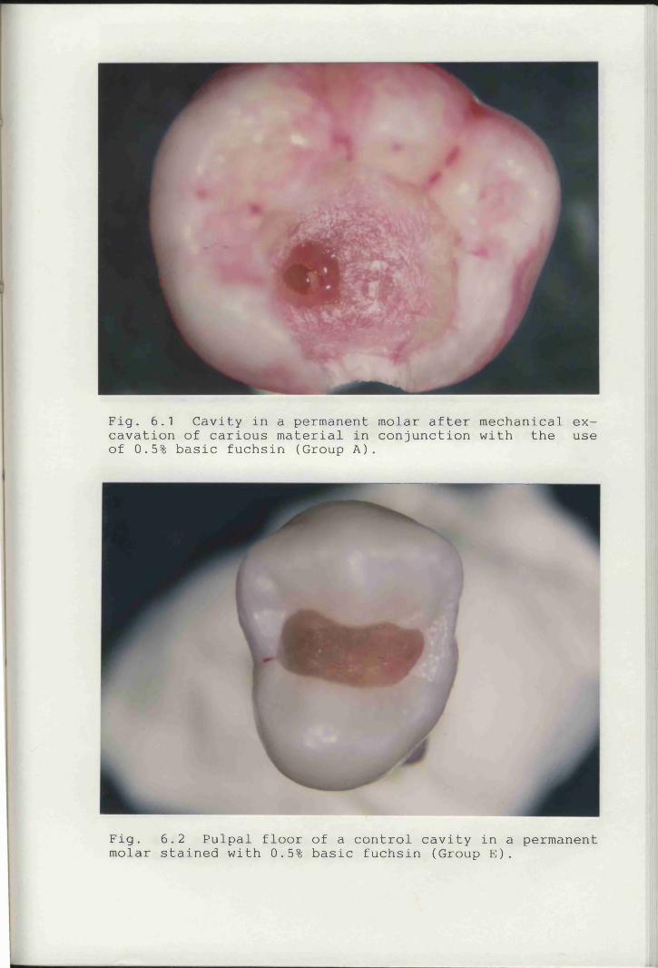
Fig. 6.1 Cavity in a permanent molar after mechanical excavation of carious material in conjunction with the use of 0.5% basic fuchsin (Group A).
Fig. 6.2 Pulpal floor of a control cavity in a permanent molar stained with 0.5% basic fuchsin (Group E).
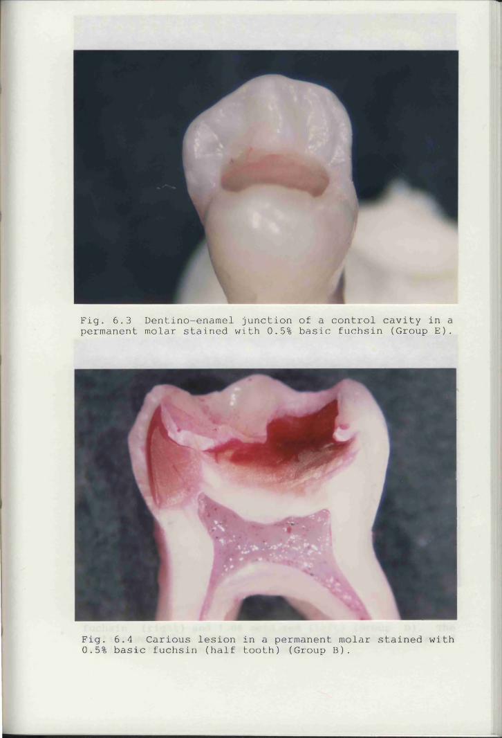
Fig. 6.3 Dentino-enamel junction of a control cavity in a permanent molar stained with 0.5% basic fuchsin (Group E).
Fig. 6.4 Carious lesion in a permanent molar stained with 0.5% basic fuchsin (half tooth) (Group B).
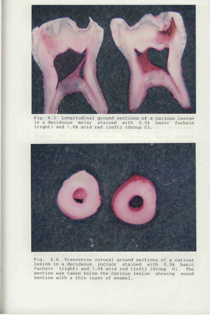
Fig. 6.5 Longitudinal ground sections of a carious lesion in a deciduous molar stained with 0.5% basic fuchsin (right) and 1.0% acid red (left) (Group C).
Fig. 6.6 Transverse coronal ground sections of a carious lesion in a deciduous incisor stained with 0.5% basic fuchsin (right) and 1.0% acid red (left) (Group D). The section was taken below the carious lesion showing sound dentine with a thin layer of enamel.
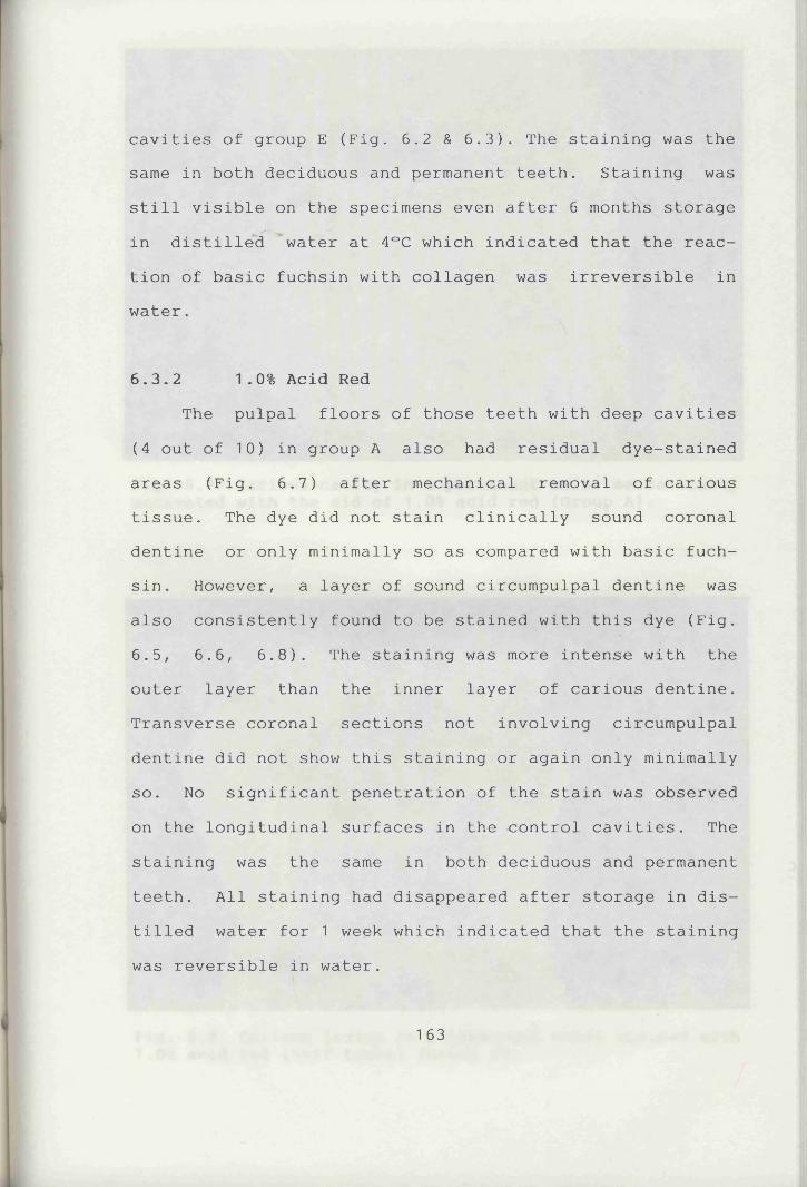
cavities of group E (Fig. 6.2 & 6.3). The staining was the same in both deciduous and permanent teeth. Staining was still visible on the specimens even after 6 months storage in distilled water at 4°C which indicated that the reaction of basic fuchsin with collagen was irreversible in water.
6.3.2 1.0% Acid RedThe pulpal floors of those teeth with deep cavities
(4 out of 10) in group A also had residual dye-stained areas (Fig. 6.7) after mechanical removal of carious tissue. The dye did not stain clinically sound coronal dentine or only minimally so as compared with basic fuchsin. However, a layer of sound circumpulpal dentine was also consistently found to be stained with this dye (Fig. 6.5, 6.6, 6.8). The staining was more intense with theouter layer than the inner layer of carious dentine. Transverse coronal sections not involving circumpulpal dentine did not show this staining or again only minimally so. No significant penetration of the stain was observed on the longitudinal surfaces in the control cavities. The staining was the same in both deciduous and permanent teeth. All staining had disappeared after storage in distilled water for 1 week which indicated that the staining was reversible in water.
1 63
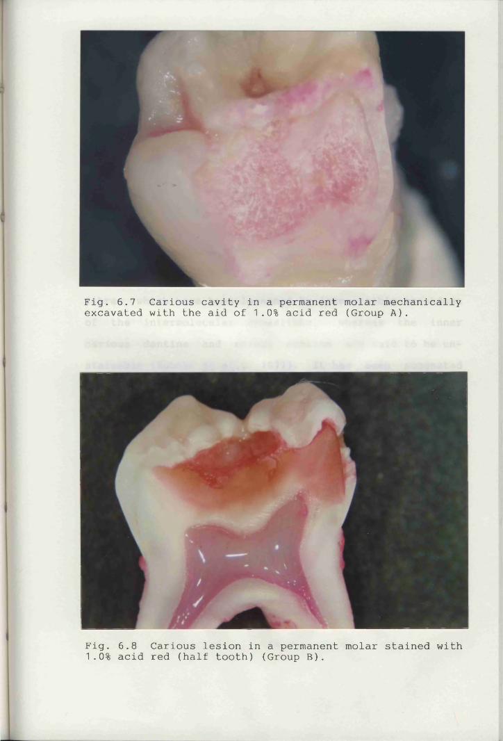
Fig. 6.7 Carious cavity in a permanent molar mechanically excavated with the aid of 1.0% acid red (Group A).
Fig. 6.8 Carious lesion in a permanent molar stained with 1.0% acid red (half tooth) (Group B).
io
6.4 Discussion and ConclusionsThe difficulty in detecting cariously infected den
tine accurately by tactile and visual examination is well recognised.
It has been suggested that the differential staining of carious dentine by basic fuchsin or acid red is not the result of a chemical bond and has no relation to either degree of demineralisation or hardness of the carious dentine (Fusayama, 1988). The outer carious dentine which is stainable with the caries detector dyes has collagen fibres which have been loosened by irreversible breakdown of the intermolecular crosslinks, whereas the inner carious dentine and normal dentine are said to be un- stainable (Kuboki et al., 1977). It has been suggestedthat only the loosened collagen fibres of the outer carious dentine seem to permit penetration of the solvent (propylene glycol) (Fusayama, 1988). The use of caries detector dyes has been shown to decrease the chances of viable bacteria remaining in a cavity after preparation (List et al., 1987; Boston & Graver, 1989). The dyeshowever do not stain the bacteria or delineate the bacterial front (Boston & Graver, 1989).
If the research reports concerning the effect of the caries detector dyes are accurate, the potential advantages in restorative procedures are obvious. Only the
164
infected dentine would be removed by the operator when guided by these dyes leaving the sound tooth structure to provide support for the desired restoration. Caries detec
tor dyes may also be useful for diagnosing unpigmented caries especially along the DEJ. They may also be helpful in training inexperienced clinicians, e.g. dental students, in the diagnosis of dentine caries. This would also enable the in vitro study of the effectiveness of various
caries removal agents to be carried out more accurately.Apart from its carcinogenic potential, basic fuchsin
seems more likely to stain sound dentine (Figs. 6.1 - 6.4) than does acid red. The DEJs in the sound teeth used in this study were consistently dye-stained (Fig. 6.3). There is concern that routine use of the dye would result in excess removal of tooth structure and the likelihood of mechanical exposure of the pulp (Franco & Kelsey, 1981). The risk of pulpal exposure using this dye is well illustrated by Fig. 6.1. Basic fuchsin is, therefore, neither suitable for clinical use nor precise enough for
identifying carious dentine in in vitro or animal studies, when used alone.
A large proportion of the hard pulpal floors in
prepared cavities which were judged to be clinically sound were found to be dye-stained with acid red (Kidd et al.,
1989). Similar findings are reported in this study (Fig.
165
6.7). Sound circumpulpal dentine was consistently found to be dye-stained by both acid red and basic fuchsin (Fig.6.4, 6.5, 6.6 & 6.8) a finding which does not seem to have been reported previously. This indicates that neither acid red nor basic fuchsin are sufficiently specific to differentiate between the inner layer of carious dentine and sound circumpulpal dentine in a deep cavity preparation.
Some of the dye-stained dentine found on the clinically hard pulpal floor (Fig. 6.1 & 6.7) may well be sound circumpulpal dentine. These findings will be correlated and further discussed with the findings on the degree of demineralisation of the cavity floor using the BSE and EPMA techniques, in Chapter 8.
The presence of collagen fibrils on the dentinal surface after caries removal reported in Chapter 5 (Figs.5.4, 5.7, 5.8, 5.9 & 5.10) may partly account for this inconsistency in the staining of the pulpal floor. The staining may also relate to the differences in chemical composition and physical structure between the circumpul
pal and peripheral dentine (Foreman & Soames, 1989). The findings therefore cast doubt on the specificity of the
mechanism of staining of acid red in carious dentine.The staining of dentine with acid red has been
reported to show correlation with bacterial penetration in
most lesions (Boston & Graver, 1989) but it does not
166
precisely delineate bacterial fronts. Acid red is also claimed to stain the irreversibly altered organic matrix (Fusayama, 1979; Kuboki et a l ., 1977) but not the bacteria in carious dentine. It has been suggested that in some lesions a significant amount of non-infected dye-stained dentine may be excavated if the dye stains significantly pulpally to the bacterial front (Boston & Graver, 1989).
In the current study acid red was found to stain sound circumpulpal dentine. The mineral content of the layer of circumpulpal dentine near the pulp has been reported to be less than that of other dentine (Herr et a l ., 1986) which may account for this staining. A possible consequence of its use in deep cavity preparation is the loss of sound dentine that must be preserved and the risk of pulpal exposure if dye removal is effected vigorously.
The staining characteristics of both acid red and basic fuchsin in permanent and deciduous teeth were found to be similar in this study. The use of either dye in its present formulation and regime of application would therefore appear to be contraindicated on the pulpal surface of
a deep cavity preparation in a clinical situation.Accordingly, caries detector dyes were therefore
found to lack the required specificity and were not used
in the subsequent study (Chapter 7) to assess the caries removal process. Instead, optimum examination conditions
167
involving good lighting, carefully dried surfaces, an optical loupe (X4 magnification) and a stereomicroscope were used.
6.5 Summary
a. The effectiveness of two caries detector dyes (0.5% basic fuchsin and 1.0% acid red) was investigated using extracted human permanent and deciduous teeth.b. Basic fuchsin and acid red were found to stain both carious and sound dentine. The specificity of the staining in carious dentine was low.
c. As well as carious dentine, both acid red and basic fuchsin could also stain circumpulpal dentine a finding
which has not been reported previously.d. The staining properties of both dyes were the same in both deciduous and permanent teeth.
CHAPTER 7
CHEMOMECHANICAL REMOVAL OF DENTAL CARIES IN DECIDUOUS TEETH
7.1 IntroductionSince the CMCRS using NMAB was developed (Schutzbank
et a l . , 1978), most of the clinical trials have beencarried out on the permanent dentition (Table 1.3). The system has however also been shown to be potentially useful in treating paediatric dental patients (Rothman 1985; Punwani et a l ., 1988). Although some clinical trials(McNierney & Petruzillo, 1986; Punwani et a l . , 1988) involved the use of both deciduous and permanent teeth, no attempt was made to differentiate between the two groups in assessing the results.
The first study of various caries removal agents (Chapter 3) has indicated that NMAB-Urea may be particularly effective in removing dental caries in deciduous
teeth. The use of a random sample of teeth in that study however only provided a rough guide as to the effectiveness of the caries removal agents. For a more definitive conclusion to be drawn, a more "standardised" sample was
necessary.The purpose of this in vitro study therefore was to
169
further investigate this improved reagent in removing dental caries in deciduous teeth and evaluate the results by means of light and scanning electron microscopy.
7.2 Materials and Methods
7.2.1 In vitro Study150 freshly extracted human deciduous teeth with
coronal caries were collected in phosphate buffered saline (see Section 2.2.1). The carious lesions were not restricted to any specific surface or surfaces of the teeth. All the teeth were used either immediately or within one week after extraction. This sample was selected according to the criteria described in Section 2.2.1 and was used in order to assess more accurately the improved effectiveness of NMAB containing urea in removing dentine caries as opposed to the use of NMAB alone. Since the specimen teeth were selected according to a given set of criteria (see Section 2.2.1), the variations between
specimen teeth would be less than those in the previous studies (Chapter 3) which were randomly selected. Carious tissue was removed chemomechanically using NMAB, NMAB-Urea
or isotonic saline, 50 different teeth being treated with each of the reagents.
The investigation of the effectiveness of the three
170
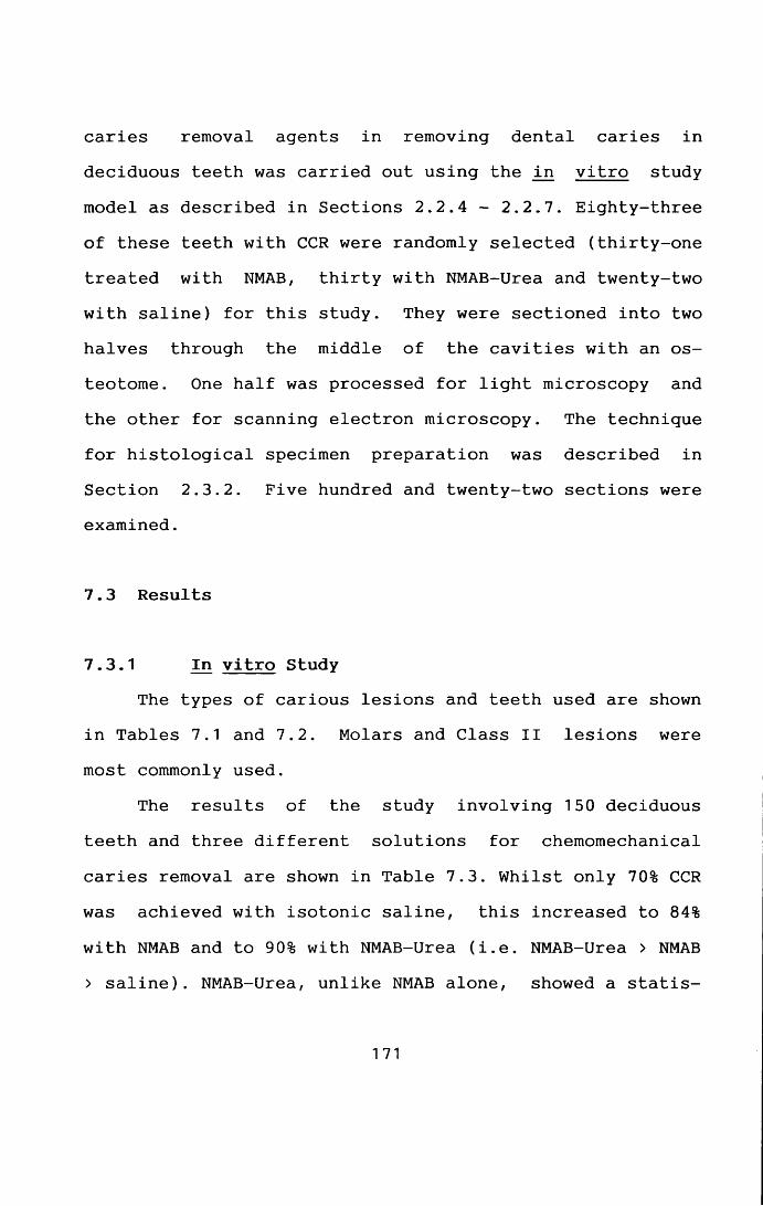
caries removal agents in removing dental caries in deciduous teeth was carried out using the in vitro study model as described in Sections 2.2.4 - 2.2.7. Eighty-three
of these teeth with CCR were randomly selected (thirty-one treated with NMAB, thirty with NMAB-Urea and twenty-two with saline) for this study. They were sectioned into two halves through the middle of the cavities with an osteotome. One half was processed for light microscopy and the other for scanning electron microscopy. The technique for histological specimen preparation was described in Section 2.3.2. Five hundred and twenty-two sections were examined.
7.3 Results
7.3.1 In vitro StudyThe types of carious lesions and teeth used are shown
in Tables 7.1 and 7.2. Molars and Class II lesions were most commonly used.
The results of the study involving 150 deciduous teeth and three different solutions for chemomechanical
caries removal are shown in Table 7.3. Whilst only 70% CCR was achieved with isotonic saline, this increased to 84%
with NMAB and to 90% with NMAB-Urea (i.e. NMAB-Urea > NMAB
> saline). NMAB-Urea, unlike NMAB alone, showed a statis-
171
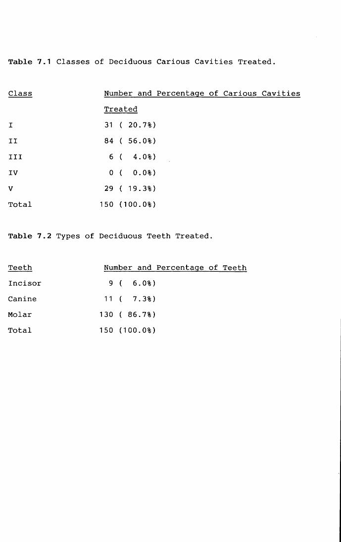
Table 7.1 Classes of Deciduous Carious Cavities Treated.
Class Number and Percentage of Carious CavitiesTreated
I 31 ( 20.7%)
II 84 ( 56.0%)
III 6 ( 4.0%)
IV 0 ( 0.0%)V 29 ( 19.3%)Total 150 (100.0%)
Table 7.2 Types of Deciduous Teeth Treated.
Teeth Number and Percentage of TeethIncisor 9 ( 6.0%)Canine 11 ( 7.3%)Molar 130 ( 86.7%)Total 150 (100.0%)
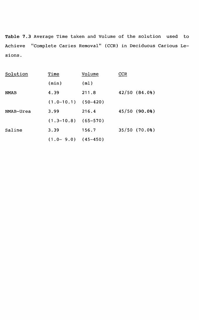
Table 7.3 Average Time taken and Volume of the solution used toAchieve ''Complete Caries Removal" (CCR) in Deciduous
sions.
Solution Time Volume CCR(min) (ml)
NMAB 4.39 211 .8 42/50 (84.0%)
(1.0-10.1) (50-420)NMAB-Urea 3.99 216.4 45/50 (90.0%)
(1.3-10.8) (65-570)
Saline 3.39 156.7 35/50 (70.0%)(1.0- 9.0) (45-450)
tically significant improvement in caries removal as compared to the saline control (Table 7.4). As in the previous studies (Chapter 3), the differences in times taken
and volumes of caries removal agents used were found notto be statistically significantly different using the
dentst-test (Table 7.5).
Stu-
7.3.2 Light MicroscopyThe histological sections prepared from teeth with
CCR were as described in Section 2.3.2. The dentinal floors prepared chemomechanically were irregular (Figs. 5.13 & 5.15) which was in accordance with the SEM findings (see Section! 5.3.1 and Chapter 4).
The presence of bacteria was detected using both H & E and Gram-Weigert stains. Cavities prepared using NMAB and NMAB-Urea showed that one-third of the teeth appeared to be bacteria free while in those prepared by saline this figure was one-fifth (Table 7.6, Fig. 5.13). In the present investigation, no attempt was made to quantify the bacteria, but merely their presence in the dentinal tubules was recorded.
The population of teeth with reparative dentine was
similar in each of the solution treatment group i.e. ~50%
(Table 7.7). Although the majority of cavity floors were
composed of secondary dentine, a number of which consisted
172
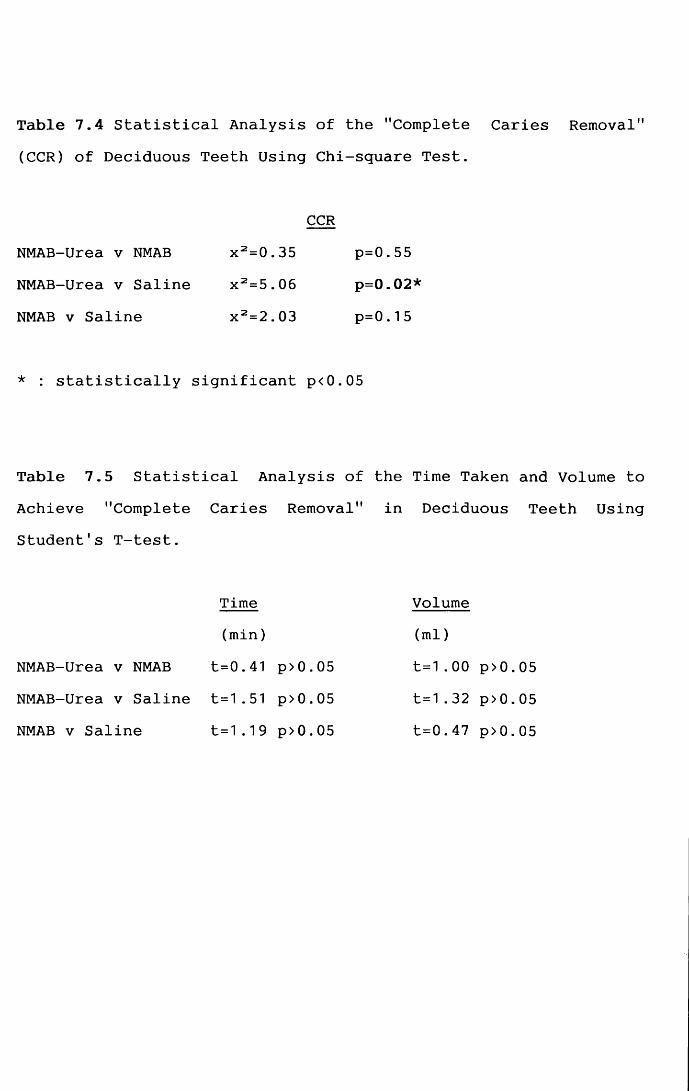
Table 7.4 Statistical Analysis of the "Complete Caries Removal" (CCR) of Deciduous Teeth Using Chi-square Test.
CCRNMAB-Urea v NMAB x 2=0.35 p=0.55NMAB-Urea v Saline x 2=5.06 p=0.02*
NMAB v Saline x 2=2.03 p=0.15
* : statistically significant p<0.05
Table 7.5 Statistical Analysis of the Time Taken and Volume to Achieve "Complete Caries Removal" in Deciduous Teeth Using Student’s T-test.
Time Volume(min) (ml)
NMAB-Urea v NMAB t=0.41 p>0.05 t=1.00 p>0.05
NMAB-Urea v Saline t=1.51 p>0.05 t=1.32 p>0.05NMAB v Saline t=1.19 p>0.05 t=0.47 p>0.05
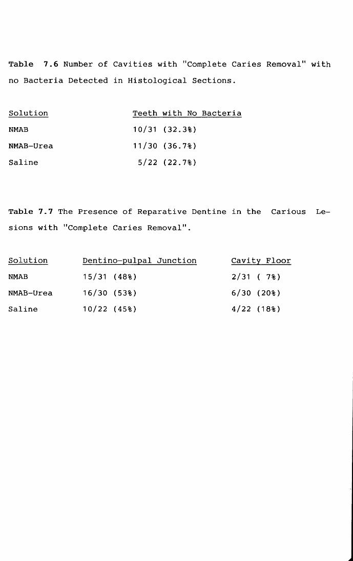
Table 7.6 Number of Cavities with "Complete Caries Removal" with no Bacteria Detected in Histological Sections.
Solution Teeth with No BacteriaNMAB 10/31 (32.3%)NMAB-Urea 11/30 (36.7%)
Saline 5/22 (22.7%)
Table 7.7 The Presence of Reparative Dentine in the Carious Lesions with "Complete Caries Removal".
Solution Dentino-pulpal Junction Cavity FloorNMAB 15/31 (48%)
NMAB-Urea 16/30 (53%)Saline 10/22 (45%)
2/31 ( 7%) 6/30 (20%) 4/22 (18%)
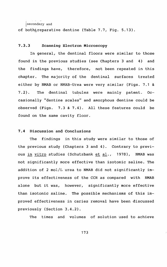
| secondary andof both/\reparative dentine (Table 7.7, Fig. 5.13).
7.3.3 Scanning Electron MicroscopyIn general, the dentinal floors were similar to those
found in the previous studies (see Chapters 3 and 4) and the findings have, therefore, not been repeated in this
chapter. The majority of the dentinal surfaces treated either by NMAB or NMAB-Urea were very similar (Figs. 7.1 &
7.2). The dentinal tubules were mainly patent. Occasionally "dentine scales" and amorphous dentine could be observed (Figs. 7.3 & 7.4). All these features could be found on the same cavity floor.
7.4 Discussion and ConclusionsThe findings in this study were similar to those of
the previous study (Chapters 3 and 4). Contrary to previous in vitro studies (Schutzbank et a l ., 1978), NMAB wasnot significantly more effective than isotonic saline. The addition of 2 mol/L urea to NMAB did not significantly im
prove its effectiveness of the CCR as compared with NMAB alone but it was, however, significantly more effective
than isotonic saline. The possible mechanisms of this im
proved effectiveness in caries removal have been discussed previously (Section 3.4.2).
The times and volumes of solution used to achieve
173
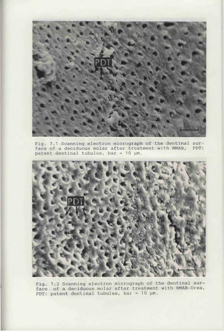
•• • %
• V
Fig. 7.1 Scanning electron micrograph of the dentinal surface of a deciduous molar after treatment with NMAB, PDT: patent dentinal tubules, bar =10 pum.
*
%?v
JSl 1 a * ̂ A1*
Fig. 7.2 Scanning electron micrograph of the dentinal surface of a deciduous molar after treatment with NMAB—Urea, PDT: patent dentinal tubules, bar = 10 y.m.
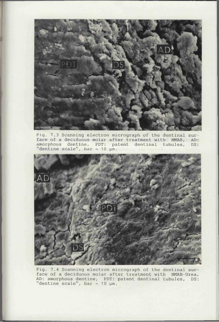
Fig. 7.3 Scanning electron micrograph of the dentinal surface of a deciduous molar after treatment with NMAB, AD: amorphous dentine, PDT: patent dentinal tubules, DS: "dentine scale", bar = 10 jjlitu
Fig. 7.4 Scanning electron micrograph of the dentinal surface of a deciduous molar after treatment with NMAB-Urea, AD: amorphous dentine, PDT: patent dentinal tubules, DS: "dentine scale", bar = 10 }im.
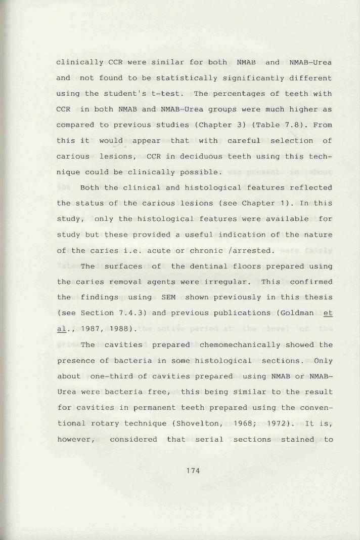
clinically CCR were similar for both NMAB and NMAB-Urea and not found to be statistically significantly different using the student's t-test. The percentages of teeth with CCR in both NMAB and NMAB-Urea groups were much higher as compared to previous studies (Chapter 3) (Table 7.8). From this it would appear that with careful selection of carious lesions, CCR in deciduous teeth using this technique could be clinically possible.
Both the clinical and histological features reflected the status of the carious lesions (see Chapter 1). In this study, only the histological features were available for study but these provided a useful indication of the nature of the caries i.e. acute or chronic /arrested.
The surfaces of the dentinal floors prepared using the caries removal agents were irregular. This confirmed the findings using SEM shown previously in this thesis (see Section 7.4.3) and previous publications (Goldman et al., 1987, 1988).
The cavities prepared chemomechanically showed the presence of bacteria in some histological sections. Only about one-third of cavities prepared using N M A B or N M A B -
Urea were bacteria free, this being similar to the result for cavities in permanent teeth prepared using the conventional rotary technique (Shovelton, 1968; 1972). It is,however, considered that serial sections stained to
1 74
demonstrate bacteria are necessary for the detection of bacteria which are present in only small numbers in the tissues. Failure to detect stained bacteria in histological sections is not an absolute proof of their absence (Watts & Paterson, 1990). Equally the presence of bacteria does not necessarily mean that they are viable.
The reactionary dentine found in this study after using various caries removal agents was present in about 50% of the dentino-pulpal junctions and 20% of the cavity
floors (except NMAB group) of the carious lesions examined (Table 7.7). The similarity of these figures irrespective of the types of caries removal agent used indicated that the lesions used in the different study groups were fairly "standardised". Corbett (1963) has, however, reported that reparative dentine was present beneath the lesions in about three-quarters of carious deciduous teeth and less than half of the permanent teeth. A basophilic arrest line is formed during the active period at the level of the primary dentine and remains thereafter throughout the life
of the tooth between the primary dentine and the subse
quently formed reparative dentine. The reparative dentine reflects the caries attack history. Its formation appears early towards the end of the active phase after the formation of the arrest line. It increases progressively in thickness with each new attack. The incremental layers
175
reflect the intermittency of the caries attack, while the quality and quantity of the layers probably reflect the severity of each attack (Kuwabara & Massler, 1966). The histological features found in this study (Fig. 5.13) were
in accordance with those quoted in the literature.The dentinal features of cavities prepared using NMAB
or NMAB-Urea can be very similar. This seemed to indicate that the ultra-microscopic features were not influenced by
the caries removal agents used as long as CCR was achieved. These features were described in detail in Chapters 3 and 4. The similarities shown here further affirmed
that the dentinal features would be predominantly determined by the status of the carious lesions prior to caries removal. In the previous studies (see Chapter 4), the dentinal surfaces of the cavities prepared by using NMAB-Urea appeared to be cleaner than that prepared using NMAB. However, the dentinal features were similar when either
NMAB or NMAB-Urea was used. It must be borne in mind that the samples used in this study were different from those used in previous studies (Chapters 3 & 4) which were
selected according to a set of criteria in order to
"standardise" the lesions (see Section 2.2.1).
In adults with some natural teeth, 68% said they were
nervous of dental treatment (Renson, 1991). This may have stemmed from their past dental experience in childhood.
176
Preventive dental treatments and less traumatic dental procedures for paediatric dental patients would be a means of reducing this nervousness in adults. A CMCRS might just provide such an atraumatic alternative.
7.5 SummaryAn in vitro study has been carried out on the CMCRS
using a "standardised" carious deciduous teeth sample.a. The order of efficacy in achieving CCR was similar to that in the previous studies (see Chapter 3) i.e.
NMAB-Urea > NMAB > salineb. Using a more "standardised" sample, the percentage of CCR achieved was higher when compared with previous studies (see Chapter 3).c. Bacteria were present in some cavities which had been considered to be clinically "caries free".d. The dentinal floors of the cavities were irregular and consisted of secondary and /or reparative dentine.
e. The dentinal features in cavities with CCR prepared by either NMAB or NMAB-Urea were shown to be similar.
177
CHAPTER 8
MINERAL CONTENT OF THE DENTINE REMAINING AFTER CHEMOMECHANICAL CARIES REMOVAL
8.1 IntroductionAlthough the dentine remaining after complete
chemomechanical caries removal appears sound by normal clinical criteria, as yet no definitive evidence has been presented to confirm that the dentine surface is in fact mineralised, and if so, to what extent. Althoughmicroradiography has been extensively used in studies on enamel mineralisation and was considered as a possibletechnique to investigate this problem, the resolution was too low to give meaningful data which could be correlatedwith SEM studies. The most appropriate technique appearedto be electron probe microanalysis (EPMA).
This technique has been reported to have been used to
study the decalcified regions of carious dentine lesions and has shown that there was a reduction in the concentrations of Ca, P, Mg and Cl, usually accompanied by an in
crease in S and Zn (Takuma et a l . , 1975). The concentra
tion gradients of these elements across the lesions varied
in accordance with the changes in decalcification indicated by microradiography. However, zones in the dentine
178
which are sometimes observed by microradiography cannot always be explained by the findings from EP M A . These discrepancies probably result from the fact that the microradiograph offers an image based on the whole thickness of the section, usually 60 - 150 pm, whereas with
EPMA only the superficial 1 - 2 pm of the specimen are
analysed (Hals et a l ., 1988). Crystals in the surfacelayer of remineralised carious dentine are also much larger than those in intact dentine (Takuma et a l ., 1975).
In texts on cariology, data on the elemental concentration in dentine usually relates to whole dentine,
and in fact the range of concentrations within any one sample is quite wide. In contrast, the EPMA technique allows the analysis of minute areas of highly differing structures. As a result, the range of concentrations within individual specimens will vary substantially (Hals et a l ., 1988). This technique may also allow the study ofthe variations in concentration of an element within a very small area of a specimen.
The aim of this study therefore was to investigate
the feasibility of the use of EPMA to ascertain the level of mineralisation of the dentine remaining after caries
removal using various caries removal agents. In previous studies carried out (Chapters 3 and 7), CMCR seemed to be potentially particularly useful in deciduous teeth. It
was, therefore, decided to use mainly deciduous teeth in this study. Furthermore, because the cost of specimen preparation, especially the resin embedded longitudinal sections, is high, the number of specimens that could be
examined in this study was substantially restricted.
8.2 Materials and MethodsThree permanent and three deciduous teeth with
cavities in which CCR had been achieved (one permanent with NMAB, one permanent with NMAB-Urea, one permanent with saline, one deciduous with NMAB, one deciduous with NMAB-Urea and one deciduous with saline) were randomly selected from the previous study of the cavity surfaces (Chapters3 & 7) .They were subjected to critical point drying and coated with gold.
A further nine deciduous teeth with cavities prepared by various caries removal agents (three with NMAB, three with NMAB-Urea and three with saline) were also randomly selected from the same previous study (Chapter 7). They were each split into two halves with an osteotome and then
mounted in epoxy resin and polished as described in Sec
tion 2.5.2 for longitudinal sectional studies.
Carious lesions in two permanent teeth were used as controls. Areas of sound dentine in the deciduous teeth served as their own internal controls. The techniques of
180
specimen preparation for BSE and EPMA are described in Section 2.5.2.
Backscattered electron imaging (BSE) in the scanning electron microscope was used to produce mineral density images of the dentine, both on the surface of the cavities
and in longitudinal sections. Some scans of the mineral density across the field of study of the surfaces of cavities and longitudinal sections were also made. The BSE images indicated the mineral density of the specimen teeth. However, if the specimens were not flat, then both the topography and morphology of the surfaces would contribute to the BSE images. As an alternative, in order to minimise these effects on the results, a mineral density assessment on the cavity surface was carried out using a line scan across the field of study (Fig. 8.1). An arbitrary range of values from 0 to 255 was used to indicate the brightness, i.e. mineral density, across the line.
EPMA studies were carried out on the dentinal surfaces and on longitudinal sections of the cavities to determine their elemental composition. The analyses were
also performed on "dentine scales" when these were present on the surface of cavities. Random points on the dentinal
surface of the treated cavities were selected and subjected to point analysis, while line scans were carried
out on the longitudinal sections. The line scans consisted
181
of a series of point analyses at regular intervals of 0.1 - 0.2 mm across the area of dentine investigated; this procedure differed from the BSE line scan which did not involve any elemental analysis. Three successive parallel
scans along the longitudinal sections of dentine at right angles to the cavity floors were carried out at the deepest part of the cavity to give a series of point analyses at regular intervals across the dentine segments between the cavity floors and the dentino-pulpal junctions. At each position, the levels of Ca, P, K, Na, S, Mg and O were measured using the electron microprobe. The software used in analysing the X-ray intensities of various elements was programmed to include all other elements that may be present in the specimen, but which were not quantified individually; they were grouped with oxygen and referred to as "0". This ensured that the calculations of the percentages of the elements studied were correct. The figure on the digitiser was recorded after a time in
terval of 100 sec. No attempt was made to correct the data
for the presence of the organic matrix. The beam occasionally hit a dentinal tubule which resulted in lower
concentrations of calcium and phosphorus; these readings were not included in the data.
The relative weight percentages of calcium and phosphorus across the the plane of the longitudinal dentine
182
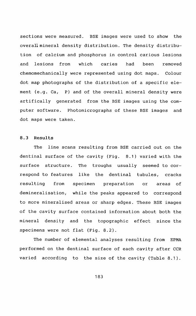
sections were measured. BSE images were used to show the overall mineral density distribution. The density distribution of calcium and phosphorus in control carious lesions and lesions from which caries had been removed chemomechanically were represented using dot maps. Colour
dot map photographs of the distribution of a specific ele
ment (e.g. Ca, P) and of the overall mineral density were artifically generated from the BSE images using the computer software. Photomicrographs of these BSE images and dot maps were taken.
8.3 ResultsThe line scans resulting from BSE carried out on the
dentinal surface of the cavity (Fig. 8.1) varied with the surface structure. The troughs usually seemed to correspond to features like the dentinal tubules, cracks resulting from specimen preparation or areas of demineralisation, while the peaks appeared to correspond
to more mineralised areas or sharp edges. These BSE images of the cavity surface contained information about both the
mineral density and the topographic effect since the specimens were not flat (Fig. 8.2).
The number of elemental analyses resulting from EPMA performed on the dentinal surface of each cavity after CCR varied according to the size of the cavity (Table 8.1).
183
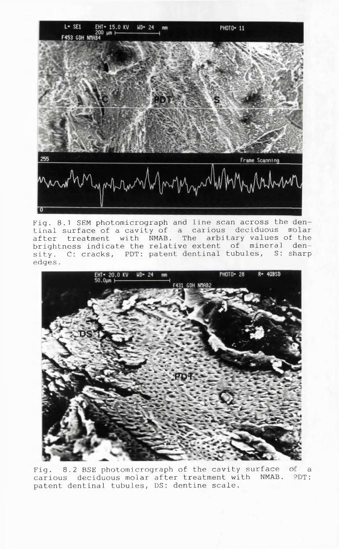
Fig. 8.1 SEM photomicrograph and line scan across the dentinal surface of a cavity of a carious deciduous molar after treatment with NMAB. The arbitary values of the brightness indicate the relative extent of mineral density. C: cracks, PDT: patent dentinal tubules, S: sharp edges.
Fig. 8.2 BSE photomicrograph of the cavity surface of a carious deciduous molar after treatment with NMAB. PDT: patent dentinal tubules, DS: dentine scale.
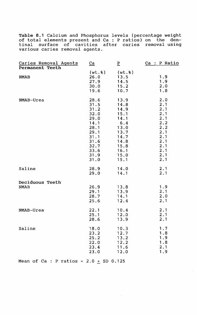
Table 8.1 Calcium and Phosphorus levels (percentage weight of total elements present and Ca : P ratios) on the den-tinal surface of cavities after caries removal usincvarious caries removal agents.
Caries Removal Agents Ca P Ca : P RatioPermanent Teeth
(wt.%) (wt.%)NMAB 26.0 13.5 1 .9
27.9 14.5 1 .930.0 15.2 2.019.6 10.7 1 .8
NMAB-Urea 28.6 13.9 2.031 .5 14.8 2.131 .2 14.9 2.132.0 15.1 2.129.0 14.1 2.114.1 6.4 2.228.1 13.0 2.229.1 13.7 2.131 .1 14.7 2.131 .6 14.8 2.132.7 15.8 2.133.6 16.1 2.131 .9 15.0 2.131 .0 15.1 2.1
Saline 28.9 14.0 2.129.0 14.1 2.1
Deciduous TeethNMAB 26.9 13.8 1 .9
29.1 13.9 2.128.7 14.1 2.025.6 12.4 2.1
NMAB-Urea 22.1 10.4 2.125.1 12.0 2.128.6 13.9 2.1
Saline 18.0 10.3 1 .723.2 12.7 1 .825.2 13.2 1 .922.0 12.2 1 .823.4 11.6 2.123.0 12.0 1 .9
Mean of Ca : P ratios = 2.0 + SD 0.125
The levels of calcium and phosphorus seemed to be similar in different cavities. However, the levels of calcium and phosphorus were lower in deciduous teeth than permanent. The calcium and phosphorus ratios were remarkably constant (2.0 +_ SD 0.125) (Table 8.1). Occasionally, parts with a lower mineral content were located; these may correspond to points where traces of decalcified material remained. The trace elements studied on the cavity surfaces were found to have low levels and fell into the range of 0.1 -1.0 wt.%. Since elemental analyses has a percentage error of _+ 0.5% and the levels of the trace elements were verylow, the latter were therefore not considered further in this study.
Both BSE imaging and EPMA were carried out on the longitudinal sections of the resin embedded specimen teeth. Due to the different depths of the carious lesions studied, the depth of the dentine from the dentinal floor to dentino-pulpal junction varied from tooth to tooth. A general trend was noted in all the specimens studied in
that the calcium level in the circumpulpal dentine was
frequently lower than in the sound dentine overlying it irrespective of the type of caries removal agent used (Table 8.2). The last measurement made in each specimen
represented the levels of calcium and phosphorus in circumpulpal dentine which, as is evident from BSE studies,
184
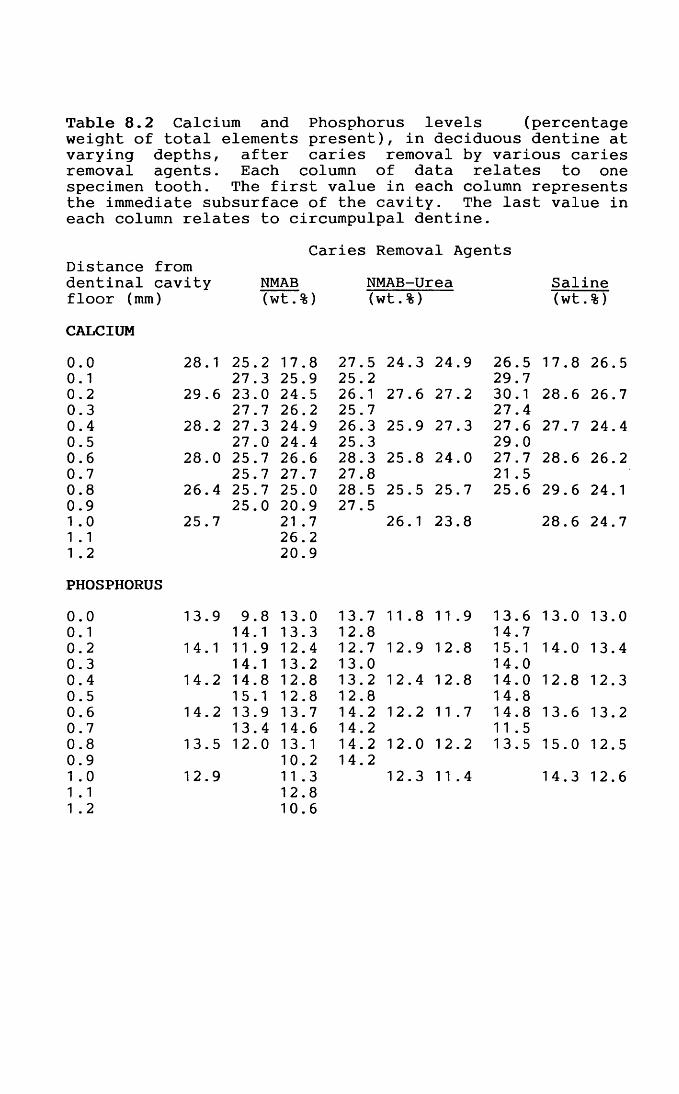
Table 8.2 Calcium and Phosphorus levels (percentage weight of total elements present), in deciduous dentine at varying depths, after caries removal by various caries removal agents. Each column of data relates to one specimen tooth. The first value in each column represents the immediate subsurface of the cavity. The last value in each column relates to circumpulpal dentine.
Caries Removal AgentsDistance fromdentinal cavity NMAB NMAB-Urea Salinefloor (mm) (wt.%) (wt.%) (wt .%)CALCIUM0.0 28.1 25.2 17.8 27.5 24.3 24.9 26.5 17.8 26.50.1 27.3 25.9 25.2 29.70.2 29.6 23.0 24.5 26.1 27.6 27.2 30.1 28.6 26.70.3 27.7 26.2 25.7 27.40.4 28.2 27.3 24.9 26.3 25.9 27.3 27.6 27.7 24.40.5 27.0 24.4 25.3 29.00.6 28.0 25.7 26.6 28.3 25.8 24.0 27.7 28.6 26.20.7 25.7 27.7 27.8 21 .50.8 26.4 25.7 25.0 28.5 25.5 25.7 25.6 29.6 24.10.9 25.0 20.9 27.51 .0 25.7 21 .7 26.1 23.8 28.6 24.71 .1 26.21 .2 20.9PHOSPHORUS0.0 13.9 9.8 13.0 13.7 11.8 11.9 13.6 13.0 13.00.1 14.1 13.3 12.8 14.70.2 14.1 11.9 12.4 12.7 12.9 12.8 15.1 14.0 13.40.3 14.1 13.2 13.0 14.00.4 14.2 14.8 12.8 13.2 12.4 12.8 14.0 12.8 12.30.5 15.1 12.8 12.8 14.80.6 14.2 13.9 13.7 14.2 12.2 11.7 14.8 13.6 13.20.7 13.4 14.6 14.2 11.50.8 13.5 12.0 13.1 14.2 12.0 12.2 13.5 15.0 12.50.9 10.2 14.21 .0 12.9 11.3 12.3 11.4 14.3 12.61 .1 12.81 .2 10.6

was generally lower than in sound dentine (Figs. 8.3 point 9 & 8.4). The levels of calcium and phosphorus in the superficial layer of dentine remaining after CMCR (0.05 -0.1 mm) on the cavity floors frequently appeared to be slightly lower than in the underlying dentine (Table 8.2).
However, the results clearly showed that the dentine remaining is almost completely normally mineralised. Both BSE images and dot maps generated from the BSE images show
the overall distribution of mineral density (Figs. 8.3,8.4 & 8.5) and specific distribution of calcium and phosphorus (Fig. 8.6) in the longitudinal sections of lesions after caries removal with NMAB-Urea. These seem to indicate clearly that there is no difference in mineral content between the cavity floor and the underlying sound dentine. The outline of the cavity floor is distinct (Figs. 8.3, 8.4, 8.5 & 8.6).
Any difference in the levels of calcium and phosphorus from specimen to specimen at different depths of dentine probably resulted from biological variation. In
general, the overall levels of calcium and phosphorus fell
within a similar range in each specimen. Due to the small
number of samples studied, statistical analysis was not possible.
In contrast, control carious lesions showed a reduction in the levels of calcium and phosphorus as compared
185
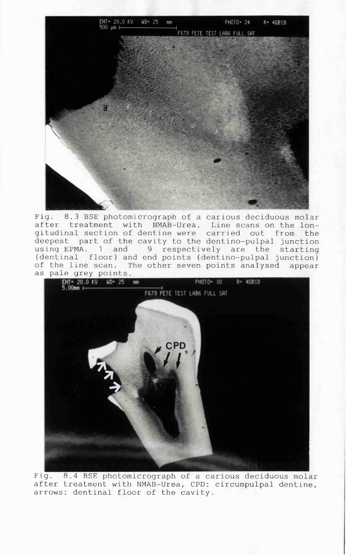
PHOTO= 24 R* 4QBSDF879 PETE TEST LHB6 FULL SAT
Fig. 8.3 BSE photomicrograph of a carious deciduous molar after treatment with NMAB-Urea. Line scans on the longitudinal section of dentine were carried out from the deepest part of the cavity to the dentino-pulpal junction using EPMA. 1 and 9 respectively are the starting (dentinal floor) and end points (dentino-pulpal junction) of the line scan. The other seven points analysed appear as pale grey points.
Fig. 8.4 BSE photomicrograph of a carious deciduous molar after treatment with NMAB-Urea, CPD: circumpulpal dentine, arrows: dentinal floor of the cavity.
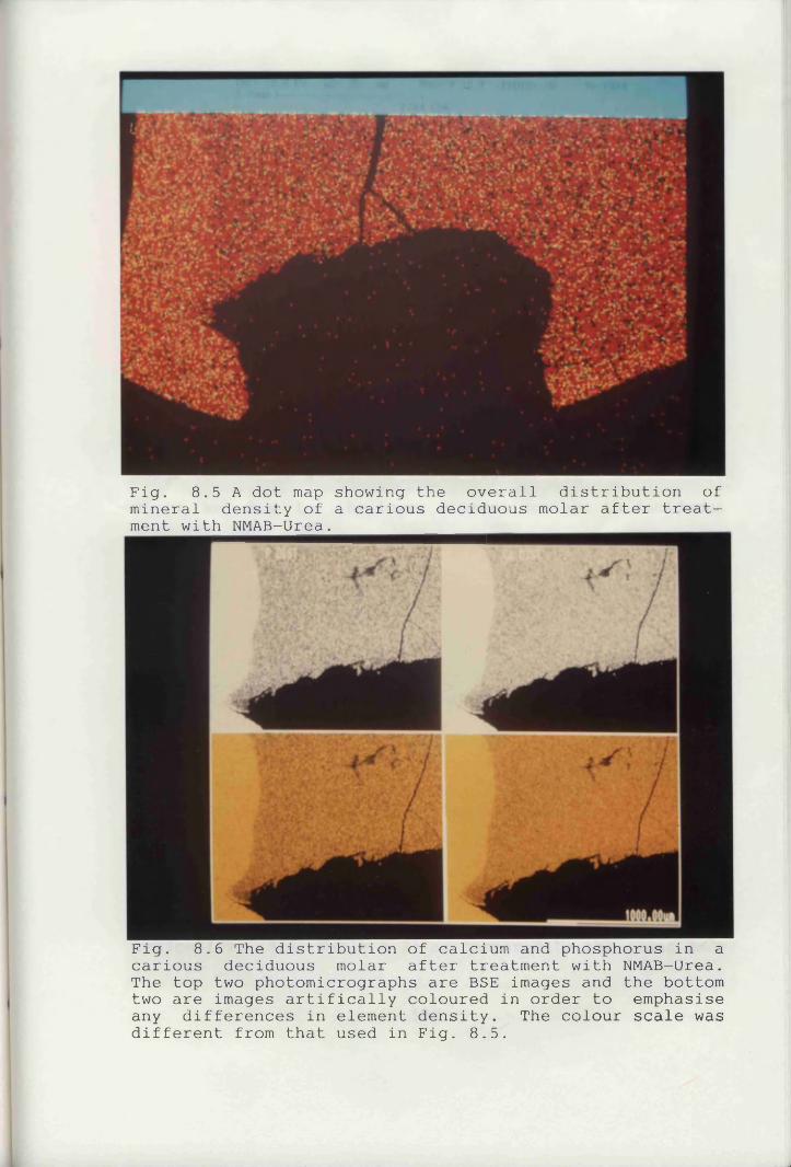
Fig. 8.5 A dot map showing the overall distribution of mineral density of a carious deciduous molar after treatment with NMAB-Urea.
Fig. 8.6 The distribution of calcium and phosphorus in a carious deciduous molar after treatment with NMAB-Urea. The top two photomicrographs are BSE images and the bottom two are images artifically coloured in order to emphasise any differences in element density. The colour scale was different from that used in Fig. 8.5.
with sound dentine (Table 8.3). Other trace elements studied across the dentine segments showed little variation. The differences in levels of these elements (i.e. K, Na, S, Mg and "0") investigated were found to be negligible between the different layers in carious permanent teeth (Table 8.3). Since the percentage error in elemental analysis is _+ 0.5% and the levels of these trace elements ranged from 0.0 - 1.0 wt.%, they were not considered further in this study. The levels of calcium and phosphorus in the sound dentine here were higher than in that of the sound dentine in deciduous teeth (Tables 8.1 & 8.2). This could be due to the lower mineral content in deciduous teeth (see Chapter 1). The number of specimens studied was however small, and no conclusion could be drawn from this study.
The overall distribution of mineral density in the control carious permanent lesion is shown using BSE image (Fig. 8.7) and an artificially coloured dot map generated from BSE images (Fig. 8.8). The density of calcium and phosphorus in the lesions are shown using either BSE images or artifically coloured dot maps to show the distribution of these two elements (Fig. 8.9). The calcium and phosphorus levels were clearly lower in the first 0.5 - 0.6 mm from the surface, i.e. the carious part of thelesion (Fig. 8.8, Table 8.3) as compared to the sound den-
186
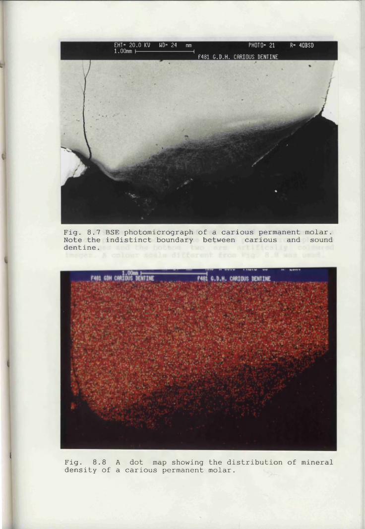
EHT- 20.0 KV UD- 24 mm PHOTO- 21 R- 4QBSD1.00mm I-----------------1 f481 G.D.H. CRRIOUS DENTINE
Fig. 8.7 BSE photomicrograph of a carious permanent molar. Note the indistinct boundary between carious and sound dentine.
1'
i Fig. 8.8 A dot map showing the distribution of mineral density of a carious permanent molar.
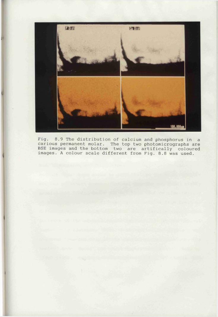
Fig. 8.9 The distribution of calcium and phosphorus in a carious permanent molar. The top two photomicrographs are BSE images and the bottom two are artifically coloured images. A colour scale different from Fig. 8.8 was used.
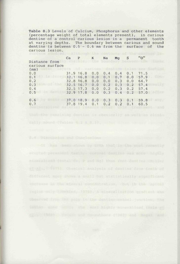
Table 8.3 Levels of Calcium, Phosphorus and other elements (percentage weight of total elements present), in carious dentine of a control carious lesion in a permanent tooth at varying depths. The boundary between carious and sound dentine is between 0.5 - 0.6 mm from the surface of the carious lesion.
Ca P K Na Mg S "0"Distance from carious surface (mm)0.0 31 .9 16.8 0.0 0.4 0.4 0.1 71 .50.1 32.1 16.8 0.0 0.1 0.7 0.0 57.90.2 32.8 16.8 0.0 0.0 0.3 0.0 64.70.3 32.5 16.7 0.0 0.2 0.3 0.2 57.40.4 32.5 17.3 0.0 0.2 0.3 0.2 57 . 40.5 32.9 17.8 0.0 0.3 0.4 0.2 57.00.60.7
37.0 18.9 0.0 0.3 0.3 0.1 59.837.8 19.4 0.1 0.2 0.2 0.1 60.5
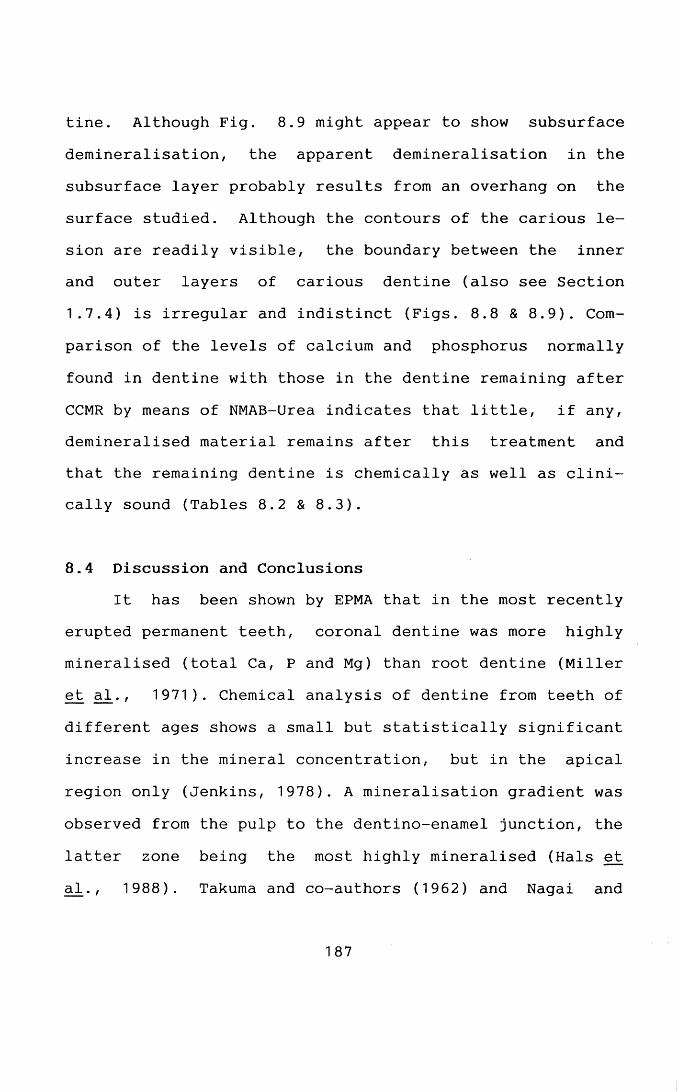
tine. Although Fig. 8.9 might appear to show subsurface demineralisation, the apparent demineralisation in the subsurface layer probably results from an overhang on the
surface studied. Although the contours of the carious lesion are readily visible, the boundary between the inner and outer layers of carious dentine (also see Section 1.7.4) is irregular and indistinct (Figs. 8.8 & 8.9). Com
parison of the levels of calcium and phosphorus normally found in dentine with those in the dentine remaining after
CCMR by means of NMAB-Urea indicates that little, if any, demineralised material remains after this treatment and that the remaining dentine is chemically as well as clinically sound (Tables 8.2 & 8.3).
8.4 Discussion and ConclusionsIt has been shown by EPMA that in the most recently
erupted permanent teeth, coronal dentine was more highly mineralised (total Ca, P and Mg) than root dentine (Miller
et a l . , 1971). Chemical analysis of dentine from teeth ofdifferent ages shows a small but statistically significant increase in the mineral concentration, but in the apical
region only (Jenkins, 1978). A mineralisation gradient was
observed from the pulp to the dentino-enamel junction, the latter zone being the most highly mineralised (Hals et
a l ., 1988). Takuma and co-authors (1962) and Nagai and
187
Takuma (1973) found the sulphur content in predentine to be variable and in many cases slightly higher than in the mineralising dentine. The specimens described in these reports were permanent teeth. Although there was no available data on deciduous teeth, the gradient of mineralisation distribution observed in the present study was found
to be in accordance with the above studies.The level of calcium can be used as an indicator of
the level of mineralisation in dentine (Figures et a l ., 1990). Taking into account the small number of specimens investigated in this study and the biological variation between them, the levels of calcium and phosphorus on the dentinal surfaces of the cavities after caries removal varied only marginally irrespective of the caries removal agent used. Although there was some variation between permanent and deciduous teeth in the levels of calcium and phosphorus, they were small (Tables 8.1, 8.2 & 8.3). The
calcium to phosphorus ratios were found to be around 2 (Table 8.1) which is of the same value as that of sound dental tissue (Jenkins, 1978). This indicated that the surfaces of cavities after caries removal were sound.
The variation in topography and morphology betweeni
the dentine surfaces of the various cavities after caries
removal made it difficult to compare the results obtained from the elemental analysis studies as the BSE and EPMA
188
techniques are best suited to flat surfaces. It was therefore decided to carry out the elemental analysis on a flat longitudinal section of each specimen in order to see whether more consistent measurements could be obtained.
Although the superficial layer (0.05 - 0.1 mm) of the dentine remaining after caries removal using various
caries removal agents appeared to be slightly less mineralised than the underlying dentine, the differences were very small. BSE images and dot maps were used to show the mineral density of the cavity floors and underlying sound dentine and showed no detectable difference between them (Figs. 8.3, 8.4, 8.5 & 8.6). This was in sharp contrast to carious dentine (Figs. 8.8 & 8.9).
The findings in this study therefore seemed to indicate that the dentinal surfaces of the cavities after caries removal with NMAB-Urea were very similar to the underlying sound dentine in the extent of their mineralisation i.e. the decalcified dentine removal is effective and
complete. The hypothesis that "dentine scales" may be
areas of remineralisation is supported by the fact that the data did not indicate any lowering of the calcium levels (i.e. mineralisation) compared with those found in
sound dentine.The phosphorus measured was the total amount present
and included both the inorganic and organic sources in the
189
dentine. These levels did not show any trend in the small number of specimen teeth studied.
The dentinal floors of cavities after caries removal using either hand or rotary instruments have however been
reported to be of a lower microhardness i.e. less mineralised, than the underlying sound dentine (Fusayama et a l ., 1966). Accordingly, the cavity surface produced by CMCR would appear to be as sound if not more so, than that produced by conventional procedures.
The boundary between the inner and outer layers of
carious dentine was irregular and indistinct in control lesions and did not exist in cavities after caries removal. In contrast to the sharp cavity outline achieved after caries removal (Figs. 8.3, 8.4, 8.5 & 8.6), those of the control carious lesions are vague and indistinct (Figs. 8.7, 8.8 & 8.9). The mineral density distributionsuggested a non-uniform demineralisation as indicated by the more "radiolucent" areas in the black and white BSE
images and less heavily coloured areas in the artifically coloured images (Fig. 8.9). These areas might represent subsurface demineralisation in carious dentine or result
from an overhang on the surface studied. However, it must be borne in mind that a two dimensional image is used to represent a three dimensional structure, these so called
"radiolucent" areas may well be sound dentine and result
190
from the plane of sectioning.The circumpulpal dentine was shown to be of a lower
mineral density (Fig. 8.4) and to have lower levels of calcium and phosphorus than sound dentine (Table 8.2). This is in accordance with previous findings (Jenkins, 1978; ten Cate, 1989).
While EPMA is a useful method for elemental analysis of dental hard tissues, there are inherent variations present in individual readings (von Zglinicki, 1992) and, in the case of dentine, there is also further variation due to the heterogeneity of the material (Figures et al., 1990). The techniques used in the present study would appear to be comparable with those used in previous studies and involved taking each separate reading in different types of dentine manually (Figures et_ a l . , 1990). Thepresent technique has the added advantage of easier and simpler operation. In the small number of specimen teeth studied, the depth of dentine remaining after treatment and the plane of sectioning which determined the nature of
the dentine available for study, varied from specimen to specimen. This further complicated the interpretation of
the findings. Averaging of the results from different
specimens would be inappropriate as there was considerable variation between them. It is, however, useful in examin
ing the levels of each element in different regions of
191
dentine in the same specimen tooth. Due to the cost of specimen preparation, particularly of longitudinal sections, only a small number of specimens was investigated here which makes a more specific conclusion difficult to reach. However, the findings clearly show that the dentine surface remaining after CMCR is sound and properly mineralised and suitable for the application of restorative materials.
8 .5 Summarya. The feasibility of using EPMA in studying the status of mineralisation of the dentine remaining after caries removal using various caries removal agents was investigated .b. The calcium levels which indicated the degree ofmineralisation of the dentinal surfaces were very similar to those in the underlying sound dentine irrespective of the type of caries removal agent used.c. Circumpulpal dentine was less mineralised than the overlying sound dentine.d. The use of EPMA in dentine analysis has limitationswhich render the interpretation of the elemental analyses difficult.e. The level of mineralisation of the surface left afterCMCR is as good as, if not superior to, that left after
192

conventional mechanical caries removal procedures.
193
CHAPTER 9
CONCLUDING DISCUSSION
9.1 IntroductionDental caries is a disease in which many inter
relating factors are involved, with the three principal variables being the host, the diet and the plaque
microflora. This thesis is concerned mainly with the use of chemomechanical caries removal systems (CMCRS) in removing coronal dentine caries as an alternative procedure for cavity preparation prior to restoration. The aims included an attempt to improve the effectiveness of CMCR reagents and to ascertain the nature of the dentine remaining on the cavity floor after caries removal using different caries removal agents. The existing system is not 100% percent effective in removing caries (McCune,
1986), and an improvement in the effectiveness of the system would be of considerable clinical value. This chapter therefore correlates the results of the various experiments described in this thesis, and suggests where further work in this field could be undertaken.
9.2 In vitro Study
The main difficulty in assessing the efficacy of
194
caries removal by a CMCRS is the large number of parameters involved. These include :
a. time taken to remove the caries,b. volume of solution used,
c. percentage of caries removed,d. size of the lesions,e. state of the lesions :
i. consistency,ii. acute, chronic or arrested,
iii. type of lesions e.g. enamel, dentine, cementum,iv. sites e.g. fissure, smooth surface, root surface,
f. inter- and intra-operator variability in assessing CCR.The assessment of staining and /or softening of den
tine on the floor of a cavity after caries removal was also found to demonstrate a fair range of subjectivity between different operators. The present study was carried out by only one operator. Different operators would have different interpretations of "lightly abrading the carious surface with the applicator tip" (Schutzbank et al.,
1978); a stronger force would emphasise the mechanical element of this system and CCR might be achieved solely by
mechanical excavation in some lesions. The problem for the clinician is to distinguish between the dentine which
should be removed and that which may be left. Currently,
clinical judgement is used to make this decision. An at
195
tempt to use basic fuchsin and acid red to detect any residual caries on the dentinal floors after caries removal was made. Both basic fuchsin and acid red were unsatisfactory due to their low specificity in the staining of carious material. In addition, the new finding made in
the course of the study that circumpulpal dentine can also be stained by these two dyes indicates that their use on
the pulpal floor of a cavity is contraindicated.Another problem is that of assessing lesion size.
Most of the clinical trials (Table 1.3) previously carried out have used visual and radiographic means to select thelesions in an attempt to standardise their size. In somestudies, the maximium length, width and depth of thecavities were measured with a periodontal probe to obtain a rough measurement of their volume for the purpose of assessing the size of the lesions (Robbins, 1987; Tavares et al., 1988). Although this is a step towards correctly measuring the volume of carious material removed, it is by
no means accurate.In order to assess the activity of potential caries
removal agents in this study, a random sample of extracted carious permanent and deciduous teeth was used in the ini
tial studies. Although the criteria for assessing the
caries removal efficacy was crude, it enabled a number of reagents to be screened quickly. It showed that the effec-
196
tiveness of NMAB in caries removal was superior to both NaOCl and saline, and the addition of urea to NMAB im
proved its effectiveness in caries removal to statisticaly significant levels as compared to controls. Although this improvement was not statistically significant as compared to NMAB, nonetheless it is the first attempt made to improve the performance of NMAB since the C a r i d e x ™ CRS was first introduced in the early nineteen eighties. Contrary
to the results given in a previous in vitro study (Schutzbank et a l ., 1978), the effectiveness in cariesremoval of NMAB was not found to be statistically more significant than that of isotonic saline.
This procedure in general has limitations in terms of treatment time, the suitability of cavities and the need for some continued use of mechanical instruments.
Since a random sample was used, the size and status of the lesions used were not uniform. Parameters such as the size and status of the lesions were not taken into account and therefore the criterion was not the caries removal efficacy but rather an indication of the effectiveness of a caries removal agent in achieving CCR. The
use of a more "standardised" sample in terms of both size
and status of the lesions enabled a higher percentage of CCR to be achieved (also see below).
197
9.2.1 Comparison of Permanent and Deciduous TeethMost of the clinical trials described previously have
been carried out on permanent teeth (Table 1.3) and no attempt was made to differentiate between the findings on permanent and deciduous teeth when a mixed dentition was used (McNierney & Petruzillo, 1986; Punwani et a l ., 1988).
Although the difference in effectiveness in caries removal
between permanent and deciduous teeth was not statistically significant as shown in previous studies (Chapter
I with3), the percentage of cavities showing teeth^CCR was, however, higher for deciduous teeth. When NMAB-Urea was used the number of teeth with CCR increased by 16% in permanent teeth i.e. to 67% and by 25% in deciduous teeth i.e. to 81% as compared to NMAB alone. Attempts to use more "standardised" deciduous tooth samples in the subsequent study (Chapter 7) enabled an even higher percentage of cavities with CCR to be achieved (84% for NMAB, 90% for NMAB-Urea). The use of this system in deciduous teeth has
received little attention hitherto and its apparently improved performance in those teeth compared with that in
permanent teeth raises the possibility that this system
might be more useful for paediatric dental patients. Although the ultramicroscopic structure of carious material is similar in permanent and deciduous teeth (Johnson et a l ., 1969), the carious material in deciduous teeth was
198
more easily removed in the studies carried out (Chapter 3). In fact, the consistency of the carious dentine in deciduous teeth was found to be softer than that in permanent teeth. Most of the previous studies on carious dentine have been carried out using permanent teeth (see sec
tion 1.7) and no published data is available comparing the
microhardness or consistency of permanent teeth with that of deciduous teeth.
9.2.2 Mode of Action
The study of the reaction mechanisms of NMAB on collagen (Habib et a l ., 1975) have yet to be confirmed andthe action of NMAB on dentinal collagen itself has not been studied. This project has shown that the performance of NMAB can be improved in terms of its caries removal properties and the quality of the dentine surface produced can also be improved, by the addition of 2 mol/L urea. Urea has been used widely as a protein denaturing agent
and acts by disrupting hydrogen bonds. The excess urea may help to disrupt the hydrogen bonds in carious dentine collagen thereby increasing its solubility. A possible reac
tion mechanism might alternatively involve the two amino groups of urea being chlorinated by NaOCl to form mono- or dichloro-derivatives; these intermediate compounds may
then attack and degrade the collagen in carious dentine.
199
The fact that guaninidium chloride which is a better protein de-naturing agent than urea resulted in no im
provement in the caries removal of the existing system suggests that active ingredients, possibly mono- or dichlorourea derivatives, might be formed and enhance the caries removal action of NMAB. A study involving amino acid analysis suggested that solubilisation of collagen by NMAB does not involve amino acid oxidation and oxidation of proteins by chlorination (e.g. sodium hypochlorite) predominantly involves glycine residues (Yip & Beeley, 1989). This finding casts doubt on the proposed reaction mechanism of NMAB.
9.3 Dentine Remaining after Caries Removal
9.3.1 Surface Morphology and TopographyThe SEM photomicrographs showed that not all the
various caries removal agents produced a fairly uniform dentine surface in all of the carious cavities treated.
None of the surfaces had a smear layer similar to that seen in conventional, mechanically prepared cavities. Under the SEM, the dentinal surfaces prepared by NMAB-Urea
appeared more uniform, cleaner and relatively free of
debris when compared with those prepared by NMAB alone. Some of the dentinal tubules appeared to be occluded and
200
this may help to reduce the permeability of the dentine and consequently also reduce moisture contamination during
bonding. One recent permeability study showed that some of
the dentinal tubules are still patent in teeth treated with NMAB and fluid permeability might be expected to be higher than with conventionally prepared dentine (Sherrer et a l ., 1989). In contrast, a reduction of 30% in per
meability has been reported when dentinal surfaces with patent dentinal tubules were treated with NMAB (Sherrer et al., 1989). Sherrer and co-authors have suggested that the "crushing and burnishing action" of the applicator tip causes the removal of mineral matrix but leaves enough residual organic debris to keep the permeability low (Sherrer et a l ., 1989). The presence of fibrillar structures and a layer of debris might also partly result from this "crushing and burnishing action". The effect of NMAB-Urea on the permeability of dentine however has not been investigated.
Normally, debris-like material was scattered on the
surface of the dentine prepared by NMAB or NMAB-Urea and
"dentine scales" were found on many of the surfaces studied. Clinically undermined areas were present in most of the dentinal floors after caries removal. However, there was clear evidence that removal of demineralised
material was sometimes incomplete, with some strand-like
201
fibrillar structures being observed in the intertubular dentine and in some areas, no mineralised dentine at all could be observed. Recently it has been suggested that these fibrillar structures may be collagen fibres which could undergo remineralisation (Burke, 1989). Oc
casionally, the presence of lamina limitans and /or odontoblast processes protruding from the dentinal tubules,
which could extend throughout the whole thickness of normal dentine and inner carious dentine (Yamada et a l . , 1983), could be observed on a clinically sound dentinal surface.
The amorphous dentine surfaces seen in some of the teeth treated with NMAB showed few or no patent dentinal tubules. A similar appearance was observed in teeth treated with NaOCl, urea or saline. These surfaces might consist of both partially demineralised and remineralised dentine in which some of the organic matrix may be degraded. The nature of this layer remains to be investigated .
The very uneven appearance with many undermined areas shown by both SEM and histological sections in most of the
dentinal cavity floors observed in this study seemed to
indicate the possibility of this technique being used to advantage in forming micromechanical interlocking with adhesive restorative materials (also see Section 1.10.6).
202
The wide range of appearances observed may indeed represent the interface between carious and sound dentine as suggested by Goldman et a_l. (1987). This is believed to
be predominantly determined by the state of the lesions. The mechanical action of the applicator tip and possibly
the chemical action of the reagents used might affect the dentinal surfaces observed.
The variety of appearances representing the possible interface between carious and sound dentine is however much greater in permanent teeth than in deciduous ones.
9.3.2 Types of DentineThe dentinal floors of the cavities with CCR prepared
by both NMAB and NMAB-Urea consisted of secondary and /or reparative dentine. The presence of different types of dentine with differences in structure may partly explain the wide variety of surface appearances found even in the same cavity. The composition of the dentinal floors might affect their bonding properties with adhesive restorative
materials. Although the bond strengths of these materials with NMAB prepared dentine have been found to be at least the same as, if not superior to, conventionally prepared dentine (Mclnnes-Ledoux et a l ., 1987, 1989; Wolski et a l .,
1989), the range of bond strengths was wide (Mclnnes-
Ledoux et a^. , 1987; Burke et al., 1989). The variation in
203
the composition and morphology of the dentinal floors may partly account for this.
9.3.3 The Fate of Organisms Remaining in the DentineThe question as to what happens to any micro
organisms remaining in the dentine at the base of the cavity floor after caries removal has involved much work and aroused a lot of interest over the years. Various methods of evaluating bacterial viability and caries progression under sealants have been developed and have been extensively reviewed by Swift (1988). These include bacteriological sampling, standardised depth measurements, radiographs, and clinical observation. These studies have shown a decided decrease in the number of viable microorganisms in lesions under intact sealants. Furthermore, caries progression appears to be negligible. These effects seem to result mainly from the barrier effect of sealants,i.e. their ability to block the supply of nutrients to bacteria within the teeth. In addition, the acid-etching
process used and inherent reparative ability of dentine
may be involved to some extent.
It has been suggested that, even if bacteria are left behind in a cavity, once they are sealed beneath a restoration they are deprived of their substrate, and progression of the lesion does not occur (MacGregor, 1961;
204
Fisher, 1966). On the other hand, fermentative micro
organisms can remain viable under non-antiseptic fillings for long periods of time (Besic, 1943; Schouboe & Mac
Donald, 1962). Therapeutic materials are available with antibacterial properties (McCom|b & Ericson, 1986). These
may encourage the arrest of the lesion and remineralisation of the softened dentine (Eidelman et. a T . , 1965). In
the deep cavity, the probability of undetected pulp exposure is high. All deep cavities should therefore be protected from salivary or other contamination and a base
of calcium hydroxide applied as rapidly as possible(Shovelton, 1972; Paterson, 1974). Success of the management of the deep carious lesion depends on the following factors (Bissell & Nattress, 1988) :a. The correct assessment of whether or not the pulp iscapable of being saved.b. The care taken by the operator with respect to protection of the pulp during cavity preparation.
c. Control of micro-organisms at the base of the cavity.d. Provision of a well-sealed restoration.
Sealants and fluorides supplement one another and
together have the potential to eliminate dental caries. Sealants and sealed restorations have been shown to arrest caries in Class I carious lesions in two 5 year clinical
trials (Going et_ aJL. , 1978; Mertz-Fairhurst et a l . , 1992).
205
The management of occlusal caries by localised excision and restoration with amalgam and sealant (Welbury et al.,1990), or even by the placement of sealed composite resin
over existing caries has proved effective over 5 years
(Mertz-Fairhurst et a l ., 1992). Thus, the continuance of"extension for prevention" philosophies in unrestored occlusal surfaces may have little justification when adhesive procedures can accomplish similar goals with minimal tooth removal. The coming decade will continue to see shifts in the approach to cavity preparation and restoration based on a decreased incidence of caries as well as the continued development of sealants in dentistry.
Although bacteria were found in cavities prepared by NMAB or NMAB-Urea, their numbers were no greater than would be found in dentine prepared conventionally (Shovelton, 1972). There is no long term study regarding the success rate of lesions removed by a CMCR method (see Table 1.3). The possibility that the bacteria remaining in
the tubules may be viable (Brannstrom et a l ., 1980b) is
open to speculation. The number of remaining bacteria detected in the dentine remaining after removal of caries by the use of this system however was minimal (Chapters 5 and 7). A recent study has indicated that the bacteria remaining after caries removal and cavity preparation may
not be viable (Joyston-Bechel et a l ., 1991). The use of
206
therapeutic restorative materials as summarised above may result in these remaining bacteria being permanently
"entombed".
9.3.4 Decalcification of DentineMost operators recognise caries in dentine by the
presence of staining and softening. Common clinical practice is to ignore staining on the pulpal floor and to ex
cavate until the dentine is felt to be "firm". Investigations have shown that softened dentine extends on average a further 484 microns after bur excavation and 706 microns after hand excavation. More detailed microscopic studies (Fusayama & Kurosaki, 1966) have demonstrated the changes associated with the progress of caries through the dentine (also see Chapter 1).
NMAB has been reported to remove the outer or first layer of carious dentine in which the organic material is substantially degraded and not remineralisable, but not to
affect the inner or second layer which has limited collagen degradation and is capable of remineralisation
(Brannstrom et al., 1980b; Kuboki et al., 1977). On the
other hand some authorities regard the dentine remaining after treatment with NMAB as sound, thus enabling the in
terface between the carious and sound dentine to be visualised under the SEM (Goldman et a l ., 1987).
207
The presence of fibrillar structures under the SEM and the differential histological staining indicated that
the superficial layers of the dentinal floors prepared by either NMAB or NMAB-Urea were mineralised. Using electron
probe microanalysis, the levels of calcium in this layer were little if at all lower than the underlying sound secondary dentine. The levels of the trace elements in the carious and sound dentine were similar. The findings using SEM, EPMA, BSE and histological staining showed that the
cavity floors after caries removal using NMAB or NMAB-Urea were very similar in mineral content to the underlying sound secondary dentine.
From both the BSE and EPMA studies carried out, the levels of calcium and phosphorus in the circumpulpal dentine, which indicate the degree of mineralisation, were found to be slightly lower than those in the sound secondary dentine. This finding is in accordance with the previous studies (Takuma et aJL. , 1962; Nagai et a_l. , 1973;
Hals et a l ., 1988). If the proposed reaction mechanism of caries detector dyes (Fusayama, 1988) is correct, the slightly lower degree of mineralisation in circumpulpal
dentine may explain why they were more susceptible to
dye-staining (see Chapter 6). As it has also been shown
that the cavity floors prepared using various caries
removal agents consisted of less mineralised circumpulpal
208
and reparative dentine (see Chapter 7), this further justifies the suggestion that the use of caries detector dyes on the pulpal floor in cavity preparation is not advisable as the dyes will stain less mineralised dental tissue (Fusayama, 1979).
9.4 Applications of a Chemomechanical Caries RemovalSystem
It has been reported that fear-related behaviour is the most difficult aspect of all patient management situations (O'Shea et al., 1984; Corah et a l ., 1985; Millar et al., 1985; Renson, 1991). It is estimated that 80% of alldental patients fear dentistry (Scott et a l ., 1984).Drilling, anaesthetic injection and dental extraction are the aspects of dentistry which people fear most (Ayer et
a l ., 1983; Berggren & Meynett, 1984). In a survey carriedout in Seattle, more than 50% of the dental patients in a high-fear group worried constantly about receiving injec
tions and nearly 70% of these patients had similar worries
about receiving drilling (Milgrom et al., 1988). This may
stem from past dental experience in childhood. Preventive,
prophylatic treatments and less traumatic dental procedures for paediatric dental patients (Blinkhorn et al.,1991) would be a means of reducing this nervous attitude. CMCRS might just provide such an atraumatic alternative,
209
particularly in dental phobics and paediatric dental
patients.Almost all clinical trials on permanent dentitions
have shown that the CMCRS is suitable for all classes of
coronal lesions, cervical lesions and root surface lesions (see section 1.10.5). Its use in deciduous teeth was found to be more successful than in permanent teeth as indicated by the finding of the studies carried out here. A CMCR technique may also be indicated in the last stage of caries removal in deep carious lesions as conventional techniques may easily cause pulpal exposure. The risk of pulpal exposure has been reported to be low with CMCRS (Table 1.3).
The success of the use of the CMCRS depends on careful selection of suitable carious lesions and improving the present formulation. In contrast to the studies summarised in Table 1.3, no rotary or hand instruments were
used before, during or after solution treatment. It is not
unreasonable therefore to suggest that with careful selec
tion of lesions, a high success rate in the use of the chemomechanical caries removal system could be achieved when it is complemented by the conventional dental proce
dures. This CMCRS may be useful in clinical dentistry,
especially when used in conjunction with the adhesive res
torative materials.
210
CCR is still a subjective judgement on the part of the operator and verification is made by means of conventional visual and /or tactile examination. It is important to remember that CMCR is not a fool-proof system! Some patients would still require a local anaesthetic although
it has been shown that this is also partly dependent on the operators' experience (Robbins and Ragan, 1988).
9.5 Future ResearchThe concept behind the CMCR technique has an under
standable appeal to certain patients, particularly those who have had previous unpleasant experiences in operative dental procedures. However, this system does not yet provide painless or "drill-free" dentistry. It appears that the clinical value of this procedure still remains highly questionable in the minds of most of the dental profession in North America (Scrabeck & List, 1989).
In this in vitro study of CMCRS, a system with more
well-controlled parameters as compared to the commercially
available system was constructed. An improved reagent was found and its effectivesness in caries removal was com
pared with the existing formulation (NMAB) in both per
manent and deciduous teeth. This is the first attempt to improve the CMCRS since its introduction into the market in the early nineteen eighties. The dentine remaining
after CCR was studied using LM and SEM. The suggestion
that the dentine remaining after CCR may represent the interface between carious and sound dentine (Goldman et al., 1987) was verified for the first time in this study. The use of caries detector dyes was also investigated and their staining of sound dentine and low specificity are
also new findings. The mineral content of the dentine remaining was also assessed using the BSE and EPMA techniques. This is the first time any attempt has been made to ascertain the degree of mineralisation of the dentine remaining after CCR using CMCRS. The findings showed the mineral content of the superficial layer of dentine on the cavity floor to be similar to that of the underlying sound dentine. This provides a plausible explanation for the bonding properties I previously reported. Comparisons were made between permanent and deciduous teeth in caries removal and it was established that the improved reagent is more effective in removing caries in deciduous teeth than in permanent ones. Accordingly, the aims (Section
1.12) set initially for this study have been achieved.The improved formulation of NMAB produced by the ad
dition of urea achieved a higher percentage of caries
removal. Neither the nature of the products formed in the solutions used nor their possible toxicity has been studied. The effects on pulpal tissue would also need to
212
be investigated before this system could be used clini
cally .The improvement in the performance of the present
system by the addition of urea indicated that it may be possible to further improve the formulation by the use of
other reagents.The possible use of either NMAB or NMAB-Urea in the
study of the interface between carious and sound dentine will need to be confirmed.
The properties of the dentine surfaces after preparation by the NMAB-Urea method investigated here were similar to those of dentine prepared by NMAB. Their great irregularity, low permeability and dentinal floors which are not demineralised may provide a possible explanation for the improved bonding properties of adhesive restorative materials used with dentine prepared chemomechani- cally. However, the exact correlation between these fac
tors has still to be established.
The viability of the bacteria remaining after cavity preparation using this system, both on the dentinal floor
and along the DEJ, has still to be investigated. There is
still no long term clinical study of cavities prepared using NMAB, alone or with the aid of dental instruments.
Only when this data is available can the use of this system in a clinical situation be assessed.
213
APPENDIX 1 MATERIALS
Chemicals were obtained from the following suppliers :BDH Ltd., Poole, Dorset, UK.
Acetic acid (AnalaR), Acetone (GPR), Aluminium potassium sulphate (potassium alum, AnalaR), Chloral hydrate (GPR),
Citric acid (AnalaR), Diaminoethanetetfa-acetic acid, disodium salt (GPR), Diethyl ether (GPR), Dimethylsulphoxide (GPR), D.P.X. mountant, Eosin (water soluble, yellow shade, C.I. 45380, "Gurr" microscopy materials), Ethanol (99/100% GPR), formal saline (phosphate buffered pH 7.3 at 25°C histological fixative "Gurr"), Formaldehyde solution (37 - 41% w/v HCHO, GPR), Glycerol (GPR), Guanidinium chloride (GPR), Haematoxylin (monohydrate, C.I. 75290, "Gurr" microscopy materials), Hydrochloric acid (AnalaR), Hydrogen peroxide solution (30%, GPR), Iodine (AnalaR), Methyl salicylate (GPR), Methylated spirit (industrial GPR), Ninhydrin (AnalaR biochemical, indanetrione hydrate), Nitrocellulose (Necoloidine solution, Stanvis for microscopy, an ether-alcohol solution of pyroxylin specially developed for embedding microscopical sections
before sectioning), Pararosaniline (C.I. 42510), Potassium chloride (AnalaR), Potassium dihydrogen orthophosphate
(AnalaR), Potassium iodide (AnalaR), Propylene glycol (GPR), Sodium azide (AnalaR), Sodium chloride (AnalaR),
214
Sodium citrate (AnalaR), Sodium dodecyl sulphate (AnalaR), Sodium hydroxide (AnalaR), Sodium hypochlorite (10 - 14%w/v available chlorine GPR), Urea (GPR), Sodium iodate (GPR), Urea hydrogen peroxide tablets (33 - 35% H 2 O 2 in 1
gram tablet), Xylene (low in sulphur special for microscopical work).
Buehler UK Ltd., Milburn Hill Road, University of Warwick, Science Park, Coventry CV4 7HS, England :Epoxide resin (20-8130-032), Epoxide hardener (20-8132-
008)
Department of Pharmacy, Glasgow Royal Infirmary :Urea peroxide paste
Fisons pic, FSA Laboratory Supplies, Bishop Meadow Road, Lougborough LE11 0RG, England :Disodium hydrogen orthophosphate (AnalaR), Magnesium sul
phate (A.R. Grade)
Hopkin & Williams, Chadwell Heath, Essex, England :
Sodium hydrogen carbonate (AnalaR)
Koch-Light Laboratories Ltd., Colnbrook, Bucks., England :
Thymol (pure 6586-00)
215
Monoject Scientific Inc., Athy, Co. Kildane, Ireland : Paraffin wax (paraplast plus tissue embedding medium)
Prolabo, 12 rue Pelee, F 75011, Paris, France :Epoxy-1,2 propane (propylene oxyde)
Searle Scientific Services, High Wycombe, Bucks, England : Crystal violet
Sigma Chemical Co. Ltd., Poole, Dorset, UK :
DL-2-amino-n-butyric acid, Sulforhodamine B (C.I. 45100), Thimerosal
Taab laboratories Equipment Ltd., Unit 3, Minerva House, Calleva Industrial Park, Aldermaston, Reading, Berkshire RG7 4QW, UK :
Glutaraldehyde (distilled, 25%)
Collagen
Koch-Light Laboratory Ltd., Colnbrook, Bucks., England :
Bovine Achilles Tendon Collagen
Diamond CompoundHyprez Five-Star diamond compound
216
(grit size (pm) : 0.25, 1, 3, 6, 8, 12)Engis Ltd., Park Wood Trading Estate, Maidstone, Kent ME159NJ, England.
Dialysis TubingThe Scientific Instrument Centre Limited, Unit 34D, Parham Drive, Boyatt Wood, Eastleigh, UK :18/32 dialysis tubing
Hypodermic NeedlesCentral Purchasing Unit, University of Glasgow, UniversityAvenue, Glasgow G12 8QQ, Scotland :
Sterile, disposable hypodermic needle, 20-! gauge , luer fitting
Photographic FilmsIlford Limited, Mobberley, Cheshire, UK :Ilford FP4 IS0125/22°
Kodak Ltd., P.O.Box 33, Swallowdale Lane, Hemel Hempstead,
Herts HP2 7EU, England :Kodak Ektachrome 50 Professional Film (Tungsten)
Radiographic FilmsKodak Ltd., P.O.Box 33, Swallowdale Lane, Hemel Hempstead,
217
Herts HP2 7EU, England :Kodak Dental Film DF-50
SEM AdhesiveJeol Technics Limited, Japan :Jeol Electroconductive Dolite XC-12
Agar Scientific ltd., 66a Cambridge Road, Stansted, Essex CM24 8DA, UK :Quick Drying Silver Paint
218
APPENDIX 2 CRITICAL POINT DRYINGHot and cold water were connected to the critical
point dryer (CPD) and the drainage tube placed in the sink. All the valves on the CPD were then closed without
over-tightening in order to avoid damage. The carbon
dioxide cylinder was connected first and opened after checking for leakages. The specimens were then loaded into the carriage and sealed into the CPD. After checking for leakages, the inlet valve was opened to fill the chamber with liquid carbon dioxide. The vent valve was opened to help the filling process. The drain valve was opened slowly (3 - 5 min) to flush out the dehydrating agent(absolute alcohol) and then closed. The liquid carbon dioxide in the chamber slowly impregnated the tissue. One hour was adequate for the specimen teeth, but longer times were used for larger and more dense specimens. When large pieces of specimen teeth were being dried, the system was flushed during the prolonged infiltration period to
facilitate impregnation. The drain valve was then opened slowly ( 3 - 5 min) to flush out the remaining dehydrating agent. The inlet valve was closed to allow the level of
liquid to fall to about the level of the top of the
specimen boat. A flow of water ~35 - 40C>C was used to
gently heat the CPD without causing violent turbulence in the chamber. When the temperature was 30°C and pressure
219
1100 psi, the surface of the liquid started to "dissolve".
When the pressure reached 1200 psi, the liquid surface would have disappeared i.e. carbon dioxide was above its critical point and changed from the liquid phase to the gaseous phase. The gaseous carbon dioxide was then very
slowly vented to avoid condensation and the tissue baskets removed from the CPD, mounted and coated. The CPD was cooled to 20°C before re-use.
220
APPENDIX 3 PARAFFIN WAX PROCESSING
Manual TechniqueThis method is suitable for pieces of hard or soft
tissue which exceed 3 mm in thickness. Dehydration was carried out at 37°C and infiltration with wax (M.P. 5 8 ^ 0
at 60°C. 50 ml or more of each reagent was used.The baskets containing the specimens from the final
washing fluid were removed and the specimens immersed in 80% ethyl alcohol overnight. This was followed by immersing in 90% ethyl alcohol for one hour and then three changes of absolute ethyl alcohol, one hour for each change.
Clearing agents of high refractive index (e.g. methyl salicylate, cedarwood oil) render pieces of tissue transparent. This change indicates that "clearing" is literally complete. They are miscible with dehydrating agents, melted paraffin wax and resinous mounting media
but not miscible with water. Tissues become transparent only when the refractive index of the clearing agent is higher than 1.47. The specimens were cleared in equal
volumes of absolute ethyl alcohol and methyl salicylate for thirty minutes. Two more changes of methyl salicylate of one hour duration each were made to ensure the tissue was clear before proceeding.
221
The specimens were then infiltrated with low viscosity nitrocellulose and methyl salicylate (1 g : 99ml) at 60°^ overnight. This was followed by infiltrating in equal volumes of methyl salicylate and paraffin wax for
3 hours. The specimens were placed in three more changes
of paraffin wax over the next 24 - 48 hours. The specimens were transferred to a vacuum oven at 62°C (Hearson Laboratory Equipment, Willow Walk, London SE1, UK) to extract the residual methyl salicylate in the paraffin wax for 8 hours before blocking out in fresh paraffin wax.
222
APPENDIX 4 HAEMATOXYLIN AND EOSIN STAINING PROCEDURE
Solutions RequiredSolution A : Ehrlich's Haematoxylin
Haematoxylin 2 g
Absolute alcohol 100 mlGlycerol 100 mlDistilled water 100 ml
Glacial Acetic Acid 10 mlPotassium alum 14 g
Haematoxylin was dissolved in the alcohol before adding the other reagents. The stain was allowed to ripen naturally by allowing to stand in a large flask, stoppered loosely with cotton wool, in a warm place exposed to sunlight. The flask was shaken frequently. Ripening by this method can take upwards of 4 weeks. When good staining was attained on a test slide, the solution was bottled. It was always filtered before use. Alternatively, chemical oxidation was achieved by the addition of 0.3 g sodium iodate
to the above mixture, which was then immediately ready for
u s e .
Solution B : 0.5% or 1.0% (w/v) aqueous eosin yellow. Solution C : 0.5% acid alcohol. This was prepared by adding 5 ml concentrated hydrochloric acid to 995 ml of 70% alcohol.
223
Solution D : Scott's tap water substituteSodium bicarbonate 3.5 gMagnesium sulphate 20.0 gWater 1000.0 ml
Thymol as preservative 1 crystal
Notes on the Preparation of the Staina. Ammonium alum may be substituted for potassium alum in the original formula and is equally satisfactory.b. The glycerin acted as a stabiliser and retarded evaporation.c. As the haematoxylin became oxidised the colour of the solution changed from purplish to deep red and the pungent odour of acetic acid was replaced by a pleasant vinous odour.
Notes on the Stain and Its Usea. Ehrlich's haematoxylin was used for approximately 20 -
40 minutes.b. Sections were differentiated in 0.5 - 1.0% hydrochloric
acid in 70% alcohol (acid alcohol) until the nuclei were
selectively stained.c. Ehrlich's haematoxylin also stains some mucopolysac
charide containing substances such as cartilage and the
"cement lines" of bone.
224
d. Areas of mineralisation are stained intensely blue.
ProcedureThe paraffin wax in the sections was removed in 2
changes of xylene, 10 minutes each. The sections were then hydrated in succesively weaker concentrations of ethyl al
cohol (Absolute, 5 min; 90% ethanol, 3 min; 70% ethanol,
3 min) and eventually fully hydrated by bringing the sections to water. They were then stained in haematoxylin for approximately 10 min, the time being dependent on the haematoxylin used, the strength of the stain and the fixative used. Running tap water was used to wash the sections
for 2 - 3 min. Excess stain was removed by decolourising (differentiating) in 0.5% acid alcohol for a few seconds. The blue staining of the haematoxylin was changed to red by the action of acid alcohol. The sections were quickly rinsed in tap water to remove the acid alcohol. The aluminium-haematein complex changed colour from reddish
brown to bluish purple at about pH 7. The latter colour
was the one desired, so stained sections were washed in tap water, to which a trace of alkali was added (e.g. Scott's tap water substitute) after staining. The
"blueing" process required approximately 30 sec. The sections were then quickly rinsed in tap water and stained in 0.5% or 1% aqueous eosin for 1 - 3 min. They were quickly
225
rinsed again in tap water to remove excess stain. When ex
amine microscopically, the cytoplasm should be deep pink, collagen a lighter pink, red blood cells and eosinophil granules should be a bright orange-red. The sections were then dehydrated in increasing concentrations of ethyl al
cohol (70% ethanol, 1 min; 90% ethanol, 1 min; absolute alcohol, 2 min). This was followed by immersing in equal volumes of ethyl alcohol and xylene for 1 min. The sections were then cleared in 2 changes of xylene, 15 min each. A synthetic resin medium, e.g. DPX was used to mount the sections.
ResultsThe staining specificity is as follows :
Nuclei - Blue to blue-blackCalcium and calcified bone - Purplish blueCytoplasm - Shades of pinkCollagen and osteoid tissue - Light pink
Limitations of the Haematoxylin and Eosin Method (H & E)
Bacteria may be stained purple or pink but this
staining does not indicate whether organisms are Gram
positive or negative, nor does it indicates whether they are viable.
226
APPENDIX 5 MODIFIED GRAM—WEIGERT STAINING TECHNIQUE
This is a modification of the Gram staining procedure for bacteria in decalcified tissue sections. The use of aniline, a carcinogen, in the traditional Gram-Weigert staining technique is avoided (Watts & Paterson, 1990).
Preparation of Solutions Crystal Violet
1 g of crystal violet was dissolved in 100 ml of distilled water.Sodium Bicarbonate
5 g of sodium bicarbonate was dissolved in 100 ml of distilled water.Gram Iodine
60 g of potassium iodide was dissolved in 900 ml of distilled water. When the potassium iodide had dissolved,
3 g of iodine was added to the solution and allowed to dissolve. (This solution is stable and should keep for many months.)
A solution of ether/acetone (50:50, v/v)was also prepared.
Mayer's (1903) HaematoxylinHaematoxylin 1.0 g
Sodium Iodate 0.2 g
227
Potassium Alum 50.0 gCitric Acid 1 .0 gChloral Hydrate 50.0 gDistilled Water 1000.0 mlThe haematoxylin, sodium iodate and potassium alum
were dissolved overnight in the 1000.0 ml of distilled
water. Chloral hydrate and citric acid were added, and the
solution was brought to boiling point. It was ready for use after cooling. If not required for immediate use, the boiling might be omitted, thereby allowing further ripening to take place at room temperature (optimum oxidation requires about three months). The chloral hydrate acted as a preservative and the citric acid sharpened nuclear staining.Scott's Tap Water Substitute
3.5 g sodium bicarbonate and 20.0 g magnesium sulphate were dissolved in 1000.0 ml of distilled water with a crystal of thymol added to inhibit microbial growth.Acid Alcohol
0.5 ml of concentrated hydrochloric acid was added to
99.5 ml of 70% ethanol.
0.25 g of Eosin Y was dissolved in 100.0 ml of distilled water.
Eosin
228
ProcedureThe sections were deparaffinised and hydrated to tap
water (also see Appendix 4). 4 parts of the 1.0% crystalviolet solution were mixed with 1 part of 5.0% sodium
bicarbonate solution and filtered. The sections were immersed in this solution for 30 seconds before rinsing in tap water. The excess water was shaken off and the sections immersed in filtered Grams iodine solution for 1
min. The sections were rinsed in running tap water to remove the excess stain. After the excess water was shaken off, the sections were differentiated in equal parts of ether /acetone solution until all blue staining, except for bacteria was removed. This staining pattern was monitored microscopically. Upon completion of differentiation (approximately 1.5 minutes), the sections were taken directly to tap water and washed in running water for 10 min. The sections were stained in Mayer's haematoxylin for approximately 10 min. and then rinsed in running tap water. The sections were then differentiated in acid alcohol solution followed by "blueing" in Scott's tap water substitute for approximately 30 sec. Nuclear staining was
monitored microscopically. The sections were then washed
for 5 minutes in tap water before staining in eosin (for every 100.0 ml of the eosin, 0.5 ml of glacial acetic acid was added to the working solution) for 10 seconds. The
229
sections were then rinsed in tap water. The same methods of dehydration, clearing and mounting the sections in a synthetic resin e.g. DPX. as detailed in Appendix 4 were
use d .
230
APPENDIX 6 STANDARD PROCEDURE FOR PREPARING EPOXY RESIN EMBEDDING MEDIA (adapted from Glauert, 1991)
a. The containers of epoxy resin and hardener, a glass
graduated cylinder and a glass conical flask were heated
at 60°C for at least 10 minutes.b. The required volume of epoxy resin was poured into thewarm graduated cylinder and the required volume of hardener was then added. The mixture of resin and hardener was poured immediately into the warm conical flask. The mixture was shaken gently by rotating the flask by handfor a few minutes until mixing is complete. A few bubbles occasionally formed but these soon dispersed.c. The required amount of the accelerator, measured with a graduated pipette or by means of previously calibrated drops was added, and shaking continued for a further 1 - 2
minutes, as the temperature would be falling steadily, little curing would be initiated.
d. The embedding medium was then ready for use. The whole
procedure takes less than 30 minute and so the embedding medium can be freshly prepared during the final stages of
dehydration of the specimens.
e. Infiltration of the embedding medium into the specimen involved the replacement of the dehydrating agent, ethanol, by the embedding medium. The specimen were placed
231
in small vials and infiltrated with epoxy resin embedding medium at room temperature as follows:i. 100% ethanol or acetone /propylene oxide (50:50, v/v) : 10 min.
ii. propylene oxide : 10 min.iii. propylene oxide /embedding medium (50:50, v/v) : 1 hr
or longeriv. epoxy resin embedding medium : overnightv. epoxy resin embedding medium : 2 hr or longer
f. The specimens were then transferred to flat embedding moulds of 1 x 1.5" diameter and placed in an embedding oven at 60°C overnight to polymerise the epoxy resin.
232
REFERENCESAnderson MH, Charbeneau GT. A comparison of digital and optical criteria for detecting carious dentin. J Prosthet Dent 1985; 53: 643-646.Anderson P, Elliott JC. Coupled diffusion as basis for subsurface demineralisation in dental caries. Caries Res 1987; 21: 522-525.Anderson DJ, Ronning GA. Osmotic excitants of pain in human dentine. Arch Oral Biol 1962; 7: 513-523.Aoba T, Moriwaki Y, Doi Y, Okazaki M, Takahashi J, Yagi T. The intact surface layer in natural enamel caries and acid dissolved hydroxyapatite pellets. J Oral Pathol 1981; 10: 32-39.Arends J, Christoffersen J, Ruben J, Christoffersen MR. Lesion progress in dentine and the role of fluoride. In: Thystrup A, Leach SA, Qvist V, e d s . Dentine and dentine reactions in the oral cavity. IRL Press Limited, 1987; 117-125.Armstrong WG. Further studies on the action of collagenase on sound and carious human dentin. J Dent Res 1958; 37:1001-1015.Ayer WA, Domoto PK, Gale EN, Joy ED, Melamed BG. Overcoming dental fear : strategies for its prevention andmanagement. J Am Dent Assoc 1983; 107: 18— 27.Baker RWR. Studies on the reaction between sodium hypochlorite and proteins. Biochem J 1947; 41: 337-342.Bancroft JD, Stevens A. Theory and practice of histological techniques. Churchill Livingstone, 1984; 18-21.Barber D, Massler M. Permeability of active and arrested carious lesions to dyes and radioactive isotopes. J Dent Child 1964; 31: 26-33.
Berggren U, Meynert. Dental fear and avoidance ; causes, symptoms, and consequences. J Am Dent Assoc 1984; 109:247-251.
Besic FC. The fate of bacteria sealed in dental cavities. J Dent Res 1943; 22: 349-354.
Bissell V, Nattress B. Management of the deep carious lesion. Dent Update 1991; 15: 102-106.
233
Black RB. Application and revaluation of air abrasive technic. J Am Dent Assoc 1955; 50: 408-414.Black GV. Dr Black's conclusions reviewed again. Dental Cosmos 1898; 40: 440-451.Blinkhorn AS, Kay EJ, Atkinson JM, Millar K. Advise for the dental team on coping with the nervous child. Dent Update 1991; 17: 415-419.Boonstra WD, ten Bosch JJ, Arends J. Protein and mineral release during in vitro demineralization of bovine dentine. J Biol Buccale 1990; 17: 43-48.Boston DW, Graver HT. Histological study of an acid red caries-disclosing dye. Oper Dent 1989; 14: 186-192.Boyar RM, Bowden GH. The microflora associated with the progression of incipient carious lesions in teeth of children living in a water-fluoridated area. Caries Res 1985; 19: 298-306.Boyde, A. Methodology of calcified tissue specimen preparation for scanning electron microscopy. In: Dickson GR, ed. Methods of calcified tissue preparation. Elsevier Science Publishers, 1984; 251-307.Boyde, A. Airpolishing effects on enamel, dentine, cement and bone. Br Dent J 1984; 156: 287-291.Boyde A, Jones SE. Backscattered electron imaging of dental tissues. Anat Embryol 1983; 168: 211-226.Boyde A, Vesely P. Comparison of fixation and drying procedures for preparation of some cultured cell lines for examination in the SEM. Scanning Electron Microsc 1972; 265-272.
Bradford EW. The dentine, a barrier to caries. Br Dent J 1960; 109: 387-398.Brain EB.The preparation of decalcified sections. 1st ed. Charles C. Thomas Publisher, 1966.Brannstrom M, Garberoglio R. The dentinal tubules and the odontoblast processes : a scanning electron microscopic study. Acta Odontol Scand 1972; 30: 291-311.
Brannstrom M, Gola G, Nordenvall KJ, Torstenson B. Invasion of microorganisms and some structural changes in incipient enamel caries. Caries Res 1980a; 14: 276-284.
234
Brannstrom M, Johnson G, Friskopp J. Microscopic observations of the dentin under caries lesions excavated with the GK-101 technique. J Dent Child 1980b; 47: 46-49.Brannstrom M, Lind PO. Pulpal response to early dental caries. J Dent Res 1965; 44: 1045-1050.British Standard. Methods of test for sodium hypochlorite solution. British Standard Institute BS 4426:1969(1988) UDC 661.432.2.Brophy JM, Brophy S. Caridex : the gentle alternative? Ill Dent J 1987; 56: 536-539.Brown JH, Brenn L. A method for the differential staining of gram-positive and gram-negative bacteria in tissue section. Bull John Hopkins Hosp 1931; 48: 69-73.Burke FM. An in vitro study of the effect of chemomechani- cal caries removal on the bond strength of glass polyalkenoate cement to dentine. M.Sc. thesis, Univ of London, 1989.Cavel WT, Kelsey III WP, Baekmeier WW, Blankenau RJ. Clinical evaluation of chemomechanical removal of cervical caries. Gen Dent 1988; 36: 405-408.Chandler JA. X-ray microanalysis in the electron microscope. 1st ed. North-Holland Publishing Company, 1977.Coffey CT, Ingram MJ, Bjorndal AM. Analysis of human dentinal fluid. Oral Surg Oral Med Oral Pathol 1970; 30:835-837.Cooley RL, Brown FH, Lubow RM. Evaluation of air-powder abrasive prophylaxix units. Gen Dent 1990; 38: 24-27.Corah NL, O'Shea RM, Ayer WA. Dentists' management of patients' fear and anxiety. J Am Dent Assoc 1985; 110:734-736.Corbett ME. Incidence of secondary dentine present in carious teeth. Br Dent J 1963; 114: 142-146.
Crawford PR. The birth of the bur (and how a Canadianchanged it all). J Can Dent Assoc 1990; 56: 123-126.Creanor SL, Strang R. Fluoridated dentifrices and early enamel lesion remineralisation. Dent Update 1989; 16:9-17.
235
Crone L. Deep dentinal caries from a microbiological point of view. Int Dent J 1968; 18: 481-488.
Daculsi G, Legeros RZ, Jean A, Kerebel B. Possible physio-chemical processes in human dentin caries. J Dent Res 1987; 66: 1356-1359.Darling A I . Studies of the early lesion of enamel caries with transmitted light, polarised light and radiography. Br Dent J 1956; 101: 289-297.Dirksen TR, Little MF, Bibby BG. The pH of carious cavities - II. The pH at different depths in isolated cavities. Arch Oral Biol 1963; 8: 91-97.Duchin S, van Houte J. Relationship of Streptococcus mutans and Lactobacilli to incipient smooth surface dental caries in man. Arch Oral Biol 1978; 23: 779-786.
Edgar WM. A 15 year retrospective survey of the distribution of clinical caries attacks in human permanent maxillary incisors. Arch Oral Biol 1974; 19: 1203-1209.Edwardsson S. Bacteriology of dentin caries. In: Thystrup A, Leach SA, Qvist V, e d s . Dentine and dentine reactions in the oral cavity. IRL Press Limited, 1987; 95-102.Eidelman E, Finn SB, Koulourides T. Remineralisation of carious dentin treated with calcium hydroxide. J Dent Res 1965; 32: 218-225.Emanuel R, Broome JC. Surface energy of chemomechanically prepared dentin. Quintessence Int 1988; 19: 369-372.Featherstone JDB, Duncan JF, Cutress TW. A mechanism for dental caries based on chemical processes and diffusion phenomena during in vitro caries simulation on human tooth enamel. Arch Oral Biol 1979; 24: 101-112.Featherstone JDB, Mellberg JR. Relative rates of progress of artificial carious lesions in bovine, ovine and human enamel. Caries Res 1981; 15: 109-114.
Figures KH, Ellis B, Lamb DJ. Fluoride penetration into dentine abutments in vitro. Caries Res 1990; 24: 301-305.Fisher FJ. The viability of micro-organisms in carious dentine beneath amalgam restorations. Br Dent J 1966; 121: 413-416.
236
Fitzgerald M. Cellular mechanisms of dentinal bridge repair using 3H-thymidine. J Dent Res 1979; 58(D): 2198-2206.Foreman PC, Soames JV. Comparative study of the composition of primary and secondary dentine. Caries Res 1989; 23: 1-4.
Franco SJ, Kelsey WP. Caries removal with and without a disclosing solution of basic fuchsin. Oper Dent 1981; 6:46-48.Frank RM. Structural events in the caries process in enamel, cementum, and dentin. J Dent Res 1990; 6 9 (SpecIss): 559-566.Frank RM, Voegel JC. Ultrastructure of the human odontoblast process and its mineralisation during dental caries. Caries Res 1980; 14: 367-380.Friedman MM. The qualitative and quantitative bacterial content of stained dentin: an experimental study. Gen Dent 1979; 27: 38-44.Frisbie HE, Nuckolls J. Caries of the enamel. J Dent Res 1947; 26: 181-202.Fusayama T. Two layers of carious dentin : diagnosis and treatment. Oper Dent 1979; 4: 63-70.Fusayama T. Clinical guide for removing caries using a caries detecting solution. Quintessence Int 1988; 19:397-401.Fusayama T, Kurosaki N. Structure and removal of carious dentin. Int Dent J 1972; 22: 401-411.
Fusayama T, Okuse K, Hosoda H. Relationship between hardness, discoloration, and microbial invasion in carious dentin. J Dent Res 1966; 45: 1033-1046.Garberoglio R, Brannstrom M. Scanning electron microscopicinvestigation of human dentinal tubules. Arch Oral Biol 1976; 21: 355-362.Geddes DAM. Methods for determining the cariogenicity of foodstuffs and their use in risk determination. In: Johnson WW, ed. Risk markers for oral diseases volume I dental caries. Cambridge : Cambridge University Press,1991; 252-263.
237
Gilmore HW, Lund MR, Bales DJ, Vernetti JP. Operative dentistry. C V Mosby Co., 1982; 52.Glauert AM. Epoxy resins : an update on their selection and use. Microscopy and Analysis 1991; 25: 15-20.Going RE, Loesche WJ, Grainger DA, Syed DA. The viability of microorganisms in carious lesions five years after covering with a fissure sealant. J Am Dent Assoc 1978; 97: 455-462.
Goldberg M, Keil B. Action of a bacterial Achromobacter collagenase on the soft carious dentine : an in vitrostudy with the scanning electron microscope. J Biol Buc- cale 1989; 17: 269-274.Goldman M, Kronman JH. A preliminary report on a chemomechanical means of removing caries. J Am Dent Assoc 1976; 93: 1149-1153.Goldman M, Kronman J, Wolski K, White RR. Caries removal to improve the bonding surface of dentine : a SEM study. N Y State Dent J 1987; 53: 20-21.Goldman M, Siu L, White RR, Kronman JA. The dentinal surface of composite restorations after chemo-mechanical caries removal. J Pedod 1988; 12: 157-166.Green RM, Green E. Adult attitudes to dentistry among dental attenders in South Wales. Br Dent J 1985; 159: 157—160.Gu Z-Q. Chen Q-M, Song W. The clinical application of a chemomechanical caries removal system (Caridex™) : a comparative study. Compend Contin Educ Dent 1987; 8: 638-640.Guggenheim B. Cariology Today. Zurich : Karger, 1983; 178-204.
Habib CM, Kronman J, Goldman M. A chemical evaluation of collagen and hydroxyproline after treatment with GK-101 (N-chloroglycine). Phar Thera Dent 1975; 2: 209-215.Hahn C-L, Falker WA, Minah GE. Microbiological studies of carious dentine from human teeth with irreversible pulpitis. Arch Oral Biol 1991; 36: 147-153.Haikel Y, Frank RM, Voegel JC. Scanning electron microscopy of the human enamel surface layer of incipient carious lesions. Caries Res 1983; 17: 1-3.
238
Haldi J, Wynn W. Protein fractions of the blood plasma and dental-pulp fluid of the dog. J Dent Res 1963; 42: 1217—1221 .Haljamae H, Rockert H. Potassium and sodium content in dentinal fluid. Odont Revy 1970; 21: 369-377.
Hallsworth A S , Weatherell JA, Robinson C. Loss of carbonate during the first stages of enamel caries. Caries Res 1973; 7: 345-348.Hals E , Tveit AB, Totdal B. X-ray microanalysis of dentin : a review. Scanning Microsc 1988; 2: 357-369.Hayat MA. Introduction to biological scanning electron microscopy. University Park Press, 1978.Herr P, Holz J, Baume LJ. Mantle dentine in man - a quantitative microradiographic study. J Biol Buccale 1986; 14: 19-146.Hoerman KC, Keene HJ, Shklair IL, et al. The association of Streptococcus mutans with early carious lesions in human teeth. J Am Dent Assoc 1972; 85: 1349-1352.Hojo S, Takahashi N, Yamada T. Acid profile in carious dentin. J Dent Res 1991; 70: 182-186.Isokawa S, Toda K, Kubota K. A scanning electron microscopic observation of etched human peritubular dentine. Arch Oral Biol 1970; 15: 1303-1306.Isokawa S, Yoshida M, Komuro A, Intake Y. A preliminary study on the peritubular structure of human dentinal tubules by scanning electron microscopy. J Nihon Univ Sch Dent 1972; 14: 122-125.
Jackson D, Fairpo CG, Burch PRJ. Distribution of symmetric and asymmetric patterns of caries attack in human permanent maxillary teeth : genetic implications. Arch Oral Biol 1973; 18: 189-195.Jenkins GN. The physiology and biochemistry of the mouth. 4th ed. Oxford : Blackwell Scientific Publications, 1978.Johansen E, Parks H F . Electron-microscopic observations on soft carious human dentin. J Dent Res 1961; 40: 235-248.
Johansen E. Electron microscopic and chemical studies of carious lesions with reference to the organic phase of affected tissues. Am N Y Acad Sci 1965; 131; 776-785.
239
Johnson WW. Risk markers for oral diseases volume I dental caries. Cambridge : Cambridge University Press, 1991; 1-4.Johnson NW, Taylor BR, Andberman DS. The response of deciduous dentine to caries studied by correlated light and electron microscopy. Caries Res 1969; 3: 348-368.
Jones SJ, Boyde A. Scanning microscopic observations on dental caries. Scanning Microsc 1987; 1: 117-128.Joyston-Bechel S, Kidd EAM, Beighton D. Assessment of caries activity related to microbiology at the enamel- dentine junction during cavity preparation. Caries Res 1991; 25: 218. Abstract No. 18.Kantouch A, Abdel-Fattah SH. Action of sodium hypochlorite on a-amino acid. Chem Zvesti 1971; 25: 222-230.Karjalainen S, Le Bell Y. Odontoblast response to caries. In: Thylstrup A, Leach SA, Qvist V. e d s . Dentine and dentine reactions in the oral cavity. IRL Press Limited, Oxford, England, 1987; 85-93.Kato S, Fusayama T. Recalcification of artifically decalcified dentin in vivo. J Dent Res 1970; 49: 1060-1067.Katz E. A comparison of the efficacies of Caridex and conventional drills in caries removal. Compend Contin Educ Dent 1988; 9: 804-807.Kidd EAM. The diagnosis and management of the "early" carious lesion in permanent teeth. Dent Update 1984; 11:69-81 .Kidd EM. Caries diagnosis within restored teeth. Oper Dent 1989; 14: 149-158.Kidd EAM, Joyston-Bechel S. Essentials of dental caries : the disease and its management. Bristol : Wright, 1987; 1-15.Kidd EAM, Joyston-Bechal S, Smith MM, Allan R, Howe L, Smith SR. The use of a caries detector dye in cavity preparation. Br Dent J 1989; 167: 132-134.
Kidd EAM, Richards A, Thylstrup A, Fejerskov 0. The susceptibility of 'young' and 'old' human enamel to artificial caries in vitro. Caries Res 1984; 18: 226-230.
Kiernan JA. Histological and histochemical methods : theory and practice. Pergamon Press : Oxford, 1990; 10-31.
240
Klont B, ten Cate JM. Denaturation and degradation of dentin collagen during dentinal caries : possible mechanisms. In: Thystrup A, Leach SA, Qvist V, e d s . Dentine and dentine reactions in the oral cavity. IRL Press limited, Oxford, England, 1987; 155-164.
_ . ^ Remineralization . .Klont B, ten Cate JM. of bovine incisorroot lesions in vitro : the role of the collagenousmatrix. Caries Res 1991a; 25: 39-45.Kobayashi Y, Ozeki M, Ogawa A, Matsumoto S, Sanjo M, Moriyama T. Invasion of streptococcus mutans, streptococcus intermeius and propionibacterium acnes into the teeth of gnotobiotic rats. Caries Res 1992; 26: 132-138.Kontturi-Narhi V, Markkanen S, Markkanen H. Effects of airpolishing on dental plaque removal and hard tissues as evaluated by scanning electron microscopy. J Periodontol 1990; 61; 334-338.Krejci J, Lutz F, Barbakow F, Katzorke RA. Adhesion promotion by chemomechanical preparation of dentin. Quintessence Int 1990; 21: 435-442.Kronman JH, Goldman M, Habib CM, Mengel L. Electron microscopic evaluation of altered collagen structure induced by N-monochloroglycine (GK— 101). J Dent Res 1977; 56: 1539-1545.Kronman JH, Goldman M, Habib CM, Mengel L. Electron microscopic study of altered collagen structure after treatment with N-monochloro-DL-2-aminobutyrate (GK-101E). J Dent Res 1979; 58: 1914.
Kuboki Y, Ohghshi K, Fusayama T. Collagen biochemistry of the two layers of carious dentin. J Dent Res 1977; 56:1233-1237.
Kurosaki N, Kuroiwa S, Yokoyama H, Fusayama T. Clinical tests of GK-101. J Jap Res Soc Dent Mat 1974; 31: 227-232.Kurosaki N, Sato Y, Iwaku M, Fusayama T. Effect of a carious dentin softener on the dentin and pulp. J Prosthet Dent 1977; 38: 169-173.Kuwabara RK, Massler M. Pulpal reaction to active and arrested caries. J Dent Child 1966; 33: 190-204.
Larmas M. Enzymes of various types of human dentin : ahistochemical and biochemical study. Acta Odontol Scand 1972; 30: 555-573.
241
Larsen MJ, Bruun C. Enamel /saliva - inorganic chemicalreactions. In: Thylstrup A, Fejerskov 0, e d s . Textbook ofcariology. Copenhagen : Munksgaard, 1986; 181-203.Lester KS, Boyde A. Some preliminary observations in caries ("remineralization") crystals in enamel and dentine by surface electron microscopy. Virchows Arch [A] 1968; 344: 196-212.Levine R S . Distribution of fluoride in active and arrested carious lesions in dentine. J Dent Res 1972; 51: 1025-1029.Levine R S . The differential inorganic composition of dentine within active and arrested carious lesions. CariesRes 1973; 7: 245-260.Levine R S . The microradiographic features of dentine caries. Br Dent J 1974; 137: 301-306.Levine RS, Rowles SK. Further studies on the mineralisation of human carious dentine in vitro. Arch Oral Biol 1973; 18: 1351-1356.Linde A. Non-collagenous proteins and poteoglycans in dentinogenesis. In: Linde A, ed. Dentin and dentinogenesis : volume II. 1984; 55-92.Lindhe J. Textbook of clinical periodontology. 2nd ed. Copenhagen : Munksgaard, 1989; 235.List G, Lommel TJ, Tilk MA, Murdoch HG. Use of a dye in caries identification. Quintessence Int 1987; 18: 343-345.Loesche WJ. Role of Streptococcus mutans in human dental decay. Microbiol Rev 1986; 50: 353-380.
Loesche WJ, Syed SA. The predominant cultivable flora of carious plaque and carious dentine. Caries Res 1973; 7:2 0 1 - 2 1 6 .
Love G. The technique of electron probe X-ray microanalysis. Microscopy and Analysis 1991; 21: 9-12.Luoma AR, Luoma H, Pelttari A. Microbial invasion and sub-surface colonization of rat enamel in early fissure caries observed by scanning electron microscopy. Scand J Dent Res 1984; 92: 120-126.MacGregor AB. The position and extent of acid in carious process. Arch Oral Biol 1961; 4: 86-91.
242
McCabe RP, Adamkiewicz VW, Pekovic DD. Invasion of bacteria in enamel carious lesions. J Can Dent Assoc 1991; 57: 403-405.McComb D, jEricsont p. Antibacterial action of new, proprietary lining cements. J Dent Res 1987; 66: 1025-1028.McCune RJ. Report on a symposium on chemomechanical caries removal : a multicenter study. Compend Contin Educ Dent 1986; 7(2): 151-159.Mclnnes-Ledoux P, Zinck J, Weinberg R, Mclnnes J. Bond strength of dentine bonding agents to chemomechanically prepared dentine. Dent Mater 1987; 3: 331-336.Mclnnes-Ledoux P, Weinberg R, Grogono A. Bonding of glass-ionomer cements to chemomechanically-prepared dentin. Dent Mater 1989; 5: 189-193.McKay G S . The histology and microbiology of acute occlusal dentine lesions in human permanent molar teeth. Arch Oral Biol 1976; 21: 51-58.McKenna G, Yeo J. The Caridex system - a chemomechanical caries removal system. In: The Dental Annual, 1989; 182- 190.McNiernery HD, Petruzillo MA. A gentle approach to operative dentistry : the Caridex™ caries removal system. Gen Dent 1986; 34: 282-284.Marsh P, Martin M. Oral microbiology. 2nd ed. Aspect of Microbiology, van Nostrand Reinhold (UK), 1984.Marsland EA, Shovelton D S . Repair in the human dental pulp following cavity preparation. Arch Oral Biol 1970; 15:411-423.Marshall GW, Staninec M, Torii Y, Marshall SJ. Comparison of backscattered scanning electron microscopy and microradiography of secondary caries. Scanning Microsc 1989; 3: 1043-1050.
Massler M. Pulpal reaction to dentinal caries. Int Dent J 1967; 17: 441-460.Massler M, Pawlak J. The affected and infected pulp. Oral Surg 1977; 45: 929-947.
243
Mendis BRRN, Darling A I . A scanning electron microscope and microradiographic study of closure of human coronal dentinal tubules related to occlusal attrition and caries. Arch Oral Biol 1979; 24: 725-733.Merck & Co. Inc. The merck index of chemicals & drugs. 7th ed. Merck & Co. Inc., 1960; 1040.Mertz-Fairhurst EJ, Richards EE, Williams JE et al. Sealed restorations : 5-year results. Am J Dent 1992; 4: 43-49.Michelich VJ, Schuster GS, Pashley, DH. Bacterial penetration of human dentin in vitro. J Dent Res 1980; 59: 1398- 1403.Midda M, Renton-Harper P. Lasers in dentistry. Br Dent J 1991; 170: 343-346.Milgrom P, Fiset L, Melnick S, Weinstein P. The prevalence and practice management consequences of dental fear in a major US city. J Am Dent Assoc 1988; 116: 641-647.Millar K, Atkinson JM, Blinkhorn AS, Kay EJ. Helping anxious adult patients. Dent Update 1991; 18: 18-25.Miller WA, Eick JD, Neiders ME. Inorganic components of the peritubular dentin in young human permanent teeth. Caries Res 1971; 5; 264-278.Miller WA, Massler M. Permeability and staining of active and arrested lesions in dentine. Br Dent J 1962; 112:187-197.Mjor IA. Dentin-predentin complexes and its permeability : pathology and treatment overview. J Dent Res 1985; 6 4 (Spec Iss): 621— 627.Mjor IA. Reaction patterns of dentin. In: Thystrup A,Leach SA, Qvist V, e d s . Dentine and dentine reactions inthe oral cavity. IRL Press Ltd., 1987; 27-31.
Morch T, Punwani I, Greve E. The possible role ofcomplex-forming substances in the decalcification phase ofthe caries process. Caries Res 1971; 5: 135-143.Morgan AJ. X-ray microanalysis in electron microscopy for biologists. Oxford University Press : Royal Microscopical Society, 1985; 3.Mortimer KV, Tranter TC. A scanning electron microscopy study of carious enamel. Caries Res 1971; 5: 240-263.
244
Nagai N, Takuma S. Electron probe and electron microscope studies of acid mucopolysaccharide in developing rat molars. J Dent Res 1973; 52: 386.Newburn E. Cariology. 2nd ed. Baltimore : Williams & Wilkins, 1983; 1-14.Nielsen AG, Bethesda MS. Ultrasonic dental cutting instrument : II. J Am Dent Assoc 1955; 50: 399-408.
Nielsen AG, Richards JR, Wolcott RB, Bethesda MS. Ultrasonic dental cutting instrument. J Am Dent Assoc 1955; 50: 392-399.Nikiforuk G. Understanding dental caries : 1 etiology and mechanisms, basic and clinical aspects. Basel : Karger, 1985.Noble H. Evolution of the dental engine. Glasgow Medicine 1985; 2(5): 14-16.O'Shea RM, Corah NL, Thines TJ. Dental patients' advice on how to reduce anxiety. Gen Dent 1986; 34; 44-47.Ogawa K, Yamashita Y, Ichijo T, Fusayama T. The ultrastructure and hardness of the transparent layer of human carious dentin. J Dent Res 1983; 62: 7-10.Ohgushi K, Fusayama T. Electron microscopic structure of the two layers of carious dentin. J Dent Res 1975; 56:1539-1545.Okamura K, Taubakimoto K, Uobe K, Nishida K, Tasahiro M. Serum proteins and secretory component in human carious dentin. J Dent Res 1979; 58: 1127-1133.Oman CR, Applebaum E. Ultrasonic cavity preparation II. progress report. J Am Dent Assoc 1955; 50: 414-417.Pashley EL, Birdsong NL, Bowman K, Pashley DH. Cytotoxic effects of NaOCl on vital tissue. J Endo 1985; 11: 525-528.Pashley DH, Livingston MJ, Greenhill JD. Regional resistances to fluid flow in human dentine in vitro. Arch Oral Biol 1978; 23: 807-810.
Pashley DH, Livingston MJ, Outwaite WC. Rate of permeation of isotopes through human dentin, in vitro. J Dent Res 1977; 56: 83-88.
245
Paterson RC. Management of the deep cavity. Br Dent J 1974; 137: 250-252.
Paunio K, Nanto V. Studies of the isolation and composition of interstitial fluid in swine dentine. Acta Odontol Scand 1965; 23: 411-421.Pearce EIF, Nelson DGA. Microstructural features of carious human enamel imaged with back-scattered electrons. J Dent Res 1989; 68: 113-118.Petruzillo MA, McNierney HD. Chemomechanical caries removal system in pediatric dentistry. N Y State Dent J 1988; 54: 29-32.
Pincus P. Production of dental caries : a new hypothesis. Br Med J 1949; 2: 358-362.Pitts NB. Diagnostic methods for caries what is appropriate when ? J Dent 1991; 19: 377-382.Punwani IC, Anderson AW, Soh JM. Efficacy of Caridex in children and adults. J Pedod 1988; 12: 351-361.Reeves R, Stanley HR. The relationship of bacterial penetration and pulpal pathosis in carious teeth. Oral Surg Oral Med Oral Pathol 1966; 22: 59-65.Renson C E . Fear-The final frontier. Dent Update 1991; 18: 137-138.Robbins A. Efficacy of GK101E solution (Caridex 100) for caries removal. Gen Dent 1987; 35: 392-396.Robbins A, Ragan MR. Dentist's influence on patient demand for local anaesthesia with a chemomechanical system. J Prosthet Dent 1988; 59: 142-145.Rompen E, Charpentier M. Elimination of carious tissue with the Caridex system : bacteriological study. Rev Odon— tostomatol 1989; 18: 13-19.Roomans GM. Introduction to X-ray microanalysis in Biology. J Electron Microsc Tech 1988; 9: 3-17.
Roth KK, Domnick E, Ahrens G. Studies into the effectivity of Caridex in caries removal. Dtsch Zahnarztl Z 1989; 44:463-465.Rothman DL. Caries removal system : preliminary casereports of four pediatric dental patients. Calif Dent Assoc J 1985; 13: 35-37.
246
Rowles SL, Levine R S . The inorganic composition of arrested carious dentine. Caries Res 1973; 7: 360-367.Sato Y, Fusayama T. Removal of dentin by fuchsin staining. J Dent Res 1976; 55: 678-683.Schatz A, Martin JJ. Proteolysis-chelation theory of dental caries. J Am Dent Assoc 1962; 65: 368-375.Scherg HW. Conditions necessary for caries. Science 1971; 171: 1199-1205.
Schouboe T, MacDonald, JB. Prolonged viability of organisms sealed in dentinal caries. Arch Oral Biol 1962; 7: 525-526.Schutzbank SG, Galaini J, Kronman JH, Goldman M, Clark RE. A comparative in vitro study of GK-101 and GK-101E in caries removal. J Dent Res 1978; 57: 861-864.Schutzbank SG, Marchwinski M, Kronman JH, Goldman M, Clark RE. In vitro study of the effect of GK-101 on the removal of carious material. J Dent Res 1975; 54: 907.Scott DS, Hirschman R, Schroder K. Historical antecedents of dental anxiety. J Am Dent Assoc 1984; 108: 42-45.Scott JN, Weber DF. Microscopy of the junctional region between human coronal primary and secondary dentin. J Mor— phol 1977; 154: 133-145.Scrabeck JG, List GM. The status of a chemomechanical caries removal system in dental education. Oper Dent 1989; 14: 8- 11.Selvig KA. Ultrastructural changes in human dentin exposed to a weak acid. Arch Oral Biol 1968; 13: 719-7 34.
Seppa L. A scanning electron microscopic study of early subsurface bacterial penetration of human molar-fissure enamel. Arch Oral Biol 1984; 29: 503-506.
Seppa L, Alakuijala P, Karvonen I. A scanning electron microscopic study of bacterial penetration of human enamel in incipient caries. Arch Oral Biol 1985; 30: 595-598.Seppa L, Louma H, Forss H, et al. Invasion of Streptococcus mutans and Lactobacillus salivarius in early caries lesions of gnotobiotic rats. Caries Res 1989; 23: 371-374.
247
Shellis RP, Hallowsworth AS. The use of scanning electron microscopy in studying enamel caries. Scanning Microsc 1987; 53: 619-622.Sherrer JD, Mullis W, Pashley DH. The effect of the Caridex system on dentin permeability, Gen Dent 1989; 37:122-124.
Shovelton DS. A study of deep carious dentine. Int Dent J 1968; 18: 392-405.Shovelton D S . The maintenance of pulp vitality. Br Dent J 1972; 133: 95-107.Silverstone LM, Hick MJ, Featherstone MJ. Dynamic factors affecting lesion initiation and progression in human dental enamel. 1. The dynamic nature of enamel caries. Quintessence Int 1988; 19: 683-710.Silverstone LM, Johnson NW, Hardie JM, Williams RAD. The caries process in dentine : the response of dentine and pulp. In: Silverstone LM, Johnson NW, Hardie JM, Williams RAD. Dental caries : aetiology, pathology and prevention. The Macmillan Press Ltd., 1981; 162-183.Soderholm KK-M. Correlation of in vivo and in vitro performance of adhesive restorative materials : a report of the ASC MD156 task group on test methods for the adhesion of restorative materials. Dent Mater 1991; 7: 74-83.Sofaer JA. Genetics and site attack in dental caries. Comments on Jackson's theory. Br Dent J 1982; 152: 267-273.Soni NN, Brudevold F. Microradiographic and polarized light studies of initial carious lesions. J Dent Res 1959; 38: 1187-1194.Stanley HR. Design for a human pulp study. Part 1. OralSurg Oral Med Oral Pathol 1968; 35: 633-647.
Stanley HR, Swerdlow H. Biological effects of various cutting methods in cavity preparation: the part pressureplays in pulpal response. J Am Dent Assoc 1960; 61: 450-456.Stephens RR. The dental handpiece - a history of itsdevelopment. Aust Dent J 1986; 31: 165-180.Stephens RG, Kogon SL, Reid JA. Non-invasive therapy for proximal enamel caries. An expanded role for the bitewing radiography. J Can Dent Assoc 1987; 53: 619-622.
248
Stevens SC, Gutch C F . Serum osmolality as a routine test. Nebraska State Med J 1960; 45: 447-452.Strang R, Moseley H, Carmichael A. Soft laser - have they a place in dentistry. Br Dent J 1988; 165: 221-225.Sturdevant CM, Barton RE, Stockwell CC, Strickland WD. The art and science of operative dentistry. 2nd ed. St. Louis, USA. C.V. Mosby Co., 1985; 128-129.Swift Jr EJ. The effect of sealants on dental caries : areview. J Am Dent Assoc 1988; 116: 700-706.Takuma S, Kurahashi Y. Electron microscopy of various zones in a carious lesion in human dentine. Arch Oral Biol 1962; 7: 439-453.Takuma S, Ogiwara H, Suzuki H. Electron-probe and electron microscope studies of carious dentinal lesions with a remineralized surface layer. Caries Res 1975; 9: 278-285.Takuma S, Sunomara H, Sekiguchi K, Egawa I. Electron microscopy of carious lesions in human dentin. Bull Tokyo Dent Coll 1967; 8: 143-165.Tavares M, Soparkar PM, Depaola PF. Evaluation of a chemomechanical method of caries removal in root surface lesions. Quintessence Int 1988; 19: 29-32.ten Cate AR. Oral histology : development, structure, and function. C V Mosby Co., 1989.Thomas HF. The extent of the odontoblast process in human dentin. J Dent Res 1979; 58(D): 2207-2218.
Thomas HF. The lamina limitans of human dentinal tubules. J Dent Res 1984; 63: 1064-1066.Thomas HF. The dentin-predentin complex and its permeability : anatomical overview. J Dent Res 1985; 6 4 (Spec Iss): 607-612.Thomas HF, Carella P. Correlation of scanning and transmission electron microscopy of human dentinal tubules. Arch Oral Biol 1984; 14: 641-646.
Thylstrup A, Fejerskov 0. Textbook of Cariology. Munksgaard : Copenhagen, 1986; 181-235.
249
Thylstrup A, Qvist V. Principle enamel and dentine reactions during caries progression. In: Thylstrup A, Leach SA, Qvist V. Dentine and dentine reactions in the oral cavity. IRL Press Limited, 1987; 3-16.Tinanoff N, Tanzer M, Freedman ML. In vitro colonization of Streptococcus mutans on enamel. Infect Immun 1978; 21: 1010-1019.Toto PD, Prendergast RC. Hyaluronidase-producing microoraganisms in carious dentin. J Dent Res 1968; 47:173.Trowbridge HO. Pathogenesis of pulpitis resulting from dental caries. J Endo 1981; 7: 52-59.Trowbridge HO, Berger J. The clinical management of deep carious lesions. J Calif Dent Assoc 1971; 47: 26-33.Waltmann E, Frank RM, Haikel Y. Evaluation du systeme caridex et de sa isiocompatibilite pulpaire. J Biol Buc- cale 1988; 16: 157-168.Ward MJ, Routledge PA. Hypernatraemia and hyperchloraemic acidosis after bleach ingestion. Hum Toxicol 1988; 7: 37-38.Watson TF, Kidd EAM. USA : the Caridex caries removal system, symposium in Boston. Br Dent J 1986; 161: 461— 462.Watts A, Paterson RC. Detection of bacteria in histological sections of the dental pulp. Int Endo J 1990; 23:1- 1 2 .
Wedenberg C, Bornstein R. Pulpal reactions in rat incisors to Caridex™. Aust Dent J 1990; 35: 505-508.Weissman AM. Collagen : its physical characteristics and degradation. J Periodontol 1969; 40: 611-616.Welbury RR, Walls AWG, Murray JJ, McCabe JF. The management of occlusal caries in permanent molars. A 5-year clinical trial comparing a minimal composite with an amalgam restoration. Br Dent J 1990; 169: 361-366.
Whitehead FI, MacGregor AB, Marsland EA. The relationship of bacterial invasion to softening of dentine in permanent and deciduous teeth. Br Dent J 1960; 108: 261-265.Williams JL. On structural changes in human enamel; with special reference to clinical observations on hard and soft enamel. Dental Cosmos 1898; 40: 505-537.
250
Wijnbergen M, van Mullem PJ. Effect of histological decalcifying agents on number and stainability of gram-positive bacteria. J Dent Res 1987; 66: 1029-1031.Wirthlin MR. Acid-reacting stains, softening, and bacterial invasion in human carious dentin. J Dent Res 1970; 49: 42-46.Wohllebe M, Carmichael DJ. Biochemical characterisation of guanidinium chloride-soluble dentine collagen from lathyritic-rat incisors. Biochem J 1979; 181: 667-676.Wolski K, Goldman M, Kronman JH, Nathanson D. Dentinal bonding after chemomechanical caries removal - effect of surface topography. Oper Dent 1989; 14: 87-92.
Wrights NC. The action of hypochlorites on amino acids and proteins. Biochem J 1926; 20: 524-532.
Yamada T, Nakamura K, Iwaku M, Fusayama T. The extent of the odontoblast process in normal and carious human dentin. J Dent Res 1983; 62: 798-802.Yip HK, Beeley JA. Studies on the reaction of NaOCl and NMAB with collagen. J Dent Res 1989; 68: 982.Yip HK, Beeley JA, Stevenson AG. The interface betweencarious and sound dentine. Med Sci Res 1991; 19: 187-188.Yoshida Y, Motokawa W. A systematic approach for a new restorative procedure in primary molars. Quintessence Int 1984; 11: 1145-1148.Young MA, Massler M. Some physical and chemical characteristics of carious dentine. Br Dent J 1963; 115: 406-412.Zakariasen KL, MacDonald R, Boran T. Spotlight on lasers : a look at potential benefits. J Am Dent Assoc 1991; 122:58-62.von Zglinicki T. Biological electron probe X-ray microanalysis - possibilities and limitations. Microscopy & Analysis 1992; 28: 11-13.Zinck JH, Mclnnes-Ledoux P, Capdeboscq C, Weinberg R. Chemomechanical caries removal - a clinical evaluation. J Oral Rehabil 1988; 15: 23-33.
251
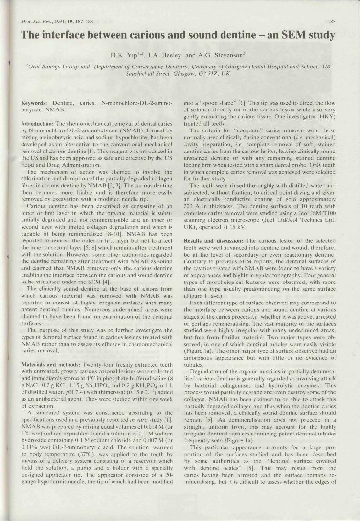
1
Med. Sci. Res. , 1991; 19, 187-188 187
The interface between carious and sound dentine - an SEM study
H .K . Y ip 1'2, J.A. B e e ley1 and A .G . Stevenson21Oral Biology Group and 2Department o f Conservative Dentistry, University o f Glasgow Dental Hospital and School, 378
Sauchiehall Street, Glasgow, G2 3JZ, UK
Keywords: D entine, caries, N-monochloro-D L-2-am ino-butyrate, NM AB.
Introduction: The chem om echanical removal of dental caries by N -m onochloro-D L-2-am inobutyrate (N M A B ), form ed by mixing aminobutyric acid and sodium hypochlorite, has been developed as an alternative to the conventional mechanical removal of carious dentine [1]. This reagent was introduced in the US and has been approved as safe and effective by the US Food and Drug A dm inistration.
The mechanism of action was claimed to involve the chlorination and disruption of the partially degraded collagen fibres in carious dentine by NM AB [2, 3], The carious dentine then becomes m ore friable and is therefore more easily rem oved by excavation with a modified needle tip.
Carious dentine has been described as consisting of an outer or first layer in which the organic m aterial is substantially degraded and not rem ineralisable and an inner or second layer with limited collagen degradation and which is capable of being rem ineralised [8-10]. N M AB has been reported to remove the ou ter or first layer but not to affect the inner or second layer [5, 8] which rem ains after treatm ent with the solution. H ow ever, some other authorities regarded the dentine rem aining after treatm ent with NM AB as sound and claimed that NM AB rem oved only the carious dentine enabling the interface betw een the carious and sound dentine to be visualised under the SEM [4],
The clinically sound dentine at the base of lesions from which carious material was rem oved with NM AB was reported to consist of highly irregular surfaces with many patent dentinal tubules. N um erous underm ined areas were claimed to have been found on exam ination of the dentinal surfaces.
The purpose of this study was to further investigate the types of dentinal surface found in carious lesions treated with NM AB rather than to assess its efficacy in chemom echanical caries removal.
Materials and methods: Tw enty-four freshly extracted teeth with un treated , grossly carious coronal lesions were collected and im mediately stored at 4°C in phosphate buffered saline (8 g NaCl, 0.2 g KC1, 1 .1 5 g N a2H P 0 4 and 0.2 g K H 2P 0 4 in 1 L of distilled w ater, pH 7.4) with thim erosal (0.15 g L -1) added as an antibacterial agent. They were studied within one week of extraction.
A simulated system was constructed according to the specifications used in a previously reported in vitro study [1], NM AB was prepared by mixing equal volumes of 0.014 M (or 1% w/v) sodium hypochlorite and a solution of 0.1 M sodium hydroxide containing 0.1 M sodium chloride and 0.007 M (or 0.11% w/v) DL-2-aminobutyric acid. The solution, warmed to body tem perature (37°C), was applied to the tooth by means of a delivery system consisting of a reservoir which held the solution, a pum p and a holder with a specially designed applicator tip. The applicator consisted of a 20- gauge hypoderm ic needle, the tip of which had been modified
into a “ spoon shape” [1]. This tip was used to direct the flow of solution directly on to the carious lesion while also very gently excavating the carious tissue. O ne investigator (H KY ) treated all teeth.
The criteria for “ com plete” caries rem oval were those normally used clinically during conventional (i.e. mechanical) cavity preparation , i.e. com plete rem oval of soft, stained dentine caries from the carious lesion, leaving clinically sound unstained dentine or with any rem aining stained dentine feeling firm when tested with a sharp dental probe. Only teeth in which com plete caries removal was achieved were selected for further study.
The teeth w ere rinsed thoroughly with distilled w ater and subjected, w ithout fixation, to critical point drying and given an electrically conductive coating of gold approxim ately 200 A in thickness. The dentine surfaces of 10 teeth with com plete caries removal w ere studied using a Jeol JSM-T100 scanning electron microscope (Jeol Ltd/Jeol Technics L td, U K ), operated at 15 kV.
Results and discussion: The carious lesion of the selected teeth were well advanced into dentine and would, therefore, be at the level of secondary or even reactionary dentine. C ontrary to previous SEM reports, the dentinal surfaces of the cavities treated with NM AB were found to have a variety of appearances and highly irregular topography. Four general types of morphological features were observed, with m ore than one type usually predom inating on the same surface (Figure 1, a -d ).
Each different type of surface observed may correspond to the interface between carious and sound dentine at various stages of the caries process i.e. w hether it was active, arrested or perhaps rem ineralising. The vast m ajority of the surfaces studied were highly irregular with many underm ined areas, but free from fibrillar m aterial. Two m ajor types were ob served, in one of which dentinal tubules were easily visible (Figure la ) . The o ther m ajor type of surface observed had an am orphous appearance but with little or no evidence of tubules.
D egradation of the organic matrices in partially dem incra- lised carious dentine is generally regarded as involving attack by bacterial collagenases and hydrolytic enzymes. This process would partially degrade and even destroy some of the collagen. NM AB has been claimed to be able to attack this partially degraded collagen and thus when the dentine caries has been rem oved, a clinically sound dentine surface should rem ain [5]. As dem ineralisation does not proceed in a straight, uniform front, this may account for the highly irregular dentinal surfaces containing patent dentinal tubules frequently seen (Figure la ) .
This particular appearance accounts for a large p ro portion of the surfaces studied and has been described by some authorities as the “ dentinal surface covered with dentine scales” [5]. This may result from the caries having been arrested and the surface perhaps re- mineralising, but it is difficult to assess w hether the edges of
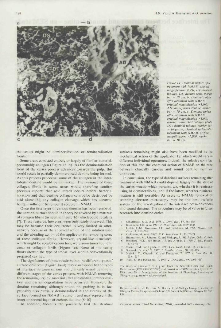
188 H.K. Yip, J. A . Beeley and A.G. Stevenson
Figure la , D en tinal surface after treatm ent with N M A B , original m agnification X 500, DT: dentinal tubules, D S: dentine scale, marker bar = 10 pm. b. Dentinal surface after treatm ent with NM AB, orig inal m agnification xl,000, A D : a m orph ou s dentine, marker bar = 1 0 pm. c, D entinal surface after treatm en t with N M AB, orig ina l m agnification x 1,000, arrow s: un m asked collagen fibrils, D T : den tin al tubules, marker bar = 10 pm. d, D en tinal surface after treatm ent with N M A B , original m agnification x 1,000, marker bar = 10 pm.
the scales might be dem ineralisation or rem ineralisation fronts.
Some areas consisted entirely or largely of fibrillar material, presum ably collagen (Figure lc , d). As the dem ineralisation front of the caries process advances tow ards the pulp, this would result in partially dem ineralised dentine being form ed. As this process proceeds, some of the collagen in the inter- tubular dentine would be unm asked. The presence of these collagen fibrils in some areas would therefore confirm previous reports tha t acid attack occurs before bacterial invasion and that dentine collagen cannot be destroyed by acid alone [6], any collagen cleavage which has occurred being insufficient to render it soluble in NM AB.
Once the first layer of carious dentine has been rem oved, the dentinal surface should in theory be covered by a m attress of collagen fibrils (as seen in Figure Id) which could recalcify [7]. These features, how ever, were only rarely observed. This may be because their occurrence is very limited or a lternatively because of the chemical action of the solution used and the abrading action of the applicator tip rem oving some of these collagen fibrils. H ow ever, crystal-like structures, which might be recalcification foci, were som etim es found in areas of collagen fibrils (Figure lc ) . None of the cavity floors showed the type of sm ear layer seen in conventionally prepared cavities.
The significance of these results is that the different types of surface observed (Figure la -d ) may correspond to the types of interface betw een carious and clinically sound dentine at different stages of the caries process, with NM AB removing the rem aining organic m aterial after substantial dem ineralisation and partial degradation have occurred. H ow ever, the dentine rem aining although sound on probing is in fact probably also partially dem ineralised in the vicinity of the surface form ed on N M AB treatm ent and may represent the inner or second layer of carious dentine [8-10].
In addition, there is the possibility that the dentinal
surfaces rem aining might also have been modified by the mechanical action of the applicator tip which would vary in d ifferent individual operators. Indeed, the relative contribution of this and the chemical action of NM AB on the zone betw een clinically carious and sound dentine itself are unknown.
In conclusion, the type of dentinal surfaces remaining after treatm ent with N M AB could depend largely on the state of the caries process which pertains, i.e. w hether it is reminera- lising or dem ineralising, and if the latter, w hether remineralisation is still possible. A t p resent, NM AB followed by scanning electron microscopy may be the best available system for the investigation of the interface between carious and sound dentine. The procedure may be of value in future research into dentine caries.
1. Schutzbank, S.G . et al. 1978. J. Dent. Res., 57, 861-8642. Kronman, J.H . et al. 1977. J. Dent. Res., 56, 1539-15453. Habib, C .M ., Kronman, J.H . and Goldm an, M. 1975. Pharm. Ther.
D ent., 2, 209-2164. G oldm an, M. et al. 1987. N Y. State Dent. J., 53, 20-215. Brannstrom, M ., Johnson, G. and Friskopp, J. 1980. J. Dent. Child., 47,46-496. Boonstra, W .D ., ten Bosch, J.J. and Arends, J. 1990. J. Biol. Buccale,
17, 43-487. Burke, F.M . and Lynch, E. 1990. Gen. Dent. Treat., 16, 2.1.6-01-118. Kurosaki, N. et al. T. 1977. J. Prosthet. Dent., 38, 169-1739. Kuboki, Y ., Ohgushi, K. and Fusayama, T. 1977. J. Dent. Res., 56,
1233-1237 '10. Kato, S. and Fusayama, T. 1970. J. Dent. Res., 49, 1060-1067
The financial assistance o f a grant from the Scottish Home and HealthDepartment (K /M R S/SO /C I380) and provision o f SEM facilities by Dr H.Y. Elder and Dr I. M ontgomery of the Institute of Physiology, University of G lasgow are gratefully acknowledged.
Reprint requests to: Dr Josie A . B eeley , Oral Biology Group, University of Glasgow Dental Hospital and School, 378 Sauchiehall Street, Glasgow G2 3JZ, UK.
P aper received: 22 n d D ecem ber, 1990; am en ded 26th February, 1991.
Publications Reprinted AbstractsYip HK, Beeley JA, Stevenson AG. An improved reagent for the chemomechanical removal of dental caries. J Dent Res 1988; 67: 663.
Yip HK, Beeley JA. Studies on the reaction of NaOCl and NMAB with collagen. J Dent Res 1989; 68: 982.
Yip HK, Beeley JA, Stevenson AG. The interface betweencarious and sound dentine - an SEM study. J Dent Res 1991; 70: 718.
Yip HK, Beeley JA, Stevenson AG. Chemomechanical removal of dental caries in deciduous teeth. Caries Res 1991; 25: 229-230.
Yip HK, Beeley JA, Stevenson AG. The specificity of caries detector dyes in dentinal caries. (submitted for the IADR meeting, 1992)
PaperYip HK, Beeley JA, Stevenson AG. The interface betweencarious and sound dentine - an SEM study. Med Sci Res1 9 9 1 ; 1 9 : 1 8 7- 1 8 8 .
252
GLASGOWUNIVERSITYLIBRARY