10645948.pdf - enlighten: theses
TRANSCRIPT

https://theses.gla.ac.uk/
Theses Digitisation:
https://www.gla.ac.uk/myglasgow/research/enlighten/theses/digitisation/
This is a digitised version of the original print thesis.
Copyright and moral rights for this work are retained by the author
A copy can be downloaded for personal non-commercial research or study,
without prior permission or charge
This work cannot be reproduced or quoted extensively from without first
obtaining permission in writing from the author
The content must not be changed in any way or sold commercially in any
format or medium without the formal permission of the author
When referring to this work, full bibliographic details including the author,
title, awarding institution and date of the thesis must be given
Enlighten: Theses
https://theses.gla.ac.uk/
Effects of Hypoxia on Proliferation and Signal Transduction Pathways in
Pulmonary and Systemic Vascular Fibroblast cells
A thesis presented by
DAVID JOHN WELSH
for the degree of Doctor of Philosophy to the Faculty of Medicine,
University of Glasgow
March- 2001 Scottish Pulmonary Vascular Unit, Western Infirmary, Glasgow, UK
®David Welsh 2001


ProQuest Number: 10645948
All rights reserved
INFORMATION TO ALL USERS The qua lity of this reproduction is d e p e n d e n t upon the qua lity of the copy subm itted.
In the unlikely e ve n t that the au tho r did not send a co m p le te m anuscrip t and there are missing pages, these will be no ted . Also, if m ateria l had to be rem oved,
a no te will ind ica te the de le tion .
uesLProQuest 10645948
Published by ProQuest LLO (2017). C opyrigh t of the Dissertation is held by the Author.
All rights reserved.This work is protected aga inst unauthorized copying under Title 17, United States C o de
M icroform Edition © ProQuest LLO.
ProQuest LLO.789 East Eisenhower Parkway
P.Q. Box 1346 Ann Arbor, Ml 4 81 06 - 1346

To my Mother, Father and Sister
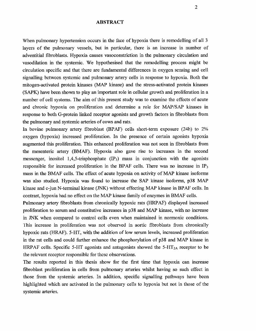
ABSTRACT
When pulmonary hypertension occurs in the face o f hypoxia there is remodelling o f all 3 layers o f the pulmonary vessels, but in particular, there is an increase in number o f adventitial fibroblasts. Hypoxia causes vasoconstriction in the pulmonary circulation and vasodilation in the systemic. We hypothesised that the remodelling process might be circulation specific and that there are fundamental differences in oxygen sensing and cell signalling between systemic and pulmonary artery cells in response to hypoxia. Both the mitogen-activated protein kinases (MAP kinase) and the stress-activated protein kinases (SAPK) have been shown to play an important role in cellular growth and proliferation in a number o f cell systems. The aim o f this present study was to examine the effects o f acute and chronic hypoxia on proliferation and determine a role for MAP/SAP kinases in response to both G-protein linked receptor agonists and growth factors in fibroblasts fi*om the pulmonary and systemic arteries o f cows and rats.In bovine pulmonary artery fibroblast (BPAF) cells short-term exposure (24h) to 2% oxygen (hypoxia) increased proliferation. In the presence of certain agonists hypoxia augmented this proliferation. This enhanced proliferation was not seen in fibroblasts from the mesenteric artery (BMAF). Hypoxia also gave rise to increases in the second messenger, inositol 1,4,5-trisphosphate (IP3) mass in conjunction with the agonists responsible for increased proliferation in the BPAF cells. There was no increase in IP3
mass in the BMAF cells. The effect o f acute hypoxia on activity o f MAP kinase isoforms was also studied. Hypoxia was found to increase the SAP kinase isoforms, p38 MAP kinase and c-jun N-terminal kinase (JNK) without effecting MAP kinase in BPAF cells. In contrast, hypoxia had no effect on the MAP kinase family o f enzymes in BMAF cells. Pulmonaiy artery fibroblasts from chronically hypoxic rats (HRPAF) displayed increased proliferation to serum and constitutive increases in p38 and MAP kinase, with no increase in JNK when compared to control cells even when maintained in normoxic conditions. This increase in proliferation was not observed in aortic fibroblasts fiom chronically hypoxic rats (HRAF). 5-HT, with the addition o f low serum levels, increased proliferation in the rat cells and could fiirther enhance the phosphorylation o f p38 and MAP kinase in HRPAF cells. Specific 5-HT agonists and antagonists showed the 5 -HT2a receptor to be the relevant receptor responsible for these observations.The results reported in this thesis show for the first time that hypoxia can increase fibroblast proliferation in cells from pulmonary arteries whilst having so such effect in those from the systemic arteries. In addition, specific signalling pathways have been highlighted which are activated in the pulmonary cells to hypoxia but not in those of the systemic arteries.
LIST OF CONTENTS PAGE NUMBERS
Title page 1
Abstract 2
List of contents 3
List o f figures 12
List of tables 17
Acknowledgements 18
Authors declaration and publications 19
List o f abbreviations 2 1
Chapter 1 - Introduction
1. General Introduction 25
LI Pulmonary Hypertension 25
1.1.1 Epidemio logy o f pulmonary hypertension 26
1.1.2 Pathological processes seen in pulmonary hypertension 26
1.2 Pulmonary Vascular Remodelling 28
1.2.1 Pathological pulmonary vascular remodelling 28
1.2.2 Morphological features o f pulmonary vascular remodelling 28
1.2.3 Mechanisms o f pulmonary vascular remodelling 29
1.2.4 Cells and extracellular matrix in remodelling in response to 30
hypoxia
1.2.5 In vivo studies o f pulmonary vascular remodelling 30
L2.6 In vitro studies of pulmonary vascular remodelling 31
1.2.7 Growth factors in remodelling 32
1.3 Hypoxia 36
1.3.1 Definition 36
1.3.2 Causes of hypoxia 36
1.3.2.1 Altitude 36
1.3.2.2 Lung Disease 37
1.3.3 Effects of hypoxia 37
1.3.3.1 Endothelial cells 38
1.3.3.2 Smooth muscle cells 39
L3.3.3 Fibroblasts 39
1.3.3.4 Intracellular targets o f hypoxia 40
1.4 Fibroblast physiology 40
1.4.1 Fibroblasts from the pulmonary and systemic circulations 41
1,5 Signalling events controlling cell proliferation 42
1.5.1 The cell cycle 42
1.5.2 Transcriptional regulation of cell cycle dependent genes 45
1 . 6 Early growth factor-stimulated events 47
1.7 Protein phosphorylation 50
1 . 8 Extracellular receptors 51
1.9 G-protein coupled receptors 51
1 . 1 0 Agonist-stimulated phospholipid hydrolysis in fibroblast cells 54
1 . 1 1 Phospholipase D 55
1 . 1 2 Phospholipase A] 55
1.13 Other lipid/lipid-derived second messengers 56
1.14 Protein kinase C 57
1.14.1 Protein kinase C structure 57
1.14.2 Role ofPKC in mitogenic signal transduction 58
1.15 Receptors with intrinsic tyrosine kinase activity 60
1.16 The mitogen activated protein kinase pathway 63
1.16.1 Regulation o f p42/p44 MAP kinase by MEK 65
1.16.2 Regulation o f MEK by Raf-1 6 8
1.16.3 Regulation o f MEK independent o f Raf-1 6 8
1.16.4 Raf-1 activation by Ras 69
1.16.5 Ras-independent activation o f MAP kinase 69
1.16.6 Cellular targets of p42 and p44 M/sP kinase 70
1.17 Stress activated protein kinase 70
1.17.1 MKK homologues involved in JNK and p38 activation 74
1.17.2 Activation o f MKK homologues by MEKKs 74
1.17.3 Possible role o f p38 and JNK activation in cell growth 75
1.18 Hypothesis 75
1.19 Aims 77
Chapter 2 - Materials & Methods
2.1 Materials 79
2.2 Animal models 79
2.2.1 Bovine 79
2.2.2 Rat 80
2.3 Primary cell culture 80
2.3.1 Routine cell maintenance 80
2.3.2 Primary fibroblast culture 83
2.3.3 Immunohislochemical characterisation of primary fibroblast 89
cultures
2.3.4 Cell freezing/Thawing 90
2.4 Hypoxia: Techniques for studying acute hypoxic fibroblast cells 92
in vitro
2.4.1 Generation o f hypoxic environment 92
2.4.2 Measurement o f hypoxia 92
2.5 Hypoxia: Methods for studying cells from chronically hypoxic 94
animals
2.5.1 Chamber design 94
2.5.2 Maintenance o f animals 94
2.5.3 Maintenance o f chronic hypoxic rats 94
2.6 Assessment of cell proliferation 95
2.7 Inositol 1,4,5-trisphosphate mass assay 96
2.7.1 Production o f binding protein 96
2.7.2 Cell Preparation 97
2.7.3 The binding assay (for standard curve) 97
2.7.4 The binding assay (for sample) 98
2.7.5 Calculation o f Ins (1,4,5 )P3 mass 100
2.8 Detection and analysis of proteins 101
2.8.1 Preparation o f samples for SDS-PAGE and immunoblotting 101
2.8.2 Assay o f protein concentration 101
2.8.3 SDS polyacrylamide gel electrophoresis (SDS-PAGE) 102
2.8.3.1 Preparation o f acrylamide gels 102
2.8 .3.2 Electrophoresis conditions 103
2.8 .3.3 Fixing and drying gels 104
2.8.4 Western blot analysis 104
2.8.4.1 Transblotting to nitrocellulose 104
2.8.4.2 Immunoblotting 104
2.8.4.3 Re-probing nitrocellulose membranes 106
2.8.5 Solid phase c-jun N-terminal kinase (JNK)activity assay 106
2.8.6 Solid phase p38 MAP kinase assay 107
2.8.7 p42/p44 MAP kinase activity assay 107
2.9 Densitometric analysis of blots 108
2.10 Data analysis 108
Chapter 3 - Establishment of a cell model for studying bovine pulmonary artery
fibroblast cell proliferation to acute hypoxia
3.1 Introduction 110
3.2 Methods 112
3.2.1 Assessment of fibroblast determination 112
3.2.2 Determination o f [^Hjthymidine uptake 112
3.2.3 Assessment o f PO2 and pH 112
3.2.4 Assessment o f proliferation o f BPAF cells grown in hypoxic 113
conditions
3.2.5 Statistics 113
3.3 Results 114
3.3.1 Assessment o f the fibroblast cell type 114
3.3.2 Determination of maximal [^Hjthymidine ineorporation 116

3.3.3 Determination o f time taken to reach an adequate hypoxic 118
environment
3.3.4 Effects o f hypoxia on proliferation of bovine pulmonary 122
artery fibroblasts
3,4 Discussion 127
Chapter 4 ■ Effects of acute hypoxia on mitogenic stimulation of bovine pulmonary
artery and bovine mesenteric artery fibroblast cells
4.1 Introduction 130
4.2 Methods 132
4.3 Results
4.3.1 Effects o f hypoxia on serum stimulated proliferation o f BPAF and 132
BMAF cells
4.3.2 Effects o f hypoxia on PDGF-stimulated proliferation o f BPAF and 136
BMAF cells
4.3.3 Effect o f hypoxia on G-protein coupled receptor (GPCR) agonist- 139
stimulation o f BPAF and BMAF cells
4.4 Discussion 149
Chapter 5 - Effects of acute hypoxia on IPj generation in BPAF and BMAF cells
5.1 Introduction 164
5.2 Methods 166
5.3 Results 155
5.3.1 Typical standard curve for IP3 generation 15 5
5.3.2 Effect o f agonists and hypoxia on IP3 generation 157
5.4 Discussion 161
Chanter 6 - Effects of acute hypoxia on the stimulation of the stress-activated
protein kinases in pulmonary artery fibroblasts
6.1 Introduction 164
6.2 Methods 166
6 .2 .1 Cell culture 166
6.2.2 Growth of cells in a hypoxic environment 166
6.2.3 Solid-phase INK assay 166
6.2.4 Solid-phase p38 MAP kinase assay 166
6.2.5 p42/p44 MAP kinase phosphorylation and activation 167
6.2.6 Statistics 167
6.3 Results 168
6.3.1 Effect o f hypoxia on JNK and p38 MAP kinases 168
6.3.2 Effect o f hypoxia on p42/p44 MAP kinase phosphorylation and activity 171
6.3.3 Inhibition o f hypoxic stimulation o f p38 MAP kinase activity by 173
SB203580
6.3.4 Reversal o f the late phase o f hypo xic-med iated p38 MAP kinase 175
activity by re-oxygenation.
6.4 Discussion 177
Chapter 7 - Effects of Serum on proliferation and generation of MAP kinases
in fibroblasts harvested from pulmonary arteries from chronically hypoxic rats.
7.1 Introduction 181
7.1.1 Normobaric versus hypobaric hypoxia 182
10
7.1.2 Normobaric hypoxia 183
7.1.3 Hypobaric hypoxia 183
7.2 Methods 185
7.2.1 Chronic hypoxic rat model o f pulmonary hypertension 185
7.2.2 Pulmonary artery fibroblast cell culture 185
7.2.3 Measurement o f serum-stimulated [^H]thymidine uptake in BPAF 186
and RAF cells
7.2.4 Assessment o f phosphorylation o f p42/p44 MAP kinase and members 186
of the stress-activated MAP kinase family
7.2.5 Statistics 187
7.3 Results
7.3.1 Effects o f acute hypoxia on RPAF and RAF cells derived from 188
normoxic and chronically hypoxic rats
7.3.2 Differential p38 and p42/p44 MAP kinase phosphorylation in RPAF 193
and RAF cells from normoxic and chronically hypoxic rats
7.3.3 Differential roles for p38 and p42/p44 MAP kinase in RPAF and 198
RAF DNA synthesis from normal and chronically hypoxic rat cells
7.4 Discussion 202
Chapter 8 - Effect of 5-HT on proliferation and generation of MAP kinases in
fibroblasts harvested from pulmonary arteries from chronically hypoxic rats
8.1 Introduction 207
8.2 Methods 210
8.2.1 Chronic hypoxic rat model o f pulmonary hypertension 210
8.2.2 Pulmonary artery fibroblast cell culture 210
8.2.3 Assessment o f proliferation 210
8.2.4 Assessment o f MAP kinase activity 211
8.2.5 Statistics 212
8.3 Results 213
8.3.1 Effects o f 5-HT agonists and antagonists on proliferation o f 213
fibroblast cells from normoxic and chronically hypoxic rats
8.3.2 Effect o f 5-HT and 5-HT antagonists on p38 phosphorylation in 220
fibroblast cells from normoxic and chronically hypoxic rats
8.3.3 Effect of 5-HT and 5-HT-antagonists on p42/p44 MAP kinase 223
phosphorylation
8.4 Discussion 228
Chapter 9 - General Discussion
9.1 Summary of results 232
9.2 Species Difference 233
9.3 Role of stress activated protein kinases 235
9.4 Hypoxia causing a phenotypic switch 236
9.5 Pulmonary vs. systemic arteries 237
9.6 Future work 238
References 240

List of Figures
12
Chapter 1
1.1 The cell cycle
1.2 Overview of extracellular signalling into the cell
1.3 Tyrosine kinase cell signalling overview
1.4 Overview of p42/p44 MAP kinase cell signalling
1.5 Overview of the p42/p44, SAPK/JNK and p38 pathway
1.6 Overview of JNK MAP kinase signalling
1.7 Overview of p38 MAP kinase cell signalling
Chapter 2
2.1 Micro flow biological laminar safety cabinet
2.2 Location of pulmonary artery from freshly excised bovine lung
2.3 Pulmonary artery from freshly excised bovine lung
2.4 Pulmonary artery prior to cleaning
2.5 Pulmonary artery after cleaning
2.6 Removal of endothelial layer
2.7 Pulmonary artery explants
2.8 Explants in 25cm^ culture flask
2.9 Pulmonary artery fibroblasts growing from explants
2.10 Olympus CK2 light microscope
2.11 Hypoxic chamber; LEEC O2/CO2 humidified temperature
controlled incubator
Chapter 3
3.1 a-actin staining of normoxic and hypoxic pulmonary artery
Page
44
53
62
64
67
72
73
82
84
85
86
86
87
87
88
88
91
93
115
13
3.2
3.3
3.4
3.5
3.6
Chapter 4
4.1
4.2
4.3
4.4
4.5
4.6
Chapter 5
5.1
5.2
fibroblasts
Determination of maximum [^H]thymidine uptake in 117
pulmonary artery fibroblasts
Effect of pH on [^HJthymidine uptake in normoxic pulmonary 121
artery fibroblasts
Effect of hypoxic pre-incubation on BPAF cell proliferation 124
Effects of a 6 h hypoxic pre-incubation with varied lengths of 125
growth time on the proliferation of BPAF cells
Visual assessment of BPAF cell proliferation 126
Effect of hypoxia on serum-induced proliferation of BPAF and 135
BMAF cells.
Effects of hypoxia on PDGF-stimuIated proliferation of BPAF 138
and BMAF cells
Effects of hypoxia on ET-l-stimulated proliferation of BPAF 142
and BMAF cells
Effect of hypoxia on AH-stimulated proliferation of BPAF and 144
BMAF cells
Effects of hypoxia on 5-HT-stimulated proliferation of BPAF 146
and BMAF cells
Effects of hypoxia on thrombin-stimulated proliferation of 148
BPAF and BMAF cells.
Typical standard curve used to measure IP3 generation 156
Effects of PDGF and hypoxia on Inositol 1,4,5-tris-phosphate 158
14
generation in BPAF and BMAF cells
5.3 Effects of ET-1 and hypoxia on Inositol 1,4,5-tris-phosphate 159
generation in BPAF and BMAF cells
5.4 Effects of Thrombin and hypoxia on Inositol 1,4,5-tris 160
phosphate generation in BPAF and BMAF cells
Chanter 6
Acute hypoxic stimulation of JNK activity in BPAF cells
6.2 Acute hypoxic stimulation of p38 MAP kinase activity in BPAF 169
cells
6.3 Acute hypoxic stimulation of p42/p44 MAP kinase activity in 170
BPAF cells
6.4 Inhibition of hypoxic stimulation of p38 MAP kinase by 172
SB203580
6.5 Reversal of the late phase of hypoxic-mediated p38 MAP kinase 176
activity by re-oxygenation
Chapter 7
7.1 The effect of serum on [^HJthymidine uptake from normoxic 191
and acutely hypoxic RPAF and RAF cells
7.2 Effect of serum on [^HJthymidine uptake in RPAF cells from 192
chronically hypoxic rats and fibroblasts from chronically
hypoxic rats exposed to acute hypoxia and in RAF cells
7.3 p38 MAP kinase phosphorylation in pulmonary artery and 195
aortic fibroblast cells from normoxic and chronically hypoxic
rats,
7.4 p42/p44 MAP kinase phosphorylation in pulmonary artery and 196
15
aortic fibroblast cells from normoxic and chronically hypoxic
rats
7.5 JNK phosphorylation in pulmonary artery and aortic fibroblast 197
cells from normoxic and chronically hypoxic rats
7.6 Effect of specific kinase inhibitors on phosphorylation of p38 200
and p42/p44 MAP kinase in pulmonary; artery and aortic
fibroblast cells from normoxic and chronically hypoxic rats
7.7 Effect of SB203580 and U0126 on the enhancement of 201
[^HJthymidine uptake observed following serum-stimulated
RPAF and RAF cells from chronically hypoxic rats
Chapter 8
8.1 Effect of 5-HT and serum on [^HJthymidine uptake in RPAF 216
cells from normoxic and chronically hypoxic animals
8.2 Effect of lOpM 5-HT and serum on the uptake of [^H]thymidine 217
pulmonary artery fibroblasts from control and chronically
hypoxic animals
8.3 Effects of specific 5-HT agonists and antagonists on the 218
replication of pulmonary artery fibroblasts from control and
chronically hypoxic rats
8.4 Effects of specific 5-HT and MAP kinase inhibitors on 5-HT- 219
induced [^Hjthymidine uptake of pulmonary artery fibroblasts
from control and chronically hypoxic animals
8.5 Effect of increasing concetration of 5-HT on p38 activity in 221
pulmonary artery fibroblast cells from control and chronically
hypoxic rats
8.6 Effect of 5-HT on p38 activity in pulmonary artery fibroblast 222
cells from control and chronically hypoxic rats
8.7 Effect of increasing doses of 5-HT on p42/p44 activity in 225
pulmonary artery fibroblast cells from control and chronically
hypoxic rats
8.8 Effect of 5-HT and serum on p42/p44 activity in pulmonary 226
artery fibroblast cells from control and chronically hypoxic rats
8.9 Effect of 5-HT on p42/p44 activity in pulmonary artery 227
fibroblast cells from control and chronically hypoxic rats
Addenda
Chapter 3
3.4a Effects of hypoxic pre-incubation on BPAF cell proliferation 124a
Chapter 4
4.2a Effects o f hypoxia on PDGF-stimu Iated proliferation on BPAF and 138aBMAF cells
17
Chanter 1
Table I.l
Chapter 2
Table 2.1
Table 2.2
Chapter 3
Table 3.1
Table 3.2
Chapter 7
Table 7.1
List of Tables
Transcription factors
Dilution of stock IP3 for use with standard curve
Primary antibody dilutions utilised for western blotting
Generation of a hypoxic environment for BPAF cells
Effect of hypoxia on pH of growth medium
Stimulation indices of DNA synthesis for control and chronic
hypoxic rat fibroblast cells from the pulmonary and aortic
circulation following acute hypoxic challenge
46
99
105
119
120
190

18
ACKNOWLEDGEMENTS
A very special thanks goes to my supervisor Dr. Andrew Peaeock for his adviee and
encouragement during this thesis. I would also like to express my sincere thanks to
him for allowing me to beeome part o f the Scottish Pulmonary Vascular Unit and for
giving me the opportunity to undertake these studies.
Many thanks to Pam Scott for her selfless help in both proof reading and presentation
o f this thesis; your help has been invaluable.
1 would like to thank Dr. Margaret Harnett for her ideas and technical advice in
chapters 7 and 8 and to Prof. Margaret MacLean for the supply o f chronic hypoxic rat
tissue.
I would like to extend my gratitude to the Chest, Heart and Stroke Scotland who
supplied financial support for my research.
A big thank you to all o f my work colleagues past and present for their support and
for providing a happy working environment (and numerous cups of tea!!), espeeially
David Raeside, Tarek Saba, and Kir sty Menzies for their friendship.
Finally, I would like to tiiank my parents and sister who could not have done more,
both in their support and love. It is to my mum, dad and sister that I dedicate this
thesis.
19
Declaration
This thesis is entirely my own composition and the experimental work detailed within
was undertaken wholly by myself, with the exception of figures 6.1 - 6.5 which were
produeed with the help o f Dr. Pamela Scott.
Signed
Some of the results within this thesis have been published, details o f which are given
below.
Publications
Full papers
Welsh DJ, Harnett M, MacLean M and Peacock AJ. (2001) Clironic hypoxia induces
constitutive p38 MAP kinase activity which correlates with enhanced cellular
proliferation in fibroblasts from rat pulmonary but not systemic arteries. Am. J. Resp.
Crit. Care Med in press
Welsh DJ. Scott P, Plevin R, Wadsworth R and Peacock AJ. (1998) Hypoxia
enhances cellular proliferation and inositol 1,4,5-trisphosphate generation in
fibroblasts from bovine pulmonary artery but not from mesenteric artery. Am. J. Resp.
Crit. Care. Med. 158: 1757-1762
20
Scott P, Paul A, Belham C, Peacock AJ, Wadsworth R, Gould GW, Welsh DJ and
Plevin R. (1998) Hypoxic stimulation o f the stress-activated protein kinases in
pulmonary artery fibroblasts. Am. J. Resp. Crit. Care. Med. 158/3: 958-62
Abstracts
Welsh DJ, Harnett M, MacLean M and Peacock AJ. (2001) HIF-1 alpha is induced
in pulmonary artery fibroblasts from chronically hypoxic rats but not in those from
control ratszlw. J. Resp. Crit. Care. Med. in press
Welsh DJ, Harnett M, MacLean M and Peacock AJ. (2000) 5-HT stimulated p38
MAP kinase activity in pulmonary artery fibroblasts from clironicaily hypoxic rats is
mediated through the S-HTza receptor. Am. J. Resp. Crit. Care. Med. 161/3: A641
Welsh DJ, Harnett M, MacLean M, Weir K and Peacock AJ. (1999) The effect o f
blocking K+ channels on proliferation o f chronically hypoxic rat fibroblasts from the
pulmonary and systemic circulation. Am. J. Resp. Crit. Care. Med. 159/3: A347
Welsh DJ, Harnett M, MacLean M and Peacock AJ. (1999) The effect o f chronic
and acute hypoxia on proliferation and stimulation of stress activated protein kinases
in rat fibroblasts from the pulmonary and systemic circulations. Am. J. Resp. Crit.
Care. Med. 159/3: A3 4 7
Welsh DJ, Harnett M, MacLean M and Peacock A. (1998) The specific p38 MAP
Kinase inhibitor SB203580 abolishes the proliferative response o f Bovine Pulmonary
Artery Fibroblasts to Hypoxia. Am. J. Resp. Crit. Care.Med. 157/3: A430
21
List of Abbreviations
A ll - Angiotensin II
ATP - Adenosine Triphosphate
BMAF - Bovine Mesenteric Artery Fibroblasts
BPAF “ Bovine Pulmonary Artery Fibroblasts
BSA - Bovine Serum Albumin
cdk - Cyclin-Dependent Kinase
CREB - cAMP Response Element Binding Protein
DAG - Diacylglycerol
DMEM - Dulbeccos’ Modified Eagles Medium
DNA - Deoxyribonucleic Acid
DPM - Disintegration’s Per Minute
ECM - Extracellular matrix
EGF - Epithelial Growth Factor
ERK - Extracellular Signal Regulated Kinase
ET-1 - Endothelin-1
FACS ~ Fluorescence-Activated Cell Sorter
FCS - Foetal Calf Serum
h - Hour
HIF - Hypoxia Inducible Factor
HO - Hemeoxygenase
HRP - Horseradish Peroxidase
5-HT - 5-hydroxytryptamine
5-HTT - 5-Hydroxytryptamine transporter
22
IL-1 - Interleukin - 1
iNos - Inducible Nitric Oxide
IP3 - Inositol 1,4,5- trisphosphate
JNK - c-jun n-Terminal Kinase
LPS - Lipopolysaccharide
MAP Kinase - Mitogen Activated Protein Kinase
MAPKAPK-2 - Mitogen Activated Protein Kinase Activated Protein Kinase -2
MEK - Mitogen-Activated Protein Kinase Kinase
min — Minute
MKP - Mitogen Activated Protein Kinase Phosphatase
mmHg - Millimetres of Mercury
MMP - Matrix Metalloproteinases
PA EC - Pulmonary Artery Endothelial Cells
PAP - Pulmonary Arterial Pressure
PBS - Phosphate Buffered Saline
PC - Phosphatidyl-choline
PDGF - Platelet-Derived Growth Factor
PI3K - Phosphatidylinositol 3-Kinase
PIP2 - Phosphatidyl inositol -4,5-Bisphosphate
PKA - Protein Kinase A
PKC - Protein Kinase C
PLA2 - Phospholipase A2
PLC - Phosolipase C
PLD - Phospholipase D
PO2 - Percentage Oxygen
23
p38 MAP Kinase - p38 Mitogen Activated Protein Kinase
pp38 - Phosphorylated p38
RAF - Rat Aortic Fibroblast
RPAF - Rat Pulmonary Artery Fibroblast
RTK - Receptor Tyrosine Kinase
s - Second
S.D. - Standard Deviation
SAPK - Stress Activated Protein Kinase
SH2 - Src Homology Domain
SMC - Smooth Muscle Cells
SRF - Serum Response Factor
TGF-p “ Transforming Growth factor - p
TNF - Tumour Necrosis Factor
VEGF - Vascular Endothelial Growth Factor

24
Chapter 1
Introduction
25
1 General Introduction
The acceleration o f pulmonary artery fibroblast cell proliferation and the resultant
remodelling is thought to be a primary event in the development o f pulmonary
hypertension (Reid and Davies, 1989). At present, no underlying mechanism for
remodelling has been found. One question is the link between vasoconstriction and
remodelling. Does vasoconstriction lead to remodelling? It is tempting to say that this
is the case because many vasoconstrictors, including hypoxia, are also growth factors,
that is, thei e is a hnk between cell replication and vascular control (“cell growth-
vasomotor coupling”) (Scott and Peacock, 1995). Understanding the cell signalling
events involved in the regulation of normal and abnormal fibroblast cell proliferation
due to hypoxia may provide an important insight into the degeneration o f growth
control during pulmonary hypertension. In some instances irregular proliferation may
be the result o f over activity or dysfunction o f component(s) o f the growth regulating,
intracellular signalling pathway(s). Investigation o f these pathways may point to
possible sites o f pharmacological intervention for the prevention o f pulmonary
hypertension.
1,1 Pulmonary Hypertension.
Pulmonary Hypertension is a condition of abnormally high pressure within the
pulmonary circulation and develops in many species, including humans. It i.s
important because it contributes to the morbidity of most heart and lung diseases. One
cause o f pulmonary hypertension is chronic exposure to normobaric and hypubaric
hypoxia (Janssens et al, 1991). Exposure to hypoxia results in an acute increase in
pulmonary arterial pressure (PAP), as a result o f vasoconstriction which can be
followed by sustained pulmonary hypertension resulting from a combination of
26
polycythemia and morphological alterations of the pulmonary vascular bed. This late
increase in PAP is not immediately or totally correctable upon improvements in
oxygen concentrations to normal values (Vender, 1994).
1.1.1 Epidemiology o f pulmonary hypertension
There are several causes o f pulmonary hypertension
Left to right shunt. This means that blood at high pressure (systemic circulation)
passes directly to the pulmonary circulation (low pressure).
Pulmonary Thrombo-Embolism. A condition in which pulmonary arteries are blocked
by a clot formed in situ or carried in the bloodstream from the site of remote
formation.
Primary pulmonary hypertension. This is a rare disorder of unknown cause.
Histological examination o f the lung shows an increase in smooth muscle and
fibroblast deposition in the small pulmonary arteries.
Chronic hypoxic lung disease. This includes:- Kyphoscoloiosis, chronic bronchitis
and emphysema, cystic fibrosis and interstitial lung disease and is thought to cause
pulmonary hypertension in two ways: alveolar hypoxia causes pulmonary
vasoconstriction and structural damage in the lung reduces pulmonary vascular
compliance.
1.1.2 Pathological processes seen in pulmonary hypertension
These fall in to three categories: -
Hypoxic pulmonary vasoconstriction.
Hypoxic pulmonary vasoconstriction has been known to exist since 1946 (von Euler
and Liljestrand, 1946) but the mechanism is not yet fully understood. It is turned on
27
within 1-2 minutes after a sudden decrease in alveolar PO2, is fully developed after 3-
5 minutes, remains more or less stable thereafter according to the experimental
conditions, and is generally reversed in less than 1 minute. Hypoxic vasoconstriction
is observed in isolated lungs devoid o f nervous connections. It is located mostly in
small order arterioles but may extend to large branches o f the pulmonary artery or to
pulmonary capillaries. The biological mechanism o f hypoxic pulmonary
vasoconstriction remains unknown (Grovner, 1983).
Destruction o f the pulmonary vascular bed.
Examples of this are loss o f lung parenchyma- e.g. emphysema, and pulmonary
vascular arteritis- e.g. polyarteritis nodosa.
Pulmonary vascular remodelling.
In addition to short-term autoregulatory changes in vascular tone, vessels can undergo
profound fibrocellular changes. This is an active process termed vascular remodelling
which results in an increase in the medial coat o f the normally muscular arteries and
an extension o f muscle into smaller and more peripheral vessels than is normal. There
are various clinical conditions in which these features are found. Hypoxia is one cause
and is a useful model because it can be tested in the laboratory. These include
Kyphoscoliosis (Naeije, 1961), chronic bronchitis and emphysema (Semmons and
Reid, 1974) and cystic fibrosis (Ryland and Reid, 1975). Pulmonary vascular
remodelling is important clinically because it renders the vessels relatively
unresponsive to vasodilators.
28
1.2 Pulmonary Vascular Remodelling.
1.2.1 Pathological pulmonary vascular remodelling.
Pathologically, pulmonary vascular remodelling represents increased muscularisation
o f both the large and small pulmonary arteries, which results in a reduction of the area
o f the pulmonary arterial bed (Reid and Meyrick, 1982). One of the main causes of
remodelling in humans is hypoxia.
Thickening of the muscular coat o f small pulmonary arteries begins a few days after
exposure to hypoxia (Hunter ei al, 1974). Furthermore, fibroblasts migrate into
arterioles and in some studies appear to undergo transformation into smooth muscle
cells (Sobin et al, 1983) that reduce the size o f the vessel lumen. In addition to this,
the proximal pulmonary arteries respond to the rise in blood pressure by an increase in
the thickness of the media and adventitia. Proliferation of adventitial fibroblasts
occurs before the other cell types (Reid and Davies, 1989). High blood flow can also
contribute to the remodelling process by increasing the wall shear stress which may
directly damage endothelial cells (Esterly et al, 1968), and activate platelets to release
growth factors leading to smooth muscle cell proliferation There may also be release
o f endothelial-derived growth factors. There is experimental evidence that mechanical
forces (e.g. stretch) directly stimulate the vascular wall to undergo remodelling in the
absence of endothelial cell damage (Archer et al, 1988).
1.2.2 Morphological features of pulmonary vascular remodelling.
One of the main features o f pulmonary vascular remodelling is the proliferation of
fibroblast and smooth muscle cells in the pulmonary arteries. When this occurs, the
proliferation of intima and media will encroach on the lumen of the vessel, which in

29
turn will alter the relationship between flow and pressure in the vessel (Owens and
Schwartz, 1983; Murphy et a/, 1981). Structural changes within the vessel can also
occur in the larger of the pulmonary arteries. Thickening o f the vessel walls also
involves an increase in the number of fibroblast cells, which may be a consequence of
chemotaxis from a distant site, mitogenesis or both (Harris and Heath, 1986).
1.2.3 Mechanisms o f pulmonary vascular remodelling.
The process of pulmonary vascular remodelling consists o f some or all of the
following events:-
1/ Altered physical and haemodynamic forces.
These include shear stress and transmural pressure. Endothelial cells are subjected to
shear stress caused by the movement o f blood over their surface. Transmural pressure
is the increased atrial pressure causing an increase in stress and strain on all cell types
across the vessel wall (Sumpio et al, 1988).
2/ Relay o f signals to cells.
This may consist of signals / substances (Sharkey et al, 2000; Santilli et al 1991)
secreted from one cell type or in the form of stress which either stimulates or
suppresses the growth or activity of another cell type within the vessel.
3/ Alteration o f vessel wall and extracellular matrix.
Marked increases in extracellular matrix protein synthesis by fibroblast and smooth
muscle cells in vivo under conditions o f chronic hypoxia have been well established
(Saed et al, 1999; Crouch et al, 1989). Studies in chronically hypoxic adult and
neonatal animals from several species demonstrate increases in the production and
accumulation of collagen and e last in in the media o f conducting pulmonary arteries
(Novotna and Her get, 1998; Xu et al, 1995). In the adult rat, substantial and rapid
30
increases in the relative rates o f connective tissue protein synthesis in the explants o f
main pulmonary arteries have been demonstrated (Poiani et al, 1990)
4/ Growth promoting substances.
There has been much work done on growth factors, which stimulate all forms of cells
from within the pulmonary artery to proliferate. Several peptide growth factors are
known to cause smooth muscle cells to proliferate (Schwartz and Reidy, 1987) and
indeed the same is true for fibroblasts (Welsh et al, 1996). The response o f cells to
these factors may also be enhanced by the addition o f environmental change, such as
hypoxia (Welsh et al, 1996).
1.2.4 Cells and extracellular matrix in remodelling in response to hypoxia
Little is known of the mechanisms by which hypoxia mediates vascular remodelling.
It is unclear whether abnormal cell proliferation and connective tissue deposition in
pulmonary arteries is a direct effect o f hypoxia itself or secondary to the rise in
pulmonary artery pressure and blood flow caused by hypoxic vasoconstriction.
1.2.5 In vivo studies o f pulmonary vascular remodelling
Ultrastructural and morphometric analysis o f pulmonary arteries o f animals exposed
to clironic hypobaric hypoxia show a significant thickening o f each of the three vessel
wall layers and a narrowing o f the vessel lumen. The nature o f the hypoxic response
in the media varies with distal progression along the vessel. Medial thickening in
large diameter muscular pulmonary arteries seen in the rat (Rabinovitch et al, 1979,
Walker et al, 1984), dog (Rorie et al, 1988), newborn domestic calf (Stenmark et al,
1988) and humans native to high altitudes (Heath et al, 1988) is caused principally by
hypertrophy o f pre-existing smooth muscle cells (SMCs). This, due mainly to an
31
increase in the organelles o f protein synthesis, the rough sarcoplasmic reticulum and
golgi apparatus (Meyrick & Reid, 1980). There is also an increase both in the
thickness of the elastic laminae separating SMCs from endothelial cells and
extracellular connective tissue deposition including elastin and collagen. Fibroblasts
too are able to migrate into the media in response to hypoxia (Sobin et al, 1983) and
express a dramatic change in phenotype, acquiring structural characteristics of
contractile cells.
Adventitial thickening during the development o f chronic pulmonary hypertension in
rats (Meryick & Reid, 1980) and neonatal calves (Stenmark et al, 1988) has been
shown to be caused by both an early and a prolonged intense fibroblast proliferative
response greater than either the endothelial cell or SMCs (Meryick & Reid, 1979). In
addition to an increase in the number of fibroblasts observed in the adventitia of
hypoxic pulmonary arteries, marked increases in matrix protein deposition has been
noted, especially that o f collagen and elastin, as described in human and animal
studies of pulmonary hypertension.
1.2.6 In vitro studies o f pulmonary vascular remodelling
Smooth muscle cells isolated from muscular pulmonary arteries o f newborn calves
exposed to hypoxia produce more elastin than cell from normal calves (Mecham et al,
1987). Cultured human fibroblasts have been shown to exhibit a proliferative response
to hypoxia (2.5% O2) (Storch & Talley, 1988). Although there is compelling
evidence that there is SMC replicatb/e response to hypoxia in vivo, this observation
has not been extended to cultured SMCs. In fact, authors have shown that hypoxia
actually inhibits proliferation of cultured bovine foetal, neonatal and adult pulmonary
artery SMCs (Benitz et al, 1990; Stenmark et al, 1991; Dempsey et al, 1991)
32
suggesting that reducing oxygen tension, at least by itself, is unlikely to be a direct
stimulus accounting for the increased quantity of smooth muscle found in the
pulmonary arteries in association with hypoxemia. However, a homogenous
population o f pulmonary arterial SMCs in a cultured cell system may not accurately
reflect their behaviour in situ. They are removed from the influence of pressure flow
and physical interactions with matrix components and other cell types usually found
in close proximity within the vessel wall, which may influence their response to
hypoxia. Other vascular cells including endothelial cells, fibroblasts and
macrophages, or blood-borne cells such as the platelet or monocyte may be oxygen
sensors themselves and facilitate SMC proliferation in response to hypoxia by their
generation of extracellular growth-promoting factors that act in a paracrine manner.
The stimulatory effect o f reducing oxygen on cultured fibroblast proliferation may
also be partially explained by the enhanced synthesis and secretion o f autocrinic
peptide mitogens. Several groups have used conditioned media from cultured ceils
exposed to hypoxic conditions to look at its effect on growth o f cells and to identify
and quantify the factors released (Ankoma-Sey et al, 2000).
1.2.7 Growth factors in remodelling
Hypoxia increases the expression of growth factors in whole lung and isolated
vascular cells (Sodhi et al, 2000; Ankoma-Sey et al, 2000). It is not clear which are
critical to the remodelling process, although endothelin-l (ET-1), platelet-derived
growth factor fPDGF), Angiotensin II (AH), Thrombin and 5-hydroxytryptamine (5-
HT) are probably important.
33
EndotheIin-1: Vender and colleagues (1987) first described a SMC macromolecular
mitogen released from hypoxic pulmonary artery endothelial cells (PA ECs). Since
then, ET-1 has been identified as an EC-derived G protein-coupled receptor agonist
peptide shown to be mitogenic for cultured SMCs and fibroblasts from the systemic
vasculature (Suzuki et al, 1999). Production o f ET-1 by cultured human umbilical
vein ECs is elevated by hypoxic exposure (Peacock et al, 1992). However, cultured
bovine SMCs from the pulmonary artery exhibit only a modest increase in growth in
response to maximal ET-1 concentrations. Indeed, bovine pulmonary artery
fibroblasts as measured by cell counts and [^H]thymidine incorporation, a reflecfion
o f DNA synthesis show no increase in proliferation to ET-1 (Hassoun et al, 1992,
Welsh et al, 1996). Furthermore, bovine PA ECs show either no change (Hassoun et
al, 1992) or a decrease (Wiebke et al, 1992) in ET-1 release in response to hypoxia o f
48h duration. These discrepant and contradictory results have made it difficult to
interpret the role o f ET-1 as a factor regulating SMC and fibroblast proliferation and
in how hypoxia modifies ET-1 production by ECs in the pulmonary cfrculation.
Platelet-derived growth factor: Platelets contain a mitogenic protein factor referred
to as PDGF (Deuel, 1987). This protein is present in platelet a granules (Gerrard et al,
1980) and is released when platelets are stimulated to aggregate (Ross et al, 1986). It
is a major mitogen in serum for many types o f cells, including fibroblasts, glial cells,
and vascular smooth muscle cells, (Heldin and Westermark, 1984; Deuel et al, 1983;
Ross et al 1986) but not in endothelial cells (Davies et al, 1978). PDGF is a dimeric
molecule that occurs as a homodimer (AA and BB) and a heterodimer (AB) o f related
polypeptide chains; PDGF -A and PDGF -B (Heldin and Westermark, 1984).
34
Platelets contain mainly PDGF-AB and some PDGF-BB. On the other hand, VSMC
secretes PDGF-AA in a growth and differentiation-dependant manner, which may
contribute to the growth-stimulating mechanisms of blood vessels. In contrast to
fibroblasts, which show an equal growth response to PDGF-AA and -B B (Mehmet et
al, 1990), the mitogenic responses o f VSMC to the three PDGF isoforms are not
uniform, e.g. PDGF -AA is a far weaker mitogen than PDGF -A B and -BB
(Sachinidis et al, 1990). In fibroblast and smooth muscle cells, PDGF has been shown
to increase the cellular replicative responses to hypoxia (Welsh et al, 1996; Stenmark
et al, 1991).
5-Hydro xytryptamine (5-HT): 5-HT, also known as serotonin, is one o f the best
known examples o f a neurotransmitter that mediates a wide variety o f physiological
effects, including peripheral and central actions, through the binding to multiple
receptor subtypes (Wilkinson and Dourish, 1991). The large diversity of 5-HT
functions is paralleled by the pharmacological complexity o f 5-HT receptors. At least
four classes o f 5-HT receptors have been distinguished pharmacologically, based on
the second messenger systems to which the receptor is coupled. The family including
5-HT] and 5-HTs subtypes o f receptors interacts negatively with adenylyl cyclase, the
5 -HT2 subfamily o f receptors is coupled to the activation o f the phospholipase C-p,
the 5-HT] receptor is a ligand-gated ion channel, and the family, including 5 -HT4, 5-
HT&, and 5-HT? subtypes o f receptors, activates adenylyl cyclase (Peroutka, 1995).
O f the receptor classes distinguished, the 5-HTi receptor is the most likel> to be
involved in the hypoxic responses seen in the pulmonary vessels. It mediates many of
the central and peripheral physiological functions o f 5-HT, including contraction of
blood vessels and cell growth and development (Tecott et al, 1995).
35
Angiotensin II (All): All stimulates a variety o f physiological responses related to
the regulation o f blood pressure, salt and fluid homeostasis (Raizada et al, 1993). In
addition, AH promotes growth responses in many cells, including cardiomyocytes,
fibroblasts (Marshall et al, 2000), and vascular smooth muscle cells (Aceto and
Baker, 1990; Sadoshima and Izumo, 1993). AH exerts effects through specific G-
protein coupled receptors, predominantly the AT| receptor subtype. ATi receptors
couple to intracellular calcium mobilisation, activation o f tyrosine kinases such as
p i25^^* and induction o f serine/threonine kinases, including protein kinase C (PKC)
and mitogen-activated protein kinases (MAP kinases) (Lindpainter and Ganten, 1994;
Sadoshima and Izumo, 1993). AH may act directly through these pathways or
indirectly via the release o f growth factors such as PDGF and TGF-p, as demonstrated
for rat vascular smooth muscle cells (Sadoshima and Izumo, 1993). Like other growth
factors, AH induces a rapid increase in the growth associated nuclear proto-oncogenes
c-myc, c-fos and c-jun and several cellular genes including tenascin, fibronectin and
collagen (Naftilan et al, 1990; Taubman et al, 1989). These studies indicate that AH
caii induce rapid changes in gene expression and function that may ultimately lead to
increased cell growth (Dosai et al, 1991)
Thrombin: Thrombin may have a role in the pathogenesis o f pulmonary vascular
disease. It is well documented as causing pulmonary vasoconstriction (Horgan et al,
1987) and has been implicated in the development o f intimai thickening after balloon
angioplasty (Sarembock et al, 1991). The action of tlirombin on vascular cells
involves the proteolytic activation o f G protein-coupled receptors that are subjected to
rapid and iireversible de-sensitisation. Tlirombin is mitogenic in neonatal rat vascular
36
smooth muscle (Huang and Ives, 1987) as well as various fibroblast cell lines
(Vouret-Craviari et al, 1992; Carney et al, 1985). Furthermore, the mitogenic effects
o f thrombin can be mimicked by thrombin receptor-derived peptides that activate the
thrombin receptor (McNamara et al, 1993).
Growth factors mediate their effect in general by binding to the cell surface via
specific receptors and initiating a cascade o f intracellular signalling events (Chapter
1.8 ).
1.3 Hypoxia.
1.3.1 Definition
Hypoxia is a physiological term for the condition in which the oxygen supply to
tissue, organs or whole animals is insufficient to maintain a normal function. The
effect o f hypoxia on an animal is characterised by tachycardia, hypertension,
peripheral vasoconstriction, and mental confusion.
1.3.2 Causes o f hypoxia
1.3.2.1 Altitude
The first clue that altitude adversely affected the lung circulation of the altitude
resident was the recognition of 'brisket disease’ in cattle. In 1913, ranchers in
Colorado (10,000 feet) asked veterinary scientists at Colorado State University to
investigate why theii' herds were dying with swelling ventral to the sternum in the area
37
of the brisket of beef, hence the name ‘brisket disease’ (Glover & Newsom, 1915). At
the turn of the century when cattle from Kansas and Texas were being introduced into
Colorado high pastures, mortality approached 40%. Investigations, which began then,
revealed that the swelling was due to accumulation o f heart failure fluid and that cattle
recovered when taken to lower altitude.
After von Euler and Liljestrand (1946) reported that acute hypoxia induced
pulmonary vasoconstriction, investigators in Colorado confirmed that pulmonary
hypertension in the cattle was the result o f sustained vasoconstriction from chronic
hypoxia (Alexander & Jenson, 1959).
1.3.2.2 Lung disease
Many lung diseases are associated with hypoxia which stimulate pulmonary
vasoconstriction and cause pulmonary vascular remodelling. Examples o f these are
clironic bronchitis, emphysema and interstitial lung disease. Chronic bronchitis leads
to airway narrowing and obstruction and in some cases polycythemia results from
chronic hypoxemia (Hasleton et al, 1968). Emphysema is the result o f destructive
changes in the alveolar wall resulting in loss of lung elasticity and gas exchange.
Interstitial lung disease may be idiopathic but may also follow industrial exposure to
inorganic dusts, such as asbestos or silica. There is scarring o f the lung tissue and
hence reduction o f the lungs ability to allow oxygen transport to the blood.
1.3.3 Effects of hypoxia
In response to hypoxia, mammals exhibit systemic responses that decrease cellular
oxygen need and dependence and increase tissue oxygen supply. Alterations in
oxygen tension elicit important responses at the organ and tissue level. Exposure to
38
chronic hypoxia causes pulmonary hypertension in most mammalian species. The
pulmonary hypertension is associated with changes in vascular tone and is nearly
always associated with significant structural changes within the pulmonary vascular
bed (pulmonary vascular remodelling) (Stenmark and Mecham, 1997). There are also
marked changes in the phenotype o f endothelial, smooth muscle and fibroblast cell
populations. Over the last few years it has become clear that the vascular responses to
hypoxia involve extremely complicated cell-cell interactions mediated by growth
factors, cytokines and biological messengers. For the purpose o f this thesis, the most
relevant effect of hypoxia in the body is its effect on the lungs where hypoxia leads to
pulmonary vasoconstriction and pulmonary vascular remodelling.
1.3.3.1 Endothelial cells
Large amounts of work have been carried out in endothelial cells. PA ECs are
continually exposed to low levels o f oxygen due to the low oxygen content of mixed
venous blood. This level o f oxygen can become even lower during hypoxic exposure.
In response to hypoxia, a number o f genes have been shown to be induced. These
include vascular endothelial growth factor (VEGF) and transforming growth factor-(3
(TGF“P). Current evidence suggests that most o f the presumably essential adaptive
responses to hypoxia exhibited by endothelial cells are mediated through the
transcription factor called hypoxia inducible factor (HIF) (Bunn and Poyton, 19^6,
Bunn ct al, 1998). Changes in endothelial gene expression regulated by the HIF
transcription factor include gene glycolytic enzyme, the glucose transporter GLUT-1,
VEGF-receptor expression, hemeoxygenase (HO), endothelin and PDGF (Ankoma-
Sey et al, 2000; Sodhi et al, 2000; Blanc her et al, 2000; Richard et al, 2000). Yan et
al (1998) demonstrated that in contrast to hypoxia-induced adaptive responses that
39
appear to be the subject mostly to regulation by HÏF-1, those responses that could be
considered maladaptive (inflammation) appear to be under the control of other
transcription factors, most notably early growth response gene product (Egr-1). Thus
hypoxia appears to activate several distinct types o f host response mechanisms in
endothelial cells.
1.3.3.2 Smooth muscle cells
Research into the effect o f hypoxia on smooth muscle cells has led to a substantial
body o f evidence which suggests that there are specific subsets o f smooth muscle
cells which display unique differences depending on the location o f the cell along the
axis o f the arterial tree, as well as in distinct layers. These differences would appear to
play an important role in the cells reaction to hypoxia (Frid et al, 1994). Cells
exhibiting unique morphological and biochemical characteristics also differ
significantly with respect to their proliferative responses to growth promoting stimuli.
In general the ‘non-muscle-like’ cell sub-populations exhibit markedly enhanced
growth capabilities under serum-stimulated conditions compared to ‘differentiated’
SMC populations (Frid et al, 1997a; Frid et al, 1997b).
1.3.3.3 Fibroblasts
In. animal models, the earliest and most dramatic structural changes following hypoxic
exposure are found in the adventitial compartment o f the vessel wall (Jones and Reid,
1995). Fibroblast cells from the adventitia exhibit increases in proliferation that
exceed those observed in other cell types (Belknap et al, 1997). The fibroproliferative
changes in the adventitia are ultimately associated with luminal narrowing and
40
progressive decreases in the ability o f the vessel wall to respond to vasodilating
stimuli (Durmowicz et al, 1993).
1.3.3.4 Intracellular targets o f hypoxia
As noted previously, the activity of several different transcription factors are known
to be influenced by low oxygen tensions. Protein phosphorylation appears to be
crucial for expression of many hypoxic-inducible genes. Inhibition o f tyrosine kinase
pathways has been shown to inhibit many HIF-inducible genes. One important target
is the mitogen-activated protein kinase (MAPK) family o f enzymes. Many studies
have demonstrated that the MAPK’s p44 (Erk 1) and p42 (Erk 2) are crucial to
proliferation in response to growth factors in a variety o f cell types (Chapter 1.16;
Segal and Greenberg, 1996)
In addition to the “classic” forms o f MAP kinase, families o f related protein
serine/threonine kinases exist in cells, and are referred to as the stress-activated
protein (SAP) kinases (Chapter 1.17). These consist o f p38 MAP kinase and JNK
homologues and have also been shown to be activated in adventitial fibroblasts to
acute hypoxia (Scott et al, 1998).
1.4 Fibroblast physiology
Within the developing vasculature, fibroblasts influence the growth and development
o f blood vessels. The normal arterial wall is predominantly composed o f tliree layers;
intima, media and adventitia. Smooth muscle cells are the principle cells found within
the media in association with a matrix of connective tissue components (collagen and
elastin) and provide mechanical and structural support to the vessel. The intima
consists of a single layer o f endothelial cells resting on a thin basal lamina. The
41
adventitia is comprised of connective tissue proteins secreted by locally resident
fibroblasts. These fibroblasts play a significant role in arterial repair.
Recent insights into arterial remodelling have implicated the adventitial layer as an
important modulator o f remodelling through its interactions with the media and
intima. In pulmonary hypertension, adventitial thickening with increased cellular it y
and extra cellular matrix (ECM) deposition are most prominent in the small, muscular
pulmonary arteries (Stenmark and Mecham, 1997; Riley et al, 1986), which typically
share a number o f structural features in common with systemic muscular arteries.
There are several factors that contribute to significant structui'ai changes in both the
adventitia and media. These include fibroblast proliferation, fibroblast apoptosis, and
fibroblast migration into the intima, adventitial fibrosis and expression of matrix
metalloproteinases (MMPs). It is important to assess evidence for these activities in
the pathogenesis o f remodelling in the pulmonary and systemic circulation because
hypoxia has opposite and contrasting effects in the 2 populations.
1.4.1 Fibroblasts from the pulmonary and systemic circulations
As mentioned previously, whereas hypoxia causes vasoconstriction in the pulmonary
arteries, it has a vasodilatory effect in the systemic arteries (Heisted and Abbound,
1980; Wagner and Mitzner, 1988). Teleologically, this vasodilation increases oxygen
delivery to hypoxic tissues; this can be problematic when treating pulmonary
hypertension as pulmonary vasodilators can give rise to systemic hypotension.
Although metabolic intermediaries such as ATP depletion or the formation of
adenosine have been suggested as likely mechanisms, how systemic vasodilation is
effected remains obscure. There has been further evidence o f this tantalising
discrepancy between the pulmonary and systemic circulations by the observations that
42
administration of ET-1 to intact-chest cats constricts pulmonary vessels and dilates
systemic vessels (Lippton et al, 1989a; Lippton et al, 1989b). The role played by
endothelium-derived mediator(s) in the opposite effects o f hypoxia on the pulmonary
and systemic circulation awaits clarification.
Although many properties are shared between adventitial fibroblasts in the muscular
arteries o f the pulmonary and systemic circulation, differences also exist that need
further elucidation, particularly in response to hypoxia. It is possible that fibroblasts
fiom the pulmonary artery exhibit increases in intracellular signalling pathways when
exposed to hypoxia that are not seen in those from the systemic circulation. By
studying the intracellular mechanisms from normal and hypoxic fibroblasts from both
the pulmonary and systemic arteries, possible targets may be found for treating
hypoxic pulmonary hypertension without causing adverse effects in the systemic
circulation.
1,5 Signalling events controlling cell proliferation
1.5.1 The Cell Cycle
The cell cycle is a chain o f r;vents which is necessary for successful cell division; this
period varies depending on the type o f cell but generally takes fiom between 24-30
hours. The cell cycle contains a number o f phases; G| (the first gap before DNA
replication). S phase (when chromosomes are replicated), Gi (the gap after DNA
replication) and M phase (the period when the replicated chromosomes are split into
two daughter cells (Figure 1.1). Many non-dividing cells in tissues (e.g. all quiescent
fibroblasts) suspend the cycle after mitosis and just prior to DNA synthesis; such
“resting” cells are said to have exited from the cell cycle and to be in the Go state. It is
43
possible to identify cells when they are in one o f the three interphase stages o f the cell
cycle, by using a fluorescence-activated cell sorter (FACS) to measure their relative
DNA content: a cell that is in Gi (before DNA synthesis) has a defined x amount of
DNA; during S (DNA replication), it has between x and 2x; and when in Gz (or M), it
has 2x of DNA. Extracellular stimuli in the form of growth factors, mitogens or stress
determine whether a quiescent cell will begin to proliferate and also whether a normal
proliferating cell in Gi will continue to traverse the cell cycle or revert to quiescence
(reviewed in Pardee, 1989). Once the S phase is entered, mainly intracellular triggers
compose regulatory events. In fact, the control regulating the onset o f the M phase is
now believed to be via levels o f various eye lins and their partner cyclin-dependent
kinases (cdks) as well as cdk inhibitors such as pi 6 and P27'^‘'’’. The use o f clonal cell
lines has greatly facilitated the analysis o f the early events induced by growth factors.
These cell lines can be made quiescent by either withdrawing serum growth factors
from the culture medium or by allowing the cells to reach confluency so that all o f the
available growth factors aie depleted and the cells contact inliibited. By re-adding
serum or defined growth factors, quiescent cells are stimulated to re-enter the cell
cycle and thus early biochemical events in response to the growth factor can be
investigated.
44
Figure 1.1
M -phase 1-2 hrs
‘S’-phase 7-12 hrs
Gl-phasevariable
GO-phasevariable
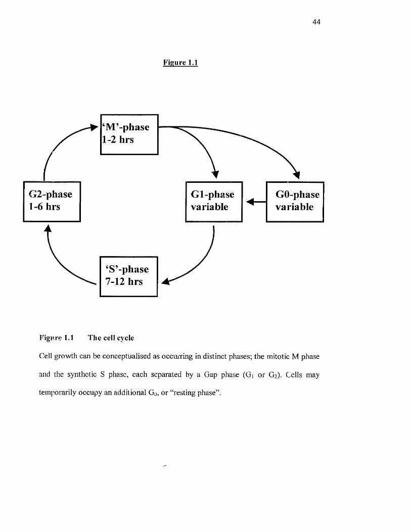
Figure 1.1 The cell cycle
Cell growth can be conceptualised as occmTing in distinct phases; the mitotic M phase
and the synthetic S phase, each separated by a Gap phase (Gi or G2). Cells may
temporarily occupy an additional Go, or “resting phase”.
45
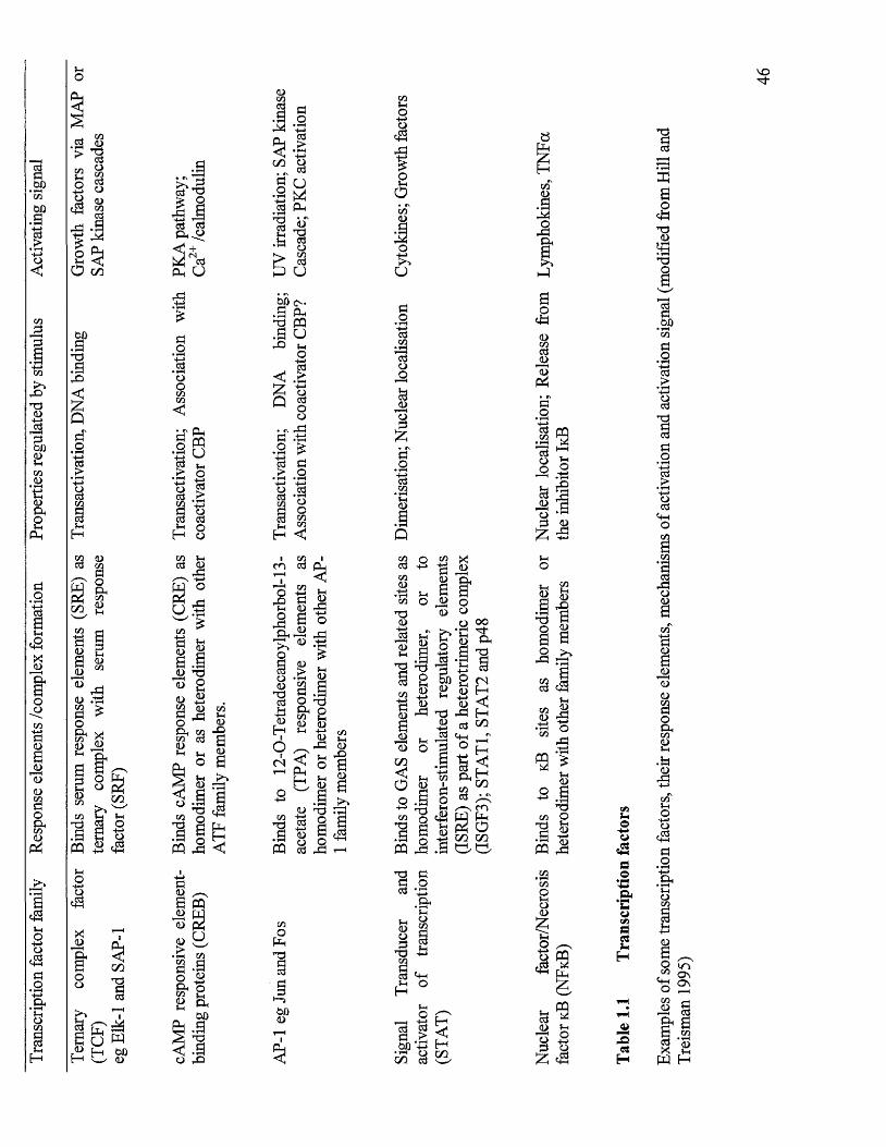
1.5.2 Transcriptional regulation o f cell cycle dependent genes.
In order for a cell to respond to extracellular signals and progress through the cell
cycle, the co-ordinate regulation of the transcription of cell cycle dependent genes is
essential. To activate or repress transcription, transcription factors must be activated
in the nucleus, bind to specific regions o f DNA (palindromic response elements) and
interact with the transcription apparatus (e.g. RNA polymerase). Regulation o f
transcription is achieved at a number o f levels:
(1) Nuclear translocation, where the location o f transcriptional factors is
determined by the activity o f either nuclear localisation signals or cytoplasmic
retention signals. For example, translocation o f NFkB to the nucleus is prevented by
its association with an inhibitory subunit, IkB, in the cytoplasm (Beg and Baldwin,
1993).
(2) Transcriptional activation, where phosphorylation and acétylation events
directly facilitate interaction with transcription machinery. For example, cAMP/Ca^^
response element binding protein (CREB) activity is dependent on regulated
phosphorylation o f Ser'^^ (Gonzales, 1991).
(3) Alteration of DNA binding properties. For example, some
phosphosphorylation events directly regulate transcription factor binding to response
elements, as is the case for serum response factor (SRF) (Rivera et al, 1993). These
mechanisms have been reviewed by Hill and Treisman (1995).
Transcriptional regulation in response to extracellular signals is both cell type and
developraentally specific. This level o f control is determined by a number of
interacting factors. Principally, the specificity o f transcriptional activation is due to
the presence o f a host o f transcription factors that specifically bind to response
elements (Table 1.1) by two mechanisms (Hill and Treisman, 1995):
'O
ICZ3
Iu
I
IIIQ
ndII
IIIb
IH <
£IaIn
Σ
cSSOh
Szn <L)
o >nd§ â
.s ^
a J
Ieg
Ia
IH
<L>M
fS"Td
nd
t g >
T )CO p
II1Iil
gH
SJDH
47
(a) Promoter activity is dependent upon the binding o f a specific set o f transcription
factors at multiple response elements immediately on the 5' side o f the promoter.
Transcription factor binding is specific for both the agonist and signal
transduction pathway activated.
(b) The activity o f certain promoters is dependent on the binding o f ternary
complexes composed o f different families o f transcription factors that, in some
cases, require the activation of a number o f different specific signalling pathways.
In addition, the level o f transcriptional activation or ‘promoter strength’ may also be
regulated by both the duration and magnitude o f signal. For example, the ability o f the
response element(s) to induce the same promoter with different activities may be
dependent on the number o f activated transcription factors bound to the response
elements, which will ultimately be dependent upon the activity o f the signalling
pathway.
1,6 Early growth factor-stimulated events
Since the initiation o f DNA synthesis is a late event in the cell cycle attention has
focused on the initial cellular responses associated with a mitogenic stimulus and their
importance in the later proliferative response. As discussed previously there is an
increasing list o f growth promoting agents acting on different types of cells that elicit
a common pleiotypic response. These initial cellular responses to mitogens include an
intrinsic rise in intracellular pH, a transient rise in cytoplasmic Ca^^, the
phosphorylation o f a common set o f proteins and an increase in c-fos and c-myc
mRNA.

48
Stimulation o f a rise in cytoplasmic pH has been reported to be a common response in
the activation o f many quiescent cell types. Growth promoting agents such as serum,
endothelin, EOF, a-thrombin, insulin, bombesin, PDGF and vasopressin have all been
shown to induce a rapid rise in pH by controlling Na^/HT exchange. The Na^/H^
exchanger is a cell membrane protein that exchanges Na^ ions for H^ ions in an
electorneural fashion. (Hesketh et al, 1985; Simonson et al, 1989; Grinstein et al,
1989).
In addition to pH changes a characteristic feature o f many growth stimuli is that they
induce increases in the intracellular free Ca^^ concentrations ([Ca^^ji). Hormone-
receptor-mediated increases in the cytosolic [Ca^^], often result from both
mobilisation of Ca^^ fi*om intracellular stores and transmembrane Ca " influx (Tsien
and Tsein, 1990; Benddge, 1993a). In individual cells, the hormone-stimulated
elevation o f intracellular Ca^^ has a complex temporal and spatial regulation, as the
rise in intracellular Ca^^ is often observed as a series of repetitive oscillations, or
spikes (Berridge and Galione, 1988; Meyer and Stryer, 1991). The spatial counterpart
o f a [Ca^^Ji spike is a wave, where an initial local [Ca^^]i elevation ultimately spreads
throughout the cell in a regenerative manner (Roony et al, 1991; Lechleiter et al,
1991). The link between hormone-receptor activation and Ca^^ release is the
intracellular messenger inositol 1, 4, 5-trisphosphate (IP3) which mobilises Ca^^ by
binding to specific intracellular receptors. These receptors also form the channels
through which stored Ca^^ is released (Taylor and Richardson ,1991; Ferris and
Snyder, 1992). In non-excitable cells the Ca^^ spikes and waves, described above, are
due to the cyclical and co-ordinated activation o f IP3 receptors (IP3R).
49
[Ca^^ji is clearly very important for the control of many essential cellular responses
and therefore the increase in [Ca^’ Ji observed with many growth factors and mitogenic
peptides may play a role in their mechanism of action.
In addition to the events in the membrane and cytosol, as described above, growth
factors rapidly and transiently induce the expression o f the cellular oncogenes c-fos
and c~myc in quiescent fibroblasts (Kelly et al, 1983). The enhanced expression of c~
fos messenger RNA (niRNA) occurs within minutes of, for example, PDGF addition
followed by increased expression of c-myc. The induction o f c-fos mRNA is one o f
the earliest nuclear events that follow the addition of PDGF. Since these cellular
oncogenes encode nuclear proteins (Abrams et al, 1982) their transient expression
may play a role in the transduction of the mitogenic signal in the nucleus.
One o f the best characterised pathways, the Ras—> Raf—> MEK~» ERK cascade
(where ERK is extracellular-signal-regulated kinase and MEK is mitogen-activated
protein (MAP) kinase (ERK) kinase) is stimulated by many mitogens and growth
factors. MAP kinase is phosphorylated by a unique dual-specificity kinase on both
tyrosine and threonine in one o f three motifs (Thr-Glu-Tyr, Thr-Phe-Tyr or Thr-Gly-
Tyr), depending on the pathway. In addition to activating one or more protein
cascades, the initiating sfimulus may also mobilise a variety o f other signalling
molecules (e.g. PKC iso forms, phosphatidylinositol-3 kinase (PI3K), phospholipid
kinases, G-protein a and subunits, phospholipases, intracellular Ca^^). These
various signals impact to a greater or lesser extent on multiple downstream effectors.
Important concepts are that signal transmission often entails the targeted relocation o f
specific proteins in the cell, and the reversible formation of protein complexes by
means o f regulated protein phosphorylation. The signalling circuits may be completed
50
by the phosphorylation o f upstream effectors by downstream kinases, resulting in a
modulation o f the signal.
1.7 Protein phosphorylation
Protein phosphorylation is a regulatory covalent modification o f a protein, in which
the phosphate group is transferred from ATP to a side chain o f the protein, catalysed
by a protein kinase. A particularly large amplification is possible if two or more
protein kinases are arranged in series - a system known as a protein kinase cascade.
There are at least two purposes o f phosphorylation cycles in cells:
1. They are the major mechanism by which extracellular signal molecules acting at
the cell surface receptors produce their ultimate intracellular effect.
2. They are the major mechanism through which events that occur discontinuously in
the cell cycle are initiated and timed.
Regulatory protein phosphorylation normally occurs on serine or threonine residues
(about two thirds) but it can also occur on tyrosine residues.
An important class o f protein kinases are activated by the binding o f second
messengers. This class includes cAMP-dependent protein kinase (protein kinase A),
cGMP-dependent protein kinase, and PKC. Another class o f protein kinases are those
activated by extracellular signals (receptor protein kinases). In these cases the protein
kinase catalytic domain is present as the cytoplasmic domain o f a transmembrane
receptor in which the external domain binds an extracellular messenger. This binding
activates the internal protein kinase, probably by causing aggregation o f receptors.
The most well-studied examples are receptors for polypeptide hormones or growth
factors in which the protein kinase is tyrosine specific, such as PDGF. However, a
few recent examples have been found where the protein kinase domain is more
51
closely related to serine/threonine kinases, for example the TGF-p receptor. As stated
previously, some protein kinases are themselves regulated by phosphorylation of
other protein kinases.
1.8 Extracellular receptors.
Cells are sensitive to a large number of chemical signals that control cell and tissue
behaviour. The ‘target cells’ for each signal are the cells that bear receptors for that
agent. Each extracellular stimulus acts at a unique receptor, but onward transmission
of information into the cells is channelled through signalling pathways built into the
plasma membrane. As a result, one signalling pathway may mediate the effects of
many extracellular agents on many intracellular responses in many target cells. The
way in which this information is processed fi'om the outside of the cell to the nucleus
is summarised in Figure 1.2.
The genome o f every cell encodes many more receptors than the number o f different
chemical signals that any individual cell ever has to recognise. A small subset o f these
genes is expressed in a differentiated cell at a particular time. This allows each cell to
respond only to the limited range o f stimuli for which it is a target, and each cell’s
receptor repertoire is constantly undergoing environmental regulation rather than
remaining static.
There are several ways in which a cell can process information from the extracellular
to the intracellular and these will be discussed below.
1.9 G - protein coupled receptors.
Some cell surface receptors signal through relay systems in which three components
act in sequence:
52
Receptor G protein -> effector protein
G proteins are guanine-nucleotide dependent coupling proteins and are the largest
most versatile class o f cell surface signal transducing proteins known. The effector
protein is usually an enzyme or an ion channel (lismaa & Shine, 1992). The receptors
that function in this way seem to be members o f a single structural family o f
polypeptides termed 7-span receptors. These ‘7-span’ receptors are embedded within
the plasma membrane and have cytoplasmic domains. G proteins are heterotrimeric
and consist o f an a subunit that binds GDP/GTP, and two intimately associated
subunits p and y. The Py subunits are common between different G proteins, and it is
mainly the a subunits that confer specificity for downstream signalling although the
Py subunits are the effectors in a few cases.
Receptor activation o f adenylate cyclase is mediated through the a subunit o f one
particular G protein, Gs- The GTPase activity o f as is such that several thousand
molecules o f cAMP are produced through activation of a single receptor. Over 20
different a subunits o f G proteins have been identified by purification or by cDNA
cloning.
G proteins are known to regulate hydrolysis o f plasma membrane phospholipids such
as phosphatidylinositides (PI) and phosphatidylcholine (PC). Gq regulates PI
hydrolysis through phospho lipase Cp (PLCp). Other G proteins are involved in the
regulation o f ion channels such as those for K" and Ca^ .
Another large family o f 7-span receptors feeds its input to the cell through a signalling
system based on the hydrolysis o f the membrane phospholipid phosphatidyl-inositol
4,5-bisphosphate (PIP2) by PLC (Berridge, 1993b). By recruiting different isozymes
of PLC, it can be activated either tlirough receptor tyrosine kinases (RTK) (via PLCy)
or by G protein coupled receptors (GPCRs).
53
Figure 1.2
Extracellular stimuli:Hormones, neurotransmitters, cytokines, etc.
Receptors
Signalling^echanisms
1
Intracellular second (and third) messengers
‘Primary’ protein kinases and phosphatases, and other directly responsive proteins.
i‘Secondary’ protein kinases and phosphatases
(on some regulatory pathways)
IRegulated enzymes/genes.
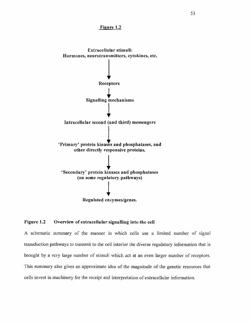
Figure 1.2 Overview of extracellular signalling into the cell
A schematic summary of the manner in which cells use a limited number o f signal
transduction pathways to transmit to the cell interior the diverse regulatory information that is
brought by a very large number of stimuli which act at an even larger number of receptors.
This summary also gives an approximate idea o f the magnitude o f the genetic resources that
cells invest in machinery for the receipt and interpretation o f extracellular information.
54
1.10 Agonist-stimulated phospholipid hydrolysis in fibroblast cells
The majority o f agonists which evoke proliferation of fibroblasts are believed to act
via stimulation o f phospholipase C (PLC) (Lee and Rhee, 1995). Stimulation o f PLC
results in the rapid breakdown o f the membrane phospholipid, phosphatidy 1 inosito 1
4.5-bisphosphate (PIP2), resulting in the generation of the second messenger inositol
1.4.5-tris phosphate (IP3) and diacylglycerol (DAG).
Following the hypothesis o f Berridge and Irvine (1984) that IP3 is the “missing link”
between the plasma membrane receptors and internal Ca^^ stores, several papers
demonstrated that IP3 acts by mobilising Ca " from intracellular pools (Berridge and
Irvine, 1989; Carsten & Miller, 1990; Coburn & Baron, 1990; Somlyo & Somlyo,
1992). It has been shown that IP3 is rapidly generated in fibroblasts and smooth
muscle cells after agonist stimulation (Berridge, 1987; Cook et al, 1990) and the
finding that IP3 can lead to release o f intracellular calcium stores are compelling
evidence that IP3 production is directly linked to the initiation o f fibroblast and
smooth muscle cell proliferation.
As noted previously, hydrolysis o f PIP2 generates IP3 and another second messenger,
DAG, which remains associated with the membrane. The principal function o f DAG
is to activate a family of plasma-membrane protein kinases collectively termed
protein kinase C (PKC). In the absence of hormone stimulation, PKC is present as a
soluble cytosolic protein that is catalytically inactive. A rise in the cytosolic Ca^^ level
causes PKC to bind to the cytoplasmic leaflet of the plasma membrane, where it can
be activated by the membrane-associated DAG. Thus activation o f certain PKC
iso forms depends on both Ca " ions and DAG, suggesting an interaction between the
two branches o f the inositol-signalling pathway.
55
The activation o f PKC in different cells results in a varied array of cellular responses,
indicating that it plays a key role in many aspects o f cellular growth and metabolism.
PKC represents a family o f related kinases, which are of central importance in
intracellular signalling pathways. At least 11 distinct isozymes o f PKC have been
identified, several of which exist in pulmonary artery fibroblasts (Pai et al, 1991).
1.11 Phospholipase D
Regulation o f phospholipase D (PLD) has received considerable attention as
hydrolysis o f the membrane phospholipid, phosphatidylcholine (PC), by this enzyme
produces phosphatidic acid, a potential second messenger in many cell types (Exton,
1994). The activity of PLD in a wide variety o f cells is influenced by many hormones,
neurotransmitters, growth factors and other agonists linked to heterotrimeric G
proteins or tyrosine kinases (Exton, 1994; Exton, 1997), The mechanisms by which G
protein-coupled receptors and receptor tyrosine kinases (RTKs) are linked to PLD are
not well understood but PKC activation appears to play a major role in many cell
types. (Elder et al, 1993; Balboa et al, 1994). Since the majority o f agonists that
promote PLD activation also stimulate PLC, a logical mechanism is that PLD
activation is secondary to PKC stimulation due to production o f DAG from PIP2
(Exton, 1997). At present, the mechanism(s) by which PKC activates PLD is
undefined, indeed the role for PLD in early mitogenic signalling is controversial, with
both supporting and conflicting evidence (Boarder, 1994).
1.12 Phospholipase Â2
The hydrolysis of PC by phospholipase A2 (PLA2) was one of the earliest
phospholipase pathways to be described. The ‘classical’ role o f PLA2 activation is to
56
generate arachidonic acid, which is the rate limiting precursor of prostaglandin and
leukotriene biosynthesis (Lark et al, 1995). It is possible that in inflammatory states
this is the major function of arachidonate, however in mitogenic signal transduction,
other pathways may be important. These alternative pathways include the activation
o f certain iso forms of PKC (Asaoka et al, 1992a) as well as PLD stimulation (Wang
et al 1992). Although PLAz is activated by many agonists linked to heterotrimeric G
proteins, there is no compelling evidence that G proteins interact directly with this
enzyme (Clark et al, 1995). RTK growth factors such as PDGF and EGF can
stimulate PLA] activity (Goldberg et al, 1990), indeed it has been demonstrated that
PLA2 can be phosphorylated and activated by MAP kinase (Lin et al, 1993). Since
growth factors stimulate MAP kinase, it is possible that both RTK-linked growth
factors and G protein-coupled receptor agonists control the activity o f PLA2 through
MAP kinase activation.
1.13 Other lipid/lipid-derived second messengers
The mechanisms discussed previously account for the major routes o f phospholipid
signalling. However, other modes o f signalling for phospholipid second messengers
are known to exist.
Arachidonic acid (apart from being a precursor for eicosanoids) and
lysophosphatidyIcholine may function as lipid second messengers through the
activation of effector enzymes such as PKC (Khan et al, 1993; Naor et al, 1988).
Lysophosphatidylcholine has been shown to have several biological actions such as
stimulation of chemotaxis and smooth muscle contraction (Asaoka et al, 1992b),
whilst arachidonic acid stimulates expression o f early growth responsive genes such
as c-fos and c-jun in VSMC (Rao et al, 1994; Rao et al, 1993).
57
The structure, regulation and function o f PI3K has been extensively reviewed by
Downes & Carter (1992) and Fry (1994). This kinase is a receptor-controlled enzyme
capable of phosphorylating the 3-OH group o f the inositol ring of PIP2, leading to the
formation of IP3. It is activated by many receptor tyrosine kinases in response to
growth factors by recruiting p85/pllO heterodimer to the membrane where they
associate via the SH2 domains in p85 with phosphorylated YXXM motifs within the
cytoplasmic receptor domain o f the receptor itself (Carpenter et al, 1993). The ability
o f GPCRs to regulate PI3K was first indicated by observations that stimulation of
platelets with thrombin resulted in the rapid accumulation o f 3-phosphorylated
phosphoinositide species (Traynor-Kaplan et al, 1988; Kazlauskas & Cooper, 1989;
Escobedo et al, 1991). PI3K has been proposed to mediate a number o f intracellular
events involved in mitogenesis such as activation o f PKC (Nakanishi et al, 1993; Liu,
1996), stimulation o f a ribosomal kinase (Lane et al, 1993) and activation o f the MAP
kinase pathway (Rodriguez-Viciana et al, 1994).
1.14 Protein kinase C
1.14.1 Protein kinase C structure
The effects o f phospholipid-derived DAG are mediated through the activation of
PKC. However, PKC exists as a growing family of serine/threonine protein kinase
iso forms that are differentially expressed in various cells and subcellular localities and
can be subdivided into three groups based on their structural and bio-^hemical
properties. Members of the PKC family so far examined are dependent on
phosphatidyIserine, but show clearly different requirements o f Ca^^ and phospholipid
metabolites for their activation. The classical or conventional PKC isoforms (cPKC)
58
include, [31, [311 and y are regulated by Ca^^ and DAG. The novel or non-classical
isoforms (nPKC) ô, 8 , q and 0 are insensitive to Ca^^ as they lack the Ca^^ -binding
C2 domain although they respond to DAG. The atypical PKC subgroup (aPKC) X and
Ç isoforms are insensitive to Ca^^ and DAG or phorbol ester (Reviewed by Musashi
et al, 2 0 0 0 ).
The specific regulatory requirements o f the different members of the PKC family
produce distinct PKC activation patterns with respect to the subspecies o f PKC that
becomes activated, the extent and duration o f the response and perhaps their
intracellular localisation. In particular, a number of reports have demonstrated
differential functions o f DAG derived from PIP2 and PC in the activation of PKC. In
IIC9 fibroblasts, translocation o f the Ca^^ -dependent PKCa isoform from the cytosol
to the plasma membrane by thrombin, regarded as a parameter o f enzyme activation,
parallels the initial peak in DAG formed from phosphoinositide, but not from PC
hydrolysis (Leach et al, 1991; Ha and Exton, 1993). Conversely, translocation o f the
Ca^” -independent isoform PKCe, also occured rapidly but was maintained during the
persistant phase o f DAG production derived from PC (Ha and Exton, 1993). The
presence of Ca^^ accompanying the initial DAG peak, rather than different molecular
species o f DAG derived from PIP2 and PC, vaiying in their fatty acid composition,
accounted for the differential activation o f the PKC iso forms (Ha and Exton, 1993).
1.14.2 Role o f PKC in mitogenic signal transduction.
PKC has been implicated as a major component of cellular signalling networks
regulating a variety o f cell functions including cell growth and differentiation
(reviewed by Nishizuka, 1992; Dempsey et al, 1997). The role of PKC in mitogenesis
has received much attention ever since the discovery that the enzyme was the major
59
receptor for tumour promoters o f the phorbol ester family, cell permeable analogues
of DAG. Exogenous application o f phorbol esters has been reported to mimic the
effect of growth factors, and down regulation o f PKC by prolonged PMA pre
treatment has been shown to inhibit the mitogenic effects of a number o f agonists in
different cell types. While the tissue-specific distribution and distinct modes of
activation suggests functional diversity o f the PKC family members, information on
their specific biological roles including the control o f proliferation, however, is still
limited as few o f the physiological substrates o f PKC have been identified, and of
those that have, no clear difference in substrate specificity among the members has
been demonstrated. Furthermore, phorbol esters are not selective in the PKC species
that they activate or downregulate, potentially exerting pleiotropic effects. The lack of
specific inhibitors has prevented the dissection o f responses attributable to particular
iso forms. Despite this, studies in which distinct PKC isoenzymes have been
overexpressed have revealed that PKC (31 and s exhibit transforming characteristics
(Housey et al, 1988). Other studies using similar approaches complimented by data
with a selective pseudo substrate peptide inhibitor indicated that the atypical PKC
iso form Ç is involved in mitogenic signalling (Berra et al, 1993). The precise role of
PKC in regulating cellular proliferation has not been clarified. However, PKC can
either directly or indirectly control the expression of certain genes by inducing
transcriptional activation through TRE, serum-response element (SRE) (Hata et al,
1993) and NFkB (Hirano et al, 1995) -responsive promoters in an isoenzyme specific
manner. These effects may be mediated by the integration o f PKC in growth factor-
initiated phosphorylation cascades, by phosphorylating and activating kinases that are
intrinsic components o f such pathways, including c-Raf-1 o f the MAP kinase cascade
60
(Chapter 1.16.3; Sozeri et al, 1992; Kolch et al, 1993) and elements upstream of the
p7 0 s6k (Susa et al, 1992).
1,15 Receptors with intrinsic tyrosine kinase activity
The role o f tyrosine kinase-linked signal transduction pathways has been examined
largely in the context o f long-term responses such as cell proliferation. RTKs form a
large and important class o f cell surface receptors. Several sub-classes o f RTK exist,
but all share a similar molecular structure consisting o f a large extracellular ligand-
binding region, a single hydrophobic transmembrane segment, a cytoplasmic portion
containing the tyrosine kinase catalytic domain and lastly, a carboxy-terminal
regulatory region (see Cardenn & Gill, 1992; Panettieri, 1996). Activation of the RTK
is necessary for transduction o f the growth factor-mediated response. Once a receptor
is phosphorylated, its kinase site opens to provide access for the phosphorylation by
other proteins (Cardenn & Gill, 1992). The tyrosine phosphorylated receptors and
other tyrosine-phosphorylated proteins then associate with a number o f target proteins
that transfer to the cell interior the signals received by the receptors. These proteins
that associate with some or all activated RTKs include:
1 RasGAP, a GTPase-activating protein which modulates the activity o f the Ras
proteins (a trio o f small GTP-dependent switch proteins encoded by c-ras proto
oncogenes).
2 R a f 1, a serine-threonine protein kinase encoded by the c-rq/ proto-oncogene.
3 The cytoplasmic protein tyrosine kinase Src which is encoded by the c-src proto
oncogene.
61
4 The phospholipase Cy (PLCy) and PI3K, which hydrolyse and phosphorylate
inositol-containing phospholipids.
5 She protein, the product o f the she gene (Pazin & Williams, 1992).
O f these, RasGAP, c-src, PLCy, the Mr regulatory subunit o f PI3K, and She all
contain one or more copies o f a shared polypeptide sequence motif, known as the Src
homology domain type 2 (SH2 domain).
Substrates with SH2 domains are responsible for coupling activated growth factor
receptors to intracellular pathways involved in the control o f a variety of cellular
functions such as proliferation and gene expression. Proteins with one or more SH2
domains include PLC, PI3K (PI 3-kinase), Ras and other cytoplasmic kinases
(Carpenter, 1992). Many o f the proteins that have SH2 domains also contain a distinct
sequence o f approximately 50 amino acid residues termed the SH3 domain. SH3
domains modulate protein-protein interactions through the recognition o f short
peptide sequences that do not require phosphorylation. Signalling complexes based on
the formation of SH2/SH3 interactions are then followed by activation o f downstream
effector proteins which involve, for example, non-receptor tyrosine kinases and MAP
kinases (Satoh & Kaziro, 1992; L’Alleman et cil, 1991; Meloche et al, i992) (Figure
1.3).
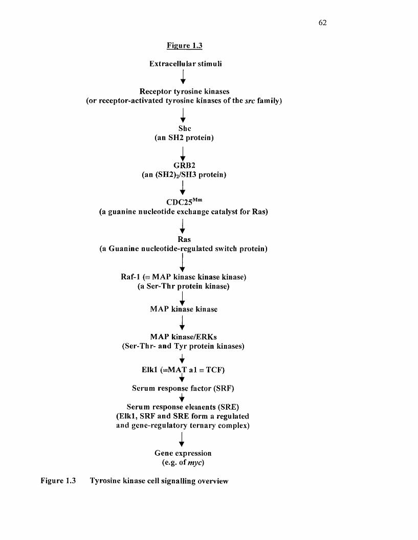
62
Figure 1.3
Extracellular stimuli
IReceptor tyrosine kinases
(or receptor-activated tyrosine kinases of the src family)
iShe
(an SH2 protein)
IGRB2
(an (SH2)2/SH3 protein)
iCDC25'**"'
(a guanine nucleotide exchange catalyst for Ras)
IRas
(a Guanine nucleotide-regulated switch protein)
1Raf-1 (= MAP kinase kinase kinase)
(a Ser-Thr protein kinase)
MAP kinase kinase
IMAP kinase/ERKs
(Ser-Thr- and Tyr protein kinases)
iElkl (= M A T al = TCF)
Serum response factor (SRF)
Serum response elements (SRE) (E lkl, SRF and SRE form a regulated and gene-regulatory ternary complex)
iGene expression
(e.g. of myc)
Figure 1.3 Tyrosine kinase cell signalling overview
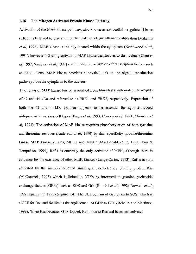
63
1.16 The Mitogen Activated Protein Kinase Pathway
Activation o f the MAP kinase pathway, also known as extraeellular regulated kinase
(ERK), is believed to play an important role in cell growth and proliferation (Milanini
et al, 1998). MAP kinase is initially located within the cytoplasm (Northwood et al,
1991), however following activation, MAP kinase translocates to the nucleus (Chen et
al, 1992; Sanghera et al, 1992) and initiates the activation o f transcription factors such
as Elk-1. Thus, MAP kinase provides a physical link in the signal transduction
pathway from the cytoplasm to the nucleus.
Two forms of MAP kinase has been purified from fibroblasts with molecular weights
of 42 and 44 kDa and referred to as ERKl and ERK2, respectively. Expression o f
both the 42 and 44-kDa iso forms appears to be essential for agonist-induced
mitogenesis in various cell types (Pages et al, 1993; Cowley et al, 1994; Mansour et
al, 1994). The activation of MAP kinase requires phosphorylation o f both tyrosine
and tlueonine residues (Anderson et al, 1990) by dual speeifieity tyrosine/threonine
kinase MAP kinase kinases, MEKl and MEK2 (MaeDonald et al, 1993; Yan &
Tempelton, 1994). Raf-1 is currently the only activator of MEK, although there is
evidence for the existence o f other MEK kinases (Lange-Carter, 1993). Raf is in turn
activated by the membrane-bound small guanine-nucleotide binding protein Ras
(McCormick, 1993) which is linked to RTKs by intermediate guanine nucleotide
exchange factors (GEFs) such as SOS and Grb (Bonfini et al, 1992; Bowtell et al,
1992; Egan et al, 1993) (Figure 1.4). The SH3 domain o f Grb binds to SOS, which is
a GEF for Ras and facilitates the replacement o f GDP to GTP (Rebello and Martinez,
1999). When Ras becomes GTP-loaded, Raf binds to Ras and becomes activated.
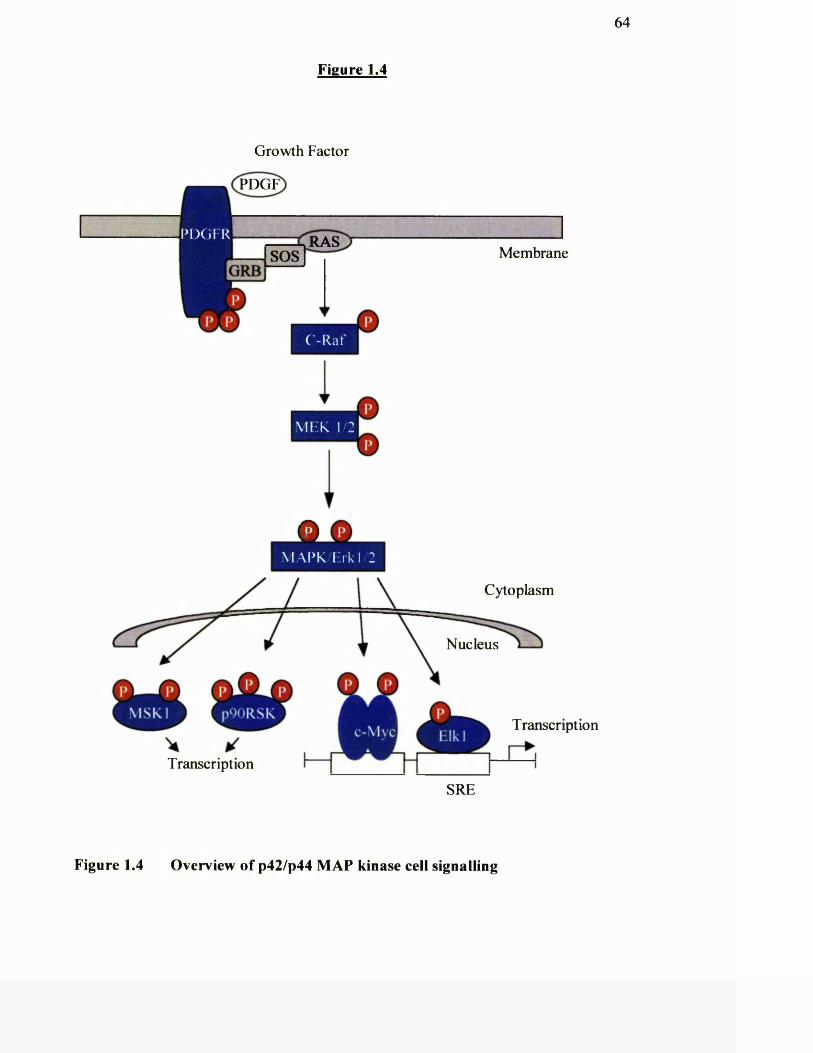
64
Figure 1.4
Growth Factor
PDGF
RASMembraneSOS
C-Raf
M F K 1/2
P D G F R
Cytoplasm
Q Q
/7 Nucleus
Transcription
Transcription
SRE
Figure 1.4 Overview of p42/p44 MAP kinase cell signalling
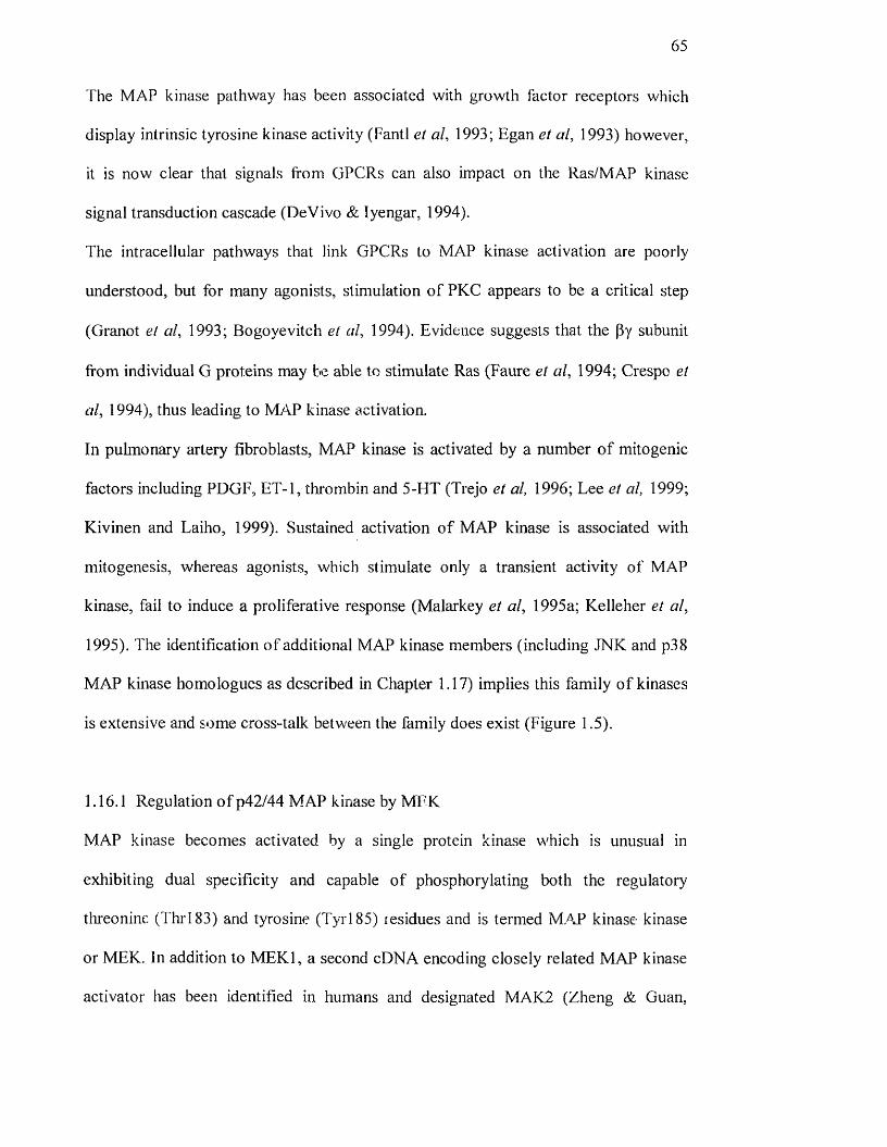
65
The MAP kinase pathway has been associated with growth factor receptors which
display intrinsic tyrosine kinase activity (FantI et al, 1993; Egan et al, 1993) however,
it is now clear that signals from GPCRs can also impact on the Ras/MAP kinase
signal transduction cascade (DeVivo & Iyengar, 1994).
The intracellular pathways that link GPCRs to MAP kinase activation are poorly
understood, but for many agonists, stimulation o f PKC appears to be a critical step
(Granot et al, 1993; Bogoyevitch et al, 1994). Evidence suggests that the (3y subunit
from individual G proteins may be able to stimulate Ras (Faure et al, 1994; Crespo et
al, 1994), thus leading to MAP kinase activation.
In pulmonary artery fibroblasts, MAP kinase is activated by a number o f mitogenic
factors including PDGF, ET-1, thrombin and 5-HT (Trejo et al, 1996; Lee et al, 1999;
Kivinen and Laiho, 1999). Sustained activation o f MAP kinase is associated with
mitogenesis, whereas agonists, which stimulate only a transient activity o f MAP
kinase, fail to induce a proliferative response (Malarkey et al, 1995a; Kelleher et al,
1995). The identification of additional MAP kinase members (including JNK and p38
MAP kinase homologues as described in Chapter 1.17) implies this family o f kinases
is extensive and some cross-talk between the family does exist (Figure 1.5).
1.16.1 Regulation o f p42/44 MAP kinase by MFK
MAP kinase becomes activated by a single protein kinase which is unusual in
exhibiting dual specificity and capable of phosphorylating both the regulatory
threonine (Thrl83) and tyrosine (Tyrl 85) residues and is termed M.AP kinase kinase
or MEK. In addition to M EKl, a second cDNA encoding closely related MAP kinase
activator has been identified in humans and designated MAK2 (Zheng & Guan,


T3
\o 1A'Qh
8I
I
IÎII
P hcuo
IP
II
I
VO
a90
pLi 'BAD m
g
12
Ja0
1I
II
li
Io
a
II
I
fe g
:i ' 2
SÎ
II
68
1993). Overexpression o f MEK in cultured cells increases MAP kinase activity (Seger
el al, 1992)
1.16.2 Regulation o f MEK by Raf-1
A 74 kDa serine/threonine protein kinase known to be involved in mitogenic
signalling is c-Raf-1 (Morrison et al, 1988), A vast amount of evidence has shown
that c-Raf-1 is at least one o f the intermediates directly upstream o f MEK in a linear
cascade leading to activation o f MAP kinase (Dent et al, 1992; Howe et al, 1992;
Kyriakis et al, 1993). Cells transformed with c-Raf, or overexpression o f oncogenic
mutants o f c-Raf-1, display elevated MEK and MAP kinase activities (Dent et al,
1992). Furthermore, c-Raf-1 immunoprecipitated from mitogen-stimulated cells can
directly phosphorylate the active MEK in vitro (Dent et al, 1992; Howe et al, 1992;
Kyriakis et al, 1993). Other studies have demonstrated that Raf-1 forms a stable
complex with MEKl in vivo and in vitro (Huang et al, 1993) and phosphorylates
MEKl exclusively on serine residues (Yan and Tempelton, 1994, Zheng and Guan,
1994).
1.16.3 Regulation o f MEK independent o f Raf-1
It has been demonstrated that other serine/threonine kinases can serve as direct
regulators o f MEK, which include other members o f the R af family o f proto
oncogene products. B-Raf has been identified as an activator o f MEK in NIH3T3
fibroblasts (Catling et al, 1994) and A-Raf in Hela cells has been shown to activate
MEKl but not MEK2 (Wu et al, 1996).
Other kinases that activate MEK include MEKl kinase (MEKKl). Initially it was
thought that MEKK may be responsible for MEK activation by heterotrimeric GPCR
69
agonists, however, recent evidence suggests that this kinase is more likely to act
upstream of another MEK family member responsible for activating the MAP kinase
homologue c-jun N-terminal kinase in the parallel stress-activated protein kinase
pathway (Chapter 1.17) (Minden et al, 1994 and Yan et al, 1994).
1.16.4 Raf-1 activation by Ras
The proto-oncogene gene product Ras plays a central role in signal transduction
pathways involved in cellular processes including cellular proliferation (Mulcahy et
al, 1986). Ras becomes active then GTP binding takes the place o f the inactive state
when GDP is bound. This can be stimulated by the addition o f growth factors. The
mechanism by which Ras aids in the activation o f Raf-1 is by causing c-Raf-1
translocation to the plasma membrane where additional protein-protein or protein-
lipid interactions regulate Raf-1 activity (Chapter 1.16 and Reuter et al, 2000).
1.16.5 Ras-independent activation of MAP kinase
It has been well documented in the literature that phorbol esters stimulate MAP kinase
activation. Furthermore, depletion of PKC by prolonged pre-treatment with phorbol
esters has been shown to block the activation o f MAP kinases following stimulation
with agonists (de Vries-Smits et al, 1992). This indicates the existence of at least two
distinct pathways regulating MAP kinase that can operate in one cell type and also
implicates PKC in this role. PKC is thought to activate the MAP kinase pathway at
the level o f Raf-1 since chronic phorbol ester pre-treatment prevents agonist-
stimulated c-R af 1 activation (Kolch et al, 1993).
70
1,16.6 Cellular targets of p42 and p44 MAP kinase
Downstream o f MAP kinase, several proteins have been identified as substrates.
These include lipases, transcription factors and other protein kinases (reviewed by
Reuter et al, 2000). MAP kinase can activate the dephosphorylated 90 kDa ribosomal
protein S6 kinase II (p90' ' ) (Sturgill et al, 1988). It can also directly regulate gene
expression by phosphorylating the ternary complex transcription factor EIk-l/p62^‘'^
(Gille et al, 1992) which in turn facilitates the transcription o f c-fos', one of the
immediate early genes induced by many stimuli. MAP kinase also phosphorylates and
activates the cytosolic PLA2 (PLA2) in vitro and in intact cells (Lin et al, 1993). A
role for PLA2 as a substrate of MAP kinase in vivo is supported by the finding that
expression o f a recombinant CPLA2 in which the MAP kinase phosphorylation site
Ser505 is mutated, does not become activated on agonist-stimulation (Lin et al, 1993).
1.17 Stress-Activated Protein Kinases
The last few years have marked the arrival o f novel members o f what now appears to
be a MAP kinase superfamily. Two protein kinases that are highly related to the
classical p42/p44 MAP kinases have been identified, and characterised as integral
components o f parallel protein kinase cascades activated by pro inflammatory
cytokines and cellular stresses. It has now become apparent that these MAP kinase
homologues may play a role in the cellular effects of an increasingly large number o f
extracellular stimuli including GPCR agonists (Coso et al, 1995)
As their name suggests, stress-activated protein kinases (SAPK) are activated by
agents which induce cellular stress, such as hypoxia, UV light and the cytokines,
tumour necrosis factor a and interleukin-1 (Scott et al, 1997; Kolesnick & Goldie,
1994). An investigation of kinases responsible for phosphorylating the transcription
71
factor c-Jun on Ser^^ and Ser^ within its amino terminal activation domain in cells
exposed to UV irradiation identified that this activity was attributable to two protein
kinases o f 46 and 55 kDa named c-Jun N-terminal kinases 1 and 2 (JNKl and JNK2),
respectively (Hibi et al, 1993). Subsequent cloning o f JNKl revealed that this protein
kinase shared sequence homology to the p42/p44 MAP kinases (Figure 1.6; Derijard
et al, 1994).
In addition to JNKs, a stress-activated MAP kinase homologue has also been
identified, termed p38, that led to the activation o f M /PK A P kinase-2, a serine kinase
responsible for phosphorylating one o f a family o f small heat shock proteins (Hsp 25)
in response to chemical stress or heat shock (Figure 1.7; Rouse et al, 1994). These are
thought to exist in signal transduction cascades distinct from MAP kinase. There is
evidence, however, for the existence o f cross-talk between the two pathways since
RTK-linked growth factors (Minden et al, 1994) and GPCR agonists (Dalton &
Treisman, 1992) can also activate the SAPK. In recent studies SAPKs were shown to
be upregulated in cells from the pulmonary vasculature exposed to hypoxia (Welsh et
al 1998; Stenmark et al 1999).
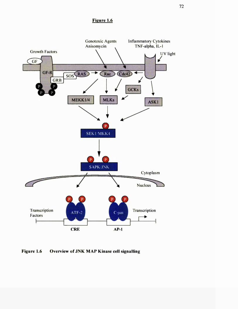
72
Growth Factors
Figure 1.6
Genotoxic Agents Inflammatory Cytokines Anisomycin TNF-alpha, lL-1
GRB ----------
v rMEKKl/4
Rac J CCdc42.
1 / /
/UV light
GCKs
MEKKl/4 MLKs
X iASK I
OS E K 1 M K K 4
TranscriptionFactors
O OSAPK/ J NK
Cytoplasm
Nucleus
Transcription
CRE A PI
Figure 1.6 Overview o f JNK M AP Kinase cell signalling
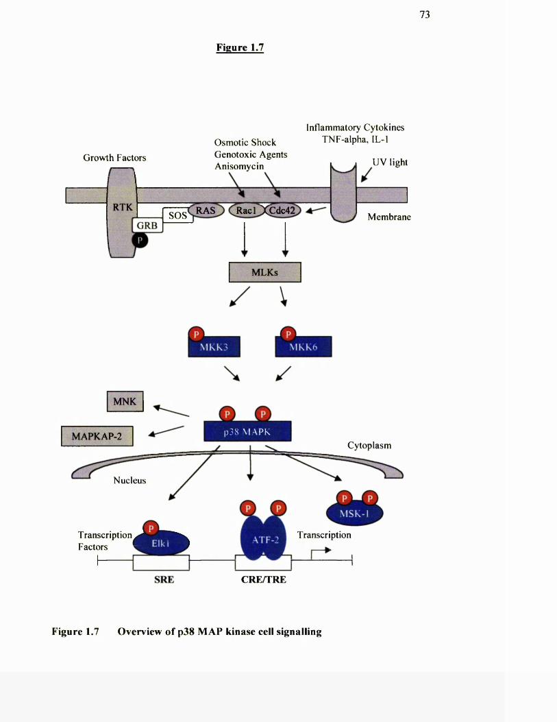
73
Figure 1.7
Inflammatory Cytokines Osmotic Shock TNF-alpha, IL-1Genotoxic Agents Anisomycin
Growth Factors UV light
RTK Racl JUCdc42RASSOS M embrane
MLKs
MNK
MAPKAP-2 p 3 8 M A R K
Cytoplasm
Nucleus
Transcription
SRE CRE/TRE
Figure 1.7 Overview o f p38 MAP kinase cell signalling
74
1.17.1 MKK homologues involved in JNK and p38 activation.
The pathways leading to the activation o f the SAPKs are not as well understood as
those of the classical p42/p44 MAP kinases. However, like the p42/p44 pathway, the
SAPK pathways consists o f several protein kinases acting in sequence; a MAP kinase
homologue (or SAPK/JNK/p38), MAP kinase kinase homologue (or
MKK/MEK/SEK/JNKK) and a MAP kinase kinase kinase (or MEKK). Again, like
MAP kinase, the activation o f the SAPKs relies on their phosphorylation at specific
dual phosphorylation motifs, although in this case the sequences are defined by TPY
for JNK (Derijard et al, 1994) and TGY for p38 MAP kinase (Rouse et al, 1994).
These residues are specifically phosphoryiated by MKK/MEK homologues (also
termed SAPK kinases (SAPKK)) distinct from MKK/MEKs 1 and 2 that are
responsible for the activation o f the classical p42/p44 MAP kinase iso forms. Another
MAP kinase kinase identified in humans and designated MKK3, has been shown to
selectively activate p38 MAP kinase (Derijard et al, 1995).
1.17.2 Activation o f MKK homologues by MEKKs
Activation o f the relevant MKK/MEK/SAPKK activity requires its phosphorylation
by an upstream kinase analogous to the Raf kinase isoforms that function upstream in
the classical p42/p44 M.AP kinase pathway. The novel serine/threonine kinase
M EKKl, which was originally identified as a kinase able to phosphorylate MEK-1 in
vitro (Lange-Carter et al, 1993), has been subsequently shown to be a more efficient
activator o f .MEKK/>4EK4/SEK-1/JNKK in in vitro and co-infection studies (Yan et
al, 1994). In this role, MEKKl, but not Raf-1 was also found to participate in both
growth factor and TNF-stimulated activation o f JNK.
75
The identity of the equivalent MEKK-like protein upstream of MKK3/6 in the p38
pathway is less well understood. However, TAK-1 which has been isolated from
TGF-p-activated kinase and which has been related to MEKK (Yamaguchi et al,
1995) has been shown to directly activate both MKK6 and MKK3 in vitro. Thus,
activation o f p38 MAP kinase may occur via a kinase pathway involving TAK-Î,
MEKK 3/6 and p38 MAP kinase.
1.17.3 Possible role o f p38 and JNK activation in cell growth
There have been a number o f papers which link p38 MAP kinase and JNK :o
apoptosis and cell cycle arrest (Verheij et al, 1996; Xia et al, 1995). However
evidence suggests that SAPKs may have a positive role to play in cell growth (Auer et
al, 1998). Clerk and colleagues (1998) found that ET-1 and phenyieplii'ine, both
mitogens, stimulated p38 MAP kinase phosphorylation 12-fold. It has also been
shown that tlu'ombin stimulation o f JNK in airway smooth muscle cells correlates
with DNA synthesis (Shapiro et al, 1996). It is also true that these SAPK have the
ability to activate transcription factors, which regulate genes that are induced on
mitogenic stimulation.
1.18 Hypothesis
Characterisation o f the cellular signalling events that occur during cell proliferation
by hypoxia is an important issue from both biological and clinical perspectives. From
a biological view, this will further our understanding o f the processes by which
hypoxia mediates cellular effects during proliferation. From a clinical view, this will
provide a model for the pathways that operate in response to acute and chronic
hypoxia, such as during the pathogenesis o f pulmonary hypertension characterised by
76
blood vessel cell wall remodelling. In addition, intracellular targets may be identified
for the development o f novel pharmaceuticals to alleviate such disease.
The review of the literature indicates that while hypoxia causes proliferation of a
number o f cell types, the mechanisms by which this is achieved is unknown. It is
possible that clues to finding more about these mechanisms may lie in the differences
seen between pulmonary arteries, which constrict to hypoxia, and systemic arteries
which dilate in response to hypoxia in vivo. It is not known whether isolated
pulmonary artery fibroblast cells would proliferate in vitro to cellular stresses such as
hypoxia and if so whether these cells may differ with regards to the potency of
agonists, activity o f intracellular pathways as well as growth characteristics to
hypoxia. It is also unknown whether hypoxia would have a differential effect on
fibroblast cells isolated from the systemic circulations. Furthermore, there is very
little information on the involvement o f the MAP kinase signalling pathway or the
SAPKs in hypoxia-stimulated mitogenesis o f pulmonary and systemic artery
fibroblasts.
Cunently defined signalling phenomena associated with stimulation o f cell growth
have been exposed by studies, which have routinely used immortalised cell lines and
therefore their growth regulatory mechanisms have been fundamentally altered. This
may be problematic particularly when examining pathways involved in proliferation.
Caution must therefore be taken in predicting how cells will behave physiologically
by extrapolating from signalling events characterised in these cells. The investigations
in this thesis have been carried out in fibroblasts obtained from primary cultures. We
believe that these represent a better model of mammalian cells under physiological or
pathophysiological conditions.
77
We have hypothesised that hypoxia may cause increased proliferation o f pulmonary
artery fibroblast cells by an intracellular mechanism(s) present in the pulmonary
vasculature which is not present in systemic fibroblasts.
1.19 Aims
The aims of this thesis were as follows:
1 To establish a cell model for studying proliferation o f fibroblasts from the
pulmonary and systemic arteries in response to acute hypoxia.
2 To study acute hypoxia in mitogenic proliferation of bovine pulmonary artery
and bovine mesenteric artery fibroblasts.
3 To look at early signalling events involved in acute hypoxic proliferation o f
pulmonary and mesenteric artery fibroblasts.
4 To study the effects o f acute hypoxia on classical and stress-activated protein
kinases in bovine pulmonary artery fibroblasts.
We also aimed to compare the effects o f acute hypoxia on fibroblasts with fibroblasts
obtained from rats exposed to chronic hypoxia by:
5 Studying the proliferation and generation o f MAP kinases in pulmonary and
aortic fibroblasts harvested from chronically hypoxic rats.
6 Examining the effects o f 5-HT on proliferation and generation o f MAP
kinases in pulmonary aitery fibroblasts harvested from chronically hypoxic
rats.

78
Chapter 2
Materials & Methods
79
2. MATERIALS & METHODS
2.1 Materials
Ail general chemicals were o f Analar grade and were supplied by Merck
(Thornliebank, Glasgow) or Sigma (Poole, Dorset) unless otherwise stated. All tissue
culture plastics were from Greiner Labortechnick Ltd (Gloucestershire.UK). All
components o f tissue culture medium were purchased from Gibco Life Technologies
(Paisley, Scotland). [y-^^P]ATP and [^H]thymidine were from Amersham (Little
Chalfont, Bucks). Antibodies used for Western Blot analysis were from New England
Biolabs (Hertfordshire, UK). Gel electrophoresis equipment was purchased from Bio-
Rad Laboratories (Herts, UK). Incubators were supplied by LEEC (Nottingham, UK).
1 would like to thank Dr R. Plevin (University o f Strathclyde) for the gift o f GST-c-
jun and GST-MAPKAP kinase 2 used as substrates for measuring activity o f JNK and
p38 MAP kinase.
2.2 Animal Models
2.2.1 Bovine
Cells derived from bovine pulmcmary and mesenteric arteries were used. Tissue was
obtained fresh on the day o f experimentation from a local abattoir (Sandyford,
Paisley). Lungs were removed from fieshly slaughtered cattle and transported to the
laboratory in a container filled with chilled Krebs-Henseleit Solution (NaCi 118mM,
NaHCOs 25mM, KCl 4.7mM, KH2PO4 1.2mM, MgS0 4 1.2mM, C aC f 2.5mM, and
Glucose 1 ImM).
80
2.2.2 Rat
Cells derived from the pulmonary artery and aorta of adult Wistar rats from the “In
House” breeding stock o f the University o f Glasgow’s Central Animal Facility were
also used. Thirty to thirty-three day old rats were maintained in either normoxic
conditions or hypoxic conditions in a hypobaric chamber (P02= 70mmHg) for 2
weeks (Chapter 2.5). Animals were maintained on a 12 hour (h) light/dark cycle and
allowed free access to standard diet and water.
2.3 Primary Cell Culture
Fibroblasts used throughout these studies were obtained from primary culture by
explant using tissues derived either from bovine or rat.
Since the conditions (warm, humid and nutrient rich) necessary for culturing the cells
are an ideal environment for promoting fungal and bacterial growth, extreme caution
must be taken to avoid contamination. All steps such as making up solutions,
changing media, etc were conducted under sterile conditions, that is, within a clean.
Micro flow laminar flow hood (model number M25121/1) (Figure 2.1). The flow hood
was dismantled and cleaned regularly and before use each day was sprayed liberally
with 70% (v/v) ethanol. Anything taken inside the flow hood ^.e. pipettes and reagent
bottles) was also sprayed with ethanol and sterile gloves were worn throughout.
Pipette tips and distilled water were sterilised using a prestige Medical “Omega”
autoclave (model number 220140).
2,3.1 Routine Cell Maintenance
Cells were grown routinely in 75cm^ culture flasks in Dulbeccos modification of
Eagles medium (DMEM), supplemented with penicillin (200 units/ml) and
streptomycin (200 ug/ml), L-glutamine (27 mg/ml) and 10% foetal calf serum (FCS).
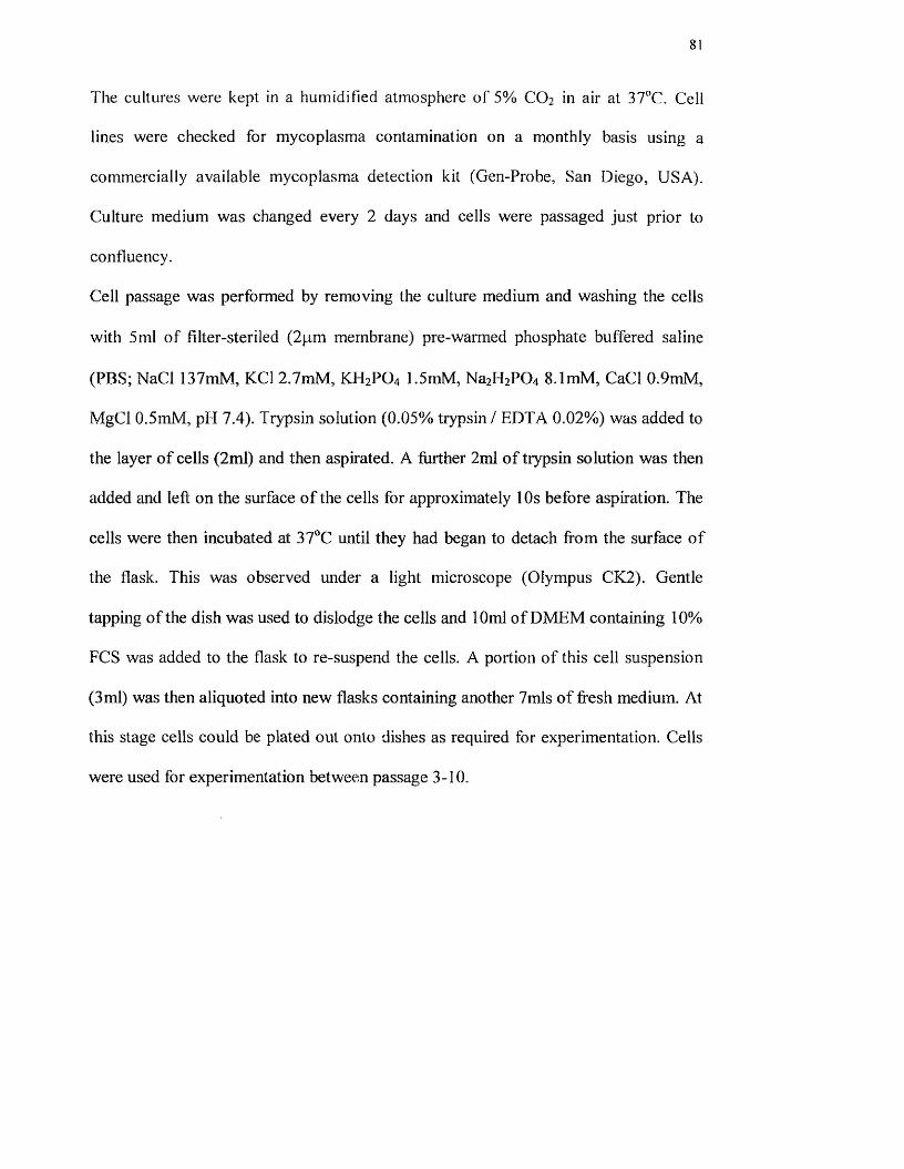
The cultures were kept in a humidified atmosphere of 5% CO2 in air at 37”C. Cell
lines were checked for mycoplasma contamination on a monthly basis using a
commercially available mycoplasma detection kit (Gen-Probe, San Diego, USA).
Culture medium was changed every 2 days and cells were passaged just prior to
confluency.
Cell passage was performed by removing the culture medium and washing the cells
with 5ml of filter-steriled (2pm membrane) pre-warmed phosphate buffered saline
(PBS; NaCl 137mM, KCl 2.7mM, KH2PO4 1.5mM, Na2H2P0 4 8Jm M , CaCl 0.9mM,
MgCl 0.5mM, pH 7.4). Trypsin solution (0.05% trypsin / EDTA 0,02%) was added to
the layer o f cells (2ml) and then aspir ated. A further 2ml o f trypsin solution was then
added and left on the surface o f the cells for approximately 10s before aspiration. The
cells were then incubated at 37^3 until they had began to detach from the surface o f
the flask. This was observed under a light microscope (Olympus CK2). Gentle
tapping o f the dish was used to dislodge the cells and 10ml o f DMEM containing 10%
FCS was added to the flask to re-suspend the cells. A portion o f this cell suspension
(3 ml) was then aliquoted into new flasks containing another 7mls o f fresh medium. At
this stage cells could be plated out onto dishes as required for experimentation. Cells
were used for experimentation between passage 3-10.
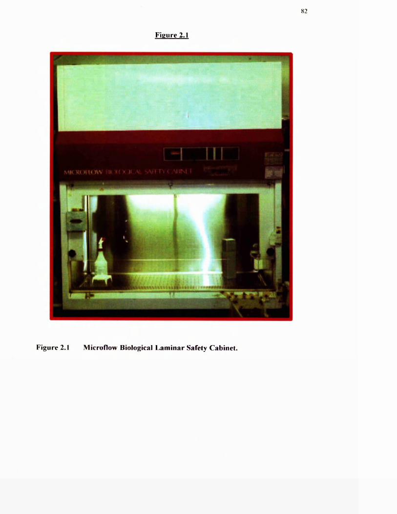
82
Figure 2.1
Figure 2.1 Microflow Biological Laminar Safety Cabinet.
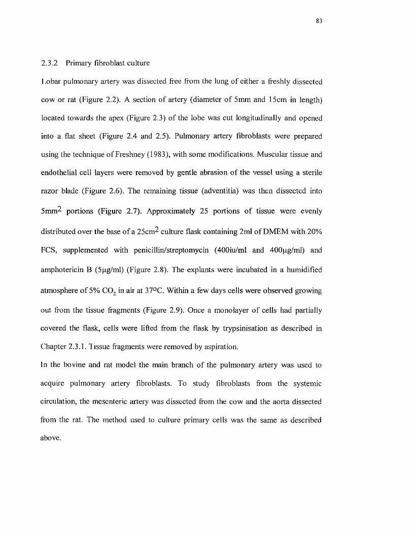
83
2.3.2 Primary fibroblast culture
Lobar pulmonary artery was dissected free from the lung o f either a freshly dissected
cow or rat (Figure 2.2). A section o f artery (diameter o f 5mm and 15cm in length)
located towards the apex (Figure 2.3) of the lobe was cut longitudinally and opened
into a flat sheet (Figure 2.4 and 2.5). Pulmonary artery fibroblasts were prepared
using the technique o f Freshney (1983), with some modifications. Muscular tissue and
endothelial cell layers were removed by gentle abrasion o f the vessel using a sterile
razor blade (Figure 2.6). The remaining tissue (adventitia) was then dissected into
5mm^ portions (Figure 2.7). Approximately 25 portions o f tissue were evenly
distributed over the base o f a 25cm^ culture flask containing 2ml of DMEM with 20%
FCS, supplemented with penicillin/streptomycin (400iu/ml and 400pg/ml) and
amphotericin B (5pg/ml) (Figure 2.8). The explants were incubated in a humidified
atmosphere o f 5% CO^ in air at 37^C. Within a few days cells were observed growing
out from the tissue fragments (Figure 2.9). Once a monolayer o f cells had partially
covered the flask, cells were lifted from the flask by trypsinisation as described in
Chapter 2.3.1. Tissue fragments were removed by aspiration.
In the bovine and rat model the main branch o f the pulmonary artery was used to
acquire pulmonary aitery fibroblasts. To study fibroblasts from the systemic
circulation, the mesenteric artery was dissected from the cow and the aorta dissected
from the rat. The method used to culture primary cells was the same as described
above.
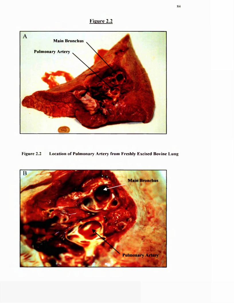
Figure 2.2
84
Main Bronchus
Pulmonary Artery
Figure 2.2 Location of Pulmonary Artery from Freshly Excised Bovine Lung
Main Bronchus
Pulmonary
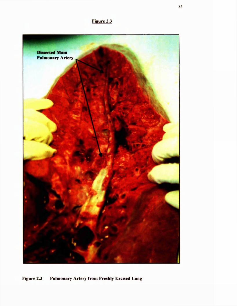
85
Figure 2.3
Dissected Mam Pulmonary Artery
T ,
Figure 2.3 Pulmonary Artery from Freshly Excised Lung
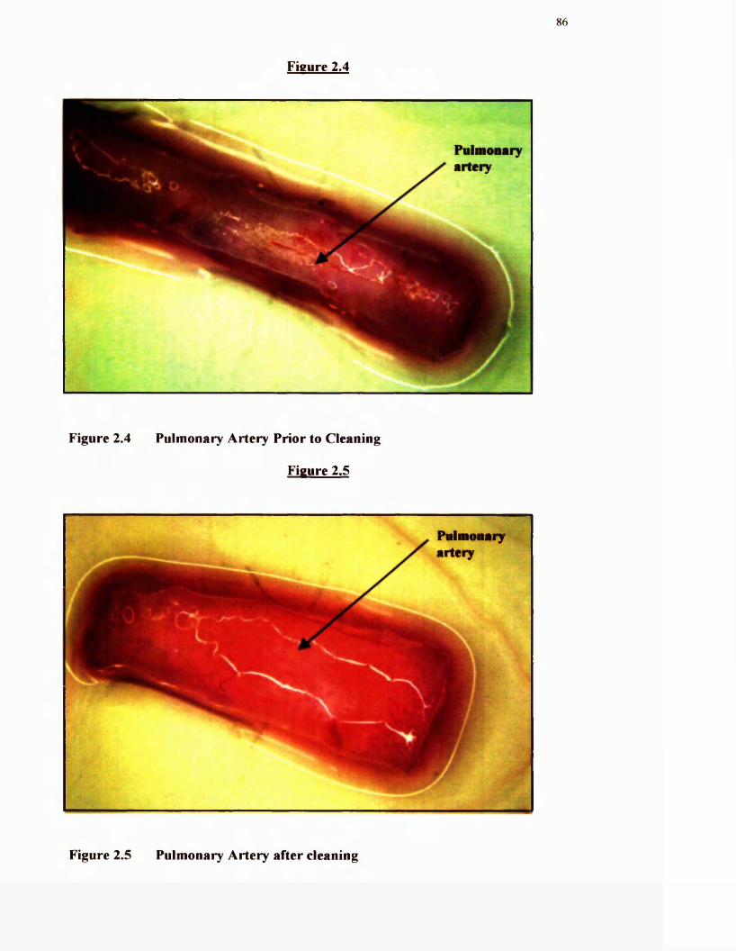
86
Figure 2.4
PnlaioBary aitery
Figure 2.4 Pulmonary Artery Prior to Cleaning
Figure 2.5
PulmoBaryartery
Figure 2.5 Pulmonary Artery after cleaning
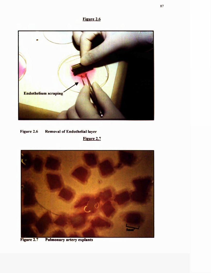
87
Figure 2.6
Endothelium scraping
Figure 2.6 Removal o f Endothelial layer
Figure 2.7
Figure 2.7 Pulmonary artery explants

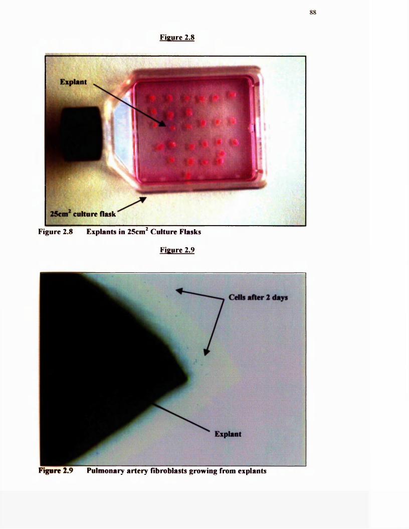
88
Figure 2.8
Expiant
25cni^ culture flask
Figure 2.8 Explants in 25cm Culture Flasks
Figure 2.9
C d b after 2 days
Explant
Figure 2.9 Pulmonary artery fibroblasts growing from explants
89
2.3.3 Immunohistochemicai Characterisation of Primary Fibroblast Cultures
All primary cell cultures were established as having a fibroblast-like origin by
determining whether they expressed smooth muscle a-actin. Primary fibroblast
cultures were allowed to grow to confluence on sterile acid-etched coverslips grown
in 35mm^ culture flasks. These were then fixed using 70% methanol / 10ml 30%
hydrogen peroxide (Sigma, UK) for 30 mins at room temperature. The coverslips
were then washed in sterile PBS. 50pl PBS containing 3% (w/v) bovine serum
albumin (BSA, Sigma, UK) was placed onto each coverslip, for 30 minutes, in a
humidified incubator at 37“C. Without rinsing, 10% goat serum (Sigma, UK) in PBS
was added and the coverslips were then incubated for a further 30 mins. The
coverslips were then rinsed once in PBS and the primary monoclonal antibody against
smooth muscle a-actin (Tsukada et al, 1987) (Sigma, UK) was added to each
coverslip (diluted in PBS at 1:1000). The coverslips were then left for Ih at 37°C
without agitation.
Each coverslip was then rinsed 3x in PBS and a biotinylated anti-mouse IgG (1:500 in
PBS) was added. The coverslips were then incubated for a further hour at 37“C, after
which they were rinsed 3x in PBS. Strepavidin-peroxidase (Sigma, UK) diluted
1:1000 in PBS was then added to each coverslip. The coverslips were then incubated
for an hour at 37°C, and then rinsed 3x in Tris Buffer (0.05M, pH 7.4). Coverslips
were then placed in Tris Buffer containing diaminobenzidine tetrahydrochloride
(0.5mg/ml) (Sigma, UK) and 10.5 % (v/v) hydrogen peroxide (30% solution). They
were incubated for 10 minutes and then washed 3x in PBS. Coverslips were then
mounted onto microscope slides using DPEX mountant (Sigma, UK) and observed
under a light microscope (Olympus) (Figure 2.10). A distinct brown coloration
determined the localisation o f smooth muscle a-actin. Primary fibroblasts derived
90
from bovine or rat pulmonary and systemic arteries were compared to smooth muscle
cells cultured from bovine trachea and Rat 2 fibroblast cell lines that acted as positive
and negative controls, respectively.
2.3.4 Cell Freezing/Thawing
A cell suspension was collected from a 75cm^ flask by trypsinisation (Chapter 2.3.1)
in 10ml o f culture medium. Cells were centrifuged at lOOOg for 10 minutes. The cell
pellet was then resuspended in 1ml Cryopreservation Medium growth medium
(DMEM containing 10% FCS and 10% DMSO). The resuspended cells in the
freezing medium were placed in a 2ml cryotube and left in a fridge for 20 minutes
followed by a -80°C freezer overnight. The tube was then transferred to the vapour
phase o f the liquid nitrogen for Ih, then placed directly into the liquid nitrogen.
Frozen cells were removed from liquid nitrogen and thawed rapidly in a water bath at
37°C. The cryotubes were then swabbed with tissue paper soaked in 70% ethanol and
the caps loosened. Holding the vial in one hand, the cap was removed and the
contents taken up into a pipette, then placed in a 10ml centrifuge tube. The cell
suspension was diluted slowly with 5mis o f fresh growth medium and the tubes
centrifuged at lOOOg for lOmins. The cells were resuspended in lOmls fresh growth
medium and seeded onto a 25cm^ flask. After approximately 4h the cells attached to
the flask. The medium was replaced with fresh medium.
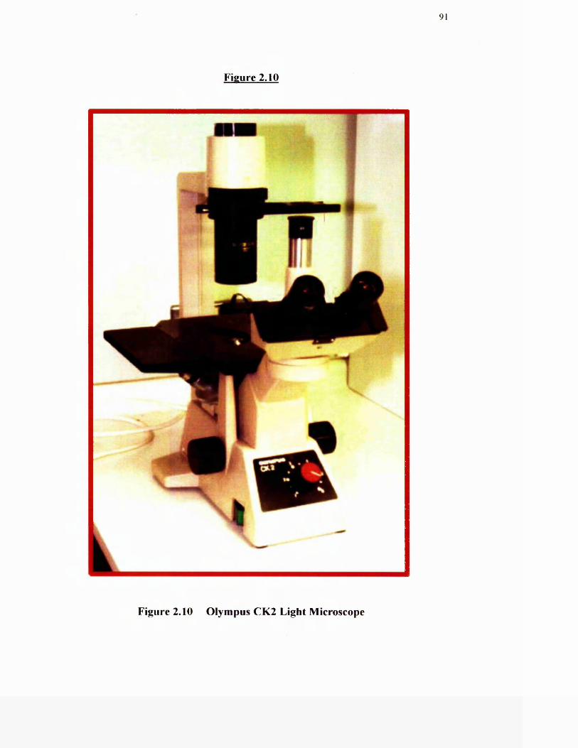
91
Figure 2.10
»
Figure 2.10 Olympus CK2 Light Microscope
92
2,4 Hypoxia: Methods for studying acute hypoxic fibroblast cells in vitro
2.4.1 Generation o f Hypoxic Environment
A humidified temperature controlled incubator (Model GA156; LEEC, Colwick,
Nottingham, UK) (Figure 2.11) was used to produce a hypoxic environment. This
incubator allows control of internal oxygen levels between 0 and 21% using medical
grade nitrogen, while the CO2 level is simultaneously controlled at 5%. Due to the
large volume o f nitrogen required to sustain a suitable degree o f hypoxia, nitrogen
cylinders were linked using a Pneuchange automatic gas cylinder change over unit
(NTC, Woulton, Liverpool, UK) which activated a fresh supply o f nitrogen as
required.
2.4.2 Measurement o f Hypoxia
The levels o f hypoxia achieved within the environment o f the incubator could be
monitored with the oxygen probe that was an integral part o f the unit. Measurements
from within the bathing medium o f the cells were also analysed with a portable
oxygen probe (Jenway: 9015. Dunmon, Essex, UK) to determine the rate o f gaseous
diffusion, and the pH o f the medium analysed throughout the course o f hypoxic
exposure with the use o f a portable pH probe (Mettler Delta 340, Hanstead, UK).
Cells were routinely maintained for acute hypoxia in an atmosphere o f 2% O2 and 5%
CO2 at 3 7T .
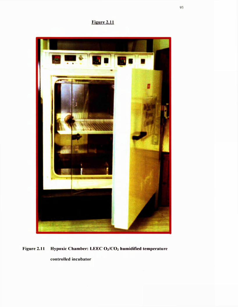
93
Figure 2.11
Figure 2.11 Hypoxic Chamber: LEEC O 2/C O 2 humidified temperature
controlled incubator
94
2.5 Hypoxia: Methods for studying cells from chronically hypoxic animals
2.5.1 Chamber design
The chamber is designed to hold two standard rat cages, with up to four rats in each
cage. The structure of the chamber is made from transparent high resistance Plexiglas.
Air is continually removed from the chamber by the pump. Air constantly flows
through the chamber at 45L/minute, ensuring that moisture and CO2 do not build up.
Temperatures are similar both inside and outside o f the chamber.
2.5.2 Maintenance o f animals
The chamber was housed in a specially designed environmental room which
maintains temperature at approximately 21 “C, humidity at 55%, gives 20 changes o f
filtered air per hour and maintains a 12h on 12h off light cycle. Animals used in the
experiments were obtained from credited commercial suppliers (Harlan UK Ltd,
Nottingham). The rats supplied were male specific pathogen-free and ordered in at
age 28-30 days (approximately 60g weight). Animals were allowed to acclimatise
within the environmental chamber for five days before being split into two groups of
four. One group o f animals were placed in the chamber and the other group remained
in normal atmospheric conditions to act as age-matched controls.
2.5.3 Maintenance o f Chronic Hypoxic Rats
The chamber door was removed and the rat cage placed in the ehamber with the food
and water dispensers at the rear, thus allowing observation o f the rats. The chamber
door was replaced and the pump switched on. The chamber was taken down to the
desired pressure (55mbar - 418mmHg) in small steps by slowly closing the inlet
95
valve and observing the pressure reading on the gauge. Once the stable experimental
pressure was reached, rats were checked every 15min for the following hour to ensure
that the pressure remained stable and the animals were not in distress. The chamber
could then be left to run for the experimental time required. Checks were made 5
times a day to ensure that pressure readings were not fluctuating beyond desired
levels, temperature inside and outside the chamber remained between 20 and 23^C,
animals were not showing signs of distress and that the pump was in good operating
condition. At weekends a member o f the Central Animal Facility staff (University o f
Glasgow) checked the chamber. When the animals required fresh food and water,
usually every three days, the chamber was taken down to atmospheric pressure over
2h by opening the inlet valve. Once atmospheric pressure was reached, the pump was
switched off and allowed to cool for half an hour' while the rat cages were cleaned and
animals given fresh food and water. Following this, the animals were placed back in
the chamber, the pump switched on and the chamber taken back to 55mbar over a
period o f two hours. After 14 days in the chamber, two o f the rats were removed and
the remaining two rats taken back to the experimental chamber for a further two days.
O f the two rats removed, one was immediately sacrificed and studied on that day
along with an aged matched control. The second rat was left in room air to be studied
no longer than 24h after removal from the chamber. The same procedure occurred on
day 16 for the remaining two rats.
2.6 Assessment of Cell Proliferation
Cell proliferation was measured by determining the uptake o f [^H]thymidine into
DNA. Cells were seeded at a density o f 5x10^ cells / well into 24-well plates in 500pl
o f culture medium. Cells were grown to 60% confluency in 24-well plates and then
96
growth-arrested for 48h by replacing the medium with 500p,l serum-free DMEM.
Cells were then stimulated with appropriate agonists and incubated for 24h, either in a
normal CO2 incubator (5% CO2) or in the hypoxic incubator to obtain an acute
hypoxic exposure (Chapter 2.4). In the latter case, the O2 content o f the atmosphere
was reduced from 21% to 2% by flooding with N2.
For the final 4h o f agonist stimulation, cells were labelled with thymidine
(O.lpCi/well). The reaction was stopped by washing the cells twice in ice-cold PBS
(500|ul/well). Proteins were precipitated by washing three times with 500p,l/well 5%
trichloroacetic acid (TCA) and lipid fractions were solubilised by washing twice with
90% ethanol (500pl/well) for 30mins. The remaining cell contents were incubated in
0.3M NaOH (SOOpl/well) for 30mins. The contents o f each well was transferred to
scintillation vials, to each o f which was added 3mis o f Ecoscint A (Ecoscint, Atlanta,
Georgia, USA) scintillation fluid. Vials were vortexed for Imin before radioactivity
was measured by scintillation counting using a Wallac scintillation counter. Results
are expressed as disintegrations per minute (DPM).
2.7 Inositol 1,4,5-Trisphosphate Mass Assay
2.7.1 Production o f Binding Protein
Approximately 20 bovine adrenal glands were obtained from the local abattoir,
immediately placed in ice, and brought back to the laboratory. Fat was removed from
the glands and they were dissected longitudinally. The Central straw-coloured
medulla was scraped off and disposed of leaving the red/brown cortex, which was
homogenised and blended. The homogenate was then centrifuged at 5000g for 15min
at 4‘’C and the resulting supernatant centrifuged at 20,000g for 23mins at 4‘ C. The
97
final pellet was then resuspended in Homogenisation Buffer (20mM NaHCO] and
1 mM DTT, pH 7.5, 4"C) and centrifuged again at 20,000g for 23mins. The binding
protein was stored in 10ml portions at -80"C and was stable for at least 6 months. The
binding protein could tolerate approximately four freeze-thaw cycles without loss of
binding activity. 20 adrenal glands produced approximately 100ml o f binding protein
normally giving 40mg protein/ml which was sufficient for ~ 4000 individual assay
samples.
2.7.2 Cell Preparation
Cells were seeded at a density o f 5x10^ cells/well into 24-well dishes and grown for a
further 3 days until confluent. Cells were then growth-arrested for 24h by culturing in
serum-free DMEM. The reaction was started by the addition o f agonists and then
stopped by the addition of 25pl ice-cold 10% perchloric acid (PCA, v/v). 30-40pi of
Neutralisation Buffer (60mM Hepes pH 8.9, 1.5M KOH) and 3pi aliquots of
Universal Indicator was then added until the bathing media had turned green. Cells
were mechanically disrupted by scraping using a cell scraper and the contents placed
in micro fuge tubes on ice. The samples were centrifuged in a bench top micro frige at
5000g at 4°C for 5mins. The resulting supernatant was stored at -2 0 ‘ C until time of
assay.
2.7.3 The binding assay (for Standard Curve)
A stock o f unlabelled Ins (1,4,5)P] (4pM) was diluted into Eppendorf tubes in dH20
as shown in Table 2.1 in a final volume o f 25pl. The following solutions were then
added to these standards (25pi):
98
(1) Neutralisation Buffer (lOOpI)
(2) Incubation Buffer (Tris-Base 0.04M, pH9.0, EDTA 4mM, EOTA 4mM, 0.4%
BSA).
(3) pH]Ins(l,4,5)P3 (5|^Ci) diluted in dHzO (150)11).
(4) Binding protein (40mg/ml stock) diluted in Homogenisation Buffer (125pl) to a
final concentration of 8mg/ml.
The binding protein was always added last.
Samples were then mixed by vortexing and left on ice for 30mins. After this time the
samples were centrifuged at 3660g for ISmins at 4“C. The supernatant was removed
and 1ml o f scintillation fluid was then added to each pellet and mixed by vortexing to
dissolve. Radioactivity was measured in a scintillation counter.
2.7.4 The binding assay (for Samples)
This procedure was the same as the assay for the standard curve except 25 pi o f
sample (prepared as in Chapter 2.6.2) replaced the standards.
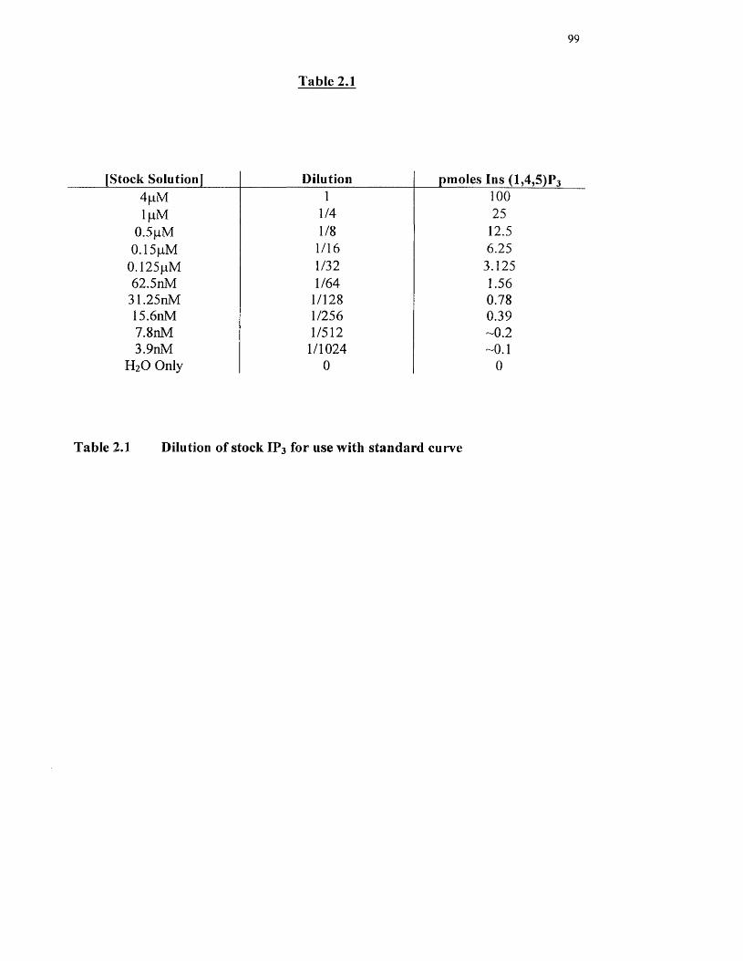
Table 2,1
99
[Stock Solution] Dilution pmoles Ins (1 ,4 ,5 ) ? 3
4pM 1 100IpM 1/4 25
0.5pM 1/8 12.50.15pM 1/16 6.25
0.125pM 1/32 3.12562.5nM 1/64 1.56
31.25nM 1/128 0.7815.6nM 1/256 0.397.8nM 1/512 -0.23.9nM 1/1024 -0.1
H2O Only 0 0
Table 2,1 Dilution of stock IP3 for use with standard curve
100
2.7.5 Calculation o f lns( 1,4,5)P] mass.
% B/ Bo was calculated where:
B ^ Specific binding (of unknown or of standard)
= DPM in pellet - DPM in non-specific binding sample
Bo = maximum specific
= DPM in pellet o f 0 pmol IP3 tubes - DPM in non-specific binding sample.
%B/Bo vs. pmol Ins(l,4,5)P3 was then plotted as a standard curve on semi-log paper.
(See Figure 5.1)
The %B/Bo values o f unknowns from the standard curve = pmoles / sample (i.e. not a
concentration).
Concentration o f IP3 in assay was dependent upon the assay volume used,
i.e. 5pmol in 500pl assay = lOnM.
101
2,8 Detection and Analysis of Proteins
2.8.1 Preparation o f samples for SDS-PAGE and immunoblotting
Cells were seeded at a density o f 5x10^ cells/well onto 6-well dishes. After the cells
had reached 60% confluency, they were growth-arrested in serum-free DMEM for a
period o f 24h. After the cells had been agonist-stimulated, the medium was removed,
the cells placed on ice and the cell monolayer washed 2x with 500pl ice-cold PBS.
The cells are then lysed with 50pl RIPA buffer (Tris-HCl 50mM pH 7.4, NP-40 1%,
C24H3904Na 0.25%, NaCl 150mM, EGTA ImM, PMSF ImM, Na3V0 4 ImM, NaF
ImM, CLAP 1/1000, pH 7.4)
The cells were scraped on ice into the RIPA buffer and the contents placed in
microcentrifuge tubes. After a further 15mins on ice, the lysates were centrifuged at
14,000g at 4°C for 15mins. The supernatant was then transferred to fresh tubes,
aliquoted and stored at ~70°C.
2.8.2 Assay o f protein concentration
Quantification o f protein concentration was determined using a Bio-Rad™ DC Protein
Assay Kit based on a colorimetric assay devised by Lowry et al (1951). Protein
samples were prepared in appropriate lysis buffer. For each assay performed, a
standard curve was prepared using five dilutions of BSA as a protein standard diluted
in RIPA buffer over the concentration range o f lmg-16pg/ml protein. Appropriate
dilutions of both samples and standards were made in H2O to a volume o f 200pl.
Reagent A (an alkaline copper tartrate solution supplemented with 5% SDS) (lOOpl)
was added to each of the standards and samples. These were vortexed and SOOpls
102
Reagent B (a dilute Folin Reagent) added and immediately vortexed. Samples were
left for 15min for colour development from pink to green as a result o f reduction of
Folin reagent by copper-protein complex. Colour development was quantified by
absorbance spectroscopy at 750nm within Ih. Estimates o f protein content in samples
were made from the standard curve.
2.8.3 SDS Polyacrylamide Gel Electrophoresis (SDS-PAGE)
Protein samples were analysed using a SDS-PAGE system
2.8.3.1 Preparation o f acrylamide gels
The casting apparatus was Bio-Rad™ mini gel electrophoresis units assembled
according to manufacturer instructions. The separating and stacking gels were made
as follows:
Resolving polyacrylamide gels: Normally 10% resolving gels were used which were
prepared using the following composition; 37% acrylamide / 0.8% bisacrylamide (to a
final 10%) (w/v); 0.375M Tris-HCl pH 8.5, 0.1% SDS (w/v), 0.05% ammonium
persulphate (w/v), 0.05% TEMED (v/v). Polymerisation was initiated by addition of
the ammonium persulphate and TEMED. The solution was mixed and poured into the
assembled glass plates, leaving sufficient space at the top for the stacking gel. Gels
were overlaid with 200-400pl of 0.1% SDS and allowed to polymerise for 30-45
mins.
Stacking polyacrylamide gels: Following polymerisation o f the resolvmg gels, the
overlay was removed and the surface o f the gel rinsed with water to remove any
unpolymerised acrylamide. The stacking polyacrylamide gels used were o f the
following composition: 5% acrylamide/bisacrylamide (w/v), 0.125M Tris-HCl pH
103
8.5, 0.1% SDS (w/v), 0.05% ammonium persulphate (w/v) and 0.1% TEMED (v/v).
Stacking polyacrylamide gel components were overlaid on the polymerised resolving
gel and combs added immediately. After polymerisation of the stacking gel, combs
were removed and the wells rinsed with Electrophoresis Buffer (Tris-base 25mM,
glycine 192mM and SDS 0.1%).
2.8.3.2 Electrophoresis conditions
All protein samples to be examined by SDS-PAGE were first diluted to 50pg in SDS
Sample Buffer, (93mM Tris-HCl pH 6.8 and 20niM dithiothreitol (DTT), ImM
sodium-EDTA, 10% glycerol (v/v), 2% SDS (w/v) and 0.002% bromophenol blue
(w/v)). DTT was added fi’esh, immediately before use. Samples were boiled for 5 min
to denature proteins and disrupt disulphide bonds. The requiied volume was loaded
into the individual wells o f the stacking gel using loading tips. Pre-stained SDS
protein molecular weight markers (Biorad) o f known size were also placed in one lane
o f the gel. After loading samples, polyacrylamide gels were assembled into a Bio-Rad
electrophoresis unit. The two chambers of the unit were filled with Electrophoresis
Buffer and the polyacrylamine gel subjected to electrophoresis for approximately 1.5-
2h at a constant current of 240mA.
The molecular weight o f proteins was estimated by comparing their mobility to that o f
pre-stained SDS-PAGE standards. The standards used were myosin (205kDa), P-
galactosidase (120kDa), bovine serum albumin (87kDa) and ovalbumin (48kDa).
104
2.8.3.3 Fixing and drying gels
Gels containing radioactive proteins were fixed in 20% methanol (v/v)/10% acetic
acid (v/v) for 30 min and rinsed in distilled water. The gel was sandwiched between
cellophane sheets in a drying frame and dried under vacuum for 60 min at 80°C by a
Floeffer Eazy-Breeze™ gel dryer. The radio labelled protein was detected by
autoradiography. The dried gel was exposed to Kodak X-OMAT LS X-ray film for 4-
24h at ~20'^C in a spring loaded metal cassette and developed by a KODAK M35-M
X-OMAT processor.
2.8.4 Western blot analysis.
2.8.4.1 Transblotting to nitrocellulose.
Following completion o f electrophoresis, polyacrylamide gels were removed from the
apparatus and washed once in Blotting Buffer (20% methanol (v/v), 25mM Tris-base,
192mM glycine). Proteins were transferred from the polyacrylamide gel to the
nitrocellulose by assembling a transfer cassette with the nitrocellulose juxtapositioned
between the polyacrylamide gel and the cathode. By this method negatively charged
proteins were transferred to nitrocellulose for 1.5-2h at a constant 1 amp current.
2.8.4.2 Immunoblotting
Samples (30-50pg of protein) were subjected to SDS-PAGE (Chapter 2.8.3) and the
proteins transferred to nitrocellulose by Western blotting (Chapter 2.8.4.1). The
nitrocellulose blots were washed in PBS/T (PBS containing 0.02% Tween-20 (v/v))
and blocked for non-specific binding for 3h on a rocking platform at room
temperature in PBS/T supplemented with 10% non-fat milk (Marvel) (w/v). Blots
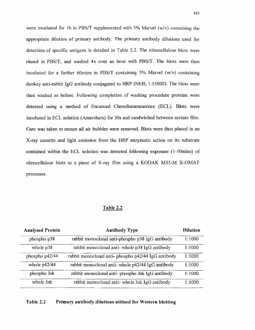
105
were incubated for Ih in PBS/T supplemented with 5% Marvel (w/v) containing the
appropriate dilution of primary antibody. The primary antibody dilutions used for
detection o f specific antigens is detailed in Table 2.2. The nitrocellulose blots were
rinsed in PBS/T, and washed 4x over an hour with PBS/T. The blots were then
incubated for a further 60mins in PBS/T containing 5% Marvel (w/v) containing
donkey anti-rabbit IgG antibody conjugated to HRP (NEB; 1:15000). The blots were
then washed as before. Following completion of washing procedure proteins were
detected using a method o f Encanced Chemilumenescence (ECL). Blots were
incubated in ECL solution (Amersham) for 30s and sandwiched between acetate film.
Care was taken to ensure all aii' bubbles were removed. Blots were then placed in an
X-ray cassette and light emission from the HRP enzymatic action on its substrate
contained within the ECL solution was detected following exposure (1-1 Omins) o f
nitrocellulose blots to a piece o f X-ray film using a KODAK M35-M X-OMAT
processor.
Analysed Protein
Table 2.2
Antibody Type Dilution
phospho p38 rabbit monoclonal anti-phospho p38 IgG antibody 1:1000
whole p38 rabbit monoclonal anti- whole p38 IgG antibody 1:1000
phospho p42/44 rabbit monoclonal anti- phospho p42/44 IgG antibody 1:1000
whole p42/44 rabbit monoclonal anti- whole p42/44 IgG antibody 1:1000
phospho Ink rabbit monoclonal anti- phospho Jnk IgG antibody 1:1000
whole ink rabbit monoclonal anti- whole Jnk IgG antibody 1:1000
Table 2.2 Primary antibody dilutions utilised for Western blotting
106
2.8.4.3 Re-probing nitrocellulose membranes
In instances where the same membrane was used to probe for different proteins,
primary and secondary antibodies were stripped from the nitrocellulose by incubating
in Stripping Buffer (lOOmM B-mercaptoethanol, 2% SDS and 62.5mM Tris-HCl, pH
6.7) for 30 minutes at 50°C with agitation. Blots were then rinsed with PBS/T 4x over
40 mins before the immunodetection protocol was repeated (Chapter 2.8.4.2)
2.8.5 Solid phase JNK activity assay
JNK activity was measured using recombinant truncated N-terminus o f c-jun (c-Jung.
89) immobilised on glutathione (GSH)-sepharose beads (GST-c-jun) (Dai et al, 1995).
Cells were grown to subconfluence on 6-well plates and treated as required. To stop
the reaction, cells were washed 2x with ice-cold PBS, then stimulated eells were
scraped on ice in 500pl Solubilisation Buffer (20mM HEPES pH7.7, 50mM NaCl,
O.lmM EDTA, 0.2mM PMSF, 0.5mg/ml leupeptin, 0.5mg/ml aprotinin and 1% (v/v)
Triton X-100). Lysates were centrifuged at 14,000g for 15min at 4°C. Protein
concentration was measured in the supernatant as in chapter 2.8.2. Aliquots (150 pg)
of solubilised cell extracts were added to 20pl o f GST-c-jun and mixed for 3h on a
rotary wheel at 4°C to allow binding o f active JNK in the cell extracts. The sepharose
beads were centrifuged at 10,000g for Imin at 4“C and the supernatant removed. The
beads were washed 3x in 500pl Solubilisation Buffer and Ix in 500pl Kinase Buffer
(25mM HEPES pH 7.6. 20mM MgCb, 5mM p-glycerophosphate, O.lmM Na]V0 4
and 2mM DTT). The beads were resuspended in 30pl o f Kinase Buffer [y-^^P]ATP
(20pM, 2pCi) and the tubes were incubated for 30min at 30°C. The reaction was
terminated by adding lOpl 4x Laemmli SDS sample buffer. Samples were boiled for 5
107
minutes and proteins resolved by SDS-PAGE (Chapter 2.8.3) on 10% polyacrylamide
gels. Gels were fixed and dried as in Chapter 2.8.3.3 and incorporation o f [y-^^P] into
GST-c-Jun detected by autoradiography.
2.8.6 Solid phase p38 MAP kinase assay
The activity o f p38 MAP kinase was assayed in vitro by a procedure based on
Bogoyevich, et al (1996). Cell extracts were prepared in a p38 MAP kinase
Solubilisation Buffer (20mM Tris-HCl pH7.4, 150mM NaCl, 1% Triton X-100, 10%
glycerol, 2mM EDTA, 20mM NaF, 2.5mM p-glycerophosphate, 0.2mM PMSF,
0.5mg/ml leupeptin and 0.5 mg/ml aprotinin) and p38 affinity-purified by incubating
lysates with MAPKAP kinase-2 immobilised on GSH-sepharose beads (GST-
MAPKAP kinase-2). The kinase reaction was carried out in the same buffer as that
used for the c-jun activity assay (Chapter 2.8.5). The incorporation o f [y-^^P]ATP into
MAPKAP kinase-2 was carried out using a similar method to that for GST-c-jun.
2.8.7 p42/44MAP kinase activity assay
In vitro MAP kinase activity was assessed using a BIOTRAK MAP kinase assay kit
to measure the incorporation o f radio label led phosphate into a specific MAP kinase
substrate peptide (KRELVEPLT^^^PAGEAPNALLAR) derived from a portion of the
EGF receptor (EGFR^^'"^^^). This method was carried out according to the
manufacturers instructions.
After treatment, cells in 6-well plates were rinsed 2x in ice-cold PBS and scraped on
ice in 500pl Lysis Buffer (lOmM Tris pH7.5, 150mM NaCl, ImM EDTA, ImM
EGTA, 0.2mM Na3V0 4 , 1% Triton X-100, 0.5% NP-40, 0.2mM PMSF, 0.5 pg/ml
leupeptin and 0.5pg/ml). Lysates were transferred to eppendorf tubes then left on ice
108
for 30mins followed by were centrifugation at 13000g for 5mins at 4"C. Protein
concentration was measured in the supernatant as in Chapter 2.8.2. Equal quantities
(approximately 50pg protein) o f supernatant were transferred to fresh tubes in a
volume o f 15pl to which was added lOpM (600pM). The reactions were
initiated by addition of 5pi [y-^^P]ATP (50pM, IpCi) in 75mM HEPES buffer (pH
7.4) containing 1.2mM MgCb. The reactions were incubated at 37”C for 15 min.
Reactions were stopped by the addition o f 25pl o f 300mM H3PO4 and aliquots (25pl)
were spotted onto 3x3cm P-81 phosphocellulose paper. Filters were washed 2x for
5min with 75mM H3PO4 and 2x for 5min with dHiO, then transferred to 6ml plastic
scintillation vials to which 3mis Ecoscint A, was added. Radioactive phosphopeptide
bound to the filter was quantified by liquid scintillation counting in a Wallac
scintillation counter. In every assay, background activity was measured from a sample
of kinase buffer reaction mixture in the absence of lysate.
2.9 Densitometric analysis of blots
Densitometric analysis o f blots was carried out using a computer programme that
allowed for comparison o f blot density in graphical form (Quantiscan).
2.10 Data analysis
Data are expressed as mean + S.D. for replicate plates from the same experiment.
Experiments were repeated in cells ranging from 4-8 different animals. The statistical
significance o f differences between mean values from control and treated groups were
determined by Student’s t-tests, unpaired and paiied where applicable, using OXTAT
software. Two-tailed probability values o f less than 0.05 (P<0.05) were considered to
be significant.

109
Chapter 3
Establishment of a Cell Model for Studying Bovine Pulmonary Artery Fibroblast Cell Proliferation in Acute Hypoxia
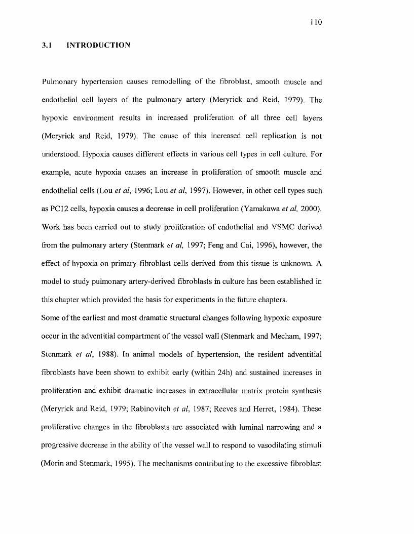
no
3.1 INTRODUCTION
Pulmonary hypertension causes remodelling o f the fibroblast, smooth muscle and
endothelial cell layers of the pulmonary artery (Meryrick and Reid, 1979). The
hypoxic environment results in increased proliferation of all three cell layers
(Meryrick and Reid, 1979). The cause o f this increased cell replication is not
understood. Hypoxia causes different effects in various cell types in cell culture. For
example, acute hypoxia causes an increase in proliferation o f smooth muscle and
endothelial cells (Lou et 1996; Lou et al, 1997). However, in other cell types such
as PC I2 cells, hypoxia causes a decrease in cell proliferation (Yamakawa et al, 2000).
Work has been carried out to study proliferation of endothelial and VSMC derived
from the pulmonary artery (Stenmark et al, 1997; Feng and Cai, 1996), however, the
effect o f hypoxia on primary fibroblast cells derived from this tissue is unknown. A
model to study pulmonary artery-derived fibroblasts in culture has been established in
this chapter which provided the basis for experiments in the future chapters.
Some o f the earliest and most dramatic structural changes following hypoxic exposure
occur in the adventitial compartment o f the vessel wall (Stenmark and Mecham, 1997;
Stenmark et al, 1988). In animal models o f hypertension, the resident adventitial
fibroblasts have been shown to exliibit early (within 24h) and sustained increases in
proliferation and exhibit dramatic increases in extracellular matrix protein synthesis
(Meryrick and Reid, 1979; Rabinovitch et al, 1987; Reeves and Herret, 1984). These
proliferative changes in the fibroblasts are associated with luminal narrowing and a
progressive decrease in the ability of the vessel wall to respond to vasodilating stimuli
(Morin and Stenmark, 1995). The mechanisms contributing to the excessive fibroblast

I l l
growth in the pulmonary adventitia under conditions of acute and chronic hypoxia
remain poorly understood.
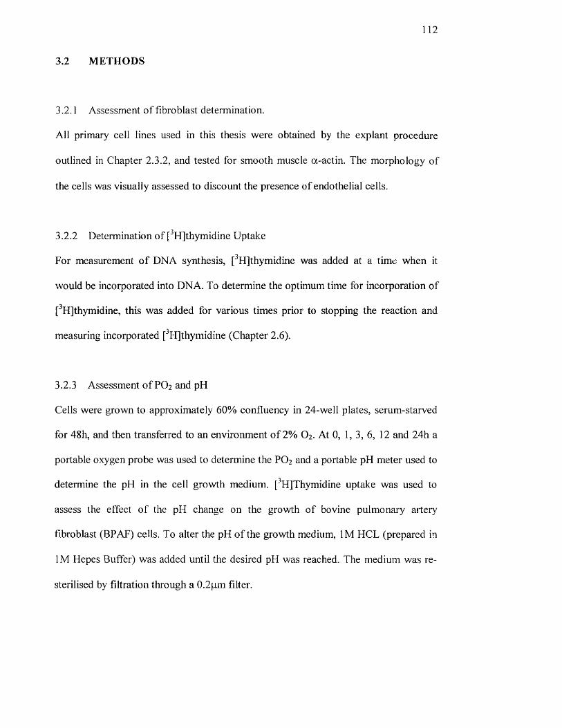
112
3.2 METHODS
3.2.1 Assessment of fibroblast determination.
All primary cell lines used in this thesis were obtained by the explant procedure
outlined in Chapter 2.3.2, and tested for smooth muscle a-actin. The morphology of
the cells was visually assessed to discount the presence o f endothelial cells.
3.2.2 Determination o f ["Hjthymidine Uptake
For measurement o f DMA synthesis, [^HJthymidine was added at a time when it
would be incorporated into DNA. To determine the optimum time for incorporation of
[^HJthymidine, this was added for various times prior to stopping the reaction and
measuring incorporated [^H]thymidine (Chapter 2.6).
3.2.3 Assessment o f PO2 and pH
Cells were grown to approximately 60% confluency in 24-well plates, serum-starved
for 48h, and then transferred to an environment o f 2% O2 . At 0, 1, 3, 6, 12 and 24h a
portable oxygen probe was used to determine the PO2 and a portable pH meter used to
determine the pH in the cell growth medium. ['H]Thymidine uptake was used to
assess the effect of the pH change on the growth o f bovine pulmonary arter)/
fibroblast (BPAF) cells. To alter the pH o f the growth medium, IM HCL (prepared in
IM Hepes Buffer) was added until the desired pH was reached. The medium was re-
sterilised by filtration through a 0.2pm filter.
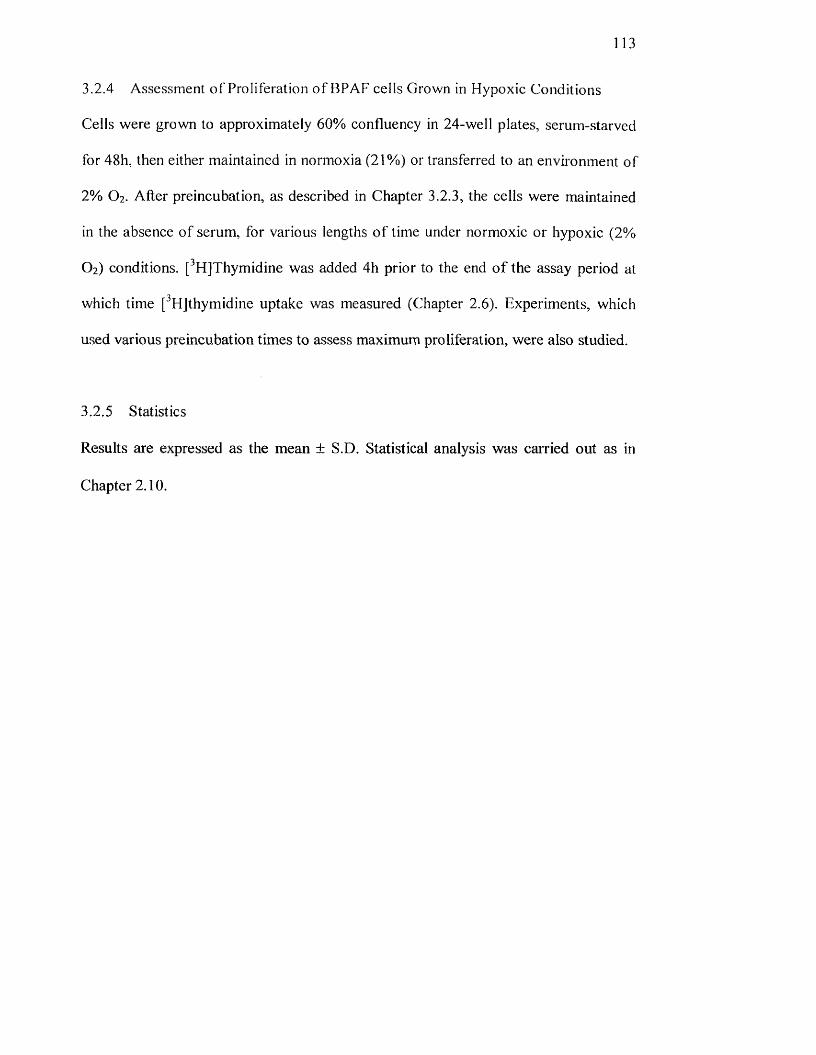
113
3.2.4 Assessment o f Proliferation of BPAF cells Grown in Hypoxic Conditions
Cells were grown to approximately 60% confluency in 24-well plates, serum-starved
for 48h, then either maintained in normoxia (21%) or transferred to an environment of
2% O2. After preincubation, as described in Chapter 3.2.3, the cells were maintained
in the absence of serum, for various lengths o f time under normoxic or hypoxic (2%
O2) conditions. [^HjThymidine was added 4h prior to the end o f the assay period at
which time [^HJthymidine uptake was measured (Chapter 2.6). Experiments, which
used various preincubation times to assess maximum proliferation, were also studied.
3.2.5 Statistics
Results are expressed as the mean ± S.D. Statistical analysis was carried out as in
Chapter 2.10.

114
3.3 RESULTS
3.3.1 Assessment of the fibroblast cell type
a-Actin staining was used routinely to assess the presence o f smooth muscle cell
contamination in cells derived from the pulmonary artery. A dark brown colouration
is a positive stain for smooth muscle a-actin. Figure 3.1 A shows fibroblasts derived
from the pulmonary artery, which were found to be contaminated with smooth muscle
cells and would not have been used for further experimentation. Figure 3 .IB show
fibroblasts derived from the pulmonary artery after hypoxic exposure. The shape o f
the cell also provided an identification o f the cell type. The fibroblast were sub-
confluent by which they assume multipolar or bipolar shapes and are well spaced on
the culture surface, but at confluence they are bipolar and less well spread (Freshney,
1994).
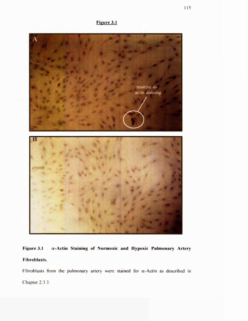
Figure 3.1
115
positive a- actin staining
B TT
V *
Figure 3.1 a-A ctin Staining of Normoxic and Hypoxic Pulmonary Artery
Fibroblasts.
Fibroblasts from the pulmonary artery were stained for a-Actin as described in
Chapter 2.3.3.
116
3.3.2 Determination of maximal [^Hlthymidine incorporation
To assess the growth of primary fibroblast cell cultures it was first necessary to
optimise the method for determining [^HJthymidine uptake in these cells. Figure 3.2
demonstrates the time o f maximal [^HJthymidine incorporation in pulmonary artery
fibroblast cells. As thymidine is incorporated whilst the cells are synthesising DNA
this gives a good estimation o f whether cells are proliferating or not. The addition of
[^HJthymidine for time points > 4h prior to the end of a 24h-proliferation assay
resulted in maximum incorporation. It was decided to add the thymidine as late as
possible as to reduce background incorporation.
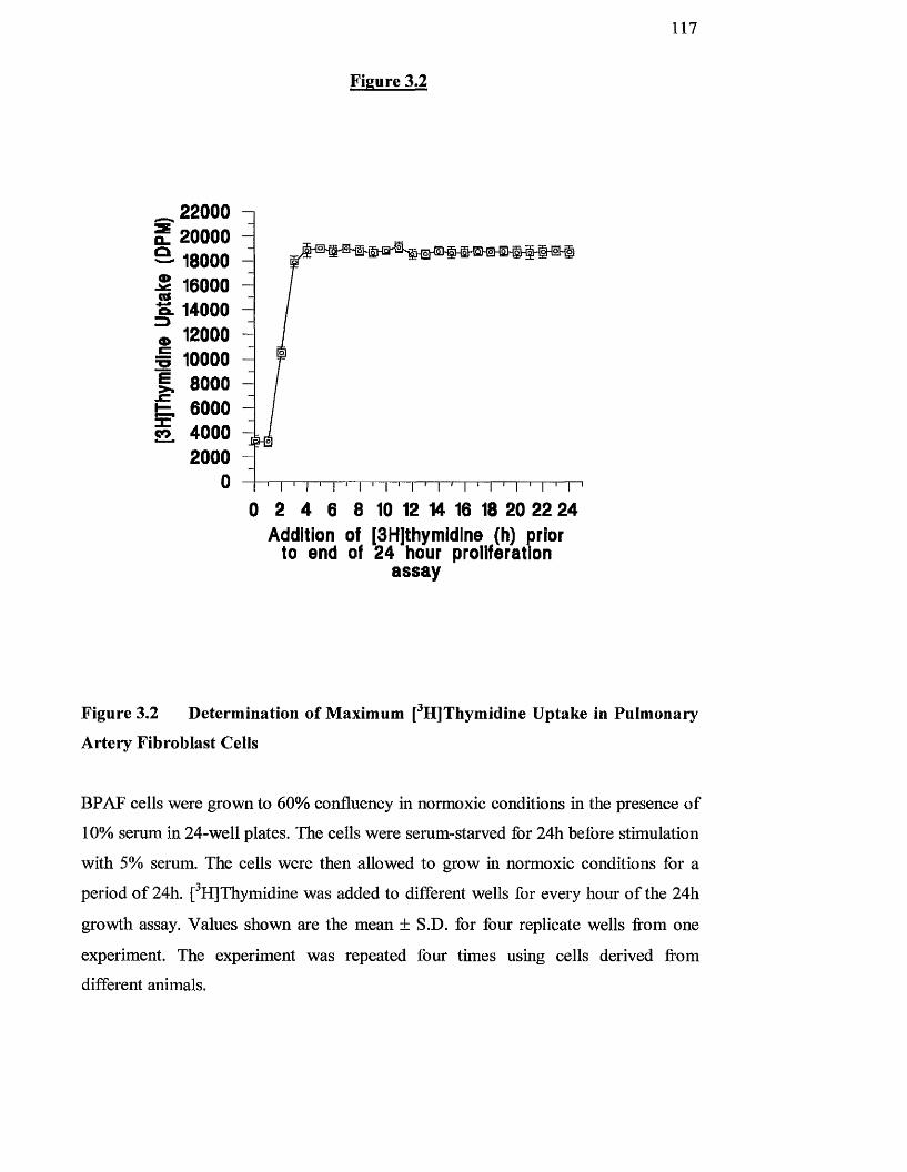
1 1 7
SCO
<t>C
E
Fco
220002000018000160001400012000100008000600040002000
Figure 3.2
0 2 4 6 8 10 12 14 16 18 20 22 24Addition of |3H|thymîdlne (h) prior
to end of 24 hour proiiferatlon a ssay
Figure 3.2 Determination of Maximum [^H]Thymidine Uptake in Pulmonary
Artery Fibroblast Cells
BPAF cells were grown to 60% confluency in normoxic conditions in the presence o f
10% serum in 24-well plates. The cells were serum-starved for 24h before stimulation
with 5% serum. The cells were then allowed to grow in normoxic conditions for a
period o f 24h. [^H]Thymidine was added to different wells for every horn o f the 24h
growth assay. Values shovm are the mean ± S.D. for four replicate wells from one
experiment. The experiment was repeated four times using cells derived from
different animals.
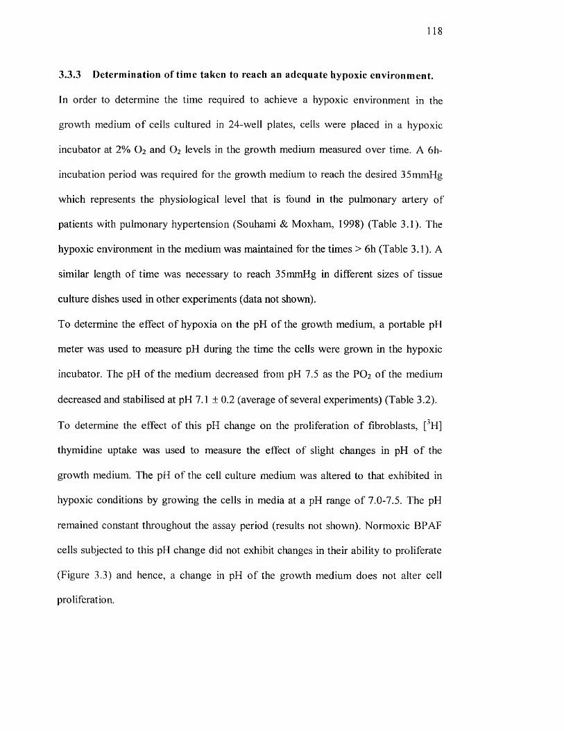
118
3.3.3 Determination of time taken to reach an adequate hypoxic environment.
In order to determine the time required to achieve a hypoxic environment in the
growth medium o f cells cultured in 24-well plates, cells were placed in a hypoxic
incubator at 2% O2 and O2 levels in the growth medium measured over time. A 6h-
incubation period was required for the growth medium to reach the desired 35mmHg
which represents the physiological level that is found in the pulmonary artery of
patients with pulmonary hypertension (Souhami & Moxham, 1998) (Table 3.1). The
hypoxic environment in the medium was maintained for the times > 6h (Table 3.1). A
similar length of time was necessary to reach 35mmHg in different sizes o f tissue
culture dishes used in other experiments (data not shown).
To determine the effect o f hypoxia on the pH of the growth medium, a portable pH
meter was used to measure pH during the time the cells were grown in the hypoxic
incubator. The pH of the medium decreased from pH 7.5 as the PO2 o f the medium
decreased and stabilised at pH 7.1 ± 0.2 (average of several experiments) (Table 3.2).
To determine the effect o f this pH change on the proliferation o f fibroblasts, [^H]
thymidine uptake was used to measure the effect of slight changes in pH o f the
growth medium. The pH of the cell culture medium was altered to that exhibited in
hypoxic conditions by growing the cells in media at a pH range o f 7.0-7.5. The pH
remained constant throughout the assay period (results not shown). Normoxic BPAF
cells subjected to this pH change did not exhibit changes in their ability to proliferate
(Figure 3.3) and hence, a change in pH of the growth medium does not alter cell
proliferation.
1 1 9
Table 3.1
Hours of preincubation
in 2% O2
PO2 in Bathing Medium (mmHg)
0 1471 1263 636 3512 3524 35
Table 3.1. Generation of a Hypoxic Environment for BPAF Cells
BPAF cells were grown to 60% confluency in 24-well plates before being placed in
the hypoxic incubator set at an oxygen level o f 2%. At the times indicated a portable
oxygen probe was used to determine the pOz in the cell growth medium. Values
shown are representative o f one individual well per time point. The experiment was
repeated four times and results shown are typical o f those obtained.
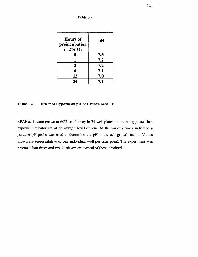
Table 3.2
1 2 0
Hours of preincubation
in 2% O2
pH
0 7.51 7.23 7.26 7.112 7.024 7.1
Table 3.2 Effect of Hypoxia on pH of Growth Medium
BPAF cells were grown to 60% confluency in 24-well plates before being placed in a
hypoxic incubator set at an oxygen level o f 2%. At the various times indicated a
portable pH probe was used to determine the pH in the cell growth media. Values
shown are representative o f one individual well per time point. The experiment was
repeated four times and results shovm are typical o f those obtained.
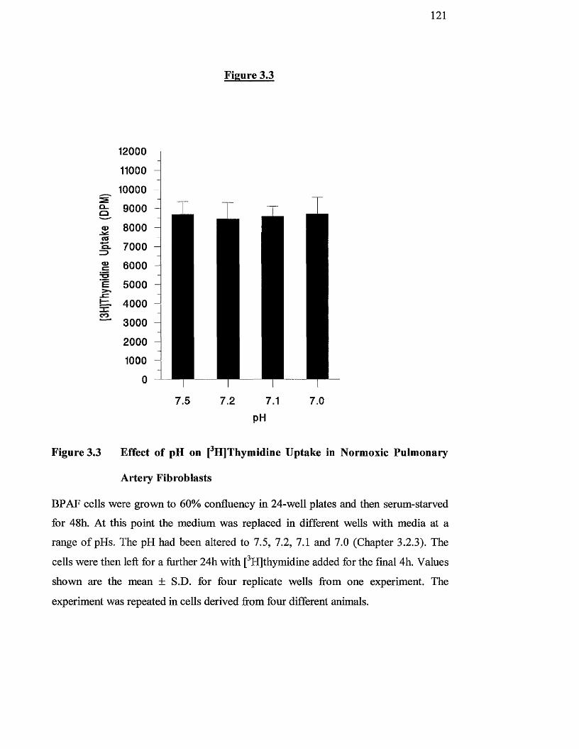
121
Q_a
12000
11000
10000
9000
8000a>
M- 70000»cisE
3CCO
6000
5000
4000
3000
20001000
0
7.5
Figure 3.3
7.2 7.1 7.0
pH
Figure 3.3 Effect of pH on [^H]Thymidine Uptake in Normoxic Pulmonary
Artery Fibroblasts
BPAF cells were grown to 60% confluency in 24-well plates and then serum-starved
for 48h. At this point the medium was replaced in different wells with media at a
range o f pHs. The pH had been altered to 7.5, 7.2, 7.1 and 7.0 (Chapter 3.2.3). The
cells were then left for a hirther 24h with [^HJthymidine added for the final 4h. Values
shown are the mean ± S.D. for four replicate wells from one experiment. The
experiment was repeated in cells derived from four different animals.
122
3.3.4 Effects of hypoxia on proliferation of Bovine Pulmonary Artery
Fibroblasts
To determine the effect of hypoxia on the growth o f BPAF’s, serum-starved ceils
were pre-incubated for various lengths o f time prior to a 24h proliferation assay as
described in Chapter 3.2.3. In the absence o f a 6h pre-incubation period in hypoxia,
which was shown in Table 3.1 to be required for the cell growth medium to become
hypoxic, no hypoxic growth o f the fibroblast cells was noted over a 24h period (288 ±
43.5 DPM for hypoxic cells which was not significantly different from control cell
levels at 255 ± 33.7 DPM). Figure 3.4 demonstrates that following a 6h preincubation
in hypoxic conditions BPAF cells proliferated at an increased level (1753.3 ± 69.6
DPM) which was significantly (P<0.001 for data sets) greater than those of control
cells (286.9 ± 19.4 DPM). Further pre-incubation in hypoxic conditions did not
enhance the growth o f these cells. The graph suggests that further preincubation may
cause the cells to either die or stop proliferating, however, the apparent reduction in
growth was due to the cells becoming over confluent and lifting from the base o f the
cell culture plate.
To determine whether a longer growth assay period following the 6h pre-incubation
would result in greater ['HJthymidine uptake, the effects o f varied lengths of growth
time was examined. Figure 3.5 demonstrates that there would appear to be no
advantage to allowing the cells a longer period of growth over 24h to enhance the
hypoxic response.
Visual confirmation of the hypoxic proliferation of these cells over a 24h period when
compared with those maintained in normoxia can be seen in Figure 3.6. Figure 3.6A
and B shows serum-starved normoxic fibroblast cells at To. Figure 3.6C and D shows
fibroblast cells 24h later after 5% serum stimulation. Figure 3.6C shows fibroblasts

123
maintained in normoxia and figure 3.60 fibroblasts kept in hypoxic conditions. Even
visually, increased proliferation o f the hypoxic cells it is quite apparent.
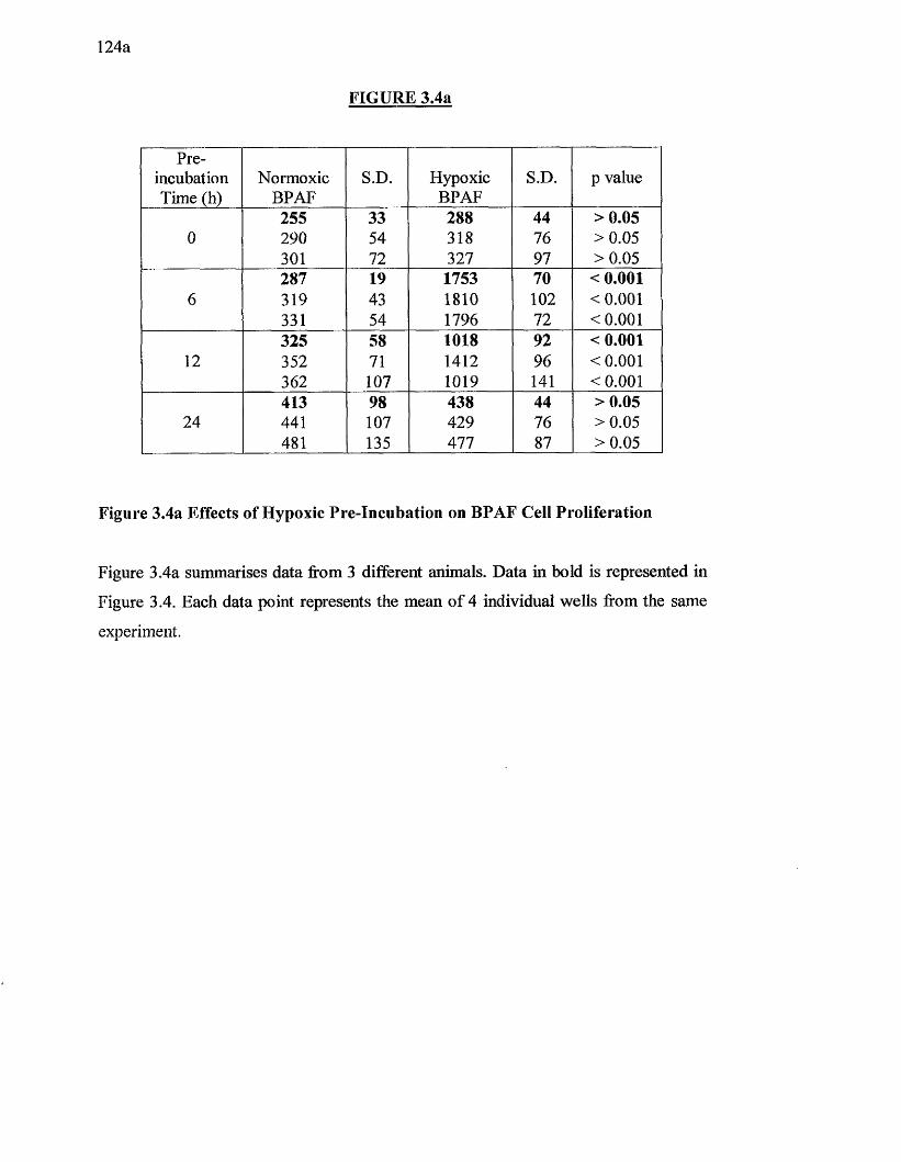
124a
FIGURE 3.4a
Preincubation Time (h)
NormoxicBPAF
S.D. HypoxicBPAF
S.D. p value
255 33 288 44 >0.050 290 54 318 76 >0.05
301 72 327 97 >0.05287 19 1753 70 < 0.001
6 319 43 1810 102 < 0.001331 54 1796 72 <0.001325 58 1018 92 < 0.001
12 352 71 1412 96 <0.001362 107 1019 141 < 0.001413 98 438 44 >0.05
24 441 107 429 76 >0.05481 135 477 87 >0.05
Figure 3.4a Effects of Hypoxic Pre-Incubation on BPAF Cell Proliferation
Figure 3.4a summarises data from 3 different animals. Data in bold is represented in
Figure 3.4. Each data point represents the mean o f 4 individual wells from the same
experiment.
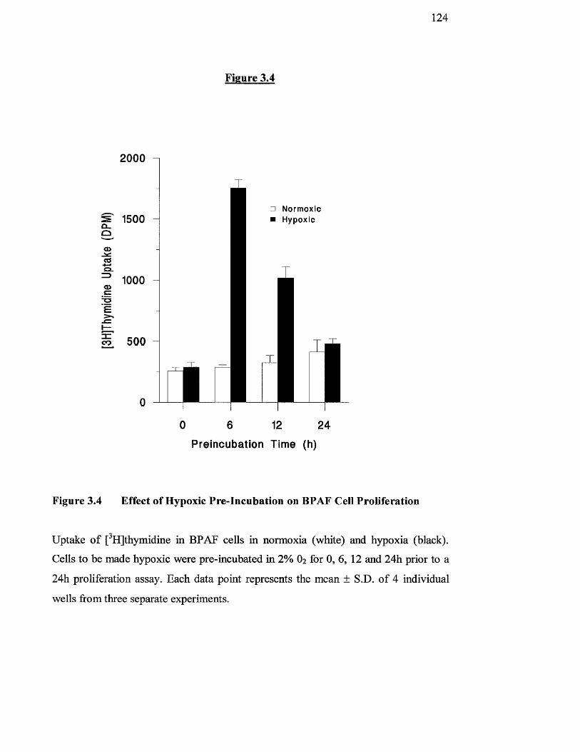
124
2000
Ou0 -<DBo .=3<DCSE
3 :co
1500
1000
500
Figure 3.4
□ Normoxic ■ Hypoxic
0 6 12 24
Preincubation Time (h)
Figure 3.4 Effect of Hypoxic Pre-Incubation on BPAF Cell Proliferation
Uptake o f [^HJthymidine in BPAF cells in normoxia (white) and hypoxia (black).
Cells to be made hypoxic were pre-incubated in 2% O2 for 0 ,6 , 12 and 24h prior to a
24h proliferation assay. Each data point represents the mean ± S.D. o f 4 individual
wells from three separate experiments.
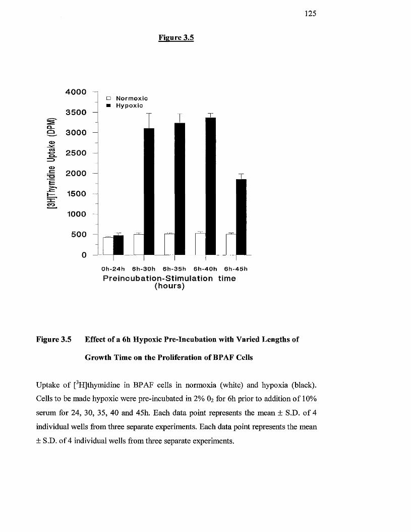
125
4 0 0 0
3 5 0 0sQ_Q 3 0 0 0<X>
<—» 2 5 0 0=>o>c 2 0 0 0E
15 0 0znCO
1000
5 0 0
0
Figure 3.5
□ N o r m o x i c ■ H y p o x ic
0 h - 2 4 h 6 h - 3 0 h 6 h - 3 5 h 6 h - 4 0 h 6 h - 4 5 h
Preincubation-Stim ulation time (hours)
Figure 3.5 Effect of a 6h Hypoxic Pre-Incubation with Varied Lengths of
Growth Time on the Proliferation of BPAF Cells
Uptake of [^HJthymidme in BPAF cells in normoxia (white) and hypoxia (black).
Cells to be made hypoxic were pre-incubated in 2% 0% for 6h prior to addition o f 10%
serum for 24, 30, 35, 40 and 45h. Each data point represents the mean ± S.D. o f 4
individual wells from three separate experiments. Each data point represents the mean
± S.D. of 4 individual wells from three separate experiments.
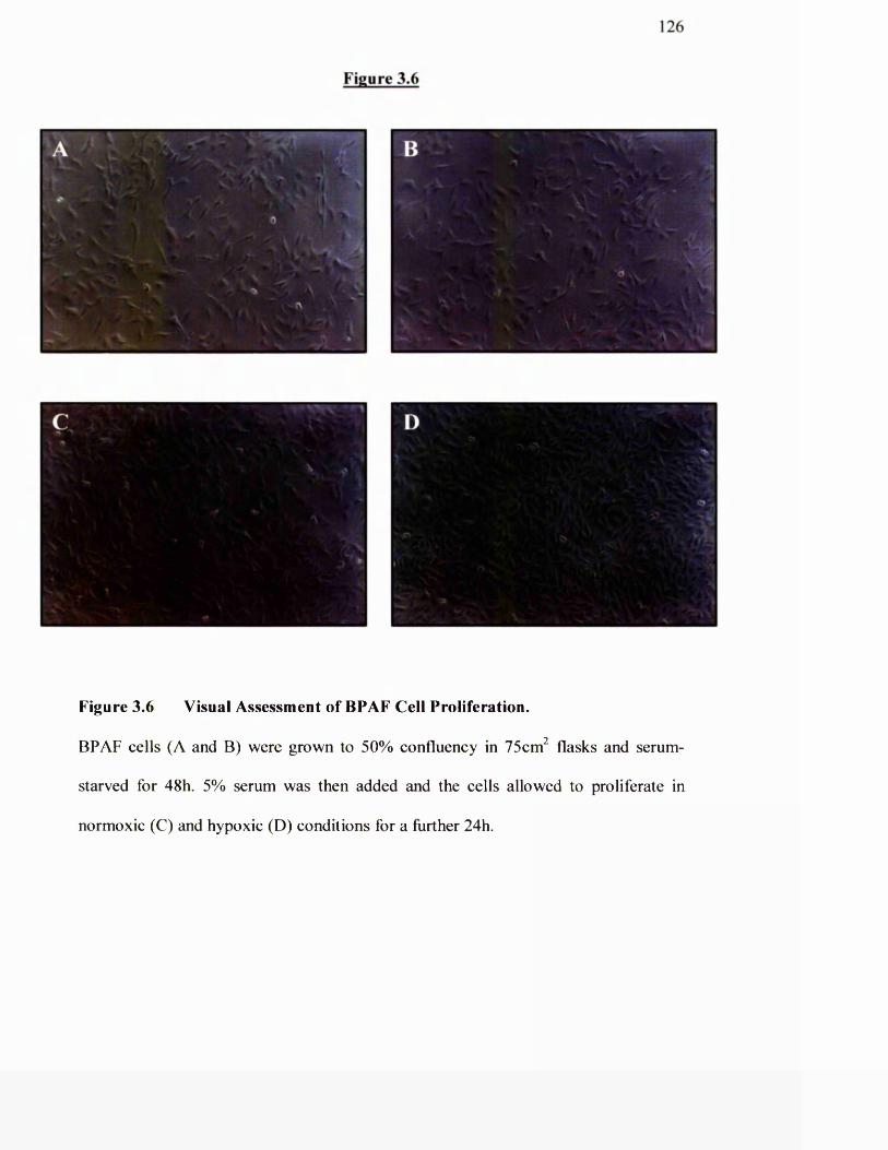
126
Figure 3.6
Figure 3.6 Visual Assessment of BPAF Cell Proliferation.
BPAF cells (A and B) were grown to 50% confluency in 75cm^ flasks and serum-
starved for 48h. 5% serum was then added and the cells allowed to proliferate in
normoxic (C) and hypoxic (D) conditions for a further 24h.
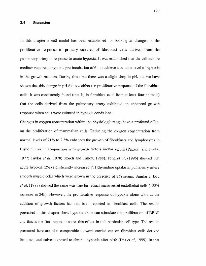
127
3.4 Discussion
In this chapter a cell mode! has been established for looking at changes in the
proliferative response o f primary cultures o f fibroblast cells derived from the
pulmonary artery in response to acute hypoxia. It was established that the cell culture
medium required a hypoxic pre-incubât ion o f 6h to achieve a suitable level o f hypoxia
in the growth medium. During this time there was a slight drop in pH, but we have
shown that this change in pH did not effect the proliferative response o f the fibroblast
cells. It was consistently found (that is, in fibroblast cells from at least four animals)
that the cells derived from the pulmonary artery exhibited an enhanced growth
response when cells were cultured in hypoxic conditions.
Changes in oxygen concentration within the physiologic range have a profound effect
on the proliferation o f mammalian cells. Reducing the oxygen concentration from
normal levels o f 21% to 2.5% enhances the growth o f fibroblasts and lymphocytes in
tissue culture in conjunction with growth factors and/or serum (Packer and Fuehr,
1977; Taylor et al, 1978; Storch and Talley, 1988). Feng et al, (1996) showed that
acute hypoxia (2%) significantly increased [^Hjthymidine uptake in pulmonary artery
smooth muscle cells which were grown in the presence o f 2% serum. Similarly, Lou
et al, (1997) showed the same was true for retinal microvessel endothelial cells (133%
increase in 24h). However, the proliferative response o f hypoxia alone without the
addition of growth factors has not been reported in fibroblast cells. The results
presented in this chapter show hypoxia alone can stimulate the proliferation o f BPAF
and this is the first report to show this effect in this particular cell type. The results
presented here are also comparable to work carried out on fibroblast cells derived
from neonatal calves exposed to clironic hypoxia after birth (Das et al, 1999). In that
128
study, cells from chronically hypoxic animals showed increased growth
characteristics to purified mitogens when compared to those from control animals.
In general, techniques used to assess proliferation measure the assimilation of
radio label led nucleotides such as [^HJthymidine into newly synthesised DNA.
Thymidine is commonly used for this purpose. Most tissue cultures comprise cells at
various stages o f the cell cycle, so cells are typically growth-arrested in serum-free
media. This synchronises cells to the Go/Gi phase o f the cell cycle in which cells
minimally incorporate [^HJthymidine (Panettieri et al, 1989).
There are a number o f potential drawbacks in relating [^HJthymidine uptake to DNA
synthesis. Firstly, although most DNA synthesis occurs during cell replication, some
o f the DNA synthesis may be for ongoing reparative processes rather than for
proliferation. Secondly, [^HJthymidine may be incorporated into cellular
macro molecules other than DNA, such as RNA and lipids. Despite these potential
artefacts, measuring uptake o f [^H]thymidine remains one o f the best and most widely
used methods for assessing cell proliferation.
These studies have established that pulmonary artery fibroblasts exposed to acute
hypoxia are associated with a significant change in the proliferative phenotype. It is
now important to understand the mechanisms contributing to the excessive fibroblast
proliferation under these conditions. The results presented in this chapter are the
foundations for the remaining chapters, which study further the responses of
pulmonary artery fibroblasts with respect to acute and chronic hypoxia and compare
these responses to those o f cells from the systemic circulation.

129
Chapter 4
Effect of Acute Hypoxia on Mitogenic Stimulation of Bovine Pulmonary Artery and Bovine Mesenteric Artery Fibroblast cells.
130
4.1 INTRODUCTION
In the previous chapter a cell model was established and characterised for measuring
acute hypoxic effects on the growth of primary cultures o f fibroblast cells. Results
from that chapter showed that hypoxia increased [^Hjthymidine uptake in the
fibroblast cells derived from bovine pulmonary artery.
It has been known for some time that hypoxia causes vasoconstriction in the
pulmonary circulation and vasodilation in the systemic circulation (von Euler and
Liljestrand, 1946). We hypothesised that there are fundamental differences in oxygen
sensing and cell signalling between systemic and pulmonary artery cells in response
to hypoxia, that is there is a link between the observed physiological vasoconstriction
and vascular remodelling. Growth factors are implicated in a wide variety o f
physiological and pathological processes. These include growth and development and
cell survival and tissue repair (Evered et al, 1985). An important link between growth
factors or their receptors and oncogene products has also been established (He Id in and
Westermark, 1984; Bishop, 1985; Weinberg, 1985). Thus, the elucidation o f the
mechanism o f action o f growth factors has emerged as one o f the fundamental
problems in biological sciences and may prove a crucial prerequisite for
understanding the cause or causes underlying the unrestrained proliferation of
pulmonary cells to hypoxia in disease states like pulmonary hypertension or o f other
hypoxic lung diseases (Lafont, 1995). In fact, it has been shown that there may be
some synergistic effects between hypoxia and mitogens on the proliferation o f cells.
For example, Feng and Cai (1996) demonstrated a synergistic effect o f hypoxia on
smooth muscle cell proliferation with ET-1, whilst others (Das et al, 1999) recently
131
showed synergism o f hypoxia on proliferation o f neonatal bovine pulmonary artery
fibroblasts to PDGF, basic fibroblast growth factor and insulin-like growth factor-1.
Previous work has shown that in pulmonary hypertension there is an increase in the
levels o f several mitogens such as ET-1, PDGF, thrombin, angiotensin II and 5-HT
(Rose et al, 1986; Ono and Voelkel, 1991 ; Giaid et al, 1993). In this chapter, we have
determined the effect o f hypoxia in combination with these mitogens to determine the
proliferative response o f BPAF and BMAF. We chose to compare the response with
that o f systemic vascular cells from the mesentery since previous work has suggested
that hypoxia affects ion exchange (potassium currents) differently in mesenteric and
pulmonary artery cells (Weir and Archer, 1995).
132
4.2 METHODS
BPAF and BMAF were grown in 24-welI plates as described in Chapter 2.3.1.
Following growth arrest, cells were either transferred to a hypoxic environment o f 2%
O2 or left in normoxic 21% O2 conditions. After a 6h preincubation to allow the cell
culture medium to become hypoxic, cells were stimulated with various agonists for
24h, [^HJthymidine being added for the final 4h. The uptake o f [^Hjthymidine was
measured as in Chapter 2.6.
4.3 RESULTS
4,3.1 Effect of hypoxia on serum-stimulated proliferation of BPAF and BMAF
cells.
In the previous chapter it was shown that growth arrested BPAF cells proliferated in
response to acute hypoxia in the absence o f mitogens (Chapter 3.4). To test the effects
o f hypoxia on proliferating bovine fibroblasts, growth-arrested BPAF were exposed to
2% O2 for 6h to make the cells hypoxic and then stimulated with 5% serum for 24h.
Thymidine uptake was used as a measure o f proliferation. These cells were
compared with those grown under normoxic conditions. In addition, growth of
fibroblasts derived ftom the pulmonaiy artery were compared to those from the
mesenteric artery. Serum concentrations as low as 0.1% were enough to stimulate
proliferation o f both BPAF and BMAF (Figure 4.1) by 6.6-fold and 5.5-fold
respectively. Hypoxia significantly enhanced the effect o f serum in BPAF cells by

133
about 6-fold (p<0.001). In contrast, there was no elfect o f hypoxia in growth arrested
or serum-stimulated BMAF cells (Figure 4 .IB).


134
Figure 4.1 Effect of Hypoxia on Serum -Induced Proliferation of BPAF and
BMAF cells.
BPAF (A) and BMAF (B) cells were grown to 60% confluency in 24-well plates and
then quiesced in serum-free medium. Cells were either maintained in a normoxic
environment for a further 6 h or were placed in a hypoxic incubator (2 % O2) for 6 h.
The cells were then stimulated for 24h with a range o f serum concentrations as
indicated in the graph. The cells were then allowed to grow in either normoxic or
hypoxic conditions for a period o f 24h. Cell growth was assessed by thymidine
uptake. Values shown are the mean ± S.D. for four replicate wells from the same
experiment. The experiment was repeated in cells derived from four different animals.
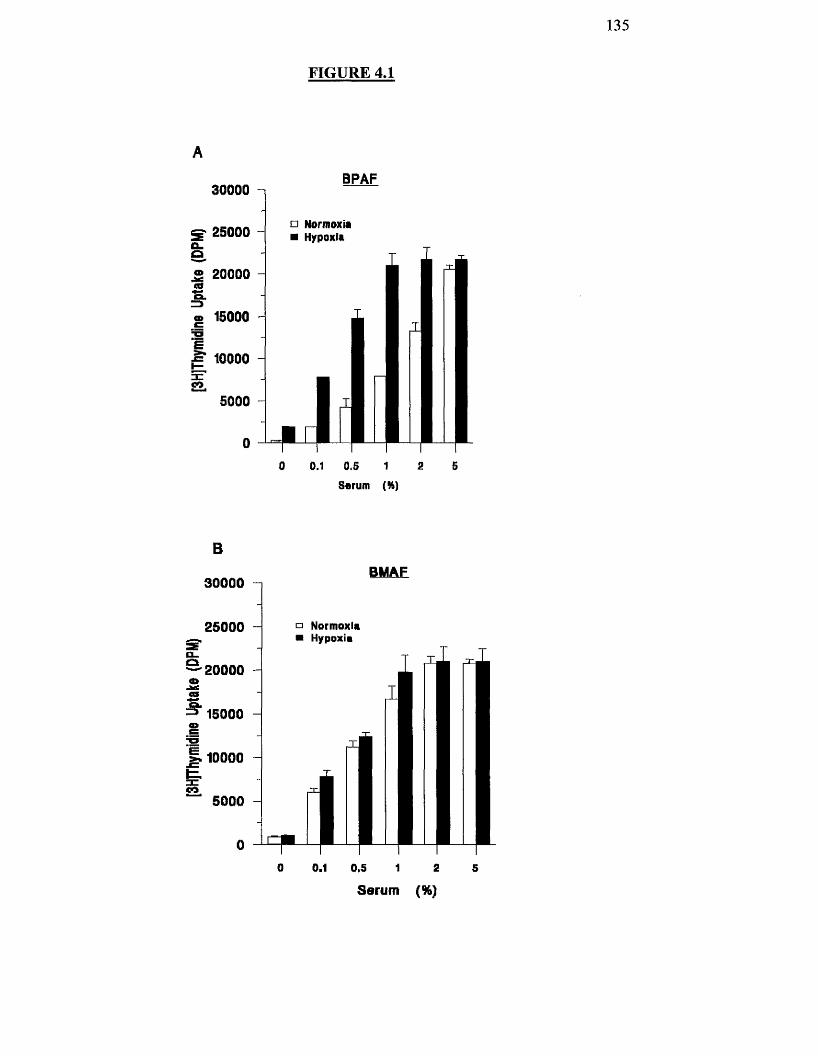
13 5
FIGURE 4.1
BPAF30000
□ Normoxia ■ Hypoxiag 25000
è® 20000 taex
<D 15000
g 10000
5000
00 0.1 0.5 a 51
Serum (%)
BMAF30000
a Normoxia25000Hypoxia
20000
15000
0.1 0.5 1
Serum (%)
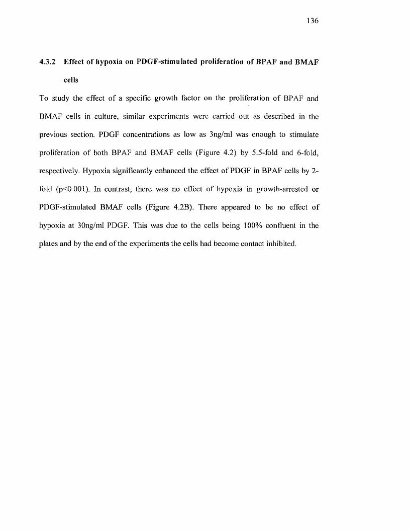
136
4.3.2 Effect of hypoxia on PDGF-stimulated proliferation of BPAF and BMAF
cells
To study the effect o f a specific growth factor on the proliferation o f BPAF and
BMAF cells in culture, similar experiments were carried out as described in the
previous section. PDGF concentrations as low as 3ng/ml was enough to stimulate
proliferation o f both BPAF and BMAF cells (Figure 4.2) by 5.5-fold and 6 -fold,
respectively. Hypoxia significantly enhanced the effect o f PDGF in BPAF cells by 2-
fold (p<0..001). In contrast, there was no effect o f hypoxia in growth-arrested or
PDGF-stimulated BMAF cells (Figure 4.2B). There appeared to be no effect o f
hypoxia at 30ng/ml PDGF. This was due to the cells being 100% confluent in the
plates and by the end o f the experiments the cells had become contact inhibited.

1 3 7
Figure 4.2 Effect of Hypoxia on PDGF-Stimulated Proliferation of BPAF and
BMAF Cells.
BPAF (A) and BMAF (B) cells were grown to 60% confluency in 24-well plates and
then quiesced in serum-free medium. Cells were either maintained in a normoxic
environment for a further 6 h or were placed in a hypoxic incubator (2 % O2) for 6 h.
The cells were then stimulated for 24h with a range o f PDGF concentrations as
indicated in the graph. The cells were then allowed to grow in either normoxic or
hypoxic conditions for a period o f 24h. Cell growth was assessed by [^HJthymidine
uptake. Values shown are the mean ± S.D. for four replicate wells from the same
experiment. The experiment was repeated in cells derived from four different animals.
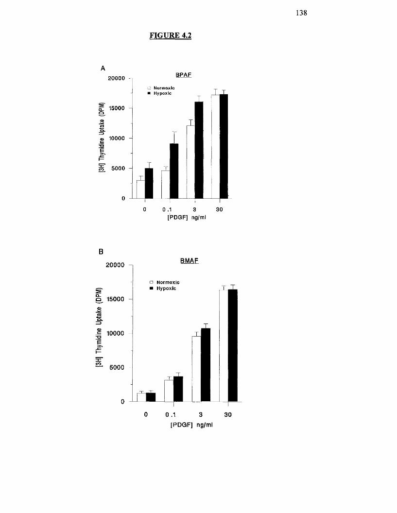
138
FIGURE 4.2
20000
f 15000Q
10000
BPAF
5000
□ Normoxic ■ Hypoxic T
là0 0 .1 3 30
[PDGF] ng/ml
B
20000
Q 15000
tJ 10000Ë
BMAF
rcco
5000
□ Norm oxicH ypoxic
0 0 .1 3 30
[PDGF] ng/ml
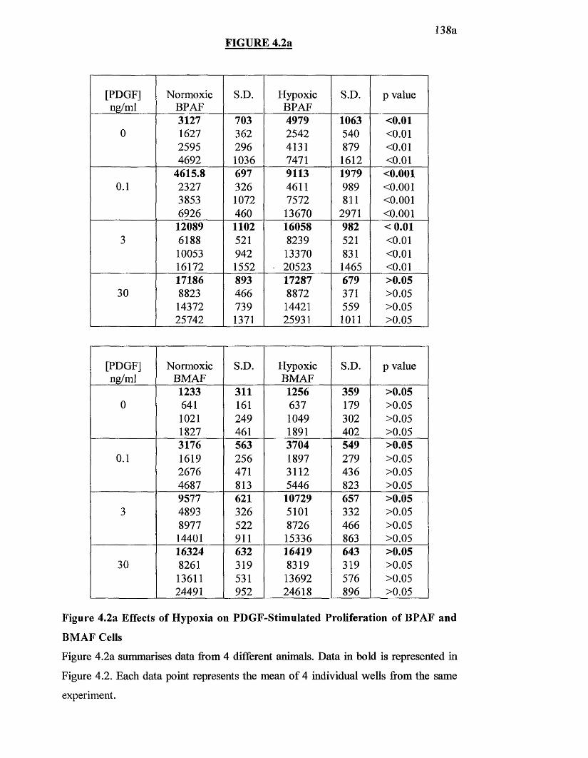
FIGURE 4.2a138a
[PDGF]ng/ml
NormoxicBPAF
S.D. HypoxicBPAF
S.D. p value
3127 703 4979 1063 <0.010 1627 362 2542 540 <0 .0 1
2595 296 4131 879 <0 .0 1
4692 1036 7471 1612 <0 . 0 1
4615.8 697 9113 1979 <0.0010 .1 2327 326 4611 989 <0 . 0 0 1
3853 1072 7572 811 <0 . 0 0 1
6926 460 13670 2971 <0 . 0 0 1
12089 1102 16058 982 < 0 .013 6188 521 8239 521 <0 .0 1
10053 942 13370 831 <0 .0 1
16172 1552 20523 1465 <0 .0 1
17186 893 17287 679 >0.0530 8823 466 8872 371 >0.05
14372 739 14421 559 >0.0525742 1371 25931 1 0 1 1 >0.05
[PDGF]ng/ml
NormoxicBMAF
S.D. HypoxicBMAF
S.D. p value
1233 311 1256 359 >0.050 641 161 637 179 >0.05
1 0 2 1 249 1049 302 >0.051827 461 1891 402 >0.053176 563 3704 549 >0.05
0 .1 1619 256 1897 279 >0.052676 471 3112 436 >0.054687 813 5446 823 >0.059577 621 10729 657 >0.05
3 4893 326 5101 332 >0.058977 522 8726 466 >0.0514401 911 15336 863 >0.0516324 632 16419 643 >0.05
30 8261 319 8319 319 >0.0513611 531 13692 576 >0.0524491 952 24618 896 >0.05
Figure 4.2a Effects of Hypoxia on PDGF-Stimulated Proliferation of BPAF and
BMAF Cells
Figure 4.2a summarises data from 4 different animals. Data in bold is represented in
Figure 4.2. Each data point represents the mean o f 4 individual wells from the same
experiment.
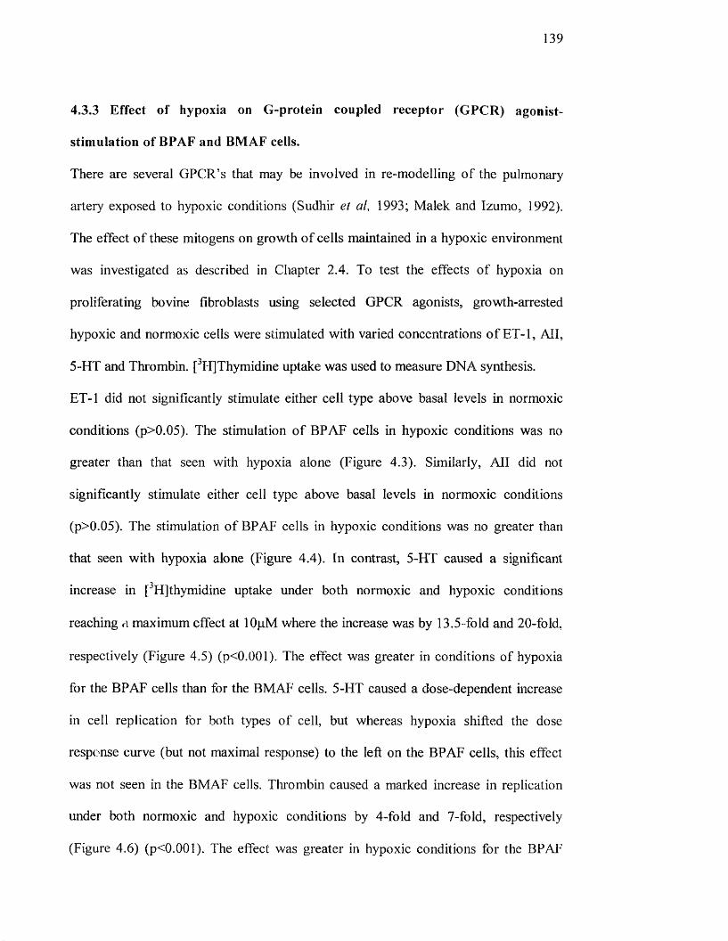
139
4,3,3 Effect of hypoxia on G-protein coupled receptor (GPCR) agonist-
stimulation of BPAF and BMAF cells.
There are several GPCR’s that may be involved in re-modelling o f the pulmonary
artery exposed to hypoxic conditions (Sudhir et al, 1993; Malek and Izumo, 1992).
The effect o f these mitogens on growth o f cells maintained in a hypoxic environment
was investigated as described in Chapter 2.4. To test the effects o f hypoxia on
proliferating bovine fibroblasts using selected GPCR agonists, growth-arrested
hypoxic and normoxic cells were stimulated with varied concentrations o f ET-1, All,
5-HT and Thrombin. [^H]Thymidine uptake was used to measure DNA synthesis.
ET-1 did not significantly stimulate either cell type above basal levels in normoxic
conditions (p>0.05). The stimulation o f BPAF cells in hypoxic conditions was no
greater than that seen with hypoxia alone (Figure 4.3). Similarly, All did not
significantly stimulate either cell type above basal levels in normoxic conditions
(p>0.05). The stimulation o f BPAF cells in hypoxic conditions was no greater than
that seen with hypoxia alone (Figure 4.4). In contrast, 5-HT caused a significant
increase in [^Hjthymidine uptake under both normoxic and hypoxic conditions
reaching <\ maximum effect at lOpM where the increase was by 13.5-fold and 20-fold,
respectively (Figure 4.5) (p<0.001). The effect was greater in conditions of hypoxia
for the BPAF cells than for the BMAF cells. 5-HT caused a dose-dependent increase
in cell replication for both types o f cell, but whereas hypoxia shifted the dose
response curve (but not maximal response) to the left on the BPAF cells, this effect
was not seen in the BMAF cells. Thrombin caused a marked increase in replication
under both normoxic and hypoxic conditions by 4-fold and 7-fold, respectively
(Figure 4.6) (p<0.001). The effect was greater in hypoxic conditions for the BPAF

140
cells than for the BMAF cells by about 1.7-fold. Thrombin caused a dose-dependent
increase in cell replication for both types o f cell, but whereas hypoxia shifted the dose
response curve (but not maximal response) to the left on the BPAF cells, this effect
was not seen in the BMAF cells.


141
Figure 4.3 Effect of Hypoxia on ET-l-Stimulated Proliferation of BPAF and
BMAF Cells.
BPAF (A) and BMAF (B) cells were grown to 60% confluency in 24-well plates and
then quiesced in serum-free medium. Cells were either maintained in a normoxic
environment for a further 6 h or were placed in a hypoxic incubator (2 % 0 %) for 6 h.
The cells were then stimulated for 24h with a range o f ET-1 concentrations as
indicated in the graph. The cells were then allowed to grow in either normoxic or
hypoxic conditions for a period o f 24h. Cell growth was assessed by [^H]thymidine
uptake. Values shown are the mean ± S.D. for four replicate wells from the same
experiment The experiment was repeated in cells derived from four different animals.
142
FIGURE 4.3
BPAF10000
a Normoxic Hypoxic9000 -
7000 -
M 5000
4000
en 3000
Control 0.1 3[ET-1]uM
10
BMAF
□ N o rm o x ic ■ Hypoxic
10000
8000
7000
6000
5000
22. 3000
2000
Control 0.1 3[ET-1]uM
10

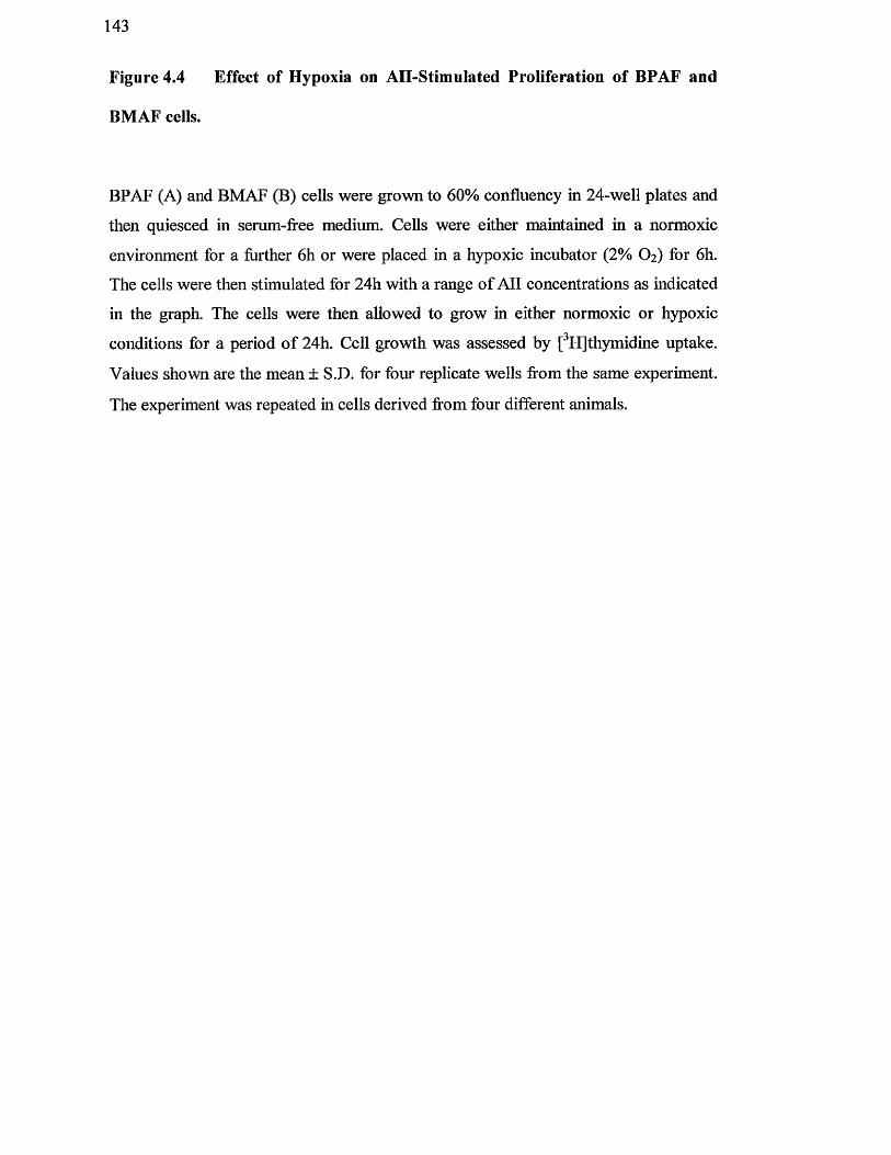
143
Figure 4.4 Effect of Hypoxia on AH-Stimulated Proliferation of BPAF and
BMAF cells.
BPAF (A) and BMAF (B) ceUs were grown to 60% confluency in 24-well plates and
then quiesced in serum-free medium. Cells were either maintained in a normoxic
environment for a further 6 h or were placed in a hypoxic incubator (2 % O2) for 6 h.
The cells were then stimulated for 24h with a range o f A ll concentrations as indicated
in the graph. The cells were then allowed to grow in either normoxic or hypoxic
conditions for a period o f 24h. Cell growth was assessed by [^HJthymidine uptake.
Values shown are the mean ± S.D. for four replicate wells from the same experiment.
The experiment was repeated in cells derived from four different animals.
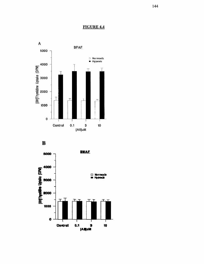
144
FIGURE 4.4
ABPAF
5000
4000
o NormoxEc ■ Hypoxic
1Q3000
2000
1000
Control 0.1 3 10[AII]uM
B
5000 -1 BMAF
I4000
* 9000
2000
1000
□ NBrmnxto■ HbrfMKie
mmOontrol 0.1 3
lAMuM10

145
Figure 4.5 Effect of Hypoxia on 5-HT-Stimulated Proliferation of BPAF and
BMAF Cells.
BPAF (A) and BMAF (B) cells were grown to 60% confluency in 24-well plates and
then quiesced in serum-free medium. Cells were either maintained in a normoxic
environment for a further 6 h or were placed in a hypoxic incubator (2 % O2) for 6 h.
The cells were then stimulated for 24h with a range of 5-HT concentrations as
indicated in the graph. The cells were then allowed to grow in either normoxic or
hypoxic conditions for a period o f 24h. Cell growth was assessed by [^H]thymidine
uptake. Values shown are the mean ± S.D. for four replicate wells from the same
experiment. The experiment was repeated in cells derived from four different animals.
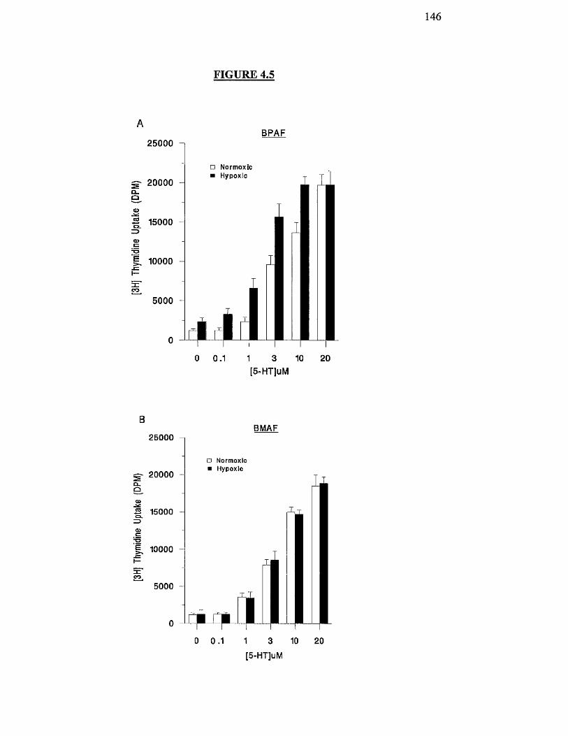
146
25000
20000OuO
^ 15000
CDCT=1g. 10000
5000
FIGURE 4.5
BPAF
□ Normoxic ■ Hypoxic
0 0.1 1 3 10 20[5"HT]uM
BMAF25000
□ Normoxic ■ Hypoxic
20000Q.Q
B 15000
<3>
K 10000
3 =eo
5000 -
0 0.1 1 3 10 20[5-HT]uM

147
Figure 4.6 Effect of Hypoxia on Thrombin-Stimulated Proliferation of BPAF
and BMAF Cells.
BPAF (A) and BMAF (B) cells were grown to 60% confluency in 24-well plates and
then quiesced in serum-free medium. CeUs were either maintained in a normoxic
environment for a fiirther 6 h or were placed in a hypoxic incubator (2 % O2) for 6 h.
The cells were then stimulated for 24h with a range o f thrombin concentrations as
indicated in the graph. The cells were then allowed to grow in either normoxic or
hypoxic conditions for a period o f 24h. Cell growth was assessed by [^HJthymidine
uptake. Values shown are the mean ± S.D. for four replicate wells from the same
experiment. The experiment was repeated in cells derived from four different animals.
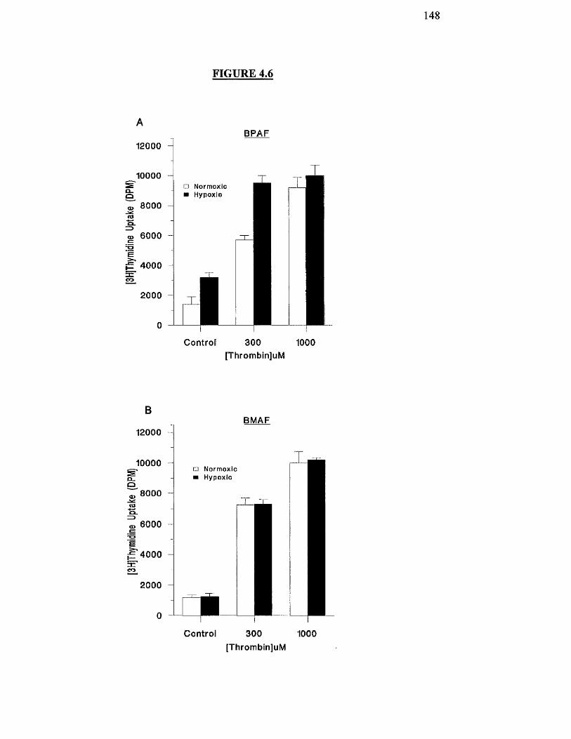
148
FIGURE 4.6
Q_Q
A
12000
10000
8000
BPAF
o>sa .
g 6000
’e§5 4000 S '£2.
2000
□ Norm oxicHypoxic
C ontro l 300 1000[Thrombin]uM
O uo
r nco
B
12000
10000
8000
6000
4000
2000
BMAF
□ NormoxicHypoxic
Control 300 1000[Thrombin]uM
149
4.4 Discussion
This chapter demonstrates that oxygen concentrations at the physiological
concentration of 5%, which is similar to that observed in pulmonary hypertensive
patients (Souhami & Moxham, 1998), increases the proliferation o f BPAFs over and
above the response o f these cells to serum and specific growth factors and GPCR’s.
DNA synthesis was increased in BPAF’s in response to serum, as well as to PDGF, 5-
H l' and thrombin and this was enhanced further under hypoxic conditions. This effect
was specific to cells derived from the pulmonary artery since cells derived from the
systemic circulation were non-responsive to an acute hypoxic challenge. We have
previously shown ET-1 to be a potent agonist on pulmonary artery fibroblasts in the
rat (Peacock et al, 1992), but this did not affect bovine pulmonary artery cells in
either hypoxic or normoxic conditions. This is consistent with our previous studies
where we showed that ET-1 does not promote replication in bovine cells (Peacock
and Aidulis, 1994). We have been unable to find any other study where BMAF cell
replication has been studied.
As well as changes observed here in hypoxia-stimulated proliferation between BPAF
and BMAF cells, other workers have observed physiological differences between the
two types o f cell. For example, there is also a differential effect o f hypoxia on
channel activation in isolated pulmonary and mesenteric artery cells (Weir and
Archer 1995).
Increased pulmonary artery fibroblast proliferation in cells derived from the cow in
response to hypoxia was accompanied by changes in specific protein kinase C (PKC)
isozymes, suggesting a signal transduction pathway linking hypoxia with
150
proliferation in the adventitial fibroblast (Stenmark and Mecham, 1997). These
results agree with the findings by others that oxygen concentrations within the
physiological range may control proliferation indirectly by altering the activity o f a
signalling pathway that regulates the response to growth factors.
It is possible that the additive effect o f hypoxia over and above normal growth
responses leading to increased proliferation triggers a novel signalling pathway, or
enhances the activation o f an existing pathway leading to the increased proliferation.
Hypoxia may also cause the increase in transcription o f proteins involved in cell
growth, for example, VEGF or LNOS. The gene for this has already been show to be
turned on in hypoxic conditions (Feldkamp et al, 1999) There is evidence in other cell
types o f specific genes switched on by hypoxia such as EPO (Firth et al, 1994). It is
possible that pulmonary artery cells contain genes not present in BMAF cells allowing
both contraction and replication to hypoxia and that these phenomena are coupled
(Scott and Peacock, 1995).

151
Chapter 5
Effects of Acute Hypoxia on IPi Generation in
BPAF and BMAF cells.
152
5.1 INTRODUCTION
In the previous chapter, it was shown that exposing quiescent cells to an oxygen
concentration at a physiological level o f hypoxia could increase proliferation. In
addition, it also enhances the effect o f some growth factors and GPCR agonists. This
effect appeared to be specific for pulmonary derived fibroblast cells since this did not
occur in the same cell type derived from the mesenteric artery. If low oxygen
concentrations can control cell proliferation then it might be expected that the activity
o f signalling enzymes, and pathways, might be altered in the cellular response to
hypoxia. The aim o f the studies described in this chapter was to investigate the
possible importance o f the second messenger IP3 to the hypoxic response o f cells.
The actions of many GPCR agonists and growth factors are mediated by the
hydrolysis of PIP2 catalysed by PLC (Berridge and Irvine, 1989). This hydrolysis
releases IP3, which mobilises Ca " from the endoplasmic reticulum, and DAG, which
activates some PKC iso forms (Hug and Sarre, 1993).
Previous work has shown that hypoxia increases cytosolic [Ca^^]i in pulmonary artery
smooth muscle cells. It was not known whether or not the source o f this increased
Ca " was intracellular or extracellular, since previous reports have not specifically
looked at the effect o f hypoxia in IP3 mediated generation of Ca^^ (Cornfield et al,
1993). The Ca^^ may be derived from intracellular stores or by Ca^^ influx mediated
by voltage-dependent or receptor-operated Ca^^ channels (Berridge, 1993b). In
general, transient increases in [Ca^ ],- are mediated by IP3 release o f Ca^^ from
intracellular stores whilst sustained increases in [Ca " ]j are mediated by Ca^^ influx
through activation of the Ca " channels (Chadwick et al, 1990). M inami et al (1991)
examined AH induced changes in IP] levels in VSMC from hypertensive rats and
153
found a rapid increase in IP3 levels peaking at 5 seconds. Similar results were
demonstrated by Yokokawa et al (1995), who studied the response of endothelial cells
from hypertensive rats to ET-1. Transient increases in IP3 levels were also reported
when frogs were subjected to hypoxic/anoxic conditions (Holden and Storey, 1997).
In the present study, IP3 levels were measured in quiescent BPAF and BMAF cells
exposed acutely to hypoxic conditions in the presence and absence of various
agonists, which have been shown to be increased in patients with pulmonary
hypertension.

154
5.2 METHODS
BPAF and BMAF cells were plated out into 24-well plates and quiesced as described
in chapter 2.3.1. Cells were then either maintained in normoxic conditions or exposed
to 2% O2 for 6 h prior to addition o f agonists. IP3 mass was measured as described in
2 .6 .

155
5.3 RESULTS
5,3.1 Typical standard curve for IP3 generation.
Figure 5.1 shows a typical standard curve generated using a competitive binding assay
with bovine adrenal glands which was used to find the unknown levels o f IP3 in
stimulated cell extracts.
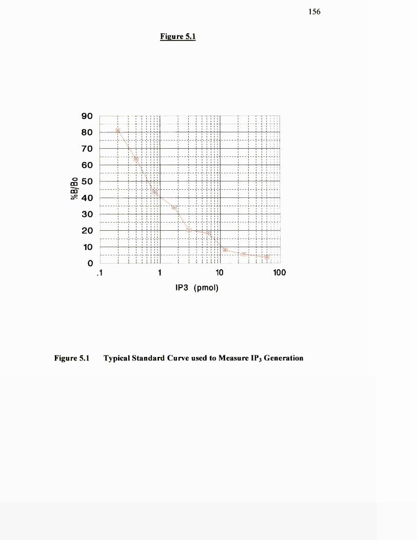
156
Figure 5.1
9 0
8 0
7 0
6 0
° 5 0COm
4 0
3 0
20
10
0
-----------------
j
- - . . . - . . . - - . . . - - - -
4
r - - - -- -
To]"'
4
i i i i i L i l L i i i I T 11 10
IP3 (pmol)
100
Figure 5.1 Typical Standard Curve used to Measure IP3 Generation
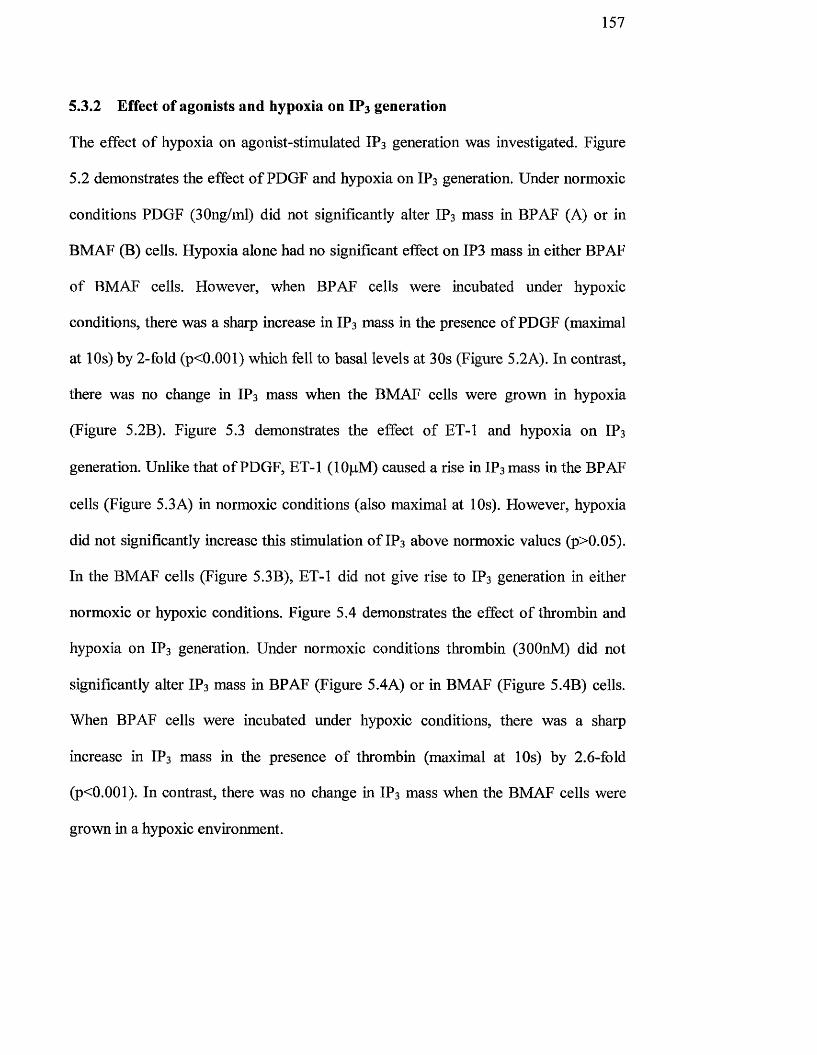
1 5 7
5.3.2 Effect of agonists and hypoxia on IP3 generation
The effect o f hypoxia on agonist-stimulated IP3 generation was investigated. Figure
5.2 demonstrates the effect o f PDGF and hypoxia on IP3 generation. Under normoxic
conditions PDGF (30ng/ml) did not significantly alter IP3 mass in BPAF (A) or in
BMAF (B) cells. Hypoxia alone had no significant effect on IP3 mass in either BPAF
o f BMAF cells. However, when BPAF cells were incubated under hypoxic
conditions, there was a sharp increase in IP3 mass in the presence o f PDGF (maximal
at 10s) by 2-fold (p<0.001) which fell to basal levels at 30s (Figure 5.2A). In contrast,
there was no change in IP3 mass when the BMAF cells were grown in hypoxia
(Figui'e 5.2B). Figure 5.3 demonstrates the effect o f ET-1 and hypoxia on IP3
generation. Unlike that o f PDGF, ET-1 (lOpM) caused a rise in IP3 mass in the BPAF
cells (Figure 5.3A) in normoxic conditions (also maximal at 10s). However, hypoxia
did not significantly increase this stimulation o f IP3 above normoxic values (p>0.05).
In the BMAF cells (Figure 5.3B), ET-1 did not give rise to IP3 generation in either
normoxic or hypoxic conditions. Figure 5.4 demonstrates the effect o f thrombin and
hypoxia on IP3 generation. Under normoxic conditions thrombin (300nM) did not
significantly alter IP3 mass in BPAF (Figure 5.4A) or in BMAF (Figure 5.4B) cells.
When BPAF cells were incubated under hypoxic conditions, there was a sharp
increase in IP3 mass in the presence o f thrombin (maximal at 10s) by 2.6-fold
(p<0.001). In contrast, there was no change in IP3 mass when the BMAF cells were
grown in a hypoxic environment.
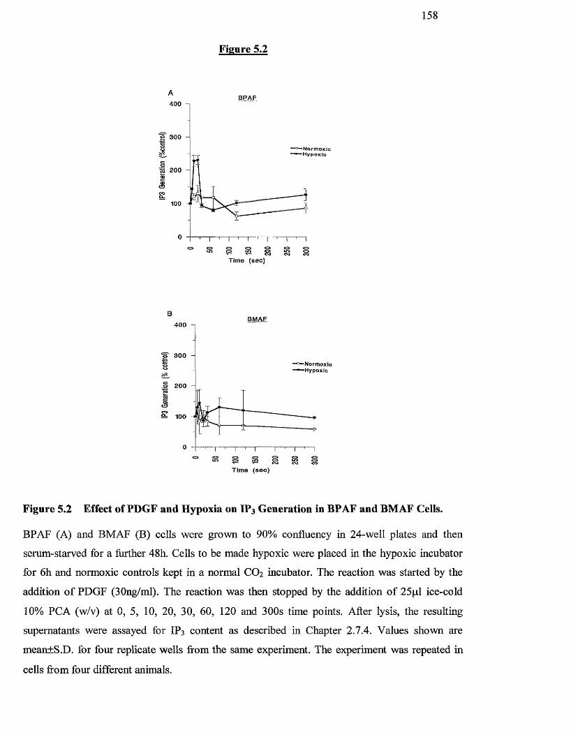
158
Figure 5.2
BPAF4 0 0 n
200
O
100
s S gg gTime (sec)
BMAF4 0 0
3 0 0
.1 200
& 100
g sggTim e (se c )
Figure 5.2 Effect of PDGF and Hypoxia on ÏP3 Generation in BPAF and BMAF Cells.
BPAF (A) and BMAF (B) cells were grown to 90% confluency in 24-well plates and then
serum-starved for a further 48h. Cells to be made hypoxic were placed in the hypoxic incubator
for 6 h and normoxic controls kept in a normal CO2 incubator. The reaction was started by the
addition o f PDGF (30ng/ml). The reaction was then stopped by the addition o f 25pi ice-cold
10% PGA (w/v) at 0, 5, 10, 20, 30, 60, 120 and 300s time points. After lysis, the resulting
supernatants were assayed for IP3 content as described in Chapter 2.7.4. Values shown aie
mean+S.D. for four replicate wells from the same experiment. The experiment was repeated in
cells from four different animals.
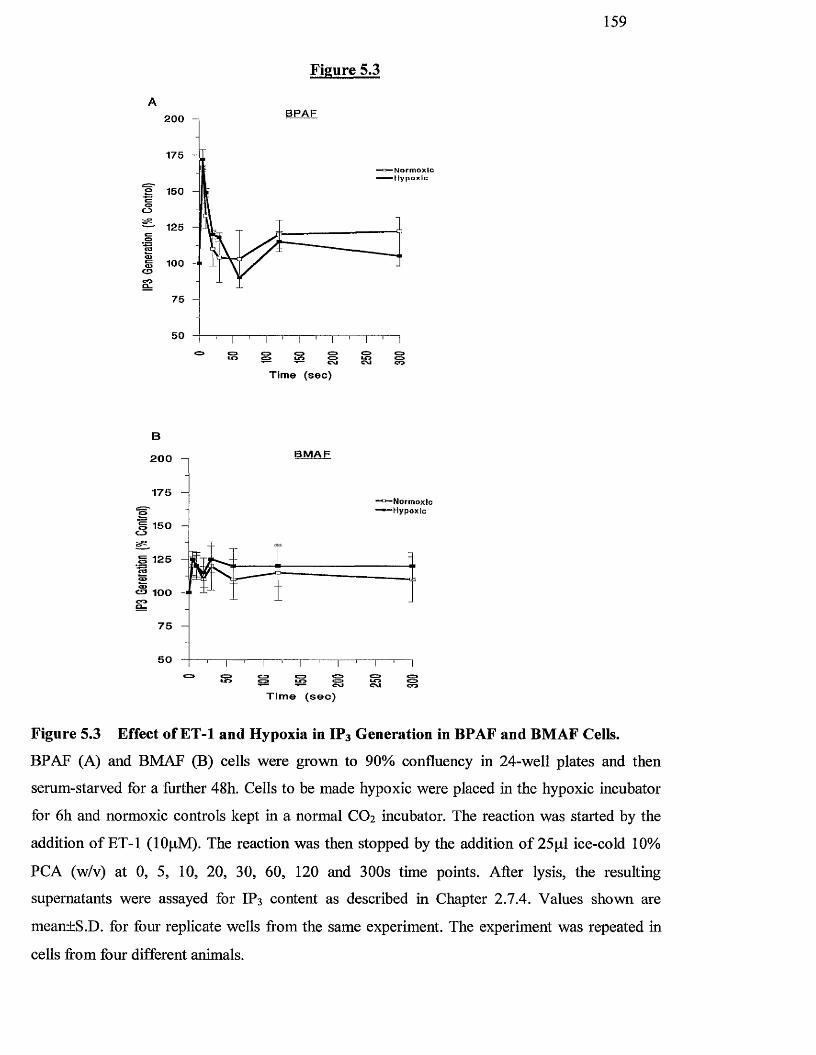
1 5 9
Figure 5.3
B P A F200 n
175 -
- - H y p o x i c
S 150
<3125
fë 100
C£J
275
50
SS Ss sT i m e ( s e c )
B M A F200
175
150
125
75
50
T i m e ( s e c )
Figure 5.3 Effect of ET-1 and Hypoxia in IP3 Generation in BPAF and BMAF Cells.
BPAF (A) and BMAF (B) cells were grown to 90% confluency in 24-well plates and then
serum-starved for a further 48h. Cells to be made hypoxic were placed in the hypoxic incubator
for 6 h and normoxic controls kept in a normal CO2 incubator. The reaction was started by the
addition o f ET-1 (lOpM). The reaction was then stopped by the addition o f 25pi ice-cold 10%
PCA (w/v) at 0, 5, 10, 20, 30, 60, 120 and 300s time points. After lysis, the resulting
supernatants were assayed for IP3 content as described in Chapter 2.7.4. Values shown are
mean±S.D. for foui' replicate wells fi'om the same experiment. The experiment was repeated in
cells fi'om four different animals.
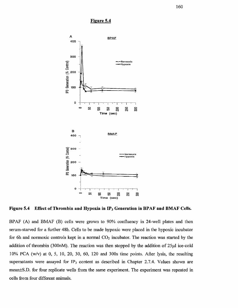
1 6 0
Figure 5.4
B P A F400
300
oss 200
o 100
0
sT im e ( s e c )
BM AF400
"5 3 0 0
&2 200
100
S mT i m e ( s e c )
Figure 5.4 Effect of Thrombin and Hypoxia in IF3 Generation in BPAF and BMAF Cells.
BPAF (A) and BMAF (B) cells were grown to 90% confluency in 24-well plates and then
serum-starved for a further 48h. Cells to be made hypoxic were plaeed in the hypoxic incubator
for 6h and normoxic controls kept in a normal CO2 incubator. The reaction was started by the
addition o f thrombin (300nM). The reaction was then stopped by the addition o f 25p,l ice-cold
10% PCA (w/v) at 0, 5, 10, 20, 30, 60, 120 and 300s time points. After lysis, the resulting
supernatants were assayed for IP3 content as described in Chapter 2.7.4. Values shown are
mean±S.D. for four replicate wells ftom the same experiment. The experiment was repeated in
cells from four different animals.

161
5.4 DISCUSSION
In this chapter, IP3 mass was analysed in both BPAF and BMAF cells in response to
various agonists in cells grown under normoxic or hypoxic conditions. Hypoxia alone
did not increase basal IP3 mass but stimulated an early rise in IP3 mass when cells
were stimulated with PDGF or thrombin. ÏP3 mass was found to be increased in
response to ET-1 in the BPAF cells hrespective o f the oxygen environment. BMAF
cells did not display this IP3 peak in response to hypoxia with any o f the agonists
used, and basal levels o f IP3 in the cells made hypoxic were not altered.
In the previous chapter, hypoxia was found to increase cell proliferation over and
above that o f agonists alone, for example, in response to PDGF and thrombin. We
have shown for the first time that there is a rapid rise in IP3 mass when hypoxic cells
ai’e stimulated with agonists, which also stimulate proliferation, such as thrombin. In
the case o f E T - 1 , which does not stimulate B P A F proliferation, there is no additional
increase in IP3 mass with hypoxia. In contrast to the pulmonary cells, fibroblast cells
from the mesenteric artery did not have an increase in IP3 mass generation under
hypoxic conditions.
The difference in response to hypoxia between pulmonary cells and mesenteric cells
may be due to an hypoxia-activated pathway which is either present only in
pulmonary artery cells, or is repressed in mesenteric artery cells. Activation o f cell-
surface receptors and their associated tyrosine kinase and phospholipases (C-y, A2 or
D) leads to the generation o f the lipid seeond messengers IP3 and DAG (Kohno et al,
1992; Nishizuka, 1992). IP3 promotes mobilisation o f intracellular Ca^^ stores (Short
et al, 1993) whereas DAG is an endogenous activator o f PKC (Nishizuka 1992).
Hence, an increase in IP3, caused in BPAF by hypoxia, may accompany an increase in
162
DAG or PKC activity. Although DAG was not examined in this chapter, it has been
reported that hypoxia can increase PKC activity in PASMC (Xu et al, 1994). Thus,
PKC activity could be enhanced in two ways; firstly, by increasing DAG and,
secondly, by IP3 induced release o f Ca^^ from intracellular Ca^^ stores. Increases in
intracellular Ca^^ can directly activate some isoforms o f PKC (Ho et al, 1988).
Interestingly, activators o f PKC have been shown to promote many o f the same
cellular responses (contraction, hypertrophy and proliferation) attributed to hypoxia
(Orton et al, 1990; Starksen et al, 1986; Dempsey et al, 1991). In addition, induction
and phosphorylation o f PKCa has been observed in response to hypoxia in lung
fibroblast cells (Hasen et al, 1996). This iso form is involved in the PKC-dependent
regulation o f the erythropoetin (EPO) gene, which is induced by hypoxia (Fandrey et
al, 1994). If this explanation o f the activation o f hypoxia is true it is not clear why the
same pathway is not acth'ated in mesenteric cells. The differences within the two cell
types studied here do, however, show the same trends as seen in the circulation o f
humans in vivo when given hypoxic challenges (Von Euler and Liljestrand, 1946).
That is, the pulmonary arteries contract and cellular proliferation ensues, whereas in
the systemic circulations this does not happen.
It is likely that pulmonary artery cells contain genes allowing both contraction and
replication to hypoxia and that these phenomena are coupled (Scott and Peacock,
1995). It is tempting to speculate that the phosphoinositide pathway might be
involved downstream from any hypoxic sensor and responsible for vasomotor - cell
growth coupling, that is, IP3 causes increased intracellular Ca^^ resulting in cellular
contractions but also augmentations o f DAG-induced cell replication.

163
Chapter 6
n f Acute H v n o x i a on the Stimi.iation of the Stress-Activated prnfAln kinases in Pulmonary Artery Fibroblasts
164
6.1 INTRODUCTION
As described in previous chapters, serum-deprived BPAF cells proliferate under
hypoxic conditions, and the second messenger, IP3, is increased in these hypoxic cells.
However, the intracellular signalling mechanisms by which hypoxia stimulates cell
proliferation in pulmonary artery fibroblasts need to be further characterised.
O f particular importance in growth factor signalling is the activation o f a series o f
kinase cascades involving multiple serine/threonine and tyrosine phosphorylation
events (Malarkey et al, 1995). One such pathway is the MAP kinase cascade (Davis,
1993) which stimulates the phosphorylation o f several intracellular substrates, such as
p9 0 rsk and Elk-1, believed to play a role in initiating mitogenesis (Ballou et al, 1991 ;
Gille et al, 1995). MAP kinases are pro line-directed protein kinases that mediate the
effects o f numerous extracellular stimuli on a wide array o f biological processes, such
as cellular proliferation, differentiation and death (Datta et al, 1999). MAP kinase is
also regulated in a cell cycle-dependent fashion and is required for cells to enter S-
phase (Tamemoto et al, 1992). For example, in Chinese hamster lung fibroblasts,
Pages and co-workers (1993) found that activation o f both p42 and p44 iso forms o f
the ‘classical’ p42/p44 Map kinases were essential for proliferation. Similarly, using
human airway smooth muscle cells (HASM), Orsini and co-workers (1999) found that
proliferation in this cell type required prolonged activation o f p42/p44 MAP kinase.
Not only have these forms o f MAP kinase been shown to be activated in response to
mitogens, but also have been shown to be activated in response to hypoxia. For
example, in rat cardiac myocytes, components o f the MAP kinase cascade, including
Raf-1, MEK and MAP kinase, have been shown to be activated in response to low
oxygen conditions (Seko et al, 1996). However, these responses are very small when
165
compared to mitogen stimulation o f these pathways. Other workers (Richard et al,
1999), again using Chinese hamster lung fibroblasts, found that p42/p44 MAP kinase
phosphorylated hypoxia-inducible Factor l a (H IF-la) and enhanced the
transcriptional activity ofF tlF-l.
In recent years, protein serine/threonine kinases related to the ‘classical’ forms of
MAP kinase have been identified, and are referred to as the stress-activated protein
kinases (SAPK) (Kyriakis, ei al, 1994). These kinases, consisting o f at least two
homologues, c-Jun N-tenninal kinase (JNK) and p38 MAP kinase (Han et al, 1994;
Rouse et al, 1994; Raingeaud et al, 1995), are strongly activated by environmental
stress including heat shock and UV irradiation, cytokines such as tumour necrosis
factor (TNF), interleukin-1 (IL-1), toxins such as lipopolysaccharide (LPS) and
hypoxia (Han et al, 1994; Rouse et al, 1994; Raingeaud et al, 1995; Verheij et al,
1996; Lee et al, 1994b). Others have shown that the JNK cascade is a key pathway by
which agonists stimulate DNA synthesis in primary cultures o f rat hepatocytes (Auer
et al, 1998) whilst a role for p38 has been shown to increase myocardial cell growth
and gene expression (Zechner et al, 1997), and also is involved in the proliferation of
adventitial fibroblasts from chronically hypoxic calves (Das et al, 1999). Since these
kinase pathways play a critical role in responding to cellular stress and promoting cell
growth and survival (Widmann et al, 1999), we have investigated the effect of
hypoxia upon the p38 KAP kinase, JNK and p42/p44 MAP kinase signalling
enzymes.
166
6.2 METHODS
6.2.1 Cell Culture
Bovine lungs from adult cows were obtained from the local abattoir and pulmonary
artery fibroblasts from the freshly excised lung tissue were harvested as described in
Chapter 2.3.2. The identity o f fibroblast cells was confirmed by immunohistochemical
stains as described in Chapter 2.3.3.
6.2.2 Growth of cells in a hypoxic environment
A humidified temperature controlled incubator (LEEC model GA156 Colwick,
Nottingham, UK) was used as a hypoxic chamber as described in Chapter 2,4,1.
6.2.3 Solid-Phase JNK Assay
JNK activity was measured by a solid-state kinase assay with GST-c-Jun as the
substrate (Chapter 2.8.5). Phosphorylated proteins were separated by SDS-PAGE and
detected by autoradiography.
6.2.4 Solid-Phase p38 MAP kinase Assay
p38 MAP kinase activity was measured by a solid-state kinase assay utilising GST-
MAPKAP kinase-2 as the substrate (Chapter 2.8.6).
167
6.2.5 p42/44 MAP kinase phosphorylation and activation
MAP kinase phosphorylation status was assessed by Western blotting using
retardation on SDS-PAGE gels as a marker o f phosphorylation (Chapter 2.8.4).
Activity was assayed in solubilised cell lysates using the EGF receptor peptide
EGF^^O-681 as substrate (Chapter 2.8.7). The phosphorylated peptide was separated
from other products by ion-exchange chromatography on Whatman p81 paper and
quantified by liquid scintillation counting.
6.2.6 Statistics
Results are expressed as the mean ± S.D. Statistical analysis was undertaken as
reported in Chapter 2.10.
168
6.3 RESULTS
6.3.1 Effect of hypoxia on JNK and p38 MAP kinases
Hypoxia is an extremely common physiological stressor. To investigate the effects of
hypoxia on the stress-activated signalling pathways, BPAF cells were exposed to 2%
oxygen for various times. Cell lysates were used to study the phosphorylation of GST-
c-jun (JNK activity) or GST-MAPKAPK-2 (p38 activity).
Figure 6 .1 shows the effect o f hypoxia on JNK activity over a 24 h period. After a lag
period o f up to 3h a large increase in JNK activity was observed in response to
hypoxia which peaked at approximately 6h before decreasing slowly towards basal
values for the remainder o f the time course. Stimulation o f JNK activity under
hypoxic conditions at the 6h time point was 10.5-fold increased over basal activity,
and was at least as great as that induced by 0.5M sorbitol, a known activator o f JNK.
Figure 6.2 shows the effect o f hypoxia on p38 MAP kinase activity over a 3Oh time
period. It was found that hypoxia could also stimulate p38 MAP kinase activity. Like
JNK, maximum activity was reached by 6h before returning to basal between by 12h
(Figure 6.2A). However, in contrast to JNK activity, a second peak o f p38 MAP
kinase activity was observed which was maximal between 18-24 h before returning to
basal levels again by 30h (Figure 6.2B).
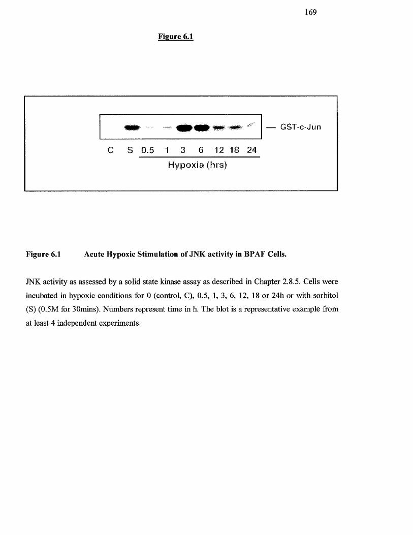
1 6 9
Figure 6.1
GST-c-Jun
C S 0.5 1 3 6 12 18 24
Hypoxia (hrs)
Figure 6.1 Acute Hypoxic Stimulation of JNK activity in BPAF Cells.
JNK activity as assessed by a solid state kinase assay as described in Chapter 2.8.5. Cells were
incubated in hypoxic conditions for 0 (control, C), 0.5, 1, 3, 6, 12, 18 or 24h or with sorbitol
(S) (0.5M for 30mins). Numbers represent time in h. The blot is a representative example from
at least 4 independent experiments.
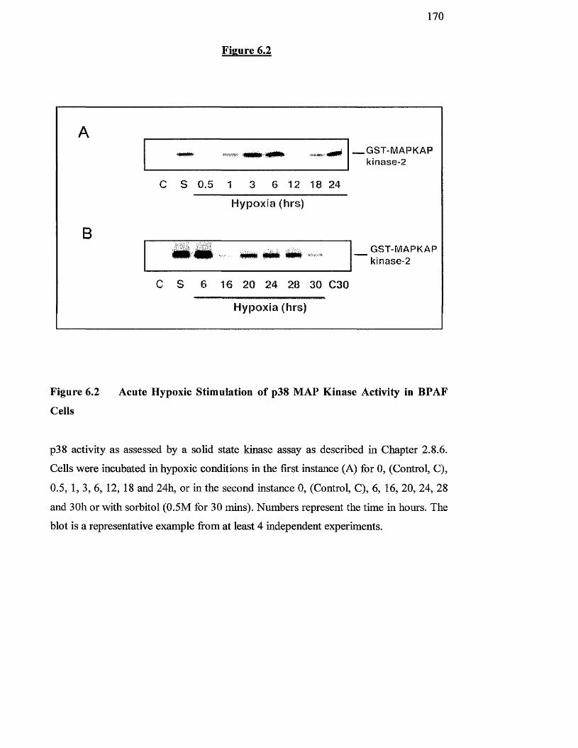
170
Figure 6.2
A'iauwtfr _GST-MAPKAP
kinase-2
C S 0.5 1 3 6 12 18 24
Hypoxia (hrs)
Bm m tm MgggggL - __ GST-MAPKAP
kinase-2
C S 6 16 20 24 28 30 C30
Hypoxia (hrs)
Figure 6.2 Acute Hypoxic Stimulation of p38 MAP Kinase Activity in BPAF
Cells
p38 activity as assessed by a solid state kinase assay as described in Chapter 2.8.6.
Cells Avere incubated in hypoxic conditions in the first instance (A) for 0, (Control, C),
0.5, 1, 3, 6, 12, 18 and 24h, or in the second instance 0, (Control, C), 6, 16, 20, 24, 28
and 3Oh or with sorbitol (0.5M for 30 mins). Numbers represent the time in hours. The
blot is a representative example firom at least 4 independent experiments.
171
6.3.2 Effect of hypoxia on p42/44 MAP kinase phosphorylation and activity
To determine whether the observed increases in p38 MAP kinase and JNK activities
were components o f a general response to hypoxic conditions by all MAP kinase
family members, we analysed the effect o f lowered O2 on p42/44 MAP kinase
activity. Figuie 6.3 shows the effect o f acute hypoxia upon the phosphorylation and
activity o f p42/44 MAP kinase. A decrease in the electrophoretic mobility o f a protein
was indicative o f its phosphorylation. Cells incubated over a time cour se o f up to 28h
in 2% O2 showed no change in electrophoretic mobility o f p42 or p44 MAP kinase
iso forms representative o f their phosphorylation (Figure 6.3A). In contrast,
stimulation o f cells with 10% serum for 30min resulted in a decrease in the
electrophoretic mobility o f both p42 and p44 MAP kinase iso forms, representative o f
their phosphorylation (Figure 6.3 A). In addition, there was a weak activation o f MAP
kinase activity (Figure 6.3B) in response to lowered 0 2 with a maximum 1.8-fold
increase in activity after 3h o f hypoxic challenge. In contrast, 10% serum stimulated
a substantial 6-8 fold increase in activity o f MAP kinase.
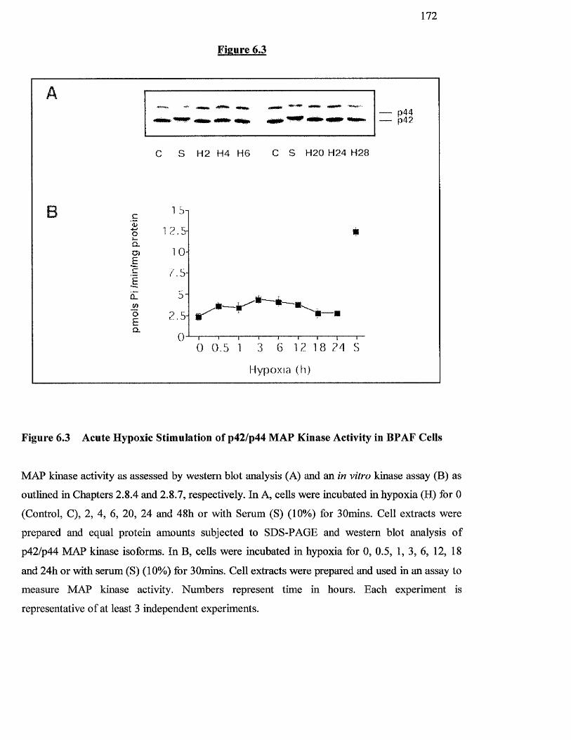
1 7 2
Figure 6.3
c'<uo
aiÊ
û_
oECL
p 4 4p42
C S H 2 H 4 HG C S H 2 0 H 2 4 H 2 8
7 . 5-
2 . 5-
0 0 .5 1 3 G 12 18 24 S
Hypoxia (h)
Figure 6.3 Acute Hypoxic Stimulation of p42/p44 MAP Kinase Activity in BPAF Ceils
MAP kinase activity as assessed by western blot analysis (A) and an in vitro kinase assay (B) as
outlined in Chapters 2.8.4 and 2.8.7, respectively. In A, cells were incubated in hypoxia (H) for 0
(Control, C), 2, 4, 6, 20, 24 and 48h or with Serum (S) (10%) for SOmins. Cell extracts were
prepared and equal protein amounts subjected to SDS-PAGE and western blot analysis o f
p42/p44 MAP kinase isoforms. In B, cells were incubated in hypoxia for 0, 0.5, 1, 3, 6, 12, 18
and 24h or with serum (S) (10%) for BOmins. Cell extracts were prepared and used in an assay to
measure MAP kinase activity. Numbers represent time in hours. Each experiment is
representative o f at least 3 independent experiments.
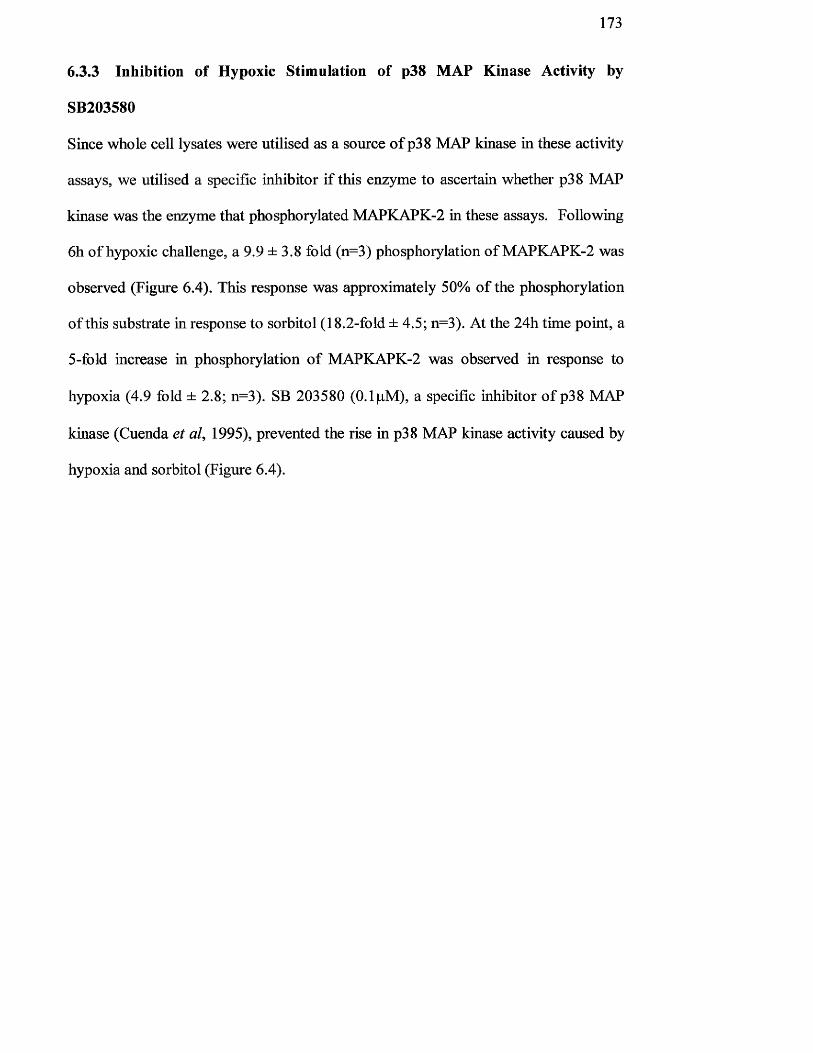
173
6.3.3 Inhibition of Hypoxic Stimulation of p38 MAP Kinase Activity by
SB203580
Since whole cell lysates were utilised as a source o f p38 MAP kinase in these activity
assays, we utilised a specific inhibitor if this enzyme to ascertain whether p38 MAP
kinase was the enzyme that phosphorylated MAPKAPK-2 in these assays. Following
6h o f hypoxic challenge, a 9.9 ± 3.8 fold (n=3) phosphorylation o f MAPKAPK-2 was
observed (Figure 6.4). This response was approximately 50% o f the phosphorylation
o f this substrate in response to sorbitol (18.2-fold ± 4.5; n=3). At the 24h time point, a
5-fold increase in phosphorylation o f MAPKAPK-2 was observed in response to
hypoxia (4.9 fold ± 2.8; n=3). SB 203580 (0.1 pM), a specific inhibitor o f p38 MAP
kinase (Cuenda et al, 1995), prevented the rise in p38 MAP kinase activity caused by
hypoxia and sorbitol (Figure 6.4).
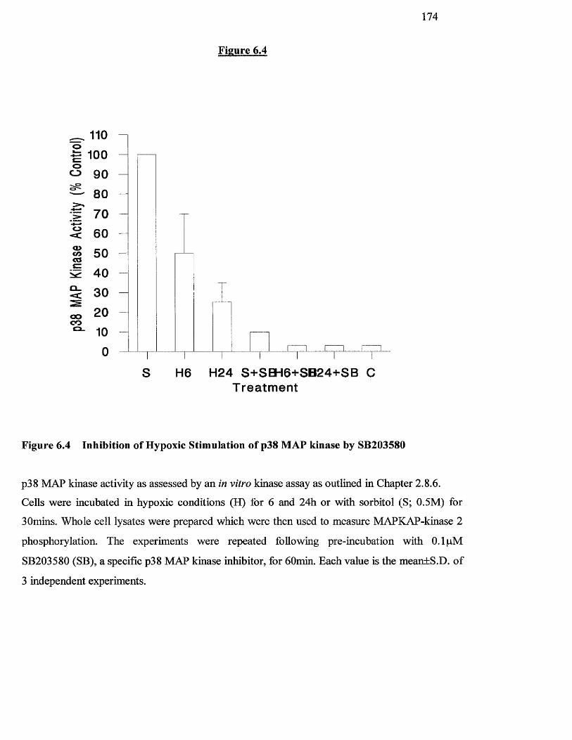
174
^ 1100 100 o0 90
— 80
: t 70 § 60
1 50 M 40 0 30
ooC O
20100
Figure 6.4
H6 H24 S+SBH6+SB24+SB C Treatment
Figure 6.4 Inhibition of Hypoxic Stimulation of p38 MAP kinase by SB203580
p38 MAP kinase activity as assessed by an in vitro kinase assay as outlined in Chapter 2.8.6.
Cells were incubated in hypoxic conditions (H) for 6 and 24h or with sorbitol (S; 0.5M) for
30mins. Whole cell lysates were prepared which were then used to measure MAPKAP-kinase 2
phosphorylation. The experiments were repeated following pre-incubation with 0.1 pM
SB203580 (SB), a specific p38 MAP kinase inhibitor, for 60min. Each value is the mean+S.D. o f
3 independent experiments.
1 7 5
6.3.4 Reversal of the late phase of hypoxie-mediated p38 MAP kinase activity
by re-oxygenation.
In order to determine if continuous hypoxia was requked for the late phase (24h) of
p38 MAP kinase activity observed in Figure 6.2, cells were re-oxygenated at different
times after the initial stimulation period. Re-oxygenation prevented the hypoxic
stimulated p38 MAP kinase activity after 6 and 12h (H6, H I2) but not after 18 and
24h (HI 8, H24) (Figuie 6.5), suggesting the requirement o f a continued hypoxic
environment for the initiation o f the second phase o f the response.
Figure 6.5
1 7 6
# * mm _ _ GST-MAPKAPkinase-2
C H6 H24 H6 H12 H18 H24
Reoxygenation - R18 R12 R6 RO
Figure 6.5 Reversal of the Late Phase of Hypoxic-Mediated p38 MAP Kinase Activity
by Re-Oxygenation.
Effect o f re-oxygenation on reversal o f hypoxic mediated p38 MAP kinase activity by re
oxygenation. Cells were either exposed to hypoxic conditions for various times (H6 and
H24) without subsequent re-oxygenation (R) or were exposed to hypoxia for 6, 12 and 18h
followed by 18, 12 and 6h o f re-oxygenation, respectively. Following this time, cell
extracts were prepared and used to measure p38 MAP kinase activity as in Chapter 2.8.6.
Each value is representative o f at least three experiments (R = re-oxygenation).
177
6.4 DISCUSSION
In Chapter 4 we showed that acute hypoxia stimulated a significant increase in
incorporation o f pHJthymidine into DNA in fibroblast cells derived from the
pulmonary artery. This suggests that hypoxia could increase the proliferation o f these
cells. The present chapter has focussed on the effect o f acute hypoxia on the MAP
kinase family o f enzymes that have been implicated as playing an important role in
cell growth and proliferation (Milanini et al, 1998). A study by Seko et al (1996),
showed that the classical p42/p44 MAP kinase isoforms were involved in the
initiation o f hypoxia-mediated increases in cell growth and division in rat cardiac
myocytes. In that study only a small stimulation o f p42/44 MAP kinase activity was
observed which agrees with the findings o f this chapter. Whilst some studies have
implicated stress-activated MAP kinases in the stress responses o f cardiac myocytes
to ischaemic damage and reperfusion (Morooka et al, 1995; Bogoyevitch et al, 1996;
Bogoyevitch et al, 1995), this is the first study which implicates a role for stress-
activated protein kinase activation in the response o f cells derived from the pulmonary
vasculature to hypoxia.
An initial peak o f JNK and p38 MAP kinase activity was observed between 3 and 6
hours o f hypoxic challenge which was comparable in magnitude to sorbitol
stimulation, a well-recognised activator o f the SAP kinases. This result was similar to
that described by Conrad et al (1999) who observed maximal p38 MAP kinase
phosphorylation after 6h exposure by hypoxia in PC12 cells. This is unlike the p38
MAP kinase and JNK response to other environmental stress agents and cytokines,
where peak activation occurs between 30 and 60 min (Han et al, 1994; Rouse et al,
1994; Raingeaud et al, 1995; Verheij et al, 1996; Lee et al, 1994b). One possible
178
reason for this may be the time required to equilibrate the media to low oxygen
conditions in this study. However, this time course o f activation is similar to that
observed in PC 12 cells following serum-deprivation (Xia et al, 1995) and so it is
possible that this is a physiologically relevant pattern o f cellular activation o f stress-
activated protein kinases following certain stimuli, including hypoxia. We also found
that the classical p42/p44 MAP kinases were stimulated in response to hypoxia,
however, this was a weak stimulation in comparison to that seen with the stress-
activated forms.
A second peak o f p38 MAP kinase activity was observed under hypoxic conditions in
pulmonary artery fibroblasts, occurring between 18 and 24h at a time when JNK
activity was low in these cells. Significantly, this second phase o f p38 MAP kinase
activity coincided with the time required for the initiation o f DNA synthesis in this
cell type (Belham et al, 1996). This second phase also required the continued
presence o f a hypoxic environment suggesting a clear specificity in the signals
required to initiate this response. The effect was blocked by the specific p38 MAP
kinase inhibitor, SB 203580. To date only one other study has indicated activation o f
p38 MAP kinase in the absence o f JNK and this is following ischemic challenge o f
cardiac myocytes (Bogoyevitch et al, 1996). Thus, under certain conditions related to
the oxygen environment, p38 MAP kinase may be activated specifically.
Following the early peak in JNK and p38 MAP kinase activity in response to hypoxia,
the activation o f these enzymes decreases. It is possible that these are being
inactivated by phosphatases. Interestingly, recent publications have shown specific
mitogen-activated protein kinase phosphatases (MKP) which are induced by low
oxygen conditions and are specific for SAPKs (Laderoute et al, 1999; Keyse/, 1999;
17 9
Thoedosiou et al, 1999). Such phosphatases may be responsible for the observed
decreases in JNK and p38 MAP kinase activities.
Following the initial peak in p38 MAP kinase, there was a second peak in activity and
it may be that the activity o f this enzyme is regulated during the cell cycle in response
to hypoxia. Since pulmonary artery cells are stimulated to contract and proliferate in
response to hypoxia, it is possible that p38 MAP kinase plays a positive role in cell
division by regulating cell cycle progression (Lavoie et al, 1996).
These studies have established that both JNK and p38 MAP kinase are activated in
response to acute hypoxic stimulation o f pulmonary artery-derived fibroblasts. Since
the effects o f pulmonary hypertension are a result o f continuous, chronic exposure of
cells to hypoxia, it is o f interest to establish if these enzymes are also activated by
long-term hypoxic exposure.
180
Chapter 7
Effects of Serum on Proliferation and Generation of MAP Kinases in Fibroblasts Harvested from Pulmonary Arteries from Chronically
Hypoxic Rats
181
7.1 INTRODUCTION
In the previous chapters it was shown that acute hypoxia was capable of causing
BPAFs to proliferate, either on its own or in conjunction with mitogens. In addition, it
was also shown that hypoxia gave rise to increases in SAPK activity, p38 MAP kinase
and JNK. Although hypoxia induces structural remodelling and vasoconstriction of
the pulmonary arteries in vivo, the effects o f hypoxia on systemic vascular cells are
quite different (Welsh et al, 1998). As hypoxia does not result in vasoconstriction but
rather induces vasodilation o f systemic vessels (von Euler and Liljestrand, 1946), we
have proposed that there are fundamental differences in oxygen sensing and cell
signalling between systemic and pulmonary artery cells. Indeed, our own work has
shown that hypoxia stimulates proliferation and second messenger production in
pulmonary artery fibroblasts but does not affect fibroblasts derived from the
mesenteric artery.
For the first time our studies have shown a link between vasoconstriction and
remodelling since acute hypoxia was found to increase the rate o f replication of
pulmonary artery derived fibroblasts but not fibroblasts from the systemic circulation
(Welsh et al, 1998). The biochemical and molecular mechanisms by which hypoxia
stimulates proliferation of pulmonary artery fibroblasts are unknown. There is
however, considerable evidence in the literature that p42/p44 MAP kinases, and the
related stress-activated kinases, JNK and p38 MAP kinase, which have been
implicated as key regulators o f cell proliferation (Scott et al, 1998), can be activated
in response to hypoxic stress (Robinson and Cobb, 1997; Takahashi and Berk, 1998).
These modulate the phosphorylation, and hence activation status o f transcription
factors, and link transmembrane signalling with gene induction events in the nucleus
(Robinson and Cobb, 1997).
182
In this current study we have investigated whether fibroblasts derived from the
pulmonary and systemic circulations o f chronically hypoxic rats, in which irreversible
pulmonary vascular re-modelling has taken place (Hunter et al, 1974), exhibit
differential replicative properties. In addition, we compared some of the cell
signalling enzymes that were activated in pulmonary artery fibroblasts derived from
chronically hypoxic rats with those derived from control rats kept under normoxic
conditions. In particular, we investigated the roles of p42/p44 MAP kinase and stress-
activated MAP kinases in hypoxia-induced pulmonary artery fibroblast proliferation
and pulmonary vascular remodelling following chronic exposure to hypoxia.
7.1.1 Normobaric versus Hypobaric hypoxia
Hypoxic animal models (mainly rats and mice) have been used since the 1920’s to
study various environmental conditions and disease states. The animals were exposed
to hypoxic environments by the use o f environmental chambers (normobaric hypoxia;
a decreased inspired O2 at normal atmospheric pressure, or hypobaric hypoxia; a
decreased inspired O2 due to decreased atmospheric pressure) or by actual relocation
o f the animals to altitude where the inspired O2 levels are lower than at sea level
(Campbell, 1927a,b,c; Timiras et al,, 1957). These early investigations studied the
acclimatisation of animals to altitude, or the O2 tensions that would be experienced at
altitude. Animals exposed to these hypoxic environments exhibited initial weight loss
and alterations in certain organ weights (Camprell, 1935; Timiras et al., 1957). The
basic principles o f no rmo baric and hypobaric hypoxia are described below.
183
7.1.2 Normobaric hypoxia
The normobaric method o f chronic hypoxia used by most investigators has been
adapted from a chamber described by Cryer and Bartley (1974). The O2 concentration
within the chamber is reduced from the normal 21% to -10% (IbOmmHg to
~80mmHg O2) by intermittent gaseous infusion o f N 2 . To prevent the build up of
CO2, humidity and other gases, the air is circulated through specific chemical
absorbers.
7.1.3 Hypobaric hypoxia
Hypobaric hypoxia reduces the inspired O2 content o f the environment by reducing
the atmospheric pressure within the chamber. This is the equivalent o f taking the
animals to high altitude. As the atmospheric pressure decreases, the partial pressure of
the gaseous components of air decreases. Therefore, while the percentage o f the
gaseous components o f the air remains the same (O2 ~ 21% and N2 - 78%), the
effective partial pressure o f inspired O2 declines. Hypobaric hypoxia is achieved by
withdrawing air from the chamber by use o f a pump until the pressure within the
chamber is equivalent to -0.5 atmospheres, which reduces the inspired O2 pressure
from IbOmmHg to 80 mmHg. The chamber is continuously flushed with room air to
maintain conditions o f low humidity and CO2 .
Wliile normobaric and hypobaric chambers are both commonly used (mainly for the
study o f pulmonary hypertension), the hypoxic hypobaric chamber has proved to be
more econimical and it is the type which was used in these studies. The Royal
Hallamshire Hospital (Sheffield) designed and manufactured the hypoxic hypobaric
chamber for Dr Margaret MacLean and her pulmonary research group based in the
Institute of Biomedical and Life Sciences at the University o f Glasgow. The chamber
conforms to the high safety standards required by the Home Office.

184
Cells were derived from animals maintained in the hypobaric chamber and compared
to those from animals from a normoxic environment. Differences in proliferation and
signalling mechanisms between these cells have been studied.
185
7.2 METHODS
7.2.1 Chronic hypoxic rat model of pulmonary hypertension.
Pulmonary hypertensive rats were made hypoxic using the technique of MacLean et
al (1995) (Chapter 2.5).
7.2.2 Pulmonary artery fibroblast cell culture
Rat pulmonary artery fibroblast (RPAF) and rat aortic fibroblast (RAF) cells were
hai vested from fr-eshly excised lung tissue o f control and chronically hypoxic animals
as described in Chapter 2.3. All tissue was obtained from the laboratory o f Dr. M.
McLean (Dept, o f Physiology and Pharmacology, University o f Glasgow) and age-
matched animals were always used as controls. The identity o f the fibroblast cells was
confirmed by immunocytochemical staining as described in Chapter 2.3.3.
186
7,2.3 Measurement of Serum-Stimulated [^HjThymidine Uptake in RPAF and
RAF cells.
For these experiments, primary cultures o f RPAF or RAF cells derived from normoxic
or chronically hypoxic rats were transferred to 24-well plates. For acute hypoxic
conditions we used an atmosphere o f 2% O2 (Chapter 3.2.3). Our previous studies
(Chapter 3.2.3) have established that a 6h preincubation under these conditions o f
hypoxia is required to achieve the desired PO2 levels (35mmHg) in the culture
medium. Once established, we found the PO2 levels to remain constant at 35mmHg
(Welsh gr a/, 1998).
Proliferation o f rat pulmonary artery fibroblasts was assessed by measuring the uptake
o f [^H]thymidine (Chapter 2.6). Cells were grown to approximately 60% confluency
in 24-well plates at 37®C and then serum-starved for 24h. After this time 5% serum
was added for a further 24h in the presence and absence of the p38 MAP kinase
inhibitor, SB203580 (0.1 pM) or o f the MAP kinase inhibitor, U0126 (IpM ).
[^H]Thymidine was added for the final 4h o f the incubation. The reaction as stopped
and assessed as in Chapter 2.6.
7.2.4 Assessment of Phosphorylation of p42/p44 MAP kinase and members of
the Stress-Activated MAP kinase family
Cells were grown to approximately 90% confluency in 6-well plates at 37^C and then
serum starved for 24h. After this time the following additions were made as described
below.
187
To determine the effect o f serum on p38 MAP kinase, p42/p44 MAP kinase and JNK
activity on RPAF and RAF cells from normoxic and chronically hypoxic cells, 5%
serum was added to the cells for 0, 1,2, 4, 8, 16, 24, 32 and 48h.
To determine the effect o f SB203580 on p38 MAP kinase activity, RPAF and RAF
cells from normoxic and chronically hypoxic animals, 0.1 pM SB203580 was added to
the cells for Ih prior to serum-stimulation for 2h.
To determine the effect o f U0126 on p42/p44 activity on RPAF and RAF cells from
normoxic and chronically hypoxic animals, IpM U0126 was added to the cells for Ih
prior to serum stimulation for 2h.
Assessment o f the phosphorylation status o f these enzymes was by SDS-PAGE gel
electrophoresis and western blot analysis using phospho-specific antibodies (Chapter
2.8). In each case, the primary rabbit anti-dual phosphorylated, activated MAP kinase
antibody for the protein o f interest (p42/44, p38 and JNK) were utilised.
7.2.5 Statistics
Results are expressed as the mean ± S.D. Statistical analysis was undertaken as
reported in Chapter 2.10.
7 3 RESULTS
73.1 Effect of Acute Hypoxia on RPAF and RAF cells Derived from Normoxic
and Chronically Hypoxic Rats,
In Chapter 3 it was shown that BPAFs proliferate in response to hypoxia in the
absence o f any mitogens. To test the effect o f acute hypoxia on rat cells, growth-
arrested and serum-stimulated pulmonary artery-derived rat fibroblasts (RPAF) were
exposed to 2% O2 or left under normoxic conditions. The effect o f acute hypoxia on
the same cells derived from rats exposed to chronic hypoxia (2 weeks 18% O2) was
also investigated. Figure 7.1 shows the effects o f acute hypoxia on proliferation o f
RPAF and RAF cells from rats either kept in normoxic conditions or kept in hypoxia
for 2 weeks.
There was a 9.3-fold increase in [^H]thymidine uptake when RPAF cells were
cultured in the presence o f 5% serum following serum-starvation for 24 h (p<0.01)
(Figure 7.1 A). When RPAF cells were simultaneously exposed to 5% serum and acute
hypoxia, there was a 19.2-fold increase in the resultant level o f DNA synthesis
observed (p<0.01) in comparison to control, unstimulated cells (Figure 7.1 A).
Similarly, RAF cells showed a 9-fold increase in [^Hjthymidine uptake in response to
serum (Figure 7.IB). In contrast to RPAF cells, however, RAF cells do not exhibit an
enhanced level o f DNA synthesis in response to acute hypoxia (p>0.05) (Figure
7 .IB). Unlike pulmonary artery fibroblasts derived fiom cows, the same cells derived
from rats do not respond to acute hypoxia alone (Figure 7.1 A).
RPAF cells from chronically hypoxic rats exhibited an 18-fold increase in the level of
DNA synthesis relative to cells from control rats (p<0.01) (Figure 7.1 A and 7.2A).
Further increase above the enhanced level o f DNA synthesis observed in RPAF cells

189
from chronically hypoxic rats could not be achieved by exposing these cells to acutely
hypoxic conditions (p>0.05) (Figure 7.2A). RAF cells showed similar levels o f
replication regardless of whether the explants were derived from normoxic or
chronically hypoxic rats (639±47) (Figures 7.1B & 7.2B) (also Table 1: stimulation
indices for pooled data for 8 different animals).
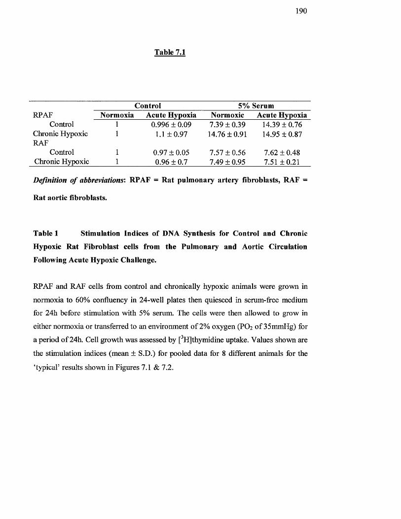
1 9 0
Table 7.1
Control 5% SerumRPAF
Control Chronic Hypoxic RAF
Control Chronic Hypoxic
Normoxia Acute Hypoxia Normoxic Acute Hypoxia0.996 ± 0.09
1 .1+0.97
0.97 ± 0.05 0.96 ± 0.7
7.39 ± 0.39 14,76 ± 0.91
7.57 + 0.56 7.49 ± 0.95
14.39 ± 0.76 14.95 ± 0.87
7.62 ± 0.48 7.51+0.21
Definition o f abbreviations: RPAF = Rat pulmonary artery fibroblasts, RAF =
Rat aortic fibroblasts.
Table 1 Stimulation Indices of DNA Synthesis for Control and Chronic
Hypoxic Rat Fibroblast cells from the Pulmonary and Aortic Circulation
Following Acute Hypoxic Challenge.
RPAF and RAF cells from control and chronically hypoxic animals were grown in
normoxia to 60% confluency in 24-well plates then quiesced in serum-free medium
for 24h before stimulation with 5% serum. The cells were then allowed to grow in
either normoxia or transferred to an environment o f 2% oxygen (PO2 o f 35mmHg) for
a period o f 24h. Cell growth was assessed by [^H]thymidine uptake. Values shown are
the stimulation indices (mean ± S.D.) for pooled data for 8 different animals for the
‘typicaT results shovm in Figures 7.1 & 7.2.
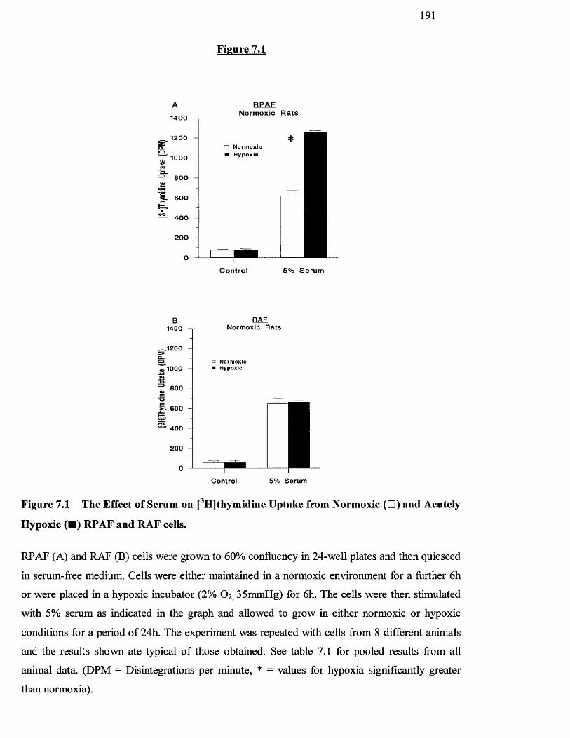
191
Figure 7.1
Normoxic R a ts14 00
1200□ N o r m o x l o
H y p o x ia1000
i . 6 0 0 -
— 4 0 0
Control 5% S e r u m
B1400
,1200
'lOOO
I 800
600 —
' 400
2 0 0
RAF Normoxic Rats
□ N o rm o x ic ■ H ypox ic
Control 5% Serum
Figure 7.1 The Effect of Serum on [^H]thymidine Uptake from Normoxic (□ ) and Acutely
Hypoxic (■ ) RPAF and RAF cells.
RPAF (A) and RAF (B) cells were grown to 60% confluency in 24-well plates and then quiesced
in serum-free medium. Cells were either maintained in a normoxic environment for a further 6h
or were placed in a hypoxic incubator (2% O2,35mmHg) for 6h. The cells were then stimulated
with 5% serum as indicated in the graph and allowed to grow in either normoxic or hypoxic
conditions for a period o f 24h. The experiment was repeated with cells from 8 different animals
and the results shown ate typical o f those obtained. See table 7.1 for pooled results from all
animal data. (DPM = Disintegrations per minute, * = values for hypoxia significantly greater
than normoxia).
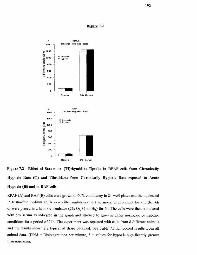
1 9 2
Figure 7.2
A1400
,1200
1000
800
• BOO H
■ 400
200
0
RPAFChronic Hypoxic Rats
o N orm oxic N Hypoxic
Controi 5% Sérum
RAFChronic Hypoxic R a ts1400
1200a N orm oxic
H ypoxic
3C 400 —
200 -
Controi 5% Sérum
Figure 7.2 Effect of Serum on [^H]thymidine Uptake in RPAF cells from Chronically
Hypoxic Rats (□ ) and Fibroblasts from Chronically Hypoxic Rats exposed to Acute
Hypoxia (■ ) and in RAF cells
RPAF (A) and RAF (B) cells were grown to 60% confluency in 24-well plates and then quiesced
in serum-free medium. Cells were either maintained in a normoxic environment for a further 6h
or were placed in a hypoxic incubator (2% O2,35mmHg) for 6h. The cells were then stimulated
with 5% serum as indicated in the graph and allowed to grow in either normoxic or hypoxic
conditions for a period o f 24h. The experiment was repeated with cells from 8 different animals
and the results shown are typical o f those obtained. See Table 7.1 for pooled results from all
animal data. (DPM = Disintegrations per minute, * = values for hypoxia significantly greater
than normoxia.
193
7.3.2 Differential p38 and p42/p44 MAP kinase phosphorylation in RPAF and
RAF cells from normoxic and chronically hypoxic rats.
We have previously shown that exposure o f BPAFs to acute hypoxia induces the
activation o f the SAPKs, JNK and p38 MAP kinase. We therefore investigated
whether these signalling enzymes played a role in the enhanced proliferation and
remodelling o f fibroblasts observed after prolonged (14 days) exposure to chronic
hypoxia. To do tliis, we have examined the activation of members o f the MAP kinase
family by detecting their dually phosphorylated (tyr/thr) forms by Western blotting
using specific anti-active phosphokinase antibodies (Feng et al, 1999). Serum-
stimulation o f RPAF from control rats incubated under normoxic conditions showed a
weak activation o f p38 MAP kinase as indicated by its dual phosphorylation of its
regulatory thr/tyr motif which peaked at between 1 and 4 h stimulation with serum
(Figure 7.3A, upper panel). In contrast, p38 MAP kinase was found to be strongly
constitutively active in RPAF cells derived from rats subjected to chronically hypoxic
conditions, and this activity was further enhanced in the presence o f serum,
particularly in the period I6-48h post-stimulation (Figure 7.3A, upper panel). This
increase in p38 MAP kinase phosphorylation was not due to an upregulation o f the
p38 protein expression, as Western Blot analysis using an antibody which detects total
amounts o f p38, revealed compaiable levels o f p38 MAP kinase expression in RPAF
cells from chronically hypoxic and normoxic rats (Figure 7.3A, lower panel). RAF
cells, however, despite expressing substantial levels of p38, did not show significant
change in the phosphorylation o f p38 MAP kinase in response to serum, nor was this
activity modulated following chronic exposure o f the rats to hypoxia (Figure 7.3B).
194
A similar pattern was observed for p42/p44 MAP kinase. Serum-induced p42/p44
MAP kinase (particularly the p42 MAP kinase isoform) activity in normoxic RPAF
cells in a multiphasic manner (peaks at Ih and 24h), and RPAF from chronically
hypoxic rats showed strong constitutive activity o f p42/p44 MAP kinase (Figure
7.4A). Although, there appeared to be a slight upregulation in p42/p44 MAP kinase
expression in these cells, this upregulation was not sufficient to explain the very high
levels o f kinase activity observed (Figure 7.4A). As with p38 MAP kinase, chronic
exposure to hypoxia did not modulate p42/p44 MAP kinase activation in RAF cells,
which already appeared to exhibit a considerable level o f constitutive p42/p44 MAP
kinase activity which was not significantly modulated by addition o f serum (Figure
7.4B). There was a small increase in phosphorylation of JNK on the addition o f serum
to normoxic and hypoxic RPAF cells, however, there was no significant difference in
the amount o f phosphorylated JNK or amount o f total JNK protein following serum-
stimulation o f hypoxic RPAF relative to that observed in RPAF from control
normoxic rats (Figure 7.5A). There was no significant change in the phosphorylation
o f JNK in response to serum in RAF cells derived from either normoxic or hypoxic
rats (Figure 7.5B).
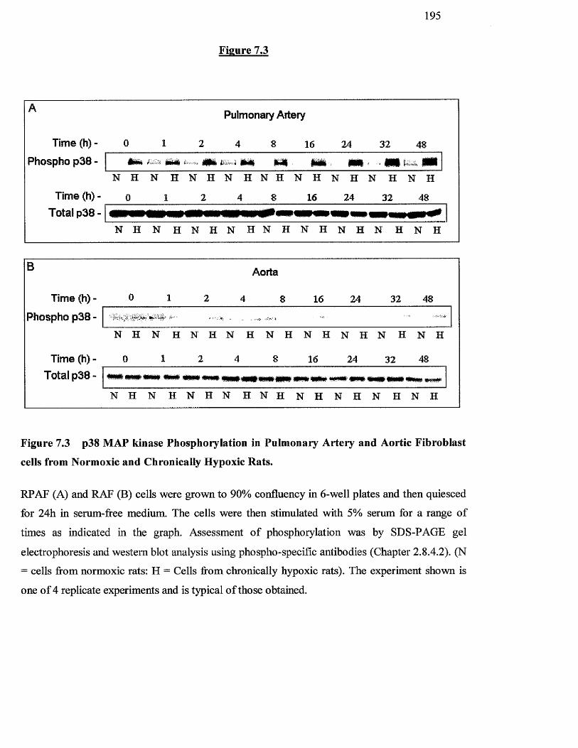
Time (h) -
Phospho p38 -
Time (h) Total p38
195
Figure 7.3
Pulmonary Artery
16 24 32 48* # m i # #
N H N H N H N H N H N H N H N H N H
0 1 2 4 8 16 24 32 48
N H N H N H N H N H N H N H N H N H
B
Time (h) - 0 1
Phospho p38 -
Aorta
8 16 24 32 48
N H N H N H N H N H N H N H N H N H
Time (h) - 0 1 2 4 8 16 24 32 48Total p38 - **#«#»«## # # #M# #*# ### ww **#' ###' #*# ### *w*
N H N H N H N H N H N H N H N H N H
Figure 7.3 p38 M AP kinase Phosphorylation in Pulm onary A rtery and Aortic F ibroblast
cells from Normoxic and Chronically Hypoxic Rats.
RPAF (A) and RAF (B) cells were grown to 90% confluency in 6-well plates and then quiesced
for 24h in serum-free medium. The cells were then stimulated with 5% serum for a range o f
times as indicated in the graph. Assessment o f phosphorylation was by SDS-PAGE gel
electrophoresis and western blot analysis using phospho-speciflc antibodies (Chapter 2.S.4.2). (N
= cells from normoxic rats: H = Cells from chronically hypoxic rats). The experiment shown is
one o f 4 replicate experiments and is typical o f those obtained.
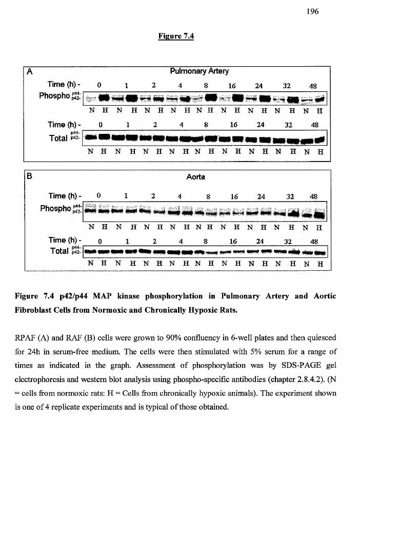
B
1 9 6
ATime (h) - o
Phospho JJJ:
Figure 7.4
Pulmonary Artery
2 4 8 16 24 32 48
N H N H N H N H N H N H N H N H N H
Time (h) - 0 1 2 4 8 16 24 32 48
Total # # # # # » # # #N H N H N H N H N H N H N H N H N H
Aorta
Time (h) - 0Phospho%:
16 24 32 48
# # # # # # # # #
N H N H N H N H N H N H N H N H N H
Time (h)- o 1 2 4 8 16 24 32 48Total % ## mm « # ' # # m # w# ##
N H N H N H N H N H N H N H N H N H
Figure 7.4 p42/p44 MAP kinase phosphorylation in Pulmonary Artery and Aortic
Fibroblast Cells from Normoxic and Chronically Hypoxic Rats.
RPAF (A) and RAF (B) cells were grown to 90% confluency in 6-well plates and then quiesced
for 24h in serum-free medium. The cells were then stimulated with 5% serum for a range o f
times as indicated in the graph. Assessment o f phosphorylation was by SDS-PAGE gel
electrophoresis and western blot analysis using phospho-speciflc antibodies (chapter 2.S.4.2). (N
= cells from normoxic rats: H = Cells from chronically hypoxic animals). The experiment shown
is one o f 4 replicate experiments and is typical o f those obtained.
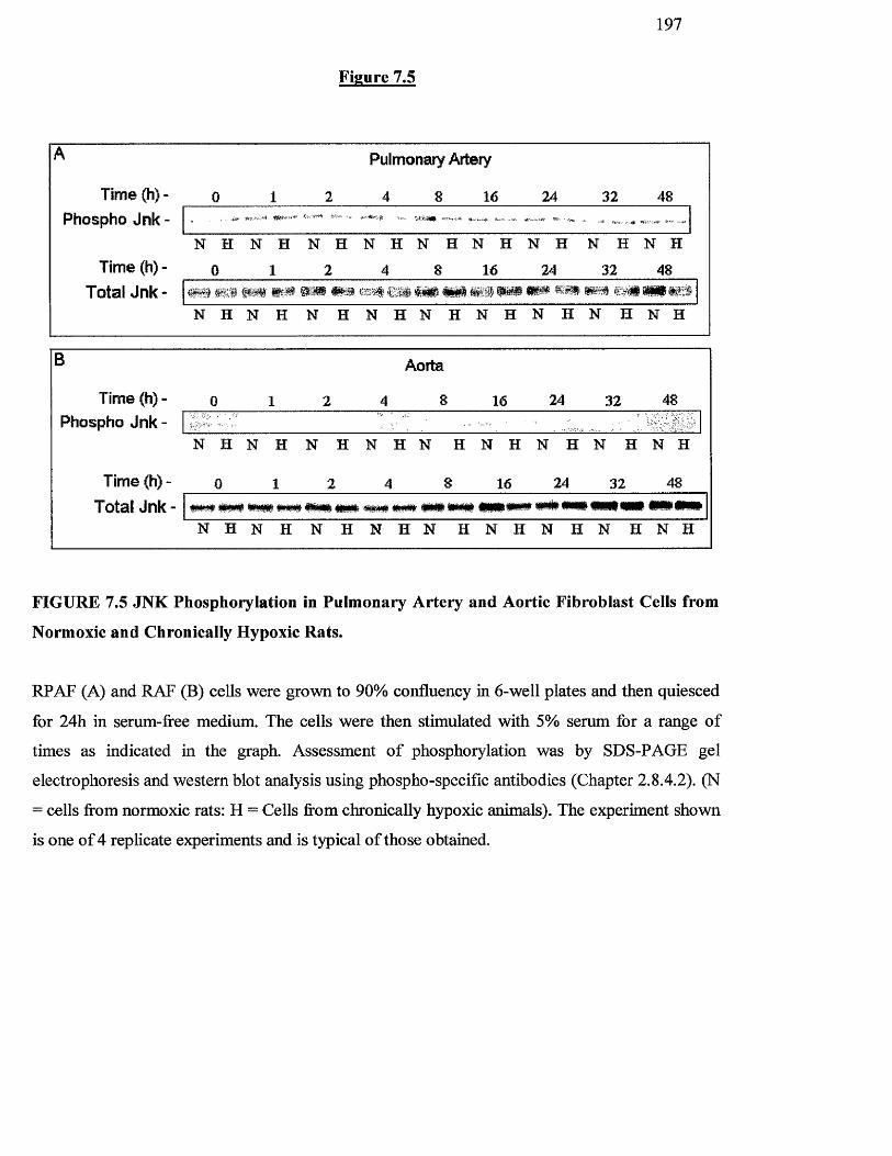
1 9 7
Figure 7.5
A Pulmonary Artery
Time (h) - 0 1 2 4 8 16 24 32 48
Phospho Jnk - . , , -itf
Time (h) -N H
0
N H
1
N H
2
N H N H
4 8
N H
16
N H
24
N H
32
N H
48
Total Jnk- t . * k
N H N H N H N H N H N H N H N H N H
B Aorta
Time (h) - 0 1 2 4 8 16 24 32 48Phospho Jnk -
N H N H N H N H N H N H N H N H N H
Time (h) - 0 1 2 4 8 16 24 32 48
Total Jnk- P W5TSP »# *# # * #####* # # # # W mN H N H N H N H N H N H N H N H N H
FIGURE 7.5 JNK Phosphorylation in Pulmonary Artery and Aortic Fibroblast Cells from
Normoxic and Chronically Hypoxic Rats.
RPAF (A) and RAF (B) cells were grown to 90% confluency in 6-well plates and then quiesced
for 24h in serum-free medium. The cells were then stimulated with 5% serum for a range o f
times as indicated in the graph. Assessment o f phosphorylation was by SDS-PAGE gel
electrophoresis and western blot analysis using phospho-speciflc antibodies (Chapter 2.8.4.2). (N
= cells from normoxic rats; H = Cells from chronically hypoxic animals). The experiment shown
is one o f 4 replicate experiments and is typical o f those obtained.
198
7.3.3 Differential Roles for p38 and p42/p44 MAP kinase in RPAF and RAF
DNA synthesis from Normal and Chronically Hypoxic Rat Cells.
As p38 and p42/p44MAP kinase were highly phosphorylated in RPAF cells derived
from hypoxic but not normoxic rats, we decided to investigate, using specific
pharmacological inhibitors, the role of these kinases in the hypoxia-enhanced
proliferation o f RPAF cells. A selective and potent inhibitor o f the p42/p44 MAP
kinase cascade, U0126, mediates its effects by binding to and inactivating MEK,
whereas it has no effect on any o f the components o f the JNK or p38 MAP kinase
cascades (Feng et al, 1999). Similarly, the compound SB203580 is a selective and
potent inhibitor o f p38 MAP kinase that does not affect either p42/p44 MAP kinase or
JNK (Cuenda et al, 1995). These reagents are usefiil pharmacological tools in
identifying the functional activities mediated by p38 and p42/p44 MAP kinases. Pre
incubation for Ih with SB203580 (0.1 pM) inhibited p38 phosphorylation (Figure
7.6A) in RPAF and RAF cells. In addition, pre-incubation for Ih with U0126 (IpM )
profoundly inhibited both the hypoxia- and serum-stimulated p42/p44 MAP kinase
activation in these cells (Figure 7.6B).
Having demonstrated the activity o f these kinase inhibitors, we used them to test the
importance o f p42/44 and p38 MAP kinase activity observed in RPAF, but not RAF
cells derived from chronically hypoxic rats on [^HJthymidine uptake (Figure 7.7).
These results showed that SB203580 and U0126 had no effect on the DNA synthesis
resulting from basal (results not shown) or serum-stimulation o f RPAF or PAF cells
derived from normoxic rats (Figures 7.7A & 7.7B). In contrast, the p38 inhibitor
SB203580 completely abrogated the enhanced serum-stimulated DNA synthesis
observed in RPAF (701+72 with the inhibitor compared for 1217+118 without
p<0.01), but not RAF cells from chronically hypoxic rats (981+117 with the inhibitor

199
compared for 952+96 without p>0.05) (Figures 1.1 A & 7.7B), such that the response
was now equivalent to that observed in cells derived from control normoxic rats
(652+51). The p42/p44 MAP kinase cascade inhibitor, U0126 had no effect on this
hypoxia-induced enhancement o f DNA synthesis (1171+120 with U0126 compared
with 1217+118 without p>0.05).
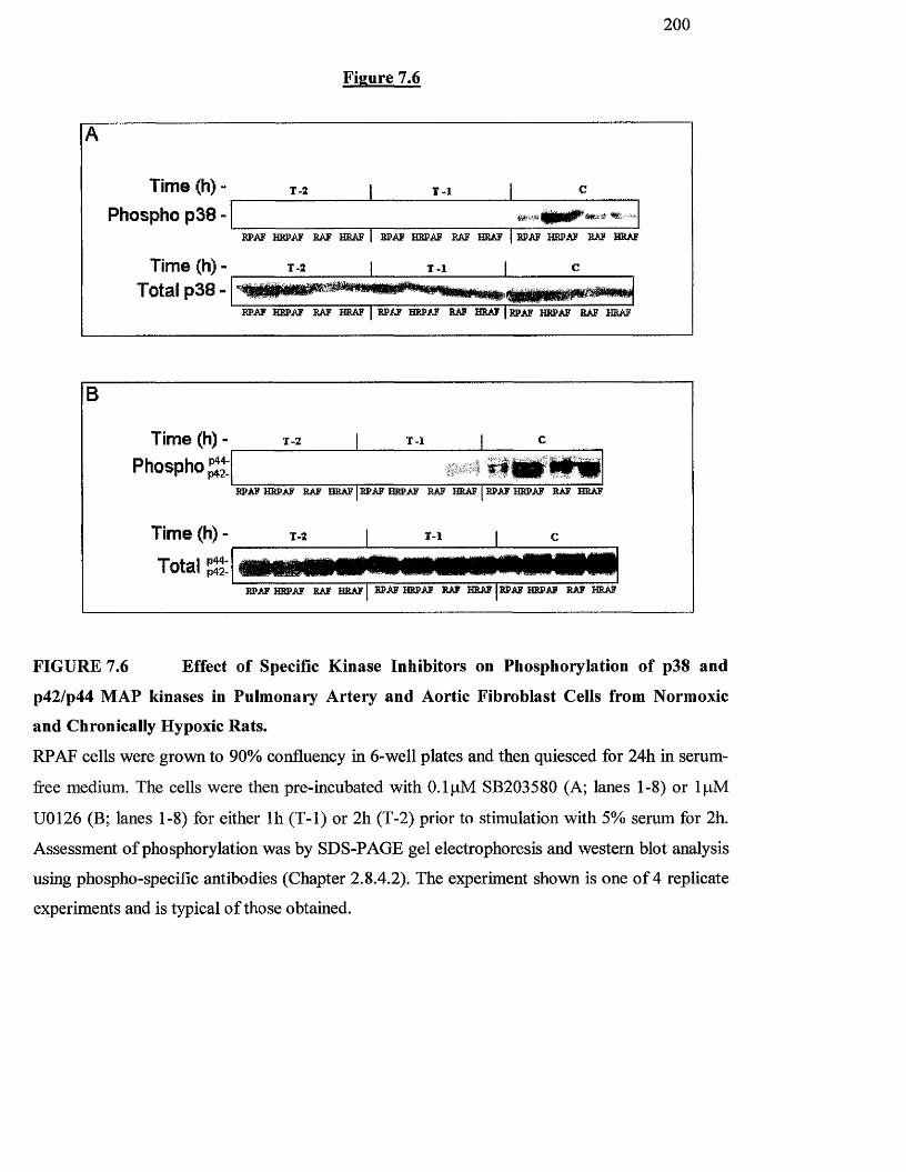
2 0 0
Figure 7.6
A
Time (h) - r - 2 1 T - l 1 c
Phospho p38 -
Time (h) -
RPAF HRPAF RAF HRAF | RPAF HRPAF RAF HRAF j RPAF mPAF RAF HRAF
T -2 1 T -1 1 C
Total p38-RPAF HRPAF RAF HRAF | RPAF HRPAF RAF HRAF | RPAF HRPAF RAF HRAF
B
Time (h) -p44 p42.Phospho
T -2 T - i
■r#BPAF HBPAF RAF HRAF BPAF HKPAF KAF HRAF EPAT HBPAF RAF HRAF
Time (h) -
TotalBPAF HRPAF RAF HRAF | HPAF HRPAF RAF HRAF jRPAF HSPAF RAF HRAF
FIGURE 7.6 Effect of Specific Kinase Inhibitors on Phosphorylation of p38 and
p42/p44 MAP kinases in Pulmonary Artery and Aortic Fibroblast Cells from Normoxic
and Chronically Hypoxic Rats.
RPAF cells were grown to 90% confluency in 6-well plates and then quiesced for 24h in serum-
free medium. The cells were then pre-incubated with 0.1 pM SB203580 (A; lanes 1-8) or IpM
U0126 (B; lanes 1-8) for either Ih (T-1) or 2h (T-2) prior to stimulation with 5% serum for 2h.
Assessment o f phosphorylation was by SDS-PAGE gel electrophoresis and western blot analysis
using phospho-speciflc antibodies (Chapter 2.S.4.2). The experiment shown is one o f 4 replicate
experiments and is typical o f those obtained.
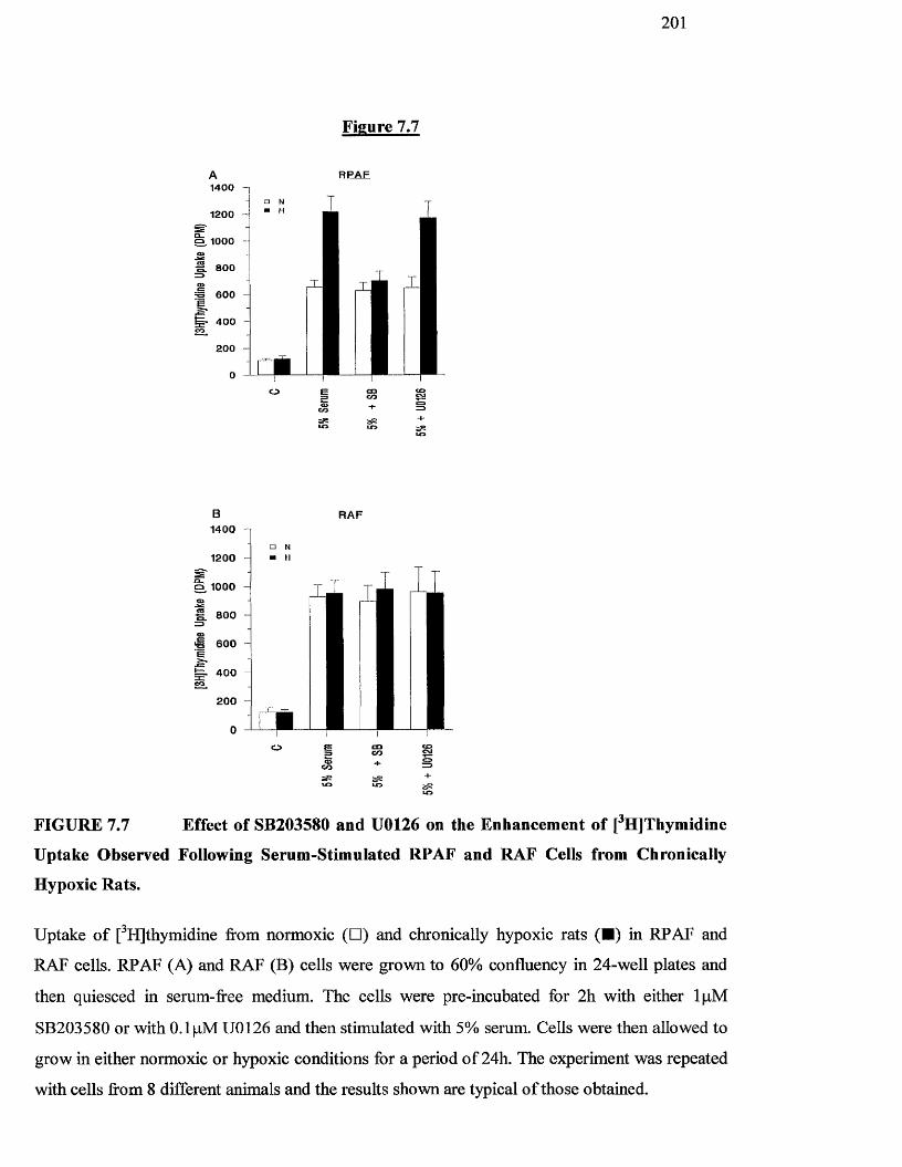
2 0 1
Figure 7.7
RPAF1400
1200
'o 600
RAF1400 n
1200
S 1000
S 800
60 0
40 0
200
§o eo
FIGURE 7.7 Effect of SB203580 and U0126 on the Enhancement of [^HJThymidine
Uptake Observed Following Serum-Stimulated RPAF and RAF Cells from Chronically
Hypoxic Rats.
Uptake o f [^H]thymidme from normoxic (□ ) and chronically hypoxic rats (■ ) in RPAF and
RAF cells. RPAF (A) and RAF (B) cells were grown to 60% confluency in 24-well plates and
then quiesced in serum-free medium. The cells were pre-incubated for 2h with either IpM
SB203580 or with 0.1 fxM U0126 and then stimulated with 5% serum. Cells were then allowed to
grow in either normoxic or hypoxic conditions for a period o f 24h. The experiment was repeated
with cells from 8 different animals and the results shown are typical o f those obtained.
202
7.4 DISCUSSION
In this chapter we have shown that pulmonary artery fibroblasts from chi’onically
hypoxic rats exhibit enhanced proliferative responses to serum relative to those
observed with RPAF derived from normoxie animals. This hypoxia-enhanced
proliferation was maintained following multiple passages o f these primary cultures of
cells, which were maintained under normoxic conditions following their eulture from
the artery. This mirrors the changes observed in vivo in patients with hypoxic
pulmonary hypertension, where remodelling o f the pulmonary arteries due to
increased cellular proliferation remains even after oxygen therapy (Traver et al,
1979). Sustained enhanced proliferative capacities have recently also been reported
for fibroblasts isolated from the pulmonary artery o f neonatal calves with the severe
fibroproliferative changes characteristic o f hypoxic neonatal pulmonary hypertension
(Das et al, 1999). This latter study showed that the proliferative responses o f such
fibroblasts could be further enhanced following subsequent exposure to acute
hypoxia. However, in our system, the augmented responses to serum observed in
RPAF could not be further enhanced by re-exposure to acute hypoxia. This suggests
that the mechanisms regulating pulmonary vascular remodelling in neonates and
adults may be different and may provide a rationale for the widely established finding
that the effects o f hypoxia are more marked in neonates relative to adults (Das et al,
1999). Interestingly, no remodelling o f the systemic circulation is observed in the
patients with hypoxic pulmonary hypertension (von Euler and Liljestrand, 1946) and
this is reflected in our rat model as fibroblasts from rat aorta did not show enhanced
proliferative capacities following chronic exposure to hypoxia.
The molecular mechanisms by which hypoxia stimulates proliferation o f pulmonary
artery fibroblasts, but not fibroblasts from the systemic circulation, are unknown.
There is considerable evidence in the literature, however, that p42/p44 MAP kinases,
203
As well as JNK and p38 MAP kinase can be activated in response to hypoxic stress
(Robinson and Cobb, 1997; Takahashi and Berk, 1998). Indeed, we have shown in
our model o f acute bovine pulmonary artery hypoxia, that the SAPKs JNK and p38
MAP kinase, but not the classical p42/p44 MAP kinase isoforms, are activated in
response to acute hypoxia (Scott et al, 1998). We therefore addressed the role o f all
three classes of these MAP kinases by investigating whether there was differential
activation o f these kinases in response to serum in fibroblasts derived from the
pulmonary and systemic circulation of normoxic versus chronically hypoxic rats.
Although JNK was strongly activated by hypoxia in the acute model (Chapter 6; Scott
et al, 1998), we could find no evidence o f JNK phosphorylation in RPAF derived
from rats subjected to chronic hypoxia (Figure 7.5). Moreover, we could not detect
any differential phosphorylation o f JNK in response to serum in RPAF derived from
chronically hypoxic versus normoxic rats (Figure 7.5). However, as the JNK
activation observed in response to hypoxia in the acute model was transient, peaking
between 3-6 h following hypoxic challenge, these results may indicate that whereas a
JNK kinase signal may be required for the initiation o f hypoxia-mediated events, it is
not involved in the remodelling process. In contrast, we found strong and constitutive
activation o f p42/p44 and p38 MAP kinases in RPAF from chronically hypoxic rats
but not normoxic rats (Figures 3 and 4). In addition, this induction o f constitutive
p42/p44 and p38 MAP kinase activity was not observed in RAF from either
chronically hypoxic rats or normoxic rats. Taken together, these results suggested that
p42/p44 and/or p38 MAP kinase played a role in the enhanced proliferative responses
observed in remodelled fibroblasts from chronically hypoxic rats.
To address the relative roles o f p42/p44 and p38 MAP kinases in serum and/or
hypoxia-induced proliferation of RPAF and RAF cells, we performed our
204
proliferation assays in the presence o f spécifié inhibitors o f the p42/p44 (U0126) and
p38 (SB203580) MAP kinase cascades. Given the widely established roles for
p42/p44 MAP kinases in promoting cell proliferation (Lew et al, 1999; Belham et al,
1996; Pages et al, 1993), and the strong constitutive activation o f this MAP kinase in
RPAF cells by chronic hypoxia (Figure 4), it could be considered surprising that the
p42/p44 cascade inhibitor U0126 had no effect on the enhancement o f serum-
stimulated proliferation observed in RPAF from chronically hypoxic rats. However,
examination o f the levels of p42/p44 MAP kinase activity in RAF cells showed that
these cells also exhibited considerable levels o f constitutive p42/p44 MAP kinase
activity despite an inability to display enhanced proliferative capacities following
exposure to chronic hypoxia. Indeed, p42/p44 MAP kinase activity was found to be
strongly and constitutively activated in RAF cells regardless o f the oxygen status o f
the rats they were derived from. Taken together with the finding that p42/p44 activity
could be stimulated in RPAF cells from normoxic rats following stimulation with
serum, these results could suggest that p42/p44 MAP kinase activity serves to render
eells permissive for growth factor mediated-stimulation o f proliferation. Thus, the
enhanced p42/p44 MAP kinase signals observed in hypoxic RPAF cells could
contribute to the augmented growth factor-stimulated responses observed in response
to chronic hypoxia reported in the bovine neonatal study (Das et al, 1999).
In contrast, and consistent with the differential patterns o f p38 MAP kinase activity
observed in RPAF and RAF from chronically hypoxic versus normoxic rats (Figure
3), we found that although p38 MAP kinase activity appeared to be essential for the
enhanced proliferative response observed in RPAF from chronically hypoxic rats, it
did not appear to play an important role in transducing serum-induced proliferation in
either RPAF or RAF cells. This important role for p38 MAP kinase in transducing
proliferative signals in response to hypoxia reflects our earlier studies on the bovine
2 0 5
acute hypoxia model which showed that p38 MAP kinase activation was dependent
on the presence o f a maintained hypoxic environment as sustained p38 MAP kinase
activity was blocked by re-oxygenation. Moreover, although p38 MAP kinase has
generally been considered to play a role in the transduction o f apoptotic (Takahashi
and Berk, 1998) rather than proliferative signals as evidenced by negative regulation
o f cyclin D expression (Lavoie et al, 1996) and its activation under conditions of
cellular stress (Xia et al, 1995; Chen et al, 1996) including ischemic damage and
reperfiision o f cardiac myocytes (Morooka et al, 1995; Bogoyevitch et al, 1995), there
is some evidence not only that crosstalk between JNK and p38 MAP kinase may be
required for apoptosis (Wilson et al, 1996; Ichijo et al, 1997) but also that activation
o f p38 MAP kinase can promote cellular activation, proliferation and differentiation
(Zhang et al, 1999). Whereas most o f the physiologieal and chemieal stresses which
can induce apoptosis generally activate both JNK and p38 MAP kinase (Wilson et al,
1996; Ichijo et al, 1997), we find that under these stress conditions of chronic
hypoxia, JNK does not appear to be activated and p38 MAP kinase acts to promote
cellular proliferation.
Finally, there is an urgent need for drugs that eould reverse or prevent the pulmonary
vascular remodelling whieh occurs in nearly all forms o f cardiac and respiratory
disease. At present, however, most vasodilating drugs used to counteract pulmonary
hypertension result in systemic hypotension. However, since systemic fibroblasts do
not replicate in the presence o f hypoxia and p38 MAP kinase does not appear to be
neeessary for the normal replication o f pulmonary systemic vascular fibroblasts, it is
possible that therapies targeting RPAF p38 MAP kinase could lead to the control of
pulmonary vascular remodelling without affecting the systemic circulation.
206
Chapter 8
Effect of 5-HT on Proliferation and Generation of MAP kinases in Fibroblasts Harvested from Pulmonary Arteries from Chronically
Hypoxic Rats
207
8.1 INTRODUCTION
As described in Chapter 1, numerous groups have studied changes that take place in the
pulmonary arteries as a result o f pulmonary hypertension. As a result o f these studies,
numerous pro-inflammatory factors have been identified which can influence these changes
(Bitterman and Henke, 1991). The aim of this chapter was to examine the possible role o f
one such factor, serotonin, on fibroblast cell proliferation and intracellular signalling from
the pulmonary arteries o f eontrol and chronically hypoxic animals.
Serotonin, also known as 5-hydroxytryptamine (5-HT), is secreted from neuroendocrine
cells in the gut, and tumours o f these cells, called carcinoid tumours, are a source o f
increased production o f 5-HT (Hart and Block, 1989; Perry and Vinik, 1996). Pulmonary
neuroendocrine cells secrete vasoactive substances in response to airway hypoxia and
hypercapnia (Johnson and Georgieff, 1989; Lauweryns et al, 1983). These eells commonly
proliferate in patients with pulmonary hypertension, producing a variety o f peptides in
addition to large amounts o f 5-HT (Gosney et al, 1989). In lung transplant recipients with
end stage primary pulmonary hypertension the degree of hyperplasia o f these eells was
found to correlate with the extent o f proliferation o f myofibroblasts in the pulmonary
arteries (Madden et al, 1994). On a molar basis, 5-HT is the most potent pulmonary
vasoconstrictor identified to date in humans (Heffner et al, 1987) but in the systemic
vasculature it causes profound vasodilation (Comroe et al, 1953).
Because o f the prevalence of chronic lung disease, hypoxia is probably the commonest of
all causes o f pulmonary hypertension. As stated previously, the mechanisms o f hypoxic
vasoconstriction have been reviewed, but much remains unknown (Voelkel, 1986). In
healthy subjects hypoxia per se does not appear to increase circulating 5-HT levels (Rahda
208
el al 1976), but levels are increased in patients with several types of chronic lung disease
and associated pulmonary hypertension (Bobrov et al, 1985; Pribilova, 1976).
5-HT is a mitogen causing hyperplastic and hypertrophic changes in smooth muscles
(Pakala et al, 1994). At least ten classes of 5-HT receptors have been identified, these
include 5 - H T i a - f , 5 - H T 2 a -c , 5 -HT3 and 5 -HT4 (Hoyer et al, 1994), and different receptors
have been implicated in the pathogenesis of a number o f vascular disorders. For example,
5-HT 1 and 5-HT] are the principal receptors relevant to the pulmonary arteries (Frishman el
al, 1996). Under most experimental conditions, stimulation o f the 5-HT, receptor eauses
vasodilation and the 5-H U receptor often mediates vasospasm (Frishman et ai, 1996).
Use o f several drugs has indicated a strong link between pulmonary hypertension and levels
o f 5-HT. Pulmonary hypertension can be induced in most mammals by numerous
serotonergic drugs, some o f whieh are sympathomimetic anorexients. One such drug is
dexfenfluramine (Brenot et al, 1993). In dogs, intravenous dexfenfluramine augments
hypoxic pulmonary vasoconstriction and, with long term oral use, pulmonary vascular
resistance is increased (Naeije, 1996). How this drug can induce pulmonary hypertension is
unknown. In contrast to this, drugs that block 5-HT receptors reduce pulmonary
hypertension. For example, the specific 5-HT]A receptor antagonist ketanserin is the best
studied agent o f this type for reducing pulmonary hypertension or vascular resistance
(Brogden and Sorkin, 1990). Like most vasodilators this drug has varying efficacy,
reflecting the complex pathophysiology o f the disease. In the treatment o f primary
pulmonary hypertension, McGoon and Vliestra (1984) detected an average reduction of
pulmonary vascular resistance o f 18% with intravenous use in 10 patients. The effects of
ketanserin on pulmonary hypertension suggest that serotonergic mechanisms may be
contributing to the problem.

209
The aim of this chapter is to study the effects of 5-HT on the proliferation and generation o f
MAP kinases in rat pulmonary fibroblasts from control and chronically hypoxic animals. At
the same time, the effects of specific 5-HT receptor antagonists, such as the 5-HT]a
receptor antagonist, ketanserin, will be used to help identify if a specific 5-HT receptor is
involved in the remodelling o f pulmonary arteries during pulmonary hypertension.
210
8,2 METHODS
8.2.1 Chronic hypoxic rat model of pulmonary hypertension.
Pulmonary hypertensive rats were prepared using the technique of MacLean et al (1995)
(Chapter 2.5)
8.2.2 Pulmonary artery fibroblast cell culture
Rat pulmonary artery fibroblast (RPAF) cells were harvested from freshly excised lung
tissue o f control and chronically hypoxic animals as described in Chapter 2.3.2.
All tissue was obtained from the laboratory o f Dr. M. McLean and age-matched animals
were always used as controls.
The identity o f the fibroblast cells was confirmed by immunocytochemical staining as
described in Chapter 2.3.3
8.2.3 Assessment of Proliferation
Proliferation o f rat pulmonary artery fibroblasts was assessed by measuring the uptake of
[■"Fljthymidine (Chapter 2.6). Cells were grown to approximately 60% confluency in 24-
well plates at 37®C and then serum-starved for 24h. After this time the following additions
were made as described below.
211
To determine the effect of serum and 5-HT on proliferation in the normoxic and chronically
hypoxic cells, 0.2% serum and lOpM 5-HT were added to the cells for 24h. lOpM of
specific 5-HT agonists, a-methyl-5-HT, BW723C86 and MK212 were also studied. In
addition, the incubations were also carried out in the presence and absence of 5-HT
receptor antagonists, 0.1 pM Ketanserin and IpM SDZ SER 082 and R S I02221. All drugs
were added 2h prior to addition o f growth factors for 24h.
In all experiments, for the last 4h of agonist stimulation, cells were labelled with
[^H]thymidine (O.lpCi/ml) before the reaction was stopped by washing in PBS (Chapter
2.6). Radioactivity was determined by liquid scintdation counting and results expressed as
disintegrations per minute (DPM).
8.2.4 Assessment of MAP kinase activity.
Cells were grown to approximately 90% confluency in 6-well plates at 37®C and then
serum-starved for 24h. After this time, the following additions were made as described
below. To determine the effect o f serum and 5-HT on p42/44, p38 and JNK activity in the
normoxic and chionically hypoxic cells, 0.2% serum and lOpM 5-HT were added to the
cells for 24h. To determine the effects of the specific 5-HT antagonists on p42/44 MAP
kinase, p38 MAP kinase and JNK activity in the normoxic and chronically hypoxic cells,
0.1 pM Ketanserin and IpM GR55562 were added 2h prior to addition o f growth factors for
24h.
Assessment o f phosphorylation o f these MAP kinase family o f proteins was by SDS-PAGE
gel electrophoresis and western blot analysis (Chapter 2.8.3). In each case, the primary
212
rabbit anti-dual phosphorylated, activated MAP kinase antibody for the protein o f interest
(p42/44, p38 and JNK) was incubated with the blot for at least Ih at room temperature. The
blots were then washed in PBS/Tween before incubating with sheep anti-rabbit Ig HRP in
5% non-fat dried milk for one hour with constant agitation.
8.2.5 Statistics
Results are expressed as the mean ± S.D. Statistical analysis was undertaken as reported in
Chapter 2.10.
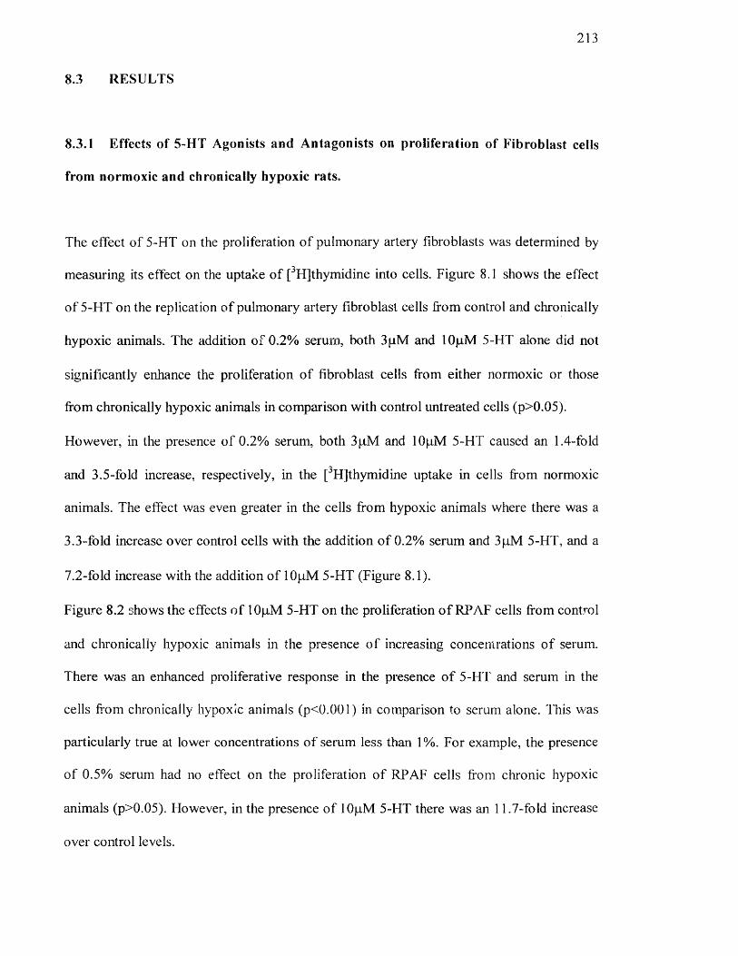
213
8.3 RESULTS
8.3.1 Effects of 5-HT Agonists and Antagonists on proliferation of Fibroblast cells
from normoxic and chronically hypoxic rats.
The effect o f 5-HT on the proliferation o f pulmonary artery fibroblasts was determined by
measuring its effect on the uptake o f [^H]thymidine into cells. Figure 8.1 shows the effect
o f 5-HT on the replication of pulmonary artery fibroblast cells from control and chronically
hypoxic animals. The addition o f 0,2% serum, both 3pM and lOjuM 5-HT alone did not
significantly enhance the proliferation o f fibroblast cells from either normoxic or those
from chronically hypoxic animals in comparison with control untreated cells (p>0.05).
However, in the presence o f 0.2% serum, both 3pM and lOpM 5-HT caused an 1.4-fold
and 3.5-fold increase, respectively, in the [^Hjthymidine uptake in cells from normoxic
animals. The effect was even greater in the cells from hypoxic animals where there was a
3.3-fold increase over control cells with the addition o f 0.2% serum and 3pM 5-HT, and a
7.2-fold increase with the addition of lOjuM 5-HT (Figure 8.1).
Figure 8.2 shows the effects o f lOjuM 5-HT on the proliferation o f RPAF cells from control
and chronically hypoxic animals in the presence o f increasing concentrations o f serum.
There was an enhanced proliferative response in the presence o f 5-HT and serum in the
cells fi'om chronically hypoxic animals (p<0.001) in comparison to serum alone. This was
particularly true at lower concentrations o f serum less than 1 %. For example, the presence
o f 0.5% serum had no effect on the proliferation of RPAF cells from chronic hypoxic
animals (p>0.05). However, in the presence o f 10p.M 5-HT there was an 11.7-fold increase
over control levels.
214
Figure 8.3 shows the effects of specific 5-HT agonists and antagonists on the replication of
pulmonary artery fibroblasts from control and chronically hypoxic animals. Unstimulated
RPAF cells from control and hypoxic animals showed no difference in [^H]thymidine
uptake (p>0.05). In addition 0.2% serum alone and 5-HT (lOpM) had no significant effect.
However, as described for Figure 8.2, the addition of 5-HT (lOpM) in the presence of 0.2%
serum resulted in a 2.7-fold increase in proliferation in normoxic cells (p<0.005). This
response was further enhanced in cells from chronically hypoxic animals where there was
an 6-fold increase in comparison to control, untreated cells.
To examine this effect more closely, we studied a range o f more specific 5-HT agonists.
The addition o f a 5 -HT2 agonist, a-methyl-5-HT (10p,M) enhanced the proliferation o f cells
from hypoxic rats to the same degree as that o f lOp-M 5-HT alone (1750+251 DPM and
854+196 DPM respectively). In contrast, the 5-HTzB agonist BW723C86 (lOfiM) did not
increase proliferation in either cell type (332+52 DPM for the hypoxic cells and 325+1 10
DPM for the normoxic cells which was not significantly different from control cells). This
was also true for the 5 -HT2C agonist, MK212 (lOpM) which did not increase proliferation
in either cell type (324+225 DPM for the hypoxic cells and 339+211 DPM for the
normoxic cells which was not significantly different from control cells).
To study this further, we also looked at the effect o f a range o f 5-HT antagonists on the 5-
HT-induced [^H]thymidine uptake. The addition of the 5-HTiA antagonist, ketanserin
(0.1 pM), abrogated the enhanced growth response observed in the cells from chronically
hypoxic animals induced with 5-HT and 0.2% serum (926+115 DPM for the hypoxic cells
which was not significantly different from normoxic cells 849+310 DPM). In contrast,
neither the 5 -HT213+C inhibitor SDZ SER 082 (IpM ) nor the 5-HT2b inhibitor R SI02221
(IpM ) had any effect on reducing proliferation o f cells in the presence o f 5-HT and 0.2%
215
serum (1796+121 and 1847+314 DPM for the hypoxic cells which was significantly
different (p<0.05) from normoxic cells 882+265 and 886+172 DPM, respectively).
Figure 8.4 shows the effect o f 5-HT with 0.2% serum and specific inhibitors for 5-HT
(Ketanserin and GR55562) and MAP kinases (SB203580 and U0126) on the replication of
pulmonary artery fibroblast cells from control and chronically hypoxic animals. Basal
levels o f replication in normoxic cells (220+15 DPM) and in cells from chronically hypoxic
animals (230+20 DPM) were not significantly different (p>0.05). lOpM 5-HT with the
addition of 0.2% serum gave rise to an 4-fold increase in proliferation in the normoxic cells
and a further increase (6.2-fold) in the cells from chronically hypoxic animals (p<0.005).
The addition o f IpM SB203580 (a specific p38 inhibitor) abolished this enhanced 5-HT
growth response observed in the cells from chronically hypoxic animals (923+42 DPM). In
the presence o f this compound the increase in [^H]thymidine uptake in cells from hypoxic
rats was not significantly different to that from normoxic animals (793+102 DPM)
(p>0.05). The addition of 0.1 pM of the MEK inhibitor U0126 completely abolished the
proliferative response o f normoxic cells (312+28 DPM) and those from chronically hypoxic
animals (331+38 DPM) which was not significantly different from control levels (p>0.05).
The effect o f 5-HT antagonists were also investigated (Figure 8.4). The addition o f the 5-
HT2A antagonist Ketanserin (0.1 pM) abolished the enhanced 5-HT growth response seen in
the cells from clironically hypoxic animals (913+50 DPM). Like SB203580, the enhanced
response was reduced to the level o f that observed for cells from normoxic animals
(832+78 DPM) (p>0.05). In contrast, the 5 - H T i b - i d antagonist GR55562 (IpM ) had no
effect on the proliferation o f cells from either the normoxic or hypoxic animals.
2 1 6
Figure 8.1
1400
1200Q_O® 1000 coo _
O)c=800
600
^ 400
200 ir#r#r#O □cLO
co
L O
=3<=>
C/D
CM<=>Hh3
CO
ooCM
Figure 8.1 Effect of 5-HT and Sérum on [^H]Thymidine Uptake in RPAF ceils from Normoxic
and Chronically Hypoxic Rats.
Uptake o f [^Hjthymidine from normoxic (□ ) and chronically hypoxic (■ ) rats. RPAF cells were
grown to 60% confluency in 24-well plates and then quiesced in serum-free medium. The cells were
stimulated with either 3pM or lOpM 5-HT alone or in conjunction with 0.2% serum as indicated in
the graph. Cells were then allowed to grow in normoxic conditions for a period o f 24h. The
experiment was repeated with cells from 8 different animals and the results shown are typical o f those
obtained.
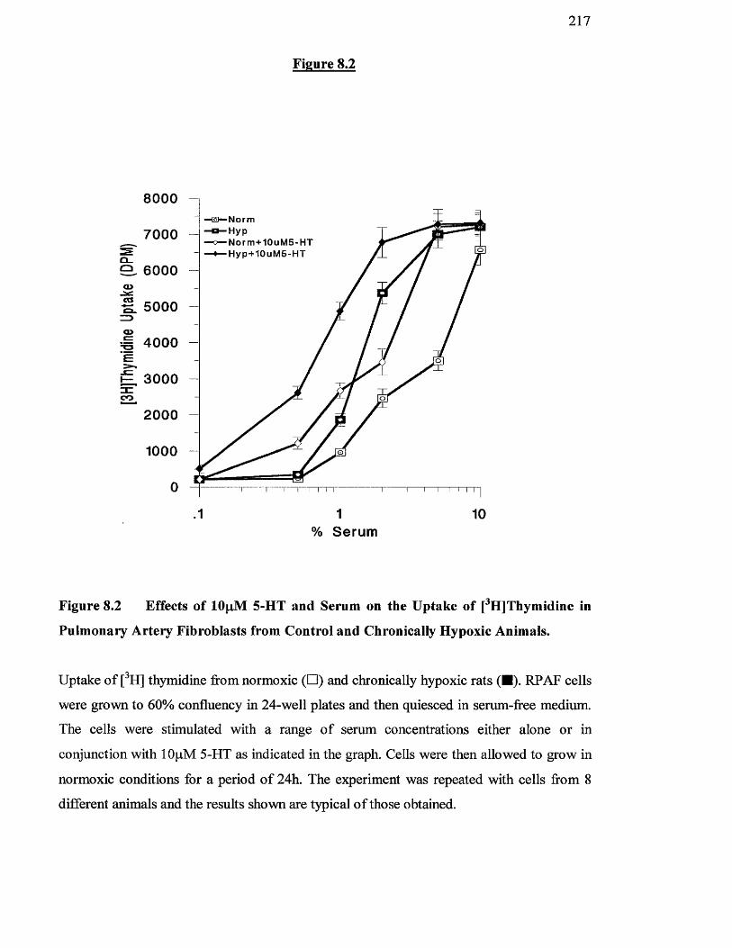
2 1 7
Figure 8.2
8 0 0 0
7 0 0 0
a . 6 0 0 0
■Norm■Hyp■ N o r m + 1 0 u M S - H T■Hy p -+ 10 uM5 -HT
5 0 0 0
4 0 0 0
3 0 0 0
2000
1000
.1 1% Serum
10
Figure 8.2 Effects of lOpiM 5-HT and Serum on the Uptake of [^H]Thymidine in
Pulmonary Artery Fibroblasts from Control and Chronically Hypoxic Animals.
Uptake o f [^H] thymidine from normoxic (□ ) and chronically hypoxic rats (■). RPAF cells
were grown to 60% confluency in 24-well plates and then quiesced in serum-free medium.
The cells were stimulated with a range o f serum concentrations either alone or in
conjunction with lOpM 5-HT as indicated in the graph. Cells were then allowed to grow in
normoxic conditions for a period o f 24h. The experiment was repeated with cells from 8
different animals and the results shown are typical o f those obtained.
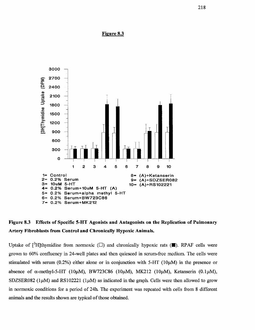
2 1 8
Figure 8.3
0>03Q .
1>1.
J =I—Xco
3 0 0 0
2 7 0 0
2 4 0 0
2100
1 8 0 0
1 5 0 0
1200
9 0 0
6 0 0
3 0 0
0
2 3 6 8 9 104 5 71
1“ C o n t r o l 2 ” 0 . 2 % S e r u m 3 - lOuM 5 - H T4 “ 0 . 2 % S e r u m + I O u M 5 - H T (A)5 - 0 . 2 % S e r u m + a i p h a m e t h y l 5 - H T 6 “ 0 . 2 % S e r u m + B W 7 2 3 C 8 6 7 - 0 . 2 % S e r u m + M K 2 1 2
8 “ ( A ) + K e t a n s e r i n 9 - ( A ) + S D Z S E R 0 8 2
10= ( A ) + R S 1 0 2 2 2 1
Figure 8.3 Effects of Specific 5-HT Agonists and Antagonists on the Replication of Pulmonary
Artery Fibroblasts from Control and Chronically Hypoxic Animals.
Uptake o f [^HJthymidine from normoxic (□ ) and chronically hypoxic rats (■). RPAF cells were
grown to 60% confluency in 24-well plates and then quiesced in serum-free medium. The cells were
stimulated with serum (0.2%) either alone or in conjunction with 5-HT (lOpM) in the presence or
absence o f a-methyl-5-HT (lOpM), BW723C86 (lOpM), MK212 (lOpM), Ketanserin (0.1 pM),
SDZSER082 (IpM ) and R S I02221 (IpM ) as indicated in the graph. Cells were then allowed to grow
in normoxic conditions for a period o f 24h. The experiment was repeated with cells from 8 different
animals and the results shown are typical o f those obtained.
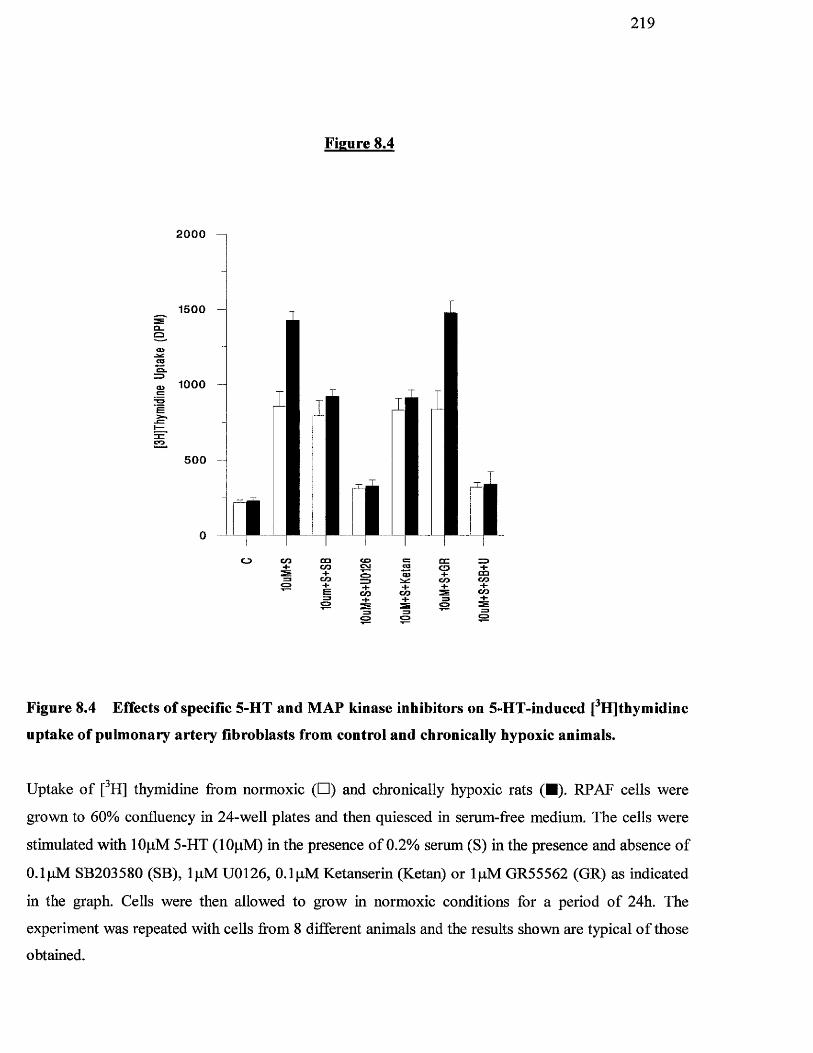
2 1 9
Figure 8.4
2000
1500Q_O
1000
500 -
m
to4-E
f :oo oo
o s
Figure 8.4 Effects of specific 5-HT and MAP kinase inhibitors on 5-HT-induced [^H]thymidine
uptake of pulmonary artery fibroblasts from control and chronically hypoxic animals.
Uptake o f [^H] thymidine from normoxic (□ ) and chronically hypoxic rats (■). RPAF cells were
grown to 60% confluency in 24-well plates and then quiesced in serum-free medium. The cells were
stimulated with lOpM 5-HT (lOpM) in the presence o f 0.2% serum (S) in the presence and absence o f
0.1 pM SB203580 (SB), IpM U0126, 0.1 pM Ketanserin (Ketan) or IpM GR55562 (GR) as indicated
in the graph. Cells were then allowed to grow in normoxic conditions for a period o f 24h. The
experiment was repeated with cells from 8 different animals and the results shown are typical o f those
obtained.
220
8.3.2 Effect of 5-HT and 5-HT Antagonists on p38 phosphorylation in Fibroblast
cells from normoxic and chronically hypoxic rats.
Watts (1998) have shown that 5-HT can lead to the phosphorylation of MAP kinase. We
were interested to see if 5-HT could further enhance p38 MAP kinase phosphorylation
already observed (Chapter 7.32, Figure 7.3) in pulmonary artery fibroblasts from hypoxic
rats.
Figure 8.5 shows the effect of increasing doses of 5-HT on phosphorylated p38 M /iP
kinase (pp38) in pulmonary artery fibroblast cells from control and chronically hypoxic
animals. In the unstimulated control cells from chronically hypoxic rats, a clear increase in
pp38 can be seen which is not apparent in the cells from normoxic animals (Figure 8.5
compare upper panel, lane 1 with lane 7). In the cells from chronically hypoxic animals
pp38 MAP kinase was increased in a 5-HT do se-dependent manner with maximum
phosphorylation occurring at lOpM 5-HT. In contrast, no increase was seen in the cells
from normoxic animals. The total amount of p38 MAP kinase was examined (lower panel)
to ensure equal loading of protein on the polyacrylamide gel.
Figure 8.6 shows the effect o f 5-HT on pp38 MAP kinase in pulmonary artery fibroblast
cells from control and chronically hypoxic animals. The upper panel shows that, 3pM and
lOpM 5-HT gave rise to a substantial increase in pp38 MAP kinase in the cells from
chronically hypoxic animals in comparison to control untreated cell extracts. There was no
increase in pp38 MAP kinase in extracts from normoxic cells. The increase in pp38 MAP
kinase in the cells from chi’onically hypoxic animals could be abrogated with the addition
ofO .lpM Ketanserin, a specific 5-HTiA antagonist. In contrast, addition o f IpM GR55562,
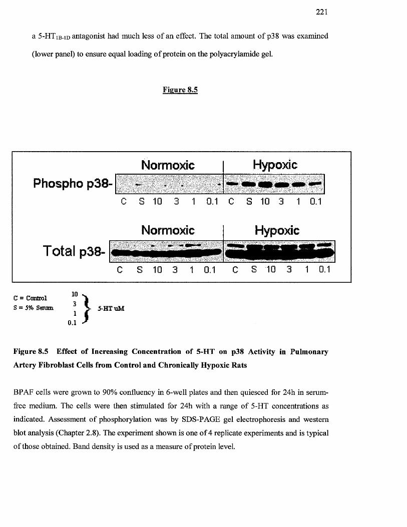
221
a 5“HT ib-id antagonist had much less o f an effect. The total amount o f p38 was examined
(lower panel) to ensure equal loading o f protein on the polyacrylamide gel.
Figure 8.5
Normoxic Hypoxic
C S 10 3 1 0.1
Normoxic
C S 10 3 1 0.1
Hypoxic"... -
Phospho p38-
TOtal p38-C S 10 3 1 0.1 C S 10 3 1 0.1
C = ComHrol
S ~ 5% Sem m
103
10.1 } j-H T u M
Figure 8.5 Effect of Increasing Concentration of 5-HT on p38 Activity in Pulmonary
Artery Fibroblast Cells from Control and Chronically Hypoxic Rats
BPAF cells were grown to 90% confluency in 6-well plates and then quiesced for 24h in serum-
free medium. The cells were then stimulated for 24h with a range o f 5-HT concentrations as
indicated. Assessment o f phosphorylation was by SDS-PAGE gel electrophoresis and western
blot analysis (Chapter 2.8). The experiment shown is one o f 4 replicate experiments and is typical
o f those obtained. Band density is used as a measure o f protein level.
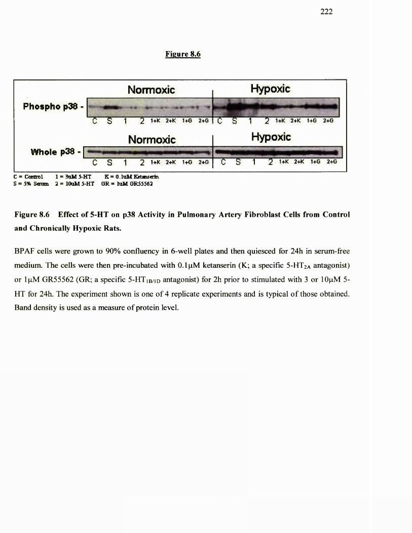
Phospho p38 -
Whole p38 -
222
Figure 8.6
Normoxic Hypoxic
. FC S 1 2 1+K 2+K 1+C 2+G
Normoxic
C S 1 2 1+K 2+K 1+G 2+G
Hypoxic
C S 1 2 1+K 2+K 1+G 2+G C S 1 2 1+K 2+K 1+G 2+G
c = Control 1 = 3uU 5-HT K = 0. loM Ecftinseim S - 5% Seran 2 = lOuM 5-HT CR = ïoM GR55562
Figure 8.6 Effect of 5-HT on p38 Activity in Pulmonary Artery Fibroblast Cells from Control
and Chronically Hypoxic Rats.
BPAF cells were grown to 90% confluency in 6-well plates and then quiesced for 24h in serum-free
medium. The cells were then pre-incubated with 0.1 pM ketanserin (K; a specific 5-HTzA antagonist)
or IpM GR55562 (GR; a specific 5-H T ib /id antagonist) for 2h prior to stimulated with 3 or lOpM 5-
HT for 24h. The experiment shown is one o f 4 replicate experiments and is typical o f those obtained.
Band density is used as a measure o f protein level.
2 2 3
8.3,3 Effect of 5-HT and 5-HT Antagonists on p42/p44 MAP Kinase
Phosphorylation
We wanted to see o f 5-HT could further enhance p42/p44 MAP kinase phosphorylation
already observed in pulmonary artery fibroblasts from hypoxic rats. Figure 8.7 shows the
effect o f increased doses of 5-HT on phosphorylated p42/44 MAP kinase activity in
pulmonary artery fibroblast cells from control and chronically hypoxic animals. Again, in
the unstimulated control cells f om chronically hypoxic rats, a clear increase of p42/44
MAP kinase activity can be seen which is not apparent in the cells from normoxic animals.
Neither cell type responded to 5-HT under any of the concentrations used with respect to
increases in p42/p44 MAP kinase phosphorylation. The total amount o f p42/p44 MAP
kinase was examined (lower panel) to ensure equal loading of protein on the
polyacrylamide gel.
Figure 8.8 shows the effects o f 5-HT on phosphorylated p42/44 MAP kinase activity in
pulmonary artery fibroblast cells from control and chronically hypoxic animals in the
presence o f 0.2% serum. In the unstimulated control cells from chronically hypoxic rats, a
clear increase o f p42/44 MAP kinase phosphorylation was observed in comparison to
extracts from untreated normoxic rats. 3pM and lOpM 5-HT alone did not increase
p42/p44 phosphorylation in either cell types as in Figure 8.7. In the presence of 0.2%
serum, however, an increase in p42/p44 MAP kinase phosphorylation by 5-HT occurred in
a dose-dependent fashion in the cells from chronically hypoxic animals. The total amount
o f p42/44 MAP kinase was examined to ensure equal loading of protein on the
polyacrylamide gel.
Figure 8.9 shows the effect of 5-HT on phosphorylation o f p42/44 MAP kinase in
pulmonary artery fibroblast cells from control and chronically hypoxic animals.
Unstimulated cells from clironically hypoxic animals displayed increased levels o f p42/44
224
MAP kinase activity compared with normoxic cells. There was a further increase in
p42/p44 MAP kinase phosphorylation with the addition o f 5% serum, in RPAF from
hypoxic animals. Neither 3pM nor lOpM 5-FIT gave rise to increased p42/p44 MAP kinase
phosphorylation in cells from chronically hypoxic animals or in the normoxic cells, over
control levels. The addition o f Ketanserin, a specific 5-HT2a antagonist, and GR55562, a 5-
FITiB-iD antagonist had no effect on phosphorylation of p42/p44 MAP kinase. The total
amount of p42/p44 MAP kinase was examined (lower panel) to ensure equal loading of
protein on the polyacrylamide gel.
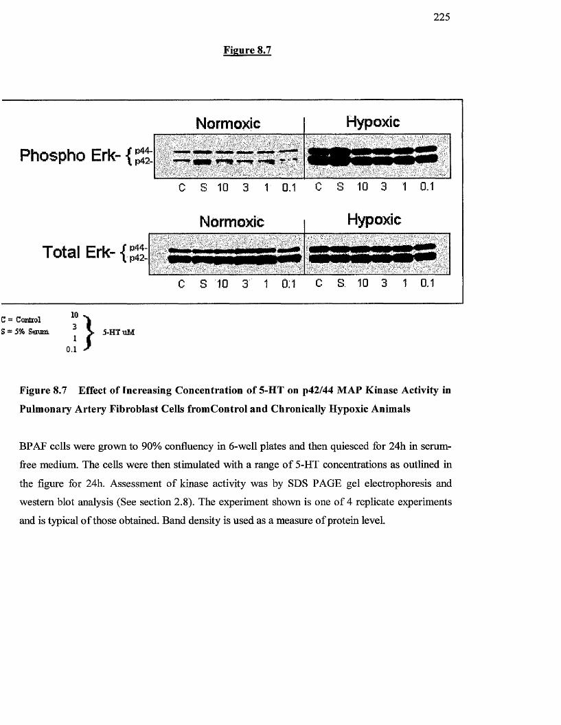
Figure 8.7
2 2 5
Phospho Erk- {
Total Erk-
Normoxic Hypoxic
mimm mmm «mmw mmm- _ w'#* ■'
C S 10 3 1 0.1 C S 10 3 1 0.1
Normoxic Hypoxic
‘
c s 10 3 1 0:1 C S 10 3 1 0.1
C = Coxdiol S = 5% Som i il 5-HTuM
Figure 8.7 Effect o f Increasing Concentration of 5-HT on p42/44 MAP Kinase Activity in
Pulmonary Artery Fibroblast Cells fromControl and Chronically Hypoxic Animals
BPAF cells were grown to 90% confluency in 6-well plates and then quiesced for 24h in serum-
free medium. The cells were then stimulated with a range o f 5-HT concentrations as outlined in
the figure for 24h. Assessment o f kinase activity was by SDS PAGE gel electrophoresis and
western blot analysis (See section 2.8). The experiment shown is one o f 4 replicate experiments
and is typical o f those obtained. Band density is used as a measure o f protein level.
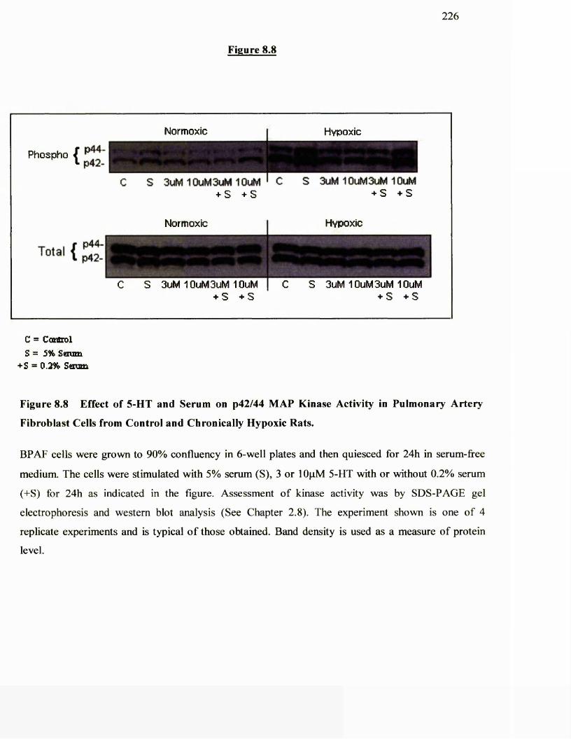
Figure 8.8
226
Phospho fI p42-
Normoxic Hypoxic
S 3uM 10uM3uM 10uM ' C + S + S
S 3uM 10uM3uM 10uM + S + S
Total { %
Normoxic Hypoxic
S 3uM 10uM3uM 10uM + S + S
S 3uM 10uM3uM 10uM + S + S
C = Control S = 5% Sdum
+S = 0.2% S«Eum
Figure 8.8 Effect o f 5-HT and Serum on p42/44 MAP Kinase Activity in Pulmonary Artery
Fibroblast Cells from Control and Chronically Hypoxic Rats.
BPAF cells were grown to 90% confluency in 6-well plates and then quiesced for 24h in serum-free
medium. The cells were stimulated with 5% serum (S), 3 or lOpM 5-HT with or without 0.2% serum
(+S) for 24h as indicated in the figure. Assessment o f kinase activity was by SDS-PAGE gel
electrophoresis and western blot analysis (See Chapter 2.8). The experiment shown is one o f 4
replicate experiments and is typical o f those obtained. Band density is used as a measure o f protein
level.
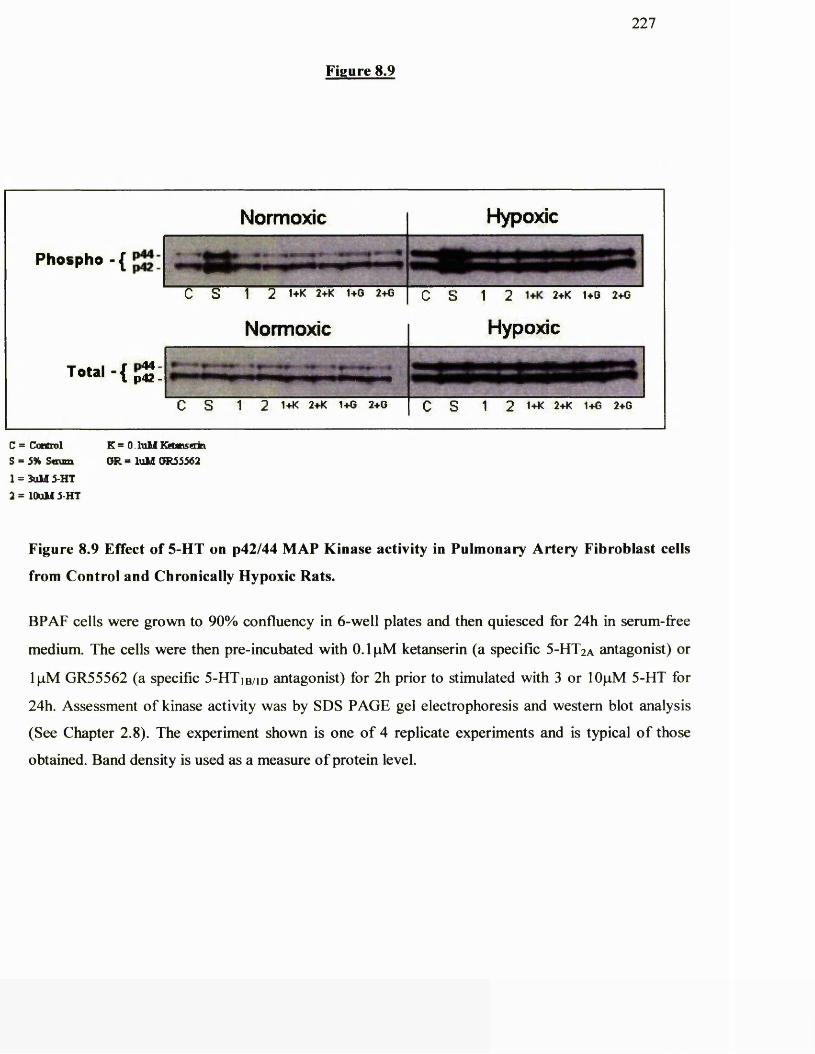
Figure 8.9
227
Phospho- { ^
T otal -{
Normoxic Hypoxic
e s 1 2 1+K 2+K 1+0 2+G I Q s 1 2 1+K 2+K 1+G 2+G
Normoxic l Hypoxic
e s 1 2 1+K 2+K 1+G 2+0 S 1 2 1+K 2+K 1+G 2+G
C = Control S = 5% S«nim
1 = îuM 5-HT2 = lOuU 5-HT
K = 0. hilul Keunsain GR = luU 0R55562
Figure 8.9 Effect o f 5-HT on p42/44 MAP Kinase activity in Pulmonary Artery Fibroblast cells
from Control and Chronically Hypoxic Rats.
BPAF cells were grown to 90% confluency in 6-well plates and then quiesced for 24h in serum-free
medium. The cells were then pre-incubated with 0.1 pM ketanserin (a specific 5-HTiA antagonist) or
IpM GR55562 (a specific 5-H T ib /id antagonist) for 2h prior to stimulated with 3 or lOpM 5-HT for
24h. Assessment o f kinase activity was by SDS PAGE gel electrophoresis and western blot analysis
(See Chapter 2.8). The experiment shown is one o f 4 replicate experiments and is typical o f those
obtained. Band density is used as a measure o f protein level.
228
8.4 DISCUSSION
This chapter focused on the effects of 5-HT on the proliferation and cell signalling of
RPAF cells from normoxic and chronic hypoxic rats. 5-HT alone did not produce a
mitogenic response in pulmonary artery fibroblast cells from either control or chronically
hypoxic animals. These results are in keeping with the work o f others (Pitt et al, 1994;
Seuwen and Pouyssegur, 1990) who studied 5-HT induced proliferation o f bovine and rat
pulmonary vascular smooth muscle cells in culture. In those studies, 5-HT had a co-
mitogenic effect on the cells, requiring incubation with other growth factors such as PDGF
and EGF. This is similar to the present work. The addition o f a low concentration o f serum
(which in itself does not increase proliferation) in conjunction with 5-HT resulted in an
increase o f proliferation in both the normoxic and especially in the chronically hypoxic
cells. This effect may requii'c either binding o f 5-HT to cell membrane receptors or active
transport of 5-HT into the cell via the 5-HT transporter (5-HTT) (Eddahibi et al, 2000).
One hypothesis could be that other co-mitogens change the structural conformity of the 5-
HT receptor allowing 5-HT to bind, or that it opens up the 5-HTT allowing 5-HT to enter
the cell. Previous work has demonstrated that the proliferative effect o f 5-HT on
[^HJthymidine incorporation in bovine pulmonary artery smooth muscle cells was
abolished after inhibition of the 5-HT transporter into the cell (Lee et al, 1991; Lee et al,
1994a).
Our previous work (Chapter 7) has demonstrated that cells from chronically hypoxic
animals display increased basal levels o f p38 and p42/p44 MAP kinase phosphorylation in
comparison to those from normoxic cells, and further induced that phosphorylation which
occurred in response to serum. In this present study, 5-HT increased pp38 above that of
basal levels and in a dose-dependent manner to 5-HT. This occurred in the absence o f any
229
co-mitogens. In contrast, 5-HT was unable to further increase p42/p44 MAP kinase
phosphorylation above basal levels. However, in the presence of low levels o f serum, there
was a further enhancement of p42/p44 MAP kinase activation. Other work has
demonstrated that 5-HT mediates its response by phosphorylating p42/44 MAP kinase
(Florian and Watts, 1998; Watts, 1996). The mechanisms for this activation are unclear,
however, signalling pathways for 5-HT have classically included activation o f PLC and
plasma membrane calcium channels that are sensitive to inhibition by dihydropyridines
(Watts 1996). In our work, p42/p44 MAP kinase activation was activated by 5-HT only in
the presence o f co-mitogens. To date, this is the first study to show that pp38 is activated by
5-HT and also that the specific p38 MAP kinase inhibitor, SB203580, can block the
enhanced hypoxia-associated proliferation with 5-HT.
Previous work has indicated that the 5-HTi receptor is involved in contraction and the 5-
HTz receptor is involved in proliferation o f cells (Frishman et al 1996). We were therefore
particularly interested in the effects o f 5-HTi and S-HTz receptor antagonists on
proliferation o f RPAF cells and MAP kinase activation in these cells. Studies by other
groups in rat aortic smooth muscle cells indicate that it is the 5-HT2a receptor that mediates
5-HT stimulated phosphorylation and activation of the p42/p44 MAPKs (Kelleher et al,
1995). This may not be the case for RPAF cells since 5-HT alone does not increase p42/p44
phosphorylation. Other work has shown that the mitogenic effect o f 5-HT on rat pulmonary
vascular smooth muscle cells was attenuated by the 5-HTi receptor antagonist, ketanserin
(Pitt et al, 1994). This would agree with our findings since the use o f ketanserin blocked
the increased proliferation in the cells from chronically hypoxic animals associated with
hypoxia.
230
To be certain of which receptor was important in the proliferation and MAP kinase
activation in chronically hypoxic animals with respect to 5-HT, further inhibitor studies in
conjunction with specific agonist studies were undertaken. To date the 5 -HT2 receptor has 3
sub-types identified, 5 -HT2A, 5-HT2b and 5 -HT2C. Unfortunately, there are no specific 5-
HT2A agonists commercially available at this time, however, there are specific 5 -HT2B
(BW72386) and 5 -HT2C (MK212) agonists and a specific 5 -HT2 agonist (a-methyl 5-HT).
Use o f these compounds allowed us to determine the specificity o f the sub-types involved
in proliferation o f RPAF cells. There are also specific antagonists available naamely, 5-
HT2A (ketanserin), 5-HT2c (RSI02221) and a specific 5-HT2b/c antagonist (SDZ SER 082)
which allowed us to determine further the action o f the individual receptor sub-types.
The results demonstrated that, neither the actions o f the 5 -HT2B nor 5 -HT2C agonists in the
presence o f low serum could produce a proliferative effect on either cell type. However, the
selective 5 -HT2 agonist gave rise to a proliferative response in both cells types, which
equalled the response in terms of magnitude o f 5-HT itself. This suggests that the 5 -HT2A
receptor may be responsible for proliferation o f these cells. Inhibitor studies were then used
to corroborate these results.
Again, the use o f specific 5-HTiB/c inhibitors could not inhibit the replicative responses o f
these cells to 5-HT. The hypoxic-associated growth o f these cells however, could be
successfully abrogated by the use o f ketanserin (S-HT] inhibitor). These findings agreed
with the agonist studies and firmly pointed towards the 5-HT2a receptor as being the
receptor responsible for hypoxic-associated growth of pulmonary artery fibroblast cells
from chronically hypoxic animals. The 5 -HT2A receptor also appears to signal via p38
MAP kinase because, by blocking the actions of this receptor we also blocked pp38
activation. pp38 has been shown previously in this thesis (Chapter 7) to be essential for
hypoxic growth o f pulmonary artery fibroblasts.

23
C hapter 9
General Discussion
232#
9.1 Summary of Results
The data presented in this thesis shows that hypoxia has a marked effect on
proliferation and cell signalling in pulmonary artery fibroblast cells. Cell models have
been established to study the effects of acute and chronic hypoxia on fibroblast cells
derived from the pulmonary and systemic circulations.
We have shown that in response to acute hypoxia:
• Acute hypoxia can stimulate pulmonary artery fibroblast cells to proliferate and
can also increase the effect o f mitogens. In contrast, acute exposure to hypoxia
has no effect on proliferation o f cells derived from the systemic circulation.
• This increase in proliferation also coincides with an increase in the second
messenger IP3 in pulmonary artery fibroblasts. Mitogens that caused increased
proliferation to hypoxia in the pulmonary artery fibroblasts also gave rise to
increases in IP3 mass.
• Acute hypoxia also led to increased phosphorylation of members o f the MAP
kinase family o f proteins that are known to be involved in cell growth and
development.
We have also shown that in cells derived from chronically hypoxic rats but
maintained in normoxia:
• Pulmonary artery fibroblast cells from clironically hypoxic animals ha\c
undergone a phenotypic switch whereby these cells proliferate to a level similar
to those made acutely hypoxic.
233
• These cells also display increased MAP kinase phosphoiylation when compared
to those of control cells.
• Increased sensitivity to proliferation by 5-HT, a potent vasoconstrictor known to
be released in vivo to hypoxia, was also apparent in the pulmonary artery
fibroblasts from chronic hypoxic rats. This was again related to increased MAP
kinase phosphorylation.
• The specific 5-HT receptor sub-type responsible for proliferation in the
pulmonary artery fibroblasts from control and chronically hypoxic rats was also
elucidated,
9.2 Species Difference
Closer examination o f the results indicates that the effects o f hypoxia on pulmonary
artery fibroblasts are not always consistent between different species. The most
notable example is in the p42/p44 MAP kinase responses o f pulmonary artery
fibroblasts. Acute hypoxia caused only a small increase in p42/p44 MAP kinase
activity in cells derived from the bovine model whereas the rat cells that had been
exposed to chronic hypoxia showed high levels of p42/p44 MAP kinase
phosphorylation. This was also true o f other MAP kinase isoforms, for example, in
the bovine model, JNK was activated in the face o f hypoxia which was unlike the rat
model. This was also the case when cells from different species were subjected to
hypoxia in cell proliferation studies. In the bovine cells, acute hypoxia alone increased
proliferation, whilst in rat cells hypoxia increased proliferation only in the presence of
mitogens. This apparent conflict in results may be due to an underlying difference in
the pharmacology or biochemistry o f the pulmonary artery fibroblasts o f the various

234
species. ît must also be pointed out that bovine cells were exposed to low O2 levels
(~30mmHg) for a relatively short time (24h) whereas rat cells had been exposed (m
vivo) to less severe hypoxic exposure for a longer time period (~330mmHg for 14
days). It is possible that the effect o f hypoxia on cell proliferation is dependent on
both the intensity and the duration of the hypoxic stimulus, but this remains to be
addressed by further scientific investigation.
Alternatively, the difference may be due to the different order o f pulmonary artery
used from species to species. In both animals, we used the main branch o f the
pulmonary artery. However, in the bovine cells the piece o f tissue was isolated from
well down the main branch near to the tip o f the lobe. In the rat, the tissue was
isolated just as the artery entered into the lungs. As stated in chapter 1, the response of
pulmonary arteries to hypoxia may differ between proximal and distal regions o f the
vessel. This may account for the experimental differences between the species. It
would have been interesting to conduct a study comparing various regions o f the
pulmonary artery from each species. This would have been possible in the bovine
lung where the vessels were large. However, the rat pulmonary artery rapidly
decreases in size and explanting become increasingly difficult.
Whilst the effect o f hypoxia differed between species, there was also differences in
proliferation responses to various agonists. In pulmonary artery fibroblasts from the
cow, endothelin-1 did not induce proliferation whereas in the rat, ET-1 was a good
mitogen (Peacock and Aidulis, 1994). Also, in the bovine model, 5-HT acted as a
mitogen on its own whilst in the rat model it only acted as a co-mitogen. It is possible
that this may be due to changes in receptor abundance and conformity in fibroblasts
from different species.
235
9,3 Role of Stress-Activated Protein Kinases
It is clear from the results that there are factors in both the pulmonary arteries from
cow and rat that are important to the hypoxia-associated increases in fibroblast
proliferation. The stress-activated protein p38 MAP kinase was activated in the face
o f acute hypoxia in the bovine model, and also in the cells from chronically hypoxic
rats. The ability o f a specific pharmacological inhibitor of this protein (SB203580) to
attenuate hypoxic-stimulated DNA synthesis in growth-arrested cells implies that its
biological function is to relay signals important for cells to re-enter the cell cycle.
That hypoxia evokes the activation o f multiple signalling pathways emphasises the
intricate levels of control imposed on crucial cellular decisions such as those made
prior to cell division. Ultimately, a complex process such as re-entry into the cell
cycle will depend on the specific transcription factors that become activated by the
terminal protein kinase o f the pathways to which a receptor o f a particular mitogen or
stress is coupled. How these transcription factors combine in the nucleus to induce the
co-ordinated expression o f specific early genes is critical for cell cycle progression.
Research efforts in the past few years have been successfril in identifying some o f the
transcription factors regulated by these kinases. For example, p62^^^/Elk-l is a
common nuclear target for both the p42/44 and p38 MAP kinases iso forms which
were shown in the cells from chronically hypoxic animals to be in abundance when
compared with control cells (Marais et al, 1993, Raingeaud et al, 1996). While ATF-2
is a substrate for p38 MAP kinase in vitro and in transfected cells (Raingeaud et al,
1996). In addition, specific nuclear targets for pVO®*"*, MAPKAP kinase-2 and p90^^
are also coming to light (de Groot et al, 1994). While all o f these factors would be
predicted to become activated on exposure o f pulmonary artery fibroblasts to hypoxia.
236
the full array of genes that they control, and the contribution that products o f these
genes make to the genetic program regulating the cell cycle still has to be
investigated. A better understanding o f the transcriptional control of genes will be
necessary to provide information concerning how various signalling inputs impinging
upon the nucleus are co-ordinated to initiate complex genetic responses. In the
meantime, compounds such as Ketanserin and SB203580 used in this study will serve
as valuable tools for identifying the specific nuclear apparatus by which transcription
events are regulated by allowing researchers to dissect pathways involved in
transcriptional control.
9.4 Hypoxia causing a Phenotypic Switch
The findings in this thesis that pulmonary artery fibroblasts from chronically hypoxic
animals would appear to have undergone some form of phenotypic switch whereby
signals associated with hypoxia are always switched on may help in the understanding
o f which genes are important in the response. It would be logical to assume that if the
products o f the gene were constantly available, then the genes that control their
regulation would also be constantly in production. This would allow for comparisons
o f gene amount and presence to be easily ascertained.
Despite outlining some o f the protein cascades involved in hypoxic-stimulated
mitogenesis, a number of points remain unsolved regarding upstream events involved
in their activation, the possible interplay between these pathways, and the nature of
the hypoxic “receptor/s" that they are coupled to. While signal transduction by
hypoxia is far from understood and it is unknown whether hypoxia signals via G-
protein interactions or receptor tyrosine kinases, an observation in the BP A fibroblasts
237
was the resemblance of hypoxic mediated events to signalling characteristics of
growth factors that activate receptor tyrosine kinases, such as PDGF. Thus, for
example, activation of MAP kinase by hypoxia followed a slow and gradual time
course rather than the rapid kinetics reported in other cell types that are often maximal
within 2-5 minutes. The relative importance o f G-protein-coupled receptor and
tyrosine kinase receptor-mediated signalling events triggered by stresses and growth
factors is currently a topic o f considerable interest. Research within the past few years
has provided novel paradigms to support the concept o f physiologically relevant cross
talk between G-protein-coupled receptors, and tyrosine kinase receptor-linked routes
for communication within the cell (Daub et al, 1996).
9.5 Pulm onary vs. Systemic arteries
This thesis has demonstrated for the first time, to our knowledge, a fundamental
difference in response to hypoxia between systemic and pulmonary artery cells.
Unlike pulmonary artery fibroblasts, fibroblast cells from the systemic circulation in
both the bovine and rat models do not proliferate to hypoxia, or indeed give rise to the
phosphorylation o f the proteins which we consider important. This finding is
fundamental to this thesis as it mimics the known physiological response where
hypoxia in vivo does not result in vasoconstriction or in remodelled vessels in the
systemic arteries. It also supports the results fi’om the pulmonary artery fibroblasts
with respect to increased generation of signals resulting in proliferation, which are not
seen in the systemic artery cells.
Despite questions that still need to be resolved, these studies have outlined a number
of novel cellular consequences o f hypoxia on cells, further defining the possible
238
mechanisms by which it is able to influence their behaviour. In particular, the studies
highlight pathways that are likely to operate during vascular disease states like
pulmonary hypertension and other hypoxic forms of lung disease where proliferation
of these cell types contribute to blood vessel wall remodelling. A more conclusive
role for these pathways will become apparent in future literature.
9,6 Future W ork
The work of this thesis shows quite clearly that hypoxia is an important regulator of
the pulmonary vasculature and the mechanisms by which hypoxia regulates
remodelling during pulmonary hypertension may help us identify potential therapeutic
targets.
Wliile our picture o f acute and chronic stimulation of cell signalling in our cell models
o f pulmonary hypertension is incomplete, it is clear that specific MAP kinase
signalling pathways are being activated in cells from the pulmonaiy circulation.
Furthermore, these pathways induce HIF-1 (Minet et al, 2000) which can lead to the
upregulation o f several genes such as the mitogen VEGF. This raises the possibility o f
exploiting differences between pathways as seen in this thesis in order to specifically
inhibit hypoxia-induced signalling pathways in cardiopulmonary diseases. Achieving
this goal requires a detailed biochemical and molecular understanding o f the
signalling pathways activated in pulmonary fibroblasts in response to hypoxia and a
study o f the regulatory events that these control.
Defining exactly how MAP kinase activation is involved in pulmonary hypertension
is essential if we are to identify potential sites for therapeutic intervention. Although
we have established that hypoxia causes an increase in proliferation o f FAF cells
239
derived from the pulmonary artery of rats and cows, the effects of this on progression
of these cells through the cell cycle is unknown. Recent work has shown that MAP
kinase pathways converge to regulate cyclin D1 during hypoxic exposure of PC 12
cells (Conrad et al 1999). Since hypoxia increases proliferation in our cell model, the
role o f cyclin D1 in hypoxia-induced proliferation of PAF cells and the regulation of
its expression by p38 MAPK. and ERK pathways is unclear and would be an attractive
addition to this research.
We have shown that intracellular processes are specifically altered by decreased O2 in
pulmonary artery but not systemic fibroblasts. Now we should take the investigation
one step further and look at which transcription factors are turned on by hypoxia and
which genes are thus activated.
Along with the increase in proliferation o f fibroblasts seen in pulmonary vascular
remodelling, fibroblasts have an important role to play in laying dovm matrix proteins
such as collagen, which render the vessels indistensible. Future work should include
the examination o f extracellular matrix protein production fi'om normoxic and
hypoxic pulmonary artery fibroblasts and compare such protein generation to those o f
systemic vascular cells. Studying how the intracellular signalling changes seen in
fibroblast cells due to hypoxic exposure may effect extracellular matrix production
could also ultimately hold a clue for future treatments o f pulmonary vascular
remodelling during pulmonary hypertension.

240
References
241
Abrams HD, Rohrschneider LR and Eisenman RN (1982) Nuclear location of the
putative transforming protein o f avian myelocytomatosis virus Cell 29: 427-439
Aceto J. F. and Baker K. M. (1990) [Sar 1] angiotensin II receptor-mediated
stimulation of protein synthesis in chick heart cells Am. J. Physiol. 158: H806-H813.
Alexander A. F and Jenson R (1959) Gross cardiac changes in cattle with high
mountain (Brisket) disease, and in experimental cattle maintained at high altitudes
Am. J. Vet. Res., 20: 680.
Anderson NG, Mailer JL, Tonks NK and Sturgill TW (1990) Requirement for
integration o f signals from two distinct phosphorylation pathways for activation of
MAP kinase. Nature 343: 651-653.
Ankoma-Sey V, Wang Y and Dai Z (2000) Hypoxic stimulation of vascular
endothelial growth factor expression in activated rat hepatic stellate cells. Hepatology
31(1): 141-148.
Archer SL, Eaton LW and Nelson DP (1988) Oxygen radicals and antioxidant
enzymes alter pulmonary vascular reactivity in the rat lung. J. Appl. Physiol, 66: 102-
1 1 1
Asaoka Y, Nakamura S, Yoshida K and Nishizuka Y (1992a). Protein kinase C,
calcium and phospholipid degradation. TIBS. 17: 414-417.
242
Asaoka Y, Yoshida K, Oka M, Shinomura T, Mishima H, Matsushima S and
Nishizuka Y (1992b). The signal-induced phospholipid degradation cascade and
protein kinase C activation. Ciba Foundation Symposium. 164: 50-54.
Auer KL, Contessa J, Brenz-Verca S, Pirola L, Rusconi S, Cooper G, Abo A,
Wymann MP, Davis RJ, Birrer M and Dent P (1998) The Ras/Racl/Cdc42/
SEK/JNK/c-jun cascade is a key pathway by which agonists stimulate DNA synthesis
in primary cultures o f rat hepatocytes. Mol. Biol. Cell 9: 561-573.
Balboa MA, Firestein BE, Godson C, Bekk KS and Insel PA (1994) Protein kinase C
mediates phospholipase D activation by nucleotides and phorbol ester in Madin-
Darby canine kidney cells. Stimulation o f phospholipase D is independent o f
activation o f polyphosphoinositide-specific phospholipase C and phospholipase Az. J.
Biol. Chem. 269: 10511-10516.
Ballou, L. M., Luther, H. and Thomas, G. (1991) MAP2 kinase and 70K S6 kinase lie
on distinct signalling pathways. Nature 349: 348-350
Beg, AA and Baldwin, AJ (1993). The IkB proteins: multifunctional regulators of
Rel/NFKB transcription factors. Genes and Development 7: 2064-2070.
Belham CM, Rithwelle TJ, Scott PH, Pemberton AD, Miller HRP, Wadsworth RM,
Gould GW and Plevin R. (1996) Trypsin stimulates proteinase-activated receptor-2-
243
dependent and-independent activation o f mitogen-activated protein kinases Biochem.
J. 320: 939-946.
Belknap JK, Orton EC, Ensley B and Stenmark KR (1997) Hypoxia increases
bromodeoxyuridine labeling indicies in bovine neonatal pulmonary arteries. Am. J.
Respir Cell Mol Biol 16: 366-371.
Benitz WE, Kelly RT, Anderson CM, Lorant DE and Bernfield M ( 1990) Endothelial
heparin sulphate proteoglycan. Inhibitory effects on smooth muscle cell proliferation.
Am. J. Respir. Cell. Mol. Biol. 2: 13-24.
Berridge MJ (1987) Inositol lipids and cell proliferation. Biochem. Biophys. Acta 907:
33-45
Berridge MJ (1993a) Inositol trisphosphate and calcium signalling. Nature 361: 315-
325.
Berridge MJ (1993b) Intracellular channels and calcium spiking. Biomedical
Research (Japan) 14: 21-27.
Berridge MJ and Galione A (1988) Cytosolic calcium oscillators. FASEB J 2: 3074-
3082
Berridge MJ and Irvine RF (1989) Inositol phosphates and cell signalling. Nature
341: 197-203.
244
Berra E, Diaz-Meco MT, Dominguez I, Municio MM, Sanz L, Lozano J, Chapkin RS
and Moscat J (1993) Protein kinase C iso form is critical for mito genic signal
transduction Cell 74: 555-563.
Bishop JM (1985) Viral Oncogenes. Cell 42: 23-38
Bitterman PB and Henke CA (1991) Fibroproliferative disorders. Chest 99: 185-192.
Blancher C, Moore JW, Talks KL, Houlbrook S and Harris AL (2000) Relationship of
hypoxia-inducible factor (HIF)-l alpha and FIIF-2 alpha expression to vascular
endothelial growth factor induction and hypoxia survival in humans. Cancer Res
60(24): 106-113.
Boarder M (1994) A role for phospholipase D in control o f mitogenisis. TIPS, 15: 57-
62.
Bobrov VA, Fushtei IM and Polivoda SN (1985) Pulmonary hemodynamics and
serotonin level in patients with chronic penumonia with arterial hypertension. Vrach.
Delo. 12: 33-5.
Bogoyevitch MA, Ketterman AJ. and Sudgen PH. (1995) Cellular stresses
differentially activate c-jun N-terminal protein kinases and extracellular signal-
regulated protein kinases in cultured ventricular myocytes. J. Biol. Chem 270: 29710-
29717.
245
Bogoyevitch MA, Gillespie-Brown J, Ketterman AJ, Fuller SJ, Ben-Levy R,
Ashworth A, Marshall CJ and Sugden PH (1996) Stimulation o f the stress-activated
mitogen-activated protein kinase subfamilies in perfused heart. Circ. Res. 79: 162-
173.
Bogoyevitch MA, Glennon PE, Andersson MB, Clerk A, Lazou A, Marshall CJ,
Parker PJ and Sugden PH (1994) Endothelin-1 and fibroblast growth factor stimulate
the mitogen-activated protein kinase signalling cascade in cardiac myocytes. The
potential role o f the cascade in the integration o f two signalling pathways leading to
myocyte hypertrophy. J. Biol. Chem. 269: 1110-1119.
Bonfini L, Karlovich CA, Dasgupta C and Banerjee U. (1992) The son o f sevenless
gene product: A putative activator of Ras. Science 255: 603-606.
Bowtell D, Fu P, Simon M and Senior P (1992) Identification o f murine homologues
o f the Drosophila son o f sevenless gene: Potential activators o f ras. Proc. Nat. Acad.
Sci. USA. 89: 6511-6515.
Brenot F, Herve P, Petitpretz P, Parent F, Duroux P and Simmonneau G (1993)
Primary Pulmonary hypertenstion and fenfluramine use. Br Heart J 19: 537-41.
Brogden RN and Sorkin EM (1990) Ketanserin. A review of its pharmacodynamic
and pharmacokinetic properties, and therapeutic potential in hypertension and
peripheral vascular disease. Drugs 40: 903-949.
246
Bunn HF and Poyton RO. (1996) Oxygen sensing and molecular adaptation to
hypoxia. Physiol. Rev. 76: 839-885.
Bunn HF, Gu J, Huang LE, Park J-W and Zhu H (1998) Erythropoietin; a model
system for studying oxygen-dependent gene regulation. J. Exp. Biol. 201: 1197-1201.
Campbell JA (1927a) Further observations on oxygen acclilatisation. J, Physiol. 63:
325-342.
Campbell JA (1927b) Note on some pathological changes in the tissues during
attempted acclimatisation to alterations o f 02-pressure in the air. Br. J. Exp. PathoL
8: 347-351.
Campbell JA (1927c) Prolonged alterations o f oxygen pressure in the inspired air with
special reference to tissue oxygen tension, tissue carbon dioxide and haemoglobin. J,
Physiol. 62: 211-231.
Campbell JA (1935) Further evidence that mammels cannot acclimatise to 10 p.c.
oxygen or 20,000 feet altitude. Br. J. Exp. Path, lb: 39-48.
Cardenn DL and Gill GN (1992) Receptor tyrosine kinases. FASEB J. 6: 2332-2337.
247
Carney DH, Scott DL, Gordon EA and LaBelle EF (1985). Phosphoinositides in
mitogenisis: neomycin inhibits thrombin-stimulated phosphoinositide turnover and
initiation of cell proliferation. Cell. 42: 479-488.
Carpenter CL, Auger KR, Chanudhuri M, Yoakim M, Schaffhaussen B, Shoe Ison S
and Cant ley LC (1993) Phosphoinositide 3- kinase is activated by phosphopeptides
that bind to the SFI2 domains o f 85 kDa subunit. J. Biol. Chem. 268: 9478-9483.
Carpenter G (1992) Receptor tyrosine kinase substrates: src homology domains and
signal transduction. FASEB J., 6: 3283-3289.
Carsten ME and Miller JD (1990) In “Uterine function, molecular and cellular
aspects” eds. Carsten ME and Miller JD p i21-167. Plenum, New York.
Catling AD, Reuter CWM, Cox M, Parsons SJ and Weber MJ (1994) Partial
purification o f a mitogen-activated protein kinase from bovine brain. J. B iol Chem.
269: 30014-3002L
Chadwick CC, Saito A and Fleischer S (1990) Isolation and characterisation o f the
inositol trisphosphate receptor from smooth muscle. Proc. Nat. Acad. Sci. USA 87:
2132-2136.
Chen RH, Sarnecki C and Blenis J (1992) Nuclear localisation and regulation o f
ERK- and RSK-encoded protein kinases. MCB. 12: 915-927.
248
Chen YR, Wang X, TempeIton D, Davis RJ. and Tan TH. (1996) The role of c-Jun
N-terminal (JNK) in apoptosis induced by ultraviolet C and gamma radiotion.
Duration of JNK activation may determine cell death and proliferation. J. Biol Chem
271: 31929-31936.
Clark JD, Schievella AR, Nalefski EA and Lin LL (1995) Cytosolic phospholipase A2
Journal o f Lipid Mediators and Cell Signalling. 12: 83-117
Clerk A, Michael A and Sugden P (1998) Stimulation of the p38 Mitogen-activated
protein kinase pathway in neonatal rat ventricular myocytes by the G-protein-coupled
receptor agonists, endothelin-1 and phenylephrine: A role in cardiac myocyte
hypertrophy? Y Cell Biol. 142: 523-535
Cobum RF and Baron CB (1990) Coupling mechanisms in airway smooth muscle.
Am. J. Physiol. 258: LI 19-L133.
Comroe JH, van Lingen B and Stroud RC (1953) Reflex and direct cardiopulmonary
effects o f 5 OH trypyamine (serotonin). Their possible role in pulmonary embolism
and coronary thrombosis. Am. J. Physiol. 173: 379-386.
Conrad PW, Rust RT Han J Millhorn DE and Beitner-Johnson D (1999) Selective
Activation o f p38a and p38y by Hypoxia. Role in regulation of cyclin D1 by hypoxia
in PC12 cells. J. Biol. Chem. 274: 23570-23576
249
Cook SJ, Palmer S, Plevin R and Wake lam MJO (1990) Mass measurement of
inositol 1,4,5-trisphosphate and sn-1, 2-diacylglycerol in bombesin-stimulated Swiss
3T3 mouse fibroblasts Biochem. J. 265: 617-620
Cornfield DN, Stevens T, McMurtry IF, Abman SH and Rodman DM (1993) Acute
hypoxia increases cytosolic calcium in foetal pulmonary artery smooth muscle cells.
Am. J. Physiol. 265: L53-L56
Coso OA, Chiariello M, Kalinec G, Kyriakis JM, Woodgett J and Gutkind S (1995)
Transforming G protein-coupled receptors potently activate JNK (SAPK). J.
Biol.Chem.270: 5620-5624
Cowley S, Paterson H, Kemp P and Marshall CJ (1994) Activation o f MAP kinase
kinase in necessary and sufficient for PC 12 differentiation and for transformation of
NIH 3T3 cells. Cell 77: 841-852.
Crespo P, Simonds WF and Gutkind JS (1994) Ras-dependent activation o f MAP
kinase pathway mediated by G-protein betagamma subunits. Nature 369: 418-420.
Crouch EC, Parks WC, Rosenbaum JL, Chang D, Whitehouse L and Wu LJ (1989)
Regulation o f collagen production by medial smooth muscle cells in hypoxic
pulmonary hypertension. Am Rev Respir Dis 140(4): 1045-51
Cryer A and Bartly WE (1974) The design and use of a hypoxic chamber for small
animals. Laboratory Practice 23: 713-715.

250
Cuenda A, Rouse J, Doza YN, Meier R, Cohen P, Gallagher TF, Young P R and Lee
JC (1995) SB203580 is a specific inhibitor o f a MAP kinase homologue which is
stimulated by cellular stresses and interleukin-1. FEBS Lett. 364: 229-233.
Dai T, Rubie E, Franklin CC, Kraft A, Gillespie DAF, Avruch J, Kyriakis JM and
Woodget JR (1995). Stress-activated protein kinases bind directly to the ô domain o f
c-Jun in resting cells; implications for repression of c-Jun function. Oncogene. 10:
849-855.
Dalton S and Treisman R (1992) Characterisation o f SAP-1, a protein recruited by
serum response factor to the c-fos serum response element. Cell 68: 597-612.
Das M, Dempsey EC, Bouchey D, Reyland ME and Stenmark KR (1999) Chronic
hypoxia induces exaggerated growth responses in pulmonary artery adventitial
fibroblasts. Am. J. Resp. Cell Mol. Biol. 22: 15-25.
Datta SR, Brunet A and Greenberg L4E (1999) Cellular Survival: a play in three Akts.
Gen. Dev. 13: 2905-2927.
Daub H, Weiss FU, Wallasch C and Ullich A (1996) Role for transactivation of the
EGF receptor in signalling by G-protein-coupled receptors. Nature 379; 557-560.
251
Davies PF and Ross R (1978) Mediation of pinocytosis in cultured arterial smooth
muscle and endothelial cells by platelet-derived growth factor. J. C ell Biol. 79, 663-
671.
Davis RJ (1993) The mitogen-activated protein kinase signal transduction pathway.
J.BiolChem. 268: 14553-14556
De Groot RP, Ballou LM and Sassone-Corsi P (1994) Positive regulation o f the
cANJP-responsive activator CREM by the p70 S6 kinase: an alternative route to
mitogen induced gene expression. Cell 79: 81-91.
de Vries-Smits AMM, Burgering BMT, Leevers SJ, Marshall CJ and Bos JL (1992)
Involvement o f p21ras in activation o f extracellular signal-related kinase-1. Nature
357: 602-604.
Dempsey EC, Frid MG, Aldashev AA, Das M and Stenmark KR. (1997)
Heterogeneity in the proliferative response o f bovine pulmonary artery smo.oth
muscle cells to mitogens and hypoxia: Importance of protein kinase C. Can J Physiol
Pharmacol 75(7): 936-944
Dempsey EC, McMurty and O 'Brian RF (1991). Protein kinase C activation allows
pulmonary artery smooth muscle cells to proliferate to hypoxia. Am. J. Physiol. 260:
L136-L145.
252
Dent P, Haser W, Haystead TAJ, Vincent LA, Roberts TM and Sturgill TW (1992)
Activation o f mitogen-activated protein kinase kinase by v-Raf in NIH3T3 cells and
in vitro Science 257: 1404-1406.
Derijard B, Hibi M, Wu IH, Barratt T, Su B, Deng T, Karin M and Davis RJ (1994)
Jnk: a protein kinase stimulated by UV light and Ha-Ras that binds and
phosphorylates the c-Jun activation domain. Cell 76: 1025-1037.
Derijard B, Raingeaud J, Barrett T, Wu I-T, Han J, Ulevitch RJ and Davis RJ (1995)
Independent human MAP kinase signal transduction pathways defined by MEK and
MKK iso forms. Science 267: 682-685.
Deuel TF, Tong BD and Huang JS. (1983) Platelet-derived growth factor: structure,
function and roles in normal and transformed cells. Curr. Top. Cell. Regiil. 26: 51-64.
DeVivo M and lyenrar R (1994) G protein pathways: Signal processing by effectors.
M ol Cell Endocrin. 100: 65-70.
Dosai DE, Baker KM and Peach MJ (1991) in Horizons in Endocrinology (Maggi, M.
and Greenen V. eds) pp. 265-272, Raven Press, New York
Downes CP and Carter N. (1992) Phosphoinositide 3-kinase: a new effector in signal
transduction. Cellular Signalling. 3: 501-513.
253
Durmowicz AG, Orton EC and Stenmark KR (1993) Progressive loss of vasodilator
responsive component o f pulmonary hypertension in neonatal calves exposed to
4,570m. Am J. Physiol. 265: H2175-2183.
Eddahibi S, Hanoun N, Lanfumey L, Lesch KP, Raffestin B, Hamon M and Adnot S
(2000) Attenuated hypoxic pulmonary hypertension in mice lacking the 5-
hydroxytryptamine transporter gene. Am J Respir Crit Care Med 161 (3): A143
Egan SE, Giddings BW, Brooks MW, Buday L, Sizeland AM and Weinburg RA
(1993). Association o f mSOS/Ras exchange protein with Grb2 is implicated in
tyrosine kinase signal transduction and transformation. Nature 363: 45-51.
Elder H, Ben-Av P, Schmidt U-S, Livneh E and Liseovitch M (1993). Upregulation o f
phospholipase D activity induced by overexpression o f protein kinase C-a. Studies in
intact Swiss/3T3 cells and in detergent-solubilised membranes in vitro. J.Biol. Chem..
268: 12560-12564.
Escobedo JA, Navankasattusas S, Kavanaugh WM, Milfrey D, Fried VA and
Williams LT (1991) cDNA cloning o f a novel 85 kD protein that has SH2 domains
and regulates binding o f PI-3 kinase to the (3-receptor. Cell 65: 75- 82.
Esterly JA, Glagov S and Ferguson DJ (1968). Morphogenesis o f intimai
oblitererative hyperplasia o f small arteries in experimental pulmonary hypertension.
An ultrastructural study o f the role o f smooth muscle cells. Am. J. Pathol., 52: 325-
347
254
Evered D, Nudgent J and Whelan J. (1985) Growth factors in biology and medicine
Ciba Found. Symp 116: 1-283
Exton JH (1994) Phosphatidylcholine breakdown and signal transduction. Biochem,
Biophys. Acta. 1212: 26-42.
Exton JH (1997) Cell signalling through guanine-nucleotide-binding regulatory
proteins (G proteins) and phospholipases. Eur. J. Biochem. 243: 10-20.
Fandrey J, Huwiler A, Frede S, Pfeilschifter J and Jelkmarm W (1994) Distinct
signalling pathways mediate phorbol-ester induced and cytokine-induced inhibition o f
erythropoietin gene expression. Eur. J. Biochem. 226: 335-340.
Fantl WJ, Johnson DE and Williams LT (1993) Signalling by receptor tyrosine
kinases. Ann. Rev. Biochem. 62; 543-481.
Faure M, Voyno-Yasenetskaya TA and Bourne HR (1994) cAMP and betagamma
subunits o f heterotrimeric G proteins stimulate the mitogen-activated protein kinase
pathway in COS-7 cells. J. Biol. Chem. 269: 7851-7854.
Feldkamp MM, Lau N, Rak J, Kerbel RS and Guha A (1999) Normoxic and hypoxic
regulation o f vascular endothelial growth factor is mediated by Ras. Int. J. Cancer
81(1): 118-24.
255
Feng G, Goodridge H. S. Harnett M M, Wei X, Nikolaev A. V., Higson A. P. and
Liew F. (1999) Extracellular signal-related kinase (ERK) and p38 mitogen-activated
protein (MAP) kinase differentially regulate the lipopolysaccharide-mediated
induction o f mducible nitric oxide synthase and ÏL-12 in macrophages: Leishmania
phosphoglycans subvert macrophage IL-12 production by targeting ERK MAP
kinase. J. Immunol 163: 6403-6412.
Feng X and Cai Y (1996) Hypoxia and endothelin-1 stimulate DNA synthesis o f
pulmonary artery smooth muscle cells. Chin. Med. Sci. J 11:1 28-31
Ferris CD and Snyder SH (1992) Inositol 1,4,5-trisphosphate-activated calcium
channels. Annu. Rev. Physiol 54: 469-488
Firth JD, Edbert BE Pugh CW and Ratcliffe PJ. (1994) Oxygen-regulated control
elements in the phosphoglycerate kinase 1 and lactate dehydrogenase A genes:
Similarities with the erythropoietin 3’ enhancer. Proc. Nat. Ac. Sc. Am. 91: No. 14
6496-6500.
Florian JA and Watts SW (1998) Integration o f mitogen activated protein kinase
kinase activation m vascular 5 - hydro xytryptamine 2a receptor signal transduction. J
Pharmacol Exp Ther 284: 346-355.
Freshney IR (1994) from Culture of Animal Cells. A Manual o f Basic Techniques 3*
Edition. Wiley-Liss & Sons Inc.
256
Freshney IR (1983) Culture of Animals Cells. A. R. Liss, New York. 99-118.
Frid MG, Aldashev AA, Dempsey EC and Stenmark KR (1997a) Smooth muscle cells
isolated from discrete compartments o f the mature vascular media exhibit unique
phenotypes and distinct growth capabilities. Circ. Res. 81: 940-952
Frid MG, Dempsey EC, Durmowicz AG and Stenmark KR. (1997b) Smooth muscle
cell heterogeneity in pulmonary and systemic vessels. Arterioscler. Thromb. Vase.
Biol. 17: 1203-1209.
Frid MG, Moiseeva EP and Stenmark KR (1994) Multiple phenotypically distinct
smooth muscle cell populations exist in the adult and developing bovine pulmonary
arterial media in vivo. Circ. Res. 75: 669-681.
Frishman WH, Huberfeld S, Okin S, Wang YH, Kumar A and Shareef B (1995)
Serotonin and serotonin antagonism in cardiovascular and non-cardiovascular disease.
J. Clin. Pharmacol. 35: 541-72.
Fry MJ (1994) Structure, regulation and function o f phosphoinositide 3-kinases.
Biochem. Biophys. Acta 1226: 237-268.
Gerrard J, Phillips D, Rad G, Plow E, Walz D, Ross R, Herker L and White J (1980)
Biochemical studies o f two patients with the gray platelet syndrome. Selective
deficiency o f platelet alpha granules J. Clin. Invest. 66: 102-109
257
Giaid A, Yanagisawa M, Langleben D, Michel RP, Levy R, Shennib H, Kimuva S,
Masaki T, Duguid WP and Stewart DJ (1993) N. Engl. J. Med328: 1732-1739
Gille, H, Kortenjann, M, Thomae, O, Moo maw, C, Slaughter, C, Cobb, M. and Shaw
PE (1995) ERK phosphortylation potentiates ELK-1-mediated tertiary complex
formation and transactivation EM B O J 14: 951-962
Gille H, Sharrocks AD and Shaw P (1992) Phosphorylation o f trabscription factor
p62TCF by MAP kinase stimulates ternary cemplex formation at c-fos promotor.
Nature 358: 414-421
Glover GH and Newsom IE (1915) Brisket disease (dropsy of high altitude). Agric.
Station o f Col. Agric. Coll. Bull, 204: 3-24.
Goldberg HJ, Viegas MM, Margis BL, Schlessinger J and Skorecki KL (1990) The
tyrosine kinase activity o f the epidermal growth factor receptor is necessary for
phoapholipase A2 activation. Biochem. J. 267: 461-465.
Gonzales, GA (1991) Characterisation o f motifs which are critical for activity o f the
cyclic AMP-responsive transcription factor CREB. MCB 11: 1306-1312.
Gosney J, Heath D, Smith P Harris P and Yacoub M (1989) Pulmonary endocrine
cells in pulmonary arterial disease. Arch. Pathol. Lab. Med. 113: 337-41.
258
Granot Y, Erikson E, Fridman H, Van Putten V, Williams B, Schrier RW and Mailer
.IL. (1993) Direct evidence for tyrosine and threonine phosphorlyation and activation
of mitogen-activated protein kinase by vasopressin in cultured rat vascular smooth
muscle cells. J. Biol. Chem. 268: 9564-9569.
Grinstein S, Rotin D and Mason MJ (1989) NaVH" exchange and growth factor-
induced cytosolic pFI changes: role in cellular proliferation. Biochem. Biophys. Acta
988: 73-97.
Grovner RF, Wagner WW, McMurty IF and Reeves JT (1983) Pulmonary circulation,
in Handbook o f Physiology. Section 2: The cardiovascular system, v o l.l l l:
Peripheral Circulation and Organ Blood FIomk Am. Physiol. Soc., Bethesa MD, part
1, pp. 103-366
Ha KS and Exton JH (1993) Differential translocation o f protein kinase C isozymes
by thrombin and platelet-derived growth factor. J. Biol. Chem. 268: 10534-10539
Han J, Lee J-D, Bibbs L and Ulevitch RJ (1994) A MAP kinase targeted by an
endotoxin and hyperosmolarity in mammalian cells Science 265: 808-811
Harris P and Heath D (1986) The human pulmonary circulation, Churchhill
Livingstone, New York 279-280
Hart CM and Block ER (1989) Lung serotonin metabolism Clin. Chest. Med. 10: 59-
70.
259
Hasen NM, Parker PJ and Adams GE (1996) Induction and phosphorylation of
protein kinase C-alpha and mitogen-activated protein kinase by hypoxia and by
radiation in Chinese hamster V79 cells. Rad. Res. 145: 128-133.
Hasleton PS, Heath D and Brewer DB (1968) Hypertensive pulmonary vascular
disease in states o f chronic hypoxia. J. Pathol. Basceriol. 95: 431-440.
Hassoun PM, Thappa V, Landman MJ and Fanburg BL (1992) Endothelin-1:
mitogenic activity on pulmonary artery smooth muscle cells and release from hypoxic
endothelial cells. Proceedings o f the Society of Experimental Biological Medicine
199: 165-170.
Hata A, Akita Y, Suzuki K and Ohno S (1993) Functional divergence o f protein
kinase C (PKC) family members. J. Biol. Chem. 268: 9122-9129.
Heath D, Smith P, Harris P and Yacoub M (1988) Plexiform lesion in
bronchopulmonary anastomosis Br. J. Dis. Chest 82: 294-299.
Heffner JE, Sahn SA and Repine JE. (1987) The role o f platelets in the adult
respiratory distress syndrome. Culprits or bystanders? Am. Rev. Respir. Dis. 135: 482-
92.
Heldin C-H and Westmark B (1984) Growth factors: mechanism o f action and
relation to oncogenes Cell 37: 9-20.
260
Hesketh TR, Moore .IP, Morris JD, Taylor MV, Rogers J, Smith G and Metcalfe JC
(1985) A common sequence o f calcium and pH signals in the mitogenic stimulation of
eukaryotic cells. Nature 313: 481-484
Hibi M, Lin A, Smeal T, Minden A and Karin M (1993) Identification o f an
oncoprotein- and UV-responsive protein kinase that bind and potentiate the c-jun
activation domain. Gen. Dev, 7: 2135-2148.
Hill CS and Treisman R (1995) Transcriptional regulation by extracellular signals;
mechanisms and specificity. Cell 80: 199-211.
Hirano M, Hirai SI, Mizuno K, Osada SI, Hosaka M and Ohno S (1995) A protein
kinase C isozyme, nPKCe, is involved in the activation o f NF-kB by 12-o-
tetradecanoylphorbol-13-acetate (TPA) in rat 3Y1 fibroblasts. Biochem. Biophys. Res.
Comm. 206: 429-436
Ho AK, Thomas TP, Chik CL, Anderson WB and Klein DC (1988) Protein kinase C:
Subcellular redistribution o f protein kinase C is involved in potentiation o f beta-
adrenergic stimulation o f pineal cAMP and cGMP by K and A23187. J. Biol. Chem.
263: 9292-9297.
Holden CP and Storey KB (1997) Second messenger and cAMP-dependent protein
kinase responses to dehydration and anoxia stresses in frogs. J. Comp. Physiol. 167:
305-12
261
Morgan MJ, Fenton II JW and Malik AB (1987) Thrombin-induced pulmonary
vasoconstriction J. Appl. Physiol. 63: 1993-2000.
Housey GM, Johnson MD, Hsiao WLW, O’Brian CA, Murphy J, Kirschmeier P and
Weinstein IB (1988) Overproduction of protein kinase C causes disordered growth
control in rat fibroblasts Cell 52: 343-354
Howe LR, Leevers SJ, Gomez N, Nakielny S, Cohen P and Marshall CJ (1992;
Activation o f the MAP kinase pathway by the protein kinase raf. Cell 71: 335-342.
Hoyer D, Clarke DE, Fozard JR, Hertig PR, Martin GR, Mylecharane EJ, Sexena PR
and Humphrey PPA (1994) VII. International Union o f Pharmacology classicication
o f receptors for 5-hydroxytryptamin (serotonin). Pharmacol Rev. 46: 157-203.
Huang CL and Ives FIE (1987) Growth inliibition by protein kinase C late in
mitogenisis. Nature (Lond.) 329: 849-850.
Huang W, Alessandrini A, Crew CM and Erikson RL (1993) R af 1 forms a stable
complex with Mek 1 and activates Mek Î by serine phosphorylation. Proc. Natl.
Acad. Sci USA 90: 10947-10951
Hug R and Sarre TF (1993) Protein kinase C isoenzymes; divergence in signal
transduction? Biochem. J. 291: 329-343
262
Hunter C, Barer GR, Shaw JW & Clegg EJ (1974) Growth o f the heart and lungs in
hypoxic rodents : A model of human hypoxic disease. Clin. Sci. Mol. Med. 46: 375-
391.
Ichijo H, Nishida E, Irie K, ten Dijke P, Saitoh M, Moriguchi T, Takagi M and
Matsumoto K, Miyazono K and Gotoh Y (1997) Induction o f apoptosis by ASKl, a
mammalian MAPKKK that activates SAPK/JNK and p38 signaling pathways.
Science 275, 90-94.
lismaa TP & Shine JG (1992) G protein-coupled receptors, Curr. Opinion Cell. Biol.
4: 195-202.
Janssens SP, Thompson BT, Spence CR and Hales CA (1991) Polycythemia and
vascular remodelling in chronic hypoxic pulmonary hypertension in guinea pigs J.
Clin. Invest. 96: 1823-1833,
Johnson DA and Georgieff MK (1989) Pulmonary neuroendocrine cells. Their
secretory products and their potential roles in health and chronic lung disease in
infancy. yJw. Rev. Respir. Dis. 140: 1807-12.
Jones R and Reid L (1995) Vascular remodelling in clinical and experimental
pulmonary hypertensions. In: Pulmonary vascular remodelling. Edited by Bishop JE.
Reeves JT Luarent GJ eds: London: Portland Press; 47-115.
263
Kazlauskas A and Cooper JA (1989) Autophosphorylation o f the PDGF receptor in
the kinase insert region regulates the interactions with cellular proteins. Cell 58:
1121-1133.
Kelleher MD, Abe MK, Chao TSO, Jain M, Green JM, Solway J, Rosner MR and
Hershenson MB (1995) Role of MAP kinase activation in bovine tracheal smooth
muscle mitogenisis. Am J Physiol 268: L894-L901.
Kelly K, Cochran BH, Stiles CD and Leder P (1983) Cell-specific regulation of the c-
myc gene by lymphocyte mitogens and platelet-derived growth factor Cell 35, 603-
610
Keyse SM (1999) The role o f protein phosphatases in the regulation of mitogen and
stress activated protein kinases. Free Radical Research 31; 341-349.
Khan WA, Blobe G, Halpern A, Taylor W, Wetsel WC, Burns D, Loomis C and
Hannun YA (1993) Selective regulation o f protein kinase C isoenzymes by oleic acid
in human platelets. J. Biol.Chem. 264: 1483-1487.
Kivinen L and Laiho M (1999) Ras- and mitogen-activated protein kinase kinase-
dependent and-independent pathways in p21Cipl/W afl induction by fibroblast
growth factor-2, platelet-derived growth factor, and transforming gro’vth factor-beta
1. Cell Growth Differ. 10: 621-8.
264
Kohno M, Chalani Y, Tanaka E, Hattori A and Nishizawa N. (1992) Mitogen-
induced tyrosine phosphorylation of 41 kDa and 43 kDa proteins: Potential role in
integrating multiple mitogenic signalling pathways. Biochem. J. 287: 917-924.
Kolch W, Heidecker G, Kocks G, Hummel R, Vahidi H, Mischak H, Finkenzeller G,
Marme D and Rapp UR (1993) Protein kinase C a activates Raf-I by direct
phosphorylation Nature 364: 249-252
Kolesnick R and Goldie DW (1994) The sphingomyelin pathway in tumor necrosis
factor and interleukin-1 signalling Cell 77: 325-328
Kyriakis JM, Force TL, Rapp UR, Bonventre JV and Avruch J (1993) Mitogen
regulation o f c-Raf-1 protein kinase activity toward mitogen-activated protein kinase-
kinase. J. Biol Chem. 268: 16009-16019.
Kyriakis JM, Banerjee P, Nikolakaki E, Dai T, Rubie EA, Avruch J. and Woodgett JR
(1994) The stress-activated protein kinase subfamily o f c-jun kinases Nature 369:
156-160
L’AlIemain G, Pouyssegur J and Beber MJ (1991) P42-MAP kinase (mitogen
activated protein kinase) as a converging target for different growth factor signalling
pathways: use of pertussis toxin as a discrimination factor. Cell Regulation 2: 674-
684.
265
Laderoute KR, Mendonca HL, Caiaoagan JM, Knapp AM, Giaccia AJ and Stork PJS
(1999) Mitogen-activated protein kinase phosphatase-1 (MKP-1) expression is
induced by low oxygen conditions found in solid tumour microenvironments: A
candidate MKP for the inactivation o f hypoxia-inducible stress-activated protein
kinase/c-jun N-terminal protein kinase activity. J. Biol. Chem. 274: 12890-12897.
Lafont A, Guzman LA, Whitlow PL, Goormastic M, Comhill JF and Chisholm GM
(1995) Restenosis after experimental angioplasty: intimai, medial and adventitial
changes associated with constrictive remodelling Circ. Res. 76: 996-1002.
Lane HA, Fernandez A, Lamb NJC and Thomas G (1993). p70® *‘ function is essential
for G1 progression. Nature 363: 170-172.
Lange-Carter CA, Pleimen CM, Gardener AM, Blumer KJ and Johnson GL (1993) A
divergence in the MAP kinase regulatory network defined by MAK kinase and Raf.
Science 260: 315-319.
Lark JD, Schievella AR, Nalefski EA and Lin LL (1995) Cytosolic phospholipase A2.
Journal o f Lipid Mediators and Cell Signalling. 12: 83-117.
Lauweryns .TM, de Bock V, Guelinckx P and Decramer M (1983) Effects of unilateral
hypoxia on neuroepithelial bodies in rabbit lungs. J Appl Physiol 55: 1665-8.
266
Lavoie J, L’Allemain GL, Brunet A, Mulier R and Poussegur J (1996) Cyclin D1
expression is regulated positively by the p42/p44^'^’’’ and negatively by the
p38/HOG'^^'’’ pathway. J. Biol. Chem. 271: 20608-20616.
Leach KL, Ruff VA, Wright TM, Pessin MS and Rabin DM (1991) Dissociation of
protein kinase C activation and sn-\, 2-diacylglycerol formation. J. Biol. Chem. 266:
3215-3221.
Lechleiter J, Girard S, Peralta E and Clap ham D (1991) Spiral calcium vyave
propagation and annihilation in Xenopus Laevis oocytes. Science 252: 123-126
Lee SB and Rhee SG (1995) Significance of PIP2 hydrolysis and regulation of
phospho lipase C isozmes. Current Opinions in Cell Biol. 7: 183-189.
Lee SL, Wang WW, Finlay GA and Fanburg BL (1999) Serotonin stimulates
mitogen-activated protein kinase activity through the formation o f superoxide anion.
Am J Physiol. 277: L282-91
Lee SL, Wang WW, Lanzillo JJ and Fanburg BL (1994a) Regulation of serotonin-
induced DNA synthesis o f bovine pulmonary artery smooth muscle cells Am. J.
Physiol. 266: L53-L60.
Lee JC, Laydon JT, McConnell PC, Gallagher TF, Kumar S, Green D, McNulty D,
et.al (1994b) A protein kinase involved in the regulation o f inflammatory cytokine
biosynthesis Nature 372: 739-746.

267
Lee SL, Wang WW, Moore BJ and Fanburg BL (1991) Dual effect of serotonin on
growth of bovine pulmonary smooth muscle cell in culture. Circ. Res. 68: 1362-1368.
Lew BD, Dempsey KB, Zhao Y, Muthalif M, Fatima S and Malik KU (1999) p-
Hexosamimidase-induced activation o f p44/42 mitogen-activated protein kinase is
dependent on p21 Ras and protein kinase C and mediates bovine airway smooth-
muscle proliferation. Am. J. Respir. Cell. Mol. Biol. 21: 111-118.
Lin LL, Wartmann M, Lin AY, Knopf JL Seth A and Davis RJ (1993) cPLA] is
phosphorylated and activated by MAP kinase Cell 72: 286-290.
Lindpainter K. and Ganten D. (eds) (1994) in The Cardiac Renin-Angiotensin System,
Fatura Publishing Co., Armonk, NY
Lippton HL, Hauth TA, Summer WR and Hyman AL (1989a) Endothelin produces
pulmonary vasoconstriction and systemic vasodilation. J. Appl. Physiol. 66(2): 1008-
12
Lippton HL, Pellett A, Cairo J, Summer WR, Lowe RF, Sander GE, Giles TD, Cohen
G and Levitzky MG (1989b) Endothelin produces systemic vasodilation independent
o f the state of consciousness. Peptides 10(5): 939-43
Liu JP (1996) Protein kinase C and its substrates. Mol. Cell. Endocrin. 116: 1-29.
268
Lou Y, Oberpriller JC and Carlson EC (1997) Effect o f hypoxia on the proliferation
o f retinal micro vessel endothelial cells in culture. Anal. Rec. 248:3 366-73
Lowrey OH, Rosebrough NJ, Farr AL and Randell RJ (1951) Protein measurements
with the folin phenol reagent. J.BioL Chem. 193: 265-275.
MacDonald SG, Crews CM, Wu L, Driller J, Clark R, Erikson RL and M cCormick F.
(1993) Reconstitution o f the raf-l-M EK -ERK signal transduction pathway in vitro
MCR 13: 6615-6620.
MacLean MR, McCullock KM and Baird M (1995) Pulmonary arterial ETA-and ETb-
receptor-mediated vasoconstriction, tone and nitric oxide mediation in control and
pulmonary hypertensive rats. J. Cardiovasc. Pharmacol, 26: 822-830.
M'^Cormick F (1993) Signal transduction. How receptors turn Ras on. Nature 363: 15-
16.
Madden BP, Gosney J, Coghlan JG, Kamalvand K, Casiin AW, Smiih P, Yacoub M
and Heath D (1994) Pretransplant clinicopathological correlation in end-stage primary
pulmonary hypertension. Eur. Respir. J. 7: 672-8.
Malarkey K, Belham C, Paul A, Graham A, McLees A, Scott PH and Plevin R
(1995a) The regulation o f tyrosine kinase signalling pathways by growth factor and
G-protein-coupled receptors Biochem. J. 309: 361-375
269
Malarkey K, Chilvers ER, Lawson ME and Plevin R (1995b) Stimulation by
endothelin-1 o f mitogen activated protein kinases and DNA synthesis in bovine
tracheal smooth muscle cells. Br. J. Pharm. 116: 2267-2273.
Malek A and Izumo S (1992) Physiological fluid shear stress causes downregulation
o f endothelin-1 niRNA horn bovine aortic endothelium Am. J. Physiol 263: C389-
C396.
Mansour SJ, M atten WT, Hermann AS, Candia JM, Rong S, Fukasawa K, Vande
Woude GF and Ahn NG (1994) Transformation o f mammalian cells by constitutively
active MAP kinase kinase. Science 265: 966-969.
Marais R, Wynne J and Treisman R (1993) The SRF accessory protein ELK-1
contains a growth factor-regulated transcriptional activation domain. Cell 73: 381-393
Marshall RP, McAnulty RJ and Laurent GJ (2000) Angiotensin II is mitogenic for
human lung fibroblasts via activation o f the autocrine action o f TGF(3. Am J Respir
Crit Care Med 161(6): 1999-2004.
McGoon MD, Viiestra RE (1984) Vasodilator therapy for primary pulmonary
hypertension. Mayo. Clin. Proc. 59: 672-677.

270
McNamara C A, Sarembock I J, Gimple LW, Fenton II JW, Coughlin SR and Owens
GK (1993) Thrombin stimulates proliferation o f cultured rat aortic smooth muscle
cells by a proteolytically activated receptor. J. Clin. Invest. 91: 94-98.
Mecham RP Whitehouse LA and Wrenn DS (1987) Smooth muscle mediated
connective tissue remodelling in pulmonary hypertension. Science 237: 423-426
Mehmet H, Nanberg E, Lehmann W, Murrey MJ and Rozenqurt E (1990) Early
signals in the mitogenic response o f Swiss 3T3 cells: a comparitive study o f purifod
PDGF homodimers Growth Factors 3: 83-95.
Meloche S, Seuwen K, Pages G and Pouyssegur J (1992) Biphasic and synergistic
activation o f p44 M APK (E R K l) by growth factors: correlation between late phase
activation and mitogenicity. Mol. Endocrin. 6: 845-854.
Meyer T and Stryer L (1991) Calcium spiking. Annu. Rev. Biophys. Biophys.Chem.
20: 153-174
Meyrick B and Reid L (1980) H>q)oxia-induced structural changes in the media and
adventitia o f the rat hilar pulmonary artery and their regression. Am. J. Pathol. 100:
151-78
Meyrick B and Reid L (1979) Hypoxia and incorporation o f ['"HJthymidine by cells o f
the rat pulmonary arteries and alveolar walls. Am. J. Pathol. 96: 51-70.
271
Milanini J, Vinals F, Pouyssegur J and Pages G (1998) p42/p44 MAP kinase module
plays a key role in the transcriptional regulation o f the vascular endothelial growth
factor gene in fibroblasts. J. Biol. Chem. 273: 18165-18172.
Minami M, Kanayama Y, Inariba H, Negoro N, Inoue T and Takeda T (1991)
Angiotensin II induced biphasic inositol 1,4,5-bisphosphate response in rat vascular
smooth muscle cells. Eur. J. Pharmacol. 208(1): 93-95
Minden A, Lin A, McM ahon M, Lange-Carter C, Derijard B, Davis RJ, Johnson GL
and Karin M (1994) Differential activation o f ERK and JNK mitogen-activated
protein kinases by Raf-1 and M EKK Science 266: 1719-1723.
Minet E, Amould T, Michel G, Roland I, Mottet D, Raes M, R em ade J and Michiels
C (2000) ERK activation upon hypoxia: involvement in HIF-1 activation. FEBS Lett
468:53-58.
Morin EC and Stenmark KR (1995) Persistent pulmonary hypertension o f the
newborn. Am. J. Resp. Crit. Care Med. 151: 2010-2032
Morooka H, Bonventre JV, Pombo CM, Kyriakis JM and Force RJ (1995) Ischemia
and reperfusion enhance ATF-2 and c-jun binding to the cAMP response elements
and to an AP-1 binding site from the c-jun pro motor. J. Biol. Chem. 270: 30084-
30092.
272
Morrison DK, Kaplan DR, Rapp UR and Roberts TM (1988) Signal transduction from
membrane to cytoplasm: growth factors and membrane-bound oncogene products
increase Raf-1 phosphorylation and associated protein kinase activity. Proc. Nai. Aca.
Sci. USA 85: 8855-8859.
Mulchahy L, Smith M and Stacy D (1985) Requirement for ras proto-oncogene
function during serum-stimulated growth o f NIH3T3 cells. Nature 313: 241-243
Murphy JD, Rabinovitch M, Goldstein JD and Reid LM (1981)The structural basis o f
persistant pulmonary hypertension o f the newborn infant. J. Pediatr. 98: 962-967
M usashi M, Ota S and Shiroshita N (2000) The role o f protein kinase C isoforms in
cell proliferation and apoptosis. Int. J. Hem. 72(1): 12-19.
Naeije R, M aggiorini M, Delcroix M, Leeman M and Melot C (1996) Effects o f
chronic dexfenfluramine treatment on pulmonary hemodynamics in dogs. Am. J.
Respir. Crit. Care. Med. 154: 1347-50.
Naeije, R l (1961) Kyphoscoliosis and cor pulmonale. A study o f the pulmonary
vascular bed. Am. J. Pathol. 38: 561-73
Naff iIan AJ, Gilliland GK, Eldridge CS and Kraft AS (1990) Induction o f the proto
oncogene c-jun by angiotensin II. Mol. Cell. Biol. 10: 5536-5540
273
Nakanishi H, Brewer KA and Exton JH (1993) Activation o f the zeta isozyme o f
protein kinase C by phosphatidyl inositol 3,4,5-trisphosphate. J. Biol. Chem. 268: 13-
16.
N aor Z, Shearman MS, KishimotoA and Nishizuka Y (1988) Calcium-independent
activation o f hypothalamic type 1 protein kinase C by unsaturated fatty acids. Mol.
Endocrin. 2: 1043-1048.
Nishizuka Y (1992) Intracellular signalling by hydrolysis o f phospholipids and
activation o f protein kinase C. Science 258: 607-614.
N orthwood IC, Gonzalez FA, Wartmann M, Raden DL and Davis RJ (1991) Isolation
and characterisation o f two growth factor-stimulated protein kinases that
phosphorylate the epidermal growth factor receptor at threonine 669. J. Biol. Chem.
266: 15266-15276.
Novotna J and Herget J (1998) Exposure to chronic hypoxia induces quantitative
changes o f collagen in the walls o f peripheral pulmonary arteries. Life Sci. 62(1): 1-12
Ono S and Voelkel NF (1991)PAF antagonists inhibit monocrotaline-induced lung
injury and pulmonary hypertension. J. Appl Physiol 71: 2483-2492.
Orsini MJ, Krymskaya VP, Eszterhas AJ, Benovic JL, Panettieri RA and Penn RB.
(1999) MAPK superfamily activation in human airway smooth muscle: mitogenisis
requires prolonged p42/p44 activation. Am. J. Physiol. 277: L479-L488.
274
Orton EC, Raffestin B, and McMurty TF (1990) Protein kinase C influences in rat
pulmonary vascular reactivity. Am. Rev. Res. Dis. 141: 654-658.
Owens GK and Schwartz S (1983) Vascular smooth muscle cell hypertrophy and
hyperploidy in the Goldblatt hypertensive rat Circ. Res. 53; 491-501
Packer L and Fuehr K (1977) Low oxygen concentration extends the lifespan o f
cultured human diploid cells. Nature (London) 267; 423-425.
Pages G, Lenormand P, L 'A llem ain G, Chambard J-C, Meloche S and Pouyssegur J.
(1993) Mitogen-activated protein kinases p42"'“*’' and p44'"“*’* are required for
fibroblast proliferation. Proc. Natl. Acad. Sci USA 90: 8319-8323
Pai J-K, Patcher JA, Weinstein BI and Bishop RW (1991) Overexpression o f protein
kinase C (31 enhances phospho lipase D activity and diacylglycerol formation in
phorbol ester-stimulated rat fibroblasts. Proc. Natl. Acad. Sci. USA 88: 598-602.
Pakala R, Willerson JT, Benedict CR. (1994) Mitogenic effect o f serotonin on
vascular endothelial cells. Circulation 9 '. 1919-26.
Panettieri RA (1996) Airway smooth muscle cell growth and proliferation. In, Airway
smooth muscle: Development and regulation o f contractility. Ed. Raeburn D and
Giembycz. Birkhauser Verlag, Basel, Switzerland.
275
Panettieri RA, DePalo LR, Murray PA, Yadvish PA and Kotlikoff Ml (1989) A
human airway smooth muscle cell line that retains physiological responsiveness. Am.
J. Physiol. 256: C329-335.
Pardee AB (1989) G1 events and regulation o f cell proliferation. Science 24: 603-608
Pazin M J & Williams LT (1992) Triggering signalling cascades by receptor tyrosine
kinases. TIBS 17: 374-378.
Peacock AJ and Aidulis D (1994) Species variation in replicative response o f
pulmonary artery fibroblasts to endothelin-1. Am. J. Resp. Crit. Care Med. 149:
A825.
Peacock AJ, Dawes KE, Shock A, Gray AJ, Reeves JT and Laurent GJ. (1992)
Endothelin-1 and endothelin-3 induce chemotaxis and replication o f pulmonary artery
fibroblasts. Am. J. Respir. Cell. Mol. Biol. 7: 492-499.
Peroutka SJ (1995) 5-HT receptors: past, present and future. Trends Neurosci. 18; 68-
69
Perry RR and Vinik AI (1996) Endocrine tumors o f the GI tract. Annu. Rev. Med. 47:
59-62.
276
Pitt BR, Weng W and Steve AR (1994) Serotonin increases DNA synthesis in rat
proximal and distal pulmonary vascular smooth muscle cells in culture. Am. J.
Physiol. 266: L I78-186.
Poiani GJ, Tozzi CA, Yohn SA, Pierce RA, Belsky SA, Berg RA, Yu SY, Deak SB
and Riley DJ (1990) Collagen and elastin metabolism in hypertensive pulmonary
arteries o f rats. Circ. Res. 66: 968-978
Pribilova NN (1976) Serotonin metabolism and pulmonary hypertension in chronic
lung disease. Sov. Med. 12: 107-111.
Rabinovitch M, Gamble W, Nadas AS, M iettinen OS and Reid L (1979) Rat
pulmonary circulation after chronic hypoxia: haemodynamic and structural features.
Am. J. Physiol 236: H 818-27.
Rabinovitch M (1987) Prostaglandins and structural changes in pulmonary arteries.
Am. Rev. Respir. Dis. 136: 777-779
Rahda TG, Venkitasubramanian TA. Viswanathan R. (1976) Effect o f acute hypoxia
on blood serotonin in human beings and rats. Respiration 33: 64-69.
Raingeaud J, Whitmarch AJ, Barrett T, Derijard B and Davis RJ (1996) MKK3- and
M KK6-regulated gene expression is mediated by the p38 mitogen-activated protein
kinase signal transduction pathway. JJo/. Cell Biol. 16: 1247-1255.

277
Raingeaud J, Gupta S, Rogers JS, Dickens M, Han J, Ulevitch RJ and Davis RJ
(1995) Pro-inflammatory cytokines and environmental stress cause p38 mitogen-
activated protein kinase activation by dual phosphorylation on tyrosine and threonine
J, Biol. Chem. 270: 7420-7426
Raizada MK, Philips MI and Summers C (eds) (1993) in Cellular and Molecular
Biology o f Renin-Angiotensin Systems, CRC Press, Inc., Boca Raton, FL
Rao GN, Baas AS, Glasgow WC, Eling TE, Runge MS and Alexander RW (1994)
Activation o f mitogen-activated protein kinases by arachidonic acid and its
metabolites in vascular smooth muscle cells. J. Biol Chem. 269: 32586-32591.
Rao GN, Lassegue B, Griendling KK and Alexander RW (1993) Hydrogen peroxide
stimulates transcription o f c-jun in vascular smooth muscle cells: role o f arachidonic
acid. Oncogene: 2759-2764.
Rebello A and Martinez CA (1999) Ras proteins: recent advances and new functions.
Blood 2971-2980
Reid LM and Davies P (1989) in Lung Biology in Health and Disease (Lenfant, C,
exec.ed). Pulmonary Vascular Physiology and Pathophysiology. (Weir, E.K. and
Reeves, J. T., eds), 38: 541-611, Marcel Decker, NY.
Reid L and Meyrick B (1982) Microcirculation: definition and organisation at tissue
level. Arm. N.Y. Acad. Sci., 384; 3-20
278
Reuter CWM, Morgan MA and Bergmann L (2000) Targeting the Ras signalling
pathwaay: a rational, mechanism-based treatment for hematologic malignancies?
Blood 96i 1655-1669.
Richard DE, Berra E and Pouyssegur J (2000) Nonhypoxic pathway mediates the
induction o f hypoxia-inducible factor 1 alpha in vascular smooth muscle cells.
275(35); 26765-71.
Richard DE, Berra E, Gothie E, Roux D and Pouyssegur J (1999) p42/p44 Mitogen-
activated Protein Kinases Phosphorylate Hypoxia-inducible Factor l a (HIF-1 a ) and
Enhance the Transcriptional Activity o f HIF-1. J. Biol. Chem, 274: 32631-32637.
Riley DJ, Poiani GJ, Tozzi CA, Rosenbloom J, Pierce RA and Deak SB. (1986)
Collagen and elastin gene expression in the hypertensive pulmonary artery o f the rat.
Trans. Assoc. Am. Phys. 99: 180-188.
Rivera VM, Miranti CK, Misra RP, Ginty DD, Chen RH, Blenis J and Greenberg ME
(1993) A growth factor-induced kinase phosphorylates the serum response factor at a
site that regulates its DNA-binding activity. MCB 13: 6260-6273.
Robinson MJ and Cobb MH (1997) M itogen-activated protein kinase pathways. Curr.
Opin. Cell Biol 9\ 180-186.
279
Rodriguez-Viciana P, Warne PH, Dhand R, Vanhaesebroeck B, Gout I, Fry MJ,
Waterfield MD and Downward J (1994) Phosphatidylinositol-3-OH kinase as a direct
target o f Ras. Nature 370: 527-532.
Roony TA, Renard DC, Sass EJ and Thomas AP (1991) Beta-adrenergic receptor-
mediated phospho lipase C activation independent o f cAMP formation in turkey
erythrocyte membranes. Biol. Chem. 226: 12272-12282
Rorie DK, Tyce GM, Edwards W, Sittipong R and Kaye MP (1988) Chronic hypoxia
alters structure and transm itter dynamics in dog pulmonary artery. Respir. Physiol.
74: 211-127.
Ross R, Raines EW and Bowden-Pope DF (1986) The biology o f the platelet-derived
growth factor. Cell 46: 155-169.
Rouse J, Cohen P, Trigon S, Morange M, Alonso-llamazares A, Zamanillo D, Hunt T
and Nebrada AR (1994) A novel kinase cascade triggered by stress and heat shock
that stimulates MAPKAP kinase-2 and phosphorylation o f the small heat shock
proteins Cell 78: 1027-1037.
Ryland D and Reid L (1975) The pulmonary circulation in cystic fibrosis. Thorax 30:
285-291
280
Sachinidis A, I.ocher R, Vetter W, Tat je D and Hoppe J (1990) Different effects o f
platelet-derived growth factor isoforms on rat vascular smooth muscle cells J. Biol.
Chem. 265: 10238-10243.
Sadoshima J and Izumo S (1993) Signal transduction pathways o f angiotensin II-
induced c-fos gene expression in cardiac myocyctes in vitro. Roles o f phospholipid-
derived second messengers. Cir. Res. 73: 424-438
Sued GM, Zhang W, Chegini N, Holnidahl L and Diamond MP (1999) Alteration o f
type I and III collagen expression in human pertonea mésothélial cells in response to
hypoxia and transforming growth factor-beta 1. Wound repair Regen. 7(6): 504-10.
Sanghera JS, Peter M, Nigg EA and Pelech SL (1992) Immunological characterisation
o f avian MAP kinases: Evidence for nuclear localisation. Mol. Biol. Cell 3: 775-787.
Santilli SM, Fiegel VD, Aldridge DE and Knighton DR (1991) Rabbit aortic
endothelial cell hypoxia induces secretion o f transforming growth factor beta and
augments macrophage adhesion in vitro. Ann. Vase. Surg. 5(5): 429-438,
Sarembock IJ, Gertz SD, Gimple LW, Owen RM, Powers ER and Roberts WC (1991)
Effectiveness o f recombinant desulphatohirudin in reducing restenosis after balloon
angioplasty o f atherosclerotic femoral arteries in rabbits. Circulation. 84: 232-243.
Satoh T and Kaziro Y (1992) Ras in signal transduction. Sem.Cancer Biol. 3: 169-
177.
281
Schwartz SM and Reidy MA (1987) Common mechanisms o f proliferation o f smooth
muscle in atherosclerosis and hypertension. Hum. Pathol. 18: 240-247.
Scott PH, Paul A, Belham CM, Peacock AJ, W adsworth RM, Gould GW, Welsh D
and Plevin R (1998) Hypoxic stimulation o f the stress-activated protein kinases in
pulmonary artery fibroblasts. Am. J. Respir. Crit. Care Med. 158: 958-962.
Scott PH and Peacock AJ (1995) Cell signalling in pulmonary vascular cells: Do not
shoot the messenger! Thorax 51: 864-866.
Scott PH, Belham CM Peacock AJ and Plevin R (1997) Intracellular signalling
pathways that regulate vascular cell proliferation: Effect o f hypoxia Advances Exp.
Physiol. 82: 317-326
Segal RA and Greenberg ME (1996) Intracellular signalling pathways activated by
neurotrophic factors. Ann Rev. Neurosci. 19: 463-489
Seger R, Seger D, Lozeman FL, Ahn NG, Graves LM, Campbell JS, Ericsson L.
Harry lock M, Jensen AM and Krebs EG (1992) Human T-cell mitogen-activated
protein kinase kinase are related to yeast signal transduction kinases. J. Biol. Chem.
267: 25628-25631
282
Seko Y, Tobe K, Ueki K, Kadowaki T and Yazaki Y (1996) Hypoxia and
hypoxia/reoxygenation activate Src family tyrosine kinases and p21(ras) in cultured
rat cardiac myocytes Cir. Res. 78: 82-90
Semmens M and Reid L (1974) Pulmonary artery muscularity and right ventricular
hypertrophy in chronic bronchitis and emphysema. Br. J. Dis. Chest. 68: 253-7
Seuwen K and Pouyssegur J (1990) Seiotonin as a growth factor. Biochem.
Pharmacol. 39; 985-990.
Shapiro PS, Evans JN, Davis RJ and Posada JA (1996) The seven-transmembrane-
spanning-receptors for endothelin and thrombin cause proliferation o f airway smooth
muscle cells and activation o f the extracellular regulated kinase and c-Jun NH2-
terminal kinase groups o f mitogen-activated protein kinases. J. Biol. Chem. 271:
5750-5754.
Sharkey AM, Day K, McFerson A, Malik S, Licence D, Smith SK and Charnock-
Jones DS (2000) Vascular endothelial growth factor expression in human
endometrium is regulated by hypoxia. J. Clin. Endocrinol. Metab. 85(1); 402-9
Short AD, Bian J, Ghosh TK, Waldron RT, Rybak SL and Gill DL (1993)
Intracellular Ca^^ pool content is linked to control o f cell growth. Proc. Natl. Acad.
Sci. USA 90: 4986-4990.
283
Simonson MS, Wann S, Mene P, Dubyak GR, Rester M, Nakazato Y, Sedor JR, and
Dunn MJ. (1989) Endothelin stimulates phospho lipase C, Na+/H+ exchange, c-fos
expression and mitogenisis in rat mesangial cells. J.Clin. Invest. 83: 708-712
Singhal, Henderson R, Horsfield K, Harding K and Cummings G (1973)
M orphometry o f the human pulmonary arterial tree. Circ. Res. 33: 190-197.
Sobin SS, Tremer HM, Hardy JD and Chiodi HP (1983) Changes in arteriole in acute
and chronic hypoxic pulmonary hypertension and recovery in rat. J. Appl Physiol:
Respir. Environ. Excercise Physiol. 55: 1445-1455
Sodhi A, M ontaner S, Patel V, Zohar M, Bais C, Mesri EA and Gutkind JS (2000)
The K aposi’s sarcoma-associated herpes virus G protein-coupled receptor up-
regulates vascular endothelial growth factor expression and secretion through
mitogen-activated protein kinase and p38 pathways acting on hypoxia -inducible
factor alpha. 60(17): 4873-80
Somlyo AP and Somlyo AY (1992) In ‘T h e Heart and Cardiovascular System” eds.
Fozzard HA, Jennings RB, Haber E, Katz AM and Morgan HE 2"^ ed. P I294-1324.
Souhami RL and M oxam J (Eds) (1998) Textbook o f Medicine 3rd Edition pp 507.
Churchill Livingstone.
284
Sozeri O, Vo 11 mer M, Liyanaage D, Firth G, Kour GE, Mark GE and Stabel S. (1992)
Activation o f the c-raf protein kinase by protein kinase C phosphoryltion. Oncogene
7: 2259-2262
Starksen NF, Simpson PC and Bishopric N (1986) Cardiac myocyte hypertrophy is
associated with c-myc protooncogene expression. Proc. Natl. Acad. Sci. USA 83;
8348-8350.
Stenmark KR, B.adesch DB, Durmowicz AG and Voelkel NF (1991) Control o f
vascular tone and cell proliferation in the pulmonary circulation. In Basic
Mechanisms o f Paediatric Respiratory Disease Cellular and Integrative, ed. V
Chemick, RB Mellins, pp 169-87. Philadelphia: Decker.
Stenmark KR, Frid M, N em enoff R, Dempsey and Das M (1999) Hypoxia induces
cell-specific changes in gene expression in vascular wall cells: implications for
pulmonary hypertension. Adv. Exp. Med. Biol. 474; 231-258
Stenmark KR and M echam RP (1997) Cellular and molecular mechanisms o f
pulmonary vascular remodelling. Annu. Rev. Physiol. 59: 89-144
Stenmark KR, Orton EC, Crouch EC, Parks WC, Voelkel NF, Crouch EC, Parks WC
and Mecham RP (1988) Vascular remodelling in neonatal pulmonary hypertension:
role o f the smooth muscle cell. Chest 92: SI 27-32.
285
Storch TG and Talley GD (1988) Oxygen concentrations regulates the proliferative
response o f human fibroblasts to serum and growth factors. Exp. Cell Res. 175: 317-
25.
Sturgill TW, Ray LB, Erikson E and M ailer JL (1988) Insulin-stimulated MAP-2
kinase phospho rylates and activates ribosomal protein kinase II. Nature 334: 715-718.
Sudhir K, Wilson E, Chatterlee K and Ives HE (1993) J. Clin. Invest. 92: 3003-3007.
Sumpio BE, Banes AJ, Link WG and Johnson G (1988) Enhanced collagen
production by smooth muscle cells during repetitive mechanical streching. Arch.
Surg. 123: 1233-1236
Susa M, Vulevic D, Lane HA and Thomas G (1992) Inhibition o f downregulation o f
protein kinase C attenuates late phase p70s6k activation induced by epidermal growth
factor but not by plate let-derived growth factor or insulin. J. Biol. Chem. 267: 6905-
6909.
Suzuki E, Nagata D, Kakoki M, Hayakawa H, Goto A, Omata M and Hirata Y (1999)
Molecular mechanisms o f endothelin-1 induced cell-cycle progression involvement o f
extracellular signal-regulated kinase, protein kinase C and phosphatidylinositol 3-
kinase at distinct points. Circ. Res. 84(5): 611-619.
Takahashi E and Berk BC (1998) MAP kinases and vascular smooth muscle function.
Acta. Physiol Scand. 164: 611-621.

286
Tamemoto H, Kadowaki T, Tobe K, Ueki K, Izumi T, Chatani Y, Kohno M, Kasuga
M, Yazaki Y and Akanuma Y (1992) Biphasic activation o f two mitogen-activated
protein kinases during the cell cycle in mammalian cells J.Biol.Chem. 267: 20293-
20297.
Taubman MB, Berk BC, Izumo S, Tsuda T, Alexander RW and Nadal-Cinard B.
(1989) Angiotensin II induces c-fos mRNA in aortic smooth muscle. Role o f Ca2^
mobilisation and protein kinase C activation. J. Biol. Chem. 264: 526 530.
Taylor W, Camalier R and Sanford K (1978) Density-dependent effects o f oxygen on
the growth o f mammalian fibroblasts in culture. J. Cell. Physiol. 95: 33-40.
Taylor CW and Richardson A (1991) Structure and function o f inositol trisphosphate
VQCQpioYs. Pharmacol. Ther 51: 97-137.
Tecott L, Shtrom S and Julius D (1995) Expression o f a serotonin-gated ion channel
in embronic neural and nonneural tissues. Mol. Cell. Neurosci. 6: 43-55.
Theodosiou A, Smith A, Gillieron C, Ark install S and Ashworth A (1999) MKP5, a
new member o f the MAP kinase phosphatase family which selectively
dephosphorylates stress-activated kinases. Oncogene 18: 6981-6988
Timiras PS, Krum AA and Pace N (1957) Body and organ weights o f rats during
acclimatisation to an altitude o f 12,470. Am. J. Physiol. 191: 598-604.
287
T raver G, Cline MG and Burrows S (1979) Predictors o f mortality in chronic
obstructive pulmonary disease. yJw. Rev. Respir. Dis. 119: 895-902.
Traynor-Kaplan AE, Harris A, Thompson B, Taylor P and Sklar LA (1988) An
inositol tetrakisphosphate-containing phospholipid in activated neurophils. Nature
334: 353-334.
Trejo J, Connolly J and Coughlin SR (1996) The cloned throm bin receptor is
necessary and sufficient for activation o f mitogen-activated protein kinase and
mitogenesis in mouse lung fibroblasts. Loss o f responses in fibroblasts from knockout
mice. J. Biol. Chem. 271: 21536-21541
Tsien BW and Tsein RY (1990) Calcium channels, stores and oscillations Rev.
Cell Biol. 6: 715-760
Vender RL (1994) Chronic hypoxic pulmonary hypertension. Chest 106: 236-243.
Vender RL, Clemmons DR, Kwock L, Friedman M (1987) Reduced oxygen tension
induces pulmonary endothelium to release a pulmonary smooth muscle cell
mitogen(s). Am. Rev. Respir. Dis. 135: 622-627.
Verheij M, Bose R, Lin XH, Yao B, Jarvis WD, Grant S, Birrer MJ, Szabo E, Zon LI,
Kyriakis JM, Haimovitz-Friedman A, Kuks Z and Kolesnick RN (1996) Requirement
288
for ceramide-iniliated SAPK/JNK signalling in stress-induced apoptosis. Nature 380:
75-79.
Voelkel NF (1986) State o f the art. Mechanisms o f hypoxic pulmonary
vasoconstriction. Am Rev Respir Dis 133: 1186-95.
von Euler, U S. and Liljestrand G. 1946. Observations on the pulmonary arterial
blood pressure in the cat. Acta Physiol. Scand. 12: 301-320
Vouret-Craviari V, Van Obberghen-Schilling E, Rasmussen UB, Pavirani A, Lecocq
JP and Pouyssegur J (1992) Synthetic a-throm bin receptor peptides activate 0
protein-coupled signalling pathways but are unable to induce mitogenisis. Mol. Cell.
Biol. 3: 95-102.
Wagner EM and Mitzner WA (1988) Effects o f hypoxia on bronchial circulation J.
Appl. Physiol. 65 (4): 1627-33.
Wang P, Anthes JC, Siegal MI, Egan R\V and Billah MM (1992) Existence o f
cytosolic phospholipase D. y. Biol. Chem. 266: 14877-14880.
Watts SW (1996) Serotonin activates the mitogen-activated protein kinase pathway in
vascular smooth muscle: Use o f the mitogen-activated protein kinase kinase inhibitor
PD098059. J. Pharmacol. Exp. Ther. 279: 1541-1550.
289
Watts SW (1998) Activation o f the mitogen activated protein kinase pathway via the
5 -HT2A receptor. v4 a7/7. NY Acad. Sci. 15;861: 162-8.
Weibke JL, M ontrose-Rafizadeh C, Zeitlin PL and Guggino WB (1992) Effect o f
hypoxia on endothelin-1 production by pulmonary vascular endothelial cells. Biochem
Biophs Acta 1134: 105-111
Weinberg RA (1985) The action o f oncogenes in the cytoplasm and nucleus Science
230: 770
Weir EK and Archer SL (1995) The mechanism o f acute hypoxic pulmonary
vasoconstriction: the tale o f two channels. FASEB J. 9: 183-189.
Welsh D, Scott P, Plevin R, W adsworth R and Peacock AJ (1996) Effects o f hypoxia
on IP3 generation and DNA synthesis in bovine pulmonary artery fibroblasts
(abstract). Am. J. Respir, Crit. Care Med. 153: A576.
Welsh D.L Scott P, Plevin R, W adsworth R and Peacock AJ (1998). Hypoxia
enhances cellular proliferation and inositol 1,4,5-triphosphate generation in fibroblasts
hom bovine pulmonary artery but not from mesenteric artery. Am. J. Resp. Crit. Care.
Med. 158: 1757-1762.
Widmann C, Gibson S, Jarpe MB and Johnson GL (1999) Mitogen-activated protein
kinase: conservation o f a three-kinase module from yeast to human. Physiol. Rev. 79:
143-180
290
Wilkinson LO and Dourish CT (1991) in Serotonin Receptor Subtypes: Basic and
Clinical Aspects (Peroutka S. J. eds) Vol 15, pp. 147-210 Wiley-Liss, New York
Wilson DJ, Fortner KA, Lynch DH. et al (1996) JNK, but not MAPK activation is
assiciated with Fas-mediated apoptosis in human T cells. Eur J Immunol. 26: 989-
994.
Wu X, Hoh Si, Zhou G, Dixon JE and Guan KL (1996) Selective activation o f M EK l
and MEK2 by A -R af from epidermal grow th factor-stimulated He la cells. J. Biol.
Chem. 271; 3265-3271.
Xia Z, Dickens M, Raingeaud J, Davis RJ and Greenburg ME (1995) Opposing
effects o f ERK and JNK-p38 MAP kinases in apoptosis. Science 270: 1326-1331.
Xu Z, Wu K, Yan Y, Liu J and Liu B (1995) Changes o f collagen distribution in
extrapulmonary arteries o f hypoxia-induced pulmonary hypertensive rats. Chung Kuo
I Hsueh Ko Hsueh Yuan Hsueh Pao 17(3): 167-71.
Yamaguchi K, Shirakabe K, Shibuta H, Irie K, Oishi I, Ueno N, Taniguchi T, Nishida
E and Matsumoto K (1995) Identification o f a member o f the MAPKKK family as a
potential mediator o f TGF-p signal transduction. Science 270: 2008-2011.
Yamakawa H, Banno Y, Nakashima S, Sawada M, Yaniada J, Yoshimura S,
Nishimura Y, Nozawa Y and Sakai N (2000) Increased phospholipase D2 activity
291
during hypoxia-induced death o f PC 12 cells: its possible anti-apoptotic role.
Neuroreport 9: 3647-3650.
Yan M and Templeton DJ (1994) Identification o f 2 serine residues o f MEK-1 that are
differentially phosphorylated during activation by R af and M EK kinase. J. Biol.
Chem. 269: 19067-19073.
Yan M, Dai T, Deak JC, Kyriakis JM, Zon LI, W oodgett JR and Templeton DJ (1994)
Activation o f stress-activated protein kinase by M EKKl phosphorylation o f its
activator SEK l. Nature 372: 798-800.
Yan SH, Zoa YS, Gao Y, Zhai C, M ackman N, Lee SL, Milbrandt J, Pinsky D, Kisiel
W and Stem D (1998) Tissue factor transcription driven by Erg-1 is a critical
mechanism o f murine pulmonary fibrin deposition in hypoxia. Proc Natl. Acad. Sci
95: 8298-8303.
Yokokawa K, Kohno M, Minami M, Ikeda M, Horio T, Kano H, Hanehira T,
Yasunari K and Tekeda T (1995) Enhanced phosphoinositide turnover signalling
stimulated by endothelin B-type receptor in endothelial cells from spontaneously
hypertensive rats. Clin. Exp. Pharm. Physiol. 22: S I95-196.
Zechner D, Thuerauf DJ, Hanford DS, M cConough M and Glembotski C (1997) A
role for the p38 mitogen-activated protein kinase pathway in myocardial cell growth,
sarcomeric organisation and cardiac-specific gene expression. J. Cell Biol. 139: 115-
127.
292
Zhang J, Salojin KV, Gao JX, Cameron M J,Bergerot I and Delovitch TL (1999) p38
mitogen-activated protein kinase mediates signal integration o f TCR/CD28 co
stimulation in primary murine T cells. J. Immunol. 162: 3819-3829.
Zheng CF and Guan KL (1993) Properties o f MEKs, the kinase that phosphorylate
and activate the extracellular signal-related kinases. J.Biol. Chem. 268: 23933-23939,
Zheng CF and Guan KL (1994) Activation o f M EK family kinases requires
phosphorylation o f two conserved Ser/Thr residues. EMBO J, 13: 1123-1131.
rGLASGO\\f I t^NlVElisSTY
